Enhanced photodynamic efficacy and efficient delivery of ...
9
ORIGINAL PAPER Enhanced photodynamic efficacy and efficient delivery of Rose Bengal using nanostructured poly(amidoamine) dendrimers: potential application in photodynamic therapy of cancer Krishnamoorthy Karthikeyan & Anish Babu & Sang-Jae Kim & Ramachandran Murugesan & Kadarkaraithangam Jeyasubramanian Received: 28 June 2011 /Accepted: 27 July 2011 /Published online: 13 August 2011 # Springer-Verlag 2011 Abstract Photodynamic therapy (PDT) is a promising treatment methodology whereby diseased cells and tissues are destroyed by reactive oxygen species (ROS) by using a combination of light and photosensitizers (PS). The medical application of Rose Bengal (RB), photosensitizer with very good ROS generation capability, is limited due to its intrinsic toxicity and insufficient lipophilicity. In this report, we evaluate the potential of polyamidoamine (PAMAM) dendrimers in delivering RB and its phototoxic efficiency towards a model cancer cell line. The spherical, nanoscaled dendrimers could efficiently encapsulate RB and showed characteristic spectral responses. The controlled release property of dendrimer–RB formulation was clearly evident from the in vitro drug release study. ROS generation was confirmed in dendrimer–RB system upon white light illumination. Photosensitization of Dalton’ s Lymphoma Ascite (DLA) cells incubated with dendrimer–RB formula- tion caused remarkable photocytotoxicity. Importantly, the use of dendrimer-based delivery system reduced the dark toxicity of RB. Keywords Photodynamic therapy . Dendrimer . Drug delivery . Phototoxicity . Dark toxicity . Reactive oxygen species 1 Introduction Photodynamic therapy is a method of clinical treatment whereby diseased cells and tissues are destroyed by a combination of light and special drugs called photosensi- tizers (Lopez et al. 2010; Silva et al. 2009; Roy et al. 2003). In addition, the presence of adequate molecular oxygen in the tissue is also required. These components, tolerated singly by the diseased cells, generate cytotoxic oxygen- based molecular species when combined in proper dosage and concentration (Robertson et al. 2009; Guo et al. 2010). PDT is noninvasive and is recognized as a useful initial treatment for malignant tumors (Dolmans et al. 2003; Macdonald and Dougherty 2001). PDT using porfimer sodium (Photofrin®) has been approved for the treatment of esophageal cancer in the United States and Canada, early Electronic supplementary material The online version of this article (doi:10.1007/s12645-011-0019-3) contains supplementary material, which is available to authorized users. K. Karthikeyan : S.-J. Kim Nanomaterials and System Laboratory, Department of Mechanical Engineering, Jeju National University, Jeju, South Korea K. Karthikeyan (*) : K. Jeyasubramanian (*) Department of Nanoscience and Technology, Mepco Schlenk Engineering College, Sivakasi, Tamilnadu, India e-mail: [email protected] e-mail: [email protected] A. Babu : R. Murugesan School of Biological Sciences, Madurai Kamaraj University, Madurai, Tamilnadu, India Cancer Nano (2011) 2:95–103 DOI 10.1007/s12645-011-0019-3
Transcript of Enhanced photodynamic efficacy and efficient delivery of ...

ORIGINAL PAPER
Enhanced photodynamic efficacy and efficient deliveryof Rose Bengal using nanostructured poly(amidoamine)dendrimers: potential application in photodynamic therapyof cancer
Krishnamoorthy Karthikeyan & Anish Babu &
Sang-Jae Kim & Ramachandran Murugesan &
Kadarkaraithangam Jeyasubramanian
Received: 28 June 2011 /Accepted: 27 July 2011 /Published online: 13 August 2011# Springer-Verlag 2011
Abstract Photodynamic therapy (PDT) is a promisingtreatment methodology whereby diseased cells and tissuesare destroyed by reactive oxygen species (ROS) by using acombination of light and photosensitizers (PS). The medicalapplication of Rose Bengal (RB), photosensitizer with verygood ROS generation capability, is limited due to itsintrinsic toxicity and insufficient lipophilicity. In this report,we evaluate the potential of polyamidoamine (PAMAM)dendrimers in delivering RB and its phototoxic efficiencytowards a model cancer cell line. The spherical, nanoscaleddendrimers could efficiently encapsulate RB and showedcharacteristic spectral responses. The controlled release
property of dendrimer–RB formulation was clearly evidentfrom the in vitro drug release study. ROS generation wasconfirmed in dendrimer–RB system upon white lightillumination. Photosensitization of Dalton’s LymphomaAscite (DLA) cells incubated with dendrimer–RB formula-tion caused remarkable photocytotoxicity. Importantly, theuse of dendrimer-based delivery system reduced the darktoxicity of RB.
Keywords Photodynamic therapy . Dendrimer .
Drug delivery . Phototoxicity . Dark toxicity .
Reactive oxygen species
1 Introduction
Photodynamic therapy is a method of clinical treatmentwhereby diseased cells and tissues are destroyed by acombination of light and special drugs called photosensi-tizers (Lopez et al. 2010; Silva et al. 2009; Roy et al. 2003).In addition, the presence of adequate molecular oxygen inthe tissue is also required. These components, toleratedsingly by the diseased cells, generate cytotoxic oxygen-based molecular species when combined in proper dosageand concentration (Robertson et al. 2009; Guo et al. 2010).PDT is noninvasive and is recognized as a useful initialtreatment for malignant tumors (Dolmans et al. 2003;Macdonald and Dougherty 2001). PDT using porfimersodium (Photofrin®) has been approved for the treatment ofesophageal cancer in the United States and Canada, early
Electronic supplementary material The online version of this article(doi:10.1007/s12645-011-0019-3) contains supplementary material,which is available to authorized users.
K. Karthikeyan : S.-J. KimNanomaterials and System Laboratory,Department of Mechanical Engineering, Jeju National University,Jeju, South Korea
K. Karthikeyan (*) :K. Jeyasubramanian (*)Department of Nanoscience and Technology,Mepco Schlenk Engineering College,Sivakasi, Tamilnadu, Indiae-mail: [email protected]: [email protected]
A. Babu : R. MurugesanSchool of Biological Sciences, Madurai Kamaraj University,Madurai, Tamilnadu, India
Cancer Nano (2011) 2:95–103DOI 10.1007/s12645-011-0019-3
and late stage lung cancers in the Netherlands, bladdercancer in Canada, and early stage lung, esophageal, gastricand cervical cancers in Japan (Fisher et al. 1996). Photo-sensitizers can be divided into hydrophilic and hydrophobiccompounds. The major drawbacks of the hydrophobicphotosensitizers are that they cannot be simply injectedintravenously since they form aggregates in solution thatrestricts their medical applications (Orenstein et al. 1996;Labouebe et al. 2006). Hence, hydrophobic photosensitizersneed complex formulation for systematic delivery (Fenga etal. 2004; Shive and Anderson 1997). Hydrophilic photo-sensitizers are advantageous than hydrophobic photosensi-tizers since they can be easily delivered intravenously andsignificantly improve tumor killing (Moore et al. 2009;Vrouenraets et al. 2002). However, hydrophilic photo-sensitizers poorly accumulate in tumor cells as it findsdifficulty in crossing cell membranes. This is mainlybecause the cellular transport systems in cancer cells areslowly accelerated for hydrophilic drugs to pass throughwhen compared to normal cells (Kessel 1981).
Rose Bengal (RB) is a hydrophilic photosensitizer with ahigh absorption coefficient in the visible region of thespectrum at 552 nm showing good quantum yield of singletoxygen (Kochevar et al. 1996). Although it has potential inphotodynamic therapy of tumors, its tendency to aggregatein solution under physiological condition decreases the yieldof reactive oxygen species (ROS) (Killig et al. 2004).Therefore, it is essential to have an appropriate formulationfor the delivery of this hydrophilic photosensitizer intherapeutic levels. The ideal drug delivery system for carryingPDT should be biodegradable, have minimum toxicity,incorporate the photosensitizer without loss or alteration ofthe sensitizer activity and provide an environment where thephotosensitizer can be administered in monomeric form(Konan et al. 2002). Importantly, the delivery system shouldenable selective accumulation of the PS within the diseasedtissue in therapeutic concentrations with little or no uptakeby nontarget cells (Chatterjee et al. 2008).
It is expected that charged or slightly lipophilic nano-scaled drug delivery systems can be used for efficientdelivery of highly hydrophilic photosensitizers for PDT ofcancer. In this regard, the use of dendrimer-based nano-carriers is a promising method for the tumor specificdelivery of PS. Nanoparticles, such as nanospheres andnanocapsules, possess high impact in delivery system as PScarriers because they can meet all the requirements for anideal PDT agent (Premanathan et al. 2011; Murday et al.2009; Koo et al. 2005). Dendrimers can be considered asthe most versatile, compositionally and structurally con-trolled synthetic nanoscale building blocks available today(Koda et al. 2008; Bechet et al. 2008). Dendrimers havehigh degree of molecular uniformity, loading capacity,biocompatibility and a highly functionalized terminal
surface that facilitates modification of the solubility ofdrugs to help target the drug to its therapeutic sites, or toalter the release profile of the therapeutic agent (Svensonand Tomalia 2005; Jansen et al. 1994). With the aim ofimproving the drug delivery and release kinetics suitablefor carrying PDT and diminishing the dark toxicity of RB,dendrimer-based delivery system could be a better choice.
The present study relies on polyamidoamine (PAMAM)dendritic nanostructures as an efficient drug delivery systemfor a well-known hydrophilic photosensitizer, Rose Bengalthat was evaluated by investigating the interaction betweenthe dendrimer and RB and the photodynamic efficacy.However, our studies explored the influence of G2.5PAMAM+RB on Dalton’s Lymphoma Ascite (DLA) cancercell lines. A potential application of PAMAMdendrimers as anefficient drug delivery system for a hydrophilic photosensitizerwill provide new opportunities in nanomedicine for PDT ofcancer.
2 Material and methods
2.1 Materials
Methanol was obtained from Spectrum Chemicals, India.Methyl acrylate was procured from Loba Chemicals, India.Ethylenediamine, ammonium molybdate, pyridine anddiethyl ether were obtained from Merck, India. Ammoniaand potassium iodide were obtained from SD FineChemicals, India. Rose Bengal was purchased fromAldrich, USA. Dialysis tubing (12–14 kDa cutoff size)was obtained from Himedia, India. All the chemicals wereused as received except methanol, which was distilled twicebefore use.
2.2 Synthesis of PAMAM dendrimer
PAMAM dendrimers with various generations have beensynthesized using a divergent method. The synthesis ofPAMAM dendrimers undergoes Michael addition reaction(Hedden and Bauer 2003). Ammonia and 100 equiv. ofmethylacrylate (MA) were dissolved in methanol, respectively.Ammonia solution was added to MA solution dropwise.Reactionmixture was stirred in 37°C for 2 days. After reaction,solvent was removed by a rotary evaporator and residualproduct was stored in vacuum.
NH3 þ 3CH2CHCOOCH3 ����������!37�C for 48 hrs N CH2CH2COOCH3ð Þ3ðG0:5Þ
PAMAM G 0.5 and 100 equiv. of ethylenediamine(EDA) was dissolved in methanol, respectively. PAMAM
96 K. Karthikeyan et al.
solution was added to EDA solution and stirred at 50°C for2 days. After reaction was completed, solvent was removed
using a rotary evaporator. Residual product was precipitatedin ethyl ether two times and stored in vacuum.
N CH2CH2COOCH3ð Þ þ 3NH2CH2NH2 ���������!50�C48 hrs N CH2CH2CONHCH2CH2NH2ð Þ3 þ 3CH3OH ðG1:0Þ
The first generation dendrimer G1.0 was purified by threetimes centrifugation and redispersion in methanol. Addition ofMA in proper molar ratios with G1.0 under heating (50°C) for48 h results in production of G1.5 PAMAM dendrimer.Subsequent addition of EDA generates G2.0 dendrimer.Likewise, the chain reaction is continued till the synthesisfor G2.5 dendrimer. The dendrimer was purified at each stepby centrifugation for 30 min at 16,000g and resuspending inmethanol solution. Finally, the dendrimer solution wasdialyzed against methanol water (1:10) mixture for 24 h inorder to remove any nonreacted chemical species.
2.3 Encapsulation of Rose Bengal into the PAMAMdendritic box
The G2.5 PAMAMdendrimer and RBwere mixed in the ratioof 10:1 in a solution of 20 ml of methanol and 5 ml of water.The resulting solution was vigorously stirred at 500 rpm for24 h using a magnetic stirrer. After 24 h, the solvent wasremoved under vacuum using a rotary evaporator. The finalproduct obtained was purified by centrifugation at 16,000gfor 30 min followed by dialysis and stored at 4°C.
2.4 Characterization techniques
Surface morphology of G2.5 PAMAM dendrimer wasanalyzed using atomic force microscopy (AFM) in contactmode using XE 70, SPM, Park System, South Korea, in a scanarea of 20 μm. The sample preparation was performed bytaking 5 μl of G2.5 PAMAM and diluting 100 times. The zetapotential analysis was performed at 25°C, in MilliQ® waterusing Zetasizer Nano, Malvern Instruments, UK. A few dropsof the prepared solution were allowed to spin coat on a glasssubstrate for 10 min and then dried before measurement. UV–vis spectra (Lambda 25, Perkin Elmer, USA) of PAMAMdendrimers were measured in methanol:water (50% v/v).Fourier transform infrared (FTIR) spectra of the sampleswere recorded in liquid mode using a modern Bruker opticGmbH-Alpha T spectrometer, Germany. Fluorescence spectraof were obtained in aqueous environment using a spectro-flourimeter (Jasco FP-6300, Japan).
2.5 Estimation of drug loading and encapsulation efficiency
The amount of drug loaded into the dendrimer and theencapsulation efficiency of the G2.5 PAMAM dendrimers
were measured spectrophotometrically, using the formulasgiven below:
Drug loading %ð Þ ¼ W1 W2=½ � � 100 ð1Þ
Encapsulation efficiency ¼ W3 �W4ð Þ W4=½ � � 100 ð2Þwhere
W1 weight of the drug present in dendrimerW2 net weight of the dendrimerW3 weight of the drug added, andW4 weight of the drug released into the supernatant.
2.6 Measurement of drug release kinetics
The release of RB from the G2.5 PAMAM was measuredspectrophotometrically as follows: 50 mg of RB encapsulateddendrimer was made up to 1 ml using a mixture of methanol:water (50:50, v/v). This solution was dialyzed using a dialysistubing with a MW cutoff 12,000–14,000 Da (~2.4 nm)(Himedia, India) against phosphate buffer saline (PBS) of pH7.4 at 37°C with mild stirring. This was continued for 72 hand at each time interval 1 ml was withdrawn from the PBSfor spectrophotometric analysis at λmax 540 nm and wasreplaced by fresh PBS of the same amount. A graph wasplotted with cumulative release% against time interval inhours representing the drug release profile.
2.7 Light source for PDT
A 150 W xenon arc lamp was used as a light source. Thetherapeutic window was adjusted by using 10% KI (5 cmpath length) and pyridine (1 cm path length) as a filter forUV radiation. The specimen was kept in an open quartzcuvette and air saturated by magnetic stirring. It wasirradiated at a distance of 12 cm from the light source.
2.8 Measurement of quantum yield of ROS generationby iodide method
The iodide assay was used for the evaluation of ROSgeneration of RB encapsulated G2.5 PAMAM dendrimers(Mosinger and Micka 1997). This assay is based on thereaction of singlet oxygen (1O2) (produced in the photody-namic reaction) with I− in the presence of ammonium
Dendritic nanostructures for enhanced photodynamic efficacy 97

molybdate as a catalyst. The reaction product is I3−, the
amount of which (measured spectrophotometrically at λ=351 nm) is directly proportional to the generated 1O2.
2.9 In vitro cell viability test—MTT assay
For MTT cell viability assay, 2.5×10⁴ DLA cells per wellwere seeded onto a well of 96-well plates in RPMI 1640media for 2 h incubation, treated with various concen-trations of RB in free and dendrimer encapsulated form
and photoirradiated for 10 min using a xenon arc lamp.The media was changed to fresh RPMI 1640 media with10% PBS and incubated for 12 h. Then, 5 mg/ml MTTsolution (20 μl/well) was added to each well, and cellswere incubated for an additional 4 h at 37°C. Thesupernatant was aspirated and 100 μl of isopropanol wasadded to the wells to dissolve any blue precipitate present.The absorbance was then measured at 570 nm by amicroplate reader. Cell viability was calculated using thefollowing formula:
Cell viability ¼ Average absorbance of treated group Average absorbance of the control group=ð Þ � 100 ð3Þ
3 Results and discussion
3.1 Synthesis and characterization of PAMAM dendrimers
The PAMAM dendrimers of various generations (G0.5,G1.0, G1.5, G2.0 and G2.5) are synthesized using theMichael addition method as described in the previoussection. The PAMAM dendrimers with ammonia as the coremolecule possess an ester group (R-COO-R) and amine (R-CO-NH2) group as terminal surfaces in the successive halfand full generation. The various generation dendrimerswere characterized by UV–vis spectroscopy, FTIR spec-troscopy, AFM and zeta potential. The UV–vis spectrosco-py of all generations of PAMAM dendrimers shows acharacteristic absorption at 275 to 290 nm (Figure S1). TheFTIR spectra of the half generation (G0.5/G1.5/G2.5)shows a characteristic –C=O stretching vibrations around1,729 cm−1 due to the presence of a free ester (C=O) groupin the end surface (Kolhe et al. 2003). In addition to this, a
band at 1,000 cm−1 to 1,200 cm−1 also appeared, which isassigned to the –C–O stretching mode [Fig. 3, 2.5PAMAM]. The FTIR spectrum of the full generationdendrimers shows characteristic N–H stretching vibrationsaround 1,620–1,650 cm−1 because of the reaction of esterwith amine making it as an amine end (Figure S2).
From the zeta potential values of various generations, itis possible to evaluate the nature of the surface groupspresent in the dendrimers (Tomalia et al. 2007). The halfgeneration PAMAM dendrimers are electrically neutral andshow positive potential values. The full generationPAMAM dendrimers are electronegative and they possessnegative potential values. The zeta potential data alsoconfirmed the presence of the charged/neutral terminals atthe surfaces. The zeta potential values of all the PAMAMdendrimers of various generations are given in Table 1. TheAFM images of the 2.5 G PAMAM dendrimer (2D and 3D)recorded in noncontact mode is given as Fig. 1(a) and (b)respectively. From the image, it is clear that most of the
Fig. 1 Surface morphology of G2.5 PAMAM dendrimer measured using AFM
98 K. Karthikeyan et al.
particles appeared spherical in shape with particle sizearound 20 nm.
3.2 Dendrimer–Rose Bengal interaction
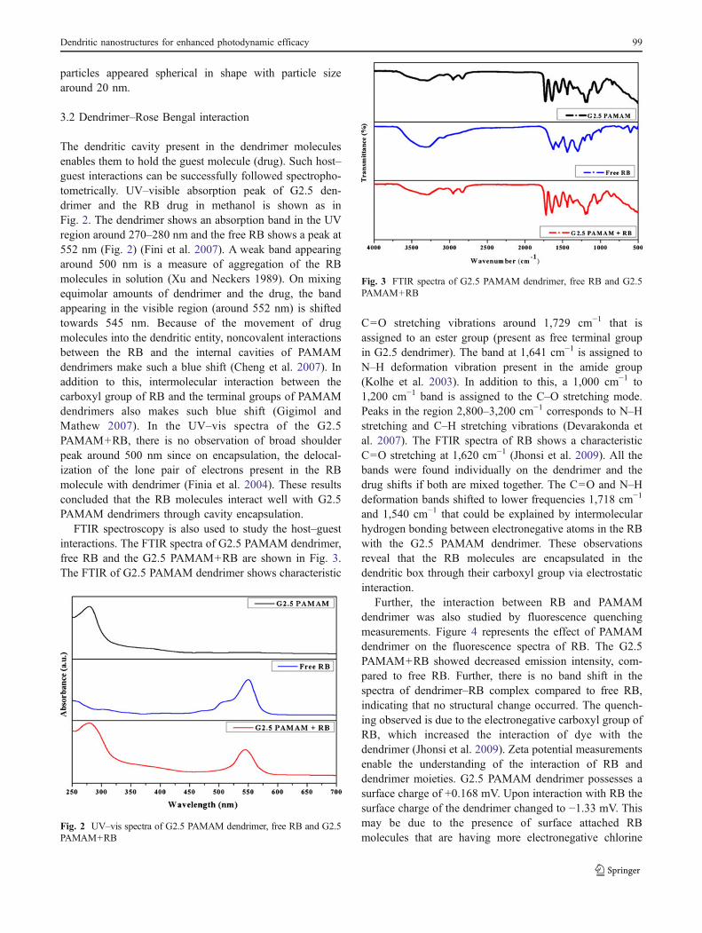
The dendritic cavity present in the dendrimer moleculesenables them to hold the guest molecule (drug). Such host–guest interactions can be successfully followed spectropho-tometrically. UV–visible absorption peak of G2.5 den-drimer and the RB drug in methanol is shown as inFig. 2. The dendrimer shows an absorption band in the UVregion around 270–280 nm and the free RB shows a peak at552 nm (Fig. 2) (Fini et al. 2007). A weak band appearingaround 500 nm is a measure of aggregation of the RBmolecules in solution (Xu and Neckers 1989). On mixingequimolar amounts of dendrimer and the drug, the bandappearing in the visible region (around 552 nm) is shiftedtowards 545 nm. Because of the movement of drugmolecules into the dendritic entity, noncovalent interactionsbetween the RB and the internal cavities of PAMAMdendrimers make such a blue shift (Cheng et al. 2007). Inaddition to this, intermolecular interaction between thecarboxyl group of RB and the terminal groups of PAMAMdendrimers also makes such blue shift (Gigimol andMathew 2007). In the UV–vis spectra of the G2.5PAMAM+RB, there is no observation of broad shoulderpeak around 500 nm since on encapsulation, the delocal-ization of the lone pair of electrons present in the RBmolecule with dendrimer (Finia et al. 2004). These resultsconcluded that the RB molecules interact well with G2.5PAMAM dendrimers through cavity encapsulation.
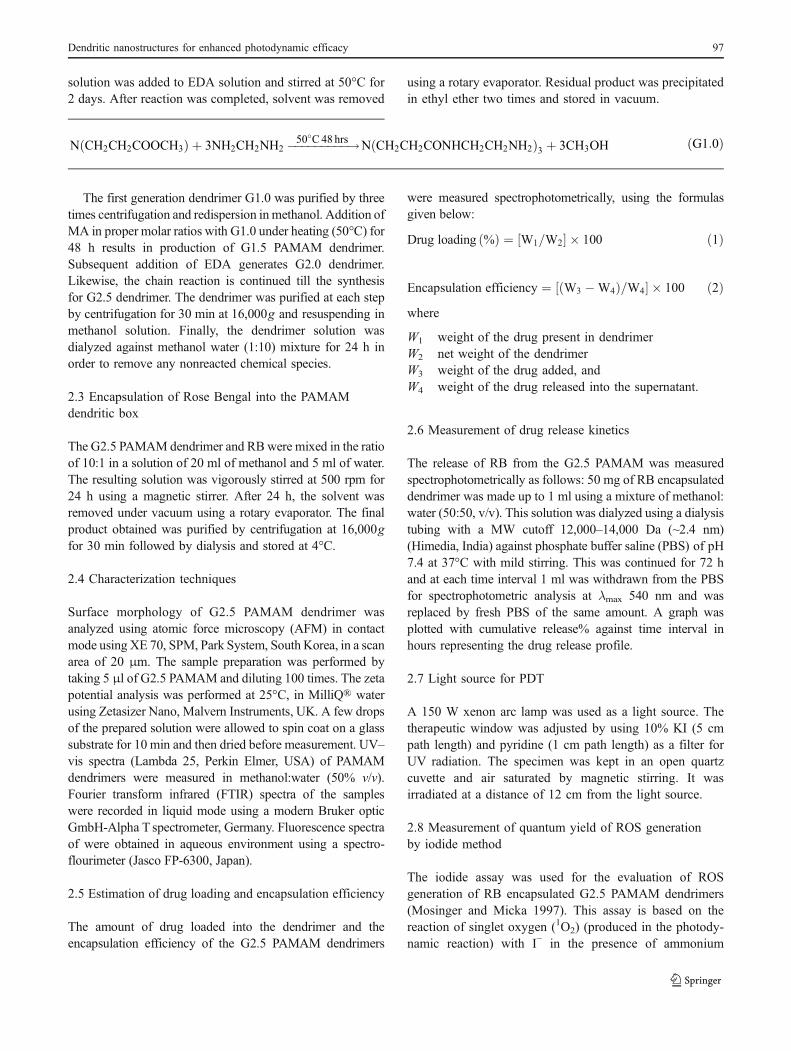
FTIR spectroscopy is also used to study the host–guestinteractions. The FTIR spectra of G2.5 PAMAM dendrimer,free RB and the G2.5 PAMAM+RB are shown in Fig. 3.The FTIR of G2.5 PAMAM dendrimer shows characteristic
C=O stretching vibrations around 1,729 cm−1 that isassigned to an ester group (present as free terminal groupin G2.5 dendrimer). The band at 1,641 cm−1 is assigned toN–H deformation vibration present in the amide group(Kolhe et al. 2003). In addition to this, a 1,000 cm−1 to1,200 cm−1 band is assigned to the C–O stretching mode.Peaks in the region 2,800–3,200 cm−1 corresponds to N–Hstretching and C–H stretching vibrations (Devarakonda etal. 2007). The FTIR spectra of RB shows a characteristicC=O stretching at 1,620 cm−1 (Jhonsi et al. 2009). All thebands were found individually on the dendrimer and thedrug shifts if both are mixed together. The C=O and N–Hdeformation bands shifted to lower frequencies 1,718 cm−1
and 1,540 cm−1 that could be explained by intermolecularhydrogen bonding between electronegative atoms in the RBwith the G2.5 PAMAM dendrimer. These observationsreveal that the RB molecules are encapsulated in thedendritic box through their carboxyl group via electrostaticinteraction.
Further, the interaction between RB and PAMAMdendrimer was also studied by fluorescence quenchingmeasurements. Figure 4 represents the effect of PAMAMdendrimer on the fluorescence spectra of RB. The G2.5PAMAM+RB showed decreased emission intensity, com-pared to free RB. Further, there is no band shift in thespectra of dendrimer–RB complex compared to free RB,indicating that no structural change occurred. The quench-ing observed is due to the electronegative carboxyl group ofRB, which increased the interaction of dye with thedendrimer (Jhonsi et al. 2009). Zeta potential measurementsenable the understanding of the interaction of RB anddendrimer moieties. G2.5 PAMAM dendrimer possesses asurface charge of +0.168 mV. Upon interaction with RB thesurface charge of the dendrimer changed to −1.33 mV. Thismay be due to the presence of surface attached RBmolecules that are having more electronegative chlorine
Fig. 3 FTIR spectra of G2.5 PAMAM dendrimer, free RB and G2.5PAMAM+RB
Fig. 2 UV–vis spectra of G2.5 PAMAM dendrimer, free RB and G2.5PAMAM+RB
Dendritic nanostructures for enhanced photodynamic efficacy 99

atoms and a keto group in the molecule. Hence, the abovecharacterization techniques reveal that the RB moleculesare encapsulated in the dendritic cavity and also some ofthe RB molecules are absorbed at the terminal surface ofthe PAMAM dendrimers.
3.3 Drug loading and encapsulation efficiency of G2.5PAMAM+RB nanocapsules
High drug loading and better encapsulation efficiency isexpected for an ideal drug delivery agent, thereby, reducingthe quantity of the matrix materials for drug administration(Mohanraj and Chen 2006). The drug loading in the G2.5PAMAM+RB is through the absorption technique. Hence,the delivery system should be ideal in case of drug loadingefficiency and drug release kinetics for carrying PDT, notsuppressing the quantum yield of the PS after encapsula-tion. The amount of drug loaded into the dendrimer and theencapsulation efficiency of the G2.5 PAMAM dendrimerswere measured spectrophotometrically during purificationof G2.5 PAMAM+RB by centrifugation and are to beobserved as 1.8% and 92.5% respectively.
3.4 In vitro drug release kinetics of G2.5 PAMAM+RB
In our G2.5 PAMAM+RB, the RB molecules are physicallyencapsulated in the cavity of the PAMAM dendrimer and aminimum quantity is absorbed at the terminal surface. Thepossible drug release kinetics is through diffusion process(Kedar et al. 2010). Figure 5 shows the in vitro drug releaseprofile of the RB encapsulated G2.5 PAMAM dendrimersfollowed spectrophotometrically for a period of 72 h. It isobserved that the systematic release of RB after 12, 24 and48 h are 35%, 50% and 74% respectively. After 72 h, 83%of drug release is noticed. From the graph, it is clear that therate of release of RB from the dendrimer at the initial stage
is high whereas on the final stage it is found as low. Thequicker release in the initial hours is the release of smallamount of RB attached to the surface groups of thedendrimer. The drug release becomes somewhat slower, i.e.,after 12 h is probably due to the encapsulated drug that ispresent in the dendritic cavity or the inner core of PAMAM.Sustained release was noticed after 48 h. The drug releasestudies shows that G2.5 PAMAM dendritic system possessesexcellent controlled release properties suitable for carryingPDT of hydrophilic photosensitizers.
3.5 ROS quantum efficiency of G2.5 PAMAM+RB
The significant factor influencing the PDT efficiency is thequantum yield of ROS generation from the PS. The ROSgeneration of free RB and its encapsulated form wasevaluated by determining the quantum yield by iodidemethod. It is experimentally found that the 1O2 quantumyield for free RB is 0.76. The 1O2 quantum yield for G2.5PAMAM+RB was measured as 0.71 from using the iodidemethod (Mosinger and Micka 1997). Figure 6 depicts thegraphical representation of the change in absorbance of theiodide band (351 nm) against the irradiation time forvariously concentrated solutions of RB encapsulated inG2.5 PAMAM dendrimers. It is clear from Fig. 6. that the
Fig. 5 RB release kinetics from the G2.5 PAMAM+RB
Fig. 4 Fluorescence spectra of free RB and G2.5 PAMAM+RB
Fig. 6 Estimation of quantum yield of ROS generation from thenanocapsules by the iodide method. Graph shows the change inabsorbance of the iodide band (351 nm) against the irradiation time forvariously concentrated solutions of RB encapsulated in G2.5 PAMAMdendrimers
100 K. Karthikeyan et al.
absorption of the iodide band increases with the increase inthe concentration of the G2.5 PAMAM+RB, whichindirectly show the increase in the ROS generation ofG2.5 PAMAM+RB at higher concentration. These resultsstrongly demonstrate that the G2.5 PAMAM can be an idealdrug delivery agent since no such alteration in the ROS activityis noticed when compared to the activity of the free RB.
3.6 Phototoxicity and dark toxicity of G2.5 PAMAM+RB
The aim was to investigate the photosensitizing activity ofG2.5 PAMAM+RB against DLA cells and its efficiency asa potential PDT treatment for cancer. The cell viability ofDLA cells upon photoirradiation as a function of concen-tration of G2.5 PAMAM+RB indicates the photodynamiceffect in vitro. Figure 7 illustrates the photodynamic effecton the DLA cell line, as a function of the photosensitizerconcentration. The free RB produced the lower photody-namic effect when compared to G2.5PAMAM+RB. Thebeneficial effect of the RB loaded dendrimers was mainlyhighlighted at a RB concentration of 510 nM. When thedoses of the free RB and the G2.5 PAMAM+RB areincreased to 510 nM, it was found that the cell viabilitypercentage for G2.5 PAMAM+RB-treated cells was 24.9%when compared to the 38% cell viability for free RB-treatedcells. The phototoxicity results show that the G2.5PAMAM+RB are more toxic to the DLA cells comparedto the toxicity of free RB. PAMAM dendrimer is ahyperbranched molecule and the amount of drug loadingis as low as 1.8%, which ensures the uniform distribution ofthe photosensitizer in the dendritic matrix and this enablessustained drug release kinetics suitable for carrying PDT.
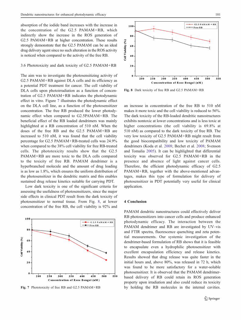
Low dark toxicity is one of the significant criteria forassessing the usefulness of photosensitizers, since the majorside effects in clinical PDT result from the dark toxicity ofphotosensitizer to normal tissue. From Fig. 8, at lowerconcentration of the free RB, the cell viability is 92% and
an increase in concentration of the free RB to 510 nMmakes it more toxic and the cell viability is reduced to 56%.The dark toxicity of the RB-loaded dendritic nanostructuresexhibits nontoxic at lower concentrations and is less toxic athigher concentrations (the cell viability is 69.8% at510 nM) as compared to the dark toxicity of free RB. Thevery low toxicity of G2.5 PAMAM+RB might result fromthe good biocompatibility and low toxicity of PAMAMdendrimers (Koda et al. 2008; Bechet et al. 2008; Svensonand Tomalia 2005). It can be highlighted that differentialtoxicity was observed for G2.5 PAMAM+RB in thepresence and absence of light against cancer cells.Therefore, the efficient photodynamic efficacy of G2.5PAMAM+RB, together with the above-mentioned advan-tages, makes this type of formulation for delivery ofphotosensitizer in PDT potentially very useful for clinicalapplication.
4 Conclusion
PAMAM dendritic nanostructures could effectively deliverRB photosensitizers into cancer cells and produce enhancedphotodynamic efficacy. The interaction between thePAMAM dendrimer and RB are investigated by UV–visand FTIR spectra, fluorescence quenching and zeta poten-tial measurements. Our systemic investigation of thedendrimer-based formulation of RB shows that it is feasibleto encapsulate even a hydrophilic photosensitizer withexcellent encapsulation efficiency and release kinetics.Results showed that drug release was quite faster in theinitial hours and, above 80%, was released in 72 h, whichwas found to be more satisfactory for a water-solublephotosensitizer. It is observed that the PAMAM dendrimer-based delivery of RB could retain its ROS generationproperty upon irradiation and also could reduce its toxicityby holding the RB molecules in the internal cavities.
Fig. 8 Dark toxicity of free RB and G2.5 PAMAM+RB
Fig. 7 Phototoxicity of free RB and G2.5 PAMAM+RB
Dendritic nanostructures for enhanced photodynamic efficacy 101

Importantly, the dendritic formulation exhibited minimizeddark toxicity within the concentration used and enhancedphototoxicity to DLA cells compared to the dark phototoxicityof free RB. The key findings of our work with the above-mentioned advantages ensure that PAMAM dendrimer-basednanocarriers of PS delivery should be a promising candidatefor PDT.
Acknowledgements The authors are thankful to the Managementand the Principal of Mepco Schlenk Engineering College forproviding the necessary facilities to carry out this work. S.-J. Kimacknowledges support from Korea Research Foundation (KRF)project number 2011–0015829.
References
Bechet D, Couleaud P, Frochot C, Viriot ML, Guillemin F, Heyob MB(2008) Nanoparticles as vehicles for delivery of photodynamictherapy agents. Trends Biotechnol 26:612–621
Chatterjee DK, Fong LS, Zhang Y (2008) Nanoparticles in photody-namic therapy: an emerging paradigm. Adv Drug Deliv Rev60:1627–1637
Cheng Y, Qu H, Ma M, Xu Z, Xu P, Fang Y, Xu T (2007)Polyamidoamine (PAMAM) dendrimers as biocompatible carriersof quinolone antimicrobials: an in vitro study. Eur J Med Chem42:1032–1038
Devarakonda B, Otto DP, Judefeind A, Hill RA, Villiers MM (2007)Effect of pH on the solubility and release of furosemide frompolyamidoamine (PAMAM) dendrimer complexes. Int J Pharm345:142–153
Dolmans DEJGJ, Fukumura D, Jain RK (2003) Photodynamic therapyfor cancer. Nat Rev Cancer 3:380–387
Fenga SS, Ruanb G, Li QT (2004) Fabrication and characterizationsof a novel drug delivery device liposomes-in-microsphere (LIM).Biomaterials 25:5181–5189
Fini P, Loseto R, Catucci L, Cosma P, Agostiano A (2007) Study onthe aggregation and electrochemical properties of Rose Bengal inaqueous solution of cyclodextrins. Bioelectrochemistry 70:44–49
Finia P, Longobardi F, Catuccia L, Cosmaa P, Agostiano A (2004)Spectroscopic and electrochemical study of Rose Bengal inaqueous solutions of cyclodextrins. Bioelectrochemistry 63:107–110
Fisher AMR, Mulphree AL, Gomer CJ (1996) Clinical and preclinicalphotodynamic therapy. Lasers Surg Med 17:2–32
Gigimol MG, Mathew B (2007) Effect of the nature and degree ofcross linking on the Rose Bengal uptake by DVB-, NNMBA-,HDODA-, and TTEGDA-cross linked aminopolyacrylamides. JAppl Polym Sci 104:2856–2867
Guo H, Qian H, Idris NM, Zhang Y (2010) Singlet oxygen-inducedapoptosis of cancer cells using upconversion fluorescent nano-particles as a carrier of photosensitizer. Nanomedicine: NBM6:486–495
Hedden RC, Bauer BJ (2003) Structure and dimensions of PAMAM/PEG dendrimer-star polymers. Macromolecules 36:1829–1835
Jansen JF, Berg EMB, Meijer EW (1994) Encapsulation of guestmolecules into a dendritic box. Science 266:1226–1229
Jhonsi MA, Kathiravan A, Renganathan R (2009) Photoinducedinteraction between xanthene dyes and colloidal CdS nano-particles. J Mol Struct 921:279–284
Kedar U, Phutane P, Shidhaye S, Kadam V (2010) Advances inpolymeric micelles for drug delivery and tumor targeting.Nanomedicine: NBM 6:714–729
Kessel D (1981) Transport and Binding of Hematoporphyrin derivativeand related porphyrins by murine leukemia L1210 cells. CancerResearch 41:1318–1323
Killig F, Stark G, Apell HJ (2004) Photodynamic inactivation of theNa, K-ATPase occurs via different pathways. J Membrane Biol200:133–144
Kochevar IE, Lambert CR, Lynch MC, Tedesco AC (1996) Comparisonof photosensitized plasma membrane damage caused by singletoxygen and free radicals. Biochimica et Biophysica Acta 1280:223–230
Koda S, Inoue Y, Iwata H (2008) Gene transfection into adherent cellsusing electroporation on a dendrimer-modified gold electrode.Langmuir 24:13525–13531
Kolhe P, Misra E, Kannan RM, Kannan S, Lai ML (2003) Drugcomplexation, in vitro release and cellular entry of dendrimersand hyperbranched polymers. Int J Pharm 259:143–160
Konan YN, Gurny R, Allemann E (2002) State of the art in thedelivery of photosensitizers for photodynamic therapy. J PhotochemPhotobiol, B Biol 66:89–106
Koo OM, Rubinstein I, Onyuksel H (2005) Role of nanotechnology intargeted drug delivery and imaging: a concise review. Nanomedicine:NBM 1:193–212
Labouebe MZ, Lange N, Gurny R, Delie F (2006) Hypericin-loadednanoparticles for the photodynamic treatment of ovarian cancer.Int J Pharm 3:174–181
Lopez T, Ortiz E, Alvarez M, Navarrete J, Odriozola JA, OrtegaFM, Mozo EP, Escobar P, Espinoza KA, Rivero IA (2010)Study of the stabilization of zinc phthalocyanine in sol–gelTiO2 for photodynamic therapy applications. Nanomedicine:NBM 6:777–785
Macdonald IJ, Dougherty TJ (2001) Basic principles of photodynamictherapy. J Porphyrins Phthalocyanines 5:105–129
Mohanraj VJ, Chen Y (2006) Nanoparticles—a review. Trop J Pharm5:561–573
Moore CM, Pendse D, Emberton M (2009) Photodynamic therapy forprostate cancer—a review of current status and future promise.Nat Clin Prac Urol 6:18–30
Mosinger J, Micka Z (1997) Quantum yields of singlet oxygen ofmetal complexes of meso-tetralds (sulphonatophenyl) porphine. JPhotochem Photobiol, A Chem 107:77–82
Murday JS, Siegel RW, Stein J, Wright JF (2009) Translationalnanomedicine: status assessment and opportunities. Nanomedicine:NBM 5:251–273
Orenstein A, Kostenich G, Roitman L, Shechtman Y, Kopolovic Y,Ehrenberg B, Malik Z (1996) A comparative study of tissuedistribution and photodynamic therapy selectivity of chlorine6,photofrin II and ALA-induced protoporphyrin IX in a coloncarcinoma model. Br J Cancer 73:937–944
Premanathan M, Karthikeyan K, Jeyasubramanian K, Manivannan G(2011) Selective toxicity of ZnO nanoparticles toward Grampositive bacteria and cancer cells by apoptosis through lipidperoxidation. Nanomedicine: NBM 7:184–192
Robertson CA, Evans DH, Abrahamse H (2009) Photodynamictherapy (PDT): a short review on cellular mechanisms andcancer research applications for PDT. J Photochem Photobiol BBiol 96:1–8
Roy I, Ohulchanskyy TY, Pudavar HE, Bergey EJ, Oseroff AR,Morgan J,Dougherty TJ, Prasad PN (2003) Ceramic-based nanoparticlesentrapping water-insoluble photosensitizing anticancer drugs: anovel drug-carrier system for photodynamic therapy. J Am ChemSoc 125:7860–7865
Shive MS, Anderson JM (1997) Biodegradation and biocompatibilityof PLA and PLGA microspheres. Adv Drug Deliv Rev 28:5–24
Silva AR, Inada NM, Rettori D, Baratti MO, Vercesi AE, Jorge RA(2009) In vitro photodynamic activity of chloro(5,10,15,20-tetraphenylporphyrinato) indium(III) loaded-poly(lactide-co-gly-
102 K. Karthikeyan et al.

colide) nanoparticles in LNCaP prostate tumour cells. J Photo-chem Photobiol, B Biol 94:101–112
Svenson S, Tomalia DA (2005) Dendrimers in biomedical applications—reflections on the field. Adv Drug Deliv Rev 57:2106–2129
Tomalia DA, Reyna LA, Svenson S (2007) Dendrimers as multi-purpose nanodevices for oncology drug delivery and diagnosticimaging. Biochem Soc Trans 35:61–67
Vrouenraets MB, Visser GW, Stigter M, Oppelaar H, Snow GB, DongenGA (2002) Comparison of aluminium (III) phthalocyanine tetrasul-fonate and meta-tetrahydroxyphenylchlorin-monoclonal antibodyconjugates for their efficacy in photodynamic therapy in vitro. Int JCancer 98:793–798
Xu D, Neckers DC (1989) Aggregation of Rose Bengal molecules insolution. J Photochem Photobiol, A Chem 47:213–222
Dendritic nanostructures for enhanced photodynamic efficacy 103