Profiling tumor circulating cell-free DNA with an enhanced ...
lable at ScienceDirect
Biomaterials 32 (2011) 8281e8290
Contents lists avai
Biomaterials
journal homepage: www.elsevier .com/locate/biomater ia ls
Enhanced anti-tumor efficacy by co-delivery of doxorubicin and paclitaxelwith amphiphilic methoxy PEG-PLGA copolymer nanoparticles
Hai Wang a, Ying Zhao a, Yan Wu a, Yu-lin Hu b, Kaihui Nan c, Guangjun Nie a,*, Hao Chen c,**
aCAS Key Laboratory for Biological Effects of Nanomaterials & Nanosafety, National Center for Nanoscience and Technology, Beijing 100190, Chinab The First Affiliated Hospital of Jilin University, Changchun, Jilin Province 130021, Chinac Eye Hospital of Wenzhou Medical College, Key Laboratory for Ophthalmology & Optometry of Zhejiang Province, Wenzhou, China
a r t i c l e i n f o
Article history:Received 17 May 2011Accepted 11 July 2011Available online 31 July 2011
Keywords:NanotechnologyCo-delivery of drugsMultifunctional nanoparticlesDouble emulsionControlled drug release
Abbreviations: NPs, nanoparticles; mPEG-PLGA, mpoly(lactide-co-glycolide); DOX, doxorubicin; TAX, pacin-water; FITC, fluorescein isothiocyanate; PVA, popressure liquid chromatography; DLS, dynamic ligtransform infrared spectroscopy; NMR, nuclear magmission electron microscopy; EE, encapsulation effici* Corresponding author. Tel.: þ86 10 82545529; fax** Corresponding author.
E-mail addresses: [email protected] (G. Nie), chenh
0142-9612/$ e see front matter � 2011 Elsevier Ltd.doi:10.1016/j.biomaterials.2011.07.032
a b s t r a c t
The use of single chemotherapeutic drug has shown some limitations in anti-tumor treatment, such asdevelopment of drug resistance, high toxicity and limited regime of clinical uses. The combination of twoor more therapeutic drugs is feasible means to overcome the limitations. Co-delivery strategy has beenproposed to minimize the amount of each drug and to achieve the synergistic effect for cancer therapies.Attempts have been made to deliver chemotherapeutic drugs simultaneously using drug carriers, such asmicelles, liposomes, and inorganic nanoparticles (NPs). Here we reported core-shell NPs that weredoubly emulsified from an amphiphilic copolymer methoxy poly(ethylene glycol)-poly(lactide-co-glycolide) (mPEG-PLGA). These NPs offered advantages over other nanocarriers, as they were easy tofabricate by improved double emulsion method, biocompatible, and showed high loading efficacy. Moreimportantly, these NPs could co-deliver hydrophilic doxorubicin (DOX) and hydrophobic paclitaxel (TAX).The drug-loaded NPs possessed a better polydispersity, indicating that they are more readily subject tocontrolled size distribution. Studies on drug release and cellular uptake of the co-delivery systemdemonstrated that both drugs were effectively taken up by the cells and released simultaneously.Furthermore, the co-delivery nanocarrier suppressed tumor cells growth more efficiently than thedelivery of either DOX or TAX at the same concentrations, indicating a synergistic effect. Moreover, theNPs loading drugs with a DOX/TAX concentration ratio of 2:1 showed the highest anti-tumor activity tothree different types of tumor cells. This nanocarrier might have important potential in clinical impli-cations for co-delivery of multiple anti-tumor drugs with different properties.
� 2011 Elsevier Ltd. All rights reserved.
1. Introduction
Existing chemotherapeutic drugs are far from perfect withundesirable severe side effects, low bioavailability or developmentof drug resistance. Overcoming these limitations requires effectivedelivery of chemotherapeutic drugs to tumor tissues witha minimal amount to other sites. To improve therapeutic efficacyand reduce toxicity and frequency of drug administration, variousdrug delivery systems have developed. Over the past few decades,
ethoxy poly(ethylene glycol)-litaxel; W/O/W, water-in-oil-lyvinyl alcohol; HPLC, Highht scattering; FTIR, fourier-netic resonance; TEM, trans-ency.: þ86 10 62656765.
[email protected] (H. Chen).
All rights reserved.
there has been an increasing interest in the potential use ofcopolymer nanoparticles (NPs) as delivery vehicles for chemo-therapeutic drugs and the studies have demonstrated that thesenanocarriers can significantly enhance the anti-tumor efficiency ofvarious chemotherapeutic drugs [1e3].
Generally, synergistic combination of two or more drugs isa promising strategy to overcome undesirable toxicity and otherside effects that limit the utility of many potential drugs by coun-tering biological compensation, allowing reduced dosage of eachagent or accessing context-specific multiple targets [4e7]. Co-delivery systems, loading different drugs with different physi-ochemical properties simultaneously to the same tumor cellsin vitro and in vivo, have been proposed to minimize the amount ofdrug and to achieve the synergistic therapeutic effect in treatingcancers after a single injection [8,9]. Recently, many multifunc-tional delivery systems have been designed for co-delivery ofdifferent guests, including micelles [10e12], liposomes [13], andinorganic nanoparticles [14]. Although many efforts have been
H. Wang et al. / Biomaterials 32 (2011) 8281e82908282
made on a single carrier for two ormore therapeutic agents, such aschemicals, siRNA, plasmid DNA or peptide [15], co-delivery ofchemotherapeutic drugs with distinct water solubility properties isnot easy to realize. Because of the compartmentalized internalstructure, double emulsion method can make amphiphilic copol-ymers form nanoparticles with the ability to encapsulate both polarand non-polar cargos and can improve the controlled release oftherapeutic molecules [16,17]. Water-in-oil-in-water (W/O/W)emulsion, an example in double emulsion, utilizes a two-stepprocess to form an ‘inverse’ water-in-oil emulsion first, followedby emulsification of this mixture in water using a combination ofsurfactants [18e20].
Doxorubicin (DOX) and paclitaxel (TAX) are among the mostcommon chemotherapeutic drugs in clinical use currently, due totheir excellent anti-tumor efficiency against various solid tumors[21]. They, however, are drugswith distinct solubility characteristicsand different anticancer mechanisms [22e24]. DOX is a hydrophiliccompoundwhich binds to DNA by intercalation and induces a seriesof biochemical events inducing apoptosis in a number of differenttumor cells [25,26]. TAX, a naturally occurring antimitotic agent, isa highly hydrophobic drug, which can inhibit microtubules disas-sembly and promote the formation of unusually stable microtu-bules, thereby disrupting normal dynamic reorganization of themicrotubule network required formitosis and cell proliferation, andin turn causing cell apoptosis [27e31]. Some clinical studies haveshown that incorporation of DOX and TAX increases tumorregression rates relative to the individual drugs [21] and has beenused as first-line treatment for metastatic breast cancer [24].
As the key point for successful combination therapy is to designsimple co-delivery systems. Double emulsion method is an activeprocess to incorporate hydrophilic drugs into amphiphilic copoly-mers nanoparticles. This method generating nanoparticles with“core-shell droplet” morphology [32,33] is important for industrialapplications, such as in the food, cosmetic and pharmaceuticalindustries. However, encapsulation of a drug in a double emulsioncan be achieved only when the drug is soluble in the inner aqueousphase (W) but insoluble in the intermediate oil phase (O) of theemulsion [34]. In this study, we have developed a convenientmethod for improved double emulsion suitable for a large-scaleproduction. Inspired from W/O/W form prepared by double emul-sion method, we utilized the improved double emulsion method toload hydrophobic drugs at the O layer and hydrophilic drugs at thehydrophilic core. We chose mPEG-PLGA as the amphiphilic copol-ymermodel to realize co-delivery and controlled release of DOX andTAX, mainly because PEG and PLGA aremet with FDA’s approval forclinical use as drug adjuvants. Furthermore, this double emulsionmethod also suits other amphiphilic copolymers to form nano-carriers for co-delivering other hydrophilic and hydrophobic drugs.
2. Methods
2.1. Materials
DOX was purchased from Beijing Huafeng United Technology Co. Ltd., and TAXwas obtained from Beijing Norzer Pharmaceutical Co. Ltd.. Poly(lactic-co-glycolic acid)(PLGA, molar ratio of D, L-lactic to glycolic acid, 75:25), monomethoxy poly(ethyleneglycol) (mPEG) was purchased from Jinan Daigang Biotechnology Co. Ltd.. Fluoresceinisothiocyanate (FITC) was obtained from SigmaeAldrich (St. Louis, MO, USA). CellCounting Kit-8 was purchased from Dojindo Molecular Technologies (Tokyo, Japan).Dulbecco’s modified Eagle’s medium (DMEM), RMPI 1640 medium, and fetal bovineserumwere purchased fromGibco BRL (Grand Island, NY, USA). Propidium iodide (PI),Hoechst 33258, penicillin, and streptomycin were provided by SigmaeAldrich (St.Louis, MO, USA). All chemicals used were of analytical reagent quality.
2.2. Synthesis and characterizations of copolymer
Diblock copolymer mPEG-PLGA was synthesized as described in the referencewith minor modifications [35,36]. In brief, PLGA (8 g) and mPEG (2 g) were
copolymerized in freshly distilled toluene (150 mL) using stannous 2-ethylhexanoate (20 mg) as a catalyst at 114 �C for 8 h with stirring at 250 rpm.After vacuum evaporation of the solvent, the residue was dissolved in 200 mL ofDCM, filtered, and then added into vigorously stirred water (1000 mL) at 60 �C. AfterDCM evaporation, the mPEG-PLGA solid was isolated from the aqueous phase bydecantation. Subsequently, fluorescein isothiocyanate (FITC, 20 mg) was conjugatedto mPEG-PLGA (1.5 g) in 30 mL of DMSO at 90 �C for 2 h in darkness. The resultantsolution was dialyzed against distilled water for 3 days to remove DMSO andunreacted FITC, and then lyophilized [37]. Characterization of products wasconfirmed by 1H NMR spectrometer (Bruker AVANCE 400 NMR spectrometer, Bill-erica, MA, USA) with DMSO as the solvent and Fourier-transform infrared spec-trometer (FTIR, Magna FTIR-750, Nicolet, USA). FTIR spectra were obtained froma neat film cast from the chloroform copolymer solution between KBr tablets.
2.3. Preparations of mPEG-PLGA NPs, NPs-DOX, NPs-TAX and NPs-DOX-TAX
mPEG-PLGA NPs and NPs-DOX were prepared by the double emulsion (W/O/W)method with a little modification. Briefly, 20 mg of mPEG-PLGA dissolved in 1 mL ofmethylene chloride and 0.2 mL of water or DOX solution were transferred toa centrifuge tube, and the mixture was emulsified by sonication for 3 min. Then theemulsion and 2 mL of 2% polyvinyl alcohol (PVA) were emulsified by sonication for5 min. The emulsionwas then slowly dropped into 10 mL of 0.6% PVA and stirred for10 min at room temperature. After vacuum evaporation of the solvent, the NPs werecollected by centrifugation at 13,000 rpm for 10 min at room temperature andwashed twice using distilled water.
NPs-DOX-TAX was prepared by the improved double emulsion method. Afterthe first emulsification, the mixture and 2 mL of 2% PVA were stirred for 3 min atroom temperature. Meanwhile, 0.2 mL of TAX-dissolved methylene chloride wasadded slowly, and then emulsified again. The subsequent steps were identical withthe preparation of mPEG-PLGA NPs and NPs-DOX.
Distinctively, NPs-TAX was produced using an emulsion/solvent evaporationtechnique. 20mg of copolymer and 1mg of TAXwere dissolved in 1mL ofmethylenechloride. The solutionwas stirred for 10 min at room temperature and emulsified bysonication in 10 mL of aqueous solution with 1% PVA. After vacuum evaporation ofthe solvent, the NPs were collected by centrifugation at 13,000 rpm for 10 min atroom temperature and washed twice using distilled water.
2.4. Measurements of particle size distribution and zeta potential
The NPs size (diameter, nm), polydispersity index and surface charge (zetapotential, mV) were determined using a ZetaSizer Nano series Nano-ZS (MalvernInstruments Ltd., Malvern, UK) equipped with a HeeNe Laser beam at a wavelengthof 633 nm and a fixed scattering angle of 90� . Determinations were performed at25 �C for samples appropriately diluted in distilled water.
2.5. Morphological characterization
The morphology of NPs-DOX-TAX was confirmed using a transmission electronmicroscopy (TEM, JEM-200CX, Jeol Ltd., Japan) after negative staining with sodiumphosphotungstate solution (2%, w/w).
2.6. Stability and in vitro drug release
Nanoparticles (50mg) were suspended in phosphate-buffered saline at pH 7.4 orpH 4.4, under stirring at 110 rpm/37 �C. The nanoparticles sizes were determined at0, 1 and 2 days to examine the stability of the nanoparticles at different pH values bydynamic light scattering (DLS).
For in vitro drugs release study, drug-loaded nanoparticles (20e30 mg) werereconstituted in PBS (5 mL, pH 4.4 or 7.4) and transferred to dialysis bags (MWCO:3500 Da) placed in 30 mL of PBS with stirring at 110 rpm/37 �C. At appropriateintervals, the environmental buffer solution was replaced with fresh PBS, and theconcentration of the released DOX in the removed PBS was determined usinga calibration curve at the wavelength where DOX showed its maximum absorbance(483.5 nm), quantitatively monitored by Lambda 950 Visible-UV spectrophotometer(PerkineElmer Fremont, CA, USA). Then, the accumulative ratios of the released DOXwere calculated as a function of time.
High performance liquid chromatography (HPLC) was used to determine thereleased amount of TAX [38]. The PBS used for DOX detection in the quartz cell alsoused for TAX detection. The accumulative percentage of the released TAX wascalculated as a function of incubation time. An Agilent 1200 series quaternary pumpand a Rheodyne sample injector with a 20 mL loop, an Agilent 1200 series UVeVisDAD detector, and a chemstation data processing system were used. The mobilephase was water/acetonitrile (1:1 v/v). The HPLC analysis was carried out with anAgilent Zorbax Eclipse XDB C18 reversed-phase column (150 � 4.6 mm, 5 mm,1 mL/min, Agilent Technologies).
Encapsulation efficiency of the NPs was calculated by the following equation.
Encapsulation EfficiencyðEEÞ ¼ AB� 100%
H. Wang et al. / Biomaterials 32 (2011) 8281e8290 8283
where A represents the amount of drug obtained in nanoparticles, and B is the initialamount of drug putted in the system.
in vitro drugs release of nanoparticles was calculated by following equation.
mt�act ¼ Ct þ v
V
Xt�1
0
Ct
!V
where mteact is the actual quality of drugs released at time t, Ct is the drugsconcentration in release fluid at time t measured by visible-UV spectrophotometeror HPLC, n is the sampled volume taken at a predetermined time interval, and V is thetotal volume of release fluid.
2.7. in vitro cytotoxicity
A549 (human lung cancer cell), B16 (mouse melanoma cell) and HepG2 (humanhepatocellular carcinoma cell) cells were maintained onto 96-well plates at 37 �C for24 h in 100 mL of DMEM or RMPI 1640 medium, respectively. Both DMEM and RPMI1640 were supplemented with 10% (v/v) fetal bovine serum. The culturemedia werereplaced with 100 mL of media containing blank NPs, free DOX, free TAX, NPs-DOX,NPs-TAX, and NPs-DOX-TAX. The total drug contents in all the groups were kept at0.25 mmol/mL, and the concentration ratio of DOX to TAX in NPs-DOX-TAX wascalculated by the drug amount used in preparation of NPs-DOX-TAX. After 24 h ofincubation, the amount of viable cells was evaluated via a CCK-8 assay.
2.8. Confocal microscopy
For investigation of cellular uptake, DOX could show red fluorescence by itself,and TAX-FITC was prepared for confocal microscopy observation [37]. A549 cellswere seeded onto a borosilicate chambered cover glass (Nunc, U.S.A.) at a density of2 � 105 cells per well at 37 �C for 24 h. Then, the spent growth media were replacedwith 2 mL of the pre-prepared free DOX, TAX-FITC and NPs-DOX-TAX-FITC solutionsin serum-free media. After 2.5 h of incubation at 37 �C, the cells were washed withPBS three times before image acquisition and observed at 60� magnification usingLSM 510 DUO confocal unit (Carl Zeiss, U.S.A.). Confocal fluorescence microscopeimages were acquired with a Nikon Ti-e microscope equipped with an UltraVIEWVox confocal attachment (PerkineElmer).
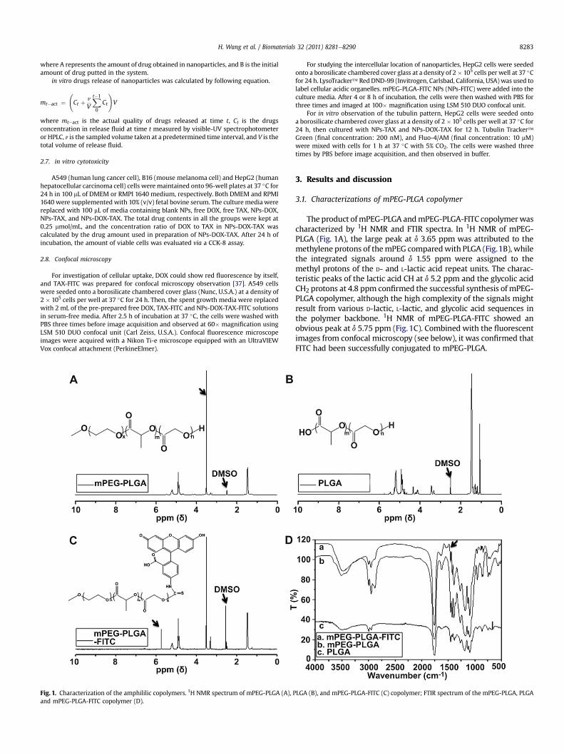
Fig. 1. Characterization of the amphililic copolymers. 1H NMR spectrum of mPEG-PLGA (A), Pand mPEG-PLGA-FITC copolymer (D).
For studying the intercellular location of nanoparticles, HepG2 cells were seededonto a borosilicate chambered cover glass at a density of 2� 105 cells per well at 37 �Cfor 24 h. LysoTracker� Red DND-99 (Invitrogen, Carlsbad, California, USA)was used tolabel cellular acidic organelles. mPEG-PLGA-FITC NPs (NPs-FITC) were added into theculture media. After 4 or 8 h of incubation, the cells were then washed with PBS forthree times and imaged at 100� magnification using LSM 510 DUO confocal unit.
For in vitro observation of the tubulin pattern, HepG2 cells were seeded ontoa borosilicate chambered cover glass at a density of 2� 105 cells per well at 37 �C for24 h, then cultured with NPs-TAX and NPs-DOX-TAX for 12 h. Tubulin Tracker�Green (final concentration: 200 nM), and Fluo-4/AM (final concentration: 10 mM)were mixed with cells for 1 h at 37 �C with 5% CO2. The cells were washed threetimes by PBS before image acquisition, and then observed in buffer.
3. Results and discussion
3.1. Characterizations of mPEG-PLGA copolymer
The product ofmPEG-PLGA andmPEG-PLGA-FITC copolymerwascharacterized by 1H NMR and FTIR spectra. In 1H NMR of mPEG-PLGA (Fig. 1A), the large peak at d 3.65 ppm was attributed to themethylene protons of themPEG comparedwith PLGA (Fig.1B), whilethe integrated signals around d 1.55 ppm were assigned to themethyl protons of the D- and L-lactic acid repeat units. The charac-teristic peaks of the lactic acid CH at d 5.2 ppm and the glycolic acidCH2 protons at 4.8 ppm confirmed the successful synthesis of mPEG-PLGA copolymer, although the high complexity of the signals mightresult from various D-lactic, L-lactic, and glycolic acid sequences inthe polymer backbone. 1H NMR of mPEG-PLGA-FITC showed anobvious peak at d 5.75 ppm (Fig. 1C). Combined with the fluorescentimages from confocal microscopy (see below), it was confirmed thatFITC had been successfully conjugated to mPEG-PLGA.
LGA (B), and mPEG-PLGA-FITC (C) copolymer; FTIR spectrum of the mPEG-PLGA, PLGA

Scheme 1. Illustration of biodegradable amphiphilic copolymer NPs loaded with both DOX and TAX using improved double emulsion method. Emulsification procedure used togenerate double emulsions. Step (I), generating water-in-oil for encapsulations of DOX; Step (II), generating water-in-oil-in-water for encapsulations of TAX. Green represents the oilphase containing amphiphilic copolymer and red the aqueous phase containing DOX.
Table 1Characterization of mPEG-PLGA amphiphilic copolymers and drug-loaded NPs.
Groups Particle size (nm)a Polydispersity Zeta potential (mV)a
H. Wang et al. / Biomaterials 32 (2011) 8281e82908284
FTIR spectra further testified the construct of mPEG-PLGA andFITC connected copolymer (Fig. 1D). The absorption band at3509.9 cm�1 was assigned to terminal hydroxy groups in thecopolymer. The bands at 3010 and 2955 cm�1 were CeH stretch ofeCH2, and the peak at 2885 cm�1 was CeH stretch of eCHe. Astrong band at 1762.6 cm�1 was attributed to C]O stretch, and theabsorption at 1186e1089.6 cm�1 was assigned to CeO stretch. TheFTIR spectra of mPEG-PLGA and PLGA had similar characteristicpeaks, since they basically had same functional groups. However,mPEG-PLGA-FITC showed two particular bands at 1578 and1483 cm�1, corresponding to the benzene ring of FITC.
mPEG-PLGA NPs 226.83 � 10.72 0.190 �25.0 � 0.50NPs-DOX 229.67 � 13.27 0.146 �23.2 � 0.27NPs-DOX-TAX 243.63 � 12.36 0.131 �21.6 � 0.14
a Determined by dynamic light scattering (DLS). NPs were prepared by directlydissolving copolymers or drug-loaded NPs in distilled water at a concentration of0.5 mg/mL followed by 10 min sonication. Results means � SD (n ¼ 3).
3.2. Preparation of DOX and TAX loaded mPEG-PLGA NPs
Synergistic combination of two or more drugs attracts more andmore attentions as a promising strategy to overcome the
undesirable toxicity and other side effects that limit the utility ofmany potential drugs [1]. Therefore, it is significant to find a co-delivery system which could load two or more drugs in samecarrier and transport them to the same cancer cell simultaneously.Especially, co-delivery of drugs with different water solubility
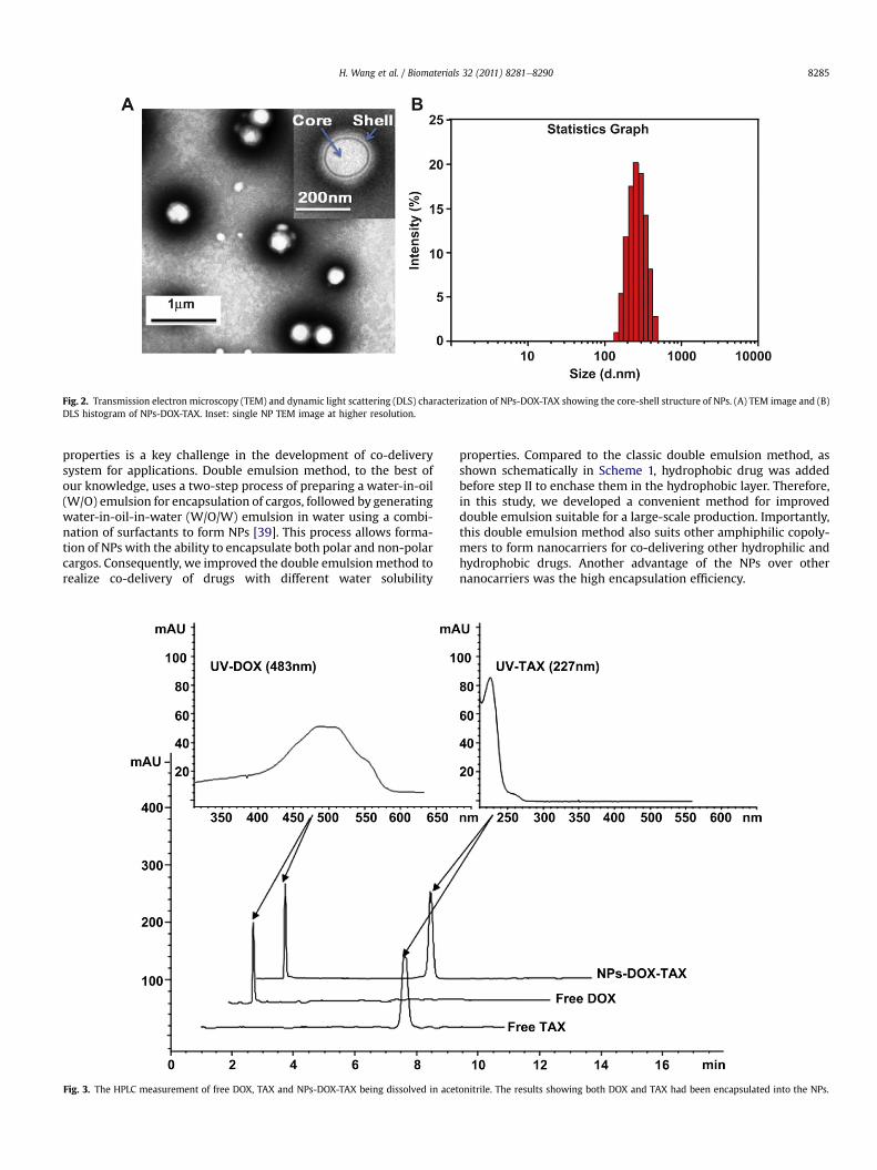
Fig. 2. Transmission electron microscopy (TEM) and dynamic light scattering (DLS) characterization of NPs-DOX-TAX showing the core-shell structure of NPs. (A) TEM image and (B)DLS histogram of NPs-DOX-TAX. Inset: single NP TEM image at higher resolution.
H. Wang et al. / Biomaterials 32 (2011) 8281e8290 8285
properties is a key challenge in the development of co-deliverysystem for applications. Double emulsion method, to the best ofour knowledge, uses a two-step process of preparing a water-in-oil(W/O) emulsion for encapsulation of cargos, followed by generatingwater-in-oil-in-water (W/O/W) emulsion in water using a combi-nation of surfactants to form NPs [39]. This process allows forma-tion of NPs with the ability to encapsulate both polar and non-polarcargos. Consequently, we improved the double emulsion method torealize co-delivery of drugs with different water solubility
Fig. 3. The HPLC measurement of free DOX, TAX and NPs-DOX-TAX being dissolved in acet
properties. Compared to the classic double emulsion method, asshown schematically in Scheme 1, hydrophobic drug was addedbefore step II to enchase them in the hydrophobic layer. Therefore,in this study, we developed a convenient method for improveddouble emulsion suitable for a large-scale production. Importantly,this double emulsion method also suits other amphiphilic copoly-mers to form nanocarriers for co-delivering other hydrophilic andhydrophobic drugs. Another advantage of the NPs over othernanocarriers was the high encapsulation efficiency.
onitrile. The results showing both DOX and TAX had been encapsulated into the NPs.
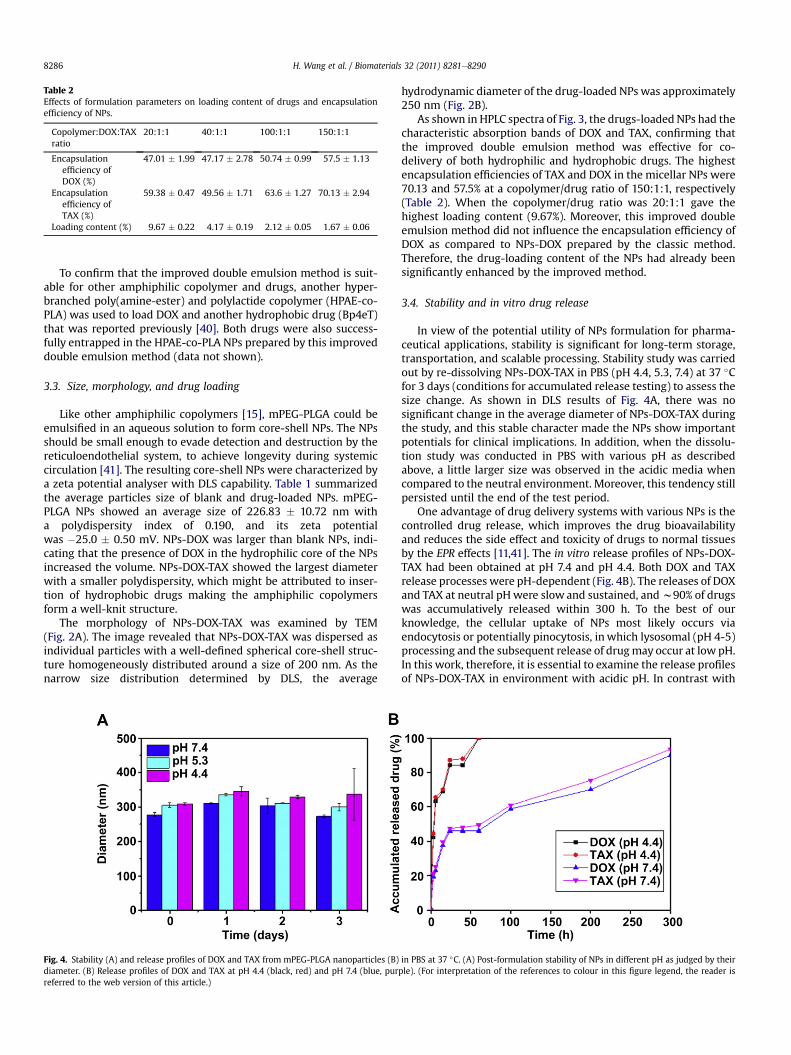
Table 2Effects of formulation parameters on loading content of drugs and encapsulationefficiency of NPs.
Copolymer:DOX:TAXratio
20:1:1 40:1:1 100:1:1 150:1:1
Encapsulationefficiency ofDOX (%)
47.01 � 1.99 47.17 � 2.78 50.74 � 0.99 57.5 � 1.13
Encapsulationefficiency ofTAX (%)
59.38 � 0.47 49.56 � 1.71 63.6 � 1.27 70.13 � 2.94
Loading content (%) 9.67 � 0.22 4.17 � 0.19 2.12 � 0.05 1.67 � 0.06
H. Wang et al. / Biomaterials 32 (2011) 8281e82908286
To confirm that the improved double emulsion method is suit-able for other amphiphilic copolymer and drugs, another hyper-branched poly(amine-ester) and polylactide copolymer (HPAE-co-PLA) was used to load DOX and another hydrophobic drug (Bp4eT)that was reported previously [40]. Both drugs were also success-fully entrapped in the HPAE-co-PLA NPs prepared by this improveddouble emulsion method (data not shown).
3.3. Size, morphology, and drug loading
Like other amphiphilic copolymers [15], mPEG-PLGA could beemulsified in an aqueous solution to form core-shell NPs. The NPsshould be small enough to evade detection and destruction by thereticuloendothelial system, to achieve longevity during systemiccirculation [41]. The resulting core-shell NPs were characterized bya zeta potential analyser with DLS capability. Table 1 summarizedthe average particles size of blank and drug-loaded NPs. mPEG-PLGA NPs showed an average size of 226.83 � 10.72 nm witha polydispersity index of 0.190, and its zeta potentialwas �25.0 � 0.50 mV. NPs-DOX was larger than blank NPs, indi-cating that the presence of DOX in the hydrophilic core of the NPsincreased the volume. NPs-DOX-TAX showed the largest diameterwith a smaller polydispersity, which might be attributed to inser-tion of hydrophobic drugs making the amphiphilic copolymersform a well-knit structure.
The morphology of NPs-DOX-TAX was examined by TEM(Fig. 2A). The image revealed that NPs-DOX-TAX was dispersed asindividual particles with a well-defined spherical core-shell struc-ture homogeneously distributed around a size of 200 nm. As thenarrow size distribution determined by DLS, the average
Fig. 4. Stability (A) and release profiles of DOX and TAX from mPEG-PLGA nanoparticles (B)diameter. (B) Release profiles of DOX and TAX at pH 4.4 (black, red) and pH 7.4 (blue, purreferred to the web version of this article.)
hydrodynamic diameter of the drug-loaded NPs was approximately250 nm (Fig. 2B).
As shown in HPLC spectra of Fig. 3, the drugs-loaded NPs had thecharacteristic absorption bands of DOX and TAX, confirming thatthe improved double emulsion method was effective for co-delivery of both hydrophilic and hydrophobic drugs. The highestencapsulation efficiencies of TAX and DOX in the micellar NPs were70.13 and 57.5% at a copolymer/drug ratio of 150:1:1, respectively(Table 2). When the copolymer/drug ratio was 20:1:1 gave thehighest loading content (9.67%). Moreover, this improved doubleemulsion method did not influence the encapsulation efficiency ofDOX as compared to NPs-DOX prepared by the classic method.Therefore, the drug-loading content of the NPs had already beensignificantly enhanced by the improved method.
3.4. Stability and in vitro drug release
In view of the potential utility of NPs formulation for pharma-ceutical applications, stability is significant for long-term storage,transportation, and scalable processing. Stability study was carriedout by re-dissolving NPs-DOX-TAX in PBS (pH 4.4, 5.3, 7.4) at 37 �Cfor 3 days (conditions for accumulated release testing) to assess thesize change. As shown in DLS results of Fig. 4A, there was nosignificant change in the average diameter of NPs-DOX-TAX duringthe study, and this stable character made the NPs show importantpotentials for clinical implications. In addition, when the dissolu-tion study was conducted in PBS with various pH as describedabove, a little larger size was observed in the acidic media whencompared to the neutral environment. Moreover, this tendency stillpersisted until the end of the test period.
One advantage of drug delivery systems with various NPs is thecontrolled drug release, which improves the drug bioavailabilityand reduces the side effect and toxicity of drugs to normal tissuesby the EPR effects [11,41]. The in vitro release profiles of NPs-DOX-TAX had been obtained at pH 7.4 and pH 4.4. Both DOX and TAXrelease processes were pH-dependent (Fig. 4B). The releases of DOXand TAX at neutral pH were slow and sustained, andw90% of drugswas accumulatively released within 300 h. To the best of ourknowledge, the cellular uptake of NPs most likely occurs viaendocytosis or potentially pinocytosis, in which lysosomal (pH 4-5)processing and the subsequent release of drugmay occur at low pH.In this work, therefore, it is essential to examine the release profilesof NPs-DOX-TAX in environment with acidic pH. In contrast with
in PBS at 37 �C. (A) Post-formulation stability of NPs in different pH as judged by theirple). (For interpretation of the references to colour in this figure legend, the reader is

Fig. 5. Cell viability measurements. Cell viability of A549, HepG2, and B16 cells after exposure to free DOX, TAX, DOX&TAX, NPs-DOX, NPs-TAX and NPs-DOX-TAX with differentratios at 37 �C for 24 h. The total drug contents in all groups were kept at 0.25 mmol/mL for all tests. *p < 0.05 compared with DOX group.
H. Wang et al. / Biomaterials 32 (2011) 8281e8290 8287
the release profiles at pH 7.4, the drug releases were much faster inthe simulated acidic environment in the lysosome, with close to90% of the total drug content being released within the first 48 h. Inaddition, the similar release profiles obtained from both DOX andTAX indicated that the co-delivery system provided a possibility oftheir synergistic effect. Theoretically speaking, TAX was released atfirst when the outermost copolymer layer broke off, after that, theunstable core structure with hydrophobic surface disassembledquickly, accompanied by the release of DOX.
3.5. in vitro cytotoxicity
Here, DOX and TAX were used as a pair to demonstrate thesynergistic effect of co-delivery of two chemotherapeutical drugs inthe same vehicle. A549, HepG2 and B16 cells were first separatelyincubated in 96-well plates. After 24 h, the cells were treated for24 hwith the free DOX, free TAX, NPs-DOX, NPs-TAX, and NPs-DOX-TAX containing 0.25 mmol/mL of drug concentration. As shown in
Fig. 5, co-delivery of DOX and TAX significantly reduced cellviability. Free single drug and single drug-loaded NPs inducedsimilar cytotoxicity, demonstrating that the triggering mechanismfor the release of the drug from the endosomes/lysosomes into thecytosols is highly efficient. Furthermore, the role of drug ratio in theinhibition of cancer cells was also evaluated. As reflected by thecorresponding viability value for HepG2 cells, cell viability of NPs-DOX-TAX with the DOX/TAX concentration ratio of 2:1 were morethan seven times lower compared to the sample with a ratio of 1:4.Moreover, the NPs with a DOX/TAX concentration ratio of 2:1showed the highest anti-tumor activity to all the three differenttypes of tumor cells.
It was suggested that the synergistic effect might result from thecombination of individual anti-tumor mechanism for each drug. Asmentioned above, DOX binds to DNA by intercalation andmotivatesa series of biochemical events inducing apoptosis in tumor cells,and TAX can inhibit microtubules disassembly and promote theformation of unusually stable microtubules, thereby disrupting

Fig. 6. Confocal laser scanning microscopy images of free DOX, TAX-FITC and NPs-DOX-TAX-FITC. Cellular uptake of DOX and TAX-FITC delivered by the NPs was studied in A549, thecells were cultured on 35 mm glass bottom dishes for 24 h, followed by incubation with free drugs or NPs loaded with both drugs for 2.5 h at 37 �C.
H. Wang et al. / Biomaterials 32 (2011) 8281e82908288
normal dynamic reorganization of the microtubule networkrequired for mitosis and cell proliferation, and in turn causing cellapoptosis [13]. Treating cancer cells using DOX and TAX togetherwith a ratio of 2:1 might achieve the best synergistic effects andaccelerate tumor cell death. According to our in vitro studies, whenusing other drug ratios (1:4 or 4:1), the synergistic effects could notdisplay efficiently, and balanced dosage of the two individual drugsgave the highest anti-tumor efficacy.
Fig. 7. Observation of cellular uptake of NPs and their intracellular distribution in HepG2incubation with LysoTracker Red for 45 min at 37 �C and subsequently examined by confocalOnly small amount of NPs had entered into lysosomes (second row). While for 8 h, most oreferences to colour in this figure legend, the reader is referred to the web version of this
3.6. Cellular uptake of nanoparticles
To further verify that DOX and TAX could be simultaneouslydelivered into the same cells and distributed closely, a confocalmicroscopic study was conducted. Fig. 6 showed fluorescencemicroscopy photographs of A549 cells treated with free DOX, freeTAX-FITC, and NPs-DOX-TAX-FITC. Upon releases of two drugs fromthe NPs into the endosomal/lysosomal lumen, the red and green
cells. The cells were cultured on 35 mm glass bottom dishes for 24 h, followed bymicroscopy. After incubated with NPs-FITC for 4 h, many NPs had phagocytize by cells.f lysosomes could emit both red and green light (third row). (For interpretation of thearticle.)
H. Wang et al. / Biomaterials 32 (2011) 8281e8290 8289
fluorescence signals from DOX and TAX-FITC respectively in cyto-plasm were expected to accumulate and overlapped significantly,indicating that the two drugs could be released simultaneously.Nuclei were expected to turn red because of DOX binding to DNA.However, the cells treated with free TAX-FITC remained with littlegreen florescence, since FITC could not be internalized by tumorcells. In aword, this nanocarrier could efficiently deliver and releaseboth hydrophilic and hydrophobic drugs in the same tumor cells.
3.7. Intracellular localization of nanoparticles
After uptake of NPs by cells, the next important question is theirintracellular location and translocation, which is directly related tothe cytotoxicity or medical functions of the internalized NPs.HepG2 cells treated with empty mPEG-PLGA-FITC NPs wereobserved by a confocal microscopy to show the intracellularlocalizations of LysoTracker Red (tracking lysosomes) [42] and FITC(tracking NPs). After 4 and 8 h of incubation with empty NPs, thegreen fluorescence of NPs was observed (Fig. 7). Increasing theincubation timewould increase the fluorescent intensity, indicatingthat the NPs could be internalized continuously. From the images of8 h, the red and green fluorescence signals were observed tooverlap significantly, suggesting that the internalized NPs weretranslocated via lysosomal pathway after entering cells. Lysosomescontain varying amounts of hydrolases and hence lead to the rapiddestruction and degradation of the internalized NPs, resulting in
Fig. 8. Confocal laser scanning microscopy images and western blot of tubulin in HepG2 cell24 h. After cultured with NPs and NPs-DOX-TAX for 12 h, the cells were followed by incubconfocal microscopy. For western blot (B), HepG2 cells were cultured on 10 cm dishes fortrophoresed and then transferred onto immobilon-p membrane. Membrane was incubatesecondary antibody (1:3000). (For interpretation of the references to colour in this figure l
a burst release of drugs from NPs-DOX-TAX under acidic environ-ment with the lysosomal pH (Fig. 5B).
3.8. Possible mechanism for synergistic therapeutic effect
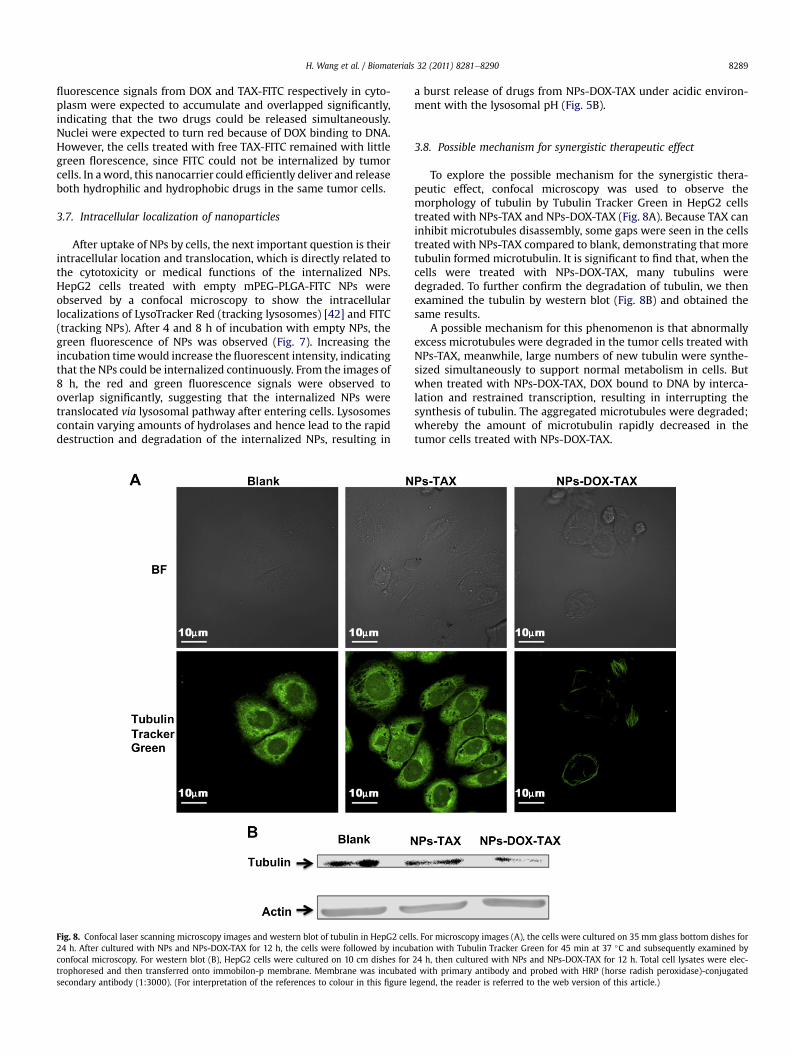
To explore the possible mechanism for the synergistic thera-peutic effect, confocal microscopy was used to observe themorphology of tubulin by Tubulin Tracker Green in HepG2 cellstreated with NPs-TAX and NPs-DOX-TAX (Fig. 8A). Because TAX caninhibit microtubules disassembly, some gaps were seen in the cellstreated with NPs-TAX compared to blank, demonstrating that moretubulin formed microtubulin. It is significant to find that, when thecells were treated with NPs-DOX-TAX, many tubulins weredegraded. To further confirm the degradation of tubulin, we thenexamined the tubulin by western blot (Fig. 8B) and obtained thesame results.
A possible mechanism for this phenomenon is that abnormallyexcess microtubules were degraded in the tumor cells treated withNPs-TAX, meanwhile, large numbers of new tubulin were synthe-sized simultaneously to support normal metabolism in cells. Butwhen treated with NPs-DOX-TAX, DOX bound to DNA by interca-lation and restrained transcription, resulting in interrupting thesynthesis of tubulin. The aggregated microtubules were degraded;whereby the amount of microtubulin rapidly decreased in thetumor cells treated with NPs-DOX-TAX.
s. For microscopy images (A), the cells were cultured on 35 mm glass bottom dishes foration with Tubulin Tracker Green for 45 min at 37 �C and subsequently examined by24 h, then cultured with NPs and NPs-DOX-TAX for 12 h. Total cell lysates were elec-d with primary antibody and probed with HRP (horse radish peroxidase)-conjugatedegend, the reader is referred to the web version of this article.)

H. Wang et al. / Biomaterials 32 (2011) 8281e82908290
4. Conclusion
In summary, core-shell NPs prepared by an improved doubleemulsion method from copolymer mPEG-PLGA were employed ascarriers to co-deliver DOX and TAX for pH responsive drug releasein tumor therapy. The releases of DOX and TAX at neutral pH wereslow and sustained, with about 90% of the total drug content beingreleased within 300 h; however, the drug releases were muchfaster at acidic environment with nearly 100% of the total drugcontent being released within the first 48 h. Studies on drug releaseand cellular uptake of the co-delivery system demonstrated thatboth drugs were effectively taken up by the cells and releasedsimultaneously. Furthermore, the co-delivery nanocarriersuppresses tumor cells growth more efficiently than the delivery ofeither DOX or TAX at the same concentrations, indicating a syner-gistic effect. The NPs loading drugs with a DOX/TAX concentrationratio of 2:1 showed the highest anti-tumor activity to threedifferent types of tumor cells. Moreover, we also suggesteda possible mechanism enhancing the tumor regression rate for thesynergistic therapeutic effect of DOX and TAX.
Acknowledgments
This work was supported by grants from MOST 973 program(2011CB9334000, 2010CB934004, 2010CB933600),NSFC (10979011;30900278) and MOST 863 program (2007AA021807). GN acknowl-edges the support of Chinese Academy of Sciences, Hundred TalentsProgram.
References
[1] Gao Y, Chen L, Gu W, Xi Y, Lin L, Li Y. Targeted nanoassembly loaded withdocetaxel improves intracellular drug delivery and efficacy in murine breastcancer model. Mol Pharm 2008;5:1044e54.
[2] Tang N, Du G, Wang N, Liu C, Hang H, Liang W. Improving penetration intumors with nanoassemblies of phospholipids and doxorubicin. J Natl CancerInst 2007;99:1004e15.
[3] Wang J, Mongayt D, Torchilin VP. Polymeric micelles for delivery of poorlysoluble drugs: preparation and anticancer activity in vitro of paclitaxelincorporated into mixed micelles based on poly(ethylene glycol)-lipidconjugate and positively charged lipids. J Drug Target 2005;13:73e80.
[4] Lehar J, Krueger AS, Avery W, Heilbut AM, Johansen LM, Price ER, et al.Synergistic drug combinations tend to improve therapeutically relevantselectivity. Nat Biotechnol 2009;27:659e66.
[5] Sharom JR, Bellows DS, Tyers M. From large networks to small molecules. CurrOpin Chem Biol 2004;8:81e90.
[6] Kaelin Jr WG. The concept of synthetic lethality in the context of anticancertherapy. Nat Rev Cancer 2005;5:689e98.
[7] Keith CT, Borisy AA, Stockwell BR. Multicomponent therapeutics for net-worked systems. Nat Rev Drug Discov 2005;4:71e8.
[8] Xiao W, Chen X, Yang L, Mao Y, Wei Y, Chen L. Co-delivery of doxorubicin andplasmid by a novel FGFR-mediated cationic liposome. Int J Pharm 2010;393:119e26.
[9] Wiradharma N, Tong YW, Yang YY. Self-assembled oligopeptide nano-structures for co-delivery of drug and gene with synergistic therapeutic effect.Biomaterials 2009;30:3100e9.
[10] Lee AL, Wang Y, Cheng HY, Pervaiz S, Yang YY. The co-delivery of paclitaxel andHerceptin using cationic micellar nanoparticles. Biomaterials 2009;30:919e27.
[11] Wang H, Zhao P, Su W, Wang S, Liao Z, Niu R, et al. PLGA/polymeric liposomefor targeted drug and gene co-delivery. Biomaterials 2010;31:8741e8.
[12] Fan L, Li F, Zhang H, Wang Y, Cheng C, Li X, et al. Co-delivery of PDTC anddoxorubicin by multifunctional micellar nanoparticles to achieve active tar-geted drug delivery and overcome multidrug resistance. Biomaterials 2010;31:5634e42.
[13] Saad M, Garbuzenko OB, Minko T. Co-delivery of siRNA and an anticancer drugfor treatment of multidrug-resistant cancer. Nanomedicine 2008;3:761e76.
[14] Chen AM, Zhang M, Wei D, Stueber D, Taratula O, Minko T, et al. Co-delivery ofdoxorubicin and Bcl-2 siRNA by mesoporous silica nanoparticles enhances theefficacy of chemotherapy in multidrug-resistant cancer cells. Small 2009;5:2673e7.
[15] Wang Y, Gao S, Ye WH, Yoon HS, Yang YY. Co-delivery of drugs and DNA fromcationic core-shell nanoparticles self-assembled from a biodegradablecopolymer. Nat Mater 2006;5:791e6.
[16] Pays K, Giermanska-Kahn J, Pouligny B, Bibette J, Leal-Calderon F. Doubleemulsions: how does release occur? J Control Release 2002;79:193e205.
[17] Okochi H, Nakano M. Preparation and evaluation of W/O/W type emulsionscontaining vancomycin. Adv Drug Deliv Rev 2000;45:5e26.
[18] Yafei W, Tao Z, Gang H. Structural evolution of polymer-stabilized doubleemulsions. Langmuir 2006;22:67e73.
[19] Goubault C, Pays K, Olea D, Gorria P, Bibette J, Schmitt V, et al. Shear rupturingof complex fluids: application to the preparation of quasi-monodispersewater-in-oil-in-water double emulsions. Langmuir 2001;17:5184e8.
[20] Okushima S, Nisisako T, Torii T, Higuchi T. Controlled production of mono-disperse double emulsions by two-step droplet breakup in microfluidicdevices. Langmuir 2004;20:9905e8.
[21] Ahmed F, Pakunlu RI, Brannan A, Bates F, Minko T, Discher DE. Biodegradablepolymersomes loaded with both paclitaxel and doxorubicin permeate andshrink tumors, inducing apoptosis in proportion to accumulated drug.J Control Release 2006;116:150e8.
[22] Tanaka T, Shiramoto S, Miyashita M, Fujishima Y, Kaneo Y. Tumor targetingbased on the effect of enhanced permeability and retention (EPR) and themechanism of receptor-mediated endocytosis (RME). Int J Pharm 2004;277:39e61.
[23] Han L, Huang R, Li J, Liu S, Huang S, Jiang C. Plasmid pORF-hTRAIL anddoxorubicin co-delivery targeting to tumor using peptide-conjugated poly-amidoamine dendrimer. Biomaterials 2011;32:1242e52.
[24] Gustafson DL, Merz AL, Long ME. Pharmacokinetics of combined doxorubicinand paclitaxel in mice. Cancer Lett 2005;220:161e9.
[25] Barry MA, Behnke CA, Eastman A. Activation of programmed cell death(apoptosis) by cisplatin, other anticancer drugs, toxins and hyperthermia.Biochem Pharmacol 1990;40:2353e62.
[26] Lee S, Baek M, Kim HY, Ha JH, Jeoung DI. Mechanism of doxorubicin-inducedcell death and expression profile analysis. Biotechnol Lett 2002;24:1147e51.
[27] Wani MC, Taylor HL, Wall ME, Coggon P, McPhail AT. Plant antitumor agents.VI. Isolation and structure of taxol, a novel antileukemic and antitumor agentfrom Taxus brevifolia. J Am Chem Soc 1971;93:2325e7.
[28] Holmes FA, Walters RS, Theriault RL, Forman AD, Newton LK, Raber MN, et al.Phase II trial of taxol, an active drug in the treatment of metastatic breastcancer. J Natl Cancer Inst 1991;83:1797e805.
[29] Tishler RB, Geard CR, Hall EJ, Schiff PB. Taxol sensitizes human astrocytomacells to radiation. Cancer Res 1992;52:3495e7.
[30] Liu Y, Bhalla K, Hill C, Priest DG. Evidence for involvement of tyrosine phos-phorylation in taxol-induced apoptosis in a human ovarian tumor cell line.Biochem Pharmacol 1994;48:1265e72.
[31] Huang Y, Johnson KR, Norris JS, Fan W. Nuclear factor-kappaB/IkappaBsignaling pathway may contribute to the mediation of paclitaxel-inducedapoptosis in solid tumor cells. Cancer Res 2000;60:4426e32.
[32] Pal R. Dielectric behavior of double emulsions with “core-shell droplet”morphology. J Colloid Interface Sci 2008;325:500e7.
[33] van der Graaf S, Schroen C, Boom R. Preparation of double emulsions bymembrane emulsification e A review. J Membr Sci 2005;251:7e15.
[34] Regan JO, Mulvihill DM. Water soluble inner aqueous phase markers asindicators of the encapsulation properties of water-in-oil-in-water emulsionsstabilized with sodium caseinate. Food Hydrocolloids 2009;23:2339e45.
[35] Bazile D, Prud’homme C, Bassoullet MT, Marlard M, Spenlehauer G, Veillard M,et al. PEG-PLA nanoparticles avoid uptake by the mononuclear phagocytessystem. J Pharm Sci 1995;84:493e8.
[36] Li YP, Pei YY, Zhou ZH, Zhang XY, Gu ZH, Ding J, et al. PEGylated poly-cyanoacrylate nanoparticles as tumor necrosis factor-alpha carriers. J ControlRelease 2001;71:287e96.
[37] Wu DQ, Lu B, Chang C, Chen CS, Wang T, Zhang YY, et al. Galactosylatedfluorescent labeled micelles as a liver targeting drug carrier. Biomaterials2009;30:1363e71.
[38] Qi H, Hu P, Xu J, Wang A. Encapsulation of drug reservoirs in fibers byemulsion electrospinning: morphology characterization and preliminaryrelease assessment. Biomacromolecules 2006;7:2327e30.
[39] Hanson JA, Chang CB, Graves SM, Li Z, Mason TG, Deming TJ. Nanoscale doubleemulsions stabilized by single-component block copolypeptides. Nature2008;455:85e8.
[40] Miao Q, Xu D, Wang Z, Xu L, Wang T, Wu Y, et al. Amphiphilic hyper-branchedco-polymer nanoparticles for the controlled delivery of anti-tumor agents.Biomaterials 2010;31:7364e75.
[41] Santander-Ortega MJ, Csaba N, González L, Bastos-González D, Ortega-Vinuesa JL, Alonso MJ. Protein-loaded PLGA-PEO blend nanoparticles:encapsulation, release and degradation characteristics. Colloid Polym Sci2010;288:141e50.
[42] Yuan XM, Li W, Dalen H, Lotem J, Kama R, Sachs L, et al. Lysosomal desta-bilization in p53-induced apoptosis. Proc Natl Acad Sci U S A 2002;99:6286e91.