Engineered catalytic biofilms: Site- specific enzyme ...
36
Engineered catalytic biofilms: Site-specific enzyme immobilization onto E. coli curli nanofibers Citation Botyanszki, Zsofia, Pei Kun R. Tay, Peter Q. Nguyen, Martin G. Nussbaumer, and Neel S. Joshi. 2015. “ Engineered Catalytic Biofilms: Site-Specific Enzyme Immobilization onto E. Coli Curli Nanofibers .” Biotechnology and Bioengineering 112 (10) (May 20): 2016–2024. doi:10.1002/ bit.25638. Published Version doi:10.1002/bit.25638 Permanent link http://nrs.harvard.edu/urn-3:HUL.InstRepos:27708817 Terms of Use This article was downloaded from Harvard University’s DASH repository, and is made available under the terms and conditions applicable to Open Access Policy Articles, as set forth at http:// nrs.harvard.edu/urn-3:HUL.InstRepos:dash.current.terms-of-use#OAP Share Your Story The Harvard community has made this article openly available. Please share how this access benefits you. Submit a story . Accessibility
Transcript of Engineered catalytic biofilms: Site- specific enzyme ...
Engineered catalytic biofilms: Site-specific enzyme immobilization onto E. coli curli nanofibers
CitationBotyanszki, Zsofia, Pei Kun R. Tay, Peter Q. Nguyen, Martin G. Nussbaumer, and Neel S. Joshi. 2015. “ Engineered Catalytic Biofilms: Site-Specific Enzyme Immobilization onto E. Coli Curli Nanofibers .” Biotechnology and Bioengineering 112 (10) (May 20): 2016–2024. doi:10.1002/bit.25638.
Published Versiondoi:10.1002/bit.25638
Permanent linkhttp://nrs.harvard.edu/urn-3:HUL.InstRepos:27708817
Terms of UseThis article was downloaded from Harvard University’s DASH repository, and is made available under the terms and conditions applicable to Open Access Policy Articles, as set forth at http://nrs.harvard.edu/urn-3:HUL.InstRepos:dash.current.terms-of-use#OAP
Share Your StoryThe Harvard community has made this article openly available.Please share how this access benefits you. Submit a story .
Accessibility
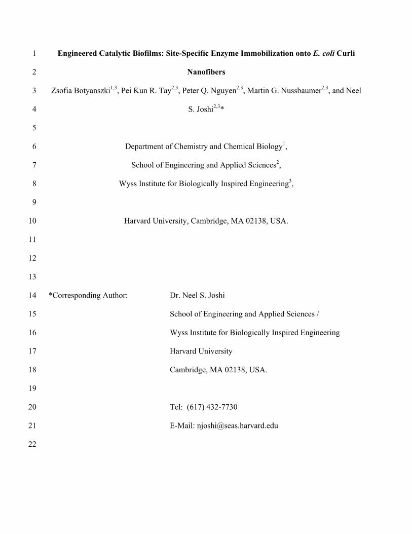
Engineered Catalytic Biofilms: Site-Specific Enzyme Immobilization onto E. coli Curli 1
Nanofibers 2
Zsofia Botyanszki1,3, Pei Kun R. Tay2,3, Peter Q. Nguyen2,3, Martin G. Nussbaumer2,3, and Neel 3
S. Joshi2,3* 4
5
Department of Chemistry and Chemical Biology1, 6
School of Engineering and Applied Sciences2, 7
Wyss Institute for Biologically Inspired Engineering3, 8
9
Harvard University, Cambridge, MA 02138, USA. 10
11
12
13
*Corresponding Author: Dr. Neel S. Joshi 14
School of Engineering and Applied Sciences / 15
Wyss Institute for Biologically Inspired Engineering 16
Harvard University 17
Cambridge, MA 02138, USA. 18
19
Tel: (617) 432-7730 20
E-Mail: [email protected] 21
22
2
Abstract 23
Biocatalytic transformations generally rely on purified enzymes or whole cells to perform 24
complex transformations that are used on industrial scales for chemical, drug, and biofuel 25
synthesis, pesticide decontamination and water purification. However, both of these systems 26
have inherent disadvantages related to the costs associated with enzyme purification, the long-27
term stability of immobilized enzymes, catalyst recovery and compatibility with harsh reaction 28
conditions. We developed a novel strategy for producing rationally designed biocatalytic 29
surfaces based on Biofilm Integrated Nanofiber Display (BIND), which exploits the curli system 30
of E. coli to create a functional nanofiber network capable of covalent immobilization of 31
enzymes. This approach is attractive because it is scalable, represents a modular strategy for site-32
specific enzyme immobilization, and has the potential to stabilize enzymes under denaturing 33
environmental conditions. We site-specifically immobilized a recombinant α-amylase, fused to 34
the SpyCatcher attachment domain, onto E. coli curli fibers displaying complementary SpyTag 35
capture domains. We characterized the effectiveness of this immobilization technique on the 36
biofilms and tested the stability of immobilized α-amylase in unfavorable conditions. This 37
enzyme-modified biofilm maintained its activity when exposed to a wide range of pH and 38
organic solvent conditions. In contrast to other biofilm-based catalysts, which rely on cellular 39
metabolism to remain active, the modified curli-based biofilm remained active even after cell 40
death due to organic solvent exposure. This work lays the foundation for a new and versatile 41
method of using the extracellular polymeric matrix of E. coli for creating novel biocatalytic 42
surfaces. 43
44

3
Keywords: Biofilm, bacterial immobilization, curli fibers, biocatalysis, enzyme display, 45
extracellular matrix46

4
Introduction 47
Biocatalysis provides an environmentally friendly alternative to chemical synthesis with 48
its ability to perform complex chemical transformations in a scalable manner (Wohlgemuth, 49
2007). Enzymes are inherently attractive as catalysts due to their ability to perform chemo-, 50
regio- and stereo-selective catalysis even on large, complex molecules. This fuels their use in the 51
pharmaceutical industry and elsewhere, as alternatives to less selective synthetic chemical 52
transformations (Murphy, 2012; Pollard and Woodley, 2007). 53
Enzymes can be used in purified form, in crude cell lysates, encased in synthetic 54
protective materials such as a polymer matrix or lipid vesicle, or within whole cells. The 55
attributes of these biocatalytic approaches have been extensively reviewed in the literature 56
(Halan et al., 2012; Krishna, 2002; Pollard and Woodley, 2007; Rosche et al., 2009; Zhou and 57
Hartmann, 2012). Whole cell catalysis is used widely in industry, however its production 58
efficiency is limited by low mass transport stemming from hindered diffusion of the substrate or 59
product across the cell membrane (Chen, 2007; Leon et al., 1998). Cell surface display methods 60
have been explored in order to circumvent the problem of mass transport, but these approaches 61
are hindered by the limited area on the bacterial cell surface and logistical difficulties in adapting 62
the technique for multimeric enzyme complexes and multi-enzyme transformations (Daugherty, 63
2007; Löfblom, 2011; van Bloois et al., 2011). An approach that optimally combines the criteria 64
of high surface area, enhanced enzyme stability, rapid mass transport, and modularity remains 65
elusive. 66
Recently, our lab, and others, have explored a new immobilization surface on bacteria – 67
the amyloid nanofibers of biofilms (Chen et al., 2014; Nguyen et al., 2014; Van Gerven et al., 68
2014). Biofilms are matrix-encapsulated bacteria adhered to each other and to surfaces or 69
5
interfaces (Costerton et al., 1995). Along with the other extracellular matrix components, these 70
biosynthetic supramolecular polymers protect the cells against toxic chemicals, metals and 71
physical stresses (Fang et al., 2002; Gross et al., 2010; Harrison et al., 2007), making biofilm-72
based materials well suited for industrial applications. 73
We developed a protein immobilization platform that modifies curli nanofibers, the 74
amyloid fiber component of E. coli biofilms, with a peptide domain that can covalently capture 75
proteins (Nguyen et al., 2014). In our approach, Biofilm Integrated Nanofiber Display (BIND), 76
heterologous functional peptide domains are genetically fused to the amyloidogenic protein 77
CsgA. When CsgA-peptide fusions assemble into curli fibers, the peptide domains become 78
functional handles that can be used to modify the properties of the fibers, to capture metals, 79
template nanoparticle growth or to enhance adhesion to surfaces. Recent advances in the 80
engineering of this system have demonstrated that the curli pathway can be used to export a 81
variety of CsgA-functional chimeras and also completely heterologous amyloidogenic 82
sequences, suggesting that this could be a highly generalizable approach to functional materials 83
synthesis (Chen et al., 2014; Nguyen et al., 2014; Sivanathan and Hochschild, 2012; Sivanathan 84
and Hochschild, 2013; Van Gerven et al., 2014; Zhong et al., 2014). 85
In this work, we demonstrate that a large, industrially relevant enzyme, α-amylase, can be 86
immobilized onto the curli fibers of E. coli biofilms, which we have termed catalytic-BIND 87
(Figure 1). We used a genetically programmable, irreversible immobilization method – the 88
spontaneous covalent bond formation between 13-amino acid SpyTag and 15 kDa SpyCatcher 89
split protein (Zakeri et al., 2012). As previously shown, SpyTag fused to CsgA (CsgA-ST) 90
assembles into fibers that closely resemble the native curli fibers with the SpyTag accessible for 91
conjugation to SpyCatcher (Nguyen et al., 2014). When SpyCatcher is fused to α-amylase, the 92
6
immobilization reaction is robust, with the ability to form site-specific attachment between the 93
two components, even in a complex mixture. We characterized the immobilization and activity 94
of the enzyme on the biofilm using a filter plate assay and showed that α-amylase activity is 95
retained after incubation in a range of pH and organic solvents, even when metabolic activity of 96
the cells is disrupted. Our results suggest that this technology may be able to combine the 97
scalability of whole cell catalysis with the modularity of enzyme surface immobilization through 98
the transformation of E. coli biofilm extracellular matrices into designer functionalized surfaces. 99
Materials and Methods 100
Cell Strains, Plasmids and Reagents 101
All strains and vectors are listed in Supplementary Table I and II. CsgA and csgA-SpyTag genes 102
were cloned into pBbE1a vectors. The csgA deletion mutant PHL628-ΔcsgA (MG1655 malA-103
Kan ompR234 ΔcsgA (Vidal et al., 1998)) used for biofilm experiments was a kind gift from the 104
Hay Laboratory (Toba et al., 2011). CsgA was expressed in YESCA media, containing 10 g/L of 105
casamino acids (Fisher, BP1424) and 1 g/L of yeast extract (Fisher, BP1422). YESCA plates 106
also contained 15 g/L of agar. DPBS (LifeTechnologies, 14190-144) without calcium or 107
magnesium was used as the general buffer for enzymatic reactions (abbrev. PBS). TBST (2.4g 108
Tris base, 8.8g NaCl, 1 mL Tween-20, per L, pH 7.4-7.6) was used as wash buffer. Organic 109
solvents were purchased from Sigma Aldrich, BDH Solvents, EMD, in >98% purity or HPLC 110
grade. The α-amylase gene was isolated from Bacillus licheniformis ATCC 14580. SpyCatcher 111
gene was acquired from Addgene (plasmid # 35044). α-Amylase was inserted at the N-terminus 112
of SpyCatcher and the construct was subcloned into a pET28b vector (Novagen, 69865). 113
Amylase-SpyCatcher (Amylase-SC) was expressed in Rosetta cells (Novagen, 70953) grown in 114
Terrific Broth (Sigma T0918). Cells were lysed using a Misonix Probe Sonicator 4000. Millipore 115

7
PCF and hydrophilic PTFE filter plates (MSSLBPC10, MSRLN0410) and the Millipore 116
MultiScreen vacuum manifold apparatus was used for filter plate assays. Amylase-SC was also 117
immobilized onto His-Pur magnetic beads (LifeTechnologies, 88831). For amylase activity, 4-118
nitrophenyl-α-D-maltopentaoside (pNPMP, Sigma, 66068-38-0) was used as a substrate and 119
recombinant purified α-amylase from Bacillus licheniformis (Sigma, A3403) as a standard. An 120
iBlot Dry Blotting system (LifeTechnologies) was used for transferring gels to PVDF 121
membranes (LifeTechnologies, IB4010). Anti-His antibody was purchased from Pierce Sci. 122
(MA1-21315) and Western Blots were developed using Clarity ECL Substrate (BioRad, 170-123
5060). pH was measured using a Mettler Toledo FE20-Basic pH meter with an InLab®Routine 124
probe. LC/MS/MS analysis was performed at the Taplin Mass Spectroscopy Facility. Scanning 125
Electron Microscope (SEM) images were taken on a Zeiss Ultra Plus FESEM and confocal 126
microscopy was performed on a Leica SP5 X MP Inverted Confocal Microscope. 127
Curli Expression 128
PHL628-ΔcsgA cells were transformed with either an empty pBbE1a plasmid or pBbE1a 129
plasmids with CsgA, CsgA-ST, CsgATEVEKHis-ST (abr. CsgA[25AA]His-ST). The cells were 130
then streaked onto YESCA plates with 100-200 µg/mL ampicillin. Transformed PHL628 cells 131
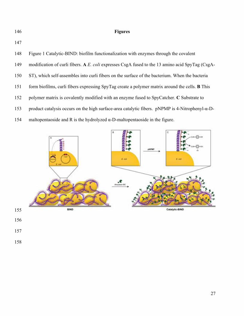
were grown up in YESCA with ampicillin until an OD of 0.4-0.6 at 30°C. Curli expression was 132
induced with 0.3 mM IPTG. Cultures were shaken for 18h-24h at 25°C and 150 rpm. 133
Quantitative Congo Red (CR) Binding Assays 134
Congo Red (CR) binding assay was adapted from previously published methods (Chapman, 135
2002). 1 mL of induced culture was pelleted at 5000g for 10 min and resuspended gently in PBS. 136
Congo Red was added to 0.025 mM and allowed to incubate at 25°C for 10 min. The cells were 137
then pelleted at 21,000g and the absorbance of the supernatant was measured at 490 nm in a 138

8
BioTek H1 microplate reader. The amount of CR binding was determined by subtracting the 139
amount of this measurement from a PBS + CR control. 140
Amylase-SpyCatcher Expression 141
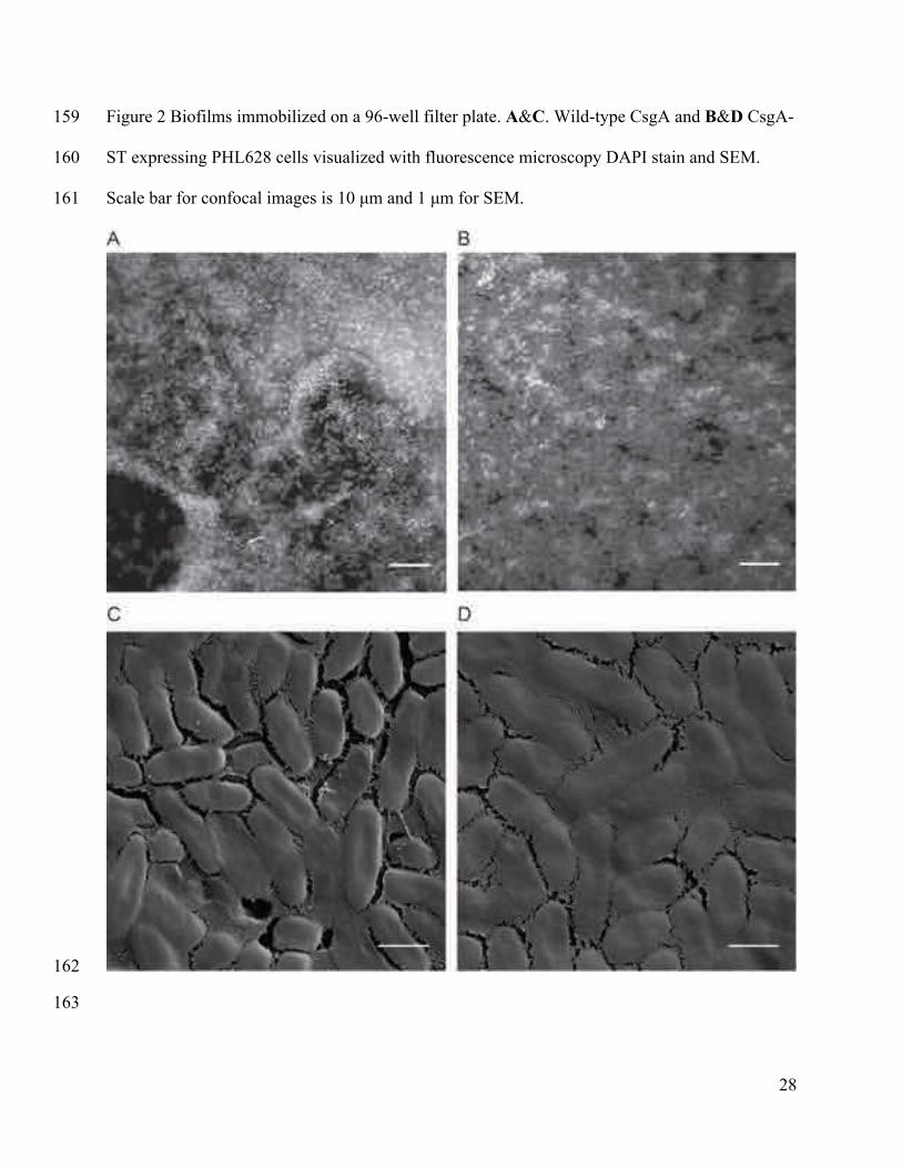
Rosetta cells transformed with pET28b Amylase-SC were grown up in overnight cultures in LB 142
at 30°C with 100 µg/mL kanamycin. 1L of Terrific Broth was supplemented with kanamycin, 143
inoculated with the overnight culture and grown up at 30°C until an OD of 0.4. Amylase-SC 144
expression was induced with 0.5 mM IPTG and allowed to express overnight at 18°C. Cells were 145
harvested and lysed in TBST and Amylase-SC was purified on a Ni-NTA column. 146
Amylase-SpyCatcher Activity Assay 147
4-Nitrophenyl-α-D-maltopentaoside (pNPMP) was chosen as the substrate to measure α-amylase 148
activity because hydrolysis of 4-nitrophenol (pNP) from the pentasaccharide can be monitored at 149
405 nm. Note, the absorbance of pNP is dependent on its protonation state. 150
Curli Biofilm Assays 151
PHL628 biofilms expressing wild-type CsgA or CsgA-ST were cultured for 18h at 25°C at 150 152
rpm as described above. Curli content was measured using the quantitative CR binding assay. 153
50-100 µL of cells (normalized to CR absorption) were transferred onto filter plates, which were 154
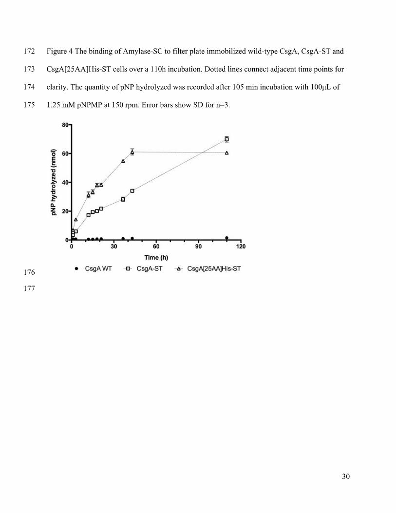
previously blocked with 0.5-2% BSA for at least 1.5h. For suspended biofilm assays, the 155
biofilms were distributed into Eppendorf tubes and the same conjugation procedures followed. 156
The media was filtered through using a vacuum manifold. Cells were washed with PBS or 157
TBST. Cells were incubated with Amylase-SC in PBS with BSA or TBST overnight. To 158
determine the remaining activity of the SpyCatcher on Amylase-SC that was left in solution after 159
incubation, the filtrate was reacted with MBP-ST for 3h. The reaction mixture was concentrated, 160
dissolved in 2x Laemmli buffer and a Western Blot run. For activity assays on the biofilm, the 161
9
conjugation mixture was removed using vacuum filtration and the biofilms were washed six 162
times with 0.3% BSA in PBS or TBST over 90 min. For activity assays at different pH, 1.25 mM 163
pNPMP in PBS was pH-ed with NaOH and HCl and added to the cells. Plates were placed on a 164
desktop shaker and shaken at 150 rpm at room temperature for 1.5-2h. At the end of the 165
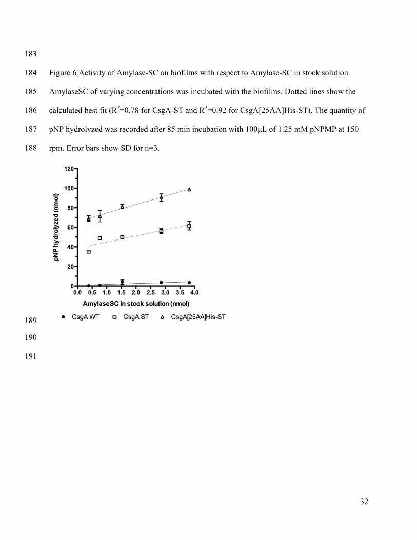
experiment, the supernatant was vacuum filtered into a new 96-well plate, 5 M NaOH was added 166
to increase pH to 12-14 (to bring pNP to a uniform protonation state) and pNP hydrolyzation was 167
measured at 405 nm. For activity assays in organic solvents, biofilms were incubated with the 168
solvents for 1-2h. The solvents were removed and cells washed with PBS. 1.25 mM pNPMP in 169
PBS was added to the biofilm. Plates were placed on a desktop shaker at room temperature (rt). 170
At the end of the experiment, the supernatant was vacuum filtered into a new 96-well plate and 171
pNP release measured at 405 nm. In the data analysis, the reference activity is to pH 7 PBS. All 172
data points are averages of reactions done in triplicate with error bars indicating standard 173
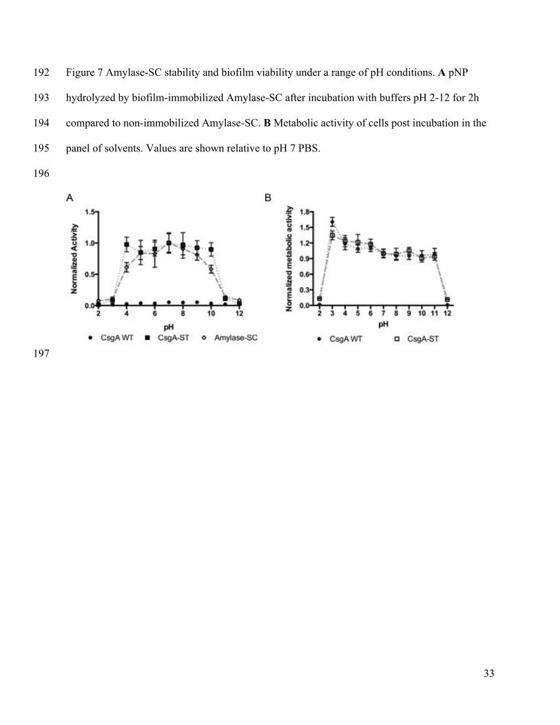
deviation. 174
MTS Assay 175
Cell viability was tested using Promega CellTiter 96 Aqueous Non-Radioactive Cell 176
Proliferation Assay. Functionalized biofilms were prepared as described above. Subsequent to 177
exposure of biofilms to pH, miscible and immiscible organic solvents, biofilms were washed 178
with PBS, incubated with assay buffer for 1h, filtered through and results read optically at 490 179
nm. In the data analysis, biofilms incubated in pH 7 PBS were used as the normalization for 180
activity. 181
10
Results and Discussion 182
Amylase-SC Stability 183
We chose α-amylase as our model enzyme because of its wide use, industrial applicability, and 184
the commercial availability of a water-soluble colorimetric substrate. The stability of α-amylase 185
and its substrate also allowed us to correlate the observed enzyme activity to the stability of the 186
biofilm as a whole, since we could assume that the enzyme would not degrade on the timescale 187
of our experiments. While multiple proteins have been successfully attached to SpyCatcher 188
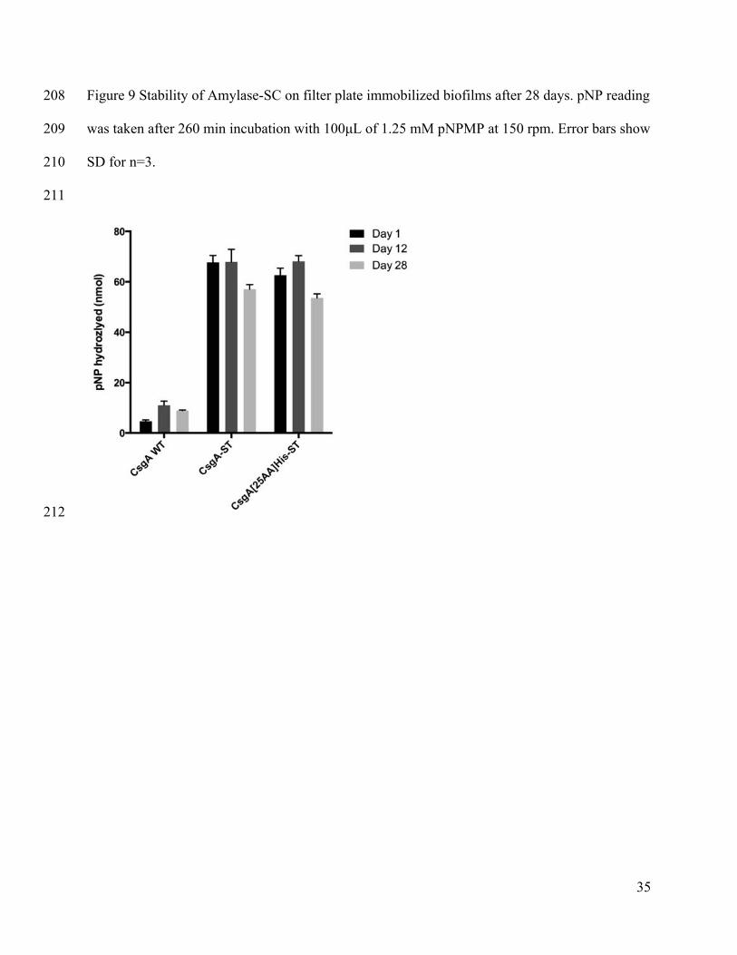
(Fairhead et al., 2014; Nguyen et al., 2014; Schoene et al., 2014; Zakeri et al., 2012), this paper 189
represents the first example of using the SpyTag and SpyCatcher fusions to immobilize an 190
industry-relevant enzyme onto curli fibers. We designed a construct, named Amylase-191
SpyCatcher (abr. Amylase-SC), consisting of an α-amylase gene fused to the N-terminus of 192
SpyCatcher via a 13 amino acid flexible linker. We confirmed that the fusion of the SpyCatcher 193
domain to α-amylase had minimal impact on the kinetics of the two enzymes, and the difference 194
in their stability over time and under a range of temperatures was negligible compared to the 195
wild-type enzymes (Supplementary Figure 1 and Supplementary Table III). 196
Covalent Amylase-SC Attachment to Biofilms 197
We chose the SpyTag-SpyCatcher immobilization strategy because of its ability to form 198
site-specific covalent bonds between the enzyme fusion proteins and the modified curli fibers, 199
even in complex mixtures (Nguyen et al., 2014). This feature makes it particularly attractive 200
because it obviates the need for time consuming and expensive enzyme purification efforts. In 201
order for this strategy to be effective, the SpyTag domain must remain sufficiently accessible 202
after amyloid assembly to participate in covalent bond formation. We confirmed that covalent 203
conjugation between Amylase-SC and fully formed curli fibers displaying SpyTag was feasible 204

11
through a SDS-PAGE gel shift assay and subsequent LC/MS/MS analysis (Supplementary 205
Figure 2 and Supplementary Table IV). 206
A filter plate-based assay was used to assess the catalytic potential of the curli-207
immobilized enzymes under a variety of conditions. Cells were grown in culture and induced to 208
produce recombinant CsgA before being transferred onto 96-well filter plates. Confocal 209
microscopy and scanning electron microscopy (Figure 2) revealed that the filter surface was 210
coated with one to two layers of cells surrounded by a thick mat of extracellular material. DAPI 211
staining of cells throughout the mass of the biofilm, done previous to sample drying for SEM 212
analysis, suggests that it is porous enough that small molecules can permeate it in the hydrated 213
state. In order to determine whether the filtration process resulted in lower SpyTag accessibility, 214
Amylase-SC was reacted overnight with either filter plate immobilized cells or suspended cells 215
after curli induction from a strain that secretes CsgA with a short, 6 amino acid, linker to ST 216
(CsgA-ST) and one with a 31 amino acid linker to ST (CsgA[25AA]His-ST). As shown in 217
Figure 3, the activity resulting from these two immobilization techniques after overnight 218
incubation was similar. This indicates that filtration does not affect the availability of the SpyTag 219
sites under these experimental conditions and longer incubation times. This is reasonable, since 220
the protocol yields essentially a monolayer of cells with their extracellular material surrounding 221
them. If thicker biofilms are used, the issue of enzyme-SC diffusivity will need to be further 222
investigated. 223
SpyCatcher conjugation is known to proceed rapidly in homogeneous solution, with the 224
conjugation reaction effectively complete within a half hour under a variety of conditions (Zakeri 225
et al., 2012). However, since the SpyTag domain is displayed in a dense array on the surface of 226
the curli fibers, we wanted to compare the kinetics of the Amylase-SC conjugation reaction 227

12
between soluble and fiber-bound SpyTag. Accordingly, we reacted purified Amylase-SC with 228
either a soluble protein, maltose binding protein, fused to SpyTag (abr. MBP-ST) or SpyTag 229
displayed on fully assembled fibers in suspension culture. The gel shown in Supplementary 230
Figure 3 illustrates that essentially all Amylase-SC reacts with MBP-ST within 10 minutes, 231
indicating that the presence of the α-amylase does not hinder accessibility to SpyCatcher’s active 232
site, nor does it significantly alter SpyCatcher’s reaction kinetics. Next, we monitored the 233
kinetics of the conjugation reaction performed in suspension with CsgA-ST and 234
CsgA[25AA]His-ST. The results, shown in Figure 4, illustrate that Amylase-SC binding to the 235
biofilms on filter plates is much slower than in solution – on the order of hours to days instead of 236
minutes. The construct with the longer linker exhibited significantly faster conjugation kinetics 237
(36 hours to maximum product formation) compared to the shorter linker construct, suggesting 238
that SpyTag accessibility may be a parameter worth optimizing in order to increase conjugation 239
efficiencies for future efforts. 240
There may be several reasons to explain the extended times needed for conjugation to the 241
biofilms (Figure 4). One possibility is a degradation of the SpyCatcher protein. To test this, 242
Amylase-SC that remained unattached after being incubated with biofilms expressing CsgA, 243
CsgA-ST and CsgA[25AA]His-ST for 56h was removed and reacted with MBP-ST. The 244
Western Blot in Supplementary Figure 4 confirms that even after incubation of this length, the 245
SpyCatcher protein is able to conjugate to MBP-ST in solution, eliminating this as a possible 246
explanation. Instead, increased diffusion distances may be responsible for the slower reaction. In 247
biofilms, enzymes need to diffuse to the surface, which is a much larger distance on average than 248
diffusing to a uniformly distributed MBP-ST. Indeed, binding to curli expressing cells in 249
suspension is faster than binding to filter plate immobilized cells (Supplementary Figure 5). In 250
13
addition, Amylase-SC (72 kDa) is significantly larger than the CsgA-ST monomer (15 kDa), and 251
since the actual surface is a polymer of self-assembled CsgA-STs, both the core curli fiber and 252
previously immobilized Amylase-SCs may sterically hinder the conjugation of free Amylase-SC 253
on neighboring SpyTag sites. This may explain why the CsgA[25AA]His-ST expressing biofilms 254
can bind Amylase-SC faster. 255
Correlating Congo Red Adsorption to Enzyme Immobilization 256
We expected that the amount of curli being produced by the cells would be a good 257
predictor for the amount of enzyme that could be immobilized. We therefore attempted to 258
correlate amyloid production, as measured by CR staining, with enzyme immobilization. 259
Although CR staining can be problematic because of nonspecific staining of other proteins and 260
biopolymers, we confirmed that this was not a problem for the E. coli strain we used for these 261
experiments by demonstrating a lack of staining for cells transformed with an empty plasmid that 262
did not contain the gene encoding CsgA (Nguyen et al., 2014). We also investigated whether CR 263
staining is dependent on the amount of non-curli biomass in the sample. To do this, we diluted 264
CsgA[25AA]His-ST expressing PHL628 cells with PHL628 cells that do not express curli and 265
measured CR binding. The linear relationship observed for this curli concentration curve 266
(Supplementary Figure 6) indicates that the CR binding to curli is not blocked by the presence of 267
extra cells. 268
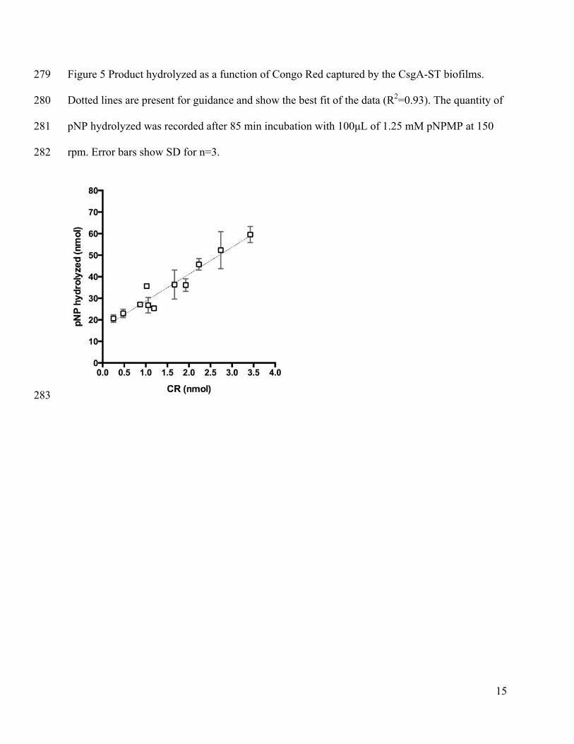
To correlate curli production to activity, we compared the CR binding to the Amylase 269
activity on the fibers of filter plate immobilized cells. As shown in Figure 5, there is a linear 270
correlation between CR adsorbed and the activity of the enzymes (reported as pNP hydrolyzed). 271
This linearity indicates that CR binding appears to be a valid measure of the relative amount of 272
14
enzyme immobilized, although absolute measurements of the immobilized enzyme concentration 273
remain elusive. 274
Enzyme activity measurement in solution and on filter plates 275
In order to determine how the amount of Amylase-SC in the stock solution (reaction mixture) 276
affected immobilization, we incubated filter plate immobilized cells with a range of 277
concentrations of Amylase-SC. The resulting activity, shown in 278
15
Figure 5 Product hydrolyzed as a function of Congo Red captured by the CsgA-ST biofilms. 279
Dotted lines are present for guidance and show the best fit of the data (R2=0.93). The quantity of 280
pNP hydrolyzed was recorded after 85 min incubation with 100µL of 1.25 mM pNPMP at 150 281
rpm. Error bars show SD for n=3. 282
283

16
284
17
Figure 6, illustrates a linear increase with the amount of Amylase-SC concentration. Despite 1
normalizing both cell cultures to CR, there is a difference between the activity seen on the CsgA-2
ST and CsgA[25AA]His-ST displaying cells because the reaction was not fully at completion. 3
When we converted the values for hydrolyzed pNP concentration to the amount of soluble 4
enzymes needed to hydrolyze that much pNP (the effective enzyme concentration at this shaking 5
speed) we found that at the highest Amylase-SC concentration in Figure 6, 6 and 12% (CsgA-ST 6
and CsgA[25AA]His-ST respectively) of the enzyme activity of the stock solution was observed. 7
Since the bulk kinetics on a catalytic surface for a neutral substrate are generally slower than in a 8
well mixed solution(Bommarius and Riebel-Bommarius, 2007; Hornby and Lilly, 1968; 9
Kobayashi and Laidler, 1973)(Supplementary Figure 7), the effective enzyme concentration 10
calculated this way provides us with a minimum estimate for the amount of enzyme 11
immobilized. 12
Biofilm Immobilized Amylase-SC Activity as a Function of pH 13
For the successful use of catalytic biofilms in many synthetic and environmental 14
applications, they may need to withstand a range of conditions that are not normally conducive 15
for bacterial growth or enzyme stability. Many enzymes used in industry today have been 16
engineered specifically to enhance their stability under extreme pH conditions, high 17
temperatures, and in the presence of detergents and organic solvents (Bommarius and Paye, 18
2013; Kirk et al., 2002). Previous characterization experiments showed that α-amylase is fully 19
active in pH range 5-9 (Nielsen et al., 2001). As shown in Figure 7A, Amylase-SC immobilized 20
onto the biofilms maintained full activity for the pH 5-9 range, but also showed full activity at 21
pH 4 and pH 10, even though the soluble Amylase-SC lost 40% of its activity at those pH values. 22
The cells show a slightly increased metabolic activity from pH 3-6, which is likely due to stress 23
response to the unfavorable pH and buffer conditions (Figure 7B). 24

18
Amylase-SC Stability on Biofilms Incubated with Organic Solvents 1
Biocatalytic systems that are able to catalyze reactions on compounds with low water 2
solubility are of particular interest in industry. Most existing methods designed to circumvent the 3
issue of water solubility use two-phase aqueous-organic systems. In these systems, the organic 4
soluble molecule briefly enters the aqueous phase, where the enzyme is able to catalyze the 5
reaction, and then exits again into the organic phase (Hertzberg et al., 1992; Kawakami et al., 6
1990; Sinisterra and Dalton, 1996). Laane et. al. proposed a positive correlation between 7
reactivity and the logarithm of the partitioning coefficient (logP) of the organic solvent (Laane et 8
al., 1987). Water-miscible solvents have a logP<0, polar organic solvents have a logP<2 and 9
nonpolar solvents have a logP>2. For whole cell catalysts, solvents that are non-polar enough to 10
have a logP between 2-5 (depending on cell type) or greater are able to maintain catalytic 11
activity. The loss of activity is believed to be due to inactivation of enzymes, the breakdown of 12
transport mechanisms, disruption of the cell membrane by the solvent and cell lysis that results 13
from exposure to the organic solvents (Leon et al., 1998). 14
We hypothesized that the enzyme-functionalized biofilms would be able to withstand 15
some exposure to non-miscible organic solvents because biofilms should remain hydrated under 16
such conditions and hence prevent the denaturation of immobilized enzymes. To test this 17
hypothesis, we incubated Amylase-SC conjugated biofilms with a panel of water-miscible and 18
non-miscible organic solvents. Since pNPMP is not soluble in most organic solvents, we first 19
incubated the biofilms in the organics and then replaced the solvent with PBS while measuring 20
activity. As shown in Figure 8A, the relative activity of immobilized Amylase-SC is only slightly 21
affected by incubation with non-miscible solvents, but completely disappears in miscible 22
solvents. Miscible solvents can access and denature the enzymes, and may disrupt curli fiber 23
assembly or anchoring, while a hydration layer separates the non-miscible solvents. Plotting the 24

19
results in Figure 8A against the partitioning coefficient of the solvents, Figure 8B shows that 1
Amylase-SC activity is mostly preserved when biofilms are incubated in solvents with logP>0. 2
Notably, 70-90% activity is retained for biofilms incubated in solvents with logP 0.6-0.8, while 3
in the whole cell systems documented in the literature, the use of these solvents resulted in little 4
to no activity for whole cell catalysts (Laane et al., 1987). Indeed, the metabolic activity of the 5
cells following organic solvent exposure shows that cell metabolism ceased in all solvents tested 6
except decane, which has a logP of 5.6 (Figure 8C). This correlates with the results previously 7
mentioned for whole cell catalysis in two-phase systems. Although direct comparisons to the 8
soluble enzyme was not possible in this case due to the insolubility of the enzyme substrate in 9
organics, Amylase-SC does show similar organic solvent tolerance when immobilized onto Ni-10
NTA beads (Supplementary Figure 8). Further information using other enzyme systems will be 11
needed to definitively establish the impact of the biofilm on enzyme stability. However, 12
operability at the logP 0-2 range may be a unique feature to our system that is afforded by the 13
fact that despite our use of cells, the catalytic component of our system does not rely on cell 14
viability. 15
It is also worth noting that we chose the PHL628 strain for these experiments specifically 16
because curli fibers are the only extracellular polymer that it produces, which simplified the 17
characterization experiments. However, for future studies, long term enzyme stability might 18
benefit from the use of strains that produce other extracellular polymers (i.e. cellulose and other 19
pili) that can serve a protective role for the immobilized enzymes. 20
Biofilm stability over time 21
The stability of the catalytic system is very important in industrial applications of 22
catalytic technologies, since cost savings can be achieved by the extended use of immobilized 23
catalysts, thus reducing reactor downtime (Halan et al., 2012). While our filter plate setup cannot 24
20
be used to determine the stability of the biofilms under flow or batch processing conditions, we 1
investigated the stability of the Amylase-SC attached to the biofilm over time. Amylase activity 2
was retested after a period of 28 days with the filter plate immobilized biofilm kept at 4oC in 3
buffer. Figure 9 shows that the biofilms displayed the same level of activity after the 12 days 4
with a slight decrease after 28 days, indicating that the biofilm and its entangled curli fibers were 5
stable enough that the fibers were not displaced through the filter during the vacuum-assisted 6
washing steps. 7
Conclusions 8
In this work, we demonstrated a novel platform for the immobilization of enzymes onto 9
the extracellular matrix of an engineered biofilm. We were able to create biofilms displaying 10
functional biochemical handles on the curli network of E. coli. Subsequently the SpyTag-11
SpyCatcher immobilization strategy was used to site-specifically conjugate α-amylase to the 12
biofilms, which revealed that enzymes remained active after exposure to various adverse 13
conditions. 14
There are several attractive features of this biofilm-based material compared to other 15
surfaces for enzyme immobilization: (1) the conjugation strategy we employ proceeds 16
spontaneously, without the need for any chemical treatment steps, and provides a simple, 17
modular way to immobilize enzymes site-specifically to surfaces; (2) the conjugation sites are 18
densely arrayed on the curli fibers, producing a high surface area for immobilization; (3) the 19
material is produced entirely biosynthetically, which is a green alternative to petroleum-derived 20
synthetic polymers. 21
This technology could be combined with more established biofilm-based biocatalytic 22
processes (Gross et al., 2010; Halan et al., 2010; Karande et al., 2014) to yield stable biofilms in 23
flow reactors that are able to catalyze reactions not accessible to currently available whole cell 24
21
catalyst systems. Furthermore, we have previously demonstrated the potential for creating 1
multifunctional BIND materials (Nguyen, at al., 2014), suggesting that the engineered curli 2
fibers could be used for immobilizing multiple enzymes for multi-step transformations, or for 3
combing catalysis with other functions that may be attractive in the context of a bioreactor, like 4
substrate adhesion. This would be useful in many forms of ‘green’ biocatalysis, including in 5
pharmaceutical synthesis, breakdown of pharmaceuticals in wastewater, removal of 6
contaminants from groundwater or the creation of catalytic surfaces for bioenergy production. 7
22
Acknowledgements 8
This work was funded by the Wyss Institute for Biologically Inspired Engineering. Z.B. 9
acknowledges NSF GRF for funding and P.R.T for funding from A*STAR National Science 10
Graduate Fellowship (Singapore). We thank Prof. Anthony G. Hay (Cornell Univ.) for providing 11
the PHL628-ΔcsgA strain. 12
References 13
Bommarius AS, Paye MF. 2013. Stabilizing biocatalysts. Chem. Soc. Rev. 42:6534. 14 Bommarius AS, Riebel-Bommarius BR. 2007. Biocatalysis. John Wiley & Sons 1 pp. 15 Chapman MR. 2002. Role of Escherichia coli Curli Operons in Directing Amyloid Fiber 16
Formation. Science 295:851–855. 17 Chen AY, Deng Z, Billings AN, Seker UOS, Lu MY, Citorik RJ, Zakeri B, Lu TK. 2014. 18
Synthesis and patterning of tunable multiscale materials with engineered cells. Nature 19 Materials 13:515–523. 20
Chen RR. 2007. Permeability issues in whole-cell bioprocesses and cellular membrane 21 engineering. Appl Microbiol Biotechnol 74:730–738. 22
Costerton JW, Lewandowski Z, Caldwell DE, Korber DR, Lappin-Scott HM. 1995. Microbial 23 biofilms. Annual Reviews in Microbiology 49:711–745. 24
Daugherty PS. 2007. Protein engineering with bacterial display. Current Opinion in Structural 25 Biology 17:474–480. 26
Fairhead M, Veggiani G, Lever M, Yan J, Mesner D, Robinson CV, Dushek O, van der Merwe 27 PA, Howarth M. 2014. SpyAvidin Hubs Enable Precise and Ultrastable Orthogonal 28 Nanoassembly. J. Am. Chem. Soc. 136:12355–12363. 29
Fang HHP, Xu L-C, Chan K-Y. 2002. Effects of toxic metals and chemicals on biofilm and 30 biocorrosion. Water Research 36:4709–4716. 31
Gross R, Lang K, Bühler K, Schmid A. 2010. Characterization of a biofilm membrane reactor 32 and its prospects for fine chemical synthesis. Biotechnol. Bioeng. 105:705–717. 33
Halan B, Buehler K, Schmid A. 2012. Biofilms as living catalysts in continuous chemical 34 syntheses. Trends in Biotechnology 30:453–465. 35
Halan B, Schmid A, Buehler K. 2010. Maximizing the productivity of catalytic biofilms on solid 36 supports in membrane aerated reactors. Biotechnol. Bioeng. 106:516–527. 37
Harrison JJ, Ceri H, Turner RJ. 2007. Multimetal resistance and tolerance in microbial biofilms. 38 Nature Reviews Microbiology 5:928–938. 39
Hertzberg S, Kvittingen L, Anthonsen T, Skjaak-Braek G. 1992. Alginate as immobilization 40 matrix and stabilizing agent in a two-phase liquid system: application in lipase-catalysed 41 reactions. Enzyme and Microbial Technology 14:42–47. 42
Hornby WE, Lilly MD. 1968. Some changes in the reactivity of enzymes resulting from their 43 chemical attachment to water-insoluble derivatives of cellulose. Biochem. J. 107:669–674. 44
Karande R, Halan B, Schmid A, Buehler K. 2014. Segmented flow is controlling growth of 45 catalytic biofilms in continuous multiphase microreactors. Biotechnol. Bioeng. 111:1831–46 1840. 47
23
Kawakami K, Tsuruda S, Miyagi K. 1990. Immobilization of Microbial Cells in a Mixed Matrix 48 of Silicone Polymer and Calcium Alginate Gel: Epoxidation of 1‐Octene by Nocardia 49 corallina B‐276 in Organic Media. Biotechnol Progress 6:357–361. 50
Kirk O, Borchert TV, Fuglsang CC. 2002. Industrial enzyme applications. Current Opinion in 51 Biotechnology 13:345–351. 52
Kobayashi T, Laidler KJ. 1973. Kinetic analysis for solid-supported enzymes. Biochim. Biophys. 53 Acta 302:1–12. 54
Krishna SH. 2002. Developments and trends in enzyme catalysis in nonconventional media. 55 Biotechnology Advances 20:239–267. 56
Laane C, Boeren S, Vos K, Veeger C. 1987. Rules for optimization of biocatalysis in organic 57 solvents. Biotechnol. Bioeng. 30:81–87. 58
Leon R, Fernandes P, Pinheiro HM, Cabral J. 1998. Whole-cell biocatalysis in organic media. 59 Enzyme and Microbial Technology 23:483–500. 60
Löfblom J. 2011. Bacterial display in combinatorial protein engineering. Biotechnology Journal 61 6:1115–1129. 62
Murphy CD. 2012. The microbial cell factory. Org. Biomol. Chem. 10:1949. 63 Nguyen PQ, Botyanszki Z, Tay PKR, Joshi NS. 2014. Programmable biofilm-based materials 64
from engineered curli nanofibres. Nat Commun 5:4945. 65 Nielsen JE, Borchert TV, Vriend G. 2001. The determinants of α-amylase pH–activity profiles. 66
Protein Eng. 14:505–512. 67 Pollard DJ, Woodley JM. 2007. Biocatalysis for pharmaceutical intermediates: the future is now. 68
Trends in Biotechnology 25:66–73. 69 Rosche B, Li XZ, Hauer B, Schmid A, Buehler K. 2009. Microbial biofilms: a concept for 70
industrial catalysis? Trends in Biotechnology 27:636–643. 71 Schoene C, Fierer JO, Bennett SP, Howarth M. 2014. SpyTag/SpyCatcher Cyclization Confers 72
Resilience to Boiling on a Mesophilic Enzyme. Angew. Chem. Int. Ed. 53:6101–6104. 73 Sinisterra JV, Dalton H. 1996. Influence of the immobilization methodology in the stability and 74
activity of P. putida UV4 immobilized whole cells. Immobilized Cells: Basics and 75 Applications: Basics and Applications 11:416. 76
Sivanathan V, Hochschild A. 2012. Generating extracellular amyloid aggregates using E. coli 77 cells. Genes & Development 26:2659–2667. 78
Sivanathan V, Hochschild A. 2013. A bacterial export system for generating extracellular 79 amyloid aggregates. Nature Protocols 8:1381–1390. 80
Toba FA, Thompson MG, Campbell BR, Junker LM, Rueggeberg KG, Hay AG. 2011. Role of 81 DLP12 lysis genes in Escherichia coli biofilm formation. Microbiology 157:1640–1650. 82
van Bloois E, Winter RT, Kolmar H, Fraaije MW. 2011. Decorating microbes: surface display 83 ofproteins on Escherichia coli. Trends in Biotechnology 29:79–86. 84
Van Gerven N, Goyal P, Vandenbussche G, De Kerpel M, Jonckheere W, De Greve H, Remaut 85 H. 2014. Secretion and functional display of fusion proteins through the curli biogenesis 86 pathway. Molecular Microbiology 91:1022–1035. 87
Vidal O, Longin R, Prigent-Combaret C, Dorel C, Hooreman M, Lejeune P. 1998. Isolation of an 88 Escherichia coli K-12 mutant strain able to form biofilms on inert surfaces: involvement of a 89 new ompR allele that increases curli expression. Journal of Bacteriology 180:2442–2449. 90
Wohlgemuth R. 2007. Modular and scalable biocatalytic tools for practical safety, health and 91 environmental improvements in the production of speciality chemicals. Biocatal 92 Biotransformation 25:178–185. 93
24
Zakeri B, Fierer JO, Celik E, Chittock EC, Schwarz-Linek U, Moy VT, Howarth M. 2012. 94 Peptide tag forming a rapid covalent bond to a protein, through engineering a bacterial 95 adhesin. Proceedings of the National Academy of Sciences 109:E690–E697. 96
Zhong C, Gurry T, Cheng AA, Downey J, Deng Z, Stultz CM, Lu TK. 2014. Strong underwater 97 adhesives made by self-assembling multi-protein nanofibres. Nature Nanotechnology:1–9. 98
Zhou Z, Hartmann M. 2012. Recent Progress in Biocatalysis with Enzymes Immobilized on 99 Mesoporous Hosts. Top Catal 55:1081–1100. 100
101
25
List of Figures 102
Figure 1 Catalytic-BIND: biofilm functionalization with enzymes through the covalent 103
modification of curli fibers. A E. coli expresses CsgA fused to the 13 amino acid SpyTag 104
(CsgA-ST), which self-assembles into curli fibers on the surface of the bacterium. When the 105
bacteria form biofilms, curli fibers expressing SpyTag create a polymer matrix around the 106
cells. B This polymer matrix is covalently modified with an enzyme fused to SpyCatcher. C 107
Substrate to product catalysis occurs on the high surface-area catalytic fibers. pNPMP is 4-108
Nitrophenyl-α-D-maltopentaoside and R is the hydrolyzed α-D-maltopentaoside in the 109
figure. .................................................................................................................................... 27 110
Figure 2 Biofilms immobilized on a 96-well filter plate. A&C. Wild-type CsgA and B&D CsgA-111
ST expressing PHL628 cells visualized with fluorescence microscopy DAPI stain and SEM. 112
Scale bar for confocal images is 10 µm and 1 µm for SEM. ............................................... 28 113
Figure 3 Parallel immobilization of Amylase-SC on biofilms displaying CsgA-ST suspended in 114
solution (S) and immobilized on filter plates (FP). Samples were taken after 20h of 115
immobilization. Suspended biofilms were filtered onto the filter plates so that the activity of 116
all biofilms was measured under identical conditions. The quantity of pNP hydrolyzed was 117
recorded after 260 min incubation with 100µL of 1.25 mM pNPMP at 150 rpm. Error bars 118
show SD for n=3. .................................................................................................................. 29 119
Figure 4 The binding of Amylase-SC to filter plate immobilized wild-type CsgA, CsgA-ST and 120
CsgA[25AA]His-ST cells over a 110h incubation. Dotted lines connect adjacent time points 121
for clarity. The quantity of pNP hydrolyzed was recorded after 105 min incubation with 122
100µL of 1.25 mM pNPMP at 150 rpm. Error bars show SD for n=3. ................................ 30 123
26
Figure 5 Product hydrolyzed as a function of Congo Red captured by the CsgA-ST biofilms. 124
Dotted lines are present for guidance and show the best fit of the data (R2=0.93). The 125
quantity of pNP hydrolyzed was recorded after 85 min incubation with 100µL of 1.25 mM 126
pNPMP at 150 rpm. Error bars show SD for n=3. ................................................................ 31 127
Figure 6 Activity of Amylase-SC on biofilms with respect to Amylase-SC in stock solution. 128
AmylaseSC of varying concentrations was incubated with the biofilms. Dotted lines show 129
the calculated best fit (R2=0.78 for CsgA-ST and R2=0.92 for CsgA[25AA]His-ST). The 130
quantity of pNP hydrolyzed was recorded after 85 min incubation with 100µL of 1.25 mM 131
pNPMP at 150 rpm. Error bars show SD for n=3. ................................................................ 32 132
Figure 7 Amylase-SC stability and biofilm viability under a range of pH conditions. A pNP 133
hydrolyzed by biofilm-immobilized Amylase-SC after incubation with buffers pH 2-12 for 134
2h compared to non-immobilized Amylase-SC. B Metabolic activity of cells post incubation 135
in the panel of solvents. Values are shown relative to pH 7 PBS. ........................................ 33 136
Figure 8 Amylase-SC stability and biofilm viability after incubation in organic solvents. A pNP 137
hydrolyzed by biofilm-immobilized Amylase-SC after incubation in panel of solvents for 138
2h. B Activity in A plotted against the log of the partition coefficient of the organic 139
solvents. C Metabolic activity of cells post incubation in the panel of solvents. Values are 140
shown relative to pH 7. ......................................................................................................... 34 141
Figure 9 Stability of Amylase-SC on filter plate immobilized biofilms after 28 days. pNP reading 142
was taken after 260 min incubation with 100µL of 1.25 mM pNPMP at 150 rpm. Error bars 143
show SD for n=3. .................................................................................................................. 35 144
145
27
Figures 146
147
Figure 1 Catalytic-BIND: biofilm functionalization with enzymes through the covalent 148
modification of curli fibers. A E. coli expresses CsgA fused to the 13 amino acid SpyTag (CsgA-149
ST), which self-assembles into curli fibers on the surface of the bacterium. When the bacteria 150
form biofilms, curli fibers expressing SpyTag create a polymer matrix around the cells. B This 151
polymer matrix is covalently modified with an enzyme fused to SpyCatcher. C Substrate to 152
product catalysis occurs on the high surface-area catalytic fibers. pNPMP is 4-Nitrophenyl-α-D-153
maltopentaoside and R is the hydrolyzed α-D-maltopentaoside in the figure. 154
155
156
157
158
28
Figure 2 Biofilms immobilized on a 96-well filter plate. A&C. Wild-type CsgA and B&D CsgA-159
ST expressing PHL628 cells visualized with fluorescence microscopy DAPI stain and SEM. 160
Scale bar for confocal images is 10 µm and 1 µm for SEM. 161
162
163

29
Figure 3 Parallel immobilization of Amylase-SC on biofilms displaying CsgA-ST suspended in 164
solution (S) and immobilized on filter plates (FP). Samples were taken after 20h of 165
immobilization. Suspended biofilms were filtered onto the filter plates so that the activity of all 166
biofilms was measured under identical conditions. The quantity of pNP hydrolyzed was recorded 167
after 260 min incubation with 100µL of 1.25 mM pNPMP at 150 rpm. Error bars show SD for 168
n=3. 169
170
171
30
Figure 4 The binding of Amylase-SC to filter plate immobilized wild-type CsgA, CsgA-ST and 172
CsgA[25AA]His-ST cells over a 110h incubation. Dotted lines connect adjacent time points for 173
clarity. The quantity of pNP hydrolyzed was recorded after 105 min incubation with 100µL of 174
1.25 mM pNPMP at 150 rpm. Error bars show SD for n=3. 175
176
177

31
Figure 5 Product hydrolyzed as a function of Congo Red captured by the CsgA-ST biofilms. 178
Dotted lines are present for guidance and show the best fit of the data (R2=0.93). The quantity of 179
pNP hydrolyzed was recorded after 85 min incubation with 100µL of 1.25 mM pNPMP at 150 180
rpm. Error bars show SD for n=3. 181
182
32
183
Figure 6 Activity of Amylase-SC on biofilms with respect to Amylase-SC in stock solution. 184
AmylaseSC of varying concentrations was incubated with the biofilms. Dotted lines show the 185
calculated best fit (R2=0.78 for CsgA-ST and R2=0.92 for CsgA[25AA]His-ST). The quantity of 186
pNP hydrolyzed was recorded after 85 min incubation with 100µL of 1.25 mM pNPMP at 150 187
rpm. Error bars show SD for n=3. 188
189
190
191
33
Figure 7 Amylase-SC stability and biofilm viability under a range of pH conditions. A pNP 192
hydrolyzed by biofilm-immobilized Amylase-SC after incubation with buffers pH 2-12 for 2h 193
compared to non-immobilized Amylase-SC. B Metabolic activity of cells post incubation in the 194
panel of solvents. Values are shown relative to pH 7 PBS. 195
196
197

34
Figure 8 Amylase-SC stability and biofilm viability after incubation in organic solvents. A pNP 198
hydrolyzed by biofilm-immobilized Amylase-SC 199
after incubation in panel of solvents for 2h. B 200
Activity in A plotted against the log of the partition 201
coefficient of the organic solvents. C Metabolic 202
activity of cells post incubation in the panel of 203
solvents. Values are shown relative to pH 7. 204
205
206
207
35
Figure 9 Stability of Amylase-SC on filter plate immobilized biofilms after 28 days. pNP reading 208
was taken after 260 min incubation with 100µL of 1.25 mM pNPMP at 150 rpm. Error bars show 209
SD for n=3. 210
211
212