Energy-dependent calcium uptake activity of microsomes from the aorta of normal and hypertensive...
12
Biochimica et Biophysica Acta, 413 (1975) 432-443 '~c-) Elsevier Scientific Publishing Company, Amsterdam - Printed in The Netherlands PBA 77146 ENERGY-DEPENDENT CALCIUM UPTAKE ACTIVITY OF MICROSOMES FROM THE AORTA OF NORMAL AND HYPERTENSIVE RATS LEON MOOREa, LEON HURWITZa*, G. RODMAN DAVENPORT b and ERWIN J. LANDONa** adept, of Pharmacology and bDepartment of Anatomy, Vanderbilt University, School of Medicine, Nashville, Tenn. 37232 (U.S.A.) (Received May 28th, 1975) SUMMARY Energy-dependent calcium uptake activity of microsomes isolated from the rat aorta has been characterized. The microsomes consist of smooth membrane vesicles which in the presence of Mg • ATP as an energy source continuously sequester calcium over a 60-min period. This calcium uptake is greatly stimulated by oxalate anion which serves as a calcium trapping agent. Unlike the calcium uptake of mito- chondria this uptake is not inhibited by sodium azide. Sucrose density gradient analysis of the microsomal calcium uptake suggests that the system is associated with the sarcoplasmic reticulum. In presence of 5 mM Mg. ATP and 20 pM calcium approximately 38 nmol of calcium per mg of microsomal protein are taken up in 20 min. In the absence of ATP, less than 2 nmol of calcium per mg of protein are taken up in the first 2 min with no further uptake of calcium in subsequent time periods, When calcium uptake activity is plotted against calcium or ATP concentra- tion of the medium, half maximal activity is calculated for 24.3 pM calcium and for 1.6 mM ATP. The calcium uptake characteristics of the rat aorta microsomes are compatible with a postulated role in the relaxation of the vascular smooth muscle and the provision of an intracellular calcium store for muscle contraction. Aorta microsomes from SHR rats (a genetic strain that is spontaneously hypertensive) have a significantly reduced calcium uptake when compared with the corresponding nonhypertensive control strain. The level of calcium and ATP for half maximal activity of the rat aorta microsomal calcium uptake system is approxi- mately the same in the SHR and the control strain. The rate of release of calcium from rat aorta microsomes is apparently identical in SHR strain and control. The calcium uptake activity of kidney and liver microsomes isolated from the SHR rat appears to be identical to that found in the control strain. Rats were treated with the steroid deoxycorticosterone acetate for ten and thirty days to induce hypertension. After ten days of deoxycorticosterone acetate, although hypertension is present, there is no change in calcium uptake activity of * Present address: Department of Pharmacology, University of New Mexico, Albuquerque, N.M., U.S.A. ** To whom correspondance should be sent.
-
Upload
leon-moore -
Category
Documents
-
view
215 -
download
0
Transcript of Energy-dependent calcium uptake activity of microsomes from the aorta of normal and hypertensive...

Biochimica et Biophysica Acta, 413 (1975) 432-443 '~c-) Elsevier Scientific Publishing Company, Amsterdam - Printed in The Netherlands
PBA 77146
E N E R G Y - D E P E N D E N T C A L C I U M U P T A K E ACTIVITY OF MICROSOMES
F R O M T H E AORTA OF N O R M A L A N D HYPERTENSIVE RATS
LEON MOORE a, LEON HURWITZ a*, G. RODMAN DAVENPORT b and ERWIN J. LANDON a**
adept, of Pharmacology and bDepartment of Anatomy, Vanderbilt University, School of Medicine, Nashville, Tenn. 37232 (U.S.A.)
(Received May 28th, 1975)
SUMMARY
Energy-dependent calcium uptake activity of microsomes isolated f rom the rat aorta has been characterized. The microsomes consist of smooth membrane vesicles which in the presence of Mg • ATP as an energy source continuously sequester calcium over a 60-min period. This calcium uptake is greatly stimulated by oxalate anion which serves as a calcium trapping agent. Unlike the calcium uptake of mito- chondria this uptake is not inhibited by sodium azide. Sucrose density gradient analysis of the microsomal calcium uptake suggests that the system is associated with the sarcoplasmic reticulum. In presence of 5 mM M g . ATP and 20 pM calcium approximately 38 nmol of calcium per mg of microsomal protein are taken up in 20 min. In the absence of ATP, less than 2 nmol of calcium per mg of protein are taken up in the first 2 min with no further uptake of calcium in subsequent time periods, When calcium uptake activity is plotted against calcium or ATP concentra- tion of the medium, half maximal activity is calculated for 24.3 pM calcium and for 1.6 mM ATP. The calcium uptake characteristics of the rat aorta microsomes are compatible with a postulated role in the relaxation of the vascular smooth muscle and the provision of an intracellular calcium store for muscle contraction.
Aorta microsomes f rom SHR rats (a genetic strain that is spontaneously hypertensive) have a significantly reduced calcium uptake when compared with the corresponding nonhypertensive control strain. The level of calcium and ATP for half maximal activity of the rat aorta microsomal calcium uptake system is approxi- mately the same in the SHR and the control strain. The rate of release of calcium from rat aorta microsomes is apparently identical in SHR strain and control. The calcium uptake activity of kidney and liver microsomes isolated from the SHR rat appears to be identical to that found in the control strain.
Rats were treated with the steroid deoxycorticosterone acetate for ten and thirty days to induce hypertension. After ten days of deoxycorticosterone acetate, although hypertension is present, there is no change in calcium uptake activity of
* Present address: Department of Pharmacology, University of New Mexico, Albuquerque, N.M., U.S.A.
** To whom correspondance should be sent.

433
aorta microsomes, renal microsomes or renal plasma membranes. After 30 days of deoxycorticosterone acetate treatment calcium uptake activity of renal microsomes is reduced. A variable decrease in calcium uptake activity is observed with aorta micro- somes. Renal plasma membrane calcium uptake remains unchanged.
INTRODUCTION
Previous studies have identified and characterized an energy-dependent calcium sequestration system in microsomes isolated from rabbit and bovine aorta [1, 2, 26] the longitudinal smooth muscle of guinea pig ileum [3] and the kidney and liver of the rat [4, 5]. Skeletal muscle microsomes derive from the sarcoplasmic reticulum. The active uptake of calcium in these microsomes is related to the relaxa- tion of the skeletal muscle [6-8]. A similar function is postulated for sarcoplasmic reticulum of vascular smooth muscle [1, 9]. The characteristics of calcium uptake in microsomes of rabbit aorta are compatible with a functional role of this process in the relaxation of the muscle fibers [1, 3]. The assumed relationship between sarco- plasmic reticular calcium pumps and smooth muscle relaxing activity suggests that these pumps play a role in blood pressure control.
The rat lends itself well to studies of blood pressure and experimental hyper- tensive states. There are available genetically selected strains of rats (SHR strains) which spontaneously develop hypertension [10]. In addition, hypertension can be induced in rats by treatment of the animals with continuous doses of mineralocorticoid steroids [11 ].
The present study examines the rat aorta, a large, easily accessible blood vessel. Calcium sequestration activity of rat aorta microsomes is described in both normal and hypertensive rats. A well defined calcium sequestration system is found in the rat aorta that appears to be associated with the sarcoplasmic reticulum. In the aorta microsomes of the SHR rat strain employed in this study, there appears to be a lower calcium uptake activity than that found in microsomes of normotensive rats.
MATERIALS AND METHODS
Microsomes were prepared according to the method of Fitzpatrick et al [1 ]. Preparative procedures were carried out in the cold at 0-4 °C. The aortas from four rats were finely minced with scissors and homogenized with a Potter-Elvehjem homo- genizer in 25 ml of 0.25 M sucrose. There were two series of six strokes at 1800 rev./ min. The homogenate was centrifuged at 1475 x0 for 10 min in a ServaU RC-2 refrigerated centrifuge. The supernatant was then centrifuged for 10 rain at 27 000 x0 in a Servall RC-2 centrifuge. This supernatant was centrifuged at 105 000 x0 for 60 rain in a Beckman L2-65 ultracentrifuge. The resulting microsomal pellet was resus- pended in 2 ml of 0.25 M sucrose. A 0.6 M KCI wash of microsomes was carried out in some preparations as follows. KC1 45 mg/ml was added to the 27 000 × 9 super- natant of the above procedure. This was centrifuged at 105 000xo for 60 rain as described above and the microsomal pellet was resuspended in 2 ml 0.25 M sucrose. Subcellular fractions from the kidney were prepared as previously described [4].
Rat aorta microsomes were prepared for electron microscopy by washing

434
with 0.25 M sucrose and recentrifuging at 105 000 ×g for 60 min in a Beckman L2-65 ultracentrifuge. This procedure was repeated. Membranes were fixed in phosphate- buffered 2 ~ OsO4 (pH 7.2, 350 mosmol) for 30 min. The fixed preparation was then dehydrated and embedded in English araldite. Thin sections were stained with uranyl acetate followed by lead citrate and examined in a Hitachi HY-11 B electron micro- scope.
Microsomal calcium uptake was measured in the following medium: KCI 100 mM, imidazole-histidine buffer (pH 6.8), 30 mM; sodium azide, 5 mM; MgC12, 5 mM; ATP, 5 mM (pH of ATP adjusted to 6.8 with imidazole); CaCI 2, 20 pM; 0. I #Ci/ml 45CAC12 in a total volume of 4 ml. The assay was initiated at 37 cC by the addition of the microsomal fraction to a concentration of 0.075 to 0.1 mg/ml. Aliquots of 500 pl were removed and filtered through 0.45 p membrane filters (Millipore Corp.). The filters were prepared with a wash of 0.25 M KCI (2 ml) followed by water (10 ml). Samples were then filtered with the aid of a vacuum apparatus and washed with 0.25 M sucrose (2 ml). The filters were dried and 45CAC12 was determined by liquid scintillation spectrophotometry in 2,5-diphenyloxazole (6 g/ l) in toluene. The release of sequestered calcium was determined as follows. Microsomal vesicles were incubated in a medium similar to that used for uptake experiments except that ammonium oxalate was omitted, microsomal protein increased five-fold and 45CAC12 increased to 0.2 pCi/ml. The vesicles were allowed to accumulate calcium for 35 rain and then were diluted 10-fold into the release medium which contained isoosmotic concentrations of sucrose or KCI. One ml aliquots were removed at timed intervals from the release medium, filtered as described above and 45CAC12 associated with the vesicles determined.
The animals that were used to characterize "normal" aortic microsomal calci um uptake were male Sprague-Dawley rats weighing approximately 250 grams. Female rats of a strain developed from Wistar rats in Japan that spontaneously develop hypertension (SHR) were usually obtained from Carworth Farms, New York, N.Y. Age-matched, normotensive female Wistar rats (NP) were obtained from the same supplier. In several instances female SHR and age-matched Wistar females were obtained from Charles Rivers Breeding Farm, Wilmington, Mass. and Taconic Farms, Germantown, New York. A group of male rats of both strains was obtained from Taconic. Animals were sacrificed between 16 and 19 weeks of age, after being housed in our animal quarters for at least four weeks. In some experiments hyperten- sion was experimentally induced in male Sprague Dawley rats by injection of deoxy- corticosterone acetate, 1 rag/rat/day intraperitoneally in corn oil. These were initially rats of 150 grams body weight. In one experimental group these injections were continued for 10 days while in another they were continued until 14 days and then increased to 1.5 mg/rat/day for 16 additional days to partly compensate for the increase in body weight. Control animals received daily injections of corn oil and tap water as their drinking water, while the deoxycorticosterone acetate treated rats received tap water supplemented with one percent NaC1 as their fluid source. In all animals systolic blood pressures were estimated with a Narco tail cuffelectrosphygmo- graph and recorder (E and M Instrument Company, Inc., Houston, Texas) [12]. Nucleotides employed were obtained from the Sigma Chemical Company, St. Louis, Mo. and 45CAC12 (8 mCi/mg calcium) was obtained from the New England Nuclear Corp., Boston, Mass.

435
Density gradient studies were carried out in a continuous 20 to 70 percent sucrose gradient (w/v) containing 3 mM imidazole-histidine, pH 6.8. Approximately 2 mg of microsomal membrane protein were layered onto a 5 ml gradient that was centrifuged at 130 0 0 0 × g for 16 h at 0 °C in a Beckman SWSOL rotor. Gradient fractions were obtained by puncturing the bottom of the tube and collecting 0.7 ml samples. The following enzyme assays were carried out on the sucrose gradient fractions: 5' nucleotidase as a plasma membrane associated enzyme [13] and N A D H oxidase as an endoplasmic reticulum associated enzyme [14]. N A D H oxidase has been a satisfactory marker for the sarcoplasmic reticulum from muscle tissue [1, 3, 27]. In addition calcium uptake activity of all gradient fractions was measured.
In some experiments either CaC12 or Mg . ATP was varied while the other reactant was held constant. The substrate concentrations at which calcium uptake was half maximal was calculated from a modification of the Lineweaver-Burke plot. In these experiments the non-ATP dependent calcium binding was determined and was subtracted from the total calcium uptake in order that the effect of the reactant concentration upon ATP dependent calcium uptake could be estimated.
Protein was measured by the method of Sutherland et al. [15]. An assay for cytochrome c oxidase [16] was employed to evaluate possible mitochondrial contami- nation of the microsomal fraction.
RESULTS
Two of the preparations of aorta microsomes were extensively examined with an electron microscope. The microsomes consist structurally of packed smooth mem- brane vesicles relatively uniform in size. The preparation is completely devoid of intact mitochondria or of recognizeable fragments of mitochondria.
Calcium uptake activity of aorta microsomes is illustrated in Fig. 1. A pro-
80 • ~ CONTROL 0,,,0 AZIDE OMLTTED
,~ ~ OXALATE OMITTED
~ 60
g, 4o
~ 2o
I0 20 30 40 50 60
MINUTES
Fig. 1. C a 2 + uptake by rat aorta microsomal membrane vesicles, Ca 2 + uptake was measured in the following medium: KCI 100 raM, imidazole-histidine buffer (pH 6.8) 30 raM, ammonium oxalate 5 raM, sodium azide 5 mM, MgCIz 5 mM, 4SCaClz (0.1 #Ci/ml) 20 pM and ATP (pH adjusted with imidazole) 5 mM in a total volume of 4 ml at 37 °C. At zero time the microsomal fraction was added (0.3 to 0.45 mg of microsomal protein) to the prewarmed incubation medium. At each time point a 500 #1 sample was removed, filtered and 4SCaZ + determined as described in the text. Each point re- presents the mean ± S.E. for the determination of Ca 2 + uptake in seven preparations from male rats. With omission of ATP there is a binding of approximately 1.75 nmol calcium/mg of membrane protein which is complete by 2 rain.

436
gressive accumula t ion o f ca lc ium by the ra t a o r t a microsomes over a 60-min pe r iod is observed when they are incuba ted in a comple te medium. A p p r o x i m a t e l y 38 nmol o f ca lc ium per mg o f p ro te in is t aken up in the first 20 min. This rate o f calcium accumula t ion by ao r t a microsomes is a lmos t identical to that previous ly demons t r a t ed for s imilar microsomes f rom the ra t k idney [4]. In the absence o f A T P there is less than 2 nmol o f ca lc ium per mg pro te in t aken up dur ing the first 2 min and no fur ther up take in subsequent t ime per iods . As in many o ther mic rosomal ca lc ium uptake systems, a sus ta ined calc ium up take is enhanced by the presence o f oxala te which serves as a t r app ing agent for the accumula ted calc ium [17]. The sod ium azide present in the incuba t ion abol ishes the accumula t ion o f calc ium by mi tochondr i a if they are present as con taminan t s [4]. The absence o f an increase in ca lc ium uptake act ivi ty when azide is omi t ted indicates the absence o f calc ium up take act ivi ty der ived f rom func t ion ing mi tochondr ia . Cy tochome c oxidase, an act ivi ty o f mi tochondr i a l mem- branes, was measured in the microsomes and in the homogena te o f five p repara t ions . A n average o f less than 3 % (2 .7±0 .95 ) o f the cy tochrome oxidase of the h o m o - genate is found in the microsomes . Some mic rosomal p repa ra t ions were washed with 0.6 M KC1 to remove possible traces o f ac tomyos in and then were resuspended in 0.25 M sucrose (see methods , ) The calc ium up take act ivi ty was unchanged or pe rhaps sl ightly improved by this procedure .
The calc ium up take by the microsomes f rom the ra t ao r t a is t empera ture - dependent , I t is r educed more than 60 o/,,o at 26 °C and is negligible at 4 ~C. The
= .
~ 0 . 5 0 - -_ 1.0
- ~ 0 . 5 ~o.25 ~
S
o
N A D H 0
/ o,/. \
{ . I / 9.,.\o
I I I I I I I
I 2 3 4 5 6 7
4o .~_
2 O ~
~o ~
g o ~
_zO.5
0 I I I I I I I
I 2 3 4 5 6 7
FRACTION
Fig. 2. Density gradient characterization of the rat aorta microsomal membrane preparation. Approximately 2 mg of the microsomal preparation was layered onto a 5 ml sucrose gradient (20 to 70 % w/v) containing 3 mM imidazole-histidine, pH 6.8: The gradient was centrifuged at 130 000 × # for 16 h in a Beckman SW50L rotor. Gradient fractions were obtained by puncturing the bottom of the tube and collecting seven 700 /~1 samples. Vesicles derived from the plasma membrane were characterized as those with 5' nucleotidase activity and vesicles from endoplasmic reticulum were characterized as those associated with NADH oxidase activity. Activities are reported as specific ac- tivity. Absolute protein concentration is in the lower portion of the chart. Ca 2+ uptake activity appears to correlate with the distribution of NADH oxidase activity.

437
omission of ATP or magnesium reduces calcium uptake to approximately 2 nmol of calcium per mg protein. This uptake in absence of ATP or magnesium is not a function of time and is complete when measured at 2 min. The calcium uptake is specific for Mg • A T E Substitution of ADP or other nucleoside triphosphates (GTP, UTP, ITP, CTP) for ATP fails to sustain the reaction. KCI is employed in the incubation medium since the calcium uptake of the microsomes is regarded as an intracellular process. Substitution of 100 mM NaC1, however, has no effect on the calcium uptake activity of the microsomal system. These findings are similar to those reported for microsomes obtained from rat kidney [4] and rat liver [5]. The optimal pH for calcium uptake activity is around 6.8.
There is a progressive increase in calcium uptake activity with increasing levels of calcium added to the solution at least up to a level of 100/~M calcium. There is also a progressive increase in calcium uptake with increasing levels of ATP added to the solution up to 5 mM. Higher levels of ATP become inhibitory. With calcium in the medium held constant and the ATP level of the medium varied, half maximal activity is estimated for 1.6 mM ATP. With the initial ATP level of the medium constant and the calcium content of the medium varied, half maximal activity is estimated for 24.3/IM medium calcium. These findings are similar to those observed with rat kidney microsomes [4].
Sucrose density gradient analysis of the microsomal calcium uptake systems is depicted in Fig. 5. N A D H oxidase is employed as a marker for the sarcoplasmic reticulum [14] and 5' nucleotidase is employed as a marker for plasma membrane [13]. Calcium uptake activity parallels the marker for N A D H oxidase. The ratio of the two activities is fairly consistent in all fractions. The peak activity for the sarco- plasmic reticulum marker is in fraction 3 and the peak for plasma membrane activity is in fraction 5.
5O N P
40
o .
o~ 30 E SHR
~ 20 * *
IO
I I I
0 I 0 20 30
M I N U T E S
Fig. 3. Ca + uptake activity of NP and SHR female rat aorta microsomal membrane vesicles. The incubation medium is described in Fig. 1. Microsomal fractions from the NP and SHR rats were prepared and assayed simultaneously. Each point represents the mean :t:S.E. for six membrane pre- parations. • p = < 0.05 **p = < 0.01.

438
The remaining studies deal with the calcium uptake activity of experimentally hypertensive animals. The studies described above utilize male Sprague-Dawley rats. Similar studies were carried out with a genetically hypertensive strain (SHR) of female Wistar rats. The controls matched for age with the SHR animals are normotensive fe- male Wistar rats (NP strain) between 16 and 19 weeks in age. The mean systolic blood pressure of NP rats is 115:~ 3.5 for a representative group of 12. Mean systolic blood pressure of SHR rats is 185~4. NP rats were heavier with a mean weight of 239 g versus 184 g for the SHR rats.
Fig. 6 represents calcium uptake activity of the aortic microsomes from female Wistar rats. Uptake of calcium in microsomes from female Wistar rats of the nor- motensive strain is identical to that f rom the male Sprague-Dawley rats. Calcium uptake activity in the aortic microsomes prepared from the hypertensive strain of female Wistar rats is approximately 50 ~ lower than that from the controls. The yield of microsomes f rom aortic tissue in the two strains is identical. The yield is 2.53:k0.3 mg of microsomal protein per gram aorta for the NP aorta. The yield of microsomal protein f rom aorta of male Sprague Dawley rats is somewhat higher.
In Fig. 4 the effect of varied levels of calcium on uptake of calcium are shown for the SHR and NP strain. The increase in activity with increase of substrate is proportionately similar in both the NP and SHR strain. The calcium level of the medium calculated for half maximal activity is similar in both strains. The observed differences in activity between the two strains do not appear to be related to difference in substrate affinity for calcium. The experiments in Fig. 4 utilized different animals than those employed in Fig. 3. At 20 pM calcium the SHR activity is again 50 ~ of that found in the NP strain. When ATP levels are varied and calcium uptake is
60
NP
._c 5O E 0
.~ 4 0
~0. SHR
~' 3o
-~ zo c:
I0
0 I I I I I I0 2 0 3 0 4 0 5 0
~M CALCIUM
Fig. 4. Mg • ATP-stimulated calcium uptake in tile NP and SHR female rat as a function of calcium concentration. The incubation medium is described in the legend of Fig. 1 except for the varied calcium levels. Calcium binding in the absence of ATP is subtracted from the observed calcium up- take. Microsomal preparations from the NP and SHR rats were prepared and assayed simultane- ously. Each point represents the mean j_S.E, for six membrane preparations. * * z p < O . O l .
439
measured, the increase in calcium uptake with increase in ATP is proportionately similar for the NP and SHR strain of rats.
Calcium uptake activity of microsomes is the same for male and female rats. This was tested with the NP and SHR strain rats. The rate of calcium release from the microsomes after calcium accumulation in the presence of ATP is apparently the same for the SHR and NP strain. Another microsomal enzyme (NADH oxidase) was measured in microsomes from the aorta of NP and SHR strain rats. This activity was identical in both strains.
An important consideration in the apparent difference between NP control and SHR rats is the stability of the microsomal calcium pump activity after isolation of the microsomes. A labile calcium pump in the SHR microsomes after isolation could explain the observed difference. Isolated microsomes were held for 20 min at room temperature (23 °C) before the calcium uptake activity was measured. NP control microsomes (3 preparations) lost 21 ~ of their activity and SHR microsomes (3 preparations) lost 19 ~ of their activity in this interval. The SHR microsomes are not more labile.
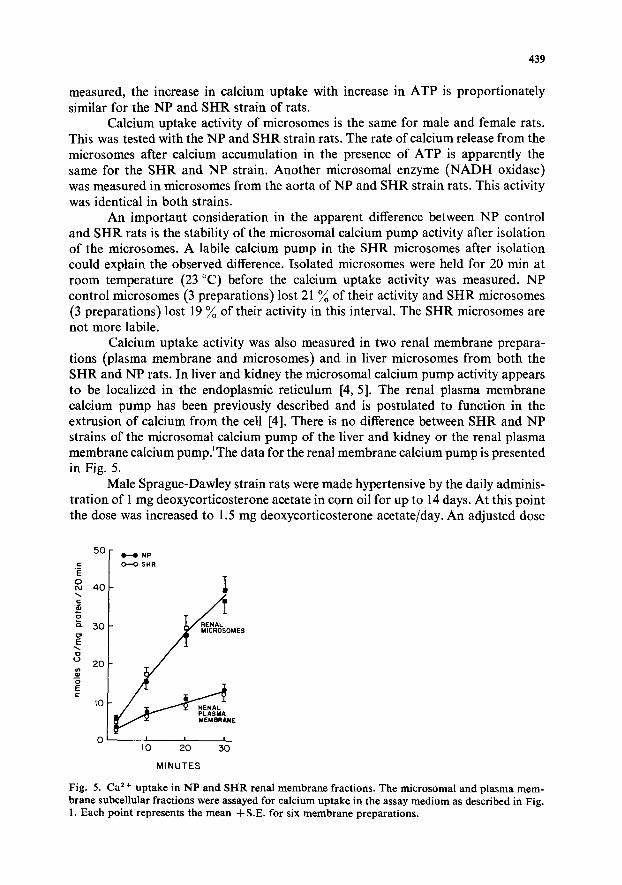
Calcium uptake activity was also measured in two renal membrane prepara- tions (plasma membrane and microsomes) and in liver microsomes from both the SHR and NP rats. In liver and kidney the microsomal calcium pump activity appears to be localized in the endoplasmic reticulum [4, 5]. The renal plasma membrane calcium pump has been previously described and is postulated to function in the extrusion of calcium from the cell [4]. There is no difference between SHR and NP strains of the microsomal calcium pump of the liver and kidney or the renal plasma membrane calcium pump.~The data for the renal membrane calcium pump is presented in Fig. 5.
Male Sprague-Dawley strain rats were made hypertensive by the daily adminis- tration of 1 mg deoxycorticosterone acetate in corn oil for up to 14 days. At this point the dose was increased to 1.5 mg deoxycorticosterone acetate/day. An adjusted dose
50 ~ H NP
"-¢ I o--o SHR E ° eJ 40
- j o ~" 30 RENAL MES g g' 2o
Io , PLASMA
I
° ,o ; ; MINUTES
Fig. 5. Ca 2+ up take in N P and SI-IR renal m e m b r a n e fractions. The mic rosomal and p lasma m e m - brane subcellular fract ions were assayed for calcium uptake in the assay m e d i u m as described in Fig. 1. Each point represents the m e a n 4-S.E. for six m e m b r a n e preparat ions .

440
£ o.
c :
50
4 0
30
20
CONTROL
OOCA O 30d
I0
I I I
0 - - - I0 20 30
M I N U T E S
50 ¸
4O
~E
3 0
_~ 2o
10
0 - -
" H CONTROL O.--O DOCA TREATED 3Od
RENAL MICROSOMES
RENAL PLASMA
NE
I I I
I0 20 ~ 3 0
M I N U T E S
Fig. 6. C a 2 + uptake in deoxycorticosterone acetate-treated rat aorta microsomes (left) and renal membrane fractions (right). The treated rats received 1 mg deoxycorticosterone acetate/rat/day intra- peritoneally for 14 days and the dosage was then increased to 1.5 rag/rat/day intraperitoneally for the following 16 days. Control animals received daily corn oil injections and the treated animals had one percent NaC1 supplementation in their drinking water. C a 2 + uptake was assayed as described in Fig. 1. Both groups were assayed simultaneously. Each point represents the mean ~:S.E. for six determi- nations. Systolic blood pressure in treated animals was 167~4.0 and in control rats was 1 1 7 i 2 . 5 mm H g . *p - - < 0.05. D O C A , deoxycorticosterone.
allows for an increase in size of the animals during a thirty day period of treatment. Control animals received daily administration of the corn oil. After ten days, blood pressure of controls was 113± 1.6 and blood pressure of animals receiving deoxycor- ticosterone acetate was 135±3.1. The calcium uptake of aorta microsomes, renal microsomes and renal plasma membranes was not affected by the ten day treatment. After thirty days the blood pressure of controls was 117~:2.5 and the blood pressure of animals receiving deoxycorticosterone acetate for this time period was 167+4. Following thirty days of treatment the calcium uptake activity in kidney microsomes is clearly depressed (Fig. 6). A varied decrease in calcium uptake is observed with the aorta microsomes (Fig. 6). There were six experimental groups of four rats. Calcium uptake activity of the aorta microsomes in four of the experimental groups fell below any activity level observed in the six control groups. The renal plasma membrane calcium uptake activity remains unaffected by treatment with deoxycorticosterone acetate.
D I S C U S S I O N
The study characterizes the calcium uptake activity of the microsomal fraction of rat aorta. This activity in the presence of Mg • ATP appears to be accumulation rather than a simple exchange of membrane bound calcium. The initial calcium content of aorta microsomes when measured chemically is less than 5 nmol/mg protein. This resembles values previously reported for liver microsomes [28]. The uptake of calcium presented in Fig. 1 exceeds 40 nmol/mg protein in 30 rain. As with skeletal muscle studies [17] the enhanced accumulation in presence of oxalate as a
441
trapping agent argues for net uptake of calcium. The progressive accumulation of calcium over a 1-h period is also more characteristic of a net accumulation. The initial uptake in absence of Mg. ATP of 2 nmol of calcium/mg protein that is essentially complete in the first 2 min may very well be an exchange.
The gradient density analysis indicates that the calcium pump activity of microsomes isolated from rat aorta is principally associated with the sarcoplasmic reticulum. Calcium pump activity of the sucrose gradient is distributed in parallel with the enzyme marker for sarcoplasmic reticulum. The sarcoplasmic reticulum of smooth muscle may serve as an intracellular store for calcium. An earlier study in this laboratory examined microsomes which were isolated from the longitudinal muscle of guinea pig ileum. On a sucrose gradient the calcium pump activity of these guinea pig ileum microsomes was found to be primarily associated with the enzyme marker for plasma membrane [3]. Guinea pig ileum muscle, which is apparently deficient in calcium pump activity associated with sarcoplasmic reticulum, loses its capacity to contract in the absence of calcium in the external medium [18]. The guinea pig muscle does have mitochondria with calcium sequestering capability but they do not seem to supply a significant quantity of calcium to affect the contractile process [3]. Devine et al. [9] have demonstrated that the capacity for mechanical activity of smooth muscle fibers in the absence of external calcium relates to the content of sarcoplasmic reticulum seen in the muscle on electron microscopic examina- tion. They showed that smooth muscles which have relatively slight content of sarcoplasmic reticulum quickly lose the capacity to contract when stimulated in a calcium free medium.
It seems possible that the sarcoplasmic reticulum of the rat aorta plays a role in muscle relaxation. The capacity of the microsomes isolated in the present study to sequester calcium is in excess of 60 nmol of calcium per gram of aorta for a 10-min period. This is a minimal estimate of the activity since the homogenization of the tissue and the recovery of microsomes is known to be incomplete. The use of a light microsomal fraction with smaller fragments of membranes in this study may also underestimate the total activity. The microsomal fraction sedimenting between 27 000 x 9 and 105 000 x O has been employed since this fraction is devoid of contami- nation by intact mitochondria. This was evident on electron microscopy and by the absence of azide sensitive calcium uptake. Martonosi and Feretos [17] have pre- viously demonstrated with skeletal muscle sarcoplasmic reticulum that "grana II" or light microsomes have lower activity than the "grana I" fraction made up of larger fragments of sarcoplasmic reticulum membranes.
Resting smooth muscle has an ionized calcium concentration in the myoplasm that is less than 0.1 #M [19]. Half maximal contraction occurs with an ionized calcium level of about 1/IM [20]. The fully contracted muscle is not likely to exceed 10/~M or 8 nmol calcium per gram of tissue. The apparent capacity of the microsomal system measured under the in vitro conditions of this study would appear to be adequate for function in the muscle relaxation process by taking up free calcium. The sarcoplasmic reticulum system is also postulated to provide an intracellular store of calcium for subsequent stimuli [21].
Calcium pump activity was examined in rats with experimental hypertension. Calcium pump activity of aorta microsomes was decreased in the seventy-five geneti- cally hypertensive SHR rats looked at in the present study. Similar activity in renal

442
and hepat ic tissue was unaffected. A br ief repor t by Aok i et al. [22] notes a 40 ~'~i decrease in A T P dependen t ca lc ium seques t ra t ion of microsomes f rom several b lood vessels in the S H R rat. This is conf i rmed in the present s tudy for the rat aor ta . They suggest that the reduced calc ium up take act ivi ty o f the sa rcoplasmic re t icu lum might induce a higher tone o f the vascular muscle and lead to an increase in b lood pressure.
Sh iba ta et al. [23] examined the capac i ty o f the ao r t a ob ta ined f rom the S H R ra t to con t rac t in a ca lc ium free medium. The ao r t a f rom the S H R rat shows a faster ra te of loss o f norep inephr ine - induced or po tas s ium- induced con t rac t ion in the absence o f external calcium. This could imply a decreased funct ional internal store o f ca lc ium in the S H R aor ta . This is consis tent with the observa t ion in the present s tudy o f an impa i red calc ium seques t ra t ion activi ty in the ao r t a microsomes of the S H R rat .
A var iable decrease in mic rosomal ca lc ium p u m p act ivi ty of the rat ao r t a is seen af ter 30 days o f deoxycor t icos te rone t r ea tment in the present study. Long te rm deoxycor t i cos te rone t rea tment in rats appears to be associa ted with an al tered ar ter ia l responsiveness. The isola ted femora l a r te ry f rom deoxycor t i cos te rone hyper- tensive rats responds more quickly to st imuli and relaxes more s lowly when the st imuli are r emoved [24]. The ventral a r te ry o f the ra t tail f rom the deoxycor t icos te- rone hypertensive ra t behaves in s imilar fashion [25]. This is compat ib le with an appa ren t decrease in calc ium p u m p activity. A n a l ternat ive exp lana t ion for increased responsiveness is offered by Somlyo and Somlyo [21]. M e m b r a n e permeabi l i ty to ca lc ium may be increased. The final result would resemble a leaky calc ium p u m p system.
ACKNOWLEDGEMENTS
This inves t iga t ion was in i t ia ted by a g ran t - in -a id f rom the Midd le Tennessee H e a r t Associa t ion . Con t inued suppo r t was p rov ided by N I H research gran t number H L 14681, N I H t ra in ing gran t number G M 0058 f rom the Na t iona l Inst i tute o f Genera l Medica l Sciences, PHS Biomedica l Sciences Suppo r t g ran t number R R 07089 and P H S Genera l Research Suppo r t grant n u m b e r F R 05424.
REFERENCES
1 Fitzpatrick, D. F., Landon, E. J., Debbas, G. and Hurwitz, L. (1972) Science 176, 305-506 2 Hess, M. L. and Ford, G. D. (1974) J. Mol. and Cell. Cardiol. 6, 275-282 3 Hurwitz, L., Fitzpatrick, D. F., Debbas, G. and Landon, E. J. (1973) Science 179, 384-386 4 Moore, L., Fitzpatrick, D. F., Chen, T. S. and Landon, E. J. (1974) Biochim. Biophys. Acta 345,
405-4 18 5 Moore, L., Chen, T., Knapp, H. R. and Landon, E. J. (1975) J. Biol. Chem. 250, 4562-4568 6 Martonosi, A. (1971) in Biomembranes (Manson, L. A., ed.), Vol, I, pp. 191-256, Plenum Press,
New York 7 Ebashi, S., Endo, M. and Ohtsuki, I. (1969) Quart. Rev. Biophys, 2, 351-384 8 Weber, A., Herz, T. and Reiss, I. (1963) J. Gem Physiol. 46, 679-702 9 Devine, C. E., Somlyo, A. V. and Somlyo, A. P. (1972) J. Cell. Biol. 52, 690-718
10 Okamoto, K. and Aoki, K. (1963) Jap. Circ. J. 27, 282-293 11 Louis, W. J., Krauss, K. R., Kopin, ]. J. and Sjoerdsma, A. (1970) Circ. Res. 27, 589-594 12 Janis, R. A. and Triggle, ZD. J. (1972) J. Pharm. Pharmacol. 24, 602-608 13 Widnell, C. and Unkeless, J. C. (1968) Proc. Natl. Acad. Sci. U.S. 61, 1050-1057 14 Avruch, J. and Wallach, D. F. H. (1971) Biochim. Biophys. Acta 233, 334-347

443
15 Sutherland, E., Cori, C. F., Haynes, R. and Olsen, N. (1949) J. Biol. Chem. 180, 826-837 16 Wharton, D. C. and Tzagoloff, A. (1967) in Methods in Enzymology (Estabrook, R. W. and
Pullman, M. E. eds), Vol. 10, pp. 245-250, Academic Press, New York 17 Martonosi, A. and Feretos, R. (1964) J. Biol. Chem. 239, 648-658 18 Hurwitz, L. and Joiner, P. S. (1970) Am. J. Physiol. 218, 12-19 19 Bohr, D. F. (1973) Circ. Res. 32, 665-672 20 Sparrow, M. L., Maxwell, L. C., Ruegg, J. C. and Bohr, D. F. (1970) Am. J. Physiol. 219, 1366-
1372 21 Somlyo, A. P. and Somlyo, A. V. (1970) Pharmacol. Rev. 22, 249-353 22 Aoki, K., Yamashita, K., Tomita, N., Tazumi, K. and Hotta, K. (1974) Jap. Heart J. 15, 180-181 23 Shibata, S., Kurahashi, K. and Kuchii, M. (1973) J. Pharmacol. Exptl. Ther. 185, 406-417 24 Holloway, E. T., Sitrin, M. D. and Bohr, D. F. (1972) in Hypertension '72 (Genest. J, and Koiw,
E., eds), pp. 400-408, Springer-Verlag, New York 25 Hinke, J. A. M. (1966) Circ. Res. 18, Suppl. 1, 23-33 26 d'Auriac, G. A., Baudoin, M. and Meyer, P. (1972) Circ. Res. 30, Suppl. II, 151-157 27 Kidwai, A. M., Radcliffe, M. A. and Daniel, E. E. (1971) Biochim. Biophys. Acta 233,538-549 28 Thiers, R. E. and VaUee, B. L. (1957) J. Biol. Chem. 226, 911-920