Endoscopic Molecular Imaging in Gastrointestinal Oncology
18
12 Endoscopic Molecular Imaging in Gastrointestinal Oncology Naoki Muguruma and Tetsuji Takayama The University of Tokushima Graduate School Japan 1. Introduction Gastrointestinal (GI) endoscopy has been widely used for detection, differentiation and staging for neoplasia in the digestive tract, and has made great progress during the last decade (Sivak, 2006). Diagnostic accuracy can be enhanced by better training, more efficient techniques, and the development of new image-processing technologies (Cotton et al., 2006; Tajiri, 2007); however, diagnosis using conventional endoscopy with optical characteristics is essentially limited because it is based on morphological changes and/or discoloration. Chromoendoscopy can enhance surface structure and to determine demarcation borders but is not enough for screening out of early cancer because it is still depending on endoscopists’ expertise and biopsy. Auto-fluorescence imaging system has been applied for lesions which have been difficult to morphologically recognize or are indistinct with conventional endoscope and this system has potential application for the diagnosis of dysplastic lesions and early cancers in the gastrointestinal tract. Optical digital enhancing method such as narrow band imaging (NBI), flexible spectral imaging color enhancement (FICE) and i- SCAN are novel endoscopic techniques which can distinguish neoplastic and non-neoplastic lesions without the dye. Magnifying endoscopy in combination with optical digital method has an obvious advantage; the analysis of the epithelial pit pattern and the vascular network. Some of other techniques are allowing us to visualize cell morphology on the micro level reflection of microscopic characteristics. However, we should solve these problems; how to combine these technologies in diagnostic strategy, how to apply them into the algorithm for therapeutic decision and how to standardize several classifications of morphology surrounding them. ‘Molecular imaging’ is a concept representing the most novel imaging methods in medicine, and the definition of the word is controversial. It is broadly defined as ‘the in vivo characterization and measurement of a biological process at the cellular molecular level’ (Weissleder & Mahmood, 2001) or as the technique that ‘directly or indirectly monitor and record the spatiotemporal distribution of molecular and cellular processes for biochemical, biological, diagnostic, or therapeutic application’ (Thakur & Lentle, 2005). Positron emission tomography (PET) might be included in a wide concept of molecular imaging methods: the detection, spatial localization, and quantification of specific molecular targets and events that form the basis of pathologies (Mahmood & Wallace, 2007). In the clinical setting of medical fields, a major paradigm shift has been rapidly taking place in imaging technology represented by PET. Similarly, in the field of GI endoscopy, the authors propose rapid www.intechopen.com
Transcript of Endoscopic Molecular Imaging in Gastrointestinal Oncology
12
Endoscopic Molecular Imaging in Gastrointestinal Oncology
Naoki Muguruma and Tetsuji Takayama The University of Tokushima Graduate School
Japan
1. Introduction
Gastrointestinal (GI) endoscopy has been widely used for detection, differentiation and staging for neoplasia in the digestive tract, and has made great progress during the last decade (Sivak, 2006). Diagnostic accuracy can be enhanced by better training, more efficient techniques, and the development of new image-processing technologies (Cotton et al., 2006; Tajiri, 2007); however, diagnosis using conventional endoscopy with optical characteristics is essentially limited because it is based on morphological changes and/or discoloration. Chromoendoscopy can enhance surface structure and to determine demarcation borders but is not enough for screening out of early cancer because it is still depending on endoscopists’ expertise and biopsy. Auto-fluorescence imaging system has been applied for lesions which have been difficult to morphologically recognize or are indistinct with conventional endoscope and this system has potential application for the diagnosis of dysplastic lesions and early cancers in the gastrointestinal tract. Optical digital enhancing method such as narrow band imaging (NBI), flexible spectral imaging color enhancement (FICE) and i-SCAN are novel endoscopic techniques which can distinguish neoplastic and non-neoplastic lesions without the dye. Magnifying endoscopy in combination with optical digital method has an obvious advantage; the analysis of the epithelial pit pattern and the vascular network. Some of other techniques are allowing us to visualize cell morphology on the micro level reflection of microscopic characteristics. However, we should solve these problems; how to combine these technologies in diagnostic strategy, how to apply them into the algorithm for therapeutic decision and how to standardize several classifications of morphology surrounding them. ‘Molecular imaging’ is a concept representing the most novel imaging methods in medicine, and the definition of the word is controversial. It is broadly defined as ‘the in vivo characterization and measurement of a biological process at the cellular molecular level’ (Weissleder & Mahmood, 2001) or as the technique that ‘directly or indirectly monitor and record the spatiotemporal distribution of molecular and cellular processes for biochemical, biological, diagnostic, or therapeutic application’ (Thakur & Lentle, 2005). Positron emission tomography (PET) might be included in a wide concept of molecular imaging methods: the detection, spatial localization, and quantification of specific molecular targets and events that form the basis of pathologies (Mahmood & Wallace, 2007). In the clinical setting of medical fields, a major paradigm shift has been rapidly taking place in imaging technology represented by PET. Similarly, in the field of GI endoscopy, the authors propose rapid
www.intechopen.com
New Techniques in Gastrointestinal Endoscopy
198
development of ‘endoscopic molecular imaging’, which is considered to be divided into three categories: (a) visualization of cell morphology on the micro to nano level; (b) reflection of spectroscopic characteristics; and (c) visualization of molecular characteristics. The future of endoscopic diagnosis is likely to be affected by a combination of biomarkers and technology (Takayama et al., 1998), and ‘endoscopic molecular imaging’ would be defined as (c), which has been described as ‘immunoscopy’ (Keller et al., 1998), ‘bioendoscopy’ (Pasricha & Motamedi, 2002), and ‘optical biopsy’ (Fujimoto et al., 1995). These innovations will allow us not only to locate a tumor but also useful to 1) differentiate malignant and benign polyps and ulcers, 2) minimize number of biopsies and frequency of surveillance, 3) accurate preoperative identification of tumor margin, 4) evaluate effectiveness of pharmacological therapy, 5) detect local dysplasia in Barrett’s mucosa or ulcerative colitis. These will also allow us to visualize its molecular characteristics (e.g. DNA mutations and polymorphisms, gene and/or protein expression), and the activity of specific molecules and biological processes that affect tumor behavior and/or its response to therapy (Weissleder, 2006). Hsiung et al. detected in vivo human colonic dysplasia using a targeted heptapeptide topically administered and confocal microendoscopy in 2008 (Hsiung et al., 2008). Recently, Goetz et al. used confocal laser endomicroscopy (CRE) and fluorescently labeled epidermal growth factor receptor (EGFR) and succeeded in differentiating EGFR expression patterns in xenograft tumors and human neoplastic tissue samples (Goetz et al., 2010). These are promising future technologies that will play a central role in gastrointestinal oncology. We have been attempting to develop a novel imaging method using antibodies labeled with a fluorescent marker excitable by infrared rays and imaging modalities. In this section, we will describe our results and future directions of endoscopic molecular imaging in gastrointestinal oncology.
2. Characteristics of infrared fluorescence
Infrared radiation is light with wavelengths between 780 nm and 100 μm, and it has high permeability and safety compared to ultraviolet rays. These characteristics have been applied to various technologies such as non-destructive analysis of agricultural products, and infrared photography has been especially investigated in the medical field (Gibson et al., 1965; Mimura & Okuda, 1981). Infrared endoscopy has been used as a special diagnostic tool for examination of the gastrointestinal tract with or without intravenous injection of indocyanine green (ICG) (Kohso et al., 1990; Ohta et al., 1994; Iseki et al., 2000; Mataki et al., 2003; Ishihara, 2010) ; ICG is widely used as a reagent for clinical examination of hepatic function (Fox & Wood, 1960). ICG is a fluorescent agent that absorbs infrared rays and produces visible spots at the maximum wavelength of 805 nm (Nimura et al., 2004). ICG emits wavelengths of 807-832 nm on excitation at around 770 nm (Benson & Kues, 1978; Mordon et al., 1998; Muguruma et al., 1999). Taking advantage of this characteristic, infrared fluorescence is used for retinal angiography (Flower, 1973) and the evaluation of burn depth (Still, 2001) and the patency of cardiac venografts (Detter et al., 2002). Recently, this property was applied to gastrointestinal blood vessels using a CCD camera (Borotto et al., 1999). In the living body, components or elements emit fluorescence of 310-540 nm when excited at 280-370 nm. In addition, there is little background noise in the living body (Shealy et al., 1995), especially in the digestive tract, which makes infrared fluorescence a likely candidate for development as a novel diagnostic system (Ganz, 2004; Okamoto et al., 2005). Several kinds of labeling agents for detecting
www.intechopen.com
Endoscopic Molecular Imaging in Gastrointestinal Oncology
199
carcinomas in the digestive tract have been reported (Tatsuta, 1989; Pelegrin, 1991; Ballou et al., 1998), some of which fluoresce in visible or ultraviolet rays. However, application of UV is not suitable because it damages living tissue (Davies, 1995). ICG seems to be a suitable molecule for immunofluorescent diagnosis in the digestive tract such as esophagus, stomach, and colon because of its spectral properties and low toxicity.
3. Fluorescent agents
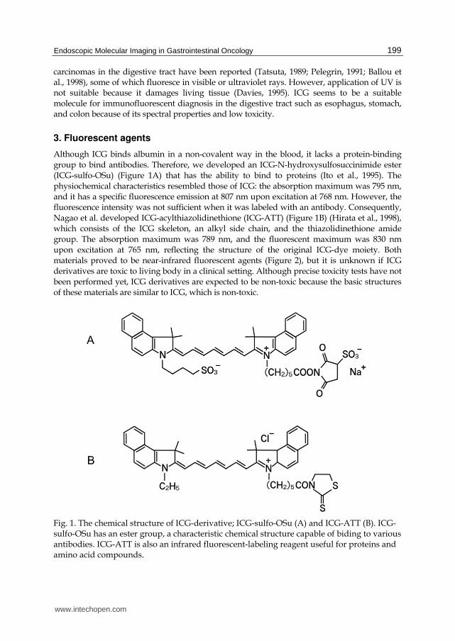
Although ICG binds albumin in a non-covalent way in the blood, it lacks a protein-binding group to bind antibodies. Therefore, we developed an ICG-N-hydroxysulfosuccinimide ester (ICG-sulfo-OSu) (Figure 1A) that has the ability to bind to proteins (Ito et al., 1995). The physiochemical characteristics resembled those of ICG: the absorption maximum was 795 nm, and it has a specific fluorescence emission at 807 nm upon excitation at 768 nm. However, the fluorescence intensity was not sufficient when it was labeled with an antibody. Consequently, Nagao et al. developed ICG-acylthiazolidinethione (ICG-ATT) (Figure 1B) (Hirata et al., 1998), which consists of the ICG skeleton, an alkyl side chain, and the thiazolidinethione amide group. The absorption maximum was 789 nm, and the fluorescent maximum was 830 nm upon excitation at 765 nm, reflecting the structure of the original ICG-dye moiety. Both materials proved to be near-infrared fluorescent agents (Figure 2), but it is unknown if ICG derivatives are toxic to living body in a clinical setting. Although precise toxicity tests have not been performed yet, ICG derivatives are expected to be non-toxic because the basic structures of these materials are similar to ICG, which is non-toxic.
Fig. 1. The chemical structure of ICG-derivative; ICG-sulfo-OSu (A) and ICG-ATT (B). ICG-sulfo-OSu has an ester group, a characteristic chemical structure capable of biding to various antibodies. ICG-ATT is also an infrared fluorescent-labeling reagent useful for proteins and amino acid compounds.
www.intechopen.com
New Techniques in Gastrointestinal Endoscopy
200
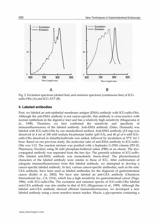
Fig. 2. Excitation spectrum (dotted line) and emission spectrum (continuous line) of ICG-sulfo-OSu (A) and ICG-ATT (B).
4. Labeled antibodies
First, we labeled an anti-epithelial membrane antigen (EMA) antibody with ICG-sulfo-OSu. Although the anti-EMA antibody is not cancer-specific, this antibody is cross-reactive with normal epithelium in the digestive tract and has a relatively high sensitivity (Muguruma et al., 1998). Therefore, we first confirmed the sensitivity and specificity of immunofluorescence of the labeled antibody. Anti-EMA antibody (Dako, Denmark) was labeled with ICG-sulfo-OSu by our standardized method. Anti-EMA antibody (2.8 mg) was dissolved in 4 ml of 100 mM sodium bicarbonate buffer (pH 8.5), and 40 μl of 6 mM ICG-sulfo-OSu dissolved in dimethylsulfoxide was added, followed by incubation at 37°C for 1 hour. Based on our previous study, the molecular ratio of anti-EMA antibody to ICG-sulfo-OSu was 1:12. The reaction mixture was purified with a Sephadex G-25M column (PD-10, Pharmacia, Sweden) using 50 mM phosphate-buffered saline (PBS) as an eluent. The dye-conjugated antibody was separated from the free dye. The greenish solution of ICG-sulfo-OSu labeled anti-EMA antibody was immediately freeze-dried. The physiochemical characters of the labeled antibody were similar to those of ICG. After confirmation of adequate immunofluorescence from this labeled antibody, we attempted to develop a cancer-specific labeled antibody. In fact, various cancer-specific antibodies, such as the anti-CEA antibody, have been used as labeled antibodies for the diagnosis of gastrointestinal cancer (Keller et al., 2002). We have also labeled an anti-CEA antibody (Chemicon International Inc., CA, USA), which has a high sensitivity for gastrointestinal cancer (Page, 1986), with ICG-sulfo-OSu. The excitation and emission spectra of ICG-sulfo-OSu labeled anti-CEA antibody was also similar to that of ICG (Muguruma et al., 1999). Although the labeled anti-CEA antibody showed efficient immunofluorescence, we developed a new labeled antibody using a more sensitive tumor marker. Mucin, a glycoprotein containing a
www.intechopen.com
Endoscopic Molecular Imaging in Gastrointestinal Oncology
201
large amount of sugar, is the main component of mucus, and the peptide structure of the mucin core protein has been clarified (Kim, 1993). The specific expression of mucin in various cancers has been reported, and we also studied the staining pattern and evaluated its sensitivity in gastrointestinal cancers (Bando et al., 2002). Based on its relatively high sensitivity (Nakamori, 1994), we labeled an anti-MUC1 antibody (MY.1E12; kindly provided by Prof. Tatsuro Irimura, The University of Tokyo) with ICG-ATT.
5. Imaging modality
5.1 Infrared fluorescence microscope We developed an infrared fluorescence microscope for observation of tissue sections by modifying a conventional infrared microscope (BHSM-IR, Olympus, Japan). The excitation filter with a transmission wavelength of 710–790 nm was placed between the halogen lamp and the sample, and the barrier filter with a transmission wavelength of 810–920 nm was placed between the sample and the charge-coupled device (CCD) camera (Ito, 1997).
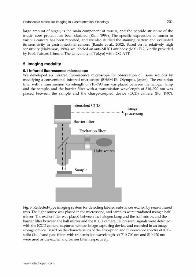
Fig. 3. Reflected-type imaging system for detecting labeled substances excited by near-infrared rays. The light source was placed in the microscope, and samples were irradiated using a half mirror. The exciter filter was placed between the halogen lamp and the half mirror, and the barrier filter between the half mirror and the ICCD camera. Fluorescent signals were detected with the ICCD camera, captured with an image capturing device, and recorded in an image storage device. Based on the characteristics of the absorption and fluorescence spectra of ICG-sulfo-Osu, band pass filters with transmission wavelengths of 710-790 nm and 810-920 nm were used as the exciter and barrier filter, respectively.
www.intechopen.com
New Techniques in Gastrointestinal Endoscopy
202
Fluorescent signals were detected by a CCD camera equipped with an image-capturing device and recorded in an image-storage device. All the images were processed by averaging original images, averaging background images, subtraction, noise filtering, and contrast enhancement. However, this transmission-type imaging system cannot be applied to an endoscopic system because the specimen is sandwiched between the excitation and barrier filters. We therefore developed a reflected-type infrared fluorescence microscopy: the light source was placed in the microscope, and the samples were irradiated using a half mirror (Figure 3) (Taoka, 1999). The excitation filter was placed between the halogen lamp and the half mirror and the barrier filter between the half mirror and the intensified charge coupled device (ICCD) camera. Light from the light source passes through the excitation filter, and about 50% of the light is reflected and irradiates the sample. About 50% of the fluorescence emitted from the sample passes through the barrier filter and is detected by the ICCD camera. Although the fluorescent input is reduced by 25% compared with that of previous type microscopy, theoretically the ICCD camera enhances the images better than the CCD camera equipped with the transmission type. Images were processed by a recursive filter, which can also average some images after emphasizing later images, on an ICCD controller to decrease noise. The efficiency of image processing using this recursive filter was expressed by the following equation: Vn · 1 + (Vin – Vn · 1) / N, where Vn = output image, Vn · 1 = the output image 1 frame earlier, Vin = input image, and N = constant (4, 16, 64).
5.2 Infrared fluorescence endoscope Based on the results of immunofluorescence using infrared fluorescence microscopy, a prototype of infrared fluorescence endoscopy was developed to observe the human gastrointestinal tract (Figure 4) (Ito et al., 2001). The system consisted of an infrared endoscope (Olympus XGIF-Q40IR, Olympus) coupled with an image-capturing device. The light source, a 300 W xenon lamp, was also equipped with an excitation filter and a barrier filter, making it possible to observe fluorescence with the infrared excitation light and produce normal images under visible light. The ICCD camera was optically connected with the scope through an adapter into which the barrier filter was inserted. The new endoscopy system with a CCD at its tip has a greatly improved resolution: this system comprises of a light source apparatus, an infrared fluorescence endoscope, and image analysis software, which is the same as that used for a conventional system (Kimura et al., 2007). The light source apparatus (XCLV-260HP-IRF, Olympus) has three built-in filters; an infrared ray cut filter, an infrared ray pass filter, and a RGB filter. White light produced by a xenon arc lamp goes through the infrared ray cut filter or infrared ray pass filter and then through the RGB rotation filter. The infrared ray cut filter is used for conventional observation and the infrared ray pass filter for infrared fluorescence observation. The infrared ray pass filter can transmit rays of wavelengths between 540 nm and 560 nm in addition to infrared rays of wavelengths between 680 nm and 770 nm. The reflected light of the former rays allow us to know where in the stomach we are looking during infrared fluorescence observation. The RGB rotation filter can transmit light in the near-infrared region as well as the visible region. The infrared fluorescence endoscope (XGIFQ-240IRFZ, Olympus) is equipped with both a CCD with high resolution for conventional observation and a CCD with high sensitivity for infrared fluorescence observation at the top. These two CCDs can be switched from one to another with a single touch of the button of the endoscope in conjunction with the switch from one filter to another (the infrared ray cut filter and infrared ray pass filter) in the light
www.intechopen.com
Endoscopic Molecular Imaging in Gastrointestinal Oncology
203
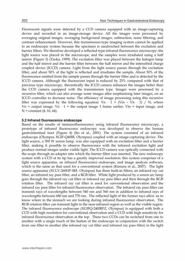
source apparatus. Infrared rays excite the ICG-derivative-labeled antibody to emit fluorescence, which is subjected to barrier filters (825 to 945 nm) placed in front of the CCD for infrared fluorescence and is monitored as green signals on a pseudo-color display with an image processor (XCV-260HP-IRF, Olympus).
Fig. 4. The schema of infrared fluorescence endoscope system. This system consisted of an infrared endoscope (Olympus XGIF-Q40IR, OLYMPUS) coupled with an image-capturing device. The light source that includes a 300 W xenon lamp which was equipped with an excitation filter and a barrier filter, so that it was possible to observe fluorescence with infrared excitation light and normal images under visible light. The ICCD camera was optically connected with the scope through an adapter into which the barrier filter was inserted.
6. Ex vivo study of immunofluorescence with human stomach
6.1 Immunofluorescence with microscope system Paraffin sections of human gastric mucosa, which had been previously proven to be positive in usual immunostaining for the anti-MUC1 antibody, were deparaffinized, and xylene was removed. After blocking endogenous peroxidase activity, sections were incubated with normal horse serum for 20 min, and then with the labeled antibodies at 500 mg/mL in 0.1M PBS for 10 min at room temperature. Observationwas performed under the infrared fluorescence imaging device to compare the fluorescence intensity of each preparation. Adjacent sections processed in the same way were incubated with the primary antibody, and then treated with the secondary antibody and the ABC reagent (avidin biotinylated
www.intechopen.com
New Techniques in Gastrointestinal Endoscopy
204
peroxidase complex) for 30 min. Preparations were visualized by 3-3‘ diaminobenzidine
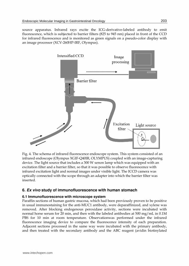
(DAB), and counter-stained by hematoxylin and methyl green. Localization of infrared fluorescence and DAB coloring was compared in serial sections. Subsequently, immunofluorescence of the paraffin sections of human gastric mucosa with ICG ATT-labeled anti-MUC1 antibody produced stronger fluorescence than that by ICG-sulfo-OSu-labeled antibody (Figure 5). Localization pattern of infrared fluorescent staining was in good agreement with that by the conventional method with oxidized DAB staining, which confirmed that the fluorescence due to the ICG ATT-labeled was more specific and sensitive to MUC1.
Fig. 5. Images of infrared fluorescent microscopy for gastric cancer specimen reacted with ICG-derivative labeled anti-MUC1 antibody. A) ICG-sulfo-OSu labeled anti-MUC1 antibody, B) ICG-ATT labeled anti-MUC1 antibody. The fluorescence intensity of the antigen stained with ICG-ATT labeled antibody was markedly stronger than that treated with ICG-sulfo-OSu labeled antibody.
6.2 Immunofluorescence with endoscope system The immunoreactions of CEA with ICG-sulfo-OSu labeled anti-CEA antibody were examined in freshly resected human gastric cancer specimens using the infrared
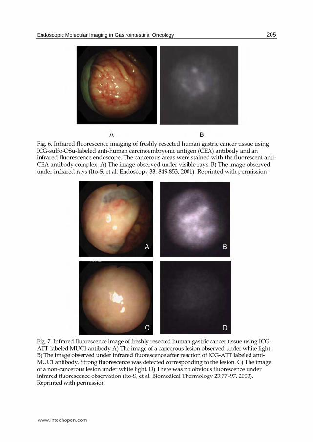
fluorescence endoscope. The resected stomach tissue was treated with warm water of 37℃ containing 20000 U of Pronase, 1 g of NaHCO3, and 4 mg of dimethylpolysiloxane for 15 min at room temperature to remove mucus adhering to the mucosa. The sample was treated with normal horse serum (blocking serum) for 15 min. The surface of the lesion and normal mucosa were then treated with ICG-derivative-labeled antibody for 60 min at room temperature (Ito et al., 2001). Then, normal mucosa and the cancerous area were compared using the infrared fluorescence endoscope. Infrared fluorescence was observed in the cancerous lesion, but not in the non-cancerous areas (Figure 6). Paraffin sections of the stomach encompassing the infrared fluorescence-positive site were stained with anti-CEA antibody using the ABC method. The immunoreactive staining was positive only in the cancerous lesions but was negative in the non-cancerous lesions. In the infrared fluorescence-positive sites, the carcinoma tissue was exposed on the mucosal surface.
www.intechopen.com
Endoscopic Molecular Imaging in Gastrointestinal Oncology
205
Fig. 6. Infrared fluorescence imaging of freshly resected human gastric cancer tissue using ICG-sulfo-OSu-labeled anti-human carcinoembryonic antigen (CEA) antibody and an infrared fluorescence endoscope. The cancerous areas were stained with the fluorescent anti-CEA antibody complex. A) The image observed under visible rays. B) The image observed under infrared rays (Ito-S, et al. Endoscopy 33: 849-853, 2001). Reprinted with permission
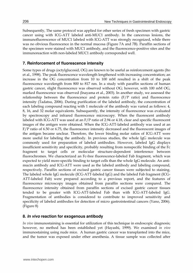
Fig. 7. Infrared fluorescence image of freshly resected human gastric cancer tissue using ICG-ATT-labeled MUC1 antibody A) The image of a cancerous lesion observed under white light. B) The image observed under infrared fluorescence after reaction of ICG-ATT labeled anti-MUC1 antibody. Strong fluorescence was detected corresponding to the lesion. C) The image of a non-cancerous lesion under white light. D) There was no obvious fluorescence under infrared fluorescence observation (Ito-S, et al. Biomedical Thermology 23:77–97, 2003). Reprinted with permission
www.intechopen.com
New Techniques in Gastrointestinal Endoscopy
206
Subsequently, The same protocol was applied for other series of fresh specimen with gastric cancer using with ICG-ATT labeled anti-MUC1 antibody. In the cancerous lesions, the immunofluorescence of MUC1 labeled with ICG-ATT was strongly recognized, while there was no obvious fluorescence in the normal mucosa (Figure 7A and 7B). Paraffin sections of the specimen were stained with MUC1 antibody, and the fluorescence-positive sites and the immunoreaction with non-labeled MUC1 antibody corresponded well.
7. Reinforcement of fluorescence intensity
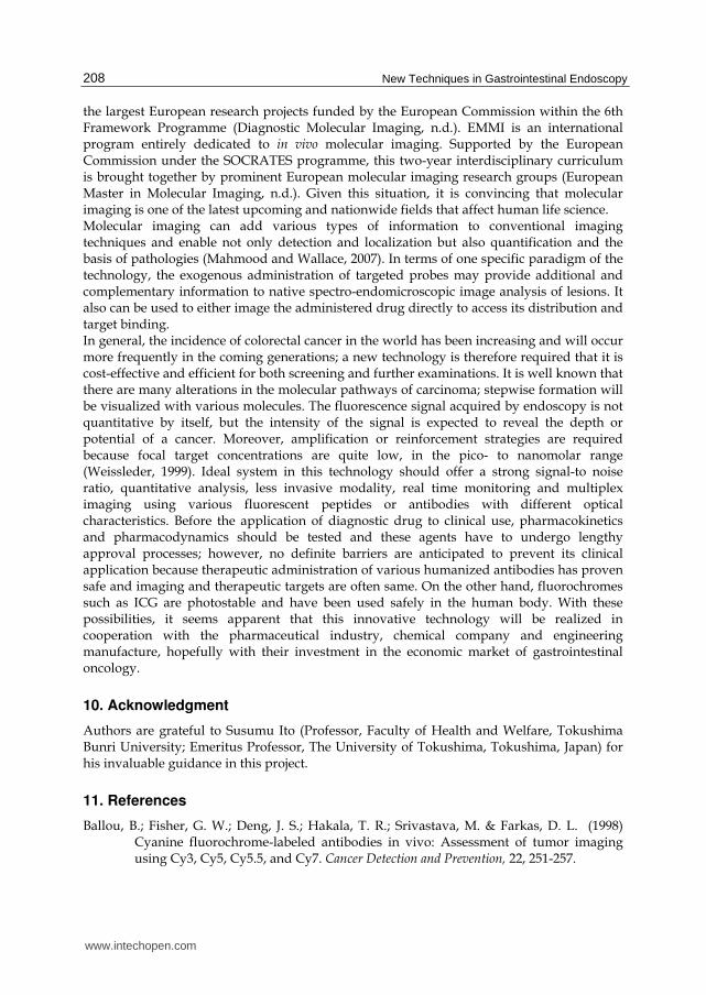
Some types of drugs (octylglucosid, OG) are known to be useful as reinforcement agents (Ito et al., 1998). The peak fluorescence wavelength lengthened with increasing concentration; an increase in the OG concentration from 10 to 100 mM resulted in a shift of the peak fluorescence wavelength from 800 to 817 nm. In a study with paraffin sections of human gastric cancer, slight fluorescence was observed without OG; however, with 100 mM OG, marked fluorescence was observed (Inayama et al., 2003). In another study, we assessed the relationship between the fluorescence and protein ratio (F/P ratio) and fluorescence intensity (Tadatsu, 2006). During purification of the labeled antibody, the concentration of each labeling compound reacting with 1 molecule of the antibody was varied as follows: 4, 8, 16, and 32 molar equivalents. Subsequently, the intensity of fluorescence was evaluated by spectroscopy and infrared fluorescence microscopy. When the fluorescent antibody labeled with ICG-ATT was used at an F/P ratio of 2.94 or 4.18, clear and specific fluorescent images of the antigen were obtained. When the ICG-ATT-labeled antibody was used at an F/P ratio of 6.50 or 6.75, the fluorescence intensity decreased and the fluorescent images of the antigen became unclear. Therefore, the lower binding molar ratios of ICG-ATT were more useful for labeling the antibody. In previous studies, the whole IgG molecule was commonly used for preparation of labeled antibodies. However, labeled IgG displays insufficient sensitivity and specificity, probably resulting from nonspecific binding of the Fc fragment to target cells or molecular structure-dependent interference between fluorochromes. We characterized an Fc-free fluorescence-labeled Fab fragment, which was expected to yield more-specific binding to target cells than the whole IgG molecule. An anti-mucin antibody and ICG-ATT were used as the labeled antibody and labeling compound, respectively. Paraffin sections of excised gastric cancer tissues were subjected to staining. The labeled whole IgG molecule (ICG-ATT-labeled IgG) and the labeled Fab fragment (ICG-ATT-labeled Fab) were prepared according to a previous report, and the features of fluorescence microscopy images obtained from paraffin sections were compared. The fluorescence intensity obtained from paraffin sections of excised gastric cancer tissues tended to be greater with ICG-ATT-labeled Fab than with ICG-ATT-labeled IgG. Fragmentation of antibodies is considered to contribute to improved sensitivity and specificity of labeled antibodies for detection of micro gastrointestinal cancers (Yano, 2006). (Figure 8)
8. In vivo reaction for exogenous antibody
In vivo immunostaining is essential for utilization of this technique in endoscopic diagnosis; however, no method has been established yet (Hayashi, 1999). We examined in vivo immunostaining using nude mice. A human gastric cancer was transplanted into the mice, and the tumor was exposed under ether anesthesia. A tissue sample was collected after
www.intechopen.com
Endoscopic Molecular Imaging in Gastrointestinal Oncology
207
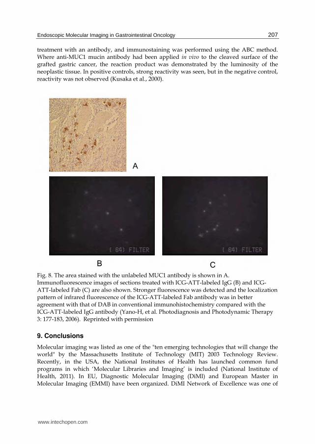
treatment with an antibody, and immunostaining was performed using the ABC method. Where anti-MUC1 mucin antibody had been applied in vivo to the cleaved surface of the grafted gastric cancer, the reaction product was demonstrated by the luminosity of the neoplastic tissue. In positive controls, strong reactivity was seen, but in the negative control, reactivity was not observed (Kusaka et al., 2000).
Fig. 8. The area stained with the unlabeled MUC1 antibody is shown in A. Immunofluorescence images of sections treated with ICG-ATT-labeled IgG (B) and ICG-ATT-labeled Fab (C) are also shown. Stronger fluorescence was detected and the localization pattern of infrared fluorescence of the ICG-ATT-labeled Fab antibody was in better agreement with that of DAB in conventional immunohistochemistry compared with the ICG-ATT-labeled IgG antibody (Yano-H, et al. Photodiagnosis and Photodynamic Therapy 3: 177-183, 2006). Reprinted with permission
9. Conclusions
Molecular imaging was listed as one of the "ten emerging technologies that will change the world" by the Massachusetts Institute of Technology (MIT) 2003 Technology Review. Recently, in the USA, the National Institutes of Health has launched common fund programs in which ‘Molecular Libraries and Imaging’ is included (National Institute of Health, 2011). In EU, Diagnostic Molecular Imaging (DiMI) and European Master in Molecular Imaging (EMMI) have been organized. DiMI Network of Excellence was one of
www.intechopen.com
New Techniques in Gastrointestinal Endoscopy
208
the largest European research projects funded by the European Commission within the 6th Framework Programme (Diagnostic Molecular Imaging, n.d.). EMMI is an international program entirely dedicated to in vivo molecular imaging. Supported by the European Commission under the SOCRATES programme, this two-year interdisciplinary curriculum is brought together by prominent European molecular imaging research groups (European Master in Molecular Imaging, n.d.). Given this situation, it is convincing that molecular imaging is one of the latest upcoming and nationwide fields that affect human life science. Molecular imaging can add various types of information to conventional imaging techniques and enable not only detection and localization but also quantification and the basis of pathologies (Mahmood and Wallace, 2007). In terms of one specific paradigm of the technology, the exogenous administration of targeted probes may provide additional and complementary information to native spectro-endomicroscopic image analysis of lesions. It also can be used to either image the administered drug directly to access its distribution and target binding. In general, the incidence of colorectal cancer in the world has been increasing and will occur more frequently in the coming generations; a new technology is therefore required that it is cost-effective and efficient for both screening and further examinations. It is well known that there are many alterations in the molecular pathways of carcinoma; stepwise formation will be visualized with various molecules. The fluorescence signal acquired by endoscopy is not quantitative by itself, but the intensity of the signal is expected to reveal the depth or potential of a cancer. Moreover, amplification or reinforcement strategies are required because focal target concentrations are quite low, in the pico- to nanomolar range (Weissleder, 1999). Ideal system in this technology should offer a strong signal-to noise ratio, quantitative analysis, less invasive modality, real time monitoring and multiplex imaging using various fluorescent peptides or antibodies with different optical characteristics. Before the application of diagnostic drug to clinical use, pharmacokinetics and pharmacodynamics should be tested and these agents have to undergo lengthy approval processes; however, no definite barriers are anticipated to prevent its clinical application because therapeutic administration of various humanized antibodies has proven safe and imaging and therapeutic targets are often same. On the other hand, fluorochromes such as ICG are photostable and have been used safely in the human body. With these possibilities, it seems apparent that this innovative technology will be realized in cooperation with the pharmaceutical industry, chemical company and engineering manufacture, hopefully with their investment in the economic market of gastrointestinal oncology.
10. Acknowledgment
Authors are grateful to Susumu Ito (Professor, Faculty of Health and Welfare, Tokushima Bunri University; Emeritus Professor, The University of Tokushima, Tokushima, Japan) for his invaluable guidance in this project.
11. References
Ballou, B.; Fisher, G. W.; Deng, J. S.; Hakala, T. R.; Srivastava, M. & Farkas, D. L. (1998) Cyanine fluorochrome-labeled antibodies in vivo: Assessment of tumor imaging using Cy3, Cy5, Cy5.5, and Cy7. Cancer Detection and Prevention, 22, 251-257.
www.intechopen.com

Endoscopic Molecular Imaging in Gastrointestinal Oncology
209
Bando, T.; Muguruma, N.; Ito, S.; Musashi, Y.; Inayama, K.; Kusaka, Y.; Tadatsu, M.; Ii, K.; Irimura, T.; Shibamura, S. & Takesako, K. (2002) Basic studies on a labeled anti-mucin antibody detectable by infrared-fluorescence endoscopy. Journal of Gastroenterology, 37, 260-269.
Benson, R. C. & Kues, H. A. (1978) Fluorescence properties of indocyanine green as related to angiography. Physics in Medicine and Biology, 23, 159-163.Borotto, E.; Englender, J.; Pourny, J. C.; Naveau, S.; Chaput, J. C. & Lecarpentier, Y. (1999) Detection of the fluorescence of GI vessels in rats using a CCD camera or a near-infrared video endoscope. Gastrointestinal Endoscopy, 50, 684-688.
Cotton, P. B.; Barkun, A.; Ginsberg, G.; Hawes, R. H.; Atkin, W.; Bjorkman, D. J.; Dykes, C.; Elta, G.; Farrell, J.; Fleischer, D.; Ganz, R.; Glenn, T.; Janowski, D.; Johnson, D.; Kochman, M.; Kowalski, T.; Megibow, A. J.; McQuaid, K.; Sasa, H.; Thompson, C. C.; Vargo, J. & Woods, K. (2006) Diagnostic endoscopy: 2020 vision. Gastrointestinal Endoscopy, 64, 395-398.
Davies, R. J. H. (1995) Ultraviolet-radiation damage in DNA. Biochemical Society Transactions, 23, 407-418.
Detter, C.; Russ, D.; Iffland, A.; Wipper, S.; Schurr, M. O.; Reichenspurner, H.; Buess, G. & Reichart, B. (2002) Near-infrared fluorescence coronary angiography: A new noninvasive technology for intraoperative graft patency control. Heart Surgery Forum, 5, 364-369.
Diagnostic Molecular Imaging. Available from: < http://www.dimi.eu/index.php?id=210> European Master in Molecular Imaging. Available from: < http://www.e-mmi.eu/en/index.php > Flower, R. W. & Hochheimer, B. F. (1973) A clinical technique and apparatus for
simultaneous angiography of the separate retinal and choroidal circulations. Investigative Ophtalmology and Visual Science, 12, 248-261.
Fox, I. J. & Wood, E. H. (1960) Indocyanine green: physical and physiologic properties. Mayo Clinic Proceedings, 35, 732-744.
Fujimoto, J. G.; Brezinski, M. E.; Tearney, G. J.; Boppart, S. A.; Bouma, B.; Hee, M. R.; Southern, J. F. & Swanson, E. A. (1995) Optical biopsy and imaging using optical coherence tomography. Nature Medicine, 1, 970-972.
Ganz, R. A. (2004) The development and the implementation of new endoscopic technology: what are the challenges? Gastrointestinal Endoscopy, 60, 592-598.
Gibson, H. L.; Buckley, W. R. & Whitemore, K. E. (1965) New vistas in infrared photography. Journal of Biological Photography Associations, 33, 1-33.
Goetz, M.; Ziebart, A.; Foersch, S.; Vieth, M.; Waldner, M. J.; Delaney, P.; Galle, P. R.; Neurath, M. F. & Kiesslich, R. (2010) In vivo molecular imaging of colorectal cancer with confocal endomicroscopy by targeting epidermal growth factor receptor. Gastroenterology, 138, 435-446.
Hayashi, S.; Muguruma, N.; Bando, T.; Taoka, S.; Ito, S. & Ii, K. (1999) Vital immunohistochemical staining for a novel method of diagnosing micro-cancer - examination of immuno-histochemical staining of non-fixed fresh tissue- Journal of Medical Investigation, 46, 178-185.
Hirata, T.; Kogiso, H.; Morimoto, K.; Miyamoto, S.; Taue, H.; Sano, S.; Muguruma, N.; Ito, S. & Nagao, Y. (1998) Synthesis and reactivities of 3-indocyanine-green-acyl-1,3-
www.intechopen.com
New Techniques in Gastrointestinal Endoscopy
210
thiazolidine-2-thione (ICG-ATT) as a new near-infrared fluorescent-labeling reagent. Bioorganic & Medicinal Chemistry, 6, 2179-2184.
Hsiung, P. L.; Hardy, J.; Friedland, S; Soetikno, R; Du, C. B.; Wu, A. P.; Sahbaie, P.; Crawford, J. M.; Lowe, A. W.; Contag, C. H. & Wang, T. D. (2008) Detection of colonic dysplasia in vivo using a targeted heptapeptide and confocal microendoscopy. Nature Medicine, 14, 454-458.
Inayama, K.; Ito, S.; Muguruma, N.; Kusaka, Y.; Bando, T.; Tadatsu, Y.; Tadatsu, M.; Ii, K.; Shibamura; S. & Takesako, K. (2003) Basic study of an agent for reinforcement of near-infrared fluorescence on tumor tissue. Digestive and Liver Disease, 35, 88-93.
Iseki, K., Tatsuta, M.; Iishi, H.; Sakai, N.; Yano, H. & Ishiguro, S. (2000) Effectiveness of the near-infrared electronic endoscope for diagnosis of the depth of involvement of gastric cancers. Gastrointestinal Endoscopy, 52, 755-762.
Ishihara, R. (2010) Infrared endoscopy in the diagnosis and treatment of early gastric cancer. Endoscopy, 42, 672-676.
Ito, S.; Muguruma, N.; Hayashi, S.; Taoka, S.; Bando, T.; Inayama, K.; Sogabe, M.; Okahisa, T.; Okamura, S.; Shibata, H.; Irimura, T.; Takesako, K. & Shibamura, S. (1998) Development of agents for reinforcement of fluorescence on near-infrared ray excitation for immunohistological staining. Bioorganic & Medicinal Chemistry, 6, 613-618.
Ito, S.; Muguruma, N.; Kakehashi, Y.; Hayashi, S.; Okamura, S.; Shibata, H.; Okahisa, T.; Kanamori, M.; Shibamura, S.; Takesako, K.; Nozawa, M.; Ishida, K. & Shiga, M. (1995) Development of fluorescence-emitting antibody labeling substance by near-infrared ray excitation. Bioorganic & Medicinal Chemistry Letters, 5, 2689-2694.
Ito, S.; Muguruma, N.; Hayashi, S.; Taoka, S.; Tsutsui, A.; Fukuda, T.; Okahisa, T.; Matsunaga, H.; Shimizu, I.; Nakamura, K.; Imaizumi, K.; Takesako, K.; & Shibamura, S. (1997) Development of an imaging system using fluorescent labeling substances excited by infrared rays. Digestive Endoscopy, 9, 278-282.
Ito, S.; Muguruma, N.; Kusaka, Y.; Tadatsu, M.; Inayama, K.; Musashi, Y.; Yano, M.; Bando, T.; Honda, H.; Shimizu, I.; Ii, K.; Takesako, K.; Takeuchi, H. & Shibamura, S. (2001) Detection of human gastric cancer in resected specimens using a novel infrared fluorescent anti-human carcinoembryonic antigen antibody with an infrared fluorescence endoscope in vitro. Endoscopy, 33, 849-853.
Keller, R.; Winde, G.; Eisenhawer, C.; Herwig, R.; Terpe, H. J.; Domschke, W. & Foerster, E. C. (1998) Immunoscopy: A technique combining endoscopy and immunofluorescence for diagnosis of colorectal carcinoma. Gastrointestestinal Endoscopy, 47,154–161.
Keller, R.; Winde, G.; Terpe, H. J.; Foerster, E. C. & Domschke W. (2002) Fluorescence endoscopy using a fluorescein-labeled monoclonal antibody against carcinoembryonic antigen in patients with colorectal carcinoma and adenoma. Endoscopy, 34, 801-807.
Kim, Y. S. (1993) Mucin glycoproteins in gastrointestinal malignancies and metastasis. European Journal of Gastroenterology and Hepatology, 5, 219-225.
Kimura, T.; Muguruma, N.; Ito, S.; Okamura, S.; Imoto, Y.; Miyamoto, H.; Kaji, M. & Kudo, E. (2007) Infrared fluorescence endoscopy for the diagnosis of superficial gastric tumors. Gastrointestinal Endoscopy, 66, 37-43.
www.intechopen.com
Endoscopic Molecular Imaging in Gastrointestinal Oncology
211
Kohso, H.; Tatsumi, Y.; Fujino, H.; Tokita, K.; Kodama, T.; Kashima, K. & Kawai, K. (1990) An investigation of an infrared ray electronic endoscope with a laser diode light source. Endoscopy, 22, 217-220.
Kusaka, Y.; Ito, S.; Muguruma, N; Tadatsu, M.; Bando, T.; Ii, K.; Irimura, T. & Shibamura, S. (2000) Vital immunostaining of human gastric and colorectal cancers grafted into nude mice: a preclinical assessment of a potential adjunct to videoendoscopy. Journal of Gastroenterology, 35, 748-752.
Mahmood, U. & Wallace, M. B. (2007) Molecular imaging in gastrointestinal disease. Gastroenterology, 132, 11-14.
Mataki, N.; Nagao, S.; Kawaguchi, A.; Matsuzaki, K.; Miyazaki, J.; Kitagawa, Y.; Nakajima, H.; Tsuzuki, Y.; Itoh, K.; Niwa, H. & Miura, S. (2003) Clinical usefulness of a new infrared videoendoscope system for diagnosis of early stage gastric cancer. Gastrointestinal Endoscopy, 57, 336-342.
Mimura, S. & Okuda, S. (1981) A new gastrocamera technique using infrared color film. Endoscopy, 13, 40-43.
Mordon, S.; Devoisselle, J. M.; Soulie-Begu, S. & Desmettre, T. (1998) Indocyanine green: Physicochemical factors affecting its fluorescence in vivo. Microvascular Research, 55, 146-152.
Muguruma, N.; Ito, S.; Bando, T.; Taoka, S.; Kusaka, Y.; Hayashi, S.; Ichikawa, S.; Matsunaga, Y.; Tada, Y.; Okamura, S.; Ii, K.; Imaizumi, K.; Nakamura, K.; Takesako, K. &Shibamura, S. (1999) Labeled carcinoembryonic antigen antibodies excitable by infrared rays: A novel diagnostic method for micro cancers in the digestive tract. Internal Medicine, 38, 537-542.
Muguruma, N.; Ito, S.; Hayashi, S.; Taoka, S.; Kakehashi, H.; Ii, K.; Shibamura, S. & Takesako, K. (1998) Antibodies labeled with fluorescence-agent excitable by infrared rays. Journal of Gastroenterology, 33, 467-471.
Nakamori, S.; Ota, D. M.; Cleary, K. R.; Shirotani, K. & Irimura, T. (1994) MUC1 mucin expression as a marker of progression and metastasis of human colorectal carcinoma. Gastroenterology, 106, 353-361.
National Institute of Health. (2011) The NIH Common Fund, Available from: < http://commonfund.nih.gov/initiativeslist.aspx >
Nimura, H.; Narimiya, N.; Mitsumori, N.; Yamazaki, Y.; Yanaga, K. & Urashima, M. (2004) Infrared ray electronic endoscopy combined with indocyanine green injection for detection of sentinel nodes of patients with gastric cancer. British Journal of Surgery, 91, 575-579.
Ohta, H.; Kohgo, Y.; Takahashi, Y.; Koyama, R.; Suzuki, H. & Niitsu, Y. (1994) Computer-assisted data processing of images of mucosal and submucosal blood vessels of the stomach obtained by visible and infrared endoscopy using a directional-contrast filter. Gastrointestinal Endoscopy, 40, 621-628.
Okamoto, K.; Muguruma, N.; Kimura, T.; Yano, H.; Imoto, Y.; Takagawa, M.; Kaji, M.; Aoki, R.; Sato, Y.; Okamura, S.; Kusaka, Y. & Ito, S. (2005) A novel diagnostic method for evaluation of vascular lesions in the digestive tract using infrared fluorescence endoscopy. Endoscopy, 37, 52-57.
Page, M.; Dalifard, I.; Bertrand, G.; Bocquillon, P. G. & Daver, A. (1986) Immunostaining of colorectal cancer with monoclonal anti-CEA antibodies compared to serum and tumor CEA content. Anticancer Research, 6, 893-896.
www.intechopen.com
New Techniques in Gastrointestinal Endoscopy
212
Pasricha, P. J. & Motamedi, M. (2002) Optical biopsies, "bioendoscopy," and why the sky is blue: The coming revolution in gastrointestinal imaging. Gastroenterology, 122, 571-575.
Pelegrin, A.; Folli, S.; Buchegger, F.; Mach, J. P.; Wagnieres, G. & van den Bergh, H. (1991) Antibody-fluorescein conjugates for photoimmunodiagnosis oh human colon carcinoma in nude mice. Cancer, 67, 2529-2537.
Shealy, D. B.; Lipowska, M.; Lipowski, J.; Narayanan, N.; Sutter, S.; Strekowski, L. & Patonay, G. (1995) synthesis, chromatographic-separation, and characterization of near-infrared-labeled DNA oligomers for use in DNA-sequencing. Analytical Chemistry, 67, 247-251.
Sivak, M. V. (2006) Gastrointestinal endoscopy: past and future. Gut, 55, 1061-1064. Still, J. M.; Law, E. J.; Klavuhn, K. G.; Island, T. C. & Holtz, J. Z. (2001) Diagnosis of burn
depth using laser-induced indocyanine green fluorescence: a preliminary clinical trial. Burns, 27, 364-371.
Tadatsu, Y.; Muguruma, N.; Ito, S.; Tadatsu, M.; Kusaka, Y.; Okamoto, K.; Imoto, Y.; Taue, H.; Sano, S. & Nagao, Y. (2006) Optimal labeling condition of antibodies available for immunofluorescence endoscopy. Journal of Medical Investigation, 53, 52-60.
Tajiri, H. (2007) What do we see in the endoscopy world in 10 years’ time? Digestive Endoscopy, 19, S174–179.
Takayama, T.; Katsuki, S.; Takahashi, Y.; Ohi, M.; Nojiri, S.; Sakamaki, S.; Kato, J.; Kogawa, K.; Miyake, H. & Niitsu, Y. (1998) Aberrant crypt foci of the colon as precursors of adenoma and cancer. New England Journal of Medicine, 339, 1277-1284.
Taoka, S.; Ito, S.; Muguruma, N.; Hayashi, S.; Kusaka, Y.; Ii, K.; Nakamura, K.; Imaizumi, K.; Takesako, K. & Shibamura, S. (1999) Reflected illumination-type imaging system for the development of infrared fluorescence endoscopy. Digestive Endoscopy, 11, 321-326.
Tatsuta, M.; Iishi, H.; Ichii, M.; Baba, M.; Yamamoto, R.; Okuda, S. & Kikuchi, K. (1989) Diagnosis of gastric cancers with fluorescein-labeled monoclonal antibodies to carcinoembryonic antigen. Lasers in Surgery and Medicine, 9, 422-426.
Thakur, M. & Lentle, B. C. (2005) Report of a summit on molecular imaging. Radiology, 236, 753-755.
Weissleder, R. (1999) Molecular imaging: Exploring the next frontier. Radiology, 212, 609-614. Weissleder, R. (2006) Molecular imaging in cancer. Science, 312, 1168-1171. Weissleder, R. & Mahmood, U. (2001) Molecular imaging. Radiology, 219, 316-333. Yano, H.; Muguruma, N.; Ito, S.; Aoyagi, E.; Kimura, T.; Imoto, Y.; Inoue, S.; Sano, S.; Nagao,
Y. & Kido, H. (2006) Fab fragment labeled with ICG-derivative for detecting digestive tract cancer. Photodiagnosis and Photodynamic Therapy, 3, 177-183.
www.intechopen.com
New Techniques in Gastrointestinal EndoscopyEdited by Prof. Oliviu Pascu
ISBN 978-953-307-777-2Hard cover, 310 pagesPublisher InTechPublished online 30, September, 2011Published in print edition September, 2011
InTech EuropeUniversity Campus STeP Ri Slavka Krautzeka 83/A 51000 Rijeka, Croatia Phone: +385 (51) 770 447 Fax: +385 (51) 686 166www.intechopen.com
InTech ChinaUnit 405, Office Block, Hotel Equatorial Shanghai No.65, Yan An Road (West), Shanghai, 200040, China
Phone: +86-21-62489820 Fax: +86-21-62489821
As result of progress, endoscopy has became more complex, using more sophisticated devices and hasclaimed a special form. In this moment, the gastroenterologist performing endoscopy has to be an expert inmacroscopic view of the lesions in the gut, with good skills for using standard endoscopes, with goodexperience in ultrasound (for performing endoscopic ultrasound), with pathology experience for confocalexamination. It is compulsory to get experience and to have patience and attention for the follow-up ofthousands of images transmitted during capsule endoscopy or to have knowledge in physics necessary forautofluorescence imaging endoscopy. Therefore, the idea of an endoscopist has changed. Examinationsmentioned need a special formation, a superior level of instruction, accessible to those who have alreadygained enough experience in basic diagnostic endoscopy. This is the reason for what these new issues ofendoscopy are presented in this book of New techniques in Gastrointestinal Endoscopy.
How to referenceIn order to correctly reference this scholarly work, feel free to copy and paste the following:
Naoki Muguruma and Tetsuji Takayama (2011). Endoscopic Molecular Imaging in Gastrointestinal Oncology,New Techniques in Gastrointestinal Endoscopy, Prof. Oliviu Pascu (Ed.), ISBN: 978-953-307-777-2, InTech,Available from: http://www.intechopen.com/books/new-techniques-in-gastrointestinal-endoscopy/endoscopic-molecular-imaging-in-gastrointestinal-oncology
© 2011 The Author(s). Licensee IntechOpen. This chapter is distributedunder the terms of the Creative Commons Attribution-NonCommercial-ShareAlike-3.0 License, which permits use, distribution and reproduction fornon-commercial purposes, provided the original is properly cited andderivative works building on this content are distributed under the samelicense.