Endoplasmic Reticulum Stress and Cell Apoptosis Ischemia ...
22
Page 1/22 Dexmedetomidine Protects against Hepatic Ischemia-Reperfusion Injury by inhibiting Endoplasmic Reticulum Stress and Cell Apoptosis in Rats Hang Li Hebei Agricultural University Jilang Tang Northeast Agricultural University Weiqi Zhang Hebei Agricultural University Liping Ai Hebei Agricultural University Shixia Zhang ( [email protected] ) Hebei Agricultural University Research Article Keywords: Dexmedetomidine, Ischemia-reperfusion, Liver, Apoptosis, Endoplasmic reticulum stress. Posted Date: June 16th, 2021 DOI: https://doi.org/10.21203/rs.3.rs-601634/v1 License: This work is licensed under a Creative Commons Attribution 4.0 International License. Read Full License
Transcript of Endoplasmic Reticulum Stress and Cell Apoptosis Ischemia ...

Page 1/22
Dexmedetomidine Protects against HepaticIschemia-Reperfusion Injury by inhibitingEndoplasmic Reticulum Stress and Cell Apoptosisin RatsHang Li
Hebei Agricultural UniversityJilang Tang
Northeast Agricultural UniversityWeiqi Zhang
Hebei Agricultural UniversityLiping Ai
Hebei Agricultural UniversityShixia Zhang ( [email protected] )
Hebei Agricultural University
Research Article
Keywords: Dexmedetomidine, Ischemia-reperfusion, Liver, Apoptosis, Endoplasmic reticulum stress.
Posted Date: June 16th, 2021
DOI: https://doi.org/10.21203/rs.3.rs-601634/v1
License: This work is licensed under a Creative Commons Attribution 4.0 International License. Read Full License

Page 2/22
AbstractBackground: Hepatic ischemia-reperfusion injury (IRI) remains a major complication of liver surgery,dexmedetomidine (DEX) has a certain protective effect on liver during ischemia-reperfusion, but theunderlying mechanisms are not fully understood. This study explored the protective effects of DEX andinvestigated whether DEX protects against hepatic IRI by inhibiting endoplasmic reticulum stress (ERS)and its downstream apoptotic pathway in a rat model.
Methods: Thirty-six male Sprague-Dawley (SD) rats were divided into six groups: S, IR, DL, DM1, DH andDM2 group. Group S was subjected to laparotomy, and exposure of the portal triad without occlusion. I-Rinjury model was induced by clamping the portal vessels supplying the middle and left hepatic lobes for30 min in IR, DL, DM1, DH and DM2 group. Then DL, DM1, DH group received DEX of 25 μg/kg, 50 μg/kgand 100 μg/kg intraperitoneally at 30 min before ischemia, respectively, DM2 group received 50 μg/kgDEX intraperitoneally 30 min after reperfusion, and IR group received normal saline. After 6 h ofreperfusion, assessment of liver function, histopathology, oxidative stress was performed. The liver cellmicrostructure was detected by transmission electron microscopy. Hepatocyte apoptosis was determinedby TUNEL assay. Real-time PCR, Western blotting were performed to analyze various ERS molecules.
Results: We observed that DEX protected the liver by alleviating hepatocytes damage, reducing thecontent of ALT and MDA, increasing the activity of SOD, reducing the number of TUNEL-positive cells,down-regulating the expression of GRP-78, PERK, ATF-6, Caspase-12 mRNA, and p-PERK, p-IRE-1 α, CHOPproteins, up-regulating Bcl-2 protein. The effect of 50 μg/kg DEX is superior to 25 μg/kg DEX, but notsigni�cantly different from 100μg/kg DEX. There was no signi�cant difference in the above monitoringindexes between DM1 and DM2 group.
Conclusions: DEX protects the liver from IRI by inhibiting ERS and cell apoptosis. The protective effect ofDEX was dose-dependent in a certain dose range, both DEX administered prior to ischemia and followingreperfusion markedly reduced liver injury induced by hepatic IRI in mice.
BackgroundIschemia reperfusion injury (IRI) takes place when blood provision to an organ is diminished orinterrupted, the restoration of blood reperfusion will aggravate the damage of the physiological function,structure, and metabolism of the organs. Hepatic IRI remain a major complication of severe liver trauma,hemorrhagic shock, liver transplantation and partial hepatectomy [1, 2]. Especially with the improvementof surgical technology and equipment, more and more liver operations are carried out. However, hepaticIRI remain one of the main factors that deteriorate morbidity and mortality of the liver surgery [3]. HepaticIRI has become a major obstacle in the development of liver surgery. Therefore, to �nd effective measuresto prevent and avoid hepatic IRI have become one of major clinical problems.
Apoptosis, a physiological mechanism of organisms, plays an extremely important role in maintainingthe stability of internal environments. During hepatic IRI for the �rst 24 hours after reperfusion, apoptosis
Page 3/22
is one of the main hepatocytes’death modes [4]. Thus, we speculated that regulation of key apoptosis-related proteins expression is an important protective mechanism for the attenuation of hepatic IRI. Sofar, the apoptotic pathway is known to include the death receptor-mediated pathway, the mitochondrialpathway, and the endoplasmic reticulum stress (ERS) pathway.
Endoplasmic reticulum (ER) is an important membranous organelle which is responsible for proteinsynthesis, folding and secretion. Previous studies have demonstrated that ERS is involved in thepathogenesis of many diseases, including liver diseases [5, 6]. Hepatic IRI induces ischemia, hypoxia, Ca2+
homeostasis, oxidative stress, and elevates protein secretion, which triggers the disruption of ER internalbalance [7]. The disturbances of ER function cause ERS, which subsequently leads to the unfolded proteinresponse (UPR). The UPR initially activates three major signal transducers, protein kinase R-like ER kinase(PERK), activating transcription factor 6 (ATF6), and inositol-requiring enzyme 1 (IRE1). However,excessive or prolonged ERS leads to the expression of apoptotic signaling pathways. Activation of C/EBPhomologous protein (CHOP) is the main pathway for apoptosis in ERS downstream [8]. During thisprocess, caspase-12 and JNK are also activated and involved in ERS cell apoptosis pathway [9]. Literatureshow that hepatic IRI is closely related to ERS and apoptosis, many studies have con�rmed inhibiting theERS response pathways can effectively alleviate hepatic IRI[10–12]. Thus, strategies targeting ERS mayhave potential protective roles in hepatic IR injury.
Among the therapeutic intervention strategies, pharmacologic strategies have been demonstrated to playan important role in protecting livers against IR injury [13]. Dexmedetomidine (DEX) is a selective andpotent α2 adrenergic receptor agonist with a good sedative effect and no respiratory depression, and ismainly used in clinical anesthesia and ICU sedation. Recently, it has been reported that DEX has otherpossible applications [14]. At the same time, there is a large literature showing that DEX exerts variouspharmacological effects, such as anti-in�ammation [15], anti-oxidant [16], and anti-apoptosis [17].Accumulationg evidence suggested that DEX has a protective effect on ischemia-reperfusion injury of theheart, kidney, and other organs [8, 18]. However, its speci�c mechanism has not been clari�ed. DEX showsdifferent functions in the protective effect of hepatic IRI. Zong et al. have con�rmed that DEX pre-treatment can suppress oxidative stress, decrease NLRC5 expression through inactivating NF-kB pathway[16]. It has also been reported that DEX preconditioning inhibited intrahepatic proin�ammatory innateimmune activation by promoting macrophage M2 activation in a PPARγ/STAT3 dependent manner [19]. Inaddition, DEX has been shown to protect the liver against IR injury via the suppression of the TLR4/NF-kBpathway [20]. However, the contribution of ERS-induced apoptosis remains unknown.
Therefore, this study is aimed to explore the protective effects of DEX and investigate whether DEXprotects against hepatic IRI by inhibiting ERS in a rat model.
MethodsAnimals and Hepatic I-R Model
Page 4/22
Thirty-six male SD rats were obtained from the Animal Center of Hebei medical University (Shijiazhang,China). Rats weighed 180-220g and were raised under a steady temperature around 20˚C with 12 h light–dark cycles for one week to adapt to the environment. The experimental protocol was approved by theAgricultural University of Hebei Ethical Committee, Baoding, China.
70% Hepatic I-R model was performed according to previous studies [21]. An atraumatic clip was appliedto the portal vessels to induce ischemia of the middle and left hepatic lobes under iso�urane anesthesia.The ischemia was con�rmed by tissue blanching. After 30 min ischemia, the clamp was removed forreperfusion. The reperfusion was con�rmed by immediate color change of the ischemic lobes afterremoval of the clamp.
Grouping and treatment
The rats were randomly divided into six groups (n=6) as follows:
1. Group S: the rats were subjected to laparotomy, and exposure of the portal triad without occlusion.
2. Group IR: the rats were subjected to laparotomy, and exposure of the portal triad with occlusion, andno drug was utilized.
3. Group DL: the rats received 20 μg/kg DEX intraperitoneal injection 30 min before ischemia of thehepatic lobes.
4. Group DM1: the rats received 50 μg/kg DEX intraperitoneal injection 30 min before ischemia of thehepatic lobes.
5. Group DH: the rats received 100 μg/kg DEX intraperitoneal injection 30 min before ischemia of thehepatic lobes.
�. Group DM2: the rats received 50 μg/kg DEX intraperitoneal injection 30 min after reperfusion.
Blood and tissue sample collection
After 6 h reperfusion, all rats were sacri�ced to collect blood samples and parts of the IR liver (middle andleft lobes). Blood samples were centrifuged at 3000 rpm for 10 min within 1 h after collection and theserum was stored at -80℃ until use. The liver tissues were rapidly separated, washed with 0.01Mphosphate-buffered saline, and placed in an ice tray. The middle liver lobe was placed in 10% formalinsolution and embedded in para�n to observe pathological changes. The left lobe was stored at -80℃ forfurther analysis.
Biochemical Analysis
The serum sample was measured to determine the levels of alanine aminotransferase (ALT) using aUniCel DxC800 Synchron chemistry system (Beckman, USA). According to the instructions of thecorresponding assay kit (Nanjing Jiancheng Bioengineering Institute, China), liver tissue was preparedinto a homogenate to detect concentrations of malondialdehyde (MDA) and the activity of theantioxidant enzyme superoxide dismutase (SOD).
Page 5/22
Histopathological Analysis
After �xation with 10% formalin solution, liver tissue samples were embedded in para�n, sectioned to5 μm thicknesses, and stained with hematoxylin and eosin (H&E). Histological changes were observedunder a light microscope. Histopathologists with no prior knowledge of the experiment evaluated thesections at 200x magni�cation. Liver injury was scored on a scale of 0 to 4 for sinusoidal congestion,vacuolization of hepatocyte cytoplasm, and parenchyma, as described by Suzuki et al.[22].
Transmission Electron Microscopy
The liver samples of each group were pre-�xed in glutaraldehyde (2.5%), washed with PBS and then �xedwith 1% osmic acid for 2 h. After dehydration in gradient ethanol, the �xed samples were embedded inepoxy resin, cut into ultrathin sections (50 nm) and stained with uranyl acetate and lead citrate. Thestained sections were observed using a transmission electron microscope (TEM; H-7650, Hitachi, Japan).
TUNEL Assay
Hepatocellular apoptosis was detected with a TUNEL apoptosis assay kit (Roche, Switzerland). Allprocedures were performed as described in the assay kit. Ten microscopic �elds within the view wererandomly selected, and the TUNEL-positive cells were counted. Liver apoptosis rate was evaluated usingthe average number of positive cells division by the average number of total cells.
Real-time PCR analysis
The total RNA isolated from liver samples using TRIzol reagent was reverse transcribed to obtaincDNA. The primers were synthesized by Takara Biomedical Technology Co., Ltd. (Dalian, China). Theprimer sequences are listed in Table 1. Real-time qPCR was performed by using FastStart Universal SYBRGreen Master (Rox) (Roche, USA). In this experiment, the response system of 20 μL was used and GAPDHwas used as the internal reference for relative quantitative analysis of gene mRNA expression level.Relative quanti�cation was performed according to 2-ΔΔCt method.
Western Blot Analysis
The expression of p-PERK p-IRE-1α CHOP Bcl-2 proteins were detected via Western blot. The liver tissueswere homogenized on ice, diluted with 10 volumes of natural saline, and then centrifuged at 2500 rpm for10 min. The supernatants were transferred into fresh tubes for biochemic alanalysis. Nuclear andcytoplasmic proteins were extracted by using nuclear and cytoplasmic extraction reagents according tothe manufacture’s procedure (Solarbio, China), and then separated by 10% sodium dodecyl sulfatepolyacrylamide gel electrophoresis (SDS-PAGE) and then transferred to polyvinylidene �uoride (PVDF)membranes. Blocking by 10% skimmed milk for 2 h, and then the membranes were incubated at4℃ overnight with rabbit anti-CHOP (1:1000; CST, USA), rabbit anti-Bcl-2 (1:1,000; CST, USA), rabbit anti-p-PERK (1:500; Bioss, China), and rabbit anti-p-IRE-1α (1:500; Bioss, China), and β-actin (1:500; Bioss,China). After cleaning with TBST buffffer, the membranes were incubated with secondary antibodies
Page 6/22
labeled by alkaline phosphatase (1:500; Bioss, China) for 2 h. Finally, BCIP/NBT substratedeveloper (Solarbio, China) was added to examine blots, and analyzed gray level by Image J software.
Statistical analysis
Data were analyzed by SPSS 22.0 (SPSS, IL, USA) statistical analysis software and expressed as mean ±standard deviation (X ± SD). Differences among groups were determined for statistical signi�cance usingoneway ANOVA and were considered statistically signi�cant at P < 0.05. Graphpad Prism 5 (San Diego,California) was used to made graphs.
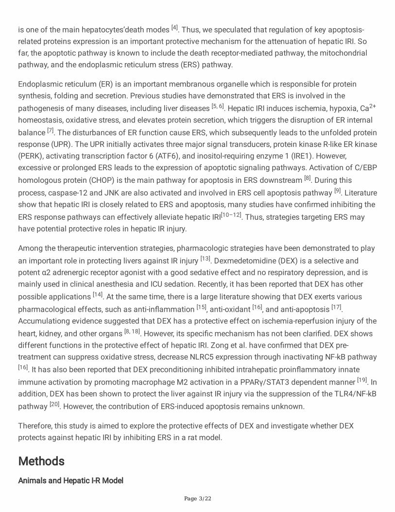
ResultsEffects of DEX on IR-induced liver histopathology
Liver histopathological changes were analyzed in all six groups. There were no pathological changes inthe liver tissue sections from the sham group, and the hepatocytes showed an intact structure with nodegeneration or necrosis, and no in�ammatory cell in�ltration (Figure 1 aA). However, IR-inducedliver histopathological changes were observed in the liver tissue including disrupted hepatic cell cords,extensive hepatic necrosis, hemorrhage, vacuolar degeneration, and in�ammatory cell in�ltration (Figure1 aB). Interestingly, DEX administration signi�cantly reduced IR‐induced liver histopathological changeswith neatly arranged hepatocytes, a small amount of in�ammatory cell in�ltration and bleeding (Figure aC-F). The pathological score of liver injury is shown in �gure 1 b. The liver injury score of IR group wassigni�cantly higher than that of other groups (p < 0.01). Compared with group DL and DH, the liver injuryscore in group DM1 was signi�cant lower (p < 0.01). There was no signi�cant difference in liver injuryscore between DM1 group and DM2 group (p 0.05).
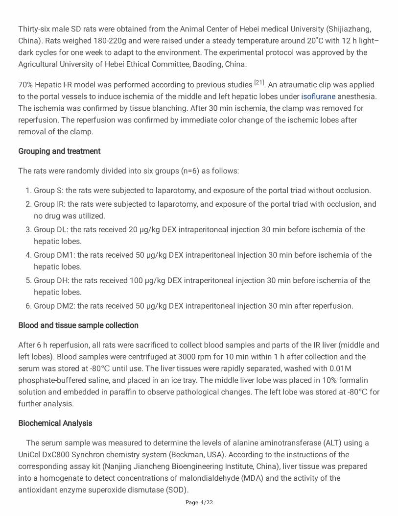
Effects of DEX on IR-induced hepatocyte morphological changes
The hepatocyte ultrastructural changes were also analyzed in all groups by transmission electronmicroscopy. As shown in Figure 2 A, the morphology structure of hepatocytes from the sham group wasnormal with intact cell and nuclear membranes. In the IRI group the hepatocyte mitochondria wereswollen and the ridges of mitochondria were uneven, the hepatocytes ER were damaged and expanded(Figure 2 B). DEX alleviated the damage in both organelles, whereas the nuclear membrane structure inthe DL and DH group is slightly incomplete.
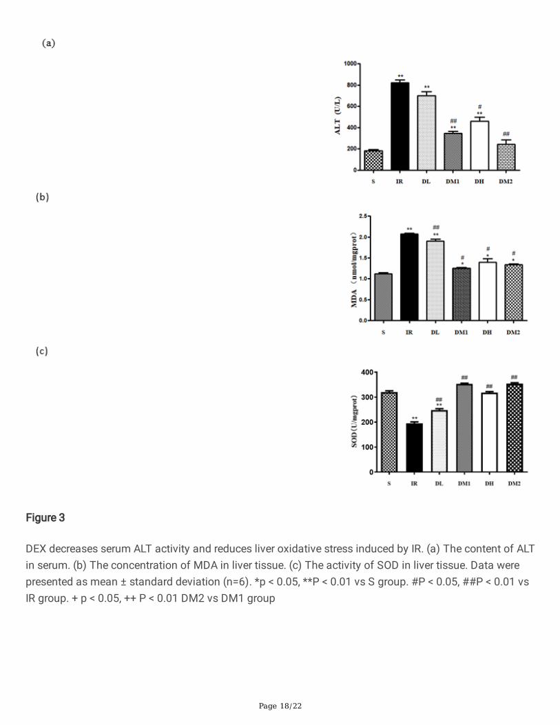
DEX decreases serum ALT activity and reduces liver oxidative stress induced by IR
We initially investigated speci�c indicators of liver function and oxidative stress damage, including ALTlevel, MDA concentration and SOD activity. As expected, serum ALT level and liver MDA concentration inthe IR-induced rats were signi�cantly higher compared with that of sham rats (Figure 3 a, b; p < 0.01), andIR signi�cantly reduced lipid peroxidation. SOD activity in the liver (Figure 3 c) was signi�cantlydecreased in group IR (p < 0.01), DEX intervention markedly inhibited the increase in serum ALT activity,liver MDA level and the decrease in liver SOD activity induced by IR, indicating that DEX has a protective
Page 7/22
effect in IR-induced liver injury. Among the three pre-administration groups, the concentration of ALT andMDA in DM1 group was the lowest and the activity of SOD was the highest. There was no signi�cantdifference in ALT and MDA concentration and SOD activity between DM1 and DM2 group (p 0.05).
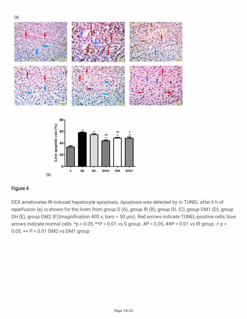
DEX ameliorates IR-induced hepatocyte apoptosis
TUNEL staining was used to assess apoptosis in the liver. The sham group showed few apoptotic hepaticcells (Figure 4 aA).The number of TUNEL positive cells increased signi�cantly in the IR group comparedto that in group S (Figure 4 aB; p < 0.01). In addition, the number of TUNEL positive cells in the DM1, DH,DM2 groups decreased signi�cantly compared to that in the IR group (Figure 4 aD-F; p < 0.01 or p <0.05). Notability, hepatocyte apoptosis rate in the DM1 group was signi�cantly lower than in the DL group(p < 0.01); hepatocyte apoptosis rate in the DH group was not signi�cantly different from that in the DM1group (p 0.05). There was no signi�cant difference in hepatocyte apoptosis rate between DM1 group andDM2 group (p 0.05).
Effect of DEX on core protein of ERS
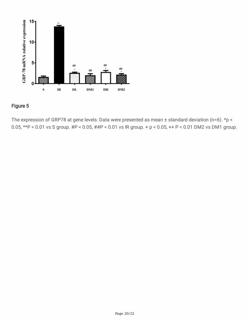
The GRP78 mRNA level is shown in �gure 5. The expression of GRP78 mRNA in the IR group wasincreased signi�cantly compared with that in group S (P < 0.01), whereas DEX weakened this increasesigni�cantly. There was no statistical difference between group S and group DM1 (p 0.05). Comparedwith group DL and DH, mRNA level of GRP78 in group DM1 was slightly decreased. There was nosigni�cant difference in the expression of GRP-78 mRNA between DM1 group and DM2 group (p 0.05).
Effect of HRS on expression of UPR signaling protein in liver
We measured the mRNA levels of PERK, ATF-6 and the protein expression levels of p-PERK, p-IRE-1 α, andfound them to be signi�cantly higher in the IR group than those in the sham group (P < 0.01; Figure 6),whereas DEX treatment reversed these effects signi�cantly. In addition, all the indicators mentionedabove in DM1 group were the lowest among the three pre-administration groups. Notably, mRNA levels ofPERK, ATF-6 and protein expression levels of p-PERK, p-IRE-1 α in the DH group and DM2 group were notsigni�cantly different from that in the DM1 group (p 0.05).
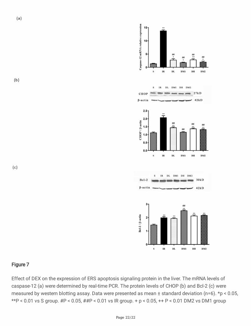
Effect of DEX on the expression of ERS apoptosis signaling protein in the liver
Compared with the sham group, the Caspase-12 mRNA level and the expression of the proteins of CHOP,Bcl-2 in the IR group were increased markedly. DEX not only reduced Caspase-12 mRNA level and theexpression level of CHOP protein signi�cantly (P < 0.01; Figure 7 a, b), but also increased the expressionlevel of Bcl-2 protein (P < 0.01; Figure 7 c). Compared with group DL and DH, the Caspase-12 mRNA leveland the expression level of CHOP protein in group DM1 was signi�cant lower (p < 0.01). There was nosigni�cant difference in the above two indicators between DM1 group and DM2 group (p 0.05). Theexpression level of Bcl-2 protein in the DM1 group was signi�cantly higher than in the DL, DH, DM2groups (p < 0.01).
Page 8/22
DiscussionPatients undergoing liver surgery remain at high risk of perioperative and postoperative complicationsdespite recent surgical advances in the �eld. Many of these potential complications are a consequence ofhepatic IRI with subsequent free radical formation, organelle failure, and release of pro-in�ammatorymediators. Unraveling the underlying processes of hepatic IRI and developing measures to address thisdevastating condition are therefore of great interest.
In recent years, more and more studies have shown that DEX has a certain protective effect on liverduring ischemia-reperfusion, and its mechanism may focus on oxidative stress and apoptosis [23]. Thepathway of apoptosis mediated by endoplasmic reticulum stress has gradually attracted the attention ofscholars. It may be a very effective method to reduce hepatic ischemia-reperfusion injury by regulatingendoplasmic reticulum stress. Therefore, in this experiment, the rat model of hepatic ischemia-reperfusioninjury was established, and different doses of DEX were given at different times to explore themechanisms underlying these hepatoprotective effects in hepatic IRI with a focus on ERS apoptosispathways. We found that hepatic IRI could upregulate the expression of ERS marker GRP78, activate thethree UPR signaling proteins PERK, ATF6 and IRE1, and induce expression of ERS-related apoptosisproteins CHOP, Caspase-12 and Bcl-2. DEX intervention can ameliorate IR-induced hepatic injuries bysuppressing hepatocyte ERS and apoptosis.
In the current study, histological analysis indicated that IR causes changes in the liver cells, such as livercell necrosis, blood cell destruction and in�ammatory cell in�ltration, DEX could ameliorate pathologicalliver damage. Hepatocyte morphological analysis indicated that IR induced mitochondria swelling,endoplasmic reticulum swelling and structure disorder, DEX could protect against liver cell organellesdamage. ALT mainly exists in hepatocytes. When hepatocytes are necrotic, ALT is released into the blood,which is one of the important signs of acute hepatocyte injury [24]. As shown in Fig. 3a, DEX signi�cantlyimproved liver function in rats undergoing hepatic IR. These results indicate that DEX can signi�cantlyreduce hepatic IRI damage.
Oxidative stress balance plays a key role in the process of ischemia and is one of the important ways tolead to hepatocyte injury. MDA is the �nal product of lipid peroxidation in the process of oxidative stress,which can directly damage the cell membrane structures such as hepatocyte membrane andmitochondrial membrane. The content of MDA is an important index to re�ect the degree of hepatocyteinjury [25]. As an antioxidant enzyme, SOD plays an important role in the antioxidant process of cells, sothe activity of SOD is also an important index to measure the degree of oxidative stress injury inhepatocytes [26]. In this study, the increase of MDA level and the decrease of SOD activity induced by IRwere reversed by DEX treatment. According to these results, the protective mechanism of IR seems to berelated to the antioxidant properties of DEX. There is evidence showing that oxidative stress, ER stressand in�ammation are inseparably linked, in particular, the degree of oxidative stress greatly in�uencesUPR signaling pathways. In the present study, we did not demonstrate a causal relationship betweenoxidative stress and ER stress; this is an area to explore in more details in our future work.
Page 9/22
The ER is a dynamic and stable organelle involved in protein translation, lipid biosynthesis and calciumhomeostasis [27]. Cells exposure to hypoxia can cause accumulation of unfolded proteins in endoplasmicreticulum lumen, leading to ERS [28]. IRI leads to hypoxia, oxidative stress and calcium overload, all ofwhich can induce ER failure that in turn triggers ERS and cell apoptotic. Li et al. showed that DEXattenuated myocardial ischemia reperfusion injury in diabetes mellitus rats and H/R injury cell, which isassociated with the reduction of ERS-induced cardiomyocyte apoptosis [29]. Consistent with this, DEXsigni�cantly alleviated the hepatocyte ERS induced by IR.
GRP78 is mainly located in endoplasmic reticulum and plays a key role in promoting protein folding andassembly, protein transport and calcium homeostasis, and is related to the regulation of ERtransmembrane transduction [30, 31]. When ERS occurs, GRP78 dissociates from its complex with threesensory proteins, which is then up-regulated and associated with misfolded and unfolded proteins. Freesensory proteins activate three signal transduction pathways of ERS [32]. Therefore, the expression levelof GRP78 can be used as one of the markers of ERS. In this study, we found that the level of GRP78mRNA in liver IR increased, indicating the activation of ERS. In addition, compared with the IR group, theexpression of GRP78 mRNA in the liver of other groups decreased signi�cantly. These results suggestthat DEX can down-regulate the increase of ERS induced by IR in liver.
ERS is also known as unfolded protein response (UPR). In the process of UPR, GRP78 is separated fromERS receptor proteins PERK, IRE1- α and ATF-6, and binds to unfolded proteins. The isolated threetransmembrane proteins begin to induce stress signals and exert their effects, clearing misfolded proteinsand leading to apoptosis [33]. In order to further explore the effect of IR on ER signaling pathway and theprotective effect of DEX on ERS, we detected the expression level of PERK mRNA, ATF-6 mRNA, p-PERKprotein and p-IRE-1 α protein in each group. In our study, IR signi�cantly up-regulated the level of PERKmRNA, ATF-6 mRNA, up-regulated the expression of p-PERK protein and p-IRE-1 α protein, while DEXsigni�cantly down-regulated its expression, down-regulated the expression of p-PERK protein and p-IRE-1α protein. These results suggest that DEX can inhibit the expression of UPR signal proteins to inhibit ERS,and thus participate in the regulation of liver IR.
Excessive or prolonged ERS activates apoptotic pathways and induces cell death. An increasing numberstudies have shown that apoptosis induced by ERS plays an important role in liver IRI [34, 35].Under theERS, Bcl-2 family proteins mediate endoplasmic reticulum Ca2+ release and caspase12 activation, whicheventually lead to apoptosis. There are three main apoptotic pathways in ERS, CHOP activation pathway,JNK activation pathway, and caspase 12 activation pathways [36]. Caspase-12 is a key protease of ERS-mediated apoptosis attached to the ER. When ER is overstressed, the complete Caspase-12 response isactivated, and then a series of proteins of the Caspase family are activated exponentially, resulting incascade reaction to induce apoptosis [37], so the protein plays an important role in initiating the ERapoptosis pathway. CHOP is a speci�c ERS transduction factor and an important signal molecule forpromoting apoptosis. In ERS, its expression is greatly increased and is considered to be one of themarkers of endoplasmic reticulum stress [38]. In this study, we detected the expression of caspase 12
Page 10/22
mRNA in each group, and detected the expression of apoptosis-related proteins (CHOP and Bcl-2) afterliver IRI by Western blot analysis. The results showed that the expression of caspase 12 mRNA, CHOPand Bcl-2 protein in liver tissue were signi�cantly up-regulated in IRI, while DEX signi�cantly inhibited itsexpression. The TUNEL assay results further con�rmed that IR can cause a signi�cant increase in liverTUNEL-positive cells, and DEX can reduce the number of TUNEL‐positive cells. Our results further provedthat DEX could inhibit the apoptotic liver of ERS induced by IRI.
In order to determine the dose-effect relationship of DEX’s protection against liver IRI and whether theeffect is relate with the administration time, 25 µg/kg, 50 µg/kg and 100 µg/kg DEX were administratedby intraperitoneal injection at 30 min before ischemia or 30 min after reperfusion. Our results indicatedthat 50 µg/kg DEX signi�cantly decreases the expression of ERS proteins and inhibits cell apoptosis, andprovides a superior protection compared with 25µg/kg DEX. Our results are consistent with the previousstudy described by Robert et al [39]. that discovered DEX inhibited iso�uraneinduced cortical injury in adose-dependent manner within 50µg/kg. Whereas the effect on liver IR-induced ERS and cell apoptosisdid not increase in 100 µg/kg DEX group, the possible reason is that the organ protection effect of DEX isassociated with decreased organ vasoconstriction, which does not occur with rapid loading of DEX or aninfusion of markedly high doses [40]. The speci�c mechanism remains to be further studied. At the doseof 50µg/kg, DEX injection 30 min before ischemia and 30 min after reperfusion did not present differenteffect on liver ERS and cell apoptosis. Our results are consistent with the previous study described by Guet al. that showed both DEX administered intraperitoneally prior to and following ischemia markedlyreduced remote lung injury induced by renal ischemia-reperfusion in mice [41]. But Schaak et al.discovered that DEX prior to ischemia, but not following it, attenuates intestinal IRinduced intestinal injury[42]. This is an area should be evaluated further in our future work.
ConclusionsIn conclusion, this study revealed that DEX can protect liver IRI by reducing ERS, thereby inhibiting ERS-related apoptosis. The protective effect of DEX on hepatic IRI was dose-dependent in a certain doserange, and both DEX administered prior to ischemia and following reperfusion markedly reduced liverinjury induced by hepatic IRI in mice.
AbbreviationsDex: dexmedetomidine; IRI: ischemia-reperfusion injury; HE: haematoxylin–eosin; TUNEL: terminaldeoxynucleotidyl transferase-mediated dUTP biotin nick end labelling; ERS: endoplasmic reticulum stress;PERK: protein kinase R-like ER kinase; ATF6: activating transcription factor 6; IRE1: Inositol-requiringenzyme 1; CHOP: C/EBP homologous protein; ALT: alanine aminotransferase; MDA: malondialdehyde;SOD: superoxide dismutase; TEM: transmission electron microscope;
Declarations

Page 11/22
Authors’ contributions
SZ and JT designed the research; WZ carried out the experiments, analysed the data; JT, HL and LAcooperated on carrying out the research; HL wrote the manuscript; JT co-wrote the manuscript; All authorsread and approved the �nal manuscript.
Author details
1College of Veterinary Medicine, Hebei Agricultural University, Baoding, People’s Republic of China.
2College of Veterinary Medicine, Northeast Agricultural University, Harbin, People’s Republic of China.
Acknowledgements
Not applicable
Competing interests
The authors declare that they have no competing interests.
Availability of data and materials
The datasets used and/or analysed during the current study are available from the corresponding authoron reasonable request.
Consent for publication
Not applicable.
Ethics approval and consent to participate
The study was allowed by the Agricultural University of Hebei Ethical Committee, Baoding, PR China.
Funding
This work was supported by the National Natural Science Foundation of China (grant number 31802250),the Scienti�c and Technological Research of Institution of Higher Learning Foundation of Hebei Provinceof China (BJ2019053).
References1. Liao XZ, Zhou SQ, Zong J, Wang ZP. Sevo�urane exerts protective effects on liver
ischemia/reperfusion injury by regulating NFKB3 expression via miR-9-5p. Exp Ther Med.2019;17(4):2632–40.
Page 12/22
2. Deng JF, Feng J, Liu T, Lu XY, Wang WW, Liu N, et al. Beraprost sodium preconditioning preventsin�ammation, apoptosis, and autophagy during hepatic ischemia-reperfusion injury in mice via theP38 and JNK pathways. Drug Des Devel Ther. 2018;12:4067–82.
3. Xie TJ, Li K, Gong X, Jiang R, Huang WY, Chen XH, et al. Paeoni�orin protects against liverischemia/reperfusion injury in mice via inhibiting HMGB1-TLR4 signaling pathway. Phytother Res.2018;32(11):2247–55.
4. Ji J, Wu LW, Feng J, Mo WH, Wu JY, Yu Q, et al. Cafestol preconditioning attenuates apoptosis andautophagy during hepatic ischemia-reperfusion injury by inhibiting ERK/PPARγ pathway. IntImmunopharmacol. 2020;84:106529.
5. Hou Y, Fu JR, Sun ST, Jin YC, Wang XF, Zhang LS, et al. BDE-209 induces autophagy and apoptosisvia IRE1α/Akt/mTOR signaling pathway in human umbilical vein endothelial cells. Environ Pollut.2019;253:429–38.
�. Zhan F, Zhao GP, Li X, Yang SK, Yang WJ, Zhou S, et al. Inositol-requiring enzyme 1 alphaendoribonuclease speci�c inhibitor STF-083010 protects the liver from thioacetamide-inducedoxidative stress, in�ammation and injury by triggering hepatocyte autophagy. Int Immunopharmacol.2019;73:261–9.
7. Malhi H, Kaufman RJ. Endoplasmic reticulum stress in liver disease. J Hepatol. 2011;54(4):795–809.
�. Li JJ, Zhao Y, Zhou N, Li LY, Li K. Dexmedetomidine attenuates myocardial ischemia-reperfusioninjury in diabetes mellitus by inhibiting endoplasmic reticulum stress. J Diabetes Res. 2019; 2019:7869318.
9. Kim I, Xu WJ, Reed JC. Cell death and endoplasmic reticulum stress: disease relevance andtherapeutic opportunities. Nat Rev Drug Discov. 2008;7(12):1013–30.
10. Li H, Bai G, Ge YS, Zhang QZ, Kong XD, Meng WJ, et al. Hydrogen-rich saline protects against small-scale liver ischemia-reperfusion injury by inhibiting endoplasmic reticulum stress. Life Sci.2018;194:7–14.
11. Jiao ZH, Liu XN, Ma YJ, Ge YS, Zhang QZ, Liu BY, et al. Adipose-derived stem cells protect ischemia-reperfusion and partial hepatectomy by attenuating endoplasmic reticulum stress. Front Cell Dev Bio.2020;8:177.
12. Chi XB, Jiang Y, Chen YB, Yang F, Cai QC, Pan F, et al. Suppression of microRNA27a protects againstliver ischemia/reperfusion injury by targeting PPARγ and inhibiting endoplasmic reticulum stress.Mol Med Rep. 2019;20(5):4003–12.
13. Selzner N, Rudiger H, Graf R, Clavien PA. Protective strategies against ischemic injury of the liver.Gastroenterology. 2003;125(3):917–36.
14. Weerink MAS, Struys M, Hannivoort LN, Barends CRM, Absalom AR, Colin P. Clinicalpharmacokinetics and pharmacodynamics of dexmedetomidine. Clin Pharmacokinet.2017;56(8):893–913.
15. Liu ZG, Wang YP, Wang YQ, Ning QQ, Zhang Y, Gong CZ, et al. Dexmedetomidine attenuatesin�ammatory reaction in the lung tissues of septic mice by activating cholinergic anti-in�ammatory
Page 13/22
pathway. Int Immunopharmacol. 2016;35:210–6.
1�. Chen Z, Ding T, Ma CG. Dexmedetomidine (DEX) protects against hepatic ischemia/reperfusion (I/R)injury by suppressing in�ammation and oxidative stress in NLRC5 de�cient mice. Biochem BiophysRes Commun. 2017;493(2):1143–50.
17. Sun ZX, Zhao TY, Lv SJ, Gao Y, Masters J, Weng H. Dexmedetomidine attenuates spinal cordischemia–reperfusion injury through both anti-in�ammation and anti-apoptosis mechanisms inrabbits. J Transl Med. 2018;16(1):209.
1�. Kiliç K, Hanci V, Selek S, Sözmen M, Kiliç N, Citil M, et al. The effects of dexmedetomidine onmesenteric arterial occlusion-associated gut ischemia and reperfusion-induced gut and kidney injuryin rabbits. J Surg Res. 2012;178(1):223–32.
19. Zhou HM, Sun J, Zhong WZ, Pan XX, Liu CM, Cheng F,et al. Dexmedetomidine preconditioningalleviated murine liver ischemia and reperfusion injury by promoting macrophage M2 activation viaPPARγ/STAT3 signaling. Int Immunopharmacol. 2020;82:106363.
20. Wang YH, Wu S, Yu XF, Zhou SL, Ge M, Chi XJ, et al. Dexmedetomidine protects rat liver againstischemia-reperfusion injury partly by the α2A-adrenoceptor subtype and the mechanism isassociated with the TLR4/NF-κB pathway. Int J Mol Sci. 2016;17(7):995.
21. Gao WD, Feng ZJ, Zhang SL, Wu B, Geng X, Fan GX, et al. Anti-in�ammatory and antioxidant effect ofeucommia ulmoides polysaccharide in hepatic ischemia-reperfusioninjury by regulating ROS and theTLR-4-NF- κ B pathway. BioMed Res Int. 2020; 2020: 1860637.
22. Suzuki S, Nakamura S, Koizumi T, Sakaguchi S, Baba S, Muro H, et al. The bene�cial effect of aprostaglandin I2 analog on ischemic rat liver. Transplantation. 1991;52(6):979–83.
23. Zhai M, Liu C, Li YX, Zhang PJ, Yu ZQ, Zhu H, et al. Dexmedetomidine inhibits neuronal apoptosis byinducing Sigma-1 receptor signaling in cerebral ischemia-reperfusion injury. Aging.2019;11(21):9556–68.
24. Chen G, Deng HZ, Song X, Lu M, Zhao L, Xia S, et al. Reactive oxygen species-responsive polymericnanoparticles for alleviating sepsis-induced acute liver injury in mice. Biomaterials. 2017;144:30–41.
25. Zhang JP, Cao H, Zhang Y, Zhang YY, Ma JJ, Wang JY, et al. Nephroprotective effect of calciumchannel blockers against toxicity of lead exposure in mice. Toxicol Lett. 2013;218(3):273–80.
2�. Zhang WQ, Xue JD, Ge M, Yu ML, Liu L, Zhang ZG. Resveratrol attenuates hepatotoxicity of ratsexposed to arsenic trioxide. Food Chem Toxicol. 2013;51:87–92.
27. Avril T, Vauléon E, Chevet E. Endoplasmic reticulum stress signaling and chemotherapy resistance insolid cancers. Oncogenesis. 2017;6(8):e373.
2�. Lee WJ, Chien MH, Chow JM, Chang JL, Wen YC, Lin YW, et al. Nonautophagic cytoplasmicvacuolation death induction in human PC-3M prostate cancer by curcumin through reactive oxygenspecies-mediated endoplasmic reticulum stress. Sci Rep. 2015;5:10420.
29. Li JJ, Zhao Y, Zhou N, Li LY, Li K. Dexmedetomidine attenuates myocardial ischemia-reperfusioninjury in diabetes mellitus by inhibiting endoplasmic reticulum stress. J Diabetes Res. 2019; 2019:7869318.
Page 14/22
30. Dudek J, Benedix J, Cappel S, Greiner M, Jalal C, Müller L, et al. Functions and pathologies of BiP andits interaction partners[J]. Cell Mol Life Sci. 2009;66(9):1556–69.
31. Lee AS. Mammalian stress response: induction of the glucose-regulated protein family. Curr Opin CellBiol. 1992;4(2):267–73.
32. Hetz C. The unfolded protein response: controlling cell fate decisions under ER stress and beyond.Nat Rev Mol Cell Bio. 2012;13(2):89–102.
33. Shen JS, Chen X, Hendershot L, Prywes R. ER Stress Regulation of ATF6 Localization by Dissociationof BiP/GRP78 Binding and Unmasking of Golgi Localization Signals. Dev Cell. 2002;3(1):99–111.
34. Fan CX, Yang Y, Liu Y, Jiang S, Di SY, Hu W, et al. Icariin displays anticancer activity against humanesophageal cancer cells via regulating endoplasmic reticulum stress-mediated apoptotic signaling.Sci Rep. 2016;6(1):69–90.
35. Rao JH, Qin JJ, Qian XF, Lu L, Wang P, Wu ZS, et al. Lipopolysaccharide pre-conditioning protectshepatocytes from ischemia/reperfusion injury (IRI) through inhibiting ATF4-CHOP pathway in mice.PLoS One. 2013;8(6):e65568.
3�. Ding WX, Zhang XF, Huang HP, Ding N, Zhang SJ, Hutchinson SZ, et al. Adiponectin protects ratmyocardium against chronic intermittent hypoxia-induced injury via inhibition of endoplasmicreticulum stress. PLoS One. 2017;9(4):e94545.
37. Chen YH, Wu XD, Yao ST, Sun S, Liu XH. Calcineurin is involved in cardioprotection induced byischemic postconditioning through attenuating endoplasmic reticulum stress. Chin Med J.2011;124(20):3334–40.
3�. Yan L, Wang SL, Ren BS, Wang J, Chen J, Lu J, et al. CHOP favors endoplasmic reticulum stress-induced apoptosis in hepatocellular carcinoma cells via inhibition of autophagy. PloS one.2017;12(8):e0183680.
39. Sanders RD, Xu J, Shu Y, Januszewski A, Halder S, Fidalgo A, et al. Dexmedetomidine attenuatesiso�urane-induced neurocognitive impairment in neonatal rats. Anesthesiology. 2009;110(5):1077–85.
40. Cai Y, Xu H, Yan J, Zhang L, Lu Y. Molecular targets and mechanism of action of dexmedetomidine intreatment of ischemia/reperfusion injury. Mol Med Rep. 2014;9(5):1542–50.
41. Gu J, Chen J, Xia P, Tao G, Zhao H, Ma D. Dexmedetomidine attenuates remote lung injury induced byrenal ischemia-reperfusion in mice. Acta Anaesthesiol Scand. 2011;55(10):1272–8.
42. Schaak S, Cussac D, Cayla C, Devedjian JC, Guyot R, Paris H, et al. Alpha(2) adrenoceptors regulateproliferation of human intestinal epithelial cells. Gut. 2000;47(2):242–250.
TablesTable 1 Primer Sequence of the genes were tested in the present study.
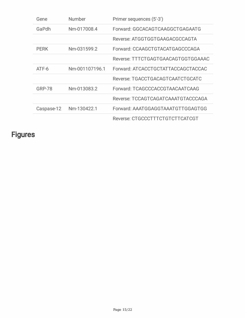
Page 15/22
Gene Number Primer sequences (5′-3′)
GaPdh Nm-017008.4 Forward: GGCACAGTCAAGGCTGAGAATG
Reverse: ATGGTGGTGAAGACGCCAGTA
PERK Nm-031599.2 Forward: CCAAGCTGTACATGAGCCCAGA
Reverse: TTTCTGAGTGAACAGTGGTGGAAAC
ATF-6 Nm-001107196.1 Forward: ATCACCTGCTATTACCAGCTACCAC
Reverse: TGACCTGACAGTCAATCTGCATC
GRP-78 Nm-013083.2 Forward: TCAGCCCACCGTAACAATCAAG
Reverse: TCCAGTCAGATCAAATGTACCCAGA
Caspase-12 Nm-130422.1 Forward: AAATGGAGGTAAATGTTGGAGTGG
Reverse: CTGCCCTTTCTGTCTTCATCGT
Figures

Page 16/22
Figure 1
Effects of DEX on IR-induced liver histopathology. Representative H&E-stained sections after 6 h ofreperfusion (a) is shown for the livers from group S (A), group IR (B), group DL (C), group DM1 (D), groupDH (E), group DM2 (F). (Magni�cation 200 x, bars = 100 μm). The red arrows represent congestion; greenarrows represent the liver sinus; blue arrows represent liver macrophages. (b) Histopathological meanliver injury scores. Data were presented as mean ± standard deviation (n = 6). *p < 0.05, **P < 0.01 vs Sgroup. #P < 0.05, ##P < 0.01 vs IR group. + p < 0.05, ++ P < 0.01 DM2 vs DM1 group.

Page 17/22
Figure 2
Effects of DEX on IR-induced hepatocyte morphological changes. TEM micrographs is shown for thelivers from group S (A), group IR (B), group DL (C), group DM1 (D), group DH (E), group DM2 (F).(Magni�cation 10000 x, bars = 2 μm). The thick black arrow indicated ER swelling, the thin black arrowindicated the disordered structure of ER, and the thin white arrow indicated mitochondria swelling.
Page 18/22
Figure 3
DEX decreases serum ALT activity and reduces liver oxidative stress induced by IR. (a) The content of ALTin serum. (b) The concentration of MDA in liver tissue. (c) The activity of SOD in liver tissue. Data werepresented as mean ± standard deviation (n=6). *p < 0.05, **P < 0.01 vs S group. #P < 0.05, ##P < 0.01 vsIR group. + p < 0.05, ++ P < 0.01 DM2 vs DM1 group
Page 19/22
Figure 4
DEX ameliorates IR-induced hepatocyte apoptosis. Apoptosis was detected by in TUNEL after 6 h ofreperfusion (a) is shown for the livers from group S (A), group IR (B), group DL (C), group DM1 (D), groupDH (E), group DM2 (F)(magni�cation 400 x, bars = 50 μm). Red arrows indicate TUNEL-positive cells; bluearrows indicate normal cells. *p < 0.05, **P < 0.01 vs S group. #P < 0.05, ##P < 0.01 vs IR group. + p <0.05, ++ P < 0.01 DM2 vs DM1 group
Page 20/22
Figure 5
The expression of GRP78 at gene levels. Data were presented as mean ± standard deviation (n=6). *p <0.05, **P < 0.01 vs S group. #P < 0.05, ##P < 0.01 vs IR group. + p < 0.05, ++ P < 0.01 DM2 vs DM1 group.

Page 21/22
Figure 6
Effect of DEX on expression of UPR signaling protein in liver. The mRNA levels of PERK (a) and ATF-6 (b)were determined by real-time PCR. The protein levels of p-PERK (c) and p-IRE-α (d) were measured bywestern blotting assay. Data were presented as mean ± standard deviation (n=6). *p < 0.05, **P < 0.01 vsS group. #P < 0.05, ##P < 0.01 vs IR group. + p < 0.05, ++ P < 0.01 DM2 vs DM1 group
Page 22/22
Figure 7
Effect of DEX on the expression of ERS apoptosis signaling protein in the liver. The mRNA levels ofcaspase-12 (a) were determined by real-time PCR. The protein levels of CHOP (b) and Bcl-2 (c) weremeasured by western blotting assay. Data were presented as mean ± standard deviation (n=6). *p < 0.05,**P < 0.01 vs S group. #P < 0.05, ##P < 0.01 vs IR group. + p < 0.05, ++ P < 0.01 DM2 vs DM1 group


![Endoplasmic reticulum[1]](https://static.fdocuments.in/doc/165x107/58ed5fc71a28aba1678b4611/endoplasmic-reticulum1.jpg)