Endometriosis - McGraw-Hill Education - Professional · INFERTILITY ... EXPECTANT MANAGEMENT........
19
Endometriosis PATHOPHYSIOLOGY............................ 225 ETIOLOGY ................................... 225 HORMONAL DEPENDENCE ...................... 226 ROLE OF THE IMMUNE SYSTEM ................... 226 RISK FACTORS ................................ 227 CLASSIFICATION AND LOCATION OF ENDOMETRIOSIS .. 228 PATIENT SYMPTOMS ........................... 229 PAIN ....................................... 229 INFERTILITY ................................. 230 DIFFERENTIAL DIAGNOSIS....................... 231 DIAGNOSIS .................................. 232 PHYSICAL EXAMINATION ....................... 232 LABORATORY TESTING ......................... 232 DIAGNOSTIC IMAGING ......................... 233 DIAGNOSTIC LAPAROSCOPY ..................... 233 PATHOLOGIC ANALYSIS ......................... 234 DIAGNOSTIC ALGORITHM ....................... 234 TREATMENT ................................. 234 EXPECTANT MANAGEMENT ...................... 235 MEDICAL TREATMENT OF ENDOMETRIOSIS-RELATED PAIN .................. 235 SURGICAL TREATMENT OF ENDOMETRIOSIS-RELATED PAIN .................. 238 REFERENCES ................................. 240 Endometriosis is a common benign gynecologic disorder defined as the presence of endometrial glands and stroma outside of the normal location. First identified in the mid-nineteenth century (Von Rokitansky, 1860), endometriosis is most commonly found on the pelvic peritoneum but may also be found on the ovaries, rectovaginal septum, ureter, and rarely in the bladder, pericar- dium, and pleura (Comiter, 2002; Giudice, 2004). Endometriosis is a hormonally dependent disease and as a result is chiefly found in reproductive-aged women. Endometrial tissue located within the myometrium is termed adenomyosis and is discussed in greater detail in Chapter 9 (p. 208). The incidence of endometriosis is difficult to quantify, as women with the disease are often asymptomatic, and imaging modalities have low sensitivities for diagnosis. Women with endometriosis may be asymptomatic, subfertile, or suffer vary- ing degrees of pelvic pain. The primary method of diagnosis is laparoscopy, with or without biopsy for histologic diagnosis (Kennedy, 2005; Marchino, 2005). Using this standard, investi- gators have reported the annual incidence of surgically diag- nosed endometriosis to be 1.6 cases per 1,000 women aged between 15 and 49 years (Houston, 1987). In asymptomatic women, the prevalence of endometriosis ranges from 2 to 22 percent, depending on the population studied (Eskenazi, 1997; Mahmood, 1991; Moen, 1997). However, because of its link with infertility and pelvic pain, endometriosis is notably more prevalent in subpopulations of women with these complaints. In infertile women, the prevalence has been reported to be between 20 to 50 percent and in those with pelvic pain, 40 to 50 percent (Balasch, 1996; Eskenazi, 2001). PATHOPHYSIOLOGY ■ Etiology Although the definitive cause of endometriosis remains unknown, several theories with supporting evidence have been described. 225 CHAPTER 10
Transcript of Endometriosis - McGraw-Hill Education - Professional · INFERTILITY ... EXPECTANT MANAGEMENT........
Endometriosis
PATHOPHYSIOLOGY . . . . . . . . . . . . . . . . . . . . . . . . . . . . 225
ETIOLOGY . . . . . . . . . . . . . . . . . . . . . . . . . . . . . . . . . . . 225
HORMONAL DEPENDENCE . . . . . . . . . . . . . . . . . . . . . . 226
ROLE OF THE IMMUNE SYSTEM . . . . . . . . . . . . . . . . . . . 226
RISK FACTORS . . . . . . . . . . . . . . . . . . . . . . . . . . . . . . . . 227
CLASSIFICATION AND LOCATION OF ENDOMETRIOSIS . . 228
PATIENT SYMPTOMS . . . . . . . . . . . . . . . . . . . . . . . . . . . 229
PAIN . . . . . . . . . . . . . . . . . . . . . . . . . . . . . . . . . . . . . . . 229
INFERTILITY . . . . . . . . . . . . . . . . . . . . . . . . . . . . . . . . . 230
DIFFERENTIAL DIAGNOSIS . . . . . . . . . . . . . . . . . . . . . . . 231
DIAGNOSIS . . . . . . . . . . . . . . . . . . . . . . . . . . . . . . . . . . 232
PHYSICAL EXAMINATION . . . . . . . . . . . . . . . . . . . . . . . 232
LABORATORY TESTING . . . . . . . . . . . . . . . . . . . . . . . . . 232
DIAGNOSTIC IMAGING . . . . . . . . . . . . . . . . . . . . . . . . . 233
DIAGNOSTIC LAPAROSCOPY . . . . . . . . . . . . . . . . . . . . . 233
PATHOLOGIC ANALYSIS . . . . . . . . . . . . . . . . . . . . . . . . . 234
DIAGNOSTIC ALGORITHM . . . . . . . . . . . . . . . . . . . . . . . 234
TREATMENT . . . . . . . . . . . . . . . . . . . . . . . . . . . . . . . . . 234
EXPECTANT MANAGEMENT . . . . . . . . . . . . . . . . . . . . . . 235
MEDICAL TREATMENT OF ENDOMETRIOSIS-RELATED PAIN . . . . . . . . . . . . . . . . . . 235
SURGICAL TREATMENT OF ENDOMETRIOSIS-RELATED PAIN . . . . . . . . . . . . . . . . . . 238
REFERENCES . . . . . . . . . . . . . . . . . . . . . . . . . . . . . . . . . 240
Endometriosis is a common benign gynecologic disorder defined as the presence of endometrial glands and stroma outside of the normal location. First identified in the mid-nineteenth century (Von Rokitansky, 1860), endometriosis is most commonly found on the pelvic peritoneum but may also be found on the ovaries, rectovaginal septum, ureter, and rarely in the bladder, pericar-dium, and pleura (Comiter, 2002; Giudice, 2004). Endometriosis is a hormonally dependent disease and as a result is chiefly found in reproductive-aged women. Endometrial tissue located within the myometrium is termed adenomyosis and is discussed in greater detail in Chapter 9 (p. 208).
The incidence of endometriosis is difficult to quantify, as women with the disease are often asymptomatic, and imaging modalities have low sensitivities for diagnosis. Women with endometriosis may be asymptomatic, subfertile, or suffer vary-ing degrees of pelvic pain. The primary method of diagnosis is laparoscopy, with or without biopsy for histologic diagnosis (Kennedy, 2005; Marchino, 2005). Using this standard, investi-gators have reported the annual incidence of surgically diag-nosed endometriosis to be 1.6 cases per 1,000 women aged between 15 and 49 years (Houston, 1987). In asymptomatic women, the prevalence of endometriosis ranges from 2 to 22 percent, depending on the population studied (Eskenazi, 1997; Mahmood, 1991; Moen, 1997). However, because of its link with infertility and pelvic pain, endometriosis is notably more prevalent in subpopulations of women with these complaints. In infertile women, the prevalence has been reported to be between 20 to 50 percent and in those with pelvic pain, 40 to 50 percent (Balasch, 1996; Eskenazi, 2001).
PATHOPHYSIOLOGY
■ EtiologyAlthough the definitive cause of endometriosis remains unknown, several theories with supporting evidence have been described.
225
CHAPTER 10
sch72576_c10.indd Page 225 12/28/07 10:47:20 AM usersch72576_c10.indd Page 225 12/28/07 10:47:20 AM user /Volumes/209/MHBD105/mhbd105indd%0/ch10/Volumes/209/MHBD105/mhbd105indd%0/ch10

226 Benign General Gynecology
SECTION
1
endometrium (Bontis, 1997). In vitro studies have demon-strated the potential for ovarian surface epithelium, in response to estrogens, to undergo transformation to form endometriotic lesions (Matsuura, 1999). Although many putative factors have been identified, their propensity to cause endometriosis in some women but not in others demonstrates the still unidenti-fied etiology of this disease.
■ Hormonal DependenceOne factor that has been definitively established as having a caus-ative role in the development of endometriosis is estrogen (Gurates, 2003). Although most estrogen in women is produced directly by the ovaries, numerous peripheral tissues are also known to create estrogens through aromatization of ovarian and adrenal androgens. Endometriotic implants have been shown to express aromatase and 17�-hydroxysteroid dehydrogenase type 1, the enzymes responsible for conversion of androstenedione to estrone and of estrone to estradiol, respectively. Implants, how-ever, are deficient in 17�-hydroxysteroid dehydrogenase type 2, which inactivates estrogen (Kitawaki, 1997; Zeitoun, 1998). This enzymatic combination ensures that implants will be exposed to an estrogenic environment. Furthermore, the locally produced estrogens within endometriotic lesions may exert their biologic effect within the same tissue or cell in which they are produced, a process referred to as intracrinology.
In contrast, normal endometrium does not express aromatase and has elevated levels of 17�-hydroxysteroid dehydrogenase type 2 in response to progesterone, which ensures that estrogenic effects are attenuated in response to progesterone (Satyaswaroop, 1982). As a result, progesterone antagonizes the estrogen effects in normal endometrium during the luteal phase of the menstrual cycle. Endometriosis, however, manifests a relative progesterone-resistant state, which prevents attenuation of the estrogen stimu-lation in this tissue (Attia, 2000).
Prostaglandin E2 (PGE2) is the most potent inducer of aro-matase activity in endometrial stromal cells, acting through the prostaglandin EP2 receptor subtype (Noble, 1997; Zeitoun, 1999). Estradiol produced in response to the increased aroma-tase activity subsequently augments PGE2 production by stim-ulating cyclooxygenase type 2 (COX-2) enzyme in uterine endothelial cells (Fig. 10-1) (Bulun, 2002; Gurates, 2003). This creates a positive feedback loop and potentiates the estro-genic effects on proliferation of endometriosis. This concept of locally produced estrogens and intracrine estrogen action in endometriosis serves as the basis for pharmacologic inhibition of aromatase activity in cases of endometriosis that are refrac-tory to standard therapy.
■ Role of the Immune SystemAlthough most women experience retrograde menstruation, which may play a role in the seeding and establishment of implants, few develop endometriosis. Menstrual tissue and endometrium that is refluxed into the peritoneal cavity is usu-ally cleared by immune cells such as macrophages, natural killer (NK) cells, and lymphocytes. For this reason, immune system dysfunction is one likely mechanism for the genesis of
Retrograde MenstruationThe earliest and most widely accepted theory relates to retro-grade menstruation through the fallopian tubes with subse-quent dissemination of endometrial tissue within the peritoneal cavity (Sampson, 1927). Refluxed endometrial fragments adhere to and invade the peritoneal mesothelium and develop a blood supply, which leads to continued implant survival and growth (Giudice, 2004).
First proposed in the 1920s, this theory has gained support with the findings of greater volumes of refluxed blood and endo-metrial tissue in the pelves of women with endometriosis (Halme, 1984). Uterine hyperperistalsis and dysperistalsis have been noted in women with endometriosis and resulted in subsequent increased endometrial reflux (Leyendecker, 2004). Additionally, D’Hooghe (1997) demonstrated that surgical obliteration of the cervical out-flow tract in baboons leads to the induction of endometriosis. Women with amenorrhea due to outflow tract obstruction simi-larly have a high incidence of endometriosis, which is often relieved by correction of the obstruction (Sanfilippo, 1986).
Lymphatic or Vascular SpreadEvidence also supports the concept of endometriosis originat-ing from aberrant lymphatic or vascular spread of endometrial tissue (Ueki, 1991). Findings of endometriosis in unusual loca-tions, such as the perineum or groin, bolster this theory (Mitchell, 1991; Pollack, 1990). The retroperitoneal region has abundant lymphatic circulation. Thus, cases in which no peri-toneal implants are found, but solely isolated retroperitoneal lesions are noted, suggest lymphatic spread (Moore, 1988). Additionally, the tendency of endometrial adenocarcinoma to spread via the lymphatic route indicates the ease at which endo-metrium can be transported by this route (McMeekin, 2003). Although this theory remains attractive, few studies have experi-mentally evaluated this form of endometriosis transmission.
Coelomic MetaplasiaThe theory of coelomic metaplasia suggests that the parietal peritoneum is a pluripotential tissue that can undergo metaplas-tic transformation to tissue histologically indistinguishable from normal endometrium. Because the ovary and the progenitor of the endometrium, the müllerian ducts, are both derived from coelomic epithelium, metaplasia may explain the development of ovarian endometriosis. In addition, the theory has been extended to include the peritoneum because of the proliferative and differentiation potential of the peritoneal mesothelium. This theory is attractive in instances of endometriosis in the absence of menstruation, such as in premenarchal and post-menopausal women, and in males treated with estrogen and orchiectomy for prostatic carcinoma (Dictor, 1988; Pinkert, 1979). However, the absence of endometriosis in other tissues derived from coelomic epithelium argues against this theory.
Induction TheoryFinally, the induction theory proposes that some hormonal or biologic factor(s) may induce the differentiation of undifferen-tiated cells into endometrial tissue (Vinatier, 2001). These substances may be exogenous or released directly from the
sch72576_c10.indd Page 226 12/28/07 10:47:21 AM usersch72576_c10.indd Page 226 12/28/07 10:47:21 AM user /Volumes/209/MHBD105/mhbd105indd%0/ch10/Volumes/209/MHBD105/mhbd105indd%0/ch10

227EndometriosisCH
AP
TER 1
0
endometriosis in the presence of retrograde menstruation (Seli, 2003). Impaired cellular and humoral immunity and altered growth factor and cytokine signaling have each been identified in endometriotic tissues.
Macrophages act as scavenger cells in various tissues and increased numbers have been found in the peritoneal cavity of women with endometriosis (Haney, 1981; Olive, 1985b). Although this increased population might logically act to sup-press endometrial proliferation, macrophages in these women, however, have a stimulatory effect on endometriotic tissue. In one study, circulating monocytes obtained from women with endometriosis enhanced the in vitro proliferation of cultured endometrial cells, whereas the monocytes from women without endometriosis had the opposite effect (Braun, 1994). It appears therefore that impaired function, and not population size, of macrophages allows endometriotic tissue proliferation.
Natural killer cells are immune cells that have cytotoxic activ-ity against foreign cells. Although the number of NK cells is unaltered in the peritoneal fluid of women with endometriosis, decreased NK cell cytotoxicity against endometrium has been demonstrated (Ho, 1995; Wilson, 1994). Specifically, the peri-toneal fluid from women with endometriosis has been found to suppress NK cell activity, suggesting that soluble factors may play a role in NK cell suppression (Oosterlynck, 1993).
Cellular immunity may also be disordered in women with endometriosis, and T lymphocytes are implicated. For example, in women with endometriosis compared with unaffected women, total lymphocyte numbers or helper/suppressor subpopulation
ratios do not differ in peripheral blood, but peritoneal fluid lymphocyte numbers are increased (Steele, 1984). Also, the cyto-toxic activity of T lymphocytes against autologous endometrium in affected women is impaired (Gleicher, 1984).
Humoral immunity has also been shown to be altered in affected women and is suggested to play a role in the develop-ment of endometriosis. Endometrial antibodies of the IgG class are more frequently detected in the serum of women with endometriosis (Odukoya, 1995). One study also identified IgG and IgA autoantibodies against endometrial and ovarian tissues in the sera and in cervical and vaginal secretions of affected women (Mathur, 1982). These results suggest that endometrio-sis may be, in part, an autoimmune disease. This may explain some of the factors influencing lower pregnancy and in vitro fertilization (IVF) implantation rates in women with endome-triosis (Dmowski, 1995).
Cytokines are small, soluble immune factors involved in paracrine and autocrine signaling of other immune cells. Numerous cytokines, especially interleukins, have been impli-cated in the pathogenesis of endometriosis. Increased levels of interleukin-1� (IL-1�) have been identified in the endome-trial fluid of those with endometriosis (Mori, 1991). Moreover, IL-6 has been shown to be increased in endometrial stromal cells of affected women (Tseng, 1996). Accordingly, IL-6 serum levels greater than 2 pg/mL and tumor necrosis factor-� (TNF-�) peritoneal fluid levels more than 15 pg/mL may be used to discriminate between those with or without endome-triosis (Bedaiwy, 2002). Similarly, IL-8 peritoneal fluid levels are elevated in affected individuals and stimulate proliferation of endometrial stromal cells (Arici, 1996; Arici, 1998; Ryan, 1995).
Other noninterleukin cytokines and growth factors are associated with the pathogenesis of endometriosis. For exam-ple, both monocyte chemoattractant protein-1 (MCP-1) and RANTES (regulated on activation, normal T-cell expressed and secreted) are chemoattractant for monocytes. Levels of these cytokines are increased in the peritoneal fluid of those with endometriosis and positively correlate with disease severity (Arici, 1997; Khorram, 1993). In addition, vascular endothelial growth factor (VEGF) is an angiogenic growth factor, which is upregulated by estradiol in endometrial stro-mal cells and peritoneal fluid macrophages. Levels of this factor are increased in the peritoneal fluid of affected women (McLaren, 1996). Although the exact role of these cytokines is not clear, perturbations in their expression and activity further support an immunologic role in the pathogenesis of endometriosis.
RISK FACTORS
■ Familial ClusteringThere is evidence of a familial inheritance pattern for endo-metriosis. Although no apparent mendelian genetics inheri-tance pattern has been identified, the increased incidence in first-degree relatives suggests a polygenic/multifactorial inher-itance pattern. For example in a genetic study of women with endometriosis, Simpson and his colleagues (1980) noted that
FIGURE 10-1 Activation of COX-2 in endometrial stromal cells results in upregulation of PGE2, a potent stimulator of aromatase in endometrial stromal cells. Aromatase activity results in intracel-lular aromatization of androgens to increase intracellular estradiol via an intracrine mechanism. a � androgen; E2 � estradiol; COX-2 � cyclooxygenase 2; PGE2 � prostaglandin E2; IL-1� � interleukin 1�; VEGF � vascular endothelial growth factor.
a
ENDOMETRIOTIC IMPLANT
aromatase
ADRENALEXTRA-OVARIAN
TISSUES
PGE2
COX-2
VEGF
arachidonicacid
IL-1�
E2�
�
�
OVARY
sch72576_c10.indd Page 227 12/28/07 10:47:21 AM usersch72576_c10.indd Page 227 12/28/07 10:47:21 AM user /Volumes/209/MHBD105/mhbd105indd%0/ch10/Volumes/209/MHBD105/mhbd105indd%0/ch10

228 Benign General Gynecology
SECTION
1
5.9 percent of female siblings and 8.1 percent of the mothers of affected women had endometriosis compared with 1 per-cent of the husband’s female first-degree relatives. Further research has revealed that women with endometriosis and an affected first-degree relative were more likely to have severe endometriosis (61%) than women without an affected first-degree relative (24 percent) (Malinak, 1980). Moreover, Stefansson and his associates (2002), in their analysis of a large population-based study in Iceland, demonstrated a higher kinship coefficient in women with endometriosis com-pared with matched controls. In this study, the risk ratios were 5.2 for sisters and 1.56 for cousins. Studies have also demon-strated concordance for endometriosis in monozygotic twin-pairs, suggesting a familial/genetic basis (Hadfield, 1997; Treloar, 1999).
■ Genetic Mutations and PolymorphismsRates of familial clustering noted above suggest polygenic inheritance and several candidate genes have been investigated. Two approaches to identify genes involved with endometriosis include sibling-pair linkage analysis and high-throughput analy-sis of gene expression patterns using microarray technology.
The largest study to date, examining over 1,000 affected sister-pair families, has identified a region on chromosome 10q26 that demonstrates significant linkage in these sisters affected with endometriosis (Treloar, 2005). This study also revealed a smaller linkage on chromosome 20p13. Two candi-date genes within or near this locus have been identified. One such gene is EMX2, a transcription factor necessary for repro-ductive tract development. It has been shown to be aberrantly expressed in the endometrium of women with endometriosis (Daftary, 2004). The second gene is PTEN, a tumor suppres-sor gene implicated in the malignant transformation of ovar-ian endometriosis (Bischoff, 2000). Studies are currently underway to further determine the role of these genes in endometriosis.
Microarray technology has been used to analyze differences in gene expression in eutopic endometrium (endometrium found normally lining the endometrial cavity) from women without endometriosis compared with that from women with endometriosis (Kao, 2003). Researchers found that several genes were differentially regulated in the eutopic endome-trium in women with endometriosis. These include those coding for interleukin 15, glycodelin, Dickkopf-1, semapho-rin E, aromatase, progesterone receptor, and various angio-genic factors. Although some of these genes have previously been shown to play a role in endometriosis, others have not been implicated until recently, and their role remains to be elucidated.
Several other genes have been identified, through genetic mutations, polymorphisms, or differential gene expression, to be associated with endometriosis. Although investigations have demonstrated polymorphisms of these genes occur with greater frequency in women suffering with endometriosis, their role in disease causation has not been determined. A more thorough review of candidate genes in the epidemiology of endometriosis can be found at the website http://www.well.ox.ac.uk/~krinaz/genepi_endo.htm.
■ Anatomic DefectsReproductive outflow tract obstruction can predispose to develop-ment of endometriosis, likely through exacerbation of retrograde menstruation (Breech, 1999). Accordingly, endometriosis has been identified in women with noncommunicating uterine horn, imperforate hymen, and transverse vaginal septum (see Chap. 18, p. 413) (Schattman, 1995). Because of this association, diagnostic laparoscopy to identify and treat endometriosis is suggested at the time of corrective surgery for many of these anomalies. Repair of such anatomic defects is thought to decrease the risk of developing endometriosis (Joki-Erkkila, 2003; Rock, 1982).
■ Environmental ToxinsThere have been numerous studies suggesting that exposure to environmental toxins may play a role in the development of endometriosis. The toxins most commonly implicated are 2,3,7,8-tetrachlorodibenzo-p-dioxin (TCDD) and other dioxin-like compounds (Rier, 2003). In binding, TCDD activates the aryl hydrocarbon receptor. This receptor functions as a basic transcription factor, and similarly to the steroid hormone recep-tor family of proteins, leads to the transcription of various genes. As a result, TCDD and other dioxin-like compounds may stimulate endometriosis through increases in interleukin levels, activation of cytochrome P-450 enzymes such as aromatase, and alterations in tissue remodeling. Moreover, TCDD in conjunc-tion with estrogen appears to stimulate endometriosis forma-tion, and TCDD appears to block the progesterone-induced regression of endometriosis (Rier, 2003).
In the environment, TCDD and dioxin-like compounds are waste by-products of industrial processing. Ingestion of contaminated foods or accidental contact is the most com-mon method of exposure. Although endometriosis and TCDD were initially linked in primates, human studies also note a higher prevalence of endometriosis in women with high breast milk dioxin concentrations (Koninckx, 1994; Rier, 1993). In addition, subsequent studies have demon-strated higher serum dioxin levels in infertile women with endometriosis compared with those in infertile controls (Mayani, 1997).
CLASSIFICATION AND LOCATIONOF ENDOMETRIOSIS
■ Classification System
The primary method of endometriosis diagnosis is visualization of endometriotic lesions by laparoscopy, with or without histo-logic confirmation. Since the extent of endometriosis can vary widely between individuals, attempts have been made to develop a standardized classification to objectively assess the extent of endometriosis. The initial classification system attempted to provide a scoring system to describe the patho-logic extent of disease. Initially created by the American Fertility Society (AFS) 1979, which has been subsequently renamed the American Society for Reproductive Medicine (ASRM), this classification system was subsequently revised by the AFS (American Fertility Society, 1985). This revision
sch72576_c10.indd Page 228 12/28/07 10:47:22 AM usersch72576_c10.indd Page 228 12/28/07 10:47:22 AM user /Volumes/209/MHBD105/mhbd105indd%0/ch10/Volumes/209/MHBD105/mhbd105indd%0/ch10
229EndometriosisCH
AP
TER 1
0
allowed for a three-dimensional view of endometriosis and differentiated between superficial and invasive disease. Unfortunately, studies revealed that both of these classification systems did not provide any prognostic information with respect to subsequent fertility or severity of pelvic pain (Guzick, 1982, 1997). For example, one study has suggested that pain correlates with depth of invasion, which is not a significant fac-tor in the scoring system (Koninckx, 1991).
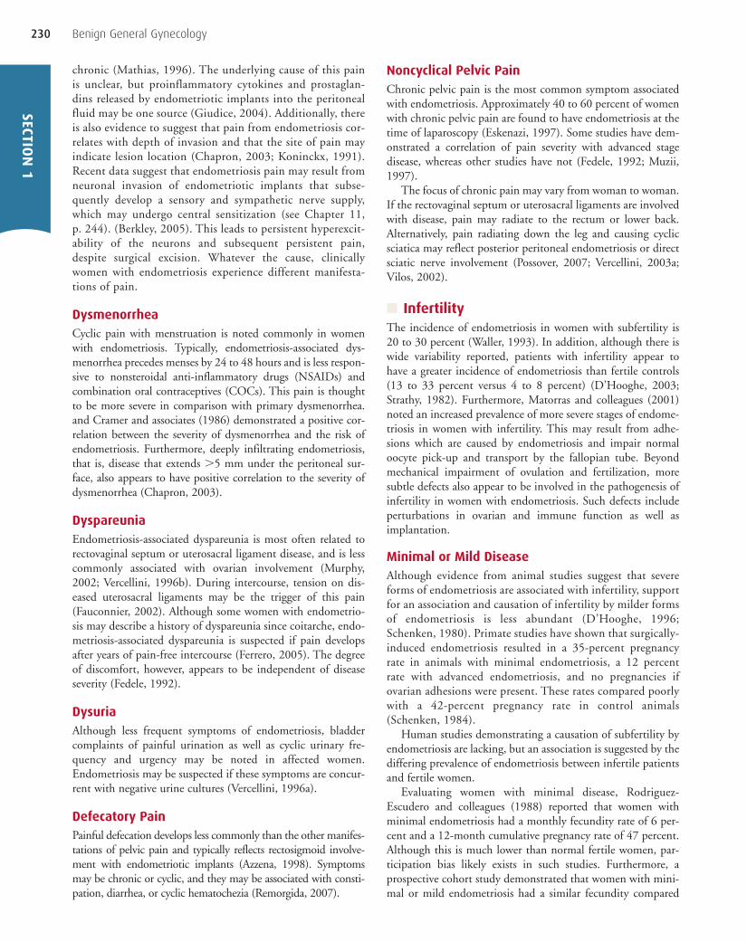
In 1996, in an attempt to further correlate surgical findings with clinical outcomes, the ASRM further revised the endome-triosis classification system (American Society of Reproductive Medicine, 1997). In this system, endometriosis is classified as stage I (minimal), stage II (mild), stage III (moderate), and stage IV (severe) (Fig. 10-2). Although there was no change in the staging system from the 1985 classification, the revised 1996 classification provided for description of endometriotic lesion morphology as white, red, or black. This modification was prompted by studies demonstrating that some biochemical activities within implants and possibly disease prognosis can be predicted by implant morphology (Vernon, 1986).
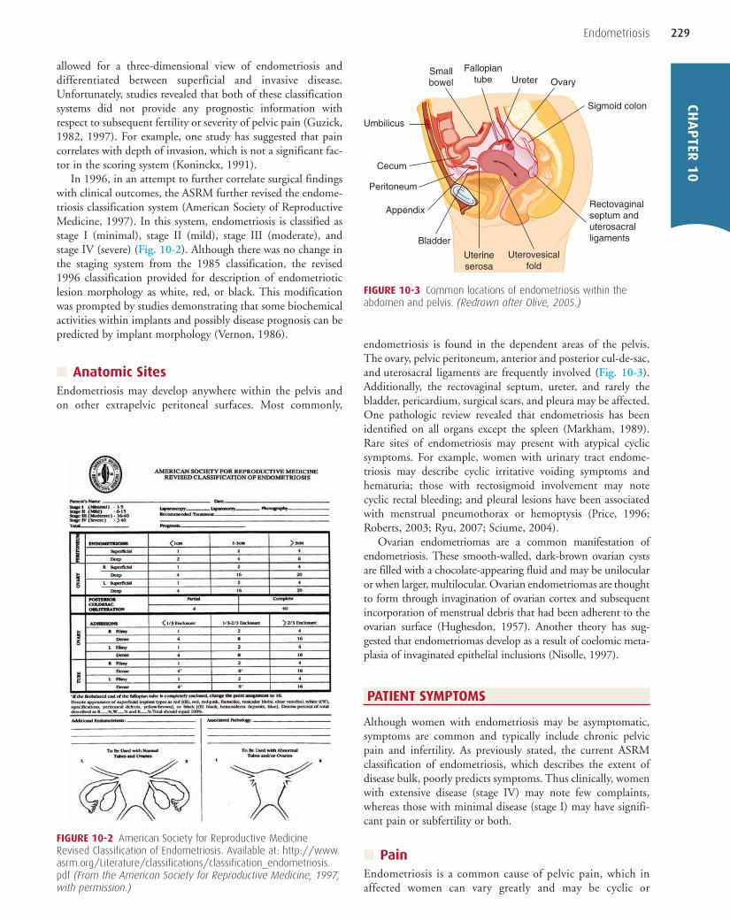
■ Anatomic SitesEndometriosis may develop anywhere within the pelvis and on other extrapelvic peritoneal surfaces. Most commonly,
endometriosis is found in the dependent areas of the pelvis. The ovary, pelvic peritoneum, anterior and posterior cul-de-sac, and uterosacral ligaments are frequently involved (Fig. 10-3). Additionally, the rectovaginal septum, ureter, and rarely the bladder, pericardium, surgical scars, and pleura may be affected. One pathologic review revealed that endometriosis has been identified on all organs except the spleen (Markham, 1989). Rare sites of endometriosis may present with atypical cyclic symptoms. For example, women with urinary tract endome-triosis may describe cyclic irritative voiding symptoms and hematuria; those with rectosigmoid involvement may note cyclic rectal bleeding; and pleural lesions have been associated with menstrual pneumothorax or hemoptysis (Price, 1996; Roberts, 2003; Ryu, 2007; Sciume, 2004).
Ovarian endometriomas are a common manifestation of endometriosis. These smooth-walled, dark-brown ovarian cysts are filled with a chocolate-appearing fluid and may be unilocular or when larger, multilocular. Ovarian endometriomas are thought to form through invagination of ovarian cortex and subsequent incorporation of menstrual debris that had been adherent to the ovarian surface (Hughesdon, 1957). Another theory has sug-gested that endometriomas develop as a result of coelomic meta-plasia of invaginated epithelial inclusions (Nisolle, 1997).
PATIENT SYMPTOMS
Although women with endometriosis may be asymptomatic, symptoms are common and typically include chronic pelvic pain and infertility. As previously stated, the current ASRM classification of endometriosis, which describes the extent of disease bulk, poorly predicts symptoms. Thus clinically, women with extensive disease (stage IV) may note few complaints, whereas those with minimal disease (stage I) may have signifi-cant pain or subfertility or both.
■ PainEndometriosis is a common cause of pelvic pain, which in affected women can vary greatly and may be cyclic or
FIGURE 10-2 American Society for Reproductive Medicine Revised Classification of Endometriosis. Available at: http://www.asrm.org/Literature/classifications/classification_endometriosis.pdf (From the American Society for Reproductive Medicine, 1997, with permission.)
FIGURE 10-3 Common locations of endometriosis within the abdomen and pelvis. (Redrawn after Olive, 2005.)
Smallbowel
Fallopiantube Ureter Ovary
Sigmoid colon
Uterovesicalfold
Rectovaginalseptum anduterosacralligaments
Uterineserosa
Bladder
Appendix
Peritoneum
Cecum
Umbilicus
sch72576_c10.indd Page 229 12/28/07 10:47:22 AM usersch72576_c10.indd Page 229 12/28/07 10:47:22 AM user /Volumes/209/MHBD105/mhbd105indd%0/ch10/Volumes/209/MHBD105/mhbd105indd%0/ch10
230 Benign General Gynecology
SECTION
1
chronic (Mathias, 1996). The underlying cause of this pain is unclear, but proinflammatory cytokines and prostaglan-dins released by endometriotic implants into the peritoneal fluid may be one source (Giudice, 2004). Additionally, there is also evidence to suggest that pain from endometriosis cor-relates with depth of invasion and that the site of pain may indicate lesion location (Chapron, 2003; Koninckx, 1991). Recent data suggest that endometriosis pain may result from neuronal invasion of endometriotic implants that subse-quently develop a sensory and sympathetic nerve supply, which may undergo central sensitization (see Chapter 11, p. 244). (Berkley, 2005). This leads to persistent hyperexcit-ability of the neurons and subsequent persistent pain, despite surgical excision. Whatever the cause, clinically women with endometriosis experience different manifesta-tions of pain.
DysmenorrheaCyclic pain with menstruation is noted commonly in women with endometriosis. Typically, endometriosis-associated dys-menorrhea precedes menses by 24 to 48 hours and is less respon-sive to nonsteroidal anti-inflammatory drugs (NSAIDs) and combination oral contraceptives (COCs). This pain is thought to be more severe in comparison with primary dysmenorrhea. and Cramer and associates (1986) demonstrated a positive cor-relation between the severity of dysmenorrhea and the risk of endometriosis. Furthermore, deeply infiltrating endometriosis, that is, disease that extends �5 mm under the peritoneal sur-face, also appears to have positive correlation to the severity of dysmenorrhea (Chapron, 2003).
DyspareuniaEndometriosis-associated dyspareunia is most often related to rectovaginal septum or uterosacral ligament disease, and is less commonly associated with ovarian involvement (Murphy, 2002; Vercellini, 1996b). During intercourse, tension on dis-eased uterosacral ligaments may be the trigger of this pain (Fauconnier, 2002). Although some women with endometrio-sis may describe a history of dyspareunia since coitarche, endo-metriosis-associated dyspareunia is suspected if pain develops after years of pain-free intercourse (Ferrero, 2005). The degree of discomfort, however, appears to be independent of disease severity (Fedele, 1992).
DysuriaAlthough less frequent symptoms of endometriosis, bladder complaints of painful urination as well as cyclic urinary fre-quency and urgency may be noted in affected women. Endometriosis may be suspected if these symptoms are concur-rent with negative urine cultures (Vercellini, 1996a).
Defecatory PainPainful defecation develops less commonly than the other manifes-tations of pelvic pain and typically reflects rectosigmoid involve-ment with endometriotic implants (Azzena, 1998). Symptoms may be chronic or cyclic, and they may be associated with consti-pation, diarrhea, or cyclic hematochezia (Remorgida, 2007).
Noncyclical Pelvic PainChronic pelvic pain is the most common symptom associated with endometriosis. Approximately 40 to 60 percent of women with chronic pelvic pain are found to have endometriosis at the time of laparoscopy (Eskenazi, 1997). Some studies have dem-onstrated a correlation of pain severity with advanced stage disease, whereas other studies have not (Fedele, 1992; Muzii, 1997).
The focus of chronic pain may vary from woman to woman. If the rectovaginal septum or uterosacral ligaments are involved with disease, pain may radiate to the rectum or lower back. Alternatively, pain radiating down the leg and causing cyclic sciatica may reflect posterior peritoneal endometriosis or direct sciatic nerve involvement (Possover, 2007; Vercellini, 2003a; Vilos, 2002).
■ InfertilityThe incidence of endometriosis in women with subfertility is 20 to 30 percent (Waller, 1993). In addition, although there is wide variability reported, patients with infertility appear to have a greater incidence of endometriosis than fertile controls (13 to 33 percent versus 4 to 8 percent) (D’Hooghe, 2003; Strathy, 1982). Furthermore, Matorras and colleagues (2001) noted an increased prevalence of more severe stages of endome-triosis in women with infertility. This may result from adhe-sions which are caused by endometriosis and impair normal oocyte pick-up and transport by the fallopian tube. Beyond mechanical impairment of ovulation and fertilization, more subtle defects also appear to be involved in the pathogenesis of infertility in women with endometriosis. Such defects include perturbations in ovarian and immune function as well as implantation.
Minimal or Mild DiseaseAlthough evidence from animal studies suggest that severe forms of endometriosis are associated with infertility, support for an association and causation of infertility by milder forms of endometriosis is less abundant (D’Hooghe, 1996; Schenken, 1980). Primate studies have shown that surgically-induced endometriosis resulted in a 35-percent pregnancy rate in animals with minimal endometriosis, a 12 percent rate with advanced endometriosis, and no pregnancies if ovarian adhesions were present. These rates compared poorly with a 42-percent pregnancy rate in control animals (Schenken, 1984).
Human studies demonstrating a causation of subfertility by endometriosis are lacking, but an association is suggested by the differing prevalence of endometriosis between infertile patients and fertile women.
Evaluating women with minimal disease, Rodriguez-Escudero and colleagues (1988) reported that women with minimal endometriosis had a monthly fecundity rate of 6 per-cent and a 12-month cumulative pregnancy rate of 47 percent. Although this is much lower than normal fertile women, par-ticipation bias likely exists in such studies. Furthermore, a prospective cohort study demonstrated that women with mini-mal or mild endometriosis had a similar fecundity compared
sch72576_c10.indd Page 230 12/28/07 10:47:29 AM usersch72576_c10.indd Page 230 12/28/07 10:47:29 AM user /Volumes/209/MHBD105/mhbd105indd%0/ch10/Volumes/209/MHBD105/mhbd105indd%0/ch10

231EndometriosisCH
AP
TER 1
0
with those with unexplained infertility. Well-designed, prospec-tive randomized controlled trials (RCTs) have found conflicting evidence as to whether surgical treatment of endometriosis improves fecundity rates and cumulative pregnancy rates in these women. One of these studies demonstrated improved fertility, but a trial with fewer women noted no improvement (Marcoux, 1997; Parazzini, 1999).
Moderate or Severe DiseaseIn moderate to severe endometriosis (stage III to IV), tubal and ovarian architecture are often distorted. As a result, impaired fertility would be expected. Unfortunately, few studies report fecundity rates in women with severe endometriosis. One investigation comparing mild, moderate, and severe endome-triosis revealed a monthly fecundity rate of 8.7 percent in those with mild disease, 3.2 percent with moderate disease, and no pregnancies with severe disease (Olive, 1985a). There are no well-designed studies examining the effectiveness of surgical therapy in patients with severe endometriosis, but cumulative pregnancy rates have reached 30 percent after surgical excision (Adamson, 1993; Osuga, 2002). This rate appears to be greater than that of women who undergo expectant management.
Folliculogenesis and Embryogenesis EffectsSome researchers have suggested that folliculogenesis is impaired in women with endometriosis. Embryo development and quality in women with endometriosis undergoing IVF was compared with that of embryos originating from women with tubal factor infertility (Pellicer, 1995). There were significantly fewer blastomeres per embryo and a significantly greater rate of embryonic developmental arrest in the endometriosis group. This suggests a possible decreased developmental competence of oocytes originating from the ovaries of women with endo-metriosis. Another investigation found that oocyte number may be decreased in women with endometriosis (Suzuki, 2005). In addition, researchers have attempted to determine if the follicular environment is different in women with endome-triosis. Specifically, studies demonstrating qualitative and quantitative changes in steroidogenesis, however, have found conflicting results (Garrido, 2002; Harlow, 1996; Pellicer, 1998). Apoptosis is another attractive theory for decreased oocyte competence in women with endometriosis, but well-designed studies are lacking.
Endometrical ChangesAbnormalities in endometrial development in women with endometriosis support the possibility that implantation defects may be responsible for subfertility associated with endometriosis. For example, researchers have revealed abnormalities in gene expression profiles in the eutopic endometrium from women with endometriosis compared with that from women without endometriosis (Kao, 2003). Specifically, deficient �v�3 integrin expression in the peri-implantation endometrium of women with endometriosis has been demonstrated, and this may be associated with decreased uterine receptivity (Lessey, 1994). The role of apoptosis on peri-implantation endometrium is another area of study that still remains largely unexplored.
Other FactorsAbnormalities in inflammation and cytokine activity in women with endometriosis may play a role in endometriosis-associated infertility. Sperm function may be affected in women with endometriosis. Studies have demonstrated increased phagocyto-sis of spermatozoa by macrophages from women with endome-triosis (Haney, 1981; Muscato, 1982). Moreover, sperm binding to the zona pellucida appears to be adversely affected (Qiao, 1998). However, investigations of the effects of endometriosis on sperm motility and the acrosome reaction reveal conflicting results (Bielfeld, 1993; Curtis, 1993; Tasdemir, 1995).
■ Intestinal ObstructionEndometriosis may involve the small bowel, cecum, appendix, or rectosigmoid colon and lead to intestinal obstruction in some cases (Cameron, 1995; Varras, 2002; Wickramasekera, 1999). Although endometriosis of the gastrointestinal tract is usually confined to the subserosa and muscularis propria, more severe cases may involve the bowel wall transmurally and lead to a clinical and radiologic picture consistent with malignancy (Decker, 2004). Accurate pre-operative diagnosis and management are difficult due to the atypi-cal presentation. Laparoscopy typically provides the definitive diagnosis. Treatment is often surgical, with resection and primary anastomosis of the affected intestinal segment. In women without obstructing symptoms, however, conservative management with hormonal therapy may be considered.
DIFFERENTIAL DIAGNOSIS
The symptoms of endometriosis are nonspecific and may mimic many disease processes. Because endometriosis is a surgi-cal diagnosis, several other diagnoses may be considered prior to surgical exploration (Table 10-1).
TABLE 10-1 Differential Diagnosis of Endometriosis
Gynecologic Pelvic inflammatory disease Tubo-ovarian abscess Salpingitis Endometritis Hemorrhagic ovarian cyst Ovarian torsion Primary dysmenorrhea Degenerating leiomyoma
Nongynecologic Interstitial cystitis Chronic urinary tract infection Renal calculi Inflammatory bowel disease Irritable bowel syndrome Diverticulitis Mesenteric lymphadenitis Musculoskeletal disorders
sch72576_c10.indd Page 231 12/28/07 10:47:30 AM usersch72576_c10.indd Page 231 12/28/07 10:47:30 AM user /Volumes/209/MHBD105/mhbd105indd%0/ch10/Volumes/209/MHBD105/mhbd105indd%0/ch10

232 Benign General Gynecology
SECTION
1
DIAGNOSIS
■ Physical Examination
Visual InspectionFor the most part, endometriosis is a disease confined to the pelvis. Accordingly, there are often no abnormalities on visual inspection. Some exceptions include endometriosis within an episiotomy scar or surgical scar, most often within a Pfannenstiel incision (Fig. 10-4) (Koger, 1993; Zhu, 2002). Rarely, endo-metriosis may develop spontaneously within the perineum or perianal region (Watanabe, 2003).
Speculum ExaminationExamination of the vagina and cervix by speculum examination often reveals no signs of endometriosis. Occasionally, bluish or red powder-burn lesions may be seen on the cervix or the pos-terior fornix of the vagina. These lesions may be tender or bleed with contact. One recent study found that speculum examina-tion displayed endometriosis in 14 percent of patients diag-nosed with deeply infiltrating endometriosis (Chapron, 2002).
Bimanual ExaminationPelvic organ palpation often reveals anatomic abnormalities sug-gestive of endometriosis. Uterosacral ligament nodularity and tenderness may reflect active disease or scarring along the liga-ment. In addition, an enlarged cystic adnexal mass may represent an ovarian endometrioma, which may be mobile or adherent to other pelvic structures. Bimanual examination may reveal a retro-verted, fixed, tender uterus, or a firm, fixed posterior cul-de-sac.
Although pelvic organ palpation may assist in the diagnosis, the sensitivity and specificity of focal pelvic tenderness in detecting endometriosis displays wide variation and ranges from 36 to 90 percent and 32 to 92 percent, respectively (Chapron, 2002; Eskenazi, 2001; Koninckx, 1996; Ripps, 1992). For example, Chapron and co-workers (2002) palpated a painful nodule in 43 percent of patients with deeply infiltrat-ing endometriosis. In another study of 91 women with chronic pelvic pain and surgically confirmed endometriosis, the biman-ual examination was normal 47 percent of the time (Nezhat, 1994). One study suggested that pelvic nodularities secondary to endometriosis may be more easily detected by bimanual examination during menses (Koninckx, 1996).
■ Laboratory TestingTo exclude other causes of pelvic pain, laboratory investigations are often undertaken. Initially, a complete blood count (CBC), urinalysis and urine cultures, vaginal cultures, and cervical swabs may be obtained to exclude infections or sexually trans-mitted infections that may cause pelvic inflammatory disease (see Chap. 3, p. 73).
Serum CA125Numerous serum markers have been studied as possible adjuncts in the diagnosis of endometriosis. No serum marker has been studied in greater detail than CA125 (cancer antigen 125). Found as an antigenic determinant on a glycoprotein, CA125 has been identified in several adult tissues such as the epithelium of the fallopian tubes, the endometrium, the endocervix, the pleura, and the peritoneum (see Chap. 35, p. 722). Recognized by monoclonal antibody assays, elevated CA125 levels have been shown to positively correlate with the severity of endometriosis (Hornstein, 1995a). Unfortunately, although demonstrating adequate specificity, the assay has poor sensitivity in detecting mild endometriosis. A meta-analysis of studies evaluating CA125 in the diagnosis of endometriosis revealed a sensitivity of only 28 percent and a specificity of 90 percent (Mol, 1998). This marker appeared to be a better test in diagnosing stage III and IV endometriosis. Although the role of this test in clinical practice is uncertain, it may be useful in the presence of a sonographically detected ovarian cyst suggestive of an endometrioma.
Other Serum MarkersCancer antigen19-9 (CA 19-9), another antigenic glycoprotein, is a serum marker that has also been shown to positively correlate with the severity of endometriosis (Harada, 2002). Serum placen-tal protein 14 (PP14; glycodelin-A) was initially shown to have adequate sensitivity (59 percent), but this has not been confirmed by other studies (Telimaa, 1989). Interleukin-6 (IL-6) serum lev-els above 2 pg/mL (90-percent sensitivity and 67-percent specific-ity) and tumor necrosis factor-� (TNF-�) peritoneal fluid levels above 15 pg/mL (100-percent sensitivity and 89-percent specific-ity) may be used to discriminate between those with or without endometriosis (Bedaiwy, 2002). Several other serum markers have been studied, with limited diagnostic accuracy (Bedaiwy, 2004). Most of these tests are rarely used outside of research settings.
FIGURE 10-4 Endometriosis within a lower vertical midline incision scar (arrows).
sch72576_c10.indd Page 232 12/28/07 10:47:30 AM usersch72576_c10.indd Page 232 12/28/07 10:47:30 AM user /Volumes/209/MHBD105/mhbd105indd%0/ch10/Volumes/209/MHBD105/mhbd105indd%0/ch10

233EndometriosisCH
AP
TER 1
0
■ Diagnostic Imaging
SonographyBoth transabdominal and the more sensitive transvaginal (TVS) sonographic approaches have been used extensively in the diagnosis of endometriosis (see Chap. 2, p. 25). Although TVS is the mainstay in evaluating symptoms associated with endometriosis and is accurate in detecting endometriomas, imaging of superficial endometriosis or endometriotic adhe-sions is inadequate. Small endometriotic plaques or nodules may occasionally be seen, but these findings are inconsistent (Carbognin, 2004).
More recently, sonovaginography, a technique involving vaginal saline instillation to more accurately localize rectovagi-nal endometriosis, and transrectal sonography have assisted in the diagnosis and evaluation of endometriosis (Brosens, 2003). Transvaginal sonography appears to be as effective as a transrec-tal approach in identifying posterior pelvic endometriosis, but the latter may delineate rectal involvement more accurately and may be more appropriate when planning surgery (Bazot, 2003).
Endometriomas can be diagnosed by TVS with adequate sensitivity in most settings if they are 20 mm in diameter or greater (Fig. 10-5). Specifically, sensitivity and specificity of TVS to diagnose endometriomas range from 64 to 90 per-cent and from 22 to 100 percent, respectively (Moore, 2002). Endometriomas often present as cystic structures with low-level internal echoes, and occasional thick septa-tions, thickened walls, and echogenic wall foci (Athey, 1989; Patel, 1999). Color Doppler transvaginal sonography often demonstrates pericystic, but not intracystic, flow (Carbognin, 2004).
Magnetic Resonance ImagingMagnetic resonance imaging has been increasingly used as a noninvasive method for diagnosis of endometriosis. Small nod-ules may be recognized as hyperintense lesions on T1-weighted
sequences, and plaque lesions have a similar appearance, with a variable signal on T2-weighted sequences (Carbognin, 2004). An endometrioma appears as a hyperintense mass on T1-weighted sequences, with a tendency towards hypointensity in T2-weighted sequences. A hypointense ring is often seen sur-rounding the endometrioma, which is enhanced after contrast administration (Fig. 10-6).
■ Diagnostic LaparoscopyDiagnostic laparoscopy is the primary method used for diag-nosing endometriosis (see Section 41-28, p. 929) (Kennedy, 2005). Laparoscopic findings are variable and may include discrete endometriotic lesions, endometrioma, and adhesion formation.
Endometriotic LesionsThe pelvic organs and pelvic peritoneum are typical locations for endometriosis. The appearance of these lesions by laparoscopy is varied and colors may include red (red, red-pink, or clear), white (white or yellow-brown), and black (black or black-blue) (Fig. 10-7). Dark lesions are pigmented by hemosiderin deposi-tion from trapped menstrual debris. White and red lesions most commonly correlate with the histologic findings of endometriosis (Jansen, 1986). In addition to color differences, endometriotic lesions may differ morphologically. They can appear as smooth blebs on peritoneal surfaces, as holes or defects within the perito-neum, or as flat stellate lesions whose points are formed by sur-rounding scar tissue. Endometriotic lesions may be superficial or may deeply invade the peritoneum or pelvic organs. Although
FIGURE 10-5 Transvaginal sonogram demonstrating ovarian endometrioma. A cyst with diffuse internal low-level echoes is seen. (Courtesy of Dr. Elysia Moschos.)
FIGURE 10-6 Magnetic resonance images of an endometrioma. T2- (A) and T1-weighted (B) images reveal an endometrioma (arrows) just lateral to the rectum. The findings are consistent with subacute blood, based on the bright signal on T-1 and the relatively low signal intensity on T-2 of the lesion. (Courtesy of Dr. Diane Twickler.)
A
B
sch72576_c10.indd Page 233 12/28/07 10:47:31 AM usersch72576_c10.indd Page 233 12/28/07 10:47:31 AM user /Volumes/209/MHBD105/mhbd105indd%0/ch10/Volumes/209/MHBD105/mhbd105indd%0/ch10

234 Benign General Gynecology
SECTION
1
these findings may allow endometriosis to be diagnosed with accuracy, pain symptoms correlate poorly with findings at lapa-roscopy (Kennedy, 2005).
EndometriomasEndometriomas are cystic endometrial lesions contained within the ovary. Typically, they have the appearance of smooth-walled, brown cysts filled with thick, chocolate-appearing liquid (Fig. 10-8). These ovarian masses may be unilocular, but are often multilocular when �3 cm in diameter (Nezhat, 1992b).
Laparoscopic visualization of ovarian endometriomas has a sensitivity and specificity of 97 percent and 95 percent, respec-tively (Vercellini, 1991). Because of this, ovarian biopsy is rarely required for diagnosis.
■ Pathologic AnalysisAlthough current guidelines do not require histologic evalua-tion for the diagnosis of endometriosis, some suggest that rely-ing solely on laparoscopic findings in the absence of histologic confirmation often results in overdiagnosis (American Society for Reproductive Medicine, 1997). Specifically, the greatest discordance between laparoscopic and histologic findings is noted in scarred lesions (Marchino, 2005a; Walter, 2001). Histologic diagnosis requires the presence of both endometrial glands and stroma found outside the uterine cavity (Fig. 10-9). Additionally, hemosiderin deposition and fibromuscular meta-plasia are frequently noted (Murphy, 2002). The gross appear-ance of endometriotic lesions often suggests certain microscopic findings. For example, when examined microscopically, red lesions are frequently vascularized, whereas white lesions more often display fibrosis and few vessels (Nisolle, 1997).
■ Diagnostic AlgorithmThe approach to diagnosis and treatment of endometriosis depends on the presenting symptoms and goals of therapy
(Fig. 10-10). If infertility is the presenting symptom, then fertility-preserving treatment without ovulation suppression will be required. In contrast, if the patient has severe, recalci-trant pain symptoms and has completed childbearing, defini-tive surgery may be warranted.
TREATMENT
Treatment for endometriosis depends on the woman’s specific symptoms, severity of symptoms, location of endometriotic lesions, goals for treatment, and desire to conserve future fertil-ity. The most important factor when determining the most appropriate management is whether a patient is seeking treat-ment for infertility or pain, as the treatment will differ based on the symptom (Olive, 2001).
FIGURE 10-8 Photographs of an endometrioma. A. A bisected endometrioma showing a shaggy hemorrhagic lining. B. Microscopic image of an endometrioma showing predomi-nantly hemosiderin-laden macrophages resulting in the brown discoloration. (Courtesy of Dr. Raheela Ashfaq.)
B
FIGURE 10-7 Below the irrigator tip, a red and white endometri-otic lesion is seen on the pelvic peritoneum during laparoscopy. (Courtesy of Dr. Karen Bradshaw.)
A
sch72576_c10.indd Page 234 12/28/07 10:47:32 AM usersch72576_c10.indd Page 234 12/28/07 10:47:32 AM user /Volumes/209/MHBD105/mhbd105indd%0/ch10/Volumes/209/MHBD105/mhbd105indd%0/ch10
235EndometriosisCH
AP
TER 1
0
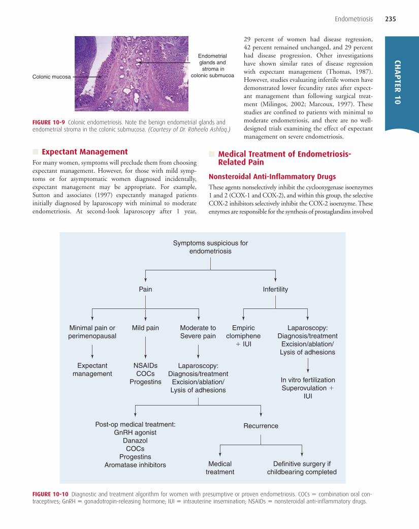
■ Expectant ManagementFor many women, symptoms will preclude them from choosing expectant management. However, for those with mild symp-toms or for asymptomatic women diagnosed incidentally, expectant management may be appropriate. For example, Sutton and associates (1997) expectantly managed patients initially diagnosed by laparoscopy with minimal to moderate endometriosis. At second-look laparoscopy after 1 year,
29 percent of women had disease regression, 42 percent remained unchanged, and 29 percent had disease progression. Other investigations have shown similar rates of disease regression with expectant management (Thomas, 1987). However, studies evaluating infertile women have demonstrated lower fecundity rates after expect-ant management than following surgical treat-ment (Milingos, 2002; Marcoux, 1997). These studies are confined to patients with minimal to moderate endometriosis, and there are no well-designed trials examining the effect of expectant management on severe endometriosis.
■ Medical Treatment of Endometriosis-Related Pain
Nonsteroidal Anti-Inflammatory DrugsThese agents nonselectively inhibit the cyclooxygenase isoenzymes 1 and 2 (COX-1 and COX-2), and within this group, the selective COX-2 inhibitors selectively inhibit the COX-2 isoenzyme. These enzymes are responsible for the synthesis of prostaglandins involved
FIGURE 10-9 Colonic endometriosis. Note the benign endometrial glands and endometrial stroma in the colonic submucosa. (Courtesy of Dr. Raheela Ashfaq.)
Endometrial glands and stroma in
colonic submucoa Colonic mucosa
FIGURE 10-10 Diagnostic and treatment algorithm for women with presumptive or proven endometriosis. COCs � combination oral con-traceptives; GnRH � gonadotropin-releasing hormone; IUI � intrauterine insemination; NSAIDs � nonsteroidal anti-inflammatory drugs.
Symptoms suspicious forendometriosis
Pain
Minimal pain orperimenopausal
Expectantmanagement
NSAIDsCOCs
Progestins
Moderate toSevere pain
Laparoscopy:Diagnosis/treatment
Excision/ablation/Lysis of adhesions
Post-op medical treatment:GnRH agonist
DanazolCOCs
ProgestinsAromatase inhibitors
Recurrence
In vitro fertilizationSuperovulation �
IUI
Empiricclomiphene
� IUI
Laparoscopy:Diagnosis/treatment
Excision/ablation/Lysis of adhesions
Mild pain
Infertility
Medicaltreatment
Definitive surgery ifchildbearing completed
sch72576_c10.indd Page 235 12/28/07 10:47:34 AM usersch72576_c10.indd Page 235 12/28/07 10:47:34 AM user /Volumes/209/MHBD105/mhbd105indd%0/ch10/Volumes/209/MHBD105/mhbd105indd%0/ch10

236 Benign General Gynecology
SECTION
1
in the pain and inflammation associated with endometriosis. For example, endometriotic tissue has been shown to express COX-2 at greater levels than eutopic endometrium (Ota, 2001). Therefore, therapy aimed at lowering these prostaglandin levels may play a role in alleviating endometriosis-associated pain.
Nonsteroidal anti-inflammatory drugs are often first-line therapy in women with primary dysmenorrhea or pelvic pain prior to laparoscopic confirmation of endometriosis, and in women with minimal or mild pain symptoms associated with known endometriosis. Although animal models have demon-strated disease regression with NSAID treatment, few studies have critically evaluated their effectiveness in disease regression in surgically-confirmed endometriosis (Efstathiou, 2005). However, evidence exists for their efficacy in patients with dys-menorrhea and pelvic pain (Table 10-2) (Nasir, 2004). Due to the cardiovascular risks with long-term use of COX-2 inhibi-tors, these medications should be used at the lowest possible dose and for the shortest duration necessary (Jones, 2005).
Combination Oral ContraceptivesThese agents have been a mainstay for the treatment of pain associated with endometriosis. Although no randomized con-trolled trials have compared COCs with placebo, abundant observational evidence supports the role of COCs in the relief of endometriosis-related pain (Vercellini, 1993; Vessey, 1993). These drugs appear to act by inhibiting gonadotropin release, decreasing menstrual flow, and decidualizing implants. In addi-tion, COCs have the added benefit of contraception, suppres-sion of ovulation, and other noncontraceptive benefits (see Table 5-6, p. 112).
These drugs can be used conventionally in a cyclic regimen or may be used continuously, without a break for withdrawal menses. The continuous regimen may be preferable for its decreased frequency of menses for women who fail to achieve pain relief with cyclic COC therapy (Vercellini, 2003b; Wiegratz, 2004). Traditionally, monophasic COCs have been used in the treatment of endometriosis, but no evidence sup-ports their clinical superiority to multiphasic COCs. Additionally, low-dose COCs (containing 20 µg ethinyl estra-diol) have not proved superior to conventional-dose COCs for
the treatment of endometriosis and may lead to higher rates of abnormal bleeding (Gallo, 2005).
ProgestinsProgestational agents have long been used in the treatment of endometriosis. Progestins are known to antagonize estrogenic effects on the endometrium, causing initial decidualization and subsequent endometrial atrophy. Progestins have been adminis-tered for the treatment of endometriosis in numerous ways and include oral progestins, depot medroxyprogesterone acetate (DMPA), a levonorgestrel-releasing intrauterine device (IUD), and the newer selective progesterone-receptor modulators (SPRMs).
Although progestin-based therapy is commonly used to effec-tively treat symptoms, there has been only one well-designed, randomized controlled trial comparing the effect of placebo with medroxyprogesterone acetate (MPA), 100 mg orally daily, given for 6 months. At second-look laparoscopy, partial or total resolu-tion of peritoneal implants in 60 percent of women was noted, compared with 18 percent in the placebo group. Furthermore, pelvic pain and defecatory pain were significantly reduced (Telimaa, 1987). Side effects of high-dose MPA included acne, edema, weight gain, and irregular menstrual bleeding. In practice, MPA is prescribed in dosages ranging from 20 to 100 mg daily. Alternatively, MPA may be given intramuscularly in depot form in a dosage of 150 mg every 3 months. In depot form, MPA may delay resump-tion of normal menses and ovulation and should not be used in women contemplating imminent pregnancy.
Norethindrone acetate (NETA) is a 19-nortestosterone syn-thetic progestin that has been used in the treatment of endome-triosis. In one study, investigators administered an initial oral dosage of NETA, 5 mg daily, with increases of 2.5 mg daily until amenorrhea or a maximal dosage of 20 mg daily was reached. They found an approximately 90-percent reduction in dysmenorrhea and pelvic pain (Muneyyirci-Delale, 1998). Additionally, NETA has been shown to be effective in conjunction with long-term gonadotropin-releasing hormone (GnRH) agonist therapy for endometriosis. In this fashion, NETA, 5 mg administered orally daily, in conjunction with prolonged GnRH agonist therapy, results in significant resolution of symptoms while protecting against bone loss (Hornstein, 1998; Surrey, 2002).
TABLE 10-2 Commonly Used Oral Nonsteroidal Anti-Inflammatory Drugs (NSAIDs) in the Treatment of Endometriosis-Associated Dysmenorrhea
Generic Name Trade Name Dosage Adverse Effects
Ibuprofen Motrin, Advil, 400 mg every 4–6 h Nausea; epigastric pain; anorexia; Nuprin constipation; gastrointestinal bleedingNaproxen Naprosyn, Aleve 500 mg initially, then 250 mg Same as above every 6–8 hNaproxen sodium Anaprox 550 mg initially, then 275 mg Same as above every 6–8 hMefenamic acid Ponstel 500 mg initially, then 250 mg Same as above every 6 h, starting with menses and continued for 3 days
Ketoprofen Orudis, Oruvail 50 mg q6–8 h Same as above
sch72576_c10.indd Page 236 12/28/07 10:47:34 AM usersch72576_c10.indd Page 236 12/28/07 10:47:34 AM user /Volumes/209/MHBD105/mhbd105indd%0/ch10/Volumes/209/MHBD105/mhbd105indd%0/ch10

237EndometriosisCH
AP
TER 1
0
The levonorgestrel-releasing intrauterine system (LNG-IUS) (Mirena, Berlex, Montville, NJ) has traditionally been used for contraception and dysfunctional uterine bleeding (see Fig. 5-5, p. 119). Recently, the LNG-IUS, however, has been used for the treatment of endometriosis. This IUD delivers levonorgestrel directly to the endometrium and is effective for up to 5 years. An observational trial revealed symptomatic improvement in patients with endometriosis using the LNG-IUS, with symptom improvement continuing up to 30 months (Lockhat, 2005). The continuation rate at 3 years, however, was only 56 percent, mostly due to intolerable bleeding, persis-tent pain, and weight gain. A randomized controlled trial com-paring LNG-IUS with GnRH agonist therapy showed equivalent improvement in pain symptoms, without the con-comitant hypoestrogenism that accompanies GnRH agonist treatment (Petta, 2005). Accordingly, these recent findings make the LNG-IUS an attractive option in treating women with endometriosis.
Selective Progesterone Receptor ModulatorsA new and novel option in the treatment of endometriosis has been the use of selective progesterone-receptor modulators (SPRMs). These are progesterone-receptor ligands (molecules that bind and activate or inactivate the progesterone receptor) and have both progesterone antagonist and antagonist activities (Elger, 2000). One common SPRM, mifepristone (RU486), is a controversial abortifacient that predominantly possesses anti-progestational activity. It has also been studied in women with endometriosis and was found to reduce pelvic pain and extent of endometriosis, when used for 6 months at oral dosages of 50 mg daily (Kettel, 1996). Asoprisnil (J867) is a SPRM that induces endometrial atrophy and amenorrhea. Currently in Phase III trials for the treatment of leiomyomas and endome-triosis, asoprisnil in Phase II studies improved dysmenorrhea and pelvic pain symptoms, whereas amenorrhea was dose dependent (Chwalisz, 2005). These novel agents hold promise for future treatment of endometriosis.
AndrogensThe first medication approved for the treatment of endometriosis in the United States was the androgen danazol. This agent is a synthetic androgen that is an isoxazole derivative of 17-�-ethinyl testosterone. The predominant mechanism of action appears to be suppression of midcycle luteinizing hormone (LH) surge, creating a chronic anovulatory state (Floyd, 1980). Danazol occupies receptor sites on sex-hormone binding globulin (SHBG) to increase serum free testosterone levels and also binds directly to androgen and progesterone receptors. As a result, danazol cre-ates a hypoestrogenic, hyperandrogenic state, inducing endome-trial atrophy in endometriotic implants (Fedele, 1990).
Danazol at dosages of 200 mg given orally three times daily proved superior to placebo for the reduction of endometriotic implants and pelvic pain symptoms after 6 months of therapy (Telimaa, 1987). The recommended dosage of danazol is 600 to 800 mg daily. Unfortunately, significant androgenic side effects develop at this dosage and include acne, hot flashes, hirsut-ism, adverse serum lipid profiles, voice deepening (possibly
irreversible), elevation of liver enzymes, and mood changes. Moreover, due to possible teratogenicity, this medication should be taken in conjunction with effective contraception. Because of this adverse side-effect profile, danazol is prescribed less frequently, and when administered, its duration should be limited.
Gestrinone (ethylnorgestrienone; R2323) is an antiprogesta-tional agent prescribed in Europe for the treatment of endome-triosis. Although it has antiprogestational, antiestrogenic, and androgenic effects, it predominantly induces a progesterone withdrawal effect and decreases the number of estrogen and progesterone receptors. Endocrinologic changes during therapy with gestrinone show that basal concentrations of gonadotro-pin levels remain unchanged, estradiol concentrations vary, and free testosterone levels increase, with concomitant androgenic side effects (Forbes, 1993).
Gestrinone equals the effectiveness of danazol and of GnRH agonists for relief of endometriosis-related pain (Prentice, 2000a). Furthermore, during 6 months of treatment, gestri-none was not associated with the bone density loss commonly seen with GnRH agonist use and was more effective in persis-tently decreasing moderate to severe pelvic pain (Gestrinone Italian Study Group, 1996). Unfortunately, gestrinone appears to lower high-density lipoprotein (HDL) levels. Gestrinone is administered orally, 2.5 to 10 mg weekly, given daily or three times weekly.
GnRH AgonistsEndogenous pulsatile release of GnRH leads to pulsatile secre-tory activity of the gonadotropes within the anterior pituitary. This pulsatile release results in pituitary release of gonadotro-pins, with subsequent ovarian steroidogenesis and ovulation. Continuous, nonpulsatile GnRH administration, however, results in pituitary desensitization and subsequent loss of ovar-ian steroidogenesis (Rabin, 1980). These features allow phar-macologic use of GnRH agonists for the treatment of endometriosis. With loss of ovarian estradiol production, the hypoestrogenic environment removes the stimulation normally provided to the endometriotic implants and creates a pseudo-menopausal state during treatment.
Pain Improvement. Agonists may be used prior to laparos-copy in women with chronic pelvic pain and clinical suspicion of endometriosis. A list of clinically used GnRH agonists is found in Table 9-3 (p. 204). After 3 months of GnRH agonist treatment (depot leuprolide acetate; Lupron Depot, TAP Pharmaceutical Products, Lake Forest, IL), pain scores were significantly reduced compared with placebo (Ling, 1999). Subsequent laparoscopy revealed that 93 percent of these women had surgically-diagnosed endometriosis. Accordingly, many suggest that in similar patients, depot leuprolide acetate may be used empirically in lieu of laparoscopy, for satisfactory improvement in symptoms.
Numerous studies have demonstrated the effectiveness of GnRH agonist therapy to improve pain symptoms in women with surgically-confirmed endometriosis. For example, in their randomized controlled trial, Dlugi and co-workers (1990) com-pared depot leuprolide acetate with placebo and found signifi-cant decreases in the severity of pelvic pain. Similar findings
sch72576_c10.indd Page 237 12/28/07 10:47:34 AM usersch72576_c10.indd Page 237 12/28/07 10:47:34 AM user /Volumes/209/MHBD105/mhbd105indd%0/ch10/Volumes/209/MHBD105/mhbd105indd%0/ch10

238 Benign General Gynecology
SECTION
1
were obtained comparing buserelin, another GnRH agonist, with expectant management during a 6-month period (Fedele, 1993). The GnRH agonists seem to provide greater relief when administered for 6 months compared with 3 months (Hornstein, 1995b).
In trials with other drugs for the treatment of endometriosis, GnRH agonists compared favorably. Vercellini and associates (1993), in their randomized controlled trial found equal degrees of pain improvement when comparing GnRH agonist therapy with a low-dose cyclic COC regimen. Dyspareunia, however, was less in the GnRH agonist–treated group. In addition, a meta-analysis revealed that GnRH agonists were equally effec-tive in improving pain scores and decreasing endometriotic implants compared with danazol (Prentice, 2000b).
Add-back Therapy. Concerns about the long-term effects of prolonged hypoestrogenism preclude extended treatment with GnRH agonists. Hypoestrogenic symptoms include hot flushes, insomnia, reduced libido, vaginal dryness, and headaches. Of particular concern is the effect of the hypoestrogenic state on bone mineral density (BMD). Evidence indicates that there are decreases in spine and hip BMD at 3 and 6 months of GnRH agonist therapy, with only partial recovery at 12 to 15 months after treatment (Orwoll, 1994). Because of the increased risk of osteoporosis, therapy is usually limited to the shortest possible duration (usually no greater than 6 months). Additionally, estro-gen in the form of COCs may be added to GnRH agonist ther-apy to counteract the bone loss and is termed add-back therapy (Fig. 10-11) (Carr, 1995). Occasionally a GnRH agonist may be used for longer periods, with hormonal add-back therapy in the form of norethindrone acetate, 5 mg orally given daily, with or without conjugated equine estrogen (Premarin, Wyeth, Madison, NJ) 0.625 mg daily for 12 months. This regimen has been shown to provide extended pain relief beyond the duration of treatment and preservation of bone density (Surrey, 2002).
Aromatase InhibitorsAs previously mentioned, endometrial tissue locally produces aromatase, the enzyme responsible for estrogen synthesis. In
endometriotic tissue, estrogen may be produced locally through aromatization of circulating androgens. This may be the reason for postmenopausal endometriosis and for intractable symp-toms in some women despite treatment. An aromatase inhibi-tor was first used for endometriosis treatment in a woman with postmenopausal endometriosis after total hysterectomy and bilateral salpingo-oophorectomy (Takayama, 1998). The patient experienced significant pain relief, significant endometriotic lesion size reduction, and a 6-percent reduction in lumbar spine BMD after 9 months of treatment. Subsequently, further study has examined aromatase inhibitors in conjunction with low-dose, continuous COC add-back therapy for 6 months. This small Phase II trial revealed a significant pain reduction in 14 of 15 women with previously intractable pain from endome-triosis (Amsterdam, 2005). Aromatase inhibitors have similar hypoestrogenic side-effect profiles as GnRH agonists, but hold promise in severe, refractory cases of endometriosis.
■ Surgical Treatment of Endometriosis-Related Pain
Lesion Removal and AdhesiolysisBecause the primary method for diagnosis of endometriosis is laparoscopy, surgical treatment of endometriosis at the time of diagnosis is an attractive option. There are numerous studies examining removal of endometriotic lesions, either through excision or ablation. Unfortunately, many of these studies are uncontrolled or retrospective. However, a single randomized controlled trial comparing laparoscopic ablation of endometri-otic lesions and laparoscopic uterine nerve ablation with diag-nostic laparoscopy performed alone revealed significant symptom relief in 63 percent of women in the ablation group, compared with 23 percent in the expectant management group. Unfortunately, recurrence is common following surgical exci-sion. Jones (2001) demonstrated pain recurrence in 74 percent of patients at a mean time following surgery of 73 months. The median time for recurrence was 20 months.
The optimal method of endometriotic implant ablation for maximal symptom relief is controversial. Laser ablation does not appear to be more effective than conventional electrosurgi-cal ablation of endometriosis (Blackwell, 1991). A randomized controlled trial comparing ablation with excision of endometri-otic lesions in women with stage I or II endometriosis revealed similar reductions in pain scores at 6 months (Wright, 2005). For deeply infiltrative endometriosis, some authors have advo-cated radical surgical excision, although well-designed trials are lacking (Chapron, 2004).
Adhesiolysis is postulated to effectively treat pain symptoms in women with endometriosis by restoring normal anatomy. Unfortunately, most studies are poorly designed and retrospec-tive. As a result, a definitive link between adhesions and pelvic pain is unclear (Hammoud, 2004). For example, one random-ized controlled trial demonstrated no overall pain relief from adhesiolysis compared with expectant management (Peters, 1992). However, within this study, one woman with severe, dense vascularized bowel adhesions experienced pain relief fol-lowing adhesiolysis.
1.0 4.0
3.0
3.0
2.02.0
1.01.0
0.0 0.0
�1.0
�2.0
�3.0
�1.0
�2.0
�3.0
�4.0
0.5
�0.5
�1.5
0.0
A.Radius C. Femoral Neck B.Spine
p �0.02 p �0.03 p �0.10
Per
cent
Cha
nge
FIGURE 10-11 Changes in bone mineral density in the radius, spine, and femoral neck in women treated for 6 months with oral contraceptive pills (yellow), gonadotropin-releasing hormone agonist (blue), or gonadotropin-releasing hormone agonist plus oral contraceptive pills (green). (From Carr, 1995, with permission).
sch72576_c10.indd Page 238 12/28/07 10:47:35 AM usersch72576_c10.indd Page 238 12/28/07 10:47:35 AM user /Volumes/209/MHBD105/mhbd105indd%0/ch10/Volumes/209/MHBD105/mhbd105indd%0/ch10

239EndometriosisCH
AP
TER 1
0
Endometrioma ResectionEndometriomas are often treated surgically, as ovarian masses often prompt surgical investigation, and their associated symp-toms may lead to more aggressive therapy (see Chap. 9, p. 211). Historically, endometriomas have been treated by total ovarian cystectomy or by aspiration coupled with ablation of the cyst capsule (see Section 41-33, p. 946). One randomized con-trolled trial has compared cystectomy with surgical drainage and bipolar coagulation of the endometrioma’s inner lining (Beretta, 1998). Cystectomy lead to lower rates of pelvic pain compared with drainage and coagulation (10 percent versus 53 percent). Additionally, cumulative pregnancy rates were higher following cystectomy during 24-month surveillance (67 percent versus 24 percent). Endometriomas may recur. Liu and co-workers (2007) found an approximately 15-percent rate of recurrence at 2 years following initial surgery.
Presacral NeurectomyFor some women, transection of presacral nerves lying within the interiliac triangle may provide relief of chronic pelvic pain. Results from a recent randomized controlled trial revealed sig-nificantly greater pain relief at 12 months postoperatively in women treated with presacral neurectomy (PSN) and endome-triotic excision compared with endometriotic excision alone (86 percent versus 57 percent) (Zullo, 2003). However, all of these women had midline pain, and an earlier meta-analysis demonstrated a significant decrease in pelvic pain after PSN compared with that following more conservative procedures, but only in those with midline pain (Wilson, 2000). Neurectomy may be performed laparoscopically, but it is technically chal-lenging. For these reasons, PSN is used in a limited manner and not recommended routinely for management of endometriosis-related pain.
Abdominal versus Laparoscopic ApproachAll of the surgical procedures listed above can be approached either through laparotomy or laparoscopy. Operative laparos-copy has been used for treatment of ovarian endometriomas for over 20 years, and strong evidence supports laparoscopy over laparotomy in managing benign ovarian masses (see Chap 9, p. 211) (Mais, 1995; Reich, 1986; Yuen, 1997). Unfortunately, a large number of endometriomas are still treated by laparot-omy, with 50 percent of physicians surveyed in the United Kingdom still treating endometriomas in this manner (Jones, 2002). Although laparoscopic treatment of endometrioma carries an associated 5 percent risk for conversion to laparot-omy, because of its efficacy and low rates of postoperative morbidity, laparoscopy should be the primary procedure of choice (Canis, 2003).
Studies also demonstrate the effectiveness and low morbid-ity rates in laparoscopic excision of endometriotic implants, and laparoscopic presacral neurectomy appears to be as effective as laparotomy (Nezhat, 1992a; Redwine, 1991). Moreover, adhesiolysis should be performed by laparoscopy when safe, and laparoscopy leads to less de novo adhesion formation than laparotomy (Gutt, 2004).
Hysterectomy with Bilateral OophorectomyHysterectomy with bilateral oophorectomy is the definitive and most effective therapy for women with endometriosis who do not wish to retain their reproductive function. Women who forego bilateral oophorectomy during hysterectomy for endo-metriosis have a sixfold greater risk of recurrent chronic pelvic pain (CPP) and an eightfold greater risk of requiring additional surgery compared with women who undergo concomitant bilateral oophorectomy (Namnoum, 1995). For this reason, hysterectomy alone has no role in the treatment of CPP second-ary to endometriosis.
Despite its effectiveness in the treatment of endometriosis, limitations of hysterectomy with bilateral oophorectomy include surgical risks, pain recurrence, and the effects of hypoestrogenism. Of women who undergo hysterectomy and bilateral oophorectomy for CPP, 10 percent have recurrent symptoms and 3.7 percent required additional pelvic surgery. Accordingly, a consensus conference recommendation from an expert panel of gynecologists in the United States stated that hysterectomy with bilateral oophorectomy should be reserved for women with symptomatic endometriosis who have com-pleted childbearing and recognize the risk of premature hypoes-trogenism, including possible osteoporosis and decreased libido (Gambone, 2002).
Approach to Hysterectomy with OophorectomyThere is no single correct procedure for hysterectomy and bilat-eral oophorectomy for patients with endometriosis, and surgery may be completed laparoscopically, abdominally, or vaginally (see Section 41-19, p. 905). However, adhesions and distorted anatomy secondary to endometriosis often makes a laparo-scopic or vaginal approach more difficult. In addition, the need to remove ovaries may make a vaginal approach less feasible. Accordingly, the choice of procedure will depend on equipment availability, operator experience, and extent of disease.
Postoperative Hormone ReplacementIn response to concerns of increased risk of cardiovascular dis-ease and breast cancer with the use of postmenopausal hormone therapy (HT), attention has been directed toward indiscrimi-nant HT use (Anderson, 2004; Rossouw, 2002). Women with endometriosis who undergo hysterectomy with oophorectomy, however, represent a subset of menopausal women who may be better candidates for HT than women who go through natural menopause. First, women who undergo surgical menopause are usually younger and would likely benefit from replacement of estrogen that is lost by removal of functional ovaries. Estrogen replacement should be considered in women with a surgical menopause for prevention of hypoestrogenic side effects such as hot flashes, osteoporosis, or decreased libido. It has been sug-gested to treat these women until the time of expected natural menopause, although evidence is lacking.
Although unopposed estrogen may be used in postmeno-pausal women in the absence of a uterus, disease recurrence has been reported with this therapy in women with severe endome-triosis first treated with hysterectomy and oophorectomy
sch72576_c10.indd Page 239 12/28/07 10:47:35 AM usersch72576_c10.indd Page 239 12/28/07 10:47:35 AM user /Volumes/209/MHBD105/mhbd105indd%0/ch10/Volumes/209/MHBD105/mhbd105indd%0/ch10
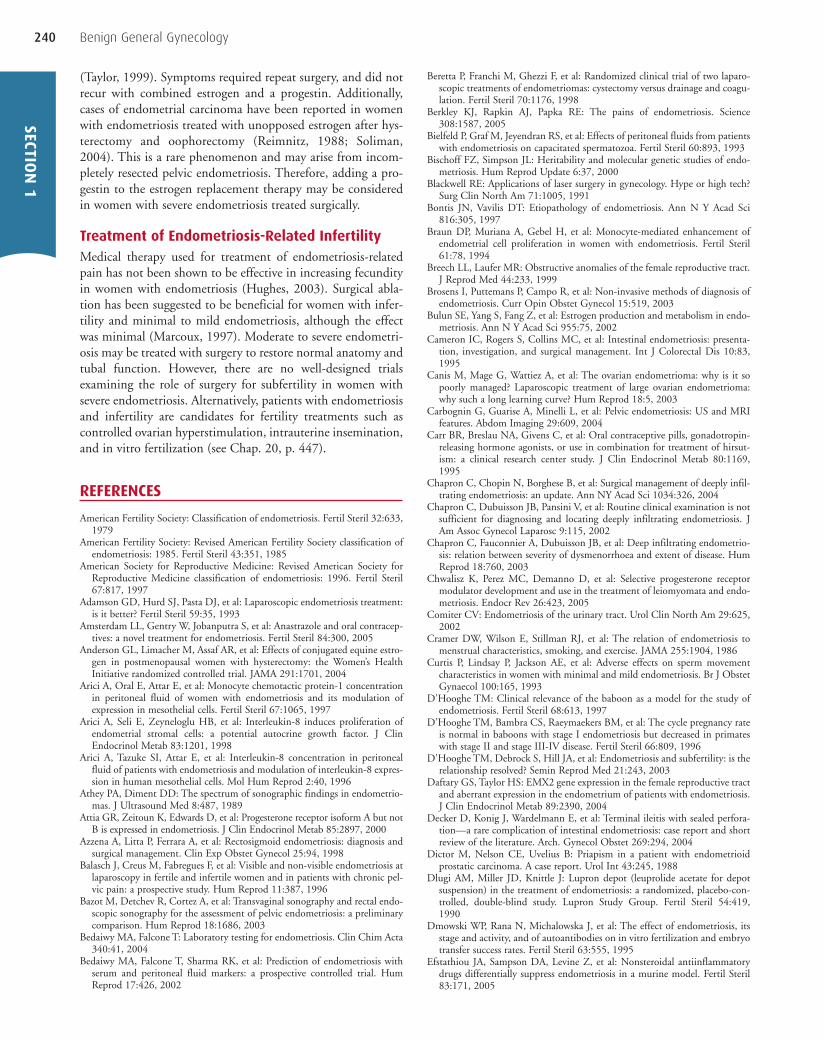
240 Benign General Gynecology
SECTION
1
(Taylor, 1999). Symptoms required repeat surgery, and did not recur with combined estrogen and a progestin. Additionally, cases of endometrial carcinoma have been reported in women with endometriosis treated with unopposed estrogen after hys-terectomy and oophorectomy (Reimnitz, 1988; Soliman, 2004). This is a rare phenomenon and may arise from incom-pletely resected pelvic endometriosis. Therefore, adding a pro-gestin to the estrogen replacement therapy may be considered in women with severe endometriosis treated surgically.
Treatment of Endometriosis-Related InfertilityMedical therapy used for treatment of endometriosis-related pain has not been shown to be effective in increasing fecundity in women with endometriosis (Hughes, 2003). Surgical abla-tion has been suggested to be beneficial for women with infer-tility and minimal to mild endometriosis, although the effect was minimal (Marcoux, 1997). Moderate to severe endometri-osis may be treated with surgery to restore normal anatomy and tubal function. However, there are no well-designed trials examining the role of surgery for subfertility in women with severe endometriosis. Alternatively, patients with endometriosis and infertility are candidates for fertility treatments such as controlled ovarian hyperstimulation, intrauterine insemination, and in vitro fertilization (see Chap. 20, p. 447).
REFERENCES
American Fertility Society: Classification of endometriosis. Fertil Steril 32:633, 1979
American Fertility Society: Revised American Fertility Society classification of endometriosis: 1985. Fertil Steril 43:351, 1985
American Society for Reproductive Medicine: Revised American Society for Reproductive Medicine classification of endometriosis: 1996. Fertil Steril 67:817, 1997
Adamson GD, Hurd SJ, Pasta DJ, et al: Laparoscopic endometriosis treatment: is it better? Fertil Steril 59:35, 1993
Amsterdam LL, Gentry W, Jobanputra S, et al: Anastrazole and oral contracep-tives: a novel treatment for endometriosis. Fertil Steril 84:300, 2005
Anderson GL, Limacher M, Assaf AR, et al: Effects of conjugated equine estro-gen in postmenopausal women with hysterectomy: the Women’s Health Initiative randomized controlled trial. JAMA 291:1701, 2004
Arici A, Oral E, Attar E, et al: Monocyte chemotactic protein-1 concentration in peritoneal fluid of women with endometriosis and its modulation of expression in mesothelial cells. Fertil Steril 67:1065, 1997
Arici A, Seli E, Zeyneloglu HB, et al: Interleukin-8 induces proliferation of endometrial stromal cells: a potential autocrine growth factor. J Clin Endocrinol Metab 83:1201, 1998
Arici A, Tazuke SI, Attar E, et al: Interleukin-8 concentration in peritoneal fluid of patients with endometriosis and modulation of interleukin-8 expres-sion in human mesothelial cells. Mol Hum Reprod 2:40, 1996
Athey PA, Diment DD: The spectrum of sonographic findings in endometrio-mas. J Ultrasound Med 8:487, 1989
Attia GR, Zeitoun K, Edwards D, et al: Progesterone receptor isoform A but not B is expressed in endometriosis. J Clin Endocrinol Metab 85:2897, 2000
Azzena A, Litta P, Ferrara A, et al: Rectosigmoid endometriosis: diagnosis and surgical management. Clin Exp Obstet Gynecol 25:94, 1998
Balasch J, Creus M, Fabregues F, et al: Visible and non-visible endometriosis at laparoscopy in fertile and infertile women and in patients with chronic pel-vic pain: a prospective study. Hum Reprod 11:387, 1996
Bazot M, Detchev R, Cortez A, et al: Transvaginal sonography and rectal endo-scopic sonography for the assessment of pelvic endometriosis: a preliminary comparison. Hum Reprod 18:1686, 2003
Bedaiwy MA, Falcone T: Laboratory testing for endometriosis. Clin Chim Acta 340:41, 2004
Bedaiwy MA, Falcone T, Sharma RK, et al: Prediction of endometriosis with serum and peritoneal fluid markers: a prospective controlled trial. Hum Reprod 17:426, 2002
Beretta P, Franchi M, Ghezzi F, et al: Randomized clinical trial of two laparo-scopic treatments of endometriomas: cystectomy versus drainage and coagu-lation. Fertil Steril 70:1176, 1998
Berkley KJ, Rapkin AJ, Papka RE: The pains of endometriosis. Science 308:1587, 2005
Bielfeld P, Graf M, Jeyendran RS, et al: Effects of peritoneal fluids from patients with endometriosis on capacitated spermatozoa. Fertil Steril 60:893, 1993
Bischoff FZ, Simpson JL: Heritability and molecular genetic studies of endo-metriosis. Hum Reprod Update 6:37, 2000
Blackwell RE: Applications of laser surgery in gynecology. Hype or high tech? Surg Clin North Am 71:1005, 1991
Bontis JN, Vavilis DT: Etiopathology of endometriosis. Ann N Y Acad Sci 816:305, 1997
Braun DP, Muriana A, Gebel H, et al: Monocyte-mediated enhancement of endometrial cell proliferation in women with endometriosis. Fertil Steril 61:78, 1994
Breech LL, Laufer MR: Obstructive anomalies of the female reproductive tract. J Reprod Med 44:233, 1999
Brosens I, Puttemans P, Campo R, et al: Non-invasive methods of diagnosis of endometriosis. Curr Opin Obstet Gynecol 15:519, 2003
Bulun SE, Yang S, Fang Z, et al: Estrogen production and metabolism in endo-metriosis. Ann N Y Acad Sci 955:75, 2002
Cameron IC, Rogers S, Collins MC, et al: Intestinal endometriosis: presenta-tion, investigation, and surgical management. Int J Colorectal Dis 10:83, 1995
Canis M, Mage G, Wattiez A, et al: The ovarian endometrioma: why is it so poorly managed? Laparoscopic treatment of large ovarian endometrioma: why such a long learning curve? Hum Reprod 18:5, 2003
Carbognin G, Guarise A, Minelli L, et al: Pelvic endometriosis: US and MRI features. Abdom Imaging 29:609, 2004
Carr BR, Breslau NA, Givens C, et al: Oral contraceptive pills, gonadotropin-releasing hormone agonists, or use in combination for treatment of hirsut-ism: a clinical research center study. J Clin Endocrinol Metab 80:1169, 1995
Chapron C, Chopin N, Borghese B, et al: Surgical management of deeply infil-trating endometriosis: an update. Ann NY Acad Sci 1034:326, 2004
Chapron C, Dubuisson JB, Pansini V, et al: Routine clinical examination is not sufficient for diagnosing and locating deeply infiltrating endometriosis. J Am Assoc Gynecol Laparosc 9:115, 2002
Chapron C, Fauconnier A, Dubuisson JB, et al: Deep infiltrating endometrio-sis: relation between severity of dysmenorrhoea and extent of disease. Hum Reprod 18:760, 2003
Chwalisz K, Perez MC, Demanno D, et al: Selective progesterone receptor modulator development and use in the treatment of leiomyomata and endo-metriosis. Endocr Rev 26:423, 2005
Comiter CV: Endometriosis of the urinary tract. Urol Clin North Am 29:625, 2002
Cramer DW, Wilson E, Stillman RJ, et al: The relation of endometriosis to menstrual characteristics, smoking, and exercise. JAMA 255:1904, 1986
Curtis P, Lindsay P, Jackson AE, et al: Adverse effects on sperm movement characteristics in women with minimal and mild endometriosis. Br J Obstet Gynaecol 100:165, 1993
D’Hooghe TM: Clinical relevance of the baboon as a model for the study of endometriosis. Fertil Steril 68:613, 1997
D’Hooghe TM, Bambra CS, Raeymaekers BM, et al: The cycle pregnancy rate is normal in baboons with stage I endometriosis but decreased in primates with stage II and stage III-IV disease. Fertil Steril 66:809, 1996
D’Hooghe TM, Debrock S, Hill JA, et al: Endometriosis and subfertility: is the relationship resolved? Semin Reprod Med 21:243, 2003
Daftary GS, Taylor HS: EMX2 gene expression in the female reproductive tract and aberrant expression in the endometrium of patients with endometriosis. J Clin Endocrinol Metab 89:2390, 2004
Decker D, Konig J, Wardelmann E, et al: Terminal ileitis with sealed perfora-tion—a rare complication of intestinal endometriosis: case report and short review of the literature. Arch. Gynecol Obstet 269:294, 2004
Dictor M, Nelson CE, Uvelius B: Priapism in a patient with endometrioid prostatic carcinoma. A case report. Urol Int 43:245, 1988
Dlugi AM, Miller JD, Knittle J: Lupron depot (leuprolide acetate for depot suspension) in the treatment of endometriosis: a randomized, placebo-con-trolled, double-blind study. Lupron Study Group. Fertil Steril 54:419, 1990
Dmowski WP, Rana N, Michalowska J, et al: The effect of endometriosis, its stage and activity, and of autoantibodies on in vitro fertilization and embryo transfer success rates. Fertil Steril 63:555, 1995
Efstathiou JA, Sampson DA, Levine Z, et al: Nonsteroidal antiinflammatory drugs differentially suppress endometriosis in a murine model. Fertil Steril 83:171, 2005
sch72576_c10.indd Page 240 12/28/07 10:47:36 AM usersch72576_c10.indd Page 240 12/28/07 10:47:36 AM user /Volumes/209/MHBD105/mhbd105indd%0/ch10/Volumes/209/MHBD105/mhbd105indd%0/ch10

241EndometriosisCH
AP
TER 1
0
Elger W, Bartley J, Schneider B, et al: Endocrine pharmacological characteriza-tion of progesterone antagonists and progesterone receptor modulators with respect to PR-agonistic and antagonistic activity. Steroids 65:713, 2000
Eskenazi B, Warner ML: Epidemiology of endometriosis. Obstet Gynecol Clin North Am 24:235, 1997
Eskenazi B, Warner M, Bonsignore L, et al: Validation study of nonsurgical diagnosis of endometriosis. Fertil Steril 76:929, 2001
Fauconnier A, Chapron C, Dubuisson JB, et al: Relation between pain symp-toms and the anatomic location of deep infiltrating endometriosis. Fertil Steril 78:719, 2002
Fedele L, Bianchi S, Bocciolone L, et al: Buserelin acetate in the treatment of pelvic pain associated with minimal and mild endometriosis: a controlled study. Fertil Steril 59:516, 1993
Fedele L, Bianchi S, Bocciolone L, et al: Pain symptoms associated with endo-metriosis. Obstet Gynecol 79:767, 1992
Fedele L, Marchini M, Bianchi S, et al: Endometrial patterns during danazol and buserelin therapy for endometriosis: comparative structural and ultra-structural study. Obstet Gynecol 76:79, 1990
Ferrero S, Esposito F, Abbamonte LH, et al: Quality of sex life in women with endometriosis and deep dyspareunia. Fertil Steril 83:573, 2005
Floyd WS: Danazol: endocrine and endometrial effects. Int J Fertil 25:75, 1980
Forbes KL, Thomas FJ: Tissue and endocrine responses to gestrinone and dan-azol in the treatment of endometriosis. Reprod Fertil Dev 5:103, 1993
Gallo MF, Nanda K, Grimes DA, et al: 20 mcg versus >20 mcg estrogen com-bined oral contraceptives for contraception. Cochrane Database Syst Rev CD003989, 2005
Gambone JC, Mittman BS, Munro MG, et al: Consensus statement for the management of chronic pelvic pain and endometriosis: proceedings of an expert-panel consensus process. Fertil Steril 78:961, 2002
Garrido N, Krussel JS, Remohi J, et al: Expression and function of 3beta hydroxysteroid dehydrogenase (3beta HSD) type II and corticosteroid bind-ing globulin (CBG) in granulosa cells from ovaries of women with and without endometriosis. J Assist Reprod Genet 19:24, 2002
Gestrinone Italian Study Group: Gestrinone versus a gonadotropin-releasing hormone agonist for the treatment of pelvic pain associated with endome-triosis: a multicenter, randomized, double-blind study. Gestrinone Italian Study Group. Fertil Steril 66:911, 1996
Giudice LC, Kao LC: Endometriosis. Lancet 364:1789, 2004Gleicher N, Dmowski WP, Siegel I, et al: Lymphocyte subsets in endometriosis.
Obstet Gynecol 63:463, 1984Gurates B, Bulun SE: Endometriosis: the ultimate hormonal disease. Semin
Reprod Med 21:125, 2003Gutt CN, Oniu T, Schemmer P, et al: Fewer adhesions induced by laparoscopic
surgery? Surg Endosc 18:898, 2004Guzick DS, Bross DS, Rock JA: Assessing the efficacy of The American Fertility
Society’s classification of endometriosis: application of a dose-response methodology. Fertil Steril 38:171, 1982
Guzick DS, Silliman NP, Adamson GD, et al: Prediction of pregnancy in infer-tile women based on the American Society for Reproductive Medicine’s revised classification of endometriosis. Fertil Steril 67:822, 1997
Hadfield RM, Mardon HJ, Barlow DH, et al: Endometriosis in monozygotic twins. Fertil Steril 68:941, 1997
Halme J, Hammond MG, Hulka JF, et al: Retrograde menstruation in healthy women and in patients with endometriosis. Obstet Gynecol 64:151, 1984
Hammoud A, Gago LA, Diamond MP: Adhesions in patients with chronic pelvic pain: a role for adhesiolysis? Fertil Steril 82:1483, 2004
Haney AF, Muscato J., Weinberg JB: Peritoneal fluid cell populations in infer-tility patients. Fertil Steril 35:696, 1981
Harada T, Kubota T, Aso T: Usefulness of CA19-9 versus CA125 for the diag-nosis of endometriosis. Fertil Steril 78:733, 2002
Harlow CR, Cahill DJ, Maile LA, et al: Reduced preovulatory granulosa cell steroidogenesis in women with endometriosis. J Clin Endocrinol Metab 81:426, 1996
Ho HN, Chao KH, Chen HF,et al: Peritoneal natural killer cytotoxicity and CD25+ CD3+ lymphocyte subpopulation are decreased in women with stage III-IV endometriosis. Hum Reprod 10:2671, 1995
Hornstein MD, Harlow BL, Thomas PP, et al: Use of a new CA 125 assay in the diagnosis of endometriosis. Hum Reprod 10:932, 1995a
Hornstein MD, Surrey ES, Weisberg GW, et al: Leuprolide acetate depot and hormonal add-back in endometriosis: a 12-month study. Lupron Add-Back Study Group. Obstet Gynecol 91:16, 1998
Hornstein MD, Yuzpe AA, Burry KA, et al: Prospective randomized double-blind trial of 3 versus 6 months of nafarelin therapy for endometriosis associated pelvic pain. Fertil Steril 63:955, 1995b
Houston DE, Noller KL, Melton LJ III, et al: Incidence of pelvic endometrio-sis in Rochester, Minnesota, 1970–1979. Am J Epidemiol 125:959, 1987
Hughes E, Fedorkow D, Collins J, et al: Ovulation suppression for endome-triosis. Cochrane Database Syst Rev, CD000155, 2003
Hughesdon PE: The structure of endometrial cysts of the ovary. J Obstet Gynaecol Br Emp 64:481, 1957
Jansen RP, Russell P: Nonpigmented endometriosis: clinical, laparoscopic, and pathologic definition. Am J Obstet Gynecol 155:1154,1986
Joki-Erkkila MM, Heinonen PK: Presenting and long-term clinical implica-tions and fecundity in females with obstructing vaginal malformations. J Pediatr Adolesc Gynecol 16:307, 2003
Jones KD, Fan A, Sutton CJ: The ovarian endometrioma: why is it so poorly managed? Indicators from an anonymous survey. Hum Reprod 17:845, 2002
Jones KD, Haines P, Sutton CJ: Long-term follow-up of a controlled trial of laser laparoscopy for pelvic pain. JSLS 5:111, 2001
Jones SC: Relative thromboembolic risks associated with COX-2 inhibitors. Ann Pharmacother 39:1249, 2005
Kao LC, Germeyer A, Tulac S, et al: Expression profiling of endometrium from women with endometriosis reveals candidate genes for disease-based implan-tation failure and infertility. Endocrinology 144:2870, 2003
Kennedy S, Bergqvist A, Chapron C, et al: ESHRE guideline for the diagnosis and treatment of endometriosis. Hum Reprod 20(10):2698, 2005
Kettel LM, Murphy AA, Morales AJ, et al: Treatment of endometriosis with the antiprogesterone mifepristone (RU486). Fertil Steril 65:23, 1996
Khorram O, Taylor RN, Ryan IP, et al: Peritoneal fluid concentrations of the cytokine RANTES correlate with the severity of endometriosis. Am J Obstet Gynecol 169:1545, 1993
Kitawaki J, Noguchi T, Amatsu T, et al: Expression of aromatase cytochrome P450 protein and messenger ribonucleic acid in human endometriotic and adenomyotic tissues but not in normal endometrium. Biol Reprod 57:514, 1997
Koger KE, Shatney CH, Hodge K, et al: Surgical scar endometrioma. Surg Gynecol Obstet 177:243,1993
Koninckx PR, Braet P, Kennedy SH, et al: Dioxin pollution and endometriosis in Belgium. Hum Reprod, 9:1001, 1994
Koninckx PR, Meuleman C, Demeyere S, et al: Suggestive evidence that pelvic endometriosis is a progressive disease, whereas deeply infiltrating endome-triosis is associated with pelvic pain. Fertil Steril 55:759, 1991
Koninckx PR, Meuleman C, Oosterlynck D, et al: Diagnosis of deep endome-triosis by clinical examination during menstruation and plasma CA-125 concentration. Fertil Steril 65:280, 1996
Lessey BA, Castelbaum AJ, Sawin SW, et al: Aberrant integrin expression in the endometrium of women with endometriosis. J Clin Endocrinol Metab 79:643,1994
Leyendecker G, Kunz G, Herbertz M, et al: Uterine peristaltic activity and the development of endometriosis. Ann N Y Acad Sci 1034:338, 2004
Ling FW: Randomized controlled trial of depot leuprolide in patients with chronic pelvic pain and clinically suspected endometriosis. Pelvic Pain Study Group Obstet Gynecol 93:51, 1999
Liu X, Yuan L, Shen F, et al: Patterns of and risk factors for recurrence in women with ovarian endometriomas. Obstet Gynecol 109(6):1411, 2007
Lockhat FB, Emembolu JO, et al: The efficacy, side-effects and continuation rates in women with symptomatic endometriosis undergoing treatment with an intra-uterine administered progestogen (levonorgestrel): A 3 year follow-up. Obstet Gynecol Surv 60:443, 2005
Mahmood TA, Templeton A: Prevalence and genesis of endometriosis. Hum Reprod 6:544, 1991
Mais V, Ajossa S, Piras B, et al: Treatment of nonendometriotic benign adnexal cysts: a randomized comparison of laparoscopy and laparotomy. Obstet Gynecol 86:770, 1995
Malinak LR, Buttram VC Jr., Elias S, et al: Heritage aspects of endometriosis. II. Clinical characteristics of familial endometriosis. Am J Obstet Gynecol 137:332, 1980
Marchino GL, Gennarelli G, Enria R, et al: Diagnosis of pelvic endometriosis with use of macroscopic versus histologic findings. Fertil Steril 84:12, 2005a
Marchino GL, Gennarelli G, Enria R., et al: Laparoscopic visualization with histologic confirmation represents the best available option to date in the diagnosis of endometriosis. Fertil Steril 84:38, 2005b
Marcoux S, Maheux R, Berube S: Laparoscopic surgery in infertile women with minimal or mild endometriosis. Canadian Collaborative Group on Endometriosis. N Engl J Med 337:217, 1997
Markham SM, Carpenter SE, et al: Extrapelvic endometriosis. Obstet Gynecol Clin North Am 16:193, 1989
Mathias SD, Kuppermann M, Liberman RF, et al: Chronic pelvic pain: preva-lence, health-related quality of life, and economic correlates. Obstet Gynecol 87:321, 1996
Mathur S, Peress MR, Williamson HO, et al: Autoimmunity to endometrium and ovary in endometriosis. Clin Exp. Immunol 50:259, 1982
sch72576_c10.indd Page 241 12/28/07 10:47:36 AM usersch72576_c10.indd Page 241 12/28/07 10:47:36 AM user /Volumes/209/MHBD105/mhbd105indd%0/ch10/Volumes/209/MHBD105/mhbd105indd%0/ch10

242 Benign General Gynecology
SECTION
1
Matorras R, Rodriguez F, Pijoan JI, et al: Women who are not exposed to spermatozoa and infertile women have similar rates of stage I endometriosis. Fertil Steril 76:923, 2001
Matsuura K, Ohtake H, Katabuchi H, et al: Coelomic metaplasia theory of endometriosis: evidence from in vivo studies and an in vitro experimental model. Gynecol Obstet Invest 47(Suppl 1):18, 1999
Mayani A, Barel S, Soback S, et al: Dioxin concentrations in women with endometriosis. Hum Reprod 12:373, 1997
McLaren J, Prentice A, Charnock-Jones DS, et al: Vascular endothelial growth factor is produced by peritoneal fluid macrophages in endometriosis and is regulated by ovarian steroids. J Clin Invest 98:482, 1996
McMeekin DS, Tillmanns T: Endometrial cancer: treatment of nodal metasta-ses. Curr Treat Options Oncol 4:121, 2003
Milingos S, Mavrommatis C, Elsheikh A, et al: Fecundity of infertile women with minimal or mild endometriosis. A clinical study. Arch Gynecol Obstet 267:37, 2002
Mitchell AO, Hoffman AP, Swartz SE, et al: An unusual occurrence of endo-metriosis in the right groin: a case report and review of the literature. Mil Med 156:633, 1991
Moen MH, Schei B: Epidemiology of endometriosis in a Norwegian county. Acta Obstet Gynecol Scand 76:559, 1997
Mol BW, Bayram N, Lijmer JG, et al: The performance of CA-125 measure-ment in the detection of endometriosis: a meta-analysis. Fertil Steril 70:1101, 1998
Moore J, Copley S, Morris J, et al: A systematic review of the accuracy of ultra-sound in the diagnosis of endometriosis. Ultrasound Obstet Gynecol 20:630, 2002
Moore JG, Binstock MA, et al: The clinical implications of retroperitoneal endometriosis. Am J Obstet Gynecol 158:1291, 1998
Mori H, Sawairi M, Nakagawa M., et al: Peritoneal fluid interleukin-1 beta and tumor necrosis factor in patients with benign gynecologic disease. Am J Reprod Immunol 26:62, 1991
Muneyyirci-Delale O, Karacan M: Effect of norethindrone acetate in the treat-ment of symptomatic endometriosis. Int J Fertil Womens Med 43:24, 1998
Murphy AA: Clinical aspects of endometriosis. Ann N Y Acad Sci 955:1, 2002
Muscato JJ, Haney AF, Weinberg JB: Sperm phagocytosis by human peritoneal macrophages: a possible cause of infertility in endometriosis. Am J Obstet Gynecol 144:503, 1982
Muzii L, Marana R, Pedulla S, et al: Correlation between endometriosis-associ-ated dysmenorrhea and the presence of typical or atypical lesions. Fertil Steril 68:19, 1997
Namnoum AB, Hickman TN, Goodman SB, et al: Incidence of symptom recurrence after hysterectomy for endometriosis. Fertil Steril 64:898, 1995
Nasir L, Bope ET: Management of pelvic pain from dysmenorrhea or endome-triosis. J Am Board Fam Pract 17(Suppl):S43, 2004
Nezhat C, Nezhat F: A simplified method of laparoscopic presacral neurectomy for the treatment of central pelvic pain due to endometriosis. Br J Obstet Gynaecol 99:659, 1992a
Nezhat C, Santolaya J, Nezhat FR: Comparison of transvaginal sonography and bimanual pelvic examination in patients with laparoscopically con-firmed endometriosis. J Am Assoc Gynecol Laparosc 1:127, 1994
Nezhat F, Nezhat C, Allan CJ, et al: Clinical and histologic classification of endometriomas. Implications for a mechanism of pathogenesis. J Reprod Med 37:771, 1992b
Nisolle M, Donnez J: Peritoneal endometriosis, ovarian endometriosis, and adenomyotic nodules of the rectovaginal septum are three different entities. Fertil Steril 68:585, 1997
Noble LS, Takayama K, Zeitoun KM, et al: Prostaglandin E2 stimulates aroma-tase expression in endometriosis-derived stromal cells. J Clin Endocrinol Metab 82:600, 1997
Odukoya OA, Wheatcroft N, Weetman AP, et al: The prevalence of endome-trial immunoglobulin G antibodies in patients with endometriosis. Hum Reprod 10:1214, 1995
Olive DL, Pritts EA: Treatment of endometriosis. N Engl J Med 345:266, 2001
Olive DL, Stohs GF, Metzger DA, et al: Expectant management and hydrotu-bations in the treatment of endometriosis-associated infertility. Fertil Steril 44:35, 1985a
Olive DL, Weinberg JB, Haney AF: Peritoneal macrophages and infertility: the association between cell number and pelvic pathology. Fertil Steril 44:772, 1985b
Oosterlynck DJ, Meuleman C, Waer M, et al: Immunosuppressive activity of peritoneal fluid in women with endometriosis. Obstet Gynecol 82:206, 1993
Orwoll ES, Yuzpe AA, Burry KA, et al: Nafarelin therapy in endometriosis: long-term effects on bone mineral density. Am J Obstet Gynecol 171:1221, 1994
Osuga Y, Koga K, Tsutsumi O, et al: Role of laparoscopy in the treatment of endometriosis-associated infertility. Gynecol Obstet Invest 53(Suppl 1):33, 2002
Ota H, Igarashi S, Sasaki M, et al: Distribution of cyclooxygenase-2 in eutopic and ectopic endometrium in endometriosis and adenomyosis. Hum Reprod 16:561, 2001
Parazzini F: Ablation of lesions or no treatment in minimal-mild endometriosis in infertile women: a randomized trial. Gruppo Italiano per lo Studio dell’Endometriosi. Hum Reprod 14:1332, 1999
Patel MD, Feldstein VA, Chen DC, et al: Endometriomas: diagnostic perfor-mance of US. Radiology 210:739, 1999
Pellicer A, Oliveira N, Ruiz A, et al: Exploring the mechanism(s) of endome-triosis-related infertility: an analysis of embryo development and implanta-tion in assisted reproduction. Hum Reprod 10(Suppl 2):91, 1995
Pellicer, A., Valbuena, D., Bauset, C., et al: The follicular endocrine environ-ment in stimulated cycles of women with endometriosis: steroid levels and embryo quality. Fertil. Steril, 69, 1135, 1998
Peters AA, Trimbos-Kemper GC, Admiraal C, et al: A randomized clinical trial on the benefit of adhesiolysis in patients with intraperitoneal adhesions and chronic pelvic pain. Br J Obstet Gynaecol 99:59, 1992
Petta CA, Ferriani RA, Abrao MS, et al: Randomized clinical trial of a levonorg-estrel-releasing intrauterine system and a depot GnRH analogue for the treatment of chronic pelvic pain in women with endometriosis. Hum Reprod 20:1993, 2005
Pinkert TC, Catlow CE, Straus R: Endometriosis of the urinary bladder in a man with prostatic carcinoma. Cancer 43:1562, 1979
Pollack R, Gordon PH, Ferenczy A, et al: Perineal endometriosis. A case report. J Reprod Med 35:109, 1990
Possover M, Chiantera V: Isolated infiltrative endometriosis of the sciatic nerve: a report of three patients. Fertil Steril 87(2):417.e17, 2007
Prentice A, Deary AJ, Bland E: Progestagens and anti-progestagens for pain associated with endometriosis. Cochrane Database Syst Rev CD002122, 2000a
Prentice A, Deary AJ, Goldbeck-Wood S, et al: Gonadotrophin-releasing hor-mone analogues for pain associated with endometriosis. Cochrane Database Syst Rev CD000346, 2000b
Price DT, Maloney KE, Ibrahim GK, et al: Vesical endometriosis: report of two cases and review of the literature. Urology 48:639, 1996
Qiao J, Yeung WS, Yao YQ, et al: The effects of follicular fluid from patients with different indications for IVF treatment on the binding of human sper-matozoa to the zona pellucida. Hum Reprod 13:128, 1998
Rabin D, McNeil LW: Pituitary and gonadal desensitization after continuous luteinizing hormone-releasing hormone infusion in normal females. J Clin Endocrinol Metab 51:873, 1980
Redwine DB: Conservative laparoscopic excision of endometriosis by sharp dissection: life table analysis of reoperation and persistent or recurrent dis-ease. Fertil Steril 56:628, 1991
Reich H, McGlynn F: Treatment of ovarian endometriomas using laparoscopic surgical techniques. J Reprod Med 31:577, 1986
Reimnitz C, Brand E, Nieberg RK, et al: Malignancy arising in endometriosis associated with unopposed estrogen replacement. Obstet Gynecol 71:444, 1988
Remorgida V, Ferrero S, Fulcheri E, et al: Bowel endometriosis: presentation, diagnosis, and treatment. Obstet Gynecol Surv 62(7):461, 2007
Rier S, Foster WG: Environmental dioxins and endometriosis. Semin Reprod Med 21: 145, 2003
Rier SE, Martin DC, Bowman RE, et al: Endometriosis in rhesus monkeys (Macaca mulatta) following chronic exposure to 2,3,7,8-tetrachlorodibenzo-p-dioxin. Fundam Appl Toxicol 21:433, 1993
Ripps BA, Martin DC: Correlation of focal pelvic tenderness with implant dimension and stage of endometriosis. J Reprod Med 37:620, 1992
Roberts LM, Redan J, Reich H: Extraperitoneal endometriosis with catamenial pneumothoraces: a review of the literature. JSLS 7:371, 2003
Rock JA, Zacur HA, Dlugi AM, et al: Pregnancy success following surgical correction of imperforate hymen and complete transverse vaginal septum. Obstet Gynecol 59:448, 1982
Rodriguez-Escudero FJ, Neyro JL, Corcostegui B, et al: Does minimal endo-metriosis reduce fecundity? Fertil Steril 50:522, 1988
Rossouw JE, Anderson GL, Prentice RL, et al: Risks and benefits of estrogen plus progestin in healthy postmenopausal women: principal results From the Women’s Health Initiative randomized controlled trial. JAMA 288:321, 2002
Ryan IP, Tseng JF, Schriock ED, et al: Interleukin-8 concentrations are ele-vated in peritoneal fluid of women with endometriosis. Fertil Steril 63:929, 1995
Ryu JS, Song ES, Lee KH, et al: Natural history and therapeutic implications of patients with catamenial hemoptysis. Resp Med 101(5):1032, 2007
sch72576_c10.indd Page 242 12/28/07 10:47:36 AM usersch72576_c10.indd Page 242 12/28/07 10:47:36 AM user /Volumes/209/MHBD105/mhbd105indd%0/ch10/Volumes/209/MHBD105/mhbd105indd%0/ch10

243EndometriosisCH
AP
TER 1
0
Sampson JA: Peritoneal endometriosis due to menstrual dissemination of endo-metrial tissue into the peritoneal cavity. Am J Obstet Gynecol 14:442, 1927
Sanfilippo JS, Wakim NG, Schikler KN, et al: Endometriosis in association with uterine anomaly. Am J Obstet Gynecol 154:39, 1986
Satyaswaroop PG, Wartell DJ, Mortel R: Distribution of progesterone receptor, estradiol dehydrogenase, and 20 alpha-dihydroprogesterone dehydrogenase activities in human endometrial glands and stroma: progestin induction of steroid dehydrogenase activities in vitro is restricted to the glandular epithe-lium. Endocrinology 111:743, 1982
Schattman GL, Grifo JA, Birnbaum S: Laparoscopic resection of a noncom-municating rudimentary uterine horn. A case report. J Reprod Med 40:219, 1995
Schenken RS, Asch RH: Surgical induction of endometriosis in the rabbit: effects on fertility and concentrations of peritoneal fluid prostaglandins. Fertil Steril 34:581, 1980
Schenken RS, Asch RH, Williams RF, et al: Etiology of infertility in monkeys with endometriosis: luteinized unruptured follicles, luteal phase defects, pelvic adhesions, and spontaneous abortions. Fertil Steril 41:122, 1984
Sciume C, Geraci G, Pisello F, et al: [Intestinal endometriosis: an obscure cause of cyclic rectal bleeding]. Ann Ital Chir 75:379, 2004
Seli E, Arici A: Endometriosis: interaction of immune and endocrine systems. Semin Reprod Med 21:135, 2003
Simpson JL, Elias S, Malinak LR, et al: Heritable aspects of endometriosis. I. Genetic studies. Am J Obstet Gynecol 137:327, 1980
Soliman NF, Evans AJ: Malignancy arising in residual endometriosis following hysterectomy and hormone replacement therapy. J Br Menopause Soc 10:123, 2004
Steele RW, Dmowski WP, Marmer DJ: Immunologic aspects of human endo-metriosis. Am J Reprod Immunol 6:33, 1984
Stefansson H, Geirsson RT, Steinthorsdottir V, et al: Genetic factors contribute to the risk of developing endometriosis. Hum Reprod 17:555, 2002
Strathy JH, Molgaard CA, Coulam CB, et al: Endometriosis and infertility: a laparoscopic study of endometriosis among fertile and infertile women. Fertil Steril 38:667, 1982
Surrey ES, Hornstein MD: Prolonged GnRH agonist and add-back therapy for symptomatic endometriosis: long-term follow-up. Obstet Gynecol 99:709, 2002
Sutton CJ, Pooley AS, Ewen SP, et al: Follow-up report on a randomized con-trolled trial of laser laparoscopy in the treatment of pelvic pain associated with minimal to moderate endometriosis. Fertil Steril 68:1070, 1997
Suzuki T, Izumi S, Matsubayashi H, et al: Impact of ovarian endometrioma on oocytes and pregnancy outcome in in vitro fertilization. Fertil Steril 83:908, 2005
Takayama K, Zeitoun K, Gunby RT, et al: Treatment of severe postmenopausal endometriosis with an aromatase inhibitor. Fertil Steril 69:709, 1998
Tasdemir M, Tasdemir I, Kodama H, et al: Effect of peritoneal fluid from infertile women with endometriosis on ionophore-stimulated acrosome loss. Hum Reprod 10:2419, 1995
Taylor M, Bowen-Simpkins P, Barrington J: Complications of unopposed oes-trogen following radical surgery for endometriosis. J Obstet Gynaecol 19:647, 1999
Telimaa S, Kauppila A, Ronnberg L., et al: Elevated serum levels of endometrial secretory protein PP14 in patients with advanced endometriosis. Suppression by treatment with danazol and high-dose medroxyprogesterone acetate. Am J Obstet Gynecol 161:866, 1989
Telimaa S, Puolakka J, Ronnberg L, et al: Placebo-controlled comparison of danazol and high-dose medroxyprogesterone acetate in the treatment of endometriosis. Gynecol Endocrinol 1:13, 1987
Thomas EJ, Cooke ID: Successful treatment of asymptomatic endometriosis: does it benefit infertile women? Br Med J (Clin Res Ed) 294:1117, 1987
Treloar SA, O’Connor DT, O’Connor VM, et al: Genetic influences on endo-metriosis in an Australian twin sample. Fertil Steril 71:701, 1999
Treloar SA, Wicks J, Nyholt DR, et al: Genomewide linkage study in 1,176 affected sister pair families identifies a significant susceptibility locus for endometriosis on chromosome 10q26. Am J Hum Genet 77:365, 2005
Tseng JF, Ryan IP, Milam TD, et al: Interleukin-6 secretion in vitro is up-regulated in ectopic and eutopic endometrial stromal cells from women with endometriosis. J Clin Endocrinol Metab 81:1118, 1996
Ueki M: Histologic study of endometriosis and examination of lymphatic drainage in and from the uterus. Am J Obstet Gynecol 165:201, 1991
Varras M, Kostopanagiotou E, Katis K, et al: Endometriosis causing extensive intestinal obstruction simulating carcinoma of the sigmoid colon: a case report and review of the literature. Eur J Gynaecol Oncol 23:353, 2002
Vercellini P, Chapron C, Fedele L, et al: Evidence for asymmetric distribution of sciatic nerve endometriosis. Obstet Gynecol 102:383, 2003a
Vercellini P, Frontino G, De Giorgi O, et al: Continuous use of an oral contra-ceptive for endometriosis-associated recurrent dysmenorrhea that does not respond to a cyclic pill regimen. Fertil Steril 80:560, 2003b
Vercellini P, Meschia M, De Giorgi O, et al: Bladder detrusor endometriosis: clinical and pathogenetic implications. J Urol 155:84, 1996a
Vercellini P, Trespidi L, Colombo A, et al: A gonadotropin-releasing hormone agonist versus a low-dose oral contraceptive for pelvic pain associated with endometriosis. Fertil Steril 60:75, 1993
Vercellini P, Trespidi L, De Giorgi O, et al: Endometriosis and pelvic pain: rela-tion to disease stage and localization. Fertil Steril 65:299, 1996b
Vercellini P, Vendola N, Bocciolone L, et al: Reliability of the visual diagnosis of ovarian endometriosis. Fertil Steril 56:1198, 1991
Vernon MW, Beard JS, Graves K, et al: Classification of endometriotic implants by morphologic appearance and capacity to synthesize prostaglandin F. Fertil Steril 46:801, 1986
Vessey MP, Villard-Mackintosh L, Painter R: Epidemiology of endometriosis in women attending family planning clinics. BMJ 306:182, 1993
Vilos GA, Vilos AW, Haebe JJ: Laparoscopic findings, management, histopa-thology, and outcome of 25 women with cyclic leg pain. J Am Assoc Gynecol Laparosc 9:145, 2002
Vinatier D, Orazi G, Cosson M, et al: Theories of endometriosis. Eur J Obstet Gynecol Reprod Biol 96:21, 2001
Von Rokitansky C: Ueber uterusdrusen-neubuildung in uterus and ovarilsarco-men. Z Ges Aerzte Wein 37:577, 1860
Waller KG, Lindsay P, Curtis P, et al: The prevalence of endometriosis in women with infertile partners. Eur J Obstet Gynecol Reprod Biol 48:135, 1993
Walter AJ, Hentz JG, Magtibay PM, et al: Endometriosis: correlation between histologic and visual findings at laparoscopy. Am J Obstet Gynecol 184:1407, 2001
Watanabe M, Kamiyama G, Yamazaki K., et al: Anal endosonography in the diagnosis and management of perianal endometriosis: report of a case. Surg Today 33:630, 2003
Wickramasekera D, Hay DJ, Fayz M: Acute small bowel obstruction due to ileal endometriosis: a case report and literature review. J R Coll Surg Edinb 44:59, 1999
Wiegratz I, Kuhl H: Long-cycle treatment with oral contraceptives. Drugs 64:2447, 2004
Wilson ML, Farquhar CM, Sinclair OJ, et al: Surgical interruption of pelvic nerve pathways for primary and secondary dysmenorrhoea. Cochrane Database Syst Rev CD001896, 2000
Wilson TJ, Hertzog PJ, Angus D, et al: Decreased natural killer cell activity in endometriosis patients: relationship to disease pathogenesis. Fertil Steril 62:1086, 1994
Wright J, Lotfallah H, Jones K, et al: A randomized trial of excision versus ablation for mild endometriosis. Fertil Steril 83:1830, 2005
Yuen PM, Yu KM, Yip SK, et al: A randomized prospective study of laparos-copy and laparotomy in the management of benign ovarian masses. Am J Obstet Gynecol 177:109, 1997
Zeitoun KM, Bulun SE: Aromatase: a key molecule in the pathophysiology of endometriosis and a therapeutic target. Fertil Steril 72:961, 1999
Zeitoun K, Takayama K, Sasano H, et al: Deficient 17beta-hydroxysteroid dehydrogenase type 2 expression in endometriosis: failure to metabolize 17beta-estradiol. J Clin Endocrinol Metab 83:4474, 1998
Zhu L, Wong F, Lang JH: Perineal endometriosis after vaginal delivery—clini-cal experience with 10 patients. Aust N Z J Obstet Gynaecol 42:565, 2002
Zullo F, Palomba S, Zupi E, et al: Effectiveness of presacral neurectomy in women with severe dysmenorrhea caused by endometriosis who were treated with laparoscopic conservative surgery: a 1-year prospective randomized double-blind controlled trial. Am J Obstet Gynecol 189:5, 2003
sch72576_c10.indd Page 243 12/28/07 10:47:37 AM usersch72576_c10.indd Page 243 12/28/07 10:47:37 AM user /Volumes/209/MHBD105/mhbd105indd%0/ch10/Volumes/209/MHBD105/mhbd105indd%0/ch10