Endometriosis and the Urinary Tract: from Diagnosis to ...
13
Review 1 Endometriosis and the Urinary Tract: from Diagnosis 2 to Surgical Treatment 3 Mathew Leonardi 1,2,3, *, Mercedes Espada 1,2 , Rosanne M Kho 4 , Javier F Magrina MD e , Anne- 4 Elodie Millischer 6 , Luca Savelli 7 , George Condous 1,2 5 1 Acute Gynaecology, Early Pregnancy and Advanced Endoscopy Surgery Unit, Nepean Hospital, 6 Kingswood, NSW, Australia 7 2 Nepean Clinical School, University of Sydney, Sydney, NSW, Australia 8 3 Endometriosis Clinic, Department of Obstetrics and Gynecology, McMaster University, Hamilton, Canada 9 4 Obstetrics, Gynecology, and Women's Health Institute, Cleveland Clinic, Cleveland, Ohio, USA 10 5 Department of Medical and Surgical Gynecology, Mayo Clinic Hospital, Phoenix, Arizona, USA 11 6 IMPC Radiology Bachaumont Paris and Radiodiagnostics Department, Hôpital Necker Paris, France 12 7 Department of Obstetrics and Gynecology, S. Orsola Malpighi Hospital, University of Bologna, Bologna, 13 Italy 14 * Correspondence: [email protected] 15 Received: date; Accepted: date; Published: date 16 Abstract: We aim to describe the diagnosis and surgical management of urinary tract endometriosis 17 (UTE). We detail current diagnostic tools including advanced transvaginal ultrasound, magnetic 18 resonance imaging, and surgical diagnostic tools such as cystourethroscopy. While discussing 19 surgical treatment options, we emphasize the importance of an interdisciplinary team, for complex 20 cases that involve the urinary tract. While bladder DE is more straightforward in its surgical 21 treatment, ureteral DE requires a high level of surgical skill. Specialists should be aware of the 22 important entity of UTE due to the serious health implications for women. When UTE exists, it is 23 important to work within a interdisciplinary radiological and surgical team. 24 Keywords: Endometriosis; ureter; bladder; ultrasound; magnetic resonance imaging; hydroureter 25 26 1. Introduction 27 Endometriosis is a chronic and benign condition thought to affect up to 10% of female-born 28 individuals. 1 In most classic teachings, it causes cyclic pelvic pain and infertility. Accordingly, it was 29 historically considered a gynecologic condition, managed by obstetrician-gynecologists. However, it 30 is increasingly recognized that endometriosis is a multi-organ and systemic inflammatory disease 31 that necessitates interdisciplinary care, led by a gynecologist with subspecialty training in clinical 32 and surgical management. 2,3 Though its etiology remains partially shrouded, symptoms are caused 33 by the presence of ectopic endometrial-like tissue outside of the uterus. 4 The term urinary tract 34 endometriosis (UTE) refers to endometriotic implants of the bladder, ureter, kidney and urethra. The 35 bladder and ureter are most commonly affected. 5 This paper will review the epidemiology, 36 pathogenesis, clinical presentation, diagnosis and differential diagnosis, with emphasis on imaging, 37 and management of UTE. 38 2. Epidemiology of UTE 39 The prevalence of UTE in the general female population remains unclear since around 50% of 40 women with UTE may be asymptomatic. 6 UTE most commonly affects the bladder. The prevalence 41 of UTE is estimated to range from 0.3 to 12% of all people affected by endometriosis and about 20- 42 50% of women with deep endometriosis (DE). 7 The urinary system is the second most common site 43 Preprints (www.preprints.org) | NOT PEER-REVIEWED | Posted: 23 September 2020 doi:10.20944/preprints202009.0541.v1 © 2020 by the author(s). Distributed under a Creative Commons CC BY license.
Transcript of Endometriosis and the Urinary Tract: from Diagnosis to ...
Review 1
Endometriosis and the Urinary Tract: from Diagnosis 2
to Surgical Treatment 3
Mathew Leonardi 1,2,3, *, Mercedes Espada 1,2, Rosanne M Kho 4, Javier F Magrina MDe, Anne-4 Elodie Millischer 6, Luca Savelli 7, George Condous 1,2 5
1 Acute Gynaecology, Early Pregnancy and Advanced Endoscopy Surgery Unit, Nepean Hospital, 6 Kingswood, NSW, Australia 7
2 Nepean Clinical School, University of Sydney, Sydney, NSW, Australia 8 3 Endometriosis Clinic, Department of Obstetrics and Gynecology, McMaster University, Hamilton, Canada 9 4 Obstetrics, Gynecology, and Women's Health Institute, Cleveland Clinic, Cleveland, Ohio, USA 10 5 Department of Medical and Surgical Gynecology, Mayo Clinic Hospital, Phoenix, Arizona, USA 11 6 IMPC Radiology Bachaumont Paris and Radiodiagnostics Department, Hôpital Necker Paris, France 12 7 Department of Obstetrics and Gynecology, S. Orsola Malpighi Hospital, University of Bologna, Bologna, 13
Italy 14 * Correspondence: [email protected] 15
Received: date; Accepted: date; Published: date 16
Abstract: We aim to describe the diagnosis and surgical management of urinary tract endometriosis 17 (UTE). We detail current diagnostic tools including advanced transvaginal ultrasound, magnetic 18 resonance imaging, and surgical diagnostic tools such as cystourethroscopy. While discussing 19 surgical treatment options, we emphasize the importance of an interdisciplinary team, for complex 20 cases that involve the urinary tract. While bladder DE is more straightforward in its surgical 21 treatment, ureteral DE requires a high level of surgical skill. Specialists should be aware of the 22 important entity of UTE due to the serious health implications for women. When UTE exists, it is 23 important to work within a interdisciplinary radiological and surgical team. 24
Keywords: Endometriosis; ureter; bladder; ultrasound; magnetic resonance imaging; hydroureter 25 26
1. Introduction 27
Endometriosis is a chronic and benign condition thought to affect up to 10% of female-born 28 individuals.1 In most classic teachings, it causes cyclic pelvic pain and infertility. Accordingly, it was 29 historically considered a gynecologic condition, managed by obstetrician-gynecologists. However, it 30 is increasingly recognized that endometriosis is a multi-organ and systemic inflammatory disease 31 that necessitates interdisciplinary care, led by a gynecologist with subspecialty training in clinical 32 and surgical management.2,3 Though its etiology remains partially shrouded, symptoms are caused 33 by the presence of ectopic endometrial-like tissue outside of the uterus.4 The term urinary tract 34 endometriosis (UTE) refers to endometriotic implants of the bladder, ureter, kidney and urethra. The 35 bladder and ureter are most commonly affected.5 This paper will review the epidemiology, 36 pathogenesis, clinical presentation, diagnosis and differential diagnosis, with emphasis on imaging, 37 and management of UTE. 38
2. Epidemiology of UTE 39
The prevalence of UTE in the general female population remains unclear since around 50% of 40 women with UTE may be asymptomatic.6 UTE most commonly affects the bladder. The prevalence 41 of UTE is estimated to range from 0.3 to 12% of all people affected by endometriosis and about 20-42 50% of women with deep endometriosis (DE).7 The urinary system is the second most common site 43
Preprints (www.preprints.org) | NOT PEER-REVIEWED | Posted: 23 September 2020 doi:10.20944/preprints202009.0541.v1
© 2020 by the author(s). Distributed under a Creative Commons CC BY license.
2 of 13
of extrapelvic endometriosis after the gastrointestinal tract.8 The prevalence of disease at specific sites 44 among women with UTE is as follows: bladder, 85-90%; ureter, 10%; kidney, 4%; and urethra, 2%.5 45
3. Pathogenesis of UTE 46
The two phenotypes of UTE are superficial endometriosis (SE) and deep endometriosis (DE).9 47 SE can be recognized at laparoscopy as black, white, or red implants depending on the degree of 48 fibrosis, scarring, and hemorrhage within the tissue. DE is defined by the invasion of endometrial-49 like glands and stroma at least 5 mm beneath the peritoneal surface. Deep implants are often 50 associated with fibrosis and/or smooth muscle proliferation and are the most frequently associated 51 with pelvic pain and infertility.10 52
General theories of endometriosis etiology include retrograde menstruation, coelomic 53 metaplasia, spread of endometrium-derived stem/progenitor cells and altered genetic/epigenetic or 54 immune factors.11 Additionally, in some women UTE appears to be iatrogenic; bladder endometriosis 55 may be more prevalent amongst women with previous Caesarean section(s).12 56
Certain anatomic structures of the female pelvis are thought to provide secluded peritoneal 57 pockets that provide protection to the ectopic endometrial-like cells from the regular peritoneal 58 clearance mechanisms allowing those cells to implant, invade and proliferate.13 This sanctuary effect 59 may explain why women with retroverted uteruses are less prone to develop bladder endometriosis, 60 or why ureteral endometriosis is more common on the left side.14 61
4. Bladder endometriosis 62
4.1. Definition of bladder endometriosis: 63
Bladder DE is defined by the presence of endometriotic tissue invading the detrusor muscle of 64 the bladder. The invasion of the detrusor muscle can be either full thickness or partial thickness.13 65
4.2. Clinical symptoms of bladder DE: 66
Endometriosis classically causes cyclic pain and infertility. Typically, women with bladder DE 67 present with dysuria8 but may also have urinary frequency, recurrent urinary tract infections8,15 and 68 hematuria, and more atypically urinary incontinence15. Bladder endometriosis can also be 69 asymptomatic and be incidentally diagnosed at the time of a cystoscopic or intra-abdominal 70 procedure for a different indication. As far as we are aware, the frequency of incidental diagnosis of 71 bladder endometriosis is still unknown. 72
4.3. Diagnosis of bladder DE: 73
Bladder DE is a histologic diagnosis. The initial evaluation of suspected bladder endometriosis 74 includes medical history, physical examination and complementary tests (laboratory testing, 75 cystourethroscopy and imaging techniques). 76
4.3.1. Medical history and physical examination 77
Women with bladder endometriosis often have endometriosis at other anatomic sites, therefore 78 the initial history and physical assessment include speculum examination (looking for lesions on the 79 posterior cervix or vaginal mucosa), tenderness on vaginal examination, nodules in the posterior 80 vaginal fornix, adnexal masses and immobility or lateral displacement of the uterus. 81
4.3.2. Laboratory testing 82
For women with clinical symptoms of bladder DE (dysuria, urinary frequency, hematuria), 83 urinalysis test to exclude infection or hematuria should be conducted. If infection is suspected, 84 urinary culture should also be done. Women with hematuria and suspected bladder endometriosis 85 should receive further testing. 86
Preprints (www.preprints.org) | NOT PEER-REVIEWED | Posted: 23 September 2020 doi:10.20944/preprints202009.0541.v1
3 of 13
4.3.3. Imaging techniques 87
4.3.3.1. Ultrasound 88
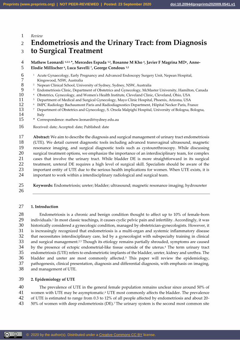
Ultrasound is the first-line imaging modality to diagnose or exclude BE and should be 89 performed transvaginally. Transabdominal ultrasound is also important for renal assessment. The 90 International Deep Endometriosis Analysis (IDEA) group recommends focused assessment of the 91 bladder and ureters (in addition to a thorough examination for other sites of DE, ovarian 92 endometriomas, and pelvic adhesions as depicted by the immobility of pelvic organs).16 It is 93 important to note that the components of this ultrasound examination exceed those of the traditional, 94 basic pelvic ultrasound,17 which does not include direct evaluation of DE16 or dynamic pelvic organ 95 mobility as a soft marker for DE.18,19 Patients should be sent to the closest radiologist or sonologist 96 with expertise in endometriosis with a requisition that specifies the need to evaluate bladder wall 97 and ureteral size and position. For transvaginal ultrasound (TVS) evaluation of the bladder, a small 98 amount of urine in the bladder is helpful in order to better identify the different portions of the 99 bladder wall. We recommend placing the TVS probe in the anterior vaginal fornix and gently swing 100 side-to-side, visualizing the mucosa and muscularis for focal thickening and hypoechoic linear or 101 nodular lesions (either spherical or comma-shaped) (Figure 1).20 Bladder nodules must be measured 102 in three orthogonal planes.16 Measuring the distance between the lesion and ureteral orifices may 103 assist in surgical planning. 104
105
Figure 1. Transvaginal ultrasound depiction of the bladder. Normal bladder (A) and three bladder 106 deep endometriosis nodules (B-D), identified with white arrows. 107
The pelvic segment of the ureters can be examined next by moving the TVS probe from the 108 midline toward the pelvic sidewalls. Operators should search for a long tubular hypoechoic structure 109 with a thick hyperechoic outer wall extending from the lateral aspect of the bladder base towards the 110 common iliac vessels.21 Vermiculation and/or the absence of color Doppler within the tubular 111 structure can be used to confirm it is a ureter rather than a vessel. The goal is to then follow the ureter 112 as proximal as possible, in the direction of its origin, assessing for dilatation, position, any asymmetry 113 between the right and left, and eventually the presence of DE nodules, which appear as hypoechoic 114 lesions around the course of the ureter (Figure 2). The presence of ureteral jets (i.e. the urinary inflow 115 from the ureter into the bladder) can be seen using color Doppler after waiting for a few 116
Preprints (www.preprints.org) | NOT PEER-REVIEWED | Posted: 23 September 2020 doi:10.20944/preprints202009.0541.v1

4 of 13
seconds/minutes.20 Carfagna et al. found in an observational study that the ureteric diameter on TVS 117 was ≥6mm with a median diameter of 6.9mm (range, 6–18 mm) in all cases of ureteral dilatation 118 confirmed at surgery.22 Nodules of the USL with or without parametrial infiltration must be 119 assessed,23 with measurements in three orthogonal planes.24 Nodules ≥17mm should especially raise 120 suspicion for ureteral involvement.25 121
122
Figure 2. Transvaginal ultrasound depiction of ureteral deep endometriosis nodule (white arrow) and 123 hydroureter (black arrow). The nodule originates from the uterosacral ligament but infiltrates the 124 parametrium and extrinsically compresses the ureter. 125
A transabdominal scan (TAS) of the kidneys is recommended in all women with concerns for 126 UTE in order to exclude the presence of hydronephrosis as this is usually asymptomatic in cases of 127 ureteral DE.26,27 128
For women with bladder lesions on TVS and asymmetrical ureteral caliber or hydronephrosis a 129 computed tomography (CT) urogram may be useful in completing the evaluation because CT best 130 depicts the course of the ureters. 131
4.3.3.2. Magnetic resonance imaging 132
Magnetic resonance imaging (MRI) is an adjunct imaging tool that can be considered to complete 133 or supplement TVS, for an accurate presurgical staging of UTE, especially in centers where advanced 134 TVS for UTE is not yet available. 135
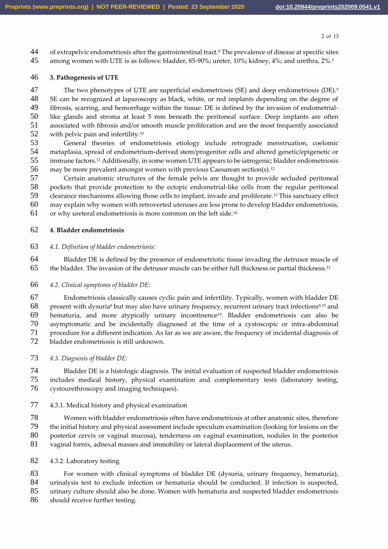
The diagnosis of bladder DE is based on a hypointense signal of nodules on T2-weighted images 136 with frequent hyperintense signal on fatty saturation T1-weighted images. The nodule is usually 137 located at the level of the vesicouterine pouch within the bladder base, forming an obtuse angle with 138 the bladder wall, and involving the muscularis layer (depicted by an obliteration of the hypointense 139 signal of the wall on T2-weighted MR images (Figure 3)) or protruding into the lumen with invasion 140 of the mucosal layer.28 Retrospective and recent studies suggest that MRI is particularly relevant to 141 diagnose bladder endometriosis with an accuracy of 96%,29 a sensitivity and specificity ranging from 142 88% and 100%, and 98% and 100%, respectively,28,29 whereas meta-analyses by Medeiros et al.30 and 143 Nisenblat et al.31 reported sensitivities of 64% and 41%, respectively. 144
145
Preprints (www.preprints.org) | NOT PEER-REVIEWED | Posted: 23 September 2020 doi:10.20944/preprints202009.0541.v1
5 of 13
146
Figure 3. Magnetic resonance imaging depiction of bladder deep endometriosis. Sagittal T2-weighted 147 plane depicting deep endometriosis nodule in hyposignalT2, which is infiltrating the detrusor muscle 148 of the bladder (white thin arrows). 149
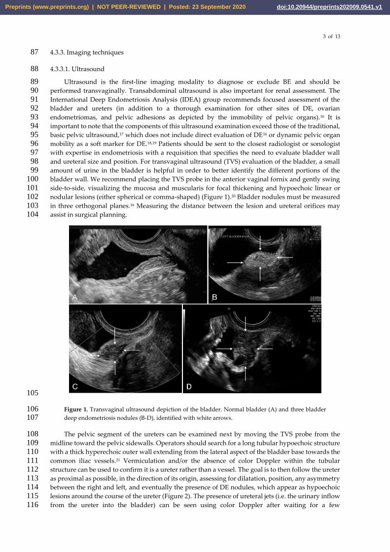
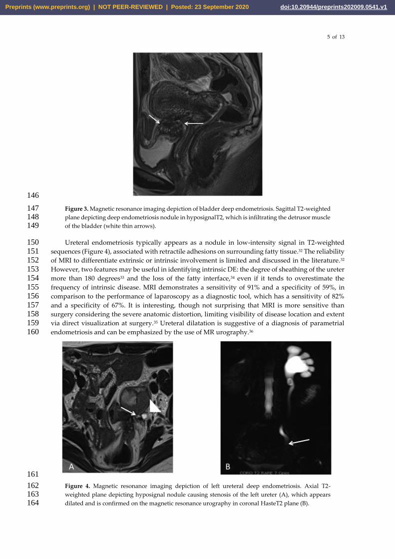
Ureteral endometriosis typically appears as a nodule in low-intensity signal in T2-weighted 150 sequences (Figure 4), associated with retractile adhesions on surrounding fatty tissue.32 The reliability 151 of MRI to differentiate extrinsic or intrinsic involvement is limited and discussed in the literature.32 152 However, two features may be useful in identifying intrinsic DE: the degree of sheathing of the ureter 153 more than 180 degrees33 and the loss of the fatty interface,34 even if it tends to overestimate the 154 frequency of intrinsic disease. MRI demonstrates a sensitivity of 91% and a specificity of 59%, in 155 comparison to the performance of laparoscopy as a diagnostic tool, which has a sensitivity of 82% 156 and a specificity of 67%. It is interesting, though not surprising that MRI is more sensitive than 157 surgery considering the severe anatomic distortion, limiting visibility of disease location and extent 158 via direct visualization at surgery.35 Ureteral dilatation is suggestive of a diagnosis of parametrial 159 endometriosis and can be emphasized by the use of MR urography.36 160
161
Figure 4. Magnetic resonance imaging depiction of left ureteral deep endometriosis. Axial T2-162 weighted plane depicting hyposignal nodule causing stenosis of the left ureter (A), which appears 163 dilated and is confirmed on the magnetic resonance urography in coronal HasteT2 plane (B). 164
Preprints (www.preprints.org) | NOT PEER-REVIEWED | Posted: 23 September 2020 doi:10.20944/preprints202009.0541.v1

6 of 13
4.3.3.3. Cystourethroscopy 165
When there is proven hematuria or a bladder nodule seen on TVS or MRI we performing a 166 cystourethroscopy may be helpful to confirm the diagnosis. Cystoscopically, BE can have a spectrum 167 of possible appearances from normal-appearing mucosa that is noticeably raised due to a nodule 168 beneath the mucosa to infiltration through the mucosa. In the latter scenario, lesions can appear 169 multiloculated with a combination of colors (from same color as the bladder mucosa to a blue/violet 170 color).37 Cystourethroscopy can also aid in excluding malignancy and measuring the distance from 171 the lesion to the ureteral openings to help urologists and gynecologists anticipate the type of urologic 172 procedure necessary (particularly if the removal of the lesion will also require ureteral resection and 173 reimplantation with ureteroneocystostomy.38,39 If the distance between the edge of the endometriotic 174 lesion and the interureteric ridge is less than 2 cm, ureteroneocystostomy is typically performed in 175 order to reduce the risk of ureteral obstruction and fistula formation5, and to optimally restore normal 176 anatomy. 177
4.4. Differential diagnosis of bladder DE: 178
4.4.1. Intraluminal bladder lesions 179
Neoplasms, angiomas, papillomas and bladder DE can be diagnosed by guided tissue sampling 180 with cystoscopy. 181
4.4.2. Urinary tract infection 182
Can be excluded with a urine culture test. 183
4.4.3. Urinary tract calculus 184
The type of pelvic pain associated to urinary tract calculus is variable in severity and duration, 185 whereas the pain associated to bladder DE is consistent. Urinary tract calculus can be identified on 186 ultrasound studies as mobile and echogenic formations with associated acoustic shadowing. They 187 can be associated with bladder wall thickening due to inflammation.5. 188
4.4.4. Interstitial cystitis 189
Interstitial cystitis is a clinical diagnosis that involves bladder discomfort associated to bladder 190 repletion. This is a diagnosis of exclusion that can only be reached when other etiologies such as 191 malignancy or bladder DE have been ruled out. 192
4.5. Treatment of bladder DE: 193
The aim of the treatment of bladder DE is to resolve symptoms and avoid possible renal 194 damage. Treatment can be expectant, medical or surgical. Conservative management with 195 sonographic follow up can be chosen for asymptomatic women without hydronephrosis while 196 surgery should always be performed in women with ureteral obstruction and hydronephrosis. Pain 197 symptoms can be managed medically or surgically. 198
4.5.1. Medical management 199
For patients with pain symptoms due to bladder DE, continuous progesterone-based regimens 200 (pills, intrauterine device, implant, injection), combined estrogen-progesterone therapy (continuous 201 or sequential regimens) and GnRH analogues (with or without add-back therapy) have all been 202 associated with improvement of symptoms from bladder DE.40,41 203
Women who respond to medical management can continue the treatment until menopause or 204 desire to conceive pregnancy or to achieve an optimal quality of life and reduce the risk of 205 progression, unless there is superimposed hydronephrosis, in which instance surgery would be the 206 first treatment choice to prevent irreversible renal failure from ureteral obstruction. 207
Preprints (www.preprints.org) | NOT PEER-REVIEWED | Posted: 23 September 2020 doi:10.20944/preprints202009.0541.v1
7 of 13
4.5.2. Surgical management 208
Procedures to surgically address bladder DE include shaving of serosal lesions and full thickness 209 resection of DE lesions. Most surgeries can be performed laparoscopically or robotically.40 210
4.5.3. Surgical approach: 211
1) Cystourethroscopy: to evaluate size of the lesion(s) and measure the distance between the 212 lesion and the ureteral ridge. The use of ureteral catheters is not supported by strong 213 evidence-based data; however their use might be helpful when the distance between the 214 lesion and the ureteral ridge is less than 2 cm or when the anatomy is distorted from 215 previous surgeries or extensive disease, in order to reduce the risk of inadvertent ureteral 216 damage at surgery. 217
2) Diagnostic laparoscopy followed by shaving of superficial serosal lesions off the bladder or 218 partial cystectomy when there is infiltration of the detrusor muscle in order to restore normal 219 anatomy, prevent hydronephrosis and recurrence of the disease.6 If the nodule affects the 220 vesical base, it can be either be approached laparoscopically (starting with dissection of the 221 vesicouterine pouch to facilitate complete resection) or through an operative cystoscope and 222 then reconstruct the bladder either laparoscopically or robotically. Resecting the underlying 223 myometrium has been proven to prevent recurrence of the symptoms when it comes to 224 bladder DE affecting the vesical base.42 225
3) Bladder closure: We recommend to close the bladder with two layers of transverse sutures. 226 At the end of the operation, the bladder is filled with methylene blue to confirm integrity of 227 the bladder, and a bladder catheter should be left in place for 10 days to prevent fistula 228 formation. 229
Surgical resection of bladder endometriosis is a risk factor for fistula formation (up to 15% of 230 patients).43 231
4.5.4. Medical versus surgical treatment 232
There are limited data comparing surgical and medical approach to bladder DE. On this basis, 233 for women with symptomatic bladder DE who do not have associated hydronephrosis, we would 234 recommend to start on a trial of COCP, progesterone or GnRH analogues (with or without add-back 235 therapy) for six months, and reserve the surgical approach to patients who either do not respond to 236 medical treatment or have hydronephrosis.6,40–43 237
5. Ureteral endometriosis 238
5.1. Definition of ureteral endometriosis (UE): 239
Ureteral endometriosis (UE) is defined by the presence of endometriotic tissue involving the 240 ureter. UE can be further subclassified as intrinsic, consisting of 40% of lesions when endometriosis 241 develops within the ureteral wall resulting in fibrosis and hypertrophy of the muscularis propria and 242 extrinsic, consisting of 60% of the lesions when the endometriosis develops primarily out of the 243 ureteral wall and causes compression from the outside.44 Though the parametrium is the main site of 244 endometriosis that leads to extrinsic ureteral compression, it is thought that many of these nodules 245 originate from the anterior rectal wall or uterosacral ligaments (USLs) DE.35 246
5.2. Clinical symptoms of UE: 247
Patients with UE generally have non-specific symptoms. It is estimated that 50% of women with 248 UE are asymptomatic, 25% present with flank pain and 15% have associated gross hematuria.45–47 249 Other concomitant symptoms associated with UE include dysmenorrhea and deep dyspareunia.48 250
5.3. Diagnosis of UE: 251
Preprints (www.preprints.org) | NOT PEER-REVIEWED | Posted: 23 September 2020 doi:10.20944/preprints202009.0541.v1
8 of 13
Tissue biopsy and histologic confirmation is the gold standard for diagnosing UE.48 The initial 252 evaluation of suspected UE includes a detailed medical history, physical examination and 253 complementary tests (laboratory testing, cystourethroscopy and imaging techniques) 254
5.3.1. Medical history and physical examination 255
UE is often associated with extensive pelvic disease and therefore the initial history and physical 256 assessment are key and should include speculum examination (looking for lesions on the posterior 257 cervix or vaginal mucosa), tenderness on vaginal examination, nodules in the posterior vaginal 258 fornix, adnexal masses and immobility or lateral placement of the uterus.48 259
5.3.2. Laboratory testing 260
It is important to exclude impaired renal function when there is suspected UE. Renal function 261 tests and urinalysis to include infection should be conducted in patients with flank pain or 262 hematuria.49 263
5.3.3. Imaging techniques 264
TAS can help to detect ureteral obstruction and evaluate the thickness of the renal parenchyma. 265 TVS can only assess the pelvic ureter, but it is very useful to evaluate other sites of implants of 266 endometriosis in the pelvis.16 Combined TAS/TVS should be performed as first-line exam suspecting 267 UE. By doing combined TAS/TVS the ureters can be visualized from the anterior parametrium to the 268 renal pelvis.50 269
When there is associated hydroureter or hydronephrosis, additional radiologic studies such as 270 MRI, CT and intravenous pyelography can help identify the sites of stenosis and assist with surgical 271 planning. Women with suspected ureteral endometriosis should also be evaluated for bladder DE. 272
5.4. Differential diagnosis of UE: 273
For women with asymptomatic UE and hydronephrosis, other causes of acute or chronic kidney 274 obstruction or injury should be excluded, particularly neoplasms. Women presenting with flank pain 275 or hematuria should be tested to exclude urinary tract infection, urinary calculi and neoplasms. 276
5.5. Treatment of UE: 277
Since medical treatment does not necessarily revert the fibrotic component of UE yielding 278 ureteral obstruction, surgical treatment of both extrinsic and intrinsic UE is generally necessary.51 279 However, in the absence of obstruction, medical management can be considered if desired by the 280 patients to reduce the risks associated with surgery. 281
The surgical treatment of UE aims at relieving ureteral obstruction and avoiding recurrence and 282 re-obstruction. The surgical approach depends on the symptoms, types of eventual previous surgery 283 for UE, location of DE, the extent of compression, and the kidney function. Regardless of the planned 284 procedure, ureteral catheters or stents can be used before surgically managing UE. In women with 285 severe stenosis, they may have placement of a ureteral pigtail stent preoperatively to limit the 286 ongoing loss of kidney function. In those without concern for kidney function loss, ureteral catheters 287 or stents may be placed intraoperatively. 288
Surgical management of UE includes conservative ureterolysis with removal of the adjacent DE, 289 or radical approaches, such as ureterectomy with end-to-end anastomosis, ureteroneocystostomy or 290 nephroureterectomy. There is a lack of prospective randomized trials given the low incidence of UE 291 and most of the studies regarding surgical management of UE are retrospective. In general terms, the 292 surgical choice depends on the renal function and the extension of the ureteral segment (or segments) 293 involved. 294
5.5.1. Ureterolysis 295
Preprints (www.preprints.org) | NOT PEER-REVIEWED | Posted: 23 September 2020 doi:10.20944/preprints202009.0541.v1
9 of 13
Any ureter affected by DE (USL/parametrial or peritoneal) will first require a ureterolysis 296 (Supplementary Figure 3).52 Co-existing DE in the posterior compartment may involve the bowel, 297 torus uterinus, posterior vaginal fornix, or rectovaginal septum. In many cases, rectouterine pouch 298 obliteration will exist and normalization of anatomy must be an early surgical priority with 299 identification of the hypogastric nerves. As the approach to normalizing distorted anatomy should 300 start retroperitoneally, the ureterolysis is often one of the first steps of the procedure. This is also 301 important because identification of the ureter course, which is often altered with posterior 302 compartment DE, should lead to a reduction in the rate of ureteral injury. Identification of the 303 hypogastric nerves as they branch from the superior hypogastric nerve plexus allows for a nerve-304 sparing procedure to preserve the patient’s bladder, bowel, and sexual functions. 305
Ureterolysis alone is indicated for minimal, extrinsic and non-obstructive UE and is 306 contraindicated in patients who have complete ureteral obstruction.35 Excision of surrounding 307 endometriosis is recommended over ablation because ablation increases the risks of ureteral thermal 308 injury, further obstruction and fistula. 309
5.5.2. Ureterolysis, ureterectomy with ureteroureteral anastomosis 310
This the procedure of choice for middle or upper third UE. 311
5.5.3. Ureterolysis, ureterectomy and ureteroneocystostomy 312
This is, in general terms, the procedure of choice for distal third UE. After transection of the 313 ureter proximal to the stricture it is re-implanted generally in the bladder dome, with or without a 314 bladder-psoas hitch to achieve a tension-free anastomosis with anti-reflux plasty of the bladder 315 valve.53,54 316
5.6. Postoperative complications: 317
It is commonly thought the risks of surgery are higher when a patient has endometriosis, but 318 this is based on general (and often retrospective) gynecologic surgery literature. For example, the 319 odds ratio of a genitourinary injury during hysterectomy for benign indication was 1.46 (95% 320 confidence interval 1.36-1.56) when endometriosis was present.55 There is less literature on the 321 complication rate amongst those who undergo combined gynecologic-urologic surgery for 322 endometriosis with a thorough interdisciplinary evaluation, including preoperative imaging. It is 323 currently thought the cumulative surgical complication rate for patients surgically treated for UE is 324 estimated to be 9%. Major complications included recurrence of ureteral obstruction (7.4%), ureteral 325 or ureterovaginal fistula (1.6%) and hemoperitoneum (0.4%).5 326
Ceccaroni et al. have recently published on a series of 160 patients who underwent laparoscopic 327 excision of endometriosis and ureteroneocystostomy (75.6% also underwent concurrent bowel 328 resection), performed by an interdisciplinary team including urologists and colorectal surgeons.53 329 They reported that seven patients who underwent reoperation (4.4%), eight experienced fever (5%), 330 four required blood transfusion (2.5%), three had intestinal fistulas (1.9%), and 24 experienced 331 impaired bladder voiding (15%) after 6 months. Their inclusion criteria for ureteroneocystostomy 332 included ≥1 of the following: mild to severe hydronephrosis (≥1 cm) with or without radiologic 333 evidence of ureteral stricture, intraoperative detection of the impossibility of performing ureterolysis 334 because of macroscopic infiltration of endometriosis, and ureteral ischemia after extensive 335 ureterolysis. 336
6. Discussion 337
Endometriosis is a complex and highly variable disease that still challenges medical practice. It 338 is estimated that UTE affects up to 1% of women with pelvic endometriosis (most commonly bladder 339 endometriosis) but its prevalence is as high as 20-50% of women with DE.6 It is increasingly 340 recognized that endometriosis is a multi-organ and systemic inflammatory disease that necessitates 341
Preprints (www.preprints.org) | NOT PEER-REVIEWED | Posted: 23 September 2020 doi:10.20944/preprints202009.0541.v1
10 of 13
interdisciplinary care. The success of multidisciplinary teams in cancer care should encourage its 342 uptake in benign but chronic conditions such as endometriosis.3 343
Gynecologists and urologists alike should be aware that DE of the bladder and ureters can be 344 visualized on either advanced TVS or MRI. Gynecologists with expertise in the surgical treatment of 345 endometriosis, are indispensable for complex cases that involve the urinary tract. While bladder DE 346 is straightforward in its surgical treatment, ureteral DE may be treated by a variety of methods, but 347 the approach should be guided by expert ultrasound/MRI, ancillary tests and the surgical expertise 348 of the urologist. Although surgical complications remain a reality due to the infiltrative and 349 anatomic-distorting nature of endometriosis, these can be mitigated by earlier recognition of UTE 350 symptoms, comprehensive non-invasive imaging diagnoses, and strong collaboration between 351 minimally invasive gynecologists and urologists. UTE that is inadvertently encountered at surgery 352 (whether for endometriosis or other indications) may be best left untreated until proper evaluation, 353 interdisciplinary discussion, and informed consent with the patient occurs. 354
It is our conviction that formalized multidisciplinary preoperative diagnosis and surgical 355 treatment in an endometriosis referral center led by a gynecologist with subspecialty training in 356 clinical and surgical management2,3 is necessary to plan patients with suspected UTE to achieve 357 optimal outcomes and mitigate surgical complications. 358
Supplementary Figure 1: Schematic drawing illustrating the four bladder zones: trigone, bladder base, 359 bladder dome and extra‐abdominal bladder. The demarcation point between the base and the dome 360 of the bladder is the uterovesical pouch. Reprinted with permission from John Wiley and Sons. 361
Supplementary Figure 2: Laparoscopic depiction of bladder deep endometriosis before (A) and during (B) full-362 thickness resection. 363
Supplementary Figure 3: Laparoscopic depiction of right ureteral deep endometriosis leading to hydroureter. 364
Supplementary Figure 4: Laparoscopic depiction of ureteroneocystostomy secondary to ureteral deep 365 endometriosis. 366
References 367
1. Zondervan KT, Becker CM, Missmer SA. Endometriosis. Longo DL, ed. N Engl J Med 2020; 382: 1244-1256. 368 2. Johnson NP, Hummelshoj L, World Endometriosis Society Montpellier Consortium. Consensus on current 369
management of endometriosis. Hum Reprod 2013; 28: 1552-1568. 370 3. Leonardi M, Lam A, Abrão MS, Johnson NP, Condous G. Ignored Because It Is Benign – It Is Time to Treat 371
Endometriosis as if It Were Cancer. J Obstet Gynaecol Canada 2020; 42: 507-509. 372 4. Johnson NP, Hummelshoj L, Adamson GD, Keckstein J, Taylor HS, Abrao MS, Bush D, Kiesel L, Tamimi 373
R, Sharpe-Timms KL, Rombauts L, Giudice LC. World endometriosis society consensus on the classification 374 of endometriosis. Hum Reprod 2017; 32: 315-324. 375
5. Berlanda N, Vercellini P, Carmignani L, Aimi G, Amicarelli F, Fedele L. Ureteral and Vesical Endometriosis: 376 Two Different Clinical Entities Sharing the Same Pathogenesis. Obstet Gynecol Surv 2009; 64: 830-842. 377
6. Seracchioli R, Mabrouk M, Montanari G, Manuzzi L, Concetti S, Venturoli S. Conservative laparoscopic 378 management of urinary tract endometriosis (UTE): Surgical outcome and long-term follow-up. Fertil Steril 379 2010; 94: 856-861. 380
7. Gabriel B, Nassif J, Trompoukis P, Barata S, Wattiez A. Prevalence and management of urinary tract 381 endometriosis: A clinical case series. Urology 2011; 78: 1269-1274. 382
8. Maccagnano C, Pellucchi F, Rocchini L, Ghezzi M, Scattoni V, Montorsi F, Rigatti P, Colombo R. Ureteral 383 endometriosis: Proposal for a diagnostic and therapeutic algorithm with a review of the literature. Urol Int 384 2013; 91: 1-9. 385
9. Espada M, Alvarez-Moreno E, Jimenez de la Pena M, Munoz Capio V, Reid S, Condous G. Imaging 386 techniques in endometriosis. J Endometr Pelvic Pain Disord 2018; 10: 136-150. 387
10. Koninckx PR, Ussia A, Adamyan L, Wattiez A, Donnez J. Deep endometriosis: definition, diagnosis, and 388 treatment. Fertil Steril 2012; 98: 564-571. 389
11. Horne AW, Saunders PTK. SnapShot: Endometriosis. Cell 2019; 179: 1677-1677.e1. 390 12. Somigliana E, Vercellini P, Gattei U, Chopin N, Chiodo I, Chapron C. Bladder endometriosis: getting closer 391
and closer to the unifying metastatic hypothesis. Fertil Steril 2007; 87: 1287-1290. 392
Preprints (www.preprints.org) | NOT PEER-REVIEWED | Posted: 23 September 2020 doi:10.20944/preprints202009.0541.v1
11 of 13
13. Fedele L, Piazzola E, Raffaelli R, Bianchi S. Bladder endometriosis: Deep infiltrating endometriosis or 393 adenomyosis? Fertil Steril 1998. 394
14. Vercellini P, Pisacreta A, Pesole A, Vicentini S, Stellato G, Crosignani PG. Is ureteral endometriosis an 395 asymmetric disease? BJOG An Int J Obstet Gynaecol 2000. 396
15. Leone Roberti Maggiore U, Ferrero S, Salvatore S. Urinary incontinence and bladder endometriosis: 397 conservative management. Int Urogynecol J Pelvic Floor Dysfunct 2014; 26: 159-162. 398
16. Guerriero S, Condous G, van den Bosch T, Valentin L, Leone FPG, Van Schoubroeck D, Exacoustos C, 399 Installé AJF, Martins WP, Abrao MS, Hudelist G, Bazot M, Alcazar JL, Gonçalves MO, Pascual MA, Ajossa 400 S, Savelli L, Dunham R, Reid S, Menakaya U, Bourne T, Ferrero S, Leon M, Bignardi T, Holland T, Jurkovic 401 D, Benacerraf B, Osuga Y, Somigliana E, Timmerman D. Systematic approach to sonographic evaluation of 402 the pelvis in women with suspected endometriosis, including terms, definitions and measurements: a 403 consensus opinion from the International Deep Endometriosis Analysis (IDEA) group. Ultrasound Obstet 404 Gynecol 2016; 48: 318-332. 405
17. AIUM Practice Guideline for the Performance of Ultrasound of the Female Pelvis. J Ultrasound Med 2014; 406 33: 1122-1130. 407
18. Arion K, Aksoy T, Allaire C, Noga H, Williams C, Bedaiwy MA, Yong PJ, Kristina A, Aksoy T, Allaire C, 408 Noga H, Williams C, Bedaiwy MA, Yong PJ. Prediction of Pouch of Douglas Obliteration: Point-of-care 409 Ultrasound Versus Pelvic Examination. J Minim Invasive Gynecol 2019; 26: 928-934. 410
19. Reid S, Leonardi M, Lu C, Condous G. The association between ultrasound-based ‘soft markers’ and 411 endometriosis type/location: A prospective observational study. Eur J Obstet Gynecol Reprod Biol 2019; 234: 412 171-178. 413
20. Savelli L, Manuzzi L, Pollastri P, Mabrouk M, Seracchioli R, Venturoli S. Diagnostic accuracy and potential 414 limitations of transvaginal sonography for bladder endometriosis. Ultrasound Obstet Gynecol 2009; 34: 595-415 600. 416
21. Bean E, Naftalin J, Jurkovic D. How to assess the ureters during pelvic ultrasound. Ultrasound Obstet Gynecol 417 2019; 53: 729-733. 418
22. Carfagna P, De Cicco Nardone C, De Cicco Nardone A, Testa AC, Scambia G, Marana R, De Cicco Nardone 419 F. Role of transvaginal ultrasound in evaluation of ureteral involvement in deep infiltrating endometriosis. 420 Ultrasound Obstet Gynecol 2018; 51: 550-555. 421
23. Leonardi M, Martins WP, Espada M, Arianayagam M, Condous G. Proposed technique to visualize and 422 classify uterosacral ligament deep endometriosis with and without infiltration into parametrium or torus 423 uterinus. Ultrasound Obstet Gynecol 2020; 55: 137-139. 424
24. Leonardi M, Condous G. A pictorial guide to the ultrasound identification and assessment of uterosacral 425 ligaments in women with potential endometriosis. Australas J Ultrasound Med 2019; 22: 157-164. 426
25. Lima R, Abdalla-Ribeiro H, Nicola AL, Eras A, Lobao A, Ribeiro PA. Endometriosis on the uterosacral 427 ligament: a marker of ureteral involvement. Fertil Steril 2017; 107: 1348-1354. 428
26. Arrieta Bretón S, López Carrasco A, Hernández Gutiérrez A, Rodríguez González R, De Santiago García J. 429 Complete loss of unilateral renal function secondary to endometriosis: A report of three cases. Eur J Obstet 430 Gynecol Reprod Biol 2013; 171: 132-137. 431
27. Nezhat C, Paka C, Gomaa M, Schipper E. Silent loss of kidney seconary to ureteral endometriosis. J Soc 432 Laparoendosc Surg 2012; 16: 451-455. 433
28. Bazot M, Darai E, Hourani R, Thomassin I, Cortez A, Uzan S, Buy J-N, Tenon H, Publique- A. Deep Pelvic 434 Endometriosis: MR Imaging for Diagnosis and Prediction of Extension of Disease. Radiology 2004; 232: 379-435 389. 436
29. Hernández Gutiérrez A, Spagnolo E, Hidalgo P, López A, Zapardiel I, Rodriguez R. Magnetic resonance 437 imaging versus transvaginal ultrasound for complete survey of the pelvic compartments among patients 438 with deep infiltrating endometriosis. Int J Gynecol Obstet 2019; 146: 380-385. 439
30. Medeiros LR, Rosa MI, Silva BR, Reis ME, Simon CS, Dondossola ER, da Cunha Filho JS. Accuracy of 440 magnetic resonance in deeply infiltrating endometriosis: a systematic review and meta-analysis. Arch 441 Gynecol Obstet 2015; 291: 611-621. 442
31. Nisenblat V, Bossuyt PMM, Farquhar C, Johnson N, Hull ML. Imaging modalities for the non-invasive 443 diagnosis of endometriosis. Cochrane Database Syst Rev 2016: Art. No.: CD009591. DOI: 444 10.1002/14651858.CD009591. 445
Preprints (www.preprints.org) | NOT PEER-REVIEWED | Posted: 23 September 2020 doi:10.20944/preprints202009.0541.v1
12 of 13
32. Bazot M, Daraï E. Diagnosis of deep endometriosis: clinical examination, ultrasonography, magnetic 446 resonance imaging, and other techniques. Fertil Steril 2017; 108: 886-894. 447
33. Sillou S, Poirée S, Millischer AE, Chapron C, Hélénon O. Urinary endometriosis: MR Imaging appearance 448 with surgical and histological correlations. Diagn Interv Imaging 2015; 96: 373-381. 449
34. Kinkel K, Frei KA, Balleyguier C, Chapron C. Diagnosis of endometriosis with imaging: A review. Eur 450 Radiol 2006; 16: 285-298. 451
35. Barra F, Scala C, Biscaldi E, Vellone VG, Ceccaroni M, Terrone C, Ferrero S. Ureteral endometriosis: A 452 systematic review of epidemiology, pathogenesis, diagnosis, treatment, risk of malignant transformation 453 and fertility. Hum Reprod Update 2018; 24: 710-730. 454
36. Abreu-Gomez J, Udare A, Shanbhogue KP, Schieda N. Update on MR urography (MRU): technique and 455 clinical applications. Abdom Radiol 2019; 44: 3800-3810. 456
37. Geng J-H, Lee Y-C. Bladder Endometriosis. N Engl J Med 2019; 381: e43. 457 38. Berlanda N, Somigliana E, Frattaruolo MP, Buggio L, Dridi D, Vercellini P. Surgery versus hormonal 458
therapy for deep endometriosis: is it a choice of the physician? Eur J Obstet Gynecol Reprod Biol 2017; 209: 459 67-71. 460
39. Chapron C, Bourret A, Chopin N, Dousset B, Leconte M, Amsellem-Ouazana D, de Ziegler D, Borghese B. 461 Surgery for bladder endometriosis: long-term results and concomitant management of associated posterior 462 deep lesions. Hum Reprod 2010; 25: 884-889. 463
40. Westney OL, Amundsen CL, McGuire EJ. Bladder endometriosis: conservative management. J Urol 2000; 464 163: 1814-1817. 465
41. Fedele L, Bianchi S, Montefusco S, Frontino G, Carmignani L. A gonadotropin-releasing hormone agonist 466 versus a continuous oral contraceptive pill in the treatment of bladder endometriosis. Fertil Steril 2008; 90: 467 183-184. 468
42. Fedele L, Bianchi S, Zanconato G, Bergamini V, Berlanda N, Carmignani L. Long-term follow-up after 469 conservative surgery for bladder endometriosis. Fertil Steril 2005; 83: 1729-1733. 470
43. Reisenauer C. Vesicovaginal fistulas: a gynecological experience in 41 cases at a German pelvic floor center. 471 Arch Gynecol Obstet 2015. 472
44. Chapron C, Chiodo I, Leconte M, Amsellem-Ouazana D, Chopin N, Borghese B, Dousset B. Severe ureteral 473 endometriosis: the intrinsic type is not so rare after complete surgical exeresis of deep endometriotic 474 lesions. Fertil Steril 2010; 93: 2115-2120. 475
45. Soriano D, Schonman R, Nadu A, Lebovitz O, Schiff E, Seidman DS, Goldenberg M. Multidisciplinary Team 476 Approach to Management of Severe Endometriosis Affecting the Ureter: Long-term Outcome Data and 477 Treatment Algorithm. J Minim Invasive Gynecol 2011; 18: 483-488. 478
46. Frenna V, Santos L, Ohana E, Bailey C, Wattiez A. Laparoscopic management of ureteral endometriosis: 479 Our experience. J Minim Invasive Gynecol 2007; 14: 169-171. 480
47. Comiter C V. Endometriosis of the urinary tract. Urol Clin North Am 2002; 29: 625-635. 481 48. Seracchioli R, Raimondo D, Di Donato N, Leonardi D, Spagnolo E, Paradisi R, Montanari G, Caprara G, 482
Zannoni L. Histological evaluation of ureteral involvement in women with deep infiltrating endometriosis: 483 analysis of a large series. Hum Reprod 2015; 30: 833-839. 484
49. Freire MJ, Dinis PJ, Medeiros R, Sousa L, Á guas F, Figueiredo A. Deep Infiltrating Endometriosis—Urinary 485 Tract Involvement and Predictive Factors for Major Surgery. Urology 2017; 108: 65-70. 486
50. Fernbach SK, Maizels M, Conway JJ. Ultrasound grading of hydronephrosis: Introduction to the system 487 used by the society for fetal urology. Pediatr Radiol 1993; 23: 478-480. 488
51. Gantt PA, Hunt JB, McDonough PG. Progestin reversal of ureteral endometriosis. Obstet Gynecol 1981; 57: 489 665-667. http://www.ncbi.nlm.nih.gov/pubmed/6452593. 490
52. Leonardi M, Condous G. Redefining ureterolysis to mirror the skills of modern gynecologists. J Minim 491 Invasive Gynecol August 2020. 492
53. Ceccaroni M, Ceccarello M, Caleffi G, Clarizia R, Scarperi S, Pastorello M, Molinari A, Ruffo G, Cavalleri S. 493 Total Laparoscopic Ureteroneocystostomy for Ureteral Endometriosis: A Single-Center Experience of 160 494 Consecutive Patients. J Minim Invasive Gynecol 2018; 26: 78-86. 495
54. Pérez MPU, Bazán AA, Alonso Dorrego JM, Hernández A, de Francisco MG, Hernández MM, de Santiago 496 J, de la Peña Barthel J. Urinary Tract Endometriosis: Clinical, Diagnostic, and Therapeutic Aspects. Urology 497 2009; 73: 47-51. 498
Preprints (www.preprints.org) | NOT PEER-REVIEWED | Posted: 23 September 2020 doi:10.20944/preprints202009.0541.v1

13 of 13
55. Dallas KB, Rogo-Gupta L, Elliott CS. Urologic Injury and Fistula After Hysterectomy for Benign 499 Indications. Obstet Gynecol 2019; 134: 241-249. 500
501
Preprints (www.preprints.org) | NOT PEER-REVIEWED | Posted: 23 September 2020 doi:10.20944/preprints202009.0541.v1