Endogenous n-3 fatty acids protect ovariectomy induced ...
13
University of Texas Rio Grande Valley University of Texas Rio Grande Valley ScholarWorks @ UTRGV ScholarWorks @ UTRGV Biology Faculty Publications and Presentations College of Sciences 10-26-2009 Endogenous n-3 fatty acids protect ovariectomy induced bone Endogenous n-3 fatty acids protect ovariectomy induced bone loss by attenuating osteoclastogenesis loss by attenuating osteoclastogenesis Md Mizanur Rahman Arunabh Bhattacharya Jameela Banu The University of Texas Rio Grande Valley Jing X. Kang Gabriel Fernandes Follow this and additional works at: https://scholarworks.utrgv.edu/bio_fac Part of the Biology Commons Recommended Citation Recommended Citation Rahman, M.M., Bhattacharya, A., Banu, J., Kang, J.X. and Fernandes, G. (2009), Endogenous n-3 fatty acids protect ovariectomy induced bone loss by attenuating osteoclastogenesis. Journal of Cellular and Molecular Medicine, 13: 1833-1844. https://doi.org/10.1111/j.1582-4934.2008.00649.x This Article is brought to you for free and open access by the College of Sciences at ScholarWorks @ UTRGV. It has been accepted for inclusion in Biology Faculty Publications and Presentations by an authorized administrator of ScholarWorks @ UTRGV. For more information, please contact [email protected], william.fl[email protected].
Transcript of Endogenous n-3 fatty acids protect ovariectomy induced ...

University of Texas Rio Grande Valley University of Texas Rio Grande Valley
ScholarWorks @ UTRGV ScholarWorks @ UTRGV
Biology Faculty Publications and Presentations College of Sciences
10-26-2009
Endogenous n-3 fatty acids protect ovariectomy induced bone Endogenous n-3 fatty acids protect ovariectomy induced bone
loss by attenuating osteoclastogenesis loss by attenuating osteoclastogenesis
Md Mizanur Rahman
Arunabh Bhattacharya
Jameela Banu The University of Texas Rio Grande Valley
Jing X. Kang
Gabriel Fernandes
Follow this and additional works at: https://scholarworks.utrgv.edu/bio_fac
Part of the Biology Commons
Recommended Citation Recommended Citation Rahman, M.M., Bhattacharya, A., Banu, J., Kang, J.X. and Fernandes, G. (2009), Endogenous n-3 fatty acids protect ovariectomy induced bone loss by attenuating osteoclastogenesis. Journal of Cellular and Molecular Medicine, 13: 1833-1844. https://doi.org/10.1111/j.1582-4934.2008.00649.x
This Article is brought to you for free and open access by the College of Sciences at ScholarWorks @ UTRGV. It has been accepted for inclusion in Biology Faculty Publications and Presentations by an authorized administrator of ScholarWorks @ UTRGV. For more information, please contact [email protected], [email protected].

Introduction
Post-menopausal osteoporosis due to oestrogen deficiency is amajor health problem, primarily because of the severe morbidityand mortality associated with osteoporotic fractures. Oestrogenand/or hormone replacement therapies (ERT and/or HRT) areable to prevent osteoporotic bone loss, however, accompaniedby adverse side-effects, such as uterine, ovarian and breast can-cer and increased risk of cardiovascular diseases [1, 2].Therefore, diet therapies that minimize bone loss would be anideal alternative.
Recently, there has been increasing evidence that deficiency ofcertain fatty acids (FA) in the diet may contribute to bone loss[3–5]. A body of scientific evidence based on results in cell cul-tures [6, 7], animals [6–11] and human beings [4] indicates thatlong-chain n-3 polyunsaturated FA may protect skeletal health andpotentially improve conditions associated with osteoporosis. Inanimal models, it has been shown that n-3 FA deficiency causedsevere osteoporosis [12]. When deficient animals were replen-ished with n-3 FA, the ratio of n-3 to n-6 FA in bone compartmentswas restored and the process of bone degradation was reversed[13]. Different dietary ratios of n-6 to n-3 FA were tested in pigletsand shown that higher n-3 FA levels in blood were associated withlower bone resorption [14]. In a clinical trial of 65 elderly womenwhose diet was low in calcium, supplementation with a lower ratioof n-6 and n-3 FA plus calcium resulted in decreased bone degra-dation and increased BMD [4]. In another randomized trial in 40 patients with osteoporosis, individuals taking a supplementrich in n-3 FA showed better calcium absorption and increasedmarkers of bone formation as compared to placebo group [12].
Endogenous n-3 fatty acids protect ovariectomy induced bone
loss by attenuating osteoclastogenesis
Md Mizanur Rahman a, Arunabh Bhattacharya a, Jameela Banu a, Jing X. Kang b, Gabriel Fernandes a, *
a Department of Medicine, Division of Clinical Immunology and Rheumatology, University of Texas Health Science Center at SanAntonio, San Antonio, TX, USA
b Department of Medicine, Massachusetts General Hospital and Harvard Medical School, Boston, MA, USA
Received: July 10, 2008; Accepted: November 7, 2008
Abstract
Beneficial effects of n-3 fatty acids (FA) on bone mineral density (BMD) have been reported in mice, rats and human beings, but the precise mechanisms involved have not been described. This study used the Fat-1 mouse, a transgenic model that synthesizes n-3 FAfrom n-6 FA to directly determine if outcome of bone health were correlated with n-3 FA. Ovariectomized (Ovx) and sham operated wild-type (WT) and Fat-1 mice were fed an AIN-93M diet containing 10% corn oil for 24 weeks. BMD was analysed by dual energy x-rayabsorptiometry. Fat-1 Ovx mice exhibited significantly lower level of osteotropic factors like receptor activator of NF-�B ligand and tartrate-resistant acid phosphatase (TRAP)5b in serum and higher BMD in distal femoral metaphysis, proximal tibial metaphysis, femoraldiaphysis and lumbar vertebra as compared to WT Ovx mice. LPS-stimulated bone marrow (BM) cells from Fat-1 Ovx mice producedsignificantly lower level of pro-inflammatory cytokines like tumour necrosis factor-�, interleukin (IL)-1-�, IL-6 and higher level of anti-inflammatory cytokines like IL-10, IFN-� and higher level of nitric oxide as compared to BM cells from WT Ovx mice. LPS-stimulatedCOX-II activity as well as NF-�B activation in BM cells from Fat-1 Ovx mice was significantly less as compared to BM cells from WT Ovxmice. Furthermore, Fat-1 BM cells generated significantly less number of TRAP osteoclast-like cells as compared to WT BM cells. In conclusion, we offer further insight into the mechanisms involved in preventing the BMD loss in Ovx mice by n-3 FA using a Fat-1transgenic mouse model.
Keywords: n-3 fatty acids • bone mineral density • inflammation • osteoporosis • osteoclasts
J. Cell. Mol. Med. Vol 13, No 8B, 2009 pp. 1833-1844
*Correspondence to: Gabriel FERNANDES, Ph.D.,Department of Medicine, Division of Clinical Immunology and Rheumatology,University of Texas Health Science Center at San Antonio,7703 Floyd Curl Dr., San Antonio, TX-78229-3900, USA.Tel.: 210–567-4663Fax: 210–567-4592E-mail: [email protected]
© 2009 The AuthorsJournal compilation © 2009 Foundation for Cellular and Molecular Medicine/Blackwell Publishing Ltd
doi:10.1111/j.1582-4934.2008.00649.x
1834
We speculate that by modulating the dietary ratio of n-6/n-3 FA,bone growth can be optimized.
Mammalian cells can neither synthesize n-3 FA nor convert n-6 to n-3 FA as they lack the converting enzyme, n-3 desat-urase. The consumption of fish rich in n-3 FA is recommendedfor its health benefits to protect against heart disease, diabetesand potentially cancer [15]. High-fat diets are pervasive inWestern cultures. American people consume very minimal n-3FA in relation to the amount of n-6 FA. n-3 FA stimulate produc-tion of anti-inflammatory eicosanoids that attenuate the produc-tion of cytokines and associated bone resorption, whereas itscousin in n-6 FA stimulate production of pro-inflammatoryeicosanoids that stimulate bone resorption by releasingcytokines to activate NF-�B [12, 16].
In 2004, Kang et al. generated transgenic Fat-1 mouse (Fat-1)on C57BL6 background carrying the fat-1 gene fromCaenorhabditis elegans, which encodes for an n-3 desaturaseenzyme that can synthesize n-3 FA from n-6 FA [17]. Different tis-sues of Fat-1 mice show increase in n-3 FA and decrease in n-6 FAleading to a significant decrease in n-6/n-3 FA ratio. Thus, Fat-1transgenic mice have an n-6/n-3 FA ratio of ~1: 1 compared towild-type (WT) mice with ratio of 20–30: 1. Preliminary studieswith Fat-1 mouse have already yielded interesting results. We andothers have shown that Fat-1 mice attenuate inflammatoryresponse following bacterial lipopolysaccharide (LPS) challenge[18, 19]. To examine the effect of FA, dietary lipid feeding studiesusing an intact animal model system are useful; however, theseare confounded by the need to formulate isocaloric diets withrespect to fat content. In addition, to formulate diets with differentn-6 to n-3 ratios requires the blending of several oil sources; thus,the fat composition between control and experimental diets is dif-ficult to control. The Fat-1 mouse model is not subject to thesepotential confounders, given that Fat-1 mice can endogenouslysynthesize n-3 FA; thus, only one diet needs to be provided to bothWT and Fat-1 mice. Thus, the Fat-1 mouse represents a significantadvance in the development of a more sophisticated researchmodel to investigate the effect of n-3 FA and n-6/n-3 FA ratio onphysiological parameters, inflammation and molecular mecha-nisms without providing exogenous n-3 FA in form of fish oil.Although the research on n-3 FA and bone health is promising,researchers have yet to establish a clear mechanism of action. Inthis study, we used this Fat-1 transgenic ovariectomized (Ovx)mouse model to establish n-3 FA as a preventive drug to post-menopausal osteoporosis, and to dissect the molecular mecha-nisms underlying this effect.
Materials and methods
Animals and diet
Male transgenic Fat-1 C57BL6 mice were obtained from Dr. Jing Kang atthe Harvard Medical School. They were mated with WT C57BL6 female
mice to obtain female fat-1 positive C57BL6 mice (Fat-1) and fat-1 nega-tive C57BL6 mice (WT) identified by genotyping using REDExtract-N-AmpTissue PCR Kit from Sigma (St Louis, MO, USA) and analyzing the FA com-position of tails by using gas chromatography as described previously[19]. Weight-matched mice were housed in a laboratory animal care facil-ity in cages (three to four mice/cage) and fed semi-purified AIN-93M dietscontaining 10% corn oil (CO) (MP Biomedicals, Irvine, CA). CO is high inlinoleic acid (18: 2n-6) and Fat-1 mice convert n-6 FA to n-3 FA. The com-position of the semi-purified diet per kilogram of diet was: 140 g of casein,424.3 g of corn starch, 145 g of dextronized corn starch, 90 g of sucrose,50 g of fibre, 35 g of AIN-93 mineral mix, 10 g of AIN-93 vitamin mix, 1.8 gof L-cystine and 2.5 g of choline bitartrate. Diets were prepared weekly andstored in aliquots at �20�C. Fresh diet was provided daily, and leftoverfood was removed to prevent rancidity. At 2 months age, 40 weight-matched WT mice and Fat-1 mice were sham operated (10 mice per group)or Ovx (10 mice per group). Forty mice with four groups of 10 were main-tained on 10% CO diet for 24 weeks until killing. The National Institutes ofHealth guidelines provided in ‘The Guide for the Care and Use ofLaboratory Animals’ were strictly followed, and all studies were approvedby the Institutional Laboratory Animal Care and Use Committee of theUniversity of Texas Health Science Center at San Antonio.
Serum RANKL and TRAP5b measurement
Four weeks before termination of the study, blood was collected retro-orbitally and serum was separated. Serum receptor activator of NF-�B ligand (RANKL) and tartrate resistant acid phosphatase (TRAP) weremeasured using mouse free soluble (s)RANKL and mouse TRAP5b ELISAassay kits from Immunodiagnostic System (IDS) Inc. (Fountain Hills, AZ,USA) according to the manufacturer’s instructions [20].
Measurement of bone mineral density (BMD)
BMD was measured by dual energy x-ray absorptiometry (DEXA) at baseline(8 weeks) and after 24 weeks on 10% CO diet using a Lunar PIXImus mousebone densitometer (General Electric, Madison, WI, USA) and data analysis wascarried out manually with PIXImus software as described previously [21, 22].
Isolation of whole bone marrow cells and culture
Whole bone marrow (BM) cells were aseptically isolated as described elsewhere [23]. Cells were counted and viability was determined by trypanblue exclusion method. Cells (10 � 106/well) were plated in 12-well platesand bacterial LPS was added at the concentration of 5.0 g/ml for 24 hrsat 37�C in a humidified atmosphere of air/CO2 95: 5 (mol%). After 24 hrs,cells and culture medium were collected together and centrifuged at 2000 rpm for 5 min. The pellets were stored at –80�C for transcription fac-tor assays and supernatants were analysed for tumour necrosis factor(TNF)-�,, interleukin (IL)-1�, IL-6, IL-10, interferon (IFN)-� and nitric oxide.
Analysis for fatty acids in bone marrow cells
Whole BM cells (~1 � 106 cells) were used for the extraction of total lipidsby the method of Folch et al. using chloroform: methanol (2: 1) as
© 2009 The AuthorsJournal compilation © 2009 Foundation for Cellular and Molecular Medicine/Blackwell Publishing Ltd
J. Cell. Mol. Med. Vol 13, No 8B, 2009
1835
described previously [24, 25]. FA methyl esters were separated and quan-tified by gas–liquid chromatography using a Hewlett-Packard 5890A seriesII gas chromatograph (Hewlett-Packard, Palo Alto, CA, USA), equippedwith a DB225MS capillary column (J&W Scientific, Folsom, CA, USA). FAmethyl esters were identified by comparison of retention times with FAmethyl ester standard (FIM-FAME-7) from Matreya, Inc. (Pleasant Gap, PA,USA). Quantification was performed by an integrator (Hewlett-Packard3396 series II) attached to a gas liquid chromatograhy (GLC) machine, andresults were expressed as area percentages.
Cytokine measurement in bone marrow culturesupernatants
TNF-�, IL-1�, IL-6, IL-10 and IFN-� were measured by ELISA using BDOptEIA™ ELISA kits from BD Biosciences Pharmingen (San Diego, CA,USA) according to the manufacturer’s instruction.
Nitric oxide measurement in bone marrow culturesupernatants
Nitric oxide was measured in LPS-treated BM culture supernatant usingquantichrome nitric oxide assay kit (DINO-250) from Bioassay Systems(Hayward, CA, USA).
Protein preparation
After 24 hrs of BM culture in the presence of LPS, cells were collected.Cytosolic and nuclear proteins were prepared as described previously [23].Protein concentrations of the nuclear extracts, and cytosolic extracts weredetermined using a bicinchoninic acid (BCA) protein assay kit.
Cyclo-oxygenase-II (COX-II) activation assay
One hundred micrograms of cytosolic protein of 24-hr LPS-treated BMcells from WT and Fat-1 mice were analysed for COX-II activity using CAYMANCOX Activity Assay Kit (Cayman, Ann Arbor, MI, USA) according to themanufacturer’s instruction.
Western blot analysis
Thirty micrograms of cytosolic extracts were subjected to SDS-PAGE.Proteins were transferred to immunoblot polyvinylidene difluoride mem-branes (BioRad, Hercules, CA, USA) and subjected to Western blot analysis. Rabbit polyclonal antibody against I�B-� and mouse monoclonalantibody against phosphorylated I�B-� were obtained from Santa CruzBiotechnology, Inc (Santa Cruz, CA, USA).
NF-�B activation assay
LPS-treated BM cells pellets obtained after collecting supernatants wereanalysed for LPS-stimulated activation of NF-�B using NF-�B transcrip-tion factor assay kit (Active Motif, Carlsbad, CA, USA) according to themanufacturer’s instructions. NF-�B-DNA binding was analysed for
NF-�B p65 and NF-�B p50 subunits. Briefly, a total of 10 g of nuclearextracts were incubated with mild agitation for 1 hr at room temperaturewith binding buffer in microwells coated with probes containing the NF-�B consensus binding sequence. The microwells were then washedthree times. Anti-NF-�B antibody was added to each well and incubatedfor 1 hr at room temperature. The microwells were then washed threetimes before being incubated with HRP-conjugated antibody for 1 hr atroom temperature. The microwells were then washed four times andthen exposed to tetramethylbenzidine for 10 min. at room temperaturebefore the stop solution was added. The optical density was read at 450 nm using a microplate reader (Dynex Technologies, Worthing, UK).
Osteoclast differentiation in BM cultures
BM cells from the tibias and femurs of WT and Fat-1 mice were col-lected and cultured as described previously by Rahman et al. [23].Briefly, cells were suspended in �-MEM containing 15% foetal calfserum and cultured in 48-well plates (1 � 106 cells/ml). Osteoclast differentiation was induced in the presence of macrophage colony-stimulating factor (M-CSF) (20 ng/ml) and sRANKL (30 ng/ml) for 4 days. At the end of the culture, the cells were fixed and then stainedwith a commercial kit for TRAP (no. 387A; Sigma), a marker enzyme forosteoclast. TRAP cells with more than three nuclei were counted asosteoclast (multinucleated cells).
Statistics
Data are expressed as means � S.E.M. To test the significance eitherStudent’s t-test or Newman-Keuls’ one-way ANOVA was used. The signifi-cance of differences in BMD from baseline to end of study between WT andtransgenic groups were analysed by unpaired t-test. The GraphPad Prism4.0 was employed for the statistical analyses. Differences were consideredsignificant when P � 0.05.
Results
Fatty acid profiles of bone marrow cells
The fat-1 gene of C. elegans encodes an n-3 fatty-acid desaturaseenzyme that converts n-6 to n-3 FA and which is absent in mostanimals, including mammals [26]. Both WT and Fat-1-transgeniclittermates born to the same mother and were maintained on anidentical diet that was high in n-6 but deficient in n-3 FA. However,the fatty-acid profiles of the two groups turned out to be quite different in BM cells (Table 1). During this dietary regime, Fat-1mice had significantly higher amounts of n-3 FA, such as eicos-apentaenoic acid (EPA), docosapentaenoic acid (DPA) anddocosahexaenoic acid (DHA), in BM cells compared with WT mice(Table 1). The ratio of the long-chain n-6 FA (18: 2n-6 20: 4n-622: 4n-6 22: 5n-6) to the long-chain n-3 FA (18: 3n-3 20:5n-3 22: 5n-3 22: 6n-3) was 5.9 in Fat-1 mice and 20.6 inWT mice and an arachidonic acid (AA)/(EPADPADHA) ratio
© 2009 The AuthorsJournal compilation © 2009 Foundation for Cellular and Molecular Medicine/Blackwell Publishing Ltd

1836
of 0.92 versus 4.84. This means transgene is functionally active in vivo and transmittable.
Effect of endogenous n-3 fatty acids on serumsRANKL and TRAP5b
We measured the sRANKL and TRAP5b levels in serum col-lected retro-orbitally 4 weeks before termination of the study todetermine the bone resorbing status in sham and Ovx mice.RANKL is one of the most pivotal osteoclastogenic factors [27, 28] and serum TRAP5b level indicates the current status ofosteoclasts function, i.e. TRAP activity. Interestingly, we foundboth serum RANKL and TRAP5b levels were significantly less inFat-1 Ovx mice than in WT Ovx mice (Fig. 1A and B). The resultsindicate that the key bone resorbing osteoclastogenic factorsare reduced due to the presence of endogenous n-3 FA, whichsupports the earlier findings that n-3 FA down-regulate osteo-clastogenic factors [10, 29].
Effect of endogenous n-3 fatty acids on BMD
We have examined the baseline BMD of different bone regionsprior to sham and Ovx surgery using DEXA. There were no differences in baseline BMD values among the groups (data notshown). To examine the effect of endogenous n-3 FA on Ovx-
induced bone loss, we measured the BMD of femur, tibia andlumber regions 24 weeks after sham and Ovx surgery usingDEXA. The results are shown in Table 2. The BMD in the distalend of the femur, the proximal end of the tibia and the lumbarregions of the spine of WT Ovx mice were significantly lowerthan that in WT sham mice. The BMD of different regions of Fat-1 Ovx mice was also lower than in Fat-1 sham mice.However, the reduction in BMD was not significant. Comparingbetween WT Ovx mice (without endogenous n-3 FA production)and Fat-1 Ovx mice (with endogenous n-3 FA production), theBMD loss was significantly higher in femoral, tibial and thirdlumber regions of the WT mice group. Thus, lower ratio of n-6/n-3 FA due to endogenous production of n-3 FA maintainshigher BMD in Fat-1 Ovx mice compared to WT Ovx mice. Thesefindings indicate that Fat-1 mice, rich in n-3 FA, are at least better protected in oestrogen deficient BMD loss. To confirm theoestrogen status of the Ovx or sham mice, the uterine wet weightwas measured at the time of killing. Ovariectomy performed in 8-week-old mice significantly decreased the uterus weight ofboth WT and Fat-1 mice (data not shown). The fat-1 transgenehad no effect on the uterus weight of sham or Ovx mice.
Effect of endogenous n-3 fatty acids on LPS stimulated cytokine production by bone marrow cells
We next examined whether the Ovx-induced BMD loss protec-tion observed in Fat-1 mice had an impact on bone resorbinginflammation-related cytokines expression. Pro-inflammatorycytokines like IL-1�, IL-6 and TNF-� are key regulators of osteo-clastogenic activity and have been shown to increase boneresorption [30–32]. Interestingly, we found significant increasein IL-1� and TNF-� production by BM cells of WT Ovx mice thanthat of WT sham mice, whereas no increase of these cytokineswas observed in Fat-1 Ovx mice when compared to Fat-1 shammice (Fig. 2). However, there was no significant difference in IL-6 production between the sham and Ovx mice in both WT andFat-1 groups. Significantly higher level of IL-1�, TNF-� and IL-6was observed in WT Ovx mice when compared to Fat-1 Ovxmice. Surprisingly, we found significantly lower level of TNF-�in Fat-1 Ovx group than in Fat-1 sham group. We then examinedif reduced n-6/n-3 FA ratio due to endogenous conversion of n-6 to n-3 FA can stimulate the production of anti-inflammatorycytokines like IL-10 and IFN-�. IL-10 is reported to inhibit boneresorption in inflammatory disorders [33, 34] and IFN-� is astrong suppressor of osteoclastogenesis [35]. Interestingly, we observed significantly higher level of both IL-10 and IFN-�production by BM cells of Fat-1 Ovx mice when compared to WTOvx mice (Fig. 2). The results indicate that the reduction of n-6/n-3 FA ratio may prevent Ovx-induced BMD loss indirectly by inhibiting the production of osteoclastogenic pro-inflammatorycytokines and by enhancing the production of anti-osteoclastogenicanti-inflammatory cytokines.
© 2009 The AuthorsJournal compilation © 2009 Foundation for Cellular and Molecular Medicine/Blackwell Publishing Ltd
PUFAs WT Fat-1
18: 2n-6 (LA) 25.34 � 0.41 24.27 � 0.83
18: 3n-3 0.24 � 0.09 0.38 � 0.01*
20: 4n-6 (AA) 6.48 � 0.10 4.16 � 0.30*
20: 5n-3 (EPA) 0.20 � 0.02 1.05 � 0.10*
22: 4n-6 0.25 � 0.01* ND
22: 5n-6 0.37 � 0.04 0.12 � 0.01*
22: 5n-3 (DPA) ND 0.32 � 0.01*
22: 6n-3 (DHA) 1.14 � 0.07 3.17 � 0.40*
n-6/n-3 20.58 � 1.10 5.89 � 0.90*
AA/EPADPADHA 4.84 � 1.11 0.92 � 0.59*
Table 1 Profiles of polyunsaturated n-6 and n-3 fatty acids in bonemarrow cells from WT or fat-1 transgenic (Fat-1) mice
Total lipids of serum were extracted, methylated and subjected toanalysis by gas chromatography. The values (% of total fatty acids) aremeans of three independent measurements � S.E.M. n 7. ND, notdetected.*Significant difference (P � 0.05) between WT and Fat-1 transgenicmice.Ratio of n-6/n-3 fatty acids is expressed as (18: 2n-6 20: 4n-6 22:4n-6 22: 5n-6)/(18: 3n-3 20: 5n-3 22: 5n-3 22: 6n-3).

J. Cell. Mol. Med. Vol 13, No 8B, 2009
1837
Effect of endogenous n-3 fatty acids on LPS stimulated nitric oxide production by bone marrow cells
Nitric oxide has been reported to be a potent anti-osteoclasto-genic and anti-osteoporotic in severe inflammatory and oestro-gen deficient animals. Therefore, we next determined if there isany effect of endogenous n-3 FA on LPS-stimulated nitric oxideproduction by BM cells. Interestingly, we observed higher levelsof nitric oxide production in BM cells from Fat-1 mice both shamand Ovx than from WT mice (Fig. 3). That might be another anti-
osteoclastogenic mechanism exerted by endogenous n-3 FA toprotect Ovx-induced BMD loss.
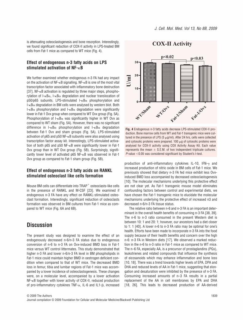
Effect of endogenous n-3 fatty acids on LPS stimulated COX-II activity
Over expression of COX-II stimulates osteoclastogenesis and boneresorption [36] and n-3 FA are reported to down-regulate COX-IIexpression. Therefore, we examined whether lower ratio of n-6/n-3FA due to endogenous production of n-3 FA also have the COX-IIreducing ability which might be one of the mechanisms by which it
© 2009 The AuthorsJournal compilation © 2009 Foundation for Cellular and Molecular Medicine/Blackwell Publishing Ltd
Fig. 1 Endogenous n-3 fatty acidsreduce serum RANKL and TRAP5b.After 20 weeks on experimental dietserum was separated from bloodcollected retro-orbitally from WTand Fat-1 transgenic sham and Ovxmice (seven mice/groups). Serumwas analysed for (A) free serumRANKL and (B) TRAP5b levels usingstandard ELISA kits. Each bar repre-sents the mean � S.E.M. of sevenduplicate samples. Value with differ-ent superscripts are significantly dif-ferent at P � 0.05 by Newman-Keuls’ one way ANOVA with multiplecomparison test.
Boneregions
WT Fat-1P-value2 P-value3
Sham Ovx P-value1 % change Sham Ovx P-value1 % change
DFM 87.02 � 2.91 72.87 � 1.43 0.001* �16.26 89.90 � 1.87 87.23 � 1.90 0.340 �2.97 0.423 0.0001*
PTM 72.17 � 1.25 62.73 � 1.99 0.003* �13.08 72.62 � 1.46 72.83 � 1.78 0.927 0.29 0.820 0.004*
FD 73.97 � 1.62 69.27 � 1.90 0.089 �6.35 80.87 � 2.52 78.63 � 2.23 0.522 �2.77 0.044* 0.01*
TD 44.93 � 0.86 45.40 � 0.42 0.596 1.05 48.63 � 0.84 49.52 � 1.52 0.622 1.83 0.018* 0.026*
L2 62.92 � 3.36 46.70 � 2.47 0.003* �25.78 58.47 � 4.46 47.88 � 6.63 0.215 �18.11 0.444 0.870
L3 60.98 � 3.49 39.27 � 2.96 0.001* �35.60 57.35 � 4.78 51.80 � 4.82 0.433 �9.68 0.553 0.05*
L4 56.23 � 1.72 43.03 � 2.64 0.002* �23.48 50.43 � 2.73 48.82 � 2.75 0.685 �3.19 0.102 0.160
Table 2 Effect of endogenous n-3 fatty acids on bone mineral density (BMD) (mg/cm2) of ovariectomized (Ovx) mice a
a Values are means � S.E.M., n 8.1 Student’s t-test comparing Ovx to sham.2 Student’s t-test comparing wild-type (WT) sham to Fat-1 transgenic (Fat-1) sham.3 Student’s t-test comparing WT Ovx to Fat-1 Ovx.DFM: distal femoral metaphysis; PTM: proximal tibial metaphysis; FD: femoral diaphysis; TD: tibial diaphysis; L2: lumbar vertebra 2; L3: lumbarvertebra 3; L4: lumbar vertebra 4; *P � 0.05 was considered significant.

1838 © 2009 The AuthorsJournal compilation © 2009 Foundation for Cellular and Molecular Medicine/Blackwell Publishing Ltd
Fig. 2 Endogenous n-3 fatty acidsmodulate LPS-stimulated cytokinesproduction. Bone marrow cells fromWT and Fat-1 transgenic mice werecultured in the presence of LPS (5 g/ml). After 24 hrs, culturemedia were collected and analysedfor TNF-�,, IL-1�, IL-6, IL-10 andIFN-� by standard ELISA tech-niques. Each value represents themean � S.E.M. of two independenttriplicate cultures. P-value �0.05was considered significant byStudent’s t-test.
Fig. 3 Endogenous n-3 fatty acids increase LPS-stimulated nitric oxideproduction. Bone marrow cells from WT and Fat-1 transgenic mice werecultured in the presence of LPS (5 g/ml). After 24 hrs, culture mediawere collected and analysed for nitric oxide using quantichrome nitricoxide assay kit. Each value represents the mean � S.E.M. of two inde-pendent triplicate cultures. P-value �0.05 was considered significant byStudent’s t-test.
J. Cell. Mol. Med. Vol 13, No 8B, 2009
1839
is attenuating osteoclastogenesis and bone resorption. Interestingly,we found significant reduction of COX-II activity in LPS-treated BMcells from Fat-1 mice as compared to WT mice (Fig. 4).
Effect of endogenous n-3 fatty acids on LPS stimulated activation of NF-�B
We further examined whether endogenous n-3 FA had any impacton the activation of NF-�B signalling. NF-�B is one of the most vitaltranscription factor associated with inflammatory bone destruction[37]. NF-�B activation is regulated by three major steps, phospho-rylation of I-�B�, I-�B� degradation and nuclear translocation ofp50/p65 subunits. LPS-stimulated I-�B� phosphorylation and I-�B� degradation in BM cells were analysed by western blot. BothI-�B� phosphorylation and I-�B� degradation were significantlylower in Fat-1 Ovx group when compared to WT Ovx group (Fig. 5A).Phosphorylation of I-�B� was significantly higher in WT Ovx ascompared to WT sham (Fig. 5A). However, there was no significantdifference in I-�B� phosphorylation and I-�B� degradationbetween Fat-1 Ovx and sham groups (Fig. 5A). LPS-stimulatedactivation of p65 and p50 NF-�B subunits were also analysed usingtranscription factor assay kit. Interestingly, LPS-stimulated activa-tion of both p65 and p50 NF-�B were significantly lower in Fat-1Ovx group than in WT Ovx group (Fig. 5B). Surprisingly, signifi-cantly lower level of activated p65 NF-�B was observed in Fat-1Ovx group as compared to Fat-1 sham group (Fig. 5B).
Effect of endogenous n-3 fatty acids on RANKLstimulated osteoclast like cells formation
Mouse BM cells can differentiate into TRAP osteoclasts-like cellsin the presence of RANKL and M-CSF [23]. We examined ifendogenous n-3 FA have any effect on RANKL-stimulated osteo-clast formation. Interestingly, significant reduction of osteoclastsformation was observed in BM cultures from Fat-1 mice as com-pared to WT mice (Fig. 6A and 6B).
Discussion
The present study was designed to examine the effect of anendogenously decreased n-6/n-3 FA status due to endogenousconversion of n-6 to n-3 FA on Ovx-induced BMD loss in Fat-1mice versus WT control littermates. This study demonstrated thathigher n-3 FA and lower n-6/n-3 FA level in BM phospholipids inFat-1 mice could maintain higher BMD in oestrogen deficient con-dition when compared to that of WT mice. The decreased BMDloss in femur, tibia and lumbar regions of Fat-1 mice was accom-panied by a lower incidence of osteoclastogenesis. These changeswere, on a molecular level, accompanied by a lower activation NF-�B together with lower activity of COX-II, reduced productionof pro-inflammatory cytokines TNF-�, IL-6 and IL1-�, increased
production of anti-inflammatory cytokines IL-10, IFN-� andincreased production of nitric oxide in BM cells of Fat-1 mice. Wepreviously showed that dietary n-3 FA fed mice exhibit less Ovx-induced BMD loss accompanied by decreased osteoclastogenesis[10]. The molecular mechanisms underlying this protective effectare not clear yet. As Fat-1 transgenic mouse model eliminatesconfounding factors between control and experimental diets, wehave chosen the Fat-1 transgenic mice to elucidate the molecularmechanisms underlying the protective effect of increased n3 anddecreased n-6/n-3 FA tissue status.
The relative ratio between n-6 and n-3 FA is an important deter-minant in the overall health benefits of consuming n-3 FA [38, 39].The n-6 to n-3 ratio consumed in the present Western diet isbetween 10: 1 and 20: 1; however, our ancestors had a diet closerto 1: 1 [40]. A lower n-6 to n-3 FA ratio may be optimal for one’shealth. Efforts have been made to incorporate n-3 FA into the foodsupply because of their health benefits and concern over the highn-6: n-3 FA in Western diets [17]. We observed a marked reduc-tion in the n-6 to n-3 ratio in Fat-1 mice as compared to WT mice.The n–6 FA, especially AA, is a precursor of prostaglandins (PGs),leukotrienes and related compounds that influence the synthesisof eicosanoids which may enhance inflammation and bone loss[10, 18]. There was a trend towards higher levels of EPA, DPA andDHA and reduced levels of AA in Fat-1 mice, suggesting that elon-gation and desaturation were inhibited by the presence of n-3 FA.Consuming increased amounts of n–3 FA results in a partialreplacement of the AA in cell membranes by EPA and DHA [18, 26]. This leads to decreased production of AA-derived
© 2009 The AuthorsJournal compilation © 2009 Foundation for Cellular and Molecular Medicine/Blackwell Publishing Ltd
Fig. 4 Endogenous n-3 fatty acids decrease LPS-stimulated COX-II pro-duction. Bone marrow cells from WT and Fat-1 transgenic mice were cul-tured in the presence of LPS (5 g/ml). After 24 hrs, cells were collectedand cytosolic proteins were prepared. 100 g of cytosolic proteins wereanalysed for COX-II activity using COX Activity Assay Kit. Each value represents the mean � S.E.M. of two independent triplicate cultures.P-value �0.05 was considered significant by Student’s t-test.

1840 © 2009 The AuthorsJournal compilation © 2009 Foundation for Cellular and Molecular Medicine/Blackwell Publishing Ltd
Fig. 5 Endogenous n-3 fatty acidsdecrease LPS-stimulated NF-�B activa-tion. Bone marrow cells from WT andFat-1 transgenic mice were cultured inthe presence of LPS (5 g/ml). After 24 hrs, cells were collected and cytoso-licand nuclear proteins were prepared.(A) 30 g of cytosolic proteins wereanalysed for phosphorylated I-�B-�and total I-�B-� level by western blot.Relative expression of I-�B-�, pI-�B-�and pI-�B-�/total I-�B-� is shown. Theintensity of the bands was determinedby densitometry. (B) 10 g of nuclearproteins were analysed for p65 NF-�Band p50 NF-�B-DNA binding activityusing TransAM Transcription FactorAssay kit. Each value represents themean � S.E.M. of two independenttriplicate cultures. P-value �0.05 wasconsidered significant by Student’s t-test.
Fig. 6 Endogenous n-3 fatty acidssuppress osteoclast differentiation inbone marrow (BM) cell culture. BMcells (1 � 106) from WT and Fat-1mice were cultured in the presenceof sRANKL and macrophage colony-stimulating factor (M-CSF). (A)Formation of TRAP multinucleatedcells in cultures of BM cells isolatedfrom WT and Fat-1 transgenic micein the presence of sRANKL and M-CSF. (B) TRAP multinucleated cellscount in cultures of BM cells isolatedfrom WT and Fat-1 mice..*Significantly different from WT con-trol at P � 0.001 by Student’s t-test.

J. Cell. Mol. Med. Vol 13, No 8B, 2009
1841
pro-inflammatory mediators, i.e. PGE2. Dr. Kang group hasalready established that transgenic overexpression of fat-1 genelowers PGE2 expression in both cells and tissues by reducing theavailability of AA [41–43]. COX-II is the key enzyme responsiblefor the conversion of AA to PGE2 and selective inhibition of COX-IIcan attenuate osteoclastogenesis as well as bone loss in inflam-matory bone diseases [44–47]. It has been further reported that n-3FA can down-regulate COX-II activity and also lower the produc-tion of PGE2 in local tissues [41, 48]. We have also found signifi-cantly reduced COX-II activity in Fat-1 mice. Thus, modification ofmembrane FA composition is one of the mechanisms by which n-3FA may protect osteoporotic BMD loss possibly by reducing thepro-inflammatory mediators.
We and others earlier have described the inhibition of pro-inflammatory cytokines production by n-3 FA in cells and tissues[10, 48–50]. Our present data also show significant reduction ofinflammatory cytokines production in LPS-treated BM cells fromFat-1 mice. This correlates well with previous findings of inflam-matory cytokine suppression by n-3 FA [18, 41, 51, 52]. Our pres-ent data also show a significant increase in the LPS-stimulatedproduction of IL-10 and IFN-� in BM cells. IL-10 has a critical rolein the in vivo regulation of pro-inflammatory cytokine levels andhas been reported to suppress osteoclastogenesis [53]. Further,IFN-� is also known to suppress osteoclastogenesis [35]. In addi-tion, nitric oxide is postulated to play an important role in bonemetabolism, and it is also known that both EPA and DHA enhancenitric oxide formation [3, 54, 55]. We have also detected higherlevel of nitric oxide production in Fat-1 mice. It was reported thatosteoclast formation and bone resorption were inhibited by ele-vated levels of nitric oxide in vivo and in vitro [56–60]. Moreover,high nitric oxide levels and nitric oxide generating compoundsinhibit osteoclast formation and bone resorption and prevent boneloss in severe inflammation or oestrogen-deficient animals [57,61–64]. Further, iNOS deficiency or pharmacological inhibition ofnitric oxide can accelerate osteoclast formation and bone resorp-tion in vivo and in vitro, decrease normal bone mass, exacerbatebone destruction in arthritis or osteoporosis models, interfere withnormal fracture healing and also iNOS knockout mice are knownto exhibit more alveolar bone loss [65–67]. Thus, this might beanother mechanism of the anti-osteoporotic action of n-3 FA.
It is now clearly emerging that n–3 FA might exert their effectson inflammatory gene expression through direct actions on theintracellular signalling pathways. Previous studies have shownthat n–3 FA can down-regulate the activity of NF-�B. It has beenreported that EPA prevents TNF-�-induced activation of NF-�B incultured pancreatic cells [68]. In another study, EPA was reportedto decrease endotoxin-induced activation of NF-�B and mitogenactivated protein kinases (MAPK) in human monocytes [69–71].Previously, we have also reported that EPA and DHA alone or incombination inhibits RANKL-induced NF-�B activation in BM cells[10]. Others have also showed that fish oil can inhibit LPS-induced NF-�B activation in a macrophage cell line [72]. Theseobservations suggest direct effects of n–3 FA on inflammatorygene expression through the inhibition of NF-�B activation. In this
study, we also observed reduced NF-�B activation in BM cells ofFat-1 mice compared to that of WT mice. The role of NF-�B in thepathogenesis of osteoporosis is well documented. Mice null forNF-�B developed osteopetrosis and contain very few osteoclastscompared with normal controls [73]. This indicates the essentialrole of the NF-�B signalling pathway in osteoclast generation andactivation [74, 75]. Activation of p38 MAPK and cJun N terminalkinase (JNK) is required for osteoclastogenesis [23]. The p38MAPK pathway is also known to be involved in the regulation ofbone resorption induced by oestrogen deficiency and selectiveinhibitors of this pathway have potential for prevention of boneloss in post-menopausal osteoporosis [76]. In a very recent study,we have found decreased activation of p38 MAPK and JNK in n-3FA-treated BM cells (data not shown). Therefore, the observedprotection of BMD loss due to oestrogen deficiency by increasedn-3 FA and reduced n-6/n-3 FA tissue status is probably due to thereduced activation of NF-�B, and MAPK signalling pathways.
We previously reported that n-3 FA inhibited TRAP activity andosteoclast formation in primary BM cells [10]. In our presentstudy, we found that higher endogenous n-3 FA and lower endoge-nous n-6/n-3 FA status in BM cells commensurate with lowerRANKL-stimulated BM osteoclastogenesis. Stimulation of osteo-clast differentiation is one of the mechanisms by which oestrogendeficiency causes bone loss [27, 28, 32, 77]. Thus, reduction ofosteoclastogenesis might be one of the mechanisms by which n-3 FA exert its protection against osteoporotic BMD loss.
Our studies on Fat-1 transgenic mice provide compelling evi-dence for the effectiveness of n-3 FA to be a novel dietary FA toprevent post-menopausal osteoporosis. Endogenous conversionof n-6 FA to n-3 FA and maintaining lower ratio of n-6/n-3 FA notonly prevents BMD loss in Ovx mice but also inhibits the inflam-matory response that underlies the disease. As human beings can-not synthesize n-3 FA, they can however lower the n-6/n-3 FA ratioby consuming more n-3 FA, either as supplement or via foodsenriched with n-3 FA, to prevent osteoporotic BMD loss. However,extensive pharmacological evaluation of this approach is requiredto fully determine the effect of long-term use of n-3 FA to preventosteoporosis and related inflammatory bone loss. Very recently,fish oil rich in n-3 FA has been approved by the Food and DrugAdministration (FDA) to use as a prescription drug to treat hightriglyceride level as well as for cardiovascular diseases [78].Because n-3 FA has many other beneficial effects, such as cardio-protective effect [79], anti-carcinogenic effect [41], triglyceridelowering effect [78, 80], as well as protective effect against inflam-matory diseases [19, 81], supplementation with n-3 FA to preventosteoporotic bone loss is a new strategy worth pursuing soon.
Acknowledgements
We thank Kazi Nishu for her technical help. This study was supported byRO1 AG023648.
© 2009 The AuthorsJournal compilation © 2009 Foundation for Cellular and Molecular Medicine/Blackwell Publishing Ltd

1842
References
1. Rosen CJ. Clinical practice. Postmeno -pausal osteoporosis. N Engl J Med. 2005;353: 595–603.
2. Lacey JV Jr, Mink PJ, Lubin JH, et al.Menopausal hormone replacement therapyand risk of ovarian cancer. Jama. 2002;288: 334–41.
3. Das UN. Essential fatty acids and osteo-porosis. Nutrition. 2000; 16: 386–90.
4. Kruger MC, Coetzer H, de Winter R, et al.Calcium, gamma-linolenic acid and eicos-apentaenoic acid supplementation in senileosteoporosis. Aging. 1998; 10: 385–94.
5. Watkins BA, Lippman HE, Le BouteillerL, et al. Bioactive fatty acids: role in bonebiology and bone cell function. Prog LipidRes. 2001; 40: 125–48.
6. Sakaguchi K, Morita I, Murota S.Eicosapentaenoic acid inhibits bone lossdue to ovariectomy in rats. ProstaglandinsLeukot Essent Fatty Acids. 1994; 50: 81–4.
7. Watkins BA, Li Y, Lippman HE, et al.Modulatory effect of omega-3 polyunsatu-rated fatty acids on osteoblast functionand bone metabolism. ProstaglandinsLeukot Essent Fatty Acids. 2003; 68:387–98.
8. Kruger MC, Horrobin DF. Calcium metab-olism, osteoporosis and essential fattyacids: a review. Prog Lipid Res. 1997; 36:131–51.
9. Schlemmer CK, Coetzer H, Claassen N,et al. Oestrogen and essential fatty acidsupplementation corrects bone loss due toovariectomy in the female Sprague Dawleyrat. Prostaglandins Leukot Essent FattyAcids. 1999; 61: 381–90.
10. Sun D, Krishnan A, Zaman K, et al.Dietary n-3 fatty acids decrease osteoclas-togenesis and loss of bone mass inovariectomized mice. J Bone Miner Res.2003; 18: 1206–16.
11. Watkins BA, Reinwald S, Li Y, et al.Protective actions of soy isoflavones andn-3 PUFAs on bone mass in ovariec-tomized rats. J Nutr Biochem. 2005; 16:479–88.
12. Heaney RP, Carey R, Harkness L. Rolesof vitamin D, n-3 polyunsaturated fattyacid, and soy isoflavones in bone health. JAm Diet Assoc. 2005; 105: 1700–2.
13. Reinwald S, Li Y, Moriguchi T, et al.Repletion with (n-3) fatty acids reversesbone structural deficits in (n-3)-deficientrats. J Nutr. 2004; 134: 388–94.
14. Weiler HA, Fitzpatrick-Wong SC.Modulation of essential (n-6): (n-3) fatty
acid ratios alters fatty acid status but notbone mass in piglets. J Nutr. 2002; 132:2667–72.
15. Connor WE. Importance of n-3 fatty acidsin health and disease. Am J Clin Nutr.2000; 71: 171S–5S.
16. Fernandes G. Attenuation of osteoporosisby n-3 lipids and soy protein. In: Holick MF(editor). Nutrition and bone health. NewJersey: Humana Press Inc.; 2004. pp.575–92.
17. Kang JX, Wang J, Wu L, et al. Transgenicmice: fat-1 mice convert n-6 to n-3 fattyacids. Nature. 2004; 427: 504.
18. Hudert CA, Weylandt KH, Lu Y, et al.Transgenic mice rich in endogenousomega-3 fatty acids are protected fromcolitis. Proc Natl Acad Sci USA. 2006; 103:11276–81.
19. Bhattacharya A, Chandrasekar B,Rahman MM, et al. Inhibition of inflam-matory response in transgenic fat-1 miceon a calorie-restricted diet. BiochemBiophys Res Commun. 2006; 349:925–30.
20. Rahman MM, Bhattacharya A, Banu J, et al. Conjugated linoleic acid protectsagainst age-associated bone loss inC57BL/6 female mice. J Nutr Biochem.2007; 18: 467–74.
21. Bhattacharya A, Rahman M, Banu J, et al. Inhibition of osteoporosis in autoim-mune disease prone MRL/Mpj-Fas(lpr)mice by N-3 fatty acids. J Am Coll Nutr.2005; 24: 200–9.
22. Nagy TR, Clair AL. Precision and accuracyof dual-energy X-ray absorptiometry fordetermining in vivo body composition ofmice. Obes Res. 2000; 8: 392–8.
23. Rahman MM, Kukita A, Kukita T, et al.Two histone deacetylase inhibitors, tricho-statin A and sodium butyrate, suppressdifferentiation into osteoclasts but not intomacrophages. Blood. 2003; 101: 3451–9.
24. Folch J, Lees M, Sloane Stanley GH. Asimple method for the isolation and purifi-cation of total lipides from animal tissues.J Biol Chem. 1957; 226: 497–509.
25. Bhattacharya A, Sun D, Rahman M, et al.Different ratios of eicosapentaenoic anddocosahexaenoic omega-3 fatty acids incommercial fish oils differentially alter pro-inflammatory cytokines in peritonealmacrophages from C57BL/6 female mice.J Nutr Biochem. 2007; 18: 23–30.
26. Kang JX. Fat-1 transgenic mice: a newmodel for omega-3 research. Prosta -
glandins Leukot Essent Fatty Acids. 2007;77: 263–7.
27. Lacey DL, Timms E, Tan HL, et al.Osteoprotegerin ligand is a cytokine thatregulates osteoclast differentiation andactivation. Cell. 1998; 93: 165–76.
28. Kong YY, Yoshida H, Sarosi I, et al. OPGLis a key regulator of osteoclastogenesis,lymphocyte development and lymph-nodeorganogenesis. Nature. 1999; 397:315–23.
29. Rahman MM, Bhattacharya A,Fernandes G. Docosahexaenoic acid ismore potent inhibitor of osteoclast differ-entiation in RAW 264.7 cells than eicos-apentaenoic acid. J Cell Physiol. 2008;214: 201–9.
30. Jilka RL, Hangoc G, Girasole G, et al.Increased osteoclast development afterestrogen loss: mediation by interleukin-6.Science. 1992; 257: 88–91.
31. Manolagas SC. Role of cytokines in boneresorption. Bone. 1995; 17: 63S-7S.
32. Manolagas SC, Jilka RL. Bone marrow,cytokines, and bone remodeling. Emerginginsights into the pathophysiology of osteo-porosis. N Engl J Med. 1995; 332: 305–11.
33. Liu D, Yao S, Wise GE. Effect of inter-leukin-10 on gene expression of osteoclas-togenic regulatory molecules in the ratdental follicle. Eur J Oral Sci. 2006; 114:42–9.
34. Owens JM, Gallagher AC, Chambers TJ.IL-10 modulates formation of osteoclastsin murine hemopoietic cultures. J Immunol. 1996; 157: 936–40.
35. Takayanagi H, Ogasawara K, Hida S, et al. T-cell-mediated regulation of osteo-clastogenesis by signalling cross-talkbetween RANKL and IFN-gamma. Nature.2000; 408: 600–5.
36. Kasukawa Y, Miyakoshi N, SrivastavaAK, et al. The selective cyclooxygenase-2inhibitor celecoxib reduces bone resorp-tion, but not bone formation, in ovariec-tomized mice in vivo. Tohoku J Exp Med.2007; 211: 275–83.
37. Jimi E, Aoki K, Saito H, et al. Selectiveinhibition of NF-kappa B blocks osteoclas-togenesis and prevents inflammatory bonedestruction in vivo. Nat Med. 2004; 10:617–24.
38. Gago-Dominguez M, Yuan JM, Sun CL,et al. Opposing effects of dietary n-3 andn-6 fatty acids on mammary carcinogene-sis: The Singapore Chinese Health Study.Br J Cancer. 2003; 89: 1686–92.
© 2009 The AuthorsJournal compilation © 2009 Foundation for Cellular and Molecular Medicine/Blackwell Publishing Ltd
J. Cell. Mol. Med. Vol 13, No 8B, 2009
1843
39. Ma DW, Ngo V, Huot PS, et al. N-3polyunsaturated fatty acids endogenouslysynthesized in fat-1 mice are enriched inthe mammary gland. Lipids. 2006; 41:35–9.
40. Leaf A, Weber PC. A new era for sciencein nutrition. Am J Clin Nutr. 1987; 45:S1048–S53.
41. Jia Q, Lupton JR, Smith R, et al. Reducedcolitis-associated colon cancer in Fat-1 (n-3fatty acid desaturase) transgenic mice.Cancer Res. 2008; 68: 3985–91.
42. Kang JX. From fat to fat-1: a tale ofomega-3 fatty acids. J Membr Biol. 2005;206: 165–72.
43. Kang ZB, Ge Y, Chen Z, et al. Adenoviralgene transfer of Caenorhabditis elegansn–3 fatty acid desaturase optimizes fattyacid composition in mammalian cells.Proc Natl Acad Sci USA. 2001; 98:4050–4.
44. Kaneko H, Mehrotra M, Alander C, et al.Effects of prostaglandin E2 andlipopolysaccharide on osteoclastogenesisin RAW 264.7 cells. Prostaglandins LeukotEssent Fatty Acids. 2007; 77: 181–6.
45. Coon D, Gulati A, Cowan C, et al. The roleof cyclooxygenase-2 (COX-2) in inflamma-tory bone resorption. J Endod. 2007; 33:432–6.
46. Richards JB, Joseph L, Schwartzman K,et al. The effect of cyclooxygenase-2inhibitors on bone mineral density: resultsfrom the Canadian MulticentreOsteoporosis Study. Osteoporos Int. 2006;17: 1410–9.
47. Kesavalu L, Bakthavatchalu V, RahmanMM, et al. Omega-3 fatty acid regulatesinflammatory cytokine/mediator messen-ger RNA expression in Porphyromonasgingivalis-induced experimental periodon-tal disease. Oral Microbiol Immunol. 2007;22: 232–9.
48. Kelley VE, Ferretti A, Izui S, et al. A fishoil diet rich in eicosapentaenoic acidreduces cyclooxygenase metabolites, andsuppresses lupus in MRL-lpr mice. J Immunol. 1985; 134: 1914–9.
49. Fernandes G, Bysani C, Venkatraman JT,et al. Increased TGF-beta and decreasedoncogene expression by omega-3 fattyacids in the spleen delays onset of autoim-mune disease in B/W mice. J Immunol.1994; 152: 5979–87.
50. Fernandes G. Effects of calorie restrictionand omega-3 fatty acids on autoimmunityand aging. Nutr Rev. 1995; 53: S72–7; dis-cussion S77–9.
51. Endres S, Ghorbani R, Kelley VE, et al.The effect of dietary supplementation with
n-3 polyunsaturated fatty acids on the syn-thesis of interleukin-1 and tumor necrosisfactor by mononuclear cells. N Engl J Med.1989; 320: 265–71.
52. Schmocker C, Weylandt KH, Kahlke L, et al. Omega-3 fatty acids alleviate chemi-cally induced acute hepatitis by suppressionof cytokines. Hepatology. 2007; 45: 864–9.
53. Evans KE, Fox SW. Interleukin-10 inhibitsosteoclastogenesis by reducing NFATc1expression and preventing its translocationto the nucleus. BMC Cell Biol. 2007; 8: 4.
54. Das UN. Nitric oxide as the mediator of theantiosteoporotic actions of estrogen,statins, and essential fatty acids. Exp BiolMed. 2002; 227: 88–93.
55. Hirafuji M, Machida T, Tsunoda M, et al.Docosahexaenoic acid potentiates inter-leukin-1beta induction of nitric oxide syn-thase through mechanism involving p44/42MAPK activation in rat vascular smoothmuscle cells. Br J Pharmacol. 2002; 136:613–9.
56. MacIntyre I, Zaidi M, Alam AS, et al.Osteoclastic inhibition: an action of nitricoxide not mediated by cyclic GMP. ProcNatl Acad Sci USA. 1991; 88: 2936–40.
57. Kasten TP, Collin-Osdoby P, Patel N, et al. Potentiation of osteoclast bone-resorption activity by inhibition of nitricoxide synthase. Proc Natl Acad Sci USA.1994; 91: 3569–73.
58. Brandi ML, Hukkanen M, Umeda T, et al.Bidirectional regulation of osteoclast func-tion by nitric oxide synthase isoforms.Proc Natl Acad Sci USA. 1995; 92:2954–8.
59. Collin-Osdoby P, Rothe L, Bekker S, et al. Decreased nitric oxide levels stimu-late osteoclastogenesis and bone resorp-tion both in vitro and in vivo on the chickchorioallantoic membrane in associationwith neoangiogenesis. J Bone Miner Res.2000; 15: 474–88.
60. Zheng H, Yu X, Collin-Osdoby P, et al.RANKL stimulates inducible nitric-oxidesynthase expression and nitric oxide pro-duction in developing osteoclasts. Anautocrine negative feedback mechanismtriggered by RANKL-induced interferon-beta via NF-kappaB that restrains osteo-clastogenesis and bone resorption. J BiolChem. 2006; 281: 15809–20.
61. van’t Hof RJ, Ralston SH. Cytokine-induced nitric oxide inhibits bone resorptionby inducing apoptosis of osteoclast progen-itors and suppressing osteoclast activity. J Bone Miner Res. 1997; 12: 1797–804.
62. van’t Hof RJ, Ralston SH. Nitric oxide andbone. Immunology. 2001; 103: 255–61.
63. van’t Hof RJ, Armour KJ, Smith LM, et al.Requirement of the inducible nitric oxidesynthase pathway for IL-1-induced osteo-clastic bone resorption. Proc Natl Acad SciUSA. 2000; 97: 7993–8.
64. Jamal SA, Browner WS, Bauer DC, et al.Intermittent use of nitrates increases bonemineral density: the study of osteoporoticfractures. J Bone Miner Res. 1998; 13:1755–9.
65. Diwan AD, Wang MX, Jang D, et al. Nitricoxide modulates fracture healing. J BoneMiner Res. 2000; 15: 342–51.
66. Veihelmann A, Landes J, Hofbauer A, et al. Exacerbation of antigen-inducedarthritis in inducible nitric oxide synthase-deficient mice. Arthritis Rheum. 2001; 44:1420–7.
67. McCartney-Francis NL, Song X, MizelDE, et al. Selective inhibition of induciblenitric oxide synthase exacerbates erosivejoint disease. J Immunol. 2001; 166:2734–40.
68. Cuzzocrea S, Mazzon E, Dugo L, et al.Inducible nitric oxide synthase mediatesbone loss in ovariectomized mice.Endocrinology. 2003; 144: 1098–107.
69. Lo CJ, Chiu KC, Fu M, et al. Fish oildecreases macrophage tumor necrosisfactor gene transcription by altering the NFkappa B activity. J Surg Res. 1999; 82:216–21.
70. Novak TE, Babcock TA, Jho DH, et al. NF-kappa B inhibition by omega-3fatty acids modulates LPS-stimulatedmacrophage TNF-alpha transcription.Am J Physiol Lung Cell Mol Physiol.2003; 284: L84–9.
71. Zhao Y, Joshi-Barve S, Barve S, et al.Eicosapentaenoic acid prevents LPS-induced TNF-alpha expression by prevent-ing NF-kappaB activation. J Am Coll Nutr.2004; 23: 71–8.
72. Camandola S, Leonarduzzi G, Musso T,et al. Nuclear factor kB is activated byarachidonic acid but not by eicosapen-taenoic acid. Biochem Biophys ResCommun. 1996; 229: 643–7.
73. Iotsova V, Caamano J, Loy J, et al.Osteopetrosis in mice lacking NF-kappaB1and NF-kappaB2. Nat Med. 1997; 3:1285–9.
74. Wong BR, Josien R, Lee SY, et al. TheTRAF family of signal transducers medi-ates NF-kappaB activation by the TRANCEreceptor. J Biol Chem. 1998; 273:28355–9.
75. Darnay BG, Haridas V, Ni J, et al.Characterization of the intracellulardomain of receptor activator of NF-kappaB
© 2009 The AuthorsJournal compilation © 2009 Foundation for Cellular and Molecular Medicine/Blackwell Publishing Ltd
1844
(RANK). Interaction with tumor necrosisfactor receptor-associated factors andactivation of NF-kappab and c-Jun N-ter-minal kinase. J Biol Chem. 1998; 273:20551–5.
76. Caverzasio J, Higgins L, Ammann P.Prevention of Trabecular Bone LossInduced by Estrogen Deficiency by aSelective p38alpha Inhibitor. J Bone MinerRes. 2008; 23: 1389–97.
77. Pacifici R. Estrogen, cytokines, andpathogenesis of postmenopausal osteo-
porosis. J Bone Miner Res. 1996; 11:1043–51.
78. Bays HE, Tighe AP, Sadovsky R, et al.Prescription omega-3 fatty acids and theirlipid effects: physiologic mechanisms ofaction and clinical implications. ExpertRev Cardiovasc Ther. 2008; 6: 391–409.
79. Wang C, Harris WS, Chung M, et al. n-3Fatty acids from fish or fish-oil supple-ments, but not alpha-linolenic acid, ben-efit cardiovascular disease outcomes inprimary- and secondary-prevention stud-
ies: a systematic review. Am J Clin Nutr.2006; 84: 5–17.
80. Qi K, Fan C, Jiang J, et al. Omega-3 fattyacid containing diets decrease plasmatriglyceride concentrations in mice byreducing endogenous triglyceride synthe-sis and enhancing the blood clearance oftriglyceride-rich particles. Clin Nutr. 2008;27: 424–30.
81. Calder PC. Immunomodulation by omega-3 fatty acids. Prostaglandins LeukotEssent Fatty Acids. 2007; 77: 327–35.
© 2009 The AuthorsJournal compilation © 2009 Foundation for Cellular and Molecular Medicine/Blackwell Publishing Ltd