Endocrinology Interpretation
382
-
Upload
hernanda-adi-purwangga -
Category
Documents
-
view
634 -
download
4
description
Endocrinology Interpretation
Transcript of Endocrinology Interpretation
Test SelectionandInterpretation
The Quest Diagnostics Manual
Endocrinology
Fourth Edition

Test SelectionandInterpretation
The Quest Diagnostics Manual
Endocrinology
Fourth Edition
Edited by:
Delbert A. Fisher, MDSenior Science Officer Quest Diagnostics Nichols InstituteProfessor Emeritus, Pediatrics and MedicineUCLA School of Medicine
Consulting Editors:
Wael Salameh, MD, FACPMedical Director, Endocrinology/MetabolismQuest Diagnostics Nichols InstituteSan Juan Capistrano, CA
Associate Clinical Professor of Medicine,David Geffen School of Medicine at UCLA
Richard W. Furlanetto, MD, PhDMedical Director, Endocrinology/MetabolismQuest Diagnostics Nichols InstituteChantilly, VA
©2007 Quest Diagnostics Incorporated. All rights reserved.Fourth EditionPrinted in the United States of America
Quest, Quest Diagnostics, the associated logo, Nichols Institute, and all associated Quest Diagnostics marks are the trademarks of Quest Diagnostics.
All third party marks − ®' and ™' − are the property of their respective owners.
No part of this publication may be reproduced or transmitted in any form or byany means, electronic or mechanical, including photocopy, recording, andinformation storage and retrieval system, without permission in writing from thepublisher. Address inquiries to the Medical Information Department, QuestDiagnostics Nichols Institute, 33608 Ortega Highway, San Juan Capistrano, CA92690-6130.
Previous editions copyrighted in 1996, 1998, and 2004.Re-order # IG1984
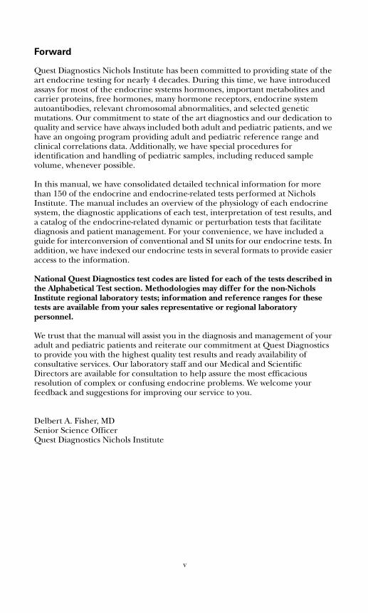
Forward
Quest Diagnostics Nichols Institute has been committed to providing state of the art endocrine testing for nearly 4 decades. During this time, we have introduced assays for most of the endocrine systems hormones, important metabolites and carrier proteins, free hormones, many hormone receptors, endocrine system autoantibodies, relevant chromosomal abnormalities, and selected genetic mutations. Our commitment to state of the art diagnostics and our dedication to quality and service have always included both adult and pediatric patients, and we have an ongoing program providing adult and pediatric reference range and clinical correlations data. Additionally, we have special procedures for identification and handling of pediatric samples, including reduced sample volume, whenever possible.
In this manual, we have consolidated detailed technical information for more than 150 of the endocrine and endocrine-related tests performed at Nichols Institute. The manual includes an overview of the physiology of each endocrine system, the diagnostic applications of each test, interpretation of test results, and a catalog of the endocrine-related dynamic or perturbation tests that facilitate diagnosis and patient management. For your convenience, we have included a guide for interconversion of conventional and SI units for our endocrine tests. In addition, we have indexed our endocrine tests in several formats to provide easier access to the information.
National Quest Diagnostics test codes are listed for each of the tests described in the Alphabetical Test section. Methodologies may differ for the non-Nichols Institute regional laboratory tests; information and reference ranges for these tests are available from your sales representative or regional laboratory personnel.
We trust that the manual will assist you in the diagnosis and management of your adult and pediatric patients and reiterate our commitment at Quest Diagnostics to provide you with the highest quality test results and ready availability of consultative services. Our laboratory staff and our Medical and Scientific Directors are available for consultation to help assure the most efficacious resolution of complex or confusing endocrine problems. We welcome your feedback and suggestions for improving our service to you.
Delbert A. Fisher, MDSenior Science OfficerQuest Diagnostics Nichols Institute
v
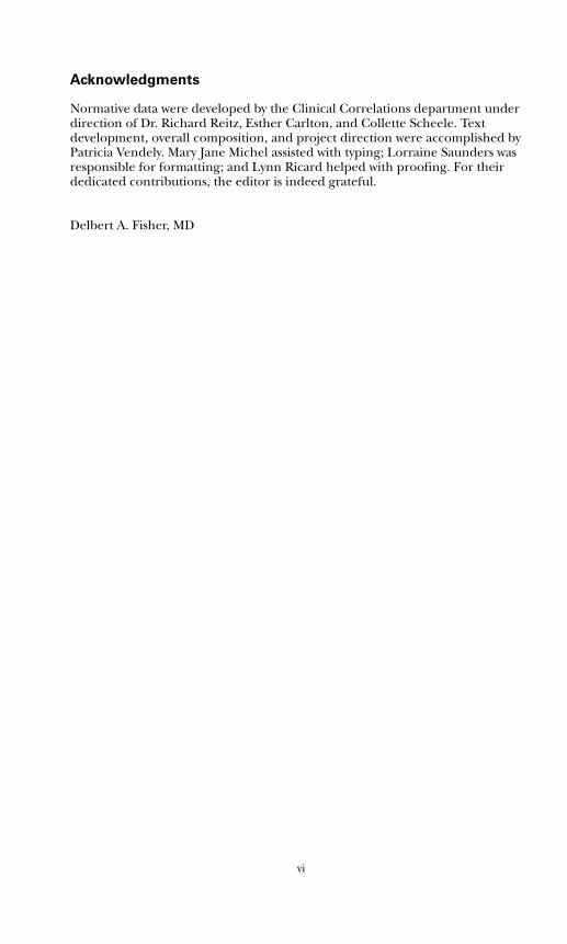
Acknowledgments
Normative data were developed by the Clinical Correlations department under direction of Dr. Richard Reitz, Esther Carlton, and Collette Scheele. Text development, overall composition, and project direction were accomplished by Patricia Vendely. Mary Jane Michel assisted with typing; Lorraine Saunders was responsible for formatting; and Lynn Ricard helped with proofing. For their dedicated contributions, the editor is indeed grateful.
Delbert A. Fisher, MD
vi
Contents
Page
Forward ....................................................................................................................v
Acknowledgments ..............................................................................................vi
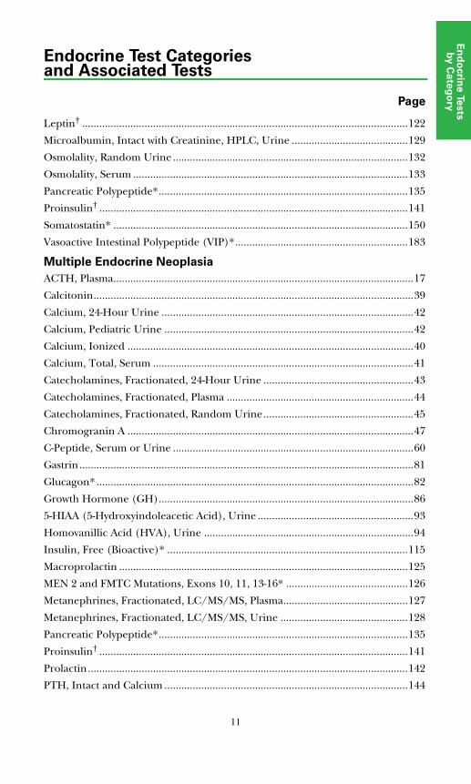
Endocrine Test Categories and Associated Tests
Adrenocortical Function ........................................................................................ 3Cardiovascular/Lipids ............................................................................................ 4Catecholamines....................................................................................................... 5Congenital Adrenal Hyperplasia (CAH)............................................................... 5Endocrine Autoimmunity....................................................................................... 5Fluid, Electrolyte, and Renal .................................................................................. 6Genetic (Biochemical and Cytogenetic) ............................................................... 7Gonadal Function ................................................................................................... 8Growth and Growth Hormone............................................................................... 9Hypothalamic and Pituitary Function ................................................................... 9Metabolic (Including Diabetes Mellitus) and Gastrointestinal Disorders......... 10Multiple Endocrine Neoplasia ............................................................................. 11Parathyroid and Mineral Metabolism.................................................................. 12Thyroid Function .................................................................................................. 12
Additional Diagnostic Profiles.............................................................................. 13Perturbation Tests ................................................................................................. 14
Alphabetical Test Section
See Test Index for list of tests included in this section....................................... 15
Test Application and Interpretation
Preface................................................................................................................. 189Disorders of Adrenal Function
Background Physiology ................................................................................. 190Adrenal Insufficiency ..................................................................................... 193Adrenal Biosynthetic Enzyme Deficiencies .................................................. 193
Classical Congenital Adrenal Hyperplasia (CAH) ................................... 193Nonclassical Disease (Partial Deficiency in Steroid Biosynthesis) .......... 205Cushing's Syndrome ................................................................................... 206
Renin-Angiotensin – Aldosterone System (RAAS) ....................................... 207Background Physiology............................................................................... 207Assessment of Disorders of the RAAS ........................................................ 208
Primary Aldosteronism ........................................................................... 210Renovascular Hypertension.................................................................... 210Dexamethasone-suppressible Hyperaldosteronism .............................. 211Pseudohypoaldosteronism...................................................................... 211
Adrenal Diagnosis via Urine GC/MS Metabolite Analysis .......................... 211
vii
Contents
Page
References ....................................................................................................... 215
Disorders of Anterior Pituitary FunctionBackground Physiology .................................................................................. 217Hypopituitarism .............................................................................................. 218Pituitary Adenomas......................................................................................... 220
General Features ......................................................................................... 220Prolactinoma ............................................................................................... 221Acromegaly .................................................................................................. 222Cushing's Disease ........................................................................................ 223Thyrotropinoma.......................................................................................... 223Gonadotropinoma ...................................................................................... 223
References ....................................................................................................... 224
Disorders of Calcium and Bone MetabolismBackground Physiology ................................................................................. 225Laboratory Assessment of Disorders of Calcium Metabolism ..................... 226
Evaluation of Hypercalcemia ..................................................................... 228Evaluation of Hypocalcemia....................................................................... 229
Vitamin D Deficiency...................................................................................... 230Vitamin D Metabolism in Renal Failure ........................................................ 230Metabolic Bone Disease ................................................................................. 232
Clinical Application of Bone Markers........................................................ 233 Diagnosis of Bone Disorders ................................................................. 233
Diagnosis of Osteoporosis ...................................................................... 236Efficacy of Therapy ................................................................................. 236Follow-up to Therapy .............................................................................. 236
References ....................................................................................................... 236
Disorders of Carbohydrate MetabolismBackground Physiology .................................................................................. 239Hyperglycemic Disorders ............................................................................... 239
Type 1 Diabetes ........................................................................................... 240Maturity Onset Diabetes of the Young (MODY) ....................................... 243Type 2 Diabetes ........................................................................................... 243Diagnosis of Diabetes Mellitus.................................................................... 244Preventing and Managing Complications ................................................. 244
Hypoglycemic Disorders................................................................................. 248Diagnostic Approach .................................................................................. 249
References ....................................................................................................... 251
Disorders of Gonadal FunctionBackground Physiology .................................................................................. 255
viii

Contents
Page
Development of the Hypothalamic–Pituitary Axis and Isolated GnRH Deficiency ........................................................................................ 255Regulation of Pubertal Onset ..................................................................... 256Regulation of the Mature Hypothalamic–Pituitary–Gonadal Axis........... 256Gonadal Determination and Differentiation ............................................ 259
Measurement of Pituitary–Gonadal Axis Hormones.................................... 261Luteinizing Hormone ................................................................................. 261Follicle Stimulating Hormone.................................................................... 261Estrogens...................................................................................................... 261Testosterone................................................................................................. 262Sex Hormone Binding Globulin ................................................................ 263Inhibin ......................................................................................................... 263
Abnormalities of Sexual Determination and Differentiation ...................... 263Disorders of Sexual Maturation ..................................................................... 264
Normal Puberty Staging............................................................................. 264Timing of Physical Changes of Puberty ..................................................... 264Normal Variants of Puberty ........................................................................ 266Precocious Puberty...................................................................................... 267Delayed or Absent Puberty ......................................................................... 267
Disorders of Testicular Function.................................................................... 269Developmental Disorders ........................................................................... 269Disorders of the Germinal Cells ................................................................. 269Testicular Aging........................................................................................... 271
Disorders of Ovarian Function....................................................................... 272Premature Ovarian Failure ......................................................................... 273
Polycystic Ovarian Syndrome ......................................................................... 274Assessment of Functional Gonadal Tumors .................................................. 275References ....................................................................................................... 276
Disorders of the Growth Hormone–Insulin-like Growth Factor AxisBackground Physiology ................................................................................. 279Growth Hormone Deficiency in Children..................................................... 282Growth Hormone Deficiency in Adults ......................................................... 283Growth Hormone Excess ............................................................................... 284Laboratory Assessment of the Growth Hormone – IGF Axis ....................... 284
Insulin-like Growth Factors and Binding Proteins ................................... 285Growth Hormone Dynamic Testing ........................................................... 289
References ...................................................................................................... 290
Disorders of Thyroid FunctionBackground Physiology .................................................................................. 293Differential Diagnosis of Thyroid Disorders ................................................. 295
Primary Hypothyroidism............................................................................. 298
ix
Contents
Page
Hyperthyroidism ......................................................................................... 298Hypothalamic-Pituitary Hypothyroidism................................................... 299Inappropriate TSH Secretion..................................................................... 299Nonthyroidal Illness.................................................................................... 299
Autoimmune Thyroid Disease ....................................................................... 300Thyroid Dysfunction in Infancy and Early Childhood ................................. 304Thyroid Neoplasia........................................................................................... 304Thyroid Protein-Binding Abnormalities........................................................ 308References ....................................................................................................... 308
Fluid & Electrolyte DisordersBackground Physiology .................................................................................. 311Primary Disorders ........................................................................................... 312Secondary Disorders ....................................................................................... 314Disorders of Potassium ...................................................................................316References .......................................................................................................317
Pheochromocytoma, Medullary Thyroid Carcinoma, and Multiple Endocrine Neoplasia
Pheochromoctyoma........................................................................................319Diagnosis of Pheochromocytoma...............................................................320Genetic Testing for Pheochromocytoma...................................................321
Medullary Thyroid Carcinoma (MTC)..........................................................322Hereditary MTC Diagnosis and Management...........................................323Sporadic MTC Diagnosis and Management..............................................324
MEN Type 1.....................................................................................................325References .......................................................................................................327
Dynamic TestsAdrenal ............................................................................................................329
ACTH Stimulation Test, Standard..............................................................329ACTH Stimulation Test, Prolonged ...........................................................329ACTH Stimulation Test, Low Dose ............................................................330Aldosterone Suppression Test ....................................................................330CRH Stimulation Test .................................................................................330CRH Stimulation Test, Adrenal Venous Sampling....................................331CRH Stimulation Test, Petrosal Venous Sampling....................................331Dexamethasone Suppression Tests ............................................................332
Carbohydrate Metabolic.................................................................................332Glucose Tolerance Test ...............................................................................332Pancreatic Hormone Response Test ..........................................................333Prolonged Fasting Test................................................................................334
x

Contents
Page
Gonadal ...........................................................................................................335GnRH Stimulation Test...............................................................................335
Growth Hormone ..........................................................................................336Glucose Suppression Test ...........................................................................336Growth Hormone Stimulation Test ...........................................................336
Multiple Endocrine Neoplasia .......................................................................337Secretin Stimulation Test............................................................................337
Posterior Pituitary ...........................................................................................337Combined Anterior Pituitary (CAP) Test ..................................................337Water Deprivation Test ...............................................................................338
Thyroid ............................................................................................................338Calcitonin-Calcium Stimulation Test .........................................................338Thyrotropin Releasing Hormone (TRH) Stimulation Test......................339
References .......................................................................................................339
SI Units ................................................................................................................345
Test Index ...........................................................................................................353
Subject Index ....................................................................................................359
xi

xii
Contents
Page
Endocrine Test Categories
andAssociated Tests

Endocrine Test Categoriesand Associated Tests
En
do
crin
e Te
sts
by
Ca
teg
ory
Page
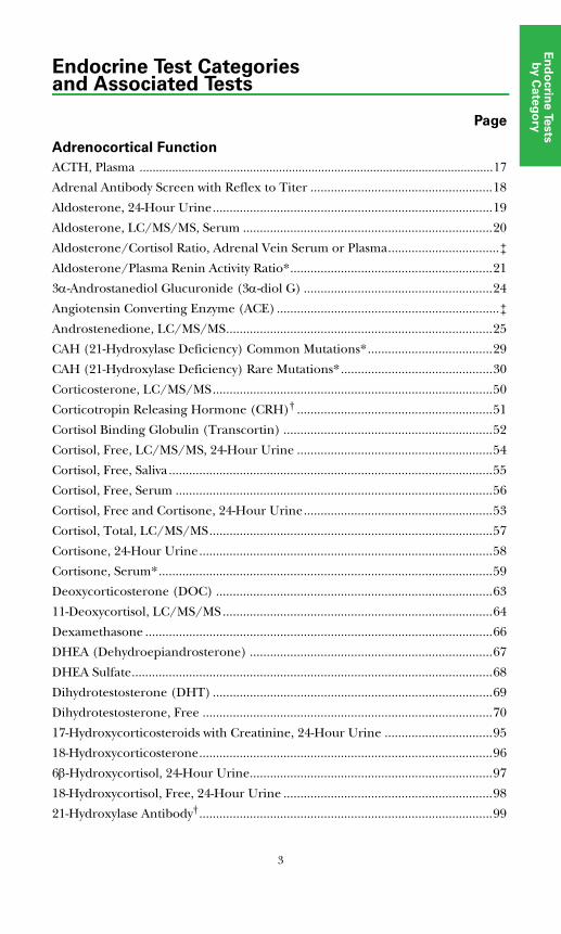
Adrenocortical Function
ACTH, Plasma .............................................................................................................17Adrenal Antibody Screen with Reflex to Titer ......................................................18
Aldosterone, 24-Hour Urine...................................................................................19
Aldosterone, LC/MS/MS, Serum ..........................................................................20
Aldosterone/Cortisol Ratio, Adrenal Vein Serum or Plasma.................................‡
Aldosterone/Plasma Renin Activity Ratio*............................................................21
3α-Androstanediol Glucuronide (3α-diol G) ........................................................24
Angiotensin Converting Enzyme (ACE) ..................................................................‡
Androstenedione, LC/MS/MS...............................................................................25
CAH (21-Hydroxylase Deficiency) Common Mutations*.....................................29
CAH (21-Hydroxylase Deficiency) Rare Mutations*.............................................30
Corticosterone, LC/MS/MS...................................................................................50
Corticotropin Releasing Hormone (CRH)† ..........................................................51
Cortisol Binding Globulin (Transcortin) ..............................................................52
Cortisol, Free, LC/MS/MS, 24-Hour Urine ..........................................................54
Cortisol, Free, Saliva ................................................................................................55
Cortisol, Free, Serum ..............................................................................................56
Cortisol, Free and Cortisone, 24-Hour Urine........................................................53
Cortisol, Total, LC/MS/MS....................................................................................57
Cortisone, 24-Hour Urine.......................................................................................58
Cortisone, Serum*...................................................................................................59
Deoxycorticosterone (DOC) ..................................................................................63
11-Deoxycortisol, LC/MS/MS ................................................................................64
Dexamethasone .......................................................................................................66
DHEA (Dehydroepiandrosterone) ........................................................................67
DHEA Sulfate...........................................................................................................68
Dihydrotestosterone (DHT) ...................................................................................69
Dihydrotestosterone, Free ......................................................................................70
17-Hydroxycorticosteroids with Creatinine, 24-Hour Urine ................................95
18-Hydroxycorticosterone.......................................................................................96
6β-Hydroxycortisol, 24-Hour Urine........................................................................97
18-Hydroxycortisol, Free, 24-Hour Urine ..............................................................98
21-Hydroxylase Antibody†.......................................................................................99
3
Endocrine Test Categoriesand Associated Tests
Page
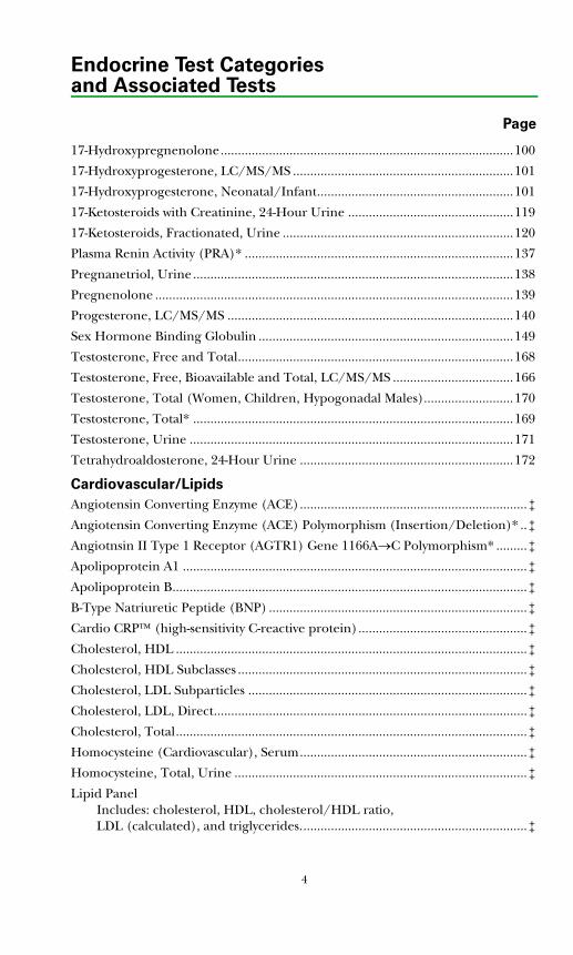
17-Hydroxypregnenolone.....................................................................................100
17-Hydroxyprogesterone, LC/MS/MS ................................................................101
17-Hydroxyprogesterone, Neonatal/Infant.........................................................101
17-Ketosteroids with Creatinine, 24-Hour Urine ................................................119
17-Ketosteroids, Fractionated, Urine ...................................................................120
Plasma Renin Activity (PRA)* ..............................................................................137
Pregnanetriol, Urine .............................................................................................138
Pregnenolone ........................................................................................................139
Progesterone, LC/MS/MS ...................................................................................140
Sex Hormone Binding Globulin ..........................................................................149
Testosterone, Free and Total................................................................................168
Testosterone, Free, Bioavailable and Total, LC/MS/MS ...................................166
Testosterone, Total (Women, Children, Hypogonadal Males)..........................170
Testosterone, Total* .............................................................................................169
Testosterone, Urine ..............................................................................................171
Tetrahydroaldosterone, 24-Hour Urine ..............................................................172
Cardiovascular/Lipids
Angiotensin Converting Enzyme (ACE)..................................................................‡
Angiotensin Converting Enzyme (ACE) Polymorphism (Insertion/Deletion)* ..‡
Angiotnsin II Type 1 Receptor (AGTR1) Gene 1166A→C Polymorphism* .........‡
Apolipoprotein A1 ....................................................................................................‡
Apolipoprotein B.......................................................................................................‡
B-Type Natriuretic Peptide (BNP) ...........................................................................‡
Cardio CRP™ (high-sensitivity C-reactive protein).................................................‡
Cholesterol, HDL ......................................................................................................‡
Cholesterol, HDL Subclasses ....................................................................................‡
Cholesterol, LDL Subparticles .................................................................................‡
Cholesterol, LDL, Direct...........................................................................................‡
Cholesterol, Total......................................................................................................‡
Homocysteine (Cardiovascular), Serum..................................................................‡
Homocysteine, Total, Urine .....................................................................................‡
Lipid Panel Includes: cholesterol, HDL, cholesterol/HDL ratio, LDL (calculated), and triglycerides..................................................................‡
4
Endocrine Test Categoriesand Associated Tests
En
do
crin
e Te
sts
by
Ca
teg
ory
Page
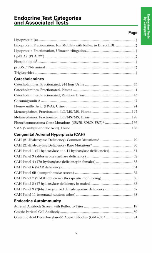
Lipoprotein (a)..........................................................................................................‡
Lipoprotein Fractionation, Ion Mobility with Reflex to Direct LDL .....................‡
Lipoprotein Fractionation, Ultracentrifugation......................................................‡
Lp-PLA2 (PLAC™′)...................................................................................................‡
Phospholipids†...........................................................................................................‡
proBNP, N-terminal ..................................................................................................‡
Triglycerides ..............................................................................................................‡
Catecholamines
Catecholamines, Fractionated, 24-Hour Urine .....................................................43
Catecholamines, Fractionated, Plasma ..................................................................44
Catecholamines, Fractionated, Random Urine.....................................................45
Chromogranin A .....................................................................................................47
Homovanillic Acid (HVA), Urine ..........................................................................94
Metanephrines, Fractionated, LC/MS/MS, Plasma............................................127
Metanephrines, Fractionated, LC/MS/MS, Urine .............................................128
Pheochromocytoma Gene Mutations (SDHB, SDHD, VHL)* .............................136
VMA (Vanillylmandelic Acid), Urine...................................................................186
Congenital Adrenal Hyperplasia (CAH)
CAH (21-Hydroxylase Deficiency) Common Mutations*.....................................29
CAH (21-Hydroxylase Deficiency) Rare Mutations*.............................................30
CAH Panel 1 (21-hydroxylase and 11-hydroxylase deficiencies)..........................31
CAH Panel 3 (aldosterone synthase deficiency) ...................................................32
CAH Panel 4 (17α-hydroxylase deficiency in females) .........................................33
CAH Panel 6 (StAR deficiency)..............................................................................34
CAH Panel 6B (comprehensive screen) ................................................................35
CAH Panel 7 (21-OH deficiency therapeutic monitoring) ..................................36
CAH Panel 8 (17-hydroxylase deficiency in males)...............................................33
CAH Panel 9 (3β-hydroxysteroid dehydrogenase deficiency)..............................37
CAH Panel 11 (neonatal random urine)...............................................................38
Endocrine Autoimmunity
Adrenal Antibody Screen with Reflex to Titer ......................................................18
Gastric Parietal Cell Antibody.................................................................................80
Glutamic Acid Decarboxylase-65 Autoantibodies (GAD-65)* ..............................84
5
Endocrine Test Categoriesand Associated Tests
Page
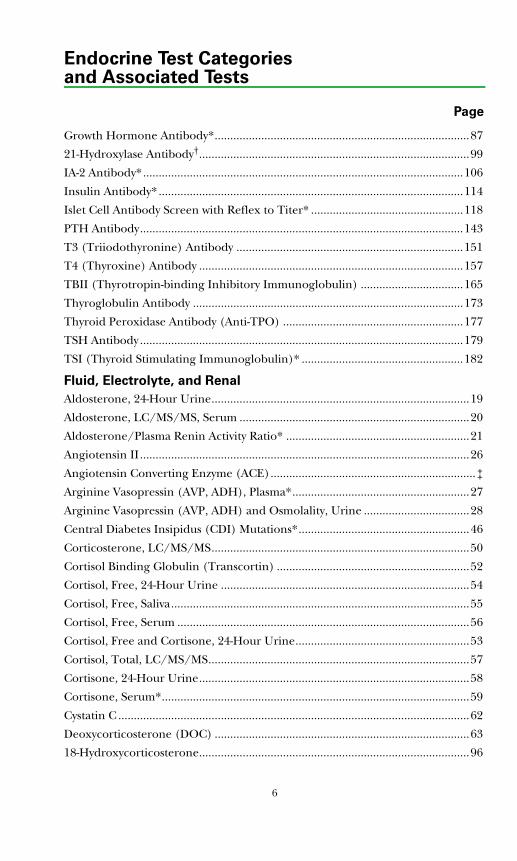
Growth Hormone Antibody*..................................................................................87
21-Hydroxylase Antibody†.......................................................................................99
IA-2 Antibody*.......................................................................................................106
Insulin Antibody* ..................................................................................................114
Islet Cell Antibody Screen with Reflex to Titer* .................................................118
PTH Antibody........................................................................................................143
T3 (Triiodothyronine) Antibody .........................................................................151
T4 (Thyroxine) Antibody .....................................................................................157
TBII (Thyrotropin-binding Inhibitory Immunoglobulin) .................................165
Thyroglobulin Antibody .......................................................................................173
Thyroid Peroxidase Antibody (Anti-TPO) ..........................................................177
TSH Antibody ........................................................................................................179
TSI (Thyroid Stimulating Immunoglobulin)* ....................................................182
Fluid, Electrolyte, and Renal
Aldosterone, 24-Hour Urine...................................................................................19
Aldosterone, LC/MS/MS, Serum ..........................................................................20
Aldosterone/Plasma Renin Activity Ratio* ...........................................................21
Angiotensin II..........................................................................................................26
Angiotensin Converting Enzyme (ACE)..................................................................‡
Arginine Vasopressin (AVP, ADH), Plasma*.........................................................27
Arginine Vasopressin (AVP, ADH) and Osmolality, Urine ..................................28
Central Diabetes Insipidus (CDI) Mutations*.......................................................46
Corticosterone, LC/MS/MS...................................................................................50
Cortisol Binding Globulin (Transcortin) ..............................................................52
Cortisol, Free, 24-Hour Urine ................................................................................54
Cortisol, Free, Saliva................................................................................................55
Cortisol, Free, Serum ..............................................................................................56
Cortisol, Free and Cortisone, 24-Hour Urine........................................................53
Cortisol, Total, LC/MS/MS....................................................................................57
Cortisone, 24-Hour Urine.......................................................................................58
Cortisone, Serum*...................................................................................................59
Cystatin C .................................................................................................................62
Deoxycorticosterone (DOC) ..................................................................................63
18-Hydroxycorticosterone.......................................................................................96
6
Endocrine Test Categoriesand Associated Tests
En
do
crin
e Te
sts
by
Ca
teg
ory
Page
Nephrogenic Diabetes Insipidus (Autosomal) Mutations* ................................130
Nephrogenic Diabetes Insipidus (X-linked) Mutations*....................................131
Osmolality, Random Urine ...................................................................................132
Osmolality, Serum .................................................................................................133
Plasma Renin Activity (PRA)* ..............................................................................137
Genetic (Biochemical and Cytogenetic)
Acylcarnitine, Plasma ................................................................................................‡
Amino Acid Analysis for MSUD, LC/MS, Plasma ...................................................‡
Amino Acid Analysis for Nutritional Status, LC/MS, Plasma .................................‡
Amino Acid Analysis, LC/MS, CSF...........................................................................‡
Amino Acid Analysis, LC/MS, Plasma......................................................................‡
Amino Acid Analysis, LC/MS, Urine .......................................................................‡
Amino Acid Analysis, Limited, LC/MS, Plasma ......................................................‡
Carnitine, LC/MS/MS..............................................................................................‡
Chromosome Analysis, Blood...................................................................................‡
Chromosome Analysis, High Resolution .................................................................‡
Chromosome Analysis, Tissue ..................................................................................‡
Cystine, Qualitative, Urine........................................................................................‡
Cystine, Quantitative, 24-Hour Urine ......................................................................‡
Cystine, Quantitative, Random Urine ......................................................................‡
FISH, Angelman* ......................................................................................................‡
FISH, Chromosome-Specific Probe* Choose one of the following: Chromosome-Specific [1-22, X and Y] Centromere or Chromosome-Specific [1-22, X and Y] Painting ....................‡
FISH, DiGeorge, Velocardiofacial (VCFS)*.............................................................‡
FISH, Kallmann* .......................................................................................................‡
FISH, Microdeletion Syndromes Panel*..................................................................‡
FISH, Neonatal Screen..............................................................................................‡
FISH, Prader Willi* ...................................................................................................‡
FISH, SRY/X Centromere* ......................................................................................‡
FISH, Subtelomere Screen*......................................................................................‡
FISH, Williams* .........................................................................................................‡
FISH, X-Linked Ichthyosis Steroid Sulfatase Deficiency*.......................................‡
Glycogen Storage Disease Type Ia Mutation Analysis (Ashkenazi Jewish)* ..........‡
7
Endocrine Test Categoriesand Associated Tests
Page
Hydroxyproline, LC/MS, Plasma .............................................................................‡
Methylmalonic Acid ..................................................................................................‡
Organic Acids, Qualitative, Urine ............................................................................‡
Organic Acids, Quantitative, Full Panel, Urine.......................................................‡
Phenylalanine ............................................................................................................‡
Phenylalanine and Tyrosine .....................................................................................‡
Prader-Willi/Angelman Syndrome* .......................................................................‡
Tryptophan, LC/MS .................................................................................................‡
Tyrosine .....................................................................................................................‡
Y Chromosome Microdeletion, DNA Analysis† .......................................................‡
Gonadal Function
Alpha Subunit*........................................................................................................23
3α-Androstanediol Glucuronide (3α-diol G) ........................................................24
Androstenedione, LC/MS/MS ..............................................................................25
DHEA (Dehydroepiandrosterone) ........................................................................67
DHEA Sulfate ..........................................................................................................68
Dihydrotestosterone (DHT)...................................................................................69
Dihydrotestosterone, Free ......................................................................................70
Estradiol, Free, LC/MS/MS ...................................................................................71
Estradiol, Ultra Sensitive, LC/MS/MS...................................................................72
Estrogen, Total, Serum ...........................................................................................73
Estrogens, Fractionated, LC/MS/MS ....................................................................74
Estrone, LC/MS/MS...............................................................................................75
Estrone Sulfate ........................................................................................................76
FSH (Follicle Stimulating Hormone) ....................................................................78
FSH (Follicle Stimulating Hormone), Pediatrics ..................................................79
hCG, Total with HAMA Treatment........................................................................90
hCG, Total, Quantitative.........................................................................................91
17-Hydroxypregnenolone.....................................................................................100
17-Hydroxyprogesterone, LC/MS/MS ................................................................101
17-Hydroxyprogesterone, Neonatal/Infant.........................................................101
Inhibin A................................................................................................................112
Inhibin B† ..............................................................................................................113
Invasive Trophoblast Antigen (ITA)(Pregnancy) ...............................................117
8
Endocrine Test Categoriesand Associated Tests
En
do
crin
e Te
sts
by
Ca
teg
ory
Page
17-Ketosteroids with Creatinine, 24-Hour Urine.................................................119
17-Ketosteroids, Fractionated, Urine ...................................................................120
LH (Luteinizing Hormone) .................................................................................123
LH (Luteinizing Hormone), Pediatrics ...............................................................124
Macroprolactin ......................................................................................................125
Pregnenolone ........................................................................................................139
Progesterone, LC/MS/MS....................................................................................140
Prolactin.................................................................................................................142
Sex Hormone Binding Globulin ..........................................................................149
Testosterone, Free and Total................................................................................168
Testosterone, Free, Bioavailable and Total, LC/MS/MS ...................................166
Testosterone, Total (Women, Children, Hypogonadal Males)..........................170
Testosterone, Total*..............................................................................................169
Testosterone, Urine...............................................................................................171
Growth and Growth Hormone
Growth Hormone (GH)..........................................................................................86
Growth Hormone Antibody*..................................................................................87
Growth Hormone Binding Protein ........................................................................88
Growth Hormone Releasing Hormone* ...............................................................89
IGF Binding Protein-1 (IGFBP-1)*.......................................................................107
IGF Binding Protein-2 (IGFBP-2)*.......................................................................108
IGF Binding Protein-3 (IGFBP-3).........................................................................109
IGF-I .......................................................................................................................110
IGF-II (Insulin-Like Growth Factor II).................................................................111
Hypothalamic and Pituitary Function
ACTH, Plasma..........................................................................................................17
Alpha Subunit* ........................................................................................................23
Arginine Vasopressin (AVP, ADH), Plasma*.........................................................27
Arginine Vasopressin (AVP, ADH) and Osmolality, Urine ..................................28
Central Diabetes Insipidus (CDI) Mutations*.......................................................46
Corticotropin Releasing Hormone (CRH)† ..........................................................51
FSH (Follicle Stimulating Hormone) ....................................................................78
FSH (Follicle Stimulating Hormone), Pediatrics ..................................................79
Growth Hormone (GH)..........................................................................................86
9
Endocrine Test Categoriesand Associated Tests
Page
Growth Hormone Antibody*..................................................................................87
Growth Hormone Binding Protein ........................................................................88
Growth Hormone Releasing Hormone (GHRH)* ...............................................89
LH (Luteinizing Hormone) .................................................................................123
LH (Luteinizing Hormone), Pediatrics ...............................................................124
Macroprolactin ......................................................................................................125
Prolactin.................................................................................................................142
Somatostatin* ........................................................................................................150
Thyrotropin-releasing Hormone (TRH)* ...........................................................178
TSH Antibody ........................................................................................................179
TSH with HAMA Treatment.................................................................................181
TSH, Ultrasensitive................................................................................................180
Metabolic (Including Diabetes Mellitus) and Gastrointestinal Disorders
C-peptide, Serum or Urine .....................................................................................60
Fructosamine ...........................................................................................................77
Gastrin......................................................................................................................81
Glucagon*................................................................................................................82
Glucose.....................................................................................................................83
Glutamic Acid Decarboxylase-65 Autoantibodies (GAD-65)* ..............................84
Glycated Albumin....................................................................................................85
Glycogen Storage Disease Type Ia Mutation Analysis (Ashkenazi Jewish) ............‡
Hemoglobin A1c ......................................................................................................92
5-HIAA (5-Hydroxyindoleacetic Acid), Urine .......................................................93
IA-2 Antibody*.......................................................................................................106
IGF Binding Protein-1 (IGFBP-1)*.......................................................................107
IGF Binding Protein-2 (IGFBP-2)*.......................................................................108
IGF Binding Protein-3 (IGFBP-3) ........................................................................109
IGF-I .......................................................................................................................110
IGF-II (Insulin Like Growth Factor II) ................................................................111
Insulin Antibody* ..................................................................................................114
Insulin, Free (Bioactive)* .....................................................................................115
Insulin, Total (Free and Antibody Bound)* .......................................................116
Islet Cell Antibody Screen with Reflex to Titer* .................................................118
10
Endocrine Test Categoriesand Associated Tests
En
do
crin
e Te
sts
by
Ca
teg
ory
Page
Leptin† ...................................................................................................................122
Microalbumin, Intact with Creatinine, HPLC, Urine .........................................129
Osmolality, Random Urine ...................................................................................132
Osmolality, Serum .................................................................................................133
Pancreatic Polypeptide*........................................................................................135
Proinsulin† .............................................................................................................141
Somatostatin* ........................................................................................................150
Vasoactive Intestinal Polypeptide (VIP)*.............................................................183
Multiple Endocrine Neoplasia
ACTH, Plasma..........................................................................................................17
Calcitonin.................................................................................................................39
Calcium, 24-Hour Urine .........................................................................................42
Calcium, Pediatric Urine ........................................................................................42
Calcium, Ionized .....................................................................................................40
Calcium, Total, Serum ............................................................................................41
Catecholamines, Fractionated, 24-Hour Urine .....................................................43
Catecholamines, Fractionated, Plasma ..................................................................44
Catecholamines, Fractionated, Random Urine.....................................................45
Chromogranin A .....................................................................................................47
C-Peptide, Serum or Urine .....................................................................................60
Gastrin......................................................................................................................81
Glucagon*................................................................................................................82
Growth Hormone (GH)..........................................................................................86
5-HIAA (5-Hydroxyindoleacetic Acid), Urine .......................................................93
Homovanillic Acid (HVA), Urine ..........................................................................94
Insulin, Free (Bioactive)* .....................................................................................115
Macroprolactin ......................................................................................................125
MEN 2 and FMTC Mutations, Exons 10, 11, 13-16* ...........................................126
Metanephrines, Fractionated, LC/MS/MS, Plasma............................................127
Metanephrines, Fractionated, LC/MS/MS, Urine .............................................128
Pancreatic Polypeptide*........................................................................................135
Proinsulin† .............................................................................................................141
Prolactin.................................................................................................................142
PTH, Intact and Calcium ......................................................................................144
11
Endocrine Test Categoriesand Associated Tests
Page
PTH, Intact and Ionized Calcium ........................................................................144
PTH-related Protein (PTH-RP)† ..........................................................................145
Serotonin, Blood ...................................................................................................148
Serotonin, Serum ..................................................................................................148
Vasoactive Intestinal Polypeptide (VIP)*.............................................................183
VMA (Vanillylmandelic Acid), Urine...................................................................186
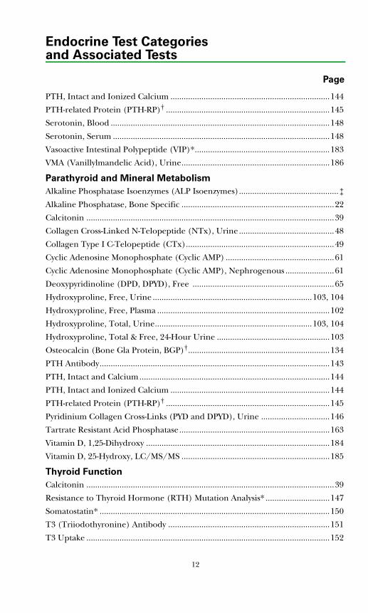
Parathyroid and Mineral Metabolism
Alkaline Phosphatase Isoenzymes (ALP Isoenzymes).............................................‡
Alkaline Phosphatase, Bone Specific .....................................................................22
Calcitonin ................................................................................................................39
Collagen Cross-Linked N-Telopeptide (NTx), Urine ...........................................48
Collagen Type I C-Telopeptide (CTx)...................................................................49
Cyclic Adenosine Monophosphate (Cyclic AMP) .................................................61
Cyclic Adenosine Monophosphate (Cyclic AMP), Nephrogenous ......................61
Deoxypyridinoline (DPD, DPYD), Free ................................................................65
Hydroxyproline, Free, Urine ........................................................................103, 104
Hydroxyproline, Free, Plasma ..............................................................................102
Hydroxyproline, Total, Urine.......................................................................103, 104
Hydroxyproline, Total & Free, 24-Hour Urine ...................................................103
Osteocalcin (Bone Gla Protein, BGP)†................................................................134
PTH Antibody........................................................................................................143
PTH, Intact and Calcium......................................................................................144
PTH, Intact and Ionized Calcium ........................................................................144
PTH-related Protein (PTH-RP)† ..........................................................................145
Pyridinium Collagen Cross-Links (PYD and DPYD), Urine ...............................146
Tartrate Resistant Acid Phosphatase....................................................................163
Vitamin D, 1,25-Dihydroxy ...................................................................................184
Vitamin D, 25-Hydroxy, LC/MS/MS ...................................................................185
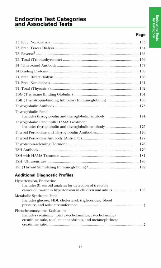
Thyroid Function
Calcitonin ................................................................................................................39
Resistance to Thyroid Hormone (RTH) Mutation Analysis* .............................147
Somatostatin* ........................................................................................................150
T3 (Triiodothyronine) Antibody .........................................................................151
T3 Uptake ..............................................................................................................152
12
Endocrine Test Categoriesand Associated Tests
En
do
crin
e Te
sts
by
Ca
teg
ory
Page
T3, Free, Non-dialysis ............................................................................................153
T3, Free, Tracer Dialysis........................................................................................154
T3, Reverse† ...........................................................................................................155
T3, Total (Triiodothyronine) ...............................................................................156
T4 (Thyroxine) Antibody .....................................................................................157
T4 Binding Proteins ..............................................................................................158
T4, Free, Direct Dialysis ........................................................................................160
T4, Free, Non-dialysis ............................................................................................161
T4, Total (Thyroxine)...........................................................................................162
TBG (Thyroxine Binding Globulin) ....................................................................164
TBII (Thyrotropin-binding Inhibitory Immunoglobulin)..................................165
Thyroglobulin Antibody........................................................................................173
Thyroglobulin Panel Includes thyroglobulin and thyroglobulin antibody. ..................................174
Thyroglobulin Panel with HAMA Treatment Includes thyroglobulin and thyroglobulin antibody. ..................................175
Thyroid Peroxidase and Thyroglobulin Antibodies............................................176
Thyroid Peroxidase Antibody (Anti-TPO)...........................................................177
Thyrotropin-releasing Hormone..........................................................................178
TSH Antibody ........................................................................................................179
TSH with HAMA Treatment.................................................................................181
TSH, Ultrasensitive................................................................................................180
TSI (Thyroid Stimulating Immunoglobulin)* ....................................................182
Additional Diagnostic Profiles
Hypertension, Endocrine Includes 31 steroid analytes for detection of treatable causes of low-renin hypertension in children and adults. ...........................105
Metabolic Syndrome PanelIncludes glucose, HDL cholesterol, triglycerides, bloodpressure, and waist circumference....................................................................‡
Pheochromocytoma Evaluation Includes creatinine, total catecholamines, catecholamine/creatinine ratio, total metanephrines, and metanephrines/creatinine ratio...................................................................................................‡
13
Endocrine Test Categoriesand Associated Tests
Page
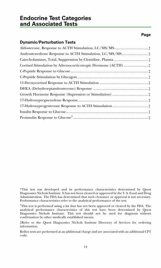
Dynamic/Perturbation Tests
Aldosterone, Response to ACTH Stimulation, LC/MS/MS...................................‡
Androstenedione Response to ACTH Stimulation, LC/MS/MS...........................‡
Catecholamines, Total, Suppression by Clonidine, Plasma....................................‡
Cortisol Stimulation by Adrenocorticotropic Hormone (ACTH) .........................‡
C-Peptide Response to Glucose ................................................................................‡
C-Peptide Stimulation by Glucagon .........................................................................‡
11-Deoxycortisol Response to ACTH Stimulation ..................................................‡
DHEA (Dehydroepiandrosterone) Response .........................................................‡
Growth Hormone Response (Supression or Stimulation) .....................................‡
17-Hydroxypregnenolone Response ........................................................................‡
17-Hydroxyprogesterone Response to ACTH Stimulation.....................................‡
Insulin Response to Glucose.....................................................................................‡
Proinsulin Response to Glucose† .............................................................................‡
*This test was developed and its performance characteristics determined by QuestDiagnostics Nichols Institute. It has not been cleared or approved by the U.S. Food and DrugAdministration. The FDA has determined that such clearance or approval is not necessary.Performance characteristics refer to the analytical performance of the test.†This test is performed using a kit that has not been approved or cleared by the FDA. Theanalytical performance characteristics of this test have been determined by QuestDiagnostics Nichols Institute. This test should not be used for diagnosis withoutconfirmation by other medically established means.
‡Refer to the Quest Diagnostics Nichols Institute Directory of Services for orderinginformation.
Reflex tests are performed at an additional charge and are associated with an additional CPTcode.
14
Alphabetical Test Section
Test components are disclosed in the individual test write-ups in this section.

Alp
ha
be
tica
l Test
Se
ctio
n
17
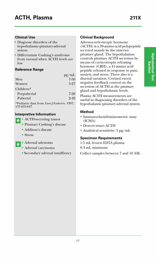
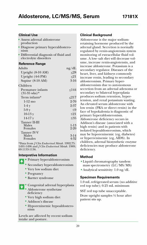
Clinical Use
• Diagnose disorders of the hypothalamic-pituitary-adrenal system
• Differentiate Cushing's syndrome from normal when ACTH levels are low
Reference Range
Interpretive Information
• ACTH-secreting tumor• Pituitary Cushing’s disease• Addison’s disease• Stress
• Adrenal adenoma• Adrenal carcinoma• Secondary adrenal insuffiency
Clinical Background
Adrenocorticotropic hormone (ACTH) is a 39-amino acid polypeptide secreted mainly by the anterior pituitary gland. The hypothalamus controls pituitary ACTH secretion by means of corticotropin releasing hormone (CRH), a 41-amino acid peptide released in response to pain, anxiety, and stress. There also is a diurnal variation. Cortisol exerts negative feedback control on the secretion of ACTH at the pituitary gland and hypothalamic levels.Plasma ACTH measurements are useful in diagnosing disorders of the hypothalamic-pituitary-adrenal system.
Method
• Immunochemiluminometric assay (ICMA)
• Detects intact ACTH• Analytical sensitivity: 5 pg/mL
Specimen Requirements
1.5 mL frozen EDTA plasma0.3 mL minimum
Collect samples between 7 and 10 AM.
pg/mLMen 7-50Women 5-27Children*
Prepubertal 7-28Pubertal 2-49
*Pediatric data from Euro J Endocrin. 1997;137:635-647.
ACTH, Plasma 211X
18
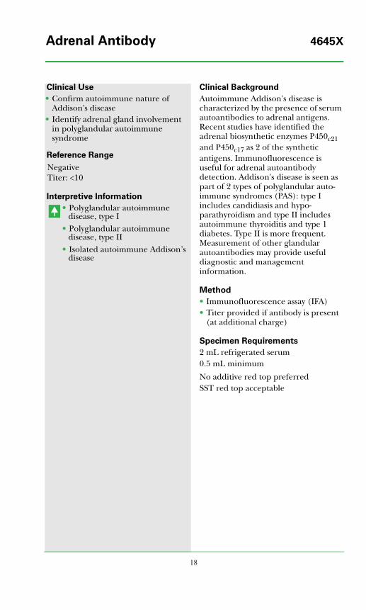
Clinical Use
• Confirm autoimmune nature of Addison's disease
• Identify adrenal gland involvement in polyglandular autoimmune syndrome
Reference Range
Interpretive Information
• Polyglandular autoimmune disease, type I
• Polyglandular autoimmune disease, type II
• Isolated autoimmune Addison’s disease
Clinical Background
Autoimmune Addison's disease is characterized by the presence of serum autoantibodies to adrenal antigens. Recent studies have identified the adrenal biosynthetic enzymes P450c21 and P450c17 as 2 of the synthetic antigens. Immunofluorescence is useful for adrenal autoantibody detection. Addison's disease is seen as part of 2 types of polyglandular auto-immune syndromes (PAS): type I includes candidiasis and hypo-parathyroidism and type II includes autoimmune thyroiditis and type 1 diabetes. Type II is more frequent. Measurement of other glandular autoantibodies may provide useful diagnostic and management information.
Method
• Immunofluorescence assay (IFA)• Titer provided if antibody is present
(at additional charge)
Specimen Requirements
2 mL refrigerated serum0.5 mL minimum
No additive red top preferredSST red top acceptable
NegativeTiter: <10
Adrenal Antibody 4645X
Alp
ha
be
tica
l Test
Se
ctio
n
19
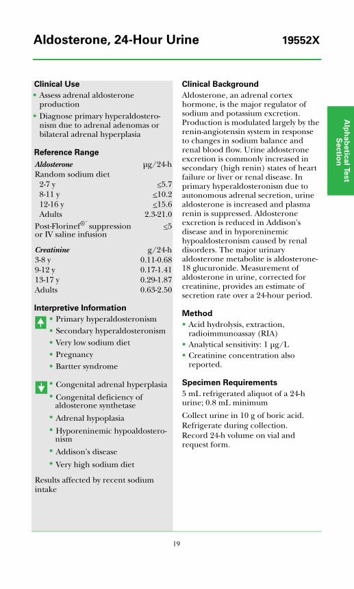
Clinical Use
• Assess adrenal aldosterone production
• Diagnose primary hyperaldostero-nism due to adrenal adenomas or bilateral adrenal hyperplasia
Reference Range
Interpretive Information
• Primary hyperaldosteronism• Secondary hyperaldosteronism• Very low sodium diet• Pregnancy• Bartter syndrome
• Congenital adrenal hyperplasia• Congenital deficiency of
aldosterone synthetase• Adrenal hypoplasia• Hyporeninemic hypoaldostero-
nism• Addison's disease• Very high sodium diet
Results affected by recent sodium intake
Clinical Background
Aldosterone, an adrenal cortex hormone, is the major regulator of sodium and potassium excretion. Production is modulated largely by the renin-angiotensin system in response to changes in sodium balance and renal blood flow. Urine aldosterone excretion is commonly increased in secondary (high renin) states of heart failure or liver or renal disease. In primary hyperaldosteronism due to autonomous adrenal secretion, urine aldosterone is increased and plasma renin is suppressed. Aldosterone excretion is reduced in Addison's disease and in hyporeninemic hypoaldosteronism caused by renal disorders. The major urinary aldosterone metabolite is aldosterone-18 glucuronide. Measurement of aldosterone in urine, corrected for creatinine, provides an estimate of secretion rate over a 24-hour period.
Method
• Acid hydrolysis, extraction, radioimmunoassay (RIA)
• Analytical sensitivity: 1 μg/L• Creatinine concentration also
reported.
Specimen Requirements
5 mL refrigerated aliquot of a 24-h urine; 0.8 mL minimum
Collect urine in 10 g of boric acid.Refrigerate during collection.Record 24-h volume on vial and request form.
Aldosterone μg/24-hRandom sodium diet
2-7 y <5.78-11 y <10.212-16 y <15.6Adults 2.3-21.0
Post-Florinef®´ suppression or IV saline infusion
<5
Creatinine g/24-h3-8 y 0.11-0.689-12 y 0.17-1.4113-17 y 0.29-1.87Adults 0.63-2.50
Aldosterone, 24-Hour Urine 19552X
20
Clinical Use
• Assess adrenal aldosterone production
• Diagnose primary hyperaldostero-nism
• Differential diagnosis of fluid and electrolyte disorders
Reference Range
Interpretive Information
••• Primary hyperaldosteronism• Secondary hyperaldosteronism• Very low sodium diet• Pregnancy• Bartter syndrome
• Congenital adrenal hyperplasia• Aldosterone synthetase
deficiency• Very high sodium diet• Addison's disease• Hyporeninemic hypoaldostero-
nism
Levels are affected by recent sodium intake and posture.
Clinical Background
Aldosterone is the major sodium-retaining hormone produced by the adrenal gland. Secretion is normally regulated by renin-angiotensin system monitoring of extracellular fluid vol-ume. A low- salt diet will decrease vol-ume, increase renin-angiotensin, and increase aldosterone. Potassium is a secondary regulator. Diseases of the heart, liver, and kidneys commonly increase renin, leading to secondary aldosteronism. Primary hyper-aldosteronism due to autonomous secretion from an adrenal adenoma or secondary to bilateral hyperplasia produces sodium retention, hyper-tension, and renal potassium wasting. An elevated serum aldosterone with low renin (PRA or direct renin) in the face of hypokalemia is diagnostic of primary hyperaldosteronism. Aldosterone deficiency occurs in Addison's disease (associated with a high renin) and in patients with isolated hypoaldosteronism, which may be hyporeninemic (eg, diabetes) or hyperreninemic (eg, AIDS). In children, adrenal biosynthetic enzyme deficiencies may produce aldosterone deficiency.
Method
• Liquid chromatography tandem mass spectrometry (LC/MS/MS)
• Analytical sensitivity: 1.0 ng/dL
Specimen Requirements
1.0 mL refrigerated serum (no additive red top tube); 0.25 mL minimum
SST red top tube unacceptable.Draw upright samples ½ hour after patient sits up.
Adults ng/dLUpright (8-10 AM) <28Upright (4-6 PM) <21Supine (8-10 AM) 3-16
ChildrenPremature infants (31-35 wks)*
<144
Term infants* <2171-12 mo 2-701-4 y 2-375-9 y ≤910-13 y ≤2114-17 y ≤35
Tanner II-IIIMalesFemales
1-132-20
Tanner IV-VMalesFemales
3-144-32
*Data from J Clin Endocrinol Metab. 1992;75:1491-1496 and J Clin Endocrinol Metab. 1989;69:1133-1136.
Aldosterone, LC/MS/MS, Serum 17181X
Alp
ha
be
tica
l Test
Se
ctio
n
21
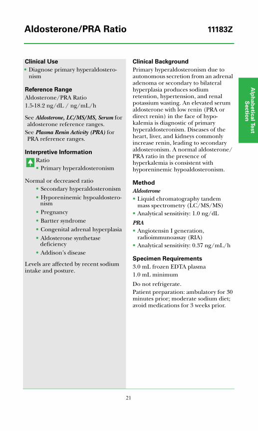
Clinical Use
• Diagnose primary hyperaldostero-nism
Reference Range
Aldosterone/PRA Ratio1.5-18.2 ng/dL / ng/mL/h
See Aldosterone, LC/MS/MS, Serum for aldosterone reference ranges.
See Plasma Renin Activity (PRA) for PRA reference ranges.
Interpretive Information
Ratio• Primary hyperaldosteronism
Normal or decreased ratio• Secondary hyperaldosteronism• Hyporeninemic hypoaldostero-
nism• Pregnancy• Bartter syndrome• Congenital adrenal hyperplasia• Aldosterone synthetase
deficiency• Addison’s disease
Levels are affected by recent sodium intake and posture.
Clinical Background
Primary hyperaldosteronism due to autonomous secretion from an adrenal adenoma or secondary to bilateral hyperplasia produces sodium retention, hypertension, and renal potassium wasting. An elevated serum aldosterone with low renin (PRA or direct renin) in the face of hypo-kalemia is diagnostic of primary hyperaldosteronism. Diseases of the heart, liver, and kidneys commonly increase renin, leading to secondary aldosteronism. A normal aldosterone/PRA ratio in the presence of hyperkalemia is consistent with hyporeninemic hypoaldosteronism.
Method
Aldosterone• Liquid chromatography tandem
mass spectrometry (LC/MS/MS) • Analytical sensitivity: 1.0 ng/dL
PRA• Angiotensin I generation,
radioimmunoassay (RIA)• Analytical sensitivity: 0.37 ng/mL/h
Specimen Requirements
3.0 mL frozen EDTA plasma1.0 mL minimum
Do not refrigerate.Patient preparation: ambulatory for 30 minutes prior; moderate sodium diet; avoid medications for 3 weeks prior.
Aldosterone/PRA Ratio 11183Z
22
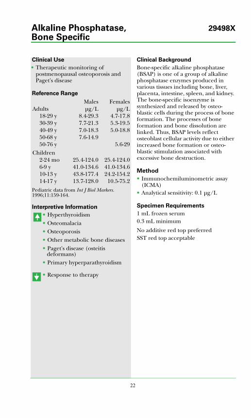
Clinical Use
• Therapeutic monitoring of postmenopausal osteoporosis and Paget's disease
Reference Range
Pediatric data from Int J Biol Markers. 1996;11:159-164.
Interpretive Information
• Hyperthyroidism• Osteomalacia• Osteoporosis• Other metabolic bone diseases• Paget's disease (osteitis
deformans)• Primary hyperparathyroidism
• Response to therapy
Clinical Background
Bone-specific alkaline phosphatase (BSAP) is one of a group of alkaline phosphatase enzymes produced in various tissues including bone, liver, placenta, intestine, spleen, and kidney. The bone-specific isoenzyme is synthesized and released by osteo-blastic cells during the process of bone formation. The processes of bone formation and bone dissolution are linked. Thus, BSAP levels reflect osteoblast cellular activity due to either increased bone formation or osteo-blastic stimulation associated with excessive bone destruction.
Method
• Immunochemiluminometric assay (ICMA)
• Analytical sensitivity: 0.1 μg/L
Specimen Requirements
1 mL frozen serum0.3 mL minimum
No additive red top preferredSST red top acceptable
Males FemalesAdults μg/L μg/L
18-29 y 8.4-29.3 4.7-17.830-39 y 7.7-21.3 5.3-19.540-49 y 7.0-18.3 5.0-18.850-68 y 7.6-14.950-76 y 5.6-29
Children2-24 mo 25.4-124.0 25.4-124.06-9 y 41.0-134.6 41.0-134.610-13 y 43.8-177.4 24.2-154.214-17 y 13.7-128.0 10.5-75.2
Alkaline Phosphatase, 29498X
Bone Specific
Alp
ha
be
tica
l Test
Se
ctio
n
23
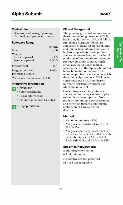
Clinical Use
• Diagnose and manage pituitary, placental, and pancreatic tumors
Reference Range
*Varies with concentration of hCG
Interpretive Information
• Pregnancy• Choriocarcinoma• Hydatidiform mole• Pituitary adenomas (selected)
• Hypopituitarism
Clinical Background
The pituitary glycoprotein hormones–thyroid stimulating hormone (TSH), luteinizing hormone (LH), and follicle stimulating hormone (FSH)–are comprised of identical alpha subunits and unique beta subunits that confer biological specificity. Some pituitary adenomas unassociated with a clinical syndrome of hormonal over secretion produce the alpha subunit, which serves as a useful tumor marker. Measurement of the alpha subunit can be useful in differentiating TSH-secreting pituitary adenomas, in which the ratio of alpha-to-intact TSH serum concentrations is >1, from thyroid hormone resistance syndromes, in which the ratio is <1.Gonadotropin-secreting pituitary adenomas producing excessive alpha subunit have been reported. Non-pituitary tumors, eg, choriocarcinoma and carcinoid tumors, secreting the alpha subunit have also been described.
Method
• Radioimmunoassay (RIA)• Analytical sensitivity: 0.1 ng/mL at
90% B/Bo• Analytical specificity: cross-reactivity
is 1.4% with intact hCG, <0.01% with beta subunit hCG, 4.5% with LH, 3.4% with FSH, and 2.0% with TSH
Specimen Requirements
2 mL refrigerated serum0.3 mL minimum
No additive red top preferredSST red top acceptable
ng/mLMen <0.6Women
PremenopausalPostmenopausal
<1.50.9-3.3
Hypothyroid <3.7
Pregnant or hCG-producing tumors
1.8-360*
Alpha Subunit 8658X
24
Clinical Use
• Marker for disorders of peripheral androgen formation and action, such as in hirsutism and acne
Reference Range
Interpretive Information
• Idiopathic hirsutism (females)• PCO-hirsutism (females)• Acne (females)
• 5α-reductase deficiency (males)
Clinical Background
3α-Androstanediol glucuronide (3α-diol G) is a metabolite of dihydro-testosterone (DHT) produced in androgen responsive tissues, such as hair follicles. 3α-diol G is a marker of a peripheral androgen formation and action.The most important application of this assay is in hirsute states. Patients with hirsutism of either the idiopathic type or secondary to polycystic ovarian diseases (PCO) may have markedly elevated levels. 3α-diol G has also been shown to be a marker of therapeutic response in congenital adrenal hyperplasia (CAH).
Method
• Extraction, enzyme digestion, chromatography, radioimmunoassay (RIA)
• Analytical sensitivity: 5 ng/dL
Specimen Requirements
2 mL refrigerated serum0.3 mL minimum
No additive red top preferredSST red top acceptable
ng/dLMen 260-1500Women 60-300Children
Prepubertal 10-60Tanner II-III
Males 19-164Females 33-244
Tanner stage data from J Clin Endocrinol Metab. 1991;72:46-50.
3α-Androstanediol Glucuronide 5276X(3α-diol G)
Alp
ha
be
tica
l Test
Se
ctio
n
25
Clinical Use
• Diagnose hirsutism, polycystic ovarian disease, virilization, and congenital adrenal hyperplasia
Reference Range
Interpretive Information
• Adrenal tumors• Cushing’s disease• Congenital adrenal hyperplasia• Polycystic ovarian disease• Idiopathic hirsutism
• Addison's disease
Clinical Background
Androstenedione is an androgenic steroid hormone produced by both the adrenal cortex and the gonads. In males, the compound's activity is relatively unimportant, compared with the predominant testicular androgen testosterone. In females, andros-tenedione–and the testosterone and dihydrotestosterone to which it is metabolized–normally contribute to the growth of sexually dependent axillary and pubic hair. In women, the concentration of androstenedione varies with the menstrual cycle, with the ovaries and adrenals contributing equally during the follicular phase. At mid-cycle, increased ovarian andros-tenedione secretion accounts for 70% of its overall production.Excessive androstenedione production may cause hirsutism and contribute to virilization. This can occur in the polycystic ovary syndrome, idiopathic hirsutism, ovarian and adrenal neoplasms, and congenital adrenal hyperplasia due to 21-hydroxylase or 17β-hydroxylase deficiencies. In hirsute patients with an elevated androstenedione concentration, serial measurements are useful to monitor therapy, since clinical improvement typically lags behind the biochemical response by months.
Method
• Liquid chromatography tandem mass spectrometry (LC/MS/MS)
• Analytical sensitivity: 5 ng/dL
Specimen Requirements
1 mL refrigerated serum0.25 mL minimum
No additive red top tubeSST red top unacceptable
ng/dLMen
18-30 y31-50 y51-60 y
50-22040-19050-220
WomenFollicularMidcycleLutealPostmenopausal
35-25060-28530-23520-75
Children1-12 m1-4 y5-9 y10-13 y14-17 y
6-785-51
6-11512-22122-225
Tanner II-IIIMales 17-82Females 43-180
Tanner IV-VMales 57-150Females 7-68
Pediatric data from J Clin Endocrinol Metab. 1991;73:674-686 and J Clin Endocrinol Metab. 1989;69:1133-1136.
Androstenedione, LC/MS/MS 17182X
26
Clinical Use
• Differential diagnosis of renal hypertension
Reference Range
Interpretive Information
• High renin hypertension• Renin-secreting juxta-
glomerular renal tumor• Volume depletion• Congestive heart failure• Cirrhosis• Angiotensin II receptor blocking
drug therapy
• Anephric patients• Primary aldosteronism• Cushing's syndrome• ACE inhibitor therapy
Clinical Background
Angiotensin II is the primary regulator of renal aldosterone secretion and a potent vasoconstrictor. It is generated through the renin angiotensin system (RAS): circulating angiotensinogen is cleaved by renin to form angiotensin I, which is then converted to angiotensin II via angiotensin converting enzyme (ACE). The rate-limiting step in the RAS is renin secretion by the renal juxtaglomerular cells, modulated by renal blood flow. Low renal blood flow and low perfusion pressure increases renin secretion, stimulating angio-tensin II and aldosterone production with a resulting increase in blood pressure and renal sodium retention. These changes then produce inhibition of renin secretion and complete the feedback control loop.
Method
• Radioimmunoassay (RIA)
Specimen Requirements
1 mL frozen EDTA plasma0.3 mL minimum
Adults 10-50 ng/L
Angiotensin II 36718X
Alp
ha
be
tica
l Test
Se
ctio
n
27
Clinical Use
• Diagnose central diabetes insipidus (DI)
• Differential diagnosis of central DI vs nephrogenic DI
• Diagnose syndrome of inappropriate ADH secretion (SIADH)
Reference Range
Interpretive Information
• SIADH• Ectopic ADH syndrome• Nephrogenic DI•• Phenothiazine, carbamazepine
• Central DI
Clinical Background
Arginine vasopressin (AVP), or antidiuretic hormone (ADH), is a nonapeptide produced by the hypo-thalamus and released from the posterior pituitary in response to extracellular fluid hyperosmolarity and hypovolemia. AVP promotes concen-tration of the urine by increasing water reabsorption in the kidney tubules. Inadequate AVP action causes diabetes insipidus (DI), a syndrome characterized by nonglycosuric polyuria, polydipsia, and dehydration. Central DI refers to insufficient AVP release due to diseases of the hypo-thalamus, pituitary stalk, and pituitary gland. Nephrogenic DI is the result of impaired renal responsiveness to AVP and may be congenital or due to renal disease, hypokalemia, hypercalcemia, systemic disorders (eg, multiple myeloma and amyloidosis), or drugs (eg, lithium or demeclocycline and ethanol).DI diagnosis is based on the presence of hyperosmolar serum with in-appropriately dilute urine. Central and nephrogenic DI can be differentiated by measuring the plasma AVP level and interpreting it in light of the simul-taneous plasma osmolality.The syndrome of inappropriate secretion of antidiuretic hormone (SIADH) is manifest by hyponatremia and inappropriately concentrated urine. The diagnosis is confirmed by plasma or urine AVP levels in-appropriate for serum osmolality.
Method
• Extraction, radioimmunoassay (RIA)• Analytical sensitivity: 1.0 pg/mL
Specimen Requirements
4 mL frozen EDTA plasma1.1 mL minimum
Draw blood in prechilled tube; centrifuge immediately in refrigerated centrifuge.
1.0-13.3 pg/mLNote: 2.5 pg = 1 μU
Arginine Vasopressin, Plasma 252X
28
Clinical Use
• Diagnose central diabetes insipidus (DI)
• Differential diagnosis of central DI vs nephrogenic DI
• Confirm syndrome of inappropriate ADH secretion (SIADH) diagnosis
Reference Range
Interpretive Information
• Low AVP and urine osmolality = central DI
• High AVP and low urine osmolality = nephrogenic DI
• Normal AVP despite low serum osmolality = SIADH; in SIADH, the kidney responds normally to AVP
Clinical Background
Arginine vasopressin (AVP), also known as antidiuretic hormone (ADH), is a 9-amino acid polypeptide synthesized by the hypothalamus and stored and secreted by the posterior pituitary. Secretion is regulated by osmoreceptors and volume receptors and is increased by dehydration or increased blood osmolarity. Conversely, secretion is decreased by increased blood volume or decreased blood osmolarity. AVP acts primarily on the kidney, where it exerts an antidiuretic effect.AVP plasma concentrations correlate with osmolarity. Significant amounts of AVP are filtered via the glomerulus and appear in urine. Concentrations are higher in urine than in plasma. Urine AVP provides an integrated view of recent blood levels. Urine AVP determinations are useful in the differential diagnosis of hyponatremic states.
Method
AVP• Radioimmunoassay (RIA)• Analytical sensitivity: 1.0 pg/mL
Osmolality•• Freezing point depression
Specimen Requirements
3 mL refrigerated random urine1.2 mL minimum
Do not use a preservative.
AVP 1-112 pg/mLOsmolality 50-1200 mOsm/kg
Arginine Vasopressin (AVP) 36572Xand Osmolality, Random Urine
Alp
ha
be
tica
l Test
Se
ctio
n
29
Clinical Use
• Confirm diagnosis of 21-hydroxylase deficiency (21-OHD)
• Screen for 21-OHD carrier status in at-risk individuals
• Prenatal diagnosis of 21-OHD
Reference Range
Negative (no mutations detected)
Interpretive Information
Positive (mutations detected)• 21-hydroxylase deficiency
(carrier or affected)
Clinical Background
21-Hydroxylase deficiency, the most common cause of congenital adrenal hyperplasia (CAH), is an autosomal recessive disorder caused by mutations or rearrangements in the CYP21A2 gene on chromosome 6. The deficiency is characterized by decreased cortisol and increased androgen blood levels. Severe reduction in 21-hydroxylase activity causes classic CAH, the simple virilizing form (25% of cases), and/or the salt-wasting form, which is further characterized by decreased aldosterone levels. Onset occurs prenatally and, if detected prenatally, it can be treated to reduce virilization in affected females. Non-classic CAH, on the other hand, presents postnatally with signs of hyperandrogenism. Testing for the more common mutations can detect both relevant mutations in 81% and 1 mutation in about 18% of affected individuals. Preparation for prenatal testing requires mutation analysis of parents and/or an affected offspring.
Method
• Polymerase chain reaction (PCR) and DNA mini-sequencing
• Common mutations sought: P30L; Intron 2 "g"; G110del8nt; I172N; exon 6 cluster of I236N, V237E & M239K; V281L; F306+1nt; Q318X; R356W; and P453S
• Deduction of deletions and recombinations between CYP21A2 and its pseudogene
Specimen Requirements
5 mL room temperature whole blood3 mL minimum
Collect blood in a lavender-top (EDTA) or yellow-top (ACD solution B) tube.For prenatal testing, submit amniotic fluid, CVS sample, or cultured cells from either source.
CAH (21-Hydroxylase Deficiency) 14755XCommon Mutations
30
Clinical Use
• Confirm diagnosis of 21-hydroxylase deficiency (21-OHD)
• Determine 21-OHD carrier status in at-risk individuals
• Prenatal diagnosis of 21-OHD
Reference Range
Negative (no mutations detected)
Interpretive Information
Positive (mutations detected)• 21-hydroxylase deficiency (carrier or
affected)
Clinical Background
21-Hydroxylase deficiency, the most common cause of congenital adrenal hyperplasia (CAH), is an autosomal recessive disorder caused by mutations or rearrangements in the CYP21A2 gene on chromosome 6. The deficiency is characterized by decreased cortisol and increased androgen blood levels. Severe reduction in 21-hydroxylase activity causes classic CAH, the simple virilizing form (25% of cases), and/or the salt-wasting form, which is further characterized by decreased aldosterone levels. Onset occurs prenatally and, if detected prenatally, it can be treated to reduce virilization in affected females. Non-classic CAH, on the other hand, presents postnatally with signs of hyper-androgenism.This test is appropriate for affected individuals in whom only 1 or no muta-tions were detected in the CAH (21-Hydroxylase Deficiency) Common Mutations test. It is also appropriate for carrier testing in family members of such individuals, once a rare mutation has been identified. About 1% of affected individuals have rare muta-tions on both chromosomes, and about 18% have a rare mutation on only 1 chromosome.
Method
• Polymerase chain reaction (PCR) and DNA sequencing
• Entire CYP21A2 gene sequenced
Specimen Requirements
5 mL room temperature whole blood3 mL minimum
Collect blood in a lavender-top (EDTA) or yellow-top (ACD solution B) tube.For prenatal testing, submit amniotic fluid, CVS sample, or cultured cells from either source.
Call 1-866-GENEINFO (1-866-436-3436) before collecting or submitting samples.
CAH (21-Hydroxylase Deficiency) 16072XRare Mutations
Alp
ha
be
tica
l Test
Se
ctio
n
CAH Panel 1 (21-Hydroxylase vs 15269X11β-Hydroxylase Deficiency)
31
Clinical Use
• Diagnose 21-hydroxylase or 11β-hydroxylase deficiency
Reference Range
Interpretive Information
Clinical Background
21-Hydroxylase deficiency (21-OHD, P450c21) accounts for 90% to 95% of CAH cases. Classic 21-OHD is characterized by markedly diminished or absent 21-hydroxylase activity. Affected individuals typically present at birth or in the neonatal period with either a virilizing or salt-wasting form. Female infants with virilizing 21-OHD have varying degrees of masculinization ranging from clitoral enlargement to complete development of male external genitalia. Male infants have normal male genitalia. Symptoms of hyponatremia, hyperkalemia, volume depletion, and decreased blood pressure generally appear within the first 2 weeks of life in patients with the salt-wasting form.Nonclassic 21-OHD is characterized by marginally decreased 21-hydroxylase activity. Affected females usually present in childhood or post puberty with evidence of androgen excess (eg, increased pubic hair growth and/or clitoral enlargement). In women, non-classic 21-OHD is frequently confused with polycystic ovary syndrome. Diagnosis may require ACTH stimulation.11β-Hydroxlase deficiency (CYP11B1, 11βHSD) accounts for 5% to 8% of CAH cases. Salt-wasting does not occur; however, virilization of female fetuses can be severe. Elevated blood pressure manifests early in life in approximately two-thirds of patients and, along with elevated deoxycorticosterone and 11-deoxycortisol levels, clinically distinguishes 11β- from 21-hydroxylase deficiency. A late-onset form that typically presents with signs of androgen excess is analogous to nonclassic 21-OHD.
Method
• Liquid chromatography, tandem mass spectrometry (LC/MS/MS)
Specimen Requirements
0.6 mL refrigerated serum0.3 mL minimum
No additive red top tubeAn early morning specimen is preferred.Specify age and sex on test request form.
Androstenedione *Cortisol *11-Deoxycortisol *17-Hydroxyprogesterone *Testosterone **See individual assays for age-related reference ranges.
17-OH progesterone/11-deoxycortisol ratio*
21-OH: >6
11-Deoxycortisol/cortisol ratio*
11β-OH:>100 (exceptprematures)
Androstenedione ↑Testosterone ↑*See Test Application and Interpretation, Disorders of Adrenal Function, Tables 6-8.
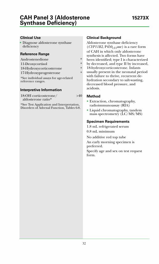
CAH Panel 3 (Aldosterone 15273XSynthase Deficiency)
32
Clinical Use
• Diagnose aldosterone synthase deficiency
Reference Range
Interpretive Information
Clinical Background
Aldosterone synthase deficiency (CYP11B2, P450c11ase) is a rare form of CAH in which only aldosterone synthesis is affected. Two forms have been identified; type I is characterized by decreased, and type II by increased, 18-hydroxycorticosterone. Infants usually present in the neonatal period with failure to thrive, recurrent de-hydration secondary to salt-wasting, decreased blood pressure, and acidosis.
Method
• Extraction, chromatography, radioimmunoassay (RIA)
• Liquid chromatography, tandem mass spectrometry (LC/MS/MS)
Specimen Requirements
1.8 mL refrigerated serum0.8 mL minimum
No additive red top tubeAn early morning specimen is preferred.Specify age and sex on test request form.
Androstenedione *11-Deoxycortisol *18-Hydroxycorticosterone *17-Hydroxyprogesterone **See individual assays for age-related reference ranges.
18-OH corticosterone/aldosterone ratio*
>40
*See Test Application and Interpretation, Disorders of Adrenal Function, Tables 6-8.
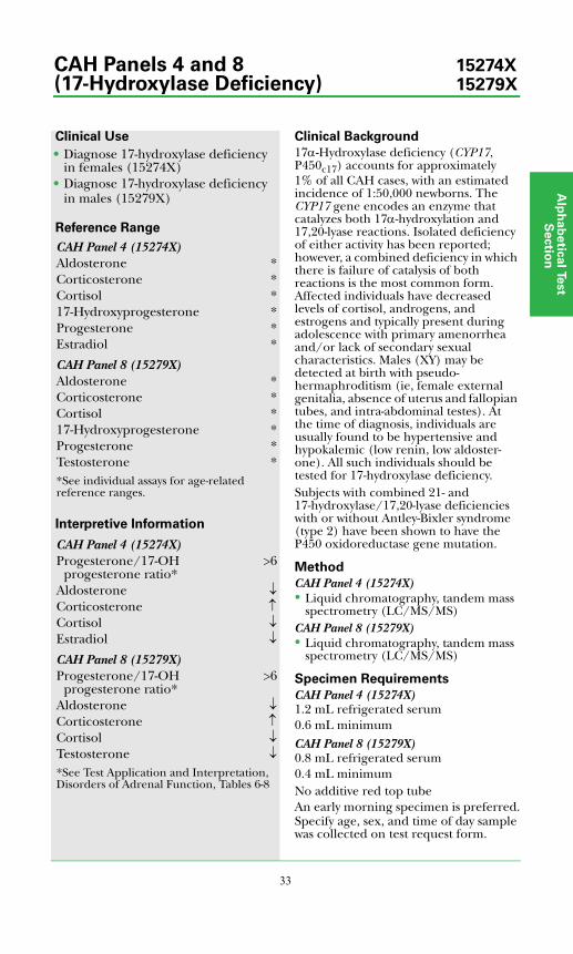
Alp
ha
be
tica
l Test
Se
ctio
n
33
Clinical Use
• Diagnose 17-hydroxylase deficiency in females (15274X)
• Diagnose 17-hydroxylase deficiency in males (15279X)
Reference Range
Interpretive Information
Clinical Background
17α-Hydroxylase deficiency (CYP17, P450c17) accounts for approximately 1% of all CAH cases, with an estimated incidence of 1:50,000 newborns. The CYP17 gene encodes an enzyme that catalyzes both 17α-hydroxylation and 17,20-lyase reactions. Isolated deficiency of either activity has been reported; however, a combined deficiency in which there is failure of catalysis of both reactions is the most common form. Affected individuals have decreased levels of cortisol, androgens, and estrogens and typically present during adolescence with primary amenorrhea and/or lack of secondary sexual characteristics. Males (XY) may be detected at birth with pseudo-hermaphroditism (ie, female external genitalia, absence of uterus and fallopian tubes, and intra-abdominal testes). At the time of diagnosis, individuals are usually found to be hypertensive and hypokalemic (low renin, low aldoster-one). All such individuals should be tested for 17-hydroxylase deficiency.Subjects with combined 21- and 17-hydroxylase/17,20-lyase deficiencies with or without Antley-Bixler syndrome (type 2) have been shown to have the P450 oxidoreductase gene mutation.
Method
CAH Panel 4 (15274X)• Liquid chromatography, tandem mass
spectrometry (LC/MS/MS)CAH Panel 8 (15279X)• Liquid chromatography, tandem mass
spectrometry (LC/MS/MS)
Specimen Requirements
CAH Panel 4 (15274X)1.2 mL refrigerated serum0.6 mL minimumCAH Panel 8 (15279X)0.8 mL refrigerated serum0.4 mL minimumNo additive red top tubeAn early morning specimen is preferred.Specify age, sex, and time of day sample was collected on test request form.
CAH Panel 4 (15274X)Aldosterone *Corticosterone *Cortisol *17-Hydroxyprogesterone *Progesterone *Estradiol *
CAH Panel 8 (15279X)Aldosterone *Corticosterone *Cortisol *17-Hydroxyprogesterone *Progesterone *Testosterone **See individual assays for age-related reference ranges.
CAH Panel 4 (15274X)Progesterone/17-OH
progesterone ratio*>6
Aldosterone ↓Corticosterone ↑Cortisol ↓Estradiol ↓
CAH Panel 8 (15279X)Progesterone/17-OH
progesterone ratio*>6
Aldosterone ↓Corticosterone ↑Cortisol ↓Testosterone ↓*See Test Application and Interpretation, Disorders of Adrenal Function, Tables 6-8
CAH Panels 4 and 8 15274X(17-Hydroxylase Deficiency) 15279X
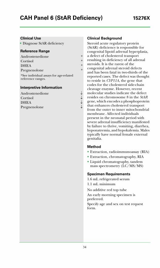
34
Clinical Use
• Diagnose StAR deficiency
Reference Range
Interpretive Information
Clinical Background
Steroid acute regulatory protein (StAR) deficiency is responsible for congenital lipoid adrenal hyperplasia, a defect of cholesterol transport resulting in deficiency of all adrenal steroids. It is the rarest of the congenital adrenal steroid defects and has been fatal in two-thirds of the reported cases. The defect was thought to reside in CYP11A, the gene that codes for the cholesterol side-chain cleavage enzyme. However, recent molecular studies indicate the defect resides on chromosome 8 in the StAR gene, which encodes a phosphoprotein that enhances cholesterol transport from the outer to inner mitochondrial membrane. Affected individuals present in the neonatal period with severe adrenal insufficiency manifested by failure to thrive, vomiting, diarrhea, hyponatremia, and hypokalemia. Males typically have normal female external genitalia.
Method
• Extraction, radioimmunoassay (RIA)• Extraction, chromatography, RIA• Liquid chromatography, tandem
mass spectrometry (LC/MS/MS)
Specimen Requirements
1.6 mL refrigerated serum1.1 mL minimum
No additive red top tubeAn early morning specimen is preferred.Specify age and sex on test request form.
Androstenedione *Cortisol *DHEA *Pregnenolone **See individual assays for age-related reference ranges.
Androstenedione ↓Cortisol ↓DHEA ↓Pregnenolone ↓
CAH Panel 6 (StAR Deficiency) 15276X
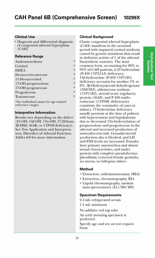
Alp
ha
be
tica
l Test
Se
ctio
n
35
Clinical Use
• Diagnosis and differential diagnosis of congenital adrenal hyperplasia (CAH)
Reference Range
Interpretive Information
Results vary depending on the defect (21-OH, 11β-OH, 17α-OH, 17,20-lyase, 3β-HSD, StAR, or CYPOR deficiency). See Test Application and Interpreta- tion, Disorders of Adrenal Function, Tables 6-8 for more information.
Clinical Background
Classic congenital adrenal hyperplasia (CAH) manifests in the neonatal period with impaired cortisol synthesis caused by genetic mutations that result in deficient activity of 1 of the adrenal biosynthetic enzymes. The most common form, accounting for 90% to 95% of CAH patients, is 21-hydroxylase (P-450 CYP21A2) deficiency; 11β-hydroxylase (P-450 CYP11B1) deficiency accounts for another 5% to 8%. 3β-Hydroxysteroid dehydrogenase (HSD3B2), aldosterone synthase (CYP11B2), steroid acute regulatory protein (StAR), and P-450 oxido-reductase (CYPOR) deficiencies constitute the remainder of cases in infancy. 17-hydroxylase deficiency usually presents at the time of puberty with hypertension and hypokalemia due to decreased 17α-hydroxylation of pregnenolone and progesterone in the adrenal and increased production of mineralocorticoids. Gonadal steroid production also is blocked, and LH and FSH levels are increased. Females have primary amenorrhea and absent sexual characteristics, and males present with complete pseudoherma-phroditism (external female genitalia, no uterus, no fallopian tubes).
Method
• Extraction, radioimmunoassay (RIA)• Extraction, chromatography, RIA• Liquid chromatography, tandem
mass spectrometry (LC/MS/MS)
Specimen Requirements
2.4 mL refrigerated serum1.5 mL minimum
No additive red top tubeAn early morning specimen is preferred.Specify age and sex on test request form.
Androstenedione *Cortisol *DHEA *Deoxycorticosterone *11-Deoxycortisol *17-OH pregnenolone *17-OH progesterone *Progesterone *Testosterone **See individual assays for age-related reference ranges.
CAH Panel 6B (Comprehensive Screen) 10299X
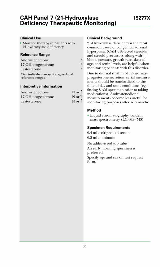
36
Clinical Use
• Monitor therapy in patients with 21-hydroxylase deficiency
Reference Range
Interpretive Information
Clinical Background
21-Hydroxylase deficiency is the most common cause of congenital adrenal hyperplasia (CAH). Selected steroids and steroid precursors, along with blood pressure, growth rate, skeletal age, and renin levels, are helpful when monitoring patients with this disorder. Due to diurnal rhythm of 17-hydroxy-progesterone secretion, serial measure-ments should be standardized to the time of day and same conditions (eg, fasting 8 AM specimen prior to taking medications). Androstenedione measurements become less useful for monitoring purposes after adrenarche.
Method
• Liquid chromatography, tandem mass spectrometry (LC/MS/MS)
Specimen Requirements
0.4 mL refrigerated serum0.2 mL minimum
No additive red top tubeAn early morning specimen is preferred.Specify age and sex on test request form.
Androstenedione *17-OH progesterone *Testosterone **See individual assays for age-related reference ranges.
Androstenedione N or ↑17-OH progesterone N or ↑Testosterone N or ↑
CAH Panel 7 (21-Hydroxylase 15277XDeficiency Therapeutic Monitoring)
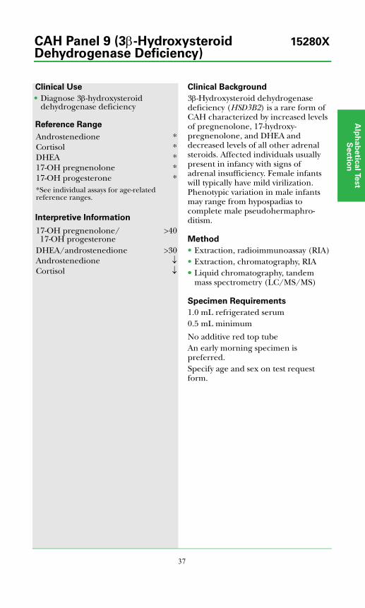
Alp
ha
be
tica
l Test
Se
ctio
n
37
Clinical Use
• Diagnose 3β-hydroxysteroid dehydrogenase deficiency
Reference Range
Interpretive Information
Clinical Background
3β-Hydroxysteroid dehydrogenase deficiency (HSD3B2) is a rare form of CAH characterized by increased levels of pregnenolone, 17-hydroxy-pregnenolone, and DHEA and decreased levels of all other adrenal steroids. Affected individuals usually present in infancy with signs of adrenal insufficiency. Female infants will typically have mild virilization. Phenotypic variation in male infants may range from hypospadias to complete male pseudohermaphro-ditism.
Method
• Extraction, radioimmunoassay (RIA)• Extraction, chromatography, RIA•• Liquid chromatography, tandem
mass spectrometry (LC/MS/MS)
Specimen Requirements
1.0 mL refrigerated serum0.5 mL minimum
No additive red top tubeAn early morning specimen is preferred.Specify age and sex on test request form.
Androstenedione *Cortisol *DHEA *17-OH pregnenolone *17-OH progesterone **See individual assays for age-related reference ranges.
17-OH pregnenolone/17-OH progesterone
>40
DHEA/androstenedione >30Androstenedione ↓
↓Cortisol
CAH Panel 9 (3β-Hydroxysteroid 15280XDehydrogenase Deficiency)
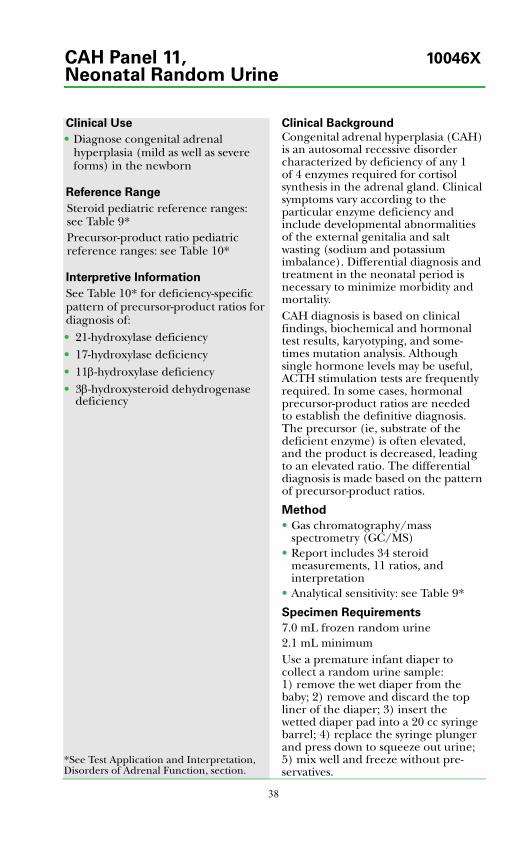
38
Clinical Use
• Diagnose congenital adrenal hyperplasia (mild as well as severe forms) in the newborn
Reference Range
Steroid pediatric reference ranges: see Table 9*Precursor-product ratio pediatric reference ranges: see Table 10*
Interpretive Information
See Table 10* for deficiency-specific pattern of precursor-product ratios for diagnosis of:• 21-hydroxylase deficiency• 17-hydroxylase deficiency• 11β-hydroxylase deficiency• 3β-hydroxysteroid dehydrogenase
deficiency
*See Test Application and Interpretation, Disorders of Adrenal Function, section.
Clinical BackgroundCongenital adrenal hyperplasia (CAH) is an autosomal recessive disorder characterized by deficiency of any 1 of 4 enzymes required for cortisol synthesis in the adrenal gland. Clinical symptoms vary according to the particular enzyme deficiency and include developmental abnormalities of the external genitalia and salt wasting (sodium and potassium imbalance). Differential diagnosis and treatment in the neonatal period is necessary to minimize morbidity and mortality.CAH diagnosis is based on clinical findings, biochemical and hormonal test results, karyotyping, and some-times mutation analysis. Although single hormone levels may be useful, ACTH stimulation tests are frequently required. In some cases, hormonal precursor-product ratios are needed to establish the definitive diagnosis. The precursor (ie, substrate of the deficient enzyme) is often elevated, and the product is decreased, leading to an elevated ratio. The differential diagnosis is made based on the pattern of precursor-product ratios.
Method
• Gas chromatography/mass spectrometry (GC/MS)
• Report includes 34 steroid measurements, 11 ratios, and interpretation
• Analytical sensitivity: see Table 9*
Specimen Requirements
7.0 mL frozen random urine2.1 mL minimumUse a premature infant diaper to collect a random urine sample: 1) remove the wet diaper from the baby; 2) remove and discard the top liner of the diaper; 3) insert the wetted diaper pad into a 20 cc syringe barrel; 4) replace the syringe plunger and press down to squeeze out urine; 5) mix well and freeze without pre-servatives.
CAH Panel 11, 10046XNeonatal Random Urine
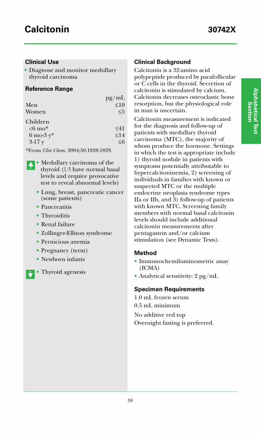
Alp
ha
be
tica
l Test
Se
ctio
n
39
Clinical Use
• Diagnose and monitor medullary thyroid carcinoma
Reference Range
• Medullary carcinoma of the thyroid (1/3 have normal basal levels and require provocative test to reveal abnormal levels)
• Lung, breast, pancreatic cancer (some patients)
• Pancreatitis• Thyroiditis• Renal failure• Zollinger-Ellison syndrome• Pernicious anemia• Pregnancy (term)• Newborn infants
• Thyroid agenesis
Clinical Background
Calcitonin is a 32-amino acid polypeptide produced by parafollicular or C cells in the thyroid. Secretion of calcitonin is stimulated by calcium. Calcitonin decreases osteoclastic bone resorption, but the physiological role in man is uncertain.Calcitonin measurement is indicated for the diagnosis and follow-up of patients with medullary thyroid carcinoma (MTC), the majority of whom produce the hormone. Settings in which the test is appropriate include 1) thyroid nodule in patients with symptoms potentially attributable to hypercalcitoninemia, 2) screening of individuals in families with known or suspected MTC or the multiple endocrine neoplasia syndrome types IIa or IIb, and 3) follow-up of patients with known MTC. Screening family members with normal basal calcitonin levels should include additional calcitonin measurements after pentagastrin and/or calcium stimulation (see Dynamic Tests).
Method
• Immunochemiluminometric assay (ICMA)
• Analytical sensitivity: 2 pg/mL
Specimen Requirements
1.0 mL frozen serum0.5 mL minimum
No additive red topOvernight fasting is preferred.
pg/mLMenWomen
≤10≤5
Children<6 mo*6 mo-3 y*3-17 y
≤41≤14
≤6*From Clin Chem. 2004;50:1828-1829.
Calcitonin 30742X
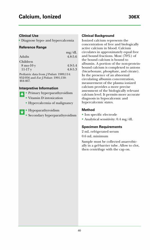
40
Clinical Use
• Diagnose hypo- and hypercalcemia
Reference Range
Interpretive Information
• Primary hyperparathyroidism• Vitamin D intoxication• Hypercalcemia of malignancy
•• Hypoparathyroidism• Secondary hyperparathyroidism
Clinical Background
Ionized calcium represents the concentration of free and biologically active calcium in blood. Calcium circulates in approximately equal free and bound fractions. Most (70%) of the bound calcium is bound to albumin. A portion of the non-protein-bound calcium is complexed to anions (bicarbonate, phosphate, and citrate). In the presence of an abnormal circulating albumin concentration, measurement of the plasma ionized calcium provides a more precise assessment of the biologically relevant calcium level. It permits more accurate diagnosis in hypocalcemic and hypercalcemic states.
Method
• Ion specific electrode• Analytical sensitivity: 0.4 mg/dL
Specimen Requirements
2 mL refrigerated serum0.6 mL minimum
Sample must be collected anaerobic-ally in a gel-barrier tube. Allow to clot, then centrifuge with the cap on.
mg/dLAdults 4.8-5.6Children
8 mo-10 y11-17 y
4.9-5.44.8-5.3
Pediatric data from J Pediatr. 1989;114:952-956 and Eur J Pediatr. 1991;150:464-467.
Calcium, Ionized 306X
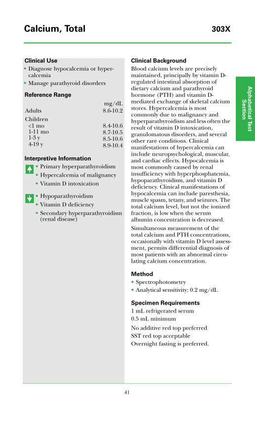
Alp
ha
be
tica
l Test
Se
ctio
n
41
Clinical Use
• Diagnose hypocalcemia or hyper-calcemia
• Manage parathyroid disorders
Reference Range
Interpretive Information
• Primary hyperparathyroidism• Hypercalcemia of malignancy• Vitamin D intoxication
•• Hypoparathyroidism• Vitamin D deficiency• Secondary hyperparathyroidism
(renal disease)
Clinical Background
Blood calcium levels are precisely maintained, principally by vitamin D-regulated intestinal absorption of dietary calcium and parathyroid hormone (PTH) and vitamin D-mediated exchange of skeletal calcium stores. Hypercalcemia is most commonly due to malignancy and hyperparathyroidism and less often the result of vitamin D intoxication, granulomatous disorders, and several other rare conditions. Clinical manifestations of hypercalcemia can include neuropsychological, muscular, and cardiac effects. Hypocalcemia is most commonly caused by renal insufficiency with hyperphosphatemia, hypoparathyroidism, and vitamin D deficiency. Clinical manifestations of hypocalcemia can include paresthesia, muscle spasm, tetany, and seizures. The total calcium level, but not the ionized fraction, is low when the serum albumin concentration is decreased.Simultaneous measurement of the total calcium and PTH concentrations, occasionally with vitamin D level assess-ment, permits differential diagnosis of most patients with an abnormal circu-lating calcium concentration.
Method
• Spectrophotometry• Analytical sensitivity: 0.2 mg/dL
Specimen Requirements
1 mL refrigerated serum0.5 mL minimum
No additive red top preferredSST red top acceptableOvernight fasting is preferred.
mg/dLAdults 8.6-10.2Children
<1 mo1-11 mo1-3 y4-19 y
8.4-10.68.7-10.58.5-10.68.9-10.4
Calcium, Total 303X
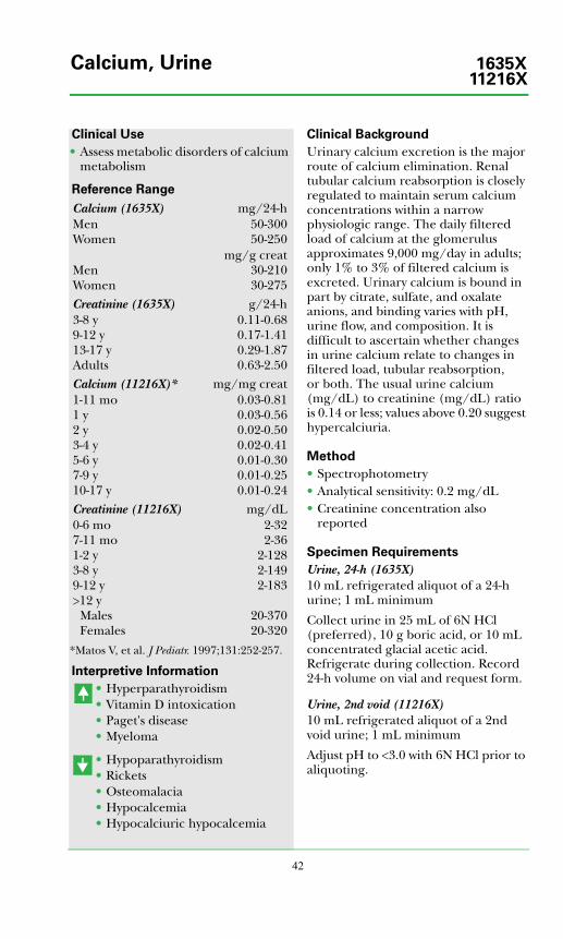
42
Clinical Use
• Assess metabolic disorders of calcium metabolism
Reference Range
*Matos V, et al. J Pediatr. 1997;131:252-257.
Interpretive Information
• Hyperparathyroidism• Vitamin D intoxication• Paget's disease• Myeloma
• Hypoparathyroidism• Rickets• Osteomalacia• Hypocalcemia• Hypocalciuric hypocalcemia
Clinical Background
Urinary calcium excretion is the major route of calcium elimination. Renal tubular calcium reabsorption is closely regulated to maintain serum calcium concentrations within a narrow physiologic range. The daily filtered load of calcium at the glomerulus approximates 9,000 mg/day in adults; only 1% to 3% of filtered calcium is excreted. Urinary calcium is bound in part by citrate, sulfate, and oxalate anions, and binding varies with pH, urine flow, and composition. It is difficult to ascertain whether changes in urine calcium relate to changes in filtered load, tubular reabsorption, or both. The usual urine calcium (mg/dL) to creatinine (mg/dL) ratio is 0.14 or less; values above 0.20 suggest hypercalciuria.
Method
• Spectrophotometry• Analytical sensitivity: 0.2 mg/dL• Creatinine concentration also
reported
Specimen Requirements
Urine, 24-h (1635X)10 mL refrigerated aliquot of a 24-h urine; 1 mL minimum
Collect urine in 25 mL of 6N HCl (preferred), 10 g boric acid, or 10 mL concentrated glacial acetic acid. Refrigerate during collection. Record 24-h volume on vial and request form.
Urine, 2nd void (11216X)10 mL refrigerated aliquot of a 2nd void urine; 1 mL minimum
Adjust pH to <3.0 with 6N HCl prior to aliquoting.
Calcium (1635X) mg/24-hMenWomen
50-30050-250
MenWomen
mg/g creat30-21030-275
Creatinine (1635X) g/24-h3-8 y9-12 y13-17 yAdults
0.11-0.680.17-1.410.29-1.870.63-2.50
Calcium (11216X)* mg/mg creat1-11 mo1 y2 y3-4 y5-6 y7-9 y10-17 y
0.03-0.810.03-0.560.02-0.500.02-0.410.01-0.300.01-0.250.01-0.24
Creatinine (11216X) mg/dL0-6 mo7-11 mo1-2 y3-8 y9-12 y>12 y
MalesFemales
2-322-36
2-1282-1492-183
20-37020-320
Calcium, Urine 1635X11216X
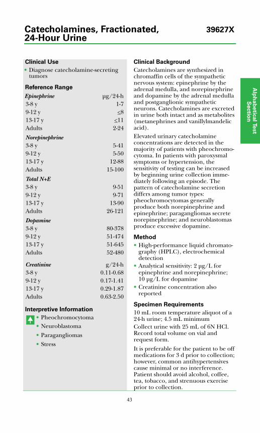
Alp
ha
be
tica
l Test
Se
ctio
n
43
Clinical Use
• Diagnose catecholamine-secreting tumors
Reference Range
Interpretive Information
• Pheochromocytoma• Neuroblastoma
• Paragangliomas
• Stress
Clinical Background
Catecholamines are synthesized in chromaffin cells of the sympathetic nervous system: epinephrine by the adrenal medulla, and norepinephrine and dopamine by the adrenal medulla and postganglionic sympathetic neurons. Catecholamines are excreted in urine both intact and as metabolites (metanephrines and vanillylmandelic acid).Elevated urinary catecholamine concentrations are detected in the majority of patients with pheochromo-cytoma. In patients with paroxysmal symptoms or hypertension, the sensitivity of testing can be increased by beginning urine collection imme-diately following an episode. The pattern of catecholamine secretion differs among tumor types: pheochromocytomas generally produce both norepinephrine and epinephrine; paragangliomas secrete norepinephrine; and neuroblastomas produce excessive dopamine.
Method
• High-performance liquid chromato-graphy (HPLC), electrochemical detection
• Analytical sensitivity: 2 μg/L for epinephrine and norepinephrine; 10 μg/L for dopamine
• Creatinine concentration also reported
Specimen Requirements
10 mL room temperature aliquot of a 24-h urine; 4.5 mL minimumCollect urine with 25 mL of 6N HCl. Record total volume on vial and request form.
It is preferable for the patient to be off medications for 3 d prior to collection; however, common antihypertensives cause minimal or no interference. Patient should avoid alcohol, coffee, tea, tobacco, and strenuous exercise prior to collection.
Epinephrine μg/24-h3-8 y 1-79-12 y <813-17 y <11Adults 2-24
Norepinephrine3-8 y 5-419-12 y 5-5013-17 y 12-88Adults 15-100
Total N+E3-8 y 9-519-12 y 9-7113-17 y 13-90Adults 26-121
Dopamine3-8 y 80-3789-12 y 51-47413-17 y 51-645Adults 52-480
Creatinine g/24-h3-8 y 0.11-0.689-12 y 0.17-1.4113-17 y 0.29-1.87Adults 0.63-2.50
Catecholamines, Fractionated, 39627X24-Hour Urine
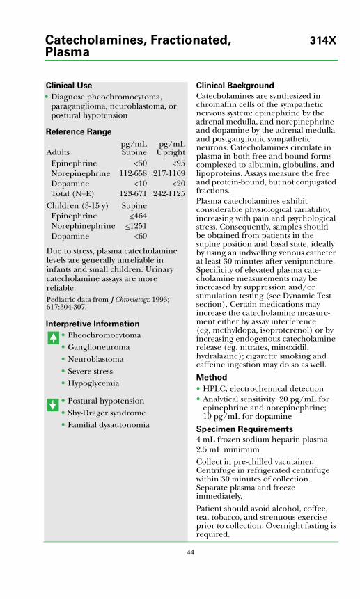
44
Clinical Use
• Diagnose pheochromocytoma, paraganglioma, neuroblastoma, or postural hypotension
Reference Range
Due to stress, plasma catecholamine levels are generally unreliable in infants and small children. Urinary catecholamine assays are morereliable.Pediatric data from J Chromatogr. 1993;617:304-307.
Interpretive Information
• Pheochromocytoma• Ganglioneuroma• Neuroblastoma • Severe stress • Hypoglycemia
•• Postural hypotension• Shy-Drager syndrome• Familial dysautonomia
Clinical Background
Catecholamines are synthesized in chromaffin cells of the sympathetic nervous system: epinephrine by the adrenal medulla, and norepinephrine and dopamine by the adrenal medulla and postganglionic sympathetic neurons. Catecholamines circulate in plasma in both free and bound forms complexed to albumin, globulins, and lipoproteins. Assays measure the free and protein-bound, but not conjugated fractions.Plasma catecholamines exhibit considerable physiological variability, increasing with pain and psychological stress. Consequently, samples should be obtained from patients in the supine position and basal state, ideally by using an indwelling venous catheter at least 30 minutes after venipuncture. Specificity of elevated plasma cate-cholamine measurements may be increased by suppression and/or stimulation testing (see Dynamic Test section). Certain medications may increase the catecholamine measure-ment either by assay interference (eg, methyldopa, isoproterenol) or by increasing endogenous catecholamine release (eg, nitrates, minoxidil, hydralazine); cigarette smoking and caffeine ingestion may do so as well.
Method
• HPLC, electrochemical detection• Analytical sensitivity: 20 pg/mL for
epinephrine and norepinephrine; 10 pg/mL for dopamine
Specimen Requirements
4 mL frozen sodium heparin plasma2.5 mL minimum
Collect in pre-chilled vacutainer. Centrifuge in refrigerated centrifuge within 30 minutes of collection. Separate plasma and freeze immediately.
Patient should avoid alcohol, coffee, tea, tobacco, and strenuous exercise prior to collection. Overnight fasting is required.
Adultspg/mLSupine
pg/mLUpright
Epinephrine <50 <95Norepinephrine 112-658 217-1109Dopamine <10 <20Total (N+E) 123-671 242-1125
Children (3-15 y) SupineEpinephrine <464Norephinephrine <1251Dopamine <60
Catecholamines, Fractionated, 314XPlasma
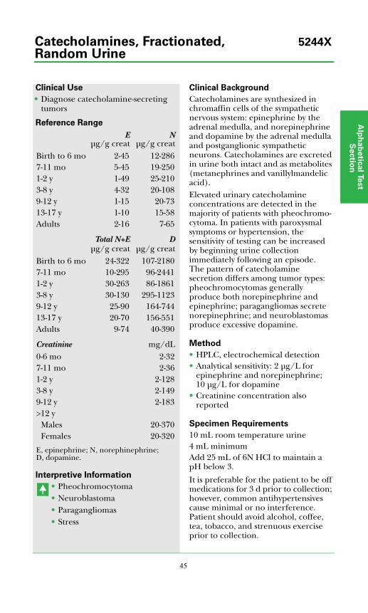
Alp
ha
be
tica
l Test
Se
ctio
n
45
Clinical Use
• Diagnose catecholamine-secreting tumors
Reference Range
E, epinephrine; N, norephinephrine; D, dopamine.
Interpretive Information
• Pheochromocytoma• Neuroblastoma• Paragangliomas• Stress
Clinical Background
Catecholamines are synthesized in chromaffin cells of the sympathetic nervous system: epinephrine by the adrenal medulla, and norepinephrine and dopamine by the adrenal medulla and postganglionic sympathetic neurons. Catecholamines are excreted in urine both intact and as metabolites (metanephrines and vanillylmandelic acid).Elevated urinary catecholamine concentrations are detected in the majority of patients with pheochromo-cytoma. In patients with paroxysmal symptoms or hypertension, the sensitivity of testing can be increased by beginning urine collection immediately following an episode. The pattern of catecholamine secretion differs among tumor types: pheochromocytomas generally produce both norepinephrine and epinephrine; paragangliomas secrete norepinephrine; and neuroblastomas produce excessive dopamine.
Method
• HPLC, electrochemical detection• Analytical sensitivity: 2 μg/L for
epinephrine and norepinephrine; 10 μg/L for dopamine
• Creatinine concentration also reported
Specimen Requirements
10 mL room temperature urine4 mL minimumAdd 25 mL of 6N HCl to maintain a pH below 3.
It is preferable for the patient to be off medications for 3 d prior to collection; however, common antihypertensives cause minimal or no interference. Patient should avoid alcohol, coffee, tea, tobacco, and strenuous exercise prior to collection.
Eμg/g creat
Nμg/g creat
Birth to 6 mo 2-45 12-2867-11 mo 5-45 19-2501-2 y 1-49 25-2103-8 y 4-32 20-1089-12 y 1-15 20-7313-17 y 1-10 15-58Adults 2-16 7-65
Total N+Eμg/g creat
Dμg/g creat
Birth to 6 mo 24-322 107-21807-11 mo 10-295 96-24411-2 y 30-263 86-18613-8 y 30-130 295-11239-12 y 25-90 164-74413-17 y 20-70 156-551Adults 9-74 40-390
Creatinine mg/dL0-6 mo 2-327-11 mo 2-361-2 y 2-1283-8 y 2-1499-12 y 2-183>12 y
Males 20-370Females 20-320
Catecholamines, Fractionated, 5244XRandom Urine
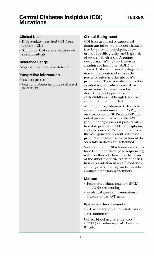
46
Clinical Use
• Differentiate inherited CDI from acquired CDI
• Screen for CDI carrier status in at-risk individuals
Reference Range
Negative (no mutations detected)
Interpretive Information
Mutation present• Central diabetes insipidus (affected
or carrier)
Clinical Background
CDI is an acquired or autosomal dominant inherited disorder character-ized by polyuria, polydipsia, a low urinary specific gravity, and high risk of severe dehydration. Arginine vasopressin (AVP), also known as antidiuretic hormone (ADH), is absent. CDI stems from the degenera-tion or destruction of cells in the posterior pituitary, the site of AVP production. Thus, it is also referred to as pituitary, neurohypophyseal, or neurogenic diabetes insipidus. The disorder typically presents in infancy or early childhood, although late-onset cases have been reported.Although rare, inherited CDI can be caused by mutations in the AVP gene on chromosome 20. Prepro-AVP, the initial protein product of the AVP gene, undergoes several post-transla-tional steps to yield AVP, neurophysin, and glycoprotein. When mutations in the AVP gene are present, cytotoxic products that lead to destruction of the secretory neurons are generated. Since more than 30 relevant mutations have been identified, gene sequencing is the method of choice for diagnosis of the inherited form. After identifica-tion of a mutation in an affected indi-vidual, genetic testing can be used to evaluate other family members.
Method
• Polymerase chain reaction (PCR) and DNA sequencing
• Analytical specificity: mutations in 3 exons of the AVP gene
Specimen Requirements
5 mL room temperature whole blood3 mL minimum
Collect blood in a lavender-top (EDTA) or yellow-top (ACD solution B) tube.
Central Diabetes Insipidus (CDI) 15035XMutations
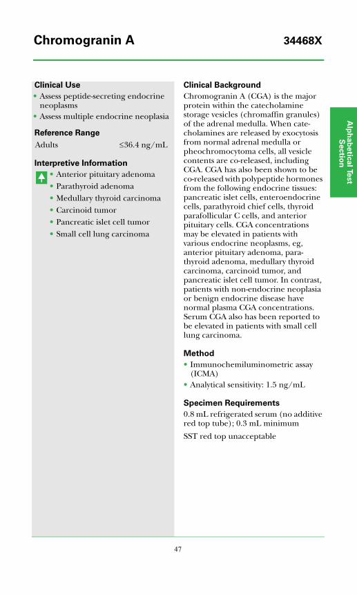
Alp
ha
be
tica
l Test
Se
ctio
n
47
Clinical Use
• Assess peptide-secreting endocrine neoplasms
• Assess multiple endocrine neoplasia
Reference Range
Interpretive Information
• Anterior pituitary adenoma• Parathyroid adenoma• Medullary thyroid carcinoma• Carcinoid tumor• Pancreatic islet cell tumor• Small cell lung carcinoma
Clinical Background
Chromogranin A (CGA) is the major protein within the catecholamine storage vesicles (chromaffin granules) of the adrenal medulla. When cate-cholamines are released by exocytosis from normal adrenal medulla or pheochromocytoma cells, all vesicle contents are co-released, including CGA. CGA has also been shown to be co-released with polypeptide hormones from the following endocrine tissues: pancreatic islet cells, enteroendocrine cells, parathyroid chief cells, thyroid parafollicular C cells, and anterior pituitary cells. CGA concentrations may be elevated in patients with various endocrine neoplasms, eg, anterior pituitary adenoma, para-thyroid adenoma, medullary thyroid carcinoma, carcinoid tumor, and pancreatic islet cell tumor. In contrast, patients with non-endocrine neoplasia or benign endocrine disease have normal plasma CGA concentrations. Serum CGA also has been reported to be elevated in patients with small cell lung carcinoma.
Method
• Immunochemiluminometric assay (ICMA)
• Analytical sensitivity: 1.5 ng/mL
Specimen Requirements
0.8 mL refrigerated serum (no additive red top tube); 0.3 mL minimum
SST red top unacceptable
Adults ≤36.4 ng/mL
Chromogranin A 34468X
48
Clinical Use
• Monitor therapeutic response in patients with metabolic bone disorders
• Predict future bone mineral density (BMD)
• Predict therapeutic response prior to initiation of antiresorptive therapy
• Detect bone metastasis in patients with various malignancies
Reference Range
Interpretive Information
• Osteoporosis• Osteopenia• Celiac disease • Paget’s disease• Primary hyperthyroidism•• Rheumatoid arthritis• Growth hormone deficiency
(non-adult onset)
• Therapeutic response (ie, decrease relative to baseline)
Clinical Background
Healthy levels of bone mineral density (BMD) are maintained by a balance between bone resorption and bone formation. N-telopeptide (NTx), the amino-terminal cross-linked peptide of type I collagen, is released during bone resorption and has been correlated with BMD T-scores. Multiple studies have shown that NTx not only correlates inversely with BMD response to therapy, but also is an early marker or predictor of BMD response. Thus, therapeutic response can be determined within 3 to 6 months of therapy rather than 1 to 2 years. Studies have also demonstrated that elevated pretreatment NTx values predict positive response to therapies such as hormone replacement therapy in postmenopausal women. In patients with malignancies, elevated levels of NTx may indicate bone metastases.
Method
• Immunochemiluminometric assay (ICMA)
• Analytical sensitivity: 10 nmol BCE/L
Specimen Requirements
Urine, 2nd morning void (36167X)Urine, 24-h collection (36421X)
2 mL refrigerated aliquot of urine1 mL minimum Do not use preservatives.Record total 24-h volume on the vial and request form.
Men Units*2nd AM urine
18-29 y 12-9930-59 y 9-60
24-h urine 5-87Women, premenopausal
2nd AM urine 4-6424-h urine 5-79
Test results within the premenopausal reference range do not rule out osteo-porosis nor the need for therapy.
*nmol BCE/mmol creat; BCE, bone collagen equivalent.
Collagen Cross-Linked 36167XN-Telopeptide (NTx), Urine 36421X
Alp
ha
be
tica
l Test
Se
ctio
n
49
Clinical Use
• Monitor therapeutic response in patients with metabolic bone disorders
• Predict future bone mineral density (BMD)
• Predict therapeutic response prior to initiation of antiresorptive therapy
• Detect bone metastasis in patients with various malignancies
Reference Range
Interpretive Information
• Osteoporosis• Osteopenia• Celiac disease• Paget's disease• Primary hyperthyroidism• Rheumatoid arthritis• Growth hormone deficiency
(non-adult onset)
• Therapeutic response (ie, decrease relative to baseline)
Clinical Background
Healthy levels of bone mineral density (BMD) are maintained by a balance between bone resorption and bone formation. C-telopeptide (CTx), a cross-linked peptide of type I collagen, is released during bone resorption and has been correlated with BMD T-scores. CTx correlates inversely with BMD response to therapy and is an early marker or predictor of BMD response. Thus, therapeutic response can be determined within 3 to 6 months of therapy rather than 1 to 2 years. Studies have demonstrated that elevated pretreatment CTx values predict positive response to therapies such as hormone replacement therapy in postmenopausal women. In patients with malignancies, elevated levels of CTx may indicate bone metastases.
Method
• Electrochemiluminescent immunoassay (ECLIA)
• Analytical sensitivity: 30 pg/mL
Specimen Requirements
1 mL frozen serum (no additive red top tube); 0.5 mL minimum
Collect sample between 8 and 10 am after an 8 to 12 hour fast.
Men pg/mL18-29 y 87-120030-39 y 70-78040-49 y 60-70050-68 y 87-345
Women18-29 y 64-64030-39 y 60-65040-49 y 40-465
Collagen Type 1 17406XC-Telopeptide (CTx)
50
Clinical Use
• Assess adrenal gland function• Marker for P450c17 enzyme
deficiency
Reference Range
Interpretive Information
• Adrenal adenoma• Cushing’s disease• P450c17 (17-hydroxylase)
deficiency • P450c11ase (aldosterone
synthetase) deficiency
• Addison’s disease•• Adrenal hypoplasia• ACTH deficiency
Clinical Background
Corticosterone is an intermediate in the adrenal corticosteroid biosynthetic pathway. It is the substrate for further 18-hydroxylation and dehydrogenation to aldosterone. Corticosterone synthesis from 11-deoxycorticosterone (DOC) is catalyzed by P450c11. Measurement is useful in Cushing’s syndrome when exogenous steroid use is suspected. Rare cases of cortico-sterone-secreting adenomas have been reported.
Method
• Liquid chromatography, tandem mass spectrometry (LC/MS/MS)
• Analytical sensitivity: 20.0 ng/dL
Specimen Requirements
1 mL refrigerated serum (no additive red top tube); 0.25 mL minimum
SST red top unacceptable
Adults ng/dL ng/dL8-10 AM 59-12934-6 PM ≤386
ChildrenPremature infants(31-35 wks)*
≤235
Term infants* ≤170Male Female
<1 y 78-1750 89-12001-4 y 120-1290 160-20405-9 y ≤677 ≤67710-13 y ≤1420 ≤142014-17 y ≤1238 ≤1238Tanner stages
II-III 115-1220 110-600IV-V 165-840 160-390
*J Clin Endocrinol Metab. 1992;75:1491-1496.
Corticosterone, LC/MS/MS 6547X
Alp
ha
be
tica
l Test
Se
ctio
n
51
Clinical Use
• Differential diagnosis of Cushing’s syndrome
Reference Range
Interpretive Information
• Cushing’s syndrome (ectopic CRH)
• Pregnancy• Fetus
• Cushing's syndrome (adrenal)
Clinical Background
CRH, a 41-amino acid peptide normally produced in the hypo-thalamus, stimulates the release of ACTH. Normal blood levels of CRH are low, but CRH is also produced by the placenta, and high levels are measured during the second and third trimesters of pregnancy. CRH levels also are increased in Cushing's syndrome due to ectopic production.
Method
• Extraction, radioimmunoassay (RIA)• Analytical sensitivity: 28 pg/mL• Analytical specificity: no cross-
reactivity with ACTH, LH releasing hormone, PACAP-38, arginine vasopressin, urocortin, urocorin, or BNP
Specimen Requirements
3.1 mL refrigerated plasma1.1 mL minimum
Collect blood in special Nichols Institute PTH-RP and Releasing Factor collection tube. Centrifuge immediately in refrigerated centrifuge, remove plasma, and refrigerate.
pg/mLMen <34Women
Nonpregnant <341st trimester pregnancy <402nd trimester pregnancy <1533rd trimester pregnancy <847
ChildrenCord Blood <338
Pregnancy and cord blood reference ranges from J Clin Endocrinol Metab. 1986;63:1199-1203.
Corticotropin Releasing 36589XHormone (CRH)
52
Clinical Use
• Useful in evaluating unexplained cortisol concentrations
Reference Range
Interpretive Information
• Pregnancy• Estrogen therapy• Chronic, active hepatitis• Inherited abnormality
• Hyperinsulinemic states• Nephrotic syndrome• Severe liver disease• Malnutrition• Newborn• Inherited abnormality
Clinical Background
Cortisol-binding globulin (CBG) is a serum alpha-2-globulin-binding protein with high affinity and limited capacity for cortisol. It binds most (±92%) of the serum cortisol; the remaining cortisol (±8%) circulates in a free form or bound to albumin.CBG is synthesized in the liver. Circulating levels increase in preg-nancy or after administration of estrogen or oral contraceptives. Insulin may inhibit synthesis, a fact that might explain the low CBG levels observed in hyperinsulinemic states (eg, diabetes, obesity, PCO). CBG levels are also reduced in nephrotic syndrome, starvation, or severe chronic liver disease.The CBG assay is helpful in assessing unexpected values of blood cortisol. Other assays useful in diagnosis and therapy of cortisol abnormalities include plasma cortisol, ACTH, β-lipotropin, urinary cortisol (by HPLC), or free cortisol in plasma or serum.
Method
• Radioimmunoassay (RIA)• Analytical sensitivity: 0.1 mg/L
Specimen Requirements
1 mL refrigerated serum0.2 mL minimum
No additive red top preferredSST red top acceptable
mg/LAdults 19-45Children
Preterm infants (<8 d) 6-26Pediatric data from Pediatr Res.1994;37:112-116.
Cortisol-Binding Globulin (CBG) 37371X
Alp
ha
be
tica
l Test
Se
ctio
n
53
Clinical Use
• Diagnose Cushing’s syndrome• Differentiate between exogenous
and endogenous Cushing’s syndrome
• Marker for apparent mineral-ocorticoid excess (AME) syndrome
Reference Range
Interpretive Information
•• Cushing’s syndrome
• Exogenous Cushing’s syndrome• Adrenal insufficiency
Clinical Background
The differential diagnosis of Cushing's syndrome has been based on ACTH and cortisol assays in urine or serum. Recent data have shown utility for combined urinary free cortisol and cortisone measurements by high performance liquid chromatography (HPLC). Using HPLC, the diagnostic sensitivity for Cushing's syndrome is 93%, an increase of 17% over cortisol and 7% over cortisone. Both cortisol and cortisone levels are elevated in patients with pituitary, ectopic, and adrenal Cushing's syndrome, while both cortisol and cortisone levels are decreased in patients receiving exogenous glucocorticoid (eg, pred-nisolone). Additionally, the measure-ment of cortisol and cortisone can be used for the diagnosis of apparent mineralocorticoid excess (AME) in which cortisone formation is reduced, allowing cortisol to act as a mineral-ocorticoid.
Method
• Liquid chromatography, tandem mass spectrometry (LC/MS/MS)
• Analytical sensitivity: 1 μg/L• Analytical specificity: high
concentrations of 20β-DHE/DHF decrease results; high concentrations of prednisolone interfere with measurement.
• Creatinine concentration also reported
Specimen Requirements
10 mL refrigerated aliquot of a 24-h urine; 2.6 mL minimum
Collect urine with 10 g of boric acid or keep refrigerated during collection. Record 24-h volume on vial and request form.
Cortisol, Free μg/24-hAdults 4.0-50.0Children
1-4.9 y 0.9-8.25-9.9 y 1.0-30.010-13.9 y 1.0-45.014-17.9 y 3.0-55.0
Cortisone μg/24-hAdults 23-195Children
1-4.9 y 5-405-9.9 y 9-15010-13.9 y 11-14514-17.9 y 1-166
Creatinine g/24-h3-8 y 0.11-0.689-12 y 0.17-1.4113-17 y 0.29-1.87Adults 0.63-2.50
Cortisol, Free and Cortisone, 37355X24-Hour Urine
54
Clinical Use
• Screen for Cushing's syndrome and adrenal insufficiency
Reference Range
Interpretive Information
• Adrenal hyperfunction, ie, Cushing’s disease
• Adrenal insufficiency
Clinical Background
Cortisol primarily circulates bound to cortisol-binding globulin (CBG). Only the unbound (free) cortisol appears to be biologically active. The free fraction is 5% to 8% of total cortisol in normal individuals.Since the cortisol binding capacity of blood proteins is limited, adrenal hyperfunction (Cushing's syndrome) is invariably associated with a many-fold increase in urinary cortisol levels. Urinary free cortisol is not substantially increased in pregnancy, obesity, diabetes, or hyperthyroidism, entities that in the past could represent a diagnostic dilemma when older assays of cortisol or metabolites were used.Exogenous Cushing's syndrome due to surreptitious use of glucocorticoid can now be diagnosed by finding low cortisol and presence of synthetic steroids in the urine.Urinary cortisol is the best screening test for Cushing's syndrome and can be used to rule out Addison's disease.
Method
• Liquid chromatography, tandem mass spectrometry (LC/MS/MS)
• Analytical sensitivity: 1 μg/L• Creatinine concentration also
reported
Specimen Requirements
2 mL frozen aliquot of a 24-h urine0.5 mL minimum
No preservative (preferred)25 mL 6N HCL acceptable10 g boric acid acceptable10 mL concentrated glacial acetic acid acceptable
Record 24-h volume on vial and request form.
Cortisol, Free μg/24-hAdults 4-50Children
1-4 y 0.9-8.25-9 y 1.0-30.010-13 y 1.0-45.014-17 y 3.0-55.0
Creatinine g/24-h3-8 y 0.11-0.689-12 y 0.17-1.4113-17 y 0.29-1.87Adults 0.63-2.50
Cortisol, Free, 24-Hour Urine 11280X
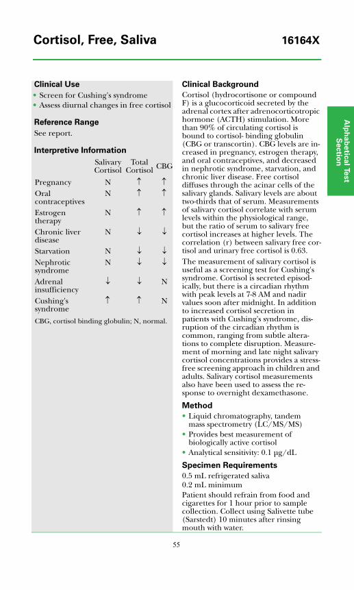
Alp
ha
be
tica
l Test
Se
ctio
n
55
Clinical Use
• Screen for Cushing's syndrome• Assess diurnal changes in free cortisol
Reference Range
See report.
Interpretive Information
Clinical Background
Cortisol (hydrocortisone or compound F) is a glucocorticoid secreted by the adrenal cortex after adrenocorticotropic hormone (ACTH) stimulation. More than 90% of circulating cortisol is bound to cortisol- binding globulin (CBG or transcortin). CBG levels are in-creased in pregnancy, estrogen therapy, and oral contraceptives, and decreased in nephrotic syndrome, starvation, and chronic liver disease. Free cortisol diffuses through the acinar cells of the salivary glands. Salivary levels are about two-thirds that of serum. Measurements of salivary cortisol correlate with serum levels within the physiological range, but the ratio of serum to salivary free cortisol increases at higher levels. The correlation (r) between salivary free cor-tisol and urinary free cortisol is 0.63.The measurement of salivary cortisol is useful as a screening test for Cushing's syndrome. Cortisol is secreted episod-ically, but there is a circadian rhythm with peak levels at 7-8 AM and nadir values soon after midnight. In addition to increased cortisol secretion in patients with Cushing's syndrome, dis-ruption of the circadian rhythm is common, ranging from subtle altera-tions to complete disruption. Measure-ment of morning and late night salivary cortisol concentrations provides a stress-free screening approach in children and adults. Salivary cortisol measurements also have been used to assess the re-sponse to overnight dexamethasone.
Method
• Liquid chromatography, tandem mass spectrometry (LC/MS/MS)
• Provides best measurement of biologically active cortisol
• Analytical sensitivity: 0.1 μg/dL
Specimen Requirements
0.5 mL refrigerated saliva0.2 mL minimumPatient should refrain from food and cigarettes for 1 hour prior to sample collection. Collect using Salivette tube (Sarstedt) 10 minutes after rinsing mouth with water.
SalivaryCortisol
TotalCortisol CBG
Pregnancy N ↑ ↑Oral contraceptives
N ↑ ↑
Estrogen therapy
N ↑ ↑
Chronic liver disease
N ↓ ↓
Starvation N ↓ ↓Nephrotic syndrome
N ↓ ↓
Adrenal insufficiency
↓ ↓ N
Cushing's syndrome
↑ ↑ N
CBG, cortisol binding globulin; N, normal.
Cortisol, Free, Saliva 16164X

56
Clinical Use
• Determine glucocorticoid status
Reference Range
Interpretive Information
Clinical Background
Cortisol (hydrocortisone or compound F) is a glucocorticoid secreted by the adrenal cortex after adrenocortico-tropic hormone (ACTH) stimulation. More than 90% of circulating cortisol is bound to cortisol-binding globulin (CBG or transcortin). CBG levels are increased in pregnancy, estrogen therapy, and oral contraceptives, and decreased in nephrotic syndrome, starvation, and chronic liver disease. Thus, total cortisol levels can be affected by these conditions.Free cortisol, however, is the physio-logically active form. Free cortisol levels change independently and accurately reflect the clinical status; thus serum free cortisol is the most reliable test for glucocorticoid status. It is markedly elevated in Cushing's syndrome and falls to almost undetectable levels after dexamethasone (1 mg overnight) in normal individuals.
Method
• Liquid chromatography, tandem mass spectrometry (LC/MS/MS)
• Provides best measurement of biologically active cortisol
• Analytical sensitivity: 0.1 μg/dL
Specimen Requirements
2 mL refrigerated serum0.7 mL minimum
No additive red top tube preferredSST red top acceptable
See report
FreeCortisol
Total Cortisol CBG
Pregnancy N ↑ ↑Oral contraceptives
N ↑ ↑
Estrogen therapy
N ↑ ↑
Chronic liverdisease
N ↓ ↓
Starvation N ↓ ↓Nephrotic syndrome
N ↓ ↓
Adrenal insufficiency
↓ ↓ N
Cushing’s syndrome
↑ ↑ N
CBG, cortisol binding globulin; N, normal.
Cortisol, Free, Serum 36423X
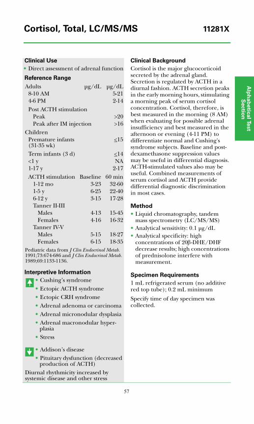
Alp
ha
be
tica
l Test
Se
ctio
n
57
Clinical Use
• Direct assessment of adrenal function
Reference Range
Interpretive Information
• Cushing’s syndrome• Ectopic ACTH syndrome• Ectopic CRH syndrome• Adrenal adenoma or carcinoma• Adrenal micronodular dysplasia• Adrenal macronodular hyper-
plasia• Stress
• Addison’s disease• Pituitary dysfunction (decreased
production of ACTH)Diurnal rhythmicity increased by systemic disease and other stress
Clinical Background
Cortisol is the major glucocorticoid secreted by the adrenal gland. Secretion is regulated by ACTH in a diurnal fashion. ACTH secretion peaks in the early morning hours, stimulating a morning peak of serum cortisol concentration. Cortisol, therefore, is best measured in the morning (8 AM) when evaluating for possible adrenal insufficiency and best measured in the afternoon or evening (4-11 PM) to differentiate normal and Cushing's syndrome subjects. Baseline and post-dexamethasone suppression values may be useful in differential diagnosis. ACTH-stimulated values also may be useful. Combined measurements of serum cortisol and ACTH provide differential diagnostic discrimination in most cases.
Method
• Liquid chromatography, tandem mass spectrometry (LC/MS/MS)
• Analytical sensitivity: 0.1 μg/dL• Analytical specificity: high
concentrations of 20β-DHE/DHF decrease results; high concentrations of prednisolone interfere with measurement.
Specimen Requirements
1 mL refrigerated serum (no additive red top tube); 0.2 mL minimum
Specify time of day specimen was collected.
Adults μg/dL μg/dL8-10 AM 5-214-6 PM 2-14Post ACTH stimulation
Peak >20Peak after IM injection >16
ChildrenPremature infants (31-35 wk)
<15
Term infants (3 d) <14<1 y NA1-17 y 2-17ACTH stimulation Baseline 60 min
1-12 mo 3-23 32-601-5 y 6-25 22-406-12 y 3-15 17-28Tanner II-III
Males 4-13 15-45Females 4-16 16-32
Tanner IV-VMales 5-15 18-27Females 6-15 18-35
Pediatric data from J Clin Endocrinol Metab. 1991;73:674-686 and J Clin Endocrinol Metab. 1989;69:1133-1136.
Cortisol, Total, LC/MS/MS 11281X
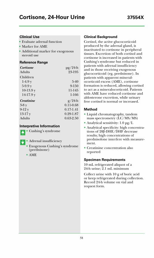
58
Clinical Use
• Evaluate adrenal function• Marker for AME• Additional marker for exogenous
steroid use
Reference Range
Interpretive Information
• Cushing’s syndrome
• Adrenal insufficiency• Exogenous Cushing’s syndrome
(prednisone)• AME
Clinical Background
Cortisol, the active glucocorticoid produced by the adrenal gland, is inactivated to cortisone in peripheral tissues. Excretion of both cortisol and cortisone is increased in patients with Cushing's syndrome but reduced in patients with adrenal insufficiency and in those receiving exogenous glucocorticoid (eg, prednisone). In patients with apparent mineral-ocorticoid excess (AME), cortisone formation is reduced, allowing cortisol to act as a mineralocorticoid. Patients with AME have reduced cortisone and aldosterone excretion, while urinary free cortisol is normal or increased.
Method
• Liquid chromatography, tandem mass spectrometry (LC/MS/MS)
• Analytical sensitivity: 1.0 μg/L• Analytical specificity: high concentra-
tions of 20β-DHE/DHF decrease results; high concentrations of prednisolone interfere with measure-ment.
• Creatinine concentration also reported
Specimen Requirements
10 mL refrigerated aliquot of a 24-h urine; 2.1 mL minimum
Collect urine with 10 g of boric acid or keep refrigerated during collection. Record 24-h volume on vial and request form.
Cortisone μg/24-hAdults 23-195Children
1-4.9 y 5-405-9.9 y 9-15010-13.9 y 11-14514-17.9 y 1-166
Creatinine g/24-h3-8 y 0.11-0.689-12 y 0.17-1.4113-17 y 0.29-1.87Adults 0.63-2.50
Cortisone, 24-Hour Urine 37554X
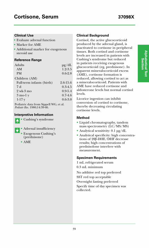
Alp
ha
be
tica
l Test
Se
ctio
n
59
Clinical Use
• Evaluate adrenal function• Marker for AME• Additional marker for exogenous
steroid use
Reference Range
Interpretive Information
• Cushing’s syndrome
• Adrenal insufficiency• Exogenous Cushing’s
(prednisone)• AME
Clinical Background
Cortisol, the active glucocorticoid produced by the adrenal gland, is inactivated to cortisone in peripheral tissues. Both cortisol and cortisone levels are increased in patients with Cushing's syndrome but reduced in patients receiving exogenous glucocorticoid (eg, prednisone). In apparent mineralocorticoid excess (AME), cortisone formation is reduced, allowing cortisol to act as a mineralocorticoid. Patients with AME have reduced cortisone and aldosterone levels but normal cortisol levels. Licorice ingestion can inhibit conversion of cortisol to cortisone, thereby decreasing circulating cortisone levels.
Method
• Liquid chromatography, tandem mass spectrometry (LC/MS/MS)
• Analytical sensitivity: 0.1 μg/dL• Analytical specificity: high concentra-
tions of 20β-DHE/DHF decrease results; high concentrations of prednisolone interfere with measurement.
Specimen Requirements
1 mL refrigerated serum0.3 mL minimum
No additive red top preferredSST red top acceptableOvernight fasting preferred Specify time of day specimen was collected.
Adults μg/dLAM 1.2-3.5PM 0.6-2.8
Children (AM)Full-term infants (birth) 2.6-15.67 d 0.3-4.52 wk-3 mo 0.9-5.43 mo-1 y 0.7-4.61-17 y 0.6-3.0
Pediatric data from Sippell WG, et al. Pediatr Res. 1980;14:39-46.
Cortisone, Serum 37098X
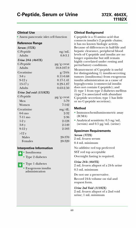
60
Clinical Use
• Assess pancreatic islet cell function
Reference Range
Interpretive Information
• Insulinoma• Type 2 diabetes
• Type 1 diabetes• Exogenous insulin
administration
Clinical Background
C-peptide is a 31-amino acid that connects insulin's 2 peptide chains; it has no known biologic activity. Because of differences in half-life and hepatic clearance, peripheral blood levels of C-peptide and insulin are no longer equimolar but still remain highly correlated under resting and perturbatory conditions.Measurement of C-peptide is useful for distinguishing 1) insulin-secreting tumors (insulinoma) from exogenous insulin administration as a cause of hypoglycemia (commercial insulin does not contain C-peptide); and 2) type 1 from type 2 diabetes mellitus (type 2 is associated with abundant C-peptide secretion; type 1 has little or no C-peptide secretion).
Method
• Immunochemiluminometric assay (ICMA)
• Analytical sensitivity: 0.5 ng/mL (serum) and 0.5 μg/mL (urine)
Specimen Requirements
Serum (372X)2 mL frozen serum0.4 mL minimum
No additive red top preferredSST red top acceptableOvernight fasting is required.
Urine 24-h (4643X)2 mL frozen aliquot of a 24-h urine0.5 mL minimum
Do not use a preservative.Record 24-h volume on vial and request form.
Urine 2nd Void (11182X)2 mL frozen aliquot of a 2nd void urine; 1 mL minimum
Serum (372X)C-Peptide ng/mL
Adults 0.8-3.1Urine 24-h (4643X)C-Peptide μg/g creat
Adults 10.8-107.9Creatinine g/24-h
3-8 y 0.11-0.689-12 y 0.17-1.4113-17 y 0.29-1.87Adults 0.63-2.50
Urine 2nd void (11182X)C-Peptide μg/g creat
Men 5-79Women 7-142
Creatinine mg/dL0-6 mo 2-327-11 mo 2-361-2 y 2-1283-8 y 2-1499-12 y 2-183>12 y
Males 20-370Females 20-320
C-Peptide, Serum or Urine 372X. 4643X,11182X
Alp
ha
be
tica
l Test
Se
ctio
n
61
Clinical Use
• Diagnose hyperparathyroidism• Diagnose pseudohypoparathyroidism
subtypes• Assess severity of hypoparathyroidism
Reference Range
Interpretive Information
• Hyperparathyroidism• Hypercalcemia of malignancy
• Hypoparathyroidism• Type I pseudohypopara-
thyroidism• Renal insufficiency
Clinical Background
3´, 5´ cyclic adenosine monophosphate (cAMP) is an intracellular second messenger produced by the action of adenyl cyclase upon ATP. Significant amounts of intracellular cAMP appear in plasma with a diurnal rhythm peak at 1200 hours and a nadir in late evening. Approximately half of urinary cAMP is filtered from plasma while the other half is derived from kidney tissue. cAMP in the kidney is generated following parathyroid hormone (PTH) stimulation of renal PTH receptors. Urinary cAMP is generally elevated in patients with hyperparathyroidism. It may also be increased in cancer patients with hypercalcemia. Some patients with pseudohypopara-thyroidism, a condition characterized by decreased renal responsiveness to parathyroid hormone, fail to respond with a rise in urinary cAMP when exogenous PTH is given. Measurement of nephrogenous cAMP may still be used on rare occasions to diagnose subtypes of pseudohypopara-thyroidism.
Method
• Extraction, radioimmunoassay
Specimen Requirements
Nephrogenous (37555X)25 mL frozen urine and 5 mL frozen EDTA plasma; 2.5 mL and 1.2 mL minimum, respectively
Collect urine with 2.0 mL of 6N HCl to maintain pH below 3.
Plasma (242X)3.1 mL frozen EDTA plasma1 mL minimum
Random urine (225X)1 mL frozen urine0.2 mL minimum
Collect urine with 2.0 mL of 6N HCl to maintain pH below 3.
Nephrogenous(37555X)1.4-5.0 nmol/dL
Plasma (242X, 37555X)3.9-13.1 nmol/L
Random urine (225X, 37555X)0.8-7.5 μmol/L
Creatinine, Plasma (37555X) mg/dL
Adults 0.6-1.4Children, full-term infants*
0-2 d 0.7-1.43-5 d 0.4-1.16-7 d 0.3-0.91 wk-1 mo 0.3-0.81 mo-1 y 0.2-0.72-9 y 0.3-0.710-17 y 0.5-1.0
Creatinine, random urine (37555X)mg/dL
0-6 mo 2-327-11 mo 2-361-2 y 2-1283-8 y 2-1499-12 y 2-183>12 y
Males 20--370Females 20-320
*Values for children <2 y from Savory DJ, Ann Clin Biochem. 1990;27:99-101.
Cyclic Adenosine Monophosphate 37555X, 242X,(Cyclic AMP) 225X
62
Clinical Use
• Assess glomerular filtration rate in type 1 and 2 diabetes mellitus
Reference Range
Interpretive Information
• Impaired glomerular filtration
Clinical Background
Cystatin C is a low molecular weight (13,359 Da) protein belonging to the superfamily of cystine protease inhibitors. It is produced by all nucleated cells at a reasonably constant rate; production is minimally affected by diet, inflammatory states, lean body mass, or circadian rhythm. Like creatinine, cystatin C is freely filtered by the glomeruli. It is reabsorbed and metabolized by renal tubular cells and does not appear in urine. Tan et al (Diabetes Care. 2002;25:2004-2009) showed serum cystatin C has good correlation with creatinine clearance (p = 0.74) and iohexol clearance (p = -0.80) and is a useful marker of renal dysfunction in diabetic patients with minimal tubular reabsorption. Because creatinine production is relatively variable, cystatin C has been proposed as a better marker of glomerular filtration.
Method
• Nephelometry
Specimen Requirements
1.0 mL refrigerated serum0.5 mL minimum
No additive red top preferredSST red top acceptableOvernight fasting is required.
Children & Adults 0.5-1.0 mg/L
Cystatin C 10570X
Alp
ha
be
tica
l Test
Se
ctio
n
63
Clinical Use
• Diagnose adrenal enzyme deficiencies• Assess adrenal functional disorders
Reference Range
Interpretive Information
• Congenital adrenal hyperplasia (P-450c11 deficiency)
• P-450c17 deficiency
• P-450c11ase (aldosterone synthetase) deficiency
• Cushing’s syndrome
• Primary aldosteronism
• Low renin essential hypertension
• Adrenal carcinoma (some cases)
• Adrenal insufficiency
• Adrenal hypoplasia
Clinical Background
Deoxycorticosterone (DOC) is a weak mineralocorticoid derived from 21-hydroxylation of progesterone in the adrenal cortex. DOC is a precursor of corticosterone and aldosterone and, like cortisol, is under the primary control of ACTH. DOC can be used therapeutically as a replacement mineralocorticoid in patients with primary adrenal insufficiency or isolated hypoaldosteronism.
Method
• Extraction, chromatography, radioimmunoassay (RIA)
• Analytical sensitivity: 1 ng/dL
Specimen Requirements
3 mL refrigerated serum1.1 mL minimum
No additive red top preferredSST red top acceptable
ng/dLMen 3.5-11.5Women
Follicular phase 1.5-8.5Luteal phase 3.5-13.0Pregnancy
First trimester 5-25Second trimester 10-75Third trimester 30-110
ACTH stimulation
Baselineng/dL
60 minng/dL
Men and premeno-pausal women (follicular phase)
14-33
<1 y 7-57 20-1601-5 y 4-49 26-1406-12 y
Males 9-34 33-140Females 2-13 19-61
Tanner II-IIIMales 4-30 12-74Females 2-12 13-63
Tanner IV-VMales 5-14 19-46Females 5-10 23-40
Pediatric data from J Clin Endocrinol Metab. 1991;73:674-686.
Deoxycorticosterone (DOC) 6559X
64
Clinical Use
• Marker for adrenal P-450c11 (11β- hydroxylase) enzyme deficiency
• Assess pituitary-adrenal reserve with the metyrapone test
Reference Range
Marked elevation of 21-deoxycortisol, as may occur in 21-hydroxylase defi-ciency, can increase apparent levels of 11-deoxycortisol.
Interpretive Information
• Congenital adrenal hyperplasia (P-450c11 deficiency)
• After metyrapone in normal subjects
• Adrenal insufficiency
Clinical Background
Cortisol is formed in the adrenal gland by the enzymatic action of 11β-hydroxylase (P-450c11) on 11-deoxycortisol. Deficiency of this enzyme causes congenital adrenal hyperplasia (CAH) characterized by hypertension and virilization. ACTH, 11-deoxycortisol, and androgen levels are all increased. This defect accounts for about 5% of CAH cases.The drug metyrapone blocks 11β-hydroxylase, which in normal individuals results in increased ACTH and 11-deoxycortisol production. Thus, the drug is used to test adrenal and pituitary reserve. In patients with primary and secondary adrenal insufficiency, there is little or no increase in 11-deoxycortisol in response to metyrapone administra-tion. In patients with Cushing's syndrome caused by ectopic ACTH or adrenal tumors, metyrapone does not change ACTH or 11-deoxycortisol levels (see Dynamic Test section). Measurements of cortisol and ACTH have generally replaced the metyrapone test.
Method
• Liquid chromatography, tandem mass spectrometry (LC/MS/MS)
• Analytical sensitivity: 20.0 ng/dL
Specimen Requirements
1 mL frozen serum (no additive red top tube); 0.25 mL minimum
SST red top unacceptable.An early-morning specimen is preferred.
Men ng/dL18-29 y <11930-39 y <13540-49 y <7650-59 y <42
Women18-29 y <10730-39 y <5140-49 y <6250-66 y <37
ChildrenPremature infants (31-35 wk)*
<235
Term infants* <1701-12 mo 10-2001-4 y 7-2105-9 y <12210-13 y <24514-17 y <302Tanner II-III
Males 11-150Females 15-130
Tanner IV-VMales 14-120Females 17-120
*Data from J Clin Endocrinol Metab. 1991;73:674-686 and J Clin Endocrinol Metab. 1989;69:1133-1136.
11-Deoxycortisol, LC/MS/MS 30543X
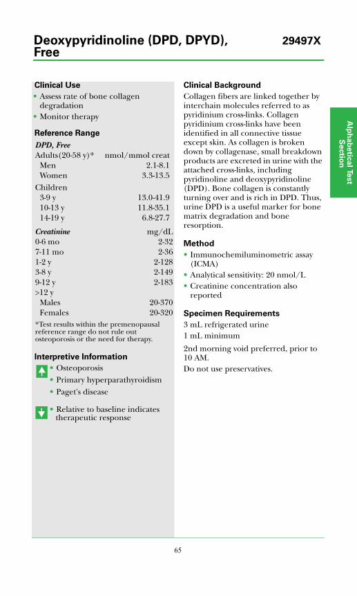
Alp
ha
be
tica
l Test
Se
ctio
n
65
Clinical Use
• Assess rate of bone collagen degradation
• Monitor therapy
Reference Range
Interpretive Information
• Osteoporosis• Primary hyperparathyroidism• Paget's disease
• Relative to baseline indicates therapeutic response
Clinical Background
Collagen fibers are linked together by interchain molecules referred to as pyridinium cross-links. Collagen pyridinium cross-links have been identified in all connective tissue except skin. As collagen is broken down by collagenase, small breakdown products are excreted in urine with the attached cross-links, including pyridinoline and deoxypyridinoline (DPD). Bone collagen is constantly turning over and is rich in DPD. Thus, urine DPD is a useful marker for bone matrix degradation and bone resorption.
Method
• Immunochemiluminometric assay (ICMA)
• Analytical sensitivity: 20 nmol/L• Creatinine concentration also
reported
Specimen Requirements
3 mL refrigerated urine1 mL minimum
2nd morning void preferred, prior to 10 AM.Do not use preservatives.
DPD, FreeAdults(20-58 y)* nmol/mmol creat
Men 2.1-8.1Women 3.3-13.5
Children3-9 y 13.0-41.910-13 y 11.8-35.114-19 y 6.8-27.7
Creatinine mg/dL0-6 mo 2-327-11 mo 2-361-2 y 2-1283-8 y 2-1499-12 y 2-183>12 y
Males 20-370Females 20-320
*Test results within the premenopausal reference range do not rule out osteoporosis or the need for therapy.
Deoxypyridinoline (DPD, DPYD), 29497XFree
66
Clinical Use
• Monitor therapy or compliance• Evaluate hypothalamic-pituitary-
adrenal axis
Reference Range
Interpretive Information
• Dexamethasone administration
Clinical Background
Dexamethasone is a potent synthetic glucocorticoid widely used in medicine. The dexamethasone suppression test (DST) is a standard diagnostic tool for Cushing’s disease and has been applied to the evaluation of hypothalamic-pituitary-adrenal axis function in patients with psychiatric disease. The measurement of dexamethasone in serum allows the physician to assess compliance during therapy or when evaluating DST results.
Method
• Liquid chromatography, tandem mass spectrometry (LC/MS/MS)
• Analytical sensitivity: 20 ng/dL
Specimen Requirements
0.85 mL refrigerated serum (no additive red top tube); 0.25 mL minimum
SST red top unacceptableCollect specimen between 8 and 10 AM.
ng/dLBaseline <208-10 AM, after 1 mg overnight dexamethasone
180-550
Dexamethasone 29391X
Alp
ha
be
tica
l Test
Se
ctio
n
67
Clinical Use
• Marker of adrenal androgen production
• Assess adrenal reserve after ACTH stimulation
Reference Range
Interpretive Information
• Adrenal tumors• Cushing’s disease• Congenital adrenal hyperplasia• Premature adrenarche
• Addison’s disease• Anorexia nervosa
Clinical Background
Dehydroepiandrosterone (DHEA) is a weak androgen synthesized by the adrenal cortex. It has a short half-life and is usually converted to dehydro-epiandrosterone sulfate.Excessive DHEA secretion can produce acne, hirsutism, and virilization via conversion to testosterone.
Method
Serum (410X)• Extraction, chromatography,
radioimmunoassay (RIA)• Analytical sensitivity: 3 ng/dL
Urine (38954X)• Gas chromatography, mass
spectrophotometry (GC/MS)
Specimen Requirements
Serum (410X)1 mL refrigerated serum0.3 mL minimum
No additive red top preferredSST red top acceptable
Overnight fasting is preferred. Specify age and sex on test request form.
Urine (38954X)5 mL frozen aliquot of a 24-h urine2.1 mL minimum
Refrigerate during collection; do not use a preservative. Record 24-h volume on vial and request form.
Serum (410X) ng/dLMen 180-1250Women 130-980
Pregnancy 135-810Post-ACTH stimulation 60 min
Men and women 545-1845Children*
Premature infants (31-35 wk)
<3343
Term infants (1st wk of life)
<761
ACTH stimulation
Baselineng/dL
60 minng/dL
<1 y 26-585 18-14551-5 y 9-42 21-986-12 y 11-155 34-320
Tanner II-IIIMales 25-300 62-390Females 69-605 95-885
Tanner IV-VMales 100-400 195-510Females 165-690 325-1460
Urine (38954X) μg/24-h μg/g/creatDHEA, Adults 21-2710DHEA, Men 24-1640DHEA, Women 13-730Creatinine g/24-h
3-8 y 0.11-0.689-12 y 0.17-1.4113-17 y 0.29-1.87Adults 0.63-2.50
*Pediatric data from J Clin Endocrinol Metab. 1991;73:674-686.
DHEA (Dehydroepiandrosterone) 410X38954X
68
Clinical Use
• Marker for adrenal cortical function and disease
Reference Range
Interpretive Information
• Congenital adrenal hyperplasia• Adrenal carcinoma• Virilizing tumors of the adrenals• Cushing’s disease, pituitary
dependent
• Addison’s disease• Adrenal hypoplasia
Clinical Background
Dehydroepiandrosterone sulfate (DHEA-S), which is synthesized almost exclusively by the adrenals, is a weak androgen, the most abundant C19 plasma steroid, and the major source of urinary 17-ketosteroids. Measuring serum DHEA-S, therefore, can replace older urinary 17-ketosteroid deter-minations.DHEA-S levels are markedly increased in patients with congenital adrenal hyperplasia or adrenal carcinoma. Moderate increases are seen in the majority of patients with pituitary-dependent Cushing's disease, whereas low or normal levels are usually exhibited by patients with Cushing's syndrome due to an adrenal adenoma. Adrenal cancer is typically associated with very high levels of DHEA-S. Since DHEA-S levels reflect adrenal androgen production, the measure-ment of serum concentrations serves as an early indicator for the onset of adrenarche.Serum DHEA-S measurements are most commonly employed in the differential diagnosis of a virilized patient. In patients with virilizing tumors, DHEA-S levels usually exceed 7,000 μg/dL.
Method
• Immunochemiluminometric assay (ICMA)
• Analytical sensitivity: 15 μg/dL
Specimen Requirements
1 mL refrigerated serum0.5 mL minimum
No additive red top preferredSST red top acceptableSpecify age and sex on test request form.
Men μg/dL μg/dL18-29 y 110-51030-39 y 110-37040-49 y 45-34550-59 y 25-24060-69 y 25-9570-90 y <75
Women18-29 y 45-32030-39 y 40-32540-49 y 25-22050-59 y 15-17060-69 y <18570-90 y <90
Children Male Female0-1 mo* <316 15-2611-6 mo* <58 <747-12 mo* <26 <261-3 y* <15 <224-6 y* <27 <347-9 y <91 <9210-13 y <138 <14814-17 y 38-340 37-307Tanner stages
I <89 <46II <81 15-113III 22-126 42-162IV 33-177 42-241V† 110-510 45-320
*From Clin Chem. 1993;39(6):1171 [abstract 0243].†Same as adults aged 18-29 years.
DHEA Sulfate 402X
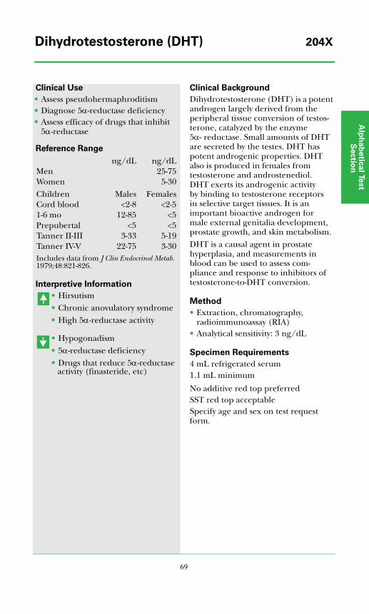
Alp
ha
be
tica
l Test
Se
ctio
n
69
Clinical Use
• Assess pseudohermaphroditism• Diagnose 5α-reductase deficiency• Assess efficacy of drugs that inhibit
5α-reductase
Reference Range
Interpretive Information
• Hirsutism• Chronic anovulatory syndrome• High 5α-reductase activity
• Hypogonadism• 5α-reductase deficiency• Drugs that reduce 5α-reductase
activity (finasteride, etc)
Clinical Background
Dihydrotestosterone (DHT) is a potent androgen largely derived from the peripheral tissue conversion of testos-terone, catalyzed by the enzyme 5α- reductase. Small amounts of DHT are secreted by the testes. DHT has potent androgenic properties. DHT also is produced in females from testosterone and androstenediol. DHT exerts its androgenic activity by binding to testosterone receptors in selective target tissues. It is an important bioactive androgen for male external genitalia development, prostate growth, and skin metabolism.DHT is a causal agent in prostate hyperplasia, and measurements in blood can be used to assess com-pliance and response to inhibitors of testosterone-to-DHT conversion.
Method
• Extraction, chromatography, radioimmunoassay (RIA)
• Analytical sensitivity: 3 ng/dL
Specimen Requirements
4 mL refrigerated serum1.1 mL minimum
No additive red top preferredSST red top acceptableSpecify age and sex on test request form.
ng/dL ng/dLMen 25-75Women 5-30Children Males FemalesCord blood <2-8 <2-51-6 mo 12-85 <5Prepubertal <5 <5Tanner II-III 3-33 5-19Tanner IV-V 22-75 3-30Includes data from J Clin Endocrinol Metab. 1979;48:821-826.
Dihydrotestosterone (DHT) 204X
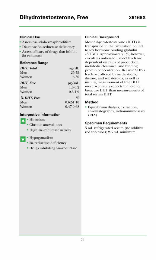
70
Clinical Use
• Assess pseudohermaphroditism• Diagnose 5α-reductase deficiency• Assess efficacy of drugs that inhibit
5α-reductase
Reference Range
Interpretive Information
• Hirsutism• Chronic anovulation• High 5α--reductase activity
• Hypogonadism• 5α-reductase deficiency• Drugs inhibiting 5α--reductase
Clinical Background
Most dihydrotestosterone (DHT) is transported in the circulation bound to sex hormone binding globulin (SHBG). Approximately 1%, however, circulates unbound. Blood levels are dependent on rates of production, metabolic clearance, and binding protein concentration. Because SHBG levels are altered by medications, disease, and sex steroids, as well as insulin, measurement of free DHT more accurately reflects the level of bioactive DHT than measurements of total serum DHT.
Method
• Equilibrium dialysis, extraction, chromatography, radioimmunoassay (RIA)
Specimen Requirements
5 mL refrigerated serum (no additive red top tube); 2.5 mL minimum
DHT, Total ng/dLMen 25-75Women 5-30
DHT, Free pg/mLMen 1.0-6.2Women 0.3-1.9
% DHT, Free %Men 0.62-1.10Women 0.47-0.68
Dihydrotestosterone, Free 36168X
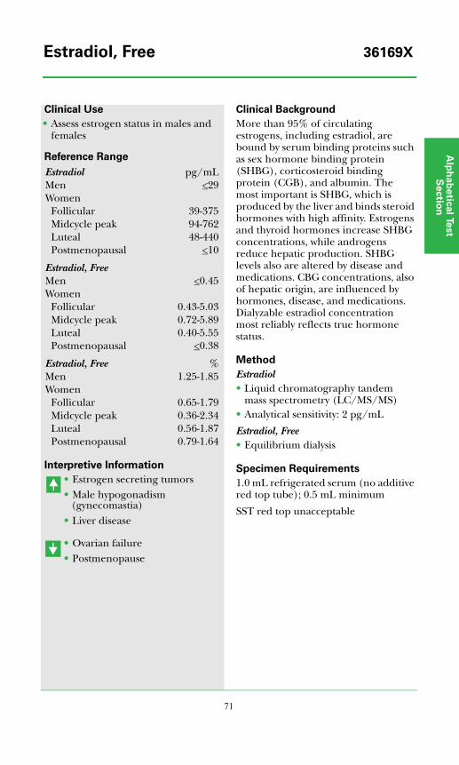
Alp
ha
be
tica
l Test
Se
ctio
n
71
Clinical Use
• Assess estrogen status in males and females
Reference Range
Interpretive Information
• Estrogen secreting tumors• Male hypogonadism
(gynecomastia)• Liver disease
• Ovarian failure• Postmenopause
Clinical Background
More than 95% of circulating estrogens, including estradiol, are bound by serum binding proteins such as sex hormone binding protein (SHBG), corticosteroid binding protein (CGB), and albumin. The most important is SHBG, which is produced by the liver and binds steroid hormones with high affinity. Estrogens and thyroid hormones increase SHBG concentrations, while androgens reduce hepatic production. SHBG levels also are altered by disease and medications. CBG concentrations, also of hepatic origin, are influenced by hormones, disease, and medications. Dialyzable estradiol concentration most reliably reflects true hormone status.
Method
Estradiol• Liquid chromatography tandem
mass spectrometry (LC/MS/MS)• Analytical sensitivity: 2 pg/mL
Estradiol, Free• Equilibrium dialysis
Specimen Requirements
1.0 mL refrigerated serum (no additive red top tube); 0.5 mL minimum
SST red top unacceptable
Estradiol pg/mLMen <29Women
Follicular 39-375Midcycle peak 94-762Luteal 48-440Postmenopausal <10
Estradiol, FreeMen <0.45Women
Follicular 0.43-5.03Midcycle peak 0.72-5.89Luteal 0.40-5.55Postmenopausal <0.38
Estradiol, Free %Men 1.25-1.85Women
Follicular 0.65-1.79Midcycle peak 0.36-2.34Luteal 0.56-1.87Postmenopausal 0.79-1.64
Estradiol, Free 36169X
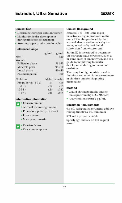
72
Clinical Use
• Determine estrogen status in women• Monitor follicular development
during induction of ovulation• Assess estrogen production in males
Reference Range
Interpretive Information
• Ovarian tumors• Adrenal feminizing tumors• Precocious puberty (female)• Liver disease• Male gynecomastia
• Ovarian failure• Oral contraceptives
Clinical Background
Estradiol-17β (E2) is the major bioactive estrogen produced in the ovary. E2 is also produced by the adrenal glands, and in males by the testes, as well as by peripheral conversion from testosterone. Serum E2 is measured to determine the estrogen status of women, such as in some cases of amenorrhea, and as a guide to monitoring follicular development during induction of ovulation. The assay has high sensitivity and is therefore well suited for measurements in children and for diagnosing menopause.
Method
• Liquid chromatography tandem mass spectrometry (LC/MS/MS)
• Analytical sensitivity: 2 pg/mL
Specimen Requirements
0.5 mL refrigerated serum(no additive red top tube); 0.2 mL minimum
SST red top unacceptableSpecify age and sex on test request form.
pg/mL pg/mLMen <29Women
Follicular phase 39-375Midcycle peak 94-762Luteal phase 48-440Postmenopausal <10
Children Males FemalesPre-pubertal (1-9 y) <4 <1610-11 y <12 <6512-14 y <24 <14215-17 y <31 <283
Estradiol, Ultra Sensitive 30289X
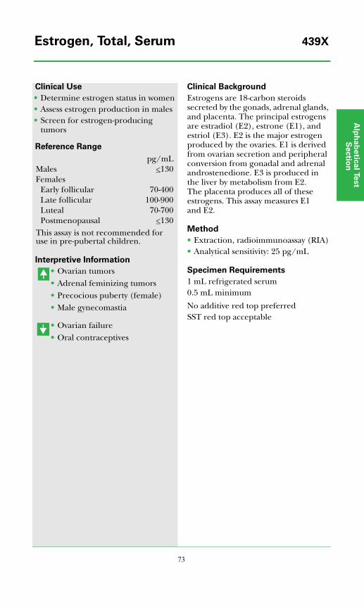
Alp
ha
be
tica
l Test
Se
ctio
n
73
Clinical Use
• Determine estrogen status in women• Assess estrogen production in males• Screen for estrogen-producing
tumors
Reference Range
Interpretive Information
• Ovarian tumors• Adrenal feminizing tumors• Precocious puberty (female)• Male gynecomastia
• Ovarian failure• Oral contraceptives
Clinical Background
Estrogens are 18-carbon steroids secreted by the gonads, adrenal glands, and placenta. The principal estrogens are estradiol (E2), estrone (E1), and estriol (E3). E2 is the major estrogen produced by the ovaries. E1 is derived from ovarian secretion and peripheral conversion from gonadal and adrenal androstenedione. E3 is produced in the liver by metabolism from E2. The placenta produces all of these estrogens. This assay measures E1 and E2.
Method
• Extraction, radioimmunoassay (RIA)• Analytical sensitivity: 25 pg/mL
Specimen Requirements
1 mL refrigerated serum0.5 mL minimum
No additive red top preferredSST red top acceptable
pg/mLMales <130Females
Early follicular 70-400Late follicular 100-900Luteal 70-700Postmenopausal <130
This assay is not recommended for use in pre-pubertal children.
Estrogen, Total, Serum 439X
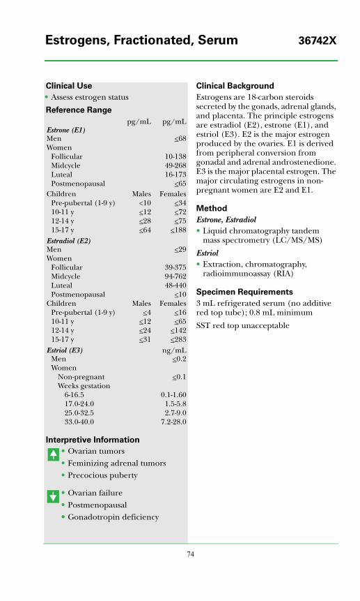
74
Clinical Use
• Assess estrogen status
Reference Range
Interpretive Information
• Ovarian tumors• Feminizing adrenal tumors• Precocious puberty
• Ovarian failure• Postmenopausal•• Gonadotropin deficiency
Clinical Background
Estrogens are 18-carbon steroids secreted by the gonads, adrenal glands, and placenta. The principle estrogens are estradiol (E2), estrone (E1), and estriol (E3). E2 is the major estrogen produced by the ovaries. E1 is derived from peripheral conversion from gonadal and adrenal androstenedione. E3 is the major placental estrogen. The major circulating estrogens in non-pregnant women are E2 and E1.
Method
Estrone, Estradiol• Liquid chromatography tandem
mass spectrometry (LC/MS/MS)
Estriol• Extraction, chromatography,
radioimmunoassay (RIA)
Specimen Requirements
3 mL refrigerated serum (no additive red top tube); 0.8 mL minimum
SST red top unacceptable
pg/mL pg/mLEstrone (E1)Men <68Women
Follicular 10-138Midcycle 49-268Luteal 16-173Postmenopausal <65
Children Males FemalesPre-pubertal (1-9 y) <10 <3410-11 y <12 <7212-14 y <28 <7515-17 y <64 <188
Estradiol (E2)Men <29Women
Follicular 39-375Midcycle 94-762Luteal 48-440Postmenopausal <10
Children Males FemalesPre-pubertal (1-9 y) <4 <1610-11 y <12 <6512-14 y <24 <14215-17 y <31 <283
Estriol (E3) ng/mLMen <0.2Women
Non-pregnant <0.1Weeks gestation
6-16.5 0.1-1.6017.0-24.0 1.5-5.825.0-32.5 2.7-9.033.0-40.0 7.2-28.0
Estrogens, Fractionated, Serum 36742X

Alp
ha
be
tica
l Test
Se
ctio
n
75
Clinical Use
• Assess estrogen status in post-menopausal women
• Assess placental estrogen production
Reference Range
Interpretive Information
• Pregnancy• Postmenopause (by peripheral
conversion)• Aging and obesity• Luteal phase of menstrual cycle
• Hypogonadism
Clinical Background
Estrone (E1) is 1 of 2 major ovarian estrogens in nonpregnant women and a major estrogen produced by the placenta. The primary ovarian estrogen is estradiol (E2). Circulating E1 is derived from ovarian secretion and peripheral conversion from gonadal and adrenal androstenedione. E1 has lower estrogenic potency than E2. E1 is conjugated to glucuronide or sulfate by the liver and excreted in urine and bile. The extragonadal conversion of androstenedione to E1, catalyzed by P-450 aromatase, occurs largely in lipid tissue; thus, E1 tends to be increased in obese subjects. Extragonadal conversion is the major source of circulating estrogens in postmenopausal women. A change in the estradiol/estrone ratio is a reflection of ovarian senescence and development of menopause.
Method
• Liquid chromatography tandem mass spectrometry (LC/MS/MS)
• Analytical sensitivity: 10 pg/mL
Specimen Requirements
0.5 mL refrigerated serum (no additive red top tube); 0.25 mL minimum
SST red top unacceptableSpecify age and sex on test request form.
pg/mL pg/mLMen <68Women
Follicular 10-138Midcycle 49-268Luteal 16-173Postmenopausal <65
Children Males FemalesPre-pubertal (1-9 y) <10 <3410-11 y <12 <7212-14 y <28 <7515-17 y <64 <188
Estrone, Serum 23244X
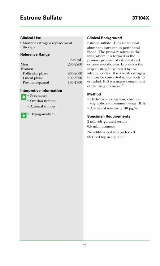
76
Clinical Use
• Monitor estrogen replacement therapy
Reference Range
Interpretive Information
• Pregnancy• Ovarian tumors• Adrenal tumors
• Hypogonadism
Clinical Background
Estrone sulfate (E1S) is the most abundant estrogen in peripheral blood. The primary source is the liver, where it is formed as the primary product of estradiol and estrone metabolism. E1S also is the major estrogen secreted by the adrenal cortex. It is a weak estrogen but can be converted in the body to estradiol. E1S is a major component of the drug Premarin®’.
Method
• Hydrolysis, extraction, chroma-tography, radioimmunoassay (RIA)
• Analytical sensitivity: 40 pg/mL
Specimen Requirements
2 mL refrigerated serum0.5 mL minimum
No additive red top preferredSST red top acceptable
pg/mLMen 230-2200Women
Follicular phase 300-2600Luteal phase 100-3200Postmenopausal 100-1300
Estrone Sulfate 37104X
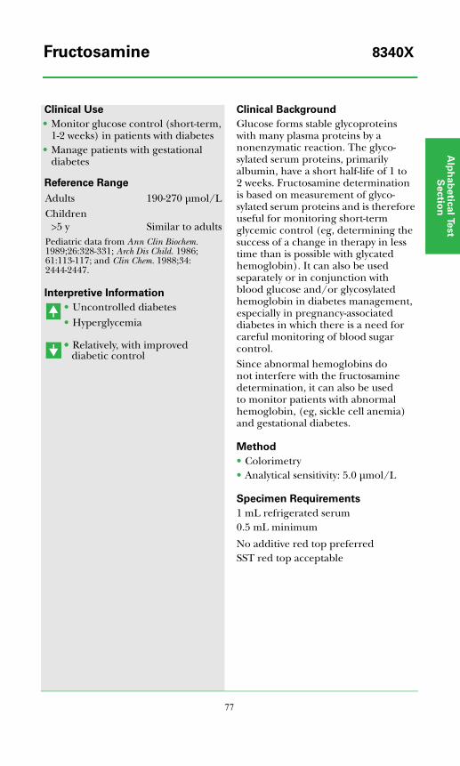
Alp
ha
be
tica
l Test
Se
ctio
n
77
Clinical Use
• Monitor glucose control (short-term, 1-2 weeks) in patients with diabetes
• Manage patients with gestational diabetes
Reference Range
Interpretive Information
• Uncontrolled diabetes• Hyperglycemia
•• Relatively, with improved diabetic control
Clinical Background
Glucose forms stable glycoproteins with many plasma proteins by a nonenzymatic reaction. The glyco-sylated serum proteins, primarily albumin, have a short half-life of 1 to 2 weeks. Fructosamine determination is based on measurement of glyco-sylated serum proteins and is therefore useful for monitoring short-term glycemic control (eg, determining the success of a change in therapy in less time than is possible with glycated hemoglobin). It can also be used separately or in conjunction with blood glucose and/or glycosylated hemoglobin in diabetes management, especially in pregnancy-associated diabetes in which there is a need for careful monitoring of blood sugar control.Since abnormal hemoglobins do not interfere with the fructosamine determination, it can also be used to monitor patients with abnormal hemoglobin, (eg, sickle cell anemia) and gestational diabetes.
Method
• Colorimetry• Analytical sensitivity: 5.0 μmol/L
Specimen Requirements
1 mL refrigerated serum 0.5 mL minimum
No additive red top preferredSST red top acceptable
Adults 190-270 μmol/LChildren
>5 y Similar to adultsPediatric data from Ann Clin Biochem. 1989;26:328-331; Arch Dis Child. 1986;61:113-117; and Clin Chem. 1988;34:2444-2447.
Fructosamine 8340X
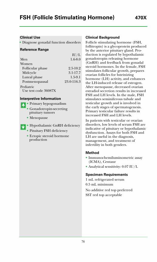
78
Clinical Use
• Diagnose gonadal function disorders
Reference Range
Interpretive Information
• Primary hypogonadism• Gonadotropin-secreting
pituitary tumors• Menopause
• Hypothalamic GnRH deficiency• Pituitary FSH deficiency• Ectopic steroid hormone
production
Clinical Background
Follicle stimulating hormone (FSH, follitropin) is a glycoprotein produced by the anterior pituitary gland. Pro-duction is regulated by hypothalamic gonadotropin releasing hormone (GnRH) and feedback from gonadal steroid hormones. In the female, FSH stimulates follicular growth, prepares ovarian follicles for luteinizing hormone (LH) activity, and enhances the LH-induced release of estrogen. After menopause, decreased ovarian estradiol secretion results in increased FSH and LH levels. In the male, FSH stimulates seminiferous tubule and testicular growth and is involved in the early stages of spermatogenesis. Primary testicular failure results in increased FSH and LH levels.In patients with testicular or ovarian disorders, low levels of serum FSH are indicative of pituitary or hypothalamic dysfunction. Assays for both FSH and LH are useful in the diagnosis, management, and treatment of infertility in both genders.
Method
• Immunochemiluminometric assay (ICMA), Centaur
• Analytical sensitivity: 0.07 IU/L
Specimen Requirements
1 mL refrigerated serum0.5 mL minimum
No additive red top preferredSST red top acceptable
IU/LMen 1.6-8.0Women
Follicular phase 2.5-10.2Midcycle 3.1-17.7Luteal phase 1.5-9.1Postmenopausal 23.0-116.3
PediatricUse test code 36087X
FSH (Follicle Stimulating Hormone) 470X
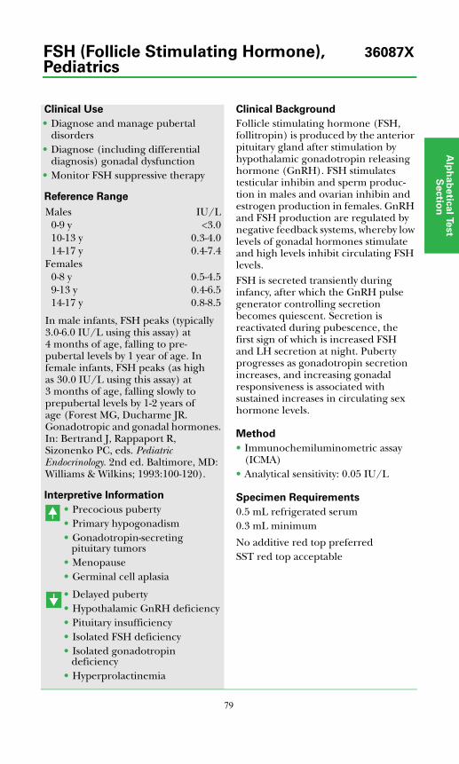
Alp
ha
be
tica
l Test
Se
ctio
n
79
Clinical Use
• Diagnose and manage pubertal disorders
• Diagnose (including differential diagnosis) gonadal dysfunction
• Monitor FSH suppressive therapy
Reference Range
In male infants, FSH peaks (typically 3.0-6.0 IU/L using this assay) at 4 months of age, falling to pre-pubertal levels by 1 year of age. In female infants, FSH peaks (as high as 30.0 IU/L using this assay) at 3 months of age, falling slowly to prepubertal levels by 1-2 years of age (Forest MG, Ducharme JR. Gonadotropic and gonadal hormones. In: Bertrand J, Rappaport R, Sizonenko PC, eds. Pediatric Endocrinology. 2nd ed. Baltimore, MD: Williams & Wilkins; 1993:100-120).
Interpretive Information
• Precocious puberty• Primary hypogonadism• Gonadotropin-secreting
pituitary tumors• Menopause• Germinal cell aplasia
• Delayed puberty• Hypothalamic GnRH deficiency• Pituitary insufficiency• Isolated FSH deficiency• Isolated gonadotropin
deficiency• Hyperprolactinemia
Clinical Background
Follicle stimulating hormone (FSH, follitropin) is produced by the anterior pituitary gland after stimulation by hypothalamic gonadotropin releasing hormone (GnRH). FSH stimulates testicular inhibin and sperm produc-tion in males and ovarian inhibin and estrogen production in females. GnRH and FSH production are regulated by negative feedback systems, whereby low levels of gonadal hormones stimulate and high levels inhibit circulating FSH levels. FSH is secreted transiently during infancy, after which the GnRH pulse generator controlling secretion becomes quiescent. Secretion is reactivated during pubescence, the first sign of which is increased FSH and LH secretion at night. Puberty progresses as gonadotropin secretion increases, and increasing gonadal responsiveness is associated with sustained increases in circulating sex hormone levels.
Method
• Immunochemiluminometric assay (ICMA)
• Analytical sensitivity: 0.05 IU/L
Specimen Requirements
0.5 mL refrigerated serum0.3 mL minimum
No additive red top preferredSST red top acceptable
Males IU/L0-9 y <3.010-13 y 0.3-4.014-17 y 0.4-7.4
Females0-8 y 0.5-4.59-13 y 0.4-6.514-17 y 0.8-8.5
FSH (Follicle Stimulating Hormone), 36087XPediatrics
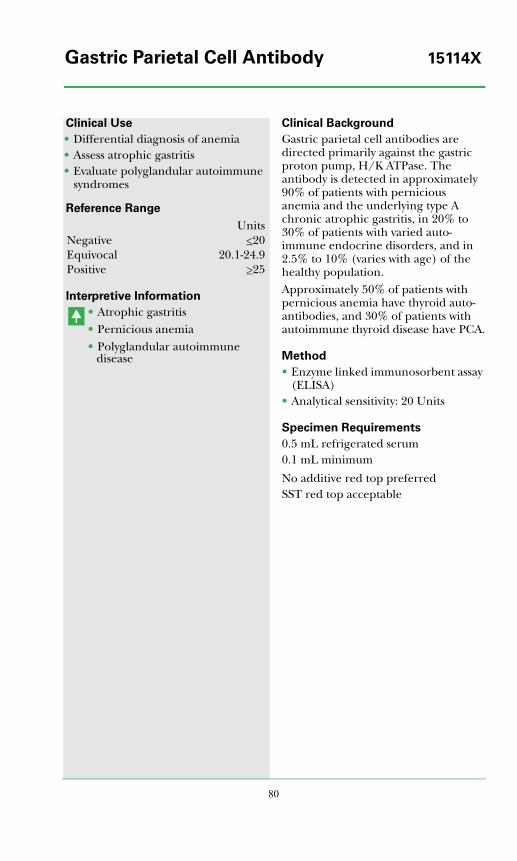
80
Clinical Use
• Differential diagnosis of anemia• Assess atrophic gastritis• Evaluate polyglandular autoimmune
syndromes
Reference Range
Interpretive Information
• Atrophic gastritis• Pernicious anemia• Polyglandular autoimmune
disease
Clinical Background
Gastric parietal cell antibodies are directed primarily against the gastric proton pump, H/K ATPase. The antibody is detected in approximately 90% of patients with pernicious anemia and the underlying type A chronic atrophic gastritis, in 20% to 30% of patients with varied auto-immune endocrine disorders, and in 2.5% to 10% (varies with age) of the healthy population.Approximately 50% of patients with pernicious anemia have thyroid auto-antibodies, and 30% of patients with autoimmune thyroid disease have PCA.
Method
• Enzyme linked immunosorbent assay (ELISA)
• Analytical sensitivity: 20 Units
Specimen Requirements
0.5 mL refrigerated serum0.1 mL minimum
No additive red top preferredSST red top acceptable
UnitsNegative <20Equivocal 20.1-24.9Positive >25
Gastric Parietal Cell Antibody 15114X

Alp
ha
be
tica
l Test
Se
ctio
n
81
Clinical Use
• Diagnose gastrinoma• Differential diagnosis of peptic ulcer
disease and gastrin-secreting tumors
Reference Range
Interpretive Information
• Gastrinoma• Pernicious anemia• Atrophic gastritis with anacidity
Zollinger-Ellison syndrome: gastrin levels >200 pg/mL (Roy PK, et al. Medicine. 2000;79:379-411.)
Zantac®’, Prilosec®’, and pepsid interfere with gastrin production.
Clinical Background
Gastrin is a polypeptide hormone secreted by the G cells of the gastric antral mucosa and proximal duodenal cells. Gastrin is a potent stimulator of antral acid secretion, and also has trophic actions on the parietal cells, chief cells, and enterochromaffin-like cells in the oxyntic mucosa. Gastrin levels increase with food intake (protein, amino acids) and age. Levels decrease with gastric acid secretion.In Zollinger-Ellison syndrome (ZE syndrome), a pancreatic neoplasm produces large amounts of gastrin, which in turn stimulates excess gastric acid secretion and peptic ulcer disease. A variety of pathological, but non-tumorous, disorders in which gastric acid secretion is low also lead to elevations in blood gastrin; these disorders include pernicious anemia and atrophic gastritis with anacidity. Secretin- or calcium-stimulation tests can assist in differentiating tumors from gastrin secreted by the antrum in patients with slight or moderate hypergastrinemia.
Method
• Immunochemiluminometric assay (ICMA)
• Analytical sensitivity: 10 pg/mL• Analytical specificity: detects gastrin
(G17); 7% cross reactivity with big gastrin (G34); does not cross react with G13.
Specimen Requirements
1 mL frozen serum0.5 mL minimum
No additive red top preferredSST red top acceptableOvernight fasting is required.
pg/mLAdults <100Children
5-7 y 13-64
Gastrin 478X
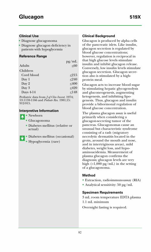
82
Clinical Use
• Diagnose glucagonoma• Diagnose glucagon deficiency in
patients with hypoglycemia
Reference Range
Interpretive Information
• Newborn• Glucagonoma• Diabetes mellitus (relative or
actual)
• Diabetes mellitus (occasional)• Hypoglycemia (rare)
Clinical Background
Glucagon is produced by alpha cells of the pancreatic islets. Like insulin, glucagon secretion is regulated by blood glucose concentrations; however, regulation is reciprocal in that high glucose levels stimulate insulin and inhibit glucagon release. Conversely, low insulin levels stimulate glucagon secretion. Glucagon secre-tion also is stimulated by a high-protein meal.Glucagon acts to increase blood sugar by stimulating hepatic glycogenolysis and gluconeogenesis, augmenting ketogenesis, and inhibiting lipo-genesis. Thus, glucagon and insulin provide a bihormonal regulation of blood glucose concentration.The plasma glucagon assay is useful primarily when considering a glucagon-secreting tumor of the pancreas. Glucagonomas cause an unusual but characteristic syndrome consisting of a rash (migratory necrolytic dermatitis located in the groin, around the mouth and nose, and in intertriginous areas), mild diabetes, weight loss, and hypo-aminoacidemia. Measurement of plasma glucagon confirms the diagnosis; glucagon levels are very high (>1,000 pg/mL) in the setting of a glucagonoma.
Method
• Extraction, radioimmunoassay (RIA)• Analytical sensitivity: 50 pg/mL
Specimen Requirements
3 mL room temperature EDTA plasma1.1 mL minimum
Overnight fasting is required.
pg/mLAdults <60Children
Cord blood <215Day 1 <240Day 2 <400Day 3 <420Days 4-14 <148
Pediatric data from J of Clin Invest. 1974; 53:1159-1166 and Pediatr Res. 1981;15: 912-915.
Glucagon 519X
Alp
ha
be
tica
l Test
Se
ctio
n
83
Clinical Use
• Assess carbohydrate metabolic status• Diagnose diabetes mellitus• Diagnose hypoglycemia
Reference Range
Interpretive Information
• Diabetes mellitus• Cushing’s syndrome• Pheochromocytoma
• Islet cell tumor• Glucagon deficiency• Addison’s disease• Hypoglycemic syndromes
Clinical Background
Blood glucose concentrations are narrowly maintained by balancing hormonal factors. Insulin promotes glucose utilization and storage, lowering blood levels. Carbohydrate ingestion raises blood glucose concentrations; peak levels are modulated by stimulated insulin secretion. Several counter regulatory hormones (glucagon, growth hormone, cortisol, catecholamines) function to increase glucose concentrations by stimulating glycogenolysis, gluconeogenesis and increased hepatic glucose output, raising blood glucose at the expense of carbohydrate and other substrate stores.
Method
• Spectrophotometry (hexokinase)• Analytical sensitivity: 2 mg/dL
Specimen Requirements
1 mL refrigerated serum0.2 mL minimum
No additive red top preferredSST red top acceptableOvernight fasting is preferred.
Adolescents and adults* mg/dLNormal 65-99Impaired 100-125Diabetes >125
*Fasting
Glucose, Serum 483X
84
Clinical Use
• Assess risk of type 1 diabetes• Predict onset of type 1 diabetes• Diagnose new onset type 1 diabetes
Reference Range
Interpretive Information
• Type 1 diabetic patients• Patients at risk for developing
type 1 diabetes• Stiff-man syndrome (SMS)
Clinical Background
Type 1 diabetes mellitus is charac-terized by lymphocytic infiltration of islet cells and circulating auto-antibodies against a variety of islet cell antigens (ICA). These include glutamic acid decarboxylase (GAD), insulin, IA-2, GM2-1, and others. Direct assays for autoantibodies to GAD-65, IA-2, and insulin are now available for detecting individuals with a genetic predisposition to type 1 diabetes.GAD-65 autoantibodies are observed in 70% to 80% of prediabetic and type 1 diabetic subjects including 7% to 8% of adult onset diabetics with type 1 diabetes. When a positive GAD-65 antibody test is coupled with positive autoantibody tests for IA-2 and insulin, the risk of diabetes increases to >90% (see Test Application and Interpretation section).
Method
• Radiobinding assay• Analytical sensitivity: 0.2 U/mL
Specimen Requirements
1 mL frozen serum0.2 mL minimum
No additive red top preferredSST red top acceptable
<1 U/mL
Glutamic Acid Decarboxylase-65 34878XAutoantibodies (GAD-65)
Alp
ha
be
tica
l Test
Se
ctio
n
85
Clinical Use
• Assess glucose control (short-term, 1-2 weeks) in patients with diabetes, especially those with pregnancy-associated diabetes
Reference Range
Interpretive Information
• Uncontrolled diabetes• Hyperglycemia• Microvascular disease/diabetic
complications
• Relatively, with improved diabetic control
Clinical Background
Glucose forms stable glycoproteins with many plasma proteins by a nonenzymatic reaction. Among the plasma proteins, albumin is quantitatively the most important glycated protein. Concentrations of glycated albumin are high in diabetic patients and have been shown to be proportional to the degree of hyper-glycemia. Since albumin turns over in 1 to 2 weeks, glycated albumin determinations are useful for moni-toring short-term glucose control in diabetic patients. In contrast, glycated hemoglobin, which has a half-life of 120 days, is best for assessment of long-term control in such patients.
Method
• Affinity column chromatography• % glycated albumin is based on
percentage of microalbumin relative to amount of total albumin.
Specimen Requirements
3 mL refrigerated EDTA plasma2 mL minimum
Overnight fasting required
Adults and children 0.8-1.4%
Glycated Albumin 5032X
86
Clinical Use
• Assess pituitary growth hormone disorders
Reference Range
Interpretive Information
• Gigantism• Acromegaly• Selected pituitary tumors• Pregnancy, due to HPL cross-
reactivity• Laron dwarfism (GH resistance)
• Pituitary GH deficiency• Hypopituitarism, congenital or
acquired• GH secretory dysfunction
Because of diurnal variability, random growth hormone values are not reliable for diagnosis of growth hormone deficiency, acromegaly (adults), or gigantism (pediatrics).
Clinical Background
GH is a polypeptide (~ 20,000 MW) secreted by the anterior pituitary gland under the control of hypo-thalamic growth hormone releasing hormone (GHRH) and somatostatin, which stimulate and inhibit release, respectively.Excess GH production can be caused by pituitary adenoma. In adults, excess GH production produces acromegaly; in children, it produces gigantism. Insufficient GH production in children results in growth retardation and, in adults, decreased muscle mass.Measuring GH concentration in children is useful for clinically evaluating short stature and helping to differentiate between abnormal GH production and other causes of growth failure. In adults a GH deficiency syndrome has been described that is associated with osteoporosis, dys-lipidemia, increased visceral obesity, and poor quality of life. Because random GH measurements are not usually meaningful, inhibitory proto-cols, provocative tests, and prolonged sampling usually are employed. See Disorders of Growth Hormone in the Test Application and Interpretation section.
Method
• Immunochemiluminometric assay (ICMA)
• Analytical sensitivity: 0.1 ng/mL
Specimen Requirements
1 mL refrigerated serum0.5 mL minimum
No additive red top preferredSST red top acceptable
Adults ng/mLFasting <10After 75 g oral glucose <1
Children <13
Growth Hormone (GH) 521X
Alp
ha
be
tica
l Test
Se
ctio
n
87
Clinical Use
• Monitor patients receiving GH therapy
Reference Range
Interpretive Information
• Selected patients on GH therapy• Possibly in patients with auto-
immune hypophysitis
Clinical Background
Human GH is now a routine replace-ment therapy in GH-deficient patients. Most of the GH used is biosynthetically produced with recombinant DNA technology and is purified to minimize antigenicity. Nonetheless, some patients will develop autoantibodies; these may not be neutralizing and may not inhibit the growth response. About 5% of patients will produce neutral-izing antibodies; thus, all patients with documented GH deficiency who do not respond as expected to GH therapy should receive GH antibody testing.
Method
• Radiobinding assay (RBA)• Titer reported when antibody is
present
Specimen Requirements
0.5 mL refrigerated serum0.2 mL minimum
No additive red top preferredSST red top acceptable
Adults and children Negative
Growth Hormone (GH) Antibody 37072X
88
Clinical Use
• Diagnose Laron dwarfism• Differential diagnosis of growth
hormone resistance• Indicate GH responsiveness
Reference Range
Interpretive Information
• Estrogen treatment• Familial short stature with high
GHBP (J Clin Endocrinol Metab. 2004;89:1031-1044.)
• Laron dwarfism
Clinical Background
Human growth hormone (GH) exists in several isometric forms in serum – either free or bound to a specific binding protein (GHBP). This protein is identical to the extracellular domain of the GH receptor and appears to be derived from the GH receptor gene by alternative splicing. Levels vary with age, being low in neonates and highest in young adults. Normal or elevated levels in patients with classical Laron syndrome indicate a defect in the transmembrane, intracellular, or post GH receptor domains. Very low or absent GHBP denotes a defect in the extracellular domain. A low serum GHBP in relatives of patients with Laron syndrome helps identify heterozygous carriers of patients with defects in the extracellular domain.
Method
• Enzyme-linked immunosorbent assay (ELISA)
• Analytical sensitivity: 10 pmol/L
Specimen Requirements
1 mL frozen serum (no additive red top tube); 0.1 mL minimum
SST red top unacceptable
pmol/LAdults 400-4260Children
3-8 y 320-38209-13 y 240-289014-17 y 290-3140
Growth Hormone-Binding Protein 39573X(GHBP)
Alp
ha
be
tica
l Test
Se
ctio
n
89
Clinical Use
• Differential diagnosis of acromegaly
Reference Range
Interpretive Information
• Ectopic GHRH production• Pregnancy (maternal and fetal
samples)
Clinical Background
Growth hormone releasing hormone (GHRH) is secreted by the hypo-thalamus and stimulates the pituitary to release growth hormone. GHRH deficiency is probably the most common cause of growth hormone deficiency in man.Acromegaly results from hyper-secretion of growth hormone and is due, in over 99% of cases, to a primary pituitary tumor. The remaining cases result from GHRH hypersecretion due to hypothalamic tumor or peripheral tumors (pancreatic islet tumors, bronchial or thymic carcinoid tumors, and rarely neuroendocrine tumors). In patients with acromegaly due to ectopic GHRH secretion, peripheral GHRH levels are elevated.The differential diagnosis between a pituitary tumor and ectopic GHRH hypersecretion is achieved by measur-ing peripheral GHRH levels. Circula-ting GHRH is nonhypothalamic in origin.
Method
• Extraction, radioimmunoassay (RIA)• Analytical sensitivity: 16 pg/mL
Specimen Requirements
4 mL frozen plasma1.1 mL minimum
Collect in special PTH-Related Protein (PTH-RP) and Releasing Factors tube provided by Quest Diagnostics Nichols Institute. Centrifuge immediately in refrigerated centrifuge, separate plasma, and freeze.
pg/mLAdults <49Children (4-14 y) 6.8-19.0Pediatric data from Horm Metabol Res. 1987;19:434-436.
Growth Hormone Releasing 37557XHormone (GHRH)
90
Clinical Use
• Diagnose pregnancy• Diagnose chorionic tumors and
ectopic hCG-producing tumors
Reference Range
Interpretive Information
• Pregnancy• Trophoblastic tumors• Gonadal germ cell tumors• Midline trophoblastic tumors
• Ectopic pregnancy (relative to uterine pregnancy)
Clinical Background
Heterophilic antibodies, defined as antibodies against animal immuno-globulins, can be a source of inter-ference in many immunoassays. The most frequently characterized interference comes from endogenous human anti-mouse antibodies (HAMA), which can react with the mouse monoclonal antibodies utilized in the assay. HAMA presence in the patient sample can lead to over- or underestimation of human chorionic gonadotropin (hCG). In this test, HAMA is removed by precipitation prior to hCG determination.
Method
• HAMA precipitation• Immunochemiluminometric assay
(ICMA)• Analytical sensitivity: 2 mIU/mL• Analytical specificity: detects intact
and free beta hCG
Specimen Requirements
2 mL room temperature serum1.0 mL minimum
No additive red top preferredSST red top acceptable
hCG, Total, HAMA <5 mIU/mLhCG, Total <5 mIU/mL
hCG, Total with HAMA Treatment 19720X
Alp
ha
be
tica
l Test
Se
ctio
n
91
Clinical Use
• Diagnose pregnancy• Diagnose chorionic tumors and
ectopic hCG-producing tumors
Reference Range
Interpretive Information
• Pregnancy• Trophoblastic tumors• Gonadal germ cell tumors• Midline trophoblastic tumors
• Ectopic pregnancy (relative to uterine pregnancy)
Clinical Background
Human chorionic gonadotropin (hCG) is secreted by the developing placenta often as early as 6 days after conception; maximal production and peak serum levels occur during the first trimester but remain significant throughout gestation. The immuno-chemiluminometric assay for serum hCG provides a fast and accurate test for pregnancy. Because the assay is highly sensitive, it is useful for early determination of pregnancy when hCG levels are relatively low.A variety of other tissues are capable of hCG production, and increased levels in men or in nonpregnant women suggest neoplasia. In this context, hCG levels >10,000 mIU/mL occur only in germ cell tumors, in patients with trophoblastic differentia-tion of a lung or gastric primary cancer, or in women with gestational trophoblastic disease. Levels are prognostic in germ cell tumors: <5,000 mIU/mL indicates good prognosis; 5,000 to 50,000 mIU/mL indicates intermediate prognosis; and >50,000 mIU/mL indicates poor prognosis.hCG measurement also is appropriate in the evaluation of men with gyneco-mastia and boys with isosexual pre-cocious puberty to diagnose testicular malignancies.
Method
• Immunochemiluminometric assay (ICMA)
• Analytical sensitivity: 2 mIU/mL• Analytical specificity: detects intact
and free beta hCG
Specimen Requirements
1 mL refrigerated serum0.4 mL minimum
No additive red top preferredSST red top acceptable
mIU/mLMen <5Women
Non-pregnant, premenopausal
<5
Postmenopausal <10Pregnant Expected*
<1 wk 5-501-2 wk 50-5002-3 wk 100-5,0003-4 wk 500-10,0004-5 wk 1,000-50,0005-6 wk 10,000-100,0006-8 wk 15,000-200,0002-3 mo 10,000-100,000
*Bayer Centaur product insert
Values from different assay methods may vary.
hCG, Total, Quantitative 8396X
92
Clinical Use
• Monitor glucose control (long-term, 2-3 months) in patients with diabetes
Reference Range
Interpretive Information
• Diabetes mellitus• Chronic hyperglycemia• Presence of hemoglobin S
(sickle cell)• Presence of hemoglobin C
variant
• Relatively decreased by improved diabetic control
• High levels of hemoglobin F
Clinical Background
Hemoglobin A1 (HbA1 or glycated hemoglobin) is structurally related to adult hemoglobin (HbA) but has a glucose molecule attached to the terminal valine of the beta chain. Glycosylation is a nonenzymatic, irreversible process dependent on the glucose concentration and the duration of exposure of the erythro-cyte to glucose. HbA1 is continuously formed during the 120-day life of the erythrocyte, and a single measurement of HbA1 reflects the average blood glucose level during the preceding months. HbA1 can be separated into HbA1a, HbA1b, and HbA1c fractions. HbA1c correlates best with high glucose concentrations.
Method
• Colorimetry, immunoturbidimetric• Analytical sensitivity: 3% at a
hemoglobin concentration of 8.2 mmol/L (13.2 g/dL)
Specimen Requirements
2 mL refrigerated whole blood0.3 mL minimum
Collect in EDTA (lavender-top) tube.
Non-diabetic <6.0%
Hemoglobin A1c 496X
Alp
ha
be
tica
l Test
Se
ctio
n
93
Clinical Use
• Diagnose carcinoid tumors
Reference Range
Interpretive Information
• Carcinoid tumor
Clinical Background
Serotonin metabolism yields 5-HIAA as a major urinary metabolite. Because carcinoid tumors secrete large amounts of serotonin and 5-HIAA, tests for 5-HIAA are useful in their diagnosis. However, many foods (see below) and drugs also contain large amounts of serotonin and should be avoided at least 48 hours before collecting urine. Drugs to avoid include phenothiazines, glyceryl guaiacolate, reserpine, MAO inhib-itors, and imipramine.
Method
• High-performance liquid chroma-tography (HPLC), electrochemical detection
• Analytical sensitivity: 0.2 mg/L• Creatinine concentration reported
Specimen Requirements
10 mL room temperature aliquot of 24-h urine (39625X) or random urine (1648X); 5 mL minimum
Collect 24-h urine with 25 mL of 6N HCl. Add 6N HCl to random urine to maintain pH <3. Record 24-h urine total volume on vial and request form.
Patient preparation: avoid tobacco, tea, and coffee for 3 days prior; avoid food high in indoles, eg, avocado, banana, tomato, plum, walnut, pineapple, and eggplant.
5-HIAA (39625X) mg/24-h2-10 y <8>10 y <6
Creatinine (39625X ) g/24-h3-8 y 0.11-0.689-12 y 0.17-1.4113-17 y 0.29-1.87Adults 0.63-2.50
5-HIAA (1648X) mg/g creat2-10 y <12>10 y <10
Creatinine (1648X) mg/dL0-6 mo 2-327-11 mo 2-361-2 y 2-1283-8 y 2-1499-12 y 2-183>12 y
Male 20-370Female 20-320
5-HIAA (5-Hydroxyindoleacetic Acid), 39625XUrine 1648X
94
Clinical Use
• Diagnose catecholamine-producing tumors
Reference Range
Interpretive Information
• Neuroblastoma• Ganglioblastoma
Clinical Background
Measurements of urinary vanillyl-mandelic acid (VMA) and homo-vanillic acid (HVA) are valuable in identifying patients with neuro-blastoma. About 80% of patients with neuroblastoma will have elevated urinary VMA. Other metabolites of dopamine, such as urinary dopamine, HVA, and 3-methoxy-4-hydroxy-phenylglycol (MHPG), are also usually elevated.Neuroblastomas are malignant tumors occurring in children. About half of the cases occur before 2 years of age. Neuroblastomas secrete mostly pre-cursors of norepinephrine (dopamine and its metabolites) and seldom produce hypertension. Patients usually present with an abdominal mass. Surgery and radiation therapies offer significant hope for survival. Early diagnosis is therefore essential.
Method
• High-performance liquid chroma-tography (HPLC), electrochemical detection
• Analytical sensitivity: 0.5 mg/L• Creatinine concentration also
reported
Specimen Requirements
10 mL room temperature aliquot of a 24-h urine (39527X) or random urine (6346X); 5 mL minimum
Collect 24-h urine in 25 mL 6N HCl; add 6N HCl to random urine to maintain pH <3. Record 24-h total volume on vial and request form.
Patient preparation: avoid alcohol, coffee, tea, tobacco, and strenuous exercise prior to collection; preferably abstain from medications for 3 days, but common antihypertensives (diuretics, ACE inhibitors, calcium channel blockers, alpha and beta blockers) cause minimal or no inter-ference.
24-H urine (39527X)Homovanillic acid mg/24-h
3-8 y 0.5-6.79-12 y 1.1-6.813-17 y 1.4-7.2Adults 1.6-7.5
Creatinine g/24-h3-8 y 0.11-0.689-12 y 0.17-1.4113-17 y 0.29-1.87Adults 0.63-2.50
Random urine (6346X)Homovanillic acid mg/g creat
Birth to 6 mo 9.1-367-11 mo 11.2-331-2 y 8.5-383-8 y 2.1-239-12 y 1.1-12Adults 1.4-5.3
Creatinine mg/dL0-6 mo 2-327-11 mo 2-361-2 y 2-1283-8 y 2-1499-12 y 2-183>12 y
Male 20-370Female 20-320
Homovanillic Acid (HVA), Urine 39527X6346X
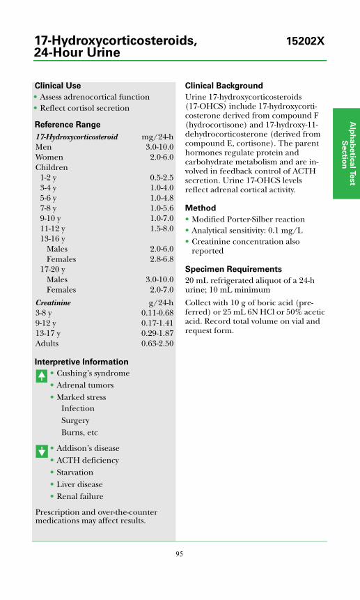
Alp
ha
be
tica
l Test
Se
ctio
n
95
Clinical Use
• Assess adrenocortical function• Reflect cortisol secretion
Reference Range
Interpretive Information
• Cushing’s syndrome• Adrenal tumors• Marked stress
InfectionSurgeryBurns, etc
• Addison’s disease• ACTH deficiency• Starvation• Liver disease• Renal failure
Prescription and over-the-counter medications may affect results.
Clinical Background
Urine 17-hydroxycorticosteroids (17-OHCS) include 17-hydroxycorti-costerone derived from compound F (hydrocortisone) and 17-hydroxy-11-dehydrocorticosterone (derived from compound E, cortisone). The parent hormones regulate protein and carbohydrate metabolism and are in-volved in feedback control of ACTH secretion. Urine 17-OHCS levels reflect adrenal cortical activity.
Method
• Modified Porter-Silber reaction• Analytical sensitivity: 0.1 mg/L• Creatinine concentration also
reported
Specimen Requirements
20 mL refrigerated aliquot of a 24-h urine; 10 mL minimum
Collect with 10 g of boric acid (pre-ferred) or 25 mL 6N HCl or 50% acetic acid. Record total volume on vial and request form.
17-Hydroxycorticosteroid mg/24-hMen 3.0-10.0Women 2.0-6.0Children
1-2 y 0.5-2.53-4 y 1.0-4.05-6 y 1.0-4.87-8 y 1.0-5.69-10 y 1.0-7.011-12 y 1.5-8.013-16 y
Males 2.0-6.0Females 2.8-6.8
17-20 yMales 3.0-10.0Females 2.0-7.0
Creatinine g/24-h3-8 y 0.11-0.689-12 y 0.17-1.4113-17 y 0.29-1.87Adults 0.63-2.50
17-Hydroxycorticosteroids, 15202X24-Hour Urine
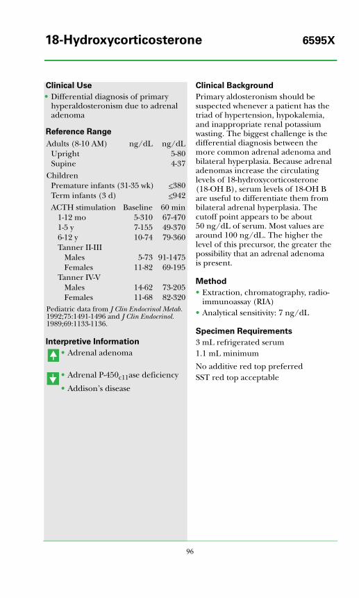
96
Clinical Use
• Differential diagnosis of primary hyperaldosteronism due to adrenal adenoma
Reference Range
Interpretive Information
• Adrenal adenoma
• Adrenal P-450c11ase deficiency
• Addison’s disease
Clinical Background
Primary aldosteronism should be suspected whenever a patient has the triad of hypertension, hypokalemia, and inappropriate renal potassium wasting. The biggest challenge is the differential diagnosis between the more common adrenal adenoma and bilateral hyperplasia. Because adrenal adenomas increase the circulating levels of 18-hydroxycorticosterone (18-OH B), serum levels of 18-OH B are useful to differentiate them from bilateral adrenal hyperplasia. The cutoff point appears to be about 50 ng/dL of serum. Most values are around 100 ng/dL. The higher the level of this precursor, the greater the possibility that an adrenal adenoma is present.
Method
• Extraction, chromatography, radio-immunoassay (RIA)
• Analytical sensitivity: 7 ng/dL
Specimen Requirements
3 mL refrigerated serum1.1 mL minimum
No additive red top preferredSST red top acceptable
Adults (8-10 AM) ng/dL ng/dLUpright 5-80Supine 4-37
ChildrenPremature infants (31-35 wk) <380Term infants (3 d) <942ACTH stimulation Baseline 60 min
1-12 mo 5-310 67-4701-5 y 7-155 49-3706-12 y 10-74 79-360Tanner II-III
Males 5-73 91-1475Females 11-82 69-195
Tanner IV-VMales 14-62 73-205Females 11-68 82-320
Pediatric data from J Clin Endocrinol Metab. 1992;75:1491-1496 and J Clin Endocrinol. 1989;69:1133-1136.
18-Hydroxycorticosterone 6595X
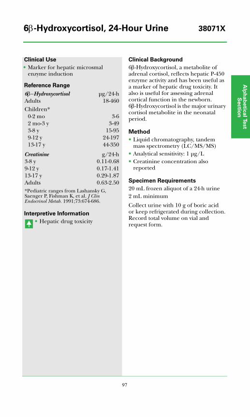
Alp
ha
be
tica
l Test
Se
ctio
n
97
Clinical Use
• Marker for hepatic microsmal enzyme induction
Reference Range
Interpretive Information
• Hepatic drug toxicity
Clinical Background
6β-Hydroxycortisol, a metabolite of adrenal cortisol, reflects hepatic P-450 enzyme activity and has been useful as a marker of hepatic drug toxicity. It also is useful for assessing adrenal cortical function in the newborn. 6β-Hydroxycortisol is the major urinary cortisol metabolite in the neonatal period.
Method
• Liquid chromatography, tandem mass spectrometry (LC/MS/MS)
• Analytical sensitivity: 1 μg/L• Creatinine concentration also
reported
Specimen Requirements
20 mL frozen aliquot of a 24-h urine2 mL minimum
Collect urine with 10 g of boric acid or keep refrigerated during collection. Record total volume on vial and request form.
6β−Hydroxycortisol μg/24-hAdults 18-460Children*
0-2 mo 3-62 mo-3 y 3-493-8 y 15-959-12 y 24-19713-17 y 44-350
Creatinine g/24-h3-8 y 0.11-0.689-12 y 0.17-1.4113-17 y 0.29-1.87Adults 0.63-2.50*Pediatric ranges from Lashansky G, Saenger P, Fishman K, et al. J Clin Endocrinol Metab. 1991;73:674-686.
6β-Hydroxycortisol, 24-Hour Urine 38071X
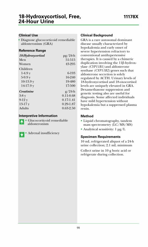
98
Clinical Use
• Diagnose glucocorticoid remediable aldosteronism (GRA)
Reference Range
Interpretive Information
• Glucocorticoid remediable aldosteronism
• Adrenal insufficiency
Clinical Background
GRA is a rare autosomal dominant disease usually characterized by hypokalemia and early onset of severe hypertension refractory to conventional antihypertensive therapies. It is caused by a chimeric duplication involving the 11β-hydrox-ylase (CYP11B1) and aldosterone synthase (CYP11B2) genes such that aldosterone secretion is solely regulated by ACTH. Urinary levels of 18-hydroxycortisol and 18-oxocortisol levels are uniquely elevated in GRA. Dexamethasone suppression and genetic testing also are useful for diagnosis. Some affected individuals have mild hypertension without hypokalemia but a suppressed plasma renin.
Method
• Liquid chromatography, tandem mass spectrometry (LC/MS/MS)
• Analytical sensitivity: 1 μg/L
Specimen Requirements
10 mL refrigerated aliquot of a 24-h urine collection; 2.1 mL minimum
Collect urine in 10 g boric acid or refrigerate during collection.
18-Hydroxycortisol μg/24-hMen 51-515Women 43-295Children
1-4.9 y 6-1935-9.9 y 16-24010-13.9 y 19-48014-17.9 y 17-500
Creatinine g/24-h3-8 y 0.11-0.689-12 y 0.17-1.4113-17 y 0.29-1.87Adults 0.63-2.50
18-Hydroxycortisol, Free, 11178X24-Hour Urine
Alp
ha
be
tica
l Test
Se
ctio
n
99
Clinical Use
• Confirm autoimmune Addison’s disease
• Identify adrenal gland involvement in PAS
Reference Range
Interpretive Information
• PAS, type I• PAS, type II• Isolated autoimmune Addison’s
disease
Clinical Background
Autoimmune Addison's disease is characterized by the presence of autoantibodies to adrenal cortical cell antigens. Recent studies have identified the adrenal biosynthetic enzyme P-450c21 as one of these antigens. Autoimmune Addison's disease is part of 2 polyglandular autoimmune syndromes (PAS), type I and type II. Type II, including auto-immune thyroiditis and diabetes, is the more frequent.This direct autoantibody assay for anti-21-hydroxylase is more sensitive and quantifiable than the anti-adrenal immunofluorescence (IFA) method. Measurement of other glandular autoantibodies provides additional diagnostic and manage-ment information.
Method
• Radiobinding assay (RBA)• Analytical sensitivity: 1.0 U/mL
Specimen Requirements
1 mL refrigerated serum0.2 mL minimum
No additive red top preferredSST red top acceptable
Adults and children <1.0 U/mL
21-Hydroxylase Antibody 37916X
100
Clinical Use
• Marker for adrenal 3β-hydroxy-steroid dehydrogenase enzyme deficiency
Reference Range
Interpretive Information
• CAH due to 3β-hydroxysteroid dehydrogenase enzyme deficiency
• Hirsutism (polycystic ovary or idiopathic)
• Adrenal tumors
• Adrenal insufficiency
Clinical Background
17-Hydroxypregnenolone, a Δ5 steroid, is an intermediate in the steroid hormone biosynthetic pathways. It is formed by the hydroxylation of pregnenolone and metabolized to 17-hydroxyprogesterone or dehydroepiandrosterone (DHEA). 17-hydroxypregnenolone production is stimulated by ACTH and suppressed by dexamethasone.Production is increased in patients with congenital adrenal hyperplasia (CAH) associated with deficiency of the 3β-hydroxysteroid dehydrogenase enzyme.
Method
• Extraction, chromatography, radioimmunoassay (RIA)
• Analytical sensitivity: 10 ng/dL
Specimen Requirements
2 mL refrigerated serum0.4 mL minimum
No additive red top preferredSST red top acceptable
ng/dL ng/dLAdults 20-450
Post ACTH stimulation, 60 min, men and premenopausal women (follicular phase)
290-910
ChildrenPremature infants (31-35 wk) <2409Term infants (3 d) <830ACTH stimulation
Baseline 60 min
1-12 mo 14-830 395-32901-5 y 10-100 45-7406-12 y 11-190 70-660Tanner II-III
Males 20-360 88-675Females 58-450 250-800
Tanner IV-VMales 32-300 220-860Females 53-540 500-1600
Pediatric data from J Clin Endocrinol Metab. 1991;73:674-686 and J Clin Endocrinol Metab. 1989;69:1133-1136.
17-Hydroxypregnenolone 8352X
Alp
ha
be
tica
l Test
Se
ctio
n
101
Clinical Use
• Marker for adrenal P450c21 (21-hydroxylase) enzyme deficiency
Reference Range
Clinical Background
17-Hydroxyprogesterone (17-OHP) is an adrenal steroid intermediate in the biosynthesis of cortisol. Its synthesis from progesterone is catalyzed by the enzyme P450c17; it is then further metabolized to cortisol or androstene-dione. 17-OHP measurement is appropriate when complete or partial 21-hydroxyl-ase deficiency is suspected in 1) infants with features of adrenal insufficiency (hypotension, vomiting, fever, hypoglycemia, and hyper-kalemia) or ambiguous genitalia and 2) women with clinical evidence of possible androgen excess, particularly Ashkenazi Jews who have a high prevalence of nonclassical 21-hydroxyl-ase deficiency. Some authorities consider measurement of ACTH-stimulated 17-OHP levels essential to exclude the diagnosis. Cushing's disease may also cause elevated 17-OHP levels and should be excluded before glucocorticoid treatment of presumed nonclassical 21-hydroxylase deficiency is considered.
Method
• Liquid chromatography, tandem mass spectrometry (LC/MS/MS)
• Analytical sensitivity: 8 ng/dL
Specimen Requirements
0.5 mL frozen serum (no additive red top tube); 0.25 mL minimumSST red top unacceptable
Interpretive Information
• Congenital adrenal hyperplasia (CAH), 21-hydroxylase deficiency
• Male pseudohermaphroditism (P450c17 deficiency)
• Late-onset CAH• Cushing’s disease
• Steroid treatment (cortisone, hydrocortisone)
• Addison’s disease
Adults (17180X) ng/dL ng/dLMen
18-30 y 32-30731-40 y 42-19641-50 y 33-19551-60 y 37-129
WomenFollicular phase <185Midcycle phase <225Luteal phase <285Postmenopausal <45
Neonatal/Infant (17654X)*Premature infants (31-35 wk) <360Term infants (3 d) <4201-12 mo 11-1701-4 y 4-1155--9 y <9010-13 y <16914-17 y 16-283ACTH stimulation
Baseline 60 min
1-12 mo 11-170 85-4651-5 y 4-115 50-3506-12 y 7-69 75-220
Tanner II-IIIMales 12-130 69-310Females 18-220 80-420
Tanner IV-VMales 51-190 105-230Females 36-200 80-225
*Includes data from J Clin Metab. 1991;73:674-686; J Clin Endocrinol Metab. 1989;69:1133-1136; and J Clin Endocrinol Metab. 1994;78:266-270.
17-Hydroxyprogesterone, LC/MS/MS 17180X17654X
102
Clinical Use
• Assess bone matrix turnover
Reference Range
Interpretive Information
• Paget's disease• Bone metastatic cancer• Hyperparathyroidism• Osteoporosis• Glucocorticoid excess
• Fluoride excess• Bisphosphonate therapy• Anabolic steroid therapy
Clinical Background
Collagen metabolism releases a variety of collagen constituents into the circulation. These include amino- and carboxy-terminal extension peptides, pyridinoline, and deoxypyridinoline cross-links, and hydroxyproline. Hydroxyproline in plasma appears as both peptide bound and free forms. Bone resorption is the major con-tributor to collagen turnover, and the constituent molecules, including hydroxyproline, are useful as markers of the rate of bone matrix resorption. Hydroxyproline is most useful in conditions associated with markedly increased bone resorption (Paget's disease, malignancy, hyperpara-thyroidism).
Method
• High performance liquid chroma-tography (HPLC)
• Analytical sensitivity: 1 μmol/L
Specimen Requirements
2.0 mL frozen sodium heparin plasma0.5 mL minimum
Separate plasma within 30 minutes of collection and freeze. Provide patient age, sex, clinical history, tentative diagnosis, and therapy during the last 3 days (eg, drugs, X-ray, infant formula, diet).
μmol/L0-30 d 0-9131 d-24 mo 0-632-18 y 3-45>18 y 0-53
Hydroxyproline, Free, Plasma 37558X
Alp
ha
be
tica
l Test
Se
ctio
n
103
Clinical Use
• Index for the rate of bone formation and turnover
• Assess efficacy of therapy for bone disease
Reference Range
Interpretive Information
• Paget’s disease• Hyperparathyroidism (primary
and secondary)• Burns• Psoriasis• Acromegaly• Inborn errors of metabolism• Hydroxyprolinemia• Familial aminoglycinuria
Clinical Background
Hydroxyproline is an amino acid present in appreciable quantities in collagen and excreted in the urine after collagen breakdown. Because urinary hydroxyproline is derived almost entirely from collagen, it reflects the rate of collagen catabolism. Small amounts of urinary hydroxy-proline are present as free amino acids, while most is associated with small and large nondialyzable peptides. This combination is collectively referred to as total hydroxyproline.Urine hydroxyproline levels are greatly increased in Paget's disease and to a lesser extent in hyperpara-thyroidism (primary and secondary). Elevated levels are also observed in patients with burns, psoriasis, and acromegaly. Urinary hydroxyproline has been suggested as a tumor marker for lymphomas, mammary carcinoma, and bone metastatic prostate carcinoma.Levels of free hydroxyproline are in-creased in patients with inborn errors of metabolism, hydroxyprolinemia, and familial aminoglycinuria.
Method
• Colorimetry• Analytical sensitivity: 0.5 μg/mL and
2 μg/mL, free and total, respectively
• Creatinine concentration also reported
Specimen Requirements
25 mL refrigerated aliquot of a 24-h urine; 2.1 mL minimum for free (685X); 2.1 mL minimum for total (535X); 4.2 mL minimum for free and total (17190X)
Collect with 25 mL of 6N HCl. Record total volume on vial and request form.
Avoid meat and collagen-derived therapeutics for 24 to 48 hours prior to collection.
Hydroxyproline, Free mg/24-h(685X, 17190X)Adults <2.7
Hydroxyproline, Total (535X, 17190X)Men 9-73Women 7-49Children*
Premature infants, 3 wk old 6-35Full-term infants
3 d 8-201 mo 32-631-10 y 15-15011-14 y 68-169
Creatinine (685X, 535X, 17190X)
g/24-h
3-8 y 0.11-0.689-12 y 0.17-1.4113-17 y 0.29-1.87Adults 0.63-2.50*Pediatric data from J Pediatr Gastroent Nutr. 1991;13:176-181; J Clin Invest. 1970;49:716-729; Pediatr Res. 1967;1:266-270; J Pediatr. 1966;69:266-273; and J Clin Invest. 1962;41:1928-1935.
Hydroxyproline, Free and Total, 17190X, 685X,24-Hour Urine 535X
104
Clinical Use
• Index for the rate of bone formation and turnover
• Assess efficacy of therapy for bone disease
Reference Range
Interpretive Information
• Paget's disease• Hyperparathyroidism (primary
and secondary)• Burns• Psoriasis• Acromegaly• Inborn errors of metabolism• Hydroxyprolinemia• Familial aminoglycinuria
Clinical Background
Hydroxyproline is an amino acid present in appreciable quantities in collagen and excreted in the urine after collagen breakdown. Because urinary hydroxyproline is derived almost entirely from collagen, it reflects the rate of collagen catabolism. Small amounts of urinary hydroxy-proline are present as free amino acids, while most is associated with small and large nondialyzable peptides. This combination is collectively referred to as total hydroxyproline.Urine hydroxyproline levels are greatly increased in Paget's disease and to a lesser extent in hyperparathyroidism (primary and secondary). Elevated levels are also observed in patients with burns, psoriasis, and acromegaly. Urinary hydroxyproline has been suggested as a tumor marker for lymphomas, mammary carcinoma, and bone metastatic prostate carcinoma.Levels of free hydroxyproline are in-creased in patients with inborn errors of metabolism, hydroxyprolinemia, and familial aminoglycinuria.
Method
• Colorimetry• Analytical sensitivity: 0.5 μg/mL and
2 μg/mL, free and total, respectively• Creatinine concentration also
reported
Specimen Requirements
25 mL refrigerated aliquot of a random or 2nd void urine; 2.1 mL minimum
Patient should be on a diet sufficiently low in collagen for 24 hours prior.
Hydroxyproline, Free (11258X) μg/mLAdults <1.6
Hydroxyproline, Total (37407X)Adults <60
Creatinine mg/dL0-6 mo 2-327-11 mo 2-361-2 y 2-1283-8 y 2-1499-12 y 2-183>12 y
Male 20-370Female 20-320
Hydroxyproline, Free and Total, 11258XRandom Urine 37407X
Alp
ha
be
tica
l Test
Se
ctio
n
105
Clinical Use
• Detect treatable causes of low-renin hypertension in children and adults
Reference Range
Interpretive Information
Clinical Background
Primary aldosteronism manifests with hypertension, increased aldosterone levels, potassium loss, and suppression of the renin-angiotensin system. Diagnosis is based on increased serum aldosterone and urinary tetrahydroaldosterone. Cortisol metabolism, including urinary metabolites, is generally normal.Glucocorticoid-suppressible aldosteronism is an autosomal dominant form of low-renin hypertension usually detected in children. It is diagnosed by elevated serum aldosterone and urinary excre-tion of tetrahydroaldosterone, 18-hydroxycortisol, and 18-oxo-tetra-hydrocortisol.Apparent mineralocorticoid excess (AME) is an autosomal recessive, severe form of hypertension usually detected in childhood. AME manifests with low renin and aldosterone and is diagnosed by low levels of urinary total cortisol metabolites, cortisone/cortisol ratio (or corresponding ratio of their metabolites), and tetra-hydroaldosterone.Hypoaldosteronism (Liddle's syndrome) manifests with hypertension and severe hypokalemia. Renin and aldosterone levels are often un-detectable. Diagnosis is based on normal corticosteroid (cortisol) metabolism with low urinary tetra-hydroaldosterone excretion.
Method
• Gas chromatography/mass spectrometry (GC/MS)
• Includes 31 adrenal and gonadal steroid metabolites
• Analytical sensitivity: 1.0 μg/L
Specimen Requirements
7 mL frozen aliquot of a 24-h urine2.1 mL minimumRefrigerate during collection.Do not use preservatives. Record total volume on vial and request form.
Malesμg/24-h
Femalesμg/24-h
Cortisol 7-185 13-115THF 280-2820 210-16805α-THF 100-2660 100-125018-Oxo-THF <42 <10α-Cortol 30-490 25-215β-Cortol 8-340 17-215Cortisone 19-245 24-160THE 445-5960 330-3430α-Cortolone 47-1400 90-1100β-Cortolone 8-560 10-220Tetrahydroaldosterone 6-79
6-79
THF, tetrahydrocortisol; THE, tetrahydro-cortisone.
μg/24-h*Primary aldosteronism
Tetrahydroaldosterone >8018-Oxo-THF <10
Glucocorticoid-suppressible aldosteronism
Tetrahydroaldosterone >5018-Oxo-THF >10
Apparent mineralocorticoid excess (AME)
Tetrahydroaldosterone <10Total cortisol metabolites† <3000(THF + 5α-THF)/THE >3Cortisol/cortisone >1
HypoaldosteronismTetrahydroaldosterone <10
*For all except THF+5α-THF/THE and cortisol/cortisone ratios.†Total cortisone metabolites include THF, 5α-THF, THE, α- and β-cortolone, α- and β-cortol, cortisol, and cortisone.
Hypertension, Endocrine 15026X
106
Clinical Use
• Assess risk of type 1 diabetes• Predict onset of type 1 diabetes• Diagnose new onset type 1 diabetes
Reference Range
Interpretive Information
• Patients at risk for developing type 1 diabetes
• Type 1 diabetic patients
Clinical Background
Type 1 diabetes mellitus is charac-terized by lymphocytic infiltration of islet cells and circulating auto-antibodies against a variety of islet cell antigens (ICA). These include glutamic acid decarboxylase (GAD), insulin, IA-2, GM2-1, and others. Direct assays for autoantibodies to GAD-65, IA-2, and insulin are now available for detecting individuals with a genetic predisposition to type 1 diabetes.A positive correlation exists between the IA-2 antibody concentration and the severity of the autoimmune process as well as between the number of positive autoantibodies (IA-2, GAD-65, and insulin) and the risk for type 1 diabetes. See Test Application and Interpretation section (Disorders of Carbohydrate Metabolism).
Method
• Radiobinding assay (RBA)• Analytical sensitivity: 0.8 U/mL
Specimen Requirements
1 mL refrigerated serum (no additive red top tube); 0.5 mL minimum
Adults and children <0.8 U/mL
IA-2 Antibody 36177X
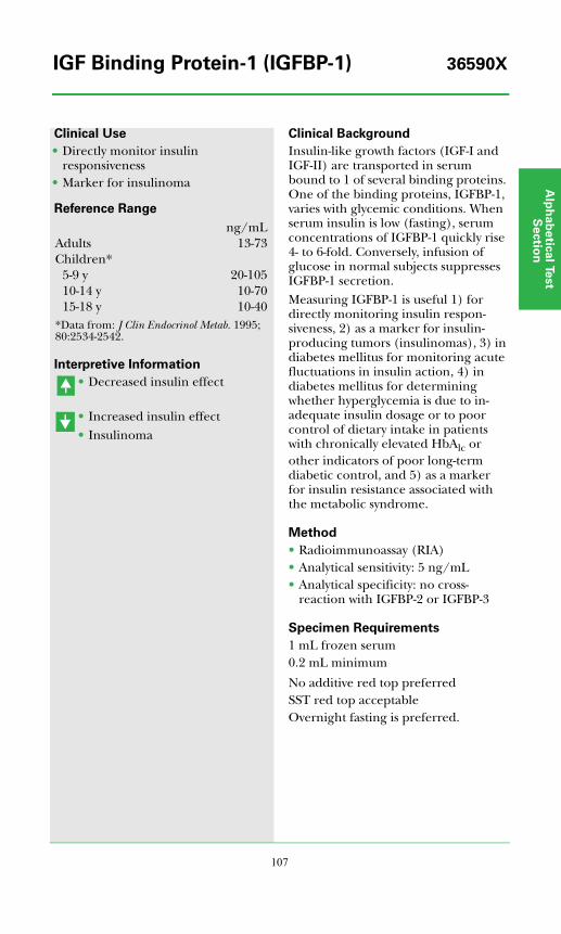
Alp
ha
be
tica
l Test
Se
ctio
n
107
Clinical Use
• Directly monitor insulin responsiveness
• Marker for insulinoma
Reference Range
Interpretive Information
• Decreased insulin effect
• Increased insulin effect• Insulinoma
Clinical Background
Insulin-like growth factors (IGF-I and IGF-II) are transported in serum bound to 1 of several binding proteins. One of the binding proteins, IGFBP-1, varies with glycemic conditions. When serum insulin is low (fasting), serum concentrations of IGFBP-1 quickly rise 4- to 6-fold. Conversely, infusion of glucose in normal subjects suppresses IGFBP-1 secretion.Measuring IGFBP-1 is useful 1) for directly monitoring insulin respon-siveness, 2) as a marker for insulin-producing tumors (insulinomas), 3) in diabetes mellitus for monitoring acute fluctuations in insulin action, 4) in diabetes mellitus for determining whether hyperglycemia is due to in-adequate insulin dosage or to poor control of dietary intake in patients with chronically elevated HbAlc or other indicators of poor long-term diabetic control, and 5) as a marker for insulin resistance associated with the metabolic syndrome.
Method
• Radioimmunoassay (RIA)• Analytical sensitivity: 5 ng/mL• Analytical specificity: no cross-
reaction with IGFBP-2 or IGFBP-3
Specimen Requirements
1 mL frozen serum0.2 mL minimum
No additive red top preferredSST red top acceptableOvernight fasting is preferred.
ng/mLAdults 13-73Children*
5-9 y 20-10510-14 y 10-7015-18 y 10-40
*Data from: J Clin Endocrinol Metab. 1995;80:2534-2542.
IGF Binding Protein-1 (IGFBP-1) 36590X
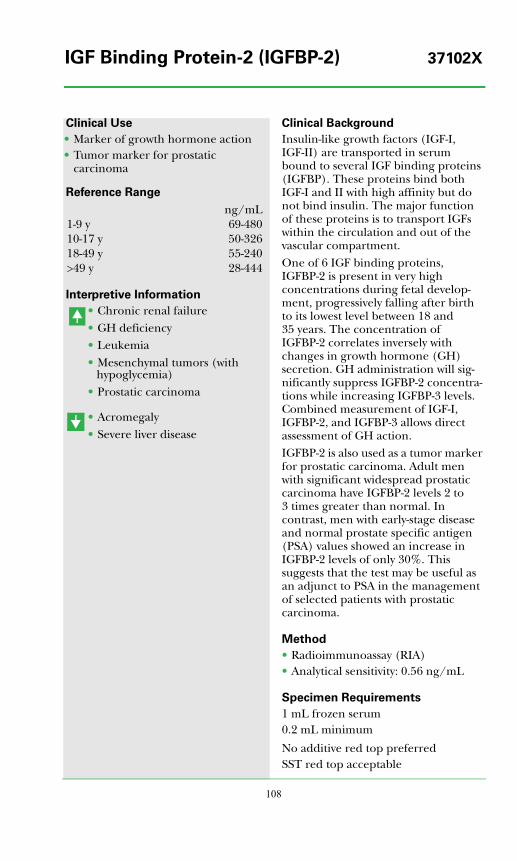
108
Clinical Use
• Marker of growth hormone action• Tumor marker for prostatic
carcinoma
Reference Range
Interpretive Information
• Chronic renal failure• GH deficiency• Leukemia• Mesenchymal tumors (with
hypoglycemia)• Prostatic carcinoma
• Acromegaly• Severe liver disease
Clinical Background
Insulin-like growth factors (IGF-I, IGF-II) are transported in serum bound to several IGF binding proteins (IGFBP). These proteins bind both IGF-I and II with high affinity but do not bind insulin. The major function of these proteins is to transport IGFs within the circulation and out of the vascular compartment.One of 6 IGF binding proteins, IGFBP-2 is present in very high concentrations during fetal develop-ment, progressively falling after birth to its lowest level between 18 and 35 years. The concentration of IGFBP-2 correlates inversely with changes in growth hormone (GH) secretion. GH administration will sig-nificantly suppress IGFBP-2 concentra-tions while increasing IGFBP-3 levels. Combined measurement of IGF-I, IGFBP-2, and IGFBP-3 allows direct assessment of GH action.IGFBP-2 is also used as a tumor marker for prostatic carcinoma. Adult men with significant widespread prostatic carcinoma have IGFBP-2 levels 2 to 3 times greater than normal. In contrast, men with early-stage disease and normal prostate specific antigen (PSA) values showed an increase in IGFBP-2 levels of only 30%. This suggests that the test may be useful as an adjunct to PSA in the management of selected patients with prostatic carcinoma.
Method
• Radioimmunoassay (RIA)• Analytical sensitivity: 0.56 ng/mL
Specimen Requirements
1 mL frozen serum0.2 mL minimum
No additive red top preferredSST red top acceptable
ng/mL1-9 y 69-48010-17 y 50-32618-49 y 55-240>49 y 28-444
IGF Binding Protein-2 (IGFBP-2) 37102X
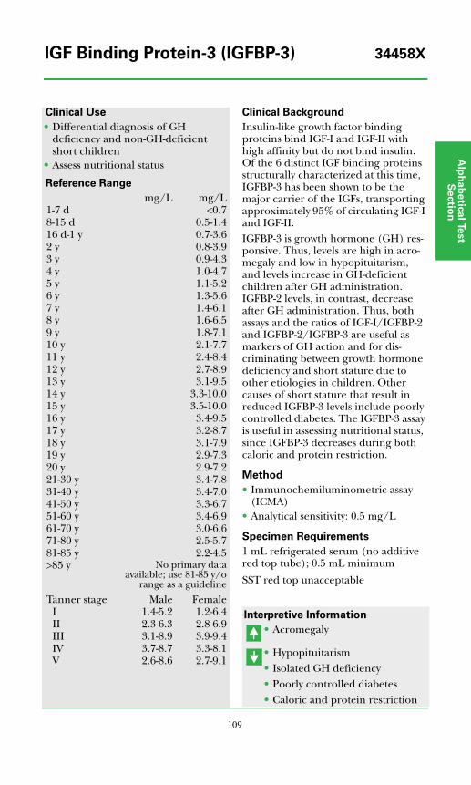
Alp
ha
be
tica
l Test
Se
ctio
n
109
Clinical Use
• Differential diagnosis of GH deficiency and non-GH-deficient short children
• Assess nutritional status
Reference Range
Clinical Background
Insulin-like growth factor binding proteins bind IGF-I and IGF-II with high affinity but do not bind insulin. Of the 6 distinct IGF binding proteins structurally characterized at this time, IGFBP-3 has been shown to be the major carrier of the IGFs, transporting approximately 95% of circulating IGF-I and IGF-II.IGFBP-3 is growth hormone (GH) res-ponsive. Thus, levels are high in acro-megaly and low in hypopituitarism, and levels increase in GH-deficient children after GH administration. IGFBP-2 levels, in contrast, decrease after GH administration. Thus, both assays and the ratios of IGF-I/IGFBP-2 and IGFBP-2/IGFBP-3 are useful as markers of GH action and for dis-criminating between growth hormone deficiency and short stature due to other etiologies in children. Other causes of short stature that result in reduced IGFBP-3 levels include poorly controlled diabetes. The IGFBP-3 assay is useful in assessing nutritional status, since IGFBP-3 decreases during both caloric and protein restriction.
Method
• Immunochemiluminometric assay (ICMA)
• Analytical sensitivity: 0.5 mg/L
Specimen Requirements
1 mL refrigerated serum (no additive red top tube); 0.5 mL minimum
SST red top unacceptable
Interpretive Information
• Acromegaly
• Hypopituitarism• Isolated GH deficiency• Poorly controlled diabetes• Caloric and protein restriction
mg/L mg/L1-7 d <0.78-15 d 0.5-1.416 d-1 y 0.7-3.62 y 0.8-3.93 y 0.9-4.34 y 1.0-4.75 y 1.1-5.26 y 1.3-5.67 y 1.4-6.18 y 1.6-6.59 y 1.8-7.110 y 2.1-7.711 y 2.4-8.412 y 2.7-8.913 y 3.1-9.514 y 3.3-10.015 y 3.5-10.016 y 3.4-9.517 y 3.2-8.718 y 3.1-7.919 y 2.9-7.320 y 2.9-7.221-30 y 3.4-7.831-40 y 3.4-7.041-50 y 3.3-6.751-60 y 3.4-6.961-70 y 3.0-6.671-80 y 2.5-5.781-85 y 2.2-4.5>85 y No primary data
available; use 81-85 y/orange as a guideline
Tanner stage Male FemaleI 1.4-5.2 1.2-6.4II 2.3-6.3 2.8-6.9III 3.1-8.9 3.9-9.4IV 3.7-8.7 3.3-8.1V 2.6-8.6 2.7-9.1
IGF Binding Protein-3 (IGFBP-3) 34458X
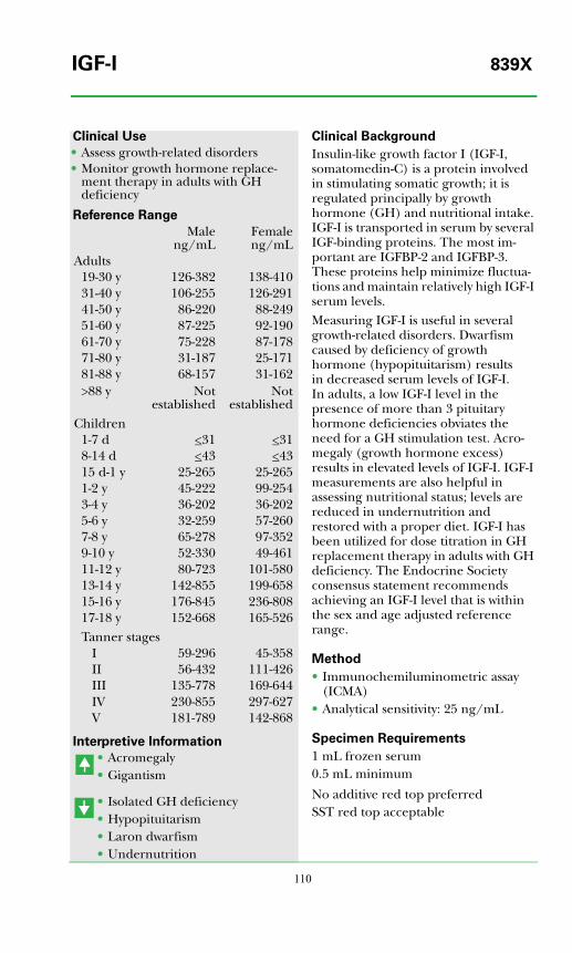
110
Clinical Use
• Assess growth-related disorders• Monitor growth hormone replace-
ment therapy in adults with GH deficiency
Reference Range
Interpretive Information
• Acromegaly• Gigantism
• Isolated GH deficiency• Hypopituitarism• Laron dwarfism• Undernutrition
Clinical Background
Insulin-like growth factor I (IGF-I, somatomedin-C) is a protein involved in stimulating somatic growth; it is regulated principally by growth hormone (GH) and nutritional intake. IGF-I is transported in serum by several IGF-binding proteins. The most im-portant are IGFBP-2 and IGFBP-3. These proteins help minimize fluctua-tions and maintain relatively high IGF-I serum levels.Measuring IGF-I is useful in several growth-related disorders. Dwarfism caused by deficiency of growth hormone (hypopituitarism) results in decreased serum levels of IGF-I. In adults, a low IGF-I level in the presence of more than 3 pituitary hormone deficiencies obviates the need for a GH stimulation test. Acro-megaly (growth hormone excess) results in elevated levels of IGF-I. IGF-I measurements are also helpful in assessing nutritional status; levels are reduced in undernutrition and restored with a proper diet. IGF-I has been utilized for dose titration in GH replacement therapy in adults with GH deficiency. The Endocrine Society consensus statement recommends achieving an IGF-I level that is within the sex and age adjusted reference range.
Method
• Immunochemiluminometric assay (ICMA)
• Analytical sensitivity: 25 ng/mL
Specimen Requirements
1 mL frozen serum0.5 mL minimum
No additive red top preferredSST red top acceptable
Maleng/mL
Femaleng/mL
Adults19-30 y 126-382 138-41031-40 y 106-255 126-29141-50 y 86-220 88-24951-60 y 87-225 92-19061-70 y 75-228 87-17871-80 y 31-187 25-17181-88 y 68-157 31-162>88 y Not
establishedNot
establishedChildren
1-7 d <31 <318-14 d <43 <4315 d-1 y 25-265 25-2651-2 y 45-222 99-2543-4 y 36-202 36-2025-6 y 32-259 57-2607-8 y 65-278 97-3529-10 y 52-330 49-46111-12 y 80-723 101-58013-14 y 142-855 199-65815-16 y 176-845 236-80817-18 y 152-668 165-526Tanner stages
I 59-296 45-358II 56-432 111-426III 135-778 169-644IV 230-855 297-627V 181-789 142-868
IGF-I 839X
Alp
ha
be
tica
l Test
Se
ctio
n
111
Clinical Use
• Evaluate non-islet-cell tumor hypoglycemia (NICTH)
• Assess growth factor status
Reference Range
Interpretive Information
• Mesenchymal tumors (with hypoglycemia)
• Hepatoma• Acromegaly (variable)• Wilms tumor
• Infants under 1 year• Growth hormone deficiency
Clinical Background
The somatomedins, insulin-like growth factors I and II (IGF-I and IGF-II), are structurally related to proinsulin and play an essential role in regulating cell proliferation and function. Their effects are mediated by way of IGF and insulin receptors. IGF-II is a neutral 7500-dalton polypeptide with 75% homology with IGF-I, and like IGF-I, it is produced in multiple tissues. It is more insulin-like in its action than IGF-I, and levels are less dependent on growth hormone. Plasma concentra-tions of IGF-II are approximately 3 times higher than IGF-I. Non-islet-cell tumor hypoglycemia (NICTH) is a syndrome associated with low levels of insulin, IGF-I, and IGFBP-3 and increased secretion of "big" IGF-II (pro-IGF-II). The IGF-I/IGFBP-3 ratio is markedly increased. The preferred treatment is tumor removal, but growth hormone treatment can normalize the IGF-I/ IGFBP-3 ratio and ameliorate the hypoglycemia.
Method
• Immunoradiometric assay (IRMA)• Analytical sensitivity: 150 ng/mL
Specimen Requirements
1 mL refrigerated serum (no additive red top tube); 0.1 mL minimum
SST red top unacceptableOvernight fasting is preferred.
Adults ng/mL18-30 y 546-126031-40 y 460-124041-50 y 414-123051-60 y 414-1248
Children0-4 y Not established5-9 y 754-121610-13 y 610-121714-17 y 649-1225
IGF-II 20707X(Insulin-Like Growth Factor II)
112
Clinical Use
• Prenatal risk assessment for Down syndrome
Reference Range
Interpretive Information
• Granulosa cell tumors• Down syndrome-affected
pregnancies
• Menopause
Clinical Background
Inhibin A is a glycoprotein produced by the placenta and by granulosa cells of the ovary, particularly during the late follicular and luteal phases of the menstrual cycle. The active form is a dimer consisting of an alpha subunit linked to a betaA subunit. Inhibin A has both endocrine (suppression of FSH by the pituitary) and paracrine (stimulation of thecal cell androgen synthesis) functions.Measurement of inhibin B may be preferable to that of inhibin A for detection of recurrent granulosa cell tumors, studies of reproductive aging in females, and studies in males.
Method
• Enzyme-linked immunosorbent assay (ELISA)
Specimen Requirements
2 mL frozen serum0.5 mL minimum
No additive red top preferredSST red top acceptable
Women pg/mLPremenopausal <98Postmenopausal <10
Men <21
Inhibin A 34472X
Alp
ha
be
tica
l Test
Se
ctio
n
113
Clinical Use
• Detect recurrence of granulosa cell tumors
• Predict ovarian reserve and outcome of in vitro fertilization
• Marker of testicular function• Differential diagnosis of
cryptorchidism versus anorchia
Reference Range
Interpretive Information
• Granulosa cell tumors
• Decreased ovarian reserve/menopause
• Anorchia
Clinical Background
Inhibin B is a glycoprotein produced by the Sertoli cells of the testis and the granulosa cells of the ovary, particularly during the early follicular phase of the menstrual cycle. The active form is a dimer consisting of an alpha subunit linked to a betaB subunit. Inhibin B suppresses pituitary synthesis of FSH and may also have additional paracrine roles within the ovary or testis. Measurement of inhibin B may be preferable to that of inhibin A for detection of recurrent granulosa cell tumors, studies of reproductive aging in females, and studies in males.
Method
• Enzyme-linked immunosorbent assay (ELISA)
Specimen Requirements
1 mL frozen serum (no additive red top tube); 0.5 mL minimum
pg/mL pg/mLMen <305Women
Premenopausal <255Postmenopausal <30
Children Males Females3-9 y <161 <3010-13 y 42-339 <9214-17 y 68-300 <139
Inhibin B 34445X
114
Clinical Use
• Assess risk for type 1 diabetes• Predict onset of type 1 diabetes• Diagnose new onset type 1 diabetes
Reference Range
Interpretive Information
• Prediabetic (type 1) subjects• Polyendocrine autoimmune
syndromes• Other autoimmune disease
Clinical Background
Type 1 diabetes is characterized by lymphocytic infiltration of pancreatic islet cells and autoantibodies against several islet cell antigens including glutamic acid decarboxylase (GAD-65), IA-2, and insulin. This assay is designed to assess the low titers of autoantibody in prediabetic subjects and other patients with autoimmune disorders who have not received exogenous insulin. For type 1 diabetes risk assessment, a combination of IA-2, GAD-65, and insulin autoantibodies increases the sensitivity.
Method
• Radiobinding assay (RBA)
Specimen Requirements
1 mL frozen serum (no additive red top tube); 0.2 mL minimum
SST red top unacceptable
Adults and children <1 U/mL
Insulin Antibodies 36178X
Alp
ha
be
tica
l Test
Se
ctio
n
115
Clinical Use
• Monitor therapy in patients with insulin antibodies
Reference Range
Interpretive Information
• Following exogenous insulin injection
• Insulinoma• Neisidioblastosis• Some type 2 diabetic patients• Insulin resistance
• Untreated type 1 diabetes mellitus
Clinical Background
The free insulin measurement is helpful in interpreting blood glucose concentration and its relationship to insulin injections in insulin-treated patients who have insulin antibodies. For instance, if a diabetic patient frequently becomes hypoglycemic at certain times of the day, concomitantly high free insulin levels would suggest the need to reduce the insulin dosage. In contrast, elevated glucose concen-trations together with low free insulin levels indicate insufficient insulin for adequate control. Multiple, sequential, free insulin levels are also helpful for evaluating the rate and quantity of insulin absorption from subcutaneous injections.The availability of C-peptide and free insulin measurements permits assess-ment of endogenous insulin secretion as well as the concentrations of circu-lating biologically active insulin (from all sources) in patients who have circu-lating insulin antibodies.
Method
• PEG precipitation, radioimmunoassay (RIA)
• Analytical sensitivity: 2.5 μU/mL•• Analytical specificity: detects free,
bioavailable insulin; does not detect antibody-bound insulin; no cross-reactivity with C-peptide; cross-reacts with proinsulin
Specimen Requirements
1.8 mL refrigerated serum0.6 mL minimum
No additive red top preferredSST red top acceptableOvernight fasting required
Adults 4.0-20.0 μU/mL
Insulin, Free (Bioactive) 36700X
116
Clinical Use
• Determine insulin dosage in patients with insulin-dependent diabetes and insulin antibodies
Reference Range
Interpretive Information
• (With circulating insulin antibodies)
• Insulinoma• Some type 2 diabetic patients• Infantile hypoglycemia• Hyperinsulinism
• Untreated type 1 diabetes mellitus
Individuals with circulating insulin antibodies may demonstrate variable concentrations depending upon the level and binding capacity of the antibodies.
Clinical Background
The total insulin concentration includes free plus antibody-bound insulin. In patients without insulin antibody, total and free insulin concentrations are similar. In patients with insulin antibodies, total insulin levels are dependent on the binding capacity of the circulating endogenous insulin antibody and the availability of insulin to bind to the antibody sites. Total insulin is measured by eluting antibody-bound insulin with acid, precipitating the endogenous anti-body, and measuring the total insulin by direct radioimmunoassay. Total insulin levels are useful in assessing the available insulin in serum, including antibody-bound insulin.Serum insulin levels are useful in the differential diagnosis of fasting hypo-glycemia (see Dynamic Test section) but must always be evaluated in the light of a simultaneous plasma glucose. With any cause of hyperglycemia, such as a meal, insulin is stimulated; with fasting or hypoglycemia, insulin is normally suppressed.
Method
• Acid treatment, PEG precipitation, radioimmunoassay (RIA)
• Analytical sensitivity: 2.5 μU/mL• Analytical specificity: detects free and
antibody-bound insulin; cross-reacts with proinsulin; no cross-reactivity with C-peptide
Specimen Requirements
1.8 mL refrigerated serum0.6 mL minimum
No additive red top preferredSST red top acceptableOvernight fasting is required.
<20.0 μU/mL
Insulin, Total (Free and Bound) 36702X
Alp
ha
be
tica
l Test
Se
ctio
n
117
Clinical Use
• Early detection of pregnancy
Reference Range
Interpretive Information
• Pregnancy• Trophoblastic tumors
Clinical Background
ITA is a hyperglycosylated form of human chorionic gonadotropin (hCG) secreted during embryonic implanta-tion and trophoblast invasion of the uterine wall. ITA levels rise early in pregnancy, during which time almost 100% of hCG-related molecules is ITA. By 12 weeks of gestation, less than 2.9% of hCG-related molecules is ITA. Measurement of ITA rather than regular hCG therefore allows more dependable detection of pregnancy in the week after a missed menstrual period.
Method
• Electrochemiluminescence assay (ECLA)
• Analytical sensitivity: 0.5 μg/L• Analytical specificity: <3.5% cross-
reactivity with hCG; <1% cross-reactivity with other glycoprotein hormones
Specimen Requirements
2 mL room temperature serum0.2 mL minimum
No additive red top preferredSST red top acceptable
<1 μg/L
Invasive Trophoblast Antigen 11303X(ITA) (Pregnancy)
118
Clinical Use
• Assess risk of type 1 diabetes• Predict onset of type 1 diabetes• Diagnose new onset type 1 diabetes
Reference Range
Interpretive Information
• Patients at risk for developing type 1 diabetes
• Type 1 diabetes
Clinical Background
Autoantibodies directed against pancreatic islet cells (islet cell anti-bodies, ICA) include glutamic acid decarboxylase (GAD), IA-2, insulin, GM2-1, and other cell surface proteins. Although ICA attacks all pancreatic islet cells, cell destruction appears specific for insulin-producing islet cells (beta cells), leading to insulin defi-ciency. ICA are present in the serum of patients during the prediabetic phase and predict development of type 1 disease.Radiobinding assays are available for assessment of GAD-65, IA-2, and insulin autoantibodies and are more reliable as markers for the prediabetic state. This IFA assay measures a variety of antibodies, has limited sensitivity, and is only semi-quantitative.
Method
• Immunofluorescence assay• Titer provided (at an additional
charge) if antibody is present
Specimen Requirements
2.0 mL refrigerated serum 0.5 mL minimum
No additive red top preferredSST red top acceptable
Antibody NegativeTiter <1.25 JDF UnitsNOTE: End point titers are compared to a single international reference standard and values are reported in JDF (Juvenile Diabetes Foundation) units.
Islet Cell Antibody (ICA) 36741X

Alp
ha
be
tica
l Test
Se
ctio
n
119
Clinical Use
• Assess adrenocortical function
Reference Range
Interpretive Information
• Cushing’s syndrome• Precocious puberty caused by
adrenal hyperplasia• Physiological stress (surgery,
burns, infectious diseases)• ACTH or gonadotropin
stimulation
• Addison’s disease• ACTH deficiency• Corticosteroids, estrogen, oral
contraceptives
Some medications may interfere with this test.
Clinical Background
17-Ketosteroids (17-KS) react with para-aminobenzoic acid to yield a colored product with an absorption maximum at 520 μM (the Zimmerman reaction). Adrenal androgens measured as 17-KS include dehydro-epiandrosterone, androstenedione, etiocholanolone, androsterone, and their reduced metabolites. In men, about two-thirds of the 17-KS are secreted by the adrenal cortex, while in women, almost all are secreted by the adrenal cortex. Thus, in both sexes 17-KS excretion reflects adrenal androgen production.
Method
• Modified Zimmerman reaction• Analytical sensitivity: 0.1 mg/L• Creatinine concentration also
reported
Specimen Requirements
20 mL refrigerated aliquot of a 24-h urine; 10 mL minimum
Collect urine with 10 g of boric acid (preferred); alternatively use 30 mL 6N HCL or 25 mL 50% acetic acid.Record total volume on vial and request form.
17-Ketosteroids mg/24-hMen
17-20 y 9-22>20 y 8-20
Women>16 y 6-15
Children<1 y <1.01-4 y <2.05-8 y <3.09-12 y 3-1013-16 y 5-12
Creatinine g/24-h3-8 y 0.11-0.689-12 y 0.17-1.4113-17 y 0.29-1.87Adults 0.63-2.50
17-Ketosteroids (17-KS), Urine 15201X
120
Clinical Use
• Assess adrenal and gonadal androgen status
Reference Range
See next page
Interpretive Information
• Hyperandrogenism• Cushing’s syndrome• Adrenal hyperplasia
• Adrenal insufficiency• Chronic illness• 5α-Reductase deficiency
(decreased androsterone/etiocholanolone ratio)
Clinical Background
Androgens secreted by the adrenal gland or gonads are metabolized and excreted in the urine as 17-ketosteroid compounds. Fractionation of specific metabolites allows more specific assessment of androgen status than the older modified Zimmerman reaction assay. Detection by mass spectrometry further increases specificity as com-pared to capillary gas chromatography. Androstenedione is metabolized to androsterone by 5α-reductase activity and to etiocholanolone by 5β-reduct-ase activity. Both androsterone and etiocholanolone reflect overall levels of androstenedione (and, less directly, the levels of DHEA, testosterone, and 17-hydroxyprogesterone). The ratio of androsterone to etiocholanolone can be used as a relative index of 5α-reductase to 5β-reductase activity.The 11-oxo and 11β-hydroxy metabolites of androsterone and etiocholanolone tend to reflect the patterns of the parent compounds. Urinary DHEA reflects overall serum DHEA levels. Pregnanetriol is a metabolite of 17-hydroxyprogesterone.
Method
• Gas chromatography/mass spectrometry (GC/MS)
• Creatinine concentration also reported
Specimen Requirements
5 mL frozen aliquot of a 24-h urine2.6 mL minimum
Refrigerate during collection; do not use preservatives.
Continued
17-Ketosteroids, Fractionated, 4932X24-Hour Urine
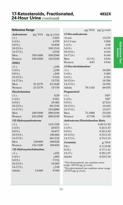
Alp
ha
be
tica
l Test
Se
ctio
n
121
Reference Range
Androsterone μg/24-h μg/g creat<1 y 3-2251-4.9 y 4-3785-9.9 y 16-83010-13.9 y 102-151014-17.9 y <2750Men 320-5400 430-2500Women 240-2300 150-2100DHEA<1 y <1001-4.9 y <1005-9.9 y <10010-13.9 y 5-31614-17.9 y 15-732Men 21-2170 24-1640Women 21-2170 13-730Etiocholanolone<1 y 6-291-4.9 y 2-2615-9.9 y 18-46510-13.9 y 98-174614-17.9 y 104-2006Men 430-3300 280-2100Women 245-2300 280-210011β-Hydroxyandrosterone<1 y 143-11801-4.9 y 23-6735-9.9 y 44-67710-13.9 y 100-80214-17.9 y 68-1152Men 130-900 180-850Women 195-1500 180-85011β-Hydroxyetiocholanolone<1 y <1051-4.9 y <4035-9.9 y 7-71610-13.9 y 10-36314-17.9 y <740Adults 14-680 8-580
μg/24-h μg/g creat11-Oxo-androsterone<6 mo 13-5786-11.9 mo 1-2001-4.9 y 3-695-9.9 y 6-11810-13.9 y 4-10414-17.9 y 8-106Men 15-111 13-84Women 8-87 13-8411-Oxo-etiocholanolone<1 y 16-2961-4.9 y 3-4935-9.9 y 6-76510-13.9 y 18-78614-17.9 y 16-765Adults 78-1165 68-870Pregnanetriol<1 y ND*1-4.9 y ND†
5-9.9 y 27-21410-13.9 y 19-56314-17.9 y 15-817Men 71-1000 53-530Women 47-790 53-530Androsterone/Etiocholanolone Ratio<1 y 0.60-15.501-4.9 y 0.25-5.375-9.9 y 0.45-3.4310-13.9 y 0.55-3.1214-17.9 y 0.74-3.18Creatinine g/24-h3-8 y 0.11-0.689-12 y 0.17-1.4113-17 y 0.29-1.87Adults 0.63-2.50
*Not determined; use random urine range (49-345 μg/g creat). †Not determined; use random urine range (16-233 μg/g creat).
17-Ketosteroids, Fractionated, 4932X24-Hour Urine (continued)
122
Clinical Use
• Assess leptin control pathway
Reference Range
Interpretive Information
• Obesity• Leptin receptor defect
• Under nutrition• Leptin deficiency
Clinical Background
Leptin is an adipocyte-derived hormone essential for normal body weight regulation. Production is under neuroendocrine control such that blood levels vary directly with the amount of triglyceride stored in adipose tissue depots; there is a negative correlation between leptin and testosterone blood levels. Leptin acts through specific receptors in the hypothalamus to modulate appetite and thermogenesis; increased levels suppress appetite and increase thermogenesis. Leptin deficiency leads to hypopituitarism including gonado-tropin, growth hormone, and thyroid deficiencies. Gene mutations resulting in leptin deficiency or leptin receptor defects lead to massive obesity.
Method
• Radioimmunoassay (RIA)• Analytical sensitivity: 0.5 ng/mL
Specimen Requirements
1.0 mL refrigerated serum0.2 mL minimum
No additive red top preferredSST red top acceptable
Malesng/mL
Femalesng/mL
Adults (BMI* 18-25)
1.2-9.5 4.1-25.0
Children†
Prepubertal 1.6-10.8 1.7-10.6Tanner II-III 2.1-11.6 2.6-11.5Tanner IV-V 3.4-10.2 3.4-13.0
*BMI = body mass index = wt (kg)/ht (m2)†Pediatric reference ranges from Clin Endocrinol. 1997;46:727-733.
Leptin 37109X

Alp
ha
be
tica
l Test
Se
ctio
n
123
Clinical Use
• Diagnose gonadal function and pituitary disorders
Reference Range
Interpretive Information
•• Primary hypogonadism• Gonadotropin-secreting
pituitary tumors• Menopause
• Hypothalamic GnRH deficiency• Pituitary LH deficiency• Ectopic steroid hormone
production• GnRH analog treatment
Clinical Background
Luteinizing hormone (LH, lutropin) is a glycoprotein produced by the anterior pituitary gland. Production is regulated by hypothalamic gonado-tropin releasing hormone (GnRH) and feedback from gonadal steroid hormones. LH stimulates ovulation and ovarian steroid production (estrogen and progesterone) in the female. In the male, LH controls Leydig cell secretion of testosterone.In females, LH concentrations are low during the follicular phase of the menstrual cycle, rise to a midcycle peak to cause ovulation, and, following ovulation, fall to levels lower than during the follicular phase. After menopause, LH concentrations rise to levels as high as or higher than those found in the midcycle peak; similar high levels are seen in castrated men.
Method
• Immunochemiluminometric assay (ICMA)
• Analytical sensitivity: 0.7 IU/L
This assay is appropriate for most purposes. The LH "third generation" assay (test code 36086X) is sensitive to 0.03 mIU/mL and is more appropriate for use in children.
Specimen Requirements
1.0 mL refrigerated serum0.5 mL minimum
No additive red top preferredSST red top acceptable
mIU/mLMen 1.5-9.3Women
Follicular phase 1.9-12.5Mid-cycle 8.7-76.3Luteal phase 0.5-16.9Postmenopausal 5.0-52.3
LH (Luteinizing Hormone) 615X
124
Clinical Use
• Diagnose (and differential diagnosis of) gonadal dysfunction
• Monitor LH suppressive (Lupron®’) therapy
• Applicable to pediatric samples
Reference Range
Interpretive Information
•• Primary hypogonadism• Gonadotropin-secreting
pituitary tumors
• Hypothalamic GnRH deficiency • Pituitary LH deficiency
Clinical Background
Luteinizing hormone (LH, lutropin) is produced by the anterior pituitary gland. Production is regulated by hypothalamic gonadotropin releasing hormone (GnRH). LH stimulates ovulation and ovarian steroid hormone production in the female and testosterone secretion by the testes in the male.GnRH and LH secretion are regulated by negative feedback systems, whereby reduced levels of gonadal hormones stimulate and increased levels inhibit circulating LH concentrations.LH levels are useful in assessing disorders of gonadal function.GnRH stimulation of LH is a useful provocative test in selected patients with delayed puberty and in confirming pituitary hypofunction in secondary hypogonadism.
Method
• Electrochemiluminescence assay (ECL)
• Analytical sensitivity: 0.03 mIU/mL• Analytical specificity: 1.1% cross-
reactivity with TSH; no cross-reactivity with FSH, GH, hCG, or prolactin
Specimen Requirements
0.5 mL refrigerated serum0.2 mL minimum
No additive red top preferredSST red top acceptable
Males FemalesChildren mIU/mL mIU/mL
1-7 y <0.10 <0.458-9 y <0.44 <3.3610-11 y <2.28 <5.6512-14 y 0.31-5.29 <11.0015-17 y 0.15-5.33 <15.80
LH (Luteinizing Hormone), 36086XPediatrics
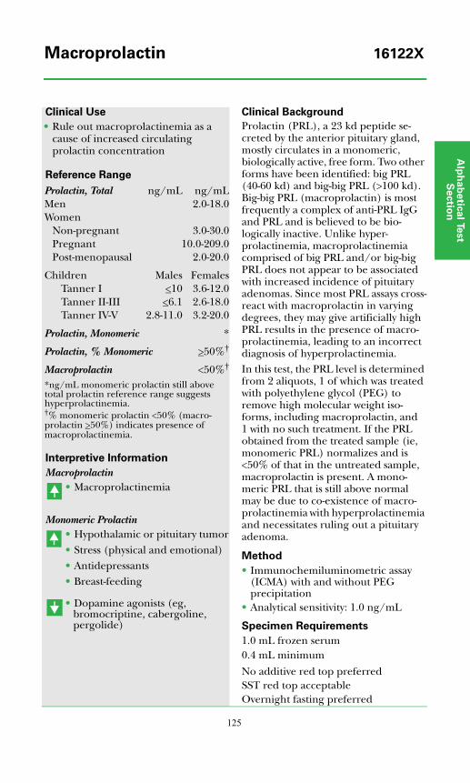
Alp
ha
be
tica
l Test
Se
ctio
n
125
Clinical Use
• Rule out macroprolactinemia as a cause of increased circulating prolactin concentration
Reference Range
Interpretive Information
Macroprolactin• Macroprolactinemia
Monomeric Prolactin• Hypothalamic or pituitary tumor• Stress (physical and emotional)• Antidepressants• Breast-feeding
• Dopamine agonists (eg, bromocriptine, cabergoline, pergolide)
Clinical Background
Prolactin (PRL), a 23 kd peptide se-creted by the anterior pituitary gland, mostly circulates in a monomeric, biologically active, free form. Two other forms have been identified: big PRL (40-60 kd) and big-big PRL (>100 kd). Big-big PRL (macroprolactin) is most frequently a complex of anti-PRL IgG and PRL and is believed to be bio-logically inactive. Unlike hyper-prolactinemia, macroprolactinemia comprised of big PRL and/or big-big PRL does not appear to be associated with increased incidence of pituitary adenomas. Since most PRL assays cross-react with macroprolactin in varying degrees, they may give artificially high PRL results in the presence of macro-prolactinemia, leading to an incorrect diagnosis of hyperprolactinemia.In this test, the PRL level is determined from 2 aliquots, 1 of which was treated with polyethylene glycol (PEG) to remove high molecular weight iso-forms, including macroprolactin, and 1 with no such treatment. If the PRL obtained from the treated sample (ie, monomeric PRL) normalizes and is <50% of that in the untreated sample, macroprolactin is present. A mono-meric PRL that is still above normal may be due to co-existence of macro-prolactinemia with hyperprolactinemia and necessitates ruling out a pituitary adenoma.
Method
• Immunochemiluminometric assay (ICMA) with and without PEG precipitation
• Analytical sensitivity: 1.0 ng/mL
Specimen Requirements
1.0 mL frozen serum 0.4 mL minimum
No additive red top preferredSST red top acceptableOvernight fasting preferred
Prolactin, Total ng/mL ng/mLMen 2.0-18.0Women
Non-pregnant 3.0-30.0Pregnant 10.0-209.0Post-menopausal 2.0-20.0
Children Males FemalesTanner I <10 3.6-12.0Tanner II-III <6.1 2.6-18.0Tanner IV-V 2.8-11.0 3.2-20.0
Prolactin, Monomeric *
Prolactin, % Monomeric >50%†
Macroprolactin <50%†
*ng/mL monomeric prolactin still above total prolactin reference range suggests hyperprolactinemia.†% monomeric prolactin <50% (macro-prolactin >50%) indicates presence of macroprolactinemia.
Macroprolactin 16122X

126
Clinical Use
• Screen at-risk individuals for MEN 2 and FMTC
• Diagnose MEN 2 and FMTC• Differentiate between familial and
sporadic MTC
Reference Range
Interpretive Information
Mutation present• FMTC and/or MEN 2A• MEN 2B
Clinical Background
Multiple endocrine neoplasia type 2 (MEN 2) is an autosomal dominant syndrome associated with a high risk of medullary thyroid carcinoma (MTC). The MEN 2A subtype is associated with hyperparathyroidism, and both MEN 2A and MEN 2B subtypes are associated with an increased risk for pheochromo-cytoma. MEN 2B is further character-ized by mucosal neuromas on the lips and tongue and in the gastrointestinal tract. Over 95% of MEN 2A cases and over 85% of familial MTC (FMTC) cases have a germline mutation in 1 of 5 conserved cysteine residues encoded in exon 10 or 11 of the RET proto-oncogene. A point mutation in exon 16 is present in over 95% of MEN 2B cases. Other mutations in these exons and in exons 13, 14, and 15 have also been associated with MEN 2. Once the mutation in an affected family member has been identified, testing of first-degree relatives allows presympto-matic identification of those at risk for MEN 2 and prophylactic treatment. Correlation between specific mutations and aggressiveness of MTC may aid in appropriately timing prophylactic thyroidectomy and pheochromocytoma screening. Genetic testing is recom-mended for all individuals with MTC since RET mutations have been detect-ed in 1% to 20% of those who appear to have sporadic MTC (ie, those without a known family history of MEN 2).
Method
• Polymerase chain reaction (PCR) and semi-automated DNA sequencing
• Analytical specificity: mutations in exons 10, 11, 13, 14, 15, and 16 of the RET proto-oncogene
Specimen Requirements
5 mL room temperature whole blood3 mL minimum
Collect blood in a lavender-top (EDTA) or yellow-top (ACD solution B) tube. Alternatively, submit amniotic fluid, bone marrow, tissue biopsy (eg, dissected CVS) or extracted DNA.
Negative (no mutations detected)
MEN 2 and FMTC Mutations, 36587XExons 10, 11, and 13-16
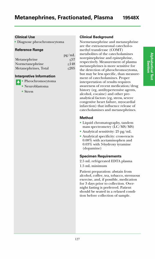
Alp
ha
be
tica
l Test
Se
ctio
n
127
Clinical Use
• Diagnose pheochromocytoma
Reference Range
Interpretive Information
• Pheochromocytoma• Neuroblastoma• Stress
Clinical Background
Normetanephrine and metanephrine are the extra-neuronal catechol-o-methyl transferase (COMT) metabolites of the catecholamines norepinephrine and epinephrine, respectively. Measurement of plasma metanephrines is more sensitive for the detection of pheochromocytoma, but may be less specific, than measure-ment of catecholamines. Proper interpretation of results requires awareness of recent medication/drug history (eg, antihypertensive agents, alcohol, cocaine) and other pre-analytical factors (eg, stress, severe congestive heart failure, myocardial infarction) that influence release of catecholamines and metanephrines.
Method
• Liquid chromatography, tandem mass spectrometry (LC/MS/MS)
• Analytical sensitivity: 25 pg/mL• Analytical specificity: cross-reacts
0.08% with acetaminophen and 0.03% with 3-hydroxy tyramine (dopamine)
Specimen Requirements
2.5 mL refrigerated EDTA plasma1.5 mL minimum
Patient preparation: abstain from alcohol, coffee, tea, tobacco, strenuous exercise, and, if possible, medication for 3 days prior to collection. Over-night fasting is preferred. Patient should be seated in a relaxed condi-tion before collection of sample.
pg/mLMetanephrine <57Normetanephrine <148Metanephrines, Total <205
Metanephrines, Fractionated, Plasma 19548X
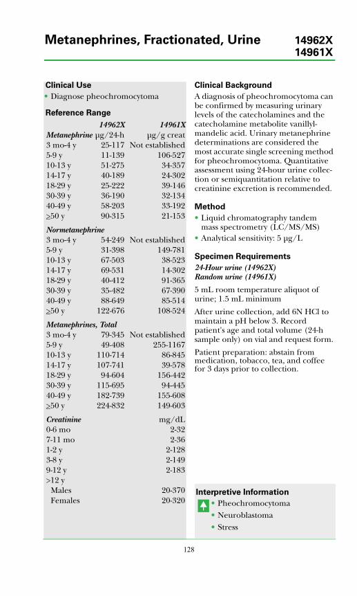
128
Clinical Use
• Diagnose pheochromocytoma
Reference Range
Clinical Background
A diagnosis of pheochromocytoma can be confirmed by measuring urinary levels of the catecholamines and the catecholamine metabolite vanillyl-mandelic acid. Urinary metanephrine determinations are considered the most accurate single screening method for pheochromocytoma. Quantitative assessment using 24-hour urine collec-tion or semiquantitation relative to creatinine excretion is recommended.
Method
• Liquid chromatography tandem mass spectrometry (LC/MS/MS)
• Analytical sensitivity: 5 μg/L
Specimen Requirements
24-Hour urine (14962X) Random urine (14961X)
5 mL room temperature aliquot of urine; 1.5 mL minimum
After urine collection, add 6N HCl to maintain a pH below 3. Record patient's age and total volume (24-h sample only) on vial and request form.
Patient preparation: abstain from medication, tobacco, tea, and coffee for 3 days prior to collection.
Interpretive Information
• Pheochromocytoma• Neuroblastoma• Stress
14962X 14961XMetanephrine μg/24-h μg/g creat3 mo-4 y 25-117 Not established5-9 y 11-139 106-52710-13 y 51-275 34-35714-17 y 40-189 24-30218-29 y 25-222 39-14630-39 y 36-190 32-13440-49 y 58-203 33-192>50 y 90-315 21-153
Normetanephrine3 mo-4 y 54-249 Not established5-9 y 31-398 149-78110-13 y 67-503 38-52314-17 y 69-531 14-30218-29 y 40-412 91-36530-39 y 35-482 67-39040-49 y 88-649 85-514>50 y 122-676 108-524
Metanephrines, Total3 mo-4 y 79-345 Not established5-9 y 49-408 255-116710-13 y 110-714 86-84514-17 y 107-741 39-57818-29 y 94-604 156-44230-39 y 115-695 94-44540-49 y 182-739 155-608>50 y 224-832 149-603
Creatinine mg/dL0-6 mo 2-327-11 mo 2-361-2 y 2-1283-8 y 2-1499-12 y 2-183>12 y
Males 20-370Females 20-320
Metanephrines, Fractionated, Urine 14962X14961X
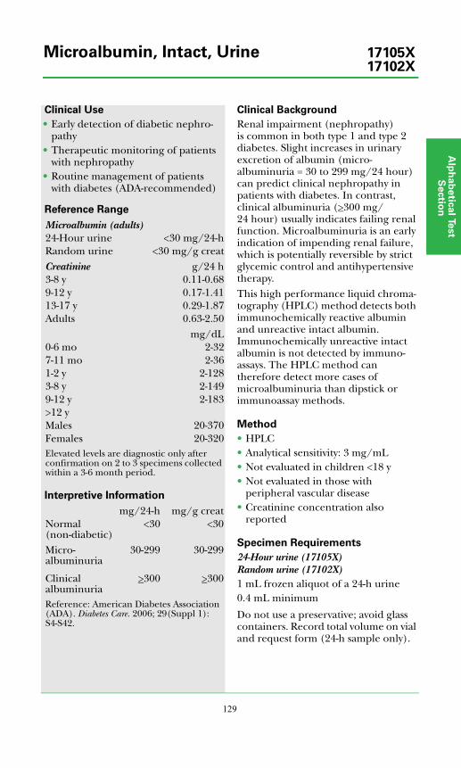
Alp
ha
be
tica
l Test
Se
ctio
n
129
Clinical Use
• Early detection of diabetic nephro-pathy
• Therapeutic monitoring of patients with nephropathy
• Routine management of patients with diabetes (ADA-recommended)
Reference Range
Interpretive Information
Clinical Background
Renal impairment (nephropathy) is common in both type 1 and type 2 diabetes. Slight increases in urinary excretion of albumin (micro-albuminuria = 30 to 299 mg/24 hour) can predict clinical nephropathy in patients with diabetes. In contrast, clinical albuminuria (>300 mg/24 hour) usually indicates failing renal function. Microalbuminuria is an early indication of impending renal failure, which is potentially reversible by strict glycemic control and antihypertensive therapy.This high performance liquid chroma-tography (HPLC) method detects both immunochemically reactive albumin and unreactive intact albumin. Immunochemically unreactive intact albumin is not detected by immuno-assays. The HPLC method can therefore detect more cases of microalbuminuria than dipstick or immunoassay methods.
Method
• HPLC• Analytical sensitivity: 3 mg/mL• Not evaluated in children <18 y• Not evaluated in those with
peripheral vascular disease• Creatinine concentration also
reported
Specimen Requirements
24-Hour urine (17105X)Random urine (17102X)1 mL frozen aliquot of a 24-h urine0.4 mL minimum
Do not use a preservative; avoid glass containers. Record total volume on vial and request form (24-h sample only).
Microalbumin (adults)24-Hour urine <30 mg/24-hRandom urine <30 mg/g creatCreatinine g/24 h3-8 y 0.11-0.689-12 y 0.17-1.4113-17 y 0.29-1.87Adults 0.63-2.50
mg/dL0-6 mo 2-327-11 mo 2-361-2 y 2-1283-8 y 2-1499-12 y 2-183>12 yMales 20-370Females 20-320Elevated levels are diagnostic only after confirmation on 2 to 3 specimens collected within a 3-6 month period.
mg/24-h mg/g creatNormal (non-diabetic)
<30 <30
Micro-albuminuria
30-299 30-299
Clinical albuminuria
>300 >300
Reference: American Diabetes Association (ADA). Diabetes Care. 2006; 29(Suppl 1): S4-S42.
Microalbumin, Intact, Urine 17105X17102X
130
Clinical Use
• Differentiate inherited NDI from acquired NDI
• Screen for autosomal NDI carrier status in at-risk individuals
Reference Range
Interpretive Information
Mutation present• Nephrogenic diabetes insipidus
(affected or carrier)
Clinical Background
Nephrogenic diabetes insipidus (NDI) is characterized by inability of the kidneys to concentrate urine despite the presence of arginine vasopressin (AVP), also known as antidiuretic hormone (ADH). This insensitivity to AVP results in polyuria, polydipsia, low urinary specific gravity, and high risk of severe dehydration, especially in affected infants. Although acquired NDI is more common than inherited disease, genetic forms also occur. They can be transmitted in X-linked recessive, autosomal recessive, or autosomal dominant patterns. X-linked NDI, caused by mutations in the AVPR2, is the most common genetic cause of NDI. Most cases of autosomal NDI have a recessive mode of inheritance, but about 10% of them (1% of all in-herited NDI) result from mutations with dominant expression. Insertion of the aquaporin 2 (AQP2) protein into the luminal membrane of collecting duct cells is the final step in the antidiuretic action of AVP, provid-ing specific channels that increase water permeability of the membrane. Mutations disrupting the AQP2 gene on chromosome 12 alter the amino acid sequence of the AQP2 protein and cause about 10% of familial NDI. Since about 25 different mutations have been identified in the AQP2 gene, gene sequencing is the method of choice for detecting AQP2 mutations. After identi-fication of a mutation in an affected individual, genetic testing can be used to evaluate other family members.
Method
• Polymerase chain reaction (PCR) and DNA sequencing
• Analytical specificity: mutations in 4 exons of the AQP2 gene
Specimen Requirements
5 mL room temperature whole blood3 mL minimumCollect blood in a lavender-top (EDTA) or yellow-top (ACD solution B) tube
Negative (no mutations detected)
Nephrogenic Diabetes Insipidus 15028X(Autosomal) Mutations
Alp
ha
be
tica
l Test
Se
ctio
n
131
Clinical Use
• Differentiate inherited NDI from acquired NDI
• Screen for X-linked NDI carrier status in at-risk females
Reference Range
Interpretive Information
Mutation present• Nephrogenic diabetes insipidus
(affected or carrier)
Clinical Background
Nephrogenic diabetes insipidus (NDI) is characterized by inability of the kidneys to concentrate urine despite the presence of arginine vasopressin (AVP), also known as antidiuretic hormone (ADH). This insensitivity to AVP results in polyuria, polydipsia, low urinary specific gravity, and high risk of severe dehydration, especially in affected infants. Although acquired NDI is more common than inherited disease, genetic forms also occur. They can be transmitted in X-linked recessive, autosomal recessive, or autosomal dominant patterns. The X-linked form is the most common cause of inherited NDI.When functional AVP receptor 2 (AVPR2) protein is not present on the basolateral membrane of collecting duct cells, the kidney is not able to respond to the antidiuretic action of AVP. Mutations disrupting the AVPR2 gene on the X chromosome cause about 90% of inherited NDI. Because the AVPR2 gene is on the X chromo-some, the disorder is most common in males, but some female hetero-zygotes also have symptoms. Since about 180 different mutations have been identified in the AVPR2 gene, gene sequencing is the method of choice for detecting AVPR2 mutations. After identification of a mutation in an affected individual, genetic testing can be used to evaluate other family members.
Method
• Polymerase chain reaction (PCR) and DNA sequencing
• Analytical specificity: mutations in the X-linked AVPR2 gene
Specimen Requirements
5 mL room temperature whole blood3 mL minimum
Collect blood in a lavender-top (EDTA) or yellow-top (ACD solution B) tube.
Negative (no mutations detected)
Nephrogenic Diabetes Insipidus 15034X(X-Linked) Mutations
132
Clinical Use
• Diagnose and manage fluid and electrolyte disorders
Reference Range
Interpretive Information
• Dehydration• Syndrome of inappropriate
ADH secretion (SIADH)• Liver disease• Heart disease
• Overhydration• Hypokalemia• Diabetes insipidus
Clinical Background
Osmolality is a measurement of the amount of nonelectrolyte substances dissolved per kilogram of pure water. Urine osmolality is a measure of solute concentration in the urine and reflects the concentrating ability of the kidney. The major factors regulating urine osmolality are vasopressin (antidiuretic hormone) and the urine-concen-trating apparatus of the kidney, which responds to vasopressin. Consequently, pituitary and renal disorders, as well as cardiovascular dysfunction impairing renal blood flow, can impair physio-logical regulation of urine osmolality.
Method
• Freezing point depression
Specimen Requirements
1 mL refrigerated, random urine0.2 mL minimum
Do not use a preservative.
50-1200 mOsm/kg
Osmolality, Random Urine 678X
Alp
ha
be
tica
l Test
Se
ctio
n
133
Clinical Use
• Assess fluid and electrolyte balance
Reference Range
Interpretive Information
• Dehydration• Hyperglycemia• Hypernatremia• Alcohol poisoning• Diabetes insipidus
• Overhydration• Hyponatremia• Syndrome of inappropriate
ADH secretion (SIADH)
Clinical Background
Osmolality is a measurement of the amount of nonelectrolyte substances dissolved per kilogram of pure water. Serum osmolality is an index of solute concentration in the blood. It is precisely controlled by hypothalamic-posterior pituitary regulation of vasopressin secretion, leading to modulation of water excretion by the kidneys. Measurement of serum osmolality is helpful in assessing effectiveness or dysfunction of this system.
Method
• Freezing point depression
Specimen Requirements
1 mL room temperature serum0.2 mL minimum
No additive red top preferredSST red top acceptable
278-305 mOsm/kg
Osmolality, Serum 677X
134
Clinical Use
• Determine efficacy of therapy in osteoporosis and metastatic bone disease
• Useful marker of bone formation and remodeling
Reference Range
Interpretive Information
• Primary hyperparathyroidism• Secondary osteosarcoma• Healing bone fractures• Hyperthyroidism• Paget’s disease• Therapeutic agents such as
phenytoin and 1,25-dihydroxy-vitamin D
• Hypoparathyroidism• Cushing’s syndrome
Clinical Background
Osteocalcin is a 6-kd protein consti-tuting 1% to 2% of total bone protein. It contains 3 gamma-carboxyglutamic acid (Gla) residues that bind hydroxy-lapatite. Osteocalcin is produced exclusively by bone osteoblasts, and production is dependent on 1,25-dihydroxyvitamin D, vitamin K, and vitamin C. Measurement provides a specific biochemical index of bone activity. As assessed by bone histo-morphometry, serum osteocalcin levels correlate with bone formation and not directly with bone resorption. Serum osteocalcin is an important bone formation marker.
Method
• Immunoradiometric assay (IRMA)• Analytical sensitivity: 2.0 ng/mL
Specimen Requirements
1 mL frozen serum0.2 mL minimum
No additive red top preferredSST red top acceptableOvernight fasting is preferred.Avoid hemolysis and lipemia.
ng/mLMen 11.3-35.4Women 7.2-27.9Children
6-9 y 40.2-108.010-13 y 35.8-165.514-17 y
Males 27.8-194.1Females 16.3-68.7
Osteocalcin (Bone Gla Protein, BGP) 5586X
Alp
ha
be
tica
l Test
Se
ctio
n
135
Clinical Use
• Assess pancreatic tumor burden• Assist in early diagnosis of pancreatic
tumors• Monitor pancreatic carcinoma
therapy• Predict recurrence of pancreatic
tumors
Reference Range
Interpretive Information
• VIPoma• Insulinoma• Medullary thyroid carcinoma• Zollinger-Ellison syndrome
Clinical Background
Pancreatic polypeptide (PP) is a 32-amino acid peptide that is produced primarily by the endocrine cells of the pancreas; it is also present in extracts of gastric mucosa. The physiological role of PP is not clearly defined. Secretion is influenced by hypo-glycemia, pentagastrin injection, and ingestion of protein meals. Plasma concentrations normally change with age, type of food ingestion, and time of blood collection.PP concentrations are used in the management of PP-producing tumors. High plasma levels have been observed in some patients with pancreatic endocrine tumors (VIPomas and insulinomas), multiple endocrine neoplasia syndrome type 1 (MEN 1), medullary thyroid carcinoma, and Zollinger-Ellison syndrome.In patients with MEN 1, a PP level greater than 3 times the age-matched normal mean is suggestive of an islet cell tumor.
Method
• Extraction, radioimmunoassay (RIA)• Analytical sensitivity: 50 pg/mL• Analytical specificity: no cross-
reaction with glucagon or insulin
Specimen Requirements
2 mL refrigerated EDTA plasma0.6 mL minimum
Overnight fasting is preferred.
Adults pg/mL18-29 y <48030-39 y 70-40040-49 y 70-43050-62 y 100-780>62 y Not established
ChildrenCord blood
Term infants* <163Preterm infants* <180
Term infants-6 d* <2761 mo-2 y* <6443-9 y <51910-13 y <36114-17 y <297
*Data from Reprod Nutr Dev. 1990;30:65-70, Materia Medica Polona. 1989;21:38-42, and Acta Paediatr Scand. 1980;69:211-214.
Pancreatic Polypeptide 4789X
136
Clinical Use
• Diagnose or exclude pheochromo-cytoma or paraganglioma caused by mutations in the SDHB (test code 19568X), SDHD (test code 19567X), and VHL (test code 19571X) genes
• Screen individuals who have a family history of pheochromocytoma and/or paraganglioma
• Assist in treatment selection for patients with a gene mutation
Reference Range
Interpretive Information
SDHB mutation present• Abdominal or thoracic
pheochromocytoma• Paraganglioma type 4
SDHD mutation present• Head or neck pheochromocytoma• Paraganglioma type 1
VHL mutation present• Adrenal or thoracic
pheochromocytoma• von Hippel-Lindau syndrome
Clinical Background
Pheochromocytomas are catechola-mine-producing tumors that arise from chromaffin cells, the largest compact collection of which is in the adrenal medulla. Tumors that arise within the adrenal medulla are referred to as pheochromocytomas, whereas those arising outside the adrenal are called paragangliomas or extraadrenal pheochromocytomas. These tumors are characterized by hypertension and elevated levels of catecholamines and should be differentiated from ganglioneuromas. Although affecting only a small percentage of patients with hypertension, pheochromo-cytomas are usually curable; however, they are often fatal if left untreated.Pheochromocytoma is familial in up to 25% of affected individuals; age of onset is <30 years. The genes involved include NF-1; RET (see MEN and FMTC Mutations, test code 36587X); succinate dehydrogenase subunits B (SDHB), C (SDHC), and D (SDHD); and VHL. Pheochromocytoma causing NF-1 and SDHC mutations are rare.
Method
• Polymerase chain reaction (PCR) and DNA sequencing
• Analytical specificity: SDHB (8 exons) (test code 19568X), SDHD (4 exons) (test code 19567X), and VHL (3 exons) (test code 19571X); large deletions and deep intronic mutations affecting mRNA splicing are not detected
Specimen Requirements
5 mL room temperature whole blood 3 mL minimum
Collect in lavender-top (EDTA) tube
Negative (no mutations detected)
Pheochromocytoma Gene 19568X, 19567X,Mutations 19571X
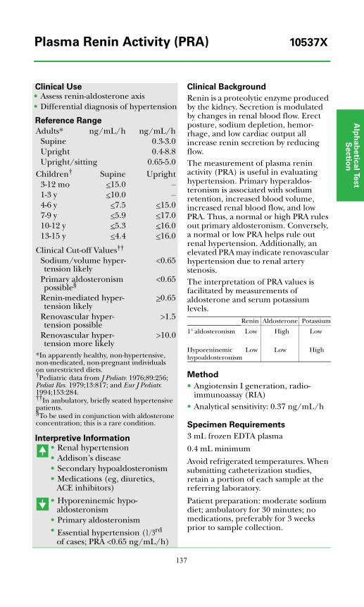
Alp
ha
be
tica
l Test
Se
ctio
n
137
Clinical Use• Assess renin-aldosterone axis • Differential diagnosis of hypertension
Reference Range
Interpretive Information• Renal hypertension• Addison’s disease• Secondary hypoaldosteronism• Medications (eg, diuretics,
ACE inhibitors)
• Hyporeninemic hypo-aldosteronism
• Primary aldosteronism• Essential hypertension (1/3rd
of cases; PRA <0.65 ng/mL/h)
Clinical Background
Renin is a proteolytic enzyme produced by the kidney. Secretion is modulated by changes in renal blood flow. Erect posture, sodium depletion, hemor-rhage, and low cardiac output all increase renin secretion by reducing flow.The measurement of plasma renin activity (PRA) is useful in evaluating hypertension. Primary hyperaldos-teronism is associated with sodium retention, increased blood volume, increased renal blood flow, and low PRA. Thus, a normal or high PRA rules out primary aldosteronism. Conversely, a normal or low PRA helps rule out renal hypertension. Additionally, an elevated PRA may indicate renovascular hypertension due to renal artery stenosis.The interpretation of PRA values is facilitated by measurements of aldosterone and serum potassium levels.
Method
• Angiotensin I generation, radio-immunoassay (RIA)
• Analytical sensitivity: 0.37 ng/mL/h
Specimen Requirements
3 mL frozen EDTA plasma
0.4 mL minimum
Avoid refrigerated temperatures. When submitting catheterization studies, retain a portion of each sample at the referring laboratory.
Patient preparation: moderate sodium diet; ambulatory for 30 minutes; no medications, preferably for 3 weeks prior to sample collection.
Adults* ng/mL/h ng/mL/hSupine 0.3-3.0Upright 0.4-8.8Upright/sitting 0.65-5.0
Children† Supine Upright3-12 mo <15.0 –1-3 y <10.0 –4-6 y <7.5 <15.07-9 y <5.9 <17.010-12 y <5.3 <16.013-15 y <4.4 <16.0
Clinical Cut-off Values††
Sodium/volume hyper-tension likely
<0.65
Primary aldosteronism possible§
<0.65
Renin-mediated hyper-tension likely
>0.65
Renovascular hyper-tension possible
>1.5
Renovascular hyper-tension more likely
>10.0
*In apparently healthy, non-hypertensive, non-medicated, non-pregnant individuals on unrestricted diets. †Pediatric data from J Pediatr. 1976;89:256; Pediat Res. 1979;13:817; and Eur J Pediatr. 1994;153:284.††In ambulatory, briefly seated hypertensive patients.§To be used in conjunction with aldosterone concentration; this is a rare condition.
Renin Aldosterone Potassium
1° aldosteronism Low High Low
Hyporeninemic Low hypoaldosteronism
Low High
Plasma Renin Activity (PRA) 10537X
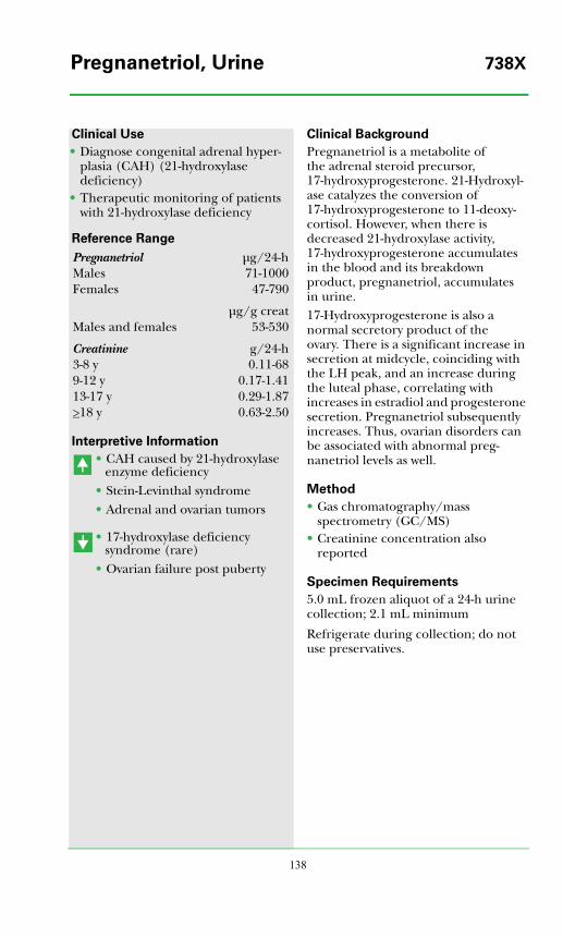
138
Clinical Use
• Diagnose congenital adrenal hyper-plasia (CAH) (21-hydroxylase deficiency)
• Therapeutic monitoring of patients with 21-hydroxylase deficiency
Reference Range
Interpretive Information
• CAH caused by 21-hydroxylase enzyme deficiency
• Stein-Levinthal syndrome• Adrenal and ovarian tumors
• 17-hydroxylase deficiency syndrome (rare)
• Ovarian failure post puberty
Clinical Background
Pregnanetriol is a metabolite of the adrenal steroid precursor, 17-hydroxyprogesterone. 21-Hydroxyl-ase catalyzes the conversion of 17-hydroxyprogesterone to 11-deoxy-cortisol. However, when there is decreased 21-hydroxylase activity, 17-hydroxyprogesterone accumulates in the blood and its breakdown product, pregnanetriol, accumulates in urine.17-Hydroxyprogesterone is also a normal secretory product of the ovary. There is a significant increase in secretion at midcycle, coinciding with the LH peak, and an increase during the luteal phase, correlating with increases in estradiol and progesterone secretion. Pregnanetriol subsequently increases. Thus, ovarian disorders can be associated with abnormal preg-nanetriol levels as well.
Method
• Gas chromatography/mass spectrometry (GC/MS)
• Creatinine concentration also reported
Specimen Requirements
5.0 mL frozen aliquot of a 24-h urine collection; 2.1 mL minimum
Refrigerate during collection; do not use preservatives.
Pregnanetriol μg/24-hMales 71-1000Females 47-790
μg/g creatMales and females 53-530
Creatinine g/24-h3-8 y 0.11-689-12 y 0.17-1.4113-17 y 0.29-1.87>18 y 0.63-2.50
Pregnanetriol, Urine 738X
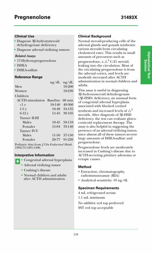
Alp
ha
be
tica
l Test
Se
ctio
n
139
Clinical Use
• Diagnose 3β-hydroxysteroid dehydrogenase deficiency
• Diagnose adrenal virilizing tumors
Related Assays• 17-Hydroxypregnenolone• DHEA• DHEA-sulfate
Reference Range
Interpretive Information
• Congenital adrenal hyperplasia• Adrenal virilizing tumor• Cushing’s disease• Normal children and adults
after ACTH administration
Clinical Background
Normal steroid-producing cells of the adrenal glands and gonads synthesize various steroids from circulating cholesterol ester. This results in small amounts of precursors such as pregnenolone, a 5 C-21 steroid, leaking into the circulation. Most of the circulating pregnenolone is from the adrenal cortex, and levels are modestly increased after ACTH administration in normal children and adults.This assay is useful in diagnosing 3β-hydroxysteroid dehydrogenase (3β-HSD) deficiency, an unusual form of congenital adrenal hyperplasia associated with blocked cortisol synthesis and increased levels of 5 steroids. After diagnosis of 3β-HSD deficiency, the test can evaluate gluco-corticoid replacement therapy. The assay is also helpful in suggesting the presence of an adrenal virilizing tumor, since almost all of these tumors secrete large amounts of DHEA-sulfate and pregnenolone.Pregnenolone levels are moderately increased in Cushing’s disease due to ACTH-secreting pituitary adenoma or ectopic causes.
Method
• Extraction, chromatography, radioimmunoassay (RIA)
• Analytical sensitivity: 10 ng/dL
Specimen Requirements
4 mL refrigerated serum1.1 mL minimum
No additive red top preferredSST red top acceptable
ng/dL ng/dLMen 10-200Women 10-230Children
ACTH stimulation Baseline 60 min<1 y 10-140 49-3601-5 y 10-48 34-1356-12 y 15-45 39-105Tanner II-III
Males 10-45 58-110Females 15-84 33-140
Tanner IV-VMales 11-50 37-150Females 20-77 91-220
Pediatric data from J Clin Endocrinol Metab. 1992;75:1491-1496.
Pregnenolone 31493X
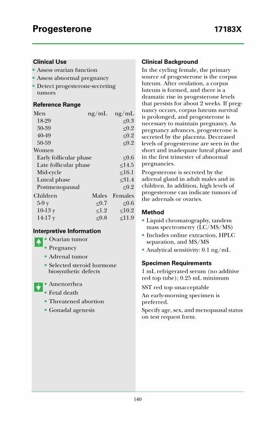
140
Clinical Use
• Assess ovarian function• Assess abnormal pregnancy• Detect progesterone-secreting
tumors
Reference Range
Interpretive Information
• Ovarian tumor• Pregnancy• Adrenal tumor• Selected steroid hormone
biosynthetic defects
• Amenorrhea• Fetal death• Threatened abortion• Gonadal agenesis
Clinical Background
In the cycling female, the primary source of progesterone is the corpus luteum. After ovulation, a corpus luteum is formed, and there is a dramatic rise in progesterone levels that persists for about 2 weeks. If preg-nancy occurs, corpus luteum survival is prolonged, and progesterone is necessary to maintain pregnancy. As pregnancy advances, progesterone is secreted by the placenta. Decreased levels of progesterone are seen in the short and inadequate luteal phase and in the first trimester of abnormal pregnancies.Progesterone is secreted by the adrenal gland in adult males and in children. In addition, high levels of progesterone can indicate tumors of the adrenals or ovaries.
Method
• Liquid chromatography, tandem mass spectrometry (LC/MS/MS)
• Includes online extraction, HPLC separation, and MS/MS
• Analytical sensitivity: 0.1 ng/mL
Specimen Requirements
1 mL refrigerated serum (no additive red top tube); 0.25 mL minimum
SST red top unacceptableAn early-morning specimen is preferred.Specify age, sex, and menopausal status on test request form.
Men ng/mL ng/mL18-29 <0.330-39 <0.240-49 <0.250-59 <0.2
WomenEarly follicular phase <0.6Late follicular phase <14.5Mid-cycle <16.1Luteal phase <31.4Postmenopausal <0.2
Children Males Females5-9 y <0.7 <0.610-13 y <1.2 <10.214-17 y <0.8 <11.9
Progesterone 17183X
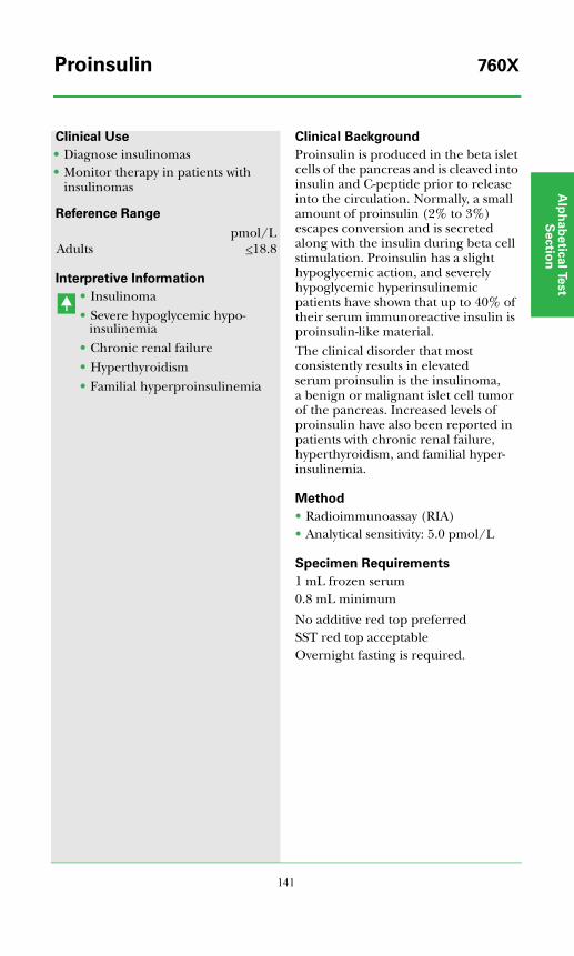
Alp
ha
be
tica
l Test
Se
ctio
n
141
Clinical Use
• Diagnose insulinomas• Monitor therapy in patients with
insulinomas
Reference Range
Interpretive Information
• Insulinoma• Severe hypoglycemic hypo-
insulinemia• Chronic renal failure• Hyperthyroidism• Familial hyperproinsulinemia
Clinical Background
Proinsulin is produced in the beta islet cells of the pancreas and is cleaved into insulin and C-peptide prior to release into the circulation. Normally, a small amount of proinsulin (2% to 3%) escapes conversion and is secreted along with the insulin during beta cell stimulation. Proinsulin has a slight hypoglycemic action, and severely hypoglycemic hyperinsulinemic patients have shown that up to 40% of their serum immunoreactive insulin is proinsulin-like material.The clinical disorder that most consistently results in elevated serum proinsulin is the insulinoma, a benign or malignant islet cell tumor of the pancreas. Increased levels of proinsulin have also been reported in patients with chronic renal failure, hyperthyroidism, and familial hyper-insulinemia.
Method
• Radioimmunoassay (RIA)• Analytical sensitivity: 5.0 pmol/L
Specimen Requirements
1 mL frozen serum0.8 mL minimum
No additive red top preferredSST red top acceptableOvernight fasting is required.
pmol/LAdults <18.8
Proinsulin 760X
142
Clinical Use
• Diagnose and manage pituitary adenomas
• Differential diagnosis of male and female hypogonadism
Reference Range
Interpretive Information
• Hypothalamic or pituitary tumor• Stress (physical and emotional)• Antidepressants• Breast-feeding
• Bromocriptine (dopamine agonist)
Clinical Background
Prolactin is a protein hormone secreted by the anterior pituitary gland and the placenta. It may modulate the number of follicles developing in the follicular phase of each menstrual cycle. During and following pregnancy, prolactin, in association with other hormones, stimulates breast development and milk production.Prolactin secretion is stimulated by sleep, stress (physical and emotional), and the hypothalamic hormone, thyro-tropin releasing hormone (TRH). Prolactin secretion is decreased by dopamine and dopamine analogs such as bromocriptine.Hypersecretion of prolactin can be caused by pituitary tumors, hypo-thalamic disease, breast or chest wall stimulation, hypothyroidism, renal failure, acute exercise, stress, eating, and several medications (eg, pheno-thiazines, metoclopramide). Hyper-prolactinemia inhibits gonadotropin secretion and can produce hypo-gonadism in men and women with accompanying low or inappropriately "low normal" LH and FSH levels.
Method
• Immunochemiluminometric assay (ICMA), ADVIA Centaur®’
• Analytical sensitivity: 1.0 ng/mL
Specimen Requirements
1 mL refrigerated serum0.5 mL minimum
No additive red top preferredSST red top acceptableOvernight fasting is preferred.
ng/mLMen 2.0-18.0Women
Nonpregnant 3.0-30.0Pregnant 10.0-209.0Postmenopausal 2.0-20.0
ChildrenMales
Tanner I <10Tanner II-III <6.1Tanner IV-V 2.8-11.0
FemalesTanner I 3.6-12.0Tanner II-III 2.6-18.0Tanner IV-V 3.2-20.0
Prolactin 746X
Alp
ha
be
tica
l Test
Se
ctio
n
143
Clinical Use
• Assess possibility of antibody interference with PTH measure-ments
Reference Range
Interpretive Information
• Autoimmune disease• PTH therapy for osteoporosis
Clinical Background
Parathyroid hormone (PTH) auto-antibodies may lead to misleading results when measuring PTH by immunoassay methods. Such auto-antibodies are rare but may be seen in autoimmune disease states and in patients who are receiving PTH therapy for osteoporosis.
Method
•• Radiobinding assay (RBA)
Specimen Requirements
1 mL refrigerated serum0.2 mL minimum
No additive red top preferredSST red top acceptable
Negative
PTH Antibody 36578X
144
Clinical Use
• Discriminate primary hyper-parathyroidism from tumor hyper-calcemia
• Diagnose hypoparathyroidism• Monitor severity of secondary
hyperparathyroidism in chronic renal failure
Reference Range
Interpretive Information
• Primary hyperparathyroidism• Secondary hyperparathyroidism• Renal failure• Pseudohypoparathyroidism
• Hypoparathyroidism• Hypercalcemia of malignancy
Clinical Background
Parathyroid hormone (PTH) acts to increase calcium absorption from the gut and to mobilize calcium from bone. The net effect is to increase the extra-cellular concentration of calcium and prevent hypocalcemia. PTH secretion by the parathyroid gland is modulated by serum calcium concentration. Low calcium stimulates and high calcium inhibits PTH secretion.PTH levels are used to assess disorders of calcium metabolism, including primary and secondary hyperpara-thyroidism, tumor hypercalcemia, and hypoparathyroidism.
Method
PTH (8837X, 36736X)• Immunochemiluminometric assay
(ICMA)• Analytical sensitivity: 3 pg/mL• Analytical specificity: 100% cross-
reactivity with intact PTH (amino acids 1-84) and 45% with the 7-84 C-terminal fragment; no detectable cross-reactivity with other PTH fragments or with calcitonin
Calcium, total (8837X)• Spectrophotometry• Analytical sensitivity: 0.2 mg/dL
Calcium, ionized (36736X)• Ion specific electrode• Analytical sensitivity: 0.4 mg/dL
Specimen Requirements
PTH and total calcium (8837X)2 mL frozen serum 1 mL minimum (infants, 0.5 mL)
PTH and ionized calcium (36736X)PTH2 mL frozen serum (no additive red top or SST red top tube); 1 mL minimum
Calcium, ionized2 mL refrigerated serum (SST red-top tube, centrifuged, unopened; 0.6 mL minimum
PTH, Intact pg/mLAdults 10-65Children
6-9 y 9-5910-13 y 11-7414-17 y 9-69
Calcium mg/dLAdults 8.8-10.1Ionized Calcium mg/dLAdults 4.6-5.4Children
8 mo-10 y 4.9-5.411-17 y 4.8-5.3
Pediatric data from J Pediatr. 1989;114:952-956 and Eur J Pediatr. 1991;150:464-467.
PTH, Intact and Calcium 8837X36736X
Alp
ha
be
tica
l Test
Se
ctio
n
145
Clinical Use
• Differential diagnosis of hyper-calcemia
• Manage patients with solid tumors and hypercalcemia
Reference Range
Interpretive Information
• Tumor-associated hypercalcemia• Fetus• Newborn
Clinical Background
Parathyroid hormone-related protein (PTH-RP) has an amino-terminal structure similar to parathyroid hormone (PTH). PTH-RP can bind to and stimulate PTH receptors and is produced mainly by solid tumors. It is also found in the normal lactating mammary gland, keratinocytes, placenta, and parathyroid gland.PTH-RP levels are high in approxi-mately 70% of the patients with tumor-associated hypercalcemia. It may be useful in the differential diagnosis of hypercalcemia, specifically differen-tiating primary hyperparathyroidism from hypercalcemia related to cancer. It is not elevated in primary or chronic renal failure.
Method
• Immunoradiometric assay (IRMA)• Analytical sensitivity: 2.5 pmol/mL• Analytical specificity: intact PTH-RP;
no detectable cross-reactivity with PTH (C-terminal, mid-molecule, amino acids 39-84, or intact)
Specimen Requirements
1.5 mL frozen plasma
1.0 mL minimum
Collect blood in syringe or red-top tube and immediately transfer to a cold, special Nichols Institute PTH-RP collection tube; mix thoroughly. Centrifuge in refrigerated centrifuge. Transfer plasma to a plastic tube and freeze.
<4.7 pmol/L
PTH-related Protein (PTH-RP) 34478X
146
Clinical Use
• Assess rate of bone collagen degradation
• Monitor response to therapy
Reference Range
Clinical Background
Collagen fibers are linked together by interchain molecules referred to as pyridinium cross-links. Collagen pyridinium cross-links have been identified in all connective tissue except skin. As collagen is broken down by collagenase, small breakdown products are excreted in urine with the attached cross-links, including pyridinoline (PYD) and deoxy-pyridinoline (DPYD). Bone collagen is constantly turning over and is rich in DPYD. Thus, urine PYD and, especially, DPYD are useful markers for bone matrix degradation and resorption.
Method
• High-performance liquid chroma-tography (HPLC)
• Analytical sensitivity: 20 pmol/mL (PYD); 13 pmol/mL (DPYD)
• Creatinine concentration also reported
Specimen Requirements
24-Hour urine (36098X)5 mL refrigerated aliquot of a 24-h urine; 1 mL minimumCollect urine with 25 mL 6N HCl or 10 g of boric acid. Mix and aliquot. Record total volume on vial and request form.
2-Hour urine (36097X)5 mL refrigerated aliquot of a 2-h urine; 0.5 mL minimumDiscard first morning void. Collect urine for next 2 hours, in presence of 2 mL 6N HCl or 1 g of boric acid. Mix well and aliquot.
Interpretive Information
• Osteoporosis• Primary hyperparathyroidism• Paget’s disease
• Relative to baseline indicates successful therapy
PYDnmol/mmol
creat
DPYDnmol/
mmol creatMen
24-h urine 20-61 3.7-192-h urine 23-65 6.2-26
Women*24-h urine 22-89 4.4-212-h urine 25-83 5.7-23
Children (24-h urine)2-10 y 158-442 31-11211-14 y 107-398 17-10115-17 y 42-200 <59
Children (2-h urine)2-10 y 153-401 39-12011-17 y 17-408 3.0-117Tanner I 112-353 27-103Tanner II 95-471 20-139Tanner III 68-489 14-135Tanner IV 17-448 10-136
Creatinine Randomurine mg/dL
24-h urineg/24-h
0-6 mo 2-327-11 mo 2-361-2 y 2-1283-8 y 2-149 0.11-0.689-12 y 2-183 0.17-1.4113-17 y 0.29-1.87Adults 0.63-2.50
>12 yMale 20-370Female 20-320
*Premenopausal. A value within the pre-menopausal range does not rule out osteo-porosis or the need for therapy.
Pyridinium Collagen Cross-links, Urine 36098X(PYD and DPYD) 36097X
Alp
ha
be
tica
l Test
Se
ctio
n
147
Clinical Use
• Differential diagnosis of hyper-thyroidism
• Screen individuals at risk for resistance to thyroid hormone
Reference Range
Interpretive Information
Mutation present• Resistance to thyroid hormone
Clinical Background
Resistance to thyroid hormone (RTH) is caused by reduced target tissue response to thyroid hormone, resulting in the loss of the normal feedback mechanism that suppresses the secretion of thyrotropin (TSH). It is characterized by the presence of elevated serum levels of the free thyroid hormones thyroxine (T4) and triiodothyronine (T3) with slightly elevated or normal TSH. In 90% of cases, RTH is caused by mutations in the thyroid hormone receptor beta (THRB) gene that alter the hormone-binding region of the protein. The disorder is usually inherited in an autosomal dominant manner, and over 100 different mutations have been described. New mutations can give rise to isolated cases of RTH.Since genetic variation and other problems can affect the accuracy of direct mutation testing, test results should always be interpreted in light of clinical and family data. Genetic counseling may be helpful for individuals and families undergoing testing for THRB mutations.
Method
• Polymerase chain reaction (PCR) and DNA sequencing
• Analytical specificity: mutations in exons 3-10 of the THRB gene and their flanking intronic regions
Specimen Requirements
5 mL room temperature whole blood 3 mL minimum
Collect in lavender-top (EDTA) tube
Negative (no mutations detected)
Resistance to Thyroid Hormone 16053X
148
Clinical Use
• Diagnose paraneoplastic carcinoid syndrome
Reference Range
Interpretive Information
• Carcinoid syndrome• Medullary thyroid carcinoma• Multiple endocrine neoplasia
(MEN) types 1 and 2• Oat cell carcinoma of the lung• Islet cell tumors
• Depression
Clinical Background
Carcinoid syndrome manifests with flushing, diarrhea, pain, and hepatomegaly. The ileum, stomach, pancreas, duodenum, bronchus, thymus, thyroid, testis, and ovary may be involved. Small bowel carcinoids most commonly metastasize to the liver. These tumors secrete increased amounts of serotonin, which metabo-lizes to 5-hydroxyindoleacetic acid (5-HIAA). Measurement of serotonin concentration may be helpful in selected cases.
Method
• High performance liquid chroma-tography (HPLC)
•••• Fluorescent detection
Specimen Requirements
Whole blood (818X)4 mL frozen whole blood1 mL minimum
Collect blood using the Serotonin Kit (provided by Quest Diagnostics)
Serum (29851X)2 mL frozen serum1 mL minimum
No additive red top preferredSST red top acceptable
Patients should avoid avocados, bananas, tomatoes, plums, walnuts, pineapples, eggplant, tobacco, tea, and coffee for 3 d prior to specimen collection.
ng/mLBlood (818X) 22-180Serum (29851X)Adults 26-165Children (1-12 y) 81-349Pediatric reference range from Brazilian J Med Biol Res. 1993;26:309-317.
Serotonin 818X29851X
Alp
ha
be
tica
l Test
Se
ctio
n
149
Clinical Use
• Assess abnormal serum gonadal hormone concentrations
Reference Range
Interpretive Information
• Estrogen administration• Pregnancy• Alcoholism• Male hypogonadism• Hyperthyroidism• Testicular feminization
• Hirsutism• Virilization• Massive obesity• Exogenous androgen therapy
or abuse
Clinical Background
The majority of circulating gonadal steroid hormones are protein bound. Testosterone, dihydrotestosterone, and the estrogens are bound to circulating sex hormone binding globulin (SHBG or testosterone-binding globulin, TeBG), which is produced by the liver. Consequently, total circulating steroid hormone levels are affected by altera-tions in SHBG concentration as well as concentration of the hormones them-selves. Evaluation of the gonadal steroid hormone system, therefore, includes measurements of total serum testosterone, dihydrotestosterone, or estrogen species in addition to measurements of the free hormones and SHBG. SHBG concentrations are increased by estrogens and pregnancy and decreased by testosterone. Insulin lowers SHBG levels in high insulin states (eg, diabetes, obesity, polycystic ovary syndrome).
Method
• Immunochemiluminometric assay (ICMA)
• Analytical sensitivity: 2 nmol/L
Specimen Requirements
1 mL room temperature serum0.5 mL minimum
No additive red top preferredSST red top acceptableSpecify age and sex on request form.
Males FemalesAdults nmol/L nmol/L
18-29 y 7-49 6-11230-39 y 8-48 14-10240-49 y 9-45 11-10050-59 y 18-47 17-7860-69 y 17-54 17-9570-79 y 23-65 21-9080-91 y 20-63 26-77
Children3-9 y 18-136 18-13610-13 y 17-123 17-12314-17 y 11-71 11-71Tanner stages
I 39-155 38-114II 33-135 24-90III 21-72 22-112IV 11-92 22-69V 18-54 18-76
Sex Hormone Binding Globulin 30740X
150
Clinical Use
• Diagnose somatostatin-producing tumors
Reference Range
Interpretive Information
• Somatostatinoma• Medullary thyroid carcinoma•• Pheochromocytoma
• Following vagotomy
Clinical Background
Somatostatin, also referred to as somatotropin release inhibiting factor (SRIF), is a neurohormone produced as a 14-amino acid peptide in the brain and hypothalamus and as a 28-amino acid N-extended product in the gastrointestinal tract. In the pituitary gland, SRIF inhibits secretion of growth hormone, TSH, and, under certain conditions, prolactin and ACTH. It exerts inhibitory effects on all endocrine and exocrine secretions of the pancreas and gut and a variety of endocrine-secreting tumors including insulinomas, glucagonomas, VIPomas, carcinoid tumors, and gastrinomas. Pancreatic somatostatinomas cause mild diabetes mellitus, cholelithiasis, steatorrhea, indigestion, and hypo-chlorhydria secondary to the inhibitory effects of secreted SRIF. SRIF may also be secreted by medullary thyroid carcinomas or pheochromocytomas.
Method
• Extraction and radioimmunoassay (RIA)
• Analytical sensitivity: 10 pg/mL
Specimen Requirements
1.8 mL frozen EDTA plasma0.6 mL minimum
Draw in pre-chilled lavender-top tube. Separate and freeze plasma immediately.
Adults 10-22 pg/mL
Somatostatin 34480X
Alp
ha
be
tica
l Test
Se
ctio
n
151
Clinical Use
• Evaluate discordant serum T3 levels
Reference Range
Interpretive Information
• Autoimmune thyroid disease• Other autoimmune disease
Clinical Background
Patients with autoimmune thyroid diseases, including autoimmune (Hashimoto) thyroiditis and Graves disease, may rarely develop antibodies to T3 (triiodothyronine) and/or T4 (thyroxine). T3 antibodies interfere with T3 assay measurement by com-peting with the anti-T3 immuno-globulins used in the T3 assay.When a patient has a serum T3 con-centrati\on that is discordant with other test results, eg, elevated serum T3 in the presence of a detectable serum TSH concentration, then it may be appropriate to determine if anti-T3 antibodies are present.
Method
• Radiobinding assay (RBA)
Specimen Requirements
1 mL refrigerated serum0.1 mL minimum
No additive red top preferredSST red top acceptable
Negative
T3 Antibody 36574X
152
Clinical Use
• Assess T4 (thyroxine) protein binding
• Indirect estimate of free T4 concentration
Reference Range
Interpretive Information
• Hyperthyroidism• Renal failure•• Malnutrition
• Hypothyroidism• Pregnancy• Acute hepatitis
Clinical Background
Thyroid hormones circulate bound to several binding proteins. The most significant among these is thyroxine-binding globulin (TBG). TBG levels are influenced by a variety of factors including genetic abnormalities, pregnancy, oral contraceptives, glucocorticoids, androgens, and renal disease. Fluctuations in TBG concentrations lead to changes in T4 and T3 levels that may generate a false diagnosis of thyroid dysfunction in basically euthyroid patients.There are several methods of assessing TBG-binding activity in serum. The T3 uptake provides an indirect assessment of TBG binding. Chemiluminescence-labeled T3 competitively binds to a solid phase-bound antibody or the patient TBG. The T3 bound to anti-body is measured relative to that in a standard serum. Increased antibody binding reflects decreased TBG binding (TBG-binding site saturation). Decreased antibody binding reflects increased TBG binding (binding site desaturation).
Method
• Immunochemiluminometric assay (ICMA)
Specimen Requirements
1 mL refrigerated serum0.5 mL minimum
No additive red top preferredSST red top acceptable
Adults 0.79-1.16
T3 Uptake 17732X
Alp
ha
be
tica
l Test
Se
ctio
n
153
Clinical Use
• Diagnose hyperthyroidism• Clarify thyroid status in presence of
possible protein binding abnormality
Reference Range
Interpretive Information
• Graves disease• T3 thyrotoxicosis• Thyroid hormone resistance• Functional thyroid adenoma
(T3-producing)
• Nonthyroidal illness• Hypothyroidism (only 1/3 of
cases)
Clinical Background
Most circulating T3 (triiodothyronine) is bound to plasma proteins; only 0.3% exists in the free, unbound state and is available for exchange with intra-cellular T3 receptors. T3 measurements are used to diagnose and monitor treatment of hyper-thyroidism (see T3, Total). When an increase in circulating thyroxine-binding proteins is suspected as the cause of an elevated total T3 level, the free T3 assay can differentiate this condition from true hyperthyroidism. Non-dialysis free T3 immunoassay methods tend to underestimate free T3 due to nonspecific absorption of free T3 to proteins or matrix reagents. Although they do provide a useful free T3 index, the most reliable free T3 measurement is the equilibrium dialysis method.
Method
• Immunochemiluminometric assay (ICMA)
• Analytical sensitivity: 50 pg/dL
Specimen Requirements
1 mL refrigerated serum0.5 mL minimum
No additive red top preferredSST red top acceptable
pg/dLAdults 230-420Children
<1 y Not established1-9 y 337-50610-13 y 335-48014-18 y 287-455
T3, Free, Non-dialysis 34429X
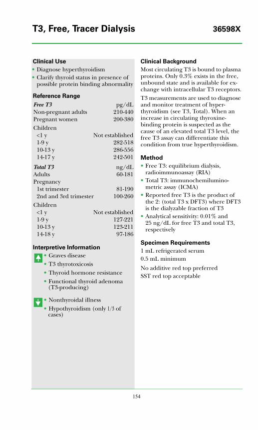
154
Clinical Use
• Diagnose hyperthyroidism• Clarify thyroid status in presence of
possible protein binding abnormality
Reference Range
Interpretive Information
• Graves disease• T3 thyrotoxicosis• Thyroid hormone resistance• Functional thyroid adenoma
(T3-producing)
• Nonthyroidal illness• Hypothyroidism (only 1/3 of
cases)
Clinical Background
Most circulating T3 is bound to plasma proteins. Only 0.3% exists in the free, unbound state and is available for ex-change with intracellular T3 receptors.T3 measurements are used to diagnose and monitor treatment of hyper-thyroidism (see T3, Total). When an increase in circulating thyroxine-binding protein is suspected as the cause of an elevated total T3 level, the free T3 assay can differentiate this condition from true hyperthyroidism.
Method
• Free T3: equilibrium dialysis, radioimmunoassay (RIA)
• Total T3: immunochemilumino-metric assay (ICMA)
• Reported free T3 is the product of the 2: (total T3 x DFT3) where DFT3 is the dialyzable fraction of T3
• Analytical sensitivity: 0.01% and 25 ng/dL for free T3 and total T3, respectively
Specimen Requirements
1 mL refrigerated serum0.5 mL minimum
No additive red top preferredSST red top acceptable
Free T3 pg/dLNon-pregnant adults 210-440Pregnant women 200-380Children
<1 y Not established1-9 y 282-51810-13 y 286-55614-17 y 242-501
Total T3 ng/dLAdults 60-181Pregnancy
1st trimester 81-1902nd and 3rd trimester 100-260
Children<1 y Not established1-9 y 127-22110-13 y 123-21114-18 y 97-186
T3, Free, Tracer Dialysis 36598X
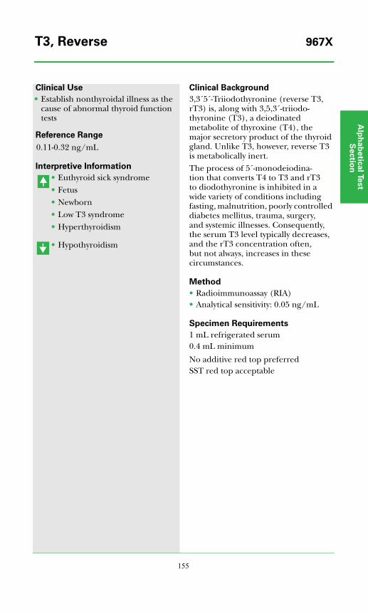
Alp
ha
be
tica
l Test
Se
ctio
n
155
Clinical Use
• Establish nonthyroidal illness as the cause of abnormal thyroid function tests
Reference Range
Interpretive Information
• Euthyroid sick syndrome• Fetus• Newborn• Low T3 syndrome• Hyperthyroidism
• Hypothyroidism
Clinical Background
3,3´5´-Triiodothyronine (reverse T3, rT3) is, along with 3,5,3´-triiodo-thyronine (T3), a deiodinated metabolite of thyroxine (T4), the major secretory product of the thyroid gland. Unlike T3, however, reverse T3 is metabolically inert.The process of 5´-monodeiodina-tion that converts T4 to T3 and rT3 to diodothyronine is inhibited in a wide variety of conditions including fasting, malnutrition, poorly controlled diabetes mellitus, trauma, surgery, and systemic illnesses. Consequently, the serum T3 level typically decreases, and the rT3 concentration often, but not always, increases in these circumstances.
Method
• Radioimmunoassay (RIA)• Analytical sensitivity: 0.05 ng/mL
Specimen Requirements
1 mL refrigerated serum0.4 mL minimum
No additive red top preferredSST red top acceptable
0.11-0.32 ng/mL
T3, Reverse 967X
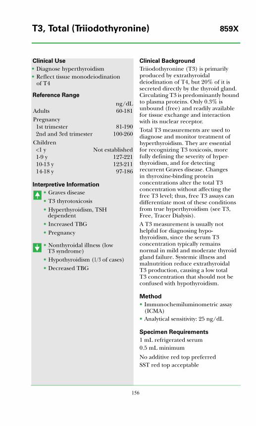
156
Clinical Use
• Diagnose hyperthyroidism• Reflect tissue monodeiodination
of T4
Reference Range
Interpretive Information
• Graves disease• T3 thyrotoxicosis• Hyperthyroidism, TSH
dependent• Increased TBG• Pregnancy
• Nonthyroidal illness (low T3 syndrome)
• Hypothyroidism (1/3 of cases)• Decreased TBG
Clinical Background
Triiodothyronine (T3) is primarily produced by extrathyroidal deiodination of T4, but 20% of it is secreted directly by the thyroid gland. Circulating T3 is predominantly bound to plasma proteins. Only 0.3% is unbound (free) and readily available for tissue exchange and interaction with its nuclear receptor.Total T3 measurements are used to diagnose and monitor treatment of hyperthyroidism. They are essential for recognizing T3 toxicosis, morefully defining the severity of hyper-thyroidism, and for detecting recurrent Graves disease. Changes in thyroxine-binding protein concentrations alter the total T3 concentration without affecting the free T3 level; thus, free T3 assays can differentiate most of these conditions from true hyperthyroidism (see T3, Free, Tracer Dialysis).A T3 measurement is usually not helpful for diagnosing hypo-thyroidism, since the serum T3 concentration typically remains normal in mild and moderate thyroid gland failure. Systemic illness and malnutrition reduce extrathyroidal T3 production, causing a low total T3 concentration that should not be confused with hypothyroidism.
Method
• Immunochemiluminometric assay (ICMA)
• Analytical sensitivity: 25 ng/dL
Specimen Requirements
1 mL refrigerated serum0.5 mL minimum
No additive red top preferredSST red top acceptable
ng/dLAdults 60-181Pregnancy1st trimester 81-1902nd and 3rd trimester 100-260
Children<1 y Not established1-9 y 127-22110-13 y 123-21114-18 y 97-186
T3, Total (Triiodothyronine) 859X

Alp
ha
be
tica
l Test
Se
ctio
n
157
Clinical Use
• Evaluate discordant serum T4 levels
Reference Range
Interpretive Information
• Autoimmune thyroid disease• Other autoimmune disease
Clinical Background
Patients with autoimmune thyroid diseases, including autoimmune (Hashimoto) thyroiditis and Graves disease, may rarely develop antibodies to T4 (thyroxine) and/or T3 (triiodothyronine). T4 antibodies interfere with T4 assay measurement by competing with the anti-T4 immunoglobulins used in the T4 assay.When a patient has a serum T4 concentration that is discordant with other test results, eg, elevated serum T4 in the absence of thyroxine binding globulin (TBG) excess and presence of a detectable serum TSH concentration, then it may be appropriate to determine if anti-T4 antibodies are present.
Method
• Radiobinding assay (RBA)
Specimen Requirements
1 mL refrigerated serum0.1 mL minimum
No additive red top preferredSST red top acceptable
Negative
T4 (Thyroxine) Antibody 36576X
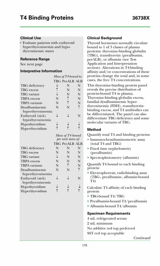
158
Clinical Use
• Evaluate patients with euthyroid hyperthyroxinemia and hypo-thyroxinemic states
Reference Range
See next page
Interpretive Information
Clinical Background
Thyroid hormones normally circulate bound to 1 of 3 classes of plasma proteins: thyroxine-binding globulin (TBG), transthyretin (prealbumin, preALB), or albumin (see Test Application and Interpretation section). Alterations in T4-binding affinity and/or concentrations of these proteins change the total and, in some cases, the free T4 concentrations.This thyroxine-binding protein panel reveals the precise distribution of protein-bound T4 in plasma. Thyroxine-binding globulin excess, familial dysalbuminemic hyper-thyroxinemia (FDH), transthyretin-binding excess, and T4 antibodies can be differentiated. The panel can also differentiate TBG deficiency and some molecular variants of TBG.
Method
Quantify total T4 and binding proteins• Immunochemiluminometric assay
(total T4 and TBG)• Fixed time nephelometry
(prealbumin)• Spectrophotometry (albumin)
Quantify T4 bound to each binding protein• Electrophoresis, radiobinding assay
(TBG-, prealbumin-, albumin-bound T4)
Calculate T4 affinity of each binding protein• TBG-bound T4/TBG• Prealbumin-bound T4/prealbumin• Albumin-bound T4/albumin
Specimen Requirements
4 mL refrigerated serum2 mL minimumNo additive red top preferredSST red top acceptable
Continued
Mass of T4 bound to: TBG PreALB ALB
TBG deficiency ↓ N NTBG excess ↑ N NTBG variant ↓ N NTBPA excess N ↑ NTBPA variants N ↑ NDysalbuminemic
hyperthyroxinemiaN N ↑
Euthyroid (sick) hypothyroxinemia
↓ ↓ N
Hypothyroidism ↓ ↓ ↓Hyperthyroidism ↑ ↑ ↑
Mass of T4 bound per unit mass of:
TBG PreALB ALBTBG deficiency N N NTBG excess N N NTBG variant ↓ N NTBPA excess N N NTBPA variants N ↑ NDysalbuminemic hyperthyroxinemia
N N ↑
Euthyroid (sick) hypothyroxinemia
↓ ↓ N
Hypothyroidism ↓ ↓ ↓Hyperthyroidism ↑ ↑ ↑
T4 Binding Proteins 36738X
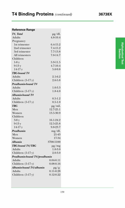
Alp
ha
be
tica
l Test
Se
ctio
n
159
Reference Range
T4, Total μg/dLAdults 4.8-10.4Pregnancy
1st trimester 6.4-15.22nd trimester 7.4-15.23rd trimester 7.7-13.8All trimesters 7.0-14.7
Children1-8 y 5.9-11.59-13 y 4.7-10.414-17 y 5.0-9.8
TBG-bound T4Adults 2.1-6.2Children (5-17 y) 2.6-5.8Prealbumin-bound T4Adults 1.0-3.3Children (5-17 y) 1.8-4.0Albumin-bound T4Adults 0.5-1.2Children (5-17 y) 0.5-1.0TBG μg/mLMen 12.7-25.1Women 13.5-30.9Children
3-8 y 16.1-24.29-13 y 12.5-25.814-17 y 9.8-23.7
Prealbumin mg/dLMen 21-43Women 17-34Albumin 3700-5100TBG-bound T4/TBG μg/mgAdults 1.0-3.0Children (5-17 y) 2.0-3.0Prealbumin-bound T4/prealbuminAdults 0.04-0.11Children (5-17 y) 0.08-0.16Albumin-bound T4/albumin μg/gAdults 0.11-0.28Children (5-17 y) 0.12-0.22
T4 Binding Proteins (continued) 36738X
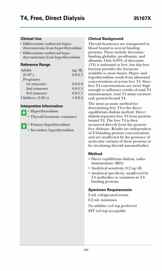
160
Clinical Use
• Differentiate euthyroid hyper-thyroxinemia from hyperthyroidism
• Differentiate euthyroid hypo-thyroxinemia from hypothyroidism
Reference Range
Interpretive Information
• Hyperthyroidism•• Thyroid hormone resistance
• Primary hypothyroidism• Secondary hypothyroidism
Clinical Background
Thyroid hormones are transported in blood bound to several binding proteins. These include thyroxine-binding globulin, prealbumin, and albumin. Only 0.03% of thyroxine (T4) is unbound or free, but this free fraction provides the hormone available to most tissues. Hyper- and hypothyroidism result from abnormal concentrations of serum free T4. Since free T4 concentrations are never high enough to influence results of total T4 immunoassays, total T4 assays measure only protein-bound T4.The most accurate method for determining free T4 is the direct equilibrium dialysis method. Direct dialysis separates free T4 from protein-bound T4. The free T4 is then measured directly from the protein-free dialysate. Results are independent of T4-binding protein concentrations and are unaffected by the presence of molecular variants of these proteins or by circulating thyroid autoantibodies.
Method
• Direct equilibrium dialysis, radio-immunoassay (RIA)
• Analytical sensitivity: 0.2 ng/dL• Analytical specificity: unaffected by
T4 antibodies or variations in T4-binding proteins
Specimen Requirements
2 mL refrigerated serum0.2 mL minimum
No additive red top preferredSST red top acceptable
Adults ng/dL21-87 y 0.8-2.7Pregnancy
1st trimester 0.9-2.02nd trimester 0.8-1.53rd trimester 0.8-1.7
Children (3-20 y) 1.0-2.4
T4, Free, Direct Dialysis 35167X
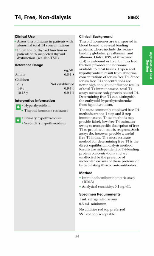
Alp
ha
be
tica
l Test
Se
ctio
n
161
Clinical Use
• Assess thyroid status in patients with abnormal total T4 concentrations
• Initial test of thyroid function in patients with suspected thyroid dysfunction (see also TSH)
Reference Range
Interpretive Information
• Hyperthyroidism•• Thyroid hormone resistance
• Primary hypothyroidism• Secondary hypothyroidism
Clinical Background
Thyroid hormones are transported in blood bound to several binding proteins. These include thyroxine-binding globulin, prealbumin, and albumin. Only 0.03% of thyroxine (T4) is unbound or free, but this free fraction provides the hormone available to most tissues. Hyper- and hypothyroidism result from abnormal concentrations of serum free T4. Since serum free T4 concentrations are never high enough to influence results of total T4 immunoassays, total T4 assays measure only protein-bound T4. Determining free T4 can distinguish the euthyroid hyperthyroxinemias from hypothyroidism.The most commonly employed free T4 methods are the 1-step and 2-step immunoassays. These methods may provide falsely low free T4 estimates owing to nonspecific absorption of free T4 to proteins or matrix reagents. Such assays do, however, provide a useful free T4 index. The most accurate method for determining free T4 is the direct equilibrium dialysis method. Results are independent of T4-binding protein concentrations and are unaffected by the presence of molecular variants of these proteins or by circulating thyroid autoantibodies.
Method
• Immunochemiluminometric assay (ICMA)
• Analytical sensitivity: 0.1 ng/dL
Specimen Requirements
1 mL refrigerated serum0.5 mL minimum
No additive red top preferredSST red top acceptable
ng/dLAdults 0.8-1.8Children
<1 y Not established1-9 y 0.9-1.610-18 y 0.9-1.4
T4, Free, Non-dialysis 866X
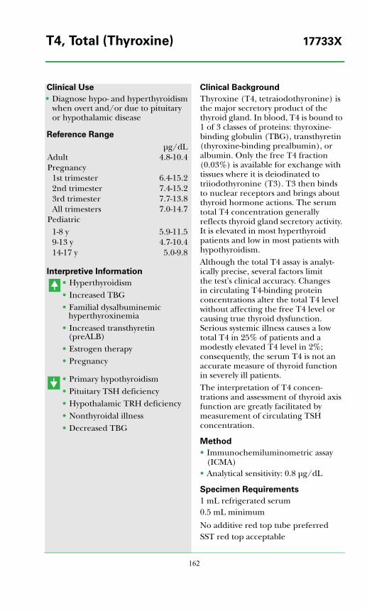
162
Clinical Use
• Diagnose hypo- and hyperthyroidism when overt and/or due to pituitary or hypothalamic disease
Reference Range
Interpretive Information
• Hyperthyroidism• Increased TBG• Familial dysalbuminemic
hyperthyroxinemia• Increased transthyretin
(preALB)• Estrogen therapy• Pregnancy
• Primary hypothyroidism• Pituitary TSH deficiency• Hypothalamic TRH deficiency• Nonthyroidal illness• Decreased TBG
Clinical Background
Thyroxine (T4, tetraiodothyronine) is the major secretory product of the thyroid gland. In blood, T4 is bound to 1 of 3 classes of proteins: thyroxine-binding globulin (TBG), transthyretin (thyroxine-binding prealbumin), or albumin. Only the free T4 fraction (0.03%) is available for exchange with tissues where it is deiodinated to triiodothyronine (T3). T3 then binds to nuclear receptors and brings about thyroid hormone actions. The serum total T4 concentration generally reflects thyroid gland secretory activity. It is elevated in most hyperthyroid patients and low in most patients with hypothyroidism.Although the total T4 assay is analyt-ically precise, several factors limit the test's clinical accuracy. Changes in circulating T4-binding protein concentrations alter the total T4 level without affecting the free T4 level or causing true thyroid dysfunction. Serious systemic illness causes a low total T4 in 25% of patients and a modestly elevated T4 level in 2%; consequently, the serum T4 is not an accurate measure of thyroid function in severely ill patients.The interpretation of T4 concen-trations and assessment of thyroid axis function are greatly facilitated by measurement of circulating TSH concentration.
Method
• Immunochemiluminometric assay (ICMA)
• Analytical sensitivity: 0.8 μg/dL
Specimen Requirements
1 mL refrigerated serum0.5 mL minimum
No additive red top tube preferredSST red top acceptable
μg/dLAdult 4.8-10.4Pregnancy
1st trimester 6.4-15.22nd trimester 7.4-15.23rd trimester 7.7-13.8All trimesters 7.0-14.7
Pediatric1-8 y 5.9-11.59-13 y 4.7-10.414-17 y 5.0-9.8
T4, Total (Thyroxine) 17733X
Alp
ha
be
tica
l Test
Se
ctio
n
163
Clinical Use
• Assess osteoclast activity• Assess bone turnover• Detect bone metastasis in patients
with breast cancer
Reference Range
Interpretive Information
• Osteoporosis• Osteomalacia• Metastatic cancer• Paget's disease• Primary hyperparathyroidism• Advanced renal failure
• Bisphosphonate therapy• Anabolic steroid therapy• Fluoride excess • Hypoparathyroidism
Clinical Background
Tartrate resistant acid phosphatase (TRAP) is secreted primarily by osteo-clasts, but also by erythrocytes and macrophages. Serum levels can be useful as a measure of bone resorption; however, the newer resorption markers (eg, N-telopeptide [NTx], free deoxypyridinoline [free DYPD, Pyrilinks®’ D], and C-telopeptide [CTx, CrossLaps®’]) are more reliable. TRAP is a potential marker of bone metastasis in patients with breast cancer; levels appear to increase in proportion to the number of bone metastases.TRAP is adversely affected by enzyme inhibitors in serum, has limited stability and analytical sensitivity, and lacks specificity for bone. Erythrocytic acid phosphatase released during in vitro clotting, as well as other non-bone sources, may falsely increase values.
Method
• Enzymatic• Analytical sensitivity: 1.2 U/L• Analytical specificity: measures level
of acid phosphatase that is resistant to tartrate inhibition
Specimen Requirements
1.0 mL frozen serum0.3 mL minimum
No additive red top preferredSST red top acceptable
Refrigerate prior to serum separation; freeze immediately afterwards.12-h fast required.
Adults 3.0-6.0 U/L
Tartrate Resistant Acid Phosphatase 37351X
164
Clinical Use
• Differentiate quantitative TBG derangements from thyroid dysfunction
Reference Range
Interpretive Information
• Pregnancy• Infancy• Familial excess• Hepatitis• Estrogen treatment• Tamoxifen treatment
• Androgenic steroid treatment• Glucocorticoids (large dose)• Nephrotic syndrome• Acromegaly• Familial deficiency
Clinical Background
Thyroxine-binding globulin (TBG) is a glycoprotein produced in the liver. TBG binds both thyroxine (T4) and triiodothyronine (T3) with high affinity and accounts for 75% of plasma protein thyroxine-binding activity. Thus, an increase or decrease in its circulating level alters total concentrations of T4 and T3 in blood, leading to potential confusion with true thyroid gland function. A number of diseases and medications, as well as inherited alterations in TBG gene expression, can change the serum TBG concentration (see Test Application and Interpretation section). Common causes of increased serum TBG include estrogen therapy (especially oral contraceptive agents), pregnancy, and hepatitis. Conversely, serum TBG may be decreased in cirrhosis, in the nephrotic syndrome, and by androgens.The measurement of TBG can be used to establish the presence of TBG deficiency or excess suggested by abnormal total serum T4 and T3 concentrations in the presence of normal free levels of these hormones. Definitive documentation of a TBG derangement may avoid unnecessary diagnostic procedures and therapy in individuals with harmless congenital TBG anomalies, and in their relatives.
Method
• Immunochemiluminometric assay (ICMA)
• Analytical sensitivity: 3.5 μg/mL
Specimen Requirements
1 mL refrigerated serum (no additive red top tube); 0.5 mL minimum
SST red top unacceptable
μg/mLMen 12.7-25.1Women 13.5-30.9Children
3-8 y 16.1-24.29-13 y 12.5-25.814-17 y 9.8-23.7
TBG (Thyroxine-Binding Globulin) 870X

Alp
ha
be
tica
l Test
Se
ctio
n
165
Clinical Use
• Diagnose and manage Graves disease, neonatal hypothyroidism, and postpartum thyroid dysfunction
Reference Range
Interpretive Information
• Graves disease• Atrophic thyroiditis• Postpartum autoimmune
thyroid disease• Neonatal Graves disease• Transient neonatal
hypothyroidism
Clinical Background
Autoimmune thyroid diseases are associated with thyroid autoantibodies, including several types directed against the TSH receptor. TSH receptor autoantibodies may be stimulatory, mimicking the actions of TSH, as in Graves disease. Alternatively, they may inhibit TSH binding and block the action of endogenous TSH. Such blocking autoantibodies produce transient neonatal hypothyroidism and chronic atrophic thyroiditis. TBII, which measures the ability of antibodies to inhibit TSH binding to its receptor, reflects the presence of either or both the stimulatory and inhibitory immunoglobulin classes.
Method
• Radioreceptor assay• Analytical sensitivity: 2% inhibition
Specimen Requirements
1 mL refrigerated serum0.5 mL minimum
No additive red top preferredSST red top acceptable
Adults and children: <16% inhibition
Graves disease: 16% - 100% inhibition
TBII (Thyrotropin-binding 5738XInhibitory Immunoglobulin)
166
Clinical Use
• Assess androgen status in male hypogonadism; female hirsutism, virilization, acne, and amenorrhea; or in pediatric patients
Reference Range
See next page.
Interpretive Information
• Polycystic ovarian disease• Cushing’s disease• Congenital adrenal hyperplasia• Androgen resistance
• Hypogonadism• P-450c17 enzyme deficiency
• Delayed puberty in boys• Gonadotropin deficiency• Testicular defects
Clinical Background
Most testosterone is transported in blood by sex hormone binding globulin (SHBG). Free testosterone is the small amount of testosterone circulating unbound. Blood testosterone levels are dependent on rates of production, interconversion, metabolic clearance, and SHBG concentration. SHBG levels are altered by medications, disease, sex steroids, and insulin. SHBG increases with age in men and decreases with androgen excess in hirsute women. Thus, determination of free testosterone more accurately reflects the level of bioactive testosterone than measure-ments of total testosterone and offers greater sensitivity in diagnosis of hypo-gonadism in aging men and evaluation of mildly hyperandrogenemic women.In general, total testosterone by ICMA, a less technically demanding and more economical procedure, is satisfactory for screening potential male hypogonadism. The Endocrine Society has stated that ICMA or other "direct" assays are unreliable for diagnosing patients with low levels (eg, <300 ng/dL) of testosterone.
Method
Testosterone, Total• Liquid chromatography tandem
mass spectrometry (LC/MS/MS)Testosterone, Free• CalculationTestosterone, Bioavailable• CalculationSHBG• Immunochemiluminometric assay
(ICMA)Albumin• Spectrophotometry
Specimen Requirements
2.8 mL refrigerated serum (no additive red top tube); 1.3 mL minimumSST red top unacceptable
Continued
Testosterone, Calculated Free, 14966XBioavailable, and Total
Alp
ha
be
tica
l Test
Se
ctio
n
167
Reference Range
Testosterone, FreeAdults
Malespg/mL
Femalespg/mL
18-69 y 46.0-224.0 0.2-5.070-89 y 6.0-73.0 0.3-5.0
Children1-10.9 y <1.3 <1.511-11.9 y <1.3 <1.512-13.9 y <64.0 <1.514-17.9 y 4.0-100.0 <3.6
Testosterone, Bioavailable
ng/dL ng/dL
Adults18-69 y 110.0-575.0 0.5-8.570-89 y 15.0-150.0 0.5-8.8
Children1-11.9 y <5.4 <3.412-13.9 y <140.0 <3.414-17.9 y 8.0-210.0 <7.8
SHBG nmol/L nmol/LAdults
18-29 y 7-44 8-11230-39 y 5-49 9-10740-49 y 8-46 8-9650-59 y 15-49 10-8960-69 y 23-38 20-10270-79 y 21-68 21-7680-91 y 20-6380-84 y 26-77
Children3-9 y 18-136 18-13610-13 y 17-123 17-12314-17 y 11-71 11-71Tanner stages
I 39-155 38-114II 33-135 24-90III 21-72 22-112IV 11-92 22-69V 18-54 18-76
Albumin 3.6-5.1 g/dL
See Testosterone, Total (Women, Children, Hypogonadal Males), LC/MS/MS for total testosterone reference ranges.
Testosterone, Calculated Free, 14966XBioavailable, and Total (continued)
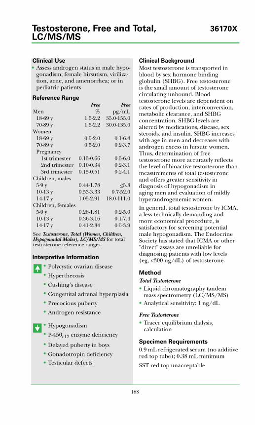
168
Clinical Use
• Assess androgen status in male hypo-gonadism; female hirsutism, viriliza-tion, acne, and amenorrhea; or in pediatric patients
Reference Range
Interpretive Information
• Polycystic ovarian disease• Hyperthecosis• Cushing’s disease• Congenital adrenal hyperplasia• Precocious puberty• Androgen resistance
• Hypogonadism• P-450c17 enzyme deficiency
• Delayed puberty in boys• Gonadotropin deficiency• Testicular defects
Clinical Background
Most testosterone is transported in blood by sex hormone binding globulin (SHBG). Free testosterone is the small amount of testosterone circulating unbound. Blood testosterone levels are dependent on rates of production, interconversion, metabolic clearance, and SHBG concentration. SHBG levels are altered by medications, disease, sex steroids, and insulin. SHBG increases with age in men and decreases with androgen excess in hirsute women. Thus, determination of free testosterone more accurately reflects the level of bioactive testosterone than measurements of total testosterone and offers greater sensitivity in diagnosis of hypogonadism in aging men and evaluation of mildly hyperandrogenemic women.In general, total testosterone by ICMA, a less technically demanding and more economical procedure, is satisfactory for screening potential male hypogonadism. The Endocrine Society has stated that ICMA or other "direct" assays are unreliable for diagnosing patients with low levels (eg, <300 ng/dL) of testosterone.
Method
Total Testosterone• Liquid chromatography tandem
mass spectrometry (LC/MS/MS)• Analytical sensitivity: 1 ng/dL
Free Testosterone• Tracer equilibrium dialysis,
calculation
Specimen Requirements
0.9 mL refrigerated serum (no additive red top tube); 0.38 mL minimum
SST red top unacceptable
Free FreeMen % pg/mL
18-69 y 1.5-2.2 35.0-155.070-89 y 1.5-2.2 30.0-135.0
Women18-69 y 0.5-2.0 0.1-6.470-89 y 0.5-2.0 0.2-3.7Pregnancy
1st trimester 0.15-0.66 0.5-6.02nd trimester 0.10-0.34 0.2-3.13rd trimester 0.15-0.51 0.2-4.1
Children, males5-9 y 0.44-1.78 <5.310-13 y 0.53-3.33 0.7-52.014-17 y 1.05-2.91 18.0-111.0
Children, females5-9 y 0.28-1.81 0.2-5.010-13 y 0.36-3.16 0.1-7.414-17 y 0.41-2.34 0.5-3.9
See Testosterone, Total (Women, Children, Hypogonadal Males), LC/MS/MS for total testosterone reference ranges.
Testosterone, Free and Total, 36170XLC/MS/MS
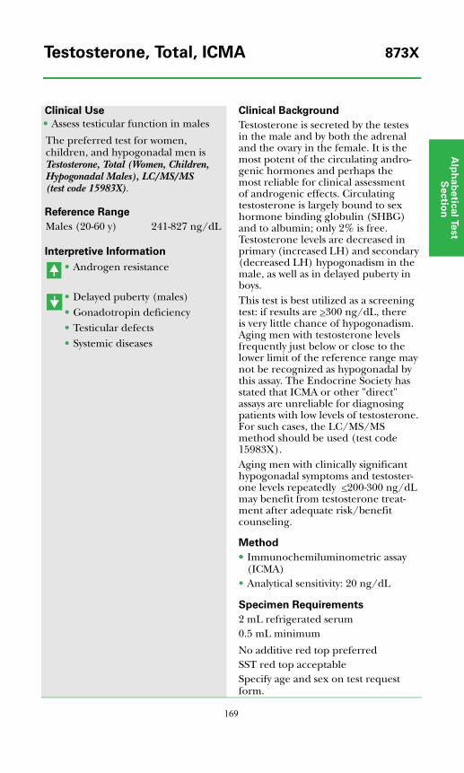
Alp
ha
be
tica
l Test
Se
ctio
n
169
Clinical Use
• Assess testicular function in males
The preferred test for women, children, and hypogonadal men is Testosterone, Total (Women, Children, Hypogonadal Males), LC/MS/MS (test code 15983X).
Reference Range
Interpretive Information
• Androgen resistance
• Delayed puberty (males)• Gonadotropin deficiency• Testicular defects • Systemic diseases
Clinical Background
Testosterone is secreted by the testes in the male and by both the adrenal and the ovary in the female. It is the most potent of the circulating andro-genic hormones and perhaps the most reliable for clinical assessment of androgenic effects. Circulating testosterone is largely bound to sex hormone binding globulin (SHBG) and to albumin; only 2% is free. Testosterone levels are decreased in primary (increased LH) and secondary (decreased LH) hypogonadism in the male, as well as in delayed puberty in boys.This test is best utilized as a screening test: if results are >300 ng/dL, there is very little chance of hypogonadism. Aging men with testosterone levels frequently just below or close to the lower limit of the reference range may not be recognized as hypogonadal by this assay. The Endocrine Society has stated that ICMA or other "direct" assays are unreliable for diagnosing patients with low levels of testosterone. For such cases, the LC/MS/MS method should be used (test code 15983X).Aging men with clinically significant hypogonadal symptoms and testoster-one levels repeatedly <200-300 ng/dL may benefit from testosterone treat-ment after adequate risk/benefit counseling.
Method
•• Immunochemiluminometric assay (ICMA)
• Analytical sensitivity: 20 ng/dL
Specimen Requirements
2 mL refrigerated serum0.5 mL minimum
No additive red top preferredSST red top acceptableSpecify age and sex on test request form.
Males (20-60 y) 241-827 ng/dL
Testosterone, Total, ICMA 873X
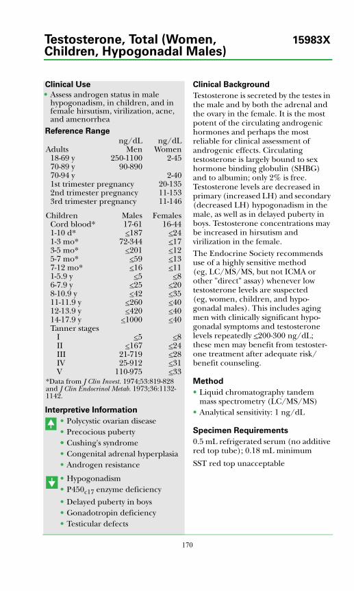
170
Clinical Use
• Assess androgen status in male hypogonadism, in children, and in female hirsutism, virilization, acne, and amenorrhea
Reference Range
Interpretive Information
• Polycystic ovarian disease• Precocious puberty•• Cushing's syndrome• Congenital adrenal hyperplasia• Androgen resistance
• Hypogonadism• P450c17 enzyme deficiency
• Delayed puberty in boys• Gonadotropin deficiency• Testicular defects
Clinical Background
Testosterone is secreted by the testes in the male and by both the adrenal and the ovary in the female. It is the most potent of the circulating androgenic hormones and perhaps the most reliable for clinical assessment of androgenic effects. Circulating testosterone is largely bound to sex hormone binding globulin (SHBG) and to albumin; only 2% is free. Testosterone levels are decreased in primary (increased LH) and secondary (decreased LH) hypogonadism in the male, as well as in delayed puberty in boys. Testosterone concentrations may be increased in hirsutism and virilization in the female.The Endocrine Society recommends use of a highly sensitive method (eg, LC/MS/MS, but not ICMA or other "direct" assay) whenever low testosterone levels are suspected (eg, women, children, and hypo-gonadal males). This includes aging men with clinically significant hypo-gonadal symptoms and testosterone levels repeatedly <200-300 ng/dL; these men may benefit from testoster-one treatment after adequate risk/benefit counseling.
Method
• Liquid chromatography tandem mass spectrometry (LC/MS/MS)
• Analytical sensitivity: 1 ng/dL
Specimen Requirements
0.5 mL refrigerated serum (no additive red top tube); 0.18 mL minimum
SST red top unacceptable
ng/dL ng/dLAdults Men Women
18-69 y 250-1100 2-4570-89 y 90-89070-94 y 2-401st trimester pregnancy 20-1352nd trimester pregnancy 11-1533rd trimester pregnancy 11-146
Children Males FemalesCord blood* 17-61 16-441-10 d* <187 <241-3 mo* 72-344 <173-5 mo* <201 <125-7 mo* <59 <137-12 mo* <16 <111-5.9 y <5 <86-7.9 y <25 <208-10.9 y <42 <3511-11.9 y <260 <4012-13.9 y <420 <4014-17.9 y <1000 <40Tanner stages
I <5 <8II <167 <24III 21-719 <28IV 25-912 <31V 110-975 <33
*Data from J Clin Invest. 1974;53:819-828 and J Clin Endocrinol Metab. 1973;36:1132-1142.
Testosterone, Total (Women, 15983XChildren, Hypogonadal Males)
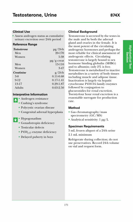
Alp
ha
be
tica
l Test
Se
ctio
n
171
Clinical Use
• Assess androgen status as cumulative urinary excretion over 24-h period
Reference Range
Interpretive Information
• Androgen resistance• Cushing's syndrome• Polycystic ovarian disease• Congenital adrenal hyperplasia
• Hypogonadism• Gonadotropin deficiency• Testicular defects• P450c17 enzyme deficiency
• Delayed puberty in boys
Clinical Background
Testosterone is secreted by the testes in the male and by both the adrenal gland and ovaries in the female. It is the most potent of the circulating androgenic hormones and perhaps the most reliable for clinical assessment of androgenic effects. Circulating testosterone is largely bound to sex hormone binding globulin (SHBG) and to albumin; only 2% is free. Testosterone is metabolized to inactive metabolites in a variety of body tissues including muscle and adipose tissue. Inactivation is largely via hepatic cytochrome P450-3A family enzymes followed by conjugation to glucuronides for renal excretion. Twenty-four hour renal excretion is a reasonable surrogate for production rate.
Method
• Gas chromatography/mass spectrometry (GC/MS)
• Analytical sensitivity: 1 μg/L
Specimen Requirements
5 mL frozen aliquot of a 24-h urine2.1 mL minimum
Refrigerate during collection; do not use preservatives. Record 24-h volume on vial and request form.
Testosterone μg/24-hMen 20-170Women 5-38
μg/g creatMen 13-110Women 3-47
Creatinine g/24-h3-8 0.11-0.689-12 0.17-1.4113-17 0.29-1.87Adults 0.63-2.50
Testosterone, Urine 874X
172
Clinical Use
• Assess aldosterone production• Diagnose hypo- and hyper-
aldosteronism
Reference Range
Interpretive Information
• Primary hyperaldosteronism•• Secondary hyperaldosteronism
• Hypoaldosteronism• Liddle's syndrome (pseudo-
aldosteronism)•• Apparent mineralocorticoid
excess (AME)• Licorice root or tobacco
chewing
Clinical Background
Tetrahydroaldosterone is a urinary metabolite of aldosterone and serves as an index of aldosterone secretion. Measurement is useful in the diagnosis of disorders of aldosterone produc-tion, since it provides an integrated measure of the 24-hour plasma aldos-terone level.
Method
• Gas chromatography/mass spectrometry (GC/MS)
• Analytical sensitivity: 1 μg/L
Specimen Requirements
5.0 mL frozen aliquot of a 24-h urine collection; 2.1 mL minimum
Refrigerate during collection; do not use preservatives.
TetrahydroaldosteroneAdults 6-79 μg/24-h
6-50 μg/g creat
Creatinine g/24-h3-8 0.11-.689-12 0.17-1.4113-17 0.29-1.87>18 0.63-2.50
Tetrahydroaldosterone, 39613X24-Hour Urine
Alp
ha
be
tica
l Test
Se
ctio
n
173
Clinical Use
• Confirm presence of autoimmune thyroid disease
Reference Range
Interpretive Information
• Hashimoto thyroiditis (HT)• Primary hypothyroidism
(due to HT)• Graves disease• Postpartum thyroiditis
Clinical Background
Circulating immunoglobulins directed against thyroglobulin are commonly present in patients with autoimmune (Hashimoto) thyroiditis and, to a lesser extent, Graves disease. Thyroglobulin is the 660-kd protein precursor of thyroid hormones. Thyroglobulin antibodies are present in the sera of approximately 70% of patients with autoimmune thyroiditis.The principal use of this test is to confirm that a patient's diffuse goiter and/or hypothyroidism is due to autoimmune thyroiditis. The test may also help distinguish Graves disease from toxic nodular goiter. Although thyroglobulin antibodies are present less often than thyroid microsomal antibodies (eg, thyroid peroxidase antibodies, anti-TPO) in patients with autoimmune thyroid diseases, screening for both immunoglobulins increases diagnostic sensitivity.Thyroglobulin antibodies may be detected in individuals without clinically significant thyroid disease. They do not define the patient's thyroid functional status.
Method
• Immunochemiluminometric assay (ICMA)
• Analytical sensitivity: 20 IU/mL
Specimen Requirements
1 mL room temperature serum0.5 mL minimum
No additive red top preferredSST red top acceptable
<20 IU/mL
Thyroglobulin Antibody 267X
174
Clinical Use
• Screen patients with thyroid carcinoma for metastases after thyroid gland ablation
• Indicate presence of thyroid gland tissue in infants with congenital hypothyroidism
• Indicate deficient thyroglobulin synthesis in infants with goitrous hypothyroidism
Reference Range
Interpretive Information
• Graves disease• Multinodular goiter• Papillary and follicular thyroid
carcinomas• Thyroiditis• TSH-dependent hyper-
thyroidism
• Thyroidectomy• Thyroid aplasia• Thyroglobulin synthetic defect• Exogenous thyroid hormone use
Clinical Background
Thyroglobulin (Tg) is a 660-kd protein synthesized exclusively in the thyroid gland. It serves as the precursor molecule for thyroid hormones, since it contains tyrosine moieties that are iodinated to form iodotyrosines and coupled to form the thyroid hormones T4 and T3. Most synthesized Tg is stored as colloid for later pinocytosis and hydrolysis within thyroid follicular cells. Small amounts are secreted directly from the endoplasmic reticulum during synthesis. Tg release is increased in a variety of thyroid disease states.Tg measurements are most useful in the postoperative monitoring of patients with papillary, follicular, and Hürthle cell carcinomas. Tg measurement also is helpful in the differential diagnosis of congenital hypothyroidism and in detecting thyrotoxicosis caused by exogenous medication (iatrogenic or factitious), in which case the serum Tg concentration should be low. Measurements are always preceded by assessment of Tg antibodies that interfere in the assay. In the presence of endogenous antibody, the Tg measurement is not reliable.
Method
Thyroglobulin• Immunochemiluminometric assay
(ICMA)• Analytical sensitivity: 0.2 ng/mL• Analytical specificity:
underestimation caused by thyroglobulin autoantibodies
Thyroglobulin Antibody• Immunochemiluminometric assay
(ICMA)• Analytical sensitivity: 20 IU/mL
Specimen Requirements
2 mL room temperature serum0.8 mL minimumNo additive red top preferredSST red top acceptable
Thyroglobulin ng/mLAdults 2.0-35.0Thyroglobulin Antibody IU/mLAdults and children <20**Higher levels can interfere with thyro-globulin quantification.
Thyroglobulin Panel 30278X
Alp
ha
be
tica
l Test
Se
ctio
n
175
Clinical Use
• Screen patients with thyroid carcinoma for metastases or recurrence
Reference Range
Interpretive Information
• • Graves disease• Multinodular goiter• Papillary and follicular thyroid
carcinomas• Thyroiditis• TSH-dependent hyper-
thyroidism
• Thyroidectomy• Thyroid aplasia• Thyroglobulin synthetic defect• Exogenous thyroid hormone use
Clinical Background
Heterophilic antibodies, defined as antibodies against animal immuno-globulins, can be a source of inter-ference in many immunoassays. The most frequently characterized interference comes from endogenous human anti-mouse antibodies (HAMA), which can react with the mouse monoclonal antibodies utilized in the assay. HAMA presence in the patient sample can lead to over- or underestimation of thyroglobulin. In this test, HAMA is removed by precipitation prior to thyroglobulin or thyroglobulin antibody determination.
Method
• HAMA precipitation
Thyroglobulin••••• Immunochemiluminometric
assay (ICMA)• Analytical sensitivity: 0.2 ng/mL• Analytical specificity: under-
estimation caused by thyroglobulin autoantibodies
Thyroglobulin Antibody•• Immunochemiluminometric
assay (ICMA)• Analytical sensitivity: 20 IU/mL
Specimen Requirements
2 mL room temperature serum1.5 mL minimum
No additive red top preferredSST red top acceptable
Thyroglobulin ng/mLAdults 2.0-35.0
Thyroglobulin Antibody IU/mLAdults and children <20**Higher levels can interfere with thyro-globulin quantification.
Thyroglobulin Panel with 19584X
HAMA Treatment
176
Clinical Use
••• Confirm presence of autoimmune thyroid disease
Reference Range
Interpretive Information
• Hashimoto thyroiditis• Primary hypothyroidism• Graves disease• Postpartum thyroiditis
Clinical Background
Thyroid peroxidase (TPO) antibodies are present in approximately 95% of patients with Hashimoto thyroiditis and 85% of patients with Graves disease. Thyroglobulin (Tg) antibodies are positive in about 60% and 30% of adult Hashimoto and Graves disease patients, respectively. Tg antibodies are less frequently detected in children with these diseases. For additional information, see Clinical Background for the individual tests (267X and 5081X).
Method
Thyroid Peroxidase Antibody• Immunochemiluminometric assay
(ICMA)• Analytical sensitivity: 10 IU/mL
Thyroglobulin Antibody• Immunochemiluminometric assay
(ICMA)• Analytical sensitivity: 20 IU/mL
Specimen Requirements
1 mL refrigerated serum0.6 mL minimum
No additive red top preferredSST red top acceptable
IU/mLThyroid Peroxidase Antibody <35Thyroglobulin Antibody <20
Thyroid Peroxidase and 7260XThyroglobulin Antibodies
Alp
ha
be
tica
l Test
Se
ctio
n
177
Clinical Use
• Confirm presence of autoimmune thyroid disease
Reference Range
Interpretive Information
• Hashimoto thyroiditis (HT)• Primary hypothyroidism
(due to HT)• Graves disease• Postpartum thyroiditis
Clinical Background
Thyroid peroxidase (TPO) is an enzyme that catalyzes the iodination and coupling steps in thyroid hormone biosynthesis. It is now known to be the principal microsomal antigen against which antibodies are directed in autoimmune thyroid diseases. The main use of this test is to confirm that a patient's diffuse goiter and/or hypothyroidism is due to autoimmune thyroiditis. The test may also help distinguish Graves disease from toxic nodular goiter. The TPO antibody measurement has replaced the thyroid antimicrosomal antibody (AMA) measurement.TPO antibodies may be detected in individuals without clinically significant thyroid disease. They do not define the patient's thyroid functional status.
Method
• Immunochemiluminometric assay (ICMA)
• Analytical sensitivity: 10 IU/mL
Specimen Requirements
1 mL refrigerated serum0.5 mL minimum
No additive red top preferredSST red top acceptable
<35 IU/mL
Thyroid Peroxidase Antibody 5081X(Anti-TPO)
178
Clinical Use
• Potential marker for TRH-secreting tumors
Reference Range
Interpretive Information
• After TRH injection• Fetus and newborn• Ectopic (extrahypothalamic)
production• Primary hypothyroidism
(variable)
Clinical Background
Thyrotropin-releasing hormone (TRH) is a tripeptide produced in the hypothalamus, other central nervous system sites, and the gastrointestinal tract, especially the pancreas. TRH stimulates TSH synthesis and release. Most circulating TRH, which is rapidly degraded, is probably derived from extra-hypothalamic tissues. Circulating TRH levels are increased in fetal and neonatal serum because of increased TRH production from fetal gut tissues and placenta and low levels of TRH-degrading activity in fetal blood. Plasma TRH levels have not been shown to be useful in diagnosis of any known thyroid condition.
Method
• Radioimmunoassay (RIA)• Analytical sensitivity: 5 pg/mL
Specimen Requirements
2 mL frozen plasma0.6 mL minimum
Collect blood in syringe or red-top tube and immediately transfer to a cold, special Nichols Institute PTH-RP collection tube; mix thoroughly. Centrifuge in refrigerated centrifuge. Transfer plasma to a plastic tube and freeze.
<5 pg/mL
Thyrotropin-releasing Hormone (TRH) 36588X
Alp
ha
be
tica
l Test
Se
ctio
n
179
Clinical Use
• Evaluate discordant serum TSH levels
Reference Range
Interpretive Information
• Autoimmune thyroid disease• Other autoimmune disease
Clinical Background
Patients may rarely develop antibodies to thyroid stimulating hormone (TSH). These TSH antibodies inter-fere with the TSH assay measurement by competing with the anti-TSH immunoglobins used in the assay.When a patient has a serum TSH concentration that is discordant with other test results, it may be appropriate to determine if TSH antibodies are present. For example, a TSH antibody may be the cause of an inappropriately reduced serum TSH level in a patient with simultaneously reduced free thyroid hormone levels (free T4 and/or free T3). Other causes of inappropriate TSH levels must also be considered:
• Hypopituitarism• Isolated TSH deficiency• Hypothalamic disease• Nonthyroidal illness• Subclinical thyrotoxicosis• Normal elderly
Method
• Radiobinding assay (RBA)
Specimen Requirements
1 mL refrigerated serum0.1 mL minimum
No additive red top preferredSST red top acceptable
Negative
TSH Antibody 36577X
180
Clinical Use
• Diagnose hypo- and hyperthyroidism• Monitor T4-replacement or T4-
suppressive therapy• Quantify TSH levels in the sub-
normal range
Reference Range
Interpretive Information
• Primary hypothyroidism• Decreased thyroid reserve
(sub-clinical hypothyroidism)• TSH-dependent hyper-
thyroidism• Thyroid hormone resistance
• Graves disease• Autonomous thyroid hormone
secretion• TSH deficiency
Clinical Background
The serum TSH measurement is 1 of the most important tools in the diagnosis of thyroid disorders. TSH secretion from the pituitary gland is controlled by hypothalamic TRH and a negative feedback effect from circulating, free thyroid hormones. Thus, in subjects with a normal hypothalamic-pituitary system, thereis an inverse correlation between free thyroid hormone and TSH concentrations in serum. Increased serum TSH is an early and sensitive indicator of decreased thyroid reserve and overt primary hypothyroidism. A third-generation TSH assay using chemiluminescent technology produces functional sensitivities of 0.01 mU/L, permitting reliable quantification of serum TSH concen-trations in the subnormal range. This allows differentiation of euthyroid from hyperthyroid patients. This assay also helps diagnose essentially all patients with TSH-independent hyperthyroidism (Graves disease, etc.) with a single blood sample. In addition, the assay allows adjustment of exogenous thyroxine dosage in hypothyroid patients and in patients on suppressive thyroxine therapy for thyroid neoplasia.
Method
• Immunochemiluminometric assay (ICMA)
• Analytical sensitivity: 0.01 mU/L
Specimen Requirements
2 mL refrigerated serum (no additive red top tube); 0.5 mL minimum
Adults mU/LMen and non-pregnant women
0.40-4.50
Pregnant women1st trimester 0.30-4.502nd trimester 0.50-4.603rd trimester 0.80-5.20
Children1-4 d 3.20-35.005-6 d Not established*1-4 wk 1.70-9.101-12 mo 0.80-8.201-19 y 0.50-4.30
*TSH levels decline rapidly during the first week of life in most children, but may remain transiently elevated in a few individuals despite normal free T4 levels. For confirmatory testing following a newborn thyroid screen, a free (or total) T4 level is usually required for proper interpretation of TSH levels in this age group.
TSH, Ultrasensitive 8660X
Alp
ha
be
tica
l Test
Se
ctio
n
181
Clinical Use
• Monitor T4-replacement or T4-suppressive therapy
Reference Range
Interpretive Information
• Primary hypothyroidism• Decreased thyroid reserve
(sub-clinical hypothyroidism)• TSH-dependent hyper-
thyroidism• Thyroid hormone resistance
• Graves disease• Autonomous thyroid hormone
secretion• TSH deficiency
Clinical Background
Heterophilic antibodies, defined as antibodies against animal immuno-globulins, can be a source of interference in many immunoassays. The most frequently characterized interference comes from endogenous human anti-mouse antibodies (HAMA), which can react with the mouse monoclonal antibodies utilized in the assay. HAMA presence in the patient sample can lead to over- or underestimation of thyroid stimulating hormone (TSH). In this test, HAMA is removed by precipitation prior to TSH determination.
Method
• HAMA precipitation• Immunochemiluminometric
assay (ICMA)• Analytical sensitivity: 0.01 mU/L
Specimen Requirements
2 mL refrigerated serum1.5 mL minimum
No additive red top preferredSST red top acceptable
TSH, Ultrasensitive mU/LMen and non-pregnant women
0.40-4.50
Pregnant women1st trimester 0.30-4.502nd trimester 0.50-4.603rd trimester 0.80-5.20
Children1-4 d 3.20-35.005-6 d Not established*1-4 wk 1.70-9.101-12 mo 0.80-8.201-19 y 0.50-4.30
*TSH levels decline rapidly during the first week of life in most children, but may remain transiently elevated in a few individuals despite normal free T4 levels. For confirmatory testing following a newborn thyroid screen, a free (or total) T4 level is usually required for proper interpretation of TSH levels in this age group.
TSH with HAMA Treatment 19537X
182
Clinical Use
• Diagnose and manage Graves disease
Reference Range
Interpretive Information
• Graves disease• Hashitoxicosis• Neonatal thyrotoxicosis
Clinical Background
Autoimmune thyroid disease is associated with a variety of thyroid-related autoantibodies. These include anti-thyroglobulin, anti-thyroid peroxidase, and TSH receptor anti-bodies. The TSH receptor antibodies may be stimulatory, exerting a TSH-like effect, or inhibitory, blocking the effect of TSH. In the thyroid follicular cell, TSH stimulates cyclic AMP (cAMP), which serves a pivotal role in thyroid cellular activation. Because TSI also stimulates cAMP production, the measurement of TSI bioactivity is accomplished by measuring its capacity to increase production of cAMP in a TSH receptor-expressing cell line.Studies have shown that TSI is present in 80% to 90% of patients with active Graves disease and absent in some patients with remission. In patients with Graves disease, monitoring TSI levels during pregnancy helps predict neonatal Graves disease. Patients with preexisting Hashimoto thyroiditis who develop hyperthyroidism will also often manifest measurable TSI bioactivity.TSH receptor antibodies can also be measured by their capacity to inhibit TSH binding to TSH receptors. In this case, they are referred to as TSH receptor binding inhibiting immunoglobulins (TBII, test code 5738X).
Method
• In vitro bioassay (luciferase)• Analytical sensitivity: 75%• Analytical specificity: under-
estimation caused by thyrotropin blocking antibody presence
Specimen Requirements
1 mL refrigerated serum0.2 mL minimum
No additive red top preferredSST red top acceptable
<125% of basal activity
TSI (Thyroid Stimulating 30551XImmunoglobulin)
Alp
ha
be
tica
l Test
Se
ctio
n
183
Clinical Use
• Diagnose watery diarrhea syndrome (WDS)
• Assess patients with suspected multiple endocrine neoplasia (MEN)
Reference Range
Interpretive Information
• VIPoma/watery diarrhea syndrome
• MEN type 1
Clinical Background
Vasoactive intestinal polypeptide (VIP) is a 28-amino acid neuropeptide produced by neural stimulation in a variety of tissues. It has broad biological effects on the cardiovascular, gastrointestinal, and respiratory systems. These effects include in-creased secretion of bicarbonate and electrolytes and decreased absorption of electrolytes and water.VIPomas can occur as part of the type 1 multiple endocrine neoplasia (MEN) syndrome. VIP is the principal mediator of the watery diarrhea syndrome (also known as pancreatic cholera, Werner-Morrison syndrome, or diarrheogenic islet cell tumor). Watery diarrhea syndrome, with elevated VIP, has also been associated with neurogenic tumors (ganglioneuroma, neuroblastoma, pheochromocytoma), bronchogenic tumors, and islet cell hyperplasia.
Method
• Extraction, radioimmunoassay (RIA)• Analytical sensitivity: 12.5 pg/mL
Specimen Requirements
3 mL frozen EDTA plasma
1.1 mL minimum
Adults and children <50 pg/mL
Vasoactive Intestinal Polypeptide 920X(VIP)
184
Clinical Use
• Measurement of bioactive vitamin D• Differential diagnosis of hypo-
calcemia• Monitor patients with renal osteo-
dystrophy or chronic renal failure
Reference Range
Interpretive Information
• Chronic granulomatous disorders
• Hyperparathyroidism• Hypercalcemia associated with
lymphoma• Type II vitamin D-dependent
rickets
• Severe vitamin D deficiency• Type I vitamin D-resistant rickets• Hypoparathyroidism• Pseudohypoparathyroidism• Renal osteodystrophy, renal
failure• Tumor-induced osteomalacia• Tumor-induced hypercalcemia
(except with lymphoma)
Clinical Background
Vitamin D is a fat-soluble vitamin that is synthesized from cholesterol when the skin is exposed to ultraviolet light; it can also be derived from the diet. The liver hydroxylates vitamin D to 25-hydroxyvitamin D [25(OH)D], the major circulating species of vitamin D. The kidney, along with placenta and granuloma tissues, hydroxylates 25(OH)D to form 1, 25-dihydroxy-vitamin D [1,25(OH)2D], the bioactive form of the vitamin. Kidney 1-hydroxylation of 25(OH)D is stimulated by parathyroid hormone (PTH) and hypophosphatemia. Levels of 1,25(OH)2D are approximately 1,000- fold less than those of 25(OH)D.
1,25(OH)2D increases intestinal absorption of calcium and is required to maintain adequate bone calcifica-tion. Vitamin D deficiency is assessed with measurements of 25(OH)D. Measurements of 1,25(OH)2D assess bioactive vitamin and may be useful in renal disease, parathyroid disease, and vitamin D resistance or dependence.
Method
• Extraction, chromatography, radioreceptor assay
• Analytical sensitivity: 5 pg/mL
Specimen Requirements
3 mL frozen serum1.1 mL minimum
No additive red top preferredSST red top acceptable
pg/mLAdults 15-60Children
3-17 y 27-71
Vitamin D, 1,25-Dihydroxy 4729X
Alp
ha
be
tica
l Test
Se
ctio
n
185
Clinical Use
• Diagnose vitamin D deficiency or intoxication
Reference Range
Interpretive Information
• Vitamin D intoxication
• Nutritional rickets• Secondary hyperparathyroidism• Osteomalacia• Severe cholestatic or
parenchymal liver disease• Vitamin D-resistant metabolic
bone disease due to multiple drugs for seizure control
• Nephrotic syndrome with marked proteinuria
• Intestinal malabsorption
Decreased, but usually not below reference range• Obesity• Sarcoidosis• Hyperphosphatemic tumoral
calcinosis• Tuberculosis• Primary hyperparathyroidism• Type II vitamin D-dependent
ricketsLevels vary with exposure to sunlight, peaking in the summer months.
Clinical Background
25-Hydroxyvitamin D (25OHD) is the major circulating form of vitamin D and the precursor of the active form (1,25-dihydroxyvitamin D). Because of its long half-life, 25OHD measure-ments are useful for assessing vitamin D status in patients.Vitamin D occurs in 2 forms: vitamin D3 (cholecalciferol) and vitamin D2 (ergocalciferol). Vitamin D3 is obtained from foods of animal origin and from ultraviolet light-stimulated conversion of 7-dehydrocholesterol in the skin, whereas vitamin D2 is obtained from foods of plant origin. Vitamin D2 is used in a high potency (50,000 IU) formulation for treating severe vitamin D deficiency. Both forms are used in over-the-counter supplements and fortified foods and are metabolized to their respective 25OHD forms (ie, 25OHD3 and 25OHD2). Thus, analytical methods that can accurately quantitate both forms are essential for diagnosis and monitoring patients with vitamin D deficiency as well as differentiating between intoxication and other hypercalcemic disorders.
Method
• Liquid chromatography tandem mass spectrometry (LC/MS/MS)
• Analytical sensitivity: 4 ng/mL for 25OHD2 and 25OHD3
• Analytical specificity: no cross-reaction with vitamin D2 or D3; 1α,25(OH)2D2; 1α,25(OH)2D3, calcitriol; 25,26(OH)2D3; 1α(OH)D2, doxercalciferol; and 1α(OH)D3, alfacalcidiol
Specimen Requirements
0.3 mL room temperature serum0.15 mL minimumNo additive red top preferredSST red top acceptableOvernight (8-12 h) fasting is preferred.
Total 25OHD 20-100 ng/mL25OHD2 Not established25OHD3 Not established
25OHD levels <20 ng/mL reflect vitamin D deficiency. The optimal level is 30-50 ng/mL. 25OHD2 levels >4 ng/mL suggest compliance with supplements.
Vitamin D, 25-Hydroxy, LC/MS/MS 17306X
186
Clinical Use
• Diagnose catecholamine-producing tumors
Reference Range
Interpretive Information
• Pheochromocytoma• Neuroblastoma• Ganglioblastoma• Stress
Clinical Background
Urinary vanillylmandelic acid (VMA) is the end product of catecholamine metabolism and reflects catecholamine production by chromaffin cells of the adrenal medulla and the sympathetic nervous system.Pheochromocytomas are rare tumors of the chromaffin cells located in or near the adrenal glands. These tumors are diagnosed on the basis of elevated levels of urinary metanephrines, urinary VMA, and plasma and/or urine catecholamines. Measurement of homovanillic acid (HVA) has little value in identifying patients with pheochromocytoma but differentiates those with neuroblastoma.Neuroblastomas are malignant tumors that usually occur before 2 years of age; both VMA and HVA levels helpin diagnosing these tumors.Gangliomas are rare, benign, well-differentiated tumors in young adults and are associated with excess production of catecholamines and their metabolites.
Method
• High-performance liquid chromato-graphy (HPLC), electrochemical detection
• Analytical sensitivity: 0.5 mg/L• Creatinine concentration also
reported
Specimen Requirements
10 mL room temperature aliquot of urine; 5 mL minimumCollect 24-h urine in 25 mL 6N HCl. Record 24-h volume on vial and request form. Adjust pH of random urine to <3.0 using 6N HCl.It is preferable for the patient to be off medications for 3 days prior to collection; however, common antihypertensives cause minimal or no interference. Patients should avoid alcohol, coffee, tea, tobacco, nicotine patch, bananas, citrus fruit, and strenuous exercise prior to collection
24-Hour urine (39517X)VMA mg/24-h
Adults <6.0Children
3-8 y <2.39-12 y <3.413-17 y <3.9
Creatinine g/24 -hAdults 0.63-2.50Children
3-8 y 0.11-0.689-12 y 0.17-1.4113-17 y 0.29-1.87
Random urine (1710X)VMA mg/g creat
Adults 1.1-4.1Children
Birth-6 mo 5.5-267-11 mo 6.1-201-2 y 2.5-213-8 y 1.7-6.59-12 y 1.4-5.113-17 y 1.5-3.6
Creatinine mg/dL0-6 mo 2-327-11 mo 2-361-2 y 2-1283-8 y 2-1499-12 y 2-183>12 y
Males 20-370Females 20-320
VMA (Vanillylmandelic Acid), 39517XUrine 1710X
Test Application
andInterpretation

Test Application and Interpretation
Test A
pp
licatio
n &
In
terp
reta
tion
PREFACE
Advances in our understanding of endocrine physiology and pathophysiologyand the availability of highly sensitive and specific hormone assays permitincreasingly accurate endocrine diagnosis and differential diagnosis. As a result,dynamic tests measuring endocrine gland reserve or autonomy are less oftennecessary; however, they are still useful in evaluation of hormonal systems thatchange rapidly, such as growth hormone. Stimulation and suppression tests arestill applied to the assessment of selected patients with pituitary and adrenaldisorders.
In this test manual, we have attempted to summarize the state of the art withregard to endocrine assay applications, dynamic tests, and their interpretationcirca 2007. We hope that this guide provides helpful information for physiciansentrusted with the care of patients with known and suspected endocrinedisorders.
189
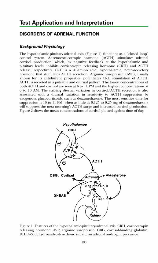
Test Application and Interpretation
DISORDERS OF ADRENAL FUNCTION
Background Physiology
The hypothalamic-pituitary-adrenal axis (Figure 1) functions as a "closed loop"control system. Adrenocorticotropic hormone (ACTH) stimulates adrenalcortisol production, which, by negative feedback at the hypothalamic andpituitary levels, inhibits corticotropin releasing hormone (CRH) and ACTHrelease, respectively. CRH is a 41-amino acid, hypothalamic, neurosecretoryhormone that stimulates ACTH secretion. Arginine vasopressin (AVP), usuallyknown for its antidiuretic properties, potentiates CRH stimulation of ACTH.ACTH is secreted in a pulsatile and diurnal pattern. The lowest concentrations ofboth ACTH and cortisol are seen at 6 to 11 PM and the highest concentrations at6 to 10 AM. The striking diurnal variation in cortisol/ACTH secretion is alsoassociated with a diurnal variation in sensitivity to ACTH suppression byexogenous glucocorticoids, such as dexamethasone. The most sensitive time forsuppression is 10 to 11 PM, when as little as 0.125 to 0.25 mg of dexamethasonewill suppress the next morning's ACTH surge and increased cortisol production.Figure 2 shows the mean concentrations of cortisol plotted against time of day.
Figure 1. Features of the hypothalamic-pituitary-adrenal axis. CRH, corticotropinreleasing hormone; AVP, arginine vasopressin; CBG, cortisol-binding globulin;DHEA-S, dehydroandrostenedione sulfate, an adrenal androgen precursor.
190
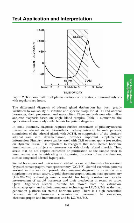
Test Application and Interpretation
Test A
pp
licatio
n &
In
terp
reta
tion
Figure 2. Temporal pattern of plasma cortisol concentrations in normal subjectswith regular sleep hours.
The differential diagnosis of adrenal gland dysfunction has been greatlyfacilitated by availability of sensitive and specific assays for ACTH and adrenalhormones, their precursors, and metabolites. These methods now often allowaccurate diagnosis based on single blood samples. Table 1 summarizes theapplication of commonly available tests for patient diagnosis.
In some instances, diagnosis requires further assessment of pituitary-adrenalreserve or adrenal steroid biosynthetic pathway integrity. In such patients,stimulation of the adrenal glands with ACTH, or suppression of the pituitary-adrenal axis with dexamethasone, provides important supplementaryinformation. Pituitary reserve can be tested with CRH or metyrapone (see sectionon Dynamic Tests). It is important to recognize that most steroid hormoneimmunoassays are subject to cross-reaction with closely related steroids. Thus,assays that do not employ extraction or purification of the sample prior toimmunoassay may be misleading in diagnosing disorders of enzyme function,such as congenital adrenal hyperplasia.
Steroid hormones and their urinary metabolites can be definitively characterizedby gas chromatography/mass spectrometry (GC/MS). Steroid excretion patternsassessed in this way can provide discriminating diagnostic information as asupplement to serum assays. Liquid chromatography, tandem mass spectrometry(LC/MS/MS) technology now is available for highly sensitive and specificmeasurement of steroid hormones and their metabolites in serum or urine.Quest Diagnostics Nichols Institute has moved from the extraction,chromatography, and radioimmunoassay technology to LC/MS/MS as the nextgeneration platform for steroid hormone assay. There is a high correlationbetween steroid hormone concentrations measured by extraction,chromatography, and immunoassay and by LC/MS/MS.
191
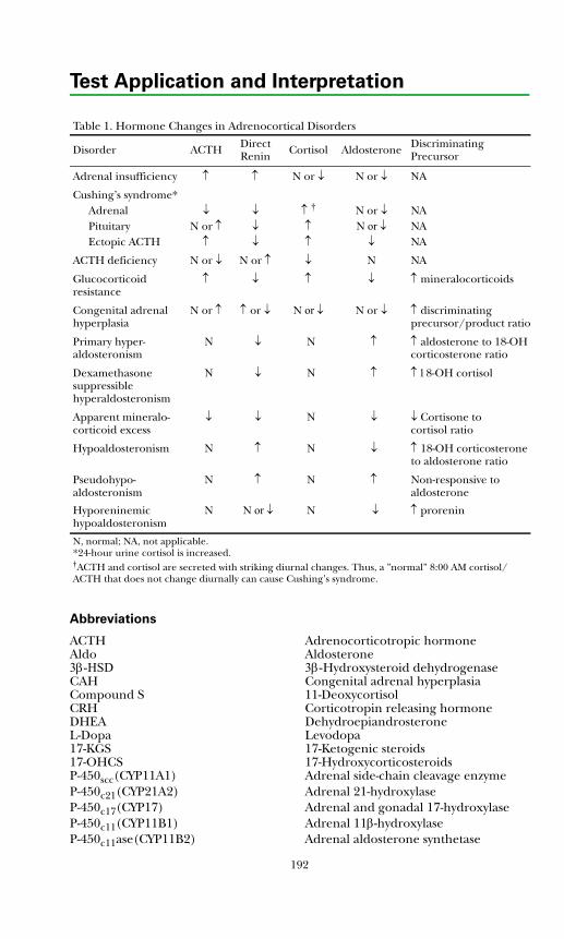
Test Application and Interpretation
Abbreviations
ACTH Adrenocorticotropic hormoneAldo Aldosterone3β-HSD 3β-Hydroxysteroid dehydrogenase CAH Congenital adrenal hyperplasiaCompound S 11-Deoxycortisol CRH Corticotropin releasing hormoneDHEA DehydroepiandrosteroneL-Dopa Levodopa17-KGS 17-Ketogenic steroids17-OHCS 17-HydroxycorticosteroidsP-450scc(CYP11A1) Adrenal side-chain cleavage enzymeP-450c21(CYP21A2) Adrenal 21-hydroxylaseP-450c17(CYP17) Adrenal and gonadal 17-hydroxylaseP-450c11(CYP11B1) Adrenal 11β-hydroxylaseP-450c11ase(CYP11B2) Adrenal aldosterone synthetase
Table 1. Hormone Changes in Adrenocortical Disorders
Disorder ACTH Direct Renin Cortisol Aldosterone Discriminating
Precursor
Adrenal insufficiency ↑ ↑ N or ↓ N or ↓ ΝΑ
Cushing’s syndrome*Adrenal PituitaryEctopic ACTH
↓N or ↑
↑
↓↓↓
↑ †
↑↑
N or ↓N or ↓
↓
NANANA
ACTH deficiency N or ↓ N or ↑ ↓ N NA
Glucocorticoid resistance
↑ ↓ ↑ ↓ ↑ mineralocorticoids
Congenital adrenal hyperplasia
N or ↑ ↑ or ↓ N or ↓ N or ↓ ↑ discriminating precursor/product ratio
Primary hyper-aldosteronism
N ↓ N ↑ ↑ aldosterone to 18-OH corticosterone ratio
Dexamethasone suppressible hyperaldosteronism
Ν ↓ Ν ↑ ↑ 18-OH cortisol
Apparent mineralo-corticoid excess
↓ ↓ Ν ↓ ↓ Cortisone to cortisol ratio
Hypoaldosteronism N ↑ Ν ↓ ↑ 18-OH corticosterone to aldosterone ratio
Pseudohypo-aldosteronism
N ↑ Ν ↑ Non-responsive to aldosterone
Hyporeninemic hypoaldosteronism
N N or ↓ N ↓ ↑ prorenin
N, normal; NA, not applicable. *24-hour urine cortisol is increased. †ACTH and cortisol are secreted with striking diurnal changes. Thus, a ”normal” 8:00 AM cortisol/ACTH that does not change diurnally can cause Cushing’s syndrome.
192
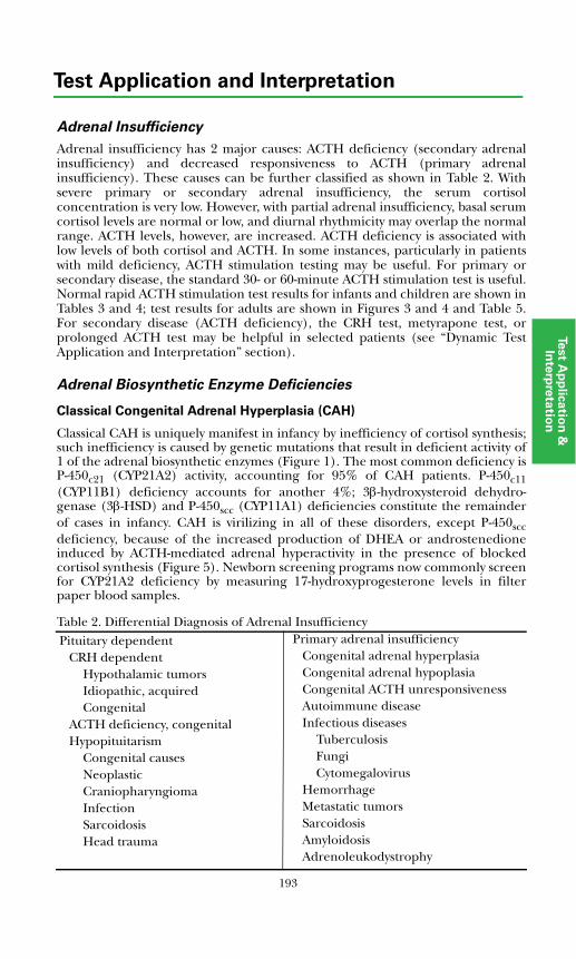
Test Application and Interpretation
Test A
pp
licatio
n &
In
terp
reta
tion
Adrenal Insufficiency
Adrenal insufficiency has 2 major causes: ACTH deficiency (secondary adrenalinsufficiency) and decreased responsiveness to ACTH (primary adrenalinsufficiency). These causes can be further classified as shown in Table 2. Withsevere primary or secondary adrenal insufficiency, the serum cortisolconcentration is very low. However, with partial adrenal insufficiency, basal serumcortisol levels are normal or low, and diurnal rhythmicity may overlap the normalrange. ACTH levels, however, are increased. ACTH deficiency is associated withlow levels of both cortisol and ACTH. In some instances, particularly in patientswith mild deficiency, ACTH stimulation testing may be useful. For primary orsecondary disease, the standard 30- or 60-minute ACTH stimulation test is useful.Normal rapid ACTH stimulation test results for infants and children are shown inTables 3 and 4; test results for adults are shown in Figures 3 and 4 and Table 5.For secondary disease (ACTH deficiency), the CRH test, metyrapone test, orprolonged ACTH test may be helpful in selected patients (see “Dynamic TestApplication and Interpretation” section).
Adrenal Biosynthetic Enzyme Deficiencies
Classical Congenital Adrenal Hyperplasia (CAH)
Classical CAH is uniquely manifest in infancy by inefficiency of cortisol synthesis;such inefficiency is caused by genetic mutations that result in deficient activity of1 of the adrenal biosynthetic enzymes (Figure 1). The most common deficiency isP-450c21 (CYP21A2) activity, accounting for 95% of CAH patients. P-450c11(CYP11B1) deficiency accounts for another 4%; 3β-hydroxysteroid dehydro-genase (3β-HSD) and P-450scc (CYP11A1) deficiencies constitute the remainderof cases in infancy. CAH is virilizing in all of these disorders, except P-450sccdeficiency, because of the increased production of DHEA or androstenedioneinduced by ACTH-mediated adrenal hyperactivity in the presence of blockedcortisol synthesis (Figure 5). Newborn screening programs now commonly screenfor CYP21A2 deficiency by measuring 17-hydroxyprogesterone levels in filterpaper blood samples.
Table 2. Differential Diagnosis of Adrenal InsufficiencyPituitary dependent
CRH dependentHypothalamic tumorsIdiopathic, acquiredCongenital
ACTH deficiency, congenitalHypopituitarism
Congenital causesNeoplasticCraniopharyngiomaInfectionSarcoidosisHead trauma
Primary adrenal insufficiencyCongenital adrenal hyperplasiaCongenital adrenal hypoplasiaCongenital ACTH unresponsivenessAutoimmune diseaseInfectious diseases
TuberculosisFungiCytomegalovirus
HemorrhageMetastatic tumorsSarcoidosisAmyloidosisAdrenoleukodystrophy
193
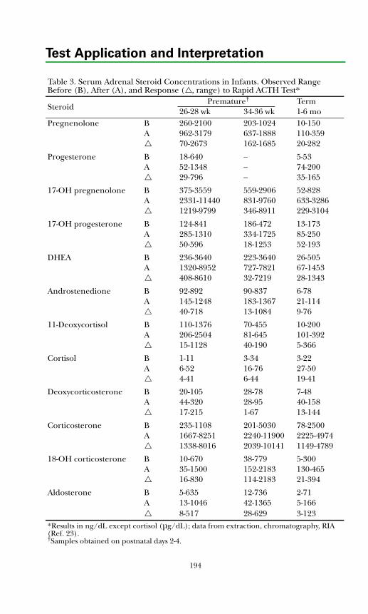
Test Application and Interpretation
Table 3. Serum Adrenal Steroid Concentrations in Infants. Observed Range Before (B), After (A), and Response ( , range) to Rapid ACTH Test*
Steroid Premature† Term26-28 wk 34-36 wk 1-6 mo
Pregnenolone B 260-2100 203-1024 10-150A 962-3179 637-1888 110-359
70-2673 162-1685 20-282
Progesterone B 18-640 − 5-53A 52-1348 − 74-200
29-796 − 35-165
17-OH pregnenolone B 375-3559 559-2906 52-828A 2331-11440 831-9760 633-3286
1219-9799 346-8911 229-3104
17-OH progesterone B 124-841 186-472 13-173A 285-1310 334-1725 85-250
50-596 18-1253 52-193
DHEA B 236-3640 223-3640 26-505A 1320-8952 727-7821 67-1453
408-8610 32-7219 28-1343
Androstenedione B 92-892 90-837 6-78A 145-1248 183-1367 21-114
40-718 13-1084 9-76
11-Deoxycortisol B 110-1376 70-455 10-200A 206-2504 81-645 101-392
15-1128 40-190 5-366
Cortisol B 1-11 3-34 3-22A 6-52 16-76 27-50
4-41 6-44 19-41
Deoxycorticosterone B 20-105 28-78 7-48A 44-320 28-95 40-158
17-215 1-67 13-144
Corticosterone B 235-1108 201-5030 78-2500A 1667-8251 2240-11900 2225-4974
1338-8016 2039-10141 1149-4789
18-OH corticosterone B 10-670 38-779 5-300A 35-1500 152-2183 130-465
16-830 114-2183 21-394
Aldosterone B 5-635 12-736 2-71A 13-1046 42-1365 5-166
8-517 28-629 3-123*Results in ng/dL except cortisol (μg/dL); data from extraction, chromatography, RIA (Ref. 23).†Samples obtained on postnatal days 2-4.
194
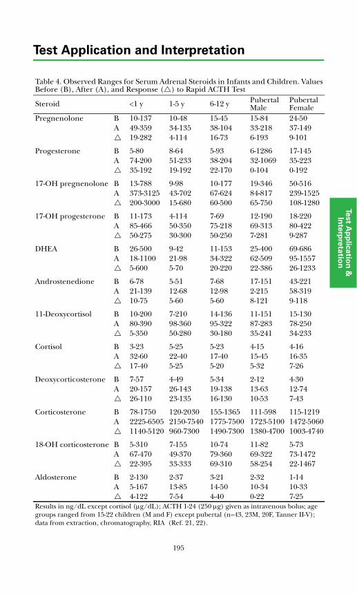
Test Application and Interpretation
Test A
pp
licatio
n &
In
terp
reta
tion
Table 4. Observed Ranges for Serum Adrenal Steroids in Infants and Children. Values Before (B), After (A), and Response ( ) to Rapid ACTH Test
Steroid <1 y 1-5 y 6-12 y Pubertal Male
Pubertal Female
Pregnenolone B 10-137 10-48 15-45 15-84 24-50A 49-359 34-135 38-104 33-218 37-149
19-282 4-114 16-73 6-193 9-101
Progesterone B 5-80 8-64 5-93 6-1286 17-145A 74-200 51-233 38-204 32-1069 35-223
35-192 19-192 22-170 0-104 0-192
17-OH pregnenolone B 13-788 9-98 10-177 19-346 50-516A 373-3125 43-702 67-624 84-817 239-1525
200-3000 15-680 60-500 65-750 108-1280
17-OH progesterone B 11-173 4-114 7-69 12-190 18-220A 85-466 50-350 75-218 69-313 80-422
50-275 30-300 50-250 7-281 9-287
DHEA B 26-500 9-42 11-153 25-400 69-686A 18-1100 21-98 34-322 62-509 95-1557
5-600 5-70 20-220 22-386 26-1233
Androstenedione B 6-78 5-51 7-68 17-151 43-221A 21-139 12-68 12-98 2-215 58-319
10-75 5-60 5-60 8-121 9-118
11-Deoxycortisol B 10-200 7-210 14-136 11-151 15-130A 80-390 98-360 95-322 87-283 78-250
5-350 50-280 30-180 35-241 34-233
Cortisol B 3-23 5-25 5-23 4-15 4-16A 32-60 22-40 17-40 15-45 16-35
17-40 5-25 5-20 5-32 7-26
Deoxycorticosterone B 7-57 4-49 5-34 2-12 4-30A 20-157 26-143 19-138 13-63 12-74
26-110 23-135 16-130 10-53 7-43
Corticosterone B 78-1750 120-2030 155-1365 111-598 115-1219A 2225-6505 2150-7540 1775-7500 1723-5100 1472-5060
1140-5120 960-7300 1490-7300 1380-4700 1003-4740
18-OH corticosterone B 5-310 7-155 10-74 11-82 5-73A 67-470 49-370 79-360 69-322 73-1472
22-395 33-333 69-310 58-254 22-1467
Aldosterone B 2-130 2-37 3-21 2-32 1-14A 5-167 13-85 14-50 10-34 10-33
4-122 7-54 4-40 0-22 7-25Results in ng/dL except cortisol (μg/dL); ACTH 1-24 (250 μg) given as intravenous bolus; age groups ranged from 15-22 children (M and F) except pubertal (n=43, 23M, 20F, Tanner II-V); data from extraction, chromatography, RIA (Ref. 21, 22).
195
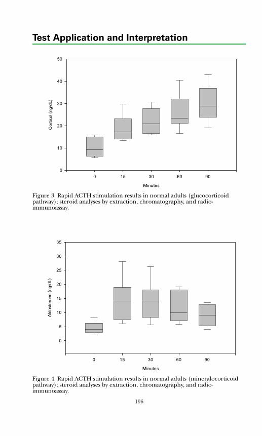
Test Application and Interpretation
Figure 3. Rapid ACTH stimulation results in normal adults (glucocorticoid pathway); steroid analyses by extraction, chromatography, and radio-immunoassay.
Figure 4. Rapid ACTH stimulation results in normal adults (mineralocorticoid pathway); steroid analyses by extraction, chromatography, and radio-immunoassay.
Minutes
0 15 30 60 90
Cor
tisol
(ng
/dL)
0
10
20
30
40
50
Minutes
0 15 30 60 90
Ald
oste
rone
(ng
/dL)
0
5
10
15
20
25
30
35
196
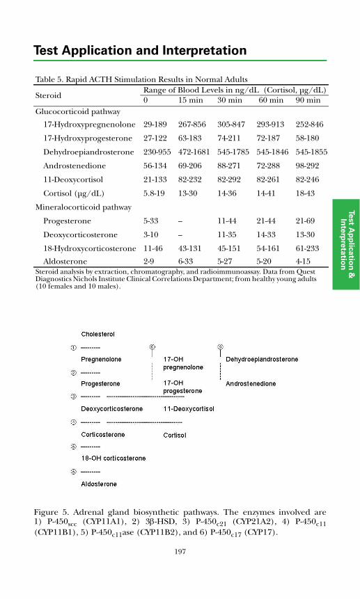
197
Test Application and Interpretation
Test A
pp
licatio
n &
In
terp
reta
tion
Table 5. Rapid ACTH Stimulation Results in Normal Adults
SteroidRange of Blood Levels in ng/dL (Cortisol, μg/dL)0 15 min 30 min 60 min 90 min
Glucocorticoid pathway
17-Hydroxypregnenolone 29-189 267-856 305-847 293-913 252-846
17-Hydroxyprogesterone 27-122 63-183 74-211 72-187 58-180
Dehydroepiandrosterone 230-955 472-1681 545-1785 545-1846 545-1855
Androstenedione 56-134 69-206 88-271 72-288 98-292
11-Deoxycortisol 21-133 82-232 82-292 82-261 82-246
Cortisol (μg/dL) 5.8-19 13-30 14-36 14-41 18-43
Mineralocorticoid pathway
Progesterone 5-33 -- 11-44 21-44 21-69
Deoxycorticosterone 3-10 -- 11-35 14-33 13-30
18-Hydroxycorticosterone 11-46 43-131 45-151 54-161 61-233
Aldosterone 2-9 6-33 5-27 5-20 4-15Steroid analysis by extraction, chromatography, and radioimmunoassay. Data from Quest Diagnostics Nichols Institute Clinical Correlations Department; from healthy young adults (10 females and 10 males).
Figure 5. Adrenal gland biosynthetic pathways. The enzymes involved are1) P-450scc (CYP11A1), 2) 3β-HSD, 3) P-450c21 (CYP21A2), 4) P-450c11(CYP11B1), 5) P-450c11ase (CYP11B2), and 6) P-450c17 (CYP17).
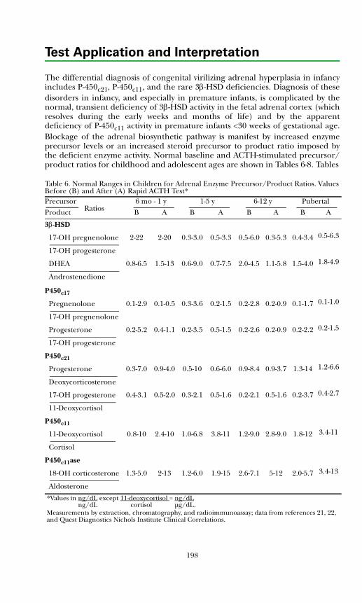
198
Test Application and Interpretation
The differential diagnosis of congenital virilizing adrenal hyperplasia in infancyincludes P-450c21, P-450c11, and the rare 3β-HSD deficiencies. Diagnosis of thesedisorders in infancy, and especially in premature infants, is complicated by thenormal, transient deficiency of 3β-HSD activity in the fetal adrenal cortex (whichresolves during the early weeks and months of life) and by the apparentdeficiency of P-450c11 activity in premature infants <30 weeks of gestational age.Blockage of the adrenal biosynthetic pathway is manifest by increased enzymeprecursor levels or an increased steroid precursor to product ratio imposed bythe deficient enzyme activity. Normal baseline and ACTH-stimulated precursor/product ratios for childhood and adolescent ages are shown in Tables 6-8. Tables Table 6. Normal Ranges in Children for Adrenal Enzyme Precursor/Product Ratios. Values Before (B) and After (A) Rapid ACTH Test*Precursor 6 mo - 1 y 1-5 y 6-12 y Pubertal
Product B A B A B A B A
3β-HSD
17-OH pregnenolone 2-22 2-20 0.3-3.0 0.5-3.3 0.5-6.0 0.3-5.3 0.4-3.4 0.5-6.3
17-OH progesterone
DHEA 0.8-6.5 1.5-13 0.6-9.0 0.7-7.5 2.0-4.5 1.1-5.8 1.5-4.0 1.8-4.9
Androstenedione
P450c17
Pregnenolone 0.1-2.9 0.1-0.5 0.3-3.6 0.2-1.5 0.2-2.8 0.2-0.9 0.1-1.7 0.1-1.0
17-OH pregnenolone
Progesterone 0.2-5.2 0.4-1.1 0.2-3.5 0.5-1.5 0.2-2.6 0.2-0.9 0.2-2.2 0.2-1.5
17-OH progesterone
P450c21
Progesterone 0.3-7.0 0.9-4.0 0.5-10 0.6-6.0 0.9-8.4 0.9-3.7 1.3-14 1.2-6.6
Deoxycorticosterone
17-OH progesterone 0.4-3.1 0.5-2.0 0.3-2.1 0.5-1.6 0.2-2.1 0.5-1.6 0.2-3.7 0.4-2.7
11-Deoxycortisol
P450c11
11-Deoxycortisol 0.8-10 2.4-10 1.0-6.8 3.8-11 1.2-9.0 2.8-9.0 1.8-12 3.4-11
Cortisol
P450c11ase
18-OH corticosterone 1.3-5.0 2-13 1.2-6.0 1.9-15 2.6-7.1 5-12 2.0-5.7 3.4-13
Aldosterone
*Values in ng/dL except 11-deoxycortisol = ng/dL ng/dL cortisol μg/dL.Measurements by extraction, chromatography, and radioimmunoassay; data from references 21, 22, and Quest Diagnostics Nichols Institute Clinical Correlations.
Ratios
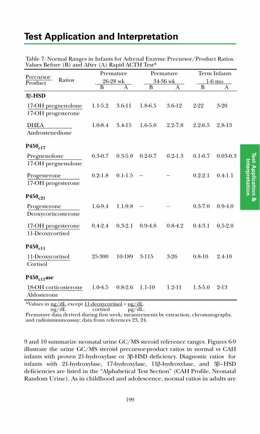
Test Application and Interpretation
Test A
pp
licatio
n &
In
terp
reta
tion
9 and 10 summarize neonatal urine GC/MS steroid reference ranges. Figures 6-9illustrate the urine GC/MS steroid precursor-product ratios in normal vs CAHinfants with proven 21-hydroxylase or 3β-HSD deficiency. Diagnostic ratios forinfants with 21-hydroxylase, 17-hydroxylase, 11β-hydroxylase, and 3β−HSDdeficiencies are listed in the “Alphabetical Test Section” (CAH Profile, NeonatalRandom Urine). As in childhood and adolescence, normal ratios in adults are
Table 7. Normal Ranges in Infants for Adrenal Enzyme Precursor/Product Ratios. Values Before (B) and After (A) Rapid ACTH Test*
PrecursorProduct
Premature Premature Term Infants26-28 wk 34-36 wk 1-6 mo
B A B A B A3β-HSD
17-OH pregnenolone 1.1-5.2 3.6-11 1.8-6.5 3.6-12 2-22 3-2017-OH progesterone
DHEA 1.0-8.4 3.4-15 1.6-5.0 2.2-7.8 2.2-6.5 2.8-13Androstenedione
P450c17
Pregnenolone 0.3-0.7 0.3-5.0 0.2-0.7 0.2-1.3 0.1-0.7 0.03-0.317-OH pregnenolone
Progesterone 0.2-1.8 0.1-1.5 − − 0.2-2.1 0.4-1.117-OH progesterone
P450c21
Progesterone 1.6-9.4 1.1-9.8 − − 0.3-7.0 0.9-4.0Deoxycorticosterone
17-OH progesterone 0.4-2.4 0.3-2.1 0.9-4.8 0.8-4.2 0.4-3.1 0.5-2.011-Deoxycortisol
P450c11
11-Deoxycortisol 25-300 10-189 3-115 3-26 0.8-10 2.4-10Cortisol
P450c11ase
18-OH corticosterone 1.0-4.5 0.8-2.6 1.1-10 1.2-11 1.3-5.0 2-13Aldosterone
*Values in ng/dL except 11-deoxycortisol = ng/dL ng/dL cortisol μg/dL.Premature data derived during first week; measurements by extraction, chromatography, and radioimmunoassay; data from references 23, 24.
Ratios
199
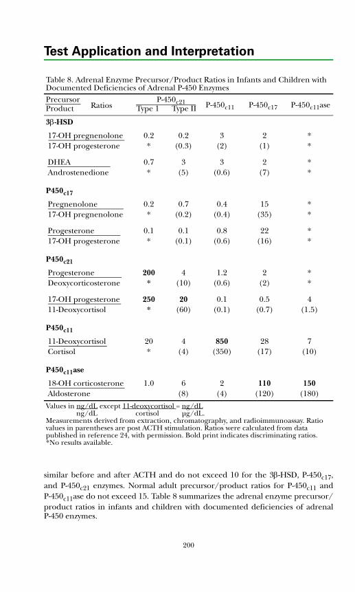
Test Application and Interpretation
similar before and after ACTH and do not exceed 10 for the 3β-HSD, P-450c17,and P-450c21 enzymes. Normal adult precursor/product ratios for P-450c11 andP-450c11ase do not exceed 15. Table 8 summarizes the adrenal enzyme precursor/product ratios in infants and children with documented deficiencies of adrenalP-450 enzymes.
Table 8. Adrenal Enzyme Precursor/Product Ratios in Infants and Children with Documented Deficiencies of Adrenal P-450 EnzymesPrecursor P-450c21 P-450c11 P-450c17 P-450c11aseProduct Type 1 Type II
3β-HSD
17-OH pregnenolone 0.2 0.2 3 2 *17-OH progesterone * (0.3) (2) (1) *
DHEA 0.7 3 3 2 *Androstenedione * (5) (0.6) (7) *
P450c17
Pregnenolone 0.2 0.7 0.4 15 *17-OH pregnenolone * (0.2) (0.4) (35) *
Progesterone 0.1 0.1 0.8 22 *17-OH progesterone * (0.1) (0.6) (16) *
P450c21
Progesterone 200 4 1.2 2 *Deoxycorticosterone * (10) (0.6) (2) *
17-OH progesterone 250 20 0.1 0.5 411-Deoxycortisol * (60) (0.1) (0.7) (1.5)
P450c11
11-Deoxycortisol 20 4 850 28 7Cortisol * (4) (350) (17) (10)
P450c11ase
18-OH corticosterone 1.0 6 2 110 150Aldosterone (8) (4) (120) (180)
Values in ng/dL except 11-deoxycortisol = ng/dL ng/dL cortisol μg/dL.Measurements derived from extraction, chromatography, and radioimmunoassay. Ratio values in parentheses are post ACTH stimulation. Ratios were calculated from data published in reference 24, with permission. Bold print indicates discriminating ratios. *No results available.
Ratios
200
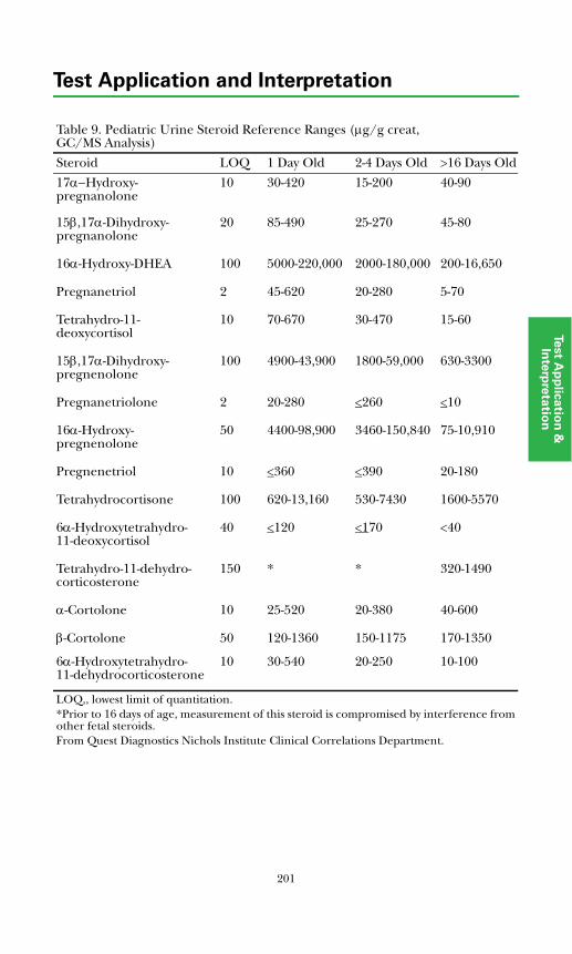
Test Application and Interpretation
Test A
pp
licatio
n &
In
terp
reta
tion
Table 9. Pediatric Urine Steroid Reference Ranges (μg/g creat, GC/MS Analysis)
Steroid LOQ 1 Day Old 2-4 Days Old >16 Days Old
17α−Hydroxy-pregnanolone
10 30-420 15-200 40-90
15β,17α-Dihydroxy-pregnanolone
20 85-490 25-270 45-80
16α-Hydroxy-DHEA 100 5000-220,000 2000-180,000 200-16,650
Pregnanetriol 2 45-620 20-280 5-70
Tetrahydro-11-deoxycortisol
10 70-670 30-470 15-60
15β,17α-Dihydroxy-pregnenolone
100 4900-43,900 1800-59,000 630-3300
Pregnanetriolone 2 20-280 <260 <10
16α-Hydroxy-pregnenolone
50 4400-98,900 3460-150,840 75-10,910
Pregnenetriol 10 <360 <390 20-180
Tetrahydrocortisone 100 620-13,160 530-7430 1600-5570
6α-Hydroxytetrahydro-11-deoxycortisol
40 <120 <170 <40
Tetrahydro-11-dehydro-corticosterone
150 * * 320-1490
α-Cortolone 10 25-520 20-380 40-600
β-Cortolone 50 120-1360 150-1175 170-1350
6α-Hydroxytetrahydro-11-dehydrocorticosterone
10 30-540 20-250 10-100
LOQ,, lowest limit of quantitation. *Prior to 16 days of age, measurement of this steroid is compromised by interference from other fetal steroids. From Quest Diagnostics Nichols Institute Clinical Correlations Department.
201
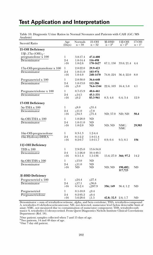
Test Application and Interpretation
Table 10. Diagnostic Urine Ratios in Normal Neonates and Patients with CAH (GC/MS Analysis)
Steroid Ratio Age(Days)
Normalsn = 59
21-OHn = 32
3β-HSDn = 2a
11β-OHn = 2b
17-OHn = 1c
21-OH Deficiency15β-,17α-(OH)2 -pregnanolone x 100 1 3.8-17.4 47.4-400Denominator 2-4
>161.0-14.41.0-2.6
116-498170-1627 67.1; 150 33.6; 21.4 4.4
17α-OH-pregnanolone x 100 1 2.0-22.0 29.9-423Denominator 2-4
>161.0-11.01.0-4.0
103-974248-1470 74.8; 224 36.4; 22.0 8.0
Pregnanetriol x 100 1 2.0-30.0 36.6-648Denominator 2-4
>161.0-13.0<3.0
112-28676.6-1344 22.6; 103 16.4; 5.8 4.1
Pregnanetriolone x 100 1 0.7-15.9 40.6-461Denominator 2-4
>16<14.5<0.2
45.1-33183.3-961 8.3; 4.6 6.4; 3.4 12.9
17-OH Deficiency5α-THA x 100 1 <8.0 <55.4Denominator 2-4
>16<11.0<34.3
<7.8<75.4 ND; 57.0 ND; ND 50.4
6α-OH-THA x 100 1 1.0-28.0 NDDenominator 2-4
>161.0-11.01.0-2.0
NDND ND; ND NMC;
NMC29,983
16α-OH-pregnenolone 1 0.3-1.3 1.2-4.416α-Hydroxy-DHEA 2-4
>160.1-2.20.2-0.7
1.9-11.41.0-11.7 0.9; 0.4 9.5; 8.1 156
11β-OH DeficiencyTHS x 100 1 2.9-25.0 13.6-34.0Denominator 2-4
>161.1-26.00.5-1.4
10.4-49.11.5-136 15.6; 27.0 366; 97.2 14.2
6α-OH-THS x 100 1 <13.0 NDDenominator 2-4
>16<11.0ND
NDND ND; ND 190,482;
117,723ND
3β-HSD DeficiencyPregnenetriol x 100 1 <24.4 <27.4Denominator 2-4
>16<17.10.5-2.4
<36.6<287.9 356; 149 36.4; 1.2 ND
Pregnenetriol 1 0.1-10.8 <0.4Pregnanetriolone 2-4
>160.2-95.25.6-28.6
<0.4<1.5 42.8; 32.5 2.6; 1.7 ND
Denominator = sum of tetrahydrocortisone, alpha, and beta cortolone; THA, tetrahydro-compound A, tetrahydro-11-dehydrocorticosterone; ND, not detected, numerator level below detectable limit of assay; NMC, not measured due to contamination of numerator component; THS, tetrahydro-sub-stance S, tetrahydro-11-deoxycortisol. From Quest Diagnostics Nichols Institute Clinical Correlations Department (Ref. 18).aOne patient; samples collected when 7 and 15 days of age.bTwo patients, 14 and 49 days of age.cOne 7 day old patient.
202
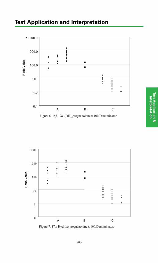
203
Test Application and Interpretation
Test A
pp
licatio
n &
In
terp
reta
tion
A B CFigure 9 . 1 5β ,1 7α-(OH)2pregnanolone x 10 0 /Denominator. A, 2 1-hydroxylase; B, 3β-HSD; C , normal samples.
0.1
1.0
10.0
100.0
1000.0
10000.0
Rat
io V
alue
A B CFigure 9 . 1 5β ,1 7α-(OH)2pregnanolone x 10 0 /Denominator. A, 2 1-hydroxylase; B, 3β-HSD; C , normal samples.
0.1
1.0
10.0
100.0
1000.0
10000.0
Rat
io V
alue
Figure 6. 15β,17α-(OH)2pregnanolone x 100/Denominator.
0
1
10
100
1000
10000
Rat
io V
alue
A B CFigure 6 . 1 7α -Hydroxypregnanolone x 1 0 0/D enominator. A , 21-hydroxylase; B , 3β -H SD; C , normal samples.
0
1
10
100
1000
10000
Rat
io V
alue
A B CFigure 6 . 1 7α -Hydroxypregnanolone x 1 0 0/D enominator. A , 21-hydroxylase; B , 3β -H SD; C , normal samples.Figure 7. 17α-Hydroxypregnanolone x 100/Denominator.
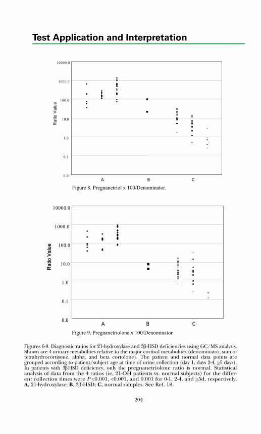
Test Application and Interpretation
Figures 6-9. Diagnostic ratios for 21-hydroxylase and 3β-HSD deficiencies using GC/MS analysis.Shown are 4 urinary metabolites relative to the major cortisol metabolites (denominator, sum oftetrahydrocortisone, alpha, and beta cortolone). The patient and normal data points aregrouped according to patient/subject age at time of urine collection (day 1, days 2-4, >5 days).In patients with 3βHSD deficiency, only the pregnanetriolone ratio is normal. Statisticalanalysis of data from the 4 ratios (ie, 21-OH patients vs. normal subjects) for the differ-ent collection times were P <0.001, <0.001, and 0.001 for 0-1, 2-4, and >5d, respectively.A, 21-hydroxylase; B, 3β-HSD; C, normal samples. See Ref. 18.
0.0
0.1
1.0
10.0
100.0
1000.0
10000.0
Rat
io V
alue
A B C
Figure 7 . P regnane triol x 1 0 0 /Denomina tor. A , 2 1 -hydroxylase; B , 3β -HSD ; C , normal samples.
Figure 8. Pregnanetriol x 100/Denominator.
A B CFigure 8. Pregnanetriolone x 100/D enominator. A , 21-hydroxylase; B , 3β-H SD; C , normal samples.
0.0
0.1
1.0
10.0
100.0
1000.0
10000.0
Rat
io V
alue
A B CFigure 8. Pregnanetriolone x 100/D enominator. A , 21-hydroxylase; B , 3β-H SD; C , normal samples.
0.0
0.1
1.0
10.0
100.0
1000.0
10000.0
Rat
io V
alue
Figure 9. Pregnanetriolone x 100/Denominator.
204
205
Test Application and Interpretation
Test A
pp
licatio
n &
In
terp
reta
tion
Nonclassical Disease (Partial Deficiency in Steroid Biosynthesis)
Nonclassical, late-onset, partial, or attenuated forms of P-450c21, 3β-HSD, andP-450c11 adrenal biosynthetic enzyme deficiencies are more common than severedeficiencies producing CAH. The overall prevalence of CAH approximates 1 in8,000 births but is higher in the Eskimo and Jewish populations. The prevalenceof nonclassical P-450c21 deficiency varies from 1 in 27 Ashkenazi Jews to 1 in 1,000in other Caucasians. Nonclassical 3β-HSD and P-450c11 deficiencies are much lesscommon, but precise prevalence data are not available. Among Caucasianhyperandrogenic women, nonclassical P-450c21 deficiency affects 1% to 2% whilenonclassical 3β-HSD deficiency and P-450c11 deficiency probably each accountfor less than 1%. P-450c21 gene mutations have been characterized in patientswith nonclassical P-450c21 enzyme deficiency, but the genetic defects for the3β-HSD and P-450c11 varieties have not been demonstrated. It has been proposedthat most hirsute women suspected to have nonclassical 3β-HSD enzymedeficiency have, instead, a nonspecific dysregulation of P-450c17 involving theadrenal glands and ovaries.
The common clinical manifestations of nonclassical disease include hirsutismand oligomenorrhea, which result from the excess androgen secretion.Androgenic symptoms usually are mild; clitoromegaly, male habitus, andtemporal baldness are infrequent manifestations. Diagnosis is based on elevatedprecursor and/or precursor-to-product ratios, and detection usually requiresACTH stimulation. Nonclassical disease is confirmed if the discriminatingprecursor/product ratio is clearly elevated. Stimulated ratios usually exceed 40 innonclassical P-450c21 deficiency patients. In postpubertal females, a morningfollicular phase serum 17-hydroxyprogesterone level <200 ng/dL excludes adiagnosis of nonclassical P-450c21 enzyme deficiency. ACTH stimulation testingis recommended only when the 8 AM fasting serum 17-hydroxyprogesterone levelexceeds this level. Nonclassical P-450c21 deficiency is diagnosed by a post-ACTH17-hydroxyprogesterone level >1,000 ng/dL, a value 4 times the upper normallimit (Figure 3). For the diagnosis of nonclassical 3β-HSD in postpubertalsubjects, it is recommended that the post-ACTH 17-hydroxypregnenolonelevel exceed 1,500 ng/dL, a value 3 SD above the mean of control subjects(Figure 3), and the 17-hydroxypregnenolone/17-hydroxyprogesterone or DHEA/androstenedione ratio should exceed 10, a value exceeding the approximate 3SD value of control pubertal subjects (Table 6). For the diagnosis of nonclassicalP-450c11 deficiency in postpubertal patients, the 11-deoxycortisol response toACTH should exceed 350 ng/dL and the 11-deoxycortisol/cortisol ratio shouldexceed 15, values approximating 3 SD above the mean for normal subjects(Tables 5 and 6).
Test Application and Interpretation
Cushing’s Syndrome
Cushing’s syndrome, the clinical manifestations of adrenal cortical hormoneexcess, can be the result of excessive pituitary or ectopic ACTH production,autonomous adrenal cortical steroid secretion, or exogenous glucocorticoidmedication (Table 11). Endogenous Cushing’s syndrome is associated withincreased 24-hour excretion of urinary cortisol, while administration of syntheticglucocorticoids (eg, prednisone, dexamethasone) is typically accompanied bydecreased cortisol excretion, since the adrenal axis is suppressed (Table 12).
If endogenous cortisol hypersecretion is suspected, the simplest approach todiagnosis is collection of a 24-hour urine sample for measurement of free cortisol(with creatinine to assess the completeness of collection). If excessive urine freecortisol is confirmed on two 24-hour urine collections, Cushing's syndrome isusually present, particularly if the value is >200 μg/24-h. Depression, alcoholabuse, and other stresses can also produce lesser degrees of excessive urine freecortisol excretion (pseudo-Cushing's). Alternative approaches to confirmhypercortisolism are to 1) determine the suppressibility of serum cortisol usingthe overnight dexamethasone suppression test (see "Dynamic Test Applicationand Interpretation" section), or 2) measure the serum or salivary cortisol level at11 PM to detect a disrupted circadian rhythm.
Once endogenous hypercortisolism is established, differentiating the cause aspituitary tumor, ectopic neoplasm, or adrenal tumor can be accomplished byplasma ACTH measurement, dynamic testing, and imaging procedures (Table11). In patients with ACTH-secreting pituitary adenomas (Cushing’s disease),plasma ACTH is typically 30 to 150 pg/mL. An exception is Nelson syndrome, inwhich previous adrenalectomy is associated with increased production of ACTHand growth of a pituitary corticotropinoma. The ectopic ACTH syndrome isoften associated with very high plasma ACTH levels, although these may overlapthe range of ACTH-secreting pituitary adenomas; ectopic ACTH production isalso characterized by secretion of "big" molecular species of ACTH and, rarely, byco-secretion of corticotropin releasing hormone (CRH). Autonomous adrenalcortisol production by a benign or malignant neoplasm is likely if plasma ACTH Table 11. Differential Diagnosis of Cushing’s Syndrome
Pituitary dependentACTH adenomaAdrenotroph hyperplasiaIntermittent Cushing’s
Adrenal diseaseAdenomaCarcinomaMacronodular diseaseMicronodular dysplasiaOther (McCune Albright syndrome, Carney syndrome)
Ectopic (cancer-associated) Cushing’sEctopic ACTHEctopic CRH
Pseudo-Cushing’s (alcohol-related)Exogenous glucocorticoid administration (eg, prednisone)
206
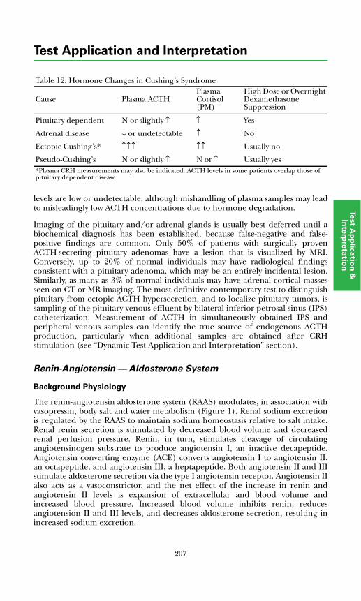
Test Application and Interpretation
Test A
pp
licatio
n &
In
terp
reta
tion
levels are low or undetectable, although mishandling of plasma samples may leadto misleadingly low ACTH concentrations due to hormone degradation.
Imaging of the pituitary and/or adrenal glands is usually best deferred until abiochemical diagnosis has been established, because false-negative and false-positive findings are common. Only 50% of patients with surgically provenACTH-secreting pituitary adenomas have a lesion that is visualized by MRI.Conversely, up to 20% of normal individuals may have radiological findingsconsistent with a pituitary adenoma, which may be an entirely incidental lesion.Similarly, as many as 3% of normal individuals may have adrenal cortical massesseen on CT or MR imaging. The most definitive contemporary test to distinguishpituitary from ectopic ACTH hypersecretion, and to localize pituitary tumors, issampling of the pituitary venous effluent by bilateral inferior petrosal sinus (IPS)catheterization. Measurement of ACTH in simultaneously obtained IPS andperipheral venous samples can identify the true source of endogenous ACTHproduction, particularly when additional samples are obtained after CRHstimulation (see “Dynamic Test Application and Interpretation” section).
Renin-Angiotensin ⎯ Aldosterone System
Background Physiology
The renin-angiotensin aldosterone system (RAAS) modulates, in association withvasopressin, body salt and water metabolism (Figure 1). Renal sodium excretionis regulated by the RAAS to maintain sodium homeostasis relative to salt intake.Renal renin secretion is stimulated by decreased blood volume and decreasedrenal perfusion pressure. Renin, in turn, stimulates cleavage of circulatingangiotensinogen substrate to produce angiotensin I, an inactive decapeptide.Angiotensin converting enzyme (ACE) converts angiotensin I to angiotensin II,an octapeptide, and angiotensin III, a heptapeptide. Both angiotensin II and IIIstimulate aldosterone secretion via the type I angiotensin receptor. Angiotensin IIalso acts as a vasoconstrictor, and the net effect of the increase in renin andangiotensin II levels is expansion of extracellular and blood volume andincreased blood pressure. Increased blood volume inhibits renin, reducesangiotension II and III levels, and decreases aldosterone secretion, resulting inincreased sodium excretion.
Table 12. Hormone Changes in Cushing’s Syndrome
Cause Plasma ACTHPlasma Cortisol (PM)
High Dose or Overnight Dexamethasone Suppression
Pituitary-dependent N or slightly ↑ ↑ Yes
Adrenal disease ↓ or undetectable ↑ No
Ectopic Cushing’s* ↑↑↑ ↑↑ Usually no
Pseudo-Cushing’s N or slightly ↑ N or ↑ Usually yes*Plasma CRH measurements may also be indicated. ACTH levels in some patients overlap those of pituitary dependent disease.
207
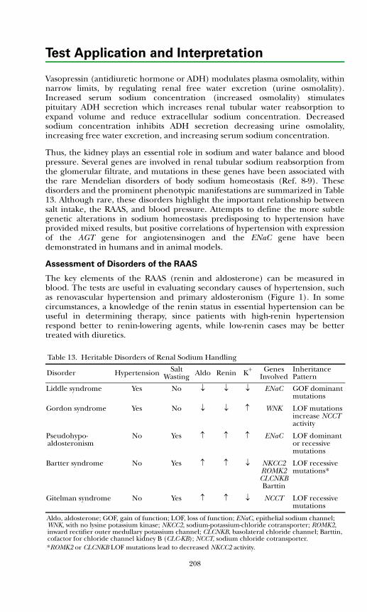
Test Application and Interpretation
Vasopressin (antidiuretic hormone or ADH) modulates plasma osmolality, withinnarrow limits, by regulating renal free water excretion (urine osmolality).Increased serum sodium concentration (increased osmolality) stimulatespituitary ADH secretion which increases renal tubular water reabsorption toexpand volume and reduce extracellular sodium concentration. Decreasedsodium concentration inhibits ADH secretion decreasing urine osmolality,increasing free water excretion, and increasing serum sodium concentration.
Thus, the kidney plays an essential role in sodium and water balance and bloodpressure. Several genes are involved in renal tubular sodium reabsorption fromthe glomerular filtrate, and mutations in these genes have been associated withthe rare Mendelian disorders of body sodium homeostasis (Ref. 8-9). Thesedisorders and the prominent phenotypic manifestations are summarized in Table13. Although rare, these disorders highlight the important relationship betweensalt intake, the RAAS, and blood pressure. Attempts to define the more subtlegenetic alterations in sodium homeostasis predisposing to hypertension haveprovided mixed results, but positive correlations of hypertension with expressionof the AGT gene for angiotensinogen and the ENaC gene have beendemonstrated in humans and in animal models.
Assessment of Disorders of the RAAS
The key elements of the RAAS (renin and aldosterone) can be measured inblood. The tests are useful in evaluating secondary causes of hypertension, suchas renovascular hypertension and primary aldosteronism (Figure 1). In somecircumstances, a knowledge of the renin status in essential hypertension can beuseful in determining therapy, since patients with high-renin hypertensionrespond better to renin-lowering agents, while low-renin cases may be bettertreated with diuretics.
Table 13. Heritable Disorders of Renal Sodium Handling
Disorder Hypertension Salt Wasting Aldo Renin K+ Genes
InvolvedInheritance Pattern
Liddle syndrome Yes No ↓ ↓ ↓ ENaC GOF dominant mutations
Gordon syndrome Yes No ↓ ↓ ↑ WNK LOF mutations increase NCCT activity
Pseudohypo-aldosteronism
No Yes ↑ ↑ ↑ ENaC LOF dominant or recessive mutations
Bartter syndrome No Yes ↑ ↑ ↓ NKCC2 ROMK2 CLCNKB Barttin
LOF recessive mutations*
Gitelman syndrome No Yes ↑ ↑ ↓ NCCT LOF recessive mutations
Aldo, aldosterone; GOF, gain of function; LOF, loss of function; ENaC, epithelial sodium channel; WNK, with no lysine potassium kinase; NKCC2, sodium-potassium-chloride cotransporter; ROMK2, inward rectifier outer medullary potassium channel; CLCNKB, basolateral chloride channel; Barttin, cofactor for chloride channel kidney B (CLC-KB); NCCT, sodium chloride cotransporter.*ROMK2 or CLCNKB LOF mutations lead to decreased NKCC2 activity.
208
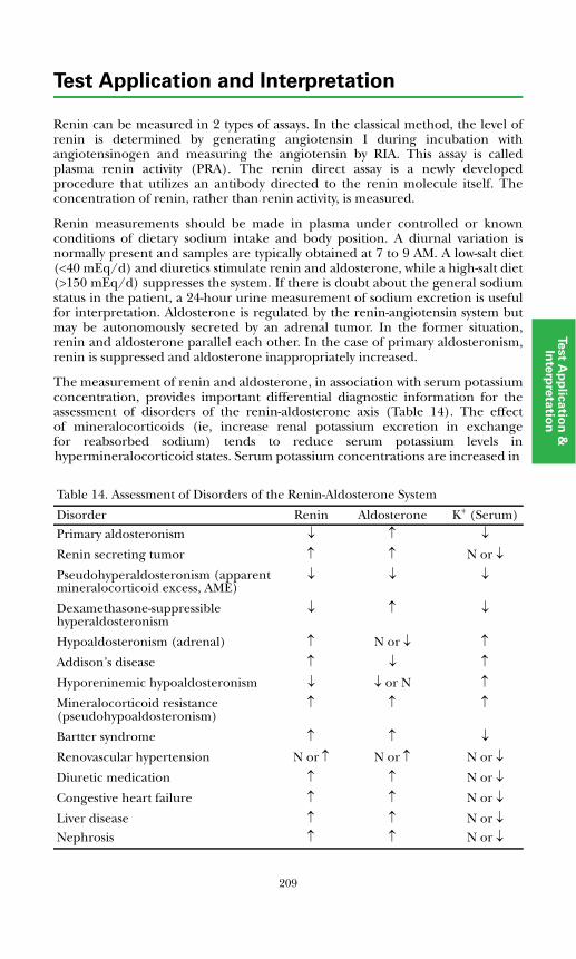
Test Application and Interpretation
Test A
pp
licatio
n &
In
terp
reta
tion
Renin can be measured in 2 types of assays. In the classical method, the level ofrenin is determined by generating angiotensin I during incubation withangiotensinogen and measuring the angiotensin by RIA. This assay is calledplasma renin activity (PRA). The renin direct assay is a newly developedprocedure that utilizes an antibody directed to the renin molecule itself. Theconcentration of renin, rather than renin activity, is measured.
Renin measurements should be made in plasma under controlled or knownconditions of dietary sodium intake and body position. A diurnal variation isnormally present and samples are typically obtained at 7 to 9 AM. A low-salt diet(<40 mEq/d) and diuretics stimulate renin and aldosterone, while a high-salt diet(>150 mEq/d) suppresses the system. If there is doubt about the general sodiumstatus in the patient, a 24-hour urine measurement of sodium excretion is usefulfor interpretation. Aldosterone is regulated by the renin-angiotensin system butmay be autonomously secreted by an adrenal tumor. In the former situation,renin and aldosterone parallel each other. In the case of primary aldosteronism,renin is suppressed and aldosterone inappropriately increased.
The measurement of renin and aldosterone, in association with serum potassiumconcentration, provides important differential diagnostic information for theassessment of disorders of the renin-aldosterone axis (Table 14). The effectof mineralocorticoids (ie, increase renal potassium excretion in exchangefor reabsorbed sodium) tends to reduce serum potassium levels inthypermineralocorticoid states. Serum potassium concentrations are increased in he
Table 14. Assessment of Disorders of the Renin-Aldosterone System
Disorder Renin Aldosterone K+ (Serum)Primary aldosteronism ↓ ↑ ↓
Renin secreting tumor ↑ ↑ N or ↓
Pseudohyperaldosteronism (apparent mineralocorticoid excess, AME)
↓ ↓ ↓
Dexamethasone-suppressible hyperaldosteronism
↓ ↑ ↓
Hypoaldosteronism (adrenal) ↑ N or ↓ ↑
Addison’s disease ↑ ↓ ↑
Hyporeninemic hypoaldosteronism ↓ ↓ or N ↑
Mineralocorticoid resistance (pseudohypoaldosteronism)
↑ ↑ ↑
Bartter syndrome ↑ ↑ ↓
Renovascular hypertension N or ↑ N or ↑ N or ↓
Diuretic medication ↑ ↑ N or ↓
Congestive heart failure ↑ ↑ N or ↓
Liver disease ↑ ↑ N or ↓Nephrosis ↑ ↑ N or ↓
209
Test Application and Interpretation
states of deficient mineralocorticoid production or action. Patients withsecondary hyperaldosteronism caused by congestive heart failure, liver disease, ornephrosis may manifest hypokalemia, but these states are clinically evident. InBartter syndrome, hyperaldosteronism is caused by juxtaglomerular hyperplasiaand is associated with hypokalemic alkalosis in the absence of hypertension oredema.
Primary Aldosteronism
Primary aldosteronism is an uncommon, but usually curable, cause ofhypertension. Since aldosterone is the major regulator of potassium balance,hyperaldosteronism is associated with potassium wasting. One marker of thedisorder is hypertension with unexplained hypokalemia (and alkalosis).Screening hypertensive patients for hypokalemia should be accomplished priorto the use of antihypertensive therapy, especially diuretics. Hypokalemia per se isnormally associated with low aldosterone levels, so the combination of aninappropriate serum aldosterone (>15 ng/dL) and a low renin (<0.2 ng/dL) isdiagnostic in the presence of hypokalemia. An elevated (>20) aldosterone/PRAratio, or an aldosterone/direct renin ratio >5, also is useful in the diagnosis ofprimary aldosteronism (see Ref. 20). Two maneuvers are sometimes useful toconfirm the diagnosis. An infusion of saline or administration ofmineralocorticoid (Florinef®’ 0.4 mg/d x 2 days) will in each case reduce plasmaaldosterone to <5 ng/dL.
Two etiologies are recognized. Conn syndrome is caused by an aldosterone-secreting adrenal adenoma and is characterized by elevated levels of theprecursor 18-hydroxycorticosterone with values >50 ng/dL. Less often (20%),the idiopathic form is caused by unexplained bilateral hypersecretion ofaldosterone, in which case precursor levels are normal. The diagnosis ofsuspected primary hyperaldosteronism in patients with hypertension andhypokalemia requires measurements of serum and urine aldosterone and plasmarenin (or renin activity) concentrations. 18-Hydroxycorticosterone and18-hydroxycortisol levels in serum and urine are increased in patients withadrenal adenoma, and levels may be useful to differentiate adenoma fromidiopathic hyperplasia. Serum 18-hydroxycortisol levels in >300 ng/dLor 18-hydroxycorticosterone levels >100 ng/dL are suggestive of adrenaladenoma. Diagnosis in some cases may be enhanced by aldosterone suppressiontesting using saline or fluorohydrocortisone (see “Dynamic Test Application andInterpretation” section). CT or MRI scans identify most adrenal tumors. Rarely,bilateral adrenal vein catheterization is utilized to determine whether thedisorder is unilateral or bilateral. Recent reports emphasize the importance ofdiagnosing primary aldosteronism, since in addition to the hypertension,aldosterone excess leads to a cardiomyopathy and increased incidence of heartfailure.
Renovascular Hypertension
Renovascular hypertension is a relatively common cause of primary hypertensionand is more common in the elderly (atherosclerosis) and in a cohort of youngwomen (fibronodular). Hypokalemia is observed only in the more severe cases.Suspicion and either a family history (fibronodular) or sudden development ofhypertension in an older patient may be the only clues. Peripheral renin is
210
Test Application and Interpretation
Test A
pp
licatio
n &
In
terp
reta
tion
usually, but not invariably, increased. Aldosterone levels may be either normal orincreased. Vascular studies are the key to diagnosis. The best indicator of apositive response to vascular surgery is demonstration of increased renin or reninactivity levels (x2) from the ischemic kidney.
Dexamethasone-suppressible Hyperaldosteronism
Dexamethasone-suppressible hyperaldosteronism is a rare form of hypertensioncharacterized by high levels of aldosterone, suppressed renin (or PRA), and adominant mode of inheritance. Chronic ACTH administration stimulates acontinued rise in aldosterone concentration; dexamethasone administrationproduces a rapid and complete suppression of serum aldosterone. Urinaryexcretion of 18-hydroxycortisol is increased. The pathogenesis appears to beabnormal regulation of aldosterone secretion by ACTH, perhaps due tointergenic gene promotor sequence recombination such that ACTH simulatestranscription of the P-450c11 isozyme normally active in the zona glomerulosa.
Pseudohyperaldosteronism
Pseudohyperaldosteronism, a syndrome of apparent mineralocorticoid excess(AME), was first described in children with hypertension and hypokalemia. Inadults, the syndrome has been associated with licorice ingestion and Cushing’ssyndrome. The mechanism has recently been discovered. Adjacent to themineralocorticoid receptor is the enzyme 11β-hydroxysteroid dehydrogenase.This enzyme normally protects the aldosterone receptor from cortisol, convertingcortisol to cortisone. In AME or after heavy licorice ingestion, the enzyme isinactive and cortisol is free to act as a mineralocorticoid. In these patients, bothrenin and aldosterone are suppressed. Diagnosis can be confirmed by measuringurine cortisol and cortisone metabolites (THF/THE).
Adrenal Diagnosis via Urine GC/MS Metabolite Analysis
Gas chromatography/mass spectrometry (GC/MS) of urinary adrenal steroidsoffers a useful supplement to serum precursor/product analysis of adrenalsteroid enzymatic abnormalities. This approach allows comprehensiveprecursor/product and pattern analysis of adrenal steroid excretion using small-volume (5-mL) random urine samples. Production data can be estimated fromanalysis of 24-hour or other timed-interval urine collections. In newborn infants,urine can be collected in a clean diaper and squeezed out into a test tube.Urinary steroids are measured with the methods described by Shackleton orUlick, et al (Ref. 27, 28). The free and conjugated steroids are first extractedfrom urine, whereupon the glucuronides and sulfates are cleaved by enzymehydrolysis. The freed steroids are re-extracted, chemically derivatized to renderthem heat stable, and separated by fused-silica gas chromatography prior to massspectrometric quantitation. Table 15 summarizes the measured urinary steroidsand their abbreviations. Table 10 summarizes normal and abnormal urine steroidmetabolite precursor/product ratios in neonates, children, and adolescents.Normative data for urinary steroid metabolite excretion in adults are shown inTable 16. Table 17 summarizes normal and abnormal steroid precursor/productratios for the assessment of adrenal biosynthetic enzyme deficiencies inadolescents and adults.
211
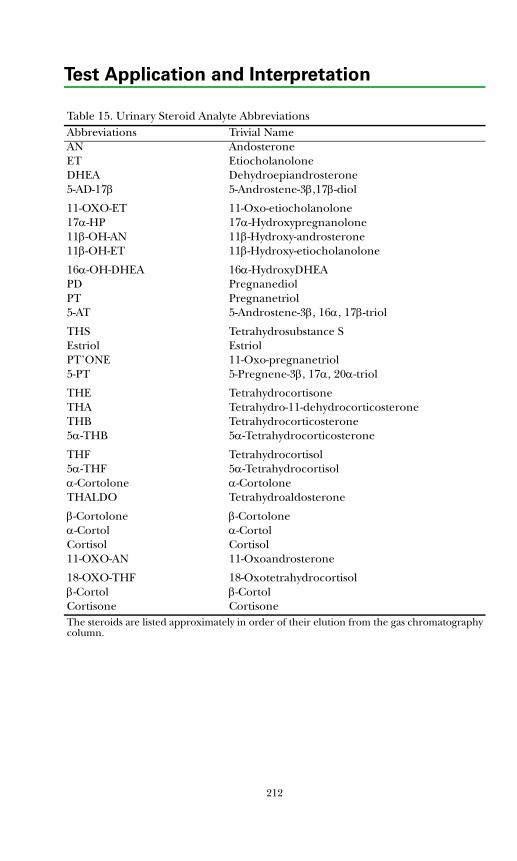
Test Application and Interpretation
Table 15. Urinary Steroid Analyte AbbreviationsAbbreviations Trivial NameAN AndosteroneET EtiocholanoloneDHEA Dehydroepiandrosterone5-AD-17β 5-Androstene-3β,17β-diol
11-OXO-ET 11-Oxo-etiocholanolone17α-HP 17α-Hydroxypregnanolone11β-OH-AN 11β-Hydroxy-androsterone11β-OH-ET 11β-Hydroxy-etiocholanolone
16α-OH-DHEA 16α-HydroxyDHEAPD PregnanediolPT Pregnanetriol5-AT 5-Androstene-3β, 16α, 17β-triol
THS Tetrahydrosubstance SEstriol EstriolPT’ONE 11-Oxo-pregnanetriol5-PT 5-Pregnene-3β, 17α, 20α-triol
THE TetrahydrocortisoneTHA Tetrahydro-11-dehydrocorticosteroneTHB Tetrahydrocorticosterone5α-THB 5α-Tetrahydrocorticosterone
THF Tetrahydrocortisol5α-THF 5α-Tetrahydrocortisolα-Cortolone α-CortoloneTHALDO Tetrahydroaldosterone
β-Cortolone β-Cortoloneα-Cortol α-CortolCortisol Cortisol11-OXO-AN 11-Oxoandrosterone
18-OXO-THF 18-Oxotetrahydrocortisolβ-Cortol β-CortolCortisone CortisoneThe steroids are listed approximately in order of their elution from the gas chromatography column.
212
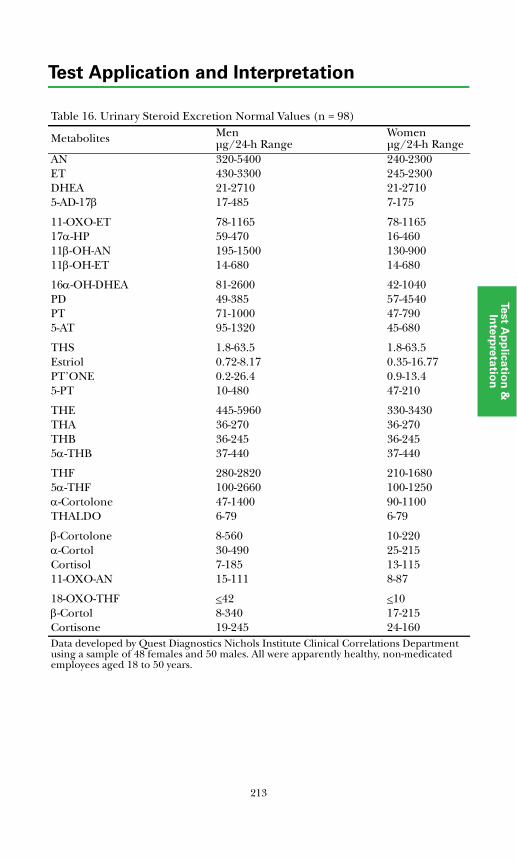
Test Application and Interpretation
Test A
pp
licatio
n &
In
terp
reta
tion
Table 16. Urinary Steroid Excretion Normal Values (n = 98)
Metabolites Menμg/24-h Range
Womenμg/24-h Range
AN 320-5400 240-2300ET 430-3300 245-2300DHEA 21-2710 21-27105-AD-17β 17-485 7-175
11-OXO-ET 78-1165 78-116517α-HP 59-470 16-46011β-OH-AN 195-1500 130-90011β-OH-ET 14-680 14-680
16α-OH-DHEA 81-2600 42-1040PD 49-385 57-4540PT 71-1000 47-7905-AT 95-1320 45-680
THS 1.8-63.5 1.8-63.5Estriol 0.72-8.17 0.35-16.77PT’ONE 0.2-26.4 0.9-13.45-PT 10-480 47-210
THE 445-5960 330-3430THA 36-270 36-270THB 36-245 36-2455α-THB 37-440 37-440
THF 280-2820 210-16805α-THF 100-2660 100-1250α-Cortolone 47-1400 90-1100THALDO 6-79 6-79
β-Cortolone 8-560 10-220α-Cortol 30-490 25-215Cortisol 7-185 13-11511-OXO-AN 15-111 8-87
18-OXO-THF <42 <10β-Cortol 8-340 17-215Cortisone 19-245 24-160Data developed by Quest Diagnostics Nichols Institute Clinical Correlations Department using a sample of 48 females and 50 males. All were apparently healthy, non-medicated employees aged 18 to 50 years.
213
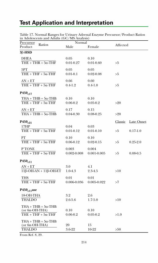
Test Application and Interpretation
Table 17. Normal Ranges for Urinary Adrenal Enzyme Precursor/Product Ratios in Adolescents and Adults (GC/MS Analysis) Precursor Normal
AffectedProduct Male Female
3β-HSD
DHEA 0.05 0.10THE + THR + 5α-THF 0.01-0.27 0.01-0.60 >5
5PT 0.05 0.05THE + THF + 5α-THF 0.01-0.1 0.02-0.08 >5
AN + ET 0.66 0.60THE + THF + 5α-THF 0.4-1.2 0.4-1.0 >5
P450c17
THA + THB + 5α-THB 0.10 0.10THE + THF + 5α-THF 0.06-0.2 0.05-0.2 >20
AN + ET 0.17 0.15THA + THB + 5α-THB 0.04-0.30 0.08-0.25 >20
P450c21 Classic Late Onset17HP 0.04 0.03THE + THF + 5α-THF 0.01-0.12 0.01-0.10 >5 0.17-1.0
PT 0.10 0.10THE + THF + 5α-THF 0.06-0.12 0.02-0.15 >5 0.25-2.0
P’TONE 0.003 0.004THE + THF + 5α-THF 0.002-0.008 0.001-0.005 >5 0.08-0.5
P450c11
AN + ET 3.0 4.111β-OH-AN + 11β-OH-ET 1.0-4.3 2.3-4.5 >10
THS 0.01 0.01THE + THF + 5α-THF 0.006-0.036 0.005-0.022 >7
P450c11ase
18-OH-THA 3.2 2.6THALDO 2.6-5.6 1.7-5.0 >10
THA + THB + 5α-THB(or 6α-OH-THA) 0.10 0.10THE + THF + 5α-THF 0.06-0.2 0.05-0.2 >1.0
THA + THB + 5α-THB (or 6α-OH-THA) 20 15THALDO 3.6-22 10-22 >50
From Ref. 8, 29.
Ratios
214
Test Application and Interpretation
Test A
pp
licatio
n &
In
terp
reta
tion
References
1. Stewart PM. The adrenal cortex. In: Larsen PR, Kronenberg HM, Melmed S,Polonsky KS, eds. Williams Textbook of Endocrinology. 10th ed. Philadelphia, PA:WB Saunders; 2003:491-551.
2. Clark AJL, Grossman AB. Adrenal insufficiency. In: DeGroot LJ, Jameson JL,eds. Endocrinology. 5th ed. Philadelphia, PA: Elsevier-Saunders; 2006:2343-2351.
3. Cavagnini F, Giraldi FP. Adrenal causes of hypercortisolism. In: DeGroot LJ,Jameson JL, eds. Endocrinology. 5th ed. Philadelphia, PA: Elsevier-Saunders;2006:2353-2386.
4. Ilias I, Torpy DJ, Pacak K, et al. Cushings syndrome due to ectopic cortico-tropin secretion: twenty years experience at the National Institutes of Health.J Clin Endocrinol Metab. 2005;90:4955-4962.
5. Malchoff DM, Malchoff CD. Generalized glucocorticoid resistance. In:DeGroot LJ, Jameson JL, eds. Endocrinology. 5th ed. Philadelphia, PA: Elsevier-Saunders; 2006:2387-2392.
6. Conton P, Maccino B, Montero F. Adrenal cancer. In: DeGroot LJ, JamesonJL, eds. Endocrinology. 5th ed. Philadelphia, PA: Elsevier-Saunders; 2006:2455-2460.
7. Dluhy RG, Lawrence JE, Williams GH. Endocrine hypertension. In: LarsenPR, Kronenberg HM, Melmed S, Polonsky KS, eds. Williams Textbook ofEndocrinology. 10th ed. Philadelphia, PA: WB Saunders; 2003:552-586.
8. O’Shaughnessy KM, Karet FE. Salt handling and hypertension. J Clin Invest.2004;113:1075-1081.
9. Jeunemaitre X. Renin-angiotensin-aldosterone system polymorphisms andessential hypertension: where are we? J Hypertens. 2003;21:2219-2222.
10. Kasperlik-Zaluska AA, Czarnocka B, Czech W. Autoimmunity as the mostfrequent cause of idiopathic, secondary adrenal insufficiency: report of 111cases. Autoimmunity. 2003;36:155-159.
11. Root AW, Shulman DI. Clinical adrenal disorders. In: Pescovitz OH, EugsterEA, eds. Pediatric Endocrinology, Philadelphia, PA: Lippincott Williams andWilkins, 2004:568-600.
12. Wajnrajch MP, New MI. Defects in adrenal steroidogenesis. In: DeGroot LJ,Jameson JL, eds. Endocrinology. 5th ed. Philadelphia, PA: Elsevier-Saunders;2006:2393-2416.
13. Speiser PW, White PC. Congenital adrenal hyperplasia. N Engl J Med.2003;349:776-788.
14. Clayton PE, Miller WL, Oberfield SE et al. Consensus statement on 21-hydroxylase deficiency from the European Society for PaediatricEndocrinology and the Lawson Wilkins Pediatric Endocrine Society. HormRes. 2002;58:188-195.
15. Olgemoller B, Roscher AA, Liebl B, et al. Screening for congenital adrenalhyperplasia: adjustment of 17-hydroxyprogesterone cut-off values to both ageand birth weight markedly improves the predictive value. J Clin EndocrinolMetab. 2003;88:5790-5794.
215
Test Application and Interpretation
16. Homma K, Hasegawa T, Nogai T, et al. Urine steroid profile analysis incytochrome P450 oxidoreductase deficiency: implication for the backdoorpathway to dihydrotestosterone. J Clin Endocrinol Metab. 2006;91:2643-2649.
17. New MI, Carlson A, Obeid J, et al. Update: prenatal diagnosis of congenitaladrenal hyperplasia in 595 pregnancies. Endocrinologist. 2003;13:233-239.
18. Caulfield MP, Lynn T, Gottschalk ME, et al. The diagnosis of congenitaladrenal hyperplasia in the newborn by gas chromatography/massspectrometry analysis of random urine specimens. J Clin Endocrinol Metab.2002;87:3682-3690.
19. New MI. Extensive clinical experience: nonclassical 21-hydroxylasedeficiency. J Clin Endocrinol Metab. 2006;91:4205-4214.
20. Mulatero P, Stowasser M, Loh KC, et al. Increased diagnosis of primaryaldosteronism, including surgically correctable forms, in centers from fivecontinents. J Clin Endocrinol Metab. 2004;89:1045-1050.
21. Lashansky G, Saenger P, Fishman K, et al. Normative data for adrenalsteroidogenesis in a healthy pediatric population: age- and sex-relatedchanges after adrenocorticotropin stimulation. J Clin Endocrinol Metab.1991;73:674-686.
22. Lashansky G, Saenger P, Dimartino-Nardi J, et al. Normative data for thesteroidogenic response of mineralocorticoids and their precursors toadrenocorticotropin in a healthy pediatric population. J Clin Endocrinol Metab.1992;75:1491-1496.
23. Hingre RV, Gross SJ, Hingre KS, et al. Adrenal steroidogenesis in very lowbirth weight preterm infants. J Clin Endocrinol Metab. 1994;78:266-270.
24. Adrenal steroid response to ACTH. Pediatrics, Endocrine Sciences,Calabasses Hills, CA. May 1991.
25. Viardot A, Huber P, Puder JJ, et al. Reproducibility of nighttime salivarycortisol and its use in the diagnosis of hypercortisolism compared withurinary free cortisol and overnight dexamethasone suppression test. J ClinEndocrinol Metab. 2005;90:5730-5736.
26. Agha A, Tomlinson JW, Clark PM, et al. The long-term predictive accuracy ofthe short synacthen (corticotropin) stimulation test for assessment of thehypothalamic-pituitary-adrenal axis. J Clin Endocrinol Metab. 2006;91:43-47.
27. Shackleton CH. Mass spectrometry in the diagnosis of steroid-relateddisorders and in hypertension research. J Steroid Biochem Mol Biol. 1993;45:127-140.
28. Ulick S, Chan CK, Wang JZ. Measurement of 4 urinary C-18 oxygenatedcorticosteroids by stable isotope dilution mass fragmentography. J SteroidBiochem Mol Biol. 1991;38:59-66.
29. Corvol P, Persu A, Gimenez-Roqueplo AP, et al. Seven lessons from twocandidate genes in human essential hypertension: angiotensinogen andepithelial sodium channel. Hypertension. 1999;33:1324-1331.
216
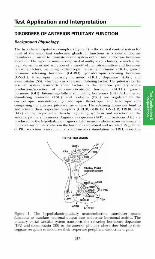
Test Application and Interpretation
Test A
pp
licatio
n &
In
terp
reta
tion
DISORDERS OF ANTERIOR PITUITARY FUNCTION
Background Physiology
The hypothalamic-pituitary complex (Figure 1) is the central control system formost of the important endocrine glands. It functions as a neuroendocrinetransducer in order to translate neural system output into endocrine hormonesecretion. The hypothalamus is comprised of multiple cell clusters, or nuclei, thatregulate synthesis and secretion of a variety of neurotransmitters and hormonereleasing factors, including corticotropin releasing hormone (CRH), growthhormone releasing hormone (GHRH), gonadotropin releasing hormone(GNRH), thyrotropin releasing hormone (TRH), dopamine \(DA), andsomatostatin (SS), which acts as a release inhibiting factor. The pituitary portalvascular system transports these factors to the anterior pituitary whereproduction/secretion of adrenocorticotropic hormone (ACTH), growthhormone (GH), luteinizing/follicle stimulating hormones (LH/FSH), thyroidstimulating hormone (TSH), and prolactin (PRL) are regulated by thecorticotropic, somatotropic, gonadotropic, thyrotropic, and lactotropic cellscomprising the anterior pituitary tissue mass. The releasing hormones bind toand activate their respective receptors (CRHR, GHRHR, GNRHR, TRHR, SSR,DAR) in the tropic cells, thereby regulating synthesis and secretion of theanterior pituitary hormones. Arginine vasopressin (AVP) and oxytocin (OT) areproduced by the hypothalamic magnocellular neurons whose axons terminate inthe posterior pituitary wherein the hormones are stored and secreted. Regulationof PRL secretion is more complex and involves stimulation by TRH, vasoactive
Figure 1. The hypothalamic-pituitary neuroendocrine transducer systemfunctions to translate neuronal output into endocrine hormonal activity. Thepituitary portal vascular system transports the releasing hormones dopamine(DA) and somatostatin (SS) to the anterior pituitary where they bind to theircognate receptors to modulate their respective peripheral endocrine organs.
217
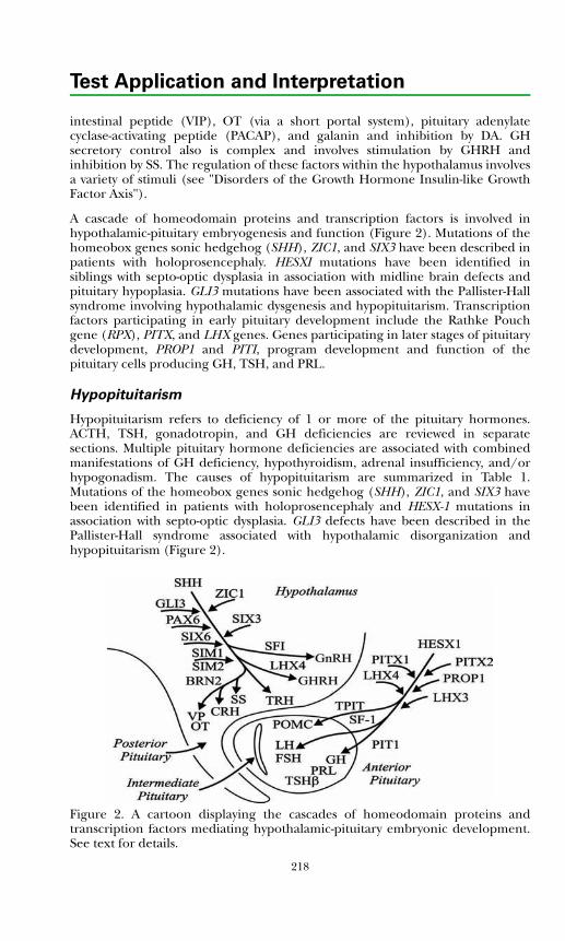
Test Application and Interpretation
intestinal peptide (VIP), OT (via a short portal system), pituitary adenylatecyclase-activating peptide (PACAP), and galanin and inhibition by DA. GHsecretory control also is complex and involves stimulation by GHRH andinhibition by SS. The regulation of these factors within the hypothalamus involvesa variety of stimuli (see "Disorders of the Growth Hormone Insulin-like GrowthFactor Axis").
A cascade of homeodomain proteins and transcription factors is involved inhypothalamic-pituitary embryogenesis and function (Figure 2). Mutations of thehomeobox genes sonic hedgehog (SHH), ZIC1, and SIX3 have been described inpatients with holoprosencephaly. HESXI mutations have been identified insiblings with septo-optic dysplasia in association with midline brain defects andpituitary hypoplasia. GLI3 mutations have been associated with the Pallister-Hallsyndrome involving hypothalamic dysgenesis and hypopituitarism. Transcriptionfactors participating in early pituitary development include the Rathke Pouchgene (RPX), PITX, and LHX genes. Genes participating in later stages of pituitarydevelopment, PROP1 and PITI, program development and function of thepituitary cells producing GH, TSH, and PRL.
Hypopituitarism
Hypopituitarism refers to deficiency of 1 or more of the pituitary hormones.ACTH, TSH, gonadotropin, and GH deficiencies are reviewed in separatesections. Multiple pituitary hormone deficiencies are associated with combinedmanifestations of GH deficiency, hypothyroidism, adrenal insufficiency, and/orhypogonadism. The causes of hypopituitarism are summarized in Table 1.Mutations of the homeobox genes sonic hedgehog (SHH), ZIC1, and SIX3 havebeen identified in patients with holoprosencephaly and HESX-1 mutations inassociation with septo-optic dysplasia. GLI3 defects have been described in thePallister-Hall syndrome associated with hypothalamic disorganization andhypopituitarism (Figure 2).
Figure 2. A cartoon displaying the cascades of homeodomain proteins andtranscription factors mediating hypothalamic-pituitary embryonic development.See text for details.
218
Test Application and Interpretation
Test A
pp
licatio
n &
In
terp
reta
tion
Mutation of the TRHR gene, the GHRHR gene, and the GNRHR gene has beenreported in association with isolated TSH, GH, and gonadotropin hormonedeficiencies, respectively (Figure 1). TRH and GHRH deficiencies are usuallydiagnosed during infancy and childhood because of their profound effect ongrowth. GNRH deficiency (Kallmann's syndrome) usually is not recognized priorto puberty. This deficiency can be due to mutations in the X-linked Kal-1 gene orthe autosomal Fgf1 receptor gene. Other forms of idiopathic hypogonadotropichypogonadism are due to mutations in the GP54 receptor gene which may play arole in the timing of puberty. This is discussed in more detail in the gonadalsection.
A variety of mutations in pituitary transcription factor genes and pituitaryhormone genes have been characterized in hypopituitary patients. Thephenotypic features of patients with pituitary transcription factor gene mutationsare summarized in Table 2 and are discussed further in related sections.
The pituitary hormones are easily measured in peripheral blood. Hypothalamichormones are not secreted into peripheral blood, but can be detected in bloodwhere they are derived from ectopic tissue sources. Quantification of selectedhypothalamic hormones in peripheral blood can be useful in the diagnosis ofpatients with excessive pituitary hormone secretion caused by ectopichypothalamic hormone production from extrapituitary tumors.
Table 1. Causes of HypopituitarismDevelopmental anomalies Vascular events
Basal encephalocele Postpartum (Sheehan necrosis)Holoprosencephaly Internal carotid aneurysmMedian facial anomalies Inflammatory diseasesSepto-optic dysplasia EncephalitisPituitary hypoplasia Meningitis, bacterial
Congenital hormone deficiencies Granulomatous diseaseHypothalamic Histiocytosis X
TRH Autoimmune hypophysitisGHRH Destructive lesionsLHRH (Kallmann’s syndrome) Intraventricular hemorrhage
Isolated pituitary hormone deficiencies SurgeryACTH RadiationTSH TraumaGH NeoplasiaLH, FSH Hypothalamic tumors
Multiple pituitary hormone deficiencies Pituitary adenomasPIT1 mutation Metastatic carcinomaPROP1 mutation PinealomaHESX1 mutation CraniopharyngiomaLHX3 and LHX4 mutation
219
Test Application and Interpretation
Pituitary Adenomas
General Features
Adenomas of the anterior pituitary gland are relatively common and are variablewith regard to their cell of origin and function. Incidental pituitary adenomashave been documented in up to 20% of adult humans at autopsy. Most of these donot produce signs or symptoms. The relative prevalence of the clinicallysignificant pituitary adenomas is summarized in Table 3. The pituitary adenomasare classified by their hormonal secretion(s), and most are characterized by theclinical effects of the excessive hormone production (Table 4). The most commonsecreting pituitary adenoma is the prolactin-secreting prolactinoma. The leastcommon of the functional varieties are the gonadotropinomas andthyrotropinomas. Most gonadotroph-derived adenomas, also referred to as achromophobe adenoma, are nonsecreting and present with compressivesymptoms associated with multiple hormone deficiencies, includinghypogonadism or mild elevations in prolactin. Most of these nonsecreting tumorsshow immunostaining for 1 or more hormones, including the alpha subunit, LHbeta, FSH beta, and, less frequently, TSH beta subunits, and ACTH. Some of thesetumors secrete alpha, LH beta, and FSH beta subunits which are detectable inserum and can be used as tumor markers. LH, FSH, and alpha subunit respondparadoxically to TRH stimulation. Thyrotrope secreting tumors lead tohyperthyroidism with an elevated or inappropriately normal TSH. The applicationof hormone assays to pituitary tumor characterization is summarized in Table 4.
Table 2. Phenotypic Features of Patients with Pituitary Trancription Factor Gene Mutations
Gene MutationsPROP1 PIT1 LHX3 LHX4 HESX1 TPIT
GH D A Α D D NProlactin D A A V Ν ΝTSH D D Α V V NLH/FSH D N Α N N NACTH V Ν Ν V Ν DPost pituitary N N N E E N
D, decreased; A, absent; N, normal; V, variable; E, ectopic.Modified from Parks JS, Brown MR. Ref. 7.
Table 3. Relative Prevalence (%) of Pituitary Adenomas*Prevalence (%)
Prolactinomas 29GH-producing 15GH + prolactin-producing 8Corticotroph adenomas 16Gonadotropinomas 7Thyrotropinomas 1Null cell adenomas 27
*From Melmed S, Kleinberg D (Ref 2).
220
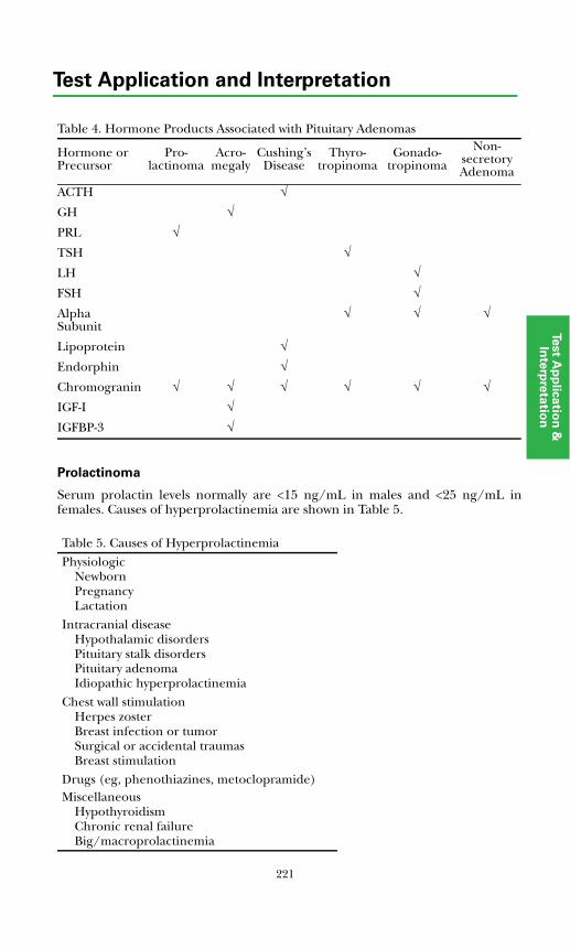
Test Application and Interpretation
Test A
pp
licatio
n &
In
terp
reta
tion
Prolactinoma
Serum prolactin levels normally are <15 ng/mL in males and <25 ng/mL infemales. Causes of hyperprolactinemia are shown in Table 5.
Table 4. Hormone Products Associated with Pituitary Adenomas
Hormone or Precursor
Pro-lactinoma
Acro-megaly
Cushing’sDisease
Thyro-tropinoma
Gonado-tropinoma
Non-secretory Adenoma
ACTH √
GH √
PRL √
TSH √
LH √
FSH √
Alpha Subunit
√ √ √
Lipoprotein √
Endorphin √
Chromogranin √ √ √ √ √ √
IGF-I √
IGFBP-3 √
Table 5. Causes of HyperprolactinemiaPhysiologic
NewbornPregnancyLactation
Intracranial diseaseHypothalamic disordersPituitary stalk disordersPituitary adenomaIdiopathic hyperprolactinemia
Chest wall stimulationHerpes zosterBreast infection or tumorSurgical or accidental traumasBreast stimulation
Drugs (eg, phenothiazines, metoclopramide)Miscellaneous
HypothyroidismChronic renal failureBig/macroprolactinemia
221
Test Application and Interpretation
A prolactinoma usually is associated with clinical evidence of gonadal dysfunctionand/or symptoms related to a pituitary mass effect. Galactorrhea is present in30% to 80% of women with prolactinomas but is uncommon in men. Basal bloodsamples are adequate to diagnose hyperprolactinemia. Stimulation testing usuallyis not required and does not aid in differential diagnosis of prolactinoma.Borderline hyperprolactinemia is best confirmed by measurements in singlesamples collected on 3 separate days or in 3 separate samples collected at30-minute intervals via indwelling catheter. Prolactin (PRL) is a stress hormone,and stress should be avoided when sampling blood. Postprandial and post-breastexamination sampling should also be avoided. Most prolactinomas are associatedwith serum PRL levels >100 ng/mL, while levels in drug-induced hyper-prolactinemia tend to be lower. Mild elevation of prolactin of <100 ng/mL is alsoseen in stalk compression due to pituitary or other parasellar masses. The stalkcompression relieves the lactotrophs from the normal inhibitory dopaminergictone. The diagnosis of prolactinoma is confirmed in patients without otherobvious causes of hyperprolactinemia by pituitary imaging techniques (Table 5).PRL exists in a variety of forms: the 26 kd monomer, the 40-60 kd dimeric or"big" prolactin, and the >100 kd macroprolactin, which is the monomercomplexed with IgG. Macroprolactin is biologically inactive but cross-reacts inseveral PRL immunoassays. Since the presence of macroprolactin may lead to anoverestimation of the biologically active form of PRL, macroprolactin should bespecifically measured in patients with apparent hyperprolactinemia that is notconsistent with the clinical presentation.
Acromegaly
Acromegaly with or without increased prolactin secretion accounts for some 15%of clinically significant pituitary adenomas. The prevalence ranges from 38 to 69cases per million population. Clinical features are related to sustained oversecretion of GH and IGF-I. The primary pituitary somatotroph adenomasgenerally are slow growing, and clinical manifestations develop insidiously overmany years. These include coarsening of the facial features, enlargement of thehands and feet, excessive sweating, amenorrhea, impotence, diabetes mellitus,hypertension, skin tags, and a variety of seemingly unrelated complaints, such asjaw malocclusion, carpal tunnel syndrome, colonic polyps, arthritis, and heartfailure. Rarely, hyperthyroidism due to increased TSH secretion may coexist.
The diagnosis is confirmed by demonstrating excessive GH secretion.Measurement of GH in random serum samples may be inadequate, since normalvalues vary during the day. In patients with suggestive clinical features and normalGH levels, the glucose suppression test may be useful. Patients with acromegalyfail to suppress serum GH to <1 ng/mL following ingestion of 75 g of glucose(see Dynamic Test section). Additional confirmation is obtained by measurementof serum IGF-I and IGFBP-3 levels. Both are GH-dependent proteins, and levelsusually are increased in patients with acromegaly. In patients with confirmedGH hypersecretion, pituitary imaging is conducted to identify a pituitary mass.In the absence of a pituitary tumor, an ectopic source of growth hormonereleasing hormone (GHRH) must be considered. Ectopic acromegaly usually canbe diagnosed by measuring serum GHRH levels in peripheral blood and by CT orMR imaging of the chest, abdomen, and/or pelvis. See the “Growth Hormone
222
Test Application and Interpretation
Test A
pp
licatio
n &
In
terp
reta
tion
Insulin-like Growth Factor Axis” section for further information. Treated patientsshould have lifelong monitoring of IGF-1 and GH suppression testing sincerecurrences are frequent and the recent broadening of therapeutic options withsomatostatin analogues and GH antagonists gives additional opportunities forintervention. Failure to suppress GH in response to glucose and persistently highIGF-1 levels are predictors of increased morbidity and mortality and thus shouldbe normalized if possible.
Cushing’s Disease
See section on “Disorders of Adrenal Function.”
Thyrotropinoma
TSH-secreting adenomas account for <1% of all pituitary tumors. They arecharacterized by hyperthyroidism and measurable (nonsuppressed) levels ofcirculating TSH. Such TSH-dependent hyperthyroidism must be distinguishedfrom other causes of inappropriate TSH secretion (ie, thyroid hormoneresistance syndromes). Co-secretion of TSH and prolactin, or GH, may occur, andalpha subunit levels may be increased. In most (88%) patients withthyrotropinoma, there is disproportionate secretion of alpha subunit and analpha subunit/TSH ratio >1.0; in non-tumorous hyperplasia the ratio does notexceed 1.0. The diagnosis is confirmed by pituitary imaging. Although thyroidhormone resistance and TSH secreting adenomas are 2 distinct clinical entities,an association of these tumors with thyroid hormone resistance or a thyroidhormone receptor defect has been described, suggesting a possible causalrelationship in some of these patients.
Gonadotropinoma
Previously, many macroadenomas now recognized by immunostaining to begonadotropin-producing were classified as nonsecreting or chromophobeadenomas, because they usually do not produce a recognizable clinicalsyndrome. Thus, the classification of gonadotropinomas versus chromophobeadenomas was variable. Gonadotroph adenomas constitute 80% to 90% of allnonfunctioning pituitary adenomas using molecular, cell culture, andimmunostaining techniques. Because of the minimal symptoms,gonadotropinomas are not detected early and present more commonly asmacroadenomas than microadenomas, often with visual or neurologicalsymptoms related to the mass effect. The tumors frequently produce increasedserum FSH levels but only uncommonly increased serum LH concentrations. Theincreased biologically active levels of FSH may lead to an ovarianhyperstimulation syndrome, and elevations of LH, FSH, or both may lead totesticular enlargement or precocious puberty. About 15% of patients evidenceincreased alpha subunit concentrations, and both intact gonadotropin andsubunit levels may increase (>30%) in response to TRH stimulation. The alphasubunit test can help differentiate primary hypogonadism from hypogonadismdue to a tumor secreting nonfunctional LH and FSH.
223
Test Application and Interpretation
A pituitary adenoma is likely to be of gonadotroph or thyrotroph origin if theserum prolactin level is <100 ng/mL, the patient has no acromegalic features, theserum IGF-I concentration is not increased, and there is no clinical or laboratoryevidence of Cushing's syndrome. In men, elevated basal levels of FSH, alphasubunit, and FSH and LH beta subunits, alone or in combination with responsesof these to TRH, are strong evidence for a gonadotroph adenoma. In women,measurements of FSH are not usually helpful unless the FSH concentration ismarkedly increased in the presence of a normal LH value. The response of intactFSH or LH levels and of the alpha and LH beta subunits to TRH may be helpful.
References
1. Cone RD, Low MJ, Elmquist JK, Cameron JL. Neuroendocrinology. In:Larsen PR, Kronenberg HM, Melmed S, Polonsky KS, eds. Williams Textbook ofEndocrinology. 10th ed. Philadelphia, PA: WB Saunders; 2003:81-176.
2. Melmed S, Kleinberg D. Anterior pituitary. In: Larsen PR, Kronenberg HM,Melmed S, Polonsky KS, eds. Williams Textbook of Endocrinology. 10th ed. Phila-delphia, PA: WB Saunders; 2003:177-277.
3. Jostel A, Lissett CA, Shalet SM. Hypopituitarism. In: DeGroot LJ, Jameson JL,eds. Endocrinology. 5th ed. Philadelphia, PA: Elsevier-Saunders; 2006:397-409.
4. Bronstein MD. Disorders of prolactin secretion and prolactinomas. In:DeGroot LJ, Jameson JL, eds. Endocrinology. 5th ed. Philadelphia, PA: Elsevier-Saunders; 2006:485-510.
5. Melmed S. Acromegaly. In: DeGroot LJ, Jameson JL, et al, eds. Endocrinology.5th ed. Philadelphia, PA: Elsevier-Saunders; 2006:411-428.
6. Snyder PJ. Gonadotroph adenomas. In: DeGroot LJ, Jameson JL, et al, eds.Endocrinology. 5th ed. Philadelphia, PA: Elsevier-Saunders; 2006:465-473.
7. Parks JS, Brown MR. Consequences of mutations in pituitary transcriptionfactor genes. In: Pescovitz OH, Eugster EA, eds. Pediatric Endocrinology. Phila-delphia, PA: Lippincott Williams and Wilkins; 2004:80-89.
8. Wu W, Rosenfeld MG. Development of the pituitary. In: DeGroot LJ, JamesonJL, eds. Endocrinology. 5th ed. Philadelphia, PA: Elsevier-Saunders; 2006:291-308.
9. Braunstein GD. Hypothalamic syndromes. In: DeGroot LJ, Jameson JL, eds.Endocrinology. 5th ed. Philadelphia, PA: Elsevier-Saunders; 2006:373-386.
10. Melmed S. Evaluation of pituitary masses. In: DeGroot LJ, Jameson JL, eds.Endocrinology. 5th ed. Philadelphia, PA: Elsevier-Saunders; 2006:387-395.
11. Raynaud R, Gueydan M, Saveanu A, et al. Genetic screening of combinedpituitatry hormone deficiency: Experience in 195 patients. J Clin EndocrinolMetab. 2006;91:3329-3336.
12. Daly AF, Jaffrain-Rea ML, Ciccarelli A, et al. Clinical characterization of famil-ial isolated pituitaty adenomas. J Clin Endocrinol Metab. 2006;91:3316-3323.
224
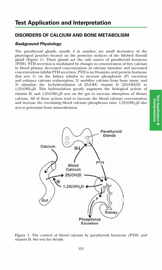
Test Application and Interpretation
Test A
pp
licatio
n &
In
terp
reta
tion
DISORDERS OF CALCIUM AND BONE METABOLISM
Background Physiology
The parathyroid glands, usually 4 in number, are small derivatives of thepharyngeal pouches located on the posterior surfaces of the bilobed thyroidgland (Figure 1). These glands are the only source of parathyroid hormone(PTH). PTH secretion is modulated by changes in concentration of free calciumin blood plasma; decreased concentrations of calcium stimulate and increasedconcentrations inhibit PTH secretion. PTH is an 84-amino acid protein hormonethat acts 1) on the kidney tubules to increase phosphorus (P) excretionand enhance calcium reabsorption, 2) mobilize calcium from bone tissue, and3) stimulate the 1α-hydroxylation of 25(OH) vitamin D [25(OH)D] to1,25(OH)2D. This hydroxylation greatly augments the biological activity ofvitamin D, and 1,25(OH)2D acts on the gut to increase absorption of dietarycalcium. All of these actions tend to increase the blood calcium concentrationand increase the circulating blood calcium/phosphorus ratio. 1,25(OH)2D alsoacts to potentiate bone mineralization.
Figure 1. The control of blood calcium by parathyroid hormone (PTH) andvitamin D. See text for details.
Phosphorus
225
Test Application and Interpretation
PTH is secreted predominantly as an intact molecule, but it is rapidly cleaved inperipheral tissues to amino terminus and carboxy terminus fragments. Theamino terminal fragment is biologically active and has a relatively short plasmahalf-life. The carboxyterminal species include a 7-84 peptide and a variety ofshorter fragments. They have a longer half-life but are biologically inactive. Themost reliable assays for PTH measure the intact molecule.
Vitamin D is a fat-soluble vitamin stored in precursor form in skin and othertissues. Hydroxylation to 25(OH)D occurs in liver, and this form is the majorcirculating form of the vitamin. It serves as a precursor for 1α-hydroxylation tothe active moiety [1,25(OH)2D], and circulating levels of 25(OH)D serve as amarker for the adequacy of vitamin D nutrition. Vitamin D is ingested as vitaminD2 (ergocalciferol), as D3 (cholecalciferol), or as precursor ergosterol, which isactivated to ergocalciferol in skin by ultraviolet light.
Disorders of the system occur because of congenital absence or acquired damageto the parathyroid glands (hypoparathyroidism); adenoma or hyperplasia ofthe glands (primary hyperparathyroidism); vitamin D deficiency; moleculardefects in the PTH receptor (pseudohypoparathyroidism), the calcium sensingreceptor (familial hypocalcemic hypercalciuria, neonatal severe primaryhyperparathyroidism, and autosomal dominant hypocalcemia), the vitamin Dreceptor (vitamin D resistance), or 1α-hydroxylase (vitamin D dependence); andrenal disease that limits phosphorus excretion and 1,25(OH)2D production withresulting hypocalcemia and (secondary) hyperparathyroidism.
Parathyroid hormone-related protein (PTH-RP) is a protein with a 14-amino acidamino terminus analogous to the amino terminus of PTH. It binds to andactivates the PTH receptor and can produce hypercalcemia. It is secreted by fetaltissues and plays a role in fetal calcium metabolism and fetal bone development.In adults, the hormone is produced in large amounts by the lactating breast andis secreted in breast milk. The physiological significance of PTH-RP in childrenand adults is not clear. It is, however, produced by a variety of tumor tissues and isthe most common cause of the hypercalcemia of malignancy.
Calcitonin (CT) is derived from the thyroid gland C-cells, which are widelydispersed in the interfollicular tissue of the gland. CT secretion is stimulated byincreased plasma calcium levels and has the capacity to inhibit the effect of PTHon bone tissue. CT levels are relatively increased in the fetus and may have a rolein fetal bone anabolism. However, there is a limited role for CT in calcium orbone metabolism in children and adults; CT deficiency has no significant effecton calcium homeostasis. CT serves as a marker for medullary thyroid carcinoma,a malignancy involving the thyroid C-cells.
Laboratory Assessment of Disorders of Calcium Metabolism
The availability of sensitive and specific tests for several hormones modulatingserum calcium levels has greatly improved our ability to assess disorders ofcalcium metabolism (Tables 1 and 2). Medullary thyroid carcinoma is discussedin the “Pheochromocytoma, Medullary Thyroid Carcinoma, and MultipleEndocrine Neoplasia” section.
226
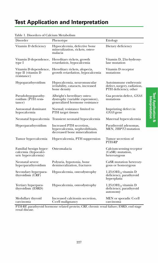
Test Application and Interpretation
Test A
pp
licatio
n &
In
terp
reta
tion
Table 1. Disorders of Calcium Metabolism
Disorder Phenotype Etiology
Vitamin D deficiency Hypocalcemia, defective bone mineralization, rickets, osteo-malacia
Dietary deficiency
Vitamin D dependence, type I
Hereditary rickets, growth retardation, hypocalcemia
Vitamin D, 21α-hydroxy-lase mutation
Vitamin D dependence, type II (vitamin D resistance)
Hereditary rickets, alopecia, growth retardation, hypocalcemia
Vitamin D receptor mutations
Hypoparathyroidism Hypocalcemia, neuromuscular irritability, cataracts, increased bone density
Autoimmune embryonic defect, surgery, radiation, PTH deficiency, other
Pseudohypoparathy-roidism (PTH resis-tance)
Albright's hereditary osteo-dystrophy (variable expression), generalized hormone resistance
Gsa protein defect, GNAS mutations
Autosomal dominant hypocalcemia
Normal; resistance limited to PTH target tissues
Imprinting defect in GNAS gene
Neonatal hypocalcemia Transient neonatal hypocalcemia Maternal hypercalcemia
Hyperparathyroidism Increased PTH secretion, hypercalcemia, nephrolithiasis, decreased bone mineralization
Parathyroid adenomas, MEN, HRPT2 mutation
Tumor hypercalcemia Hypercalcemia, PTH suppression Tumor secretion of PTH-RP
Familial benign hyper-calcemia (hypocalci-uric hypercalcemia)
Osteomalacia Calcium sensing receptor (CaSR) mutation, heterozygous
Neonatal severe hyperparathyroidism
Polyuria, hypotonia, bone demineralization, fractures
CaSR mutation heterozy-gous or homozygous
Secondary hyperpara-thyroidism (CRF)
Hypocalcemia, osteodystrophy 1,25(OH)2 vitamin D deficiency, parathyroid hyperplasia
Tertiary hyperpara-thyroidism (ESRD)
Hypocalcemia, osteodystrophy 1,25(OH)2 vitamin D deficiency, parathyroid autonomy
Medullary thyroid carcinoma
Increased calcitonin secretion, C-cell malignancy
MEN or sporadic C-cell carcinoma
PTH-RP, parathyroid hormone related protein; CRF, chronic renal failure; ESRD, end stage renal disease.
227
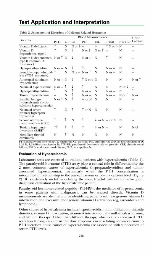
Test Application and Interpretation
PTH, parathyroid hormone; CT, calcitonin; Ca, calcium; PO, phosphorus; 25D, 25-hydroxyvitamin D;1,25 D, 1,25-dihydroxyvitamin D; PTH-RP, parathyroid hormone related protein; CRF, chronic renalfailure; ESRD, end stage renal disease; N/A, not applicable.
Evaluation of Hypercalcemia
Laboratory tests are essential to evaluate patients with hypercalcemia (Table 1).The parathyroid hormone (PTH) assay plays a central role in differentiating the2 most common causes of hypercalcemia (hyperparathyroidism and tumor-associated hypercalcemia), particularly when the PTH concentration isinterpreted in relationship to the ambient serum or plasma calcium level (Figure2). It is extremely useful in defining the most fruitful pathway for subsequentdiagnostic evaluation of the hypercalcemic patient.
Parathyroid hormone-related peptide (PTH-RP), the mediator of hypercalcemiain some patients with malignancy, can be assayed directly. Vitamin Dmeasurements are also helpful in identifying patients with exogenous vitamin Dintoxication and excessive endogenous vitamin D activation (eg, sarcoidosis andlymphoma).
Other causes of hypercalcemia include hyperthyroidism, immobilization, thiazidediuretics, vitamin D intoxication, vitamin A intoxication, the milk-alkali syndrome,and lithium therapy. Other than lithium therapy, which causes increased PTHsecretion through a shift in the dose response curve relating serum calcium toPTH secretion, these causes of hypercalcemia are associated with suppression ofserum PTH levels.
Table 2. Assessment of Disorders of Calcium-Related Hormones
DisorderBlood Measurements Urine
CalciumPTH CT Ca PO 25D 1,25D PTH-RPVitamin D deficiency ↑ N N or ↓ ↓ ↓ ↑ N or ↓ N ↓Vitamin D dependence, type I
↑ N ↓ N or ↓ N or ↑ ↓ N ↓
Vitamin D dependence, type II (vitamin D resistance)
N or ↑ N ↓ N or ↓ N ↑ N ↓
Hypoparathyroidism N or ↓ N ↓ ↑ N N or ↓ N ↓Pseudohypoparathyroid-ism (PTH resistance)
↑ N N or ↓ N or ↑ N N or ↓ N ↓
Autosomal dominant hypocalcemia
N or ↓ N ↓ ↑ N or ↓ N N N N or ↑
Neonatal hypocalcemia N or ↓ ↑ ↓ ↑ N N N or ↓ ↓Hyperparathyroidism ↑ N ↑ N or ↓ N N or ↓ N ↑Tumor hypercalcemia ↓ N ↑ N or ↓ N N or ↓ N or ↑ N or ↑Familial benign hypercalcemia (hypo-calciuric hypercalcemia)
N or ↑ N ↑ ↓ or N N N N ↓
Neonatal severe primary hyperpara-thyroidism
↑ N ↑ ↑ or N N N N ↓
Secondary hyper-parathyroidism (CRF)
↑ ↑ N ↑ ↓ or N ↓ or N N ↓
Tertiary hyperpara-thyroidsm (ESRD)
↑↑ ↑ ↑ ↑ ↓ or N ↓ N N/A
Medullary thyroid-carcinoma
N ↑ N N N N N N
228
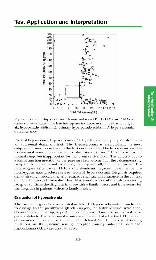
Test Application and Interpretation
Test A
pp
licatio
n &
In
terp
reta
tion
Figure 2. Relationship of serum calcium and intact PTH (IRMA or ICMA) in various disease states. The hatched square indicates normal pediatric range.
, hypoparathyroidism; , primary hyperparathyroidism; O, hypercalcemia of malignancy.
Familial hypocalciuric hypercalcemia (FHH), a familial benign hypercalcemia, isan autosomal dominant trait. The hypercalcemia is asymptomatic in mostsubjects and most prominent in the first decade of life. The hypocalciuria is dueto increased renal tubular calcium reabsorption. Serum PTH levels are in thenormal range but inappropriate for the serum calcium level. The defect is due toa loss of function mutation of the gene on chromosome 3 for the calcium-sensingreceptor that is expressed in kidney, parathyroid cell, and other tissues. Theheterozygous state causes FHH (as a dominant negative allele), while thehomozygous state produces severe neonatal hypercalcemia. Diagnosis requiresdemonstrating hypocalciuria and reduced renal calcium clearance in the contextof a family history of these disorders. Mutational analysis of the calcium sensingreceptor confirms the diagnosis in those with a family history and is necessary forthe diagnosis in patients without a family history.
Evaluation of Hypocalcemia
The causes of hypocalcemia are listed in Table 1. Hypoparathyroidism can be dueto damage to the parathyroid glands (surgery, infiltrative disease, irradiation,chemotherapeutic drugs, sepsis), to autoimmune disorders, or to moleculargenetic defects. The latter involve autosomal defects linked to the PTH gene onchromosome 11 as well as the yet to be defined X-linked variety. Activatingmutations in the calcium sensing receptor causing autosomal dominanthypocalcemia (ADH) are also causative.
229
Test Application and Interpretation
Pseudohypoparathyroidism, or PTH resistance, is due to a defect in the renalPTH receptor-adenylate cyclase complex (type I) or a more distal defect blockingthe renal phosphaturic response to cAMP (type II). Vitamin D deficiency,dependence, and resistance often are associated with rickets or osteomalacia.Type I vitamin D dependence is due to a functional defect in the 25(OH) vitaminD, 1α-hydroxylase enzyme in the kidney, impairing activation of 25(OH) vitaminD to 1,25(OH)2 vitamin D. Type II dependence is due to a defective vitamin Dreceptor.
Transient neonatal hypocalcemia is due to chronic fetal hypercalcemia associatedwith maternal hyperparathyroidism. Recovery of parathyroid gland function,which is suppressed in utero, is delayed, and there is a period of relativehypoparathyroidism lasting several days to 2 to 4 weeks in the early neonatalperiod.
Vitamin D Deficiency
Vitamin D is essential for optimal gastrointestinal absorption of calcium andmaintenance of bone mineral density (BMD). Deficiency is relatively common,especially in the elderly. Daily requirements vary with age, calcium intake, andsunlight exposure. An intake of 400 IU/d is recommended for infants andchildren and for adults with minimal sunlight exposure. In the elderly, efficacy ofUV light-stimulated synthesis of vitamin D in the skin is reduced; thus, a minimaldaily intake of 600 IU is recommended. To prevent osteoporotic fractures inelderly individuals, 800 IU/d of vitamin D and 1200 mg/d of calcium arerecommended. The recommended upper limit of intake is 2,000 IU/d.
Endogenous vitamin D is D3 (cholecalciferol), whereas supplemental vitamin Din the United States is primarily D2 (ergocalciferol). The adequacy of vitamin Dlevels is assessed by measuring serum levels of 25(OH)D; however, someimmunoassays underestimate D2 and thus total circulating 25(OH)D. Thetandem mass spectrometry method, on the other hand, accurately detects andquantifies both D2 and D3 and thus is the method of choice when monitoringpatients taking vitamin D supplements.
Figure 3 shows the relationship of serum 25(OH)D levels vs PTH concentrationsin apparently healthy 18 to 65 year-old individuals. PTH concentrations increaseperceptibly when 25(OH)D levels fall below 30 ng/mL and are clearly increasedat 25(OH)D concentrations of 10 ng/mL in agreement with earlier studies. BMDof the femoral neck also has been shown to be relatively decreased at serum25(OH)D levels <30 ng/mL, and secondary hypoparathyroidism with increasedbone turnover is associated with serum 25(OH)D concentrations <15 ng/mL.Optimal serum 25(OH)D concentrations appear to fall in the 30 to 80 ng/mLrange, particularly in elderly people.
Vitamin D Metabolism in Renal Failure
Bone and mineral metabolism is gradually altered as renal failure progresses inchronic kidney disease (CKD). By the time of progression to end stage renaldisease (ESRD), all subjects with renal failure are affected by metabolic bonedisease and are at increased fracture risk. A host of factors contribute to this
230
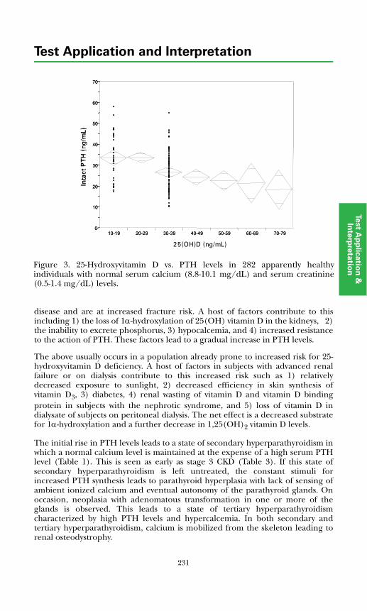
Test Application and Interpretation
Test A
pp
licatio
n &
In
terp
reta
tion
disease and are at increased fracture risk. A host of factors contribute to thisincluding 1) the loss of 1α-hydroxylation of 25(OH) vitamin D in the kidneys, 2)the inability to excrete phosphorus, 3) hypocalcemia, and 4) increased resistanceto the action of PTH. These factors lead to a gradual increase in PTH levels.
The above usually occurs in a population already prone to increased risk for 25-hydroxyvitamin D deficiency. A host of factors in subjects with advanced renalfailure or on dialysis contribute to this increased risk such as 1) relativelydecreased exposure to sunlight, 2) decreased efficiency in skin synthesis ofvitamin D3, 3) diabetes, 4) renal wasting of vitamin D and vitamin D bindingprotein in subjects with the nephrotic syndrome, and 5) loss of vitamin D indialysate of subjects on peritoneal dialysis. The net effect is a decreased substratefor 1α-hydroxylation and a further decrease in 1,25(OH)2 vitamin D levels.
The initial rise in PTH levels leads to a state of secondary hyperparathyroidism inwhich a normal calcium level is maintained at the expense of a high serum PTHlevel (Table 1). This is seen as early as stage 3 CKD (Table 3). If this state ofsecondary hyperparathyroidism is left untreated, the constant stimuli forincreased PTH synthesis leads to parathyroid hyperplasia with lack of sensing ofambient ionized calcium and eventual autonomy of the parathyroid glands. Onoccasion, neoplasia with adenomatous transformation in one or more of theglands is observed. This leads to a state of tertiary hyperparathyroidismcharacterized by high PTH levels and hypercalcemia. In both secondary andtertiary hyperparathyroidism, calcium is mobilized from the skeleton leading torenal osteodystrophy.
Inta
ct P
TH (p
g/m
L)
0
10
20
30
40
50
60
70
10-19 20-29 30-39 40-49 50-59 60-69 70-79
25(OH)D (ng/mL)
Inta
ct P
TH
(ng
/mL)
25(OH)D (ng/mL)
Inta
ct P
TH (p
g/m
L)
0
10
20
30
40
50
60
70
10-19 20-29 30-39 40-49 50-59 60-69 70-79
25(OH)D (ng/mL)
Inta
ct P
TH
(ng
/mL)
25(OH)D (ng/mL)
Figure 3. 25-Hydroxyvitamin D vs. PTH levels in 282 apparently healthyindividuals with normal serum calcium (8.8-10.1 mg/dL) and serum creatinine(0.5-1.4 mg/dL) levels.
231
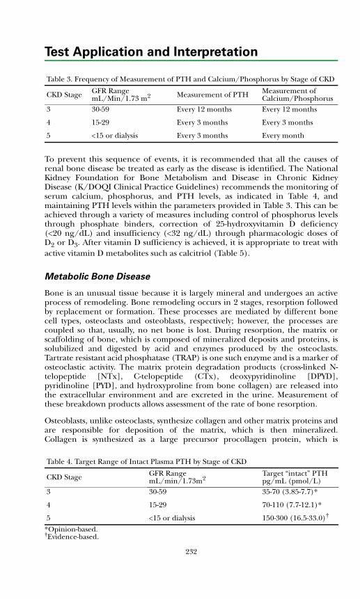
Test Application and Interpretation
To prevent this sequence of events, it is recommended that all the causes ofrenal bone disease be treated as early as the disease is identified. The NationalKidney Foundation for Bone Metabolism and Disease in Chronic KidneyDisease (K/DOQI Clinical Practice Guidelines) recommends the monitoring ofserum calcium, phosphorus, and PTH levels, as indicated in Table 4, andmaintaining PTH levels within the parameters provided in Table 3. This can beachieved through a variety of measures including control of phosphorus levelsthrough phosphate binders, correction of 25-hydroxyvitamin D deficiency(<20 ng/dL) and insufficiency (<32 ng/dL) through pharmacologic doses ofD2 or D3. After vitamin D sufficiency is achieved, it is appropriate to treat withactive vitamin D metabolites such as calcitriol (Table 5).
Metabolic Bone Disease
Bone is an unusual tissue because it is largely mineral and undergoes an activeprocess of remodeling. Bone remodeling occurs in 2 stages, resorption followedby replacement or formation. These processes are mediated by different bonecell types, osteoclasts and osteoblasts, respectively; however, the processes arecoupled so that, usually, no net bone is lost. During resorption, the matrix orscaffolding of bone, which is composed of mineralized deposits and proteins, issolubilized and digested by acid and enzymes produced by the osteoclasts.Tartrate resistant acid phosphatase (TRAP) is one such enzyme and is a marker ofosteoclastic activity. The matrix protein degradation products (cross-linked N-telopeptide [NTx], C-telopeptide (CTx), deoxypyridinoline [DPYD],pyridinoline [PYD], and hydroxyproline from bone collagen) are released intothe extracellular environment and are excreted in the urine. Measurement ofthese breakdown products allows assessment of the rate of bone resorption.
Osteoblasts, unlike osteoclasts, synthesize collagen and other matrix proteins andare responsible for deposition of the matrix, which is then mineralized.Collagen is synthesized as a large precursor procollagen protein, which isprocessed
*Opinion-based.†Evidence-based.
Table 3. Frequency of Measurement of PTH and Calcium/Phosphorus by Stage of CKD
CKD Stage GFR Range mL/Min/1.73 m2 Measurement of PTH Measurement of
Calcium/Phosphorus3 30-59 Every 12 months Every 12 months
4 15-29 Every 3 months Every 3 months
5 <15 or dialysis Every 3 months Every month
Table 4. Target Range of Intact Plasma PTH by Stage of CKD
CKD Stage GFR Range mL/min/1.73m2
Target “intact” PTH pg/mL (pmol/L)
3 30-59 35-70 (3.85-7.7)*
4 15-29 70-110 (7.7-12.1)*
5 <15 or dialysis 150-300 (16.5-33.0)†
232
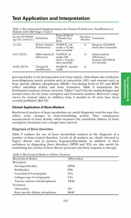
Test Application and Interpretation
Test A
pp
licatio
n &
In
terp
reta
tion
processed prior to its incorporation into bone matrix. Osteoblasts also synthesizenoncollagenous matrix proteins such as osteocalcin (OC) and enzymes such asbone specific alkaline phosphatase (BSAP). Circulating levels of OC and BSAPreflect osteoblast activity and bone formation. Table 6 summarizes thebiochemical markers of bone turnover. Tables 7 and 8 list the methodologies andnormal values for the bone resorption and formation markers. Reference rangecurves for normal values in children from 2 months to 18 years have beenrecently published (Ref 16).
Clinical Application of Bone Markers
Biochemical markers of bone metabolism are useful diagnostic tools because theyreflect acute changes in bone-remodeling activity. They complementmeasurement of bone density, which measures the cumulative balance of boneresorption/formation over a longer time interval.
Diagnosis of Bone Disorders
Table 9 outlines the use of bone metabolism markers in the diagnosis of anumber of bone-related disorders. Levels of all markers are clearly elevated inPaget's disease and in primary hyperparathyroidism. In addition to theirusefulness in diagnosing these disorders, DPYD and NTx are also useful formonitoring the activity of these disease processes and their response to therapy.
Table 5. Recommended Supplementation for Vitamin D Deficiency/Insufficiency in Patients with CKD Stages 3 and 4
Serum 25(OH)Dng/mL (nmol/L) Definition
Ergocalciferol(Vitamin D2) Dose
DurationMonths Comment
<5 (12) Severe vitamin D deficiency
50,000 IU/wk orally x 12 wks, then monthly
6 Measure 25(OH)D levels after 6 months
5-15 (12-37) Mild vitamin D deficiency
50,000 IU as single I.M. dose x 4 weeks, then monthly
6 Assure patient adherence; measure 25(OH)D at 6 months
16-30 (40-75) Vitamin D insufficiency
50,000 IU/month orally
6
Table 6. Biochemical Markers of Bone Turnover
Biochemical Marker AbbreviationResorption
Deoxypyridinoline DPYDPyridinoline PYDCross-linked N-telopeptide NTxCollagen type I C-telopeptide CTxTartrate resistant acid phosphatase TRAP
FormationOsteocalcin OCBone specific alkaline phosphatase BSAP
233
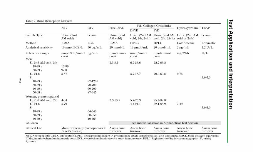
234
Test A
pp
lica
tion
an
d In
terp
reta
tion
Table 7. Bone Resorption Markers
NTx CTx Free DPYD PYD Collagen Cross-links
Hydroxyproline TRAPDPYD PYD
Sample Type Urine (2nd AM void)
Serum Urine (2nd AM void)
Urine (2nd AM void, 2-h, 24-h)
Urine (2nd AM void, 2-h, 24- h)
Urine (2nd AM void or 24-h)
Serum
Method ICMA ECL ICMA HPLC HPLC Colorimetric EnzymaticAnalytical sensitivity 10 nmol BCE/L 30 pg/mL 20 nmol/L 13 pmol/mL 20 pmol/mL 2 μg/mL 1.2 U/L
Reference ranges nmol BCE/mmol creat
pg/mL nmol/mmol creat
nmol/mmol creat
nmol/mmol creat
mg/24-h U/L
MenU, 2nd AM void, 2-h 2.1-8.1 6.2-25.6 22.7-65.2
18-29 y 12-9930-59 y 9-60
U, 24-h 5-87 3.7-18.7 20.0-60.8 9-73S 3.0-6.0
18-29 y 87-120030-39 y 70-78040-49 y 60-70050-68 y 87-345
Women, premenopausalU, 2nd AM void, 2-h 4-64 3.3-13.5 5.7-23.3 25.4-82.8U, 24-h 5-79 4.4-21.1 22.1-88.9 7-49S 3.0-6.0
18-29 y 64-64030-39 y 60-65040-49 y 40-465
Children See individual assays in Alphabetical Test Section
Clinical Use Monitor therapy (osteoporosis & Paget’s disease)
Assess bone turnover
Assess boneturnover
Assess bone turnover
Assess bone turnover
Assess bone turnover
NTx, N-telopeptide; CTx, C-telopeptide; DPYD, deoxypyridinoline; PYD, pyridinoline; TRAP, tartrate resistant acid phosphatase; BCE, bone collagen equivalent; ICMA, immunochemiluminometric assay; ECL, electrochemiluminescence assay; immunoassay; HPLC, high pressure liquid chromatography; U, urine; S, serum.
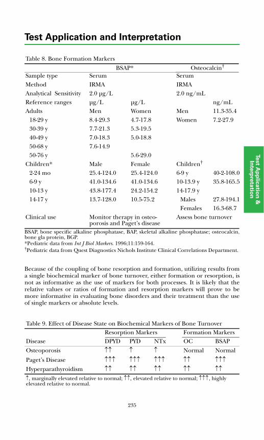
Test Application and Interpretation
Test A
pp
licatio
n &
In
terp
reta
tion
BSAP, bone specific alkaline phosphatase, BAP, skeletal alkaline phosphatase; osteocalcin,bone gla protein, BGP. *Pediatric data from Int J Biol Markers. 1996;11:159-164. †Pediatric data from Quest Diagnostics Nichols Institute Clinical Correlations Department.
Because of the coupling of bone resorption and formation, utilizing results froma single biochemical marker of bone turnover, either formation or resorption, isnot as informative as the use of markers for both processes. It is likely that therelative values or ratios of formation and resorption markers will prove to bemore informative in evaluating bone disorders and their treatment than the useof single markers or absolute levels.
Table 8. Bone Formation Markers
BSAP* Osteocalcin†
Sample type Serum SerumMethod IRMA IRMAAnalytical Sensitivity 2.0 μg/L 2.0 ng/mLReference ranges μg/L μg/L ng/mLAdults Men Women Men 11.3-35.4
18-29 y 8.4-29.3 4.7-17.8 Women 7.2-27.930-39 y 7.7-21.3 5.3-19.540-49 y 7.0-18.3 5.0-18.850-68 y 7.6-14.950-76 y 5.6-29.0
Children* Male Female Children†
2-24 mo 25.4-124.0 25.4-124.0 6-9 y 40-2-108.06-9 y 41.0-134.6 41.0-134.6 10-13.9 y 35.8-165.510-13 y 43.8-177.4 24.2-154.2 14-17.9 y14-17 y 13.7-128.0 10.5-75.2 Males 27.8-194.1
Females 16.3-68.7Clinical use Monitor therapy in osteo-
porosis and Paget’s diseaseAssess bone turnover
Table 9. Effect of Disease State on Biochemical Markers of Bone TurnoverResorption Markers Formation Markers
Disease DPYD PYD NTx OC BSAP
Osteoporosis ↑↑ ↑ ↑ Normal Normal
Paget’s Disease ↑↑↑ ↑↑↑ ↑↑↑ ↑↑ ↑↑↑Hyperparathyroidism ↑↑ ↑↑ ↑↑ ↑↑ ↑↑↑, marginally elevated relative to normal; ↑↑, elevated relative to normal; ↑↑↑, highly elevated relative to normal.
235
Test Application and Interpretation
Diagnosis of Osteoporosis
The current practice for diagnosis of osteoporosis is bone densitometry. Sincebone density can decline at a rate of 1% to 3% per year, bone densitometry is agood method of detecting established osteoporosis. It has limited usefulness,however, for immediate follow-up of treatment of osteoporotic patients becausesubtle changes in bone density cannot be detected. Measurements of biochemicalmarkers of bone formation and resorption (Table 6), unlike bone densitometry,are particularly well suited for monitoring the efficacy of treatment. These markerswill change rapidly if treatment, such as estrogen therapy, is effective.
In osteoporosis, the rate of bone resorption may be only modestly increasedafter menopause, as indicated by the overlap in the reference ranges for thesemarkers in premenopausal and postmenopausal women. Thus, the usefulnessof these markers in diagnosis of primary osteoporosis is limited. The markersare most useful in cases of high bone turnover. Additionally, in the majority ofcases, the biochemical markers will be most useful in evaluation of theefficacy of treatment. Patients with high levels of bone turnover markers maybe the most suitable for treatment with antiresorptive agents. In this case,following the bone turnover markers at a later time will be especially useful indetermining efficacy of treatment.
Efficacy of Therapy
Measurement of the levels of biochemical markers can be used to determinewhether a drug therapy is effective within 2 to 6 months. A decrease in the levelsof the markers of >25% indicates effective intervention.
Follow-up to Therapy
A similar approach can be used to determine the appropriate dose of drug. Byfollowing the changes in biochemical bone markers, the level of drugadministered can be fine-tuned to achieve the desired level of inhibition of boneresorption to maintain an acceptable rate of bone remodeling.
References
1. Bringhurst FR, Demay MB, Kronenberg HM. Hormones and disorders ofmineral metabolism. In: Larsen PR, Kronenberg HM, Melmed S, PolonskyKS, eds. Williams Textbook of Endocrinology. 10th ed. Philadelphia, PA: WBSaunders; 2003:1303-1371.
2. Bouillon R. Vitamin D: from photosynthesis, metabolism, and action toclinical applications. In: DeGroot LJ, Jameson JL, et al, eds. Endocrinology. 5thed. Philadelphia, PA: Elsevier-Saunders; 2006:1377-1418.
3. Levine MA. Hypoparathyroidism and pseudohypoparathyroidism. In:DeGroot LJ, Jameson JL, et al, eds. Endocrinology. 5th ed. Philadelphia, PA:WB Saunders; 2006:1611-1636.
236
Test Application and Interpretation
Test A
pp
licatio
n &
In
terp
reta
tion
4. Root AW, Diamond FJ Jr. Calcium metabolism, normal homeostasis. In:Sperling MA, ed. Pediatric Endocrinology. 2nd ed. Philadelphia, PA: WBSaunders; 2002:65-95.
5. Diamond FB Jr., Root AW. Disorders of calcium metabolism in the newbornand infant. In: Sperling MA, ed. Pediatric Endocrinology. 2nd ed. Philadelphia,PA: WB Saunders; 2002:97-110.
6. Diaz R, Brown EM. Familial hypocalciuric hypercalcemia and other disordersdue to calcium-sensing receptor mutations. In: DeGroot LJ, Jameson JL, eds.Endocrinology. 5th ed. Philadelphia, PA: Elsevier-Saunders; 2006:1595-1609.
7. St. Arnaud R, Glorieux FH. Genetic defects in vitamin D metabolism andaction. In: DeGroot LJ, Jameson JL, eds. Endocrinology. 5th ed. Philadelphia,PA: Elsevier-Saunders; 2006:1637-1652.
8. Delmas PD, Chapurlat RD. Osteoporosis. In: DeGroot LJ, Jameson JL, et al,eds. Endocrinology. 5th ed. Philadelphia, PA: Elsevier-Saunders; 2006:1751-1769.
9. Goodman WG, Juppner H, Salusky B, Sherrard DJ. Parathyroid hormone(PTH), PTH-derived peptides, and new PTH assays in renal osteodystrophy.Kidney Internat. 2003;63:1-11.
10. Strewler GJ. The physiology of parathyroid hormone related protein. N Engl JMed. 2000;342:177-185.
11. Raisz L, Kream BE, Lorenzo JA. Metabolic bone disease. In: Larsen PR,Kronenberg HM, Melmed S, Polonsky KS, eds. Williams Textbook ofEndocrinology. 10th ed. Philadelphia, PA: WB Saunders; 2003:1373-1410.
12. Dulipsingh L, DeSouza MJ, Ikram Z, Fall P, Willard A, Fang Z, Prestwood K,Raisz L. Clinical utility of urinary markers of bone turnover in response toantiresorptive therapies. The Endocrinologist. 2003;13:293-299.
13. Steelman J, Zeitler P. Treatment of symptomatic pediatric osteoporosis withcyclic, single day intravenous pamidronate infusions. J Pediatr. 2003;142:417-423.
14. National Kidney Foundation. K/DOQI clinical practice guidelines for bonemetabolism and disease in chronic kidney disease. Am J Kidney Dis.2003;42(Suppl 3):S1-S201.
15. Tomita T, Millard DM. C-cell hyperplasia in secondary hyperparathyroidism.Histopathol. 1992;21:469-474.
16. Rauchenzauner M, Schmid A, Heinz-Erian P, et al. Sex- and age-specificreference curves for serum markers of bone turnover in healthy childrenfrom 2 months to 18 years. J Clin Endocrinol Metab. 2007;92:443-449.
237

238
Test Application and Interpretation
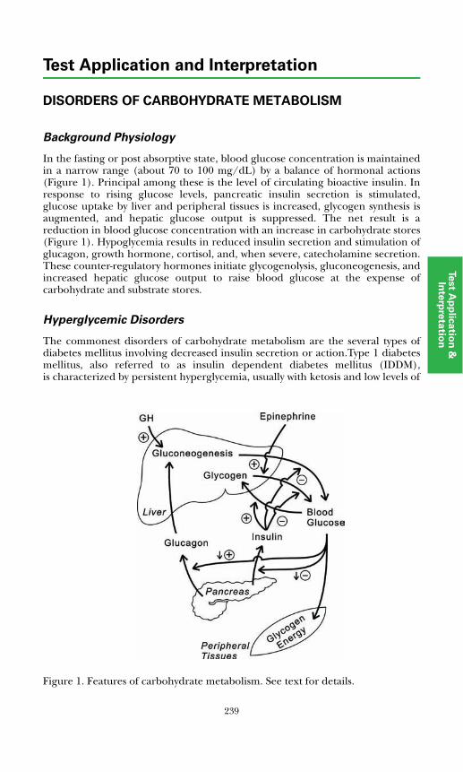
Test Application and Interpretation
Test A
pp
licatio
n &
In
terp
reta
tion
DISORDERS OF CARBOHYDRATE METABOLISM
Background Physiology
In the fasting or post absorptive state, blood glucose concentration is maintainedin a narrow range (about 70 to 100 mg/dL) by a balance of hormonal actions(Figure 1). Principal among these is the level of circulating bioactive insulin. Inresponse to rising glucose levels, pancreatic insulin secretion is stimulated,glucose uptake by liver and peripheral tissues is increased, glycogen synthesis isaugmented, and hepatic glucose output is suppressed. The net result is areduction in blood glucose concentration with an increase in carbohydrate stores(Figure 1). Hypoglycemia results in reduced insulin secretion and stimulation ofglucagon, growth hormone, cortisol, and, when severe, catecholamine secretion.These counter-regulatory hormones initiate glycogenolysis, gluconeogenesis, andincreased hepatic glucose output to raise blood glucose at the expense ofcarbohydrate and substrate stores.
Hyperglycemic Disorders
The commonest disorders of carbohydrate metabolism are the several types ofdiabetes mellitus involving decreased insulin secretion or action.Type 1 diabetesmellitus, also referred to as insulin dependent diabetes mellitus (IDDM),is characterized by persistent hyperglycemia, usually with ketosis and low levels of
Figure 1. Features of carbohydrate metabolism. See text for details.
239
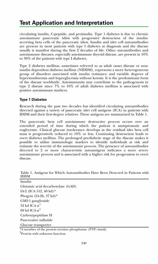
Test Application and Interpretation
circulating insulin, C-peptide, and proinsulin. Type 1 diabetes is due to chronicautoimmune pancreatic islitis with progressive destruction of the insulin-secreting beta cells of the pancreatic islets. Insulin and islet cell autoantibodiesare present in most patients with type I diabetes at diagnosis and the diseaseusually is manifest during the first 2 decades of life. Other autoantibodies andautoimmune diseases, especially autoimmune thyroid disease, are present in 10%to 30% of the patients with type I diabetes.
Type 2 diabetes mellitus, sometimes referred to as adult onset disease or non-insulin dependent diabetes mellitus (NIDDM), represents a more heterogeneousgroup of disorders associated with insulin resistance and variable degrees ofhyperinsulinemia and hyperglycemia without ketosis. It is the predominant formof the disease worldwide. Autoimmunity may contribute to the pathogenesis intype 2 disease since 7% to 10% of adult diabetes mellitus is associated withpositive autoimmune markers.
Type 1 Diabetes
Research during the past two decades has identified circulating autoantibodiesdirected against a variety of pancreatic islet cell antigens (ICA) in patients withIDDM and their first-degree relatives. These antigens are summarized in Table 1.
The pancreatic beta cell autoimmune destructive process occurs over anextended period of time during which the patient is asymptomatic andeuglycemic. Clinical glucose intolerance develops as the residual islet beta cellmass is progressively reduced to 10% or less. Continuing destruction leads toovert diabetes mellitus. The prolonged prediabetic stage of the disease makes itpossible to utilize immunologic markers to identify individuals at risk andestimate the severity of the autoimmune process. The presence of autoantibodiesdirected to 2 or more characteristic autoantigens indicates a more severeautoimmune process and is associated with a higher risk for progression to overtdisease.
Table 1. Antigens for Which Autoantibodies Have Been Detected in Patients withIDDMInsulinGlutamic acid decarboxylase (GAD)IA-2 (ICA 512, 40 kd)*Phogrin (IA-2b, 37 kd)*GM2-1 ganglioside52 kd ICA α†
69 kd ICA α†
Carboxypeptidase HPancreative sulfatideGlucose transporter
*A member of the protein tyrosine phosphatase (PTP) family.†Protein with unknown function.
240
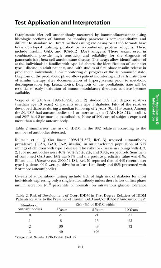
Test Application and Interpretation
Test A
pp
licatio
n &
In
terp
reta
tion
Cytoplasmic islet cell autoantibody measured by immunofluorescence usinghistologic sections of human or monkey pancreas is semi-quantitative anddifficult to standardize. Direct methods using radioassay or ELISA formats havebeen developed utilizing purified or recombinant protein antigens. Theseinclude insulin, GAD, and ICA-512 (IA-2) antigens. These assays, used incombination, provide high sensitivity and reliability for the diagnosis ofpancreatic islet beta cell autoimmune disease. The assays allow identification ofat-risk individuals in families with type 1 diabetes, the identification of late onsettype 1 disease in adult patients, and, with studies of first phase insulin release inprediabetic individuals, allow monitoring of progress of the autoimmune state.Diagnosis of the prediabetic phase allows patient monitoring and early institutionof insulin therapy after documentation of hyperglycemia prior to metabolicdecompensation (eg, ketoacidosis). Diagnosis of the prediabetic state will beessential to early institution of immunomodulatory therapies as these becomeavailable.
Verge et al (Diabetes. 1996;45:926, Ref. 2) studied 882 first degree relatives(median age 13 years) of patients with type 1 diabetes. Fifty of the relativesdeveloped diabetes during a median follow-up of 2 years (0.1-11.3 years). Amongthe 50, 98% had autoantibodies to 1 or more antigens (GAD, ICA 512, insulin),and 80% had 2 or more autoantibodies. None of 200 control subjects expressedmore than a single autoantibody.
Table 2 summarizes the risk of IDDM in the 882 relatives according to thenumber of antibodies detected.
Kulmala et al (J Clin Invest. 1998;101:327, Ref. 3) assessed autoantibodyprevalence (ICAA, GAD, IA-2, insulin) in an unselected population of 755siblings of children with type 1 disease. The risks for disease in siblings with 4, 3,2, 1, or no antibodies were 40%, 70%, 25%, 2%, and 0.8%, respectively. Sensitivityof combined GAD and IA-2 was 81% and the positive predictive value was 41%.Bilbao et al (Hormone Res. 2000;54:181, Ref. 5) reported that of 448 recent onsettype 1 patients, 90% were positive for at least 1 antibody and 68% presented with2 or more autoantibodies.
Caveats of autoantibody testing include lack of high risk of diabetes for mostindividuals expressing only a single autoantibody unless there is loss of first phaseinsulin secretion (<1st percentile of normals) on intravenous glucose tolerancetesting
Table 2. Risk of Development of Overt IDDM in First Degree Relatives of IDDMPatients Relative to the Presence of Insulin, GAD and/or ICA512 Autoantibodies*
Number ofAutoantibodies
Risk (%) of IDDM within3 Years 5 Years 10 Years
0 <1 <1 <1
1 8 15 23
2 30 43 723 49 >95
*Verge et al. Diabetes. 1996;45:926. (Ref. 2)
241
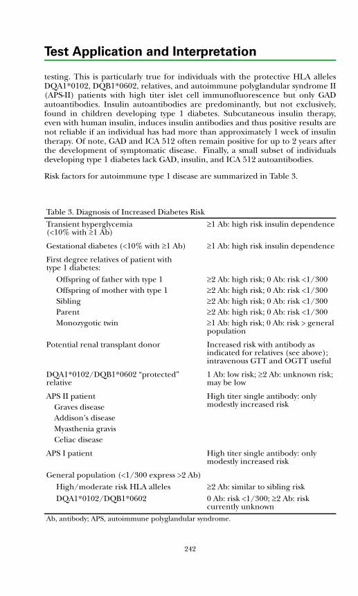
242
Test Application and Interpretation
testing. This is particularly true for individuals with the protective HLA allelesDQA1*0102, DQB1*0602, relatives, and autoimmune polyglandular syndrome II(APS-II) patients with high titer islet cell immunofluorescence but only GADautoantibodies. Insulin autoantibodies are predominantly, but not exclusively,found in children developing type 1 diabetes. Subcutaneous insulin therapy,even with human insulin, induces insulin antibodies and thus positive results arenot reliable if an individual has had more than approximately 1 week of insulintherapy. Of note, GAD and ICA 512 often remain positive for up to 2 years afterthe development of symptomatic disease. Finally, a small subset of individualsdeveloping type 1 diabetes lack GAD, insulin, and ICA 512 autoantibodies.
Risk factors for autoimmune type 1 disease are summarized in Table 3.
Table 3. Diagnosis of Increased Diabetes Risk
Transient hyperglycemia (<10% with ≥1 Ab)
≥1 Ab: high risk insulin dependence
Gestational diabetes (<10% with ≥1 Ab) ≥1 Ab: high risk insulin dependence
First degree relatives of patient with type 1 diabetes:
Offspring of father with type 1 ≥2 Ab: high risk; 0 Ab: risk <1/300Offspring of mother with type 1 ≥2 Ab: high risk; 0 Ab: risk <1/300Sibling ≥2 Ab: high risk; 0 Ab: risk <1/300Parent ≥2 Ab: high risk; 0 Ab: risk <1/300Monozygotic twin ≥1 Ab: high risk; 0 Ab: risk > general
population
Potential renal transplant donor Increased risk with antibody as indicated for relatives (see above); intravenous GTT and OGTT useful
DQA1*0102/DQB1*0602 “protected” relative
1 Ab: low risk; ≥2 Ab: unknown risk; may be low
APS II patient High titer single antibody: only modestly increased riskGraves disease
Addison’s diseaseMyasthenia gravisCeliac disease
APS I patient High titer single antibody: only modestly increased risk
General population (<1/300 express >2 Ab)High/moderate risk HLA alleles ≥2 Ab: similar to sibling riskDQA1*0102/DQB1*0602 0 Ab: risk <1/300; ≥2 Ab: risk
currently unknownAb, antibody; APS, autoimmune polyglandular syndrome.
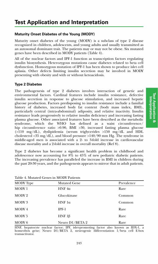
Test Application and Interpretation
Test A
pp
licatio
n &
In
terp
reta
tion
Maturity Onset Diabetes of the Young (MODY)
Maturity onset diabetes of the young (MODY) is a subclass of type 2 diseaserecognized in children, adolescents, and young adults and usually transmitted asan autosomal dominant trait. The patients may or may not be obese. Six mutatedgenes have been described in MODY patients (Table 4).
All of the nuclear factors and IPF-1 function as transcription factors regulatinginsulin biosynthesis. Heterozygous mutations cause diabetes related to beta celldysfunction. Homozygous mutation of IPF-1 has been shown to produce islet cellaplasia. Other defects limiting insulin secretion may be involved in MODYpresenting with obesity and with or without ketoacidosis.
Type 2 Diabetes
The pathogenesis of type 2 diabetes involves interaction of genetic andenvironmental factors. Cardinal features include insulin resistance, defectiveinsulin secretion in response to glucose stimulation, and increased hepaticglucose production. Factors predisposing to insulin resistance include a familialhistory of diabetes, increased body fat content (body mass index, BMI),particularly central (intra-abdominal) adiposity, and relative inactivity. Insulinresistance leads progressively to relative insulin deficiency and increasing fastingplasma glucose. Other associated features have been described as the metabolicsyndrome, which the WHO has defined as a waist circumference/hip circumference ratio >0.90, BMI >30, increased fasting plasma glucose(>110 mg/dL), dyslipidemia (serum triglycerides >150 mg/dL and HDLcholesterol <35 mg/dL), and blood pressure >140/90 mm Hg. The syndrome inmiddle-aged men is associated with a 2- to 3-fold increase in cardiovasculardisease mortality and a 2-fold increase in overall mortality (Ref 8).
Type 2 diabetes has become a significant health problem in childhood andadolescence now accounting for 8% to 45% of new pediatric diabetic patients.The increasing prevalence has paralleled the increase in BMI in children duringthe past 20-30 years, and the pathogenesis appears to mirror that in adult patients.
Table 4. Mutated Genes in MODY Patients
MODY Type Mutated Gene Prevalence
MODY 1 HNF 4α Rare
MODY 2 Glucokinase Common
MODY 3 HNF 1α Common
MODY 4 IPF-1 Rare
MODY 5 HNF 1β Rare
MODY 6 Neuro D1/BETA 2 RareHNF, hepatocyte nuclear factor; IPF, islet-promoting factor also known as IDX-1, ahomeobox gene; Neuro D1/BETA 2, neurogenic differentiation 1/beta cell E-boxtransactivator.
243
Test Application and Interpretation
Risk factors include a positive family history, obesity, polycystic ovarian syndrome(PCOS), acanthosis nigricans, ethnicity (higher prevalence in minoritypopulations), and intrauterine programming with increasing insulin resistanceleading to relative insulin deficiency and overt diabetes. Although the absence ofdiabetes autoimmune markers is presently a prerequisite for the diagnosis of type2 diabetes in pediatric patients, the prevalence of such markers appears to besimilar to the 7% to 10% prevalence in adult type 2 patients.
Female patients with variable degrees of insulin resistance associated with insulinautoantibody, variable carbohydrate intolerance, acanthosis nigricans, and PCOShave been referred to as type B insulin resistant. Males also manifest the diseasebut without gonadal dysfunction. Diabetes also may be due to a variety of insulinreceptor mutations. Females with insulin resistance type A associated withinactivating mutations in the insulin receptor gene also display acanthosisnigricans and PCOD; these features are believed to be secondary to thehyperinsulinemia, but the mechanism is not yet understood. The Rabson-Mendenhall syndrome is manifest by leprechaun-like features, acanthosisnigricans, and severe insulin resistance due to insulin receptor mutation.Heterozygous mutations in the transcription factor peroxisome proliferator-activated receptor γ (PPARγ) are associated with severe insulin resistance andearly onset type 2 diabetes. Additionally, a common polymorphism in PPARγ(Pro12Ala) predisposes to insulin resistance with an increased risk of type 2diabetes. Impaired islet beta cell function also has been associated with amaternally transmitted mutation in mitochondrial transfer RNA with variablephenotypic expression resembling type 1 or type 2 diabetes. These disorders aresummarized in Table 5, which details the blood tests available for assessingpatients with hyperglycemia.
Diagnosis of Diabetes Mellitus
The diagnostic criteria for impaired fasting glucose (IFG), impaired glucosetolerance (IGT), and diabetes mellitus are shown in Table 6. Venous blood froman antecubital vein is preferable to capillary blood samples for glucose tolerancetesting. Plasma or serum can be used for glucose measurements. Standard testsare done in the morning after 12 to 14 hours of fasting. No formal dietarypreparation is necessary unless the patient has been on a restricted diet.
Criteria for testing for diabetes mellitus in pediatric and asymptomatic adultsubjects are summarized in Tables 7 and 8.
Preventing and Managing Complications
The major complications of diabetes include dyslipidemia, cardiovasculardisease, ophthalmopathy, nephropathy, and neuropathy. Laboratory tests areuseful tools for prevention and management of these complications.
The American Diabetes Association recommends the following tests beconsidered for initial laboratory evaluation of a patient diagnosed with diabetes(Ref. 15):
• Hemoglobin A1c
244
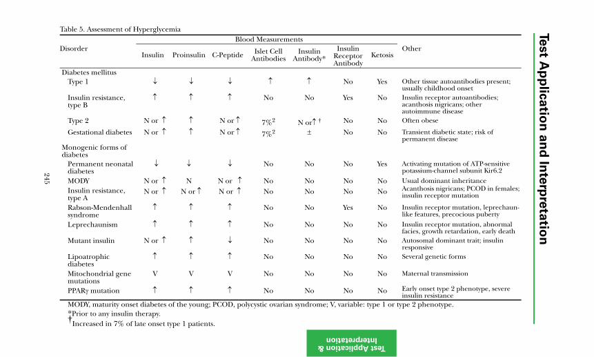
Test A
pp
lica
tion
an
d In
terp
reta
tion
Test ApplicatioInterpretatio
Table 5. Assessment of Hyperglycemia
DisorderBlood Measurements
OtherInsulin Proinsulin C-Peptide Islet Cell
AntibodiesInsulin
Antibody*Insulin
Receptor Antibody
Ketosis
No Yes Other tissue autoantibodies present; usually childhood onset
Yes No Insulin receptor autoantibodies; acanthosis nigricans; other autoimmune disease
No No Often obese
No No Transient diabetic state; risk of permanent disease
No Yes Activating mutation of ATP-sensitive potassium-channel subunit Kir6.2
No No Usual dominant inheritance
No No Acanthosis nigricans; PCOD in females; insulin receptor mutation
Yes No Insulin receptor mutation, leprechaun-like features, precocious puberty
No No Insulin receptor mutation, abnormal facies, growth retardation, early death
No No Autosomal dominant trait; insulin responsive
No No Several genetic forms
No No Maternal transmission
No No Early onset type 2 phenotype, severe insulin resistance
, variable: type 1 or type 2 phenotype.
245
n & n
Diabetes mellitusType 1 ↓ ↓ ↓ ↑ ↑
Insulin resistance, type B
↑ ↑ ↑ No No
Type 2 N or ↑ ↑ N or ↑ 7%2 N or↑ †
Gestational diabetes N or ↑ ↑ N or ↑ 7%2 ±
Monogenic forms ofdiabetes
Permanent neonatal ↓diabetes
↓ ↓ No No
MODY N or ↑ N N or ↑ No NoInsulin resistance, type A
N or ↑ N or ↑ N or ↑ No No
Rabson-Mendenhall syndrome
↑ ↑ ↑ No No
Leprechaunism ↑ ↑ ↑ No No
Mutant insulin N or ↑ ↑ ↓ No No
Lipoatrophic diabetes
↑ ↑ ↑ No No
Mitochondrial gene mutations
V V V No No
PPARγ mutation ↑ ↑ ↑ No No
MODY, maturity onset diabetes of the young; PCOD, polycystic ovarian syndrome; V*Prior to any insulin therapy.†
Increased in 7% of late onset type 1 patients.
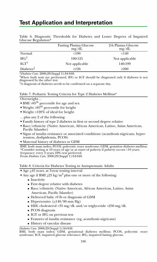
Test Application and Interpretation
Table 6. Diagnostic Thresholds for Diabetes and Lesser Degrees of ImpairedGlucose Regulation*
Fasting Plasma Glucosemg/dL
2-h Plasma Glucosemg/dL
Normal <100 <140IFG† 100-125 Not applicableIGT† Not applicable 140-199Diabetes‡ ≥126 ≥200
*Diabetes Care. 2006;29(Suppl 1):S4-S48.†When both tests are performed, IFG or IGT should be diagnosed only if diabetes is notdiagnosed by the other test. ‡A diagnosis of diabetes needs to be confirmed on a separate day.
Table 7. Pediatric Testing Criteria for Type 2 Diabetes Mellitus*Overweight…• BMI >85th percentile for age and sex• Weight >85th percentile for height • Weight >120% of ideal for height…plus any 2 of the following:• Family history of type 2 diabetes in first or second degree relative• Race/ethnicity (Native American, African American, Latino, Asian American, Pacific Islander)• Signs of insulin resistance or associated conditions (acanthosis nigricans, hyper- tension, dyslipidemia, PCOS)• Maternal history of diabetes or GDMBMI, body mass index; PCOS, polycystic ovary syndrome; GDM, gestation diabetes mellitus.*Consider testing at 10 years of age or at onset of puberty if puberty occurs <10 years; frequency: every 2 years; FPG test preferred. From Diabetes Care. 2006;29(Suppl 1):S4-S48.
Table 8. Criteria for Diabetes Testing in Asymptomatic Adults• Age >45 years, at 3-year testing interval• Any age if BMI >25 kg/m2 plus one or more of the following: Inactivity First degree relative with diabetes Race/ethnicity (Native American, African American, Latino, Asian American, Pacific Islander) Delivered baby >9 lb or diagnosis of GDM Hypertensive (>140/90 mm Hg) HDL cholesterol <35 mg/dL and/or triglyceride >250 mg/dL PCOS diagnosis IGT or IFG on previous test Features of insulin resistance (eg, acanthosis nigricans) History of vascular disease
Diabetes Care. 2006;29(Suppl 1):S4-S48.BMI, body mass index; GDM, gestational diabetes mellitus; PCOS, polycystic ovarysyndrome; IGT, impaired glucose tolerance; IFG, impaired fasting glucose.
246

Test Application and Interpretation
Test A
pp
licatio
n &
In
terp
reta
tion
• Total cholesterol, HDL cholesterol, triglycerides, LDL cholesterol
• Liver function tests; if abnormal, add tests for fatty liver or hepatitis
• Microalbuminuria
In patients with type 1 diabetes for >5 years, perhaps earlier in pubertal children
In all patients with type 2 diabetes
• Creatinine and estimated glomerular filtration rate (eGFR)
• TSH in all patients with type 1 diabetes and, when clinically indicated, inpatients with type 2
• Electrocardiogram in adults, if clinically indicated
• Urinalysis, including ketones, protein, and sediment examination
Laboratory testing recommendations (Ref. 15) for on-going patient managementfollow. Table 9 summarizes the clinically significant concentrations for each test.
• Self-monitoring of blood glucose (>3/day for patients receiving multipleinsulin injections)
Adults: pre- and postprandial
Children: preprandial and postprandial if disparity between A1c and preprandial levels
• Hemoglobin A1c determination at least 2 times per year in patients who aremeeting treatment goals and have stable glycemic control and quarterly inpatients whose therapy has changed or treatment goals are not being met
• Lipids (LDL, triglycerides, HDL)
Adults: annually
Prepubertal children: not recommended unless there is a family history of hypercholesterolemia or cardiovascular event before age 55 or family history is unknown
Pubertal children (>12 years of age): every 5 years if levels indicate acceptable risk
Children, all ages: annually if lipids are abnormal
• Microalbumin
Adults: annually in those with type 1 diabetes for >5 years and in all with type 2 diabetes (starting at diagnosis and during pregnancy)
Children: annually in those with diabetes for >5 years (beginning at 10 years of age)
• Creatinine and eGFR, at least annually in all adult patients; eGFR levelcorrelates with severity of chronic kidney disease
247
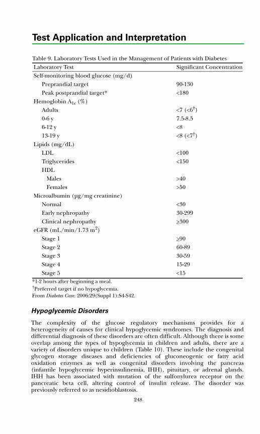
248
Test Application and Interpretation
Table 9. Laboratory Tests Used in the Management of Patients with Diabetes Laboratory Test Significant ConcentrationSelf-monitoring blood glucose (mg/d)
Preprandial targetPeak postprandial target*
90-130<180
Hemoglobin A1c (%)Adults0-6 y6-12 y13-19 y
<7 (<6†)7.5-8.5<8<8 (<7†)
Lipids (mg/dL)LDLTriglyceridesHDL
MalesFemales
<100<150
>40>50
Microalbumin (μg/mg creatinine)NormalEarly nephropathyClinical nephropathy
<3030-299>300
eGFR (mL/min/1.73 m2)Stage 1Stage 2Stage 3Stage 4Stage 5
>9060-8930-5915-29<15
*1-2 hours after beginning a meal.†Preferred target if no hypoglycemia.From Diabetes Care. 2006:29(Suppl 1):S4-S42.
Hypoglycemic Disorders
The complexity of the glucose regulatory mechanisms provides for aheterogeneity of causes for clinical hypoglycemic syndromes. The diagnosis anddifferential diagnosis of these disorders are often difficult. Although there is someoverlap among the types of hypoglycemia in children and adults, there are avariety of disorders unique to children (Table 10). These include the congenitalglycogen storage diseases and deficiencies of gluconeogenic or fatty acidoxidation enzymes as well as congenital disorders involving the pancreas(infantile hypoglycemic hyperinsulinemia, IHH), pituitary, or adrenal glands.IHH has been associated with mutation of the sulfonylurea receptor on thepancreatic beta cell, altering control of insulin release. The disorder waspreviously referred to as nesidioblastosis.
249
Test Application and Interpretation
Test A
pp
licatio
n &
In
terp
reta
tion
Any infant or child with plasma glucose under 50 mg/dL should be observedcarefully, and a value below 40 mg/dL is considered hypoglycemia. Transienthypoglycemia in the newborn infant and, particularly, the premature neonate, isrelatively common and usually resolves within 72 hours. The small-for-gestation-age infant and those with a predisposition to transient neonatal hyperinsulinemia(infant of diabetic mother, Beckwith-Wiedemann infant, infant witherythroblastosis fetalis) are at higher risk. Hypoglycemia persisting beyond72 hours suggests a pathologic condition such as persistent hyperinsulinemia,which may be due to islet beta cell hyperplasia or IHH. Inborn errors ofcarbohydrate, amino acid, or organic acid metabolism usually are manifest in thenewborn period. Pituitary or adrenal insufficiency usually is manifest at birth orduring infancy. During childhood, the most common cause of hypoglycemia isketotic hypoglycemia, a self-limited inability to maintain normoglycemia duringprolonged fasting. Islet beta-cell hyperplasia or adenoma is uncommon andusually develops in later childhood.
In adults, the differential diagnosis is extensive and includes postprandial andfasting hypoglycemias. Postprandial hypoglycemia is common and has beenclassified into 3 types: patients with normal oral glucose tolerance tests(functional hypoglycemia), those with post surgical modifications of the upperGI tract (alimentary hypoglycemia), and those with early elevations (1 to 2 hours)of glucose levels during glucose tolerance testing (early diabetes hypoglycemia).The causes of fasting hypoglycemia (8 hours or more after feeding) includeethanol ingestion, factitious or accidental intake of oral hypoglycemic agents,severe liver disease, adrenal insufficiency, inanition, certain malignancies(leukemias, hepatoma, and mesenchymal tumors), and, rarely, insulin receptorstimulating autoantibodies. The prevalence of insulinoma approximates 1 patientper 250,000 patient-years. The median age at diagnosis is 50 years except forpatients with multiple endocrine neoplasia (MEN) type I. Symptoms may presentwithin weeks or decades before diagnosis.
Diagnostic Approach
In many adult cases, the diagnosis can be confirmed by demonstratingconsistently inappropriate insulin concentrations in the presence of low bloodsugar values. Insulin-like growth factor binding protein I (IGFBP-1) is insulindependent, and elevated levels often are associated with hyperinsulinism. As inchildren, in some instances, diagnosis may require prolonged fasting. In patientswith insulinoma, insulin concentrations are >6 μIU/mL and C-peptide is>0.8 ng/mL when the blood sugar falls to 50 mg/dL or less; normal subjects havelower values. In adult patients with insulinoma, Whipple's triad (low plasmaglucose, symptoms of hypoglycemia, and amelioration of symptoms by glucoseadministration) is demonstrated in 95% to 98% of cases within 72 hours ofinitiating a fast, and blood glucose values are <46 mg/dL at the time ofdevelopment of hypoglycemic symptoms. Patients with insulinoma usually haveundetectable insulin antibody and an insulin/proinsulin plasma concentrationratio of 1:1, whereas in normal subjects, ratios approximate 6:1.
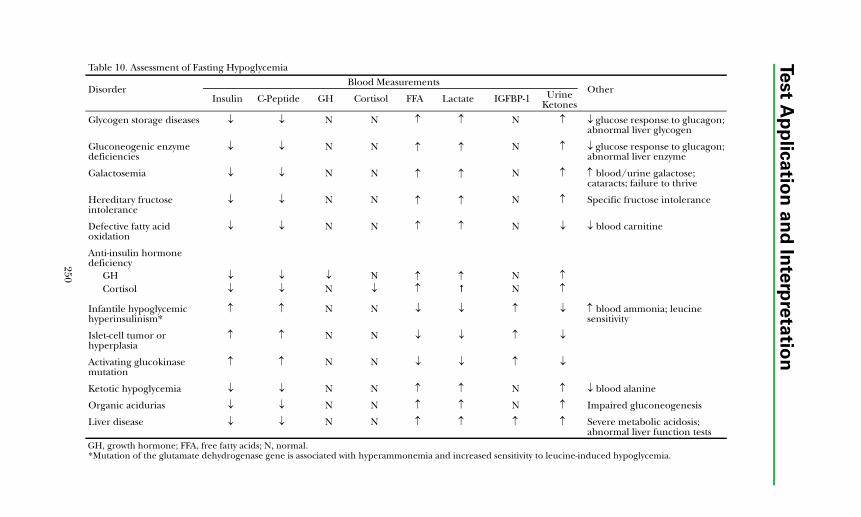
250
Test A
pp
lica
tion
an
d In
terp
reta
tion
Table 10. Assessment of Fasting Hypoglycemia
DisorderBlood Measurements
OtherInsulin C-Peptide GH Cortisol FFA Lactate IGFBP-1 Urine
Ketones
Glycogen storage diseases ↓ ↓ N N ↑ ↑ N ↑ ↓ glucose response to glucagon; abnormal liver glycogen
Gluconeogenic enzyme deficiencies
↓ ↓ N N ↑ ↑ N ↑ ↓ glucose response to glucagon; abnormal liver enzyme
Galactosemia ↓ ↓ N N ↑ ↑ N ↑ ↑ blood/urine galactose; cataracts; failure to thrive
Hereditary fructose intolerance
↓ ↓ N N ↑ ↑ N ↑ Specific fructose intolerance
Defective fatty acid oxidation
↓ ↓ N N ↑ ↑ N ↓ ↓ blood carnitine
Anti-insulin hormone deficiency
GH ↓ ↓ ↓ N ↑ ↑ N ↑Cortisol ↓ ↓ N ↓ ↑ N ↑
Infantile hypoglycemic hyperinsulinism*
↑ ↑ N N ↓ ↓ ↑ ↓ ↑ blood ammonia; leucine sensitivity
Islet-cell tumor or hyperplasia
↑ ↑ N N ↓ ↓ ↑ ↓
Activating glucokinase mutation
↑ ↑ N N ↓ ↓ ↑ ↓
Ketotic hypoglycemia ↓ ↓ N N ↑ ↑ N ↑ ↓ blood alanine
Organic acidurias ↓ ↓ N N ↑ ↑ N ↑ Impaired gluconeogenesis
Liver disease ↓ ↓ N N ↑ ↑ ↑ ↑ Severe metabolic acidosis; abnormal liver function tests
GH, growth hormone; FFA, free fatty acids; N, normal.*Mutation of the glutamate dehydrogenase gene is associated with hyperammonemia and increased sensitivity to leucine-induced hypoglycemia.
Test Application and Interpretation
Test A
pp
licatio
n &
In
terp
reta
tion
Various perturbation tests have been proposed for the diagnosis of insulinomaincluding the tolbutamide tolerance test, the glucagon stimulation test, and theC-peptide suppression test. However, all are associated with frequent false-positiveor false-negative results. The most reliable confirmatory test is the classic 72-hourfast which produces hypoglycemia in essentially all patients with insulinoma; 75%develop hypoglycemia within 24 hours (see Dynamic Test section). Patients withnesidioblastosis may have postprandial hypoglycemia associated with abnormalplasma insulin and C-peptide levels.
The C-peptide suppression test is influenced by age and body mass index (BMI);suppressibility at 60 minutes decreases with increasing age and increasing BMI.Suppressibility in normal subjects aged 20 to 50 with BMI of 20 to 34 kg/M2
ranges from 59% to 67%; values in subjects aged 51 to 80 range from 51% to61%. These test protocols are summarized in the Dynamic Test section.
The insulin autoimmune syndrome (IAS) is an unusual cause of hypoglycemiaassociated with the spontaneous development of insulin autoantibodies inpatients never exposed to exogenous insulin. These individuals often manifestother autoimmune diseases, including rheumatoid arthritis, and some havereceived penicillamine. The autoantibodies in these patients often interfere withinsulin measurement by immunoassay, producing artifactually high values in thestandard competitive RIA methods. Measurement of autoantibody levels canclarify the situation. The pathogenesis of hypoglycemia in IAS patients is thoughtto involve binding of large amounts of insulin and dissociation from antibodyduring periods of fasting.
The initial approach to the diagnosis of hypoglycemia in infants or childrenincludes measurement of blood glucose, insulin, C-peptide, cortisol, growthhormone, IGFBP-1, liver function studies, plasma free fatty acids, and urineketones. These studies should be obtained at the time of, and in the presence of,hypoglycemia. In the presence of a whole blood glucose level <40 mg/dL, thenormal glucose/insulin ratio (mg/dL/μU/mL) exceeds 4/1, cortisol exceeds10 mg/dL, and the growth hormone concentration exceeds 10 ng/mL. A lowratio suggests insulin excess; low levels of cortisol or growth hormone suggestdeficiency. In many instances, the hypoglycemia is intermittent and manifest onlyduring prolonged fasting. This is especially true in the rare patients with early,mild, inappropriate insulin secretion due to an islet cell adenoma. In suchinstances, prolonged fasting is helpful to confirm the diagnosis of hypoglycemia.IGFBP-1 is insulin dependent, and elevated levels often are associated withhyperinsulinism. In patients with insulinoma, insulin concentrations are>6 μIU/mL and C-peptide >0.6 ng/mL when the blood sugar falls to <40 mg/dL;normal subjects have lower values. Patients with insulinoma usually haveundetectable insulin antibody and an insulin/proinsulin plasma concentrationratio of 1:1, whereas in normal subjects, ratios approximate 6:1.
References
1. Eisenbarth GS, Polonsky KS, Buse JB. Type 1 diabetes mellitus. In: Larsen PR, Kronenberg HM, Melmed S, Polonsky KS, eds. Williams Textbook of Endocrinology. 10th ed. Philadelphia, PA: WB Saunders; 2003: 1485-1508.
251
Test Application and Interpretation
2. Verge CF, Gianani R, Kawasaki E, et al. Prediction of type I diabetes in first degree relatives using a combination of insulin, GAD, and ICA 512 bdc/IA2 autoantibodies. Diabetes. 1996;45:926-933.
3. Kulmala P, Savola K, Petersen JS, et al. Prediction of insulin dependent diabetes mellitus in siblings of children with diabetes. J Clin Invest. 1998;101:327-336.
4. Brooks-Worrell BM, Greenbaum CJ, Palmer JP, et al. Autoimmunity to islet proteins in children diagnosed with new onset diabetes. J Clin Endocrinol Metab. 2004;89:2222-2227.
5. Bilbao JR, Vazquez JA, Busturia MA, et al. Influence of sex and age at onset on autoantibodies against insulin, GAD65 and IA2 in recent onset type 1 diabetic patients. Hormone Res. 2000;54:181-185.
6. Gat-Yablonski G, Shalitin S, Philip M. Maturity onset diabetes of the young-review. Ped Endocrinol Rev. 2006;3(Suppl 3):514-520.
7. Buse JB, Polonsky KS, Burant CF. Type 2 diabetes mellitus. In: Larsen PR, Kronenberg HM, Melmed S, Polonsky KS, eds. Williams Textbook of Endocrinology. 10th ed. Philadelphia, PA: WB Saunders; 2003:1427-1483.
8. Lakka HM, Laaksonen DE, Lakka TA, et al. The metabolic syndrome and total and cardiovascular disease mortality in middle-aged men. JAMA. 2002;288:2709-2716.
9. Ruderman N, Shulman GI. The metabolic syndrome. In: DeGroot LJ, Jameson JL, eds. Endocrinology. 5th ed. Philadelphia, PA: Elsevier-Saunders; 2006:1149-1166.
10. Sperling MA. Diabetes mellitus. In: Sperling MA ed. Pediatric Endocrinology. 2nd ed. Philadelphia, PA: WB Saunders; 2002:323-366.
11. Gungor N, Libman IM, Arslanian SA. Type 2 diabetes mellitus in children and adolescents. In: Pescovitz OH, Eugster EA, eds. Pediatric Endocrinology. Philadelphia, PA: Lippincott Williams and Wilkins; 2004:450-466.
12. Ozanne SE, Hales CN. Early programming of glucose-insulin metabolism. Trends Endocrinol Metab. 2002;13:368-373.
13. Ibanez L, Ong K, Potau N, et al. Insulin gene variable number of tandem repeat genotype and the low birth weight, precocious pubarche, and hyperinsulinism sequence. J Clin Endocrinol Metab. 2001;86:5788-5793.
14. Brownlee M, Aiello LP, Friedman E, et al. Complications of diabetes mellitus. In: Larsen PR, Kronenberg HM, Melmed S, Polonsky KS, eds. Williams Textbook of Endocrinology. 10th ed. Philadelphia, PA: WB Saunders; 2003:1509-1583.
15. American Diabetes Association. Standards of medical care in diabetes-2006. Diabetes Care. 2006;29(Suppl 1):S4-S42.
16. American Diabetes Association. Diagnosis and classification of diabetes mellitus. Diabetes Care. 2006;29(Suppl 1):S43-S48.
252
Test Application and Interpretation
Test A
pp
licatio
n &
In
terp
reta
tion
17. Genuth S, Alberti KG, Bennett P, et al. For the Expert Committee on the Diagnosis and Classification of Diabetes Mellitus. Follow-up report on the diagnosis of diabetes mellitus. Diabetes Care. 2003;26:3160-3167.
18. Gloyn AL, Pearson ER, Antcliff JF, et al. Activating mutations in the gene encoding the ATP-sensitive potassium-channel subunit Kir6.2 and permanent neonatal diabetes. N Engl J Med. 2004;350:1838-1849.
19. Cryer PE. Glucose homeostasis and hypoglycemia. In: Larsen PR, Kronenberg HM, Melmed S, Polonsky KS, eds. Williams Textbook of Endocrinology. 10th ed. Philadelphia, PA: WB Saunders; 2003:1585-1618.
20. Gerich JE. Hypoglycemia. In: DeGroot LJ, Jameson JL, eds. Endocrinology. 5th ed. Philadelphia, PA: Elsevier-Saunders; 2006:1203-1226.
21. Stanley CA, Thornton PS, Finegold DN, Sperling MA. Hypoglycemia in neonates and infants. In: Sperling MA, ed. Pediatric Endocrinology. 2nd ed. Philadelphia, PA: WB Saunders; 2002:135-159.
22. Thornton PS, Finegold DN, Stanley CA, Sperling MA. Hypoglycemia in the infant and child. In: Sperling MA, ed. Pediatric Endocrinology. 2nd ed. Philadelphia, PA: WB Saunders; 2002:367-384.
23. Darendeliler F, Bas F. Hyerinsulinism in infancy–genetic aspects. Ped Endocrinol Rev. 2006;3(Suppl 3):520-526.
253

254
Test Application and Interpretation
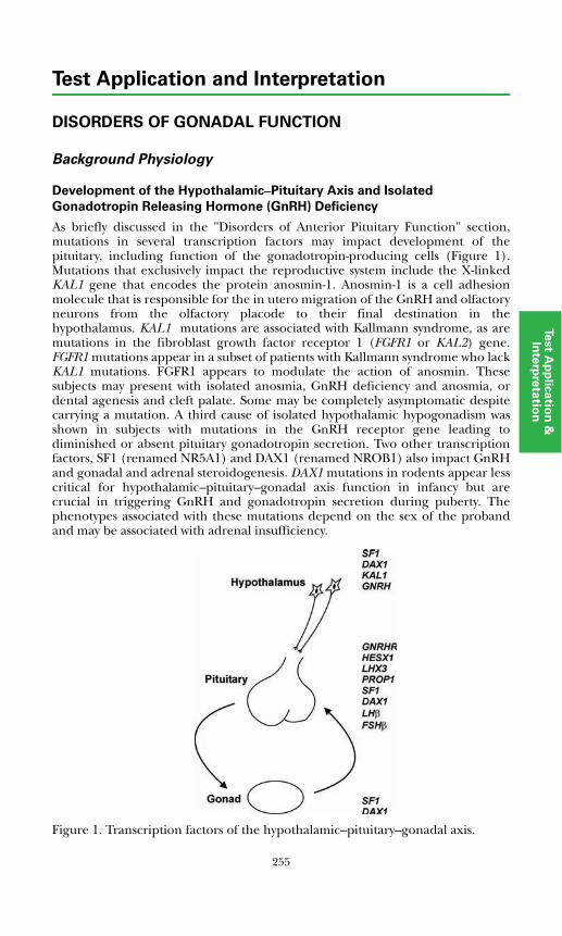
Test Application and Interpretation
Test A
pp
licatio
n &
In
terp
reta
tion
DISORDERS OF GONADAL FUNCTION
Background Physiology
Development of the Hypothalamic–Pituitary Axis and Isolated
Gonadotropin Releasing Hormone (GnRH) Deficiency
As briefly discussed in the "Disorders of Anterior Pituitary Function" section,mutations in several transcription factors may impact development of thepituitary, including function of the gonadotropin-producing cells (Figure 1).Mutations that exclusively impact the reproductive system include the X-linkedKAL1 gene that encodes the protein anosmin-1. Anosmin-1 is a cell adhesionmolecule that is responsible for the in utero migration of the GnRH and olfactoryneurons from the olfactory placode to their final destination in thehypothalamus. KAL1 mutations are associated with Kallmann syndrome, as aremutations in the fibroblast growth factor receptor 1 (FGFR1 or KAL2) gene.FGFR1 mutations appear in a subset of patients with Kallmann syndrome who lackKAL1 mutations. FGFR1 appears to modulate the action of anosmin. Thesesubjects may present with isolated anosmia, GnRH deficiency and anosmia, ordental agenesis and cleft palate. Some may be completely asymptomatic despitecarrying a mutation. A third cause of isolated hypothalamic hypogonadism wasshown in subjects with mutations in the GnRH receptor gene leading todiminished or absent pituitary gonadotropin secretion. Two other transcriptionfactors, SF1 (renamed NR5A1) and DAX1 (renamed NROB1) also impact GnRHand gonadal and adrenal steroidogenesis. DAX1 mutations in rodents appear lesscritical for hypothalamic–pituitary–gonadal axis function in infancy but arecrucial in triggering GnRH and gonadotropin secretion during puberty. Thephenotypes associated with these mutations depend on the sex of the probandand may be associated with adrenal insufficiency.
Figure 1. Transcription factors of the hypothalamic–pituitary–gonadal axis.
255
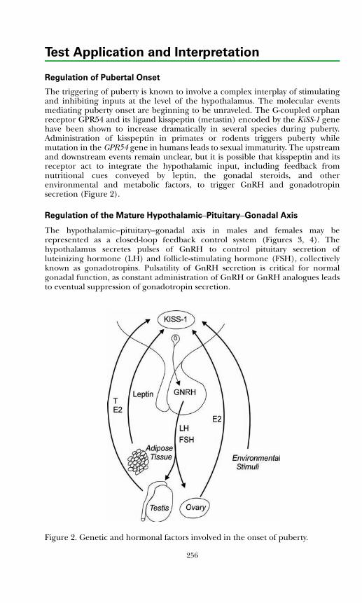
Test Application and Interpretation
Regulation of Pubertal Onset
The triggering of puberty is known to involve a complex interplay of stimulatingand inhibiting inputs at the level of the hypothalamus. The molecular eventsmediating puberty onset are beginning to be unraveled. The G-coupled orphanreceptor GPR54 and its ligand kisspeptin (metastin) encoded by the KiSS-1 genehave been shown to increase dramatically in several species during puberty.Administration of kisspeptin in primates or rodents triggers puberty whilemutation in the GPR54 gene in humans leads to sexual immaturity. The upstreamand downstream events remain unclear, but it is possible that kisspeptin and itsreceptor act to integrate the hypothalamic input, including feedback fromnutritional cues conveyed by leptin, the gonadal steroids, and otherenvironmental and metabolic factors, to trigger GnRH and gonadotropinsecretion (Figure 2).
Regulation of the Mature Hypothalamic–Pituitary–Gonadal Axis
The hypothalamic–pituitary–gonadal axis in males and females may berepresented as a closed-loop feedback control system (Figures 3, 4). Thehypothalamus secretes pulses of GnRH to control pituitary secretion ofluteinizing hormone (LH) and follicle-stimulating hormone (FSH), collectivelyknown as gonadotropins. Pulsatility of GnRH secretion is critical for normalgonadal function, as constant administration of GnRH or GnRH analogues leadsto eventual suppression of gonadotropin secretion.
Figure 2. Genetic and hormonal factors involved in the onset of puberty.
256
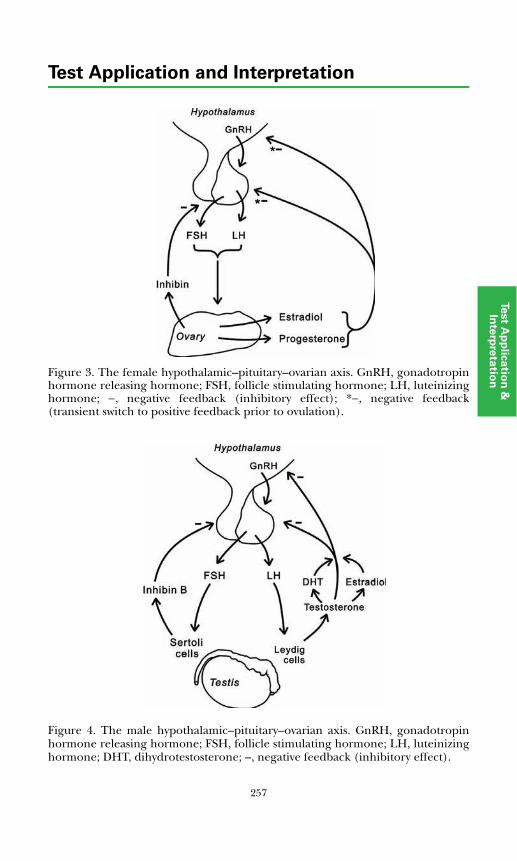
Test Application and Interpretation
Test A
pp
licatio
n &
In
terp
reta
tion
Figure 3. The female hypothalamic–pituitary–ovarian axis. GnRH, gonadotropinhormone releasing hormone; FSH, follicle stimulating hormone; LH, luteinizinghormone; −, negative feedback (inhibitory effect); *−, negative feedback(transient switch to positive feedback prior to ovulation).
Figure 4. The male hypothalamic–pituitary–ovarian axis. GnRH, gonadotropinhormone releasing hormone; FSH, follicle stimulating hormone; LH, luteinizinghormone; DHT, dihydrotestosterone; –, negative feedback (inhibitory effect).
257
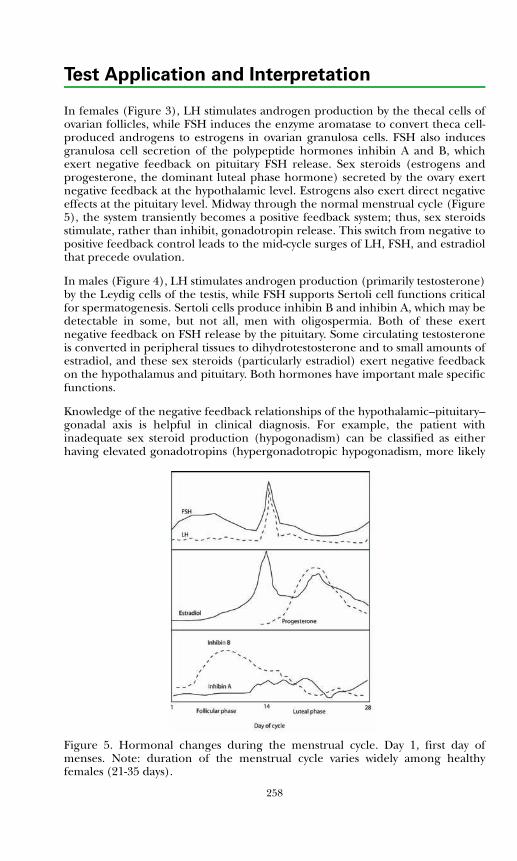
Test Application and Interpretation
In females (Figure 3), LH stimulates androgen production by the thecal cells ofovarian follicles, while FSH induces the enzyme aromatase to convert theca cell-produced androgens to estrogens in ovarian granulosa cells. FSH also inducesgranulosa cell secretion of the polypeptide hormones inhibin A and B, whichexert negative feedback on pituitary FSH release. Sex steroids (estrogens andprogesterone, the dominant luteal phase hormone) secreted by the ovary exertnegative feedback at the hypothalamic level. Estrogens also exert direct negativeeffects at the pituitary level. Midway through the normal menstrual cycle (Figure5), the system transiently becomes a positive feedback system; thus, sex steroidsstimulate, rather than inhibit, gonadotropin release. This switch from negative topositive feedback control leads to the mid-cycle surges of LH, FSH, and estradiolthat precede ovulation.
In males (Figure 4), LH stimulates androgen production (primarily testosterone)by the Leydig cells of the testis, while FSH supports Sertoli cell functions criticalfor spermatogenesis. Sertoli cells produce inhibin B and inhibin A, which may bedetectable in some, but not all, men with oligospermia. Both of these exertnegative feedback on FSH release by the pituitary. Some circulating testosteroneis converted in peripheral tissues to dihydrotestosterone and to small amounts ofestradiol, and these sex steroids (particularly estradiol) exert negative feedbackon the hypothalamus and pituitary. Both hormones have important male specificfunctions.
Knowledge of the negative feedback relationships of the hypothalamic–pituitary–gonadal axis is helpful in clinical diagnosis. For example, the patient withinadequate sex steroid production (hypogonadism) can be classified as eitherhaving elevated gonadotropins (hypergonadotropic hypogonadism, more likelya
Figure 5. Hormonal changes during the menstrual cycle. Day 1, first day ofmenses. Note: duration of the menstrual cycle varies widely among healthyfemales (21-35 days).
258
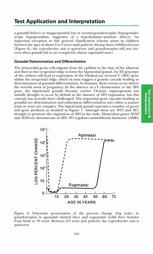
259
Test Application and Interpretation
Test A
pp
licatio
n &
In
terp
reta
tion
a gonadal defect) or inappropriately low or normal gonadotropins (hypogonado-tropic hypogonadism, suggestive of a hypothalamic–pituitary defect). Animportant exception to this general classification scheme arises in childrenbetween the ages of about 2 to 3 years until puberty; during these childhood years(Figure 6), the reproductive axis is quiescent, and gonadotropins will not rise,even when gonads fail or are completely absent (agonadal state).
Gonadal Determination and Differentiation
The primordial germ cells migrate from the epiblast to the base of the allantoisand then to the urogenital ridge to form the bipotential gonad. An XY genotypeof the embryo will lead to expression of the Y-linked sex reversal Y (SRY) genewithin the urogenital ridge, which in turn triggers a genetic cascade leading todetermination of gonadal differentiation. In humans, these events occur beforethe seventh week of pregnancy. In the absence of a Y chromosome or the SRYgene, the bipotential gonads become ovaries. Ovarian organogenesis wasinitially thought to occur by default in the absence of SRY expression, but thisconcept has recently been challenged. The sequential gene cascades leading togonadal sex determination and subsequent differentiation into either a maturetestis or ovary are complex. The bipotential gonad expresses a number of genesand gene products as detailed in Figure 7. Amongst these are WT1 and SF1,thought to promote the expression of SRY in the male. Homeobox genes SOX9and SOX8 are downstream of SRY. SF1 regulates antimüllerian hormone (AMH,leading to
Figure 6: Schematic presentation of the percent change (log scale) ingonadotropins in agonadal (dotted line) and eugonadal (solid line) femalesfrom birth to 70 years. Between 2-3 years and puberty, the reproductive axis isquiescent.
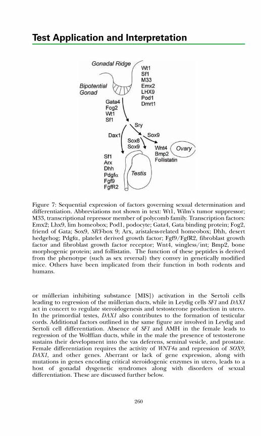
Test Application and Interpretation
Figure 7: Sequential expression of factors governing sexual determination anddifferentiation. Abbreviations not shown in text: Wt1, Wilm's tumor suppressor;M33, transcriptional repressor member of polycomb family. Transcription factors:Emx2; Lhx9, lim homeobox; Pod1, podocyte; Gata4, Gata binding protein; Fog2,friend of Gata; Sox9, SRY-box 9; Arx, aristaless-related homeobox; Dhh, deserthedgehog; Pdgfα, platelet derived growth factor; Fgf9/FgfR2, fibroblast growthfactor and fibroblast growth factor receptor; Wnt4, wingless/int; Bmp2, bonemorphogenic protein; and follistatin. The function of these peptides is derivedfrom the phenotype (such as sex reversal) they convey in genetically modifiedmice. Others have been implicated from their function in both rodents andhumans.
or müllerian inhibiting substance [MIS]) activation in the Sertoli cellsleading to regression of the müllerian ducts, while in Leydig cells SF1 and DAX1act in concert to regulate steroidogenesis and testosterone production in utero.In the primordial testes, DAX1 also contributes to the formation of testicularcords. Additional factors outlined in the same figure are involved in Leydig andSertoli cell differentiation. Absence of SF1 and AMH in the female leads toregression of the Wolffian ducts, while in the male the presence of testosteronesustains their development into the vas deferens, seminal vesicle, and prostate.Female differentiation requires the activity of WNT4a and repression of SOX9,DAX1, and other genes. Aberrant or lack of gene expression, along withmutations in genes encoding critical steroidogenic enzymes in utero, leads to ahost of gonadal dysgenetic syndromes along with disorders of sexualdifferentiation. These are discussed further below.
260
Test Application and Interpretation
Test A
pp
licatio
n &
In
terp
reta
tion
Measurement of Pituitary−Gonadal Axis Hormones
Luteinizing Hormone (LH, Lutropin)
LH consists of 2 subunits: an α-subunit (also found in FSH, TSH, and hCG) anda unique β-subunit. Competitive assays using polyclonal antibodies may demon-strate substantial cross-reaction with TSH and hCG due to presence of thecommon α-subunit. Such cross-reactivity is not seen with immunometric assays(eg, immunoradiometric [IRMA], immunochemiluminometric [ICMA], orimmunofluorometric [IFMA] assays). Immunometric assays also tend to be moresensitive. The most sensitive (ultrasensitive, third generation, pediatric) LHassays better distinguish central precocious puberty (increased LH) from the pre-pubertal state.
The pulsatile release and the relatively short (60-minute) circulatory half-life ofLH make a single LH measurement an unreliable estimate of the true serum LHconcentration, which may easily be 50% to 150% of the single measured value.An apparently low level of LH derived from a single sample is therefore of limitedclinical significance; it must be verified by repeat sampling and analysis.
Follicle Stimulating Hormone (FSH, Follitropin)
FSH, like LH, is a dimer comprised of a common α-subunit (same as for LH,TSH, and hCG) and a unique β-subunit. Unlike LH, FSH has a relatively longserum half-life (4-6 hours) and therefore shows less visible pulsatility. The majorapplications of FSH measurement are: 1) help differentiate pituitary fromgonadal causes of infertility (anovulation or defects in spermatogenesis); 2) assessovarian reserve (measurement of cycle day 3 FSH along with measurement ofestradiol and inhibin B); and 3) detection and differential diagnosis ofprecocious puberty (along with measurement of ultrasensitive LH and sex steroidlevels). While frequently used in the evaluation of menopausal symptoms, FSH isa surprisingly poor predictor of the exact timing of menopause.
Estrogens
In the non-pregnant woman, the endogenous estrogens of greatest significanceare estradiol (E2, 17β-estradiol) and the structurally similar, but less potent,estrone (E1). Estriol (E3) is the product of placental action on hormonessecreted by the fetal adrenal gland and is therefore an important indicator of thehealth of the feto-placental unit. Being a thousand-fold less potent than estradiol,E3 does not play a significant estrogenic role outside of pregnancy, however. Morethan 95% of circulating estrogens are bound to carrier proteins, including sexhormone-binding globulin (SHBG), cortisol-binding globulin (CBG), andalbumin. SHBG is the most important. Its production in the liver is increased byestrogens and thyroid hormones and decreased by androgens and growthhormone. Testosterone competes with estrogens for SHBG binding sites. Inpatients with androgen excess or deficiency, hypo- or hyperthyroidism, pituitarydysfunction, liver disease, or gonadal dysfunction, the measurement of freeestradiol may provide a more accurate assessment of bioavailable estrogen.
261
Test Application and Interpretation
Synthetic estrogens such as ethinyl estradiol (typically found in oralcontraceptives) are not detected by most estradiol assays currently in use. Many"estradiol" assays still in use show significant cross-reactivity to estrone, whichexplains why they will detect a medication such as Premarin®' which is primarilycomprised of horse estrogens (equilins) and estrone metabolites. Such assays canalso be used to document the effect of oral 17β-estradiol, which is largelybioconverted to estrone in the intestinal mucosa. More recent estradiol assaysshow a much higher analytical specificity for estradiol. However, when maximalanalytical sensitivity and specificity are needed (eg, during evaluation ofgynecomastia or in prepubertal children) or when reported estradiol levels arenot consistent with the clinical picture, the assay should be performed using LC/MS/MS technology. The LC/MS/MS assay is superior in its sensitivity andspecificity relative to all immunoassays. Table 1 summarizes the 2 estradiol assayscurrently offered by Quest Diagnostics. E1 and free E2 can also be measured byLC/MS/MS.
Testosterone
There are 3 major categories of serum testosterone measurements: 1) totaltestosterone; 2) free testosterone, the portion not bound to sex-hormone bindingglobulin (SHBG) or albumin; and 3) free plus weakly-bound testosterone, ie,"bioavailable testosterone." The total testosterone measurement is crucial, since itis the basis for calculation of free and bioavailable testosterone. Thus, a falseestimate of the total testosterone will translate to a false estimate of the free andbioavailable testosterone. When low values of testosterone are anticipated (eg,prepubertal males, all females) and when measuring levels in young infants inwhom interfering substances are common, total testosterone is best measured byLC/MS/MS (or by RIA, preceded by extraction and chromatography if LC/MS/MS is unavailable). In both of these populations, the results derived from direct Table 1. Characteristics of the Estradiol Assays Offered by Quest Diagnostics
LC/MS/MS Immunoassay
Test code 30289X 4021
Clinical use Evaluate gynecomastia inprepubertal children
IVF protocolsMonitor HRT (patch)
Cross-reactivityConjugated equine estrogens (Premarin)
Ethinyl estradiol
Oral 17β-estradiol
Transdermal 17β-estradiol
Aqueous interfering compounds
None
None
None
Yes
None
Minimal
None?
Minimal
Yes
Some
LC/MS/MS, liquid chromatography tandem mass spectrometry; HRT, hormone replace-ment therapy; IVF, in vitro fertilization.
262
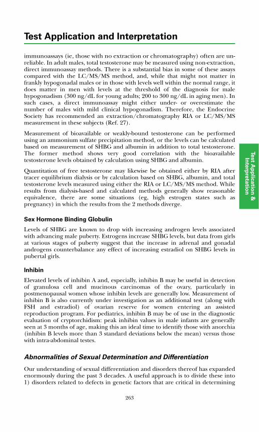
Test Application and Interpretation
Test A
pp
licatio
n &
In
terp
reta
tion
immunoassays (ie, those with no extraction or chromatography) often are un-reliable. In adult males, total testosterone may be measured using non-extraction,direct immunoassay methods. There is a substantial bias in some of these assayscompared with the LC/MS/MS method, and, while that might not matter infrankly hypogonadal males or in those with levels well within the normal range, itdoes matter in men with levels at the threshold of the diagnosis for malehypogonadism (300 ng/dL for young adults; 200 to 300 ng/dL in aging men). Insuch cases, a direct immunoassay might either under- or overestimate thenumber of males with mild clinical hypogonadism. Therefore, the EndocrineSociety has recommended an extraction/chromatography RIA or LC/MS/MSmeasurement in these subjects (Ref. 27).
Measurement of bioavailable or weakly-bound testosterone can be performedusing an ammonium sulfate precipitation method, or the levels can be calculatedbased on measurement of SHBG and albumin in addition to total testosterone.The former method shows very good correlation with the bioavailabletestosterone levels obtained by calculation using SHBG and albumin.
Quantitation of free testosterone may likewise be obtained either by RIA aftertracer equilibrium dialysis or by calculation based on SHBG, albumin, and totaltestosterone levels measured using either the RIA or LC/MS/MS method. Whileresults from dialysis-based and calculated methods generally show reasonableequivalence, there are some situations (eg, high estrogen states such aspregnancy) in which the results from the 2 methods diverge.
Sex Hormone Binding Globulin
Levels of SHBG are known to drop with increasing androgen levels associatedwith advancing male puberty. Estrogens increase SHBG levels, but data from girlsat various stages of puberty suggest that the increase in adrenal and gonadalandrogens counterbalance any effect of increasing estradiol on SHBG levels inpubertal girls.
Inhibin
Elevated levels of inhibin A and, especially, inhibin B may be useful in detectionof granulosa cell and mucinous carcinomas of the ovary, particularly inpostmenopausal women whose inhibin levels are generally low. Measurement ofinhibin B is also currently under investigation as an additional test (along withFSH and estradiol) of ovarian reserve for women entering an assistedreproduction program. For pediatrics, inhibin B may be of use in the diagnosticevaluation of cryptorchidism: peak inhibin values in male infants are generallyseen at 3 months of age, making this an ideal time to identify those with anorchia(inhibin B levels more than 3 standard deviations below the mean) versus thosewith intra-abdominal testes.
Abnormalities of Sexual Determination and Differentiation
Our understanding of sexual differentiation and disorders thereof has expandedenormously during the past 3 decades. A useful approach is to divide these into1) disorders related to defects in genetic factors that are critical in determining
263
Test Application and Interpretation
the fate of the bipotential gonad before the seventh week of pregnancy, and 2)subsequent differentiation and steroidogenetic enzymatic defects that lead toambiguous genitalia recognized during a newborn examination. The diagnosticcapabilities for evaluating such infants have improved with our understanding ofthe complexity of the sexual differentiation process. Table 2 summarizes most ofthe disorders associated with sex reversal (discrepancy between chromosomaland phenotypic sex) and associated nonsexual developmental defects. Table 3summarizes the steroidogenic enzyme defects and chromosomal abnormalitiesassociated with ambiguous genitalia and laboratory tests that may be helpful intheir evaluation.
Disorders of Sexual Maturation
Normal Puberty Staging
It is convenient to consider disorders of sexual maturation in 2 broad categories:those associated with early or precocious puberty and those associated withdelayed or absent puberty. In any patient with abnormal sexual development, it isimportant to note whether the genitalia are normal and consistent with thegenetic sex; ie, the phenotypic and genetic sex are identical. Often,determination of genetic sex requires diagnostic laboratory evaluation. Thestaging of normal puberty as outlined by Tanner is summarized in Table 4.
Timing of Physical Changes of Puberty
The normal range for puberty onset in girls has been somewhat controversial.Data from a large cross-sectional study of American girls suggest that the lowercutoff for puberty in white girls may be as young as 7 years (6 years for AfricanAmerican girls). Other data suggest that the ± 2 standard deviation (SD) rangefor breast development (thelarche) in white girls is approximately 8 to 13 years.The ± 2 SD for onset of menses (menarche) is approximately 10.5 to 15 years forwhite girls. On average, African American girls tend to have thelarche andmenarche about 6 months earlier than do white girls.
For boys, the normal range for onset of testicular enlargement (testicular volume3 mL or greater, or testicular length along the longest axis >1 inch) or genitalenlargement has traditionally been 9 to 14 years, although some recent datasuggest that a significant number of boys exhibit genital changes by 8 to 9 years ofage.
Table 2. Genetic Disorders of Steroidogenesis and External Genitalia Differentiation
Gene Mutant PhenotypeWT1 Frasier syndrome, Denys-Drash syndrome with
Wilms tumorSF1 Gonadal and adrenal dysgenesisSOX9 Gonadal dysgenesis, congenital adrenal hypoplasiaSRY Gonadal dysgenesisAMH (MIS) Persistent müllerian duct syndrome, type IAMHR (MIS receptor) Persistent müllerian duct syndrome, type II
264
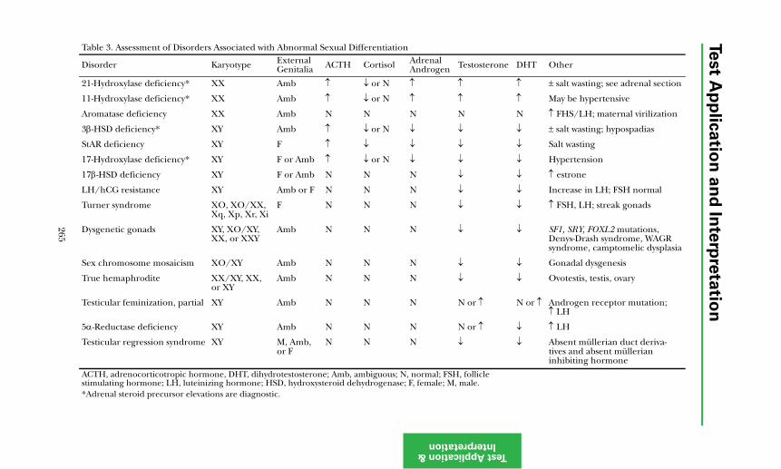
Test A
pp
lica
tion
an
d In
terp
reta
tion
Test ApplicatioInterpretatio
Table 3. Assessment of Disorders Associated with Abnormal Sexual Differentiation
Disorder Karyotype External Genitalia ACTH Cortisol Adrenal
Androgen Testosterone DHT Other
21-Hydroxylase deficiency* XX Amb ↑ ↓ or N ↑ ↑ ↑ ± salt wasting; see adrenal section
↑ ↑ May be hypertensive
N N ↑ FHS/LH; maternal virilization
↓ ↓ ± salt wasting; hypospadias
↓ ↓ Salt wasting
↓ ↓ Hypertension
↓ ↓ ↑ estrone
↓ ↓ Increase in LH; FSH normal
↓ ↓ ↑ FSH, LH; streak gonads
↓ ↓ SF1, SRY, FOXL2 mutations, Denys-Drash syndrome, WAGR syndrome, camptomelic dysplasia
↓ ↓ Gonadal dysgenesis
↓ ↓ Ovotestis, testis, ovary
N or ↑ N or ↑ Androgen receptor mutation; ↑ LH
N or ↑ ↓ ↑ LH
↓ ↓ Absent müllerian duct deriva-tives and absent müllerian inhibiting hormone
; FSH, follicle e; M, male.
265
n & n
11-Hydroxylase deficiency* XX Amb ↑ ↓ or N ↑
Aromatase deficiency XX Amb N N N
3β-HSD deficiency* XY Amb ↑ ↓ or N ↓
StAR deficiency XY F ↑ ↓ ↓
17-Hydroxylase deficiency* XY F or Amb ↑ ↓ or N ↓
17β-HSD deficiency XY F or Amb N N N
LH/hCG resistance XY Amb or F N N N
Turner syndrome XO, XO/XX, Xq, Xp, Xr, Xi
F N N N
Dysgenetic gonads XY, XO/XY, XX, or XXY
Amb N N N
Sex chromosome mosaicism XO/XY Amb N N N
True hemaphrodite XX/XY, XX, or XY
Amb N N N
Testicular feminization, partial XY Amb N N N
5α-Reductase deficiency XY Amb N N N
Testicular regression syndrome XY M, Amb, or F
N N N
ACTH, adrenocorticotropic hormone, DHT, dihydrotestosterone; Amb, ambiguous; N, normalstimulating hormone; LH, luteinizing hormone; HSD, hydroxysteroid dehydrogenase; F, femal*Adrenal steroid precursor elevations are diagnostic.
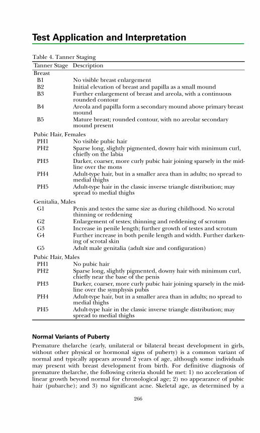
Test Application and Interpretation
Table 4. Tanner Staging
Normal Variants of Puberty
Premature thelarche (early, unilateral or bilateral breast development in girls,without other physical or hormonal signs of puberty) is a common variant ofnormal and typically appears around 2 years of age, although some individualsmay present with breast development from birth. For definitive diagnosis ofpremature thelarche, the following criteria should be met: 1) no acceleration oflinear growth beyond normal for chronological age; 2) no appearance of pubichair (pubarche); and 3) no significant acne. Skeletal age, as determined by a
Tanner Stage DescriptionBreast
B1 No visible breast enlargement B2 Initial elevation of breast and papilla as a small moundB3 Further enlargement of breast and areola, with a continuous
rounded contourB4 Areola and papilla form a secondary mound above primary breast
moundB5 Mature breast; rounded contour, with no areolar secondary
mound present
Pubic Hair, FemalesPH1 No visible pubic hair PH2 Sparse long, slightly pigmented, downy hair with minimum curl,
chiefly on the labiaPH3 Darker, coarser, more curly pubic hair joining sparsely in the mid-
line over the monsPH4 Adult-type hair, but in a smaller area than in adults; no spread to
medial thighsPH5 Adult-type hair in the classic inverse triangle distribution; may
spread to medial thighs
Genitalia, MalesG1 Penis and testes the same size as during childhood. No scrotal
thinning or reddeningG2 Enlargement of testes; thinning and reddening of scrotumG3 Increase in penile length; further growth of testes and scrotumG4 Further increase in both penile length and width. Further darken-
ing of scrotal skinG5 Adult male genitalia (adult size and configuration)
Pubic Hair, MalesPH1 No pubic hairPH2 Sparse long, slightly pigmented, downy hair with minimum curl,
chiefly near the base of the penis PH3 Darker, coarser, more curly pubic hair joining sparsely in the mid-
line over the symphysis pubisPH4 Adult-type hair, but in a smaller area than in adults; no spread to
medial thighsPH5 Adult-type hair in the classic inverse triangle distribution; may
spread to medial thighs
266
Test Application and Interpretation
Test A
pp
licatio
n &
In
terp
reta
tion
bone X-ray of the left hand and wrist, should not be advanced beyond 2 SDrelative to the chronological age. Extensive laboratory testing is generally notnecessary if the presentation is relatively classic, but periodic clinical follow-up isrecommended.
Premature adrenarche is the early appearance of pubic (occasionally axillary)hair, apocrine body odor, and mild acne. There should be: 1) no significantgrowth acceleration; 2) no other signs of androgen effect such as penile orclitoral enlargement; and 3) no breast development (thelarche) in girls ortesticular enlargement in boys. As with premature thelarche, the skeletal ageshould not be significantly advanced (greater than +2 SD). There appears to beno significant sequelae in boys, but premature adrenarche in some girls may bean early harbinger of insulin resistance and the polycystic ovarian syndrome. Ifpremature adrenarche is particularly rapid or "explosive" in onset, measurementof adrenal and gonadal steroids (eg, 17-hydroxyprogesterone, testosterone)should be considered to rule out entities such as congenital adrenal hyperplasiaor adrenal/gonadal tumors. DHEA-sulfate levels are typically in the mid-pubertalrange in premature adrenarche; much higher levels of DHEA-sulfate suggest aneed for further testing, including imaging of the adrenals.
Precocious Puberty
There are a large number of causes of precocious sexual development (Table 5).As a general rule, precocious puberty in girls is more often idiopathic, whileorganic causes (eg, central nervous system lesions and tumors) are more likely inboys with true central precocious puberty.
Ultrasound examination of the pelvis in such girls provides information aboutovarian and uterine size; uterine volumes of >1.8 mL and/or ovarian volumes>1.2 mL are consistent with onset of true puberty. Measurements of serumgonadotropin and estradiol levels and gonadotropin response to gonadotropinreleasing hormone (GnRH) usually allow differential diagnosis of prematurethelarche, central precocious puberty, and gonadotropin-independent precocity.LH and FSH usually are detectable and in the pubertal range in gonadotropin-dependent and low or undetectable in gonadotropin-independent precociouspuberty.
In boys, measurement of serum gonadotropin and testosterone levels also isnecessary, and if basal LH levels are low, GnRH stimulation will help differentiategonadotropin-dependent and -independent precocity. The clinical suspicion ofintracranial pathology in either sex requires cranial imaging. Treatment ofcentral precocious puberty with long-acting GnRH analogues requires GnRHtesting to assess treatment response.
Delayed or Absent Puberty
Delayed puberty is generally defined as the absence of pubertal sexualcharacteristics by age 13 in girls and age 14 in boys; a small percent (2.5%) of thenormal population mature in later years. The major causes of delayed puberty arelisted in Table 6. Each of these major causes may in turn be subdivided intomultiple etiologies. For example, anorexia nervosa, starvation, athletic orexercise amenorrhea, and the rare zinc deficiency can all be related to the broad
267
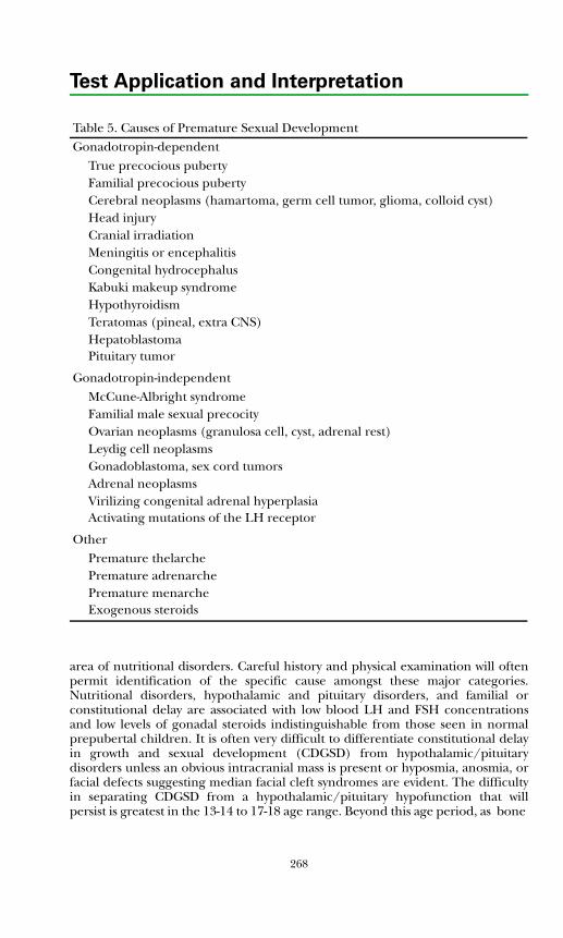
Test Application and Interpretation
area of nutritional disorders. Careful history and physical examination will oftenpermit identification of the specific cause amongst these major categories.Nutritional disorders, hypothalamic and pituitary disorders, and familial orconstitutional delay are associated with low blood LH and FSH concentrationsand low levels of gonadal steroids indistinguishable from those seen in normalprepubertal children. It is often very difficult to differentiate constitutional delayin growth and sexual development (CDGSD) from hypothalamic/pituitarydisorders unless an obvious intracranial mass is present or hyposmia, anosmia, orfacial defects suggesting median facial cleft syndromes are evident. The difficultyin separating CDGSD from a hypothalamic/pituitary hypofunction that willpersist is greatest in the 13-14 to 17-18 age range. Beyond this age period, as bone
Table 5. Causes of Premature Sexual DevelopmentGonadotropin-dependent
True precocious pubertyFamilial precocious pubertyCerebral neoplasms (hamartoma, germ cell tumor, glioma, colloid cyst)Head injuryCranial irradiationMeningitis or encephalitisCongenital hydrocephalusKabuki makeup syndromeHypothyroidismTeratomas (pineal, extra CNS)HepatoblastomaPituitary tumor
Gonadotropin-independentMcCune-Albright syndromeFamilial male sexual precocityOvarian neoplasms (granulosa cell, cyst, adrenal rest)Leydig cell neoplasmsGonadoblastoma, sex cord tumorsAdrenal neoplasmsVirilizing congenital adrenal hyperplasiaActivating mutations of the LH receptor
OtherPremature thelarchePremature adrenarchePremature menarcheExogenous steroids
268
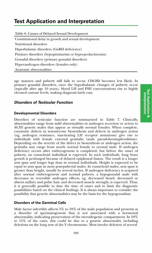
Test Application and Interpretation
Test A
pp
licatio
n &
In
terp
reta
tion
age matures and puberty still fails to occur, CDGSD becomes less likely. Inprimary gonadal disorders, once the hypothalamic changes of puberty occur(typically after age 10 years), blood LH and FSH concentrations rise to highlyelevated castrate levels, making diagnosis fairly easy.
Disorders of Testicular Function
Developmental Disorders
Disorders of testicular function are summarized in Table 7. Clinically,abnormalities range from mild abnormalities in androgen secretion or action to46,XY genetic males that appear as virtually normal females. When complete,enzymatic defects in testosterone biosynthesis and defects in androgen action(eg, androgen resistance, inactivating LH receptor mutations) give rise toindividuals with female external genitalia (male pseudohermaphroditism).Depending on the severity of the defect in biosynthesis or androgen action, thegenitalia may range from nearly normal female to normal male. If androgendeficiency occurs after embryogenesis is completed, but before the onset ofpuberty, an eunuchoid individual is expected. In such individuals, long bonegrowth is prolonged because of delayed epiphyseal fusion. The result is a longerarm span and longer legs than in normal individuals. Height is expected to beequal to arm span in most post-pubertal males. In eunuchoid males, arm span isgreater than height, usually by several inches. If androgen deficiency is acquiredafter normal embryogenesis and normal puberty, a hypogonadal male withdecreases in reversible androgen effects, eg, decreased beard, decreased orabsent axillary and pubic hair, and decreased muscle strength, is expected. Thus,it is generally possible to date the time of onset and to limit the diagnosticpossibilities based on the clinical findings. It is always important to consider thepossibility that genetic abnormalities may be the basis for the hypogonadism.
Disorders of the Germinal Cells
Male factor infertility affects 5% to 10% of the male population and presents asa disorder of spermatogenesis that is not associated with a hormonalabnormality, indicating preservation of the steroidogenic compartment. In 10%to 15% of the cases, this could be due to a genetic abnormality includingdeletions on the long arm of the Y chromosome. Most involve deletion of several
Table 6. Causes of Delayed Sexual Development
Constitutional delay in growth and sexual developmentNutritional disordersHypothalamic disorders (GnRH deficiency)Pituitary disorders (hypopituitarism or hyperprolactinemia)Gonadal disorders (primary gonadal disorders)Hyperandrogen disorders (females only)Anatomic abnormalities
269
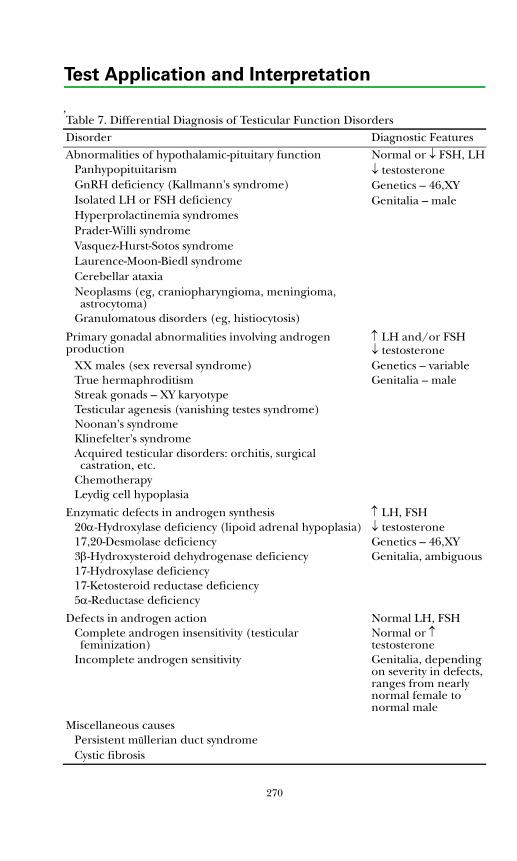
Test Application and Interpretation
,Table 7. Differential Diagnosis of Testicular Function DisordersDisorder Diagnostic FeaturesAbnormalities of hypothalamic-pituitary function Normal or ↓ FSH, LH
Panhypopituitarism ↓ testosteroneGnRH deficiency (Kallmann's syndrome) Genetics – 46,XYIsolated LH or FSH deficiency Genitalia – maleHyperprolactinemia syndromesPrader-Willi syndromeVasquez-Hurst-Sotos syndromeLaurence-Moon-Biedl syndromeCerebellar ataxiaNeoplasms (eg, craniopharyngioma, meningioma, astrocytoma)
Granulomatous disorders (eg, histiocytosis)
Primary gonadal abnormalities involving androgen production
↑ LH and/or FSH↓ testosterone
XX males (sex reversal syndrome) Genetics – variableTrue hermaphroditism Genitalia – maleStreak gonads – XY karyotypeTesticular agenesis (vanishing testes syndrome)Noonan's syndromeKlinefelter's syndromeAcquired testicular disorders: orchitis, surgical castration, etc.
ChemotherapyLeydig cell hypoplasia
Enzymatic defects in androgen synthesis ↑ LH, FSH20α-Hydroxylase deficiency (lipoid adrenal hypoplasia) ↓ testosterone17,20-Desmolase deficiency Genetics – 46,XY3β-Hydroxysteroid dehydrogenase deficiency Genitalia, ambiguous17-Hydroxylase deficiency17-Ketosteroid reductase deficiency5α-Reductase deficiency
Defects in androgen action Normal LH, FSHComplete androgen insensitivity (testicular feminization)
Normal or ↑ testosterone
Incomplete androgen sensitivity Genitalia, depending on severity in defects, ranges from nearly normal female to normal male
Miscellaneous causesPersistent müllerian duct syndromeCystic fibrosis
270
Test Application and Interpretation
Test A
pp
licatio
n &
In
terp
reta
tion
copies of the DAZ (deleted in azoospermia) gene. CFTR (cystic fibrosis) mutationslead to congenital absence of the vas deferens and subsequent infertility.Androgen receptor mutations have been documented in azoospermic individualswho appear well androgenized and have normal testes size. The only hint to thediagnosis is slightly elevated LH and testosterone levels. The most frequent sexchromosome aneuploidy leading to azoospermia is Klinefelter syndrome (XXY)with a prevalence of 0.1% to 0.2% in the general population and 3.1% in theinfertile population. The disorder primarily leads to early failure ofspermatogenesis followed by a subsequent failure of steroidogenesis presentingas primary hypogonadism. The symptoms of hypogonadism might be subtle anddelayed as testosterone may be maintained in the lower normal range at theexpense of an elevated LH. Depending on the degree of hypogonadism, it mightcome to attention as delayed puberty in a subject with a eunuchoidal bodyhabitus and small testes or as male factor infertility associated with azoospermia.Mutations in the Insl3 (insulin-like factor 3) gene have been reported in 5% to10% percent of all subjects with cryptorchidism. With the advent ofintracytoplasmic sperm injection (ICSI), it is now possible for many couples withan affected male partner to father a child. Determining presence of an abnormalkaryotype or the Insl3 mutation prior to ICSI may be of value for geneticcounseling purposes, as the newborn may also be affected and infertile. This isespecially relevant in men with mild cystic fibrosis mutations, leading tocongenital absence of the vas deferens as the sole manifestation of the disease,and in subjects with Klinefelter syndrome.
Testicular Aging
The steroidogenic capacity of the testes decreases with age, and there is a gradualdecline in testosterone levels below the lower cutoff of the young adult range.This decrease is usually not associated with an increase in LH and FSH, indicatingthat reproductive aging occurs both at the testicular and pituitary levels. Thistestosterone decrease becomes more pronounced each decade, and its extentdepends on the baseline from which the testosterone level declines in eachindividual. This decrease may be associated with increased frailty, decreasedmuscle mass, osteoporosis, and an increase in cardiovascular risk in the agingpopulation collectively labeled as the andropause. It is not clear whether thedecrease in testosterone in older men, while observationally correct, is actuallyphysiologic. Some preliminary, small, testosterone replacement trials show somebenefit in older men: increased muscle mass, and possibly strength, along withimprovement in bone mineral density. A large multicenter efficacy trial oftestosterone in older men mandated by the Institute of Medicine and funded bythe National Institutes of Health is under way. Primary endpoints include musclestrength, cardiovascular biomarker levels, bone mineral density, and boneturnover marker levels. Secondary end points are related to safety of replacementtherapy such as increased risk of benign prostatic hyperplasia and possiblyprostate cancer. In the interim, and until the results of this and other trialsbecome available, the ideal reference range of testosterone for the aging menremains a subject of debate. The Endocrine Society consensus guidelines on themanagement of hypogonadism, and similar guidelines from the AmericanAssociation of Clinical Endocrinologists, indicate that men with persistenthypogonadal symptoms and low testosterone levels (ie, below the lower cutoff for
271
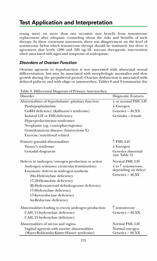
Test Application and Interpretation
young men) on more than one occasion may benefit from testosteronereplacement after adequate counseling about the risks and benefits of suchtherapy. In these consensus statements, there was disagreement on the level oftestosterone below which testosterone therapy should be instituted, but there isagreement that levels ≤200 and 320 ng/dL warrant therapeutic interventionwhen associated with signs and symptoms of andropause.
Disorders of Ovarian Function
Ovarian agenesis or hypofunction is not associated with abnormal sexualdifferentiation, but may be associated with morphologic anomalies and slowgrowth during the prepubertal period. Ovarian dysfunction is associated withdelayed puberty and with oligo or amenorrhea. Tables 8 and 9 summarize thed
Table 8. Differential Diagnosis of Primary AmenorrheaDisorder Diagnostic FeaturesAbnormalities of hypothalamic–pituitary function ↓ or normal FSH, LH
Panhypopituitarism ↓ EstrogenGnRH deficiency (Kallmann's syndrome) Genetics – 46,XXIsolated LH or FSH deficiency Genitalia – femaleHyperprolactinemia syndromesNeoplasms (eg, craniopharyngioma)Granulomatous diseases (histiocytosis X)Exercise/nutritional related
Primary gonadal abnormalities ↑ FSH, LHTurner's syndrome ↓ EstrogenGonadal dysgenesis Genetics abnormal
(see Table 3)
Defects in androgen/estrogen production or actionAndrogen resistance (testicular feminization)Enzymatic defects in androgen synthesis
20α-Hydroxylase deficiency17,20-Desmolase deficiency3β-Hydroxysteroid dehydrogenase deficiency17-Hydroxylase deficiency17-Ketoreductase deficiency5α-Reductase deficiency
Normal FSH, LH↓ to ↑ testosterone, depending on defectGenetics – 46,XY
Abnormalities leading to excess androgen production ↑ testosteroneCAH, 11-hydroxylase deficiency Genetics – 46,XXCAH, 21-hydroxylase deficiency
Abnormalities of uterus and vagina Normal FSH, LHVaginal agenesis with uterine abnormalities (Mayer-Rokitansky-Küster-Hauser syndrome)
Normal estrogenGenetics – 46,XX
272
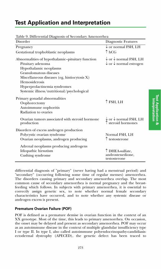
Test Application and Interpretation
Test A
pp
licatio
n &
In
terp
reta
tion
differential diagnosis of "primary" (never having had a menstrual period) and"secondary" (occurring following some time of regular menses) amenorrhea.The disorders causing primary and secondary amenorrhea overlap. The mostcommon cause of secondary amenorrhea is normal pregnancy and the breast-feeding which follows. In subjects with primary amenorrhea, it is essential tocorrectly assign genetic sex, to note whether normal female secondarycharacteristics have occurred, and to note whether any systemic disease orandrogen excess is present.
Premature Ovarian Failure (POF)
POF is defined as a premature demise in ovarian function in the context of anXX genotype. Most of the time, this leads to primary amenorrhea. On occasion,the onset may be delayed and present as secondary amenorrhea. POF may occuras an autoimmune disease in the context of multiple glandular insufficiency typeI or type II. In type I, also called autoimmune polyendocrinopathy-candidiasis-ectodermal dystrophy (APECED), the genetic defect has been traced to
Table 9. Differential Diagnosis of Secondary AmenorrheaDisorder Diagnostic FeaturesPregnancy ↓ or normal FSH, LHGestational trophoblastic neoplasms ↑ hCG
Abnormalities of hypothalamic–pituitary functionPituitary adenomaHypothalamic neoplasmsGranulomatous diseasesMiscellaneous diseases (eg, histiocytosis X)HemosiderosisHyperprolactinemia syndromesSystemic illness/nutritional/psychological
↓ or ↓ normal FSH, LH↓ or ↓ normal estrogen
Primary gonadal abnormalitiesOophorectomyAutoimmune oophoritis Radiation to ovaries
↑ FSH, LH
Ovarian tumors associated with steroid hormone production
↓ or ↓ normal FSH, LH↑ steroid hormones
Disorders of excess androgen productionPolycystic ovarian syndromeOvarian neoplasms, androgen producing
Normal FSH, LH ↑ testosterone
Adrenal neoplasms producing androgensIdiopathic hirsutismCushing syndrome
↑ DHEA-sulfate, androstenedione, testosterone
273
Test Application and Interpretation
mutations in the transcriptional activator autoimmune regulator gene (AIRE).Autoimmune ovarian failure may also be associated with type II polyglandularinsufficiency, although no genetic abnormality has been identified for thissyndrome. When not associated with other autoimmune disease, POF is alsoknown as XX gonadal dysgenesis. Affected females have no other dysmorphicfeatures, and their clinical presentation is limited to the consequences of earlyovarian failure, streak ovaries by ultrasound, and elevated LH and FSH levels.There are candidate loci on the long arm of the X chromosome associated withthis disease (POF1 and POF2). The strongest evidence for the POF1 locus is forthe fragile X mental retardation 1 (FMR1) gene, followed by an X chromosomedeletion that disrupts three candidate genes: heparan sulfate 6-O-sulfotransferase(HS6ST), E2F-related transcription factor, and glypican 3 (GPC3). There is alsoevidence implicating 2 other genes on the POF2 locus that affect 3 othercandidate genes: diaphanous homolog 2 (DIAPH2), dachshund homolog 2(DACH2), and premature ovarian failure, 1B (POF1B). The most prominentautosomal gene implicated in this phenotype is the FSH receptor. A growing listof other genes is constantly added to this repertoire and has been recentlyreviewed by Fassnacht et al (Ref. 22).
Polycystic Ovarian Syndrome (PCOS)
PCOS is extremely prevalent amongst females of all ages and leads to importantreproductive (infertility) and nonreproductive comorbidities. It is also associatedwith an increased risk of cardiovascular disease. The etiology is not clear andis probably multifactorial, but there is familial clustering of such cases.PCOS presents with menstrual irregularities ranging from secondaryamenorrhea to oligomenorrhea. It is also associated with increased adrenal andovarian androgen production, which leads to varying degrees of hirsutismincluding male pattern baldness, acne, and, occasionally, virilization. The ovariesare polycystic with associated failure of ovulation and absent corpus luteumformation. Progesterone secretion is decreased, leading to unopposed estrogenaction with resultant endometrial hyperplasia and increased risk of endometrialcancer. PCOS frequently occurs in obese women. It is felt that the insulinresistance in such women may be at the center of the reproductive abnormalities,since increased insulin levels may increase androgen production in the ovariantheca cells.
A consensus conference convened in Rotterdam in 2003 established the minimaldiagnostic criteria and work-up required for the diagnosis of PCOS. Patients needto fulfill 2 of 3 criteria: 1) presence of polycystic ovaries (with predefinedmorphologic features) on an abdominal or transvaginal ovarian ultrasound; 2)oligo- or anovulation; 3) clinical or biochemical evidence of hyperandrogenism.Exclusion of other causes of polycystic ovaries, while not stated as a criterion, isimplicit. Recent guidelines by the Androgen Excess Society differ somewhat asthey combine the first and second criteria into one, add hyperandrogenism(clinical or biochemical) as an essential component, and add exclusion of otherconditions leading to hyperandrogenism as a third criterion. This definitionexcludes women who have oligo-amenorrhea and polycystic ovaries but nohyperandrogenism from the diagnosis.
274
Test Application and Interpretation
Test A
pp
licatio
n &
In
terp
reta
tion
Many other diseases may masquerade as PCOS. These include Cushing's disease,abnormalities in thyroid function, nonclassical 21-hydoxylase deficiency,hyperprolactinemia, and adrenal or ovarian virilizing tumors. The diagnosticworkup should include laboratory tests guided by the clinical presentation. Aminimum laboratory workup includes:
• Total and free testosterone, as free testosterone is more sensitive in detectingbiochemical hyperandrogenism. The recommended method(s) for totaltestosterone is LC/MS/MS or a chromatography/ extraction RIA, since theanalogue assays are highly inaccurate in this context. Free testosterone canthen be determined by either the calculation or dialysis method.
• DHEA-S (sole androgen elevated in 10% of all cases)
• Tests aimed at ruling out other entities: prolactin, 17-OH progesterone, andTSH; 24-hour cortisol or estradiol, along with LH and FSH, are not necessaryunless there is strong clinical suspicion of Cushing's disease orhypogonadotropic hypogonadism. The LH to FSH ratio usually is increasedin PCOS but is highly variable and is not useful as a diagnostic criterion.
• Measures of insulin resistance such as a fasting glucose, OGTT, and a fastinglipid profile should be performed. Dietary modification, a decrease in bodyweight, or metformin therapy may lead decreased androgen production andresumption of ovulation and menses.
Others tests may be useful but are not universally endorsed. These includeandrostenedione, glucose/insulin ratio, or homeostatic model assessment ofinsulin resistance (HOMA-IR) for diagnosis of insulin resistance. It is importantto make the diagnosis of PCOS, not only because of its impact on reproductivefunction, but because of the opportunity it provides for early interventiondesigned to prevent long-term cardiovascular and metabolic comorbidities.
Assessment of Functional Gonadal Tumors
Nearly all of the cell types in the testis and ovary can undergo neoplastictransformation. The majority of testicular neoplasms, however, are germ celltumors that account for 90% to 95% of testicular cancer. These tumors originateearly in life as pre-invasive precursors that transform into overt tumors duringyoung adulthood. Such tumors may be as frequent as 1 in 10,000 young whitemales. Other testicular tumor types, and the usual hormonal changes, aresummarized in Table 10.
Ovarian tumors with endocrine manifestations constitute <5% of all ovarianneoplasms and <10% of malignant ovarian cancer. However, as many as 50% mayproduce estrogens at a subclinical rate as evidenced by maturation in vaginalsmears from postmenopausal women with ovarian cancer. Sex cord stromaltumors (those with granulosa cells, theca cells, Sertoli cells, or Leydig cells singlyor in combination) are the neoplasms most frequently associated with theendocrine manifestations shown in Table 10.
275
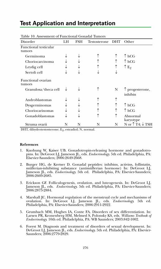
Test Application and Interpretation
References
1. Kuohung W, Kaiser UB. Gonadotropin-releasing hormone and gonadotro-pins. In: DeGroot LJ, Jameson JL, eds. Endocrinology. 5th ed. Philadelphia, PA:Elsevier-Saunders; 2006:2649-2668.
2. Burger HG, de Kretser D. Gonadal peptides: inhibins, activins, follistatin,müllerian-inhibiting substance (antimüllerian hormone) In: DeGroot LJ,Jameson JL, eds. Endocrinology. 5th ed. Philadelphia, PA: Elsevier-Saunders;2006:2669-2685.
3. Erickson GF. Folliculogenesis, ovulation, and luteogenesis. In: DeGroot LJ,Jameson JL, eds. Endocrinology. 5th ed. Philadelphia, PA: Elsevier-Saunders;2006:2873-2884.
4. Marshall JC. Hormonal regulation of the menstrual cycle and mechanisms ofovulation. In: DeGroot LJ, Jameson JL, eds. Endocrinology. 5th ed.Philadelphia, PA: Elsevier-Saunders; 2006:2911-2922.
5. Grumbach MM, Hughes IA, Conte FA. Disorders of sex differentiation. In:Larsen PR, Kronenberg HM, Melmed S, Polonsky KS, eds. Williams Textbook ofEndocrinology. 10th ed. Philadelphia, PA: WB Saunders; 2003:842-1002.
6. Forest M. Diagnosis and treatment of disorders of sexual development. In:DeGroot LJ, Jameson JL, eds. Endocrinology. 5th ed. Philadelphia, PA: Elsevier-Saunders; 2006:2779-2829.
Table 10. Assessment of Functional Gonadal TumorsDisorder LH FSH Testosterone DHT OtherFunctional testicular tumors
Germinoma ↓ ↓ ↑ ↑ ↑ hCGChoriocarcinoma ↓ ↓ ↑ ↑ ↑ hCGLeydig cell ↓ ↓ ↑ ↑ ↑ E2
Sertoli cell ↓ ↓ ↓ ↓
Functional ovarian tumors
Granulosa/theca cell ↓ ↓ ↑ N ↑ progesterone, inhibin
Androblastomas ↓ ↓ ↑ ↑Dysgerminomas ↓ ↓ ↑ ↑ ↑ hCGChoriocarinomas ↓ ↓ ↑ ↑ ↑ hCGGonadoblastomas ↓ ↓ ↑ ↑ Abnormal
karyotypeStruma ovarii N N N N N or ↑ T4; ↓ TSH
DHT, dihydrotestosterone; E2, estradiol; N, normal.
276
Test Application and Interpretation
Test A
pp
licatio
n &
In
terp
reta
tion
7. Camerino G, Parma P, Radi O, Valentini S. Sex determination and sexreversal. Curr Opin Genet Dev. 2006;16:289-292.
8. MacLaughlin DT, Donahoe PK. Sex determination and differentiation[published correction appears in N Engl J Med. 2004;351:306]. N Engl J Med.2004;350:367-378.
9. Park SY, Jameson JL. Minireview: transcriptional regulation of gonadaldevelopment and differentiation. Endocrinology. 2005;146:1035-1042.
10. Nikolova G, Vilain E. Mechanisms of disease: Transcription factors in sexdetermination--relevance to human disorders of sex development. Nat ClinPract Endocrinol Metab. 2006;2:231-238.
11. Quigley CA. Genetic basis of gonadal and genital development. In: DeGrootLJ, Jameson JL, eds. Endocrinology. 5th ed. Philadelphia, PA: WB Saunders;2006:2699-2738.
12. Odell WD, Jameson JL. Endocrinology of sexual maturation. In: DeGroot LJ,Jameson JL, eds. Endocrinology. 5th ed. Philadelphia, PA: WB Saunders;2006:2749-2758.
13. Ehrmann DA, Barnes RB, Rosenfield RL. Hyperandrogenism, hirsutism, andthe polycystic ovarian syndrome. In: DeGroot LJ, Jameson JL, eds.Endocrinology. 5th ed. Philadelphia, PA: WB Saunders; 2006:2963-2982.
14. Griffin JE, Wilson JD. Disorders of the testes and the male reproductive tract.In: Larsen PR, Kronenberg HM, Melmed S, Polonsky KS, eds. WilliamsTextbook of Endocrinology. 10th ed. Philadelphia, PA: WB Saunders; 2003:709-770.
15. Bhadin S, Berman J, Berman L, et al. Sexual dysfunction in men and women.In: Larsen PR, Kronenberg HM, Melmed S, Polonsky KS, eds. WilliamsTextbook of Endocrinology. 10th ed. Philadelphia, PA: WB Saunders; 2003:771-794.
16. Eugster EA, Pescovitz OH. Delayed puberty. In: DeGroot LJ, Jameson JL, eds.Endocrinology. 5th ed. Philadelphia, PA: Elsevier-Saunders; 2006:2847-2860.
17. Eugster EA, Pescovitz OH. Precocious puberty. In: DeGroot LJ, Jameson JL,eds. Endocrinology. 5th ed. Philadelphia, PA: Elsevier-Saunders; 2006:2831-2846.
18. Illingworth P. Amenorrhea, anovulation, and dysfunctional uterine bleeding.In: DeGroot LJ, Jameson JL, eds. Endocrinology. 5th ed. Philadelphia, PA:Elsevier-Saunders; 2006:2923-2937.
19. Keeney GL. Ovarian tumors with endocrine manifestations. In: DeGroot LJ,Jameson JL, eds. Endocrinology. 5th ed. Philadelphia, PA: Elsevier-Saunders;2006:3033-3044.
20. Rajpert-De Meyts E, Toppari J, Skakkebaek NE. Testicular tumors withendocrine manifestations. In: DeGroot LJ, Jameson JL, eds. Endocrinology. 5thed. Philadelphia, PA: Elsevier-Saunders; 2006:3257-3264.
21. Ferlin A, Arredi B, Foresta C. Genetic causes of male infertility. Reprod Toxicol.2006;22:133-141.
277
Test Application and Interpretation
22. Fassnacht W, Mempel A, Strowitzki T, et al. Premature ovarian failure (POF)syndrome: towards the molecular clinical analysis of its genetic complexity.Curr Med Chem. 2006;13:1397-1410.
23. Azziz R, Carmina E, Dewailly D, et al. Position statement: criteria for definingpolycystic ovary syndrome as a predominantly hyperandrogenic syndrome: anAndrogen Excess Society guideline. J Clin Endocrinol Metab. 2006;91:4237-4245.
24. The Rotterdam ESHRE/ASRM-Sponsored PCOS consensus workshop group.Revised 2003 consensus on diagnostic criteria and long-term health risksrelated to polycystic ovary syndrome (PCOS). Hum Reprod. 2004:19:41-47.
25. Bhasin S, Cunningham GR, Hayes FJ, et al. Testosterone therapy in adult menwith androgen deficiency syndromes: an Endocrine Society clinical practiceguideline [published correction appears in J Clin Endocrinol Metab.2006;91:2688]. J Clin Endocrinol Metab. 2006;91:1995-2010.
26. American Association of Clinical Endocrinologists. American Association ofClinical Endocrinologists Medical Guidelines for clinical practice for theevaluation and treatment of hypogonadism in adult male patients--2002update. Endocr Pract. 2002;8:440-456.
27. Rosner W, Auchus RJ, Azziz, R, et al. Position statement: Utility, limitations,and pitfalls in measuring testosterone: an Endocrine Society positionstatement. J Clin Endocrinol Metab. 2007;92:405-413.
278
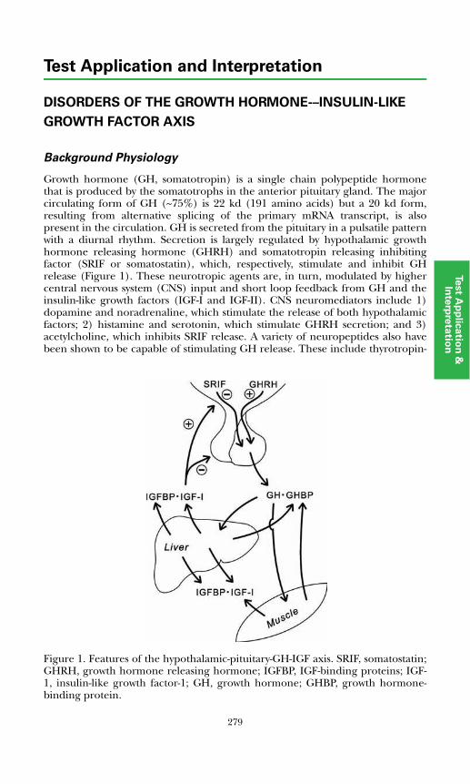
Test Application and Interpretation
Test A
pp
licatio
n &
In
terp
reta
tion
DISORDERS OF THE GROWTH HORMONE–INSULIN-LIKE
GROWTH FACTOR AXIS
Background Physiology
Growth hormone (GH, somatotropin) is a single chain polypeptide hormonethat is produced by the somatotrophs in the anterior pituitary gland. The majorcirculating form of GH (~75%) is 22 kd (191 amino acids) but a 20 kd form,resulting from alternative splicing of the primary mRNA transcript, is alsopresent in the circulation. GH is secreted from the pituitary in a pulsatile patternwith a diurnal rhythm. Secretion is largely regulated by hypothalamic growthhormone releasing hormone (GHRH) and somatotropin releasing inhibitingfactor (SRIF or somatostatin), which, respectively, stimulate and inhibit GHrelease (Figure 1). These neurotropic agents are, in turn, modulated by highercentral nervous system (CNS) input and short loop feedback from GH and theinsulin-like growth factors (IGF-I and IGF-II). CNS neuromediators include 1)dopamine and noradrenaline, which stimulate the release of both hypothalamicfactors; 2) histamine and serotonin, which stimulate GHRH secretion; and 3)acetylcholine, which inhibits SRIF release. A variety of neuropeptides also havebeen shown to be capable of stimulating GH release. These include thyrotropin-releasing
Figure 1. Features of the hypothalamic-pituitary-GH-IGF axis. SRIF, somatostatin;GHRH, growth hormone releasing hormone; IGFBP, IGF-binding proteins; IGF-1, insulin-like growth factor-1; GH, growth hormone; GHBP, growth hormone-binding protein.
279
Test Application and Interpretation
releasing hormone (TRH), corticotropin releasing hormone (CRH), vasoactiveintestinal peptide (VIP), gastrin, neurotensin, substance P, and severalhypothalamic peptides (galanin and pituitary adenylate cyclase activatingpeptides, PACAP). A number of synthetic growth hormone releasing peptides(GHRP-1, GHRP-2, GHRP-6) also have been described. These peptides act via adistinct 7 transmembrane, G-protein coupled receptor termed the GHsecretagogue receptor (GHSR). Recently it has been reported that theendogenous ligand for the GHSR is ghrelin, a 28 amino acid peptide producedprimarily in gastric mucosal neuroendocrine cells. In normal subjects ghrelinlevels increase during fasting and decline rapidly with feeding, suggesting thatthis hormone may play a critical role linking energy intake and GH secretion.Physiological factors known to stimulate GH release, presumably via one or moreof the above mechanisms, include hypoglycemia, exercise, feeding, certain aminoacids, and stage IV sleep. The diagnostic tests for assessment of GH secretorycapacity (insulin-induced hypoglycemia, arginine infusion, alpha-adrenergicagonist stimulation by clonidine, L-dopa stimulation, vigorous exercise, sleepmonitoring) acutely activate one or more of these pathways.
At physiologic concentrations, approximately half of the circulating GH is boundto the GH-binding protein (GHBP), a molecule identical to the extracellulardomain of GH receptor. In humans, the GHBP appears to be produced bycleavage of membrane-bound GH receptor rather than by direct synthesis viaalternative mRNA splicing, as occurs in rodents. In humans, GHBP levelscorrelate inversely with 24 hour GH production but are also responsive toestrogen and testosterone, particularly when administered orally. GHBP bloodconcentrations increase progressively during childhood and correlate withgrowth rate and adult height. The binding affinity of GHBP is such that about50% of GH is bound at GH concentrations <10 ng/mL; GHBP saturationincreases with increasing GH levels.
GH acts via plasma membrane GH receptors (GHR) in the liver and other tissues.The mature GHR has 620 amino acids and is composed of an extracellularhormone binding domain of 246 amino acids, a single transmembrane domain,and an intracellular domain of 350 amino acids. Binding of GH activates thecytoplasmic tyrosine kinase JAK2 which then phosphorylates the GHR, thetranscription factors STAT 1, 3, and 5b, and other proteins involved in signaltransduction. The major target genes regulated by growth hormone includethose for insulin-like growth factor-I (IGF-I), IGF binding protein-3 (IGFBP-3),and the acid-labile subunit (ALS) that associates with the IGF-I:BP-3 complex.
The IGFs, IGF-I and IGF-II, are single chain polypeptide hormones that arestructurally similar to each other (62% amino acid homology) and to insulin(50% homology). The IGFs are often referred to as somatomedins because theymediate many of the anabolic and mitogenic actions of GH (the somatomedinhypothesis). IGF-II is expressed at relatively high levels in the fetus and is believedto be a major regulator of fetal growth. The IGF-II gene is imprinted, withexpression of the maternal allele being suppressed. IGF-II is also expressedpostnatally in humans but not in mice or rats. Levels decrease rapidly after birth.IGF-I is expressed at low levels during fetal development, but the blood levelsincrease throughout gestation, reaching about 35% of adult levels at term.Postnatally, IGF-I is expressed in many tissues and, unlike IGF-II, its expression is
280
Test Application and Interpretation
Test A
pp
licatio
n &
In
terp
reta
tion
under relatively tight GH control. IGF-I secretion is also nutritionally regulatedand age-dependent. In addition to its anabolic and mitogenic actions, IGF-I alsoinhibits apoptosis and exerts a negative feedback effect on GH release via actionsat both the hypothalamic and pituitary levels (Figure 1).
The biological actions of the IGFs are mediated by the type I IGF receptor(IGFIR). This receptor is a transmembrane tyrosine kinase that is structurallysimilar to the insulin receptor. Binding of either IGF-I or IGF-II to the IGFIRresults in tyrosine phosphorylation of intracellular target proteins, includingmembers of the IRS family and Shc, and activation of intracellular anabolic andmitogenic pathways. IGF-II, but not IGF-I, also binds with high affinity to the typeII IGF receptor; this receptor, which is identical to the mannose-6-phosphatereceptor, is not anabolic and functions to clear IGF-II from the tissues.Interestingly, the type II IGF receptor is also imprinted, but in this case,expression of the paternally derived allele is suppressed. IGF-II is produced bycertain tumors, particularly sarcomas, and can cause tumor-associatedhypoglycemia.
The IGFs circulate in blood bound to several binding proteins (IGFBPs). Sixdistinct IGFBPs have been characterized, and 4 have been detected in humanblood. They are glycoproteins whose core proteins range in size from 25 to 31 kd.Ninety-five percent to 99% of the circulating IGFs are bound to the bindingproteins; only 1% to 5% circulates in unbound form. Seventy-five percent to 80%of the IGFs circulates in blood as a 150-kd complex comprised of IGF-I or IGF-II,IGFBP-3, and an 88-kd protein termed the acid labile subunit (ALS). IGFBP-3and ALS are produced mainly by the liver. Synthesis of IGFBP-3 and ALS arestimulated by GH so that levels are increased in patients with acromegaly anddecreased in those with growth hormone-deficiency. IGFBP-1, -2, and -4 alsocirculate in blood and bind most of the IGF not bound by IGFBP-3. All of thebinding proteins inhibit IGF activity by competing with the IGFIR for thehormone, but IGF enhancing and IGF independent activities have also beenreported for specific binding proteins and cell types.
The IGFs are synthesized by many cell types and tissues and act via bothendocrine and autocrine/paracrine pathways. Circulating IGFs are derivedmainly from hepatic synthesis. Mice in which the IGF-I gene was deletedselectively in the liver demonstrated marked reduction in circulating IGF-Iconcentrations and increased GH levels but normal growth, emphasizing theimportance of autocrine/paracrine pathways in the growth response.
The major biological effect of GH is the stimulation of anabolic processes: lineargrowth during childhood and adolescence and maintenance of bodycomposition and organ function in adults. Most of these effects are mediatedthrough IGF-I. GH has little or no effect in the fetus because of a paucity of GHreceptors. The growth effect becomes manifest during the early postnatal monthsand continues until epiphyseal closure. GH also has direct metabolic effects,including effects on insulin secretion and carbohydrate metabolism that are notmediated by the IGFs; large doses of GH paradoxically impair carbohydratetolerance in spite of increased insulin secretion. GH deficiency is associated withincreased fat deposition. In addition to its generalized anabolic activity, GH also
281
Test Application and Interpretation
has tissue-specific anabolic effects. For example, GH (by stimulating localproduction of IGF-I) works synergistically with gonadotropins to stimulate steroidbiosynthesis in the gonads.
Growth Hormone Deficiency in Children
GH deficiency can occur at any age. In children, the hallmark of GH deficiency isa subnormal growth velocity, resulting in short stature and delayed physicaldevelopment. However, a large variety of non-GH factors impact growth,including genetic endowment, availability of calories and nutrients, systemicdiseases, and other hormones, notably thyroxine, gonadal steroids, cortisol, andvitamin D. In practice, the vast majority of slow growing children are not GHdeficient, and the differential diagnosis of growth failure is long. A thoroughhistory, including a review of growth records, and physical examination areessential components of any work up of a short child. Routine laboratory studiesincluding serum electrolytes, creatinine, calcium, liver function tests, bloodcount, ESR, urine analysis, and thyroid function tests should be obtained to ruleout non-GH causes of poor growth. The measurement of IGF-I and IGFBP-3 arewarranted if GH deficiency is a possibility. A bone age examination is oftenhelpful for determining etiology and estimating growth potential. In short girls, akaryotype should be considered because gonadal dysgenesis may be causing theshort stature.
GH deficiency can be congenital or acquired and can be isolated or involvemultiple pituitary hormones. Congenital GH deficiency is an uncommondisorder having a prevalence of 1 in 4,000 to 1 in 10,000 births. Documentedcauses include hypothalamic and/or pituitary malformations, mutations affectinggenes required for pituitary development (PIT1, PROP1, HESX1, etc.),hypothalamic GHRH deficiency, GHRH or GHRH receptor gene mutations, andGH gene mutations/deletions. In addition, the GH deficiency phenotype can becaused by primary GH resistance attributable to mutations/deletions in the genesfor the GH receptor, members of the GH signal transduction pathway, IGF-I, ALS,or the IGFIR.
Acquired forms of GH deficiency are more common than congenital forms.These include GH deficiency resulting from CNS tumors (particularlycraniopharyngiomas), inflammatory processes (sarcoidosis, histiocytosis), andradiation or trauma to the hypothalamus and/or pituitary. Autoimmunehypophysitis with antibodies directed at the pituitary somatotrophs has also beendescribed in children and adults with apparent acquired GH deficiency andappears to be more common in subjects with isolated GH deficiency. Any of theseetiologies may produce a complete or partial GH deficient phenotype so thatdiagnosis and differential diagnosis is difficult.
There is suggestive evidence in some short, slow growing children for a syndromecharacterized by inadequate spontaneous GH secretion but normalresponsiveness to pharmacological stimulation tests (hypothalamicneurosecretory dysfunction). To distinguish such patients, measurement ofintegrated GH secretion has been proposed (ie, frequent plasma GHmeasurements over a 12-hour (overnight) or 24-hour period). There is noconsensus regarding the frequency of sampling and the parameters to be assessed
282
Test Application and Interpretation
Test A
pp
licatio
n &
In
terp
reta
tion
(area under the curve, sum of peak heights, mean concentration). Moreover,inter-individual reproducibility may be as high as 30%; thus, this approach hasremained investigational. Urinary growth hormone measurement also remainsinvestigational.
Growth Hormone Deficiency in Adults
It is estimated that 50,000 adults have GH deficiency and that each year 6,000 newcases are diagnosed in the United States. In adults, the signs and symptoms of GHdeficiency can be subtle. They include increased adipose tissue (especiallyvisceral fat); decreased lean body mass, bone mass, cardiac contractibility andexercise capacity; and alterations in mood and motivation. These features, alongwith osteopenia and increased risk of overall and cardiovascular mortality due todyslipidemia and a proinflammatory state, led the FDA in 1996 to approverecombinant GH treatment for GH deficient adults. It is uncertain, however, ifGH therapy leads to a decrease in cardiovascular mortality.
The term "somatopause" has been used to describe the gradual decline in GHsecretion associated with aging, but pathologic conditions can also cause GHdeficiency in adults (Table 1) and must be differentiated from the somatopause.
Table 1. Causes of Growth Hormone Deficiency
GeneticTranscription factor defects (Pit1, Prop1, Lhx3/4, Hesx1, Pitx2)GHRH receptor defectsGH secretagogue receptor gene defectsGH gene defectsGH receptor/post receptor defects*
Development anomaliesSepto-optic dysplasiaHoloprosencephalyEncephaloceleHydrocephalusArachnoid cystMidline facial defectsPituitary hypoplasia
Inflammatory/granulomatous diseasesEncephalitisMeningitisHistiocytosisSarcoidosisAutoimmune hypophysitis
Destructive lesionsIntraventricular hemorrhagePostpartum (Sheehan necrosis)Cranial radiationTraumaSurgery
Hypothalamic/pituitary tumors
283
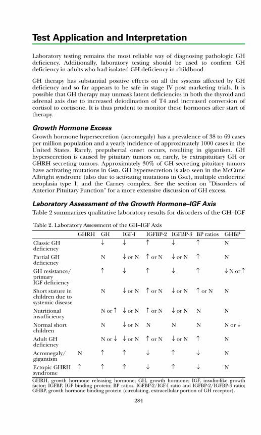
Test Application and Interpretation
Laboratory testing remains the most reliable way of diagnosing pathologic GHdeficiency. Additionally, laboratory testing should be used to confirm GHdeficiency in adults who had isolated GH deficiency in childhood.
GH therapy has substantial positive effects on all the systems affected by GHdeficiency and so far appears to be safe in stage IV post marketing trials. It ispossible that GH therapy may unmask latent deficiencies in both the thyroid andadrenal axis due to increased deiodination of T4 and increased conversion ofcortisol to cortisone. It is thus prudent to monitor these hormones after start oftherapy.
Growth Hormone Excess
Growth hormone hypersecretion (acromegaly) has a prevalence of 38 to 69 casesper million population and a yearly incidence of approximately 1000 cases in theUnited States. Rarely, prepubertal onset occurs, resulting in gigantism. GHhypersecretion is caused by pituitary tumors or, rarely, by extrapituitary GH orGHRH secreting tumors. Approximately 30% of GH secreting pituitary tumorshave activating mutations in Gsα. GH hypersecretion is also seen in the McCuneAlbright syndrome (also due to activating mutations in Gsα), multiple endocrineneoplasia type 1, and the Carney complex. See the section on "Disorders ofAnterior Pituitary Function" for a more extensive discussion of GH excess.
Laboratory Assessment of the Growth Hormone–IGF Axis
Table 2 summarizes qualitative laboratory results for disorders of the GH–IGF
Table 2. Laboratory Assessment of the GH–IGF Axis
GHRH GH IGF-I IGFBP-2 IGFBP-3 BP ratios GHBP
Classic GH deficiency
↓ ↓ ↑ ↓ ↑ N
Partial GHdeficiency
N ↓ or N ↑ or N ↓ or N ↑ N
GH resistance/primaryIGF deficiency
↑ ↓ ↑ ↓ ↑ ↓ N or ↑
Short stature in children due to systemic disease
N ↓ or N ↑ or N ↓ or N ↑ or N N
Nutritional insufficiency
N or ↑ ↓ or N ↑ or N ↓ or N N N
Normal shortchildren
N ↓ or N N N N N or ↓
Adult GHdeficiency
N or ↓ ↓ or N ↑ or N ↓ or N ↑ N
Acromegaly/gigantism
N ↑ ↑ ↓ ↑ ↓ N
Ectopic GHRHsyndrome
↑ ↑ ↑ ↓ ↑ ↓ N
GHRH, growth hormone releasing hormone; GH, growth hormone; IGF, insulin-like growthfactor; IGFBP, IGF binding protein; BP ratios, IGFBP-2/IGF-I ratio and IGFBP-2/IGFBP-3 ratio;GHBP, growth hormone binding protein (circulating, extracellular portion of GH receptor).
284
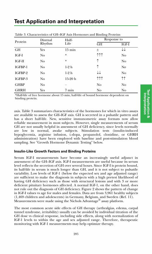
Test Application and Interpretation
Test A
pp
licatio
n &
In
terp
reta
tion
axis. Table 3 summarizes characteristics of the hormones for which in vitro assaysare available to assess the GH–IGF axis. GH is secreted in a pulsatile pattern andhas a short half-life. New, sensitive immunometric assay formats now allowreliable measurement in most subjects. However, single measurements of serumGH are not usually helpful in assessment of GH deficiency, since levels normallyare low in normal, awake subjects. Stimulation tests (insulin-inducedhypoglycemia, arginine infusion, L-dopa, propanolol, clonidine, or GHRHadministration) have been employed with baseline and post-stimulation bloodsampling. See "Growth Hormone Dynamic Testing" below.
Insulin-Like Growth Factors and Binding Proteins
Serum IGF-I measurements have become an increasingly useful adjunct inassessment of the GH–IGF axis. IGF-I measurements are useful because its serumlevel reflects the secretion of GH over several hours. Since IGF-I is protein bound,its half-life in serum is much longer than GH, and it is not subject to pulsatilevariability. Low levels of IGF-1 (below the expected sex and age adjusted range)are sufficient to make the diagnosis in subjects with a high pretest likelihood ofhaving GH deficiency such as those with structural lesions and with 3 or moredeficient pituitary hormones affected. A normal IGF-1, on the other hand, doesnot rule out the diagnosis of GH deficiency. Figure 2 shows the pattern of changein IGF-I values vs age for males and females. Data are from 3,961 healthy subjects(1,469 children and adolescents) in Germany, Belgium, and Sweden (Ref. 11).Measurements were made using the Nichols Advantage® assay platform.
The most common acute side effects of GH therapy (arthralgias, edema, carpaltunnel syndrome, irritability) usually can be avoided by individual titration of theGH dose vs clinical response, including side effects, along with normalization ofIGF-1 levels to within the age and sex adjusted range. Therefore, therapeuticmonitoring with IGF-1 measurements may help optimize therapy.
Table 3. Characteristics of GH–IGF Axis Hormones and Binding Proteins
Protein DiurnalRhythm
Half-Life
Response toGH IGF-I
GH Yes 15 min ± ↓↓
IGF-I No * ↑↑↑ No
IGF-II No * ↑ No
IGFBP-1 No 1-2 h ↑ Nο
IGFBP-2 No 1-2 h ↓↓ No
IGFBP-3 No 15-20 h ↑↑↑ ↑↑
GHBP No – No No
GHRH Yes 7 min No No*Half-life of free hormone about 15 min; half-life of bound hormone dependent on binding protein.
285
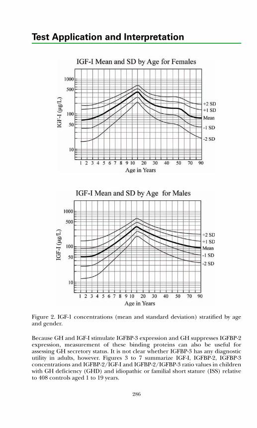
Test Application and Interpretation
Figure 2. IGF-1 concentrations (mean and standard deviation) stratified by ageand gender.
Because GH and IGF-I stimulate IGFBP-3 expression and GH suppresses IGFBP-2expression, measurement of these binding proteins can also be useful forassessing GH secretory status. It is not clear whether IGFBP-3 has any diagnosticutility in adults, however. Figures 3 to 7 summarize IGF-I, IGFBP-2, IGFBP-3concentrations and IGFBP-2/IGF-I and IGFBP-2/IGFBP-3 ratio values in childrenwith GH deficiency (GHD) and idiopathic or familial short stature (ISS) relativeto 408 controls aged 1 to 19 years.
286
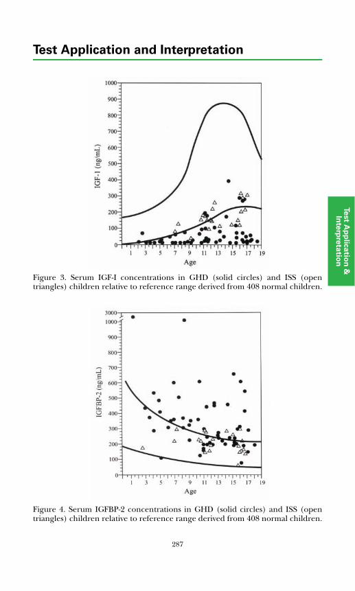
287
Test Application and Interpretation
Test A
pp
licatio
n &
In
terp
reta
tion
Figure 3. Serum IGF-I concentrations in GHD (solid circles) and ISS (opentriangles) children relative to reference range derived from 408 normal children.
Figure 4. Serum IGFBP-2 concentrations in GHD (solid circles) and ISS (opentriangles) children relative to reference range derived from 408 normal children.
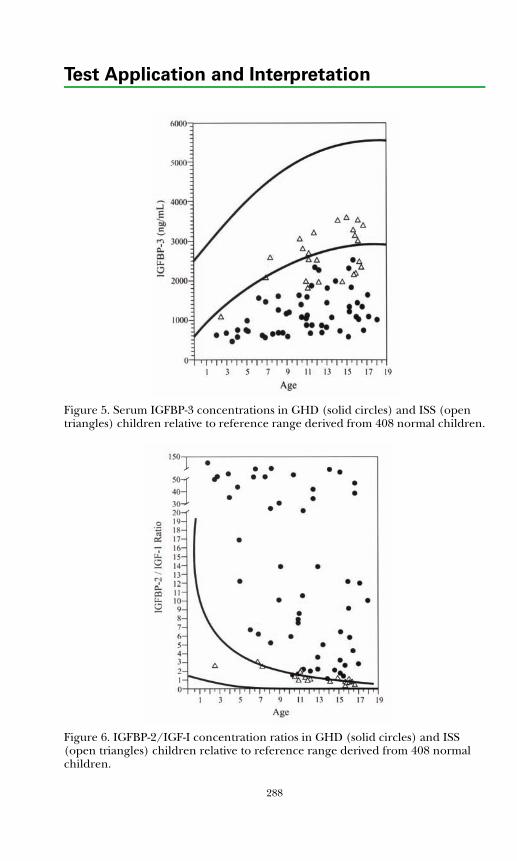
Test Application and Interpretation
Figure 5. Serum IGFBP-3 concentrations in GHD (solid circles) and ISS (open triangles) children relative to reference range derived from 408 normal children.
Figure 6. IGFBP-2/IGF-I concentration ratios in GHD (solid circles) and ISS (open triangles) children relative to reference range derived from 408 normal children.
288
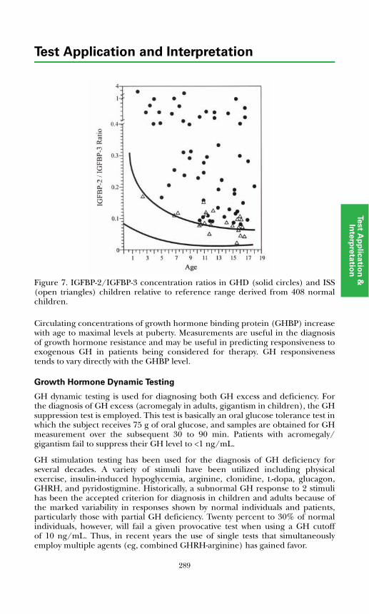
289
Test Application and Interpretation
Test A
pp
licatio
n &
In
terp
reta
tion
Figure 7. IGFBP-2/IGFBP-3 concentration ratios in GHD (solid circles) and ISS(open triangles) children relative to reference range derived from 408 normalchildren.
Circulating concentrations of growth hormone binding protein (GHBP) increasewith age to maximal levels at puberty. Measurements are useful in the diagnosisof growth hormone resistance and may be useful in predicting responsiveness toexogenous GH in patients being considered for therapy. GH responsivenesstends to vary directly with the GHBP level.
Growth Hormone Dynamic Testing
GH dynamic testing is used for diagnosing both GH excess and deficiency. Forthe diagnosis of GH excess (acromegaly in adults, gigantism in children), the GHsuppression test is employed. This test is basically an oral glucose tolerance test inwhich the subject receives 75 g of oral glucose, and samples are obtained for GHmeasurement over the subsequent 30 to 90 min. Patients with acromegaly/gigantism fail to suppress their GH level to <1 ng/mL.
GH stimulation testing has been used for the diagnosis of GH deficiency forseveral decades. A variety of stimuli have been utilized including physicalexercise, insulin-induced hypoglycemia, arginine, clonidine, L-dopa, glucagon,GHRH, and pyridostigmine. Historically, a subnormal GH response to 2 stimulihas been the accepted criterion for diagnosis in children and adults because ofthe marked variability in responses shown by normal individuals and patients,particularly those with partial GH deficiency. Twenty percent to 30% of normalindividuals, however, will fail a given provocative test when using a GH cutoffof 10 ng/mL. Thus, in recent years the use of single tests that simultaneouslyemploy multiple agents (eg, combined GHRH-arginine) has gained favor.
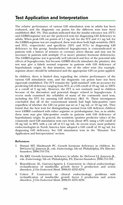
Test Application and Interpretation
The relative performance of various GH stimulation tests in adults has beenevaluated, and the diagnostic cut points for the different tests have beenestablished (Ref. 19). This analysis indicated that the insulin tolerance test (ITT)and GHRH-arginine test are the preferred tests for diagnosing GH deficiency inadults. When peak GH cut points of 5.1 ng/mL for the ITT and 4.1 ng/mL forthe GHRH-arginine test are employed, these tests show both high sensitivity (96%and 95%, respectively) and specificity (92% and 91%) in diagnosing GHdeficiency in this group. Insulin-induced hypoglycemia is contraindicated inpatients with a history of seizures or coronary artery disease and may not benecessary in patients with multiple (3 or more) pituitary hormone deficiencies.The GHRH-arginine test is generally well tolerated and free of the potential sideeffects of hypoglycemia, but because GHRH directly stimulates the pituitary, thistest may give a falsely normal response in patients with GH deficiency ofhypothalamic origin. In that situation, one of the other tests (for example,arginine alone) should be substituted and the appropriate GH cut point applied.
In children, there is limited data regarding the relative performance of thevarious GH stimulation tests, and the diagnostic cut points have not beenrigorously established. The ITT remains the "gold standard" with an accuracy forpredicting GH deficiency ranging from 85% at a GH cutoff of 10 ng/mL to 100%at a cutoff of 3 ng/mL. However, the ITT is not routinely used in childrenbecause of the discomfort and potential danger related to hypoglycemia. Arecent study examined the reliability of many of the commonly used tests,including the ITT, for assessing GH deficiency (Ref. 9). These investigatorsconcluded that all of the conventional stimuli had high false-positive ratesregardless of whether the GH cut point was set at 7 ng/mL or 10 ng/mL. Theyfound that the best tests for distinguishing normal from GH deficient childrenwere GHRH combined with either arginine or pyridostigmine, but, as in adults,these tests may give false-positive results in patients with GH deficiency ofhypothalamic origin. In general, the sensitivity (positive predictive value) of thecommonly used GH stimulation tests vary from about 40% using a GH cutoff of10 ng/mL to 80% with a cut off of 2.5 ng/mL. In recent years, most pediatricendocrinologists in North America have adopted a GH cutoff of 10 ng/mL fordiagnosing GH deficiency. See GH stimulation tests in the "Dynamic TestApplication and Interpretation" section.
References
1. Dattani MT, Hindmarsh PC. Growth hormone deficiency in children. In:DeGroot LJ, Jameson JL, eds. Endocrinology. 5th ed. Philadelphia, PA: Elsevier-Saunders; 2006:733-754.
2. Ho KKY. Growth hormone deficiency in adults. In: DeGroot LJ, Jameson JL,eds. Endocrinology. 5th ed. Philadelphia, PA: Elsevier-Saunders; 2006:755-765.
3. Rosenbloom AL, Guevara-Aguirre J. Controversy in clinical endocrinology:reclassification of insulin-like growth factor I production and actiondisorders. J Clin Endocrinol Metab. 2006;91:4232-4234.
4. Cohen P. Controversy in clinical endocrinology: problems withreclassification of insulin-like growth factor I production and actiondisorders. J Clin Endocrinol Metab. 2006;91:4235-4236.
290
Test Application and Interpretation
Test A
pp
licatio
n &
In
terp
reta
tion
5. Melmed S, Kleinberg D. Anterior pituitary. In: Larsen PR, Kronenberg HM,Melmed S, Polonsky KS, eds. Williams Textbook of Endocrinology. 10th ed.Philadelphia, PA: WB Saunders; 2003:177-279.
6. Murray RD. The phenotype of adults with partial growth hormone deficiency.Horm Res. 2005;64(Suppl 2):12-17.
7. Mukherjee A, Monson JP, Jonsson PJ, et al. Seeking the optimal target rangefor insulin-like growth factor I during the treatment of adult growth hormonedisorders. J Clin Endocrinol Metab. 2003;88:5865-5870.
8. Gharib H, Cook DM, Saenger PH, et al. American Association of ClinicalEndocrinologists medical guidelines for clinical practice for growth hormoneuse in adults and children–2003 update. Endocr Pract. 2003;9:64-76.
9. Sizonenko PC, Clayton PE, Cohen P, et al. Diagnosis and management ofgrowth hormone deficiency in childhood and adolescence. Part 1: diagnosisof growth hormone deficiency. Growth Horm IGF Res. 2001;11:137-165.
10. Rosenfeld RH, Cohen P. Disorders of growth hormone/insulin-like growthfactors secretion and action. In: Sperling MA, ed. Pediatric Endocrinology. 2nded. Philadelphia, PA: WB Saunders; 2002:211-288.
11. Brabant G, von zur Muhlen A, Wuster C, et al. Serum insulin-like growthfactor I reference values for an automated chemiluminescence immunoassaysystem: results from a multicenter study. Horm Res. 2003;60:53-60.
12. Tillmann V, Buckler JM, Kibirige MS, et al. Biochemical tests in the diagnosisof childhood growth hormone deficiency. J Clin Endocrinol Metab.1997;82:531-535.
13. Smith WJ, Nam TJ, Underwood LE, et al. Use of insulin-like growth factor-binding protein-2 (IGFBP-2), IGFBP-3, and IGF-I for assessing growthhormone status in short children. J Clin Endocrinol Metab. 1993;77:1294-1299.
14. Carlsson LM, Attie KM, Compton PG, et al. Reduced concentration of serumgrowth hormone-binding protein in children with idiopathic short stature.National Cooperative Growth Study. J Clin Endocrinol Metab. 1994;78:1325-1330.
15. Wilson TA, Rose SR, Cohen P, et al. Update of guidelines for the use ofgrowth hormone in children: the Lawson Wilkins Pediatric EndocrinologySociety Drug and Therapeutics Committee. J Pediatr. 2003;143:415-421.
16. Laron Z. Laron syndrome (primary growth hormone resistance orinsensitivity): the personal experience 1958-2003. J Clin Endocrinol Metab.2004;89:1031-1044.
17. De Bellis A, Salerno M, Conte M, et al. Antipituitary antibodies recognizinggrowth hormone (GH)–producing cells in children with idiopathic GHdeficiency and in children with idiopathic short stature. J Clin EndocrinolMetab. 2006;91:2484-2489.
18. Molitch ME, Clemmons DR, Malozowski S, et al. Evaluation and treatment ofadult growth hormone deficiency: an Endocrine Society Clinical PracticeGuideline. J Clin Endocrinol Metab. 2006;91:1621-1634.
19. Biller BM, Samuels MH, Zagar A, et al. Sensitivity and specificity of six testsfor the diagnosis of adult GH deficiency. J Clin Endocrinol Metab. 2002;87:2067-2079.
291

Test Application and Interpretation
292
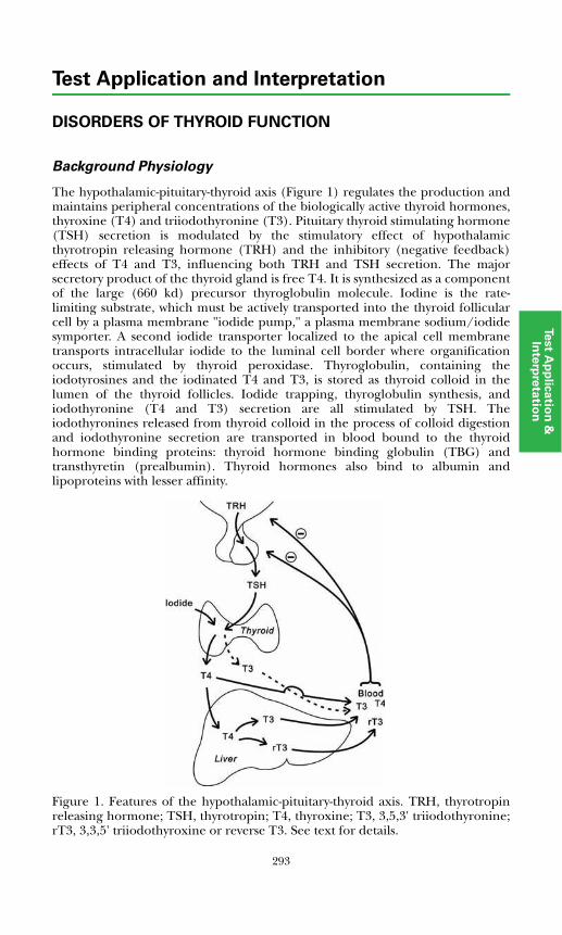
Test Application and Interpretation
Test A
pp
licatio
n &
In
terp
reta
tion
DISORDERS OF THYROID FUNCTION
Background Physiology
The hypothalamic-pituitary-thyroid axis (Figure 1) regulates the production andmaintains peripheral concentrations of the biologically active thyroid hormones,thyroxine (T4) and triiodothyronine (T3). Pituitary thyroid stimulating hormone(TSH) secretion is modulated by the stimulatory effect of hypothalamicthyrotropin releasing hormone (TRH) and the inhibitory (negative feedback)effects of T4 and T3, influencing both TRH and TSH secretion. The majorsecretory product of the thyroid gland is free T4. It is synthesized as a componentof the large (660 kd) precursor thyroglobulin molecule. Iodine is the rate-limiting substrate, which must be actively transported into the thyroid follicularcell by a plasma membrane "iodide pump," a plasma membrane sodium/iodidesymporter. A second iodide transporter localized to the apical cell membranetransports intracellular iodide to the luminal cell border where organificationoccurs, stimulated by thyroid peroxidase. Thyroglobulin, containing theiodotyrosines and the iodinated T4 and T3, is stored as thyroid colloid in thelumen of the thyroid follicles. Iodide trapping, thyroglobulin synthesis, andiodothyronine (T4 and T3) secretion are all stimulated by TSH. Theiodothyronines released from thyroid colloid in the process of colloid digestionand iodothyronine secretion are transported in blood bound to the thyroidhormone binding proteins: thyroid hormone binding globulin (TBG) andtransthyretin (prealbumin). Thyroid hormones also bind to albumin andlipoproteins with lesser affinity.
Figure 1. Features of the hypothalamic-pituitary-thyroid axis. TRH, thyrotropinreleasing hormone; TSH, thyrotropin; T4, thyroxine; T3, 3,5,3' triiodothyronine;rT3, 3,3,5' triiodothyroxine or reverse T3. See text for details.
293
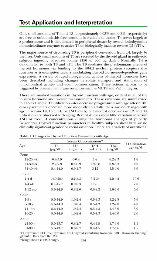
Test Application and Interpretation
294
Only small amounts of T4 and T3 (approximately 0.03% and 0.3%, respectively)are free or unbound; this free hormone is available to tissues. T4 serves largely asa prohormone and is deiodinated in peripheral tissues by several iodothyroninemonodeiodinase enzymes to active T3 or biologically inactive reverse T3 (rT3).
The major source of circulating T3 is peripheral conversion from T4, largely bythe liver. Only small amounts of T3 are secreted by the thyroid gland in euthyroidsubjects ingesting adequate iodine (150 to 300 μg daily). Normally, T4 isdeiodinated to both T3 and rT3. The T3 mediates the predominant effects ofthyroid hormones via binding to the 50-kd nuclear protein receptors thatfunction as transcription factors modulating thyroid hormone-dependent geneexpression. A variety of rapid non-genomic actions of thyroid hormones havebeen described including changes in solute transport and stimulation ofmitochondrial activity and actin polymerization. These actions appear to betriggered by plasma membrane receptors such as MCT8 and aVβ3 integrin.
There are marked variations in thyroid function with age, evident in all of thein vitro hormone and protein measurements. These variations are summarizedin Tables 1 and 2. T4 utilization rates decrease progressively with age after birth;other parameters decrease more modestly. In adults, there are no changes withage in serum T4, free T4, or TSH levels, but modest decreases in T3 and T4utilization are observed with aging. Recent studies show little variation in serumTSH or free T4 concentrations during the hormonal changes of puberty.In general, thyroid function parameters in healthy subjects show little or noclinically significant gender or racial variation. There are a variety of nutritional
Table 1. Changes in Thyroid Function Parameters with Age
AgeSerum Concentrations*
T4 Utilization μg/kg/dT4
(μg/dL)FT4
(ng/dL)TSH
(mU/L)TBG
(mg/dL)
Fetus12-20 wk 0.4-3.9 0-0.4 1-8 0.2-2.3 1.021-30 wk 2.7-7.8 0.4-0.9 1.9-8.8 0.8-3.3 2.031-40 wk 5.4-14.0 0.9-1.7 3-12 1.5-4.0 5.0
Infant1-4 d 14.0-28.4 2.2-5.3 3.2-35 2.2-4.2 10.0
1-4 wk 8.1-15.7 0.9-2.3 1.7-9.1 – 7.0
1-12 mo 5.6-14.9 0.8-2.0 0.8-8.2 1.6-3.6 6.0
Child1-5 y 5.6-14.9 1.0-2.4 0.5-4.3 1.2-2.8 5.06-10 y 5.6-14.9 1.0-2.4 0.5-4.3 1.2-2.8 4.011-15 y 5.6-14.9 1.0-2.4 0.5-4.3 1.4-3.0 3.016-20 y 5.6-14.9 1.0-2.4 0.5-4.3 1.4-3.0 2.0
Adult21-50 y 5.6-13.7 0.8-2.7 0.4-4.5 1.7-3.6 1.551-80 y 5.6-13.7 0.8-2.7 0.4-4.5 1.7-3.6 1.5
T4, thyroxine; FT4, free thyroxine; TSH, thyroid-stimulating hormone; TBG, thyroxine-binding globulin. Data from Ref. 2-7.*Range shown is ±2SD range.
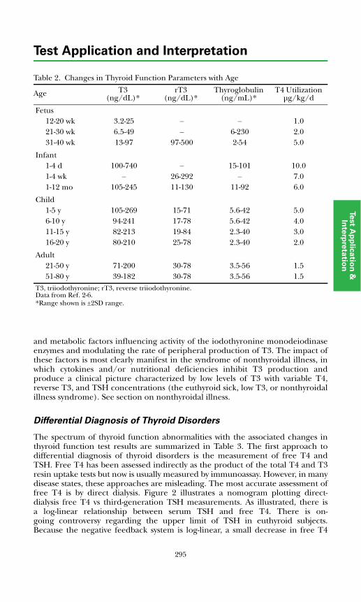
Test Application and Interpretation
Test A
pp
licatio
n &
In
terp
reta
tion
and metabolic factors influencing activity of the iodothyronine monodeiodinaseenzymes and modulating the rate of peripheral production of T3. The impact ofthese factors is most clearly manifest in the syndrome of nonthyroidal illness, inwhich cytokines and/or nutritional deficiencies inhibit T3 production andproduce a clinical picture characterized by low levels of T3 with variable T4,reverse T3, and TSH concentrations (the euthyroid sick, low T3, or nonthyroidalillness syndrome). See section on nonthyroidal illness.
Differential Diagnosis of Thyroid Disorders
The spectrum of thyroid function abnormalities with the associated changes inthyroid function test results are summarized in Table 3. The first approach todifferential diagnosis of thyroid disorders is the measurement of free T4 andTSH. Free T4 has been assessed indirectly as the product of the total T4 and T3resin uptake tests but now is usually measured by immunoassay. However, in manydisease states, these approaches are misleading. The most accurate assessment offree T4 is by direct dialysis. Figure 2 illustrates a nomogram plotting direct-dialysis free T4 vs third-generation TSH measurements. As illustrated, there isa log-linear relationship between serum TSH and free T4. There is on-going controversy regarding the upper limit of TSH in euthyroid subjects.Because the negative feedback system is log-linear, a small decrease in free T4
Table 2. Changes in Thyroid Function Parameters with Age
Age T3(ng/dL)*
rT3(ng/dL)*
Thyroglobulin (ng/mL)*
T4 Utilization μg/kg/d
Fetus12-20 wk 3.2-25 – – 1.021-30 wk 6.5-49 – 6-230 2.031-40 wk 13-97 97-500 2-54 5.0
Infant1-4 d 100-740 – 15-101 10.01-4 wk – 26-292 – 7.01-12 mo 105-245 11-130 11-92 6.0
Child1-5 y 105-269 15-71 5.6-42 5.06-10 y 94-241 17-78 5.6-42 4.011-15 y 82-213 19-84 2.3-40 3.016-20 y 80-210 25-78 2.3-40 2.0
Adult21-50 y 71-200 30-78 3.5-56 1.551-80 y 39-182 30-78 3.5-56 1.5
T3, triiodothyronine; rT3, reverse triiodothyronine.Data from Ref. 2-6.*Range shown is ±2SD range.
295
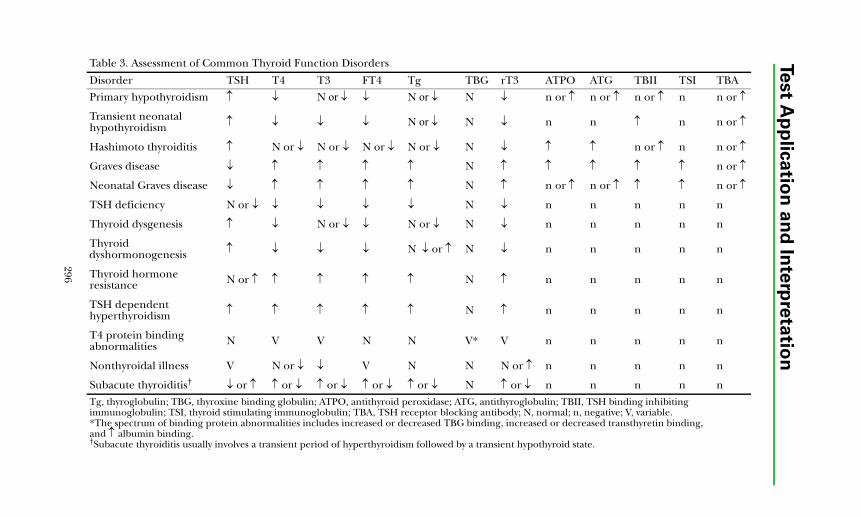
296
Test A
pp
lica
tion
an
d In
terp
reta
tion
Table 3. Assessment of Common Thyroid Function Disorders
Disorder TSH T4 T3 FT4 Tg TBG rT3 ATPO ATG TBII TSI TBA
Primary hypothyroidism ↑ ↓ N or ↓ ↓ N or ↓ N ↓ n or ↑ n or ↑ n or ↑ n n or ↑
Transient neonatal hypothyroidism ↑ ↓ ↓ ↓ N or ↓ N ↓ n n ↑ n n or ↑
Hashimoto thyroiditis ↑ N or ↓ N or ↓ N or ↓ N or ↓ N ↓ ↑ ↑ n or ↑ n n or ↑
Graves disease ↓ ↑ ↑ ↑ ↑ N ↑ ↑ ↑ ↑ ↑ n or ↑
Neonatal Graves disease ↓ ↑ ↑ ↑ ↑ N ↑ n or ↑ n or ↑ ↑ ↑ n or ↑
TSH deficiency N or ↓ ↓ ↓ ↓ ↓ N ↓ n n n n n
Thyroid dysgenesis ↑ ↓ N or ↓ ↓ N or ↓ N ↓ n n n n n
Thyroid dyshormonogenesis ↑ ↓ ↓ ↓ N ↓ or ↑ N ↓ n n n n n
Thyroid hormone resistance N or ↑ ↑ ↑ ↑ ↑ N ↑ n n n n n
TSH dependent hyperthyroidism ↑ ↑ ↑ ↑ ↑ N ↑ n n n n n
T4 protein binding abnormalities N V V N N V* V n n n n n
Nonthyroidal illness V N or ↓ ↓ V N N N or ↑ n n n n n
Subacute thyroiditis† ↓ or ↑ ↑ or ↓ ↑ or ↓ ↑ or ↓ ↑ or ↓ N ↑ or ↓ n n n n nTg, thyroglobulin; TBG, thyroxine binding globulin; ATPO, antithyroid peroxidase; ATG, antithyroglobulin; TBII, TSH binding inhibiting immunoglobulin; TSI, thyroid stimulating immunoglobulin; TBA, TSH receptor blocking antibody; N, normal; n, negative; V, variable.*The spectrum of binding protein abnormalities includes increased or decreased TBG binding, increased or decreased transthyretin binding, and ↑ albumin binding.†Subacute thyroiditis usually involves a transient period of hyperthyroidism followed by a transient hypothyroid state.
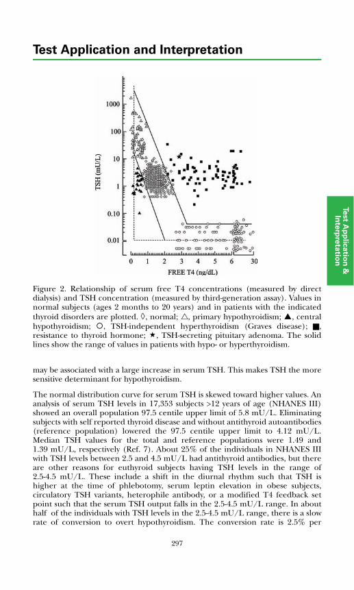
297
Test Application and Interpretation
Test A
pp
licatio
n &
In
terp
reta
tion
Figure 2. Relationship of serum free T4 concentrations (measured by directdialysis) and TSH concentration (measured by third-generation assay). Values innormal subjects (ages 2 months to 20 years) and in patients with the indicatedthyroid disorders are plotted. ◊, normal; , primary hypothyroidism; , centralhypothyroidism; , TSH-independent hyperthyroidism (Graves disease); ,resistance to thyroid hormone; , TSH-secreting pituitary adenoma. The solidlines show the range of values in patients with hypo- or hyperthyroidism.
may be associated with a large increase in serum TSH. This makes TSH the moresensitive determinant for hypothyroidism.
The normal distribution curve for serum TSH is skewed toward higher values. Ananalysis of serum TSH levels in 17,353 subjects >12 years of age (NHANES III)showed an overall population 97.5 centile upper limit of 5.8 mU/L. Eliminatingsubjects with self reported thyroid disease and without antithyroid autoantibodies(reference population) lowered the 97.5 centile upper limit to 4.12 mU/L.Median TSH values for the total and reference populations were 1.49 and1.39 mU/L, respectively (Ref. 7). About 25% of the individuals in NHANES IIIwith TSH levels between 2.5 and 4.5 mU/L had antithyroid antibodies, but thereare other reasons for euthyroid subjects having TSH levels in the range of2.5-4.5 mU/L. These include a shift in the diurnal rhythm such that TSH ishigher at the time of phlebotomy, serum leptin elevation in obese subjects,circulatory TSH variants, heterophile antibody, or a modified T4 feedback setpoint such that the serum TSH output falls in the 2.5-4.5 mU/L range. In abouthalf of the individuals with TSH levels in the 2.5-4.5 mU/L range, there is a slowrate of conversion to overt hypothyroidism. The conversion rate is 2.5% per
Test Application and Interpretation
year without and about 4.5% per year with detectable thyroid autoantibody. It isrecommended that physicians repeat normal range TSH measurements after 1-3months as clinically applicable and measure anti-TPO antibodies in individualswith levels persistently above 2.5 mU/L. All patients with persistent high normalvalues can be followed every 1-3 years with repeat serum TSH measurements(Ref. 7).
Blood samples for measurement of TSH concentrations usually are collectedrandomly. However, there is a modest, but clear, circadian variation in circulatingTSH levels in animals and in humans. TSH levels begin to rise several hoursbefore the onset of sleep, and peak levels are observed between 2300 and 0600hours. Nadir concentrations are observed during the afternoon. The diurnalvariation in TSH level approximates ± 50%, so that time of day may have someinfluence on the measured serum TSH concentration. Daytime TSH levels varyby <10%. The circadian variation in TSH secretion has minimal effect on serumT4 concentrations, however, since the hourly T4 secretion rate (3 to 5 μg)represents a very small fraction of the extrathyroidal T4 pool (500 to 600 μg).
Primary Hypothyroidism
High serum concentrations of TSH indicate thyroid hormone deficiency due todecreased thyroid gland production (primary hypothyroidism). The extent ofincrease in TSH is, in general, related to the degree of hypothyroidism, butvariations above 50 mU/L have limited significance in this regard. Mild,compensated hypothyroidism (referred to as subclinical hypothyroidism) may bedifficult to diagnose. A modest reduction in total and free T4 within the normalrange, with a normal level of serum T3, may be associated with a serum TSH levelin the 5 to 10 mU/L range. Some physicians treat and others observe suchpatients depending upon the clinical circumstances, serum cholesterol, andwhether antithyroid antibodies are present. In general, serum TSHconcentrations above 10 mU/L in patients with primary thyroid disease aretreated.
Hyperthyroidism
Decreased levels of TSH in patients with a normally functioning hypothalamicpituitary axis imply increased thyroid hormone secretion or release by the thyroidgland, suppression by exogenous thyroid hormone, or an ectopic source ofthyroid hormone, such as struma ovarii. The most common cause ofhyperthyroidism is Graves disease, which is due to TSH receptor stimulation bythe thyroid-stimulating immunoglobulin (TSI) autoantibody. Human chorionicgonadotropin has weak TSH-like bioactivity and can produce hyperthyroidismwhen greatly elevated in pregnant patients with choriocarcinoma or hydatidiformmole. Hyper-functioning autonomous thyroid nodules may secrete enoughthyroid hormone to produce hyperthyroidism. Subacute (probably viral)thyroiditis, sometimes referred to as subacute painful thyroiditis, classically ischaracterized by a transient (several week) period of hyperthyroidism, with TSHsuppression, followed by a transient (several week to several month) period ofhypothyroidism with TSH elevation. Moderate or severe hyperthyroidism usuallysuppresses serum TSH levels to 0.03 mU/L or less. However, milder sub-clinical disease or overzealous thyroxine therapy may result in levels in the 0.03 to0.3 mU/L range. After treatment of hyperthyroidism, serum TSH may remain
298
Test Application and Interpretation
Test A
pp
licatio
n &
In
terp
reta
tion
suppressed for several weeks despite normalization or reduction of free T4 andfree T3 concentrations before normal TSH regulation resumes. In treatment ofboth hyper- and hypothyroid patients, repeat measurement of TSH should bedone after a 6-week interval. This is the time required for TSH to achieve a steadystate after a change in therapeutic dose or regimen.
Hypothalamic-Pituitary Hypothyroidism
Hypothalamic TRH deficiency or pituitary TSH deficiency from any cause isassociated with thyroid gland hypofunction. Hypothalamic or pituitaryanomalies, neoplastic or inflammatory diseases involving the hypothalamic-pituitary axis, or congenital abnormalities of TRH or TSH secretion may beinvolved. These disorders have been referred to as secondary (pituitary) ortertiary (hypothalamic) hypothyroidism. Serum TSH levels are in the low ornormal range in the presence of low concentrations of free T4. TSH bioactivitytends to be reduced in these patients because of abnormal glycosylation of thesecreted and circulating TSH isoforms. Pituitary TSH deficiency can beconfirmed by showing a deficient TSH response to exogenous TRH. Patients withhypothalamic TRH deficiency and a normal pituitary gland usually manifest anormal TSH response to TRH, but the response may be relatively prolonged,persisting for 30 to 60 minutes rather than falling off after 30 minutes. Patientswith these central forms of hypothyroidism plot below or to the left of the normalrange, as shown in Figure 2.
Inappropriate TSH Secretion
Patients with inappropriate TSH secretion manifest increased free T4 and free T3levels without suppression of serum TSH concentrations. Increased TSH levelsmay be due to thyroid hormone resistance or to a TSH-secreting pituitary tumor.Thyroid hormone resistance has been shown to be due to a variety of mutationsin the beta thyroid hormone receptor gene, which variably impairs the ability ofthe receptor proteins to activate transcription of target genes. The tissuedistribution of the abnormal receptor, vs the normal thyroid hormone receptoralpha, appears to determine the phenotype of the patient in terms of thyroidfunction. Patients may appear near normal or may manifest variable signs andsymptoms of hypo- or hyperthyroidism. Patients with generalized thyroidhormone resistance involving the pituitary and peripheral tissues have normal orelevated TSH and elevated free T4 levels, with or without clinical manifestationsof hypothyroidism. Patients with isolated pituitary resistance have similar thyroidlaboratory test results but present with clinical thyrotoxicosis of variable degree.Adenomas involving pituitary thyrotroph cells are rare but can produceinappropriate secretion of TSH with associated hyperthyroidism. Patients withinappropriate TSH secretion plot above or to the right of either normalindividuals or thyroid patients with intact hypothalamic-pituitary function, asshown in Figure 2. See also the "Disorders of Anterior Pituitary Function" section.
Nonthyroidal Illness
This syndrome, also referred to as the low T3 or euthyroid sick syndrome, ischaracterized by low total and free serum T3; normal, low, or high total T4;increased T4 sulfate; normal to high free T4; and normal TSH concentrations. It
299
Test Application and Interpretation
has been reported in a variety of clinical situations including premature infantsand patients with a variety of severe acute and chronic illnesses. The latterinclude diabetic ketoacidosis, severe trauma, burns, febrile states, cirrhosis,and renal failure. A similar syndrome may be produced by a number ofdrugs including dexamethasone, selected radiographic contrast agents,propylthiouracil, propranolol, and amiodarone. Three patterns of change inthyroid hormone levels have been described (Table 4). The low serum and tissueT3 levels in these patients occur as a result of inhibition of iodothyronine β-ringmonodeiodinase-1 activity (MDI-1) and a decreased rate of T3 production fromT4 in non-thyroidal tissues. In mice, interleukin-1 (IL-1) cytokine inhibition ofiodothyronine monodeiodinase type 1 (MDI-1) transcription results in decreasedhepatic T4 to T3 conversion, and this reduction can be reversed by steroidreceptor coactivator-1 (SRC-1) induction of MDI-1. rT3 is increased because thedegradation of rT3 is mediated by the same type 1 deiodinase enzyme thatmediates T4 to T3 conversion. Thus, serum T3 levels fall, and rT3 levels tend toremain normal or increase. In some patients, serum T4 levels also fall (the low T4syndrome), and low T4 levels in patients with severe nonthyroidal illness havebeen associated with increased mortality. TBG levels may be reduced in suchpatients, and an inhibitor of T4 binding to TBG has been described in the serumand derived from the tissues of such patients. The high T4 nonthyroidal illnesssyndrome probably involves one or more induced abnormalities in the disposalpathways of T4. In some patients, such as those receiving amiodarone or thosewith psychiatric illness, free T4 may be elevated with or without a decreasedserum TSH. A suppression of TSH during the acute phase of the psychiatricillness may be associated with a transient rebound of TSH above the normalrange followed with normalization thereafter. There is no convincing evidencethat treatment with thyroid hormones is beneficial in most patients withnonthyroidal illness, but in selected clinical conditions, such as postoperativecardiac surgery, treatment has been advocated.
Autoimmune Thyroid Disease
The most frequently encountered thyroid disorders are autoimmune in nature. Itis now clear that both cellular- and antibody-mediated pathogenetic mechanismsare involved and that the autoimmune diathesis in affected patients has a familial,genetic predisposition. Table 5 summarizes the clinical spectrum of autoimmunethyroid disease. The most common disorders are Hashimoto chronic(lymphocytic) thyroiditis (HT) and Graves disease (GD). HT involves both cell-and antibody-mediated destruction of thyroid tissue, but the cell-mediatedcomponent is the most important. In patients with GD, by contrast, thehyperthyroid state is caused by TSH receptor-stimulating autoantibodiesmediating a TSH-like effect and thyroid cell hyperplasia.
Table 4. Thyroid Function Abnormalities in Nonthyroidal Illness
NTI Pattern TSH T3 rT3 T4Low T3 syndrome N ↓ N or ↑ N
Low T4 syndrome N ↓ N or ↑ ↓
High T4 syndrome N ↓ N or ↑ ↑NTI, nonthyroidal illness; rT3, reverse T3; N, normal, ↓, decreased; ↑, increased.
300
Test Application and Interpretation
Test A
pp
licatio
n &
In
terp
reta
tion
A variety of antibody types have been associated with thyroid autoimmunediseases (Table 6). The classic thyroid autoantibodies are directed againstthyroglobulin (ATG), the thyroid hormone precursor and storage protein in thethyroid gland, and against thyroid microsomal membrane antigens, the mostimportant being thyroid peroxidase (ATPO). Other prominent antibodies aredirected against the TSH receptor. Both receptor-stimulating immunoglobulins(TSI) and TSH receptor blocking antibodies (TBA) have been characterized.TSIs are identified by their capacity to stimulate cyclic AMP (cAMP) productionin thyroid follicular cells in culture; TBA blocks TSH-stimulated cAMPproduction. Both TSI and TBA will displace labeled TSH from partially purifiedTSH receptors in a membrane radioreceptor assay system. In this instance, theantibody is referred to as TSH binding inhibitory immunoglobulin (TBII). Acollagen-stimulating immunoglobulin (CSI) has been characterized thatstimulates incorporation of labeled proline into collagen by fibroblasts in tissueculture. A thyroid growth-stimulating immunoglobulin (TGI) has been describedthat stimulates incorporation of tritiated thymidine into DNA of thyroid follicularcells in culture and promotes thyroid cell replication. The CSI and TGIautoantibodies have not been utilized clinically.
Table 5. Clinical Spectrum of Autoimmune Thyroid Disease
Thyroid disordersHashimoto thyroiditisPrimary myxedemaFibrous thyroiditisThyroid lymphomaHashitoxicosisAtrophic thyroiditisGraves disease
Autoimmune polyglandular disease, type II*Hashimoto thyroiditisDiabetes mellitusAddison disease
Associated autoimmune disordersVitiligoPernicious anemiaIdiopathic thrombocytopeniaThyroid antigen/antibody nephritis
Pregnancy-associated thyroid disordersTransient postpartum hypothyroidismTransient postpartum hyperthyroidismNeonatal Graves diseaseNeonatal hypothyroidism
*May include ovarian failure, pernicious anemia, myasthenia gravis, vitiligo, and alopecia.
301
302
Test Application and Interpretation
Application of clinically useful autoantibody assays to the differential diagnosisof autoimmune disease is summarized in Table 7. The ATG and ATPO auto-antibodies are markers for thyroid autoimmune diathesis and may be detected inautoimmune disease family members who have not yet developed thyroid disease.Antibody titers are not usually indicative of disease severity. Occasional patientswith autoimmune disease may also manifest autoantibodies to T4, T3, or TSH,and such antibodies may interfere with immunoassay measurements for thesehormones.
Table 6. Thyroid Autoantibodies in Human Subjects
ATGATPOAnti-T3Anti-T4Anti-TSHTSH Receptor Antibodies
TSI TBIITBA
Other AntibodiesCSITGI
Thyroglobulin antibodyThyroid peroxidase antibodyTriiodothyronine antibodyThyroxine antibodyThyroid stimulating hormone antibody
Thyroid stimulating immunoglobulinTSH binding-inhibitory immunoglobulinTSH blocking antibody
Collagen stimulating immunoglobulinThyroid growth stimulating immunoglobulin
Table 7. Qualitative Thyroid Autoantibody Test Results in Various Thyroid Autoimmune Disease States
Type ATG ATPO TSI TBII
Hashimoto’s thyroiditisJuvenile + + +Adult +++ +++ +
Hashitoxicosis ++ ++ ++ ++
Atrophic thyroiditis + + ++
Graves diseaseJuvenile + + ++ ++Adult + + +++ +++With opthalomopathy + + +++ +++With dermopathy + + +++ +++
Transient neonatal hypothyroidism + + ++
Neonatal Graves disease + + +++ +++ATG, antithyroglobulin antibody; ATPO, antithyroid peroxidase antibody; TSI, thyroid stimulating immunoglobulin; TBII, TSH binding inhibitory immunoglobulin.
Test Application and Interpretation
Test A
pp
licatio
n &
In
terp
reta
tion
There appears to be significant autoantibody heterogeneity in most patients withautoimmune disease, and although ATPO is present in most patients withHashimoto thyroiditis and TSI in most patients with Graves disease (about 90%and 70%, respectively), adding the ATG for Hashimoto disease and adding TBIIand TGI for Graves disease increases test positivity to 95% and 90%, respectively.
Thyroid autoimmune disease may be associated with autoimmune syndromesinvolving other endocrine glands and other tissues (Table 5). The most commonassociation is with autoimmune insulin-dependent diabetes mellitus. The triad ofHashimoto thyroiditis, diabetes mellitus, and adrenal insufficiency has beenreferred to as autoimmune polyglandular disease type II, and this syndrome alsocan include ovarian atrophy, testicular failure, lymphocytic hypophysitis,pernicious anemia, myasthenia gravis, vitiligo, and alopecia. Autoimmunepolyendocrinopathy type I with candidiasis and ectodermal dystrophy (APECED)is usually associated with hypoparathyroidism, adrenal failure, diabetes mellitus,pernicious anemia, vitiligo, and alopecia but does not usually includeautoimmune thyroiditis. This rare syndrome is autosomal recessive and isfrequently associated with mutations in the AIRE (autoimmune regulator) gene.In contrast, type II polyglandular autoimmune disease is relatively common. Inpatients with insulin-dependent diabetes mellitus, the prevalence of elevatedserum TSH levels approximates 20% in female patients and 5% in males at allages.
Associated autoimmune disorders in patients with autoimmune thyroid diseasealso can include idiopathic thrombocytopenia and thyroid antigen/antibodynephritis (Table 5). Patients may have platelet antibodies with or without clinicalmanifestations of thrombocytopenia. Circulating immune complexes also arepresent in some patients with autoimmune thyroid disease, and several patientshave been reported with thyroglobulin and/or microsomal antigens identifiableby immunohistochemistry in the glomeruli as part of the kidney fixed antigen-antibody complex.
Pregnancy tends to ameliorate the manifestations of autoimmune thyroiddisease. This presumably is due to the mechanism(s) operative during pregnancyto protect the fetal allograft from rejection. This improvement in thyroidfunction during pregnancy is followed in many who are not taking medication bya return or exacerbation of clinical disease during the postpartum period. Thisusually is transient and may resemble Hashimoto thyroiditis or Graves disease.Neonatal hypothyroidism or Graves disease also may be observed. In womendeveloping postpartum thyroiditis, the thyroid autoantibody titers increase topeak levels 4 to 7 months postpartum in parallel with the changes in free T4 andTSH concentrations.
A recently described syndrome referred to as silent thyroiditis, or painlessthyroiditis with transient hyperthyroidism, resembles subacute thyroiditis in thattransient thyrotoxicosis occurs in association with a low thyroid radioiodineuptake. However, these patients occasionally have low titers of thyroidautoantibodies. The thyroid histology in this condition shows a lymphocyticinfiltrate. This postpartum hyperthyroid state also occurs in nonpregnantpersons, but the disorder is more commonly seen in the postpartum period.
303
Test Application and Interpretation
Acute thyroid cell cytotoxicity presumably accounts for the transient hyper-thyroidism and low radioiodine uptake.
Thyroid Dysfunction in Infancy and Early Childhood
Most infants with congenital hypothyroidism are detected in newborn thyroid-screening programs. The prevalence approximates 1 in 4,000 infants. Themajority manifest primary hypothyroidism due to thyroid gland agenesis ordysgenesis. Ten percent to 15% of cases are due to a congenital defect in TSHaction or a biosynthetic defect in thyroid gland hormonogenesis. In another10%, the hypothyroidism is transient because of antithyroid drugs or maternalTSH receptor-blocking antibodies. Some 3% to 5% (about 1 in 30,000 newborns)manifest hypothalamic-pituitary TSH deficiency. One percent to 3% representsfamilial cases of thyroid dysgenesis associated with mutations of the thyroidtranscription factors (TTF-1, NKF2.1; TTF-2, FOXE1), or the PAX-8 genes, whichare involved in thyroid gland embryogenesis.
Neonatal hypothyroidism due to maternal TSH receptor blocking antibodyusually occurs in association with maternal atrophic thyroiditis. Patients withatrophic thyroiditis have elevated titers of TSH receptor blocking antibody(TBA), which blocks TSH stimulation of thyroid gland function. Thyroidradioiodine uptake is low, and thyroid hormone secretion is reduced. Serum TSHlevels are elevated, and the thyroid gland is hypoplastic or atrophic. ATPO andATG titers may be elevated. The autoantibody levels in maternal and neonatalblood usually are similar, so that measurement in the mother can avoid newbornsampling. The TBII and/or TBA tests will detect TSH receptor blockingantibodies. A number of genetic abnormalities in the pituitary-thyroid axisproducing neonatal thyroid dysfunction have been described (Table 8).
Laboratory tests for the assessment of thyroid disorders in the neonatal periodare summarized in Table 9. Absent or very low levels of serum thyroglobulinsupports a diagnosis of thyroid agenesis. The presence of TSH receptor blockingantibody in maternal or newborn serum, assessed as TSH binding inhibitingimmunoglobulin (TBII) or TBA, supports a diagnosis of transienthypothyroidism. TBA or an inactivating TSH receptor gene mutation can lead toan erroneous diagnosis of thyroid agenesis by thyroid scan. Ultrasound imaging ismore reliable. Some physicians prefer to delay the differential diagnosticassessment of congenital thyroid disease to 2-3 years of age to assure early andadequate thyroxine replacement therapy.
Thyroid Neoplasia
A classification of primary thyroid neoplasms is shown in Table 10. These tumorstypically present as single thyroid nodules. Diagnosis usually involves thyroidfunction testing and fine needle aspiration/cytology with or without radioiodineor ultrasound scanning. In the diagnosis and/or management of thyroidneoplasia, 2 thyroid proteins serve as important tumor markers: calcitonin andthyroglobulin. Calcitonin is a 32-amino acid polypeptide hormone producedexclusively by parafollicular or C-cells in the thyroid. The hormone inhibitsosteoclast mediated bone resorption, but its physiological role is uncertain. The
304
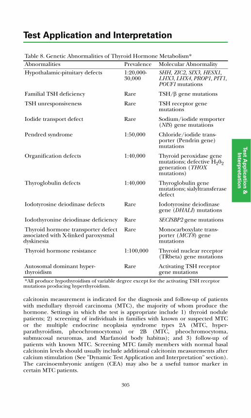
305
Test Application and Interpretation
Test A
pp
licatio
n &
In
terp
reta
tion
calcitonin measurement is indicated for the diagnosis and follow-up of patientswith medullary thyroid carcinoma (MTC), the majority of whom produce thehormone. Settings in which the test is appropriate include 1) thyroid nodulepatients; 2) screening of individuals in families with known or suspected MTCor the multiple endocrine neoplasia syndrome types 2A (MTC, hyper-parathyroidism, pheochromocytoma) or 2B (MTC, pheochromocytoma,submucosal neuromas, and Marfanoid body habitus); and 3) follow-up ofpatients with known MTC. Screening MTC family members with normal basalcalcitonin levels should usually include additional calcitonin measurements aftercalcium stimulation (See "Dynamic Test Application and Interpretation" section).The carcinoembryonic antigen (CEA) may also be a useful tumor marker incertain MTC patients.
Table 8. Genetic Abnormalities of Thyroid Hormone Metabolism*Abnormalities Prevalence Molecular AbnormalityHypothalamic-pituitary defects 1:20,000-
30,000SHH, ZIC2, SIX3, HESX1, LHX3, LHX4, PROP1, PIT1, POUF1 mutations
Familial TSH deficiency Rare TSH/β gene mutations
TSH unresponsiveness Rare TSH receptor gene mutations
Iodide transport defect Rare Sodium/iodide symporter (NIS) gene mutations
Pendred syndrome 1:50,000 Chloride/iodide trans-porter (Pendrin gene) mutations
Organification defects 1:40,000 Thyroid peroxidase gene mutations; defective H202 generation (THOXmutations)
Thyroglobulin defects 1:40,000 Thyroglobulin gene mutations; sialyltransferase defect
Iodotyrosine deiodinase defects Rare Iodotyrosine deiodinase gene (DHALI) mutations
Iodothyronine deiodinase deficiency Rare SECISBP2 gene mutations
Thyroid hormone transporter defect associated with X-linked paroxysmal dyskinesia
Rare Monocarboxylate trans-porter (MCT8) gene mutations
Thyroid hormone resistance 1:100,000 Thyroid nuclear receptor (TRbeta) gene mutations
Autosomal dominant hyper-thyroidism
Rare Activating TSH receptor gene mutations
*All produce hypothyroidism of variable degree except for the activating TSH receptor mutations producing hyperthyroidism.
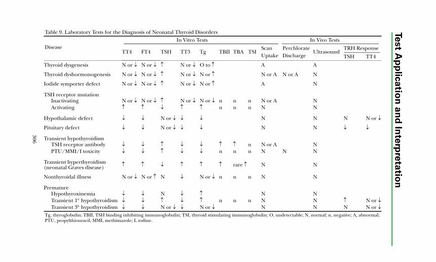
Test A
pp
lica
tion
an
d In
terp
reta
tion
Table 9. Laboratory Tests for the Diagnosis of Neonatal Thyroid DisordersIn Vitro Tests In Vivo Tests
DiseaseI
Scan PerchlorateUltrasound
TRH ResponseUptake Discharge TSH TT4
A A
N or A N or A N
A N
N or A NN N
N N N N or ↓
N N ↓ ↓
N or A NN N N
N N
N N
N NN N ↑ N or ↓N N N N or ↓
obulin; O, undetectable; N, normal; n, negative; A, abnormal;
306
TT4 FT4 TSH TT3 Tg TBII TBA TS
Thyroid dysgenesis N or ↓ N or ↓ ↑ N or ↓ O to ↑
Thyroid dyshormonogenesis N or ↓ N or ↓ ↑ N or ↓ N or ↑
Iodide symporter defect N or ↓ N or ↓ ↑ N or ↓ N or ↑
TSH receptor mutationInactivating N or ↓ N or ↓ ↑ N or ↓ N or ↓ n n nActivating ↑ ↑ ↓ ↑ ↑ n n n
Hypothalamic defect ↓ ↓ N or ↓ ↓ ↓
Pituitary defect ↓ ↓ N or ↓ ↓ ↓
Transient hypothyroidismTSH receptor antibody ↓ ↓ ↑ ↓ ↓ ↑ ↑ nPTU/MMI/I toxicity ↓ ↓ ↑ ↓ ↓ n n n
Transient hyperthyroidism (neonatal Graves disease) ↑ ↑ ↓ ↑ ↑ ↑ rare ↑
Nonthyroidal illness N or ↓ N or ↑ N ↓ N or ↓ n n n
PrematureHypothyroxinemia ↓ ↓ N ↓ ↑Transient 1° hypothyroidism ↓ ↓ ↑ ↓ ↑ n n nTransient 3° hypothyroidism ↓ ↓ N or ↓ ↓ N or ↓
Tg, thyroglobulin; TBII, TSH binding inhibiting immunoglobulin; TSI, thyroid stimulating immunoglPTU, propylthiouracil; MMI, methimazole; I, iodine.

Test Application and Interpretation
Test A
pp
licatio
n &
In
terp
reta
tion
It is now clear that familial MTC and most cases of sporadic MTC are caused bymutations in the RET protooncogene. Definitive diagnosis is now possible bymutation analysis and early RET genotyping of young children in knownkindreds, followed by prophylactic thyroidectomy of affected patients (see"Pheochromocytoma, Medullary Thyroid Carcinoma, and Multiple EndocrineNeoplasia").
Thyroglobulin (Tg) is a 660-kd protein synthesized exclusively by the thyroidfollicular cells. Tg serves as the prohormone for iodothyronines and normally islargely sequestered within the thyroid gland. However, small amounts arereleased in the process of Tg synthesis. Circulating Tg levels are increased in allforms of endogenous hyperthyroidism, including Graves disease, toxic nodulargoiter, and subacute thyroiditis. Levels also are increased in many patients withthyroid adenoma and most patients with papillary/follicular carcinoma. Thus, Tgmeasurements cannot distinguish benign and malignant thyroid nodules, butserial measurements are very valuable in the postoperative monitoring of patientswith papillary, follicular, and Hürthle cell carcinomas. The test is more sensitivefor detection of residual or recurrent disease when serum samples are obtainedafter withdrawal of thyroid suppression therapy, when the patients' endogenousTSH concentration is high, or after administration of recombinant TSH. Thepeak serum Tg after recombinant human TSH (rhTSH) usually is seen 3 daysafter a second injection. Measurement of serum Tg currently is the most sensitivemarker for thyroid cancer recurrence. Most patients with undetectable levelsafter initial surgery and without replacement thyroxine remain free of relapseafter 15 years follow-up. Sixty percent to 80% of patients requiring thyroxinereplacement and with serum Tg levels >10 ng/mL after thyroxine withdrawal hadlocal recurrence or distant metastases as assessed by large dose radioiodinescanning. Twenty percent to 30% of patients with thyroid carcinoma havecirculating ATG, which renders Tg measurements unreliable.
Table 10. Classification of Thyroid Neoplasia
Thyroid adenomaColloidEmbryonalFollicular*
Hürthle cell tumor
Thyroid carcinomaPapillaryFollicularHürthle cellMedullaryUndifferentiatedThyroid lymphoma
*Occasionally functioning or toxic adenoma.
307
Test Application and Interpretation
Thyroid Protein Binding Abnormalities
Laboratory differentiation of patients with euthyroid hyperthyroxinemia fromthose with hyperthyroidism is sometimes difficult, especially if the hyper-thyroidism is pituitary in origin. Euthyroid hyperthyroxinemia may result from anincrease in the concentration of thyroxine-binding globulin (TBG), increasedconcentrations of transthyretin (prealbumin), or the presence of molecularvariants of transthyretin or of albumin with increased affinity for T4. The last-mentioned disorder is referred to as familial dysalbuminemic hyperthyroxinemia(FDH). Distinguishing euthyroid hypothyroxinemia from hypothyroidism,particularly from secondary and tertiary hypothyroidism, is also difficult.Euthyroid hypothyroxinemia can result from a decreased TBG concentration,the presence of molecular variants of TBG with reduced T4-binding affinity, orthe euthyroid hypothyroxinemia of illness (the low-T4 state of nonthyroidalillness). The TBG gene is localized to the X chromosome, and male hemizygoteswith a single mutant allele can show one of 3 phenotypes: absent, decreased, orincreased TBG and bound T4 values. There are more than 15 known X-linkedTBG mutants: some cause complete TBG deficiency, and others cause reducedaffinity for T4 binding. The prevalence of TBG deficiency in males approximates1:5000 with 1:15,000 showing complete deficiency. Thus TBG deficiency is asprevalent as congenital hypothyroidism and may contribute to confusion indiagnosis of congenital hypothyroidism in screening programs. The prevalenceof TBG excess approximates 1:25,000 newborn males.
Some clues to the T4 protein binding disorder can be obtained by assessingserum T4 and T3 levels. Increased TBG binding is associated with increased levelsof both T4 and T3, since TBG binds both hormones. Increased transthyretinlevels are associated with increased T4 but normal T3 concentrations, since thisprotein preferentially binds T4. Free T4 concentration, measured by directdialysis, is normal in these patients. However, an accurate differential diagnosisrequires measurement of the protein level and/or the distribution of T4 amongthe binding proteins in association with the concentration of protein-boundT4 per unit mass of the respective protein. This is accomplished viagel electrophoresis of radioiodinated thyroxine-labeled serum samples;measurement of T4, TBG, prealbumin (transthyretin), and albumin con-centrations; and determination of the distribution of labeled T4 among theproteins of the electrophoretogram. See "T4 Binding Proteins" in the"Alphabetical Test" section for results of such analysis in normal subjects. Relativeto the normal values, patients with abnormal protein concentrations andabnormal protein binding usually can be distinguished.
References
1. Weiss R, Wu SY, Refetoff S. Diagnostic tests of the thyroid. In: DeGroot LJ,Jameson JL, eds. Endocrinology. 5th ed. Philadelphia, PA: Elsevier-Saunders;2006:1899-1961.
2. Nelson JC, Clark SJ, Borut DL, et al. Age-related changes in serum freethyroxine during childhood and adolescence. J Pediatr. 1993;123:899-905.
3. Adams LM, Emery JR, Clark SJ, et al. Reference ranges for newer thyroidfunction tests in premature infants. J Pediatr. 1995;126:122-127.
308
Test Application and Interpretation
Test A
pp
licatio
n &
In
terp
reta
tion
4. Thorpe-Beeston JG, Nicolaides KH, McGregor AM. Fetal thyroid function.Thyroid. 1992;2:207-217.
5. Delange F, Fisher DA. The thyroid gland. In: Brook CGD, ed. Clinical PediatricEndocrinology. 3rd ed. Oxford, Blackwell Scientific; 1995:397-433.
6. Quest Diagnostics Nichols Institute Clinical Correlations Department.
7. Surks MI. Primary hypothyroidism: new issues and controversies.Endocrinologist. 2006;16:203-207.
8. Baloch Z, Caravon P, Conte-Devolx B, et al. Laboratory medicine practiceguidelines. Laboratory support for the diagnosis and monitoring of thyroiddisease. Thyroid. 2003;13:3-126.
9. Larsen PR, Davies TF, Schlumberger MJ, et al. Thyroid physiology anddiagnostic evaluation of patients with thyroid disorders. In: Larsen PR,Kronenberg HM, Melmed S, Polonsky KS, eds. Williams Textbook ofEndocrinology. 10th ed. Philadelphia, PA: WB Saunders; 2003:331-421.
10. Weetman AP. Autoimmune thyroid disease. In: DeGroot LJ, Jameson JL, eds.Endocrinology. 5th ed. Philadelphia, PA: Elsevier-Saunders; 2006:1979-1993.
11. Marino M, Chiovato L, Pinchera A. Graves disease. In: DeGroot LJ, JamesonJL, eds. Endocrinology. 5th ed. Philadelphia, PA: Elsevier-Saunders; 2006:1995-2028.
12. Amino N, Hidaka Y. Chronic (Hashimoto's) thyroiditis. In: DeGroot LJ,Jameson JL, eds. Endocrinology. 5th ed. Philadelphia, PA: Elsevier-Saunders;2006:2055-2067.
13. Knobel M, Medeiros-Neto G. An outline of inherited disorders of the thyroidhormone generating system. Thyroid. 2003;13:771-801.
14. Yu J, Koenig RJ. Induction of type I iodothyronine deiodinase to preventnonthyroidal illness syndrome in mice. Endocrinology. 2006;147:3580-3585.
15. Peeters RP, van der Geyten S, Wouters PJ, et al. Tissue thyroid hormone levelsin critical illness. J Clin Endocrinol Metab. 2005;90:6498-6507.
16. DeGroot LJ. Nonthyroidal illness syndrome. In: DeGroot LJ, Jameson JL, eds.Endocrinology. 5th ed. Philadelphia, PA: Elsevier-Saunders; 2006:2101-2112.
17. MacGillivray MH. Congenital hypothyroidism. In: Pescovitz OH, Eugster EA,eds. Pediatric Endocrinology. Philadelphia, PA: Lippincott Williams and Wilkins;2004:490-507.
18. Rivkees SA. Hypo- and hyperthyroidism in children. In: Pescovitz OH,Eugster EA, eds. Pediatric Endocrinology. Philadelphia, PA: Lippincott Williamsand Wilkins; 2004:508-521.
19. Fisher DA. Thyroid disorders in childhood and adolescence. In: Sperling MA,ed. Pediatric Endocrinology. 3rd ed. Philadelphia, PA: WB Saunders; 2007, inpress.
309
Test Application and Interpretation
20. Schlumberger MJ, Filetti S, Hay ID. Nontoxic goiter and thyroid neoplasia.In: Larsen PR, Kronenberg HM, Melmed S, Polonsky KS, eds. WilliamsTextbook of Endocrinology. 10th ed. Philadelphia, PA: WB Saunders; 2003:457-490.
21. Pacini F, DeGroot LJ. Thyroid neoplasia. In: DeGroot LJ, Jameson JL, eds.Endocrinology. 5th ed. Philadelphia, PA: Elsevier-Saunders; 2006:2147-2180.
22. Bauer AJ, Tuttle RM, Francis GL. Thyroid nodules and thyroid cancer inchildren and adolescents. In: Pescovitz OH, Eugster EA, eds. PediatricEndocrinology. Philadelphia, PA: Lippincott Williams and Wilkins; 2004:522-547.
23. Jansen J, Friesema EC, Milici C, et al. Thyroid hormone transporters in healthand disease. Thyroid. 2005;15:757-768.
24. Bergh JJ, Lin HY, Lansing L, et al. Integrin αVβ3 contains a cell surfacereceptor site for thyroid hormone that is linked to activation of mitogen-activated protein kinase and induction of angiogenesis. Endocrinology.2005;146:2864-2871.
25. Dumitrescu AM, Liao XH, Abdullah MS, et al. Mutations in SECISBP2 resultin abnormal thyroid hormone metabolism. Nat Genet. 2005;37:1247-1252.
26. van Tijn DA, de Vijlder JJ, Verbeeten B Jr, et al. Neonatal detection ofcongenital hypothyroidism of central origin. J Clin Endocrinol. Metab.2005;90:3350-3359.
27. Cooper DS, Doherty GM, Haugen BR, et al. Management guidelines forpatients with thyroid nodules and differentiated thyroid cancer. Thyroid.2006;16:109-142.
28. Costante G, Meringolo D, Durante C, et al. Value of serum calcitonin levelsfor preoperative diagnosis of medullary thyroid carcinoma in a cohort of5817 consecutive patients with thyroid nodules. J Clin Endocrinol Metab.2007;92:450-455.
310
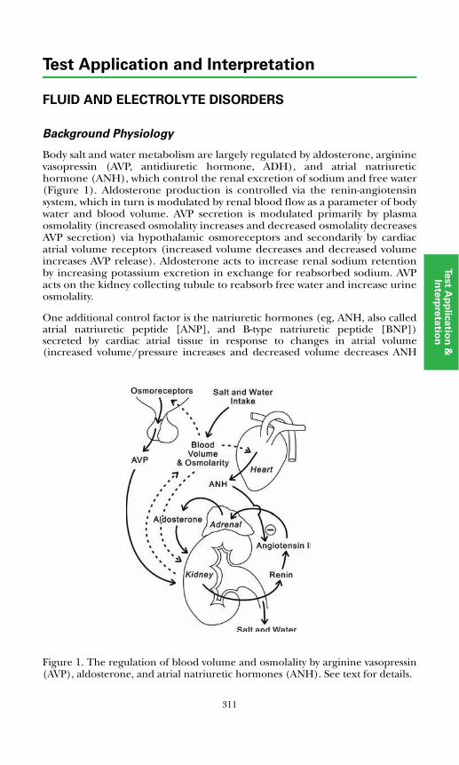
Test Application and Interpretation
Test A
pp
licatio
n &
In
terp
reta
tion
FLUID AND ELECTROLYTE DISORDERS
Background Physiology
Body salt and water metabolism are largely regulated by aldosterone, argininevasopressin (AVP, antidiuretic hormone, ADH), and atrial natriuretichormone (ANH), which control the renal excretion of sodium and free water(Figure 1). Aldosterone production is controlled via the renin-angiotensinsystem, which in turn is modulated by renal blood flow as a parameter of bodywater and blood volume. AVP secretion is modulated primarily by plasmaosmolality (increased osmolality increases and decreased osmolality decreasesAVP secretion) via hypothalamic osmoreceptors and secondarily by cardiacatrial volume receptors (increased volume decreases and decreased volumeincreases AVP release). Aldosterone acts to increase renal sodium retentionby increasing potassium excretion in exchange for reabsorbed sodium. AVPacts on the kidney collecting tubule to reabsorb free water and increase urineosmolality.
One additional control factor is the natriuretic hormones (eg, ANH, also calledatrial natriuretic peptide [ANP], and B-type natriuretic peptide [BNP])secreted by cardiac atrial tissue in response to changes in atrial volume(increased volume/pressure increases and decreased volume decreases ANHsecretion)
Figure 1. The regulation of blood volume and osmolality by arginine vasopressin(AVP), aldosterone, and atrial natriuretic hormones (ANH). See text for details.
311
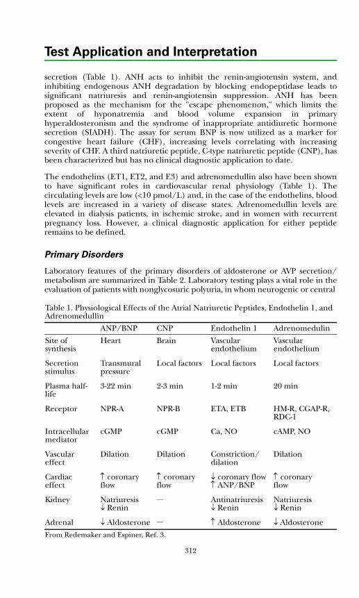
312
Test Application and Interpretation
secretion (Table 1). ANH acts to inhibit the renin-angiotensin system, andinhibiting endogenous ANH degradation by blocking endopeptidase leads tosignificant natriuresis and renin-angiotensin suppression. ANH has beenproposed as the mechanism for the "escape phenomenon," which limits theextent of hyponatremia and blood volume expansion in primaryhyperaldosteronism and the syndrome of inappropriate antidiuretic hormonesecretion (SIADH). The assay for serum BNP is now utilized as a marker forcongestive heart failure (CHF), increasing levels correlating with increasingseverity of CHF. A third natriuretic peptide, C-type natriuretic peptide (CNP), hasbeen characterized but has no clinical diagnostic application to date.
The endothelins (ET1, ET2, and E3) and adrenomedullin also have been shownto have significant roles in cardiovascular renal physiology (Table 1). Thecirculating levels are low (<10 pmol/L) and, in the case of the endothelins, bloodlevels are increased in a variety of disease states. Adrenomedullin levels areelevated in dialysis patients, in ischemic stroke, and in women with recurrentpregnancy loss. However, a clinical diagnostic application for either peptideremains to be defined.
Primary Disorders
Laboratory features of the primary disorders of aldosterone or AVP secretion/metabolism are summarized in Table 2. Laboratory testing plays a vital role in theevaluation of patients with nonglycosuric polyuria, in whom neurogenic or central
Table 1. Physiological Effects of the Atrial Natriuretic Peptides, Endothelin 1, and Adrenomedullin
ANP/BNP CNP Endothelin 1 Adrenomedulin
Site of synthesis
Heart Brain Vascular endothelium
Vascular endothelium
Secretion stimulus
Transmural pressure
Local factors Local factors Local factors
Plasma half-life
3-22 min 2-3 min 1-2 min 20 min
Receptor NPR-A NPR-B ETA, ETB HM-R, CGAP-R, RDC-1
Intracellular mediator
cGMP cGMP Ca, NO cAMP, NO
Vascular effect
Dilation Dilation Constriction/dilation
Dilation
Cardiac effect
↑ coronary flow
↑ coronary flow
↓ coronary flow↑ ANP/BNP
↑ coronary flow
Kidney Natriuresis↓ Renin
__ Antinatriuresis↓ Renin
Natriuresis↓ Renin
Adrenal ↓ Aldosterone __ ↑ Aldosterone ↓ Aldosterone
From Redemaker and Espiner, Ref. 3.

313
Test A
pp
lica
tion
an
d In
terp
reta
tion
Test Application & Interpretation
Table 2. Laboratory Features of Primary Disorders of Salt and Water Metabolism
Disorder Serum Na +
Serum K +
Serum Aldo
Urine Aldo
Urine Na +
Plasma OSM
Plasma AVP
Urine OSM
Urine AVP
Plasma ANH
Plasma Renin*
DNA Analysis
Diabetes insipidus ↑ N N N N ↑ ↓ ↓ ↓ __ N Yes†
SIADH ↓ N N N N ↓ ↑‡ ↑ ↑‡ ↑ N
Nephrogenic diabetes insipidus (AVP resistance) ↑ N N N N ↑ ↑ ↓ ↑ __ N Yes†
Psychogenic polydipsia ↓ N N N N ↓ ↓ ↓ ↓ N
Primary aldosteronism N ↓ ↑ ↑ N−↓ N N N N ↑ ↓
Hypoaldosteronism ↓ ↑ ↓ ↓ ↑ N N N N __ ↑
Hyporeninemichypoaldosteronism
N ↑ ↓ ↓ ↑ N N N N __ N or ↓
Pseudohypoaldosteronism ↓ ↑ ↑ ↑ ↑ N N N N __ ↑Aldo, aldosterone; OSM, osmolality; AVP, arginine vasopressin; ANH, atrial natriuretic hormone; SIADH, syndrome of inappropriate vasopressin (ADH) secretion.*Plasma renin, direct renin or plasma renin activity (PRA).†For genetic mutations causing central or nephrogenic diabetes insipidus.‡Increased relative to plasma osmolality but normal relative to urine osmolality.
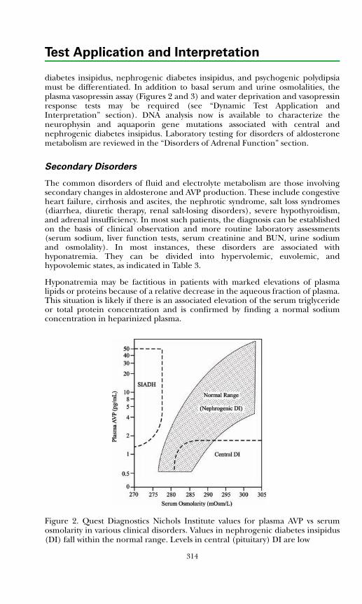
Test Application and Interpretation
diabetes insipidus, nephrogenic diabetes insipidus, and psychogenic polydipsiamust be differentiated. In addition to basal serum and urine osmolalities, theplasma vasopressin assay (Figures 2 and 3) and water deprivation and vasopressinresponse tests may be required (see “Dynamic Test Application andInterpretation” section). DNA analysis now is available to characterize theneurophysin and aquaporin gene mutations associated with central andnephrogenic diabetes insipidus. Laboratory testing for disorders of aldosteronemetabolism are reviewed in the “Disorders of Adrenal Function” section.
Secondary Disorders
The common disorders of fluid and electrolyte metabolism are those involvingsecondary changes in aldosterone and AVP production. These include congestiveheart failure, cirrhosis and ascites, the nephrotic syndrome, salt loss syndromes(diarrhea, diuretic therapy, renal salt-losing disorders), severe hypothyroidism,and adrenal insufficiency. In most such patients, the diagnosis can be establishedon the basis of clinical observation and more routine laboratory assessments(serum sodium, liver function tests, serum creatinine and BUN, urine sodiumand osmolality). In most instances, these disorders are associated withhyponatremia. They can be divided into hypervolemic, euvolemic, andhypovolemic states, as indicated in Table 3.
Hyponatremia may be factitious in patients with marked elevations of plasmalipids or proteins because of a relative decrease in the aqueous fraction of plasma.This situation is likely if there is an associated elevation of the serum triglycerideor total protein concentration and is confirmed by finding a normal sodiumconcentration in heparinized plasma.
Figure 2. Quest Diagnostics Nichols Institute values for plasma AVP vs serumosmolarity in various clinical disorders. Values in nephrogenic diabetes insipidus(DI) fall within the normal range. Levels in central (pituitary) DI are low
314
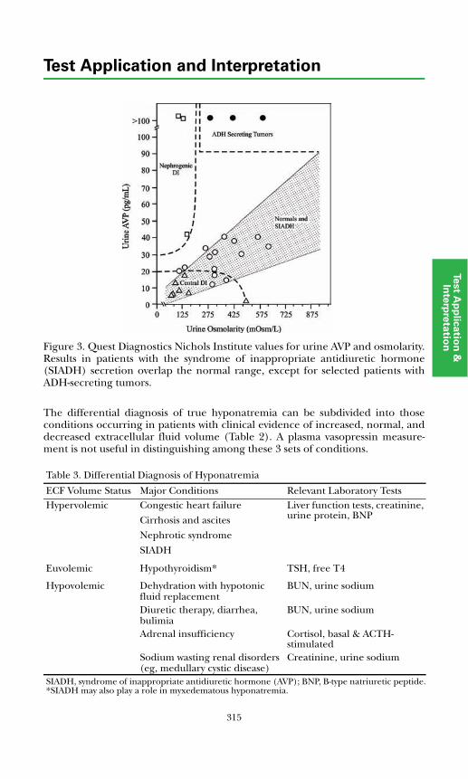
315
Test Application and Interpretation
Test A
pp
licatio
n &
In
terp
reta
tionFigure 3. Quest Diagnostics Nichols Institute values for urine AVP and osmolarity.
Results in patients with the syndrome of inappropriate antidiuretic hormone(SIADH) secretion overlap the normal range, except for selected patients withADH-secreting tumors.
The differential diagnosis of true hyponatremia can be subdivided into thoseconditions occurring in patients with clinical evidence of increased, normal, anddecreased extracellular fluid volume (Table 2). A plasma vasopressin measure-ment is not useful in distinguishing among these 3 sets of conditions. Table 3. Differential Diagnosis of Hyponatremia
ECF Volume Status Major Conditions Relevant Laboratory TestsHypervolemic Congestic heart failure
Cirrhosis and ascites
Nephrotic syndrome
SIADH
Liver function tests, creatinine, urine protein, BNP
Euvolemic Hypothyroidism* TSH, free T4
Hypovolemic Dehydration with hypotonic fluid replacement
BUN, urine sodium
Diuretic therapy, diarrhea, bulimia
BUN, urine sodium
Adrenal insufficiency Cortisol, basal & ACTH-stimulated
Sodium wasting renal disorders(eg, medullary cystic disease)
Creatinine, urine sodium
SIADH, syndrome of inappropriate antidiuretic hormone (AVP); BNP, B-type natriuretic peptide.*SIADH may also play a role in myxedematous hyponatremia.
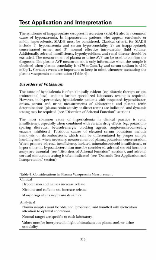
316
Test Application and Interpretation
The syndrome of inappropriate vasopressin secretion (SIADH) also is a commoncause of hyponatremia. In hyponatremic patients who appear euvolemic ormildly hypervolemic, SIADH must be considered. Classical criteria for SIADHinclude 1) hyponatremia and serum hypo-osmolality, 2) an inappropriatelyconcentrated urine, and 3) normal effective intravascular fluid volume.Additionally, adrenal insufficiency, hypothyroidism, and renal disease should beexcluded. The measurement of plasma or urine AVP can be used to confirm thediagnosis. The plasma AVP measurement is only informative when the sample isobtained when plasma osmolality is <270 mOsm/kg and serum sodium is <130mEq/L. Certain caveats are important to keep in mind whenever measuring theplasma vasopressin concentration (Table 4).
Disorders of Potassium
The cause of hypokalemia is often clinically evident (eg, diuretic therapy or gas-trointestinal loss), and no further specialized laboratory testing is required.However, in hypertensive, hypokalemic patients with suspected hyperaldoster-onism, serum and urine measurements of aldosterone and plasma renindeterminations (plasma renin activity or direct renin) are indicated, and dynamictesting may be required (see “Disorders of Adrenal Function” section)
The most common cause of hyperkalemia in clinical practice is renalinsufficiency, especially when combined with certain drug effects (eg, potassium-sparing diuretics, beta-adrenergic blocking agents, angiotensin-convertingenzyme inhibitors). Factitious causes of elevated serum potassium includehemolysis or thrombocytosis, which can be differentiated by proper samplehandling and, when necessary, measurement of plasma potassium concentration.When primary adrenal insufficiency, isolated mineralocorticoid insufficiency, orhyporeninemic hypoaldosteronism must be considered, adrenal steroid hormoneassays are essential (see “Disorders of Adrenal Function” section), and adrenalcortical stimulation testing is often indicated (see “Dynamic Test Application andInterpretation” section)
Table 4. Considerations in Plasma Vasopressin MeasurementClinical
Hypotension and nausea increase release.
Nicotine and caffeine use increase release.
Many drugs alter vasopressin dynamics.
AnalyticalPlasma samples must be obtained, processed, and handled with meticulous attention to optimal conditions.
Normal ranges are specific to each laboratory.
Values must be interpreted in light of simultaneous plasma and/or urine osmolality.
Test Application and Interpretation
Test A
pp
licatio
n &
In
terp
reta
tion
References
1. Robinson AG, Verbalis JG. Posterior pituitary gland. In: Larsen PR, Kronen-berg HM, Melmed S, Polonsky KS, eds. Williams Textbook of Endocrinology. 10thed. Philadelphia, PA: WB Saunders; 2003:281-329.
2. Goodfriend TL, Friedman AL, Shenker Y. Hormonal regulation of electrolyteand water metabolism. In: DeGroot LJ, Jameson JL, et al, eds. Endocrinology.5th ed. Philadelphia, PA: Elsevier-Saunders; 2006:2567-2578.
3. Rademaker MT, Espiner EA. Hormones of the cardiovascular system. In:DeGroot LJ, Jameson JL, et al, eds. Endocrinology. 5th ed. Philadelphia, PA:Elsevier-Saunders; 2006:2549-2566.
4. Muglia LJ, Majzoub JA. Disorders of the posterior pituitary. In: Sperling MA,ed. Pediatric Endocrinology. 2nd ed. Philadelphia, PA: WB Saunders; 2002:289-322.\
317

318
Test Application and Interpretation

Test Application and Interpretation
Test A
pp
licatio
n &
In
terp
reta
tion
PHEOCHROMOCYTOMA, MEDULLARY THYROID
CARCINOMA, AND MULTIPLE ENDOCRINE NEOPLASIA
Pheochromocytoma
Pheochromocytoma occurs at any age independently and sporadically or as afeature of familial multiple endocrine neoplasia (MEN) syndromes, von Hippel-Lindau syndrome (VHL), neurofibromatosis-1 (NF-1), or familialpheochromocytomal paraganglioma. In children, catecholamine hypersecretionis also associated with neuroblastoma or ganglioneuromata due to mutations inthe succinate dehydrogenase (SDH) gene family. Sixty percent to 70% ofpheochromocytomas are sporadic and 20% to 30% are familial. Ninety percent ofsporadic cases occur in the adrenal gland, and 90% of these are unilateral. Over90% of extra-adrenal tumors occur in the abdomen (organ of Zuckerkandl,sympathetic ganglia, urinary bladder), and 10% occur elsewhere (chest, heart,carotid body, vagus nerves). Sporadic pheochromocytomas are usually solitary,well circumscribed, encapsulated, and slow growing. Familialpheochromocytomas are more likely to be multiple and bilateral (50%).Pheochromocytomas usually range in size from 3 to 5 cm, but tumors as large as10 to 20 cm have been reported.
The clinical manifestations of pheochromocytoma vary in type and severity. Themost common symptoms and signs include headache, hypertension, posturalhypotension, tachycardia, palpitations, and sweating. Some patients haveasymptomatic hypertension; many have intermittent symptoms; and severe casesmay manifest weight loss, fasting hyperglycemia, weakness, fatigue, or seizures.Familial cases may present with a thyroid mass (medullary carcinoma).
Three types of MEN have been described: MEN 1, MEN 2A, and MEN 2B. Thesesyndromes, including the hormones/neuropeptides that can be hypersecreted ineach, are summarized in Table 1. MEN syndromes can involve the pituitary,parathyroid, thyroid, adrenal medulla, and pancreatic-gastrointestinal tissues.Pheochromoctyomas caused by familial MEN 2A and MEN 2B are transmitted asautosomal dominant traits and associated with gain of function mutations of theRET proto-oncogene on chromosome 10. The penetrance of medullary thyroidcarcinoma (MTC) in these 2 types is 100% (See "Medullary Thyroid Carcinoma"below). Fifty percent of affected individuals also develop pheochromocytoma,and 20% develop hyperparathyroidism. Patients with MEN 2B present withassociated mucosal neuromata and a marfanoid habitus as well.
Pheochromocytomas in MEN 2 patients, in contrast to sporadic forms, are rarelyextra-adrenal or malignant and are diagnosed at a younger age. This has led tothe practice of cortex-sparing adrenalectomy to avoid the risk of adrenalinsufficiency. The tumors are bilateral in 50% of patients. The risk of recurrentpheochromocytoma in the contralateral gland in patients with unilateral diseaseis 20% to 30%. Patients with unilateral disease can now be managed vialaparoscopic unilateral complete adrenalectomy. For bilateral disease, aunilateral cortical sparing procedure with removal of the entire contralateralgland is recommended.
319
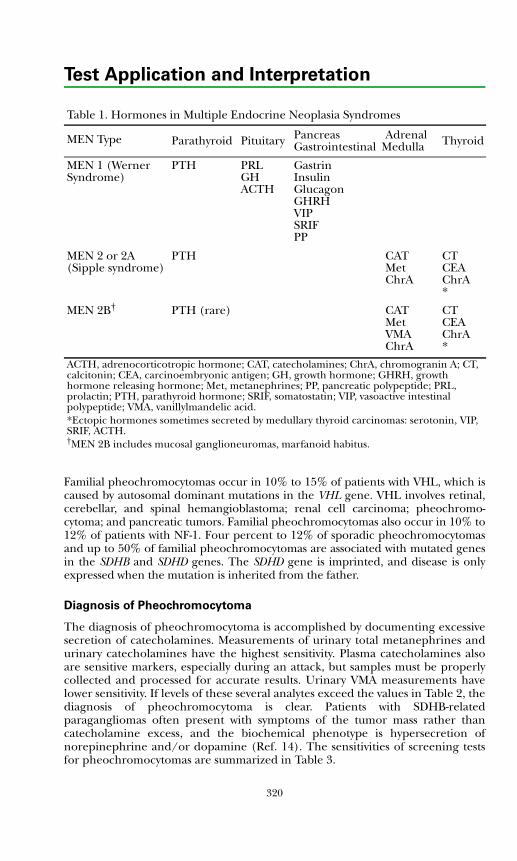
Test Application and Interpretation
Familial pheochromocytomas occur in 10% to 15% of patients with VHL, which iscaused by autosomal dominant mutations in the VHL gene. VHL involves retinal,cerebellar, and spinal hemangioblastoma; renal cell carcinoma; pheochromo-cytoma; and pancreatic tumors. Familial pheochromocytomas also occur in 10% to12% of patients with NF-1. Four percent to 12% of sporadic pheochromocytomasand up to 50% of familial pheochromocytomas are associated with mutated genesin the SDHB and SDHD genes. The SDHD gene is imprinted, and disease is onlyexpressed when the mutation is inherited from the father.
Diagnosis of Pheochromocytoma
The diagnosis of pheochromocytoma is accomplished by documenting excessivesecretion of catecholamines. Measurements of urinary total metanephrines andurinary catecholamines have the highest sensitivity. Plasma catecholamines alsoare sensitive markers, especially during an attack, but samples must be properlycollected and processed for accurate results. Urinary VMA measurements havelower sensitivity. If levels of these several analytes exceed the values in Table 2, thediagnosis of pheochromocytoma is clear. Patients with SDHB-relatedparagangliomas often present with symptoms of the tumor mass rather thancatecholamine excess, and the biochemical phenotype is hypersecretion ofnorepinephrine and/or dopamine (Ref. 14). The sensitivities of screening testsfor pheochromocytomas are summarized in Table 3.
Table 1. Hormones in Multiple Endocrine Neoplasia Syndromes
MEN Type Parathyroid Pituitary PancreasGastrointestinal
Adrenal Medulla Thyroid
MEN 1 (Werner Syndrome)
PTH PRLGHACTH
GastrinInsulinGlucagonGHRHVIPSRIFPP
MEN 2 or 2A (Sipple syndrome)
PTH CATMetChrA
CTCEAChrA*
MEN 2B† PTH (rare) CATMetVMAChrA
CTCEAChrA*
ACTH, adrenocorticotropic hormone; CAT, catecholamines; ChrA, chromogranin A; CT, calcitonin; CEA, carcinoembryonic antigen; GH, growth hormone; GHRH, growth hormone releasing hormone; Met, metanephrines; PP, pancreatic polypeptide; PRL, prolactin; PTH, parathyroid hormone; SRIF, somatostatin; VIP, vasoactive intestinal polypeptide; VMA, vanillylmandelic acid.*Ectopic hormones sometimes secreted by medullary thyroid carcinomas: serotonin, VIP, SRIF, ACTH.†MEN 2B includes mucosal ganglioneuromas, marfanoid habitus.
320
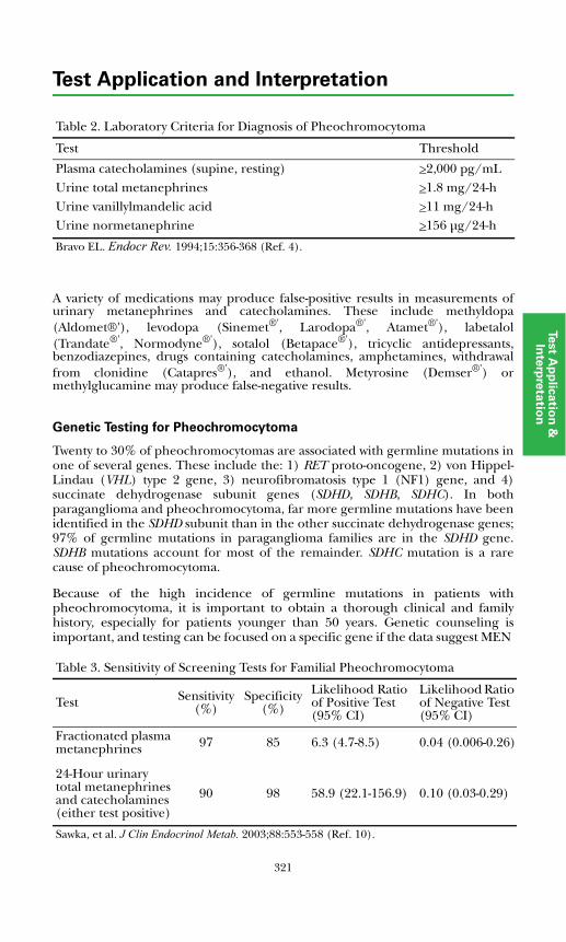
Test Application and Interpretation
Test A
pp
licatio
n &
In
terp
reta
tion
A variety of medications may produce false-positive results in measurements ofurinary metanephrines and catecholamines. These include methyldopa(Aldomet®'), levodopa (Sinemet®', Larodopa®', Atamet®'), labetalol(Trandate®', Normodyne®'), sotalol (Betapace®'), tricyclic antidepressants,benzodiazepines, drugs containing catecholamines, amphetamines, withdrawalfrom clonidine (Catapres®'), and ethanol. Metyrosine (Demser®') ormethylglucamine may produce false-negative results.
Genetic Testing for Pheochromocytoma
Twenty to 30% of pheochromocytomas are associated with germline mutations inone of several genes. These include the: 1) RET proto-oncogene, 2) von Hippel-Lindau (VHL) type 2 gene, 3) neurofibromatosis type 1 (NF1) gene, and 4)succinate dehydrogenase subunit genes (SDHD, SDHB, SDHC). In bothparaganglioma and pheochromocytoma, far more germline mutations have beenidentified in the SDHD subunit than in the other succinate dehydrogenase genes;97% of germline mutations in paraganglioma families are in the SDHD gene.SDHB mutations account for most of the remainder. SDHC mutation is a rarecause of pheochromocytoma.
Because of the high incidence of germline mutations in patients withpheochromocytoma, it is important to obtain a thorough clinical and familyhistory, especially for patients younger than 50 years. Genetic counseling isimportant, and testing can be focused on a specific gene if the data suggest MEN
Table 2. Laboratory Criteria for Diagnosis of Pheochromocytoma
Test Threshold
Plasma catecholamines (supine, resting) >2,000 pg/mLUrine total metanephrines >1.8 mg/24-hUrine vanillylmandelic acid >11 mg/24-hUrine normetanephrine >156 μg/24-h
Bravo EL. Endocr Rev. 1994;15:356-368 (Ref. 4).
Table 3. Sensitivity of Screening Tests for Familial Pheochromocytoma
Test Sensitivity(%)
Specificity(%)
Likelihood Ratio of Positive Test (95% CI)
Likelihood Ratio of Negative Test (95% CI)
Fractionated plasma metanephrines 97 85 6.3 (4.7-8.5) 0.04 (0.006-0.26)
24-Hour urinary total metanephrines and catecholamines (either test positive)
90 98 58.9 (22.1-156.9) 0.10 (0.03-0.29)
Sawka, et al. J Clin Endocrinol Metab. 2003;88:553-558 (Ref. 10).
321
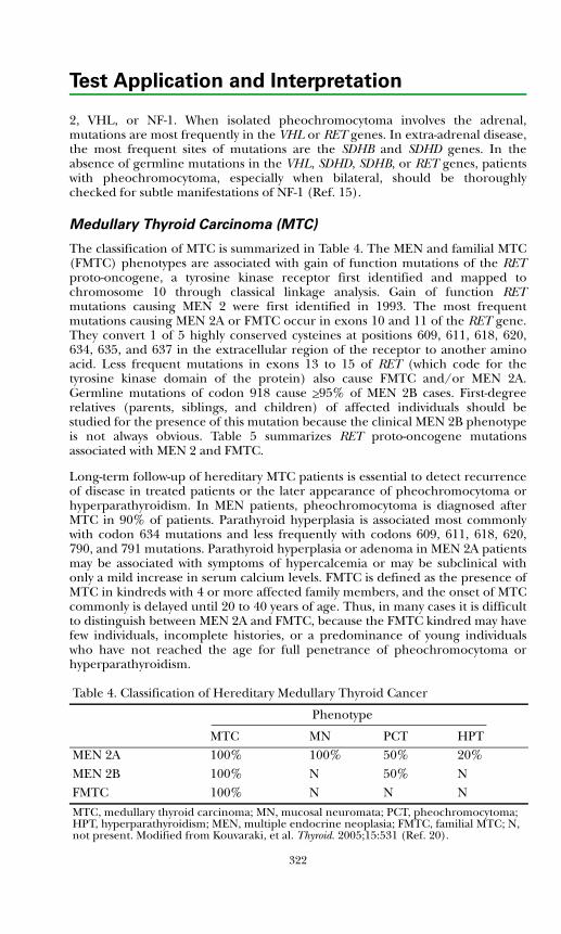
Test Application and Interpretation
2, VHL, or NF-1. When isolated pheochromocytoma involves the adrenal,mutations are most frequently in the VHL or RET genes. In extra-adrenal disease,the most frequent sites of mutations are the SDHB and SDHD genes. In theabsence of germline mutations in the VHL, SDHD, SDHB, or RET genes, patientswith pheochromocytoma, especially when bilateral, should be thoroughlychecked for subtle manifestations of NF-1 (Ref. 15).
Medullary Thyroid Carcinoma (MTC)
The classification of MTC is summarized in Table 4. The MEN and familial MTC(FMTC) phenotypes are associated with gain of function mutations of the RETproto-oncogene, a tyrosine kinase receptor first identified and mapped tochromosome 10 through classical linkage analysis. Gain of function RETmutations causing MEN 2 were first identified in 1993. The most frequentmutations causing MEN 2A or FMTC occur in exons 10 and 11 of the RET gene.They convert 1 of 5 highly conserved cysteines at positions 609, 611, 618, 620,634, 635, and 637 in the extracellular region of the receptor to another aminoacid. Less frequent mutations in exons 13 to 15 of RET (which code for thetyrosine kinase domain of the protein) also cause FMTC and/or MEN 2A.Germline mutations of codon 918 cause >95% of MEN 2B cases. First-degreerelatives (parents, siblings, and children) of affected individuals should bestudied for the presence of this mutation because the clinical MEN 2B phenotypeis not always obvious. Table 5 summarizes RET proto-oncogene mutationsassociated with MEN 2 and FMTC.
Long-term follow-up of hereditary MTC patients is essential to detect recurrenceof disease in treated patients or the later appearance of pheochromocytoma orhyperparathyroidism. In MEN patients, pheochromocytoma is diagnosed afterMTC in 90% of patients. Parathyroid hyperplasia is associated most commonlywith codon 634 mutations and less frequently with codons 609, 611, 618, 620,790, and 791 mutations. Parathyroid hyperplasia or adenoma in MEN 2A patientsmay be associated with symptoms of hypercalcemia or may be subclinical withonly a mild increase in serum calcium levels. FMTC is defined as the presence ofMTC in kindreds with 4 or more affected family members, and the onset of MTCcommonly is delayed until 20 to 40 years of age. Thus, in many cases it is difficultto distinguish between MEN 2A and FMTC, because the FMTC kindred may havefew individuals, incomplete histories, or a predominance of young individualswho have not reached the age for full penetrance of pheochromocytoma orhyperparathyroidism.
Table 4. Classification of Hereditary Medullary Thyroid Cancer
Phenotype
MTC MN PCT HPTMEN 2A 100% 100% 50% 20%MEN 2B 100% N 50% NFMTC 100% N N N
MTC, medullary thyroid carcinoma; MN, mucosal neuromata; PCT, pheochromocytoma; HPT, hyperparathyroidism; MEN, multiple endocrine neoplasia; FMTC, familial MTC; N, not present. Modified from Kouvaraki, et al. Thyroid. 2005;15:531 (Ref. 20).
322
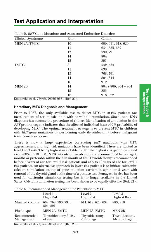
Test Application and Interpretation
Test A
pp
licatio
n &
In
terp
reta
tion
Kouvaraki, et al. Thyroid. 2005;15:531 (Ref. 20).
Hereditary MTC Diagnosis and Management
Prior to 1987, the only available test to detect MTC in at-risk patients wasmeasurement of serum calcitonin with or without stimulation. Since then, DNAdiagnosis has become the procedure of choice. Identification of a mutation in theRET protooncogene indicates that the affected individual has a >90% probability ofdeveloping MTC. The optimal treatment strategy is to prevent MTC in childrenwith RET gene mutations by performing early thyroidectomy before malignanttransformation occurs.
There is now a large experience correlating RET mutations with MTCaggressiveness, and high risk mutations have been identified. These are ranked aslevel 1 to 3 with 3 being highest risk (Table 6). For the highest risk group (mutatedexons 883 or 918 in MEN 2B patients), thyroidectomy is recommended before age 6months or preferably within the first month of life. Thyroidectomy is recommendedbefore 5 years of age for level 2 risk patients and at 5 to 10 years of age for level 1risk patients. An alternative approach in lower risk patients is to initiate calcitonin-calcium stimulation testing of gene mutation carriers at age 4 or 5 years withremoval of the thyroid gland at the time of a positive test. Pentagastrin also has beenused for calcitonin stimulation testing but is no longer available in the UnitedStates. Calcium stimulation testing has been shown to be equally effective (Ref. 21).
Kouvaraki, et al. Thyroid. 2005;15:531 (Ref. 20).
Table 5. RET Gene Mutations and Associated Endocrine DisordersClinical Syndrome Exon CodonMEN 2A/FMTC 10 609, 611, 618, 620
11 634, 635, 63713 790, 79114 80415 891
FMTC 8 532, 53311 63013 768, 79114 804, 84416 912
MEN 2B 14 804 + 806, 804 + 90415 88316 918, 922
Table 6. Recommended Management for Patients with MTCLevel 1High Risk
Level 2High Risk
Level 3Highest Risk
Mutated codons 609, 768, 790, 791, 804, 891
611, 618, 620, 634 883, 918
Phenotype MEN 2A, FMTC MEN 2A, FMTC MEN 2BRecommendedManagement
Thyroidectomy 5-10 y of age
Thyroidectomy <5 y of age
Thyroidectomy 1-6 mo of age
323
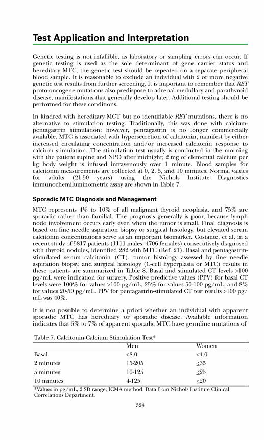
Test Application and Interpretation
Genetic testing is not infallible, as laboratory or sampling errors can occur. Ifgenetic testing is used as the sole determinant of gene carrier status andhereditary MTC, the genetic test should be repeated on a separate peripheralblood sample. It is reasonable to exclude an individual with 2 or more negativegenetic test results from further screening. It is important to remember that RETproto-oncogene mutations also predispose to adrenal medullary and parathyroiddisease, manifestations that generally develop later. Additional testing should beperformed for these conditions.
In kindred with hereditary MCT but no identifiable RET mutations, there is noalternative to stimulation testing. Traditionally, this was done with calcium-pentagastrin stimulation; however, pentagastrin is no longer commerciallyavailable. MTC is associated with hypersecretion of calcitonin, manifest by eitherincreased circulating concentration and/or increased calcitonin response tocalcium stimulation. The stimulation test usually is conducted in the morningwith the patient supine and NPO after midnight; 2 mg of elemental calcium perkg body weight is infused intravenously over 1 minute. Blood samples forcalcitonin measurements are collected at 0, 2, 5, and 10 minutes. Normal valuesfor adults (21-50 years) using the Nichols Institute Diagnosticsimmunochemiluminometric assay are shown in Table 7.
Sporadic MTC Diagnosis and Management
MTC represents 4% to 10% of all malignant thyroid neoplasia, and 75% aresporadic rather than familial. The prognosis generally is poor, because lymphnode involvement occurs early even when the tumor is small. Final diagnosis isbased on fine needle aspiration biopsy or surgical histology, but elevated serumcalcitonin concentrations serve as an important biomarker. Costante, et al, in arecent study of 5817 patients (1111 males, 4706 females) consecutively diagnosedwith thyroid nodules, identified 282 with MTC (Ref. 21). Basal and pentagastrin-stimulated serum calcitonin (CT), tumor histology assessed by fine needleaspiration biopsy, and surgical histology (C-cell hyperplasia or MTC) results inthese patients are summarized in Table 8. Basal and stimulated CT levels >100pg/mL were indication for surgery. Positive predictive values (PPV) for basal CTlevels were 100% for values >100 pg/mL, 25% for values 50-100 pg/mL, and 8%for values 20-50 pg/mL. PPV for pentagastrin-stimulated CT test results >100 pg/mL was 40%.
It is not possible to determine a priori whether an individual with apparentsporadic MTC has hereditary or sporadic disease. Available informationindicates that 6% to 7% of apparent sporadic MTC have germline mutations of
Table 7. Calcitonin-Calcium Stimulation Test* Men Women
Basal <8.0 <4.02 minutes 15-205 <355 minutes 10-125 <2510 minutes 4-125 <20*Values in pg/mL, 2 SD range; ICMA method. Data from Nichols Institute ClinicalCorrelations Department.
324
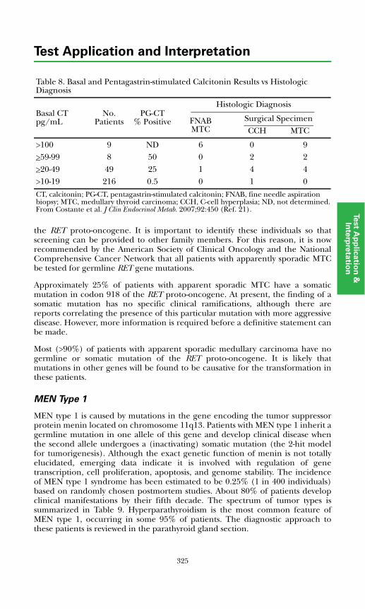
Test Application and Interpretation
Test A
pp
licatio
n &
In
terp
reta
tion
the RET proto-oncogene. It is important to identify these individuals so thatscreening can be provided to other family members. For this reason, it is nowrecommended by the American Society of Clinical Oncology and the NationalComprehensive Cancer Network that all patients with apparently sporadic MTCbe tested for germline RET gene mutations.
Approximately 25% of patients with apparent sporadic MTC have a somaticmutation in codon 918 of the RET proto-oncogene. At present, the finding of asomatic mutation has no specific clinical ramifications, although there arereports correlating the presence of this particular mutation with more aggressivedisease. However, more information is required before a definitive statement canbe made.
Most (>90%) of patients with apparent sporadic medullary carcinoma have nogermline or somatic mutation of the RET proto-oncogene. It is likely thatmutations in other genes will be found to be causative for the transformation inthese patients.
MEN Type 1
MEN type 1 is caused by mutations in the gene encoding the tumor suppressorprotein menin located on chromosome 11q13. Patients with MEN type 1 inherit agermline mutation in one allele of this gene and develop clinical disease whenthe second allele undergoes a (inactivating) somatic mutation (the 2-hit modelfor tumorigenesis). Although the exact genetic function of menin is not totallyelucidated, emerging data indicate it is involved with regulation of genetranscription, cell proliferation, apoptosis, and genome stability. The incidenceof MEN type 1 syndrome has been estimated to be 0.25% (1 in 400 individuals)based on randomly chosen postmortem studies. About 80% of patients developclinical manifestations by their fifth decade. The spectrum of tumor types issummarized in Table 9. Hyperparathyroidism is the most common feature ofMEN type 1, occurring in some 95% of patients. The diagnostic approach tothese patients is reviewed in the parathyroid gland section.
Table 8. Basal and Pentagastrin-stimulated Calcitonin Results vs Histologic Diagnosis
Basal CTpg/mL
No. Patients
PG-CT% Positive
Histologic Diagnosis
FNABMTC
Surgical SpecimenCCH MTC
>100 9 ND 6 0 9>59-99 8 50 0 2 2>20-49 49 25 1 4 4>10-19 216 0.5 0 1 0
CT, calcitonin; PG-CT, pentagastrin-stimulated calcitonin; FNAB, fine needle aspiration biopsy; MTC, medullary thyroid carcinoma; CCH, C-cell hyperplasia; ND, not determined.From Costante et al. J Clin Endocrinol Metab. 2007;92:450 (Ref. 21).
325
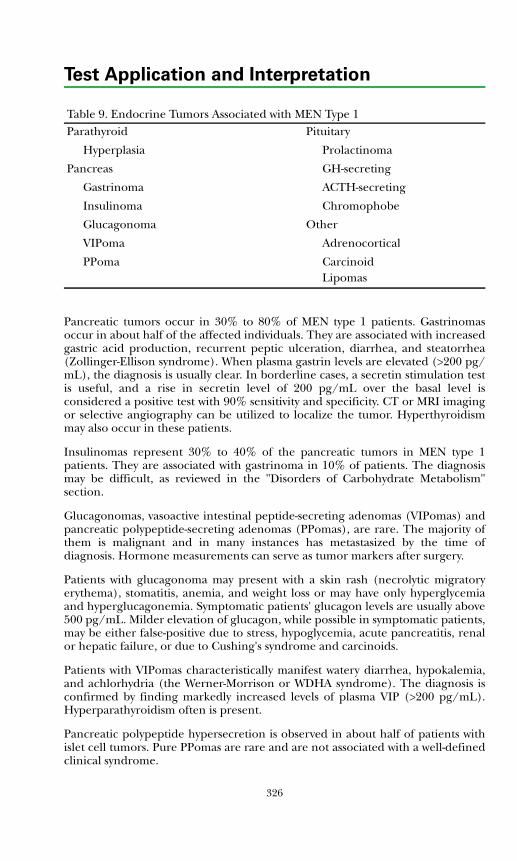
Test Application and Interpretation
Pancreatic tumors occur in 30% to 80% of MEN type 1 patients. Gastrinomasoccur in about half of the affected individuals. They are associated with increasedgastric acid production, recurrent peptic ulceration, diarrhea, and steatorrhea(Zollinger-Ellison syndrome). When plasma gastrin levels are elevated (>200 pg/mL), the diagnosis is usually clear. In borderline cases, a secretin stimulation testis useful, and a rise in secretin level of 200 pg/mL over the basal level isconsidered a positive test with 90% sensitivity and specificity. CT or MRI imagingor selective angiography can be utilized to localize the tumor. Hyperthyroidismmay also occur in these patients.
Insulinomas represent 30% to 40% of the pancreatic tumors in MEN type 1patients. They are associated with gastrinoma in 10% of patients. The diagnosismay be difficult, as reviewed in the "Disorders of Carbohydrate Metabolism"section.
Glucagonomas, vasoactive intestinal peptide-secreting adenomas (VIPomas) andpancreatic polypeptide-secreting adenomas (PPomas), are rare. The majority ofthem is malignant and in many instances has metastasized by the time ofdiagnosis. Hormone measurements can serve as tumor markers after surgery.
Patients with glucagonoma may present with a skin rash (necrolytic migratoryerythema), stomatitis, anemia, and weight loss or may have only hyperglycemiaand hyperglucagonemia. Symptomatic patients' glucagon levels are usually above500 pg/mL. Milder elevation of glucagon, while possible in symptomatic patients,may be either false-positive due to stress, hypoglycemia, acute pancreatitis, renalor hepatic failure, or due to Cushing's syndrome and carcinoids.
Patients with VIPomas characteristically manifest watery diarrhea, hypokalemia,and achlorhydria (the Werner-Morrison or WDHA syndrome). The diagnosis isconfirmed by finding markedly increased levels of plasma VIP (>200 pg/mL).Hyperparathyroidism often is present.
Pancreatic polypeptide hypersecretion is observed in about half of patients withislet cell tumors. Pure PPomas are rare and are not associated with a well-definedclinical syndrome.
Table 9. Endocrine Tumors Associated with MEN Type 1 Parathyroid Pituitary
Hyperplasia Prolactinoma
Pancreas GH-secreting
Gastrinoma ACTH-secreting
Insulinoma Chromophobe
Glucagonoma Other
VIPoma Adrenocortical
PPoma CarcinoidLipomas
326

Test Application and Interpretation
Test A
pp
licatio
n &
In
terp
reta
tion
References
1. Pacak K, Keiser H, Eisenhofer G. Pheochromocytoma. In: DeGroot LJ, Jame-son JL, eds. Endocrinology. 5th ed. Philadelphia, PA: Elsevier-Saunders;2006:2501-2534.
2. Neumann HP, Berger DP, Sigmund G, et al. Pheochromocytomas, multipleendocrine neoplasia type 2, and von Hippel-Lindau disease. New Engl J Med.1993;329:1531-1538.
3. Canale MP, Bravo EL. Diagnostic specificity of serum chromogranin A forpheochromocytoma patients with renal dysfunction. J Clin Endocrinol Metab.1994;78:1139-1144.
4. Bravo EL. Evolving concepts in the pathophysiology, diagnosis, and treatmentof pheochromoctyoma. Endocr Rev. 1994;15:356-368.
5. Thakker RV. Multiple endocrine neoplasia type 1. In: DeGroot LJ, JamesonJL, eds. Endocrinology. 5th ed. Philadelphia, PA: Elsevier-Saunders; 2006:3509-3531.
6. Taheri S, Ghatei MA, Bloom SR. Gastrointestinal hormones and tumorsyndromes. In: DeGroot LJ, Jameson JL, eds. Endocrinology. 5th ed.Philadelphia, PA: Elsevier-Saunders; 2006:3551-3570.
7. Hoff AO, Gagel RF. Multiple Endocrine neoplasia type 2. In: DeGroot LJ,Jameson JL, eds. Endocrinology. 5th ed. Philadelphia, PA: Elsevier-Saunders;2006:3533-3550.
8. Ray DW. Ectopic hormone syndromes. In: DeGroot LJ, Jameson JL, eds.Endocrinology. 5th ed. Philadelphia, PA: Elsevier-Saunders; 2006;3585-3601.
9. Bryant J, Farmer J, Kessler LJ, et al. Pheochromocytoma: the expandingdifferential diagnosis. J Natl Cancer Inst. 2003;95:1196-1204.
10. Sawka AM, Jaeschke R, Singh RJ, et al. A comparison of biochemical tests forpheochromocytoma; measurement of fractionated plasma metanephrinescompared with the combination of 24-hour urinary metanephrines andcatecholamines. J Clin Endocrinol Metab. 2003;88:553-558.
11. Neumann HP, Bausch B, McWhinney SR, et al. Germ-line mutations innonsyndromic pheochromocytoma. N Engl J Med. 2002;346:1459-1466.
12. Favier J, Briere JJ, Strompf L, et al. Hereditary paraganglioma/pheochromocytoma and inherited succinate dehydrogenase deficiency. HormRes. 2005;63:171-179.
13. Pawlu C, Bausch B, Neumann HP. Mutations in the SDHB and SDHD genes.Familial Cancer. 2005;4:49-54.
14. Timmers HJ, Kozupa A, Eisenhofer G, et al. Clinical presentations,biochemical phenotypes, and genotype-phenotype correlations in patientswith succinate dehydrogenase subunit B-associated pheochromocytomas andparagangliomas. J Clin Endocrinol Metab. 2007;92:779-786.
327
Test Application and Interpretation
15. Bausch B, Koschker AC, Fassnacht M, et al. Comprehensive mutationscanning of NF1 in apparently sporadic cases of pheochromocytoma. J ClinEndocrinol Metab. 2006;91:3478-3481.
16. Chi DD, Toshima K, Donis-Keller H, et al. Predictive testing for multipleendocrine neoplasia type 2A (MEN 2A) based on the detection of mutationsin the RET protooncogene. Surgery. 1994;116:124-132.
17. Santoro M, Carlomagno F, Romano A, et al. Activation of RET as a dominanttransforming gene by germline mutations of MEN2A and MEN2B. Science.1995;267:381-383.
18. Wohllk N, Cote GJ, Evans D, et al. Application of genetic screeninginformation to the management of medullary thyroid carcinoma andmultiple endocrine neoplasia type 2. Endocrinol Metab Clin North Am.1996;25:1-25.
19. Wohllk N, Cote GJ, Bugalho MM, et al. Relevance of RET proto-oncogenemutations in sporadic medullary thyroid carcinoma. J Clin Endocrinol Metab.1996;81:3740-3745.
20. Kouvaraki MA, Shapiro SE, Perrier ND, et al. RET proto-oncogene: a reviewand update of genotype-phenotype correlations in hereditary medullarythyroid cancer and associated endocrine tumors. Thyroid. 2005;15:531-544.
21. Costante G, Meringolo D, Durante C, et al. Predictive value of serumcalcitonin levels for preoperative diagnosis of medullary thyroid carcinoma ina cohort of 5817 consecutive patients with thyroid nodules. J Clin EndocrinolMetab. 2007;92:450-455.
328
Test Application and Interpretation
Test A
pp
licatio
n &
In
terp
reta
tion
DYNAMIC TESTS
Every effort has been made to ensure accuracy in these recommendations, butclinicians must use their judgment and refer to specific pharmaceutical resourcesto determine appropriate drug dosages for their patients.
Adrenal Dynamic Tests
ACTH Stimulation Test, Standard1-3
Indication: Suspected adrenal insufficiency; suspected adrenal biosynthetic defector ACTH unresponsiveness
Medication: 10 μg/kg, maximum 250 μg (0.25 mg), cosyntropin (Cortrosyn®',synthetic 1-24 ACTH) by IV bolus; dilute in 2 to 5 mL physiological saline.Intramuscular dosing is equally effective; dilute in 1 mL physiological saline.
Sampling: Serum cortisol at 0 and 30 or 60 minutes
Interpretation: The usual normal response is an approximate doubling of thebasal level, provided that the basal level does not exceed the normal range.Analysis of 8 reported studies including 122 patients indicated sensitivities of 97%and 57% respectively at 95% specificity for diagnosis of primary and secondaryadrenal insufficiency using a serum cortisol cutoff of 15 μg/dL.
Comments: Normal response excludes primary adrenal insufficiency. Patients withsecondary (ACTH) adrenal insufficiency usually show a blunted response butmay have a normal result. Disparate levels of baseline precursor and productsteroids (precursor > product) localize enzyme dysfunction in severe CAH cases.The ratio of precursor to product tends to be similar at baseline and peaksampling times. In children with milder defects, localization is defined bysubstantially disproportionate fold increases in precursor and product steroidlevels after ACTH.
ACTH Stimulation Test, Prolonged1,2
Indication: Suspected secondary adrenal insufficiency; differentiates primaryfrom secondary adrenal insufficiency
Medication: 0.5 IU/m2 corticotropin (Acthar®' gel) intramuscularly every 12hours for 3 days or 10 μg/kg cosyntropin (Cortrosyn) intravenously in 100-250mL of physiological saline over 8-12 hours for 1 to 2 days; maximum, 250 μg.
Sampling: Serum cortisol before and 2 hours after each injection or infusion for 3days and/or 24-hour urine samples for 4 days (baseline + 3 stimulation days)
Interpretation: Serum cortisol to exceed 25 μg/dL. Urine 17-hydroxy-corticosteroids or free cortisol values usually increase 2-fold.
329
Test Application and Interpretation
Comments: May give dexamethasone (20 μg/kg/d) to prevent adrenal crisis – willnot interfere with test. Normal subjects and patients with secondary adrenalinsufficiency will respond to prolonged stimulation.
ACTH Stimulation Test, Low Dose1,3
Indication: Suspected adrenal insufficiency, biosynthetic enzyme defect, or ACTHunresponsiveness
Medication: 1.0 μg/1.73 m2 cosyntropin (Cortrosyn, 1-24 synthetic ACTH) by IVbolus in 1.0 mL saline
Sampling: Blood for measurements of cortisol at 0, 30, and 40 minutes
Interpretation: Serum cortisol should be >15 μg/dL.
Comments: The low dose test is useful in infants, avoiding under diagnosis ofhypothalamic-pituitary-adrenal axis impairment. It is particularly useful inassessing adrenal responsiveness before discontinuation of dexamethasonetreatment. Glucocorticoid should be withheld for 12 hours prior to testing. Inolder patients, the low dose test has equal sensitivity to the standard ACTH test.
Aldosterone Suppression Test1,2,4
Indication: Suspected primary hyperaldosteronism
Medication: IV saline loading (2 L of 0.9% NaCl infused over 4 hours) or oralsodium loading for 3 to 4 days (urine sodium excretion >200 mEq/24-h) oradministration of 0.1 mg fludrocortisone acetate q 6 hours plus NaCl x 4 days
Sampling: Aldosterone and direct renin or plasma renin activity (PRA) aremeasured before and after suppression.
Interpretation: In normal subjects, the serum aldosterone is <5 ng/dL aftersuppression. PRA is suppressed to the lower limit of detection.
Comments: None
CRH Stimulation Test1,2
Indication: Suspected pituitary ACTH deficiency; suspected pituitary Cushing's;rule out ectopic disease
Medication: 1.0 μg/kg (100 μg/1.73 m2; maximum dose 100 μg) synthetic ovineor human CRH administered as IV bolus over 30 seconds. Patient fasting 4 hours.
Sampling: Blood measurements of cortisol and ACTH at -5 and -1 minutes beforeCRH and at 15, 30, 45, and 60 minutes after CRH.
Interpretation: Baseline ACTH increases 20% to 40% in 95% of normal subjects.Peak values (usually in the 20 to 100 pg/mL range) are observed at 30 to 60minutes. Serum cortisol peaks at 30 to 60 minutes in the 20 to 25 μg/dL range.
330
Test Application and Interpretation
Test A
pp
licatio
n &
In
terp
reta
tion
Comments: The test can be conducted in the AM or PM, but in normal subjectsthe responses vary with time of day. The ACTH increment is similar morning andevening, but peak values vary with baseline (higher in AM). Cortisol levels peak atsimilar values morning and evening. Patients with pituitary ACTH deficiency havesubnormal responses; those with hypothalamic disease tend to have augmentedand prolonged ACTH responses with reduced cortisol response. Patients withprimary adrenal insufficiency have high baseline ACTH, an augmented responseto CRH, and low cortisol levels before and after CRH. Patients with Cushing'sdisease (pituitary Cushing's) usually show a >20% rise in cortisol and a >50%increase in ACTH. Patients with adrenal causes of Cushing's syndrome or ectopicACTH syndrome do not respond. Rarely, bronchial carcinoids producing ACTHmay respond to CRH.
CRH Stimulation Test, Adrenal Venous Sampling5
Indication: Distinguish between an adenoma or unilateral and bilateralhyperplasia in patients with primary aldosteronism. The test also is helpful whenimaging studies fail to unequivocally identify an adenoma or when the identifiedadenoma is <1 cm in size.
Medication: 50 μg per hour of cosyntropin (Cortrosyn) is infused intravenously 30minutes prior and continuously throughout the procedure.
Sampling: In this standard protocol, measurements for both cortisol (μg/dL) andaldosterone (ng/dL) are performed in serum samples obtained from the inferiorvena cava (IVC) and each of the adrenal veins.
Interpretation: A catheterization is considered successful if the adrenal veincortisol level is 5-fold higher than the level in IVC. A lateralizing response isdefined as a serum aldosterone/cortisol ratio 4-fold higher in the adrenal veindraining the secreting side. A ratio of <3 is consistent with bilateral aldosteronehypersecretion, while a ratio between 3 and 4 is an area of overlap.
Comments: Determination of aldosterone/cortisol ratio in samples of adrenalvenous blood, obtained by an experienced radiologist, is the "gold standard" test.Unilateral disease is associated with a marked increase in the aldosterone/cortisol ratio on the side of the tumor, whereas there is little difference betweenthe 2 sides in patients with bilateral hyperplasia. In a study of 163 men and 40women with hyperaldosteronism, 56.7% of patients had a unilateral source ofaldosterone secretion. The cutoff described above was able to lateralize thesecreting site with 95% sensitivity and 100% specificity.
CRH Stimulation Test, Petrosal Venous Sinus Sampling1,6
Indication: Differentiation between ectopic and pituitary causes of Cushing'sdisease. This procedure can better distinguish between ectopic and pituitaryACTH hypersecretion than dexamethasone suppression, but it is invasive, may beassociated with serious complications, and therefore should be performed by anexperienced radiologist. This procedure appears to be less useful forlateralization of the tumor in cases in which the ACTH secretion appears to be ofpituitary origin.
331
Test Application and Interpretation
Medication: ovine CRH (1 μg/kg body weight) administered as IV bolus
Sampling: Catheters are inserted via the jugular or femoral veins into bothinferior petrosal veins. Blood is obtained from a peripheral vein via a thirdcatheter or from a port in the iliac vein. Blood samples are drawn simultaneouslyfrom both inferior petrosal sinuses and the peripheral vein for plasma ACTH. Atleast 4 sets of 3 samples each are obtained: 2 sets are drawn immediately beforeinjection of CRH as a baseline and 2 more are drawn between 2 and 3 andbetween 5 and 6 minutes after peripheral intravenous injection of CRH.
Interpretation: A central-to-peripheral plasma ACTH gradient of 2.0 before CRHadministration, or 3.0 after CRH, is diagnostic of a pituitary source of ACTH. Agradient of 1.4 to 1.5 between the 2 sinuses may predict the lateral site of theadenoma.
Comments: In 14 studies, 759 of a total of 800 patients with proven Cushing'sdisease were correctly identified (95% sensitivity), while 115 of 124 withpresumed or proven ectopic ACTH secretion were correctly identified (93%specificity). False negatives may occur in episodic Cushing's disease or in aberrantdrainage through an abnormal petrosal sinus on the side of the adenoma. Thelateralization of the adenoma in pituitary Cushing's through this procedure is ofdubious value since it is only 70% accurate. The procedure is not indicated insubjects with a pituitary adenoma >6 mm and a response suggestive of pituitaryCushing's on the CRH or dexamethasone tests.
Dexamethasone Suppression Tests6
Indication: Suspected endogenous Cushing's syndrome or dexamethasone-suppressible hyperaldosteronism (DSH). Differentiate Cushing's disease(pituitary) from ectopic source. Suggest a major adrenal source of androgen inpolycystic ovarian disease.
Overnight Suppression TestMedication: 20 μg/kg (1 mg in adults) dexamethasone at 11-12 PM
Sampling: Serum cortisol at 8 AM
Interpretation: Suppression of 8 AM serum cortisol to <2 μg/dL effectivelyexcludes Cushing's syndrome. A cortisol level >2 μg/dL merits further evaluation.
2-Day, Low Dose Suppression TestThis test has been shown to have a sensitivity of 79%, a specificity of 74%, and adiagnostic accuracy of only 71% in patients with mild Cushing's syndrome. Thetest is complex and difficult to administer and is no longer recommended.
Carbohydrate Metabolic Dynamic Tests
Glucose Tolerance Test7
Indication: Suspected impairment of glucose tolerance
332
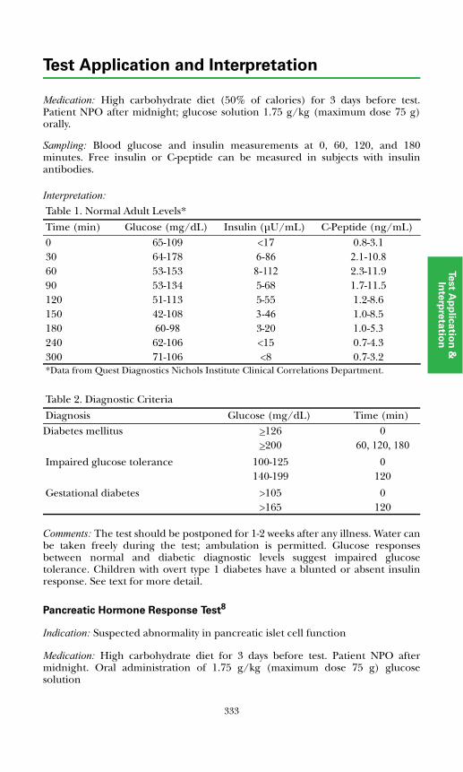
Test Application and Interpretation
Test A
pp
licatio
n &
In
terp
reta
tion
Medication: High carbohydrate diet (50% of calories) for 3 days before test.Patient NPO after midnight; glucose solution 1.75 g/kg (maximum dose 75 g)orally.
Sampling: Blood glucose and insulin measurements at 0, 60, 120, and 180minutes. Free insulin or C-peptide can be measured in subjects with insulinantibodies.
Interpretation:
Comments: The test should be postponed for 1-2 weeks after any illness. Water canbe taken freely during the test; ambulation is permitted. Glucose responsesbetween normal and diabetic diagnostic levels suggest impaired glucosetolerance. Children with overt type 1 diabetes have a blunted or absent insulinresponse. See text for more detail.
Pancreatic Hormone Response Test8
Indication: Suspected abnormality in pancreatic islet cell function
Medication: High carbohydrate diet for 3 days before test. Patient NPO aftermidnight. Oral administration of 1.75 g/kg (maximum dose 75 g) glucosesolution
Table 1. Normal Adult Levels*
Time (min) Glucose (mg/dL) Insulin (μU/mL) C-Peptide (ng/mL)0 65-109 <17 0.8-3.130 64-178 6-86 2.1-10.860 53-153 8-112 2.3-11.990 53-134 5-68 1.7-11.5120 51-113 5-55 1.2-8.6150 42-108 3-46 1.0-8.5180 60-98 3-20 1.0-5.3240 62-106 <15 0.7-4.3300 71-106 <8 0.7-3.2*Data from Quest Diagnostics Nichols Institute Clinical Correlations Department.
Table 2. Diagnostic Criteria
Diagnosis Glucose (mg/dL) Time (min)Diabetes mellitus >126 0
>200 60, 120, 180Impaired glucose tolerance 100-125 0
140-199 120
Gestational diabetes >105 0>165 120
333
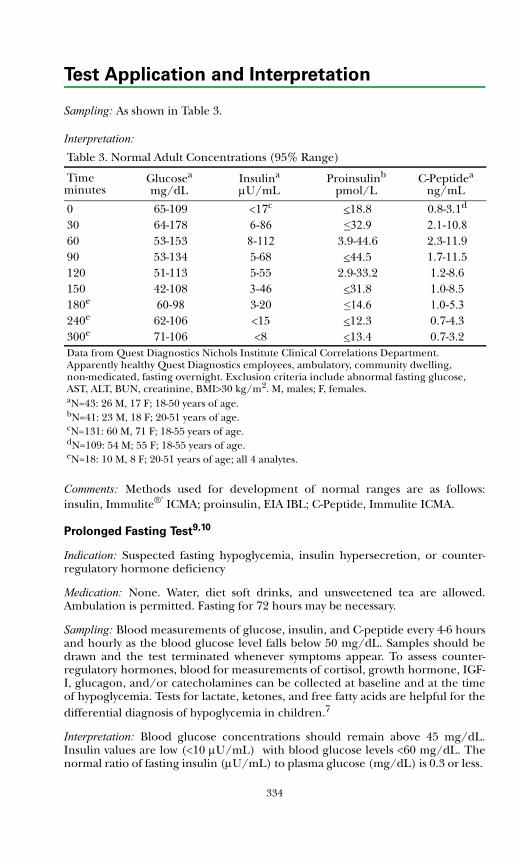
Test Application and Interpretation
Sampling: As shown in Table 3.
Interpretation:
Comments: Methods used for development of normal ranges are as follows:insulin, Immulite®' ICMA; proinsulin, EIA IBL; C-Peptide, Immulite ICMA.
Prolonged Fasting Test9,10
Indication: Suspected fasting hypoglycemia, insulin hypersecretion, or counter-regulatory hormone deficiency
Medication: None. Water, diet soft drinks, and unsweetened tea are allowed.Ambulation is permitted. Fasting for 72 hours may be necessary.
Sampling: Blood measurements of glucose, insulin, and C-peptide every 4-6 hoursand hourly as the blood glucose level falls below 50 mg/dL. Samples should bedrawn and the test terminated whenever symptoms appear. To assess counter-regulatory hormones, blood for measurements of cortisol, growth hormone, IGF-I, glucagon, and/or catecholamines can be collected at baseline and at the timeof hypoglycemia. Tests for lactate, ketones, and free fatty acids are helpful for thedifferential diagnosis of hypoglycemia in children.7
Interpretation: Blood glucose concentrations should remain above 45 mg/dL.Insulin values are low (<10 μU/mL) with blood glucose levels <60 mg/dL. Thenormal ratio of fasting insulin (μU/mL) to plasma glucose (mg/dL) is 0.3 or less.
Table 3. Normal Adult Concentrations (95% Range)
Timea
minutesGlucosea
mg/dLInsulina
μU/mLProinsulinb
pmol/LC-Peptidea
ng/mL
0 65-109 <17c <18.8 0.8-3.1d
30 64-178 6-86 <32.9 2.1-10.860 53-153 8-112 3.9-44.6 2.3-11.990 53-134 5-68 <44.5 1.7-11.5120 51-113 5-55 2.9-33.2 1.2-8.6150 42-108 3-46 <31.8 1.0-8.5180e 60-98 3-20 <14.6 1.0-5.3240e 62-106 <15 <12.3 0.7-4.3300e 71-106 <8 <13.4 0.7-3.2Data from Quest Diagnostics Nichols Institute Clinical Correlations Department. Apparently healthy Quest Diagnostics employees, ambulatory, community dwelling, non-medicated, fasting overnight. Exclusion criteria include abnormal fasting glucose, AST, ALT, BUN, creatinine, BMI>30 kg/m2. M, males; F, females.aN=43: 26 M, 17 F; 18-50 years of age.bN=41: 23 M, 18 F; 20-51 years of age.cN=131: 60 M, 71 F; 18-55 years of age.dN=109: 54 M; 55 F; 18-55 years of age.eN=18: 10 M, 8 F; 20-51 years of age; all 4 analytes.
334
Test Application and Interpretation
Test A
pp
licatio
n &
In
terp
reta
tion
C-peptide levels should fall to <0.6 ng/mL. Levels of counter-regulatoryhormones increase in the presence of hypoglycemia.
Comments: The patient should be hospitalized or observed constantly in anoutpatient environment for the test. There is a continuing risk of hypoglycemia.IV access should be maintained, and glucose for IV infusion must be available.
Gonadal Dynamic Test
GnRH Stimulation Test11,12
Indication: Suspected central precocious puberty (CPP); suspected pituitarygonadotropin deficiency; monitor long acting GnRH analogue therapy
Intravenous GnRHMedication: 2.5 μg/kg, up to 100 μg maximum, rapid intravenous injection ofGnRH (Factrel®', Wyeth®' Ayerst, available in 0.1 or 0.5 mg vials)
Sampling: Blood samples for measurements of serum LH and FSH concentrationsare obtained immediately before and at 30, 45, and 60 minutes after GnRHinjection. A single 30-minute LH sample can be used to monitor GnRH analoguetherapy.
Interpretation: Prepubertally, levels increase 2- to 4-fold with a peak LH/FSH ratioapproximating 0.7. Postpubertally, LH levels increase 6- to 10-fold and FSH 4- to6-fold with a mean peak LH/FSH ratio approximating 3.5. Cavallo et al describe>90% sensitivity and >80% specificity for a single LH concentration cutoff of 15IU/L for the diagnosis of central precocious puberty (CPP).11
Subcutaneous GnRH
Medication: 100 μg GnRH (Factrel) administered subcutaneously
Sampling: Draw blood for serum LH and FSH measurements at -15 and 0 minutesand at 40 minutes after GnRH.
Interpretation: Eckert et al report the efficacy of the single sample subcutaneoustest for the diagnosis of CPP.12 They observed that LH and FSH values generallyremain below 5 IU/L in normal prepubertal children; the FSH response usuallyexceeds the LH response. With the onset of puberty, there is a brisk LH response;values increase 4- to 6-fold above baseline and usually exceed 10 IU/L. An ICMALH value >10 IU/L after GnRH is strong evidence for CPP in a prepubertal agefemale. Levels in the 5-10 IU/L range are suggestive of CPP.
Comments: The LH response probably is most useful for differential diagnosis.The test does not usually differentiate hypothalamic and pituitary disorders anddoes not reliably differentiate gonadotropin deficiency from constitutional delayin children with delayed puberty.
335
Test Application and Interpretation
Growth Hormone Dynamic Tests
Glucose Suppression Test1-13
Indication: Suspected autonomous growth hormone (GH) hypersecretion
Medication: 1.75 g/kg body weight (maximum 75 g) oral glucose after overnightfasting (or NPO after midnight)
Sampling: Serum glucose and GH measurements at 0, 30, 60, 90, and 120 minutes
Interpretation: GH levels normally suppress to <1 ng/mL. Failure to suppress inthe presence of normally increased blood glucose suggests a pituitary adenoma.
Comments: Water may be given during the test, and ambulation is allowed. Anadequate diet should be provided for 3 days prior to testing. Failure to suppress isobserved in most patients with acromegaly. However, severe liver disease, chronicrenal disease, uncontrolled diabetes mellitus, malnutrition, Laron syndrome,thyrotoxicosis, or L-dopa ingestion may be associated with inadequate GHsuppression.
Growth Hormone Stimulation Test14-18
Indication: Suspected growth hormone (GH) deficiency
Medication: Single agents include
• Arginine, 0.5 g/kg (30 g maximum) L-arginine hydrochloride 10% solutionIV over 30 minutes
• ITT: Insulin, 0.10 U/kg IV (0.05 U/kg if high degree of suspicion) toachieve a target blood sugar level of <40 mg/dL
• Levodopa, 125 mg orally if <15 kg (250 mg if <30 kg or 500 mg if >30 kg)
• Propranalol, 0.75 mg/kg (40 mg maximum) orally
• Clonidine, 5 μg/kg (300 μg maximum) orally
• GHRH, 1.0 μg/kg IV bolus
• GHRH+arginine: GHRH bolus first followed by arginine infusion.
All medications administered after overnight fast.
Sampling: Blood GH measurements at 0, 30, 60, 90, and 120 minutes
Interpretation: Insulin tolerance test (ITT) and GHRH-arginine test are thepreferred tests for diagnosing GH deficiency in adults. When peak GH cutpointsof 5.1 ng/mL for the ITT and 4.1 ng/mL for the GHRH-arginine tests areemployed, these tests show both high sensitivity (96% and 95%, respectively) andspecificity (92% and 91%, respectively) in diagnosing GH deficiency. Inchildren, the ITT remains the "gold standard" with an accuracy for predictingGH deficiency ranging from 85% at a GH cutoff of 10 ng/mL to 100% at a cutoff
336
Test Application and Interpretation
Test A
pp
licatio
n &
In
terp
reta
tion
of 3 ng/mL. However, it is seldom utilized, and the GHRH/arginine protocol ispreferred. In recent years, most pediatric endocrinologists in North Americahave adopted a GH cutoff of 10 ng/mL for diagnosing GH deficiency.
Comments: In adults, a rigorous comparison of test results using ITT,GHRH+arginine, arginine, L-dopa, and arginine/ L-dopa protocols in the samepatients revealed poor accuracy of the latter 3 tests. In children, there are no suchstudies comparing efficacy of the most frequently utilized single agents to the ITTor GHRH+arginine tests, and the sensitivity (positive predictive value) of thecommonly used GH stimulation tests vary from about 40% using a GH cutoff of10 ng/mL to 80% with a cut off of 2.5 ng/mL. In both children and adults, theGHRH component may cause false-positive GHRH+arginine test results inpatients with GH deficiency of hypothalamic origin, since the pituitary is fairlyintact and may be responsive. In such an instance, either the ITT or argininealone can be utilized.
Multiple Endocrine Neoplasia Dynamic Test
Secretin Stimulation Test19
Indication: Suspected gastrinoma (Zollinger-Ellison syndrome)
Medication: 2 U/kg secretin administered intravenously as a bolus injection.Patients NPO after midnight. Patients off antacids and anticholinergicmedications for 12 hours prior to test. Proton pump inhibitors should not bediscontinued owing to possible complications (eg, acute bleeding and/orperforation).
Sampling: Gastrin measurement at baseline, 2, 5, 10, and 20 minutes
Interpretation: Minimal changes (<30%) in serum gastrin occur in normal subjectsor those with common duodenal ulcer.
Comments: Patients with gastrinoma show increases in serum gastrin of at least200 pg/mL or 50% by 5 or 10 minutes. The sensitivity is as high as 85% forgastrinoma.
Posterior Pituitary Dynamic Tests
Combined Anterior Pituitary (CAP) Test20
Indication: Assessment of pituitary reserve
Medication: CRH, 1.5 μg/kg (100 μg/1.73 m2); GHRH, 1.5 μg/kg (100 μg/1.73m2); GnRH, 2.5 μg/kg (100 μg maximum); TRH, 7 μg/kg (500 μg maximum),IV, sequentially over 30 seconds using separate syringes
Sampling: Blood measurements of ACTH, GH, LH, FSH, TSH, and prolactin at
337
Test Application and Interpretation
baseline, 15, 30, 60, 90 and 120 minutes
Interpretation: Responses vary with age, time of day, and target gland hormonelevels. See "Alphabetical Test" section.
Comments: The CAP is usually used as a preliminary test. It may be useful to assesspituitary function after pituitary surgery, pituitary or cranial radiation, or headtrauma, or to assess patients on chronic hormone replacement therapy (afterhormone withdrawal). Various combinations of factors can be employed fortesting selected pituitary hormone reserves.
Water Deprivation Test21
Indication: To differentiate among the causes of polyuria (not attributable tohyperglycemia)
Procedure: After an overnight fast, urine and plasma samples for osmolalityshould be obtained, followed by discontinuance of fluid intake. Hourly weightsshould be obtained, urine volumes should be measured, and hourly urine andplasma samples should be obtained for osmolality determinations. Central maybe differentiated from nephrogenic diabetes insipidus by subsequentadministration of 1 μg dDAVP (desmopressin) intravenously or 10 μg intranasally.
Interpretation: If the urine osmolality does not exceed 300 mOsm/kg before theplasma osmolality is >295 mOsm/kg or the serum sodium is >143 mmol/L,diabetes insipidus is diagnosed and primary polydipsia is excluded. Alternatively,the urine osmolality may be measured hourly, and once it has not risen by morethan 30 mOsm/kg above the preceding hour, the plasma osmolality and serumsodium are measured. After subsequent administration of dDAVP, a rise in urineosmolality of >9% indicates normal renal responsiveness. The measurement ofthe plasma and urine vasopressin concentrations provides additional usefuldiagnostic information in cases with borderline urine and plasma osmolalityresponses.
Comments: Fluid restriction in patients with diabetes insipidus may cause severeextracellular fluid volume depletion. The test should be performed under carefulsupervision and terminated if the patient loses more than 3% of body weight. Ifurine volume has previously exceeded 100 mL/kg/d, overnight fasting iscontraindicated, and the entire test should be initiated during the daytime.
Thyroid Dynamic Tests
Calcitonin-Calcium Stimulation Test8
Indication: Suspected medullary thyroid carcinoma
Medication: NPO after midnight. With the patient supine, give 20 mg/kg calciumgluconate (2 mg elemental calcium) intravenously over 1 minute.
Sampling: Blood calcitonin measurements at 0, 2, 5, and 10 minutes
338
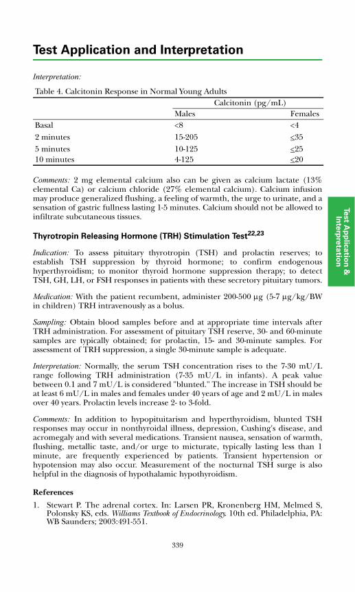
Test Application and Interpretation
Test A
pp
licatio
n &
In
terp
reta
tion
Interpretation:
Comments: 2 mg elemental calcium also can be given as calcium lactate (13%elemental Ca) or calcium chloride (27% elemental calcium). Calcium infusionmay produce generalized flushing, a feeling of warmth, the urge to urinate, and asensation of gastric fullness lasting 1-5 minutes. Calcium should not be allowed toinfiltrate subcutaneous tissues.
Thyrotropin Releasing Hormone (TRH) Stimulation Test22,23
Indication: To assess pituitary thyrotropin (TSH) and prolactin reserves; toestablish TSH suppression by thyroid hormone; to confirm endogenoushyperthyroidism; to monitor thyroid hormone suppression therapy; to detectTSH, GH, LH, or FSH responses in patients with these secretory pituitary tumors.
Medication: With the patient recumbent, administer 200-500 μg (5-7 μg/kg/BWin children) TRH intravenously as a bolus.
Sampling: Obtain blood samples before and at appropriate time intervals afterTRH administration. For assessment of pituitary TSH reserve, 30- and 60-minutesamples are typically obtained; for prolactin, 15- and 30-minute samples. Forassessment of TRH suppression, a single 30-minute sample is adequate.
Interpretation: Normally, the serum TSH concentration rises to the 7-30 mU/Lrange following TRH administration (7-35 mU/L in infants). A peak valuebetween 0.1 and 7 mU/L is considered "blunted." The increase in TSH should beat least 6 mU/L in males and females under 40 years of age and 2 mU/L in malesover 40 years. Prolactin levels increase 2- to 3-fold.
Comments: In addition to hypopituitarism and hyperthyroidism, blunted TSHresponses may occur in nonthyroidal illness, depression, Cushing's disease, andacromegaly and with several medications. Transient nausea, sensation of warmth,flushing, metallic taste, and/or urge to micturate, typically lasting less than 1minute, are frequently experienced by patients. Transient hypertension orhypotension may also occur. Measurement of the nocturnal TSH surge is alsohelpful in the diagnosis of hypothalamic hypothyroidism.
References
1. Stewart P. The adrenal cortex. In: Larsen PR, Kronenberg HM, Melmed S,Polonsky KS, eds. Williams Textbook of Endocrinology. 10th ed. Philadelphia, PA:WB Saunders; 2003:491-551.
Table 4. Calcitonin Response in Normal Young AdultsCalcitonin (pg/mL)
Males FemalesBasal <8 <42 minutes 15-205 <355 minutes 10-125 <2510 minutes 4-125 <20
339
Test Application and Interpretation
2. Fisher DA, Carlton E. Endocrine testing. In: DeGroot LJ, Jameson JL, eds.Endocrinology. 5th ed. Philadelphia, PA: Elsevier-Saunders; 2006:3605-3633.
3. Dorin RI, Qualls CR, Crapo LM. Diagnosis of adrenal insufficiency. Ann InternMed. 2003;139:194-204.
4. Mulatero P, Stowasser M, Loh KC, et al. Increased diagnosis of primaryaldosteronism, including surgically correctable forms, in centers from fivecontinents. J Clin Endocrinol Metab. 2004;89:1045-1050.
5. Young WF, Stanson AW, Thompson GB, et al. Role for adrenal venoussampling in primary aldosteronism. Surgery. 2004;136:1227-1235.
6. Raff H, Findling JW. A physiologic approach to diagnosis of the Cushingsyndrome. Ann Intern Med. 2003;138:980-991.
7. Dinneen SF, Rizza RA. Classification and diagnosis of diabetes mellitus. In:DeGroot LJ, Jameson JL, eds. Endocrinology. 5th ed. Philadelphia, PA: Elsevier-Saunders; 2006:1063-1071.
8. Quest Diagnostics Nichols Institute Clinical Correlations Department.
9. Thornton PS, Finegold DN, Stanley CA, et al. Hypoglycemia in the infant andchild. In: Sperling MA, ed. Pediatric Endocrinology. 2nd ed. Philadelphia, PA;WB Saunders; 2002:367-384.
10. Gerich JE. Hypoglycemia. In: DeGroot LJ, Jameson JL, eds. Endocrinology. 5thed. Philadelphia, PA: Elsevier-Saunders; 2006:1203-1229.
11. Cavallo A, Richards GE, Busey S, et al. A simplified gonadotropin-releasinghormone test for precocious puberty. Clin Endocrinol. 1995;42:641-646.
12. Eckert KL, Wilson DM, Bachrach LK, et al. A single-sample subcutaneousgonadotropin releasing hormone test for central precocious puberty.Pediatrics. 1996;97:517-519.
13. Giustina A, Barkan A, Casanueva FF, et al. Criteria for cure of acromegaly: aconsensus statement. J Clin Endocrinol Metab. 2000;85:526-529.
14. Sizonenko PC, Clayton PE, Cohen P, et al. Diagnosis and management ofgrowth hormone deficiency in childhood and adolescence. Part 1: diagnosisof growth hormone deficiency. Growth Horm IGF Res. 2001;11:137-165.
15. Growth Hormone Research Society. Consensus guidelines for the diagnosisand treatment of growth hormone (GH) deficiency in childhood andadolescence: summary statement of the GH Research Society. J Clin EndocrinolMetab. 2000;85:3990-3993.
16. Biller BM, Samuels MH, Zagar A, et al. Sensitivity and specificity of six testsfor the diagnosis of adult GH deficiency. J Clin Endocrinol Metab. 2002;87:2067-2079.
17. Consensus guidelines for the diagnosis and treatment of adults with growthhormone deficiency: summary statement of the Growth Hormone ResearchSociety Workshop on Adult Growth Hormone Deficiency. J Clin EndocrinolMetab. 1998;83:379-381.
340
Test Application and Interpretation
Test A
pp
licatio
n &
In
terp
reta
tion
18. Molitch ME, Clemmons DR, Malozowski S, et al. Evaluation and treatment ofadult growth hormone deficiency: an Endocrine Society Clinical PracticeGuideline. J Clin Endocrinol Metab. 2006;91:1621-1634.
19. Taheri S, Ghatei MA, Bloom SR. Gastrointestinal hormones and tumorsyndromes. In: DeGroot LJ, Jameson JL, eds. Endocrinology. 5th ed.Philadelphia, PA: Elsevier-Saunders; 2006:3551-3570.
20. Sheldon WR Jr, DeBold CR, Evans WS, et al. Rapid, sequential intravenousadministration of four hypothalamic releasing hormones as a combinedanterior pituitary function test in normal subjects. J Clin Endocrinol Metab.1985;10:623-630.
21. Ball SG, Baylis PH. Vasopressin, diabetes insipidus, and syndrome ofinappropriate antidiuresis. In: DeGroot LJ, Jameson JL, eds. Endocrinology. 5thed. Philadelphia, PA: Elsevier-Saunders; 2006:537-556.
22. Spencer CA, Schwarzbein D, Guttler RB, et al. Thyrotropin (TSH)-releasinghormone stimulation test responses employing third and fourth generationTSH assays. J Clin Endocrinol Metab. 1993;76:494-498.
23. Gruniero-Papendieck L, Chiesa A, Martinez A, et al. Nocturnal TSH surgeand TRH test response in the evaluation of thyroid axis in hypothalamicpituitary disorders in childhood. Horm Res. 1998;50:252-257.
341

342
Test Application and Interpretation

International System of Units
SI Units

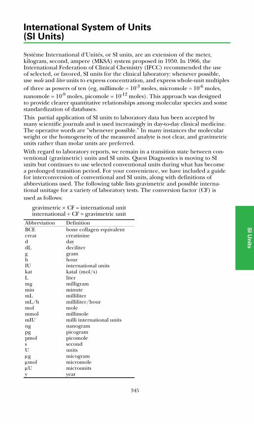
345
International System of Units(SI Units)
SI U
nits
Système International d'Unités, or SI units, are an extension of the meter, kilogram, second, ampere (MKSA) system proposed in 1950. In 1966, the International Federation of Clinical Chemistry (IFCC) recommended the use of selected, or favored, SI units for the clinical laboratory: whenever possible, use mole and liter units to express concentration, and express whole-unit multiples of three as powers of ten (eg, millimole = 10-3 moles, micromole = 10-6 moles, nanomole = 10-9 moles, picomole = 10-12 moles). This approach was designed to provide clearer quantitative relationships among molecular species and some standardization of databases. This partial application of SI units to laboratory data has been accepted by many scientific journals and is used increasingly in day-to-day clinical medicine. The operative words are "whenever possible." In many instances the molecular weight or the homogeneity of the measured analyte is not clear, and gravimetric units rather than molar units are preferred. With regard to laboratory reports, we remain in a transition state between con-ventional (gravimetric) units and SI units. Quest Diagnostics is moving to SI units but continues to use selected conventional units during what has become a prolonged transition period. For your convenience, we have included a guide for interconversion of conventional and SI units, along with definitions of abbreviations used. The following table lists gravimetric and possible interna-tional unitage for a variety of laboratory tests. The conversion factor (CF) is used as follows:
gravimetric × CF = international unitinternational ÷ CF = gravimetric unit
Abbreviation DefinitionBCE bone collagen equivalentcreat creatinined daydL deciliterg gramh hourIU international unitskat katal (mol/s)L litermg milligrammin minutemL millilitermL/h milliliter/hourmol molemmol millimolemIU milli international unitsng nanogrampg picogrampmol picomoles secondU unitsμg micogramμmol micromoleμU microunitsy year
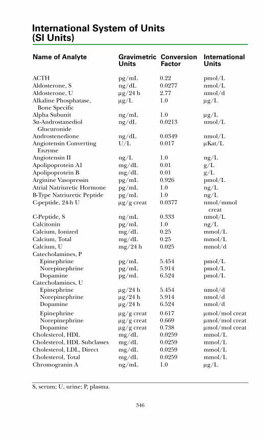
International System of Units(SI Units)
Name of Analyte Gravimetric Conversion InternationalUnits Factor Units
ACTH pg/mL 0.22 pmol/LAldosterone, S ng/dL 0.0277 nmol/LAldosterone, U μg/24 h 2.77 nmol/dAlkaline Phosphatase, μg/L 1.0 μg/L
Bone SpecificAlpha Subunit ng/mL 1.0 μg/L3α-Androstanediol ng/dL 0.0213 nmol/L
GlucuronideAndrostenedione ng/dL 0.0349 nmol/LAngiotensin Converting U/L 0.017 μKat/L
EnzymeAngiotensin II ng/L 1.0 ng/LApolipoprotein A1 mg/dL 0.01 g/LApolipoprotein B mg/dL 0.01 g/LArginine Vasopressin pg/mL 0.926 pmol/LAtrial Natriuretic Hormone pg/mL 1.0 ng/LB-Type Natriuretic Peptide pg/mL 1.0 ng/LC-peptide, 24-h U μg/g creat 0.0377 nmol/mmol
creatC-Peptide, S ng/mL 0.333 nmol/LCalcitonin pg/mL 1.0 ng/LCalcium, Ionized mg/dL 0.25 mmol/LCalcium, Total mg/dL 0.25 mmol/LCalcium, U mg/24 h 0.025 mmol/dCatecholamines, P
Epinephrine pg/mL 5.454 pmol/LNorepinephrine pg/mL 5.914 pmol/LDopamine pg/mL 6.524 pmol/L
Catecholamines, UEpinephrine μg/24 h 5.454 nmol/dNorepinephrine μg/24 h 5.914 nmol/dDopamine μg/24 h 6.524 nmol/d
Epinephrine μg/g creat 0.617 μmol/mol creatNorepinephrine μg/g creat 0.669 μmol/mol creatDopamine μg/g creat 0.738 μmol/mol creat
Cholesterol, HDL mg/dL 0.0259 mmol/LCholesterol, HDL Subclasses mg/dL 0.0259 mmol/LCholesterol, LDL, Direct mg/dL 0.0259 mmol/LCholesterol, Total mg/dL 0.0259 mmol/LChromogranin A ng/mL 1.0 μg/L
S, serum; U, urine; P, plasma.
346
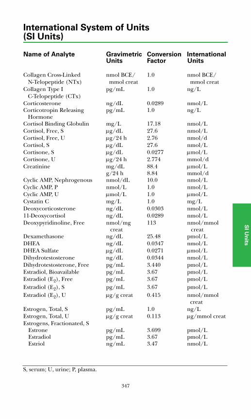
International System of Units(SI Units)
Name of Analyte Gravimetric Conversion InternationalUnits Factor Units
SI U
nits
Collagen Cross-Linked nmol BCE/ 1.0 nmol BCE/N-Telopeptide (NTx) mmol creat mmol creat
Collagen Type I pg/mL 1.0 ng/LC-Telopeptide (CTx)
Corticosterone ng/dL 0.0289 nmol/LCorticotropin Releasing pg/mL 1.0 ng/L
HormoneCortisol Binding Globulin mg/L 17.18 nmol/LCortisol, Free, S μg/dL 27.6 nmol/LCortisol, Free, U μg/24 h 2.76 nmol/dCortisol, S μg/dL 27.6 nmol/LCortisone, S μg/dL 0.0277 μmol/LCortisone, U μg/24 h 2.774 mmol/dCreatinine mg/dL 88.4 μmol/L
g/24 h 8.84 mmol/dCyclic AMP, Nephrogenous nmol/dL 10.0 nmol/LCyclic AMP, P nmol/L 1.0 nmol/LCyclic AMP, U μmol/L 1.0 μmol/LCystatin C mg/L 1.0 mg/LDeoxycorticosterone ng/dL 0.0303 nmol/L11-Deoxycortisol ng/dL 0.0289 nmol/LDeoxypyridinoline, Free nmol/mg 113 nmol/mmol
creat creatDexamethasone ng/dL 25.48 pmol/LDHEA ng/dL 0.0347 nmol/LDHEA Sulfate μg/dL 0.0271 μmol/LDihydrotestosterone ng/dL 0.0344 nmol/LDihydrotestosterone, Free pg/mL 3.440 pmol/LEstradiol, Bioavailable pg/mL 3.67 pmol/LEstradiol (E2), Free pg/mL 3.67 pmol/LEstradiol (E2), S pg/mL 3.67 pmol/LEstradiol (E2), U μg/g creat 0.415 nmol/mmol
creatEstrogen, Total, S pg/mL 1.0 ng/LEstrogen, Total, U μg/g creat 0.113 μg/mmol creatEstrogens, Fractionated, S
Estrone pg/mL 3.699 pmol/LEstradiol pg/mL 3.67 pmol/LEstriol ng/mL 3.47 nmol/L
S, serum; U, urine; P, plasma.
347
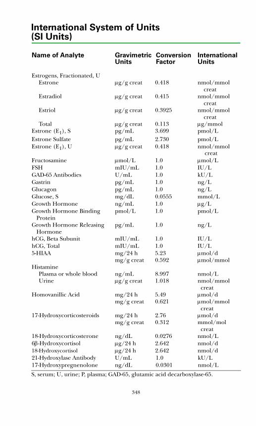
International System of Units(SI Units)
Name of Analyte Gravimetric Conversion InternationalUnits Factor Units
Estrogens, Fractionated, UEstrone μg/g creat 0.418 nmol/mmol
creatEstradiol μg/g creat 0.415 nmol/mmol
creatEstriol μg/g creat 0.3925 nmol/mmol
creatTotal μg/g creat 0.113 μg/mmol
Estrone (E1), S pg/mL 3.699 pmol/LEstrone Sulfate pg/mL 2.730 pmol/LEstrone (E1), U μg/g creat 0.418 nmol/mmol creatFructosamine μmol/L 1.0 μmol/LFSH mIU/mL 1.0 IU/LGAD-65 Antibodies U/mL 1.0 kU/LGastrin pg/mL 1.0 ng/LGlucagon pg/mL 1.0 ng/LGlucose, S mg/dL 0.0555 mmol/LGrowth Hormone ng/mL 1.0 μg/LGrowth Hormone Binding pmol/L 1.0 pmol/L
Protein Growth Hormone Releasing pg/mL 1.0 ng/L
HormonehCG, Beta Subunit mIU/mL 1.0 IU/LhCG, Total mIU/mL 1.0 IU/L5-HIAA mg/24 h 5.23 μmol/d
mg/g creat 0.592 μmol/mmolHistamine
Plasma or whole blood ng/mL 8.997 nmol/LUrine μg/g creat 1.018 nmol/mmol
creatHomovanillic Acid mg/24 h 5.49 μmol/d mg/g creat 0.621 μmol/mmol
creat17-Hydroxycorticosteroids mg/24 h 2.76 μmol/d mg/g creat 0.312 mmol/mol
creat18-Hydroxycorticosterone ng/dL 0.0276 nmol/L6β-Hydroxycortisol μg/24 h 2.642 nmol/d18-Hydroxycortisol μg/24 h 2.642 nmol/d21-Hydroxylase Antibody U/mL 1.0 kU/L17-Hydroxypregnenolone ng/dL 0.0301 nmol/L
S, serum; U, urine; P, plasma; GAD-65, glutamic acid decarboxylase-65.
348
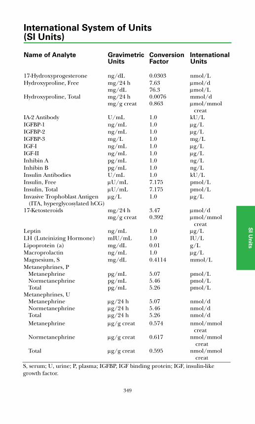
International System of Units(SI Units)
Name of Analyte Gravimetric Conversion InternationalUnits Factor Units
SI U
nits
17-Hydroxyprogesterone ng/dL 0.0303 nmol/LHydroxyproline, Free mg/24 h 7.63 μmol/d
mg/dL 76.3 μmol/LHydroxyproline, Total mg/24 h 0.0076 mmol/d
mg/g creat 0.863 μmol/mmolcreat
IA-2 Antibody U/mL 1.0 kU/LIGFBP-1 ng/mL 1.0 μg/LIGFBP-2 ng/mL 1.0 μg/LIGFBP-3 mg/L 1.0 mg/LIGF-I ng/mL 1.0 μg/LIGF-II ng/mL 1.0 μg/LInhibin A pg/mL 1.0 ng/LInhibin B pg/mL 1.0 ng/LInsulin Antibodies U/mL 1.0 kU/LInsulin, Free μU/mL 7.175 pmol/LInsulin, Total μU/mL 7.175 pmol/LInvasive Trophoblast Antigen μg/L 1.0 μg/L
(ITA, hyperglycosylated hCG)17-Ketosteroids mg/24 h 3.47 μmol/d
mg/g creat 0.392 μmol/mmolcreat
Leptin ng/mL 1.0 μg/LLH (Luteinizing Hormone) mIU/mL 1.0 IU/LLipoprotein (a) mg/dL 0.01 g/LMacroprolactin ng/mL 1.0 μg/LMagnesium, S mg/dL 0.4114 mmol/LMetanephrines, P
Metanephrine pg/mL 5.07 pmol/LNormetanephrine pg/mL 5.46 pmol/LTotal pg/mL 5.26 pmol/L
Metanephrines, UMetanephrine μg/24 h 5.07 nmol/dNormetanephrine μg/24 h 5.46 nmol/dTotal μg/24 h 5.26 nmol/dMetanephrine μg/g creat 0.574 nmol/mmol
creatNormetanephrine μg/g creat 0.617 nmol/mmol
creatTotal μg/g creat 0.595 nmol/mmol
creatS, serum; U, urine; P, plasma; IGFBP, IGF binding protein; IGF, insulin-like growth factor.
349
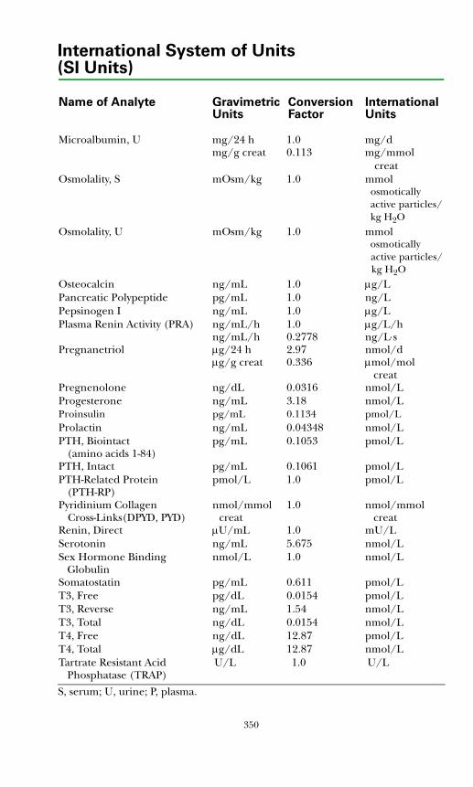
International System of Units(SI Units)
Name of Analyte Gravimetric Conversion InternationalUnits Factor Units
Microalbumin, U mg/24 h 1.0 mg/dmg/g creat 0.113 mg/mmol
creatOsmolality, S mOsm/kg 1.0 mmol
osmotically active particles/
kg H2OOsmolality, U mOsm/kg 1.0 mmol
osmoticallyactive particles/kg H2O
Osteocalcin ng/mL 1.0 μg/LPancreatic Polypeptide pg/mL 1.0 ng/LPepsinogen I ng/mL 1.0 μg/LPlasma Renin Activity (PRA) ng/mL/h 1.0 μg/L/h
ng/mL/h 0.2778 ng/L⋅sPregnanetriol μg/24 h 2.97 nmol/d
μg/g creat 0.336 μmol/mol creatPregnenolone ng/dL 0.0316 nmol/LProgesterone ng/mL 3.18 nmol/LProinsulin pg/mL 0.1134 pmol/L Prolactin ng/mL 0.04348 nmol/LPTH, Biointact pg/mL 0.1053 pmol/L
(amino acids 1-84) PTH, Intact pg/mL 0.1061 pmol/LPTH-Related Protein pmol/L 1.0 pmol/L
(PTH-RP)Pyridinium Collagen nmol/mmol 1.0 nmol/mmol
Cross-Links(DPYD, PYD) creat creatRenin, Direct μU/mL 1.0 mU/LSerotonin ng/mL 5.675 nmol/LSex Hormone Binding nmol/L 1.0 nmol/L
GlobulinSomatostatin pg/mL 0.611 pmol/LT3, Free pg/dL 0.0154 pmol/LT3, Reverse ng/mL 1.54 nmol/LT3, Total ng/dL 0.0154 nmol/LT4, Free ng/dL 12.87 pmol/LT4, Total μg/dL 12.87 nmol/LTartrate Resistant Acid U/L 1.0 U/L
Phosphatase (TRAP)
S, serum; U, urine; P, plasma.
350
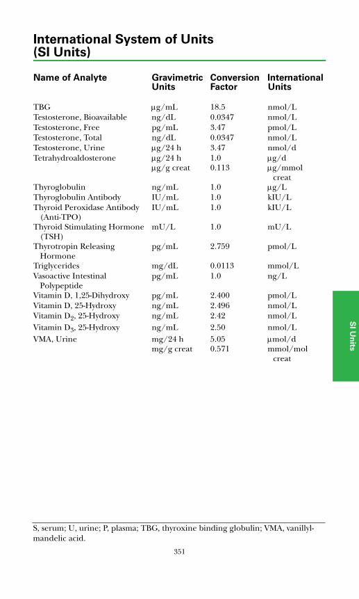
International System of Units(SI Units)
Name of Analyte Gravimetric Conversion InternationalUnits Factor Units
SI U
nits
TBG μg/mL 18.5 nmol/LTestosterone, Bioavailable ng/dL 0.0347 nmol/LTestosterone, Free pg/mL 3.47 pmol/LTestosterone, Total ng/dL 0.0347 nmol/LTestosterone, Urine μg/24 h 3.47 nmol/dTetrahydroaldosterone μg/24 h 1.0 μg/d
μg/g creat 0.113 μg/mmol creat
Thyroglobulin ng/mL 1.0 μg/LThyroglobulin Antibody IU/mL 1.0 kIU/LThyroid Peroxidase Antibody IU/mL 1.0 kIU/L
(Anti-TPO)Thyroid Stimulating Hormone mU/L 1.0 mU/L
(TSH)Thyrotropin Releasing pg/mL 2.759 pmol/L
HormoneTriglycerides mg/dL 0.0113 mmol/LVasoactive Intestinal pg/mL 1.0 ng/L
PolypeptideVitamin D, 1,25-Dihydroxy pg/mL 2.400 pmol/LVitamin D, 25-Hydroxy ng/mL 2.496 nmol/LVitamin D2, 25-Hydroxy ng/mL 2.42 nmol/LVitamin D3, 25-Hydroxy ng/mL 2.50 nmol/LVMA, Urine mg/24 h 5.05 μmol/d
mg/g creat 0.571 mmol/molcreat
S, serum; U, urine; P, plasma; TBG, thyroxine binding globulin; VMA, vanillyl-mandelic acid.
351

International System of Units(SI Units)
Name of Analyte Gravimetric Conversion InternationalUnits Factor Units
352
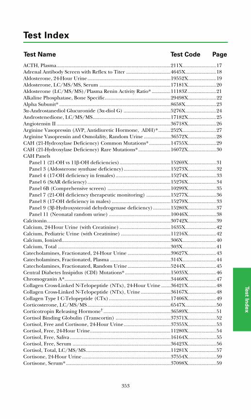
Test Index
PageTest CodeTest Name
Test In
de
x
ACTH, Plasma.....................................................................................211X.........................17Adrenal Antibody Screen with Reflex to Titer .................................4645X.......................18Aldosterone, 24-Hour Urine..............................................................19552X.....................19Aldosterone, LC/MS/MS, Serum .....................................................17181X.....................20Aldosterone (LC/MS/MS)/Plasma Renin Activity Ratio* ..............11183Z .....................21Alkaline Phosphatase, Bone Specific.................................................29498X.....................22Alpha Subunit* ...................................................................................8658X.......................233α-Androstanediol Glucuronide (3α-diol G) ...................................5276X.......................24Androstenedione, LC/MS/MS..........................................................17182X.....................25Angiotensin II .....................................................................................36718X.....................26Arginine Vasopressin (AVP, Antidiuretic Hormone, ADH)*.........252X.........................27Arginine Vasopressin and Osmolality, Random Urine ....................36572X.....................28CAH (21-Hydroxylase Deficiency) Common Mutations*................14755X.....................29CAH (21-Hydroxylase Deficiency) Rare Mutations*........................16072X.....................30CAH Panels
Panel 1 (21-OH vs 11β-OH deficiencies)......................................15269X.....................31Panel 3 (Aldosterone synthase deficiency)...................................15273X.....................32Panel 4 (17-OH deficiency in females).........................................15274X.....................33Panel 6 (StAR deficiency)..............................................................15276X.....................34Panel 6B (Comprehensive screen) ...............................................10299X.....................35Panel 7 (21-OH deficiency therapeutic monitoring) ..................15277X.....................36Panel 8 (17-OH deficiency in males) ............................................15279X.....................33Panel 9 (3β-Hydroxysteroid dehydrogenase deficiency) .............15280X.....................37Panel 11 (Neonatal random urine) ..............................................10046X.....................38
Calcitonin............................................................................................30742X.....................39Calcium, 24-Hour Urine (with Creatinine) ......................................1635X.......................42Calcium, Pediatric Urine (with Creatinine) .....................................11216X.....................42Calcium, Ionized.................................................................................306X.........................40Calcium, Total ....................................................................................303X.........................41Catecholamines, Fractionated, 24-Hour Urine ................................39627X.....................43Catecholamines, Fractionated, Plasma .............................................314X.........................44Catecholamines, Fractionated, Random Urine ................................5244X.......................45Central Diabetes Insipidus (CDI) Mutations* ..................................15035X.....................46Chromogranin A*...............................................................................34468X.....................47Collagen Cross-Linked N-Telopeptide (NTx), 24-Hour Urine .......36421X.....................48Collagen Cross-Linked N-Telopeptide (NTx), Urine ......................36167X.....................48Collagen Type I C-Telopeptide (CTx)..............................................17406X.....................49Corticosterone, LC/MS/MS..............................................................6547X.......................50Corticotropin Releasing Hormone† ..................................................36589X.....................51Cortisol Binding Globulin (Transcortin) .........................................37371X.....................52Cortisol, Free and Cortisone, 24-Hour Urine...................................37355X.....................53Cortisol, Free, 24-Hour Urine............................................................11280X.....................54Cortisol, Free, Saliva ...........................................................................16164X.....................55Cortisol, Free, Serum .........................................................................36423X.....................56Cortisol, Total, LC/MS/MS...............................................................11281X ....................57Cortisone, 24-Hour Urine ..................................................................37554X.....................59Cortisone, Serum*..............................................................................37098X.....................59
353
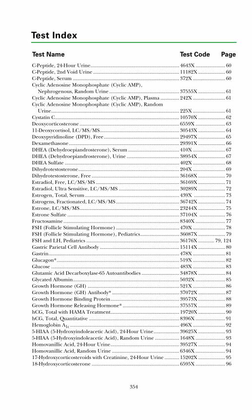
Test Index
PageTest CodeTest Name
C-Peptide, 24-Hour Urine.................................................................. 4643X ...................... 60C-Peptide, 2nd Void Urine ................................................................ 11182X .................... 60C-Peptide, Serum ............................................................................... 372X ........................ 60Cyclic Adenosine Monophosphate (Cyclic AMP),
Nephrogenous, Random Urine .................................................... 37555X .................... 61Cyclic Adenosine Monophosphate (Cyclic AMP), Plasma .............. 242X ........................ 61Cyclic Adenosine Monophosphate (Cyclic AMP), Random
Urine............................................................................................... 225X ........................ 61Cystatin C............................................................................................ 10570X .................... 62Deoxycorticosterone .......................................................................... 6559X ...................... 6311-Deoxycortisol, LC/MS/MS........................................................... 30543X .................... 64Deoxypyridinoline (DPD), Free........................................................ 29497X .................... 65Dexamethasone.................................................................................. 29391X .................... 66DHEA (Dehydroepiandrosterone), Serum ...................................... 410X ........................ 67DHEA (Dehydroepiandrosterone), Urine ....................................... 38954X .................... 67DHEA Sulfate ..................................................................................... 402X ........................ 68Dihydrotestosterone........................................................................... 204X ........................ 69Dihydrotestosterone, Free ................................................................. 36168X .................... 70Estradiol, Free, LC/MS/MS .............................................................. 36169X .................... 71Estradiol, Ultra Sensitive, LC/MS/MS ............................................. 30289X .................... 72Estrogen, Total, Serum ...................................................................... 439X ....................... 73Estrogens, Fractionated, LC/MS/MS ............................................... 36742X .................... 74Estrone, LC/MS/MS.......................................................................... 23244X .................... 75Estrone Sulfate ................................................................................... 37104X ................... 76Fructosamine...................................................................................... 8340X ...................... 77FSH (Follicle Stimulating Hormone) ............................................... 470X ........................ 78FSH (Follicle Stimulating Hormone), Pediatrics............................. 36087X .................... 79FSH and LH, Pediatrics ..................................................................... 36176X ............ 79, 124Gastric Parietal Cell Antibody ........................................................... 15114X .................... 80Gastrin................................................................................................. 478X ........................ 81Glucagon*........................................................................................... 519X ........................ 82Glucose ............................................................................................... 483X ........................ 83Glutamic Acid Decarboxylase-65 Autoantibodies ............................ 34878X .................... 84Glycated Albumin............................................................................... 5032X ...................... 85Growth Hormone (GH) .................................................................... 521X ........................ 86Growth Hormone (GH) Antibody* .................................................. 37072X .................... 87Growth Hormone Binding Protein................................................... 39573X .................... 88Growth Hormone Releasing Hormone* .......................................... 37557X .................... 89hCG, Total with HAMA Treatment................................................... 19720X .................... 90hCG, Total, Quantitative ................................................................... 8396X ...................... 91Hemoglobin A1c ................................................................................. 496X ........................ 925-HIAA (5-Hydroxyindoleacetic Acid), 24-Hour Urine................... 39625X .................... 935-HIAA (5-Hydroxyindoleacetic Acid), Random Urine .................. 1648X ...................... 93Homovanillic Acid, 24-Hour Urine................................................... 39527X .................... 94Homovanillic Acid, Random Urine .................................................. 6346X ...................... 9417-Hydroxycorticosteroids with Creatinine, 24-Hour Urine ........... 15202X .................... 9518-Hydroxycorticosterone ................................................................. 6595X ...................... 96
354
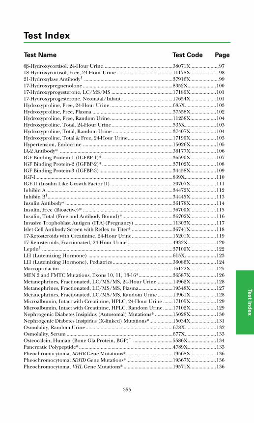
Test Index
PageTest CodeTest Name
Test In
de
x
6β-Hydroxycortisol, 24-Hour Urine...................................................38071X.....................9718-Hydroxycortisol, Free, 24-Hour Urine .........................................11178X.....................9821-Hydroxylase Antibody† .................................................................37916X.....................9917-Hydroxypregnenolone ..................................................................8352X.....................10017-Hydroxyprogesterone, LC/MS/MS .............................................17180X...................10117-Hydroxyprogesterone, Neonatal/Infant......................................17654X...................101Hydroxyproline, Free, 24-Hour Urine ..............................................685X.......................103Hydroxyproline, Free, Plasma ...........................................................37558X...................102Hydroxyproline, Free, Random Urine..............................................11258X...................104Hydroxyproline, Total, 24-Hour Urine.............................................535X.......................103Hydroxyproline, Total, Random Urine ............................................37407X...................104Hydroxyproline, Total & Free, 24-Hour Urine.................................17190X...................103Hypertension, Endocrine ..................................................................15026X...................105IA-2 Antibody* ...................................................................................36177X...................106IGF Binding Protein-1 (IGFBP-1)*....................................................36590X...................107IGF Binding Protein-2 (IGFBP-2)*....................................................37102X...................108IGF Binding Protein-3 (IGFBP-3)......................................................34458X...................109IGF-I.....................................................................................................839X.......................110IGF-II (Insulin Like Growth Factor II)..............................................20707X...................111Inhibin A .............................................................................................34472X...................112Inhibin B†............................................................................................34445X...................113Insulin Antibody* ...............................................................................36178X...................114Insulin, Free (Bioactive)* ..................................................................36700X...................115Insulin, Total (Free and Antibody Bound)*.....................................36702X...................116Invasive Trophoblast Antigen (ITA)(Pregnancy) ............................11303X...................117Islet Cell Antibody Screen with Reflex to Titer* ..............................36741X...................11817-Ketosteroids with Creatinine, 24-Hour Urine..............................15201X...................11917-Ketosteroids, Fractionated, 24-Hour Urine .................................4932X.....................120Leptin† ................................................................................................37109X...................122LH (Luteinizing Hormone) ..............................................................615X.......................123LH (Luteinizing Hormone), Pediatrics ............................................36086X...................124Macroprolactin ...................................................................................16122X...................125MEN 2 and FMTC Mutations, Exons 10, 11, 13-16*.........................36587X...................126Metanephrines, Fractionated, LC/MS/MS, 24-Hour Urine ...........14962X ..................128Metanephrines, Fractionated, LC/MS/MS, Plasma.........................19548X...................127Metanephrines, Fractionated, LC/MS/MS, Random Urine ...........14961X...................128Microalbumin, Intact with Creatinine, HPLC, 24-Hour Urine .......17105X...................129Microalbumin, Intact with Creatinine, HPLC, Random Urine .......17102X...................129Nephrogenic Diabetes Insipidus (Autosomal) Mutations* .............15028X...................130Nephrogenic Diabetes Insipidus (X-linked) Mutations*.................15034X...................131Osmolality, Random Urine ................................................................678X.......................132Osmolality, Serum ..............................................................................677X.......................133Osteocalcin, Human (Bone Gla Protein, BGP)† .............................5586X.....................134Pancreatic Polypeptide*.....................................................................4789X.....................135Pheochromocytoma, SDHB Gene Mutations* ..................................19568X...................136Pheochromocytoma, SDHD Gene Mutations*..................................19567X...................136Pheochromocytoma, VHL Gene Mutations* ....................................19571X...................136
355
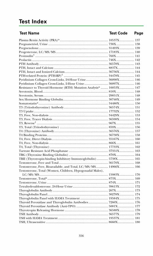
Test Index
PageTest CodeTest Name
Plasma Renin Activity (PRA)*........................................................... 10537X.................. 137Pregnanetriol, Urine ......................................................................... 738X...................... 138Pregnenolone..................................................................................... 31493X.................. 139Progesterone, LC/MS/MS................................................................ 17183X.................. 140Proinsulin† ......................................................................................... 760X...................... 141Prolactin ............................................................................................. 746X...................... 142PTH Antibody .................................................................................... 36578X.................. 143PTH, Intact and Calcium .................................................................. 8837X.................... 144PTH, Intact and Ionized Calcium..................................................... 36736X.................. 144PTH-related Protein (PTH-RP)† ...................................................... 34478X.................. 145Pyridinium Collagen Cross-Links, 24-Hour Urine........................... 36098X.................. 146Pyridinium Collagen Cross-Links, 2-Hour Urine............................. 36097X.................. 146Resistance to Thyroid Hormone (RTH) Mutation Analysis* ......... 16053X.................. 147Serotonin, Blood................................................................................ 818X...................... 148Serotonin, Serum............................................................................... 29851X.................. 148Sex Hormone Binding Globulin....................................................... 30740X.................. 149Somatostatin*..................................................................................... 34480X.................. 150T3 (Triiodothyronine) Antibody ...................................................... 36574X.................. 151T3 Uptake........................................................................................... 17732X.................. 152T3, Free, Non-dialysis ........................................................................ 34429X.................. 153T3, Free, Tracer Dialysis .................................................................... 36598X.................. 154T3, Reverse† ....................................................................................... 967X...................... 155T3, Total (Triiodothyronine) ........................................................... 859X...................... 156T4 (Thyroxine) Antibody.................................................................. 36576X.................. 157T4 Binding Proteins........................................................................... 36738X.................. 158T4, Free, Direct Dialysis..................................................................... 35167X.................. 160T4, Free, Non-dialysis ........................................................................ 866X...................... 161T4, Total (Thyroxine) ....................................................................... 17733X.................. 162Tartrate Resistant Acid Phosphatase ................................................ 37351X.................. 163TBG (Thyroxine Binding Globulin) ................................................ 870X...................... 164TBII (Thyrotropin-binding Inhibitory Immunoglobulin).............. 5738X.................... 165Testosterone, Free and Total ............................................................ 36170X.................. 168Testosterone, Free, Bioavailable, and Total, LC/MS/MS............... 14966X.................. 166Testosterone, Total (Women, Children, Hypogonadal Males),
LC/MS/MS.................................................................................... 15983X.................. 170Testosterone, Total* .......................................................................... 873X...................... 169Testosterone, Urine........................................................................... 874X...................... 171Tetrahydroaldosterone, 24-Hour Urine........................................... 39613X.................. 172Thyroglobulin Antibody.................................................................... 267X...................... 173Thyroglobulin Panel.......................................................................... 30278X.................. 174Thyroglobulin Panel with HAMA Treatment .................................. 19584X.................. 175Thyroid Peroxidase and Thyroglobulin Antibodies ........................ 7260X.................... 176Thyroid Peroxidase Antibody (Anti-TPO)....................................... 5081X.................... 177Thyrotropin Releasing Hormone ..................................................... 36588X.................. 178TSH Antibody .................................................................................... 36577X.................. 179TSH with HAMA Treatment ............................................................. 19537X.................. 181TSH, Ultrasensitive ............................................................................ 8660X.................... 180
356
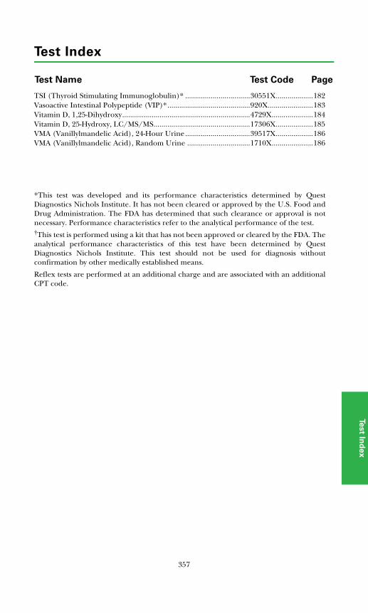
Test Index
PageTest CodeTest Name
Test In
de
x
TSI (Thyroid Stimulating Immunoglobulin)* .................................30551X...................182Vasoactive Intestinal Polypeptide (VIP)*..........................................920X.......................183Vitamin D, 1,25-Dihydroxy.................................................................4729X.....................184Vitamin D, 25-Hydroxy, LC/MS/MS.................................................17306X...................185VMA (Vanillylmandelic Acid), 24-Hour Urine.................................39517X...................186VMA (Vanillylmandelic Acid), Random Urine ................................1710X.....................186
*This test was developed and its performance characteristics determined by QuestDiagnostics Nichols Institute. It has not been cleared or approved by the U.S. Food andDrug Administration. The FDA has determined that such clearance or approval is notnecessary. Performance characteristics refer to the analytical performance of the test.†This test is performed using a kit that has not been approved or cleared by the FDA. Theanalytical performance characteristics of this test have been determined by QuestDiagnostics Nichols Institute. This test should not be used for diagnosis withoutconfirmation by other medically established means.
Reflex tests are performed at an additional charge and are associated with an additionalCPT code.
357

Test Index
PageTest CodeTest Name
358

Subject Index
Su
bje
ct In
de
x
Acne, 24, 67, 166, 168, 170, 266-267, 274
Acromegaly, 86, 89, 103-104, 108-111, 164, 221-223, 281, 284, 289, 336, 339
ACTH, 17, 190-199, 205-207, 211, 217-221, 265, 320
deficiency, 50, 57, 95, 119, 192-193, 218-219, 330-331
ectopic syndrome, 57, 192, 206-207, 330-332
secreting tumor, 17, 206-207, 326stimulation test, 57, 63, 67, 96, 100-101, 139, 191, 193-200, 205, 315, 329-330
Addison’s disease, see adrenal insuffi-ciency, primary
Adenomaadrenal, 17, 20-21, 50, 57, 68, 96, 206, 210, 331
hypothalamic, 89, 125, 142pancreatic (islet cell) 47, 60, 81-83, 89, 107, 115-116, 135, 141, 148, 183, 249-251, 326
parathyroid, 47, 226-227, 322pituitary, 23, 47, 86, 125, 139, 142, 206-207, 220-224, 273, 283-284, 297, 299, 331, 336
thyroid, 153-154, 307
Adrenaladenoma, 17, 20-21, 50, 57, 68, 96, 206, 210, 331
aldosteronism, see aldosteronismantibody, 18carcinoma, 17, 57, 63, 68, 206Cushing’s syndrome, see Cushing’s disease
dysplasia, micronodular, 57, 206enzyme precursor/product ratios, 192, 198-200, 202-205, 211, 214
feminizing tumors, 72-74function, 57-59, 190hyperfunction, see Cushing’s diseasehyperplasia, 120, 331
adrenotroph, 206congenital, see congenital adre-nal hyperplasia
juxtaglomerular, 210idiopathic, 210macronodular, 57
hypoplasia, 19, 50, 63, 68, 193
insufficiency, 34, 37, 53-59, 63-64, 98, 100-101, 120, 192-193, 249, 303
primary, 17-21, 25, 50, 57, 63, 64, 67-68, 83, 95-96, 99, 101, 119, 137, 193, 209, 242, 249, 301, 329-330
secondary, 17, 64, 193, 329-330renovascular hypertension, 137, 208, 210-211
steroid urinary metabolites, 105, 191, 201-204, 211-214
tumors (see also adrenal adenoma and carcinoma), 25, 67, 74, 76, 95, 100, 138-140
virilizing tumors, 25, 68, 139
Alcohol (ism), 133, 149, 206
Aldosterone, 19-21, 32-33, 192, 194-195, 197-200, 207-211, 311-313, 331
24-hour urine, 19, 313, 316serum, 20, 210-211, 313, 316suppression test, 210, 330synthase deficiency (P450c11ase), 32, 35, 50, 63, 96, 198-200, 214
synthetase deficiency, 19-21
Aldosteronismdexamethasone suppressible hyper-, 192, 209, 211, 332
glucocorticoid remediable, 98glucocorticoid-suppressible, 105hyper-, 19-21, 96, 172, 192, 209-211, 330-331
hypo-, 105, 137, 172, 192, 209, 313hyporeninemic hypo-, 19-21, 137, 192, 209, 313
primary, 19-21, 26, 63, 96, 105, 137, 172, 192, 208-210, 313, 330-331
pseudohyper-, see apparent mineralocortiocoid excess (AME)
pseudohypo-, 172, 192, 208-209, 313secondary, 19-21, 137, 172, 210
Alkaline phosphatase, bone specific, 22, 233, 235
Alpha subunit, 23, 220-221, 223-224
Amenorrhea, 33, 35, 72, 140, 166, 168, 170, 222, 267, 272-274
Aminoglycinuria, familial, 103-104
Androgen resistance, 166, 168-171, 269, 272
359
Subject Index
3α-Androstanediol glucuronide, 24
Androstenedione, 25, 31-32, 34-37, 193-195, 197-200, 205, 273, 275
Androsterone, 121, 212
Anemia, pernicious, 39, 80-81, 301, 303
Angiotensin II, 26, 207
Anorchia, 113, 263
Anorexia nervosa, 67, 267
Anovulatory syndrome, chronic, see polycystic ovarian syndrome
Antidiuretic hormone (ADH), see arginine vasopressin (AVP)
Apparent mineralocorticoid excess (AME), 53, 58-59, 105, 172, 192, 209, 211
AQP2 mutation, 130
Arginine vasopressin (AVP), 27-28, 190, 208, 217, 311-316
and osmolality, 28, 208ectopic syndrome, 27plasma/serum, 27, 313-316urine, 28, 313, 316
Autoimmune (ity), 193, 227, 229, 240-245, 251
polyglandular disease, 18, 80, 99, 114, 242, 273-274, 301, 303
thyroid, 151, 157, 165, 173, 176-177, 179, 182, 300-304
AVP mutations, 46
AVPR2 mutation, 131
Bartter syndrome, 19-21, 208-210
Bone, 225-237disorders, 22, 40-42, 48-49, 61, 65, 102-104, 134, 143-144, 146. 163, 184-185, 226-237
fractures, 134, 227, 230-231formation markers, 22, 134, 233, 235metabolic disease in, 222-236metastatic tumor, 102-104resorption markers, 48-49, 65, 102-104, 146, 163, 233-235
CAH, see congenital adrenal hyperpla-sia
Calcitonin, 39, 226-228, 304-305, 320, 323-325, 338-339
Calcium, 225-237disorders, 40-42, 144, 226-230hypercalcemia, 27, 40-41, 61, 144-145, 184, 226-231, 322
hypocalcemia, 40-42, 184, 226-231ionized, 40, 144, 231metabolism, 42, 226stimulation test, 39, 305, 323-324, 338-339
total, 41, 144urine, 42vitamin D, 40-42, 134, 184-185, 225-228, 230-233
Candidiasis, 18, 273, 303
Carbohydrate metabolism, 239-253, 332-335
Carcinoid syndrome, 148
Carcinoid tumor, 23, 47, 89, 93, 150, 326
Carcinomaadrenal, 17, 57, 63, 68, 206breast, 39, 163bone, 48-49, 102-104, 163chorio, 23, 90-91, 117, 276, 298insulinoma, 47, 60, 83, 89, 107, 115-116, 135, 141, 148, 150, 249-251, 326
liver, 148lung, 39, 47, 91, 148medullary, see medullary thyroid car-cinoma (MTC)
mesenchymal, 108, 111, 249ovarian, 72-74, 76, 138, 140, 263, 268, 273, 275-276
pancreatic (see also insulinoma), 23, 39, 47, 82, 89, 135
papillary-follicular, 174-175, 307pituitary, 23, 78-79, 86, 89, 123-125, 142
prostatic, 108renal, 26, 320testicular, 90-91, 275-276thyroid, medullary, 39, 47, 126, 135, 148, 150, 226-228, 305, 307, 319-320, 322-325, 338-339
trophoblastic, 90-91, 117, 273
Catecholamines, 320-321fractionated, 24-hour urine, 43, 128fractionated, plasma, 44, 127
360
Subject Index
Su
bje
ct In
de
x
fractionated, random urine, 45, 128secreting tumor, 43-45, 94, 136, 186, 319
Celiac disease, 48-49, 242
Choriocarcinoma, 23, 90-91, 117, 276, 298
Chromogranin A, 47, 221, 320
Collagen cross-linked N-telopeptide (NTx), 48, 232-235
Collagen type I C-telopeptide (CTx), 49, 232-234
Combined anterior pituitary (CAP) test, 337-338
Compound S, see 11-deoxycortisol
Congenital adrenal hyperplasia (CAH), 19-21, 24, 29-38, 63-64, 67-68, 100-101, 138-139, 166, 168, 170-171, 191-193, 198-205, 214, 264, 267-268, 329-330
aldosterone synthase deficiency (P450c11ase), 32, 35, 50, 63, 96, 192, 197-200, 214
11β-hydroxylase deficiency (P450c11), 31, 35, 38, 63-64, 192-193, 197-200, 202, 205, 214, 265, 272
17-hydroxylase deficiency (P450c17), 25, 33, 35, 38, 50, 63, 138, 166, 168, 170-171, 192, 197-200, 202, 205, 214, 265, 270, 272
21-hydroxylase deficiency (P450c21), 25, 29-31, 35-36, 38, 101, 138, 192-193, 197-200, 202-205, 214, 265, 272
3β-hydroxysteroid dehydrogenase deficiency (3β-HSD), 35, 37-38, 100, 139, 192-193, 197-200, 202-205, 214, 265, 270, 272
mutations, 29-30nonclassical, 101, 205StAR deficiency (previouslyside chain cleavage, P450scc), 34-35, 192-193, 197, 265
Conn syndrome, see aldosteronism, pri-mary
Corticosterone, 33, 50, 194-195
Corticotropin releasing hormone (CRH), 51, 190-193, 206-207, 217
ectopic syndrome, 51, 57, 206stimulation test, 190, 193, 207, 330-332
Cortisol, 31, 33-35, 37, 53-57, 190-195, 197, 206-207, 211, 213, 239, 250-251, 265, 315, 329-332
binding globulin, 52, 55-56, 190, 261free, 53-56, 206salivary, 55, 206total, 55-57, 105
Cortisone, 53, 58-59, 105, 192, 21324-hour urine, 53, 58, 105serum, 59
C-Peptide, 60, 240, 245, 249-251, 333-335
C-Telopeptide (CTx), 49, 232-234
Cushing’s disease, 17, 25-26, 50-51, 53-59, 63, 66-68, 83, 95, 101, 119-120, 134, 139, 166-168, 170-171, 192, 206-207, 211, 221, 273, 275, 326, 330-332
Cyclic adenosine monophosphate, (cyclic AMP), 61, 301
CYP21A2 mutations, 29, 30
Cystatin C, 62
Deoxycorticosterone, 35, 63, 194-195, 197-200
11-Deoxycortisol, 31-32, 35, 64, 192, 194-195, 197-200, 205
Deoxypyridinoline (DPD, DPYD), free, 65, 232-234
Dexamethasone, 66, 300suppression test, 55-57, 66, 98, 100, 190-191, 206-207, 211, 331-332
DHEA (dehydroepiandrosterone), 34-35, 37, 67, 120-121, 192-195, 197-200, 205, 213-214
DHEA sulfate, 68, 190, 267, 273, 275
Diabetes insipidus, 132-133central, 27-28, 46, 312-314, 338nephrogenic (autosomal & X-linked), 27-28, 130-131, 313-314, 338
Diabetes mellitus, 60, 82-84, 92, 106-107, 239-248, 301, 303
complications, 62, 85, 129, 244, 247-248
diagnosis, 83, 244, 246, 332-333gestational, 77, 242, 245-246
361
Subject Index
impaired glucose tolerance, 83, 244, 246
insulin resistance, 107, 115, 240, 243- 246
laboratory monitoring, 62, 77, 83, 85, 92, 107, 115-116, 129, 244, 247-248
maturity onset diabetes of the young (MODY), 243, 245
risk assessment, 84, 106, 114, 118, 240-242
type 1, 60, 62, 84, 106, 114-116, 118, 129, 239-242
type 2, 60, 62, 115-116, 129, 243-244
Dihydrotestosterone, 257-258, 265, 276free and total, 70total, 69, 149
Dynamic tests, 189adrenal, 190-191, 193-199, 205-207, 210, 316, 329-332
carbohydrate, 241-242, 249, 251, 332-335
gonadal, 267, 335growth hormone, 222, 285, 289-290, 336-337
multiple endocrine neoplasia, 326, 337
posterior pituitary, 314, 337-338thyroid, 305, 338-339
Dysalbuminemic hyperthyroxinemia, 158, 162, 308
Dysautonomia, familial, 44
Estradiol, 258, 261-263, 267, 276free, 71, 261serum, 33, 72, 74
Estrogen, 258, 261amenorrhea, 72, 272-273fractionated, 74secreting tumors, 71, 73, 275therapy, 52, 55-56, 76, 88, 119, 149, 162, 164, 262
total, 73
Estrone, 261-262, 265serum, 74-75sulfate, 76
Etiocholanolone, 121, 212-214
Euthyroid, 180
hyperthyroxinemia, 158, 160, 308sick, see nonthyroidal illness
Familial medullary thyroid carcinoma (FMTC), see medullary thyroid carci-noma (MTC)
Feminization, 72-74, 149, 265, 270, 272
Fluid and electrolyte disorders, 20, 132physiology, 311-312primary disorders, 312-314secondary disorders, 314-316
Fructosamine, 77
FSH (follicle stimulating hormone), 78-79, 217, 219-220, 256-258, 261, 263, 335
gonadal tumors, 276pituitary adenoma, 220-221, 223-224sexual differentiation, 265sexual maturation, 267-269testicular and ovarian function, 270, 272-274
GAD-65, 84, 240-242
Ganglioblastoma, 94, 186
Ganglioneuroma, 44, 183, 320
Gastrin, 81, 320, 326, 337
Gastrinoma, 39, 81, 135, 326, 337
Gastritis, atrophic, 80-81
Germinal cell aplasia, 79
Gigantism, 86, 110, 284, 289
Gitelman’s syndrome, 208
Glomerular filtration, impaired, 62, 208
Glucagon, 82-83, 239, 250-251, 320, 326
Glucocorticoidadrenocortical disorders, 192, 206excess, 102therapy, 53, 139
Glucose, 83, 239, 243-244, 246-251suppression test, 222, 336tolerance test, 241-242, 249, 289, 332-333
Glutamic acid decarboxylase-65 auto-antibodies, 84, 240-242
Glycated albumin, 85
GnRH, 217, 255-257
362
Subject Index
Su
bje
ct In
de
x
deficiency, 78-79, 123-124, 219, 255, 269-270, 272
sexual maturation, 267, 269stimulation test, 124, 267, 335
Goiter, 173-175, 177, 307
Gonadal functiondisorders
developmental, 140, 267-270germinal cell, 90-91, 269, 271ovarian function, 272-275testicular function, 269-272
hormone measurements, 71-76, 78-79, 112-113, 123-124, 149, 166-171, 261-263
physiology, 255-260puberty
onset, 256, 264staging, 264, 266
sexual differentiation, 263-265testicular aging, 271-272tumors, 90-91, 275-276
Gonadotropindeficiency, see hypogonadismreleasing hormone, see GnRH
Gordon’s syndrome, 208
Granulomatous disorders, 41, 184, 270, 272-273, 283
Granulosa cell tumor, 112-113, 263, 268, 275-276
Graves disease, 151, 153-154, 156-157, 165, 173-177, 180-182, 242, 296-298, 300-303, 306-307
Growth hormone (GH), 86, 239, 250-251, 320
antibody, 87binding protein (GHBP), 88, 279-282, 284-289
deficiency, 48-49, 86-87, 108-111, 282-284, 286-290, 336-337
excess, 86, 109-110, 284, 289, 336physiology, 217, 279-282receptor, 280-283releasing hormone, 89, 279, 282-285, 289-290, 320
resistance, 86, 88, 110, 282, 284, 289secretory dysfunction, 86, 282stimulation test, 280, 289-290, 336-337
Gynecomastia, 71-73, 91, 149, 166, 168, 170, 262-263
HAMA, 90, 175, 181
Hashimoto thyroiditis, see thyroiditis, chronic autoimmune
hCG, total, quantitative, 90-91, 273, 276, 298
Hemoglobin A1c, 92, 244, 247-248
Heterophilic antibody, see HAMA
5-HIAA (5-hydroxyindoleacetic acid), 93
Hirsutism, 31, 67-70, 95, 100, 139, 149, 166-168, 170
idiopathic, 24-25, 100, 273polycystic ovary (PCO), 24-25, 100, 274
Homovanillic acid, 94
Hydatidiform mole, 23, 298
Hydrationdehydration, 27-28, 32, 132-133, 315overhydration, 132-133
11β-Hydroxyandrosterone, 121, 212-213
17-Hydroxycorticosteroids, 95, 192
18-Hydroxycorticosterone, 32, 96, 197, 210
6β-Hydroxycortisol, 97
18-Hydroxycortisol, 98, 210-211
11β-Hydroxyetiocholanolone, 121, 212-213
21-Hydroxylaseantibody, 99deficiency (P450c21), 25, 29-31, 35-36, 38, 101, 138, 192-193, 197-200, 202-205, 214, 265, 272
11β-Hydroxylase deficiency (P450c11), 31, 35, 38, 63-64, 192-193, 197-200, 202, 205, 214, 265, 272
17-Hydroxylase deficiency (P450c17), 25, 33, 35, 38, 50, 63, 138, 166, 168, 170-171, 192, 197-200, 202, 205, 214, 265, 270, 272
17-Hydroxypregnenolone, 35, 37, 100, 197, 205
363
Subject Index
17-Hydroxyprogesterone, 31-33, 35-37, 101, 193, 197, 205, 267
Hydroxyproline, 232, 234free, 102free and total, 103-104
3β-Hydroxysteroid dehydrogenase deficiency (3β-HSD), 35, 37-38,100, 139, 192-193, 197-200, 202-205, 214, 265, 270, 272
Hyperaldosteronism, 19-21, 96, 172, 192, 209-211, 330-331
dexamethasone suppressible, 192, 209, 211, 332
pseudo-, see apparent mineralocorti-coid excess (AME)
Hyperandrogenism, 24-25, 29-30, 67-70, 100, 120, 139, 149, 166-168, 170, 205, 265, 268-269, 272, 274-275
Hypercalcemia, 27, 40-41, 61, 144-145, 184, 226-231, 322
familial benign, 227-229neonatal severe, 229tumor-related, 40-41, 61, 144-145, 184, 226-229
Hyperglycemia, 133, 239-248, 319, 326assessment, 83, 245causes, 240, 243complications, 62, 85, 129, 244, 247-248
laboratory monitoring, 62, 77, 83, 85, 92, 107, 115-116, 129, 244, 247-248
Hyperinsulinemia, 52, 116, 141, 248-251, 326, 334-335
Hyperkalemia, 21, 31, 101, 316
Hypernatremia, 133
Hyperparathyroidism, 42, 61, 102, 126, 184, 230, 235, 319, 322, 325-326
neonatal severe, 226-228primary, 22, 40-41, 65, 103-104, 134, 144-146, 163, 185, 226-229, 233
secondary, 40-41, 103-104, 144, 185, 227-228, 231
Hyperproinsulinemia, 141, 245
Hyperprolactinemia, 79, 125, 221-222, 269-270, 272-273, 275, 326
Hypertension, 19-21, 26, 33, 35, 43-45, 63-64, 96, 98, 105, 136-137, 208-221, 246, 265, 316, 319
Hyperthecosis (see also polycystic ovari-an disease), 168
Hyperthyroidism, 22, 48-49, 134, 141, 147, 149, 152-156, 158-162, 174-175, 180-182, 220, 223, 296-301, 303-308, 339
Hyperthyroxinemiaeuthyroid, 158-160, 308familial dysalbuminemic, 158, 162, 308
Hypoaldosteronism, 105, 137, 172, 192, 209, 313
hyporeninemic, 19-21, 137, 192, 209, 313, 316
Hypocalcemia, 40-42, 184, 230-231autosomal dominant, 226-229neonatal, 227-229
Hypoglycemia, 44, 60, 82-83, 101, 108, 111, 116, 248-251, 326, 334-335
Hypogonadism, 69-71, 74-76, 78-79, 123-124, 142, 149, 166-171, 255, 258-259, 263, 269-272, 275
Hypoinsulinemia, 141, 239-243, 245, 250
Hypokalemia, 20-21, 27, 33-35, 96, 98, 105, 132, 210-211, 316, 326
Hyponatremia, 27, 31, 34, 133, 311, 314-316
Hypoparathyroidism, 18, 40-42, 61, 134, 144, 163, 184, 226-230, 303
primary, 226-229pseudo-, 61, 144, 184, 226-229
Hypopituitarism, 23, 86, 109-110, 122, 124, 193, 218-219, 269
Hypotension, 44, 101, 316, 319
Hypothyroidism, 142, 152-156, 158-162, 165, 174, 268, 297-298, 308, 314-316
autoimmune, 173, 176-177, 301-303congenital/neonatal, 165, 174, 296, 302-306
hypothalamic-pituitary, 218, 221, 299, 304
primary, 160-162, 173, 176-178, 180-181, 296-298, 304
Hypothyroxinemia, 158-160, 306, 308
IA-2 antibody, 106, 240-241
364
Subject Index
Su
bje
ct In
de
x
IGF (insulin-like growth factor), 107-111
IGF-I, 110, 221-224, 282, 284-288IGF-II, 111, 285IGFBP-1, 107, 249-251, 285IGFBP-2, 108, 284-289IGFBP-3, 109, 221-222, 282, 284-286, 288-289
physiology, 279-282
Inappropriate antidiuretic hormone secretion, see SIADH
Infantile hypoglycemic hyperinsuline-mia (IHH), 115-116, 248, 250-251
Inhibin A, 112, 258, 261, 263, 276
Inhibin B, 113, 258, 261, 263, 276
Insulinantibody, 114-116, 240-242, 244-245, 249, 251
free, 115-like growth factor, see IGFresistance, 107, 115, 240, 243-246, 267, 274-275
total, 116, 333-335
Insulinoma, 47, 60, 83, 89, 107, 115-116, 135, 141, 148, 150, 249-251, 326
Invasive trophoblast antigen (ITA) 117
Islet cellantibody, 118, 240-242, 245function, 60, 333-334tumor, 47, 60, 81-83, 89, 107, 115-116, 135, 141, 148, 183, 249-251, 326
Kallmann’s syndrome, 219, 255, 269-270, 272
17-Ketosteroidsfractionated, 120-121total, 119
Kidney, see renal
Laron dwarfism, 86, 88, 110, 282, 284, 289
Leptin, 122, 297
LH (luteinizing hormone), 123-124, 217, 219-220, 256-258, 261, 335
gonadal tumors, 276pituitary adenoma, 220-221, 223-224
sexual differentiation, 265sexual maturation, 267, 269testicular and ovarian function, 269-275
Liddle's syndrome, 19-21, 105, 137, 172, 192, 208-209
Liver disease, 19-21, 26, 52, 55-56, 71-72, 95, 97, 108, 111, 132, 152, 164, 185, 209-210, 247, 249-250, 261
Lung cancer, 39, 47, 91, 148
Macroprolactin, 125
Macroprolactinemia, 125
Malnutrition, 52, 55-56, 95, 110, 122, 152, 155-156, 267-269, 272-273, 284, 294-295
Maturity onset diabetes of the young (MODY), 243, 245
Medullary thyroid carcinoma (MTC), 39, 47, 126, 135, 148, 150, 226-228, 305, 307, 319-320, 322-325, 338-339
hereditary, 126, 323-324sporadic, 126, 324-325
Menopause, 236diagnosis, 72, 75, 261post-, 48, 71-76, 78, 113, 123
Menstrual cycle, 71-72, 75, 78-79, 112-113, 123-124, 258
Mesenchymal tumors, 108, 111, 249
Metanephrines, 127-128, 320-321
Metyrapone, overnight stimulation test, 64, 191, 193
Microalbumin, 129, 247-248
Mineralocorticoid excess, see apparent mineralocorticoid excess (AME)
Mineralocorticoid insufficiency, 210, 316
Multiple endocrine neoplasia (MEN), 227, 319-326
hormones, 17, 39, 43-45, 47, 86, 89, 127-128, 135, 142, 144, 150, 183, 186, 320
mutations, 126, 319, 321-325type 1, 135, 148, 183, 319-320, 325-326
365
Subject Index
type 2, 39, 47, 126, 135, 148, 305, 307, 319-324
Nesidioblastosis, 115-116, 248, 250-251
Nephrotic syndrome, 52, 55-56, 164, 185, 231, 314-315
Neuroblastoma, 43-45, 94, 127-128, 183, 186, 319
Nonthyroidal illness, 153-156, 162, 295-296, 299-300, 306
N-telopeptide (NTx), 48, 232-235
Obesity, 52, 75, 86, 122, 149, 185, 243-244, 274, 297
Oligomenorrhea, 274
Oral contraceptives, 52, 55-56, 72-73, 119, 152, 262
Osmolality, 208serum, 133, 311, 313-314, 316, 338urine, 28, 132, 311, 313-316, 338
Osteocalcin, 134, 233, 235
Osteomalacia, 22, 42, 163, 184-185, 227, 230
Osteopenia, 48-49, 283
Osteoporosis, 22, 48-49, 65, 86, 102, 134, 143, 146, 163, 234-236, 271
Osteosarcoma, 134
Ovarian, see also gonadal functionagenesis, 272failure, 71-74, 138, 273-274, 301hypofunction, 272reserve, 113, 261, 263tumors, 72-74, 76, 138, 140, 263, 268, 273, 275-276
11-Oxo-androsterone, 121, 212-213
11-Oxo-etiocholanolone, 121, 212-213
P450, see congenital adrenal hyper-plasia
Paget’s disease, 22, 42, 48-49, 65, 102-104, 134, 146, 163, 233-235
Pancreatic, 81, 150, 183hormone response test, 333-334
islet cell tumor, 47, 60, 81-83, 89, 107, 115-116, 135, 141, 148, 183, 249-251, 326
polypeptide, 135, 320, 326
Pancreatitis, 39, 326
Paraganglioma, 43-45, 136, 319-321
Parathyroid adenoma, 47, 226-227, 322
Parathyroid hormone (PTH), 227-232, 320
antibody, 143in renal disease, 144, 228, 231-232intact, 144, 226, 229, 232physiology, 225-226
Parathyroid hormone-related protein, 145, 228
physiology, 226tumor-related, 145, 227-228
Parathyroidismhyper-, 22, 40-42, 61, 65, 102-104, 126, 134, 144-146, 163, 184-185, 226-231, 233, 235, 305, 319, 322, 325-326
hypo-, 18, 40-42, 61, 134, 144, 163, 184, 226-230, 303
pseudohypo-, 61, 144, 184, 226-228, 230
Parietal cell antibody, 80
Pheochromocytoma, 43-45, 83, 127-128, 150, 183, 186, 305, 307, 319-322
genetic tests, 136, 321-322screening tests, 43-45, 127-128, 136, 320-321
Pituitaryadenoma, 23, 47, 86, 125, 139, 142, 206-207, 220-224, 273, 283-284, 297, 299, 331, 336
acromegaly, 86, 89, 103-104, 108-111, 164, 221-223
Cushing’s syndrome, see Cushing’s disease
gonadotropinoma, 23, 78-79, 123-124, 220-221, 223-224
prolactinoma, 142, 220-222, 326thyrotropinoma, 180, 220-221, 223, 297, 299
gonadotropin-secreting tumor, 23, 78-79, 123-124, 223
hypopituitarism, 23, 86, 109-110,
366
Subject Index
Su
bje
ct In
de
x
122, 124, 193, 218-219, 269transcription factor gene mutations, 219-220
Plasma renin activity (PRA), 137, 209-211, 312-313, 316
Polycystic ovarian (PCO) disease/syn-drome, 24-25, 69-70, 100, 166-168, 170-171, 244-246, 267, 273-275, 332
Polydipsia, 27, 46, 130-131, 313-314, 338
Polyglandular autoimmune syndrome (PAS), 18, 80, 99, 114, 242, 273-274, 301-303
Potassium disorders, 20-21, 27, 31, 33-35, 38, 96, 98, 101, 105, 132, 209-211
Pregnancy, 19-21, 23, 39, 51-52, 55-56, 75-77, 85-86, 89-91, 117, 140, 142, 149, 152, 156, 162, 164, 182, 221, 247, 259, 261, 263-264, 273, 298, 301, 303
Pregnanetriol, 121, 138, 201-202, 212-213
Pregnenolone, 34, 139, 194-195, 198-200
Prematureadrenarche, 67-68, 267-268menarche, 268thelarche, 266-268
Progesterone, 33, 35, 140, 194-195, 197-200, 258, 276
Proinsulin, 141, 240, 245, 249, 251, 334
Prolactin, 125, 142, 217, 220-224, 320, 338-339
Prolonged fasting test, 249, 251, 334-335
Pseudohermaphroditism, 35, 37, 69-70, 101, 269
Psoriasis, 103-104
PTH, see parathyroid hormone
PTH-RP, see parathyroid hormone-related protein
Puberty, 219, 223, 255-256, 259, 261, 263-264, 266, 335
disorders, 72-74, 79, 91, 119, 124, 166-171, 267-269, 271-272
staging, 264, 266
Pyridinium collagen cross-links (PYD, DPYD), 65, 146, 232-235
Pyridinoline (PYD), 65, 146, 232-235
5α-Reductase deficiency, 24, 69-70, 120, 265, 270, 272
Renal failure, 39, 95, 108, 129, 141-142, 144, 152, 163, 184, 300, 326
chronic, 221, 227-228end stage, 227-228vitamin D metabolism in, 230-233
Renin, 313, 316activity (PRA), 137, 208-211, 330direct, 192, 208-211, 330hypertension, 26, 63, 105, 137, 208secreting tumor, 26, 209
Renin-angiotensin-aldosterone systemdisorders, 208-211physiology, 207-208tests, 19-21, 26, 105, 137
Resistance to thyroid hormone, 147
RET mutations, 126, 319-325
Rheumatoid arthritis, 48-49, 251
Rickets, 42, 184-185, 227-230
Sarcoidosis, 185, 193, 228, 282-283
SDHB mutation, 136, 320-322
SDHD mutation, 136, 320-322
Secretin stimulation test, 81, 326, 337
Serotonin, 148, 320
Sex hormone binding globulin (SHBG), 149, 166-167, 261-263
Sexual determination and differentiation, 29-31, 33-35, 263-265
Sexual maturation, see gonadal function,
Short stature, 86, 88, 109, 282, 284, 286
Shy-Drager syndrome, 44
SIADH, 27-28, 132-133, 311, 313, 315-316
Side chain cleavage deficiency, see congenital adrenal hyperplasia
Somatomedin-C, see IGF-I
Somatostatin (SRIF), 150, 217, 320
Somatostatinoma, 150
StAR deficiency, 34-35, 192-193, 197, 265
Stein-Levinthal syndrome, 138
367
Subject Index
Steroid therapy, 52, 55-56, 58-59, 66, 69-70, 76, 88, 101-102, 119, 125, 134, 142, 149, 162-164
Stiff-man syndrome (SMS), 84
Stress, 17, 43-45, 57, 95, 119, 125, 127-128, 142, 186, 222, 326
Syndrome of inappropriate antidiuretic hormone secretion, see SIADH
T3 (triiodothyronine), 293-296, 298-300, 308
antibody, 151, 302free, 153-154, 299reverse, 155, 293-296, 300thyrotoxicosis, 153-154, 156total, 156, 306uptake, 152, 295
T4 (thyroxine), 155-157, 162, 174, 180-181, 293-300, 308
antibody, 157, 302binding proteins, 152, 158-159, 308free, 152, 160-161, 294-300, 303, 306, 308
total, 159, 162, 295, 299, 306
Tartrate resistant acid phosphatase (TRAP), 163, 232-234
TBG (thyroxine binding globulin), 152, 156, 158-159, 162, 164, 293-294, 296, 300, 308
familial deficiency, 158-159, 164familial excess, 158-159, 164
TBII (thyrotropin-binding inhibitory immunoglobulin), 165, 182, 296, 301-304, 306
Testicularaging, 271-272developmental and germinal cell disorders, 78-79, 113, 166-171, 269-271
feminization, 149, 265, 270, 272tumors, 90-91, 275-276
Testosterone, 31, 33, 35-36, 166-167, 169-171, 257-258, 260-263, 265, 267, 270-273, 275-276
free, bioavailable, and total, 166-167free and total, 168total, 169-171
Tetrahydroaldosterone, 105, 172, 212-214
THRB mutations, 147
Thyroglobulin, 293, 295-296, 304, 306-307
antibody, 173-176, 296, 301-302synthetic defect, 174-175, 305
Thyroidadenoma, 153-154, 307agenesis, 39, 304aplasia, 174-175autoantibodies, 151, 157, 164-165, 173-177, 179, 182, 298, 300-304
autoimmune disease, 151, 157, 165, 173, 176-177, 179, 182, 300-304
carcinoma (follicular), 174-175, 304-305, 307
carcinoma (medullary), 39, 47, 126, 135, 148, 150, 226-228, 305, 307, 319-320, 322-325, 338-339
carcinoma (papillary), 174-175, 307disorders, 295-308dysgenesis, 296, 304, 306dyshormonogenesis, 296, 306-ectomy, 174-175, 307, 323function testing, 151-162, 164-165, 173-182, 338-339
genetic abnormalities, 126, 300, 304-305, 323-324
hormone resistance, 23, 147, 153-154, 160-161, 180-181, 223, 299, 305
hyper-, 22, 48-49, 134, 141, 147, 149, 152-156, 158-162, 174-175, 180-182, 220, 223, 296-301, 303-308, 339
hypo-, 142, 152-156, 158-162, 165, 173-174, 176-178, 180-181, 218, 221, 268, 296-299, 301-304, 308, 314-316
neoplasia, 174-175, 180, 304-305, 307peroxidase antibody (anti-TPO), 176-177, 296, 298, 301-304
physiology, 293-295stimulating hormone, see TSHstimulating immunoglobulin, see TSI
Thyroiditis, 39, 174-175atrophic, 165, 301-302, 304chronic autoimmune, 18, 99, 151, 157, 173, 176-177, 182, 296, 300-303
fibrous, 301
368
Subject Index
Su
bje
ct In
de
x
postpartum, 173, 176-177, 303silent (painless), 303subacute, 296, 298, 303, 307
Thyrotoxicosis, 153-154, 156, 174, 182, 299, 303
Thyrotropinblocking antibody (TBA), 296, 301-302, 304, 306
releasing hormone (TRH), 178, 217, 293, 296, 299
deficiency, 162, 219, 299 stimulation test, 220, 223-224, 299, 306, 339
TRH, see thyrotropin releasing hor-mone
Trophoblastic tumors, 90-91, 117, 273
TSH (thyroid stimulating hormone), 23, 180-181, 217, 220-221, 223, 247, 293-300, 303-304, 306-307, 339
antibody, 179, 302deficiency, 162, 180-181, 218-220, 296, 299, 304-305
receptor blocking antibodies, see thyrotropin blocking antibody (TBA)
TSI (thyroid stimulating immunoglobu-lin), 182, 296, 298, 301-303, 306
Ulcer, 81, 326, 337
Vasoactive intestinal polypeptide (VIP), 183, 320, 326
VHL mutation, 136, 320-322
VIPoma, 135, 183, 326
Virilization, 25, 29-31, 35-38, 64, 67, 149, 166-168, 170, 265, 268, 274-275
Vitamin Ddeficiency, 41, 184-185, 226-228, 230, 232-233
dependence, 184-185, 226-228, 2301,25-dihydroxy, 134, 184, 225-228, 230-231
25-hydroxy, 185, 225-226, 228, 230-231, 233
in renal failure, 184, 227-228, 230-233
intoxication, 40-42, 185, 228metabolism, 230-232
VMA, 186, 320
von Hippel-Lindau syndrome, 136, 319, 321
Water deprivation test, 314, 338
Watery diarrhea syndrome, 183
Werner-Morrison syndrome, 183, 320, 326
Wilms tumor, 111, 264
Zollinger-Ellison syndrome, 39, 81, 135, 326, 337
369