Endocrinology, Gastrointestinal Disorders, Renal/Urology Disorders, Rhythm Review Condell Medical...
100
Endocrinology, Gastrointestinal Disorders, Renal/Urology Disorders, Rhythm Review Condell Medical Center EMS System ECRN CE Prepared by: S. Hopkins, RN, BSN, EMT-P
-
Upload
adele-dean -
Category
Documents
-
view
216 -
download
0
Transcript of Endocrinology, Gastrointestinal Disorders, Renal/Urology Disorders, Rhythm Review Condell Medical...

Endocrinology, Gastrointestinal Disorders, Renal/Urology Disorders,
Rhythm ReviewCondell Medical Center
EMS System
ECRN CE
Prepared by: S. Hopkins, RN, BSN, EMT-P

Objectives• Upon successful completion of this module, the ECRN
should be able to:– identify the function of the endocrine system– distinguish a variety of medical disorders of the endocrine system– describe the type of pain experienced for gastrointestinal and
genitourinary disorders– identify and appropriately state interventions for a variety of EKG
rhythms– understand a variety of Region X SOP’s and the ECRN impact– successfully complete the quiz with a score of > 80%

Endocrine System
• Composed of glands that secrete hormones into the circulatory system
• Helps regulate various metabolic functions
• Hormones function in a lock and key fashion
• All hormones operate within a feedback system

Hormones• Act on target organs elsewhere in the body
• Control and coordinate wide spread processes on organs, tissues, or general effects on the entire body
– homeostasis
– reproduction
– growth & development
– metabolism
– response to stress

Endocrine GlandsHypothalamus
– located deep within the cerebrum of the brain; serves as a connection between the central nervous system (CNS) and the endocrine system
– secretes hormones that make other endocrine glands secrete hormones
Pituitary - anterior & posterior
– located in the brain; size of a pea
– secretes hormones essential to growth, reproduction, and water balance in the body

Endocrine Glands cont’dThyroid
– 2 lobes located in anterior neck– plays important role in controlling metabolism
Parathyroid– normally 4 glands found next to thyroid gland– secretes hormone to increase blood calcium
levels

Endocrine Glands cont’dThymus gland
– located in mediastinum behind sternum– during childhood secretes a hormone critical in maturing T
lymphocytes (cells responsible for cell-mediated immunity)
Pancreas– located in upper retroperitoneum behind stomach– secretes digestive enzymes for digestion of fats & proteins– controls production or inhibition of the hormones glucagon
& insulin

Endocrine Glands cont’dAdrenal gland
– located on superior surface of each kidney– adrenal medulla - secretes the catecholamine hormones
epinephrine & norepinephrine– adrenal cortex - secretes 3 steroidal hormones
Gonads – chief responsibility for sexual maturation or puberty and
subsequent reproduction– ovaries produce eggs– testes produce sperm


Regulation of Hormone SecretionRegulation of Hormone Secretion• Hormones operate within a positive or negative
feedback system to maintain homeostasis
• Negative feedback
– Most common feedback mechanism
– Usually refers to an increase in the serum level of hormone or hormone-related substance that suppresses further hormone output
– Hormone production is stimulated when the serum levels fall

Negative Feedback MechanismNegative Feedback Mechanism

Specific Disorders of the Endocrine System
• Disorders of the endocrine system arise from:
– the effects of an imbalance in the production of one or more hormones
– the effects of an alteration in the body’s ability to use the hormones produced

Specific Disorders of the Endocrine System
• Clinical effects of endocrine gland imbalance are determined by:the degree of dysfunctionthe age and gender of the affected person

Disorders of The Thyroid Gland• Usually seen more as part of the medical history than as a
medical emergency• Complications of thyroid disorders more likely to be seen
– hyperthyroidism - too much thyroid hormone in the blood (goiter)
– thyrotoxicosis - prolonged exposure to excess thyroid hormones (Grave’s disease)
– hypothyroidism - inadequate thyroid hormone– myxedema - long term exposure to inadequate levels of
thyroid hormones


Grave’s DiseaseGrave’s Disease• A type of excessive thyroid activity characterized by
a generalized enlargement of the gland (goiter), leading to a swollen neck and often protruding eyes (exophthalmos)– More common in women than men (6 times)– Typical onset young adulthood (20’s & 30’s)– May be due to an autoimmune process in
which an antibody stimulates the thyroid cells– Strong hereditary role in
predisposition of the disorder


Grave’s Disease
• Impact on EMS providers & ED staffcardiac dysfunction is a common event prompting
an ED visit
• tachycardia or new-onset atrial fibrillation in absence of cardiac history
• Other signs & symptomsagitation, emotional changeability, insomnia, poor
heat tolerance, weight loss with increased appetite, weakness, dyspnea

ThyrotoxicosisThyrotoxicosis• A term that refers to any toxic condition that results from
prolonged excess thyroid hormone
• Thyroid storm is a heightened and life-threatening manifestation of thyroid hyperfunction– A relatively rare condition; can be fatal
– Usually associated with exposure to physiological stress (trauma, infection)
– signs & symptoms indicate extreme hypermetabolic state (high fever (1060F), irritability, delirium or coma, tachycardia, hypotension, vomiting, diarrhea)
• EMS field care - supportive, rapid transport

Myxedema • Rare condition of long term exposure
to inadequate levels thyroid hormones
• 4 times more common in women
• Low metabolic state with poor organ function
• Lethargy, cold intolerance, mental function, puffy face, thin hair, pale & cool skin
• Triggers for myxedema coma
– infection, trauma, cold temp

Myxedema Coma• Myxedema coma difficult to identify• Impact on EMS providers & ED staff
– Heart failure not uncommon– Focus on maintenance of ABC’s– Monitor pulmonary and cardiac systems closely– Rapid transport from the field is important– Active rewarming in field not indicated
• may cause cardiac dysrhythmias• vasodilation may cause cardiovascular collapse

Disorders of Adrenal GlandsAdrenal cortex - outer portion of adrenal gland
• Secretes steroidal hormones– glucocorticoids - increase blood glucose levels– mineralocorticoids - contributes to salt & fluid
balance– androgenic hormones - influences similar to the
gonads (role in puberty and reproduction)
• Two medical emergencies of the adrenal cortexCushing’s syndromeAddison’s disease

Cushing’s SyndromeCushing’s Syndrome• Caused by an abnormally high circulating level of
corticosteroid hormones produced naturally by the adrenal glands
• May be produced:
– Directly by an adrenal gland tumor
– By prolonged administration of corticosteroid drugs (ie: Prednisone, hydrocortisone)
– By enlargement of both adrenal glands due to a pituitary tumor
• Relatively common problem of adrenals

Kidneys
Adrenal glandsAdrenal glands

Cushing’s SyndromeCushing’s Syndrome• Characteristic appearance
– Face appears round (“moon-faced”) and red– Trunk tends to become obese from disturbances in fat
metabolism; “buffalo hump” on back– Limbs become wasted from muscle atrophy– Mood swings , impaired concentration– Purple stretch marks may appear on the abdomen,
thighs, and breasts– Skin often thins and bruises easily– Weakened bones are at increased risk for fracture

Moon Face

Cushing’s Syndrome Signs &
Symptoms
Cushing’s Syndrome Signs &
Symptoms

Management of Cushing’s Syndrome
• FYI: higher incidence of cardiovascular disease stroke hypertension
• Fragile skin caution with IV startshandle the patient carefully to avoid trauma to
their skin• Treat symptoms as presented

Addison’s DiseaseAddison’s Disease• Pathophysiology
– Reduction in Adrenal steroidsGlucocorticoidsMineralocorticoidsAndrogens
– Most common cause is idiopathic atrophy of adrenal tissue (cause unknown)
– Less common causes include hemorrhage, infarctions, fungal infections, auto immune disease, therapy with steroids (ie: prednisone)

Addison’s Disease• Signs and symptoms
– Progressive weakness, fatigue– Decreased appetite & weight loss– Hyperpigmentation of skin, especially over
sun-exposed skin areas– Disturbances in water & electrolyte balance– Low blood volume– EKG changes– Abrupt stoppage of steroids may trigger
Addisonian crisis with cardiovascular collapse

Addison’s Disease
• Management– Evaluate ABC’s & correct issues– Cardiac status - watch for dysrhythmias
and circulatory collapse• Fluid resuscitation
– Respiratory status - evaluate SaO2 levels– Blood glucose levels
• Hypoglycemia very common

Diabetes Mellitus• Disease marked by inadequate insulin activity in the
body
• Glucose is important to all body cells but critical for the brain
– Glucose only substance used by the brain for energy
• Insulin maintains normal blood glucose levels
– Enables body to store energy as glycogen, protein & fats
– Action of insulin allows glucose to flow into cells

Typical Blood Glucose Levels
• Healthy persons– Overnight fast - 80-90 mg/dL– 1st hour after a meal - 120-140 mg/dL– <80mg/dL reflects hypoglycemia– >140 mg/dL reflects hyperglycemia
• Intervention necessary– Hypoglycemia -blood glucose <60 mg/dL
• Hyperglycemia - blood glucose >300mg/dL not uncommon

Type I Diabetes• Low or absent production of insulin in the
pancreas
• Too much sugar, not enough insulin
• Patients require supplemental insulin
• If untreated, glucose levels rise
– excess glucose spills into urine; patient loses large amounts of water (becomes dehydrated); fatty acids used as energy source resulting in ketosis from fat catabolism

Untreated Type I Diabetes
• Signs & symptoms due to elevated blood glucose levelsPolydipsia (constant thirst)Polyuria (excessive urination)Polyphagia (ravenous appetite)WeaknessWeight loss
• Above signs & symptoms are what usually prompt people to seek a medical checkup for “not feeling well”

Type II Diabetes• More common than Type I diabetes (90% of cases)
• Moderate decline in insulin production and inefficient use of the insulin that is produced
• Risk factors: heredity, obesity
• Treatment: dietary changes, increased exercise, oral hypoglycemics (to stimulate insulin production), possible addition of insulin if necessary

Diabetic Ketoacidosis(Diabetic Coma)• Too much sugar, not enough insulin• Onset slow (12 - 24 hours)• Increased urination; dehydration (warm, dry skin)• Excessive hunger and thirst• Tachycardia & weakness (volume depletion)• Ketoacidosis Kussmaul’s respirations (deep and rapid) to
exhale & get rid of increased CO2 levels (an acid)
• Decline in mental function• Low potassium - cardiac dysrhythmias

Diabetic Coma - Hyperglycemia• ABC’s addressed• Search for medic alert bracelet (EMS should
check for insulin in refrigerator at home)• Elevated blood glucose levels (not uncommon to
be >300)• Fluid resuscitation to treat dehydration• The higher the glucose level, the more critical
the situation and the sicker the patient

Insulin Shock - Hypoglycemia
• Too much insulin, not enough sugar• Onset rapid• Bizarre, unusual, inappropriate behavior• Diaphoretic, tachycardic• Seizures at critically low glucose levels
– These seizures are most effectively treated by administering Dextrose to restore the glucose levels
• Rapid recovery with correct treatment – supplemental glucose

Insulin Shock - Hypoglycemia• ABC’s addressed• Search for medical alert bracelet (EMS to check for insulin in refrigerator at
home)• Treated when blood sugar drops below 60• Obtain IV access to administer dextrose (EMS dosing)
– Adult - D50% (50 ml)– Child (1to 15) - D25% (2 ml/kg)– Child <1 - D12.5% (4 ml/kg)
• 1:1 dilution of D25% and normal saline
• Lack of IV access (EMS dosing protocol)– Glucagon IM: adult 1 mg; peds 0.1 mg/kg (max 1mg)

Glucagon vs Dextrose• Glucagon
– a hormone, not a sugar– helps release stores of sugar if there are any in the liver; does not
supply sugar itself– is not always effective; can take up to 20 minutes
• EMS calls and states they had no IV access, Glucagon was given, patient remains with an altered level of consciousness and now they have IV access. Can they give Dextrose IVP?
• The ECRN should order EMS to recheck the glucose level and, if indicated (<60), administer Dextrose

Gestational Diabetes• Onset can occur during pregnancy• While pregnant, most women require 2-3 times more
insulin than would usually be required when not pregnant
• During pregnancy, must be treated with insulin vs oral medication– insulin does not cross placental barrier; oral diabetic
medication does• After delivery blood glucose levels usually return to
normal

Gastrointestinal System

Gastrointestinal Emergencies• GI system includes from the mouth to anus and
all parts in between
• Risk factors for disease (usually self-induced)
– excessive alcohol consumption
– excessive smoking
– increased stress
– ingestion of caustic substances
– poor bowel habits
• Pain is the hallmark of acute abdominal problems
– visceral, somatic, or referred

Visceral PainVisceral Pain• Caused by inflammation, distention (inflation of the organ), or
ischemia (inadequate blood flow)– Pain vague, dull, or crampy– Is generally diffuse and difficult to localize
• Examples (most often hollow organs)– gallbladder (cholecystitis)– appendix (appendicitis)
• Presentation (from sympathetic stimulation)– nausea & vomiting– diaphoresis– tachycardia

Somatic PainSomatic Pain• Produced by bacterial or chemical irritation of nerve
fibers in the peritoneum (peritonitis)– Is usually constant and localized to a specific area
– Often described as sharp or stabbing
• Examples– ruptured appendix
– perforated ulcer
– inflamed pancreas
• Peritonitis can lead to sepsis & death

Somatic PainSomatic Pain
• Presentation– Patient often hesitant to move– Lies on their back or side with legs flexed to
prevent additional pain from stimulation of the peritoneal area
– Often exhibits involuntary guarding of the abdomen
– Rebound tenderness often noted during the physical examination

Referred PainReferred Pain• Pain in a part of the body considerably removed from the
tissues that cause the pain
– Results from neural pathways from various organs passing thru or over a region where the organ was initially formed in the fetal stage
– Examples• diaphragm injury refers pain to neck or shoulders• dissecting abdominal aneurysm refers pain between shoulder
blades• appendicitis refers pain to periumbilical area• gallbladder refers pain to right shoulder

Referred Pain:
Anterior View
Referred Pain:
Anterior View

Referred Pain:
Posterior View
Referred Pain:
Posterior View

Disease Entities
Upper GI Disease:
• Gastroenteritis
• Gastritis
• Peptic ulcer disease
Lower GI Disease:
• Colitis
• Crohn’s disease
• Diverticulitis
• Bowel obstruction
Other Organ Disease:
• Appendicitis
• Cholecystitis
• Pancreatitis
• Acute hepatitis

Gastroenteritis Gastroenteritis • Inflammation of the stomach and intestines that
accompanies numerous GI disorders• Causes:
– bacteria or viral infections, chemical toxins, and other conditions
• Signs and symptoms:– anorexia (loss of appetite), nausea, vomiting,
abdominal pain• EMS Field Management
– supportive

GastritisGastritis• An acute or chronic inflammation of the gastric
mucosa • Causes
– hyperacidity– alcohol or drug ingestion– infection
• Signs and symptoms:– epigastric pain– nausea and vomiting– bleeding

Peptic Ulcer DiseasePeptic Ulcer Disease• Erosions in the GI tract from gastric acid• Duodenal ulcers - most frequently in proximal
duodenum– most common 25-50 years old & in those under
stress– pain at night when the stomach is empty
• Gastric ulcers - in the stomach– more common over 50 years of age & in jobs
requiring physical activity– usually no pain at night; pain on full stomach

Peptic Ulcer Disease• Causes of peptic ulcer disease
H. pylori infection (treated with antibiotics)Nonsteroidal anti-inflammatory drug use
• aspirin, Motrin, AdvilAcid stimulating products
• alcohol, nicotineAcid secreting tumor
• Zollinger-Ellison syndrome

ColitisColitis• An inflammatory condition of the large intestine
characterized by severe diarrhea and ulceration of the mucosa of the intestine (ulcerative colitis)
• Incidence - most often 20-40 year olds• Cause is unknown• Signs and symptoms
– Nausea, vomiting, weight loss– Significant pain - cramping & colicky– Grossly bloody stools or stool containing mucus

Crohn’s DiseaseCrohn’s Disease• A chronic, inflammatory bowel disease thought to be
of autoimmune etiology, usually affecting the ileum, the colon, or both structures
• Exact cause unknown• Most prevalent in: white females, those under stress,
and in the Jewish population• The diseased segments associated with Crohn’s
disease may be separated by normal bowel segments or skip areas– Formation of fistulas from the diseased bowel to
the anus, vagina, skin surface, or to other loops of bowel are common

Crohn’s Disease• Signs and symptoms:
– GI bleeding– frequent diarrhea– abdominal cramping– diffuse abdominal pain– nausea/vomiting/diarrhea– fever and chills– weakness, anorexia, weight loss

DiverticulitisDiverticulitis• A diverticulum is a sac or pouch that develops in the
wall of the colon
– Common development with advancing years
– Associated with diets low in fiber
• Diverticulitis is inflammation of diverticula
• Signs and symptoms:
– Fever, anorexia, nausea, lower left sided pain, bright-red rectal bleeding
• Complications:
– Hypovolemic shock and sepsis

Bowel ObstructionBowel Obstruction• A partial or complete blockage of the large or small
intestines
• Causes:
– adhesions, hernias, fecal impaction, polyps, tumors
• Signs and symptoms:
– decreased appetite, nausea and vomiting, diffuse abdominal pain, constipation, and abdominal distention
• If untreated can lead to death

AppendicitisAppendicitis• A common abdominal emergency that occurs when the
opening between the lumen of the appendix and the cecum is obstructed by fecal material or from inflammation from viral or bacterial infection
• Signs and symptoms:
– early abdominal pain is diffuse, colicky, & in periumbilical area (later RLQ), abdominal tenderness & guarding, nausea, vomiting, chills, low-grade fever, anorexia
• If ruptured, increased risk of peritonitis

CholecystitisCholecystitis• Inflammation of the gallbladder, most often associated
with the presence of gallstones
• Incidence
– more common in women 30-50
• Signs & symptoms
– pain, often colicky, in RUQ with referral to right shoulder
– pain often after high fat content meal
– nausea, vomiting common
– pale, cool, clammy skin (sympathetic response)
• Giving Morphine may increase spasms

PancreatitisPancreatitis• Inflammation of the pancreas • Alcoholism causes 80% of cases in the USA• Signs and symptoms:
– severe abdominal pain• localized to LUQ or referred to back or epigastric area
– nausea and uncontrolled vomiting & retching– abdominal tenderness and distention– fever, tachycardia, diaphoresis– sepsis & shock possible, 30-40% mortality

Acute HepatitisAcute Hepatitis• Inflammation of the liver• Signs & symptoms related to severity of disease
– Associated with the sudden onset of malaise, weakness, anorexia, intermittent nausea and vomiting, and dull right upper quadrant pain or referral to right shoulder
– Usually followed within 1 week by the onset of jaundice of skin & sclera, dark urine, clay colored stool

Risk Factors for Hepatitis ARisk Factors for Hepatitis A• Spread by fecal-oral routeHealth care practice without BSI (body substance
isolation) or infection control precautionsHousehold or sexual contact with an infected personLiving in an area with HAV outbreakTraveling to developing countriesPoor handwashing hygiene practice especially after
toileting• Disease often self-limiting, lasts 2-8 weeks, low
mortality rate

Risk Factors for Hepatitis BRisk Factors for Hepatitis B• “Serum hepatitis” transmitted as bloodborne pathogen -
can stay active in body fluids outside body for daysHealth care practice without infection control precautionsInfant born to HBV infected motherEngaging in sex with infected partners and/or multiple
partnersDrug use by injectionPatients receiving hemodialysis
• Incidence with vaccine use

Risk Factors for Hepatitis CRisk Factors for Hepatitis CHealth care practice without infection control precautionsBlood transfusion recipients before July 1992Engaging in sex with infected partners and/or multiple
partnersDrug use by injectionPatients receiving hemodialysis
• #1 reason for liver transplant need in USA
• Currently no vaccine
1991

Abdominal Pain - What Could It Be?
• Naval area– small intestine
– appendix
• Upper middle abdomen (called “epigastric” area)– stomach disorders
• Left upper quadrant– uncommon area for pain
– colon, stomach, spleen, pancreas
• Right upper quadrant– gallbaldder, liver
• Lower middle abdomen– colon disorder
– for women: UTI, PID
• Lower left abdomen– lower colon
• Lower right abdomen– colon, appendicitis
• Right shoulder– gallbladder
• Between shoulder blades– pancreas

Assessing Abdominal Pain• Onset - when did it begin• Provocation/palliation - what makes the pain
worse/better• Quality - described in the patient’s own words• Region/radiation - if the patient can use one finger
the pain is localized; if the patient rubs their hands over the general entire abdomen it is diffuse
• Severity - on a scale of 0-10 (0 being no pain and 10 being the worse)
• Time - how long has the pain been present?

Management of GI Problems• Majority of care is supportive and aimed at treating signs and
symptoms presented
• Position of comfort with ability to protect airway in the case of vomiting
• Abdominal pain control - EMS needs to contact medical control for medication orders (Morphine 2 mg IVP every 2 minutes, max 10 mg)
• IV to replace fluid loss (vomiting, diarrhea, internal hemorrhage)
• Shock (hypovolemic, septic) possible and then aggressive care required

Renal/ Urology System• Functions of the urinary system
maintains blood volumemaintains proper balance of water, electrolytes and pHretains key compounds in the bloodstreamexcretes wastecontrols arterial blood pressure
• Leading causes of end-stage renal failurepoorly controlled diabetesuncontrolled or inadequately controlled B/P

Renal Calculus (Kidney Stones)

Renal Calculus
• Crystal aggregation in kidney’s collecting system
• Severe pain due to movement of stone through the urinary system
• Kidney stones recognized as one of the most painful of human problems
• Pain starts subtle and quickly escalates

Kidney Stone
• Pain starts vague over 1 flank & quickly becomes sharp in flank and radiating down and around toward the groin
• Patient agitated, uncomfortable, restless
• Skin cool, pale, clammy
• B/P and heart rate elevated due to pain
• Nausea & vomiting due to pain

EMS Management-Kidney Stones• Majority of care is supportive and aimed at treating
signs and symptoms presented
• Position of comfort with ability to protect airway in the case of vomiting
• Flank pain - EMS needs to contact medical control for medication orders (ie: morphine)
(Abdominal/Flank Pain SOP) • If patient is unstable with B/P <100mmHg, establish
IV sites and give fluid challenge (200 ml increments)
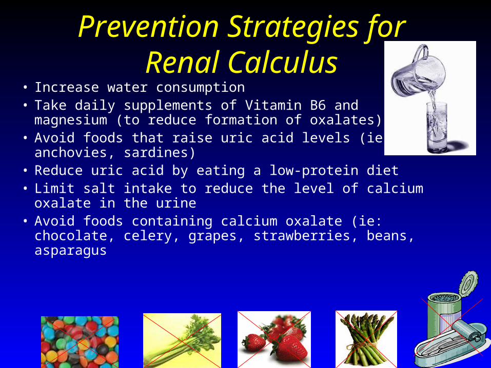
Prevention Strategies for Renal Calculus
• Increase water consumption • Take daily supplements of Vitamin B6 and magnesium (to
reduce formation of oxalates)• Avoid foods that raise uric acid levels (ie: anchovies, sardines)• Reduce uric acid by eating a low-protein diet• Limit salt intake to reduce the level of calcium oxalate in the
urine• Avoid foods containing calcium oxalate (ie: chocolate, celery,
grapes, strawberries, beans, asparagus

Identifying a variety of EKG rhythms and knowing the
Region X SOP for that particular rhythm

Rhythm Identification• What is this rhythm?
• What is your intervention?

Ventricular Tachycardia• If stable with pulse:
– Amiodarone 150 mg diluted in 100 ml D5W IVPB over 20 minutes
or (EMS choice)– Lidocaine 0.75 mg/kg IVP bolus– Contact Medical Control for further bolus/drip orders
• If no pulse - treat like ventricular fibrillation– emphasis on good quality CPR– switch CPR compressor every 2 minutes to keep CPR
effective– all shocks given at max joules & singular

Rhythm Identification
• What is this rhythm?
• What intervention is necessary?

EMS Tx-Ventricular Fibrillation• If arrest <4-5 minutes, CPR until defibrillator ready
• If arrest >4-5 minutes, CPR for 2 minutes
• Single shocks at max output of unit
• Epinephrine 1:10,000 1 mg every 3-5 minutes
• EMS choice of one antidysrhythmic alternated with Epinephrine:
– Amiodarone 300 mg rapid IVP 1st dose
• repeat 150 mg IVP in 5 minutes (2nd dose) OR
– Lidocaine 1.5 mg/kg IVP 1st dose
• repeat 0.75 mg/kg in 5 minutes (2nd dose)

• What is this rhythm?
• What intervention is necessary?
Rhythm Identification

EMS Tx-Third Degree Heart Block• If stable patient (B/P & LOC) - monitor• If unstable patient & narrow complex (QRS)
– Atropine 0.5 mg rapid IVP– May repeat every 3-5 minutes to max of 3mg– TCP if Atropine not effective
• If unstable patient & wide complex (QRS)– Begin TCP (Valium for comfort)– If TCP ineffective, then Atropine 0.5 mg repeated every
3-5 minutes to a max of 3 mg
“When they’re alive, give them 0.5”

Rhythm Identification
• What is this rhythm?

EMS Tx-Third Degree Heart Block (Complete)
In bradycardias, always need to ask 2 questions:
#1 - Is the patient stable or unstable?
Stable needs monitoring
Unstable needs intervention
#2 - Is the QRS narrow or wide?
Narrow treated initially with Atropine
Wide treated initially with TCP

Rhythm Identification
• What is this rhythm?
• What intervention is necessary?

EMS Tx-Second Degree Type II• If stable patient - monitor• If unstable patient & narrow complex (QRS)
– Atropine 0.5 mg rapid IVP– May repeat every 3-5 minutes to max of 3mg– TCP if Atropine not effective
• If unstable patient & wide complex (QRS)– Begin TCP (Valium for comfort)– If TCP ineffective, then Atropine 0.5 mg repeated
every 3-5 minutes to a max of 3 mg
“When they’re alive, give them 0.5”

Rhythm Identification• What is this rhythm?
• What intervention is necessary?

Second Degree Type I - Wenckebach• If stable patient - monitor• If unstable patient & narrow complex (QRS)
– Atropine 0.5 mg rapid IVP– May repeat every 3-5 minutes to max of 3mg– TCP if Atropine not effective
• If unstable patient & wide complex (QRS)– Begin TCP (Valium for comfort)– If TCP ineffective, then Atropine 0.5 mg repeated
every 3-5 minutes to a max of 3 mg
“When they’re alive, give them 0.5”

Rhythm Identification
• What is this rhythm (the patient has no pulse)?
• What intervention is necessary?There is no pulse!

EMS Tx - PEA (rate under 60)
• Emphasis will be on good quality CPR
• CPR is 30:2 (compressions to ventilations)
• After intubation, breaths delivered once every 6-8 seconds, compressor doesn’t stop
• Search for causes (6 H’s, 5 T’s) & treat them!
• Epinephrine 1:10,000 1 mg every 3-5 minutes
• If rate <60, Atropine 1 mg every 3-5 minutes (max 3 mg)
• If rate >60, just Epinephrine & good CPR

Scenario #1
• EMS calls with report of a 56 year-old female with left sided abdominal pain
• Based on the report, the ED MD informs you to tell EMS they must transport this patient to the closest facility.
• The patient is alert and oriented and requests transport to a farther hospital
• Can the ED MD force EMS to transport this patient to the closest facility?

Scenario #1• A patient who is alert and oriented and can
understand the risks and benefits has the right to request and expect transportation to the facility of their choice
• EMS should have the patient sign the release for not going to the closest facility
• Time should not be wasted in the field arguing with a patient about the facility to transport to
• As in the ED, not all patients make the same choice you would for healthcare issues but they do have the right to make THEIR choice

Scenario #2• EMS is on the scene with a patient who had a
diabetic reaction with a blood sugar initially of 45. The current blood sugar is now 80.
• EMS calls to inform the ED that they are obtaining a release/refusal for further care and transportation
• How is the ECRN to respond to this radio call?

Scenario #2
• Verify if EMS needed any special orders
• Often times EMS calls in to document that Medical Control was aware of the release especially if there was something unusual about the call
• Releases/refusals/AMA’s in the field can be obtained in regards to assessment, treatment, and/or transportation of the patient

Scenario #3
• EMS calls in and reports that they have a critical patient and will be providing an abbreviated radio report
• What does it mean to receive an abbreviated report and what information can you expect to receive as the ECRN?
Scenario #3• An abbreviated radio report (SOP page #4) may be
provided to Medical Control in situations where manpower is limited and/or the patient’s condition is critical
• Time in the field is more importantly spent with all focus on caring for the patient; often all available personnel need to be caring for the patient and the driver needs to focus on driving to get everyone to the hospital as safe and fast as possible

Scenario #3• Contents of an abbreviated radio report
– Identification of provider name, vehicle number and receiving hospital– Nature of situation and protocol being followed– Age and sex of patient– Chief complaint and brief history of present illness/injury– Airway and vascular access status– Current vital signs– Major interventions completed or being attempted– ETA to receiving hospital
• EMS to provide detailed information upon ED arrival