Endocrinology and Aging. The world is getting older In the developed world, people >80 y/o are the...
70
Endocrinology and Aging
-
Upload
gerard-sullivan -
Category
Documents
-
view
222 -
download
3
Transcript of Endocrinology and Aging. The world is getting older In the developed world, people >80 y/o are the...
Endocrinology and Aging

The world is getting older
In the developed world, people >80 y/o are the fastest growing subset
In the US, people over 60 will increase from 35 million (12.4%) to 71.6 million (19.6%) by 2030
Worldwide, lifespan expected to increase another 10 years by 2050
Therefore need to focus on healthy aging

Aging Process
As people get older, parts don’t work as well In the endocrine system, there is a decrease
in feedback and feed-forward systems Organs become less responsive to stimuli,
and hormones are not released at the same levels


Thyroid Axis

Changes with Aging
Decreased pituitary responsivenessLess TSH response to TRHLess rise in TSH to low T4/T3
Thyroid does not respond to TSH as wellLess T4 response to TSH surge
Decreased peripheral conversion of T4 to T3 due to decreased 5’ deiodinase activityMay be selenium dependent

Change in TSH with Age
Mariotto, et al. JCEM 1993: 77(5), 1130-1134

Change in T4 and T3
Mariotto, et al. JCEM 1993: 77(5), 1130-1134

Hypothyroidism
More common with advancing age 7-14 % of elderly have TSH above nl
range, more women than men Higher incidence in iodine replete areas Autoimmune is the most common cause,
followed by surgical

Elevated TSH with aging
Canaris et al, Archives of Int Med: 2000;160:526

Diagnosis
Symptoms can be the same as in younger patients, but more often ignored as they are attributed to “aging”
Fatigue and weakness were reported by more than 50%, but the following sxs were less commonly reported: cold intolerance, weight gain, paresthesias, and muscle
cramps Can score lower on MMSE along with other
memory and neurocognitive testing

Associated Findings
PE: Bradycardia, diastolic hypertension, pericardial effusion
Labs: Elevated TSH, low FT4 Other potential findings:
Hyponatremia elevated CKelevated LDL

Treatment
Levothyroxine replacement Start with lower dosage—0.25-0.5 mcg/kg instead of 1.6
mcg/kg as in young See if tolerate, and then can increase by 12.5-25 mcg q 4-
6 weeks One study showed that if no cardiac dz present, elderly
could tolerate full dose If have underlying cardiac disease, may not be able to
tolerate dose that would normalize TSH Final dose may be 40 mcg lower than in comparable young

Hyperthyroidism
Not as common as hypothyroidism Prevalence < 0.5% of elderly Subclinical hyperthyroidism is more
common: 1-5%, but most studies say < 2.5%

Presentation
Classically present with different symptoms
Weight loss, depression, and agitation dominate, leading to it being called “apathetic hyperthyroidism”
Sympathetic sxs less common like tremor or hyperactivity

Cardiovascular Findings
A-fib occurs in 15% of pts with hyperthyroidism, but is more common in elderly
CV complications are more common in elderly: ischemic heart dz, dysrhythmias, hypertensive hd
So think about getting TFTs in pts presenting with atrial fibrillation, worsening heart failure, systolic hypertension, or deteriorating ischemic heart disease

Effects on Bone
Leads to decreased BMD, especially in post-menopausal women
Treatment of hyperthyroidism improves BMD, but not back to baseline
One study showed a doubling of risk of death from fractured femur in treated pts
Need to get DXA and consider bisphosphonates in pts with hx of hyperthyroidism

Thyroid Nodules
Incidence of nodules increases with age Palpable nodules found in 5% of pts >60 Autopsy studies show 90% in women >70 U/S show 50% of women over 50 have nodules
Causes of goiter in older population Nontoxic MNG: 51% Toxic MNG: 23% Single nodule: 8% Graves: 5% Hashimotos: 4%

Thyroid Cancer
See same cancers as in young, but some differences
Ratio of Papillary:Follicular goes from 4:1 to 2:1
Female:male ratio narrows Decreased 10 yr survival with older age
Women <20 have 100%Women >60 have <5%

Thyroid Cancer
Increased recurrance rate in older pts PTC/Follicular risk of recurrance and death
<50: 10% and 3% >50: 32% and 30%
Direct extension worse in elderly 67% recurrance, 60% death in older 12% and 4% in young
Distant mets more deadly in older 96% vs 63%
Cady B, Sedgwick CE, Meissner WA, et al. Risk factor analysis in differentiated
thyroid cancer. Cancer 43:810-820, 1979

Anaplastic Thyroid Cancer
Peak incidence in 60’s, and more than 65% occur in pts older than 65
1-2% of all thyroid cancers Usually presents as a rapidly growing mass with local
sxs Often caused by transformation of pre-existing
differentiated thyroid cancer or longstanding goiter Older pts have worse prognosis 5 yr survival of 7%, mean survival of 11 months

Thyroid Lymphoma
Only .5-5% of all thyroid cancers Peak incidence form 50-80 Presents as rapidly enlarging goiter, usually
in someone with long standing Hashimoto’s Dx can be difficult since FNA will just show
lymphocytes. Can do flow cytometry Treatment is chemotherapy


Androgens
Testosterone levels in men decline continuously starting at age 30
There is no abrupt decrease similar to menopause
Testosterone is lower in men with chronic illness, obesity, metabolic syndrome, etc
SHBG goes up in elderly, lowering free T even more

Longitudinal Changes in Testosterone
Tes
tost
eron
eT
esto
ster
one
(nm
ol/L
) (
nmol
/L)
Age (Years)Age (Years)
1010
1212
1414
1616
1818
2020
3030 4040 5050 6060 7070 8080 9090
(177)(177)
(144)(144)(151)(151)
(158)(158)
(109)(109)
(43)(43)
Adapted from Harman SM, et al. Adapted from Harman SM, et al. J Clin Endocrinol MetabJ Clin Endocrinol Metab. 2001;86:724-731.. 2001;86:724-731.

Incidence
Not every man develops low T, unlike women who all go through menopause
Incidence varies in different studies50% of men >70 y/o vs.3% of healthy men
Health status may be a significant factor

Physiology
Testosterone production decreases with age All levels of the HPT axis are affected
LH/FSH are higher than in younger men, but not as high as expected for degree of low T, indicating loss of normal feedback
LH/FSH response to GnRH not as coordinated Less synchrony in LH pulses and T production Decreased ability of testes to make T

Effects of Low Testosterone
Decreased muscle mass and strength Decreased bone mineral density Decreased libido and sexual functioning
Low nitric oxide synthase is seen in hypogonadal men. NOS needed for PDE to work
Increased fat mass Anemia Dysthymia

Testosterone and Mortality
Several studies have shown decreased survival in men with lower T
Rancho Bernardo study showed that men in the lowest quartile of testosterone (<241 ng/dL) were 40% more likely to die over the next 20 years than those with higher levels
The increased risk of death in men with low testosterone levels was independent of multiple risk factors, including age, adiposity, and lifestyle
No studies have shown replacement increases survival. Unclear is cause:effect or association
Araujo AB, Kupelian V, Page ST, Handelsman DJ, Bremner WJ, McKinlay JB.Sex steroids and all-cause and cause-specific mortality in men. Arch Intern Med. 2007 Jun 25;167(12):1252-60

Effect of T on Survival
Shores MM, Matsumoto AM, Sloan KL, Kivlahan DR.Low serum testosterone and mortality in male veterans. Arch Intern Med. 2006 Aug 14-28;166(15):1660-5
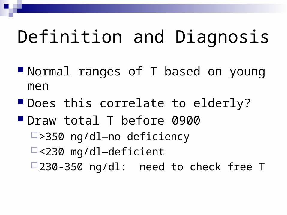
Definition and Diagnosis
Normal ranges of T based on young men Does this correlate to elderly? Draw total T before 0900
>350 ng/dl—no deficiency<230 mg/dl—deficient230-350 ng/dl: need to check free T

Benefits of Replacement
Benefit only shown in men who have low T Decreases fat mass and increases muscle
mass, but not dramatic Improved sexual functioning and libido Improves bone mineral density, but no fracture
data No good data on cognition Can improve quality of life

Should We Replace?
An expert panel of the Endocrine Society recommended against testosterone therapy for all older men with low testosterone levels.
Instead the panel suggested that “clinicians consider offering testosterone therapy on an individualized basis to older men with consistently low testosterone levels on more than one occasion and significant symptoms of androgen deficiency, after appropriate discussion of the uncertainties of the risks and benefits of testosterone therapy in older men”.
The panel’s recommendations were guided by the recognition of the paucity and low quality of evidence, and that high quality evidence of the efficacy and safety will not be available for a very long time.

Androgen deficiency in Women
As in men, testosterone levels decline with aging, particularly after oophorectomy or adrenal failure
By age 40, T levels are 50% of age 20 However, levels don’t fall at menopause like estrogen
does Hard to biochemically define because free T assays
are not good at the lower range and total not as reliable due to more SHBG variation
Testosterone levels not associated with sexual functioning in some studies

Benefits of Replacement?
Controversial that disorder even exists Decreased sexual desire is reported in 25-
53% of women, but not always perceived as a problem
Maybe improvement in BMD Conflicting results on body mass

Androgen Replacement
Multiple studies looked at transdermal testosterone replacement in surgically and naturally menopausal women on estrogen
Patch of 300 mcg worn for 24 weeks increased satisfying sexual encounters by 1-2 episodes per month—statistically significant
Other measures of mood, sense of well being, libido, and distress had mixed outcomes
No long term safety data

Outcomes
Treatment effect was not dependent on baseline testosterone levels
No significant difference in surgical or natural menopausal women except for number of satisfying encounters
Women in the 300 mcg group had testosterone levels at or above the upper range of normal

Summary
300 mcg testosterone patch modestly improved sexual functioning, but this is clinically meaningful
Baseline testosterone had no bearing on outcome, so not useful for diagnosing “androgen deficiency”


Growth Hormone
Similar properties to sex steroids Improve lean body mass and muscle
strength Decrease fat mass Improve BMD Improve sense of well being

Growth Hormone Deficiency
Increased fat and decreased lean mass Sarcopenia Increased lipids Increased CV disease, impaired cardiac
function Decreased BMD

GH Changes with Aging
GH Declines as we age Sex steroids have a significant impact on
GH levelsThe higher the testosterone, the higher the GH
levels Obesity also is a factor
The more overweight a person is, the lower their GH levels

Relationship of Age, Weight and Testosterone to GH
Iranmanesh, A, et al, Eur J Endo 1998;139:59-71
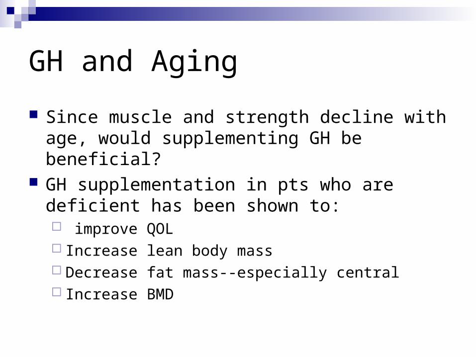
GH and Aging
Since muscle and strength decline with age, would supplementing GH be beneficial?
GH supplementation in pts who are deficient has been shown to: improve QOL Increase lean body mass Decrease fat mass--especially central Increase BMD

Risks of Supplementation
Possible increased risk of malignancy Increased insulin resistance Increased edema, arthralgias, and carpal
tunnel syndrome Some studies showed critically ill pts (ICU,
CHF) had higher mortality when treated with GH

Answer?
Don’t know if decline in GH with aging is adaptive or maladaptive
GH secretogogues may be an answer in the future
Currently, can’t recommend GH replacement for average aging patient


Osteoporosis
Peak bone mass is attained by age 20 Thereafter, bone is lost at a steady rate In women, there is an increase in BMD
loss for the 5-10 years after menopause

Rate of bone Loss
Dashed=trabecular
Solid=cortical
Khosla, et al. Pathophysiology of Age Related Bone Loss and Osteoporosis. Endo Metab Clin N Amer, DEC 2005: 1015-1030

Fracture Incidence
Khosla, et al. Pathophysiology of Age Related Bone Loss and Osteoporosis. Endo Metab Clin N Amer, DEC 2005: 1015-1030

Post-menopausal Osteoporosis
After menopause, estradiol levels fall to 10-15% of previous levels
Bone resorption increases by 90%, while formation increases by 45%
Estrogen inhibits osteoclast function and multiple cytokines that increase bone resorption. Without estrogen, these processes increase

Post-menopausal Osteoporosis
Other contributing factors: Vit D deficiency Secondary hyperparathyroidism Decreased calcium intake and increased calcium
secretion Never attaining good peak BMD Decreased GH levels Secondary causes
Also have impaired bone formation due to GH defic, estrogen defic,

Osteoporosis in Men
Men lose half as much bone as women and have 1/3 the number of fractures
Main cause is decreased testosterone, but estrogen has the main effect on bone
Therefore do not want to give aromatase inhibitors to men on androgen therapy—will decrease bone benefit

Estrogen Effect on BMD in Men
Khosla, et al. Pathophysiology of Age Related Bone Loss and Osteoporosis. Endo Metab Clin N Amer, DEC 2005: 1015-1030

Screening
The NOF recommends offering BMD testing to the following women:
1. All postmenopausal women under age 65 who have one or more additional risk factors for osteoporosis (besides menopause).
2. All women aged 65 and older regardless of additional risk factors. 3. Postmenopausal women who present with fractures (to confirm
diagnosis and determine disease severity). 4. Women who are considering therapy for osteoporosis, if BMD
testing would facilitate the decision. 5. Women who have been on hormone replacement therapy for
prolonged periods.

Screening in Men
NOF does not give specific guidelines ACP recommends:
clinicians periodically perform individualized assessment of risk factors for osteoporosis in
older men clinicians obtain DXA for men who are at increased risk for
osteoporosis and are candidates for drug therapy Main risk factors: age >70, low BMI, weight loss,
previous fracture, steroid use, inactivity, hypogonadism


Cortisol Axis
Age does not have a significant effect on CRH, ACTH, or cortisol levels
However, diurnal pattern seems to be altered, with higher levels later in the day
CRH evokes a greater ACTH and cortisol response in older pts
Stress response ACTH and cortisol release are actually 2-fold greater—seems to be a decreased sensitivity to negative feedback by cortisol
This is more prominent in women

DHEA
DHEA is not active, but it is converted to other androgens that are active
By age 80, DHEA levels are <10 % of young adult levels
ACTH which stimulates release of DHEA remain stable over this time
Likely due to decreased adrenal enzyme activity, like 3ß-HSD

DHEA Over Time

DHEA and Mortality
Multiple studies have shown that older men with lower levels of DHEA have increased mortality and lower functional status
There was no correlation seen in women DHEA replacement in adrenally insufficient pts has shown
improvement in a number of areas However, replacement in healthy elderly showed no
benefit in multiple studies It has improved sense of well being in pts with
depression, schizophrenia, and some immune mediated dzs like SLEc


Diabetes
Prevalence of DM increases with aging Changes that occur with aging
Decreased insulin secretion Decreased insulin action/increased resistance Alteration of hepatic glucose output Increased obesity Change in diet
These changes associated with genetic background increase DM in the elderly

Prevalence of DM
Third NHANES Survey 1988-1994

Presentation
May not be classic Glycemic threshold in kidney higher so
may not have polyuria Have impaired thirst drive so may not have
polydypsia Complaints may be more non-specific Often will present with a complication like
CVA, HONK, or MI

Hypoglycemia
The risk of severe hypoglycemia with oral agents or insulin increases exponentially in the elderly
Due to: Impaired secretion of counter-regulatory hormones
like glucagon Reduced awareness of autonomic symptoms Decreased ability to function with a low sugar which
decreases their ability to treat themselves Primary treatment is prevention!
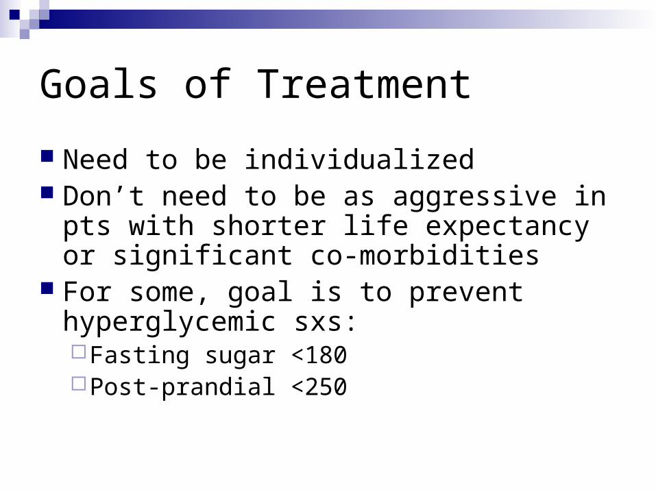
Goals of Treatment
Need to be individualized Don’t need to be as aggressive in pts with
shorter life expectancy or significant co-morbidities
For some, goal is to prevent hyperglycemic sxs:Fasting sugar <180Post-prandial <250

Medications
If mild hyperglycemia, consider meds that will not cause hypoglycemia:Metformin (as long as GFR>60)α-glucosidase inhibitorsDPP4 inhibitors
Caution with TZDs Caution with sulfonylureas and meglitinides
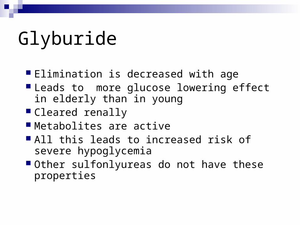
Glyburide
Elimination is decreased with age Leads to more glucose lowering effect in elderly
than in young Cleared renally Metabolites are active All this leads to increased risk of severe
hypoglycemia Other sulfonlyureas do not have these properties

Questions?