Endocrine System: Overview - Semantic Scholar
46
Copyright © 2006 Pearson Education, Inc., publishing as Benjamin Cummings Endocrine System: Overview The endocrine system interacts w/ the nervous system to coordinate & integrate the activity of body cells. The e.s. influences metabolic activities by means of hormones Binding of hormones to cellular receptors initiates responses that occur after a lag period of seconds to days Once initiated, the responses tend to be much more prolonged than those initiated by the nervous system
Transcript of Endocrine System: Overview - Semantic Scholar

Copyright © 2006 Pearson Education, Inc., publishing as Benjamin Cummings
Endocrine System: Overview
� The endocrine system interacts w/ the nervous system to
coordinate & integrate the activity of body cells.
� The e.s. influences metabolic activities by means of hormones
� Binding of hormones to cellular receptors initiates responses
that occur after a lag period of seconds to days
� Once initiated, the responses tend to be much more prolonged
than those initiated by the nervous system

Copyright © 2006 Pearson Education, Inc., publishing as Benjamin Cummings
Endocrine System: Overview
� Major processes controlled by hormones are:
� Reproduction
� Growth & development
� Body defenses
� Balance humoral electrolyte levels, water &
nutrients
� Cellular metabolism & energy balance

Copyright © 2006 Pearson Education, Inc., publishing as Benjamin Cummings
Endocrine System: Overview
� Two types of glands: Exocrine & Endocrine
� Exocrine glands: produce nonhumoral substances and have
ducts
� E.g. sweat and saliva glands
� Endocrine glands: Ductless glands. Release hormone into
surrounding tissue fluid. Have a rich vascular & lymphatic
drainage that receives the hormones.
� E.g. pituitary, thyroid, parathyroid, adrenal,
pineal, thymus, hypothalamus (neuroendocrine
organ)

Copyright © 2006 Pearson Education, Inc., publishing as Benjamin Cummings
Endocrine System: Overview
� Several organs contain areas of endocrine tissue
and produce hormones & exocrine products
� E.g. pancreas and gonads
� The hypothalamus has both neural functions and
releases hormones
� Other tissues and organs that produce hormones –
adipose cells, pockets of cells in the walls of the
small intestine, stomach, kidneys, and heart
PLAY InterActive Physiology ®: Endocrine System Review

Copyright © 2006 Pearson Education, Inc., publishing as Benjamin Cummings
Endocrine System: Overview
� Autocrines: chemicals that exert their function on
the same cell that secretes them.
� E.g. prostaglandins released by smooth muscle
cells cause those smooth muscle cells to contract
� Paracrines: act locally but affect cell types other
than those that secrete them.
� E.g. somatostatin released by one pancreatic cell
inhibits the release of insulin by another type of
pancreatic cell

Copyright © 2006 Pearson Education, Inc., publishing as Benjamin Cummings
Major Endocrine Organs
Figure 16.1

Copyright © 2006 Pearson Education, Inc., publishing as Benjamin Cummings
Hormones
� Chemistry of Hormones
� Almost all hormones can be classified either as amino acid
based or steroids.
� Most hormones are amino acid based ranging from amine
to peptides to proteins
� Steroids are synthesized from cholesterol and include
gonadal & adrenocortical hormones
� Eicosanoids: e.g. leukotrienes and prostaglandins
� These biologically active lipids are made from arachidonic
acid and are released by almost all cell membranes

Copyright © 2006 Pearson Education, Inc., publishing as Benjamin Cummings
Hormone Action
� Hormones alter target cell activity by one of two
mechanisms
� Second messengers:
� Regulatory G proteins
� Amino acid–based hormones
� Direct gene activation
� Steroid hormones
� The precise response depends on the type of the
target cell

Copyright © 2006 Pearson Education, Inc., publishing as Benjamin Cummings
Hormone Action
� Leudotrienes are signaling chemicals that mediate
inflammation & some allergic reactions
� Prostaglandins have many targets & functions

Copyright © 2006 Pearson Education, Inc., publishing as Benjamin Cummings
Mechanism of Hormone Action
� Hormones produce one or more of the following cellular changes in target cells by altering the cell’s activity, e.g. increase or decrease the rate of their normal cellular processes
� Alter plasma membrane permeability
� Stimulate protein synthesis
� Activate or deactivate enzyme systems
� Induce secretory activity
� Stimulate mitosis

Copyright © 2006 Pearson Education, Inc., publishing as Benjamin Cummings
Mechanisms of Cellular Transduction
� 1) Water soluble hormones (a.a. based) act on
receptors in the plasma membrane, e.g GPCRs
� 2) Lipid soluble hormones (steroid & thyroid) act
on intracellular receptors directly activating genes
1
2

Copyright © 2006 Pearson Education, Inc., publishing as Benjamin Cummings
Amino Acid-Based Hormone Action: cAMP Second Messenger
� Involves three components:
� Hormone receptor
� G-protein (signal transducer)
� Effector enzyme (adenylate cyclase)
� The steps are:
� Hormone (first messenger) binds to its receptor, which then binds to a G protein
� The G protein is then activated as it binds GTP, displacing GDP
� Activated G protein activates the effector enzyme adenylate cyclase
� Adenylate cyclase generates cAMP (second messenger) from ATP
� cAMP activates protein kinases, which then cause cellular effects

Copyright © 2006 Pearson Education, Inc., publishing as Benjamin Cummings
Receptor
Hormone A
ReceptorGTP GTP
GTP GTP GTP GTP
ATPcAMP
Inactive
protein kinase A
Active
protein kinase A
Catecholamines
ACTHFSHLH
GlucagonPTH
TSHCalcitonin
Triggers responses of targetcell (activates enzymes,stimulates cellular
secretion, opens ionchannels, etc.)
Adenylate cyclase Hormone B
GDPGDP
Extracellular fluid
Cytoplasm
Gs Gi
1
2 34
3 2
1
5
Figure 16.2
Amino Acid-Based Hormone Action: cAMPSecond Messenger

Copyright © 2006 Pearson Education, Inc., publishing as Benjamin Cummings
� Hormone binds to the receptor and activates
G protein
� G protein binds and activates phospholipase
� Phospholipase splits the phospholipid PIP2 (phosphatidyl
inositol biphosphate) into diacylglycerol (DAG) and inositol
triphosphate (IP3) (both act as second messengers)
� DAG activates protein kinase C; IP3 triggers release of Ca2+
stores from the E.R.
� Free Ca2+ (third messenger) binds calmodulin or to Ca2+
gated channels
Amino Acid-Based Hormone Action: PIP-Calcium

Copyright © 2006 Pearson Education, Inc., publishing as Benjamin Cummings
GTP PIP2
IP3
ReceptorGTP
GTP
Catecholamines
TRHADH
GnRHOxytocin
Triggers responses
of target cell
GDP
Extracellular fluid
Cytoplasm
Inactiveprotein
kinase C
Activeprotein kinase C
Phospholipase C
Gq
Ca2+ Ca2+- calmodulin
Hormone
Endoplasmic
reticulum
DAG1
2 34 5
5
6
Figure 16.3
Amino Acid-Based Hormone Action: PIP Mechanism

Copyright © 2006 Pearson Education, Inc., publishing as Benjamin Cummings
Intracellular Receptors & Direct Gene Activation: Steroid Hormones
� Steroid hormones diffuse into their target cells where they bind to an intracellular receptor
� Hormone-receptor complex travels to the nuclear chromatin where the hormone receptor binds to a region of the DNA (the hormone response element)
� This interaction prompts DNA transcription to produce mRNA (“turn on” the gene)
� The mRNA is translated into proteins, which bring about a cellular effect

Copyright © 2006 Pearson Education, Inc., publishing as Benjamin Cummings
Steroidhormone
Steroidhormone
Cytoplasm
Receptor-chaperonin
complex
Molecular
chaperones
Receptor-hormonecomplex
Hormoneresponseelements
Binding
Transcription
Chromatin
mRNA
Nucleus
New proteinTranslation
Ribosome
mRNA
Figure 16.4

Copyright © 2006 Pearson Education, Inc., publishing as Benjamin Cummings
Hormoneresponseelements
Binding
Transcription
Chromatin
mRNA
Nucleus
New proteinTranslation
Ribosome
mRNA
Figure 16.4

Copyright © 2006 Pearson Education, Inc., publishing as Benjamin Cummings
Target Cell Specificity
� Hormones circulate to all tissues but only activate
cells referred to as target cells
� Target cells must have specific receptors to which
the hormone binds
� These receptors may be intracellular or located on
the plasma membrane

Copyright © 2006 Pearson Education, Inc., publishing as Benjamin Cummings
Target Cell Activation
� Target cell activation depends on three factors
� Blood levels of the hormone
� Relative number of receptors on, or in, the target cell
� The affinity of those receptors for the hormone
� Up-regulation – target cells form more receptors in
response to the hormone
� Down-regulation – target cells lose receptors in response to
the hormone (to prevent overreaction of target cell to
corresponding hormone.

Copyright © 2006 Pearson Education, Inc., publishing as Benjamin Cummings
Hormone Concentrations in the Blood
� Hormones circulate in the blood in two forms –
free or bound to a protein carrier
� Steroids and thyroid hormones, which are lipid
soluble, travel in blood attached to plasma proteins
� All others are free circulating

Copyright © 2006 Pearson Education, Inc., publishing as Benjamin Cummings
Hormone Concentrations in the Blood
� Concentrations of circulating hormone reflect:
� Rate of release
� The rate that it is inactivated and removed.
� Hormones are removed from the blood by:
� Degrading enzymes
� The kidneys
� Liver enzyme systems
And are broken down & excreted in urine

Copyright © 2006 Pearson Education, Inc., publishing as Benjamin Cummings
Interaction of Hormones at Target Cells
� Three types of hormone interaction
� Permissiveness – one hormone cannot exert its
effects without another hormone being present
� Synergism – more than one hormone produces the
same effects on a target cell and their combined
effects are amplified
� Antagonism – when one hormone opposes the
action of another hormone

Copyright © 2006 Pearson Education, Inc., publishing as Benjamin Cummings
Control of Hormone Release
� Blood levels of hormones:
� Are controlled by negative feedback systems
� As hormone levels rise, they cause target organ effects and inhibit further hormone release
� Vary only within a narrow desirable range

Copyright © 2006 Pearson Education, Inc., publishing as Benjamin Cummings
Control of Hormone Release
� Hormones are synthesized and released in response
to:
� Humoral stimuli
� Neural stimuli
� Hormonal stimuli

Copyright © 2006 Pearson Education, Inc., publishing as Benjamin Cummings
Humoral Stimuli
� Humoral stimuli – secretion of hormones in direct response to changing blood levels of ions and nutrients
� Example: concentration of calcium ions in the blood
� Declining blood Ca2+
concentration stimulates the parathyroid glands to secrete PTH (parathyroid hormone)
� PTH causes Ca2+ concentrations to rise and the stimulus is removed
Copyright © 2006 Pearson Education, Inc., publishing as Benjamin Cummings
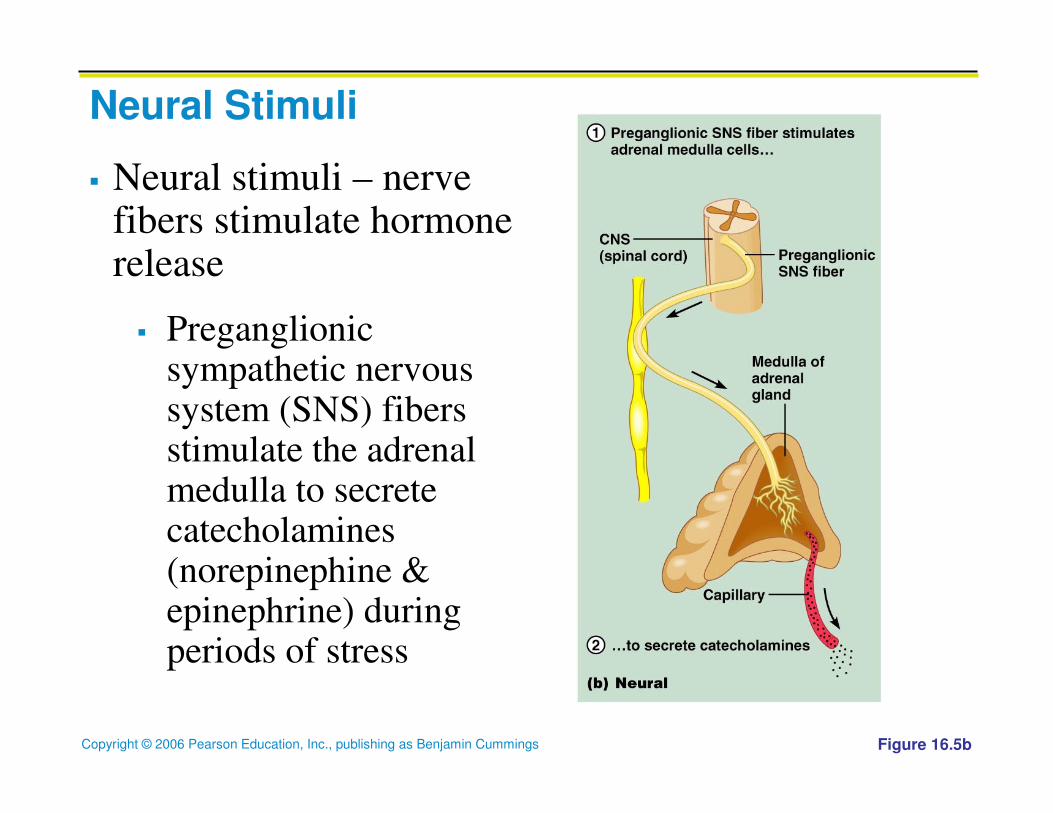
Neural Stimuli
� Neural stimuli – nerve fibers stimulate hormone release
� Preganglionicsympathetic nervous system (SNS) fibers stimulate the adrenal medulla to secrete catecholamines(norepinephine & epinephrine) during periods of stress
Figure 16.5b

Copyright © 2006 Pearson Education, Inc., publishing as Benjamin Cummings
Hormonal Stimuli
� Hormonal stimuli – release of
hormones in response to
hormones produced by other
endocrine organs
� The hypothalamic hormones
stimulate the anterior
pituitary

Copyright © 2006 Pearson Education, Inc., publishing as Benjamin Cummings
Nervous System Modulation
� The nervous system can modify both “turn on” and
“turn off” factors
� w/o nervous control, endocrine activity would act
like a thermostat and be static
� The nervous system allows for dynamic control

Copyright © 2006 Pearson Education, Inc., publishing as Benjamin Cummings
Major Endocrine Organs: Pituitary (Hypophysis)
� Located in the sell turcica of the sphenoid bone, the two-lobed organ secretes nine major hormones
� Neurohypophysis – posterior lobe (neural tissue) and the infundibulum
� Composed of pituicytes (supporting cells) & nerve fibers
� Release neurohormones (hormones secreted by neurons) received by the hypothalamus
� Hormone storage area, NOT a true endocrine gland
� Adenohypophysis – anterior lobe, made up of glandular tissue
� Synthesizes and secretes a number of hormones

Copyright © 2006 Pearson Education, Inc., publishing as Benjamin Cummings
Major Endocrine Organs: Pituitary (Hypophysis)
Figure 16.6

Copyright © 2006 Pearson Education, Inc., publishing as Benjamin Cummings
Pituitary-Hypothalamic Relationships: Posterior Lobe� The posterior lobe is a downgrowth of hypothalamic neural
tissue
� Has a neural connection with the hypothalamus (hypothalamic-hypophyseal tract)
� Neurosecretory cells synthesize two neurohormones & transport them via axons to the posterior pituitary
� Oxytocin: made in the paraventricular neurons
� ADH (antidiuretic hormone) made in the supraopticneurons
� When the nerves fire, these hormones are released into a capillary bed and transported to the posterior pituitary

Copyright © 2006 Pearson Education, Inc., publishing as Benjamin Cummings
Pituitary-Hypothalamic Relationships: Anterior Lobe
� The anterior lobe of the pituitary is an
outpocketing of the oral mucosa
� There is no direct neural contact with the
hypothalamus, only a vascular one

Copyright © 2006 Pearson Education, Inc., publishing as Benjamin Cummings
� The primary capillary plexus in the infundiblum communicates
inferiorly via the small hypophyseal portal veins with a
secondary capillary plexus in the anterior lobe
Pituitary-Hypothalamic Relationships: Anterior Lobe

Copyright © 2006 Pearson Education, Inc., publishing as Benjamin Cummings
Adenohypophyseal Hormones
� The six hormones (all proteins) of the
adenohypophysis:
� Abbreviated as GH, TSH, ACTH, FSH, LH, and
PRL
� Regulate the activity of other endocrine glands
� In addition, pro-opiomelanocortin (POMC):
� Prohormone that can be split enzymatically into
one or more hormones

Copyright © 2006 Pearson Education, Inc., publishing as Benjamin Cummings
Activity of the Adenohypophysis
� The tropin, or tropic hormones, that are released are:
� Thyroid-stimulating hormone (TSH)
� Adrenocorticotropic hormone (ACTH)
� Follicle-stimulating hormone (FSH)
� Luteinizing hormone (LH)
They regulate secretory action of other endocrine glands

Copyright © 2006 Pearson Education, Inc., publishing as Benjamin Cummings
Growth Hormone (GH)
� Produced by somatotropic cells of the anterior lobe
that:
� Stimulate most cells, but target bone and skeletal
muscle
� Promote protein synthesis and use of fats for fuel
� Most effects are mediated indirectly by insulin-like
growth factors (IGFs) like somatomedins
� GH releases fat from fat deposits increasing blood
levels of fatty acids

Copyright © 2006 Pearson Education, Inc., publishing as Benjamin Cummings
Growth Hormone (GH): IGFs
� IGFs:
� i) stimulate uptake of a.a. from the blood and
their incorporation into cellular proteins
� ii) stimulate uptake of sulfur into cartilage
matrix (needed for the synthesis of chondroitin)

Copyright © 2006 Pearson Education, Inc., publishing as Benjamin Cummings
Growth Hormone (GH)
� Secretion of GH is regulated by two hypothalmic
hormones w/ antagonistic effects
� Growth hormone–releasing hormone (GHRH)
stimulates GH release
� Growth hormone–inhibiting hormone (GHIH, aka
somatostatin) inhibits GH release
Secretion of GH is at its greatest during adolescence

Copyright © 2006 Pearson Education, Inc., publishing as Benjamin Cummings
Thyroid Stimulating Hormone (Thyrotropin)
� Stimulates the normal development and secretory
activity of the thyroid
� Release from thyrotroph cells of the anterior
pituitary is triggered by hypothalamic peptide
thyrotropin-releasing hormone (TRH)
� Rising blood levels of thyroid hormones act on the
pituitary and hypothalamus to block the release of
TSH

Copyright © 2006 Pearson Education, Inc., publishing as Benjamin Cummings
Adrenocorticotropic Hormone (Corticotropin)
� Secreted by corticotroph cell of the adenohypophysis
� Stimulates the adrenal cortex to release corticosteroid hormones, most importantly glucocorticoid
� Triggered by hypothalamic corticotropin-releasing hormone (CRH) in a daily rhythm
� Internal and external factors such as fever, hypoglycemia, and stressors can trigger the release of CRH

Copyright © 2006 Pearson Education, Inc., publishing as Benjamin Cummings
Gonadotropins
� Gonadotropins – follicle-stimulating hormone (FSH) and luteinizing hormone (LH)
� Regulate the function of the ovaries and testes
� FSH stimulates gamete (egg or sperm) production
� Absent from the blood in prepubertal boys and girls
� Triggered by the hypothalamic gonadotropin-releasing hormone (GnRH) during and after puberty

Copyright © 2006 Pearson Education, Inc., publishing as Benjamin Cummings
Functions of Gonadotropins
� In females
� LH works with FSH to cause maturation of the
ovarian follicle
� LH works alone to trigger ovulation (expulsion of
the egg from the follicle)
� LH promotes synthesis and release of estrogens
and progesterone

Copyright © 2006 Pearson Education, Inc., publishing as Benjamin Cummings
Functions of Gonadotropins
� In males
� LH stimulates interstitial cells of the testes to
produce testosterone
� LH is also referred to as interstitial cell-stimulating
hormone (ICSH)

Copyright © 2006 Pearson Education, Inc., publishing as Benjamin Cummings
Prolactin (PRL)
� Produced by lactotroph cells
� In females, stimulates milk production by the
breasts
� Triggered by the hypothalamic prolactin-releasing
hormone (PRH)
� Inhibited by prolactin-inhibiting hormone (PIH)
aka dopamine. A decrease in PIH results in a
surge of PRL

Copyright © 2006 Pearson Education, Inc., publishing as Benjamin Cummings
Prolactin (PRL)
� A brief rise in PRL levels just before the menstrual
period accounts for some of the breast swelling
and tenderness that some women experience
� Because the PRL stimulus is relatively short, no
milk is produced
� Toward the end of pregnancy PRL levels rise
dramatically and milk production becomes
possible