Endocrine system
67
CHAPTER 3 THE ENDOCRINE SYSTEM
Transcript of Endocrine system

CHAPTER 3
THE ENDOCRINE SYSTEM
AN OVERVIEW
• The endocrine system is a major controlling system of the body
• Influences metabolic activities by means of hormones transported in the blood
• Responses occur more slowly but tend to last longer than those of the nervous system
• Through hormones, it stimulates such long-term processes as growth and development, metabolism, reproduction, and body defense

• Endocrine glands are ductless, well-vascularized glands that release hormones directly into the blood or lymph
• Endocrine glands: pituitary, thyroid, parathyroid, adrenal, and pineal glands
• Exocrine glands produce nonhormonal substances, such as sweat and saliva, and have ducts
• Some organs produce both hormones and exocrine products (e.g., pancreas and gonads)

Figure 16.1
Pineal glandHypothalamus
Pituitary gland
Parathyroid glands(on dorsal aspectof thyroid gland)Thymus
Thyroid gland
Adrenal glands
Pancreas
Ovary (female)
Testis (male)

• Prostaglandins are not endocrine hormones, but they are locally acting messenger molecules which include:
a. Autocrine - act on the cell that released them b. Paracrine - act on a different cell type nearby

Function
• Controlling activity of specific organ or tissue in maintaining homeostasis by secreting hormones as in:
a. Regulator of growth and development b. Regulating the concentration of body fluids (water and electrolyte) c. Metabolism of carbohydrate, protein and
lipids (nutrient) d. Acts together with nervous system to help the body to react to stress properly

HORMONES
• Chemical substances secreted by cells into extracellular fluid (bloodstream) that regulate the metabolic activity of other cells in the body.
• All hormones are amino acid-based or steroids.

Types of Hormones
a. Amino acid based (water soluble) • Most hormones are amino acid-based except
thyroid hormones• Amines, thyroxine, peptides, and proteins
b. Steroids (lipid soluble) • Synthesized from cholesterol • Of the hormones, only gonadal and
adrenocortical hormones are steroids

Regulation of Hormones Secretion
a. Negative feedback • Response that reduces the initiating stimulus
(opposite direction) • Important in regulating hormone levels in the
blood b. Positive feedback • Reinforce the initial stimulus

Mechanism of Hormones Action • Hormones alters cell activity by stimulating or
inhibiting characteristics cellular processes of their target cells
• Two main mechanism account for how a hormone communicates with targets cells:
1. Amino acid- based hormones and second-messenger system 2. Steroid hormones and direct gene activation

Mechanisms of Hormone ActionTwo mechanisms, depending on their chemical nature
1. Water-soluble hormones (all amino acid–based hormones except thyroid hormone)• Cannot enter the target cells• Act on plasma membrane receptors• Coupled by G proteins to intracellular second
messengers that mediate the target cell’s response

Mechanisms of Hormone Action
2. Lipid-soluble hormones (steroid and thyroid hormones)• Act on intracellular receptors that directly
activate genes

Amino acid- based hormones and second-messenger system
1. Hormone (first messenger) binds to receptor2. Receptor activates G protein3. G protein activates adenylate cyclase4. Adenylate cyclase converts ATP to Cyclic adenosine
monophosphate (cAMP) (second messenger) 5. cAMP (second messenger) activates protein
kinases in the cytoplasm 6. Protein kinases activated other proteins in the cell 7. Activated proteins induce changes in the cell

Figure 16.2
Hormone (1st messenger)binds receptor.
Receptoractivates Gprotein (GS).
G proteinactivatesadenylatecyclase.
cAMP acti-vates proteinkinases.
Adenylatecyclaseconverts ATPto cAMP (2ndmessenger).
Receptor
G protein (GS)
Adenylate cyclase
Triggers responses oftarget cell (activatesenzymes, stimulatescellular secretion,opens ion channel,etc.)
Hormones thatact via cAMPmechanisms:
EpinephrineACTHFSHLH
Inactiveprotein kinase
Extracellular fluid
Cytoplasm
Activeproteinkinase
GDP
GlucagonPTHTSHCalcitonin
1
2 3 4
5

Figure 16.2, step 1
Hormone (1st messenger)binds receptor.
Receptor
Hormones thatact via cAMPmechanisms:
EpinephrineACTHFSHLH
Extracellular fluid
Cytoplasm
GlucagonPTHTSHCalcitonin
1

Figure 16.2, step 2
Hormone (1st messenger)binds receptor.
Receptoractivates Gprotein (GS).
Receptor
G protein (GS)
Hormones thatact via cAMPmechanisms:
EpinephrineACTHFSHLH
Extracellular fluid
Cytoplasm
GDP
GlucagonPTHTSHCalcitonin
1
2

Figure 16.2, step 3
Hormone (1st messenger)binds receptor.
Receptoractivates Gprotein (GS).
G proteinactivatesadenylatecyclase.
Receptor
G protein (GS)
Adenylate cyclase
Hormones thatact via cAMPmechanisms:
EpinephrineACTHFSHLH
Extracellular fluid
Cytoplasm
GDP
GlucagonPTHTSHCalcitonin
1
2 3

Figure 16.2, step 4
Hormone (1st messenger)binds receptor.
Receptoractivates Gprotein (GS).
G proteinactivatesadenylatecyclase.
Adenylatecyclaseconverts ATPto cAMP (2ndmessenger).
Receptor
G protein (GS)
Adenylate cyclase
Hormones thatact via cAMPmechanisms:
EpinephrineACTHFSHLH
Extracellular fluid
Cytoplasm
GDP
GlucagonPTHTSHCalcitonin
1
2 3 4

Figure 16.2, step 5
Hormone (1st messenger)binds receptor.
Receptoractivates Gprotein (GS).
G proteinactivatesadenylatecyclase.
cAMP acti-vates proteinkinases.
Adenylatecyclaseconverts ATPto cAMP (2ndmessenger).
Receptor
G protein (GS)
Adenylate cyclase
Triggers responses oftarget cell (activatesenzymes, stimulatescellular secretion,opens ion channel,etc.)
Hormones thatact via cAMPmechanisms:
EpinephrineACTHFSHLH
Inactiveprotein kinase
Extracellular fluid
Cytoplasm
Activeproteinkinase
GDP
GlucagonPTHTSHCalcitonin
1
2 3 4
5

Steroid hormones and direct gene activation
1. Diffuse directly through plasma membrane (target cells) 2. Binds with protein receptor and turns into steroid
protein complex 3. Entering nucleus to a specific Deoxyribonucleic acid
(DNA) region (activating DNA, which initiates messenger Ribonucleic acid (RNA) formation leading to protein synthesis)
4. Reaction between steroid-protein complex and DNA activates genes to synthesize new proteins and enzymes and induce changes in the cell

Figure 16.3
mRNA
New protein
DNA
Hormoneresponseelements
Receptor-hormonecomplex
Receptorprotein
Cytoplasm
Nucleus
Extracellular fluid
Steroidhormone
The steroid hormonediffuses through the plasmamembrane and binds anintracellular receptor.
The receptor-hormone complex entersthe nucleus.
The receptor- hormonecomplex binds a hormoneresponse element (aspecific DNA sequence).
Binding initiatestranscription of thegene to mRNA.
The mRNA directsprotein synthesis.
Plasmamembrane
1
2
3
4
5

Figure 16.3, step 1
Receptor-hormonecomplex
Receptorprotein
Cytoplasm
Nucleus
Extracellular fluid
Steroidhormone
The steroid hormonediffuses through the plasmamembrane and binds anintracellular receptor.
Plasmamembrane
1

Figure 16.3, step 2
Receptor-hormonecomplex
Receptorprotein
Cytoplasm
Nucleus
Extracellular fluid
Steroidhormone
The steroid hormonediffuses through the plasmamembrane and binds anintracellular receptor.
The receptor-hormone complex entersthe nucleus.
Plasmamembrane
1
2

Figure 16.3, step 3
DNA
Hormoneresponseelements
Receptor-hormonecomplex
Receptorprotein
Cytoplasm
Nucleus
Extracellular fluid
Steroidhormone
The steroid hormonediffuses through the plasmamembrane and binds anintracellular receptor.
The receptor-hormone complex entersthe nucleus.
The receptor- hormonecomplex binds a hormoneresponse element (aspecific DNA sequence).
Plasmamembrane
1
2
3

Figure 16.3, step 4
mRNA
DNA
Hormoneresponseelements
Receptor-hormonecomplex
Receptorprotein
Cytoplasm
Nucleus
Extracellular fluid
Steroidhormone
The steroid hormonediffuses through the plasmamembrane and binds anintracellular receptor.
The receptor-hormone complex entersthe nucleus.
The receptor- hormonecomplex binds a hormoneresponse element (aspecific DNA sequence).
Binding initiatestranscription of thegene to mRNA.
Plasmamembrane
1
2
3
4

Figure 16.3, step 5
mRNA
New protein
DNA
Hormoneresponseelements
Receptor-hormonecomplex
Receptorprotein
Cytoplasm
Nucleus
Extracellular fluid
Steroidhormone
The steroid hormonediffuses through the plasmamembrane and binds anintracellular receptor.
The receptor-hormone complex entersthe nucleus.
The receptor- hormonecomplex binds a hormoneresponse element (aspecific DNA sequence).
Binding initiatestranscription of thegene to mRNA.
The mRNA directsprotein synthesis.
Plasmamembrane
1
2
3
4
5

Control of Hormone Release
• Synthesis and release of most hormones are regulated by negative feedback system
• Endocrine glands are stimulated to manufacture and release their hormones by 3 major types of stimuli:
a. Humoral stimuli b. Neural stimuli c. Hormonal stimuli

Humoral Stimuli
• Changing blood levels of ions and nutrients directly stimulates secretion of hormones
• Example: Ca2+ in the blood– Declining blood Ca2+ concentration stimulates the
parathyroid glands to secrete PTH (parathyroid hormone)
– PTH causes Ca2+ concentrations to rise and the stimulus is removed

Figure 16.4a
(a) Humoral Stimulus
Capillary (lowCa2+ in blood)
Parathyroidglands
Thyroid gland(posterior view)
PTH
Parathyroidglands
1 Capillary blood containslow concentration of Ca2+,which stimulates…
2 …secretion ofparathyroid hormone (PTH)by parathyroid glands*

Neural Stimuli
• Nerve fibers stimulate hormone release– Sympathetic nervous system fibers stimulate the
adrenal medulla to secrete catecholamines

Figure 16.4b
(b) Neural Stimulus
CNS (spinal cord)
Medulla ofadrenalgland
Preganglionicsympatheticfibers
Capillary
1 Preganglionic sympatheticfibers stimulate adrenalmedulla cells…
2 …to secrete catechola-mines (epinephrine andnorepinephrine)

Hormonal Stimuli
• Hormones stimulate other endocrine organs to release their hormones – Hypothalamic hormones stimulate the release of
most anterior pituitary hormones– Anterior pituitary hormones stimulate targets to
secrete still more hormones– Hypothalamic-pituitary-target endocrine organ
feedback loop: hormones from the final target organs inhibit the release of the anterior pituitary hormones

Figure 16.4c
(c) Hormonal Stimulus
Hypothalamus
Thyroidgland
Adrenalcortex
Gonad(Testis)
Pituitarygland
1 The hypothalamus secreteshormones that…
2 …stimulatethe anteriorpituitary glandto secretehormonesthat…
3 …stimulate other endocrineglands to secrete hormones

MAJOR ENDOCRINE ORGANS a. Hypothalamusb. Pituitary gland c. Thyroid gland d. Parathyroid glands e. Adrenal glands f. Pancreas g. Pineal gland h. Thymus gland i. Gonads (ovaries, testes)

Figure 16.1
Pineal glandHypothalamus
Pituitary gland
Parathyroid glands(on dorsal aspectof thyroid gland)Thymus
Thyroid gland
Adrenal glands
Pancreas
Ovary (female)
Testis (male)

Hypothalamus • A neuroendocrine organ • The hypothalamus: – Link the nervous system to the endocrine system
via the pituitary gland (hypophysis)– Synthesizes two hormones (oxytoxin and ADH)
that it exports to the posterior pituitary for storage and later release
– Regulates the hormonal output of the anterior pituitary via releasing and inhibiting hormones


Pituitary Gland (Hypophysis) • Attached to hypothalamus by the
infundibulum within the sphenoid bone • Divided into 2 lobes:
a. Posterior lobe (neurohypophysis) –Store hormones from hypothalamus -
oxytocin and antidiuretic hormone (ADH)

b. Anterior lobe (adenohypophysis) – Influenced by hypothalamic hormone – Growth hormone (GH), prolactin (PRL),
adrenocorticotropic hormone ( ACTH), thyroid-stimulating hormone (TSH), follicle-stimulating hormone ( FSH) and luteinizing hormone ( LH)
– ACTH, TSH, FSH and LH are tropic hormones (regulate other endocrine gland)


Anterior Pituitary Hormones
a. Growth hormone (GH) • An anabolic and protein-conversing hormone
that promotes total body growth • It important effect is on skeletal muscles and
bones • Promotes protein synthesis and encourages
use of fats for fuel

Homeostatic Imbalances of Growth Hormone
• Hypersecretion– In children results in gigantism– In adults results in acromegaly
• Hyposecretion– In children results in pituitary dwarfism

Anterior Pituitary Hormones b. Prolactin (PRL) • Stimulates production of breast milk
(lactation) • Regulation of PRL release– Primarily controlled by prolactin-inhibiting
hormone (PIH) (dopamine)• Blood levels rise toward the end of pregnancy• Suckling stimulates PRH release and promotes
continued milk production

Anterior Pituitary Hormones
c. Adrenocorticotropic hormone (ACTH) – Stimulates the adrenal cortex to release its
hormones (mineralocorticoids, glucocorticoids and gonadocorticoids)

Anterior Pituitary Hormones
d. Thyroid-stimulating hormone (TSH) • Stimulates the thyroid gland to release thyroid
hormones (thyroxine and triiodothyronine) that are primarily responsible for regulation of metabolism.


Anterior Pituitary Hormones e. Gonadotropic hormones i. Follicle-stimulating hormone (FSH) – Beginning at puberty, stimulates follicle
development and estrogen production by female ovaries, promotes sperm production in male
ii. Luteinizing hormone (LH) – Beginning at puberty, stimulates ovulation and
stimulates ovarian to produce estrogen and progesterone, stimulates the male’s testes to produce testosterone

Posterior Pituitary Hormones
a. Oxytoxin • Stimulates powerful uterine contractions (trigger
labor and delivery of infant) and causes milk ejection in the nursing woman
• Also promote sexual arousal
b. Antidiuretic hormone (ADH) or vasopressin • Causes kidney tubule cells to reabsorb and conserve
body water and increased blood pressure by constricting blood vessels


Thyroid Gland
• Located on the trachea, just inferior to the larynx (in the anterior throat)
• Thyroid hormone (TH) includes thyroxine (T4) and triiodotyronine (T3), which increase the rate of cellular metabolism
• Calcitonin produced by parafollicular (C) cells in response to high blood calcium levels. It causes calcium to be deposited in bones

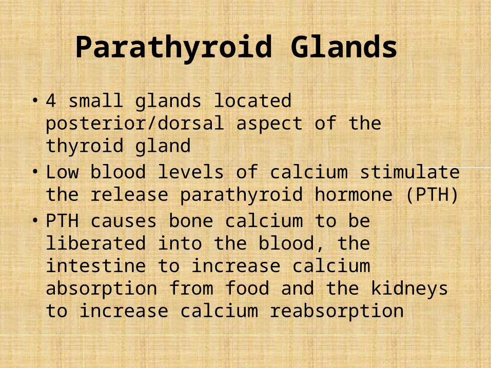
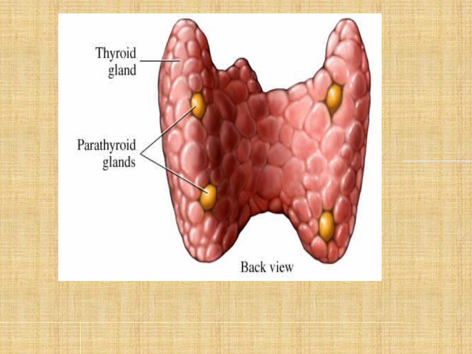
Parathyroid Glands
• 4 small glands located posterior/dorsal aspect of the thyroid gland
• Low blood levels of calcium stimulate the release parathyroid hormone (PTH)
• PTH causes bone calcium to be liberated into the blood, the intestine to increase calcium absorption from food and the kidneys to increase calcium reabsorption

Adrenal (Suprarenal) Glands
• Lies on top of kidneys • Divided into:
a. Adrenal cortex b. Adrenal medulla


Adrenal cortex
a. Mineralocorticoids (aldosterone) • Regulate sodium ion (Na+) and potassium ion (K+)
reabsorption by the kidneys • Their release is stimulated by low Na+ and/or high
K+ levels in blood b. Glucocorticoids (cortisol) • Enable the body to resist long-term stress by
increasing blood glucose levels and depressing the inflammatory response.
c. Gonadocorticoids/Sex hormones (androgens) • Responsible for sex drive in female

Adrenal medulla
• Adrenal medulla hormones produce catecholamines (epinephrine and norepinephrine) in response to sympathetic nervous system stimulation. Its hormones enhance and prolong the effects of the ‘fight-or-flight’ response to short-term stress

Pancreas • Located behind stomach • Composed of both endocrine and exocrine gland cells • Hormones produced from pancreatic islets (islets of
Langerhans) containing alpha (α) cells (glucagon) and beta (β) cells (insulin)
• Insulin is released when the blood levels of glucose are high. It increases the rate of glucose uptake and metabolism by body cells; stimulates glycogen formation
• Glucagon is released when blood levels of glucose are low, stimulates the liver to release glucose to the blood


Pineal Gland
• Located in the diencephalon/ third ventricle of the brain (epithalamus)
• Releases melatonin, which acts as biological clock; reproductive behaviour; affects daily biological rhythms such as body temperature, sleep and appetite


Thymus Gland
• Located deep to sternum • Large and conspicuous in infant and children • Diminishes in size throughout adulthood • Its hormones, thymosins, thymic factor, and
thymopoietins, are important to the normal development of the immune responses (thymosin promotes maturation of T lymphocytes, important in body defense)


Gonads Ovaries • Ovaries located in abdominopelvic cavity • Ovaries release: a. Estrogens – Release of estrogens by ovarian follicles begins at puberty
(FSH) – Estrogens stimulate maturation of female reproductive organs
and female secondary sex characteristics – With progesterone, they cause the menstrual cycle
b. Progesterone – Release in response to LH, works with estrogens establishing
the menstrual cycle


Gonads Testes • Testes located in the scrotum • Testes begin to produce testosterone at
puberty in response to LH stimulation • Testosterone promotes maturation of the
male reproductive system, male secondary sex characteristics, and production of sperm by the testes