
Emerging Computational Methods for the Rational Discovery of ...
21
Emerging Computational Methods for the Rational Discovery of Allosteric Drugs Jeffrey R. Wagner, † Christopher T. Lee, † Jacob D. Durrant, †,‡ Robert D. Malmstrom, †,‡ Victoria A. Feher, † and Rommie E. Amaro* ,†,‡ † Department of Chemistry & Biochemistry and ‡ National Biomedical Computation Resource, University of California, San Diego, La Jolla, California 92093, United States ABSTRACT: Allosteric drug development holds promise for delivering medicines that are more selective and less toxic than those that target orthosteric sites. To date, the discovery of allosteric binding sites and lead compounds has been mostly serendipitous, achieved through high-throughput screening. Over the past decade, structural data has become more readily available for larger protein systems and more membrane protein classes (e.g., GPCRs and ion channels), which are common allosteric drug targets. In parallel, improved simulation methods now provide better atomistic understanding of the protein dynamics and cooperative motions that are critical to allosteric mechanisms. As a result of these advances, the field of predictive allosteric drug development is now on the cusp of a new era of rational structure-based computational methods. Here, we review algorithms that predict allosteric sites based on sequence data and molecular dynamics simulations, describe tools that assess the druggability of these pockets, and discuss how Markov state models and topology analyses provide insight into the relationship between protein dynamics and allosteric drug binding. In each section, we first provide an overview of the various method classes before describing relevant algorithms and software packages. CONTENTS 1. Review Motivation and Organization 6370 2. Introduction 6371 2.1. Emerging Rational Design Principles 6372 3. Computational Methods for Studying Allostery 6373 3.1. Protein-Sequence Analysis Methods 6373 3.1.1. Introduction 6373 3.1.2. Single-Site Evolutionary Analysis Meth- ods 6373 3.1.3. Coupled-Site Evolutionary Analysis Methods 6374 3.2. Simulation Methods/Correlated Motions 6378 3.2.1. Molecular Dynamics and Monte Carlo Methods 6378 3.2.2. Network Representations and Protein Allostery 6378 3.2.3. Dynamical Network Analysis 6378 3.2.4. Community Analysis 6379 3.3. Pocket Detection 6379 3.3.1. Introduction 6379 3.3.2. Geometry-Based Pocket Detection 6380 3.3.3. Knowledge-Based Pocket Detection 6380 3.3.4. Energy-Based Pocket Detection 6380 3.4. Markov State Models 6381 3.4.1. Introduction 6381 3.4.2. MSMs and Drug Discovery 6381 4. Energy Landscape and Topological Analyses 6382 4.1. Introduction 6382 4.2. Protein Frustration 6382 4.3. Normal-Mode Analysis 6382 4.3.1. Elastic Network Models 6382 5. Conclusions 6383 Author Information 6383 Corresponding Author 6383 Author Contributions 6383 Notes 6383 Biographies 6383 Acknowledgments 6384 References 6384 1. REVIEW MOTIVATION AND ORGANIZATION To date, most allosteric drugs have been discovered through high-throughput screening. But growing databases of biomo- lecular structure and sequence data, in conjunction with increases in computing power and improvements in predictive algorithms, are enabling the rational de novo design of allosteric drugs. Given the large number of published algorithms for predicting allosteric mechanisms, it can be difficult to select the most appropriate method for a given target. This review serves as an introduction for those who wish to use computational techniques to develop allosteric drugs. After a broad overview of allosteric drug discovery, this review is divided into three sections. First, we discuss bioinformatics and molecular-dynamics methods to identify allosterically important sequence positions. Second, we summarize the computational Special Issue: Protein Ensembles and Allostery Received: October 24, 2015 Published: April 13, 2016 Review pubs.acs.org/CR © 2016 American Chemical Society 6370 DOI: 10.1021/acs.chemrev.5b00631 Chem. Rev. 2016, 116, 6370−6390 This is an open access article published under an ACS AuthorChoice License, which permits copying and redistribution of the article or any adaptations for non-commercial purposes.
Transcript of Emerging Computational Methods for the Rational Discovery of ...

Emerging Computational Methods for the Rational Discovery ofAllosteric DrugsJeffrey R. Wagner,† Christopher T. Lee,† Jacob D. Durrant,†,‡ Robert D. Malmstrom,†,‡ Victoria A. Feher,†
and Rommie E. Amaro*,†,‡
†Department of Chemistry & Biochemistry and ‡National Biomedical Computation Resource, University of California, San Diego, LaJolla, California 92093, United States
ABSTRACT: Allosteric drug development holds promise for delivering medicines thatare more selective and less toxic than those that target orthosteric sites. To date, thediscovery of allosteric binding sites and lead compounds has been mostly serendipitous,achieved through high-throughput screening. Over the past decade, structural data hasbecome more readily available for larger protein systems and more membrane proteinclasses (e.g., GPCRs and ion channels), which are common allosteric drug targets. Inparallel, improved simulation methods now provide better atomistic understanding ofthe protein dynamics and cooperative motions that are critical to allosteric mechanisms.As a result of these advances, the field of predictive allosteric drug development is nowon the cusp of a new era of rational structure-based computational methods. Here, wereview algorithms that predict allosteric sites based on sequence data and molecular dynamics simulations, describe tools thatassess the druggability of these pockets, and discuss how Markov state models and topology analyses provide insight into therelationship between protein dynamics and allosteric drug binding. In each section, we first provide an overview of the variousmethod classes before describing relevant algorithms and software packages.
CONTENTS
1. Review Motivation and Organization 63702. Introduction 6371
2.1. Emerging Rational Design Principles 63723. Computational Methods for Studying Allostery 6373
3.1. Protein-Sequence Analysis Methods 63733.1.1. Introduction 63733.1.2. Single-Site Evolutionary Analysis Meth-
ods 63733.1.3. Coupled-Site Evolutionary Analysis
Methods 63743.2. Simulation Methods/Correlated Motions 6378
3.2.1. Molecular Dynamics and Monte CarloMethods 6378
3.2.2. Network Representations and ProteinAllostery 6378
3.2.3. Dynamical Network Analysis 63783.2.4. Community Analysis 6379
3.3. Pocket Detection 63793.3.1. Introduction 63793.3.2. Geometry-Based Pocket Detection 63803.3.3. Knowledge-Based Pocket Detection 63803.3.4. Energy-Based Pocket Detection 6380
3.4. Markov State Models 63813.4.1. Introduction 63813.4.2. MSMs and Drug Discovery 6381
4. Energy Landscape and Topological Analyses 63824.1. Introduction 63824.2. Protein Frustration 63824.3. Normal-Mode Analysis 6382
4.3.1. Elastic Network Models 63825. Conclusions 6383Author Information 6383
Corresponding Author 6383Author Contributions 6383Notes 6383Biographies 6383
Acknowledgments 6384References 6384
1. REVIEW MOTIVATION AND ORGANIZATION
To date, most allosteric drugs have been discovered throughhigh-throughput screening. But growing databases of biomo-lecular structure and sequence data, in conjunction with increasesin computing power and improvements in predictive algorithms,are enabling the rational de novo design of allosteric drugs. Giventhe large number of published algorithms for predicting allostericmechanisms, it can be difficult to select the most appropriatemethod for a given target. This review serves as an introductionfor those who wish to use computational techniques to developallosteric drugs.After a broad overview of allosteric drug discovery, this review
is divided into three sections. First, we discuss bioinformatics andmolecular-dynamics methods to identify allosterically importantsequence positions. Second, we summarize the computational
Special Issue: Protein Ensembles and Allostery
Received: October 24, 2015Published: April 13, 2016
Review
pubs.acs.org/CR
© 2016 American Chemical Society 6370 DOI: 10.1021/acs.chemrev.5b00631Chem. Rev. 2016, 116, 6370−6390
This is an open access article published under an ACS AuthorChoice License, which permitscopying and redistribution of the article or any adaptations for non-commercial purposes.

methods to predict druggable pockets at these functionallyrelevant sites. Finally, we describe how Markov state models andtopological analyses can tie these single sequence sites to globalprotein function and dynamics.
2. INTRODUCTIONAllosteric drugs offer a number of advantages that make themdesirable as drug candidates. Allosteric effectors, by definition,alter protein activity by binding to a site distinct from theorthosteric pocket. One example of an allosteric system isfructose 1,6-bisphosphatase (shown in Figure 2). Becauseallosteric sites are typically less evolutionarily conserved,allosteric drugs can be highly selective, even among othermembers of the same protein family.1−8 In some cases, allostericsites are so unique among proteins that an effector is said to have“absolute subtype specificity.”2,3,9,10
Allosteric modulators may have spatiotemporal specificity. Forexample, they can be active only in the presence of theendogenous ligand, thus restricting their effect to certain tissuesat certain times,2−4,9,11 which may slow desensitization.10,12
Allosteric effectors are generally saturable, meaning that theyhave a maximal effect that does not necessarily correspond tocomplete inhibition or activation.2,4−6,8,10,11 This saturabilityenables safer dosing. For example, if the maximal effect is an 80%reduction in signaling, overdosing will not fully eliminate anessential signal.1,2,4
Other advantages can include noncompetitive inhibition (i.e.,drug activity cannot be “overwhelmed” by high concentrations ofthe endogenous ligand) and pathway- or substrate-specificmodulation, which reduces unwanted activity by specificallytargeting a single protein function.2,4,10 For example, if a proteinis involved inmultiple pathways, an allosteric effector may impactthe activity of each pathway differently depending on thesystems-biology context. If a protein acts on multiple substrates,the impact on activity may depend on the biological context.Despite many potential advantages of allosteric therapeutics, it
has been challenging to identify predictive approaches todiscovering allosteric drugs. In recent decades, the pharmaceut-ical industry has favored more traditional targets for threeprimary reasons: the relative ease of assay development aroundorthosteric sites; access to high-throughput, high-resolution X-ray crystallography; and advances in ligand- and receptor-basedcomputational methods to optimize ligand-binding affinity at asubstrate-competitive site. This structure-based approach isthought to significantly reduce the time and cost of hit-to-leadand lead-to-drug development by reducing the number ofcompounds that need be synthesized.13,14 Work by Doman et al.comparing computer-aided drug discovery (CADD) and high-throughput screening (HTS) reported that the two methods hadhit rates of 35 and 0.021%, respectively.15
In contrast, allosteric drugs are uniquely challenging from arational drug-design perspective. Because experimental assaystypically measure orthosteric function rather than ligand bindingat the allosteric site, efficient development of allosteric drugsrequires that the complex structure−activity relationships(SARs) governing both binding affinity and allosteric activitybe considered simultaneously.5,10,16 Further, allosteric sites areless likely to be evolutionarily conserved. While this enablesincreased subtype specificity, it also increases the chances ofevolved resistance5,9,10 and can complicate testing in evolutio-narily distant animal models.10
Additionally, allosteric effectors are particularly susceptible to“mode switching,” where relatively minor chemical changes can
drastically affect ligand efficacy.10,16 Structurally similar drugmetabolites, therefore, may have varying and unpredictabledistributions and allosteric effects.10,16 Optimizing allostericmodes of action requires methods that are very different fromthose used in orthosteric drug discovery.5
Multifunctional allosteric proteins are particularly challenging.While drug designers may desire to target a single proteinfunction, an allosteric effector may also alter other functions,hindering a full mechanistic understanding of the pharmacol-ogy.10 Also, the benefits of spatiotemporal specificity are lost ifthe distribution of the endogenous ligand changes withprogression of the disease state.10 Finally, assessment of thelimited number of known allosteric pockets indicates that theyare generally shallow5 and present flat SARs.10 These structuralfeatures similarly challenge existing rational drug-discoveryparadigms and the general practice of developing selectivecompounds by optimizing affinity.Despite these challenges, allosteric drug discovery has gained
momentum recently due to a number of developments.10 First,several allosteric drugs across a broad range of pharmacologicaltarget classes have been rationally designed,17−23 encouragingpursuit of others, as evidenced by the number of allosteric drugscurrently in clinical trials.24 The recent elucidation of newmembrane protein crystal structures for GPCRs25,26 and ionchannels27,28 have assisted in the structure-based designapproach to these successes. Finally, advances in our under-standing of allosteric mechanisms have supported developmentof additional rational design strategies (see below).Our understanding of allosteric mechanisms has advanced
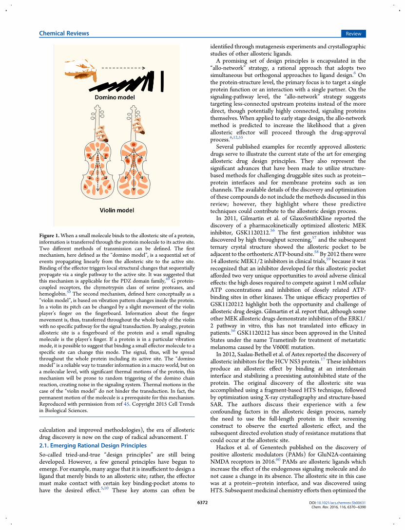
considerably since the initial conception of Monod, Wyman, andChangeux.29 Modern models of allostery consider conforma-tional ensembles.5,8,9,30−43 This revised view supports newlyestablished and emerging computational advances that compre-hensively map conformational landscapes and predict commu-nication between allosteric and orthosteric sites. For example, thephysical mechanisms of allostery generally alter the entropic andenthalpic factors that define the conformational landscape and,therefore, govern protein function.5,8,9 The observed correlationbetween allosteric modulation and protein structural dynamics isvaried: Major conformational rearrangements occur in somecases, as compared with subtle shifts in conformationalpopulations in others.4−6,9,10,35,37,44 An excellent metaphor forthese phenomena is Kornev and Taylor’s classification of“domino” versus “violin” models of allosteric signal trans-duction45 (Figure 1). Further, allosteric signals are transmittedthrough a range of structural motifs, from rigid core regions toflexible linkers.46 Allostery may occur through essential residuesalong a single allosteric path47 or through many weak pathwaysconnecting one site to another, acting in concert.48 As such, it isnot surprising that many of the allostery-prediction methodsdiscussed in this review (some of which do not use structure/geometry information at all) in practice may identify non-contiguous groups of residues as being allosterically linked. Suchpredictions should not immediately be assumed to be wrong butrather may indicate a non-“domino” model of allostery.Recent work has also revealed that protein allostery is not
merely a transition between two discrete protein conformations,as initially thought, but rather a shift in the equilibriumpopulations of many conformations, induced by effectorbinding.31,35,38,41,42,51,52 It is becoming increasingly clear thatthe kinetics of these transitions define the mechanisms ofallostery.38,51 Empowered with these new understandings andadvances in molecular simulation (in terms of speed of
Chemical Reviews Review
DOI: 10.1021/acs.chemrev.5b00631Chem. Rev. 2016, 116, 6370−6390
6371
calculation and improved methodologies), the era of allostericdrug discovery is now on the cusp of radical advancement. I’2.1. Emerging Rational Design Principles
So-called tried-and-true “design principles” are still beingdeveloped. However, a few general principles have begun toemerge. For example, many argue that it is insufficient to design aligand that merely binds to an allosteric site; rather, the effectormust make contact with certain key binding-pocket atoms tohave the desired effect.5,10 These key atoms can often be
identified through mutagenesis experiments and crystallographicstudies of other allosteric ligands.A promising set of design principles is encapsulated in the
“allo-network” strategy, a rational approach that adopts twosimultaneous but orthogonal approaches to ligand design.6 Onthe protein-structure level, the primary focus is to target a singleprotein function or an interaction with a single partner. On thesignaling-pathway level, the “allo-network” strategy suggeststargeting less-connected upstream proteins instead of the moredirect, though potentially highly connected, signaling proteinsthemselves. When applied to early stage design, the allo-networkmethod is predicted to increase the likelihood that a givenallosteric effector will proceed through the drug-approvalprocess.6,12,55
Several published examples for recently approved allostericdrugs serve to illustrate the current state of the art for emergingallosteric drug design principles. They also represent thesignificant advances that have been made to utilize structure-based methods for challenging druggable sites such as protein−protein interfaces and for membrane proteins such as ionchannels. The available details of the discovery and optimizationof these compounds do not include the methods discussed in thisreview; however, they highlight where these predictivetechniques could contribute to the allosteric design process.In 2011, Gilmartin et al. of GlaxoSmithKline reported the
discovery of a pharmacokinetically optimized allosteric MEKinhibitor, GSK1120212.56 The first generation inhibitor wasdiscovered by high throughput screening,57 and the subsequentternary crystal structure showed the allosteric pocket to beadjacent to the orthosteric ATP-bound site.58 By 2012 there were14 allosteric MEK1/2 inhibitors in clinical trials,59 because it wasrecognized that an inhibitor developed for this allosteric pocketafforded two very unique opportunities to avoid adverse clinicaleffects: the high doses required to compete against 1 mM cellularATP concentrations and inhibition of closely related ATP-binding sites in other kinases. The unique efficacy properties ofGSK1120212 highlight both the opportunity and challenge ofallosteric drug design. Gilmartin et al. report that, although someother MEK allosteric drugs demonstrate inhibition of the ERK1/2 pathway in vitro, this has not translated into efficacy inpatients.56 GSK1120212 has since been approved in the UnitedStates under the name Trametinib for treatment of metastaticmelanoma caused by the V600E mutation.In 2012, Saalau-Bethell et al. of Astex reported the discovery of
allosteric inhibitors for the HCVNS3 protein.17 These inhibitorsproduce an allosteric effect by binding at an interdomaininterface and stabilizing a preexisting autoinhibited state of theprotein. The original discovery of the allosteric site wasaccomplished using a fragment-based HTS technique, followedby optimization using X-ray crystallography and structure-basedSAR. The authors discuss their experience with a fewconfounding factors in the allosteric design process, namelythe need to use the full-length protein in their screeningconstruct to observe the exerted allosteric effect, and thesubsequent directed evolution study of resistance mutations thatcould occur at the allosteric site.Hackos et al. of Genentech published on the discovery of
positive allosteric modulators (PAMs) for GluN2A-containingNMDA receptors in 2016.60 PAMs are allosteric ligands whichincrease the effect of the endogenous signaling molecule and donot cause a change in its absence. The allosteric site in this casewas at a protein−protein interface, and was discovered usingHTS. Subsequent medicinal chemistry efforts then optimized the
Figure 1.When a small molecule binds to the allosteric site of a protein,information is transferred through the protein molecule to its active site.Two different methods of transmission can be defined. The firstmechanism, here defined as the “domino model”, is a sequential set ofevents propagating linearly from the allosteric site to the active site.Binding of the effector triggers local structural changes that sequentiallypropagate via a single pathway to the active site. It was suggested thatthis mechanism is applicable for the PDZ domain family,49 G protein-coupled receptors, the chymotrypsin class of serine proteases, andhemoglobin.50 The second mechanism, defined here conceptually as a“violin model”, is based on vibration pattern changes inside the protein.In a violin its pitch can be changed by a slight movement of the violinplayer’s finger on the fingerboard. Information about the fingermovement is, thus, transferred throughout the whole body of the violinwith no specific pathway for the signal transduction. By analogy, proteinallosteric site is a fingerboard of the protein and a small signalingmolecule is the player’s finger. If a protein is in a particular vibrationmode, it is possible to suggest that binding a small effector molecule to aspecific site can change this mode. The signal, thus, will be spreadthroughout the whole protein including its active site. The “dominomodel” is a reliable way to transfer information in a macro world, but ona molecular level, with significant thermal motions of the protein, thismechanism will be prone to random triggering of the domino chainreaction, creating noise in the signaling system. Thermal motions in thecase of the “violin model” do not hinder the transduction. In fact, thepermanent motion of the molecule is a prerequisite for this mechanism.Reproduced with permission from ref 45. Copyright 2015 Cell Trendsin Biological Sciences.
Chemical Reviews Review
DOI: 10.1021/acs.chemrev.5b00631Chem. Rev. 2016, 116, 6370−6390
6372

early hit molecule. The authors note that the validation of thisallosteric site was reinforced by its similarity to an analogousallosteric site in AMPA receptors, but that the NMDA receptorsite has elements of asymmetry that the AMPA receptor site didnot. In comparing two similar compounds, GNE-6901 andGNE-8324, the authors make comments that indicate evidence ofmode switching or a shallow SAR landscape, and they furthercharacterize the details of the allosteric mechanism usingmutagenesis experiments.In summary these examples demonstrate allosteric drug
discovery can be successful at protein sites often considered tobe undruggable. It is apparent that these successes can be furtherbuilt upon through computational methods that allow forrational rather than serendipitous HTS discovery of newallosteric binding sites and a deeper understanding of allostericmechanisms that overcome design challenges such as modeswitching.
3. COMPUTATIONAL METHODS FOR STUDYINGALLOSTERY
3.1. Protein-Sequence Analysis Methods
3.1.1. Introduction. Protein-sequence analysis is a usefultool to detect and characterize allosteric pathways and pockets.Here, we classify sequence-based methods into two groups: (1)“single site” methods, which produce a list of individualfunctional sequence positions; (2) “coupled site” methods,which produce a list of groups comprised of two or moresequence positions that appear to be functionally linked based ontheir coevolution.All sequence-based analysis methods share some challenges.
These challenges include how to select and aggregate clean,relevant sequences as input; interpret the output; and integratesequence-analysis results with other forms of data. Determiningthe biological meaning of a strong signal is also problematic.While many analysis methods identify evolutionarily importantresidues, the specific biological role of these residues cannot be
inferred without additional knowledge. For example, it is difficultto determine, based on sequence alone, whether an evolutio-narily significant residue plays an allosteric role, or whether itsrole is related to another essential process (e.g., substratebinding, maintaining protein structure, etc.).61,62 Indeed, it islikely that a given residue serves multiple purposes simulta-neously.Input sequence selection and alignment also present
challenges. Most techniques require many sequences to establishstatistical significance. To obtain the required number ofsequences, researchers often lower the stringency of their searchparameters, resulting in alignments that contain sequences withlower similarity or incomplete coverage of the original query.While some analysis methods manage to detect meaningfulcoevolution over a wide range of multiple sequence alignment(MSA) conservation and noise levels, others are moresusceptible to messy data.63,64 For a more complete discussionof these topics and how they affect coevolution analysis methods,readers are directed to an excellent recent review by de Juan etal.65
3.1.2. Single-Site Evolutionary Analysis Methods. Byour definition, single-site evolutionary analysis methods return alist of predicted functional sequence positions but do not suggestspecific linkages between sites. Once a researcher has constructedan MSA, the conservation or phylogenetic relevance of eachcolumn can be used to infer the evolutionary importance of eachsequence position. This importance is sometimes a hallmark ofthermodynamically critical residues that participate in allostery.Though single-site methods only return a list of single high-scoring sequence positions, the inner workings of some single-site methods are based on the aggregate or correlated behaviorsof multiple sequence positions (e.g., to determine baselineresidue probabilities within a multiple-sequence alignment orconstruct a phylogenetic tree).Single-site methods for detecting allostery are advantageous
because they lack much of the noise often associated with
Figure 2. Allosteric protein fructose 1,6-bisphosphatase, shown for illustration. Orthosteric and allosteric pockets (yellow and red, respectively) arebound to an endogenous ligand and an allosteric effector, respectively. Note that the allosteric site is distant from the orthosteric site such that there is nooverlap between the bound poses of the allosteric and orthosteric ligands. Despite the distance between them, the allosteric effector measurably modifiesthe enzymatic activity at the orthosteric site. Illustration derived from PDB IDs 2Y5K53 and 3IFC.54
Chemical Reviews Review
DOI: 10.1021/acs.chemrev.5b00631Chem. Rev. 2016, 116, 6370−6390
6373

correlation analysis. These analyses are also appealing because oftheir simplicity: There are usually fewer parameters to set, andthe results can be visualized directly by highlighting key residueson a three-dimensional (3D) protein structure.3.1.2.1. Single-Position Entropy. Shannon entropy, one of the
simplest nontrivial sequence-analysis metrics,66 was used widelyin early works to identify conserved sequence positions for drug-design or mutagenesis experiments.67 Similar in form tothermodynamic entropy from statistical mechanics, Shannonentropy measures the population diversity of residues at a singleMSA position. It is also central to mutual information (MI), apopular coupled-pair metric. The MI of two sequence positionsis defined as the sum of the individual position entropies, minusthe entropy of the positions considered jointly. While we do notcover the mathematical details of these methods here, interestedreaders are directed to previous articles on these topics.64,68
Shannon entropy does not consider amino acid similarity (e.g.,in the Shannon entropy framework, a leucine-to-isoleucinemutation is consideredmathematically equivalent to a leucine-to-arginine mutation). Other entropy measures, such as the relativeShannon entropy (also called the Kullback−Leibler divergence(KLD))69 and the von Neumann entropy,70,71 attempt toovercome this limitation and, as a result, may be more useful inthe search for allosteric sites. Relative Shannon entropy/KLDaccounts for some measure of the protein’s chemical environ-ment by considering each mutation with respect to thebackground amino acid frequencies calculated from the MSA.This analysis may be particularly useful when searching forallosteric sites in proteins that reside in membranes or othernoncytosolic compartments, where background residue proba-bilities or mutational preferences may be biased due to differentbiochemical contexts. In contrast, von Neumann entropy, aconcept borrowed from quantum statistical mechanics, iscalculated using amino acid similarity matrices. Identifying anoptimal amino acid similarity metric is nontrivial and may welldepend on the nature of the system (e.g., in a well-packedprotein, residue size may be a sensitive metric, whereas surface-site comparison may require the user to prioritize charge). In arecent publication describing these types of entropy, Johanssonand Toh explored how the two metrics can be mixed to detectenzyme active sites with maximum sensitivity.64
Zhang et al. constructed a variety of new analysis methods in2008 by combining Shannon or von Neumann entropy,phylogenetic tree structure, and a novel gap-treatmentapproach.70 In benchmarking their method, they comparedtheir results to Evolutionary Trace and ConSurf (discussed ingreater detail below). Two of their hybrid approaches out-performed all other techniques in detecting significant residuesacross a variety of proteins: the Improved Zoom method, whichincorporates a tree breakdown of subalignments, and thePhysiochemical Similarity Zoom method, which extends theImproved Zoom method with von Neumann entropy and tree-branch-size normalization.3.1.2.2. Evolutionary Trace. Lichtarge, Bourne, and Cohen
pioneered the evolutionary trace (ET) method. The approachhas become quite popular, largely because the algorithm isintuitive and its results are readily visualizable.72 ET aligns anumber of sequences and constructs a phylogenetic tree, andthen monitors the conservation of sequence positions at majortree branching points. By slicing the tree at different similaritycutoffs, the algorithm extracts the cluster-defining sequencepositions. The evolutionary significance of these sequencepositions is implied by their conservation in the sequences
beyond the next branch. In their first paper,72 the authorsdemonstrated that ET can detect functionally important sites inSH2, SH3, and DNA-binding domains. Work has since beenpublished on ET validation, parameter optimization, and best-use practices.73
In a method often referred to as “difference-ET,” the user runsET on two related proteins and considers differences in the high-ranking residues and their scores. The sequence positions withstrongly varying scores may suggest specificity determinants ordifferences in allosteric and/or orthosteric mechanisms. Notably,difference-ET has been used in the study of GPCRspecificity.73−75
To better account for varying rates of evolution in differentsubtrees and correlated mutations, in 2004 Mihalek et al.developed real-valued ET.76 This method incorporates entropyinformation into the standard ET framework. This work alsointroduces the zoom ETmethod (not related to improved zoom,above), which adds higher weight to sequences that are moresimilar to the protein of interest. In the introductory work, theyused real-valued and zoomET to detect the functional residues ina kinase domain, and then compared the performance of bothmethods to regular (integer-valued) ET and entropy. Givenunpruned sequence data sets, the real-valued ET and zoom ETmethods outperformed the others by a significant margin. Incontrast, integer-valued ET prevailed in most respects whenpruned data was available. An automated web server is availableto perform real-valued ET calculations, generate reports, andvisualize results (http://mammoth.bcm.tmc.edu/ETserver.html).77
3.1.2.3. H2r(s). In 2008, Merkl et al. introduced a methodcalled H2r that serves as a segue between single-site and coupled-site approaches.78 H2r generates a mutual-information matrix foran MSA, and then discards all but the strongest detected coupledpairs. For each sequence position k, the method returns conn(k),the number of top-ranked pairs that include k. Initial workproved that H2r can successfully detect functionally significantresidues across a range of proteins. More recently, H2rs, animproved version of H2r, has been released.79 This methodmodifies the original by using von Neumann instead of Shannonentropy and performing more detailed checks for statisticalsignificance. H2rs is available as a web server and a stand-aloneapplication at http://www-bioinf.uni-regensburg.de/.
3.1.3. Coupled-Site Evolutionary Analysis Methods.Second-order sequence analysis detects residue pairs that mutatein concert more frequently than would be expected givenrandom genetic events. Coevolving residue pairs are assumed tobe functionally linked, often because they serve essential roles inallostery or structural integrity.The immediate output of second-order allostery analysis is a
list of residue pairs with associated correlation strengths.Combining these individual pairwise correlations into a singlepicture of the entire protein is a separate task. On the most basiclevel, the strongest correlations that include a residue or site ofinterest can suggest thermodynamic coupling to other sites,possibly related to allostery. More complex analyses usehierarchical clustering or principal component analysis to analyzethese linkages and uncover strongly linked networks ofcoevolving residues.
3.1.3.1. Basic Coupled-Site Analyses. Several simple yetreliable residue-coupling analyses have maintained a presence inthe literature over the past decades. These basic approaches areadvantageous because they are easier to understand and havebeen shown to score consistently well in a wide range of tests.
Chemical Reviews Review
DOI: 10.1021/acs.chemrev.5b00631Chem. Rev. 2016, 116, 6370−6390
6374

However, they may fail to detect correlations in more complexcases.80,81 Though more complex methods exist, many of thesebasic methods still appear as analysis options in coevolution-detection software packages and web servers. In this review, wefocus on a few that are still widely used.3.1.3.2. Mutual Information.Mutual information (MI) is one
of the most straightforward and long-lived coupling metrics. TheMI between two sequence positions is defined as the sum of theShannon entropies of both positions, minus their joint entropy.Due to its simplicity and favorable mathematical properties, MIanalysis is the basis for a number of more complex coevolutionmethods. However, MI does present certain shortcomings. Forexample, uncorrelated pairs of high-entropy sequence positionsare likely to have a higher MI than uncorrelated pairs of low-entropy positions.82,83 To compensate for this and othershortcomings, various software packages have implemented anumber of mathematical corrections to MI.84−88 Further,methods to estimate baseline values for correlation (e.g.,resampling or sequence shuffling) can improve MI anal-ysis.83,89,90
Another relatively direct coevolution metric, the McLachlan-based substitution correlation (McBASC),91 looks for similarpatterns of variation in the columns of an MSA, weighting forresidue similarity using the McLachlan scoring matrix.92
Analogous methods can be constructed using differentsubstitution matrices, but McBASC continues to be a popularchoice in the literature.63,83,93
In 2002, Kass and Horovitz94 analyzed the GroEL complexusing a chi-squared test to detect significant residue coevolutionin an MSA. The analysis suggested intra- and interchain contactpairs and has continued to appear in the literature under thename “observed minus expected squared” (OMES).63,80,81,83,95
3.1.3.3. Statistical Coupling Analysis.The statistical couplinganalysis (SCA) method developed by Lockless and Ranganathanis perhaps the most widely used sequence-based method forallostery prediction.49 SCA draws an analogy to statistical physicsby calculating a “coupling energy” between each sequence-position pair. The original SCAmethod computes a conservationvalue for each sequence position i in an MSA, applies one ofseveral types of perturbation to another position j (depending onthe SCA version96), and finally recalculates the conservation atposition i for the sequences that satisfy the perturbation. Bycalculating the change in individual and joint conservation over avariety of perturbations, SCA establishes a “coupling energy” thatindicates the evolutionary coupling of positions i and j.The output of the SCAmethod is anN ×Nmatrix of coupling
energies, where N is the number of sequence positions in thealignment. In early work, the researchers manually identifiedstrongly coupled residue pairs that included one functionalmember (per experiment). More recent versions of SCA havegrouped this matrix into meaningful clusters of coevolvingresidues using hierarchical or spectral clustering.50
Refinements of SCA have achieved improved statisticalproperties by resampling the original distribution.97 In a 2011paper, SCA was effectively used to engineer a light-sensitiveLOV2 domain onto the surface of DHFR at a location that SCAhad identified as energetically linked to the enzyme active site.Some variants of the resulting protein chimera were found tohave acquired light-dependent activity.98
Further work has used SCA to design artificial WWdomains.99,100 In 2011, an SCA analysis of antigen 85C fromMycobacterium tuberculosis suggested new sites that potentiallycould be exploited in drug design.101 A number of projects have
also demonstrated how SCA can be used to target mutations thataffect protein function.102−108
Inspired by earlier work on the sequence correlation entropy(SCE) method,109 Dima and Thirumalai published an SCAvariant in 2006.110 This variant controls for specific proteincomposition by calculating the background probability that agiven amino acid will be present at a random sequence position.This probability is determined by considering only the sequencesbeing analyzed, as opposed to all sequences in the SWISS-PROTdatabase.111,112 Further, they borrowed a coupled two-wayclustering procedure from gene-sequence analysis to define thesectors.113 In validating this method, the authors analyzed thePDZ, GPCR, and lectin families of proteins and were able toquantitatively predict functional residues, which were inagreement with experimental findings.
3.1.3.4. Explicit Likelihood of Subset Covariation. Asmentioned above, SCA is a “perturbation-based” method inwhich correlation is established by excluding certain sequencesfrom an MSA and monitoring how entropies change. Anotherpopular perturbation-based method was published in 2004 byDekker et al.114 This method, explicit likelihood of subsetcovariation (ELSC), relies on similar principles but returnscorrelation scores in the form of probabilities rather thanstatistical energies. ELSC was shown to be superior to SCA incontact prediction when tested on a range of protein families. Ithas since been implemented on web servers115,116 and has been apopular benchmark method in the literature.80,81,93,95
3.1.3.5. Direct Coupling Analysis. In 2009, Weigt et al.proposed a mutual-information-based method called directcoupling analysis (DCA) that disentangles directly interactingresidues from large networks of indirectly coupled sequencepositions.117 While this method is typically used in structureprediction to identify spatially adjacent sequence positions, itmay find application in the study of short-range allostericinteractions. A more efficient implementation of the DCAmethod, known as “mean field” (as opposed to the original“message-passing” implementation) was published in 2011.118
Both introductory papers show that DCA is a robust predictor ofboth intra- and interprotein contacts and that it can hint at theexistence of unobserved protein conformations. Related workhas shown that DCA can be used in conjunction with structuralmodels to generate predictive models of protein com-plexes,119−121 determine the sequence positions that contributeto protein-interaction specificity,122 and describe the conforma-tional ensembles of proteins in crystallographic or near-crystallographic states.123 A web server and software packageare available to perform DCA analysis at http://dca.rice.edu/portal/dca/home.
3.1.3.6. PSICOV. PSICOV is another popular contact-detection method that may find productive use in the study ofallostery.124 Mathematically, PSICOV relies on an estimatedinverse of the MSA covariance matrix, which acts as a matrix ofcorrelations between all sequence-position pairs that inherentlycontrols for the variations in all other positions. PSICOV wassuccessful at predicting contacting protein residues based onMSA data. The code has been published online at http://bioinfadmin.cs.ucl.ac.uk/downloads/PSICOV/.
3.1.3.7. Recurrence Quantification Analysis. Recurrencequantification analysis (RQA), another second-order sequence-analysis technique, is best used when much is already knownabout the mechanism under investigation (e.g., physiochemicalamino acid properties such as charge or hydrophobicity areknown to drive the allostery). RQA itself is a general method in
Chemical Reviews Review
DOI: 10.1021/acs.chemrev.5b00631Chem. Rev. 2016, 116, 6370−6390
6375

nonlinear dynamics:125 In the context of protein sequences, itconsiders a scalar-value vector that represents some property of agiven sequence. In introductory work by Zbilut et al.,126 themethod was used to properly classify 56 TEM-1/β-lactamasemutants with impaired function based on their hydrophobicityprofiles. Further RQA work used hydrophobicity scores toclassify proteins as allosteric or nonallosteric,127 study p53mutants,128 and reveal interaction partners in viral-envelopeproteins.129
In 2005, Colafranceschi et al. investigated the effect ofchoosing different physicochemical amino acid descriptors andchanging the numerical parameters of the RQA algorithm.130
More recently, a comparison method based on RQA measure-ments, known as cross-RQA, effectively detected proteinallostery.131 Interested readers are directed to a review byZbilut-Webber, which provides examples of RQA applied to arange of computational biology problems.132
3.1.3.8. Comparative Analyses. Some work has been done tocompetitively benchmark the performance of these methods. In2004, Fodor and Aldrich compared OMES, MI, SCA, andMcBASC in a variety of tests. In short, the study found thatperformance is largely dependent on the way that differentmethods determine background residue probabilities and handlepositional conservation.63 A follow-up study investigated howeffectively coevolution analysis finds thermodynamically linkedresidue pairs.62 In general, spatially contiguous linked pairs weredetected, but long-range couplings did not agree with experi-ments.In 2010, Brown and Brown introduced a new pair-scoring
method, called Z-scored-product Normalized Mutual Informa-tion (ZNMI), and compared it to the accuracy andreproducibility of MI, two versions of SCA, OMES, andELSC.81 The authors presented a thorough meta analysis ofmethod performance and the impact of input-parameterselection. Though none of the tools tested was particularlypowerful, ZNMI was the most robust prediction tool. Brown andBrown also found that the use of multiple subalignmentsproduced more accurate and reproducible results.A comparative analysis of SCA and DCA revealed that the top
35 “sectons” found via spectral clustering of the DCA matrixcorresponded to pairs, triplets, and quadruplets of spatiallycontiguous residues.133 In contrast, a similar analysis of the SCAmatrix produced spatially adjacent clusters of many residueseach. These different results validate the stated goals of eachmethod: DCA aims to find contacting pairs, whereas SCA aims tofind potentially distant groups that are thermodynamically linkedin a certain function.In 2014, Pele et al. investigated seven coevolution analysis
methods to find the hallmark covarying pairs in GPCRalignments.80 They considered three variants of MI, McLachlanbased substitution correlation, SCA, ELSC, and OMES. OMESand ELSC were the most robust methods for finding the residuesresponsible for subfamily divergence. Their article also includedan insightful discussion of the methods.Mao et al. published a comparative analysis in 2015.134 Their
study tests OMES, two variants of MI, SCA, PSICOV, and DI,and finds that PSICOV and DI are best at identifying contactingresidues. OMES and MIp excel at removing false positives fromthe lists of predicted contacts. While the authors focused ondetecting inter- and intramolecular contacts, their analysis alsoprovided useful insights to guide the productive use of eachmethod. For example, all methods benefit from repeatedlyshuffling the MSA and rerunning the analyses in order to provide
a baseline and remove false positives. Finally, the authors foundthat the consensus of DI and PSICOV provides a more robustprediction of contacting residues than any single predictionmethod alone. The software used to perform this analysis isavailable through the ProDy Evol program (http://prody.csb.pitt.edu/evol/).135
In the course of introducing new types of MI analysis(dbZPX2, dgbZPX2, and nbZPX2) and evaluating theeffectiveness of MSA simulation (a topic beyond the scope ofthis paper), Ackerman et al. in 2012 compared many differentcoevolution analyses in their ability to predict contacting residuepairs.95 These comparisons found that the “new” methods (theZPX2 family, DCA, and log(R) (not discussed here)) weresignificantly superior to the “old” methods (OMES, McBASC,ELSC, and SCA).
3.1.3.9. Web Servers. Several web servers perform andvisualize sequence analyses. Given a PDB code, Contact MapWebViewer (CMWeb)116 automatically constructs an MSA andvisualizes a variety of coevolution analyses: mutual information,SCA, ELSC, OMES, and an early method presented by Gobel etal.136 The same server can also compare the results of thesemethods to user-uploaded data (e.g., results the user obtainedusing some other type of analysis). The CMWeb server can beaccessed at http://cmweb.enzim.hu/.The Coevolution Analysis of Protein Residues server hosted
by the Gerstein Lab115 (http://coevolution.gersteinlab.org/coevolution/) can perform a large number of the coupled-siteanalyses presented in this review, including SCA, ELSC, MI, andMcBASC-type methods employing different scoring matrices.The server can also validate the results of these methods inpredicting residue distances in a crystal structure.MISTIC (Mutual Information Server to Infer Coevolution) is
an automated web server that accepts user-submitted MSAs orcollects them from PFAM.82 MISTIC uses a corrected form ofMI to infer coevolving pairs and offers several analysis methodsthat combine structure and coevolution.87 It can be accessed athttp://mistic.leloir.org.ar/.CAPS (Coevolution Analysis using Protein Sequences) is a
unique algorithm that combines phylogenetic, 3D, andMSA datato predict coupled sequence positions.137,138 Versions 1 and 2 arehosted on web servers at http://bioinf.gen.tcd.ie/caps/ andhttp://caps.tcd.ie/, respectively.The Interprotein-COrrelated Mutations Server (I-COMS,
http://i-coms.leloir.org.ar/index.php) focuses on detectingcontacts at protein−protein interfaces, though it can also returnintrachain correlations.139 The server automatically buildsalignments; performs MI, DCA, or PSICOV analysis; generatesvisualizations of the results; and allows users to download datataken from various points in the data-collection and analysisworkflow.In 2012, Jeong and Kim published a study describing a close
MI variant.140 They employed an automated workflow to controlfor various types of noise in sequence alignments, using the MSAsequence profile to establish prior knowledge about the protein.While the authors only studied a few MI variants, they stressedthat their profile-based method could be extended to morecomplex analysis techniques. Their approach, CorrelatedMutation Analysis Tool (CMAT), is available on a web serverat http://binfolab12.kaist.ac.kr/cmat/.Access to ConSurf, a single-site detection method similar to
ET, is available at http://consurf.tau.ac.il/.141−146
3.1.3.10. Software. The ProDy Python package, referred toabove, can compute a variety of coevolution metrics.134,147,148 In
Chemical Reviews Review
DOI: 10.1021/acs.chemrev.5b00631Chem. Rev. 2016, 116, 6370−6390
6376

Table1.Tableof
Selected
Coevolution
Web
Servers/SoftwarePackagesandTheirCapabilities
web
server
downloadable
generates
MSA
Shannon
entropy
relative
Shannon/
KLD
MI
SCA
DCA
PSICOV
OMES
ELSC
notes
URL
CMWeb
136
××
××
××
×also
computesan
early
methodfrom
Gobeletal.
http://cmweb.enzim.hu/
MISTIC
82×
××
calculates
acorrectedversionof
MI
http://m
istic.leloir.org.ar/
CMAT140
××
××
manynoise-filterin
gparametersto
custom
ize;returns
MI,MIp,and
MIc
http://binfolab12.kaist.ac.kr/
cmat/
I-COMS1
39×
××
××
offerstwovariantsof
DCA
http://i-com
s.leloir.o
rg.ar/
ET77
××
runs
evolutionary
trace
http://m
ammoth.bcm.tm
c.edu/
ETserver.htm
lcoevolutionanalysis
ofprotein
residues
115
××
××
××
also
computessimilaritymatrix-based
methods
(including
McBASC
),chisquare,andquartets
coevolutionmetrics
http://coevolutio
n.gersteinlab.
org/
ConSurf146
××
performsasingle-site
type
ofanalysissimilarto
ET
http://consurf.tau.ac.il/
DCA117,118
××
returnspairw
iseDI
http://dca.rice.edu/portal/dca/
home
CAPS
137,138
××
runs
anonstandardcoevolutionanalysistechnique
http://caps.tcd.ie/
H2r
78×
runs
anonstandardsingle-site
coevolutionanalysis
technique
http://w
ww-bioinf.uni-
regensburg.de/
H2rs79
×runs
anonstandardsingle-site
coevolutionanalysis
technique
http://w
ww-bioinf.uni-
regensburg.de/
Bio3D
149
××
Rpackage;usefulforcreatin
gandmodifyingsequence
alignm
ents
http://thegrantlab.org/bio3d/
CorMut
151
××
Rpackage;also
computesKa/KsandJaccardindex
https://www.bioconductor.o
rg/
packages/release/bioc/html/
CorMut.htm
lProD
y147,148
××
××
×Python
package;cancomputeDIandmutual
inform
ationcorrectio
n/norm
alization
http://prody.csb.pitt.edu/evol/
Chemical Reviews Review
DOI: 10.1021/acs.chemrev.5b00631Chem. Rev. 2016, 116, 6370−6390
6377

2014, Skjærven et al.149 released the latest version of the powerfulBio3D R package for protein structure and sequence analysis.150
While it focuses on structural analysis, the package can computeShannon entropy and offers useful functions to create andmanipulate sequence alignments. Also in 2014, Li et al.151
published the CorMut software package for R, which computesMI, a metric called the “Jaccard index,”152 and the conditionalselection pressure metric Ka/Ks.
153 Table 1 summarizes theselected coevolution web servers/software packages and theircapabilities discussed here.
3.2. Simulation Methods/Correlated Motions
3.2.1. Molecular Dynamics and Monte Carlo Methods.Protein allostery is intimately tied to protein dynamics. Allostericeffectors communicate with orthosteric pockets by alteringprotein dynamics, either through large-scale structural changesor through more subtle changes in correlated residue motions. Aproper understanding of the allosteric mechanism is impossibleunless one fully appreciates the underlying dynamic conforma-tional flexibility and energetic landscape.Computational methods to characterize receptor flexibility
includemolecular dynamics (MD) andMonte Carlo simulations.Briefly, MD simulations represent molecular systems as beads(e.g., atoms) connected by springs (e.g., bonds). Newton’sequations of motion are numerically integrated to propagate thedynamics of this classically formulated system.154−157 Simulationtime is typically discretized into steps of 1 or 2 fs, as required toaccurately simulate the fastest atomic motions (i.e., hydrogenbond stretching). At each step, the potential energy of the systemis calculated by considering the energies associated with bondedatoms (e.g., due to bond stiffness, angle bending, torsionrotation, etc.) and nonbonded atom pairs (e.g., due toelectrostatic and van der Waals forces). The forces on eachatom, calculated from the negative gradient of the potentialenergy, are then used to update the atomic positions at each step.In a related method, called “accelerated MD” (aMD),158−163 theunderlying potential energy landscape is modified by raising theenergy wells. These modifications facilitate transitions betweenstates that do not normally occur on the time scales of traditionalMD simulations. Interested readers are directed to recent reviewsthat describe MD simulations in the context of drugdiscovery.164−167
In contrast, Monte Carlo based methods take a stochasticapproach to conformational sampling.168,169 At each MonteCarlo step, a Markov-chain procedure is used to generate aslightly modified system conformation. This new conformationis then accepted or rejected at random, biased by the relativepotential energy of the new conformation as compared to that ofthe previous step (the so-called “Metropolis criterion”168). Asthese simulations are stochastic, they do not typically exploreconformational space via the same time-dependent path traced inan MD simulation. Indeed, a key limitation of the Monte Carlomethod is that the time information connecting the states is lost.Nevertheless, Monte Carlo simulations are widely used becausethey tend to more efficiently sample system conformations thatare statistically representative of the equilibrium state. A recentlypublished book by Landau and Binder describes the technique indetail.170
3.2.2. Network Representations and Protein Allostery.As mentioned above, allosteric signals can be transmittedthrough large-scale conformational changes or subtle shifts inthe correlated motions/interactions of individual residues.31,35
Accessing the time scales required to observe many large-scale
conformational changes is presently computationally intractableusing unbiased simulation-based methods,171,172 but thesesimulations are well-suited to the study of allosteric signals thatare transmitted via fast, local fluctuations (i.e., nanosecond-scalechanges in the coordinated motions of adjacent residues).Distilling the dynamics sampled by these simulation-based
methods into a simple network representation of proteinmotions often facilitates allosteric analysis.173−179 Rather thantracking the position of every atom, each protein constituent(e.g., amino acid) is represented as a single node (located, forexample, at the residue center of mass). Each pair of nodes isconnected by an edge whose length is inversely proportional tothe degree of interdependence between their motions, such thatnodes connected by short edges are highly interdependent.Different metrics of “interdependence” have been used, includingmetrics based on correlated motions (e.g., mutual informa-tion,180,181 fluctuations in atomic positions,182 etc.) or thenumber of specific noncovalent interactions.182
Once a network representation of protein dynamics has beenconstructed, various network-analysis techniques (describedbelow) can identify important pathways of communicationbetween distant residues that may contribute to allostery.Interested readers are directed to a recent review by Atilgan etal. for more detailed information.183
3.2.3. Dynamical Network Analysis. A number ofsimulation-analysis techniques consider the interdependence ofindividual residues along an allosteric path or paths. In 2008,Bradley et al. analyzed molecular dynamics simulations ofEscherichia coli NikR, a transcriptional repressor of theNikABCDE nickel permease, using a novel network-spaceclustering approach.182 Two types of residue−residue inter-dependence were considered, based on fluctuations in residuemotions and the number of polar contacts between residue-pairmembers. Bradley et al. first identified sets of residues whosemotions and/or contact counts were correlated with residues inboth allosteric and orthosteric pockets. These sets were thenclustered using the “unweighted pair group method witharithmetic mean” (UPGMA), a hierarchical agglomerativeapproach that groups residue sets according to the degree ofsimilarity in their correlation patterns. The authors ultimatelyidentified residue sets likely to mediate communication betweenthe NikR allosteric Ni2+-binding pocket and the orthostericDNA-binding pocket.Others have pursued orthogonal methods to represent and
analyze protein dynamics in silico. In 2009, McClendon et al.introduced MutInf, a novel MD-analysis technique foridentifying statistically significant correlated motions.180 Unlikeother techniques, MutInf uses an internal coordinate scheme.Rather than considering the positional coordinates of residueatoms directly, it quantifies correlated motions using a mutual-information metric derived from residue torsion angles. Thisapproach makes MutInf particularly well-suited to studyallosteric signals transmitted through the gearlike correlatedmotions of side-chain rotamers. Using multiple short simu-lations, the authors were able to identify statistically significantcorrelations. Applying the technique to interleukin-2 provided aviable theory of allosteric transmission involving a population-shift-mediated signal transmitted between the allosteric andorthosteric pockets via a hydrophobic core. More recent workhas incorporated the Kullback−Leibler divergence metric toimprove the sensitivity of MutInf analysis.181
A paper by Van Wart et al., published in 2012, studiedimidazole glycerol phosphate synthase (IGPS), an important
Chemical Reviews Review
DOI: 10.1021/acs.chemrev.5b00631Chem. Rev. 2016, 116, 6370−6390
6378

enzyme in the histidine biosynthetic pathway of plants, fungi, andmicrobes.177 Following a molecular dynamics simulation of theenzyme, they built a representative network of residueinteractions as inferred by correlated motions. The edgesconnecting physically distant protein residues were ignored toemphasize correlations resulting from immediate physicalinteractions. The single shortest path (in network space)between the allosteric and orthosteric sites was then identified.Two of the residues along this path had been shown previouslyby experiment to be involved in allostery.Van Wart et al. later expanded on their work by creating the
freely available Weighted Implementation of Suboptimal Paths(WISP) program.184 WISP can identify not only the singleoptimal path connecting allosteric and orthosteric sites, but alsomultiple other near-optimal paths. While allosteric signaling mayoccur through a single optimal path in some cases, for manyproteins it is likely the summed (or perhaps synergistic) effect ofsignaling through multiple near-optimal pathways.While several efficient algorithms exist to calculate the single
optimal path between two nodes in a network space (e.g., theFloyd−Warshall algorithm, Dijkstra’s algorithm, etc.), calculat-ing near-optimal pathways is far more computation intensive.WISP uses several methods to first simplify the networkrepresentation of system dynamics. First, the edges betweenphysically distant nodes are removed regardless of theinterdependence of their motions, as in Van Wart’s originalpaper. Second, all nodes that cannot possibly participate inoptimal or near-optimal pathways of allosteric communicationare deleted. For each node, Dijkstra’s algorithm is used tocalculate the shortest path connecting the source node, the nodebeing considered (ni), and the sink node. If this shortest path isstill too long (in network space) to be an optimal or near-optimalpathway, no other path passing through ni can be optimal or near-optimal. Therefore, ni is deleted from the network. After thusgreatly simplifying the network, it becomes computationallytractable to calculate notable pathways of allosteric communi-cation between source and sink residues using a recursive,bidirectional approach.In 2014, LeVine et al. introduced a novel analysis framework,
called N-body Information Theory (NbIT), that uses multi-variate mutual information to measure residue interdependence.This technique is unique in that it considers not only theinterdependence (mutual information) between two residues,but also the mutual information between groups of up to Nresidues. Specifically, a metric called “coordination information”is used to measure the degree to which the correlated motions ofa set of residues are shared with another residue not in the set(i.e., the “contribution of a site to all possible n-body correlationswith another site”). The authors use this technique to analyzeMD simulations of the bacterial transporter LeuT and identifymany of the same residues previously found through experi-ment.185
3.2.4. Community Analysis. A number of simulation-analysis techniques expand on calculations of residue−residueinterdependence to consider how highly interconnected (andoften rigid) communities of residues interact with one another. Inthis view, allostery is achieved when largely isolated communitiesof highly interconnected residue nodes (“small-world networks”)engage in long-range interactions. For example, in 2012, Rivaltaet al. studied the allosteric mechanism of PRFAR binding to theIGPS heterodimer.186 After simulating the system in both theabsence and presence of the allosteric modulator, they calculatedthe mutual information between the positional vectors of all
residue−residue pairs. Ignoring the correlations between nodesthat were not frequently physically adjacent (≤5.0 Å for at least75% of the simulation), the authors next used the Girvan−Newman algorithm to identify communities of highlyinterconnected nodes. This analysis provides a powerfulillustration of how an allosteric modulator might manipulateentire protein “modules” to transmit a signal. Supported byNMRexperiments, the authors ultimately concluded that PRFARbinding decreases the correlated motions at sideL of theheterodimer and induces conformational changes in sideR thatultimately impact the hydrogen-bond network near the activesite.186 Sethi et al. used a similar technique in 2008 to studyallostery in tRNA:protein complexes.173
The use of accelerated MD (aMD) simulations in this samemutual-information/community-analysis workflow has alsoproved fruitful. aMD simulations are particularly helpful whenthe allosteric mechanism being studied occurs on time scales thatare longer than those accessible through traditional MD. Forexample, in 2012, Gasper et al. used a mutual-information/community-analysis technique to study α-thrombin.174 Ulti-mately, their results suggested that α-thrombin may have twoallosteric pathways. One occurs on slower time scales that mightnot have been accessible were it not for the aMD. Others haveused aMD to similarly study allosteric effects in the M2muscarinic receptor.187
In 2014, Blacklock and Verkhivker used a similar technique toanalyze MD trajectories of the Hsp90 chaperone in variousfunctional states.188 To verify the existence of communities inthis system, they first calculated force constants for each residue,corresponding to the energy cost of residue displacement overthe course of the equilibrium simulation. As residues with highforce constants are typically rigid, community boundaries (e.g.,the boundary between a rigid module and a flexible interdomainhinge site) often correspond to locations where these constantschange abruptly. In fact, the “hot spot” regions did correspond toresidues known experimentally to be important hinge sites orsites of dimerization.To further characterize this community-based organization,
the authors next used protein-structure network analysis to builda network representation of Hsp90 dynamics. They calculatedcross-correlation matrices from the MD simulations andidentified residues with high degrees of “centrality” (the numberof interacting residues) and “betweenness” (the frequency withwhich a given node lies along the shortest path in network spacebetween two other nodes). Based on this analysis, the authorsconcluded that Hsp allosteric communication is mediated byhighly stable central nodes, surrounded by residues with highdegrees of betweenness that may “shield” the more criticalresidues from the random Brownian/thermal motions commonto all proteins.
3.3. Pocket Detection
3.3.1. Introduction. Simple pocket-finding techniques canbe useful to identify potential allosteric sites. One straightforwardapproach is to consider any identified pocket other than theorthosteric to be allosteric. Pocket-detecting algorithms can beused to analyze static crystal structures. However, coupling thesealgorithms with techniques that account for pocket dynamics isoften useful. Some allosteric pocket conformations are extremelyrare in the absence of the allosteric effector itself. These “crypticpockets” are not readily evident in static apo- or ligand-boundcrystal structures, but they may be predictively sampled withmolecular dynamics simulations.189−193 Computational methods
Chemical Reviews Review
DOI: 10.1021/acs.chemrev.5b00631Chem. Rev. 2016, 116, 6370−6390
6379

for pocket detection can be classified as geometry, knowledge,and energy based. We describe these three types of methods insections 3.3.2, 3.3.3, and 3.3.4.3.3.2. Geometry-Based Pocket Detection. Geometry-
basedmethods identify pockets by considering only the positionsof the receptor atoms themselves. These methods are well-suitedto situations where speed of computation is critical (e.g., whenpocket detection is applied to conformations extracted fromentire MD trajectories) or where pockets are particularly well-defined. On the other hand, accuracy in pocket detection sufferswhen pockets are shallow, partially collapsed, or highly flexible.And pocket ranking by simple geometric metrics (e.g., volume)does not always correlate strongly with ligand binding affinitiesor druggability because geometry-based methods do notconsider the chemical properties of a given pocket.194−204
Many geometry-based algorithms have been described in theliterature. They can be classified into three subcategories: spherebased, α-shape based,205 and grid based. Sphere-based methods,such as PASS,197 PHECOM,206 POCASA,207 and SURFNET,208
first cover protein surfaces with spheres. Each sphere is evaluatedand eliminated if judged unlikely to occupy a surface cavity. Thelocations of the remaining spheres identify likely bindingpockets.In contrast, methods such as APROPOS,209 CAST,210
CASTp,211 and Fpocket198 use α-shape schemes to identifyprotein pockets. In brief, an α-shape is like a convex hull, exceptthat it potentially follows the surface of the source points moreclosely than a convex hull. The degree to which it deviates fromthe convex hull is controlled by the α value (for more details, seeEdelsbrunner et al.212). APROPOS compares multiple α-shaperepresentations of a protein at different values of α.209 Pocketsare identified by subtracting “higher-resolution” α-shapes from“lower-resolution” shapes. In contrast, CAST generates aVoronoi diagram based on protein-atom locations, removesany Voronoi edges and vertices that fall completely outside thereceptor, and identifies pockets as collections of empty triangles(for details, see ref 210). Fpocket uses a Voronoi tessellation tocalculate receptor α-spheres (i.e., spheres that include fourprotein atoms on their surface but no protein atoms in theirinterior).198 When clustered, these spheres tend to congregate atpotential pocket sites.Grid-based geometric methods for pocket detection include
GHECOM,213 LIGSITE,214 LIGSITECSC,215 POCKET,216
Pocket-Picker,217 PocketFinder,218 Q-SiteFinder,219 andPOVME/POVME Pocket ID.194,220 These techniques considerpoints spaced along a grid that encompasses the protein receptor.Each point is evaluated for the likelihood of pocket occupancy,and high-scoring points are clustered to identify binding pockets,whether orthosteric or allosteric. Various metrics are used toevaluate the points, depending on the algorithm. They includethe presence of steric clashes with the protein, detected protein−solvent−protein events,214,216 buriedness indices,217 predictedmethyl−probe interaction energies,218,219 etc.As a representative example of the geometry-based class,
consider POVME/POVME Pocket ID, a grid-based pocket-finding algorithm.194,220 POVME Pocket ID identifies surfaceand internal cavities by first creating a low-resolution 3D grid ofequidistant points that encompasses the entire protein. Pointsare removed if they are physically near any protein heavy atom orfall outside the convex hull defined by the protein α-carbons. Thevolumes corresponding to the remaining low-resolution points,which tend to encompass protein pockets, are then replaced withsmaller, higher-resolution 3D grids, and the same point-
eliminating process is repeated. Finally, any remaining high-resolution points that have fewer than a user-defined number ofneighbors are eliminated iteratively until no such points remain,and stretches of contiguous points are grouped together intodistinct pockets.The locations of these identified pockets can be fed into the
POVME program, which calculates pocket metrics such asvolume and shape. The latest version of POVME can efficientlyanalyze entire MD trajectories, allowing the user to monitorpocket changes over time and identify sampled conformationsthat are geometrically distinct. This approach is useful foridentifying cryptic pockets that only occasionally manifestthemselves over the course of an MD simulation.
3.3.3. Knowledge-Based Pocket Detection. Knowledge-based algorithms draw on large structural and genomic databasesto determine binding-pocket locations based on sequence andoverall structure rather than pocket shape. Algorithms such as3DLigandSite,221 FINDSITE,222 and Pocketome223 infer theselocations by querying existing databases for ligand-boundproteins that are structurally similar to the target protein.These algorithms work particularly well when the target proteinhas a highly conserved binding site. Other methods, such asFRpred, use sequence conservation, in conjunction withsupporting methods like surface-residue-prediction algorithms,to identify surface residues that are likely biologicallyimportant.224 Finally, some hybrid methods, such as theevolutionary-trace method,225 ConSeq,141 and ConCavity,226
combine sequence and structural homology to identify likelybinding pockets. The section Single-Site Evolutionary AnalysisMethods describes methods like these in further detail.
3.3.4. Energy-Based Pocket Detection. Energy-basedmethods identify binding sites by evaluating whether or not agiven protein region interacts favorably with docked smallorganic probes. These methods are particularly useful to detect agiven pocket and assess its druggability. This additionalinformation does come at a cost; energy-based methods aremore computationally demanding and so do not scale well,limiting their applicability when analyzing large-scale structuraldata sets or MD trajectories.Methods such as SiteMap201 and SITEHOUND227 use single
probes (typically methane or water molecules) to assess thelikelihood of small-molecule/protein binding. In contrast,methods such as FTMap,202,228,229 GRID,230 and MCSS231
consider multiple chemically diverse probes and so identify notonly pockets, but also druggable hot spots that are more likely tofunction as orthosteric or allosteric binding sites.FTMap202,228,229,234 in particular warrants further discussion.
FTMap is the computational equivalent of the multiple solventcrystal structures (MSCS) technique.232,233 Rather than super-imposing multiple crystal structures obtained by soaking aprotein in six to eight different organic solvents, FTMapcomputationally docks 16 distinct organic probes into a user-provided protein model. The docked positions of these probesare then clustered, and the clusters are ranked by theirBoltzmann-averaged energies. In an alternate implementationcalled FTSite,287 adjacent, mutually accessible clusters arecombined into “sites” that are then ranked by the number ofnonbonded contacts between the protein and the correspondingprobes. Both these techniques can be applied to multiplestructures, whether derived from X-ray crystallography orsimulation, to potentially identify cryptic sites that are notalways evident when single structures are considered. FTMapcan be used to identify possible allosteric sites. For example,
Chemical Reviews Review
DOI: 10.1021/acs.chemrev.5b00631Chem. Rev. 2016, 116, 6370−6390
6380

Ivetac et al. used it to detect five potentially druggable sites onβ1AR and β2AR.
235,236 It has also been used to evaluate thedruggability of p53 cryptic pockets, which were subsequentlyused to prospectively discover novel reactivation compounds.191
3.4. Markov State Models
3.4.1. Introduction. Over the past 10 years, Markov statemodels (MSMs) have been used increasingly to analyzemolecular dynamics simulations, suggesting that they are alreadypromising tools to study allostery in a drug-discoverycontext.193,237−241 In brief, a Markov state model is a stochastickinetic model that describes the probability of transitioningbetween discrete states at a fixed time interval.237,242,243 Thesemodels are required to have theMarkovian property (i.e., that theprobability of transitioning between discrete states is independ-ent of previous transitions).By clustering protein structures extracted from an MD
trajectory, it is possible to identify discrete conformational statesfor use in MSMs.237,242,243 Transitions between conformationalclusters observed over the course of an MD trajectory are tallied,and the MSM is then built from the transition probabilitiesbetween these distinct clusters. As they are built solely on thesetransition probabilities, MSMs can draw upon multipletrajectories, enabling efficient sampling of the entire conforma-tional space through many independent simulations carried outin parallel. MSMs can be built with the software packageMSMBuilder, available at http://msmbuilder.org/latest/.242
EMMA,244 available at http://www.emma-project.org/latest/,is another useful building tool. Because independent trajectoriesare aggregated as a postprocessing step, the simulations can becarried out on a variety of computational resources, includingindependent desktop machines, local clusters, or high-perform-ance computing resources. A recent example carried out byKohlhoff et al. used tens of thousands of trajectories carried outon Google’s exascale cloud computing resources. By sub-sequently “knitting together” the simulation data within aMSM framework, the researchers elucidated the activationpathway of GPCR β2AR.
245
MSM/MD analysis provides access to the thermodynamic,kinetic, and structural characteristics of the protein conforma-tional ensemble (i.e., a robust description of the free-energylandscape of the protein).237,242−244,246,247 The thermodynamicsof the various conformational states can be calculated from theequilibrium distribution. It is also possible to resolve thetransition kinetics between individual states, the concerted orprincipal protein motions, metastable states, and the transitionpathways between discrete states.242,244,246 Lastly, the MDtrajectories also provide representative receptor snapshots foruse in structure-based drug design.247
3.4.2. MSMs and Drug Discovery.MD-based MSMs wereoriginally developed to study protein folding,238,248−253 but theycan also be used to study molecular phenomena that are directlyapplicable to allostery and drug discovery, including cryptic siteidentification,190,193,245 protein−ligand interactions,254,255 andprotein function.51,245,248,256
MSMs have been used to study binding-site conformationalheterogeneity, revealing conformations that are not readilyevident in any crystal structure. In 2012, Bowman and co-workers used MSMs to identify and validate cryptic allostericsites in TEM-1 β-lactamase, interleukin-2, and RNase H.190,193
Similarly, in a recent study of GPCR conformational transitions,Kohlhoff et al. identified intermediate conformations that theyused successfully in a subsequent virtual-screening campaign.245
Shukla and co-workers also identified a key conformationalintermediate in the Src kinase activation pathway that could beused as a target for drug discovery.257While traditional moleculardynamics simulations have been used to identify cryptic allostericsites and elucidate binding-site conformational diversity, Markovstate models can characterize the probability and dynamics ofthese conformations, permitting rational design against dynamicpockets.Researchers have also employed MSMs to gain insights into
ligand−protein binding that are critical to both allosteric andorthosteric structure-based drug discovery. Buch et al. usedMSMs derived from multiple independent MD simulations toaccurately determine the kon and binding free energy ofbenzamidine to trypsin.254 Building on that work, Tiwary et al.used MSMs in conjunction with metadynamics, a variant ofmolecular dynamics, to determine the koff for the same system.
255
While this approach is currently too computationally demandingfor virtual screening, it has identified transitory sites importantfor binding and shed new light on the interactions that governligand-binding kinetics.MSMs also provide useful insights into how a protein
conformational ensemble begets function and transmitsallosteric signals. Most studies have focused on the transitionbetween two functional states. In this context, MSMs have beenused to study Src kinase,257 the β2 adrenergic receptor (β2AR),
245
and bacterial response regulator NtrC.52,256 Malmstrom et al.recently used this same technique to study allostery in the cyclic-nucleotide binding domain (CBD) of protein kinase A.51 Bybuilding and simulating models of the CBD, with and withoutbound cAMP, the study provided insight into how ligand bindingmodulates the protein conformational ensemble.As it is challenging to determine the impact of an effector
molecule on the conformational ensemble, most MSM studies todate have not examined allostery directly. Nevertheless, tworelevant themes emerge from existing studies. First, kinetics playan important role in the transition between functionalstates.51,52,256 This is best shown in our study of the PKACBD, where the effector molecule cAMP significantly modulatesthe kinetics of the transition from the active to the inactive statesof the protein, while simultaneously leaving the reverse transitionbetween the inactive and active states unmodified.51 Thismodulation seems to be regulated by transient interactionsbetween the effector molecule and the protein. Second, recentMSM studies suggest that there is not one but many pathwaysbetween functional states. Studies of β2AR,
245 NtrC,52,256 and thePKA CBD51 each revealed multiple transition pathways thatarose from transient interactions within the system, suggesting aprobabilistic model of allostery in addition to one that describesallostery in terms of concerted pathways.256 Studies like these canprovide useful insights applicable to structure-based drugdiscovery targeting transitional conformational states.MSMs can provide significant insights into protein−ligand
interactions and allostery, but building these models iscomputationally demanding, limiting their use as screeningtools. This weakness aside, MSMs do provide a strongfoundation for rational drug discovery and enhanced under-standing of allosteric mechanisms.
Chemical Reviews Review
DOI: 10.1021/acs.chemrev.5b00631Chem. Rev. 2016, 116, 6370−6390
6381

4. ENERGY LANDSCAPE AND TOPOLOGICALANALYSES
4.1. Introduction
LikeMSMs, energy-landscape theories were originally developedto study protein folding. But they too can be applied to the studyof allosteric mechanisms and protein dynamical modes. Perstatistical mechanics, the underlying free-energy landscape of asystem determines the probability of observing a particularconfiguration. Any topological analysis aimed at understandingallostery must be based on a thermodynamic understanding ofthe impact that effector binding has on this energy landscape.
4.2. Protein Frustration
It is helpful to view allosteric binding as an event that perturbs theconformational exchange, or folding pathways, of a protein. Theprinciples of statistical mechanics applied to polymers in solutionhave provided much insight into the protein-folding process. Asproteins fold, the number of native contacts increases, and theentropy of the polymer decreases. Folding is impossible when therequired conformational transitions are occluded by large free-energy barriers or wells. For example, a large local minimumalong the folding pathway could force a protein to undergo aglass transition, leading to a kinetically trapped state. Typicalfolding must be governed by the “principle of minimumfrustration” (i.e., the folding pathway must be funnel shapedand relatively smooth).258−263
In terms of allostery and energy-landscape theory, a positiveallosteric modulator can be seen as a compound that increasesthe likelihood of the binding state by deepening the binding-statewell or decreasing the depth of competing wells. On the otherhand, a negative allosteric modulator may shift the location of thebinding-site well, shallow out the well, or increase the depth ofcompeting wells. Using these guiding principles, heuristics can bedesigned to identify each of the above scenarios.Ferreiro et al. have developed a heuristic to pick out locally
frustrated residues.264,265 While the overall folding process obeysthe principle of minimum frustration, this does not preclude thepossibility of having small local barriers that must be overcome tofold. To detect these local frustrations, the authors systematicallyperturbed the amino acid sequence and compared the perturbedtotal energy with that of the native state. This comparisonprovides the effective stabilization energy associated with eachamino acid pair. If a given native-state interaction is highlystabilizing (i.e., the interaction energy is favorable compared tomutants), it is said to be “minimally frustrated.” On the otherhand, if a pair of residues is sufficiently destabilizing (i.e., theinteraction energy is weak compared with that of the mutants), itis “frustrated.” Frustrated residues can be thought of as weakpoints in the global structure, contributing to barriers that mayintroduce slow degrees of freedom. As such, they may beinvolved in ligand binding or allostery. Jenik et al. haveimplemented this heuristic as a web server http://www.frustratometer.tk/.266
Weinkam et al. used a dual-topology Go model267 to surveythe free-energy landscape of the calmodulin-GFP Ca2+ sensorprotein, the maltose binding protein, and the CSL transcriptionfactor. They introduced a truncated Gaussian distance term tothe soft-sphere atom overlap term implemented in MOD-ELLER,268−271 resulting in either one basin corresponding to thebound/unbound distances or two corresponding to both,depending on the pairwise distance and cutoff. SubsequentMD simulations of the resulting model were analyzed using their
pseudocorrelation map algorithm.272 They found that theirmodel reproduces allosteric motions and sheds insight into themacroscopic mechanism.4.3. Normal-Mode Analysis
Protein dynamics and subsequent perturbations from bindingevents can also be studied using normal-mode analysis(NMA).273−275 NMA assumes that the potential energylandscape in the vicinity of a minimized atomic structure isapproximately harmonic. This simplifying assumption allows fordiagonalization of the Hessian matrix. Solving the eigenvalueproblem produces the eigenvectors (movement direction) andeigenvalues (vibrational frequencies). NMA is known toreproduce the slow degrees of freedom of protein motionwell.273,276 Applying additional constraints to simulate theimpact of an effector on binding-site residues can yield newfundamental modes that may provide insight into the allostericmechanism.Ming and Wall applied normal-mode analysis to investigate
the effect of tri-N-acetyl-D-glucosamine upon lysozyme.277 Theyassumed that greater perturbation to the conformationaldistribution corresponds to increased likelihood that a bindingevent will occur at a regulatory or active site. The degree ofperturbation is monitored by comparing the distributionspredicted by NMA of the protein with and without the ligandbound, via the Kullback−Leibler divergence. In this work, Mingand Wall generated many arbitrary poses and investigated theallosteric potential, defined by the KLD. They showed that siteswith large KLD correspond with identified functional lysozymeresidues.The use of normal-mode analysis removes the need to
integrate the equations of motion. However, for the harmonicapproximations to be accurate, the starting structure needs to bein a local minima. An initial molecular mechanics minimizationcan ensure that proteins of interest are near what might be seen intheir native environments (e.g., a GPCR embedded in a lipidbilayer with heterogeneous composition). However, alternateoptions include simplified interaction Hamiltonians such asthose offered by elastic network models (ENMs).
4.3.1. Elastic Network Models. The complexity andcomputational cost of simulating detailed atomic potentialsinspired development of simpler ENMs in which uniformharmonic potentials are used to model all interactions.278−280 Apopular ENM is the Gaussian network model (GNM), whereinneighboring residues are connected by virtual springs to create anetwork/graph of interactions. Here the interatomic potentialenergy, U, of the system can be expressed as
∑γ δ δ= ΓU R R2
[ ]i j
N
i j, (1)
where γ is the uniform force constant of the harmonic springs, δRis the N-dimensional column vector of the instantaneousfluctuations of specific atoms, and Γ is the N × N Kirchoff(connectivity) matrix of residue interactions given by
∑Γ =
− ≠ ≤
≠ >
− Γ =≠
⎧
⎨⎪⎪⎪
⎩⎪⎪⎪
i j R R
i j R R
i j
1 if and
0 if and
ifij
ij
ij
i i j
N
ij
c
c
, (2)
where Rc is a residue cutoff distance.
Chemical Reviews Review
DOI: 10.1021/acs.chemrev.5b00631Chem. Rev. 2016, 116, 6370−6390
6382

Ming and Wall have developed a backbone-enhanced elasticnetwork model (BENM) in which the interactions betweenconnected residues are scaled by an additional factor. Using anobjective function defined by the KLD of the marginaldistribution of all-atom and Cα atomic coordinates, the authorsshowed that BENM can reproduce the atomistic mean-squareddisplacement for bovine pancreatic trypsin inhibitor (BPTI).281
To consider the effects of allostery, the authors again used theKLD of local conformational distributions before and after ligandbinding.281
To locate allosteric binding sites, Su et al. recently identifiedallosterically coupled regions in a GNM using a thermodynamicmethod.282 All neighboring residues (represented by theircorresponding Cα atoms) were connected by virtual harmonicsprings of equivalent strength. A spherical probe then sampledthe protein surface to locate the potential effector interactionpoints. Additional springs were attached between the effectorpoint and the local residues in the protein−effector model. Thefree-energy difference (ΔΔG) between the protein−ligand andprotein−effector−ligand systems was calculated. Regions withlargeΔΔG values were assumed to be important for the allostericmechanism. Using this method, Su et al. studied the Hsp70 andthe GluA2 AMPA receptors. These allosteric predictionscorresponded to experimental results.282
Berezovsky et al. used a similar coarse-grained network-interaction model to predict the location of allosteric sites.283 Toidentify potential pockets for effector/substrate binding, theauthors generated a “residue interaction graph” (RIG), withresidues as nodes and interatomic contacts as edges. They thenevaluated the “local closeness,” defined as the number of totalneighboring residues weighted by 1/r, where r is the inter-residuedistance.284 Regions with high local closeness were likely to be inpockets and, thus, were putative binding sites. The extent towhich each binding site was coupled to the intrinsic proteinmotions was then explored by estimating the strain (so-called“binding leverage”) on ligand−protein contacts under deforma-tions described by NMA. A ligand that bound to a site with highbinding leverage could restrict protein dynamics.285
The binding leverage describes the extent to which changes inbinding sites and protein conformational degrees of freedom arecoupled; it does not specifically predict allosterically linked sites.To predict allostery, Mitternacht and Berezovsky introduced“leverage coupling,” which selects for sites that are stronglycoupled to the same degree of freedom.285 The mode associatedwith themost binding leverage is consideredmost relevant, basedon the assumption that there is one dominant motion promotingallostery. These methods have been implemented in the onlineSPACER server http://allostery.bii.a-star.edu.sg/.286
5. CONCLUSIONSOver the past decade, advances in computing power andpredictive algorithms coupled with the increased availability ofstructural and biochemical data have revealed new opportunitiesfor rational design of allosteric drugs. The emergence of novelcomputational approaches to describe and predict allostericphenomena across a range of scales, from the coordinated atomicmovements in a single receptor molecule to complex allostericsignaling networks, is ushering in a new era wherein computa-tional methods can be used to prospectively predict, discover,and characterize allosteric sites and effector molecules. Withinthe context of a drug-discovery program, such approaches holdthe potential for developing drugs with increased specificity andselectivity, as well as the ability to gain new and more
comprehensive understanding of old targets. The convergenceof advances in (i) theoretical MSM-based frameworks and MSMbuilding software, (ii) community MD codes that can achieve>100 ns/day sampling for realistic sized systems on singlegaming/commodity GPU processors, and (iii) pocket anddruggable site-detection algorithms now make it possible forresearchers even in industrial settings, with fast-paced timelinesand stringent quality standards, to apply these approaches todrug targets already in their arsenals. The application of thesemethods to kinases and GPCRs seems particularly worthwhile,given the existence of assays and structural data, and thechallenges faced by existing drug candidates in the clinic.
AUTHOR INFORMATIONCorresponding Author
*E-mail: [email protected] Contributions
J.R.W. and C.T.L. contributed equally to this work.Notes
The authors declare the following competing financialinterest(s): R.E.A. is a co-founder of Actavalon, Inc., a companydeveloping anticancer therapeutics.
Biographies
Jeffrey R. Wagner was born in Glendale, California, and earned a B.A. inchemistry and physics from Claremont McKenna College in 2011. Hethen worked with Prof. Nagarajan Vaidehi at City of Hope and Dr. AbhiJain at the Jet Propulsion Laboratory, and is currently a chemistry Ph.D.student at UC San Diego in the lab of Prof. Rommie E. Amaro. Hisresearch activities include modeling work in support of drug-designprojects, studying binding pocket shapes, and development of data-fusion tools for general use in drug discovery.
Christopher T. Lee received a B.Sc. in Chemistry and a B.A. inComputer Science from the University of Virginia (UVA) in 2011. Healso received a M.Sc. degree in chemistry from UVA in 2013, under thesupervision of professors Linda Columbus and CameronMura, studyingenzyme kinetics. He is currently a Ph.D. student in the laboratories ofProf. Rommie E. Amaro and Prof. J. Andrew McCammon, both at UCSan Diego. His research focuses on diffusional processes, membranepermeability, and building models of subcellular electron tomographicdata.
Jacob D. Durrant received his Ph.D. in biomedical sciences fromUC SanDiego, under the supervision of Prof. J. AndrewMcCammon (2010). Hecurrently works with Prof. Rommie E. Amaro, also at UC San Diego, as apostdoctoral fellow. His research interests include protein dynamics,protein−ligand interactions, mesoscale simulation, and computer-aideddrug discovery.
Robert D.Malmstromwas born in Santa Clara, California, but was raisedin Texas. He received his B.S. in biochemistry from Brigham YoungUniversity in 2002. He earned his Ph.D. in biochemistry from theUniversity of Texas Medical Branch in 2011. He did his postdoctoralstudies at UC San Diego with Prof. Rommie E. Amaro. He now serves asthe Executive Director of the National Biomedical ComputationResource and is a consultant in computer-aided drug design for theCenter for Translational Drug Discovery. He also maintains a researchprogram in computational biophysical chemistry studying the role ofprotein dynamics and conformational ensembles in protein−ligandinteractions and allostery.
Victoria Feher received her Ph.D. in 1996 in chemistry from theUniversity of Oregon’s Department of Chemistry and Institute of
Chemical Reviews Review
DOI: 10.1021/acs.chemrev.5b00631Chem. Rev. 2016, 116, 6370−6390
6383

Molecular Biology with a research emphasis in structural biophysics. Asa graduate student and postdoctoral fellow, she studied proteindynamics related to protein−ligand exchange and allosteric mechanismsof bacterial signaling response regulators, as well as fragment-based drugdiscovery. She spent her early career in the pharmaceutical industry andis co-inventor of a number of clinical candidates including the MEK1/2allosteric drug TAK-733. Dr. Feher now is pursuing her researchinterests in protein dynamics and drug discovery at UC SanDiego. She isCo-director of the Drug Design Data Resource and Assistant Director ofthe Center for Drug Discovery Innovation.
Rommie E. Amaro is an Associate Professor in the Department ofChemistry and Biochemistry at UC San Diego. She received her B.S. inchemical engineering (1999) and Ph.D. in Chemistry (2005) from theUniversity of Illinois at Urbana−Champaign. She was a NIHpostdoctoral fellow with Prof. J. Andrew McCammon at UC SanDiego. Dr. Amaro is the recipient of an NIH New Innovator Award, thePresidential Early Career Award for Scientists and Engineers, the ACSCOMPOpenEye Outstanding Junior Faculty Award, and the ACS KavliFoundation Emerging Leader in Chemistry National Lecturer (spring2016). She is Director of the NIH P41 National BiomedicalComputation Resource and Co-director of the NIH U01 Drug DesignData Resource. Her research focuses on development and application ofstate-of-the-art computational methods to address outstandingquestions in drug discovery and multiscale molecular and cellularbiophysics.
ACKNOWLEDGMENTS
This work was funded in part by the Director’s New InnovatorAward Program (NIH DP2 OD007237 to R.E.A.) and theNational Science Foundation through XSEDE supercomputerresources provided via TG-CHE060073N to R.E.A. Funding andsupport from the National Biomedical Computation Resourcewas provided through NIH P41 GM103426. J.R.W. and C.T.L.are supported by a NIH Molecular Biophysics Training GrantT32 GM008326. Additional resources from the UC San DiegoCenter for Drug Discovery Innovation and the Keck IIComputing and Visualization facility are gratefully acknowl-edged.
REFERENCES(1) Christopoulos, A.; May, L.; Avlani, V.; Sexton, P. G-protein-coupled receptor allosterism: the promise and the problem(s). Biochem.Soc. Trans. 2004, 32, 873−877.(2) Groebe, D. R. Screening for positive allosteric modulators ofbiological targets. Drug Discovery Today 2006, 11, 632−639.(3) May, L. T.; Leach, K.; Sexton, P. M.; Christopoulos, A. AllostericModulation of G Protein-Coupled Receptors. Annu. Rev. Pharmacol.Toxicol. 2007, 47, 1−51.(4) Kenakin, T. P. Ligand Detection in the Allosteric World. J. Biomol.Screening 2010, 15, 119−130.(5) Nussinov, R.; Tsai, C.-J. The Different Ways through WhichSpecificity Works in Orthosteric and Allosteric Drugs. Curr. Pharm. Des.2012, 18, 1311−1316.(6) Szilagyi, A.; Nussinov, R.; Csermely, P. Allo-Network Drugs:Extension of the Allosteric Drug Concept to Protein- ProteinInteraction and Signaling Networks. Curr. Top. Med. Chem. 2013, 13,64−77.(7) Grover, A. K. Use of Allosteric Targets in the Discovery of SaferDrugs. Med. Princ. Pract. 2013, 22, 418−426.(8) Ma, B.; Nussinov, R. Druggable Orthosteric and Allosteric HotSpots to Target Protein-protein Interactions. Curr. Pharm. Des. 2014,20, 1293−1301.
(9) Gunasekaran, K.; Ma, B.; Nussinov, R. Is allostery an intrinsicproperty of all dynamic proteins? Proteins: Struct., Funct., Genet. 2004,57, 433−43.(10) Wenthur, C. J.; Gentry, P. R.; Mathews, T. P.; Lindsley, C. W.Drugs for Allosteric Sites on Receptors. Annu. Rev. Pharmacol. Toxicol.2014, 54, 165−184.(11) Groebe, D. R. In search of negative allosteric modulators ofbiological targets. Drug Discovery Today 2009, 14, 41−49.(12) Csermely, P.; Nussinov, R.; Szilagyi, A. Editorial (Hot Topic:From Allosteric Drugs to Allo-Network Drugs: State of the Art andTrends of Design, Synthesis and Computational Methods). Curr. Top.Med. Chem. 2013, 13, 2−4.(13)Muchmore, S. W.; Hajduk, P. J. Crystallography, NMR and virtualscreening: integrated tools for drug discovery.Curr. Opin. Drug DiscoveryDev. 2003, 6, 544−549.(14) Jorgensen, W. L. The Many Roles of Computation in DrugDiscovery. Science (Washington, DC, U. S.) 2004, 303, 1813−1818.(15) Doman, T. N.; McGovern, S. L.; Witherbee, B. J.; Kasten, T. P.;Kurumbail, R.; Stallings, W. C.; Connolly, D. T.; Shoichet, B. K.Molecular Docking and High-Throughput Screening for NovelInhibitors of Protein Tyrosine Phosphatase-1B. J. Med. Chem. 2002,45, 2213−2221.(16) Wood, M. R.; Hopkins, C. R.; Brogan, J. T.; Conn, P. J.; Lindsley,C. W. Molecular Switches on mGluR Allosteric Ligands That ModulateModes of Pharmacology. Biochemistry 2011, 50, 2403−2410.(17) Saalau-Bethell, S. M.; Woodhead, A. J.; Chessari, G.; Carr, M. G.;Coyle, J.; Graham, B.; Hiscock, S. D.; Murray, C. W.; Pathuri, P.; Rich, S.J.; Richardson, C. J.; Williams, P. A.; Jhoti, H. Discovery of an allostericmechanism for the regulation of HCVNS3 protein function.Nat. Chem.Biol. 2012, 8, 920−925.(18) Rawal, R.; Murugesan, V.; Katti, S. Structure-Activity RelationshipStudies on Clinically Relevant HIV-1 NNRTIs. Curr. Med. Chem. 2012,19, 5364−5380.(19) Reynolds, C.; de Koning, C. B.; Pelly, S. C.; van Otterlo, W. a. L.;Bode, M. L. In search of a treatment for HIV - current therapies and therole of non-nucleoside reverse transcriptase inhibitors (NNRTIs).Chem. Soc. Rev. 2012, 41, 4657.(20) Gentry, P. R.; Sexton, P. M.; Christopoulos, A. Novel AllostericModulators of G Protein-coupled Receptors. J. Biol. Chem. 2015, 290,19478−19488.(21) Regan, M. C.; Romero-Hernandez, A.; Furukawa, H. A structuralbiology perspective on NMDA receptor pharmacology and function.Curr. Opin. Struct. Biol. 2015, 33, 68−75.(22) Rutkowski, P.; Lugowska, I.; Kosela-Paterczyk, H.; Kozak, K.Trametinib: a MEK inhibitor for management of metastatic melanoma.OncoTargets Ther. 2015, 8, 2251.(23) Meng, H.; McClendon, C. L.; Dai, Z.; Li, K.; Zhang, X.; He, S.;Shang, E.; Liu, Y.; Lai, L. Discovery of Novel 15-Lipoxygenase ActivatorsTo Shift the Human Arachidonic Acid Metabolic Network towardInflammation Resolution. J. Med. Chem. 2015, DOI: 10.1021/acs.jmedchem.5b01011.(24) Wu, P.; Nielsen, T. E.; Clausen, M. H. FDA-approved small-molecule kinase inhibitors. Trends Pharmacol. Sci. 2015, 36, 422−439.(25) Jazayeri, A.; Dias, J. M.; Marshall, F. H. From G Protein-coupledReceptor Structure Resolution to Rational Drug Design. J. Biol. Chem.2015, 290, 19489−19495.(26) Tautermann, C. S. GPCR structures in drug design, emergingopportunities with new structures. Bioorg. Med. Chem. Lett. 2014, 24,4073−4079.(27) Karakas, E.; Furukawa, H. Crystal structure of a heterotetramericNMDA receptor ion channel. Science (Washington, DC, U. S.) 2014, 344,992−997.(28) Sciara, G.; Mancia, F. Highlights from recently determinedstructures of membrane proteins: a focus on channels and transporters.Curr. Opin. Struct. Biol. 2012, 22, 476−481.(29) Monod, J.; Wyman, J.; Changeux, J.-P. On the nature of allosterictransitions: A plausible model. J. Mol. Biol. 1965, 12, 88−118.(30) Weber, G. Ligand binding and internal equilibiums in proteins.Biochemistry 1972, 11, 864−878.
Chemical Reviews Review
DOI: 10.1021/acs.chemrev.5b00631Chem. Rev. 2016, 116, 6370−6390
6384
(31) Cooper, A.; Dryden, D. T. Allostery without conformationalchange. A plausible model. Eur. Biophys. J. 1984, 11, 103−9.(32) Jardetzky, O. Protein dynamics and conformational transitions inallosteric proteins. Prog. Biophys. Mol. Biol. 1996, 65, 171−219.(33) Kumar, S.; Ma, B.; Tsai, C.-J.; Sinha, N.; Nussinov, R. Folding andbinding cascades: Dynamic landscapes and population shifts. Protein Sci.2000, 9, 10−19.(34) Kern, D.; Zuiderweg, E. R. The role of dynamics in allostericregulation. Curr. Opin. Struct. Biol. 2003, 13, 748−757.(35) Tsai, C.-J.; del Sol, A.; Nussinov, R. Allostery: Absence of aChange in Shape Does Not Imply that Allostery Is Not at Play. J. Mol.Biol. 2008, 378, 1−11.(36) Boehr, D. D.; Nussinov, R.; Wright, P. E. The role of dynamicconformational ensembles in biomolecular recognition.Nat. Chem. Biol.2009, 5, 789−96.(37) Kar, G.; Keskin, O.; Gursoy, A.; Nussinov, R. Allostery andpopulation shift in drug discovery. Curr. Opin. Pharmacol. 2010, 10,715−722.(38) Zhou, H.-X. From Induced Fit to Conformational Selection: AContinuum of Binding Mechanism Controlled by the Timescale ofConformational Transitions. Biophys. J. 2010, 98, L15−L17.(39) Nussinov, R.; Ma, B.; Tsai, C. J. Multiple conformational selectionand induced fit events take place in allosteric propagation. Biophys.Chem. 2014, 186, 22−30.(40) Changeux, J.-P. Allostery and the Monod-Wyman-ChangeuxModel After 50 Years. Annu. Rev. Biophys. 2012, 41, 103−133.(41) Greives, N.; Zhou, H.-X. Both protein dynamics and ligandconcentration can shift the binding mechanism between conformationalselection and induced fit. Proc. Natl. Acad. Sci. U. S. A. 2014, 111,10197−202.(42) Guo, J.; Pang, X.; Zhou, H.-X. Two Pathways MediateInterdomain Allosteric Regulation in Pin1. Structure 2015, 23, 237−247.(43) Motlagh, H. N.; Wrabl, J. O.; Li, J.; Hilser, V. J. The ensemblenature of allostery. Nature 2014, 508, 331−339.(44) Tsai, C.-J.; del Sol, A.; Nussinov, R. Protein allostery, signaltransmission and dynamics: a classification scheme of allostericmechanisms. Mol. BioSyst. 2009, 5, 207.(45) Kornev, A. P.; Taylor, S. S. Dynamics-Driven Allostery in ProteinKinases. Trends Biochem. Sci. 2015, 40, 628−647.(46) Ma, B.; Tsai, C.-J.; Halilog lu, T.; Nussinov, R. Dynamic Allostery:Linkers Are Not Merely Flexible. Structure 2011, 19, 907−917.(47) del Sol, A.; Fujihashi, H.; Amoros, D.; Nussinov, R. Residuescrucial for maintaining short paths in network communication mediatesignaling in proteins. Mol. Syst. Biol. 2006, 2, 2006.0019.(48) del Sol, A.; Tsai, C.-J.; Ma, B.; Nussinov, R. The Origin ofAllosteric Functional Modulation: Multiple Pre-existing Pathways.Structure 2009, 17, 1042−1050.(49) Lockless, S. W. Evolutionarily Conserved Pathways of EnergeticConnectivity in Protein Families. Science (Washington, DC, U. S.) 1999,286, 295−299.(50) Suel, G. M.; Lockless, S. W.; Wall, M. A.; Ranganathan, R.Evolutionarily conserved networks of residues mediate allostericcommunication in proteins. Nat. Struct. Biol. 2003, 10, 59−69.(51) Malmstrom, R. D.; Kornev, A. P.; Taylor, S. S.; Amaro, R. E.Allostery through the computational microscope: cAMP activation of acanonical signalling domain. Nat. Commun. 2015, 6, 7588.(52) Pontiggia, F.; Pachov, D.; Clarkson, M.; Villali, J.; Hagan, M.;Pande, V.; Kern, D. Free energy landscape of activation in a signallingprotein at atomic resolution. Nat. Commun. 2015, 6, 7284.(53) Hebeisen, P.; et al. Orally active aminopyridines as inhibitors oftetrameric fructose-1,6-bisphosphatase. Bioorg. Med. Chem. Lett. 2011,21, 3237−3242.(54) Zarzycki, M.; Kołodziejczyk, R.; Maciaszczyk-Dziubinska, E.;Wysocki, R.; Jaskolski, M.; Dzugaj, A. Structure of E69Q mutant ofhuman muscle fructose-1,6-bisphosphatase. Acta Crystallogr., Sect. D:Biol. Crystallogr. 2011, 67, 1028−1034.(55) Nussinov, R.; Tsai, C.-J.; Csermely, P. Allo-network drugs:harnessing allostery in cellular networks. Trends Pharmacol. Sci. 2011,32, 686−693.
(56) Gilmartin, A. G.; Bleam, M. R.; Groy, A.; Moss, K. G.; Minthorn,E. A.; Kulkarni, S. G.; Rominger, C. M.; Erskine, S.; Fisher, K. E.; Yang,J.; Zappacosta, F.; Annan, R.; Sutton, D.; Laquerre, S. G. GSK1120212(JTP-74057) is an inhibitor of MEK activity and activation withfavorable pharmacokinetic properties for sustained in vivo pathwayinhibition. Clin. Cancer Res. 2011, 17, 989−1000.(57) Sebolt-Leopold, J. S.; Dudley, D. T.; Herrera, R.; Van Becelaere,K.; Wiland, a.; Gowan, R. C.; Tecle, H.; Barrett, S. D.; Bridges, a.;Przybranowski, S.; Leopold, W. R.; Saltiel, a. R. Blockade of the MAPkinase pathway suppresses growth of colon tumors in vivo. Nat. Med.1999, 5, 810−816.(58) Ohren, J. F.; et al. Structures of human MAP kinase kinase 1(MEK1) and MEK2 describe novel noncompetitive kinase inhibition.Nat. Struct. Mol. Biol. 2004, 11, 1192−1197.(59) Zhao, Y.; Adjei, A. A. The clinical development ofMEK inhibitors.Nat. Rev. Clin. Oncol. 2014, 11, 385−400.(60) Hackos, D. H.; Lupardus, P. J.; Grand, T.; Chen, Y.; Wang, T.-m.;Reynen, P.; Gustafson, A.;Wallweber, H. J. A.; Volgraf, M.; Sellers, B. D.;Schwarz, J. B.; Paoletti, P.; Sheng, M.; Zhou, Q.; Hanson, J. E. PositiveAllosteric Modulators of GluN2A-Containing NMDARs with DistinctModes of Action and Impacts on Circuit Function. Neuron 2016, 89,983−999.(61) Talavera, D.; Lovell, S. C.; Whelan, S. Covariation Is a PoorMeasure of Molecular Coevolution. Mol. Biol. Evol. 2015, 32, 2456−2468.(62) Fodor, A. A.; Aldrich, R. W. On Evolutionary Conservation ofThermodynamic Coupling in Proteins. J. Biol. Chem. 2004, 279, 19046−19050.(63) Fodor, A. A.; Aldrich, R. W. Influence of conservation oncalculations of amino acid covariance in multiple sequence alignments.Proteins: Struct., Funct., Genet. 2004, 56, 211−221.(64) Johansson, F.; Toh, H. Relative von Neumann entropy forevaluating amino acid conservation. J. Bioinf. Comput. Biol. 2010, 08,809−823.(65) de Juan, D.; Pazos, F.; Valencia, A. Emerging methods in proteinco-evolution. Nat. Rev. Genet. 2013, 14, 249−261.(66) Shannon, C. E. A Mathematical Theory of Communication. BellSyst. Tech. J. 1948, 27, 379−423.(67) Shenkin, P. S.; Erman, B.; Mastrandrea, L. D. Information-theoretical entropy as a measure of sequence variability. Proteins: Struct.,Funct., Genet. 1991, 11, 297−313.(68) Vinga, S. Information theory applications for biological sequenceanalysis. Briefings Bioinf. 2014, 15, 376−389.(69) Kullback, S.; Leibler, R. A. On Information and Sufficiency. Ann.Math. Stat. 1951, 22, 79−86.(70) Zhang, S.-W.; Zhang, Y.-L.; Pan, Q.; Cheng, Y.-M.; Chou, K.-C.Estimating residue evolutionary conservation by introducing vonNeumann entropy and a novel gap-treating approach. Amino Acids2008, 35, 495−501.(71) Nussinov, R.; Tsai, C.-J. Allostery in Disease and in DrugDiscovery. Cell 2013, 153, 293−305.(72) Lichtarge, O.; Bourne, H. R.; Cohen, F. E. An evolutionary tracemethod defines binding surfaces common to protein families. J. Mol.Biol. 1996, 257, 342−58.(73) Madabushi, S.; Gross, A. K.; Philippi, A.; Meng, E. C.; Wensel, T.G.; Lichtarge, O. Evolutionary Trace of G Protein-coupled ReceptorsReveals Clusters of Residues That Determine Global and Class-specificFunctions. J. Biol. Chem. 2004, 279, 8126−8132.(74) Raviscioni, M.; He, Q.; Salicru, E. M.; Smith, C. L.; Lichtarge, O.Evolutionary identification of a subtype specific functional site in theligand binding domain of steroid receptors. Proteins: Struct., Funct.,Genet. 2006, 64, 1046−1057.(75) Rodriguez, G. J.; Yao, R.; Lichtarge, O.; Wensel, T. G. Evolution-guided discovery and recoding of allosteric pathway specificitydeterminants in psychoactive bioamine receptors. Proc. Natl. Acad. Sci.U. S. A. 2010, 107, 7787−7792.(76) Mihalek, I.; Res, I.; Lichtarge, O. A Family of Evolution-EntropyHybrid Methods for Ranking Protein Residues by Importance. J. Mol.Biol. 2004, 336, 1265−1282.
Chemical Reviews Review
DOI: 10.1021/acs.chemrev.5b00631Chem. Rev. 2016, 116, 6370−6390
6385
(77) Mihalek, I.; Res, I.; Lichtarge, O. Evolutionary trace report_-maker: a new type of service for comparative analysis of proteins.Bioinformatics 2006, 22, 1656−1657.(78) Merkl, R.; Zwick, M. H2r: Identification of evolutionaryimportant residues by means of an entropy based analysis of multiplesequence alignments. BMC Bioinf. 2008, 9, 151.(79) Janda, J.-O.; Popal, A.; Bauer, J.; Busch, M.; Klocke, M.; Spitzer,W.; Keller, J.; Merkl, R. H2rs: Deducing evolutionary and functionallyimportant residue positions by means of an entropy and similarity basedanalysis of multiple sequence alignments. BMC Bioinf. 2014, 15, 118.(80) Pele, J.; Moreau, M.; Abdi, H.; Rodien, P.; Castel, H.; Chabbert,M. Comparative analysis of sequence covariation methods to mineevolutionary hubs: Examples from selected GPCR families. Proteins:Struct., Funct., Genet. 2014, 82, 2141−2156.(81) Brown, C. A.; Brown, K. S. Validation of Coevolving ResidueAlgorithms via Pipeline Sensitivity Analysis: ELSC and OMES andZNMI, Oh My! PLoS One 2010, 5, e10779.(82) Simonetti, F. L.; Teppa, E.; Chernomoretz, A.; Nielsen, M.;Marino Buslje, C. MISTIC: mutual information server to infercoevolution. Nucleic Acids Res. 2013, 41, W8−W14.(83) Horner, D. S.; Pirovano, W.; Pesole, G. Correlated substitutionanalysis and the prediction of amino acid structural contacts. BriefingsBioinf. 2007, 9, 46−56.(84) Gao, H.; Dou, Y.; Yang, J.; Wang, J. New methods to measureresidues coevolution in proteins. BMC Bioinf. 2011, 12, 206.(85) Dunn, S.; Wahl, L.; Gloor, G. Mutual information without theinfluence of phylogeny or entropy dramatically improves residue contactprediction. Bioinformatics 2008, 24, 333−340.(86) Wollenberg, K. R.; Atchley, W. R. Separation of phylogenetic andfunctional associations in biological sequences by using the parametricbootstrap. Proc. Natl. Acad. Sci. U. S. A. 2000, 97, 3288−3291.(87) Buslje, C. M.; Santos, J.; Delfino, J. M.; Nielsen, M. Correction forphylogeny, small number of observations and data redundancy improvesthe identification of coevolving amino acid pairs using mutualinformation. Bioinformatics 2009, 25, 1125−1131.(88) Clark, G. W.; Ackerman, S. H.; Tillier, E. R.; Gatti, D. L.Multidimensional mutual information methods for the analysis ofcovariation in multiple sequence alignments. BMC Bioinf. 2014, 15, 157.(89) Martin, L. C.; Gloor, G. B.; Dunn, S. D.; Wahl, L. M. Usinginformation theory to search for co-evolving residues in proteins.Bioinformatics 2005, 21, 4116−4124.(90) Crooks, G. E.; Wolfe, J.; Brenner, S. E. Measurements of proteinsequence-structure correlations. Proteins: Struct., Funct., Genet. 2004, 57,804−810.(91) Olmea, O.; Valencia, A. Improving contact predictions by thecombination of correlated mutations and other sources of sequenceinformation. Folding Des. 1997, 2, S25−S32.(92)McLachlan, A. Tests for comparing related amino-acid sequences.Cytochrome c and cytochrome c551. J. Mol. Biol. 1971, 61, 409−424.(93) Halperin, I.; Wolfson, H.; Nussinov, R. Correlated mutations:Advances and limitations. A study on fusion proteins and on theCohesin-Dockerin families. Proteins: Struct., Funct., Genet. 2006, 63,832−845.(94) Kass, I.; Horovitz, A. Mapping pathways of allostericcommunication in GroEL by analysis of correlated mutations. Proteins:Struct., Funct., Genet. 2002, 48, 611−617.(95) Ackerman, S. H.; Tillier, E. R.; Gatti, D. L. Accurate Simulationand Detection of Coevolution Signals inMultiple Sequence Alignments.PLoS One 2012, 7, e47108.(96) Ranganathan, R.; Rivoire, O. Note 109: A Summary of SCACalculations; 2012; pp 1−11.(97) Halabi, N.; Rivoire, O.; Leibler, S.; Ranganathan, R. ProteinSectors: Evolutionary Units of Three-Dimensional Structure. Cell 2009,138, 774−786.(98) Reynolds, K. A.; McLaughlin, R. N.; Ranganathan, R. Hot Spotsfor Allosteric Regulation on Protein Surfaces. Cell 2011, 147, 1564−1575.
(99) Russ, W. P.; Lowery, D.M.; Mishra, P.; Yaffe, M. B.; Ranganathan,R. Natural-like function in artificial WW domains. Nature 2005, 437,579−583.(100) Socolich, M.; Lockless, S. W.; Russ, W. P.; Lee, H.; Gardner, K.H.; Ranganathan, R. Evolutionary information for specifying a proteinfold. Nature 2005, 437, 512−518.(101) Baths, V.; Roy, U. Identification of distant co-evolving residuesin antigen 85C from Mycobacterium tuberculosis using statisticalcoupling analysis of the esterase family proteins. J. Biomed. Res. 2011, 25,165−169.(102) McLaughlin, R. N., Jr.; Poelwijk, F. J.; Raman, A.; Gosal, W. S.;Ranganathan, R. The spatial architecture of protein function andadaptation. Nature 2012, 491, 138−142.(103) Peterson, F. C.; Penkert, R. R.; Volkman, B. F.; Prehoda, K. E.Cdc42 Regulates the Par-6 PDZ Domain through an Allosteric CRIB-PDZ Transition. Mol. Cell 2004, 13, 665−676.(104) Hatley, M. E.; Lockless, S. W.; Gibson, S. K.; Gilman, A. G.;Ranganathan, R. Allosteric determinants in guanine nucleotide-bindingproteins. Proc. Natl. Acad. Sci. U. S. A. 2003, 100, 14445−14450.(105) Shulman, A. I.; Larson, C.; Mangelsdorf, D. J.; Ranganathan, R.Structural Determinants of Allosteric Ligand Activation in RXRHeterodimers. Cell 2004, 116, 417−429.(106) Smock, R. G.; Rivoire, O.; Russ, W. P.; Swain, J. F.; Leibler, S.;Ranganathan, R.; Gierasch, L. M. An interdomain sector mediatingallostery in Hsp70 molecular chaperones. Mol. Syst. Biol. 2010, 6, 414.(107) Chi, C. N.; Elfstrom, L.; Shi, Y.; Snall, T.; Engstrom, A.; Jemth, P.Reassessing a sparse energetic network within a single protein domain.Proc. Natl. Acad. Sci. U. S. A. 2008, 105, 4679−4684.(108) Fuentes, E. J.; Der, C. J.; Lee, A. L. Ligand-dependent dynamicsand intramolecular signaling in a PDZ domain. J. Mol. Biol. 2004, 335,1105−15.(109) Dima, R. I.; Thirumalai, D. Proteins associated with diseasesshow enhanced sequence correlation between charged residues.Bioinformatics 2004, 20, 2345−2354.(110) Dima, R. I. Determination of network of residues that regulateallostery in protein families using sequence analysis. Protein Sci. 2006,15, 258−268.(111) O’Donovan, C. High-quality protein knowledge resource:SWISS-PROT and TrEMBL. Briefings Bioinf. 2002, 3, 275−284.(112) Boeckmann, B. The SWISS-PROT protein knowledgebase andits supplement TrEMBL in 2003. Nucleic Acids Res. 2003, 31, 365−370.(113) Getz, G.; Levine, E.; Domany, E. Coupled two-way clusteringanalysis of gene microarray data. Proc. Natl. Acad. Sci. U. S. A. 2000, 97,12079−12084.(114) Dekker, J. P.; Fodor, A.; Aldrich, R. W.; Yellen, G. Aperturbation-based method for calculating explicit likelihood ofevolutionary co-variance inmultiple sequence alignments. Bioinformatics2004, 20, 1565−1572.(115) Yip, K. Y.; Patel, P.; Kim, P. M.; Engelman, D. M.; McDermott,D.; Gerstein, M. An integrated system for studying residue coevolutionin proteins. Bioinformatics 2008, 24, 290−292.(116) Kozma, D.; Simon, I.; Tusnady, G. E. CMWeb: an interactive on-line tool for analysing residue-residue contacts and contact predictionmethods. Nucleic Acids Res. 2012, 40, W329−W333.(117) Weigt, M.; White, R. A.; Szurmant, H.; Hoch, J. A.; Hwa, T.Identification of direct residue contacts in protein-protein interaction bymessage passing. Proc. Natl. Acad. Sci. U. S. A. 2009, 106, 67−72.(118) Morcos, F.; Pagnani, A.; Lunt, B.; Bertolino, A.; Marks, D. S.;Sander, C.; Zecchina, R.; Onuchic, J. N.; Hwa, T.; Weigt, M. Direct-coupling analysis of residue coevolution captures native contacts acrossmany protein families. Proc. Natl. Acad. Sci. U. S. A. 2011, 108, E1293−E1301.(119) Schug, A.; Weigt, M.; Onuchic, J. N.; Hwa, T.; Szurmant, H.High-resolution protein complexes from integrating genomic informa-tion with molecular simulation. Proc. Natl. Acad. Sci. U. S. A. 2009, 106,22124−22129.(120) Sulkowska, J. I.; Morcos, F.; Weigt, M.; Hwa, T.; Onuchic, J. N.Genomics-aided structure prediction. Proc. Natl. Acad. Sci. U. S. A. 2012,109, 10340−10345.
Chemical Reviews Review
DOI: 10.1021/acs.chemrev.5b00631Chem. Rev. 2016, 116, 6370−6390
6386

(121) Szurmant, H.; Hoch, J. A. Statistical analyses of protein sequencealignments identify structures and mechanisms in signal activation ofsensor histidine kinases. Mol. Microbiol. 2013, 87, 707−712.(122) Procaccini, A.; Lunt, B.; Szurmant, H.; Hwa, T.; Weigt, M.Dissecting the Specificity of Protein-Protein Interaction in BacterialTwo-Component Signaling: Orphans and Crosstalks. PLoSOne 2011, 6,e19729.(123) Morcos, F.; Jana, B.; Hwa, T.; Onuchic, J. N. Coevolutionarysignals across protein lineages help capture multiple proteinconformations. Proc. Natl. Acad. Sci. U. S. A. 2013, 110, 20533−20538.(124) Jones, D. T.; Buchan, D. W. A.; Cozzetto, D.; Pontil, M.PSICOV: precise structural contact prediction using sparse inversecovariance estimation on large multiple sequence alignments.Bioinformatics 2012, 28, 184−190.(125) Eckmann, J.-P.; Kamphorst, S. O.; Ruelle, D. Recurrence Plots ofDynamical Systems. Europhys. Lett. 1987, 4, 973−977.(126) Zbilut, J. P.; Giuliani, A.; Webber, C. L.; Colosimo, A.Recurrence quantification analysis in structure-function relationshipsof proteins: an overview of a general methodology applied to the case ofTEM-1 beta-lactamase. Protein Eng., Des. Sel. 1998, 11, 87−93.(127) Namboodiri, S.; Verma, C.; Dhar, P. K.; Giuliani, A.; Nair, A. S.Sequence signatures of allosteric proteins towards rational design. Syst.Synth. Biol. 2010, 4, 271−280.(128) Porrello, A.; Soddu, S.; Zbilut, J. P.; Crescenzi, M.; Giuliani, A.Discrimination of single amino acid mutations of the p53 protein bymeans of deterministic singularities of recurrence quantification analysis.Proteins: Struct., Funct., Genet. 2004, 55, 743−755.(129) Giuliani, A.; Tomasi, M. Recurrence quantification analysisreveals interaction partners in paramyxoviridae envelope glycoproteins.Proteins: Struct., Funct., Genet. 2002, 46, 171−176.(130) Colafranceschi, M.; Colosimo, A.; Zbilut, J. P.; Uversky, V. N.;Giuliani, A. Structure-related statistical singularities along proteinsequences: A correlation study. J. Chem. Inf. Model. 2005, 45, 183−189.(131) Namboodiri, S.; Giuliani, A.; Nair, A. S.; Dhar, P. K. Looking fora sequence based allostery definition: a statistical journey at differentresolution scales. J. Theor. Biol. 2012, 304, 211−8.(132) Zbilut, J. P.; Sirabella, P.; Giuliani, A.; Manetti, C.; Colosimo, A.;Webber, C. L. Review of Nonlinear Analysis of Proteins ThroughRecurrence Quantification. Cell Biochem. Biophys. 2002, 36, 67−88.(133) Rivoire, O. Elements of Coevolution in Biological Sequences.Phys. Rev. Lett. 2013, 110, 178102.(134) Mao, W.; Kaya, C.; Dutta, A.; Horovitz, A.; Bahar, I.Comparative study of the effectiveness and limitations of currentmethods for detecting sequence coevolution. Bioinformatics 2015, 31,1929−1937.(135) Bakan, A.; Meireles, L. M.; Bahar, I. ProDy: Protein DynamicsInferred from Theory and Experiments. Bioinformatics 2011, 27, 1575−1577.(136) Gobel, U.; Sander, C.; Schneider, R.; Valencia, A. Correlatedmutations and residue contacts in proteins. Proteins: Struct., Funct.,Genet. 1994, 18, 309−317.(137) Fares, M. A. A Novel Method for Detecting IntramolecularCoevolution: Adding a Further Dimension to Selective ConstraintsAnalyses. Genetics 2006, 173, 9−23.(138) Fares, M. A.; McNally, D. CAPS: coevolution analysis usingprotein sequences. Bioinformatics 2006, 22, 2821−2822.(139) Iserte, J.; Simonetti, F. L.; Zea, D. J.; Teppa, E.; Marino-Buslje, C.I-COMS: Interprotein-COrrelated Mutations Server. Nucleic Acids Res.2015, 43, W320−W325.(140) Jeong, C.-S.; Kim, D. Reliable and robust detection of coevolvingprotein residues. Protein Eng., Des. Sel. 2012, 25, 705−713.(141) Berezin, C.; Glaser, F.; Rosenberg, J.; Paz, I.; Pupko, T.; Fariselli,P.; Casadio, R.; Ben-Tal, N. ConSeq: the identification of functionallyand structurally important residues in protein sequences. Bioinformatics2004, 20, 1322−1324.(142) Glaser, F.; Pupko, T.; Paz, I.; Bell, R. E.; Bechor-Shental, D.;Martz, E.; Ben-Tal, N. ConSurf: Identification of Functional Regions inProteins by Surface-Mapping of Phylogenetic Information. Bioinfor-matics 2003, 19, 163−164.
(143) Landau, M.; Mayrose, I.; Rosenberg, Y.; Glaser, F.; Martz, E.;Pupko, T.; Ben-Tal, N. ConSurf 2005: the projection of evolutionaryconservation scores of residues on protein structures. Nucleic Acids Res.2005, 33, W299−W302.(144) Armon, A.; Graur, D.; Ben-Tal, N. ConSurf: an algorithmic toolfor the identification of functional regions in proteins by surfacemapping of phylogenetic information. J. Mol. Biol. 2001, 307, 447−63.(145) Ashkenazy, H.; Erez, E.; Martz, E.; Pupko, T.; Ben-Tal, N.ConSurf 2010: calculating evolutionary conservation in sequence andstructure of proteins and nucleic acids. Nucleic Acids Res. 2010, 38,W529−W533.(146) Celniker, G.; Nimrod, G.; Ashkenazy, H.; Glaser, F.; Martz, E.;Mayrose, I.; Pupko, T.; Ben-Tal, N. ConSurf: Using Evolutionary Datato Raise Testable Hypotheses about Protein Function. Isr. J. Chem.2013, 53, 199−206.(147) Liu, Y.; Gierasch, L. M.; Bahar, I. Role of Hsp70 ATPase DomainIntrinsic Dynamics and Sequence Evolution in Enabling its FunctionalInteractions with NEFs. PLoS Comput. Biol. 2010, 6, e1000931.(148) Liu, Y.; Bahar, I. Sequence Evolution Correlates with StructuralDynamics. Mol. Biol. Evol. 2012, 29, 2253−2263.(149) Skjærven, L.; Yao, X.-Q.; Scarabelli, G.; Grant, B. J. Integratingprotein structural dynamics and evolutionary analysis with Bio3D. BMCBioinf. 2014, 15, 399.(150) Grant, B. J.; Rodrigues, A. P. C.; ElSawy, K. M.; McCammon, J.A.; Caves, L. S. D. Bio3d: an R package for the comparative analysis ofprotein structures. Bioinformatics 2006, 22, 2695−2696.(151) Li, Z.; Huang, Y.; Ouyang, Y.; Jiao, Y.; Xing, H.; Liao, L.; Jiang, S.;Shao, Y.; Ma, L. CorMut: an R/Bioconductor package for computingcorrelated mutations based on selection pressure. Bioinformatics 2014,30, 2073−2075.(152) Rhee, S.-Y.; Liu, T. F.; Holmes, S. P.; Shafer, R. W. HIV-1Subtype B Protease and Reverse Transcriptase Amino Acid Covariation.PLoS Comput. Biol. 2007, 3, e87.(153) Hurst, L. D. The Ka/Ks ratio: diagnosing the form of sequenceevolution. Trends Genet. 2002, 18, 486−487.(154) Alder, B. J.; Wainwright, T. E. Studies in Molecular Dynamics. I.General Method. J. Chem. Phys. 1959, 31, 459.(155) Rahman, A. Correlations in the Motion of Atoms in LiquidArgon. Phys. Rev. 1964, 136, A405−A411.(156) Karplus, M.; McCammon, J. A. Molecular dynamics simulationsof biomolecules. Nat. Struct. Biol. 2002, 9, 646−652.(157) Adcock, S. A.; McCammon, J. A. Molecular dynamics: survey ofmethods for simulating the activity of proteins. Chem. Rev. 2006, 106,1589−615.(158) Hamelberg, D.; Mongan, J.; McCammon, J. A. Acceleratedmolecular dynamics: A promising and efficient simulation method forbiomolecules. J. Chem. Phys. 2004, 120, 11919.(159) Wang, Y.; Harrison, C. B.; Schulten, K.; McCammon, J. A.Implementation of accelerated molecular dynamics in NAMD. Comput.Sci. Discovery 2011, 4, 015002.(160) Miao, Y.; Feher, V. A.; McCammon, J. A. Gaussian AcceleratedMolecular Dynamics: Unconstrained Enhanced Sampling and FreeEnergy Calculation. J. Chem. Theory Comput. 2015, 11, 3584−3595.(161) Miao, Y.; Feixas, F.; Eun, C.; McCammon, J. A. Acceleratedmolecular dynamics simulations of protein folding. J. Comput. Chem.2015, 36, 1536−1549.(162) Pierce, L. C.; Salomon-Ferrer, R.; Augusto F. de Oliveira, C.;McCammon, J. A.; Walker, R. C. Routine Access to Millisecond TimeScale Events with Accelerated Molecular Dynamics. J. Chem. TheoryComput. 2012, 8, 2997−3002.(163) Lindert, S.; Bucher, D.; Eastman, P.; Pande, V.; McCammon, J.A. Accelerated Molecular Dynamics Simulations with the AMOEBAPolarizable Force Field on Graphics Processing Units. J. Chem. TheoryComput. 2013, 9, 4684−4691.(164) Amaro, R. E.; Baron, R.; McCammon, J. A. An improved relaxedcomplex scheme for receptor flexibility in computer-aided drug design. J.Comput.-Aided Mol. Des. 2008, 22, 693−705.(165) Amaro, R.; Li, W. Emerging Methods for Ensemble-BasedVirtual Screening. Curr. Top. Med. Chem. 2010, 10, 3−13.
Chemical Reviews Review
DOI: 10.1021/acs.chemrev.5b00631Chem. Rev. 2016, 116, 6370−6390
6387

(166) Durrant, J. D.; McCammon, J. A. Molecular dynamicssimulations and drug discovery. BMC Biol. 2011, 9, 71.(167) Nichols, S. E.; Swift, R. V.; Amaro, R. E. Rational Prediction withMolecular Dynamics for Hit Identification. Curr. Top. Med. Chem. 2012,12, 2002−2012.(168)Metropolis, N.; Rosenbluth, A. W.; Rosenbluth, M. N.; Teller, A.H.; Teller, E. Equation of State Calculations by Fast ComputingMachines. J. Chem. Phys. 1953, 21, 1087.(169) Hastings, W. K. Monte Carlo sampling methods using Markovchains and their applications. Biometrika 1970, 57, 97−109.(170) Landau, D. P.; Binder, K. A Guide to Monte Carlo Simulations inStatistical Physics; Cambridge University Press: Cambridge, U.K., 2009.(171) Zwier, M. C.; Chong, L. T. Reaching biological timescales withall-atom molecular dynamics simulations. Curr. Opin. Pharmacol. 2010,10, 745−752.(172) Noe, F. Beating the Millisecond Barrier in Molecular DynamicsSimulations. Biophys. J. 2015, 108, 228−229.(173) Sethi, A.; Eargle, J.; Black, A. A.; Luthey-Schulten, Z. Dynamicalnetworks in tRNA:protein complexes. Proc. Natl. Acad. Sci. U. S. A. 2009,106, 6620−6625.(174) Gasper, P. M.; Fuglestad, B.; Komives, E. A.; Markwick, P. R. L.;McCammon, J. A. Allosteric networks in thrombin distinguishprocoagulant vs. anticoagulant activities. Proc. Natl. Acad. Sci. U. S. A.2012, 109, 21216−22.(175) Freddolino, P. L.; Gardner, K. H.; Schulten, K. Signalingmechanisms of LOV domains: new insights from molecular dynamicsstudies. Photochem. Photobiol. Sci. 2013, 12, 1158.(176) Yan, W.; Zhou, J.; Sun, M.; Chen, J.; Hu, G.; Shen, B. Theconstruction of an amino acid network for understanding proteinstructure and function. Amino Acids 2014, 46, 1419−1439.(177) Van Wart, A. T.; Eargle, J.; Luthey-Schulten, Z.; Amaro, R. E.Exploring Residue Component Contributions to Dynamical NetworkModels of Allostery. J. Chem. Theory Comput. 2012, 8, 2949−2961.(178) Ghosh, A.; Vishveshwara, S. A study of communication pathwaysin methionyl- tRNA synthetase by molecular dynamics simulations andstructure network analysis. Proc. Natl. Acad. Sci. U. S. A. 2007, 104,15711−15716.(179) Collier, G.; Ortiz, V. Emerging computational approaches for thestudy of protein allostery. Arch. Biochem. Biophys. 2013, 538, 6−15.(180) McClendon, C. L.; Friedland, G.; Mobley, D. L.; Amirkhani, H.;Jacobson, M. P. Quantifying Correlations Between Allosteric Sites inThermodynamic Ensembles. J. Chem. Theory Comput. 2009, 5, 2486−2502.(181) McClendon, C. L.; Hua, L.; Barreiro, G.; Jacobson, M. P.Comparing conformational ensembles Using the Kullback-Leiblerdivergence expansion. J. Chem. Theory Comput. 2012, 8, 2115−2126.(182) Bradley, M. J.; Chivers, P. T.; Baker, N. A. Molecular DynamicsSimulation of the Escherichia coli NikR Protein: EquilibriumConformational Fluctuations Reveal Interdomain Allosteric Commu-nication Pathways. J. Mol. Biol. 2008, 378, 1155−1173.(183) Atilgan, C.; Okan, O. B.; Atilgan, A. R. Network-BasedModels asTools Hinting at Nonevident Protein Functionality. Annu. Rev. Biophys.2012, 41, 205−225.(184) VanWart, A. T.; Durrant, J.; Votapka, L.; Amaro, R. E. WeightedImplementation of Suboptimal Paths aWISPa: AnOptimized Algorithmand Tool for Dynamical Network Analysis. J. Chem. Theory Comput.2014, 10, 511−517.(185) LeVine, M. V.; Weinstein, H. NbIT - A New InformationTheory-Based Analysis of Allosteric Mechanisms Reveals Residues thatUnderlie Function in the Leucine Transporter LeuT. PLoS Comput. Biol.2014, 10, e1003603.(186) Rivalta, I.; Sultan, M. M.; Lee, N.-S.; Manley, G. A.; Loria, J. P.;Batista, V. S. Allosteric pathways in imidazole glycerol phosphatesynthase. Proc. Natl. Acad. Sci. U. S. A. 2012, 109, E1428.(187) Miao, Y.; Nichols, S. E.; Gasper, P. M.; Metzger, V. T.;McCammon, J. A. Activation and dynamic network of the M2muscarinic receptor. Proc. Natl. Acad. Sci. U. S. A. 2013, 110, 10982−7.(188) Blacklock, K.; Verkhivker, G. M. Computational modeling ofallosteric regulation in the hsp90 chaperones: a statistical ensemble
analysis of protein structure networks and allosteric communications.PLoS Comput. Biol. 2014, 10, e1003679.(189) Frembgen-Kesner, T.; Elcock, A. H. Computational Sampling ofa Cryptic Drug Binding Site in a Protein Receptor: Explicit SolventMolecular Dynamics and Inhibitor Docking to p38 MAP Kinase. J. Mol.Biol. 2006, 359, 202−214.(190) Bowman, G. R.; Geissler, P. L. Equilibrium fluctuations of asingle folded protein reveal a multitude of potential cryptic allostericsites. Proc. Natl. Acad. Sci. U. S. A. 2012, 109, 11681−11686.(191) Wassman, C. D.; Baronio, R.; Demir, O; Wallentine, B. D.;Chen, C.-K.; Hall, L. V.; Salehi, F.; Lin, D.-W.; Chung, B. P.; Hatfield, G.W.; Chamberlin, A. C.; Luecke, H.; Lathrop, R. H.; Kaiser, P.; Amaro, R.E. Computational identification of a transiently open L1/S3 pocket forreactivation of mutant p53. Nat. Commun. 2013, 4, 1407.(192) Nair, P. C.; Miners, J. O. Molecular dynamics simulations: fromstructure function relationships to drug discovery. Silico Pharmacol.2014, 2, 4.(193) Bowman, G. R.; Bolin, E. R.; Hart, K. M.; Maguire, B. C.;Marqusee, S. Discovery of multiple hidden allosteric sites by combiningMarkov state models and experiments. Proc. Natl. Acad. Sci. U. S. A.2015, 112, 2734−2739.(194) Durrant, J. D.; De Oliveira, C. A. F.; McCammon, J. A. POVME:An algorithm for measuring binding-pocket volumes. J. Mol. GraphicsModell. 2011, 29, 773−776.(195) Chovancova, E.; Pavelka, A.; Benes, P.; Strnad, O.; Brezovsky, J.;Kozlikova, B.; Gora, A.; Sustr, V.; Klvana, M.; Medek, P.;Biedermannova, L.; Sochor, J.; Damborsky, J. CAVER 3.0: A Tool forthe Analysis of Transport Pathways in Dynamic Protein Structures.PLoS Comput. Biol. 2012, 8, e1002708.(196) Eyrisch, S.; Helms, V. Transient Pockets on Protein SurfacesInvolved in Protein-Protein Interaction. J. Med. Chem. 2007, 50, 3457−3464.(197) Brady, G. P., Jr.; Stouten, P. F. Fast prediction and visualizationof protein binding pockets with PASS. J. Comput.-Aided Mol. Des. 2000,14, 383−401.(198) Le Guilloux, V.; Schmidtke, P.; Tuffery, P. Fpocket: An opensource platform for ligand pocket detection. BMC Bioinf. 2009, 10, 168.(199) Schmidtke, P.; Bidon-Chanal, A.; Luque, F. J.; Barril, X.MDpocket: open-source cavity detection and characterization onmolecular dynamics trajectories. Bioinformatics 2011, 27, 3276−3285.(200) Halgren, T. New Method for Fast and Accurate Binding-siteIdentification and Analysis. Chem. Biol. Drug Des. 2007, 69, 146−148.(201) Halgren, T. A. Identifying and Characterizing Binding Sites andAssessing Druggability. J. Chem. Inf. Model. 2009, 49, 377−389.(202) Brenke, R.; Kozakov, D.; Chuang, G.-Y.; Beglov, D.; Hall, D.;Landon, M. R.; Mattos, C.; Vajda, S. Fragment-based identification ofdruggable ’hot spots’ of proteins using Fourier domain correlationtechniques. Bioinformatics 2009, 25, 621−627.(203) Votapka, L.; Amaro, R. E. Multistructural hot spot character-ization with FTProd. Bioinformatics 2013, 29, 393−394.(204) Zheng, X.; Gan, L.; Wang, E.; Wang, J. Pocket-Based DrugDesign: Exploring Pocket Space. AAPS J. 2013, 15, 228−241.(205) Edelsbrunner, H.; Mucke, E. P. Three-dimensional alpha shapes.ACM Trans. Graph. 1994, 13, 43−72.(206) Kawabata, T.; Go, N. Detection of pockets on protein surfacesusing small and large probe spheres to find putative ligand binding sites.Proteins: Struct., Funct., Genet. 2007, 68, 516−529.(207) Yu, J.; Zhou, Y.; Tanaka, I.; Yao, M. Roll: a new algorithm for thedetection of protein pockets and cavities with a rolling probe sphere.Bioinformatics 2010, 26, 46−52.(208) Laskowski, R. A. SURFNET: A program for visualizingmolecular surfaces, cavities, and intermolecular interactions. J. Mol.Graphics 1995, 13, 323−330.(209) Peters, K. P.; Fauck, J.; Frommel, C. The Automatic Search forLigand Binding Sites in Proteins of Known Three-dimensional StructureUsing only Geometric Criteria. J. Mol. Biol. 1996, 256, 201−213.(210) Liang, J.; Woodward, C.; Edelsbrunner, H. Anatomy of proteinpockets and cavities: Measurement of binding site geometry andimplications for ligand design. Protein Sci. 1998, 7, 1884−1897.
Chemical Reviews Review
DOI: 10.1021/acs.chemrev.5b00631Chem. Rev. 2016, 116, 6370−6390
6388

(211) Binkowski, T. A. CASTp: Computed Atlas of SurfaceTopography of proteins. Nucleic Acids Res. 2003, 31, 3352−3355.(212) Edelsbrunner, H.; Kirkpatrick, D.; Seidel, R. On the shape of aset of points in the plane. IEEE Trans. Inf. Theory 1983, 29, 551−559.(213) Kawabata, T. Detection of multiscale pockets on protein surfacesusing mathematical morphology. Proteins: Struct., Funct., Genet. 2010,78, 1195−1211.(214) Hendlich, M.; Rippmann, F.; Barnickel, G. LIGSITE: automaticand efficient detection of potential small molecule-binding sites inproteins. J. Mol. Graphics Modell. 1997, 15, 359−363.(215) Huang, B.; Schroeder, M. LIGSITEcsc: predicting ligandbinding sites using the Connolly surface and degree of conservation.BMC Struct. Biol. 2006, 6, 19.(216) Levitt, D. G.; Banaszak, L. J. POCKET: a computer graphicsmethod for identifying and displaying protein cavities and theirsurrounding amino acids. J. Mol. Graphics 1992, 10, 229−34.(217) Weisel, M.; Proschak, E.; Schneider, G. PocketPicker: analysis ofligand binding-sites with shape descriptors. Chem. Cent. J. 2007, 1, 7.(218) An, J. Pocketome via Comprehensive Identification andClassification of Ligand Binding Envelopes. Mol. Cell. Proteomics2005, 4, 752−761.(219) Laurie, A. T. R.; Jackson, R. M. Q-SiteFinder: an energy-basedmethod for the prediction of protein-ligand binding sites. Bioinformatics2005, 21, 1908−1916.(220) Durrant, J. D.; Votapka, L.; Sørensen, J.; Amaro, R. E. POVME2.0: An Enhanced Tool for Determining Pocket Shape and VolumeCharacteristics. J. Chem. Theory Comput. 2014, 10, 5047−5056.(221) Wass, M. N.; Kelley, L. A.; Sternberg, M. J. E. 3DLigandSite:predicting ligand-binding sites using similar structures.Nucleic Acids Res.2010, 38, W469−W473.(222) Brylinski, M.; Skolnick, J. A threading-based method(FINDSITE) for ligand-binding site prediction and functionalannotation. Proc. Natl. Acad. Sci. U. S. A. 2008, 105, 129−134.(223) Kufareva, I.; Ilatovskiy, A. V.; Abagyan, R. Pocketome: anencyclopedia of small-molecule binding sites in 4D. Nucleic Acids Res.2012, 40, D535−D540.(224) Fischer, J. D.; Mayer, C. E.; Soding, J. Prediction of proteinfunctional residues from sequence by probability density estimation.Bioinformatics 2008, 24, 613−620.(225) Lichtarge, O.; Sowa, M. E. Evolutionary predictions of bindingsurfaces and interactions. Curr. Opin. Struct. Biol. 2002, 12, 21−27.(226) Capra, J. A.; Laskowski, R. A.; Thornton, J. M.; Singh, M.;Funkhouser, T. A. Predicting Protein Ligand Binding Sites byCombining Evolutionary Sequence Conservation and 3D Structure.PLoS Comput. Biol. 2009, 5, e1000585.(227) Hernandez, M.; Ghersi, D.; Sanchez, R. SITEHOUND-web: aserver for ligand binding site identification in protein structures. NucleicAcids Res. 2009, 37, W413−W416.(228) Kozakov, D.; Hall, D. R.; Chuang, G.-Y.; Cencic, R.; Brenke, R.;Grove, L. E.; Beglov, D.; Pelletier, J.; Whitty, A.; Vajda, S. Structuralconservation of druggable hot spots in protein-protein interfaces. Proc.Natl. Acad. Sci. U. S. A. 2011, 108, 13528−13533.(229) Kozakov, D.; Grove, L. E.; Hall, D. R.; Bohnuud, T.; Mottarella,S. E.; Luo, L.; Xia, B.; Beglov, D.; Vajda, S. The FTMap family of webservers for determining and characterizing ligand-binding hot spots ofproteins. Nat. Protoc. 2015, 10, 733−755.(230) Goodford, P. J. A computational procedure for determiningenergetically favorable binding sites on biologically important macro-molecules. J. Med. Chem. 1985, 28, 849−857.(231) Miranker, A.; Karplus, M. Functionality maps of binding sites: amultiple copy simultaneous search method. Proteins: Struct., Funct.,Genet. 1991, 11, 29−34.(232) Allen, K. N.; Bellamacina, C. R.; Ding, X.; Jeffery, C. J.; Mattos,C.; Petsko, G. A.; Ringe, D. An Experimental Approach to Mapping theBinding Surfaces of Crystalline Proteins. J. Phys. Chem. 1996, 100,2605−2611.(233) Mattos, C.; Ringe, D. Locating and characterizing binding siteson proteins. Nat. Biotechnol. 1996, 14, 595−9.
(234) Ngan, C. H.; Bohnuud, T.; Mottarella, S. E.; Beglov, D.; Villar, E.A.; Hall, D. R.; Kozakov, D.; Vajda, S. FTMAP: extended proteinmapping with user-selected probemolecules.Nucleic Acids Res. 2012, 40,W271−W275.(235) Ivetac, A.; Andrew McCammon, J. Mapping the DruggableAllosteric Space of G-Protein Coupled Receptors: a Fragment-BasedMolecular Dynamics Approach. Chem. Biol. Drug Des. 2010, 76, no−no.(236) Ivetac, A.; McCammon, J. A. A Molecular Dynamics Ensemble-Based Approach for the Mapping of Druggable Binding Sites. MethodsMol. Biol. 2012, 819, 3−12.(237) Pande, V. S.; Beauchamp, K.; Bowman, G. R. Everything youwanted to know about Markov State Models but were afraid to ask.Methods 2010, 52, 99−105.(238) Schwantes, C. R.; McGibbon, R. T.; Pande, V. S. Perspective:Markov models for long-timescale biomolecular dynamics. J. Chem.Phys. 2014, 141, 090901.(239) Chodera, J. D.; Noe, F. Markov state models of biomolecularconformational dynamics. Curr. Opin. Struct. Biol. 2014, 25, 135−144.(240) Malmstrom, R. D.; Lee, C. T.; Van Wart, A. T.; Amaro, R. E.Application of Molecular-Dynamics Based Markov State Models toFunctional Proteins. J. Chem. Theory Comput. 2014, 10, 2648−2657.(241) Shukla, D.; Hernandez, C. X.; Weber, J. K.; Pande, V. S. MarkovState Models Provide Insights into Dynamic Modulation of ProteinFunction. Acc. Chem. Res. 2015, 48, 414−422.(242) Beauchamp, K. A.; Bowman, G. R.; Lane, T. J.; Maibaum, L.;Haque, I. S.; Pande, V. S. MSMBuilder2: Modeling ConformationalDynamics on the Picosecond to Millisecond Scale. J. Chem. TheoryComput. 2011, 7, 3412−3419.(243) Prinz, J.-H.; Wu, H.; Sarich, M.; Keller, B.; Senne, M.; Held, M.;Chodera, J. D.; Schutte, C.; Noe, F. Markov models of molecularkinetics: Generation and validation. J. Chem. Phys. 2011, 134, 174105.(244) Senne, M.; Trendelkamp-Schroer, B.; Mey, A. S.; Schutte, C.;Noe, F. EMMA: A Software Package for Markov Model Building andAnalysis. J. Chem. Theory Comput. 2012, 8, 2223−2238.(245) Kohlhoff, K. J.; Shukla, D.; Lawrenz, M.; Bowman, G. R.;Konerding, D. E.; Belov, D.; Altman, R. B.; Pande, V. S. Cloud-basedsimulations on Google Exacycle reveal ligand modulation of GPCRactivation pathways. Nat. Chem. 2013, 6, 15−21.(246) Prinz, J.-H.; Keller, B.; Noe, F. Probing molecular kinetics withMarkov models: metastable states, transition pathways and spectro-scopic observables. Phys. Chem. Chem. Phys. 2011, 13, 16912.(247) Cronkite-Ratcliff, B.; Pande, V. MSMExplorer: visualizingMarkov state models for biomolecule folding simulations. Bioinformatics2013, 29, 950−952.(248) Lane, T. J.; Bowman, G. R.; Beauchamp, K.; Voelz, V. a.; Pande,V. S. Markov state model reveals folding and functional dynamics inultra-long MD trajectories. J. Am. Chem. Soc. 2011, 133, 18413−18419.(249) Bowman, G. R.; Ensign, D. L.; Pande, V. S. Enhanced Modelingvia Network Theory: Adaptive Sampling of Markov State Models. J.Chem. Theory Comput. 2010, 6, 787−794.(250) Lapidus, L. J.; Acharya, S.; Schwantes, C. R.; Wu, L.; Shukla, D.;King, M.; DeCamp, S. J.; Pande, V. S. Complex Pathways in Folding ofProtein G Explored by Simulation and Experiment. Biophys. J. 2014,107, 947−955.(251) Li, S.; Xiong, B.; Xu, Y.; Lu, T.; Luo, X.; Luo, C.; Shen, J.; Chen,K.; Zheng, M.; Jiang, H. Mechanism of the All-α to All-β Conforma-tional Transition of RfaH-CTD: Molecular Dynamics Simulation andMarkov State Model. J. Chem. Theory Comput. 2014, 10, 2255−2264.(252) Schor, M.; Mey, A. S. J. S.; Noe, F.; MacPhee, C. E. SheddingLight on the Dock-Lock Mechanism in Amyloid Fibril Growth UsingMarkov State Models. J. Phys. Chem. Lett. 2015, 6, 1076−1081.(253) Voelz, V. A.; Bowman, G. R.; Beauchamp, K.; Pande, V. S.Molecular Simulation of ab Initio Protein Folding for a MillisecondFolder NTL9(1−39). J. Am. Chem. Soc. 2010, 132, 1526−1528.(254) Buch, I.; Giorgino, T.; De Fabritiis, G. Complete reconstructionof an enzyme-inhibitor binding process by molecular dynamicssimulations. Proc. Natl. Acad. Sci. U. S. A. 2011, 108, 10184−10189.
Chemical Reviews Review
DOI: 10.1021/acs.chemrev.5b00631Chem. Rev. 2016, 116, 6370−6390
6389

(255) Tiwary, P.; Limongelli, V.; Salvalaglio, M.; Parrinello, M.Kinetics of protein-ligand unbinding: Predicting pathways, rates, andrate-limiting steps. Proc. Natl. Acad. Sci. U. S. A. 2015, 112, E386−E391.(256) Vanatta, D. K.; Shukla, D.; Lawrenz, M.; Pande, V. S. A networkof molecular switches controls the activation of the two-componentresponse regulator NtrC. Nat. Commun. 2015, 6, 7283.(257) Shukla, D.; Meng, Y.; Roux, B.; Pande, V. S. Activation pathwayof Src kinase reveals intermediate states as targets for drug design. Nat.Commun. 2014, 5, 3397.(258) Bryngelson, J. D.; Wolynes, P. G. Spin glasses and the statisticalmechanics of protein folding. Proc. Natl. Acad. Sci. U. S. A. 1987, 84,7524−7528.(259) Wolynes, P.; Onuchic, J.; Thirumalai, D. Navigating the foldingroutes. Science (Washington, DC, U. S.) 1995, 267, 1619−1620.(260) Onuchic, J. N.; Socci, N. D.; Luthey-Schulten, Z.; Wolynes, P. G.Protein folding funnels: the nature of the transition state ensemble.Folding Des. 1996, 1, 441−450.(261) Onuchic, J. N.; Luthey-Schulten, Z.; Wolynes, P. G. Theory ofProtein Folding: The Energy Landscape Perspective. Annu. Rev. Phys.Chem. 1997, 48, 545−600.(262) Wolynes, P. G. Energy landscapes and solved protein-foldingproblems. Philos. Trans. R. Soc., A 2005, 363, 453−467.(263) Karplus, M. Behind the folding funnel diagram. Nat. Chem. Biol.2011, 7, 401−4.(264) Ferreiro, D. U.; Hegler, J. A.; Komives, E. A.; Wolynes, P. G.Localizing frustration in native proteins and protein assemblies. Proc.Natl. Acad. Sci. U. S. A. 2007, 104, 19819−24.(265) Ferreiro, D. U.; Hegler, J. A.; Komives, E. A.; Wolynes, P. G. Onthe role of frustration in the energy landscapes of allosteric proteins.Proc. Natl. Acad. Sci. U. S. A. 2011, 108, 3499−3503.(266) Jenik, M.; Parra, R. G.; Radusky, L. G.; Turjanski, A.; Wolynes, P.G.; Ferreiro, D. U. Protein frustratometer: a tool to localize energeticfrustration in protein molecules. Nucleic Acids Res. 2012, 40, W348−51.(267) Go, N. The consistency principle in protein structure andpathways of folding. Adv. Biophys. 1984, 18, 149−164.(268) Sali, A.; Blundell, T. L. Comparative protein modelling bysatisfaction of spatial restraints. J. Mol. Biol. 1993, 234, 779−815.(269) Martí-Renom, M. A.; Stuart, A. C.; Fiser, A.; Sanchez, R.; Melo,F.; Sali, A. Comparative Protein Structure Modeling of Genes andGenomes. Annu. Rev. Biophys. Biomol. Struct. 2000, 29, 291−325.(270) Fiser, A.; Do, R. K. G.; Sali, A. Modeling of loops in proteinstructures. Protein Sci. 2000, 9, 1753−1773.(271) Eswar, N.; Webb, B.; Marti-Renom, M. A.; Madhusudhan, M.;Eramian, D.; Shen, M.-Y.; Pieper, U.; Sali, A. Current Protocols in ProteinScience; John Wiley & Sons, Inc.: Hoboken, NJ, USA, 2007; Chapter 2,Unit 2.9.(272)Weinkam, P.; Pons, J.; Sali, A. Structure-based model of allosterypredicts coupling between distant sites. Proc. Natl. Acad. Sci. U. S. A.2012, 109, 4875−80.(273) Bahar, I.; Rader, A. Coarse-grained normal mode analysis instructural biology. Curr. Opin. Struct. Biol. 2005, 15, 586−592.(274) Zheng, W.; Brooks, B. R.; Thirumalai, D. Low-frequency normalmodes that describe allosteric transitions in biological nanomachines arerobust to sequence variations. Proc. Natl. Acad. Sci. U. S. A. 2006, 103,7664−7669.(275) Zheng, W.; Brooks, B.; Thirumalai, D. Allosteric Transitions inBiological Nanomachines are Described by Robust Normal Modes ofElastic Networks. Curr. Protein Pept. Sci. 2009, 10, 128−132.(276) Ma, J. Usefulness and Limitations of Normal Mode Analysis inModeling Dynamics of Biomolecular Complexes. Structure 2005, 13,373−380.(277) Ming, D.; Wall, M. E. Quantifying allosteric effects in proteins.Proteins: Struct., Funct., Genet. 2005, 59, 697−707.(278) Tirion, M. M. Large Amplitude Elastic Motions in Proteins froma Single-Parameter, Atomic Analysis. Phys. Rev. Lett. 1996, 77, 1905−1908.(279) Bahar, I.; Atilgan, A. R.; Erman, B. Direct evaluation of thermalfluctuations in proteins using a single-parameter harmonic potential.Folding Des. 1997, 2, 173−181.
(280) Haliloglu, T.; Bahar, I.; Erman, B. Gaussian Dynamics of FoldedProteins. Phys. Rev. Lett. 1997, 79, 3090−3093.(281) Ming, D.; Wall, M. E. Allostery in a Coarse-Grained Model ofProtein Dynamics. Phys. Rev. Lett. 2005, 95, 198103.(282) Su, J. G.; Qi, L. S.; Li, C. H.; Zhu, Y. Y.; Du, H. J.; Hou, Y. X.;Hao, R.; Wang, J. H. Prediction of allosteric sites on protein surfaceswith an elastic-network-model-based thermodynamic method. Phys.Rev. E 2014, 90, 022719.(283) Berezovsky, I. N. Thermodynamics of allostery paves a way toallosteric drugs. Biochim. Biophys. Acta, Proteins Proteomics 2013, 1834,830−835.(284) Mitternacht, S.; Berezovsky, I. N. A geometry-based genericpredictor for catalytic and allosteric sites. Protein Eng., Des. Sel. 2011, 24,405−409.(285) Mitternacht, S.; Berezovsky, I. N. Binding Leverage as aMolecular Basis for Allosteric Regulation. PLoS Comput. Biol. 2011, 7,e1002148.(286) Goncearenco, A.; Mitternacht, S.; Yong, T.; Eisenhaber, B.;Eisenhaber, F.; Berezovsky, I. N. SPACER: server for predictingallosteric communication and effects of regulation. Nucleic Acids Res.2013, 41, W266−W272.(287) Ngan, C. H.; Hall, D. R.; Zerbe, B. S.; Grove, L. E.; Kozakov, D.;Vajda, S. FTSite: high accuracy detection of ligand binding sites onunbound protein structures. Bioinformatics 2012, 28, 286−287.
Chemical Reviews Review
DOI: 10.1021/acs.chemrev.5b00631Chem. Rev. 2016, 116, 6370−6390
6390


