Emergency ultrasound-based algorithms for diagnosing blunt ... · based clinical pathways enhance...
28
Emergency ultrasound-based algorithms for diagnosing blunt abdominal trauma (Review) Stengel D, Bauwens K, Sehouli J, Rademacher G, Mutze S, Ekkernkamp A, Porzsolt F This is a reprint of a Cochrane review, prepared and maintained by The Cochrane Collaboration and published in The Cochrane Library 2008, Issue 3 http://www.thecochranelibrary.com Emergency ultrasound-based algorithms for diagnosing blunt abdominal trauma (Review) Copyright © 2008 The Cochrane Collaboration. Published by John Wiley & Sons, Ltd.
Transcript of Emergency ultrasound-based algorithms for diagnosing blunt ... · based clinical pathways enhance...

Emergency ultrasound-based algorithms for diagnosing blunt
abdominal trauma (Review)
Stengel D, Bauwens K, Sehouli J, Rademacher G, Mutze S, Ekkernkamp A, Porzsolt F
This is a reprint of a Cochrane review, prepared and maintained by The Cochrane Collaboration and published in The Cochrane Library
2008, Issue 3
http://www.thecochranelibrary.com
Emergency ultrasound-based algorithms for diagnosing blunt abdominal trauma (Review)
Copyright © 2008 The Cochrane Collaboration. Published by John Wiley & Sons, Ltd.

T A B L E O F C O N T E N T S
1HEADER . . . . . . . . . . . . . . . . . . . . . . . . . . . . . . . . . . . . . . .
1ABSTRACT . . . . . . . . . . . . . . . . . . . . . . . . . . . . . . . . . . . . . .
2PLAIN LANGUAGE SUMMARY . . . . . . . . . . . . . . . . . . . . . . . . . . . . . .
2BACKGROUND . . . . . . . . . . . . . . . . . . . . . . . . . . . . . . . . . . . .
3OBJECTIVES . . . . . . . . . . . . . . . . . . . . . . . . . . . . . . . . . . . . .
4METHODS . . . . . . . . . . . . . . . . . . . . . . . . . . . . . . . . . . . . . .
6RESULTS . . . . . . . . . . . . . . . . . . . . . . . . . . . . . . . . . . . . . . .
7DISCUSSION . . . . . . . . . . . . . . . . . . . . . . . . . . . . . . . . . . . . .
8AUTHORS’ CONCLUSIONS . . . . . . . . . . . . . . . . . . . . . . . . . . . . . . .
8ACKNOWLEDGEMENTS . . . . . . . . . . . . . . . . . . . . . . . . . . . . . . . .
9REFERENCES . . . . . . . . . . . . . . . . . . . . . . . . . . . . . . . . . . . . .
11CHARACTERISTICS OF STUDIES . . . . . . . . . . . . . . . . . . . . . . . . . . . . .
16DATA AND ANALYSES . . . . . . . . . . . . . . . . . . . . . . . . . . . . . . . . . .
Analysis 1.1. Comparison 1 Mortality, Outcome 1 Relative risk of mortality. . . . . . . . . . . . . . . 18
Analysis 2.1. Comparison 2 Use of computed tomography (CT), Outcome 1 Difference in CT frequency. . . . . 19
Analysis 3.1. Comparison 3 Use of diagnostic peritoneal lavage (DPL), Outcome 1 Difference in DPL frequency. . 19
Analysis 4.1. Comparison 4 Cost-effectiveness, Outcome 1 Direct costs per patient (US$). . . . . . . . . . 20
Analysis 5.1. Comparison 5 Laparotomy, Outcome 1 Laparotomy rate. . . . . . . . . . . . . . . . . 20
Analysis 6.1. Comparison 6 Reduction in diagnostic time, Outcome 1 Mean reduction in diagnostic time (minutes). 21
Analysis 7.1. Comparison 7 Delayed diagnoses, Outcome 1 Risk of delayed diagnosis. . . . . . . . . . . . 21
Analysis 8.1. Comparison 8 Non-therapeutic laparotomy, Outcome 1 Risk of non-therapeutic laparotomy. . . . . 22
Analysis 9.1. Comparison 9 Duration of hospital stay, Outcome 1 Mean length of stay (days). . . . . . . . . 22
Analysis 10.1. Comparison 10 Intensive care, Outcome 1 Mean ICU days. . . . . . . . . . . . . . . . 23
23APPENDICES . . . . . . . . . . . . . . . . . . . . . . . . . . . . . . . . . . . . .
24WHAT’S NEW . . . . . . . . . . . . . . . . . . . . . . . . . . . . . . . . . . . . .
24HISTORY . . . . . . . . . . . . . . . . . . . . . . . . . . . . . . . . . . . . . . .
25CONTRIBUTIONS OF AUTHORS . . . . . . . . . . . . . . . . . . . . . . . . . . . . .
25DECLARATIONS OF INTEREST . . . . . . . . . . . . . . . . . . . . . . . . . . . . . .
25SOURCES OF SUPPORT . . . . . . . . . . . . . . . . . . . . . . . . . . . . . . . . .
25INDEX TERMS . . . . . . . . . . . . . . . . . . . . . . . . . . . . . . . . . . . .
iEmergency ultrasound-based algorithms for diagnosing blunt abdominal trauma (Review)
Copyright © 2008 The Cochrane Collaboration. Published by John Wiley & Sons, Ltd.

[Intervention Review]
Emergency ultrasound-based algorithms for diagnosing bluntabdominal trauma
Dirk Stengel1, Kai Bauwens2, Jalid Sehouli3, Grit Rademacher4, Sven Mutze5, Axel Ekkernkamp6, Franz Porzsolt7
1Centre for Clinical Research, Department of Trauma and Orthopaedic Surgery, Unfallkrankenhaus Berlin, Berlin, Germany.2Department of Trauma, Unfallfrankenhaus Berlin, 12683 Berlin, Germany. 3Department of Gynaecology & Obstetrics, Charité-
Virchow University Hospital, 13353 Berlin, Germany. 4Institute of Diagnostic & Interventional Radiology, Unfallkrankenhaus Berlin,
12683 Berlin, Germany. 5Institute of Diagnostic & Interventional Radiology, Unfallfrankenhaus Berlin, 12683 Berlin, Germany.6Department of Trauma Surgery, Ernst-Moritz-Arndt University Hospital, 17487 Greifswald, Germany. 7Evidence-Based Health Care
Working Group, Ludwig-Maximilians - University of Munich, Munich, Germany
Contact address: Dirk Stengel, Centre for Clinical Research, Department of Trauma and Orthopaedic Surgery, Unfallkrankenhaus
Berlin, Warener Str 7, Berlin, 12683, Germany. [email protected].
Editorial group: Cochrane Injuries Group.
Publication status and date: New search for studies and content updated (no change to conclusions), published in Issue 3, 2008.
Review content assessed as up-to-date: 18 February 2008.
Citation: Stengel D, Bauwens K, Sehouli J, Rademacher G, Mutze S, Ekkernkamp A, Porzsolt F. Emergency ultrasound-based
algorithms for diagnosing blunt abdominal trauma. Cochrane Database of Systematic Reviews 2005, Issue 2. Art. No.: CD004446. DOI:
10.1002/14651858.CD004446.pub2.
Copyright © 2008 The Cochrane Collaboration. Published by John Wiley & Sons, Ltd.
A B S T R A C T
Background
Ultrasonography is regarded as the tool of choice for early diagnostic investigations in patients with suspected blunt abdominal trauma.
Although its sensitivity is too low for definite exclusion of abdominal organ injury, proponents of ultrasound argue that ultrasound-
based clinical pathways enhance the speed of primary trauma assessment, reduce the number of computed tomography scans and cut
costs.
Objectives
To assess the efficiency and effectiveness of trauma algorithms that include ultrasound examinations in patients with suspected blunt
abdominal trauma.
Search methods
We searched the Cochrane Injuries Group’s Specialised Register, CENTRAL, MEDLINE, EMBASE, CINAHL, publishers’ databases,
controlled trials registers and the Internet. Bibliographies of identified articles and conference abstracts were searched for further elligible
studies. Trial authors were contacted for further information and individual patient data.The searches were last updated in January
2008.
Selection criteria
Studies: randomised controlled trials (RCTs) and quasi-randomised trials (qRCTs). Participants: patients with blunt torso, abdominal or
multiple trauma undergoing diagnostic investigations for abdominal organ injury. Interventions: diagnostic algorithms comprising
emergency ultrasonography (US). Controls: diagnostic algorithms without US ultrasound examinations (for example, primary com-
puted tomography [CT] or diagnostic peritoneal lavage [DPL]). Outcome measures: mortality, use of CT and DPL, cost-effectiveness,
laparotomy and negative laparotomy rates, delayed diagnoses, and quality of life.
1Emergency ultrasound-based algorithms for diagnosing blunt abdominal trauma (Review)
Copyright © 2008 The Cochrane Collaboration. Published by John Wiley & Sons, Ltd.

Data collection and analysis
Two authors independently selected trials for inclusion, assessed methodological quality and extracted data. Where possible, data were
pooled and relative risks (RRs), risk differences (RDs) and weighted mean differences, each with 95% confidence intervals (CIs), were
calculated by fixed- or random-effects modelling, as appropriate.
Main results
We identified four studies meeting our inclusion criteria. Overall, trials were of moderate methodological quality. Few trial authors
responded to our written inquiries seeking to resolve controversial issues and to obtain individual patient data. We pooled mortality
data from three trials involving 1254 patients; relative risk in favour of the US arm was 1.00 (95% CI 0.50 to 2.00). US-based pathways
significantly reduced the number of CT scans (random-effects RD -0.52, 95% CI -0.83 to -0.21), but the meaning of this result is
unclear. Given the low sensitivity of ultrasound, the reduction in CT scans may either translate to a number needed to treat or number
needed to harm of two.
Authors’ conclusions
There is currently insufficient evidence from RCTs to justify promotion of ultrasound-based clinical pathways in diagnosing patients
with suspected blunt abdominal trauma.
P L A I N L A N G U A G E S U M M A R Y
No evidence in favour of using ultrasound to aid diagnosis of patients with a ’blunt’ injury to the abdomen
Many people admitted to hospital after an injury have ’blunt’ (that is, not penetrating) damage to the abdomen. Doctors treating these
patients need to know whether the organs within the abdomen have been injured. Ultrasound scans are believed to help diagnose the
patient’s condition. In this review, the authors looked for studies that compared death rates in patients with an abdominal injury where
ultrasound was used to aid diagnosis with death rates where no ultrasound was used. They also looked for evidence that ultrasound
use could reduce the need to carry out other more complex and more expensive diagnostic tests. However, very few trials have been
done and the authors conclude there is insufficient evidence to justify the use of ultrasound as part of the diagnosis of patients with
abdominal injury.
B A C K G R O U N D
Description of the condition
The exact prevalence of blunt abdominal injury among trauma
admissions is unclear. Data from the German Polytrauma Reg-
istry suggest a prevalence of 20% (Bardenheuer 2000). However,
the prevalence reported in the international literature ranges from
6% to 65% (Stengel 2003). In Australia, according to the South
Western Sydney Regional Trauma Registry Report 1995 to 1999,
mortality attributable to abdominal trauma can be reliably esti-
mated at 10% (Sydney report 2003).
The detection of closed abdominal injury remains a challenge for
the trauma team, especially when there is multiple trauma. Both
false-positive and false-negative findings bear the risk of severe
complications. The clinical problem is the poor reliability of the
physical signs and symptoms that indicate the presence of visceral
lesions (Jones 1983; Prall 1994) and subsequent abdominal disten-
sion, especially in intubated or comatose patients. In one autopsy
study (Hodgson 2000a) 43% of abdominal injuries were missed
during primary screening in an emergency department.
Description of the intervention
Among the diagnostic tools available, diagnostic peritoneal lavage
(DPL) has remained the standard initial diagnostic investigation
for more than 20 years. Although regarded as a safe technique
with high sensitivity (Amoroso 1999; Hodgson 2000b), it has a
significant false-positive rate (EAST 2003). This exposes the pa-
tient to the risk of an unnecessary laparotomy. In a retrospective
analysis the incidence of short-term complications caused by neg-
2Emergency ultrasound-based algorithms for diagnosing blunt abdominal trauma (Review)
Copyright © 2008 The Cochrane Collaboration. Published by John Wiley & Sons, Ltd.

ative laparotomy was 43% (mainly pneumonia) in patients with
associated extra-abdominal injuries, and 20% in patients with no
associated extra-abdominal injuries (Morrison 1996).
Helical computed tomography (CT) is widely considered as the
diagnostic imaging standard in the trauma setting (Jhirad 1998;
Linsenmaier 2002; Livingston 1998). Even though, in specialised
trauma centres, it is now possible to schedule patients rapidly
for abdominal or whole-body CT scanning (Rademacher 2001;
Wintermark 2002), one might argue that haemodynamically crit-
ical patients should not undergo a diagnostic procedure that takes,
on average, 30 minutes. New-generation, multi-slice CT machines
only partly solve this problem; the reduction in examination time
is somewhat offset by the increased time required for data pro-
cessing and multiplanar reconstruction. For children, routine CT
might lead to considerable radiation exposure (Ruchholtz 2002).
Moreover, CT might be neither an available nor an affordable tool
for routine trauma investigation in low-volume centres, rural ar-
eas, or developing countries.
In 1968, Holm set the framework for using ultrasonography in
the trauma setting (Holm 1968). Ultrasonography is a quick, non-
invasive, repeatable and nevertheless, inexpensive tool that has
emerged as a key component of diagnostic algorithms and clin-
ical pathways (Baka 2002; Boulanger 2000). At the trauma bay
ultrasonography is mainly used in terms of focused assessment of
sonography for trauma (FAST) to detect the presence of free fluid
as an indicator of organ injury (Scalea 1999). However, the preva-
lence of organ injury without accompanying free fluid ranges from
5% to 37% (Yoshii 1998).
In a systematic review and meta-analysis of the scientific literature
(Stengel 2001) we have previously demonstrated that ultrasound
has an excellent specificity but rather low sensitivity (below 90%)
regardless of the chosen endpoint (that is, free fluid or organ in-
jury). This means that a positive sonogram proves the presence of
intraperitoneal injury, whereas a negative sonogram fails to confi-
dently exclude traumatic organ lesions. A recent cohort study has
reported a surprisingly low 42% sensitivity for ultrasound (Miller
2003). A major criticism of this study was that findings considered
false-negative encompassed a broad range of minor and possibly
trivial lesions that were unlikely to harm patients.
Outcome assessment following severe trauma is now a subject of
active research. Basically, two dimensions of outcome can be de-
fined: quantity and quality of life. A major goal of in-hospital care
of the abdominally injured is to reduce both early mortality (due
to intra-abdominal bleeding) and late mortality. The latter is today
usually the result of inflammatory complications such as systemic
inflammatory response, multi-organ failure, adult respiratory dis-
tress syndrome (ARDS) or nosocomial infections. A key part of
improving outcomes is reducing the rate of missed injuries. In-
terventions should be effective (in terms of diagnostic precision)
and efficient (in terms of invasiveness, potential harms, time con-
sumption and resource use).
Every effort should be made to reduce morbidity by avoiding non-
therapeutic laparotomy, unnecessary invasive procedures (that is,
DPL) and procedures that expose patients to the potential risks
with, for example, intravenous contrast agents and radiation expo-
sure (Scheck 1998). This is of considerable importance in women
of childbearing age, in whom it is difficult to exclude pregnancy
in an unconscious patient.
Regarding quality of life (QoL), little experience has been gained
with validated instruments in the setting of abdominal trauma.
One such instrument which has, however, been used in some stud-
ies is the Hanover Score for Polytrauma Outcome (HASPOC).
This is a two-part instrument that comprises elements of the Glas-
gow Coma Outcome Scale (GOS), the Short Form 12 (SF-12)
assessment tool, the Musculo-Functional Assessment (MFA), the
Merle d’ Aubigné Score, the Tegner Activity Score and a modified
Frankel Score (Stalp 2002). Another recently developed scoring
system is the Polytrauma Outcome (POLO) chart, which contains
parts of the Short Form 36 (SF-36) and the EUROQOL ques-
tionnaire (Pirente 2002).
Why it is important to do this review
The ideal and most widely applicable diagnostic approach for pri-
mary trauma assessment is still an issue of debate. Thus, evidence
is needed as to the effectiveness of the strategy of using ultrasound
in diagnostic investigations of patients with suspected blunt ab-
dominal injury.
O B J E C T I V E S
To study whether diagnostic algorithms using ultrasonography at
the emergency department reduce the mortality and morbidity
of patients with suspected blunt abdominal trauma and improve
functional and health-related outcomes.
The following hypotheses were tested:
• the use of ultrasonography in trauma algorithms is
associated with reduced mortality compared with algorithms that
do not involve a sonographic examination;
• algorithms that include emergency ultrasonography reduce
the incidence of missed injuries;
• some patient subgroups (that is, children, hypotensive
trauma victims) derive greater benefit from ultrasound diagnosis
than others;
• patients who are scheduled to algorithms involving
emergency ultrasonography recover with favourable measures of
quality of life;
• ultrasonography reduces the rate of non-therapeutic
laparotomies;
3Emergency ultrasound-based algorithms for diagnosing blunt abdominal trauma (Review)
Copyright © 2008 The Cochrane Collaboration. Published by John Wiley & Sons, Ltd.

• ultrasound decreases the frequency of invasive procedures,
such as diagnostic peritoneal lavage or modalities that are
associated with exposure to radiation or potentially allergenic
contrast agents (that is, computed tomography);
• ultrasound-based clinical pathways are cost-effective.
M E T H O D S
Criteria for considering studies for this review
Types of studies
We considered randomised and quasi-randomised controlled trials
that compared trauma algorithms with ultrasonography, alone or
in combination with other established diagnostic tests (that is,
computed tomography [CT], diagnostic peritoneal lavage [DPL],
clinical monitoring), to algorithms without the use of ultrasound.
Trials were included irrespective of blinding, number of patients
randomised, and language of the article.
Types of participants
Haemodynamically stable or unstable patients with suspected
blunt abdominal trauma as a single injury or an injury accom-
panying multiple trauma. Studies investigating patients with stab
wounds and gunshot wounds were excluded.
Types of interventions
Diagnostic algorithms including ultrasonography either to detect
free intra-abdominal fluid (focused assessment of sonography for
trauma [FAST]) or organ injury, including follow-up ultrasound
examinations performed by radiologists, non-radiologist clinicians
or ultrasound technicians, alone or in combination with subse-
quent confirmatory tests.
Any algorithm that uses only other established diagnostic tests (i.e.
CT, DPL, clinical monitoring).
Types of outcome measures
Primary outcomes
• overall mortality (as a proportion of patients)
Secondary outcomes
• mortality attributable to abdominal injury (i.e. rupture of
solid and hollow organs, vascular injury)
• functional and health-related measures of outcome (i.e. SF-
12, SF-36, POLO chart, HASPOC, Activities of Daily Living
[ADL])
• rates of missed injuries with and without surgical
consequences (as defined by the results of subsequent
laparotomy/laparoscopy, autopsy, follow-up examinations during
hospital stay or necessity for re-admission following discharge
because of false-negative findings)
• non-therapeutic laparotomy rates (i.e. negative laparotomy
performed for false-positive findings of index tests, including
misclassification of organ injury that, by intra-operative
judgement, would have been suitable for conservative treatment)
• short-term (until discharge) and long-term morbidity (i.e.
SIRS, ARDS, sepsis, nosocomial pneumonia, wound infection,
abdominal compartment syndrome)
• frequency of DPL procedures
• frequency of CT exams
• time spent at the trauma bay (emergency department) until
surgery, admission to the intensive care unit or peripheral wards
or ambulation
• duration of intensive care unit (ICU) stay (days)
• length of hospital stay (days)
Search methods for identification of studies
There was no language or publication restriction applied to any
component of the search strategy.
Electronic searches
The following databases were searched:
• CENTRAL (The Cochrane Library issue 1, 2008)
• MEDLINE (1966 to January 2008),
• EMBASE (1980 to January 2008),
• The Cochrane Injuries Group’s Specialised Register
(January 2008),
• CINAHL (the Cumulative Index to Nursing and Allied
Health Literature) (1980 to January 2008),
• Current Controlled Trials (to January 2008)
The detailed search strategies are presented in Appendix 1.
Searching other resources
The following publisher’s databases were searched:
• SpringerLink (including the journals Abdominal Imaging,
World Journal of Surgery, European Journal of Trauma, Chirurg,
Unfallchirurg, Radiologe, Emergency Radiology, European
Radiology)
4Emergency ultrasound-based algorithms for diagnosing blunt abdominal trauma (Review)
Copyright © 2008 The Cochrane Collaboration. Published by John Wiley & Sons, Ltd.

• ThiemeConnect (including the journals Aktuelle
Traumatologie, Zentralblatt für Chirurgie, RöFo Fortschritte auf
dem Gebiet der Röntgenstrahlen und der bildgebenden
Verfahren, Ultraschall in der Medizin)
• Lippincott Williams and Wilkins (including the Journal of
Trauma, Annals of Surgery, Critical Care Medicine, Shock,
Journal of Computer Assisted Tomography)
• CCMed (a database of German language medical journals)
Web-based resources including:
• The Radiological Society of North America-RSNA
(covering the journals Radiology and Radiographics as well as
the RSNA Index to Imaging Literature)
Handsearching
Abstracts presented to the following international scientific soci-
eties were handsearched:
• Society for Academic Emergency Medicine (1999 to 2007)
• Deutsche Gesellschaft für Chirurgie (published in
Langenbecks Arch Surg Suppl and Dtsch Ges Chir Kongress Bd
1997 to 2007)
• Deutsche Gesellschaft für Unfallchirurgie (published in
Hefte zur Unfallheilkunde and Hefte zu der Unfallchirurg 1997
to 2007)
• Deutsche Röntgen-Gesellschaft (published in RöFo 1999
to 2007)
• Deutsche Gesellschaft für Ultraschall in der Medizin (1999
to 2003)
• The American Association for the Surgery of Trauma (1999
to 2006)
• The Eastern Association for the Surgery of Trauma (1999
to 2008)
We scanned reference lists of all relevant articles for further trials.
Author queries
Authors of potentially relevant abstracts were asked to provide
full information using a data extraction form. We also asked for
individual patient data, where possible. We contacted authors of
relevant articles to enquire if they had information on any past,
present or future studies.
Data collection and analysis
Selection of studies
Two authors (DS, KB) assessed titles or abstracts of all studies iden-
tified by the initial search and excluded clearly non-relevant stud-
ies. Full text articles were obtained for potentially relevant studies
and any studies with unclear methodology. All these studies were
assessed by two authors as to whether they met the inclusion cri-
teria for this review, their method of randomisation or quasi-ran-
domisation, and their adequacy of allocation concealment. Dis-
agreements on inclusion were resolved by discussion and, if nec-
essary, by scrutiny by an independent third author (FP).
Data extraction and management
Two authors independently extracted the results of each included
paper on a data extraction sheet. Disagreements were resolved by
discussion.
Assessment of risk of bias in included studies
Each included trial was read independently by two authors for the
following aspects of internal and external validity.
A. Was the assigned treatment adequately concealed prior to
allocation?
2 = method did not allow disclosure of assignment;
1 = small but possible chance of disclosure of assignment or un-
clear;
0 = quasi-randomised or open list/tables.
B. Were the outcomes of patients/participants who withdrew
described and included in the analysis (intention to treat)?
2 = withdrawals well described and accounted for in analysis;
1 = withdrawals described and analysis not possible;
0 = no mention, inadequate mention, or obvious differences and
no adjustment.
C. Were the outcome assessors blinded to the results of the index
test (i.e. ultrasonography) and/or reference tests and/or patient
outcome?
2 = effective action taken to blind assessors;
1 = small or moderate chance of unblinding of assessors;
0 = not mentioned or not possible.
D. Were the treatment and control group comparable at entry?
2 = good comparability of groups, or confounding adjusted for in
analysis;
1 = confounding small or mentioned but not adjusted for;
0 = large potential for confounding, or not discussed.
E. Were care programmes, other than the trial options, identi-
cal?
2 = care programmes clearly identical;
1 = clear but trivial differences;
0 = not mentioned, or clear and important differences in care
programmes.
F. Were the inclusion and exclusion criteria clearly defined?
2 = clearly defined;
1 = inadequately defined;
0 = not defined.
G. Were the interventions clearly defined?
5Emergency ultrasound-based algorithms for diagnosing blunt abdominal trauma (Review)
Copyright © 2008 The Cochrane Collaboration. Published by John Wiley & Sons, Ltd.

2 = clearly defined interventions are applied with a standardised
protocol;
1 = clearly defined interventions are applied but the application
protocol is not standardised;
0 = intervention and/or application protocol are poor or not de-
fined.
H. Were the outcome measures used clearly defined (by out-
come)?
2 = clearly defined;
1 = inadequately defined;
0 = not defined.
I. Was the surveillance active, and of clinically appropriate
duration?
2 = active surveillance and appropriate duration;
1 = active surveillance, but inadequate duration;
0 = surveillance not active or not defined.
Data synthesis
Mean differences and 95% confidence intervals were calculated
for continuous variables. For dichotomous outcomes, relative risks
(RRs) and risk differences (RDs) with 95% confidence intervals
were calculated. We used MetaView statistical software in RevMan
4.2.5 to pool the effect measures within a fixed-effects or random-
effects model, where appropriate.
To evaluate the between-study variability we tested for heterogene-
ity of results. We planned sensitivity and subgroup analyses (chil-
dren, hypotensive patients, use of ultrasound as a primary versus
subsequent work-up modality, follow-up examinations, operator
experience). To control for possible publication bias, we aimed to
test for funnel plot asymmetry.
R E S U L T S
Description of studies
See: Characteristics of included studies; Characteristics of excluded
studies.
The search delivered 377 citations of studies investigating the use
of ultrasound in trauma. Since ultrasound findings prompted dif-
ferent forms of further investigation, care programmes varied be-
tween groups. We did not judge this difference a flaw but a desired
observation indicating effectiveness (that is, a change in doctor’s
decisions) and efficiency (a change in health-related outcomes) of
ultrasound-based clinical pathways.
Most studies examined the diagnostic accuracy of ultrasonogra-
phy to detect free intraperitoneal fluid or organ damage. (Read-
ers interested in the problem of efficacy [accuracy] will find a di-
agnostic meta-analysis including a QUOROM flowchart depict-
ing the study selection procedure in Stengel 2003.) We identified
nine studies that compared the effectiveness and efficiency of ul-
trasound-based clinical pathways to algorithms that did not in-
corporate ultrasound examinations. Four of these (Branney 1997;
Healey 1996; Hesse 1999; McKenney 2001) compared cohorts
of patients admitted before and after introducing ultrasound as a
screening tool and were excluded from further analysis.
Of the five remaining trials, two used a randomised format (
Melniker 2006; Rose 2001). Another randomised trial (Navarrete-
Nav. 1996) sought to prove the superiority of early computed
tomography over multimodal procedures (including bedside ul-
trasound) to clear suspected chest and abdominal trauma. As a
consequence of a recent letter to the editor responding to an ev-
idence-based emergency medicine note (Vance 2007), this trial
was dropped from the current version of the review, because it was
not clear how many patients underwent which types of diagnostic
interventions in the control arm.
Two other studies enrolled patients in a quasi-randomised fash-
ion. The suitable algorithm was defined by ultrasound availabil-
ity: ultrasound on weekdays from 8am to 5pm; no ultrasound on
weekdays from 5pm to 8am and on weekends (Arrillaga 1999) or
the presence of one of the investigators (Boulanger 1999). Since
no patient had the opportunity to influence the date of injury, we
considered these methods proper allocation at random.
Risk of bias in included studies
In general, details of the study populations were sparse or missing.
One of the randomised trials (Rose 2001) met some of our design
standards. Patients were assigned by a computer-generated list, al-
though it was not clear whether concealment was maintained (trial
author reply to clarify this issue is pending). Sample-size consider-
ations called for 50 patients in each group to detect a 20% differ-
ence in CT scan use between groups. A secondary outcome (30-
minute difference in time to laparotomy) mandating inclusion of
420 patients was mentioned in the methods section of the orig-
inal paper. However, no data were provided on this endpoint. A
flowchart sketched the study profile according to the CONSORT-
recommendations.
The Sonography Outcomes Assessment Program (SOAP)-1 Trial
(Melniker 2006) is a randomised clinical trial to assess the ef-
fect of point-of-care, limited ultrasonography (PLUS). At the
time of the first review, economic data gathered from 115 par-
ticipants had been published as an abstract (Melniker 2004).
Mean hospital charges for the PLUS arm were $13,841 (95%
CI US$11,170 to $16,512) and $33,512 (95% CI $10,465 to
$56,559). A press release (http://www.diagnosticimaging.com/
dinews/2003060301.shtml, June 2003) reported a significantly
decreased mortality in the experimental arm (6.3% versus 8.1%),
a reduced ICU length of stay (2.1 days versus 3.2 days), and a
reduced use of CT. We did not receive a response to our letter
to the research team. In the meantime, some of the results have
been published in full. Although the trial authors had laudable
6Emergency ultrasound-based algorithms for diagnosing blunt abdominal trauma (Review)
Copyright © 2008 The Cochrane Collaboration. Published by John Wiley & Sons, Ltd.

and honest goals, the original article is difficult to interpret. Of
525 patients screened, 262 were randomised and only 217 were
included in the final analysis, which contradicts the intended in-
tent-to-treat principle. All continuous measures were presented as
means, medians, and interquartile ranges, and the lack of standard
deviations did not allow for including the study in the pooled anal-
ysis. Composite complications including death were abstracted
from the medical record, thus addressed in a retrospective fashion.
Individual complication rates were neither tabulated nor indicated
in the text. We will try to contact the trial authors again to ask for
more details.
In contrast, the quasi-RCTs thoroughly described patient selection
criteria and interventions, but provided too few demographic data
to estimate the degree and direction of bias. No attempts were
made to control for effect modification by multivariate analysis.
One of these trials (Boulanger 1999) addressed a large number of
endpoints (the number of extra tests, laparotomy rates, mortality,
accuracy, diagnostic time and costs).
Effects of interventions
Owing to the small sample of studies eligible for this review, we
did not explore publication bias.
Results in each comparison category are shown in the MetaView
summary analysis.
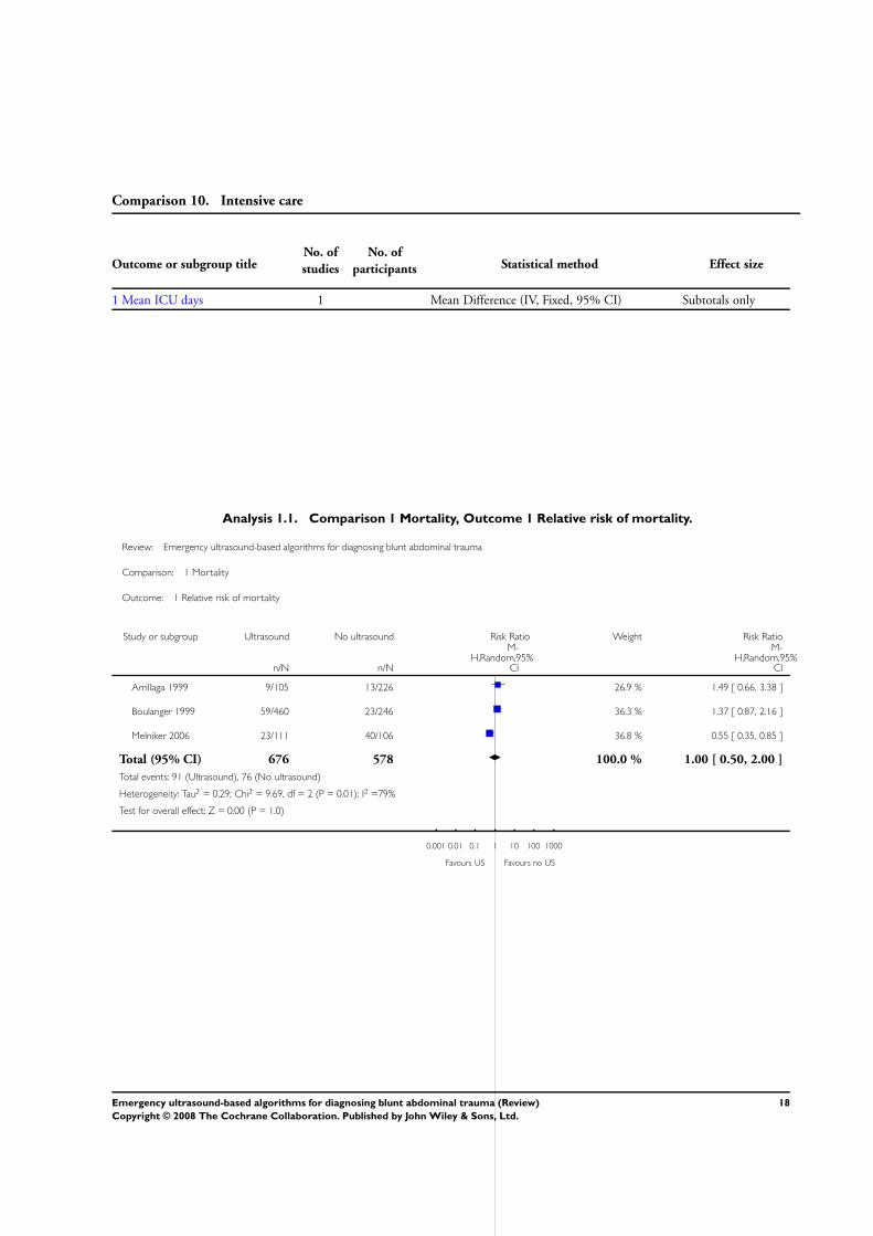
Mortality
Data were available from three studies (Arrillaga 1999; Boulanger
1999; Melniker 2006). There was no evidence of a difference in
mortality; random-effects RR = 1.00 (95% CI 0.50 to 2.00). No
data were provided on mortality attributable to abdominal injuries,
missed abdominal injuries or adverse events caused by any of the
diagnostic tests or negative laparotomy.
(The mortality outcome for Melniker 2006 also included compli-
cation rate, however the data were included since events such as
hemorrhagic shock, septic shock, and multisystem organ failure
are potentially life-threatening).
Use of computed tomography (CT) scans
Data were pooled from all four trials, showing significant hetero-
geneity (I2 = 98.4%). Ultrasound-based algorithms reduced or-
dering of CT scans by 50%; the random-effects RD = -0.52 (95%
CI -0.83 to -0.21).
Use of diagnostic peritoneal lavage (DPL)
Two studies (Arrillaga 1999; Boulanger 1999) reported data on
the use of DPL; ultrasound-based algorithms reduced the number
of DPL procedures by 6% (95% CI -0.11 to -0.02).
Cost-effectiveness analysis
Two studies that aimed to estimate costs exhibit inconclusive re-
sults.
In Boulanger 1999 the ultrasound pathway proved superior to the
control arm. We did not attempt to pool these results.
In Melniker 2006 mean hospital charges for the PLUS arm were
US$10,600 (interquartile range [IQR] US$5,700 to 19,000) and
US$16,400 (IQR US$6,700 to 43,600) for non-PLUS patients.
Laparotomy
Data from three studies were combined for this endpoint (
Boulanger 1999; Melniker 2006; Rose 2001). There was no ev-
idence of a difference in laparotomy rates with ultrasound-based
algorithms (fixed-effects, RD = 0.00, 95% CI -0.04 to 0.04).
Other secondary outcomes
We did not identify any RCTs or quasi-RCTs that explored the
impact of ultrasound-based clinical pathways on other health-re-
lated outcomes such as quality of life. In a quasi-RCT (Boulanger
1999) ultrasound reduced the mean time from arrival to hospital
to completion of the diagnostic algorithm from 151 minutes (95%
CI 127 to 174) to 53 minutes (95% CI 48 to 58). In this study
subjects undergoing ultrasound had a 60% reduced relative risk
of delayed recognition of intra-abdominal trauma (mainly small
bowel lacerations). Two non-therapeutic laparotomies were per-
formed in each group.
In Arrillaga 1999, another quasi-RCT, mean length of stay and
mean ICU days did not differ between groups. In this study, ul-
trasound significantly reduced the median disposition time from
80 minutes during weekdays, and 92 minutes during weekends,
to 20 minutes in both cases.
In the SOAP-trial (Melniker 2006), the time from ED arrival
to OR transfer was significantly shorter in the ultrasound-group
(median interval 60 [IQR 41 to 70] versus 157 [IQR 90 to 178]
minutes).
D I S C U S S I O N
Following early enthusiasm for the use of emergency ultrasound to
disclose abdominal injury after blunt trauma, there is an increas-
ing awareness of its limitations. There is no doubt that a positive
sonogram (either for free fluid or organ injury) proves the pres-
ence of intraabdominal damage. However, it is debatable whether
identifying injured patients is a significant problem for trained
emergency department teams. Given its poor overall sensitivity,
ultrasound cannot be used to rule out abdominal injury (Emery
2001; Miller 2003).
We have to admit that in the first published version of this re-
view, we had mistakenly shifted the denominators in the study
7Emergency ultrasound-based algorithms for diagnosing blunt abdominal trauma (Review)
Copyright © 2008 The Cochrane Collaboration. Published by John Wiley & Sons, Ltd.

published by Boulanger 1999. Also, the inclusion of the study
Navarrete-Nav. 1996 may have been not suitable because of a pri-
mary hypothesis which does not meet the question of interest.
However, after correcting for these mistakes, the key message of
this review remains not only similar, but may even be more alarm-
ing. It is troubling that an intervention regarded as a diagnostic
standard has been so poorly evaluated. It is open to debate whether
the reduction in CT scans is beneficial, or exposes patients with
blunt trauma to a higher risk.
The observed reduction in CT scans might, in part, reflect a false
sense of security; physicians are well advised to insist on admis-
sion and clinical monitoring, regardless of a negative sonogram.
There is some evidence that repeated examinations enhance ul-
trasound sensitivity (Nunes 2001). Although scientific data are
sparse, scheduled follow-up examinations have established them-
selves in clinical practice because of their feasibility. However, if
there is a high pre-test probability of abdominal injuries, contrast-
enhanced computed tomography still represents the diagnostic
modality of choice.
Ultrasound-based algorithms are often assumed to have merits in
shortening the primary trauma assessment, triaging patients more
precisely, avoiding unnecessary interventional procedures, and re-
ducing costs. However, such assumptions are hardly supported by
the available scientific data. Apart from a significant reduction in
the frequency of ordering CT scans, we found no beneficial ef-
fect of ultrasound on patient-centred endpoints. Divergent results
prevented pooling of data for most endpoints of interest.
Of note, two studies of higher methodological quality (Boulanger
1999; Rose 2001) showed only a marginal reduction in CT fre-
quency. Thus, it is open to debate whether abdominal ultrasound
measurably affects the doctor’s decision to order definitive diag-
nostic tests.
The meaning of the slightly increased relative risk of mortality
in the ultrasound arm of two quasi-randomised trials (Arrillaga
1999; Boulanger 1999) is not straightforward and susceptible to
residual confounding. Patients in this group might have been more
severely injured, haemodynamically unstable and considered un-
suitable for CT imaging more frequently. Although similar ISS
values were noted in both groups, no information was provided
on abbreviated injury scales (AIS) for abdominal damage. Thus,
imbalances between patient groups cannot be excluded.
A U T H O R S ’ C O N C L U S I O N S
Implications for practice
The current evidence from randomised trials focusing on patient-
centred outcomes, do not provide sufficient evidence to inform
policy on the use of ultrasound-based clinical pathways in the
initial diagnostic investigation of patients with blunt abdominal
trauma. Given the low sensitivity of ultrasound, clinical practice
guidelines must be scrutinised for the value of ultrasound exam-
inations within established trauma algorithms. Despite a lack of
diagnostic accuracy, the results of this review suggest minor effi-
ciency of ultrasonography in the trauma setting (that is, its impact
on clinical decision making, and anticipated patient benefits).
Implications for research
Given the biological plausibility of disclosing organ damage by ul-
trasound, there is still a need for high-quality randomised or clus-
ter-randomised trials to examine the efficacy of ultrasound-based
clinical pathways in diagnosing patients with suspected blunt ab-
dominal injury. Specifically, researchers must respect and report
demographic variability and follow-up policies.
A C K N O W L E D G E M E N T S
We thank Professor Bernard R Boulanger, University of Kentucky,
Lexington, Kentucky, USA and Dr O John Ma, Truman Medi-
cal Center, Kansas City, Missouri, USA for responding to our e-
mails and for their willingness to provide unpublished data. We
also thank Dr Steve Vance, Synergy Medical Education Alliance,
Michigan State University Emergency Medicine Residency, Sagi-
naw, MI, USA for his EB emergency medicine review and subse-
quent response to a critical letter.
8Emergency ultrasound-based algorithms for diagnosing blunt abdominal trauma (Review)
Copyright © 2008 The Cochrane Collaboration. Published by John Wiley & Sons, Ltd.

R E F E R E N C E S
References to studies included in this review
Arrillaga 1999 {published data only}∗ Arrillaga A, Graham R, York JW, Miller RS. Increased
efficiency and cost-effectiveness in the evaluation of the
blunt abdominal trauma patient with the use of ultrasound.
The American Surgeon 1999;65:31–5.
Boulanger 1999 {published data only}∗ Boulanger BR, McLellan BA, Brenneman FD, Ochoa J,
Kirkpatrick AW. Prospective evidence of the superiority of
a sonography-based algorithm in the assessment of blunt
abdominal injury. Journal of Trauma 1999;47:632–7.
Melniker 2006 {published data only}
Melniker L, Liebner E, Tiffany B, Lopez P, Quick G,
Sharma M, et al.Cost analysis of point-of-care, limited
ultrasonography (PLUS) in trauma patients: the sonography
outcomes assessment program (SOAP)-1 trial [Abstract].
Academic Emergency Medicine 2004;11:568.∗ Melniker LA, Leibner E, McKenney MG, Lopez P, Briggs
WM, Mancuso CA. Randomized controlled clinical trial of
point-of-care, limited ultrasonography for trauma in the
emergency department: the First Sonography Outcomes
Assessment Program Trial. Academic Emergency Medicine
2006;48:227–35.
Rose 2001 {published data only}
Hutson A. Prospective randomized trial of ED ultrasound in
blunt abdominal trauma. Does it really alter abdominal CT
utilization?. Society for Academic Emergency Medicine,
Boston, USA. 1999.∗ Rose JS, Levitt A, Porter J, Hutson A, Greenholtz J, Nobay
F, Hilty W. Does the presence of ultrasound really affect
computed tomographic scan use? A prospective randomized
trials of ultrasound in trauma. Journal of Trauma 2001;51:
545–50.
References to studies excluded from this review
Branney 1997 {published data only}
Branney SW, Moore EW, Cantrill SV, Burch JM, Terry SJ.
Ultrasound based key clinical pathway reduces the use of
hospital resources for the evaluation of blunt abdominal
trauma. Journal of Trauma 1997;42:1086–90.
Healey 1996 {published data only}
Healey MA, Simons RK, Winchell RJ, Gosink BB, Casola
G, Steele JT, Potenza BM, Hoyt DB. A prospective
evaluation of abdominal ultrasound in blunt abdominal
trauma: is it useful?. Journal of Trauma 1996;40:875–85.
Hesse 1999 {published data only}∗ Hesse S, Hörmann D, Klöppel R, Bennek J. [Einfluß der
Sonographie auf die Therapieentscheidung beim sumpfen
Bauchtrauma im Kindesalter]. Deutscher Röntgenkongress,
Wiesbaden. 1999.
Ma OJ {published data only (unpublished sought but not used)}∗ Ma OJ, Gaddis G, Steele MT, Cowan D, Kaltenbronn K.
Prospective Analysis of the Effect of Physician Experience
with the FAST Examination in Reducing Utilization of CT
Scans. Submitted for publication.
McKenney 2001 {published data only}
McKenney MG, McKenney KL, Hong JJ, Compton R,
Cohn SM, Kirton OC, Shatz DV, Sleeman D, Byers PM,
Ginzburg E, Augenstein J. Evaluating blunt abdominal
trauma with sonography: a cost analysis. American Surgeon
2001;67:930–4.
Navarrete-Nav. 1996 {published data only}∗ Navarrete-Navarro P, Vázquez G, Bosch JM, Fernández
E, Rivera R, Carazo E. Computed tomography vs clinical
and multidisciplinary procedures for early evaluation of
severe abdomen and chest trauma- a cost analysis approach.
Intensive Care Medicine 1996;22:208–12.
References to studies awaiting assessment
Ma 2001 {unpublished data only}
Ma OJ. Prospective analysis of the effect of physician
experience with the FAST exam in reducing utilization of
CT scans. Society for Academic Emergency Medicine,
Atlanta, USA. 2001.
Muniz 2003 {published data only (unpublished sought but not used)}
Muniz A. FAST vs FAST, AST, ALT and urinanalysis
in children with blunt abdominal trauma. Society for
American Emergency Medicine, Boston, USA. 2003.
Additional references
Amoroso 1999
Amoroso TA. Evaluation of the patient with blunt
abdominal trauma: an evidence-based approach. Emergency
Medicine Clinics of North America 1999;17(1):63–75.
Baka 2002
Baka AG, Delgado CA, Simon HK. Current use and
perceived utility of ultrasound for evaluation of pediatric
compared with adult trauma patients. Pediatric Emergency
Care 2002;18(3):163–7.
Bardenheuer 2000
Bardenheuer M, Obertacke U, Waydhas C, Nast-Kolb D.
Epidemiology of the severely injured patient. A prospective
assessment of preclinical and clinical management. AG
Polytrauma of DGU [Epidemiologie des Schwerverletzten.
Eine prospektive Erfassung der präklinischen und klinischen
Versorgung]. Unfallchirurg. 2000; Vol. 103, issue 5:
355–63.
Boulanger 2000
Boulanger BR, Kearney PA, Brenneman FD, Tsuei B,
Ochoa J. Utilization of FAST (focused assessment with
sonography for trauma) in 1999: results of a survey of
North American trauma centers. The American Surgeon
2000;66(11):1049–55.
EAST 2003
EAST Practice Management Guidelines Work Group.
Practice Management Guidelines for the evaluation of blunt
9Emergency ultrasound-based algorithms for diagnosing blunt abdominal trauma (Review)
Copyright © 2008 The Cochrane Collaboration. Published by John Wiley & Sons, Ltd.

abdominal trauma, 2001. Available at: http://www.east.org.
Last date of access: February 27, 2003.
Emery 2001
Emery KH, McAneney CM, Racadio JM, Johnson ND,
Evora DK, Garcia VF. Absent fluid on screening trauma
ultrasonography in children: a prospective comparison with
computed tomography. Journal of Pediatric Surgery 2001;
38:585–9.
Hodgson 2000a
Hodgson NF, Stewart TC, Girotti MJ. Autopsies and death
certification in deaths due to blunt trauma: what are we
missing?. Canadian Journal of Surgery 2000;43(2):130–6.
Hodgson 2000b
Hodgson NF, Stewart TC, Girotti MJ. Open or closed
diagnostic peritoneal lavage for abdominal trauma?. Journal
of Trauma 2000;6(48):1091–5.
Holm 1968
Holm HH, Mortensen. Ultrasonic scanning in diagnosis of
abdominal disease. Acta Chirurgica Scandinavica 1968;134
(5):333–41.
Jhirad 1998
Jhirad R, Boone D. Computed tomography for evaluating
blunt abdominal trauma in the low-volume nondesignated
trauma center: the procedure of choice?. Journal of Trauma
1998;45(1):64–8.
Jones 1983
Jones TK, Walsh JW, Maull KI. Diagnostic imaging in
blunt trauma of the abdomen. Surgery Gynecology and
Obstetrics 1983;157(4):389–98.
Linsenmaier 2002
Linsenmaier U, Krötz M, Häuser H, Rock C, Rieger J,
Bohndorf K, Pfeifer KJ, Reiser M. Whole-body computed
tomography in polytrauma: techniques and management.
European Radiology 2002;12(7):1728–40.
Livingston 1998
Livingston DH, Lavery RF, Passanante MR, Skurnick
JH, Fabian TC, Fry DE, Malangoni MA. Admission or
observation is not necessary after a negative computed
tomographic scan in patients with suspected blunt
abdominal trauma. Journal of Trauma 1998;44(2):273–82.
Miller 2003
Miller MT, Pasquale MD, Bromberg WJ, Wasser, Cox J.
Not so FAST. Journal of Trauma 2003;54(1):52–60.
Morrison 1996
Morrison JE, Wisner DH, Bodai BI. Complications after
negative laparotomy for trauma: long-term follow-up in a
health maintenance organization. Journal of Trauma 1996;
41(3):509–13.
Nunes 2001
Nunes LW, Simmons S, Hallowell MJ, Kinback R, Trooskin
S, Kozar R. Diagnostic performance of trauma US in
identifying abdominal or pelvic free fluid and serious
abdominal or pelvic injury. Academic Radiology 2001;8:
128–36.
Pirente 2002
Pirente N, Bouillon B, Schäfer B, Raum M, Helling HJ,
Berger E, Neugebauer E. Systematic development of a scale
for determination of health-related quality of life in multiple
trauma patients. The Polytrauma Outcome (POLO) Chart
[Systematische Entwicklung eines Messinstruments zur
Erfassung der gesundheitsbezogenen Lebensqualität beim
polytraumatisierten Patienten: Die Polytrauma–Outcome–
(POLO–)Chart]. Unfallchirurg 2002;105(5):413–22.
Prall 1994
Prall JA, Nichols JS, Brennan R, Moore EE. Early
definitive abdominal evaluation in the triage of unconscious
normotensive blunt trauma patients. Journal of Trauma
1994;37(5):792–7.
Rademacher 2001
Rademacher G, Stengel D, Siegmann S, Petersein J, Mutze
S. Optimization of contrast agent volume for helical CT
in the diagnostic assessment of patients with severe and
multiple injuries. Journal of Computer Assisted Tomography
2001;26(1):113–8.
Ruchholtz 2002
Ruchholtz S, Waydhas C, Schroeder T, Piepenbrink K, Kuhl
H, Nast-Kolb D. The value of computed tomography in
the early treatment of seriously injured patients [Stellenwert
der Computertomographie in der frühen klinischen
Behandlung schwer verletzter Patienten]. Chirurgie 2002;
73(10):1005–12.
Scalea 1999
Scalea TM, Rodriguez A, Chiu WC, Brenneman FD, Fallon
WF, Kato K, McKenney MG, Nerlich ML, Ochsner MG,
Yoshii H. Focused assessment with sonography for trauma
(FAST): results from an international consensus conference.
Journal of Trauma 1999;46(3):466–72.
Scheck 1998
Scheck RJ, Coppenrath EM, Kellner MW, Lehmann
KJ, Rock C, Rieger J, Rothmeier L, Schweden F, Bauml
AA, Hahn K. Radiation dose and image quality in spiral
computed tomography: multicentre evaluation at six
institutions. British Journal of Radiology 1998;71(847):
734–44.
Stalp 2002
Stalp M, Koch C, Ruchholtz S, Regel G, Panzica M, Krettek
C, Pape HC. Standardized outcome evaluation after blunt
multiple injuries by scoring systems: a clinical follow-up
investigation 2 years after injury. Journal of Trauma 2002;
52(6):1160–8.
Stengel 2001
Stengel D, Bauwens K, Sehouli J, Porzsolt F, Rademacher
G, Mutze S, Ekkernkamp A. Systematic review and meta-
analysis of emergency ultrasonography for blunt abdominal
trauma. British Journal of Surgery 2001;88(7):901–12.
Stengel 2003
Stengel D, Bauwens K, Porzsolt F, Rademacher G,
Mutze S, Ekkernkamp A. Emergency ultrasonography
for blunt abdominal trauma. Meta-analysis update 2003
10Emergency ultrasound-based algorithms for diagnosing blunt abdominal trauma (Review)
Copyright © 2008 The Cochrane Collaboration. Published by John Wiley & Sons, Ltd.

[Sonografische Diagnostik im Schockraum bei stumpfem
Bauchtrauma. Meta–Analyse Update 2003]. Zentralbl
Chirugerie 2003;128:1027–37.
Sydney report 2003
South Western Sydney Regional Trauma Registry Report
1995 -1999. Available at: http://www.swsahs.nsw.gov.au/
livtrauma/reg stat/default.asp Last date of access: February
27, 2003.
Vance 2007
Vance S. Evidence-based emergency medicine/systematic
review abstract. The FAST scan: are we improving care of
the trauma patient?. Annals of Emergency Medicine 2007;49:
364–6.
Wintermark 2002
Wintermark M, Poletti PA, Becker CD, Schnyder P.
Traumatic injuries: organization and ergonomics of imaging
in the emergency environment. European Radiology 2002;
44(5):273–82.
Yoshii 1998
Yoshii H, Sato M, Yamamoto S, Motegi M, Okusawa S,
Kitano M, Nagashima A, Doi M, Takuma K, Kato K,
Aikawa N. Usefulness and limitations of ultrasonography in
the initial evaluation of blunt abdominal trauma. Journal of
Trauma 1998;45(1):45–50.∗ Indicates the major publication for the study
11Emergency ultrasound-based algorithms for diagnosing blunt abdominal trauma (Review)
Copyright © 2008 The Cochrane Collaboration. Published by John Wiley & Sons, Ltd.

C H A R A C T E R I S T I C S O F S T U D I E S
Characteristics of included studies [ordered by study ID]
Arrillaga 1999
Methods Quasi-RCT (algorithm used was based on the daytime and weekday availability of ultrasound). Location:
Community Hospital, Level-I-Trauma Center, South Carolina, USA. Recruitment period: 9 months.
Adequacy of concealment: 0.
Intent-to-treat: 0
Blinding: 0
Comparability of treatment groups at entry: 1
Comparability of care programmes: 0
Definition of inclusion and exclusion criteria: 1
Description of interventions: 1
Definition of outcomes: 2
Duration of surveillance: 0 (not defined)
Participants Inclusion criteria: consecutive patients with suspected blunt abdominal trauma (not specified). 331 en-
rolled (US 105, no US 226). US group: mean age 38.1 (SD 22.7) years, mean ISS 13.0 (SD 11.6), 62%
males. No US group: mean age 33.6 (SD 18.6) years, mean ISS 13.4 (SD 9.7), 69% males
Interventions a. Clinical examination, focused ultrasound for free fluid, further management depended on sonograms
and hemodynamical stability
b. Clinical examination, CT in stable and DPL in unstable subjects
Outcomes 1. Number of diagnostic tests (CT, DPL).
2. Mortality.
3. Morbidity (not specified).
4. Length of stay.
5. Diagnostic accuracy.
6. Total costs.
Notes
Risk of bias
Item Authors’ judgement Description
Allocation concealment? Unclear D - Not used
12Emergency ultrasound-based algorithms for diagnosing blunt abdominal trauma (Review)
Copyright © 2008 The Cochrane Collaboration. Published by John Wiley & Sons, Ltd.

Boulanger 1999
Methods Quasi-RCT (algorithm used was determined by date of admission). Location: University Hospital, Ken-
tucky, USA. Recruitment period: October 1995 to August 1997.
Adequacy of concealment: 0.
Intent-to-treat: 1.
Blinding: 0.
Comparability of treatment groups at entry: 2.
Comparability of care programmes: 0.
Definition of inclusion and exclusion criteria: 2.
Description of interventions: 2.
Definition of outcomes: 2.
Duration of surveillance: 1.
Participants Inclusion criteria: victims of blunt trauma, older than 16 years of age, resuscitated by trauma service, no
clinical indication for laparotomy, unreliable or equivocal abdominal examination. 706 enrolled (US 460,
no US 246).
US group: mean age 38.4 (SD 17.6) years, mean ISS 23.3 (SD 12.8), 73% males. No US group: mean
age 40.2 (SD 18.2) years, mean ISS 22.8 (SD 11.3), 73% males
Interventions a. Clinical examination, focused ultrasound for free fluid, further management depended on sonograms
and hemodynamical stability.
b. Clinical examination, CT in stable and DPL in unstable participants.
Outcomes 1. Time from arrival to the completion of diagnostic algorithm.
2. Number of diagnostic tests (CT, DPL).
3. Mortality.
4. Laparotomy rates.
5. Diagnostic accuracy and number of significant injuries.
6. Total costs.
Notes
Risk of bias
Item Authors’ judgement Description
Allocation concealment? Unclear D - Not used
Melniker 2006
Methods RCT: Location - three level-1 trauma centers, New York Methodist Hospital, Maricopa Hospital, Phoenix,
Jackson Memorial Hospital, Miami, USA
Participants Inclusion criteria: patients presenting with any one of a mechanism of injury (energy reportedly delivered
to the torso), symptomatology (complaint of chest, abdominal, or pelvic pain), or physical findings (chest,
abdominal, or pelvic tenderness) suspicious of torso trauma
Exclusion criteria:
Patients or patient proxies who were unable to provide consent and those requiring immediate transfer to
the operating suite were excluded
13Emergency ultrasound-based algorithms for diagnosing blunt abdominal trauma (Review)
Copyright © 2008 The Cochrane Collaboration. Published by John Wiley & Sons, Ltd.

Melniker 2006 (Continued)
Interventions a. Diagnostic interventions that the initial evaluating physician, under ordinary circumstances, would use
to evaluate torso trauma patients plus 4-view FAST assessment
b. Ordinary diagnostic interventions to evaluate torso trauma
Outcomes 1. Time from ED arrival to direct transfer to operative care in minutes (sample size calculations: 40%
reduction, 90% power, alpha 5%).
2. Use of CT of the torso
3. Hospital length of stay in days
4. Composite complications (rate of hemorrhagic shock, septic shock, multisystem organ failure, or death)
based on CPT or ICD codes found in the medical record
5. Total charges in 2003 US dollars
Notes Of 525 patients screened, 81 went directly to OR, 136 lacked consent, 262 were randomized, and 217
were analyzed
Risk of bias
Item Authors’ judgement Description
Allocation concealment? Yes A - Adequate
Rose 2001
Methods RCT. Location: University Hospital, California, USA. Recruitment period: November 1997 to November
1998.
Adequacy of concealment: 1.
Intent-to-treat: 1.
Blinding: 0.
Comparability of treatment groups at entry: 2.
Comparability of care programmes: 1.
Definition of inclusion and exclusion criteria: 2.
Description of interventions: 2.
Definition of outcomes: 2.
Duration of surveillance: 1.
Participants Inclusion criteria: patients 18 to 75 years old meeting critical trauma triage criteria after blunt injury,
defined by the American College of Surgeons Subcommittee of trauma. 212 randomised (US 105, no US
107), 208 analysed (4 dropped because of incomplete data). US group: mean age 40.0 (SD 19.5) years,
mean ISS 9.9 (SD 12.4), 61% males. No US group: mean age 39.0 (SD 16.8) years, mean ISS 9.8 (SD
8.8), 63% males
Interventions a. Standard standard trauma management plus focused ultrasound for free fluid (none, small, moderate,
large) with 15 minutes of arrival by experienced doctors.
b. Standard trauma management.
Outcomes 1. Difference in abdominal CT scan use (sample size calculations: 20% difference, 80% power, two-tailed
alpha 5%).
2. 30-minute difference in time to laparotomy.
14Emergency ultrasound-based algorithms for diagnosing blunt abdominal trauma (Review)
Copyright © 2008 The Cochrane Collaboration. Published by John Wiley & Sons, Ltd.

Rose 2001 (Continued)
Notes Trial was stopped at 215 participants because US was recognised as standard practice and did not allow
for further patient recruitment
Risk of bias
Item Authors’ judgement Description
Allocation concealment? Unclear B - Unclear
Characteristics of excluded studies [ordered by study ID]
Study Reason for exclusion
Branney 1997 Comparison of prospectively collected ultrasound data (August 1995 to October 1995) with a historical
cohort admitted before instituting ultrasound-based clinical pathways (August 1994 to October 1994)
Healey 1996 Comparison of prospectively collected ultrasound data (May 1994 to August 1995) with a historical cohort
admitted before instituting ultrasound-based clinical pathways
Hesse 1999 Comparison of prospectively collected ultrasound data (1990 to 1994) with a historical cohort admitted
before instituting ultrasound-based clinical pathways (1986 to 1990)
Ma OJ Comparison of ultrasound accuracy and the request of CT scans among physicians with minor, moderate
and high skills in performing FAST
McKenney 2001 Comparison of prospectively collected ultrasound data (January 1995 to June 1995) with a historical cohort
admitted before instituting ultrasound-based clinical pathways (January 1993 to June 1993)
Navarrete-Nav. 1996 Trial intended to prove the superiority of computed tomography over multiple diagnostic interventions
including ultrasound
15Emergency ultrasound-based algorithms for diagnosing blunt abdominal trauma (Review)
Copyright © 2008 The Cochrane Collaboration. Published by John Wiley & Sons, Ltd.

D A T A A N D A N A L Y S E S
Comparison 1. Mortality
Outcome or subgroup titleNo. of
studies
No. of
participants Statistical method Effect size
1 Relative risk of mortality 3 1254 Risk Ratio (M-H, Random, 95% CI) 1.00 [0.50, 2.00]
Comparison 2. Use of computed tomography (CT)
Outcome or subgroup titleNo. of
studies
No. of
participants Statistical method Effect size
1 Difference in CT frequency 4 1462 Risk Difference (M-H, Random, 95% CI) -0.52 [-0.83, -0.21]
Comparison 3. Use of diagnostic peritoneal lavage (DPL)
Outcome or subgroup titleNo. of
studies
No. of
participants Statistical method Effect size
1 Difference in DPL frequency 2 1016 Risk Difference (M-H, Random, 95% CI) -0.06 [-0.11, -0.02]
Comparison 4. Cost-effectiveness
Outcome or subgroup titleNo. of
studies
No. of
participants Statistical method Effect size
1 Direct costs per patient (US$) 1 Mean Difference (IV, Random, 95% CI) Subtotals only
16Emergency ultrasound-based algorithms for diagnosing blunt abdominal trauma (Review)
Copyright © 2008 The Cochrane Collaboration. Published by John Wiley & Sons, Ltd.

Comparison 5. Laparotomy
Outcome or subgroup titleNo. of
studies
No. of
participants Statistical method Effect size
1 Laparotomy rate 3 1131 Risk Difference (M-H, Fixed, 95% CI) Not estimable
Comparison 6. Reduction in diagnostic time
Outcome or subgroup titleNo. of
studies
No. of
participants Statistical method Effect size
1 Mean reduction in diagnostic
time (minutes)
1 Mean Difference (IV, Fixed, 95% CI) Subtotals only
Comparison 7. Delayed diagnoses
Outcome or subgroup titleNo. of
studies
No. of
participants Statistical method Effect size
1 Risk of delayed diagnosis 1 Risk Ratio (M-H, Fixed, 95% CI) Subtotals only
Comparison 8. Non-therapeutic laparotomy
Outcome or subgroup titleNo. of
studies
No. of
participants Statistical method Effect size
1 Risk of non-therapeutic
laparotomy
1 Risk Ratio (M-H, Fixed, 95% CI) Subtotals only
Comparison 9. Duration of hospital stay
Outcome or subgroup titleNo. of
studies
No. of
participants Statistical method Effect size
1 Mean length of stay (days) 1 Mean Difference (IV, Fixed, 95% CI) Subtotals only
17Emergency ultrasound-based algorithms for diagnosing blunt abdominal trauma (Review)
Copyright © 2008 The Cochrane Collaboration. Published by John Wiley & Sons, Ltd.
Comparison 10. Intensive care
Outcome or subgroup titleNo. of
studies
No. of
participants Statistical method Effect size
1 Mean ICU days 1 Mean Difference (IV, Fixed, 95% CI) Subtotals only
Analysis 1.1. Comparison 1 Mortality, Outcome 1 Relative risk of mortality.
Review: Emergency ultrasound-based algorithms for diagnosing blunt abdominal trauma
Comparison: 1 Mortality
Outcome: 1 Relative risk of mortality
Study or subgroup Ultrasound No ultrasound Risk Ratio Weight Risk Ratio
n/N n/N
M-H,Random,95%
CI
M-H,Random,95%
CI
Arrillaga 1999 9/105 13/226 26.9 % 1.49 [ 0.66, 3.38 ]
Boulanger 1999 59/460 23/246 36.3 % 1.37 [ 0.87, 2.16 ]
Melniker 2006 23/111 40/106 36.8 % 0.55 [ 0.35, 0.85 ]
Total (95% CI) 676 578 100.0 % 1.00 [ 0.50, 2.00 ]
Total events: 91 (Ultrasound), 76 (No ultrasound)
Heterogeneity: Tau2 = 0.29; Chi2 = 9.69, df = 2 (P = 0.01); I2 =79%
Test for overall effect: Z = 0.00 (P = 1.0)
0.001 0.01 0.1 1 10 100 1000
Favours US Favours no US
18Emergency ultrasound-based algorithms for diagnosing blunt abdominal trauma (Review)
Copyright © 2008 The Cochrane Collaboration. Published by John Wiley & Sons, Ltd.

Analysis 2.1. Comparison 2 Use of computed tomography (CT), Outcome 1 Difference in CT frequency.
Review: Emergency ultrasound-based algorithms for diagnosing blunt abdominal trauma
Comparison: 2 Use of computed tomography (CT)
Outcome: 1 Difference in CT frequency
Study or subgroup Ultrasound No ultrasoundRisk
Difference WeightRisk
Difference
n/N n/N
M-H,Random,95%
CI
M-H,Random,95%
CI
Arrillaga 1999 9/105 223/226 25.4 % -0.90 [ -0.96, -0.85 ]
Boulanger 1999 111/460 225/246 25.4 % -0.67 [ -0.73, -0.62 ]
Melniker 2006 53/111 85/106 24.7 % -0.32 [ -0.44, -0.20 ]
Rose 2001 37/104 54/104 24.5 % -0.16 [ -0.30, -0.03 ]
Total (95% CI) 780 682 100.0 % -0.52 [ -0.83, -0.21 ]
Total events: 210 (Ultrasound), 587 (No ultrasound)
Heterogeneity: Tau2 = 0.10; Chi2 = 190.20, df = 3 (P<0.00001); I2 =98%
Test for overall effect: Z = 3.33 (P = 0.00086)
-1 -0.5 0 0.5 1
Favours US Favours no US
Analysis 3.1. Comparison 3 Use of diagnostic peritoneal lavage (DPL), Outcome 1 Difference in DPL
frequency.
Review: Emergency ultrasound-based algorithms for diagnosing blunt abdominal trauma
Comparison: 3 Use of diagnostic peritoneal lavage (DPL)
Outcome: 1 Difference in DPL frequency
Study or subgroup Ultrasound No ultrasoundRisk
Difference WeightRisk
Difference
n/N n/N
M-H,Random,95%
CI
M-H,Random,95%
CI
Arrillaga 1999 3/105 15/226 46.7 % -0.04 [ -0.08, 0.01 ]
Boulanger 1999 5/460 21/225 53.3 % -0.08 [ -0.12, -0.04 ]
Total (95% CI) 565 451 100.0 % -0.06 [ -0.11, -0.02 ]
Total events: 8 (Ultrasound), 36 (No ultrasound)
Heterogeneity: Tau2 = 0.00; Chi2 = 2.22, df = 1 (P = 0.14); I2 =55%
Test for overall effect: Z = 2.71 (P = 0.0068)
-1 -0.5 0 0.5 1
Favours US Favours no US
19Emergency ultrasound-based algorithms for diagnosing blunt abdominal trauma (Review)
Copyright © 2008 The Cochrane Collaboration. Published by John Wiley & Sons, Ltd.

Analysis 4.1. Comparison 4 Cost-effectiveness, Outcome 1 Direct costs per patient (US$).
Review: Emergency ultrasound-based algorithms for diagnosing blunt abdominal trauma
Comparison: 4 Cost-effectiveness
Outcome: 1 Direct costs per patient (US$)
Study or subgroup Ultrasound No ultrasoundMean
DifferenceMean
Difference
N Mean(SD) N Mean(SD) IV,Random,95% CI IV,Random,95% CI
Boulanger 1999 460 156 (244) 246 540 (126) -384.00 [ -411.30, -356.70 ]
Subtotal (95% CI) 0 0 0.0 [ 0.0, 0.0 ]
Heterogeneity: not applicable
Test for overall effect: Z = 0.0 (P < 0.00001)
-1000 -500 0 500 1000
Favours US Favours no US
Analysis 5.1. Comparison 5 Laparotomy, Outcome 1 Laparotomy rate.
Review: Emergency ultrasound-based algorithms for diagnosing blunt abdominal trauma
Comparison: 5 Laparotomy
Outcome: 1 Laparotomy rate
Study or subgroup Ultrasound No ultrasoundRisk
Difference WeightRisk
Difference
n/N n/N M-H,Fixed,95% CI M-H,Fixed,95% CI
Boulanger 1999 35/246 62/460 60.1 % 0.01 [ -0.05, 0.06 ]
Melniker 2006 29/111 34/106 20.3 % -0.06 [ -0.18, 0.06 ]
Rose 2001 12/104 8/104 19.5 % 0.04 [ -0.04, 0.12 ]
Total (95% CI) 461 670 100.0 % 0.00 [ -0.04, 0.04 ]
Total events: 76 (Ultrasound), 104 (No ultrasound)
Heterogeneity: Chi2 = 1.90, df = 2 (P = 0.39); I2 =0.0%
Test for overall effect: Z = 0.00 (P = 1.0)
-1 -0.5 0 0.5 1
Favours treatment Favours control
20Emergency ultrasound-based algorithms for diagnosing blunt abdominal trauma (Review)
Copyright © 2008 The Cochrane Collaboration. Published by John Wiley & Sons, Ltd.
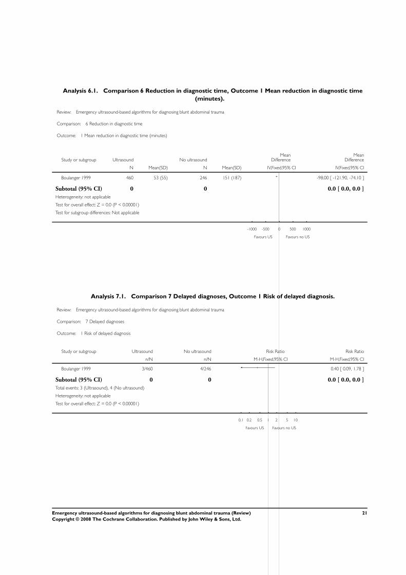
Analysis 6.1. Comparison 6 Reduction in diagnostic time, Outcome 1 Mean reduction in diagnostic time
(minutes).
Review: Emergency ultrasound-based algorithms for diagnosing blunt abdominal trauma
Comparison: 6 Reduction in diagnostic time
Outcome: 1 Mean reduction in diagnostic time (minutes)
Study or subgroup Ultrasound No ultrasoundMean
DifferenceMean
Difference
N Mean(SD) N Mean(SD) IV,Fixed,95% CI IV,Fixed,95% CI
Boulanger 1999 460 53 (55) 246 151 (187) -98.00 [ -121.90, -74.10 ]
Subtotal (95% CI) 0 0 0.0 [ 0.0, 0.0 ]
Heterogeneity: not applicable
Test for overall effect: Z = 0.0 (P < 0.00001)
Test for subgroup differences: Not applicable
-1000 -500 0 500 1000
Favours US Favours no US
Analysis 7.1. Comparison 7 Delayed diagnoses, Outcome 1 Risk of delayed diagnosis.
Review: Emergency ultrasound-based algorithms for diagnosing blunt abdominal trauma
Comparison: 7 Delayed diagnoses
Outcome: 1 Risk of delayed diagnosis
Study or subgroup Ultrasound No ultrasound Risk Ratio Risk Ratio
n/N n/N M-H,Fixed,95% CI M-H,Fixed,95% CI
Boulanger 1999 3/460 4/246 0.40 [ 0.09, 1.78 ]
Subtotal (95% CI) 0 0 0.0 [ 0.0, 0.0 ]
Total events: 3 (Ultrasound), 4 (No ultrasound)
Heterogeneity: not applicable
Test for overall effect: Z = 0.0 (P < 0.00001)
0.1 0.2 0.5 1 2 5 10
Favours US Favours no US
21Emergency ultrasound-based algorithms for diagnosing blunt abdominal trauma (Review)
Copyright © 2008 The Cochrane Collaboration. Published by John Wiley & Sons, Ltd.

Analysis 8.1. Comparison 8 Non-therapeutic laparotomy, Outcome 1 Risk of non-therapeutic laparotomy.
Review: Emergency ultrasound-based algorithms for diagnosing blunt abdominal trauma
Comparison: 8 Non-therapeutic laparotomy
Outcome: 1 Risk of non-therapeutic laparotomy
Study or subgroup Ultrasound No ultrasound Risk Ratio Risk Ratio
n/N n/N M-H,Fixed,95% CI M-H,Fixed,95% CI
Boulanger 1999 1/460 1/246 0.53 [ 0.03, 8.51 ]
Subtotal (95% CI) 0 0 0.0 [ 0.0, 0.0 ]
Total events: 1 (Ultrasound), 1 (No ultrasound)
Heterogeneity: not applicable
Test for overall effect: Z = 0.0 (P < 0.00001)
0.1 0.2 0.5 1 2 5 10
Favours US Favours no US
Analysis 9.1. Comparison 9 Duration of hospital stay, Outcome 1 Mean length of stay (days).
Review: Emergency ultrasound-based algorithms for diagnosing blunt abdominal trauma
Comparison: 9 Duration of hospital stay
Outcome: 1 Mean length of stay (days)
Study or subgroup Ultrasound No ultrasoundMean
DifferenceMean
Difference
N Mean(SD) N Mean(SD) IV,Fixed,95% CI IV,Fixed,95% CI
Arrillaga 1999 105 7.2 (9) 226 7.2 (10.1) 0.0 [ -2.17, 2.17 ]
Subtotal (95% CI) 0 0 0.0 [ 0.0, 0.0 ]
Heterogeneity: not applicable
Test for overall effect: Z = 0.0 (P < 0.00001)
Test for subgroup differences: Not applicable
-10 -5 0 5 10
Favours US Favours no US
22Emergency ultrasound-based algorithms for diagnosing blunt abdominal trauma (Review)
Copyright © 2008 The Cochrane Collaboration. Published by John Wiley & Sons, Ltd.

Analysis 10.1. Comparison 10 Intensive care, Outcome 1 Mean ICU days.
Review: Emergency ultrasound-based algorithms for diagnosing blunt abdominal trauma
Comparison: 10 Intensive care
Outcome: 1 Mean ICU days
Study or subgroup Ultrasound No ultrasoundMean
DifferenceMean
Difference
N Mean(SD) N Mean(SD) IV,Fixed,95% CI IV,Fixed,95% CI
Arrillaga 1999 105 3.1 (7.1) 226 2.5 (5.1) 0.60 [ -0.91, 2.11 ]
Subtotal (95% CI) 0 0 0.0 [ 0.0, 0.0 ]
Heterogeneity: not applicable
Test for overall effect: Z = 0.0 (P < 0.00001)
Test for subgroup differences: Not applicable
-10 -5 0 5 10
Favours US Favours no US
A P P E N D I C E S
Appendix 1. Detailed search strategies
MEDLINE 1966 to January 2008
1 abdominal injuries
2 thoracic injuries
3 wounds, nonpenetrating
4 multiple trauma OR polytrauma
5 retroperitoneum
6 rupture
7 shock, traumatic
8 hemoperitoneum OR haemoperitoneum OR free fluid OR intraperitoneal fluid
9 spleen OR splenic
10 liver OR hepatic
11 accidents
12 accidents, traffic
13 seat belts
14 bicycling
15 motorcycles
16 ultras* OR echotomogr* OR sonogr*
17 focused assessment of sonography for trauma OR FAST OR emergency ultras*
18 (1 OR 2 OR 3 OR 4 OR 5 OR 6 OR 7 OR 8 OR 9 OR 10 OR 11 OR 12 OR 13 OR 14 OR 15) AND (16 OR 17)
19 randomised controlled trial OR randomized controlled trial
20 random allocation
21 double blind method
22 single blind method
23 (19 OR 20 OR 21 OR 22)
23Emergency ultrasound-based algorithms for diagnosing blunt abdominal trauma (Review)
Copyright © 2008 The Cochrane Collaboration. Published by John Wiley & Sons, Ltd.

24 18 AND 23
EMBASE 1980 to January 2008
1 ’intermethod comparison’/exp
2 ’randomized controlled trial’/exp
3 ’non invasive measurement’/exp
4 1 OR 2 OR 3
5 ’peritoneal fluid’/exp
6 ’hemoperitoneum’/exp
7 ’spleen rupture’/exp
8 ’spleen injury’/exp
9 ’liver injury’/exp
10 ’multiple trauma’/exp
11 ’abdominal blunt trauma’/exp
12 ’abdominal bleeding’/exp
13 5 OR 6 OR 7 OR 8 OR 9 OR 10 OR 11 OR 12
14 ’peritoneum lavage’/exp
15 ’clinical observation’/exp
16 ’spiral computer assisted tomography’/exp
17 ’diagnostic approach route’/exp
18 14 OR 15 OR 16 OR 17
19 ’echography’/exp
20 ’ultrasound scanner’/exp
21 ’ultrasound transducer’/exp
22 19 OR 20 OR 21
23 4 AND 13 AND 18 AND 22
W H A T ’ S N E W
Last assessed as up-to-date: 18 February 2008.
Date Event Description
11 February 2008 New search has been performed Review updated. New studies found and included/excluded.
H I S T O R Y
Protocol first published: Issue 4, 2003
Review first published: Issue 2, 2005
Date Event Description
12 November 2007 Amended Review amended. A shift in denominators was cor-
rected based on the addition of one study (Boulanger
1999), another study was dropped from the analysis
(Navarrete-Nav 1996), the results of a recent full-text
24Emergency ultrasound-based algorithms for diagnosing blunt abdominal trauma (Review)
Copyright © 2008 The Cochrane Collaboration. Published by John Wiley & Sons, Ltd.

(Continued)
publication of a former conference abstract (Melniker
2006) were included
9 February 2005 New citation required and conclusions have changed Review first published.
C O N T R I B U T I O N S O F A U T H O R S
Dirk Stengel was the principal investigator of this study, identified relevant literature, extracted and summarised data, and wrote the
manuscript.
Kai Bauwens, Jalid Sehouli, and Franz Porzsolt assisted in literature retrieval and data extraction. Kai Bauwens co-reviewed eligible
studies for methodological quality.
Grit Rademacher, Sven Mutze, and Axel Ekkernkamp discussed core ideas, and contributed to data interpretation.
All authors critically appraised the final version of this review.
D E C L A R A T I O N S O F I N T E R E S T
None known.
S O U R C E S O F S U P P O R T
Internal sources
• Department of Trauma and Orthopaedic Surgery at the Unfallkrankenhaus, Berlin, Germany.
External sources
• No sources of support supplied
I N D E X T E R M S
Medical Subject Headings (MeSH)
∗Algorithms; Abdominal Injuries [∗ultrasonography]; Emergencies; Randomized Controlled Trials as Topic; Wounds, Nonpenetrating
[∗ultrasonography]
25Emergency ultrasound-based algorithms for diagnosing blunt abdominal trauma (Review)
Copyright © 2008 The Cochrane Collaboration. Published by John Wiley & Sons, Ltd.

MeSH check words
Humans
26Emergency ultrasound-based algorithms for diagnosing blunt abdominal trauma (Review)
Copyright © 2008 The Cochrane Collaboration. Published by John Wiley & Sons, Ltd.