Emergency Quiz Cases
83
Emergency Radiology Quiz Cases Dr Eric Heffernan St Vincent’s University Hospital
-
Upload
ejheffernan -
Category
Health & Medicine
-
view
134 -
download
1
Transcript of Emergency Quiz Cases

Emergency Radiology Quiz Cases
Dr Eric HeffernanSt Vincent’s University Hospital

Emergency Cases
• Here are twenty-one cases that were referred for imaging from the Emergency Department in SVUH
• Have a look at the images on the first slide in each case
• On the next slide are clinical details and some questions for you to consider
• The answers are on the third slide for each case, followed by a slide with the same images labelled

Case 1

Case 1
• 44 year old man with dyspnoea, cough and mild pyrexia
1. How would you describe the CXR?2. What is your diagnosis?3. What part of the lung is involved and why?4. What follow-up would you suggest, if any?

Case 1 - Answers
1. The PA CXR shows increased airspace opacification in the right lower zone. The normal silhouette of the right heart border is obscured. On the lateral view the opacity is wedge-shaped and is located anteriorly.
2. Lobar pneumonia.3. Right middle lobe – the consolidation obscures the right heart
border and on the lateral view it outlines the horizontal and oblique fissures (see next slide). Note also that the horizontal fissure has been pulled inferiorly, indicating that there is a degree of collapse as well as consolidation.
4. The patient should have a follow-up CXR after completing their antibiotic therapy to ensure complete resolution of the abnormality – occasionally there will be an underlying neoplasm.

Right heart border isobscured, but right hemidiaphragm silhouette is preserved
Right oblique fissure
Horizontal fissure

Case 2

Case 2
• 64 year old man presented with pleuritic chest pain one month post coronary artery bypass. Mild pyrexia and leukocytosis.
1. How would you describe the CXR?2. What is your diagnosis?3. What other radiographic abnormality can
potentially be found in this condition?

Case 2 - answers
1. PA CXR shows a well-defined opacity at the left lung base that obliterates the silhouette of the left hemidiaphragm and curves superiorly at its lateral aspect, i.e. a meniscus sign indicating a pleural effusion. Sternotomy wires are demonstrated and the heart is mildly enlarged.
2. Given the recent history of cardiac surgery, Dressler’s syndrome was diagnosed.
3. Pericardial effusions are common in Dressler’s syndrome but may not be large enough to be radiographically apparent – if old chest radiographs are available they may allow us to detect new diffuse enlargement of the cardiac shadow due to pericardial effusion.

Case 3

Case 3
• Query fracture.
1. What is your diagnosis?

Case 3
• Have you spotted the abnormality yet?• If not, try looking again with these clinical
details: fell earlier today and is complaining of pain around the second MCP joint, with difficulty moving his second finger.
1. What is your diagnosis?

Case 3 - answer
1. Intra-articular fracture of the base of the second proximal phalanx (see next slide).
Some of you probably spotted the fracture the first time round anyway, but the purpose of this case is to illustrate how important ‘localizing information’ can be in the setting of trauma. We frequently get referrals for ‘hand (or foot) x-ray, ?#’. Without knowing where the problem is it can be very difficult for a Radiologist to see fractures that are obvious to someone who knows exactly where to look.


Case 4

Case 4
• 40 year old man with severe pleuritic chest pain. Currently undergoing IV antibiotic therapy for pneumonia.
1. How would you describe the abnormalities on the CXR?
2. What is your diagnosis?3. Give five common causes of this diagnosis.4. How is the patient’s IV antibiotic therapy being
administered?

Case 4 - answers1. There is increased lucency of the right hemithorax. Pleural lines are visible
and the lung markings do not extend all the way to the periphery. There is an effusion at the base of the right hemithorax however it does not show a meniscus sign (it does not curve upward at the costophrenic margin).
2. Right hydropneumothorax. The absence of a meniscus sign in the effusion is very helpful in some cases where the pneumothorax is small and subtle – the meniscus sign only occurs when the lung is fully inflated and extends all the way to the chest wall, so whenever you see a pleural effusion with a perfectly straight line look very carefully for a pneumothorax.
3. Causes of spontaneous (non-traumatic) pneumothorax: primary (typically tall young males), COPD, asthma, CF, pneumonia (uncommon, but possibly the cause in this case), α-1-antitrypsin deficiency, Marfan syndrome.
4. There is a right-sided PICC line which was inserted so that the patient could undergo home IV antibiotic therapy

Case 5

Case 5
• 21 year old man presented with shoulder pain after falling off his bike.
1. What is your diagnosis?2. What other types of dislocation (a clue to #1,
above!) can occur in the shoulder?

Case 5 - answer
1. Acromioclavicular dislocation. This can be subtle, as in this example – the key to the diagnosis is knowing that the undersurface of the distal clavicle and the undersurface of the acromion should normally align with each other – see next slide. Other clues to the diagnosis include widening of the distance between the clavicle and acromion, and of the distance between the clavicle and the coracoid process of the scapula.
2. Anterior glenohumeral joint dislocation is the most common form of shoulder dislocation. Posterior glenohumeral dislocation is uncommon but should be suspected in patients with shoulder pain following a seizure.

Discordance between undersurfaces of clavicle and acromion
Increased coracoclavicular distance, due to injury to the coracoclavicular ligament

Case 6

Case 6
• 36 year old man, soccer injury.1. What abnormalities are shown?2. What needs to be performed urgently:
a. A CT scanb. Application of a cast to maintain alignment as
currently shownc. MRId. Manipulation to improve alignmente. Internal fixation

Case 6 - answers
1. There are fractures of the medial and lateral malleoli. The talus is dislocated laterally, relative to the distal tibia.
2. (d) Manipulation to improve alignment. This ankle dislocation needs to be urgently reduced – the longer the ankle is left like this, the more severe the associated soft tissue swelling. This swelling can be so severe that if surgery were performed, it would not be possible to close the wounds.

Case 7

Case 7
• 24 year old woman, fell while ice-skating.1. What abnormality is shown?2. The orthopaedic team want to be certain
that there aren’t any associated fractures – what imaging test would you suggest be performed next?

Case 7 - answer
1. There is posterior dislocation of the right elbow (see magnified slide, next). Almost all elbow dislocations occur in the posterior direction.
2. Although none is visible on this radiograph, it is not uncommon for elbow dislocations to be associated with fractures. The most common fractures in this setting are of the capitellum (from impaction by the radial head) and of the coronoid process of the ulna (impacting against the trochlea of the humerus). CT is ideal for evaluating for such injuries and is performed routinely in patients with elbow dislocations.

Radial head (R) is dislocated posterior to capitellum (C)
RC
Proximal ulna is dislocated posterior to trochlea of distal humerus

Case 8

Case 8
• 28 year old man, twisted ankle while playing Gaelic football.
1. What are the radiographic findings and what is the diagnosis?
2. The ED SHO thinks there are two fractures and asks your opinion – what would you tell her about the second ‘abnormality’?

Case 8 - answers
1. There is an oblique lucency passing through the distal fibula at the level of the syndesmosis (same level as ankle joint) with some overlying soft tissue swelling, indicating a Weber B fracture. (Weber A is below the syndesmosis, Weber C is above it).
2. The bone fragment inferior to the lateral malleolus is well-corticated and cannot be an acute fracture. This is called an ‘os subfibulare’ and is one of over a dozen accessory ossicles that are commonly found in the foot and ankle. These ossicles are usually asymptomatic but are important to know about because they are often misinterpreted as fracture fragments. The way we tell the difference is by the looking at the edges of the fragment – if there is a clearly-defined cortex all the way around it then it cannot be an acute injury.

Os subfibulare
Fracture

Case 9

Case 9
• 72 year old woman, pyrexial with productive cough and pleuritic chest pain.
1. Describe the abnormalities on this CXR.2. What is your diagnosis?3. What part(s) of the lung do you think are
involved?

Case 9 - answers
1. PA CXR shows increased opacification in the left lower zone. The normal silhouette of the left hemidiaphragm is absent, as is the inferior aspect of the left heart border. The opacity appears predominantly alveolar (airspace) however it is much denser at the lung base, and a meniscus sign is also evident here.
2. Pneumonia with parapneumonic effusion.3. In this case, because we’re not able to see the left
hemidiaphragm or the entire left heart border, it is likely that there is involvement of both the lower lobe and of the lingula of the upper lobe.

Case 10

Case 10
• 22 year old man, brought to ED by ambulance following a rugby injury.
1. What factors do we have to consider when we’re describing a fracture?
2. How would you describe this patient’s injury?

Case 101. For every fracture, there are several important things to consider when
describing the injury - it is not sufficient to simply indicate the site of the fracture. • Open (compound) or closed?• Comminuted (more than two fragments)?• Does it involve the articular surface?• Orientation (transverse, longitudinal, oblique, spiral)?• Angulated (which direction)?• Displaced (which direction)?• Is there an associated dislocation?
2. This man has a comminuted fracture of the proximal diametaphysis of the tibia. It does not involve the articular surface. The fracture is posteriorly displaced (based on the position of the distal fragment). Most importantly, there is gas (lucency) in the soft tissues around the fracture, and also in the knee joint, indicating that this is an open fracture.

Gas in knee joint
Gas in soft tissues around fracture
Multiple fracture fragments, indicating comminution

Case 11

Case 11
• 19 year old man, staggered back across the Liffey having been stabbed on the Northside.
1. Describe the radiographic findings.2. What is your diagnosis?3. On seeing this CXR, you go to see if the
patient is still in the Radiology Department and find him unconscious – what would you do next?

Case 11 - answers
1. There is markedly increased lucency in the right hemithorax, with no lung markings visible – the right lung has almost completely collapsed towards the hilum. There is a small pleural effusion, with no meniscus sign. The trachea is displaced to the left of midline (relative to the spinous processes), and the heart is also displaced to the left.
2. Tension pneumothorax.3. Place a large bore (14-16G) IV cannula through the 2nd
anterior intercostal space. Arrange urgent definitive chest drain placement.

Trachea shifted to left of spinous processes
Collapsed lung
Effusion

Case 12

Case 12
• 17 year old male, punched in left side of face. Complaining of pain, swelling, and blurred vision.
1. Describe the salient abnormality.2. What secondary radiographic sign(s) of the
above do you know of, and which of them is present in this case.
3. Given the patient’s diplopia, what would you recommend next and why?

Case 12 - answers
1. There is a step in the floor of the left orbit, indicating a fracture.
2. Helpful signs which may allow us to diagnose an otherwise subtle orbital floor fracture are:• An air-fluid level in the maxillary sinus (present in this case),
caused by haemorrhage into the sinus• A lucency in the orbit above the globe (orbital emphysema) due
to air leaking from the sinus
3. A CT facial bones should be arranged, to assess for additional fractures and to look for herniation of the inferior rectus muscle through the orbital floor (an orbital blow-out fracture)

Fracture
Air-fluid level

Case 13

Case 13
• 77 year old male, presenting with dyspnoea on minimal exertion.
1. Describe the abnormalities on the CXR.2. What is your diagnosis?

Case 13 - answers
1. The heart is mildly enlarged. There is bilateral perihilar alveolar opacification and there is upper lobe pulmonary venous diversion. Multiple fine linear opacities are demonstrated in the peripheries of both lungs, in keeping with Kerley B lines. There are no pleural effusions.
2. Cardiogenic pulmonary oedema.

Upper lobe venous diversion
Fluffy perihilar alveolar infiltrates
Kerley B lines

Case 14

Case 14
• 24 year old woman, pain following fall on outstretched hand.
1. What is the diagnosis?2. What secondary signs are present here that
support the diagnosis?3. What might we see at the wrist in some cases of
this injury?4. What pattern of injury do we see in (a)
Monteggia and (b) Galeazzi fracture-dislocations?

Case 14 - answers1. There is an intra-articular fracture of the radial head.2. There is an anterior sail sign, caused by a haemarthrosis in the
elbow joint, and there is also displacement of the posterior fat pad (which should never be visible in a normal elbow).
3. In Essex-Lopresti fracture-dislocation injuries, the radial head fracture is associated with dislocation of the distal radio-ulnar joint.
4. (a) Monteggia: fracture of the ulnar shaft with dislocation of the radial head(b) Galeazzi: fracture of distal radius with dislocation of distal
radio- ulnar joint
These fracture-dislocation patterns are important to be aware of as the fracture is usually the most painful component, and the dislocation may therefore be missed, leading to long-term functional problems

Fracture
Sail sign
Displaced posterior fat pad

Case 15

Case 15
• 60 year old man, portable AP erect CXR. 1. What hardware can you identify?2. Can you spot two significant abnormalities?

Case 15 - answers
1. Hardware:• Defibrillator pads (always an ominous sign)• Right internal jugular line• Pulmonary arterial catheter• Endotracheal tube• NG tube (tip not seen)
2. Abnormalities• Right pneumothorax – a complication of the right internal
jugular line insertion• Pneumoperitoneum – the patient was recently post-
laparotomy (liver transplant)
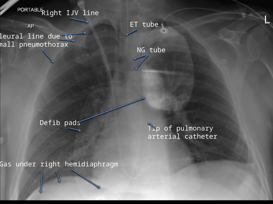
Pleural line due to small pneumothorax
Right IJV line
ET tube
NG tube
Tip of pulmonary arterial catheter
Defib pads
Gas under right hemidiaphragm

Case 16

Case 16
• 55 year old woman, presenting with cough and fever.
1. Describe the radiographic abnormalities.2. What is the diagnosis, and why?3. How are the ‘zones’ of the lungs defined?

Case 16 - answers
1. PA and right lateral CXR. There is abnormal opacification in the right mid- and upper-zones, sharply demarcated at its inferior margin on the PA study, and at its inferior and posterior margins on the lateral view.
2. Right upper lobe pneumonia. We know that it is the upper lobe that is affected as it is above the horizontal fissure, and anterior to the oblique fissure.
3. The upper zone the area above the anterior aspect of the second rib, the mid-zone is between the anterior aspects of the second and fourth ribs, while the lower zone is below the anterior aspect of the fourth rib.

Horizontal fissure Oblique fissure

Case 17

Case 17
• 78 year old woman, fell in nursing home and now unable to weight bear.
1. There are two abnormalities on this film, one acute and one old – can you identify them?
2. When a hip fracture is clinically suspected but not identified on radiographs, what imaging options are available to us?

Case 17 - answers
1. There is an acute, mildly displaced subcapital fracture of the left proximal femur. There is an old, healed fracture of the left inferior pubic ramus. These findings suggest that the patient may be osteoporotic and a DXA should be recommended if this has not already been diagnosed.
2. CT is usually the first test that is performed when radiographs have not shown a clinically suspected fracture. In a very small percentage of osteoporotic patients with hip fractures, CT will also be falsely negative and in those patients we can perform either a bone scan or an MRI (MRI has 100% sensitivity for pelvic/femoral fractures but is not performed routinely because of availability and cost issues).
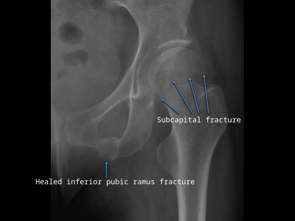
Subcapital fracture
Healed inferior pubic ramus fracture

Case 18

Case 18
• 65 year old woman, fell on ice, unable to move right arm.
1. How would you describe this fracture?

Case 18 - answer
1. There is a comminuted fracture of the surgical neck of the right humerus, also involving the greater tuberosity. The fracture is impacted (the proximal part of the humeral shaft has been displaced superior to its normal position). The humeral head remains in articulation with the glenoid but is slightly subluxed inferiorly – this is a common finding in shoulder fractures, and is in part related to the presence of a haemarthrosis in the joint, pushing the head inferiorly.

Humeral head subluxed inferiorly, relative to glenoid
Greater tuberosity fracture
Surgical neck fracture

Case 19

Case 19
• 33 year old woman, presented with tender red lumps on her lower limbs.
1. How would you describe this CXR?2. What is the diagnosis in this case?3. How is this condition staged radiographically,
and what stage is this patient?

Case 19 - answers
1. There is marked enlargement of both hila. The heart and mediastinum are normal. Both lungs are also normal and there are no pleural effusions.
2. Bihilar lymphadenopathy – sarcoidosis.3. Stage 0 – normal CXR
Stage I – nodal enlargement only – most common stage at presentation, and the stage of this
patient
Stage II – parenchymal abnormalities onlyStage III – nodal and parenchymal diseaseStage IV – pulmonary fibrosis

Case 20

Case 20
• 54 year old man, fell off a ladder and landed on his left side.
1. How would you describe this CXR?2. What is the diagnosis in this case?3. This patient had a history of previous trauma
– can you identify the old injury?

Case 20 - answers
1. There is increased lucency of the left hemithorax, lung markings are not visible at all in the upper zone or the periphery of the lower zone, and pleural lines are demonstrated. There is a subtle horizontal effusion, with no meniscus. The trachea, heart and mediastinum are shifted to the right of midline.
2. Another tension pneumothorax (you can never see enough examples of pneumothoraces).
3. Old left clavicle fracture.

Old clavicle fractureNo lung markings
Tracheal shift
Pleural line
Effusion

Case 21

Case 21
• 74 year old man, fell down stairs and complaining of severe back pain.
1. What is the abnormality?2. What test would you recommend next and
why?3. It is often difficult to determine whether
these injuries are new or old – what can we do to differentiate?

Case 21 - answers
1. There is a wedge compression fracture of the T7 vertebral body with associated kyphosis.
2. CT is routinely performed next in the work-up of vertebral fractures. It shows the full extent of the fracture, whether the posterior elements are involved, whether the fracture is stable or unstable and whether there is retropulsion of bone fragments into the spinal canal, which might compress the spinal cord.
3. Even with CT, it can be hard to work out whether a vertebral fracture is new. If we have a recent radiograph to compare to, that may be enough to confirm acuity. If none are available, and it is important to know whether the fracture is recent (i.e. it would alter management), an MRI or a bone scan will provide the answer.

Wedge-shaped fracture

Emergency Radiology Quiz Cases
Quiz complete, well done!