Emergency oesophagectomy - Thoraxthorax.bmj.com/content/thoraxjnl/23/2/204.full.pdf · Emergency...
6
Thorax (1968), 23, 204. Emergency oesophagectomy W. F. KERR' From the Thoracic Surgical Unit, Papworth Hospital, Cambridge In the treatment of instrumental perforation of the obstructed thoracic oesophagus, relief of obstruction is one of the prerequisites of success. In some cases it is better to resect both the perforation and the original lesion rather than to rely on repair and drainage. The salient features of 19 cases of emergency oesophagectomy collected from the literature have been tabulated, and three new examples are here reported. The results are encouraging. The operation usually performed for malignant cases is a one-stage oesophagogastrectomy with oesophagogastrostomy; a two-stage procedure is recommended for benign lower-end strictures. The lacerated oesophagus and the stricture are resected at the emergency operation, and the fundus of the stomach, advanced into the chest, is anastomosed to the oesophagus. The whole of the stomach is thereby preserved for the elective reconstruction which constitutes the second stage. Instrumental perforation of the oesophagus, a dangerous complication of endoscopy, is easily diagnosed, but treatment is still controversial. This paper is concerned only with the treatment of perforation associated with chronic organic obstruction. Most perforations occur as a result of foreign- body impaction in children and involve chiefly the pharynx or cervical oesophagus (Dawes, 1964). Diagnostic endoscopy in adults is responsible for the second largest group, and it is the cervical oesophagus that is most liable to injury. Recent papers have emphasized that the oesophagus is normal before the accident in the majority of patients, and opinions and statistics on treatment are coloured by this preponderance, to the detri- ment of the small but nevertheless important minority who have oesophageal disease. In them, perforation is most likely to occur during attempts to inspect, to take a biopsy, or to dilate chronic obstructive lesions of the thoracic or abdominal oesophagus, and, by contrast with the majority, the injury is almost always at or just above the upper limit of the obstruction. There is agreement that any method of treat- ment stands its best chance of success if it is started early, but the importance of unrelieved obstruction distal to the perforation appears to have gone unremarked until Davidson (1965) and Groves (1966) were provoked to reply to a much- publicized article (Mengoli and Klassen, 1965) which advocated non-operative management for 'Present address: Norfolk and Norwich Hospital, Norwich, NOR 53A all cases in the first instance. In the past, the subject has been confused by the practice of reporting together all examples of perforation seen in any one clinic, whether with or without obstruc- tion and howsoever caused. No one before Groves (1966) appears to have analysed results primarily by reference to the underlying pathology, though this would seem to be a factor of some import- ance. Hitherto, classification of oesophageal in- juries had usually been according to site, and sometimes also according to the method of pro- duction and size. The indications for instrumenta- tion have not always been stated and the final diagnosis 'has frequently been omitted. Results, measured in terms of life and death, have been related mainly to the time that had elapsed before active treatment was begun, and little attention has been paid to any other factor. This paper does not attempt to review all aspects of instru- mental perforation but discusses treatment when the injury is associated with oesophageal obstruc- tion, and it is assumed that full supportive measures and antibiotics are exhibited efficiently. I think that the effects of the original oesophageal lesion must be taken into account when treatment of an accidental perforation is being determined, and I here report three cases of emergency oeso- phagectomy carried out successfully in obedience to this principle and discuss the problem of how much of the stomach should be resected when the stricture is benign. When the diagnosis of intrathoracic perfora- tion of the oesophagus is made within 12 to 18 hours of the accident, most thoracic surgeons, 204 on 7 July 2018 by guest. Protected by copyright. http://thorax.bmj.com/ Thorax: first published as 10.1136/thx.23.2.204 on 1 March 1968. Downloaded from
Transcript of Emergency oesophagectomy - Thoraxthorax.bmj.com/content/thoraxjnl/23/2/204.full.pdf · Emergency...
Thorax (1968), 23, 204.
Emergency oesophagectomyW. F. KERR'
From the Thoracic Surgical Unit, Papworth Hospital, Cambridge
In the treatment of instrumental perforation of the obstructed thoracic oesophagus, relief ofobstruction is one of the prerequisites of success. In some cases it is better to resect both theperforation and the original lesion rather than to rely on repair and drainage. The salient featuresof 19 cases of emergency oesophagectomy collected from the literature have been tabulated, andthree new examples are here reported. The results are encouraging. The operation usuallyperformed for malignant cases is a one-stage oesophagogastrectomy with oesophagogastrostomy;a two-stage procedure is recommended for benign lower-end strictures. The laceratedoesophagus and the stricture are resected at the emergency operation, and the fundus of thestomach, advanced into the chest, is anastomosed to the oesophagus. The whole of the stomachis thereby preserved for the elective reconstruction which constitutes the second stage.
Instrumental perforation of the oesophagus, adangerous complication of endoscopy, is easilydiagnosed, but treatment is still controversial.This paper is concerned only with the treatmentof perforation associated with chronic organicobstruction.Most perforations occur as a result of foreign-
body impaction in children and involve chiefly thepharynx or cervical oesophagus (Dawes, 1964).Diagnostic endoscopy in adults is responsible forthe second largest group, and it is the cervicaloesophagus that is most liable to injury. Recentpapers have emphasized that the oesophagus isnormal before the accident in the majority ofpatients, and opinions and statistics on treatmentare coloured by this preponderance, to the detri-ment of the small but nevertheless importantminority who have oesophageal disease. In them,perforation is most likely to occur during attemptsto inspect, to take a biopsy, or to dilate chronicobstructive lesions of the thoracic or abdominaloesophagus, and, by contrast with the majority,the injury is almost always at or just above theupper limit of the obstruction.
There is agreement that any method of treat-ment stands its best chance of success if it isstarted early, but the importance of unrelievedobstruction distal to the perforation appears tohave gone unremarked until Davidson (1965) andGroves (1966) were provoked to reply to a much-publicized article (Mengoli and Klassen, 1965)which advocated non-operative management for'Present address: Norfolk and Norwich Hospital, Norwich, NOR53A
all cases in the first instance. In the past, thesubject has been confused by the practice ofreporting together all examples of perforation seenin any one clinic, whether with or without obstruc-tion and howsoever caused. No one before Groves(1966) appears to have analysed results primarilyby reference to the underlying pathology, thoughthis would seem to be a factor of some import-ance. Hitherto, classification of oesophageal in-juries had usually been according to site, andsometimes also according to the method of pro-duction and size. The indications for instrumenta-tion have not always been stated and the finaldiagnosis 'has frequently been omitted. Results,measured in terms of life and death, have beenrelated mainly to the time that had elapsed beforeactive treatment was begun, and little attentionhas been paid to any other factor. This paperdoes not attempt to review all aspects of instru-mental perforation but discusses treatment whenthe injury is associated with oesophageal obstruc-tion, and it is assumed that full supportivemeasures and antibiotics are exhibited efficiently.I think that the effects of the original oesophageallesion must be taken into account when treatmentof an accidental perforation is being determined,and I here report three cases of emergency oeso-phagectomy carried out successfully in obedienceto this principle and discuss the problem of howmuch of the stomach should be resected when thestricture is benign.When the diagnosis of intrathoracic perfora-
tion of the oesophagus is made within 12 to 18hours of the accident, most thoracic surgeons,
204
on 7 July 2018 by guest. Protected by copyright.
http://thorax.bmj.com
/T
horax: first published as 10.1136/thx.23.2.204 on 1 March 1968. D
ownloaded from
Emergency oesophagectomy
Mengoli and Klassen (1965) notwithstanding,would operate with the intention of repairing thedamage and draining the pleura and mediastinum.Some would go no further even when faced withobstruction, despite the fact that a fistula will notheal and cannot be closed in such circumstances.They hold that resection is too hazardous. Thisis the orthodox approach if the view of Logan(1965) is representative. Distinguishing betweenobstructed and unobstructed cases only byimplication, he advised against a procedure of theseverity of oesophageal resection though he wouldpermit, 'for example', repair of a hiatal hernia atthe same time as closure of the perforation 'ina patient who is not gravely ill and for whom an
effective antibiotic is available'. Terracol andSweet (1958) treated perforations on similar linesbut gave tacit approval to oesophagectomy in cer-tain cases by adding that 'if the injury appears tobe so extensive that resection may be necessary'
a left-sided approach should be chosen. On theother hand, there is the impression that immediateresection in appropriate cases is already sowidely practised as scarcely to deserve comment.It is recommended without discussion in books byBorrie (1959) and Flavell (1963) and supportedby a recent annotation in the Lancet (1966). Noneof these authorities, for or against, backed hisopinions by quoting gases within his own experi-ence, but Borrie referred to an article byMcBurney, Kirklin, Hood, and Andersen (1953)which described three cases of carcinoma of thelower oesophagus or cardia treated by resection10 hours to five days after accidental perforation.Emergency oesophagectomy is the subject of atleast two other articles in American journals(Satinsky and Kron, 1952; Blalock, 1957), whileGroves (1966) gives examples and discusses theproblem in the larger context of oesophagealperforations in general.
TABLE
Reference
Clagett (quoted byMcBurney etal., 1953)
Satinsky and Kron (1952)
McBurney et al. (1953)
McBurney et al. (1953)
McBurney et al. (1953)
Overstreet and Ochsner(1955)
Smith and Tanner (1956)
Blalock (1957)
Blalock (1957)
Nealon et al. (1961)
Nealon et al. (1961)
Brewer (1961) ..
Mathewson et al. (1962)
Mathewson et al. (1962)
Metheny (1962)
Foster et a!. (1965)
Castberg (1965)Groves (1966)Groves (1966)
Lesion
Achalasia
Carcinoma loweroesophagus
Carcinoma cardia
Carcinoma distaloesophagus
Carcinoma oesophago-gastric junction
Stricture 36 cm. tocardia followingendoscopic removalof foreign body
Carcinoma fundusinvolving cardia
Stricture followingoesophagogastro-stomy for achalasia
Carcinoma cardia
Carcinoma middlethird oesophagus
Carcinoma from lowerborder aortic arch to5 cm. above dia-phragm
Carcinoma
(intrathoracicperforation)
(intrathoracicperforation)
Recurrent carcinomacardia after partialgastrectomy
'Short stricture'
Carcinoma cardiaCarcinoma oesophagusCarcinoma oesophagus
Perforation at,'by Operation at IHydrostatic bag
Oesophagoscopy
Oesophagoscopy
Blind bouginage
Bougie
Bougie
Gastroscopy
Dilatation
9
2 days
30 hours
5 days
10 hours
6 hours
Immediately
15 hours
Hydrostatic bag 8 hours
Oesophagoscopy 4 days
Oesophagoscopy 9 hours
Dilatation
BougieOesoohagoscopyOesophagoscopy
Promptly
?
2 days
12 hours
4 hours
3 hours
Procedure
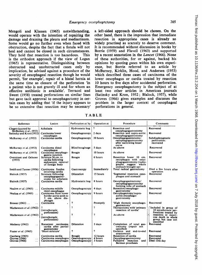
Resection andoesophagogastrostomy
Resection and supra-aorticoesophagogastrostomy
Oesophagogastrectomy/oesophagogastrostomyafter narrowing lessercurve
As above
As above
Resection lower 10 cm.oesophagus with oeso-phagogastrostomy (radio-graphs suggest wholestomach brought up)
Total radical gastrectomy
'Segmental resection oeso-phagus and stomach'
Oesophagogastrectomy/oesophagogastrostomyforming tube of stomach
Resection/oesophago-gastrostomy
Oesophagectomy/supra-aortic oesophago-gastrostomy
'High thoracic oesophago-gastrectomy'
Thoracotomy with 'primaryresection of cardia'
As above
Completion of total gas-trectomy (repair notspecified)
Excision and end-to-endanastomosis
Resection of cardiaOesophagogastrectomyPalliative resection andBerman prosthesis
Comments
Recovered
Recovered
Subdiaphragmaticperforation:recovered
Recovered
Recovered
Recovered
Died a few hiours afteroperation
Recovered
Recovered
Recovered
Recovered
Recovered
FIncluded in group of10 intrathoracic per-
forations treated byresection or repair;one death in wholegroup but case notidentified
Recovered
Recovered
RecoveredRecoveredDied 13th day
205
1-.1-* ~~~~Ii-
on 7 July 2018 by guest. Protected by copyright.
http://thorax.bmj.com
/T
horax: first published as 10.1136/thx.23.2.204 on 1 March 1968. D
ownloaded from
W. F. Kerr
Clagett in 1946 or 1947 was probably the firstto carry out successfully an emergency oeso-phagectomy and oesophagogastrostomy. Thepatient suffered from achalasia, and the oeso-phagus had been ruptured with a hydrostatic bag.This case has not been reported in full but isreferred to by the Mayo Clinic group (McBurneyet al., 1953, vide supra), who were secondin the field with published results. First inprint were Satinsky and Kron (1952) with a singlecase operated on in 1949, a carcinoma resectedtwo days after perforation of the oesophagus atoesophagoscopy. Reconstruction was by supra-aortic oesophagogastrostomy. There was no post-operative obstruction or leakage; the patient wasin hospital for six weeks and was well two yearslater. The Table summarizes 19 cases ofemergency resection from the literature. In addi-tion there are references, generally brief andlacking in detail, to eight cases of achalasiatreated by Heller's operation at the same time asclosure of the laceration (Barrett, 1956-2 cases;Dawes, 1964- 1 case; Foster, Jolly, Sawyers, andDaniel, 1965-3 cases; Groves, 1966-2 cases)and two of simultaneous repair of a hiatal hernia(Castberg, 1965; Groves, 1966). All these patientsnade good recoveries. One fatal case of benigncicatricial stenosis treated by 'Heller-type myo-tomy' and repair of hiatal hernia (Nealon, Temple-
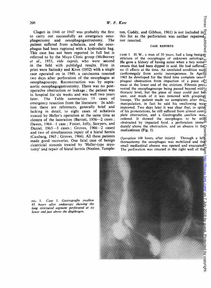
FIG. 1. Case 1. Gastrografin swallow45 hours after endoscopy showing thelong strictured segment perforated at itslower end just above the diaphragm.
ton, Cuddy, and Gibbon, 1961) is not included inthis list as the perforation was neither repairednor resected.
CASE REPORTS
CASE 1 H. W.. a man of 50 years. had a long benignstricture of the oesophagus of unknown aetiology.He gave a history of having eaten when a boy somesweets that had been dipped in acid. He had sufferedno ill effects at the time. An unrelated condition wascardiomegaly from aortic incompetence. In April1965 he developed for the third time complete oeso-phageal obstruction from impaction of a piece ofmeat at the lower end of the stricture. Fibrosis pre-vented the oesophagoscope being passed beyond mid-thoracic level, but the piece of meat could just beseen, and much of it was removed with graspingforceps. The patient made no complaints after thismanipulation, in fact he said his swallowing wasimproved. Two days later it was clear that, in spiteof his protestations, he still suffered from almost com-plete obstruction, and a Gastrografin swallow wasordered. It showed the oesophagus to be stillobstructed by impacted food, a perforation imme-diately above the obstruction, and an abscess in themediastinum (Fig. 1).
Operation (48 hours after injury) Through a leftthoracotomy the oesophagus was mobilized and thesmall mediastinal abscess was opened and evacuated.The perforation was situated in the right wall of the
206
on 7 July 2018 by guest. Protected by copyright.
http://thorax.bmj.com
/T
horax: first published as 10.1136/thx.23.2.204 on 1 March 1968. D
ownloaded from
Emergency oesophagectomy
oesophagus in an inaccessible position just above thenarrowest and most fibrotic part. The diaphragmwas incised radially to the hiatus and the lower endof the oesophagus was detached from the stomach.The segment containing the stricture, the piece ofmeat, and the perforation was resected, dividing theoesophagus again at the level of the inferior pul-monary vein where its wall was fibrotic and inelastic.After closing the cardiac orifice the stomach wasmobilized by removing the spleen and dividing theleft gastric artery, and a pyloromyotomy was carriedout. The oesophagus was then anastomosed to thefundus of the stomach. The patient made an un-eventful recovery and was discharged.He was reluctant to submit to another operation
but did so after eight months had passed. By thenhe had had several dilatations for dysphagia and hadsuffered much oesophageal pain that closely mimickedmyocardial ischaemia. At the second operation thestomach was mobilized and replaced in its normalposition below the diaphragm with an isolatedjejunal loop as substitute for the resected oesophagus.The patient again made a satisfactory recovery, com-plaining only of colicky postprandial pain for thefirst month or so. This yielded to antispasmodics.CASE 2 H. G., aged 74 years, had severe chronicbronchitis and gout in addition to dysphagia from a
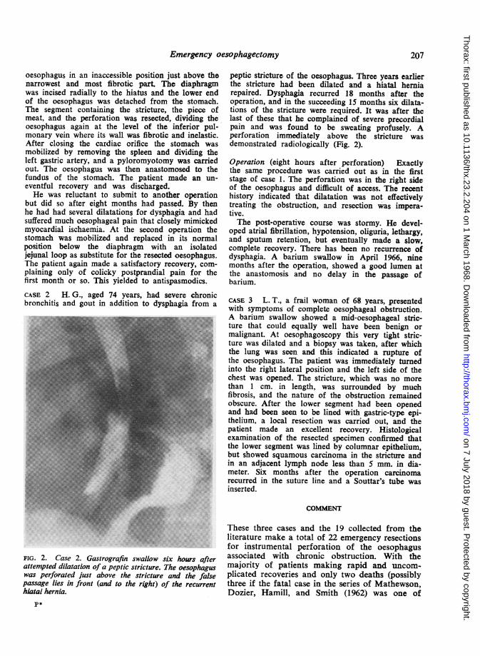
FIG. 2. Case 2. Gastrografin swallow six hours afterattempted dilatation of a peptic stricture. The oesophaguswas perforated just above the stricture and the falsepassage lies in front (and to the right) of the recurrenthiatal hernia.
peptic stricture of the oesophagus. Three years earlierthe stricture had been dilated and a hiatal herniarepaired. Dysphagia recurred 18 months after theoperation, and in the succeeding 15 months six dilata-tions of the stricture were required. It was after thelast of these that he complained of severe precordialpain and was found to be sweating profusely. Aperforation immediately above the stricture wasdemonstrated radiologically (Fig. 2).
Operation (eight hours after perforation) Exactlythe same procedure was carried out as in the firststage of case 1. The perforation was in the right sideof the oesophagus and difficult of access. The recenthistory indicated that dilatation was not effectivelytreating the obstruction, and resection was impera-tive.The post-operative course was stormy. He devel-
oped atrial fibrillation, hypotension, oliguria, lethargy,and sputum retention, but eventually made a slow,complete recovery. There has been no recurrence ofdysphagia. A barium swallow in April 1966, ninemonths after the operation, showed a good lumen atthe anastomosis and no delay in the passage ofbarium.
CASE 3 L. T., a frail woman of 68 years, presentedwith symptoms of complete oesophageal obstruction.A barium swallow showed a mid-oesophageal stric-ture that could equally well have been benign ormalignant. At oesophagoscopy this very tight stric-ture was dilated and a biopsy was taken, after whichthe lung was seen and this indicated a rupture ofthe oesophagus. The patient was immediately turnedinto the right lateral position and the left side of thechest was opened. The stricture, which was no morethan 1 cm. in length, was surrounded by muchfibrosis, and the nature of the obstruction remainedobscure. After the lower segment had been openedand had been seen to be lined with gastric-type epi-thelium, a local resection was carried out, and thepatient made an excellent recovery. Histologicalexamination of the resected specimen confirmed thatthe lower segment was lined by columnar epithelium,but showed squamous carcinoma in the stricture andin an adjacent lymph node less than 5 mm. in dia-meter. Six months after the operation carcinomarecurred in the suture line and a Souttar's tube wasinserted.
COMMENT
These three cases and the 19 collected from theliterature make a total of 22 emergency resectionsfor instrumental perforation of the oesophagusassociated with chronic obstruction. With themajority of patients making rapid and uncom-plicated recoveries and only two deaths (possiblythree if the fatal case in the series of Mathewson,Dozier, Hamill, and Smith (1962) was one of
207
on 7 July 2018 by guest. Protected by copyright.
http://thorax.bmj.com
/T
horax: first published as 10.1136/thx.23.2.204 on 1 March 1968. D
ownloaded from
W. F. Kerr
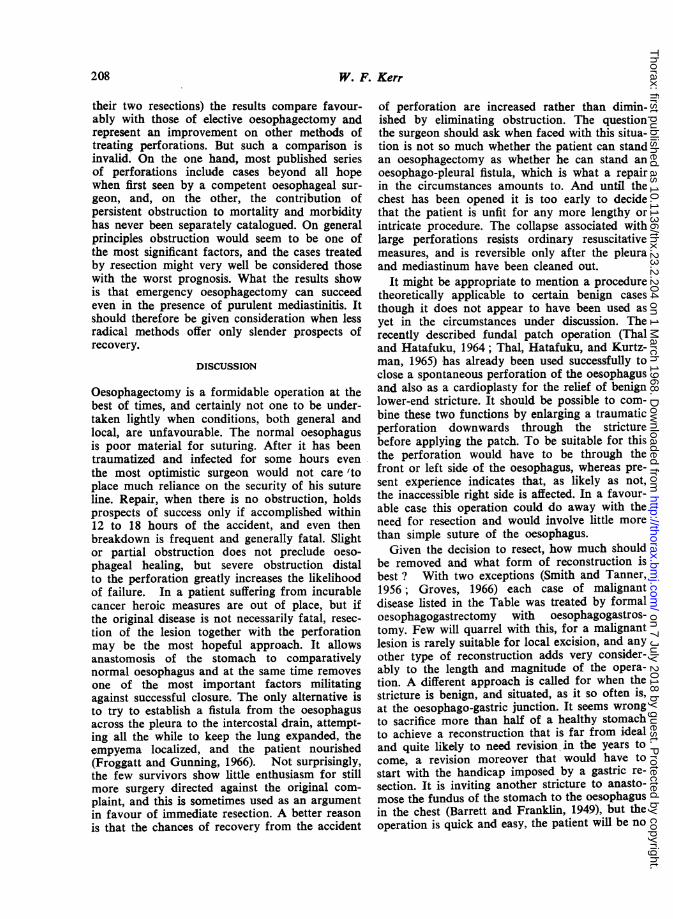
their two resections) the results compare favour-ably with those of elective oesophagectomy andrepresent an improvement on other methods oftreating perforations. But such a comparison isinvalid. On the one hand, most published seriesof perforations include cases beyond all hopewhen first seen by a competent oesophageal sur-geon, and, on the other, the contribution ofpersistent obstruction to mortality and morbidityhas never been separately catalogued. On generalprinciples obstruction would seem to be one ofthe most significant factors, and the cases treatedby resection might very well be considered thosewith the worst prognosis. What the results showis that emergency oesophagectomy can succeedeven in the presence of purulent mediastinitis. Itshould therefore be given consideration when lessradical methods offer only slender prospects ofrecovery.
DISCUSSION
Oesophagectomy is a formidable operation at thebest of times, and certainly not one to be under-taken lightly when conditions, both general andlocal, are unfavourable. The normal oesophagusis poor material for suturing. After it has beentraumatized and infected for some hours eventhe most optimistic surgeon would not care 'toplace much reliance on the security of his sutureline. Repair, when there is no obstruction, holdsprospects of success only if accomplished within12 to 18 hours of the accident, and even thenbreakdown is frequent and generally fatal. Slightor partial obstruction does not preclude oeso-phageal healing, but severe obstruction distalto the perforation greatly increases the likelihoodof failure. In a patient suffering from incurablecancer heroic measures are out of place, but ifthe original disease is not necessarily fatal, resec-tion of the lesion together with the perforationmay be the most hopeful approach. It allowsanastomosis of the stomach to comparativelynormal oesophagus and at the same time removesone of the most important factors militatingagainst successful closure. The only alternative isto try to establish a fistula from the oesophagusacross the pleura to the intercostal drain, attempt-ing all the while to keep the lung expanded, theempyema localized, and the patient nourished(Froggatt and Gunning, 1966). Not surprisingly,the few survivors show little enthusiasm for stillmore surgery directed against the original com-plaint, and this is sometimes used as an argumentin favour of immediate resection. A better reasonis that the chances of recovery from the accident
of perforation are increased rather than dimin-ished by eliminating obstruction. The questionthe surgeon should ask when faced with this situa-tion is not so much whether the patient can standan oesophagectomy as whether he can stand anoesophago-pleural fistula, which is what a repairin the circumstances amounts to. And until thechest has been opened it is too early to decidethat the patient is unfit for any more lengthy orintricate procedure. The collapse associated withlarge perforations resists ordinary resuscitativemeasures, and is reversible only after the pleuraand mediastinum have been cleaned out.
It might be appropriate to mention a proceduretheoretically applicable to certain benign casesthough it does not appear to have been used asyet in the circumstances under discussion. Therecently described fundal patch operation (Thaland Hatafuku, 1964; Thal, Hatafuku, and Kurtz-man, 1965) has already been used successfully toclose a spontaneous perforation of the oesophagusand also as a cardioplasty for the relief of benignlower-end stricture. It should be possible to com-bine these two functions by enlarging a traumaticperforation downwards through the stricturebefore applying the patch. To be suitable for thisthe perforation would have to be through thefront or left side of the oesophagus, whereas pre-sent experience indicates that, as likely as not,the inaccessible right side is affected. In a favour-able case this operation could do away with theneed for resection and would involve little morethan simple suture of the oesophagus.Given the decision to resect, how much should
be removed and what form of reconstruction isbest ? With two exceptions (Smith and Tanner,1956; Groves, 1966) each case of malignantdisease listed in the Table was treated by formaloesophagogastrectomy with oesophagogastros-tomy. Few will quarrel with this, for a malignantlesion is rarely suitable for local excision, and anyother type of reconstruction adds very consider-ably to the length and magnitude of the opera-tion. A different approach is called for when thestricture is benign, and situated, as it so often is,at the oesophago-gastric junction. It seems wrongto sacrifice more than half of a healthy stomachto achieve a reconstruction that is far from idealand quite likely to need revision in the years tocome, a revision moreover that would have tostart with the handicap imposed by a gastric re-
section. It is inviting another stricture to anasto-mose the fundus of the stomach to the oesophagusin the chest (Barrett and Franklin, 1949), but theoperation is quick and easy, the patient will be no
20;8
on 7 July 2018 by guest. Protected by copyright.
http://thorax.bmj.com
/T
horax: first published as 10.1136/thx.23.2.204 on 1 March 1968. D
ownloaded from
Emergency oesophagectomy
worse off than before the accident, and the wholeof his stomach will be available for whatever re-
construction may be thought best when condi-tions are more favourable. In case 1 both stageswere completed, and the result is satisfactory so
far. In case 2 the elderly and ailing patient under-standably refused the second operation, but itcould still be carried out should it become essential.On the other hand, the temporary repair may welllast him the rest of his life.
Grateful acknowledgement is made to Drs. G. I.
Vemey and J. Rymer for the radiological investiga-tions, and to Mr. E. W. Groves for reproducing theradiographs.
REFERENCESBarrett, N. R. (1956). Perforations of the oesophagus and of the
pharynx. Proc. roy. Soc. Med., 49, 529.-- and Franklin, R. H. (1949). Concerning the unfavourable late
results of certain operations performed in the treatment ofcardiospasm. Brit. J. Surg., 37, 194.
Blalock, J. (1957). Primary esophagogastrectomy for instrumentalperforation of the esophagus. Amer. J. Surg., 94, 393.
Borrie, J. (1959). The Management ofEnergencies in Thoracic Surgery,p. 278. Staples Press, London.
Brewer, L. A. (1961). Discussion of Nealon et al. (1961). J. thorac.cardiovasc. Surg., 41, 102.
Castberg, T. (1965). Traumatic perforation of the oesophagus. Actachir. scand., Suppl. 343, p. 56.
Clagett, 0. T. Quoted by McBurney et al. (1953).Davidson, J. S. (1965). In Medical Forum. Mod. Med. G.B., 10, 959.Dwesa, J. D. K. (1964). Traumatic perforations of the pharynx and
oesophagus. J. Laryng., 78, 18.
Flavell, G. (1963). The Oesophagus, p. 71. Butterworths, London.Foster, J. H., Jolly, P. C., Sawyers, J. L., and Daniel, R. A. (1965).
Esophageal perforation: diagnosis and treatment. Ann. Sxrg..161,701.
Froggatt, D. L., and Gunning, A. J. (1966). Treatment of oesophagealperforation. Thorax, 21, 524.
Groves, L. K. (1966). Instrumental perforation of the esophagus:What is conservative management? J. thorac. cardiovasc. Surg.,52,1.
Lancet (1966). Annotation: oesophageal perforation, 2, 788.Logan, A. (1965). Mediastinum. In Thorax, ed. d'Abreu, A. L. (Vol. 5
of Clinical Surgery, ed. Rob, C., and Smith, R.), Ch. 15, p. 217.Butterworths, London.
Mathewson. C., Dozier, W. E., Hamill, J. P., and Smith, M. (1962).Clinical experiences with perforation of the esophagus. Amer. J.Surg., 104, 257.
McBurney, R. P., Kirklin, J. W., Hood, R. T., and Andersen, H. A.(1953). One-stage esophagogastrectomy for perforated carcinomain the presence of mediastinitis. Proc. Mayo Clin., 28, 281.
Mengoli, L. R., and Klassen, K. P. (1965). Conservative managementof esophageal perforation. Arch. Surg., 91, 238.
Metheny, D. (1962). Discussion of Mathewson et al. (1962). Amer.J. Surg., 104. 265.
Nealon, T. F., Templeton, J. Y., Cuddy, V. D., and Gibbon, J. H.(1961). Instrumental perforation of the esophagus. J. thorac.cardiovasc. Surg., 41, 75.
Overstreet, J. W., and Ochsner, A. (1955). Traumatic rupture of the-e esophagus. J. thorac. Surg., 30, 164.Satinsky, V. P.. and Kron, S. D. (1952). One-stage esophagectomy in
presence of mediastinitis. Arch. Surg., 64, 124.Smith, C. C. K., and Tanner, N. C. (1956). The complications of
gastroscopy and oesophagoscopy. Brit. J. Surg., 43, 396.
Terracol, J., and Sweet, R. H. (1958). Diseases of the Esophaguis,Ch. 23, p. 443. Saunders, Philadelphia and London.
Thal, A. P., and Hatafuku, T. (1964). Improved operation for esopha-geal rupture. J. Amer. med. Ass., 188, 826._--and Kurtzman, R. (1965). A new method for reconstruc-
tion of the esophagogastric junction. Surg. Gynec. Obstet., 120,1225.
209
on 7 July 2018 by guest. Protected by copyright.
http://thorax.bmj.com
/T
horax: first published as 10.1136/thx.23.2.204 on 1 March 1968. D
ownloaded from