Emergency Neurological Life Support: Airway, Ventilation...
17
REVIEW ARTICLE Emergency Neurological Life Support: Airway, Ventilation, and Sedation David B. Seder • Richard R. Riker • Andy Jagoda • Wade S. Smith • Scott D. Weingart Published online: 13 September 2012 Ó Neurocritical Care Society 2012 Abstract Airway management is central to the resusci- tation of the neurologically ill. These patients often have evolving processes that threaten the airway and adequate ventilation. Therefore, airway, ventilation, and sedation were chosen as an Emergency Neurological Life Support (ENLS) protocol. Reviewed topics include airway man- agement; the decision to intubate; when and how to intubate with attention to cardiovascular status; mechanical ventilation settings; and the use of sedation, including how to select sedative agents based on the patient’s neurological status. Keywords Respiratory failure Airway protection Intubation Ventilation Introduction Intubation of the acutely brain-injured patient is a matter of life or death. Failure to intubate a patient with rapidly progressive neurological decline may result in respiratory arrest, secondary brain injury from hypoxia or hypercarbia, and severe aspiration pneumonitis or acute respiratory distress syndrome. The process of induction and intubation can itself provoke brain herniation when a mass lesion is present, complete a massive infarction when brain tissue is mar- ginally perfused by collateral cerebrovascular circulation, or result in temporary obliteration of the neurological examination during the early—and often most critical— period of neurological and neurosurgical decision- making. Rapid patient stabilization must occur concurrently with well-documented and detailed neurological assessment. The goals of airway management in neurological patients are therefore to maintain adequate (but not excessive) oxygenation and ventilation, and to prevent aspiration. Ideally, a neurological assessment prior to the administra- tion of sedating and paralyzing medications is recom- mended in order to provide a functional baseline. The Emergency Neurological Life Support (ENLS) suggested algorithm for the initial management of airway, ventilation, and sedation is shown in Fig. 1. Suggested items to complete within the first hour of evaluating a patient are shown in Table 1. Need Intubation? Patients with respiratory arrest or impending arrest should be intubated without delay. In addition, a patient who D. B. Seder (&) R. R. Riker Department of Critical Care Services, Maine Medical Center, Tufts University School of Medicine, Boston, MA, USA e-mail: [email protected] A. Jagoda Department of Emergency Medicine, Mount Sinai Medical Center, New York, NY, USA W. S. Smith ENLS Course Co-Chair, Department of Neurology, University of California, San Francisco, San Francisco, CA, USA S. D. Weingart ENLS Course Co-Chair, Division of ED Critical Care, Mount Sinai School of Medicine, New York, NY, USA 123 Neurocrit Care (2012) 17:S4–S20 DOI 10.1007/s12028-012-9753-6
-
Upload
truongminh -
Category
Documents
-
view
218 -
download
1
Transcript of Emergency Neurological Life Support: Airway, Ventilation...

REVIEW ARTICLE
Emergency Neurological Life Support: Airway, Ventilation,and Sedation
David B. Seder • Richard R. Riker •
Andy Jagoda • Wade S. Smith • Scott D. Weingart
Published online: 13 September 2012
� Neurocritical Care Society 2012
Abstract Airway management is central to the resusci-
tation of the neurologically ill. These patients often have
evolving processes that threaten the airway and adequate
ventilation. Therefore, airway, ventilation, and sedation
were chosen as an Emergency Neurological Life Support
(ENLS) protocol. Reviewed topics include airway man-
agement; the decision to intubate; when and how to
intubate with attention to cardiovascular status; mechanical
ventilation settings; and the use of sedation, including how
to select sedative agents based on the patient’s neurological
status.
Keywords Respiratory failure � Airway protection �Intubation � Ventilation
Introduction
Intubation of the acutely brain-injured patient is a matter of
life or death. Failure to intubate a patient with rapidly
progressive neurological decline may result in respiratory
arrest, secondary brain injury from hypoxia or hypercarbia,
and severe aspiration pneumonitis or acute respiratory
distress syndrome.
The process of induction and intubation can itself
provoke brain herniation when a mass lesion is present,
complete a massive infarction when brain tissue is mar-
ginally perfused by collateral cerebrovascular circulation,
or result in temporary obliteration of the neurological
examination during the early—and often most critical—
period of neurological and neurosurgical decision-
making.
Rapid patient stabilization must occur concurrently with
well-documented and detailed neurological assessment.
The goals of airway management in neurological patients
are therefore to maintain adequate (but not excessive)
oxygenation and ventilation, and to prevent aspiration.
Ideally, a neurological assessment prior to the administra-
tion of sedating and paralyzing medications is recom-
mended in order to provide a functional baseline.
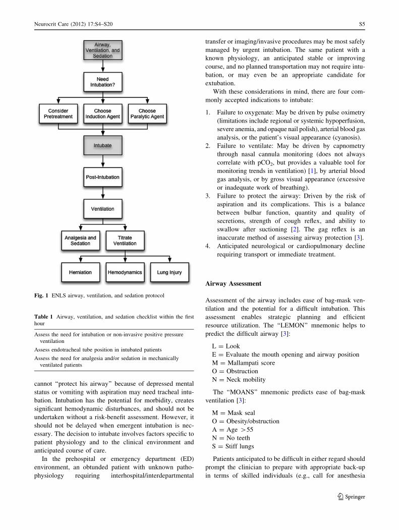
The Emergency Neurological Life Support (ENLS)
suggested algorithm for the initial management of airway,
ventilation, and sedation is shown in Fig. 1. Suggested
items to complete within the first hour of evaluating a
patient are shown in Table 1.
Need Intubation?
Patients with respiratory arrest or impending arrest should
be intubated without delay. In addition, a patient who
D. B. Seder (&) � R. R. Riker
Department of Critical Care Services, Maine Medical Center,
Tufts University School of Medicine, Boston, MA, USA
e-mail: [email protected]
A. Jagoda
Department of Emergency Medicine, Mount Sinai Medical
Center, New York, NY, USA
W. S. Smith
ENLS Course Co-Chair, Department of Neurology,
University of California, San Francisco, San Francisco,
CA, USA
S. D. Weingart
ENLS Course Co-Chair, Division of ED Critical Care,
Mount Sinai School of Medicine, New York, NY, USA
123
Neurocrit Care (2012) 17:S4–S20
DOI 10.1007/s12028-012-9753-6
cannot ‘‘protect his airway’’ because of depressed mental
status or vomiting with aspiration may need tracheal intu-
bation. Intubation has the potential for morbidity, creates
significant hemodynamic disturbances, and should not be
undertaken without a risk-benefit assessment. However, it
should not be delayed when emergent intubation is nec-
essary. The decision to intubate involves factors specific to
patient physiology and to the clinical environment and
anticipated course of care.
In the prehospital or emergency department (ED)
environment, an obtunded patient with unknown patho-
physiology requiring interhospital/interdepartmental
transfer or imaging/invasive procedures may be most safely
managed by urgent intubation. The same patient with a
known physiology, an anticipated stable or improving
course, and no planned transportation may not require intu-
bation, or may even be an appropriate candidate for
extubation.
With these considerations in mind, there are four com-
monly accepted indications to intubate:
1. Failure to oxygenate: May be driven by pulse oximetry
(limitations include regional or systemic hypoperfusion,
severe anemia, and opaque nail polish), arterial blood gas
analysis, or the patient’s visual appearance (cyanosis).
2. Failure to ventilate: May be driven by capnometry
through nasal cannula monitoring (does not always
correlate with pCO2, but provides a valuable tool for
monitoring trends in ventilation) [1], by arterial blood
gas analysis, or by gross visual appearance (excessive
or inadequate work of breathing).
3. Failure to protect the airway: Driven by the risk of
aspiration and its complications. This is a balance
between bulbar function, quantity and quality of
secretions, strength of cough reflex, and ability to
swallow after suctioning [2]. The gag reflex is an
inaccurate method of assessing airway protection [3].
4. Anticipated neurological or cardiopulmonary decline
requiring transport or immediate treatment.
Airway Assessment
Assessment of the airway includes ease of bag-mask ven-
tilation and the potential for a difficult intubation. This
assessment enables strategic planning and efficient
resource utilization. The ‘‘LEMON’’ mnemonic helps to
predict the difficult airway [3]:
L = Look
E = Evaluate the mouth opening and airway position
M = Mallampati score
O = Obstruction
N = Neck mobility
The ‘‘MOANS’’ mnemonic predicts ease of bag-mask
ventilation [3]:
M = Mask seal
O = Obesity/obstruction
A = Age >55
N = No teeth
S = Stiff lungs
Patients anticipated to be difficult in either regard should
prompt the clinician to prepare with appropriate back-up
in terms of skilled individuals (e.g., call for anesthesia
Fig. 1 ENLS airway, ventilation, and sedation protocol
Table 1 Airway, ventilation, and sedation checklist within the first
hour
Assess the need for intubation or non-invasive positive pressure
ventilation
Assess endotracheal tube position in intubated patients
Assess the need for analgesia and/or sedation in mechanically
ventilated patients
Neurocrit Care (2012) 17:S4–S20 S5
123
assistance) and devices (e.g., fiberoptic and other adjunct
intubating devices, and set-up for a cricothyrotomy).
Assessment of the expected ease of bag-mask ventilation is
critical; if deemed difficult, access to an extra-glottic
ventilation device, such as a laryngeal mask airway
(LMA), may be warranted.
Decision Made to Intubate: Perform Neurological
Assessment
Whenever possible, urgent management of the airway
should coincide with a rapid but detailed neurological
assessment. The examination can typically be conducted in
1–2 min and is crucial to subsequent emergency decision
making.
The pre-sedation/pre-intubation neurologic exam estab-
lishes a baseline that is used to assess therapeutic
interventions (e.g., patients with stroke or non-convulsive
status epilepticus) or may identify injuries that are at risk of
progressing (e.g., unstable cervical spine fractures). The
assessment identifies the type of testing and helps to avoid
unnecessary, uncomfortable interventions, such as cervical
spine immobilization with its potential for discomfort and
skin care complications. In general, the pre-intubation
neurological assessment is the responsibility of the team
leader who is coordinating the resuscitation; findings
should be documented and communicated directly to the
team that assumes care of the patient.
As demonstrated in Table 5, pre-intubation neurological
examination includes an assessment of:
• Level of arousal, interaction, and orientation
• Cranial nerves
• Motor function of each individual extremity
• Tone & reflexes
• Subtle or gross seizure activity
• Cervical spine stability
• When spinal cord injury is suspected, assess sensory
level
Intubating the Patient
Rapid sequence intubation is the preferred method of
securing the airway in patients with suspected elevated
intracranial pressure (ICP), since it provides protection
against the reflex responses to laryngoscopy and rises in ICP
[4–7]. The presence of coma should not be interpreted as an
indication to proceed without pharmacological agents, or to
administer only a neuromuscular blocking agent without
appropriate pre-treatment and induction agents. Although
the patient may seem unresponsive, laryngoscopy and
intubation usually provoke reflexes that elevate ICP unless
appropriate pre-treatment and induction agents are used [8].
Outcomes in patients with intracranial catastrophes are
related to the maintenance of both brain perfusion and
oxygenation; consequently, close assessment and man-
agement of these two parameters are critical. Cerebral
perfusion pressure (CPP) is the physiologic correlate for
blood flow to the brain and is measured by the difference
between the mean arterial pressure (MAP) and the ICP:
CPP ¼ MAP� ICP
It is generally recommended that the ICP be maintained
below 20 mmHg, MAP between 80 and 110 mmHg, and
CPP at a minimum of 60 mmHg.
Clinicians must recognize that many neurologically
impaired patients have compromised cerebral blood flow
(CBF) even with a normal ICP. For example, patients with
ischemic stroke, vasospasm, and hypoxic-ischemic brain
injury often have impaired auto regulation and are each
critically sensitive to decreases in blood pressure and CBF.
In these patients, the goal is to maintain MAP and CPP as
for the patients with known or suspected elevation of ICP.
Problems associated with elevated ICP may be com-
pounded by the techniques and drugs used in airway
management, since they may further elevate ICP. In addi-
tion, victims of multiple trauma may present with
hypotension, thus limiting the choice of agents and tech-
niques available.
Intubating the Patient with Presumed Elevated
Intracranial Pressure (ICP)
In the prehospital and ED environment, clinical evidence of
increased ICP is inferred from clinical signs of brain hernia-
tion that include altered mental status plus a unilaterally
dilated pupil, bilaterally dilated and fixed pupils, and decer-
ebrate or extensor posturing (decorticate posturing is not
predictive of elevated ICP) [9]. Since ICP measurement is
usually not available in these situations, one should proceed
with the assumption that ICP is elevated, assuming a value of
at least 20 mmHg for ICP in these circumstances. When the
airway is manipulated, two responses may result in even more
increased ICP: the reflex sympathetic response (RSR), which
results in increased heart rate, increased blood pressure, and,
consequently, increased ICP; and the direct laryngeal reflex
that stimulates an increase in ICP independent of the RSR [3].
Although the RSR may be dangerous in a hypertensive
patient, pre-treatment of the RSR is not indicated in a
hypotensive patient with known or suspected increased ICP
[10]. Elevations in ICP should be mitigated by minimizing
airway manipulation (the most experienced person should
perform the intubation) and administering medications.
S6 Neurocrit Care (2012) 17:S4–S20
123

The three common pre-medications used to prevent
increased ICP during intubation are:
Lidocaine
Administered intravenously at a dose of 1.5 mg/kg 60–90 s
before intubation, attenuates the direct laryngeal reflex;
mixed evidence that it mitigates the RSR [3, 11].
Esmolol
Short-acting beta blocker at a dose of 1–2 mg/kg 3 min
before intubation controls both heart rate and blood pres-
sure responses to intubation, but is problematic in
hypotensive patients and is rarely used in a critical care
environment [12].
Fentanyl
At doses of 2–3 lg/kg, attenuates the RSR associated with
intubation, and is administered as a single pre-treatment dose
over 30–60 s in order to reduce chances of apnea or hypo-
ventilation before induction and paralysis [13]. It is generally
not used in patients with incipient or actual hypotension, or
those who are dependent on sympathetic drive to maintain an
adequate blood pressure for cerebral perfusion.
Induction is performed using an agent that will not
adversely affect CPP (see also Table 4).
Etomidate
Short-acting imidazole derivative that provides sedation
and muscle relaxation with minimal hemodynamic effect.
Considered the most hemodynamically neutral of all
commonly used induction agents and a good choice for
patients with elevated ICP [14, 15].
Propofol
At a dose of 2 mg/kg intravenous (IV) push, is an alter-
native, but is also a potent vasodilator that routinely causes
hypotension and often requires concurrent vasopressor
administration to maintain CPP [16].
Thiopental
At a dose of 3 mg/kg IV push, confers cerebroprotective
effect by decreasing the basal metabolic rate of oxygen uti-
lization of the brain (CMRO2) and CBF, thus decreasing ICP.
However, is a potent venodilator and negative inotrope with
a strong tendency to cause hypotension and reduce CPP,
even in relatively hemodynamically stable patients [8].
Ketamine
Hemodynamically neutral dissociative agent administered
at 2 mg/kg IV push. In the past, was generally avoided as
an agent believed to raise ICP. However, recent evidence
suggests when sedation is provided concurrently, may be
safe in patients with elevated ICP, and its hemodynamic
profile argues for more widespread use [17–19].
Succinylcholine
Depolarizing agent that remains the neuromuscular block-
ade agent of choice for intubation of acutely ill neurological
patients with elevated ICP, due to its rapid onset and short
duration of action. Although it has been associated with
transient increases in ICP, the effect is not considered
clinically significant [20]. However, the neurologically ill
are at higher risk for succinylcholine-induced hyperkale-
mia, and clinicians should consider that patients with disuse
atrophy may have severe hyperkalemia following admin-
istration of a depolarizing agent. This includes patients with
prior brain or spinal cord injury but also those with as little
as 24–72 h of immobility, and patients with upper or lower
motor neuron defects [21]. Risk may be averted by instead
using a non-depolarizing agent, such as rocuronium (at
1.2–1.4 mg/kg IV push) or the longer acting agents pan-
curonium and vecuronium (at 0.2 mg/kg IV push).
ICP during intubation also rises due to body positioning
and to hypoventilation. Hypoventilation immediately cau-
ses increased pCO2, perhaps the most potent acute cerebral
vasodilator. When ICP is known or suspected to be ele-
vated, the following approach is suggested (Fig. 2):
• The head of the bed should be brought flat as briefly as
possible, and immediately raised following endotracheal
tube placement. Reverse Trendelenberg positioning
during intubation should be considered.
• MAP must be preserved throughout the procedure, with
a goal of 80–100 mmHg, or should not drop below pre-
intubation pressure. If ICP is monitored, keep CPP
>60 mmHg.
• If trauma is possible, protect the cervical-spine.
• Pain, discomfort, agitation, and fear must be controlled
with adequate analgesia and sedation.
• Hypoventilation must be avoided, and end-tidal quan-
titative capnography monitoring is suggested since
increased PaCO2 raises ICP.
• Adequate pre-oxygenation with 100 % oxygen should
be performed prior to induction, and the oxyhemoglobin
saturation maintained >92 %. However, immediately
following successful intubation and hemodynamic sta-
bilization, the FiO2 should be reduced to 0.5, or to the
lowest fraction that assures a PaO2 in the range of
Neurocrit Care (2012) 17:S4–S20 S7
123

110–300 mmHg. Conversely, hypoxia increases ICP
and can exacerbate brain injury.
Intubating the Patient with Brain Ischemia
In suspected or proven ischemic stroke, one should proceed
with intubation as with elevated ICP by avoiding hypotension
during induction and post-intubation, and taking special
precaution to avoid hypotension-inducing drugs, especially in
hypotensive patients. When an ischemic stroke is suspected or
known to be occurring, or a state of inadequate CBF exists for
other reasons, brain ischemia should be presumed present.
The cerebrovascular circulation is ordinarily well collat-
eralized, and many patients presenting with stroke symptoms
can be seen to have an infarct core and an ischemic penumbra
on perfusion computed tomography (CT) or magnetic reso-
nance imaging (MRI). Under these circumstances, the
ischemic penumbra may be conceptualized as a region of
maximally vasodilated vessels, receiving maximal shunting
of the cerebrovascular circulation, yet CBF is severely com-
promised and maximally compensated. Hypertension and
tachycardia often reflect a physiologic, not pathophysiologic,
response to this ischemia and may be necessary to maintain
perfusion of the ischemic territory.
Further, certain vasoactive agents that may not drop the
blood pressure or alter cerebral CPP may nonetheless
reverse regional vasoconstriction in normal areas of the
brain that is necessary to maintain physiologic shunting of
blood to the region of ischemia. An episode of relative or
actual hypotension, such as would be precipitated by the
administration of vasodilator sedative medications to a
relatively volume depleted patient, can worsen brain
infarction by diverting blood flow from the maximally
dilated watershed territories between vascular distributions.
Brain ischemia is not limited to ischemic stroke but can
also be present in patients with vasospasm, traumatic brain
injury (TBI), intracranial and extracranial cerebrovascular
stenosis, intracerebral hemorrhage, and hypoxic-ischemic
encephalopathy following resuscitation from cardiac arrest.
Strong associations between episodic hypotension in the
critical hours following resuscitation and poor neurological
outcome have been noted in TBI and hypoxic-ischemic
encephalopathy after cardiac arrest [23–26]. The intubating
clinician should be aware of the risks of even a transient
decrease in CBF and strive to maintain CBF and systemic
vascular tone during airway management.
In addition, brain ischemia is worsened by hyperventi-
lation—an association clearly demonstrated in TBI [27–29]
and explained in the laboratory by a dramatic and imme-
diate decrease in CBF and increase in the volume of
ischemic brain tissue when hyperventilation is performed
[30–32]. Clinicians should attempt to maintain normocap-
nea during this period, and early correlation of an arterial
CO2 sample with ETCO2 is suggested, so that continuous
capnography can be used to verify normocarbia [1, 33].
Intubating the Patient with Neuromuscular Weakness
Patients with neuromuscular weakness and acute or
impending respiratory failure require a special approach to
airway management. Respiratory failure in patients with
neuromuscular disease is often associated with a weak
cough and failure to clear secretions from the lower air-
ways, leading to pneumonia, shunting, and an inability to
meet ventilatory demands.
Although some patients with neuromuscular disease
require immediate intubation, those with preserved bulbar
function and reasonable functional ventilatory reserves
should receive an aggressive trial of non-invasive ventila-
tion combined with airway clearance by the frequent use of
chest physiotherapy and cough-assist devices [34–36].
In any patient with neuromuscular weakness and a
complaint of dyspnea, an assessment of respiratory func-
tion includes (see also the Acute Weakness protocol):
Fig. 2 Intubation with elevated ICP [22]
S8 Neurocrit Care (2012) 17:S4–S20
123
• Arterial blood gas measurement
• Interval pulmonary function testing to include negative
inspiratory force (NIF) and vital capacity (FVC)
• Assessment of bulbar function, neck strength, and
cough
Candidates for intubation include patients with neuro-
muscular weakness and bulbar dysfunction; those who
have a rapidly progressive course; and those who do not
rapidly stabilize gas exchange and work of breathing with
non-invasive ventilation [34]. Because of the potential for
exacerbating weakness and prolonged effects of the med-
ications, the administration of steroids, muscle relaxants, or
neuromuscular blocking agents is discouraged.
In myasthenia gravis, succinylcholine is safe but requires
approximately 2.5 times the dose to get the same effects
[37]. Non-depolarizing agents such as rocuronium will have
a prolonged duration [37]. In conditions such as Guillain–
Barre, succinylcholine can precipitate life-threatening
hyperkalemia, and only non-depolarizing agents should be
used. Rocuronium is safe to use in neuromuscular weakness;
therefore, if a neuromuscular blocking agent is used, the best
choice may be rocuronium at a dose of 0.6–1.2 mg/kg.
Intubating the Patient with Cervical Spine Injury
A fundamental tenet of airway management is: when spinal
column or ligamentous injury to the neck is suspected due
to the mechanism of injury—such as any blunt head trauma
resulting in loss of consciousness—all measures must be
taken to protect the spinal cord during any movements or
procedures. Pre-intubation airway maneuvers, including
jaw tilt and the bag-mask ventilation, as well as direct
laryngoscopy itself, can all injure the spinal cord when
cervical instability is present.
Basic principles of cervical spine stabilization have
been developed and refined over decades, by the Ameri-
can College of Surgeons’ Advanced Trauma Life Support
(ATLS) course. [36]. In urgent circumstances in the field,
endotracheal intubation is preferred to bag-mask ventila-
tion or cricothyrotomy and must be performed with in-
line spinal stabilization. Though cricoid pressure is no
longer recommended during intubation, it definitely
should not be used in patients with cervical spine injury,
since it may cause posterior displacement of the cervical
spine [38, 39].
Hypoxia and hypoventilation are larger risks to trauma
patients than complications of endotracheal intubation; in-
line spinal stabilization helps ensure safe intubating con-
ditions when direct laryngoscopy is performed [40].
However, video and enhanced optical intubating devices
are currently preferred when electively intubating patients
at risk of cervical spine injury, since these devices are
used with the patient’s cervical spine kept in a neutral
position.
Post-intubation and Ventilation
Basic Ventilator Settings
Immediately following intubation, respiratory and hemo-
dynamic homeostasis must be restored. Except in situations
of acute brain herniation, the goals of mechanical ventila-
tion are:
• Normalization of oxygenation, at the lowest FiO2 that
will maintain hemoglobin saturation >94 %
• Normalization of ventilation, to correct the systemic pH
to 7.35–7.45, and pCO2 to 35–45 mmHg, or the end-
tidal CO2 to 30–40 mmHg
• Normalization of the work of breathing
• Prevention of ventilator induced lung injury
In most circumstances, clinicians should default to
volume-cycled ventilation at 8 cc/kg of ideal body weight
and a respiratory rate of 12–14 per minute. However, these
settings must be influenced by the patient’s minute venti-
lation prior to induction and immediately titrated to
maintain a normal pCO2. Normal pCO2 is the appropriate
target unless there is chronic hypercarbia (i.e., CO2
retaining COPD or sleep-disordered breathing). In situa-
tions of chronic hypercarbia, the admission bicarbonate
level should be used to estimate the ‘‘baseline’’ pCO2, and
that level should subsequently be used as the target, or
metabolic acidosis, in which case the correct target is
unknown but should likely reflect a compromise between
normal pCO2 and pH.
Herniation: Intentional Hyperventilation to Treat
Brain Herniation and Increased ICP
When a patient develops brain herniation, hyperventila-
tion is an appropriate part of a series of interventions
designed to acutely decrease ICP and prevent widespread
infarction of neuronal tissues and death [41, 42]. Maximal
cerebral vasoconstriction is achieved at a pCO2 of
20 mmHg, so ventilation below this level will be inef-
fective and may further impede venous return to the
heart, decrease blood pressure, and exacerbate cerebral
hypoperfusion.
During hyperventilation, end-tidal CO2 monitoring
(quantitative capnography) is suggested. As soon as other
treatments to control ICP are in place (e.g., blood pressure
support, osmotherapy, surgical decompression, hypothermia,
Neurocrit Care (2012) 17:S4–S20 S9
123
metabolic therapy), hyperventilation must be rapidly weaned
to restore brain perfusion [43].
Hyperventilation for increased ICP is not safe or effective
when employed for a prolonged period. Hyperventilation
severely reduces CBF, increases the volume of ischemic
tissue, and when the patient is weaned off, may result in
rebound elevation of ICP [30–32]. When prolonged hyper-
ventilation must be employed, it is strongly recommended
that cerebral metabolic monitoring (jugular oximetry, CBF,
brain tissue oxygen, or cerebral microdialysis) be used
together with ICP monitoring to verify the adequacy of tissue
perfusion.
Titration of Ventilation
Induced Hyperventilation: Ventilation, Carbon Dioxide
Tension, and Clinical Outcome
Hyperventilation causes cerebral vasoconstriction and
decreased CBF, while hypoventilation causes cerebral
vasodilation and increased ICP. Presumably due to this
relationship, dysventilation (and especially hyperventila-
tion) is associated with poor outcomes in TBI [27–29]. The
Brain Trauma Foundation recommends targeting eucapnea
in patients with brain trauma [44].
However, the relationship between arterial and central
pH and pCO2 is complex and incompletely understood.
When underlying metabolic acidosis is concurrent with
acute brain injury, such as in diabetic ketoacidosis [45], it
is likely that because of the blood–brain barrier and central
nervous system (CNS) buffering capacity, CNS pH, and
CBF may often be preserved despite a severely acidic
systemic pH and very low pCO2.
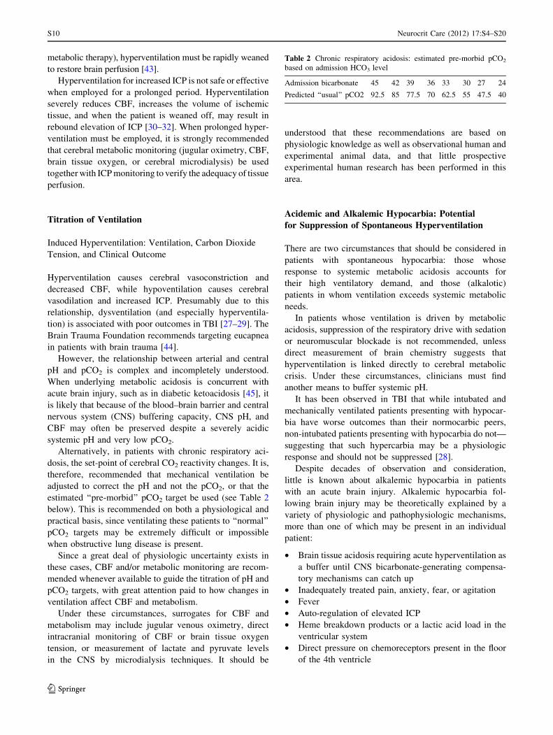
Alternatively, in patients with chronic respiratory aci-
dosis, the set-point of cerebral CO2 reactivity changes. It is,
therefore, recommended that mechanical ventilation be
adjusted to correct the pH and not the pCO2, or that the
estimated ‘‘pre-morbid’’ pCO2 target be used (see Table 2
below). This is recommended on both a physiological and
practical basis, since ventilating these patients to ‘‘normal’’
pCO2 targets may be extremely difficult or impossible
when obstructive lung disease is present.
Since a great deal of physiologic uncertainty exists in
these cases, CBF and/or metabolic monitoring are recom-
mended whenever available to guide the titration of pH and
pCO2 targets, with great attention paid to how changes in
ventilation affect CBF and metabolism.
Under these circumstances, surrogates for CBF and
metabolism may include jugular venous oximetry, direct
intracranial monitoring of CBF or brain tissue oxygen
tension, or measurement of lactate and pyruvate levels
in the CNS by microdialysis techniques. It should be
understood that these recommendations are based on
physiologic knowledge as well as observational human and
experimental animal data, and that little prospective
experimental human research has been performed in this
area.
Acidemic and Alkalemic Hypocarbia: Potential
for Suppression of Spontaneous Hyperventilation
There are two circumstances that should be considered in
patients with spontaneous hypocarbia: those whose
response to systemic metabolic acidosis accounts for
their high ventilatory demand, and those (alkalotic)
patients in whom ventilation exceeds systemic metabolic
needs.
In patients whose ventilation is driven by metabolic
acidosis, suppression of the respiratory drive with sedation
or neuromuscular blockade is not recommended, unless
direct measurement of brain chemistry suggests that
hyperventilation is linked directly to cerebral metabolic
crisis. Under these circumstances, clinicians must find
another means to buffer systemic pH.
It has been observed in TBI that while intubated and
mechanically ventilated patients presenting with hypocar-
bia have worse outcomes than their normocarbic peers,
non-intubated patients presenting with hypocarbia do not—
suggesting that such hypercarbia may be a physiologic
response and should not be suppressed [28].
Despite decades of observation and consideration,
little is known about alkalemic hypocarbia in patients
with an acute brain injury. Alkalemic hypocarbia fol-
lowing brain injury may be theoretically explained by a
variety of physiologic and pathophysiologic mechanisms,
more than one of which may be present in an individual
patient:
• Brain tissue acidosis requiring acute hyperventilation as
a buffer until CNS bicarbonate-generating compensa-
tory mechanisms can catch up
• Inadequately treated pain, anxiety, fear, or agitation
• Fever
• Auto-regulation of elevated ICP
• Heme breakdown products or a lactic acid load in the
ventricular system
• Direct pressure on chemoreceptors present in the floor
of the 4th ventricle
Table 2 Chronic respiratory acidosis: estimated pre-morbid pCO2
based on admission HCO3 level
Admission bicarbonate 45 42 39 36 33 30 27 24
Predicted ‘‘usual’’ pCO2 92.5 85 77.5 70 62.5 55 47.5 40
S10 Neurocrit Care (2012) 17:S4–S20
123

• Physiologic dysregulation of the medullary respiratory
rhythm generator, which has afferent inputs from the
pons, mesencephalon, and higher cortical centers
Since it is rarely known whether alkalemic hypocapnia
is a physiologic or pathophysiologic process, suppression
of this respiratory activity is recommended only in
response to evidence that hyperventilation is causing direct
harm, either directly by inducing cerebral ischemia or
indirectly by increased systemic metabolic demands and
work of breathing.
Oxygenation and Outcomes
Despite the traditional view that oxygen is good, it is now
apparent that the supra-physiologic levels of oxygen fre-
quently provided to acutely ill patients have the potential to
worsen reperfusion injury [46, 47]. The mechanism is likely
by potentiating the formation of reactive oxygen species in
post-ischemic tissue beds and further impairing mitochon-
drial function [48]. Conversely, hypoxia is a major source of
secondary brain injury [49], and the injured and ischemic
brain is particularly vulnerable to low oxygen levels.
Long hypothesized based on laboratory and animal data,
hyperoxia at a level of PaO2 > 300 mmHg on the first
arterial blood gas following resuscitation is independently
associated with poor outcomes in humans following TBI
[48] and cardiac arrest [47, 50], though not all data are in
accordance [51]. Hyperoxia drives the formation of reac-
tive oxygen species, overwhelming antioxidants at sites of
tissue injury; directly injures respiratory epithelium and
alveoli inducing inflammation; drives hypercarbia; and
leads to absorption atelectasis in the lung.
It is recommended that 100 % oxygen be provided for
pre-oxygenation immediately prior to intubation, but that
oxygen be immediately weaned following intubation to
50 %, or the lowest FiO2 that will support an oxyhemo-
globin saturation of 95–100 %. This normoxic
resuscitation strategy is recommended in 2010 American
Heart Association Guidelines for post-resuscitation care
after cardiac arrest [51].
Oxygenation and Ventilation Monitoring
Oxygenation should be monitored by pulse oximetry or by
arterial blood gas analysis when oximetry is suspected to
be inaccurate. Conditions of poor perfusion to the
extremities, acidosis, vasopressor use, anemia, carboxyhe-
moglobinemia and methemoglobinemia, and hypoxia all
have the potential to compromise the accuracy of pulse
oximetry measurements [52].
Ventilation is traditionally monitored by serial arterial
blood gas analysis, though venous blood gas analysis may
provide an adequate surrogate during titration of mechan-
ical ventilation, or when arterial samples cannot be
obtained. End-tidal quantitative capnography of exhaled
gases provides an appealing continuous measurement and
is extremely useful to monitor trends in ventilation.
One study showed that severely hyperventilated head
trauma patients (pCO2 < 25 mmHg) in the prehospital
environment had higher mortality, and that use of quanti-
tative capnography by paramedics significantly decreased
the incidence of hyperventilation [53]. A similar study in
patients with major trauma showed a much higher inci-
dence of ‘‘normocapnea’’ on hospital arrival when ETCO2
was monitored by medics.
Since ETCO2 measurements reflect not only ventilation
but also systemic perfusion, the correlation between
ETCO2 and pCO2 in the blood is variable. In an inpatient
environment, ETCO2 measurements should always be
correlated with an arterial pCO2 sample. ETCO2 and pCO2
may also vary significantly when lung disease and venti-
lation-perfusion mismatch are present [54].
Lung Injury
Patients with acute lung injury (ALI) or the acute respira-
tory distress syndrome (ARDS) are vulnerable to lung
injury known variably as ventilator induced lung injury
(VILI) or ventilator associated lung injury (VALI). ALI
and ARDS represent a spectrum of disease, in which there
exist bilateral parenchymal pulmonary infiltrates, signifi-
cant hypoxia, absence of left ventricular dysfunction, and
an acute onset of symptoms [55]. These patients often
require high levels of oxygen and ventilator pressures to
achieve adequate gas exchange.
Patients with ALI and ARDS have high circulating
levels of inflammatory mediators, which are associated
with injury to other vital organs, and are exacerbated by
injurious techniques of ventilation. VALI is thought to be
caused by:
• Barotauma, induced by high ventilator pressures, and
particularly a high plateau pressure
• Volutrauma, induced by higher tidal volumes and often
despite low ventilator pressures
• Atelectrauma, or the shearing injury caused by recur-
rent opening and closing of alveolar sacs that may lack
adequate surfactant
• High inspired fraction of oxygen
• High levels of circulating inflammatory cytokines
Many modes and techniques of ventilation have been
proposed to manage the severe gas exchange abnormalities
Neurocrit Care (2012) 17:S4–S20 S11
123

associated with ARDS, though only a few important issues
are covered here.
Patients with ALI or ARDS should be ventilated using a
strategy of low tidal volumes (6 cc/kg); low plateau pres-
sures (<30 mmHg); positive end expiratory pressure
(PEEP) adequate to prevent cyclic collapse of alveolar
units; and inhaled oxygen fraction rapidly weaned to 0.6 or
less, using PEEP and body positioning to best advantage.
Although the landmark study of low tidal volume
mechanical ventilation [56] emphasizes permissive hyper-
carbia, fluctuations of carbon dioxide levels are potent
mediators of CBF, and ICP and must be carefully consid-
ered in patients with elevated ICP or compromised CBF.
Several very small investigations suggest that lung
protective ventilation strategies causing mild hypercarbia
in patients with elevated ICP may be tolerated [57, 58], but
more data are needed before this may be considered safe in
routine practice. Prone positioning seems to increase ICP
[59].
When lung compliance is high, PEEP has the potential
to be transmitted to the intrathoracic vessels and indirectly
increase ICP. Although this physiology was once used to
justify low-PEEP ventilation in patients with head injury,
subsequent research has shown PEEP in brain-injured
patients to be well tolerated, especially when lung com-
pliance is poor and adequate blood pressure is maintained
[60, 61]. Ventilation without PEEP is discouraged, due to
the likelihood of inducing lung injury [62]. However, the
relationship of PEEP to ICP is of concern and should be
individually reviewed based on each patient’s physiology.
Sedation
Sedation use in the neurocritically ill is paradoxically
necessary yet fundamentally undesirable. Sedation may be
needed to alleviate fear and anxiety, reduce ICP and
cerebral oxygen consumption, facilitate intubation and
tolerance of mechanical ventilation, or to reduce sympa-
thetic nervous activity. Conversely, sedation makes
accurate neurological examination—the cornerstone of
clinical assessment—difficult or impossible. Acute changes
in brain physiology become difficult to detect, and the
accuracy of neuroprognostication is decreased [63].
Sedation often causes vasodilation, threatening cerebral
perfusion by hypotension but also by reversing physiologi-
cally advantageous shunting of blood into areas of ischemia.
It is possible that prolonged deep sedation may worsen
cognitive outcomes and contribute to muscle weakness.
Even short-acting sedatives are known to build up in fatty
tissues, causing effects well beyond their intended duration,
and no commonly employed sedative has been well studied
in a brain-injured population, as regards the duration of effect
or adverse consequences.
Decisions regarding the need for craniotomy, endovas-
cular therapies, ventricular drainage, ICP monitoring,
therapeutic temperature management, and many other
potentially morbid but also lifesaving therapies require an
accurate neurological assessment. Especially in the first
hours of care, the sedation of patients with unstable intra-
cranial pathophysiology should be minimized when it can
safely and humanely be limited.
This does not mean that patients should be agitated,
uncomfortable, severely hypertensive, or unsafe. It means
simply that the need for sedation must be weighed against the
need for accurate neurological assessment and tailored to the
individual patient and situation. Even more than with other
critically ill patients, medication-induced coma for neuro-
logically compromised patients is usually not desirable.
Necessity of Sedation
Complications associated with undersedation include ven-
tilator dysynchrony, patient injury, agitation, anxiety, device
removal [64–66], and elevated ICP [67]. Adequate sedation
is paramount in all therapeutic algorithms for the treatment
of increased ICP [68, 69], since psychomotor restlessness,
pain, and autonomic stress all adversely affect ICP, CBF,
CPP, and the cerebral metabolic rate for oxygen metabolism
(CMRO2). In this respect, adequate analgesia and sedation
make an essential contribution to preventing or limiting
secondary brain damage. Nevertheless, there are insufficient
data to confirm that sedation and analgesia per se reduce ICP
and improve neurological outcome [68]. Conversely, in a
general intensive care unit (ICU) population, the use of
excessive sedation and analgesia contributes to increased
duration of mechanical ventilation and longer length of stay
in the ICU and hospital [70–72], as well as increased rates of
depression, post-traumatic stress disorder, infections, and
long-term neurocognitive impairments [73–75].
Randomized controlled trials demonstrate that protocols
requiring decreases in sedative doses or daily interruption of
sedative and analgesic drugs can reduce lengths of
mechanical ventilation and ICU stay and reduce drug doses
administered [70–72]. Unless deep sedation or general
anesthesia is desired, analgesia should precede sedation.
Analgesia-based ‘‘sedation’’ is an evolving trend in general
critical care, and a recent randomized trial suggests
improved outcomes among mechanically ventilated patients
receiving primarily opioid analgesia with no sedation [76].
Many patients with adequate pain control do not require
sedation, and, conversely, most sedative medications pro-
vide no analgesia. Sedation without pain control may be
an important cause of delirium. Infusion of short acting
S12 Neurocrit Care (2012) 17:S4–S20
123

analgesics allows for interruption and neurological
assessment and intervals. In particular, a mechanistic
review suggests that remifentanil—a potent narcotic anal-
gesic with some sedating properties and an extremely rapid
pharmacokinetic profile—may be cost effective when ICU
length of stay is considered [77], though superiority over
fentanyl has not been demonstrated [78].
Environmental stimuli are important triggers of anxiety
and agitation. Providing a calm and reassuring environ-
ment, with attention to day–night cycles, restriction of
noise, use of appropriate music, and/or the reassuring
presence of friends and family may decrease agitation and
anxiety, making sedation unnecessary [79]. These envi-
ronmental measures are especially important when a
dominant-hemisphere lesion causes aphasia and attempts at
verbal communication generate agitated behaviors. The use
of ‘‘sitters’’ to re-direct and re-orient confused or agitated
patients is preferred to the use of sedating medications.
Patients with acute brain injury often have deficits in
short-term memory, concentration, and emotional control,
and their confusion may cause agitation. Confused patients
require gentle and repeated re-orientation to situation and
circumstances, which may obviate the need for sedation.
Given the potential of many sedatives (especially the
benzodiazepines) to cause delirium, agitated delirium
occasionally may be more effectively treated with anti-
psychotic medications than sedatives, though antipsychotic
medications may affect neurological recovery [80–82].
Examples of antipsychotic medication (each with its own
adverse-events profile) that may obviate the need for sed-
atives include haloperidol, quetiapine, olanzipine, and
risperidol.
Common Sedatives in Neurological Intensive Care
Prior to the release of dexmedetomidine in Canada, one
review of Canadian pharmacy databases and interviews
with caregivers in three Canadian neuro-intensive care
units (NICUs) [83] found that fentanyl was the most
commonly used analgesic. The same review showed that
propofol was the most common sedative agent in non-
trauma NICUs and some trauma NICUs, with midazolam
also used in some trauma NICUs.
Propofol
Propofol is perhaps better studied than other agents in
neurological critical care. Pharmacologically, its lipid for-
mulation allows for rapid penetration of the blood–brain
barrier, with rapid onset and cessation of action. It has
potent and immediate depressant effects on cerebral elec-
trical and metabolic activity, and it does not require renal
or hepatic metabolism for elimination. Disadvantages
include robust vasodilating and hypotensive effects, a
considerable intravenous lipid load, and the potential for
the rare, but frequently fatal, propofol infusion syndrome,
especially in children and in adults at higher doses.
Propofol has been used to sedate neurosurgical patients
to reduce elevated ICP [84, 85]. Propofol and morphine are
associated with improved control of ICP compared with
morphine alone in the treatment of severe TBI [85], and
propofol reduced elevated ICP more effectively than fen-
tanyl following severe TBI [86]. Propofol infusions to
reduce elevated ICP may need to be continued longer than
usually recommended for routine sedation [87], and high
doses for refractory status epilepticus or brain trauma has
been associated with propofol infusion syndrome [88].
Benzodiazepines
Midazolam is an appealing sedative option given the rapid
onset of action and short duration of effect with bolus
administration—making it an ideal agent for procedural
sedation. In addition, due to its potent gamma-aminobutyric
acid (GABA) activity and relatively benign hemodynamic
profile, midazolam is an important drug in refractory status
epilepticus. Yet as a long-term sedative for general ICU use,
midazolam accumulates in adipose tissues, significantly
prolonging duration of action unless interruptions or down-
titration of dose are routinely utilized.
Bolus-dose midazolam is a good choice for intermittent
agitation in a NICU population, while midazolam infusion
is associated with prolonged mechanical ventilation [89,
90]. Though most studies suggest the impact of midazolam
on hemodynamics is similar compared to dexmedetomi-
dine or propofol, a recent report suggests less instability
compared to dexmedetomidine [90].
Lorazepam is a longer acting benzodiazepine when used
in the short-term, but its duration of action is shorter than
midazolam when infused for more than 1 to 2 days. The
strong GABA activity of lorazepam suppresses electrical
and metabolic brain activity. Unlike midazolam, lorazepam
is formulated in propylene glycol, which can accumulate to
toxic levels causing metabolic acidosis and kidney injury.
At lorazepam infusion rates above 3 mg/h or daily doses
approaching 1 mg/kg, the osmolar gap should be followed,
and alternative agents should be used if the osmolar gap
rises above 10–12 mOsm/L [91, 92].
Dexmedetomidine
Dexmedetomidine, a centrally acting alpha agonist similar to
clonidine, but more specific for the alpha-2 receptor than
clonidine, is increasingly utilized for ICU sedation. Desir-
able properties include rapid onset and termination of
Neurocrit Care (2012) 17:S4–S20 S13
123

activity, mild to moderate sedation without significant
respiratory depressant action, analgesic effects, and less
delirium than the benzodiazepines [93]. Undesirable prop-
erties include a high incidence of bradycardia and
hypotension [90]. In addition, dexmedetomidine may pro-
duce less hemodynamic intolerance than clonidine, which is
available intravenously in many countries but not in the
United States.
In a prospective, randomized, double-blind study of 18
awake and intubated brain-injured patients using a cross-
over design [94], each patient received fentanyl–propofol
and fentanyl–dexmedetomidine. Both drugs were titrated to
the validated 100-point Hopkins Adapted Cognitive Exam
(ACE) cognitive battery. The difference in the change of
ACE scores between dexmedetomidine and propofol was
19.2 (95 % CI 12.3–26.1 p < 0.001) favoring improved
ACE scores with dexmedetomidine, suggesting better
cognitive function.
Several case reports and case series have evaluated the
potential role of dexmedetomidine for sedation in the NICU.
One reported successful sedation with dexmedetomidine in a
challenging TBI patient with alcohol withdrawal syndrome
when benzodiazepines clouded the patient’s neurological
examination and depressed ventilatory drive [95].
Another described six patients with TBI or intraven-
tricular hemorrhage treated with dexmedetomidine
sedation for 66 h at a mean dose of 0.67 mcg/kg/h in an
effort to wean off midazolam or propofol and fentanyl,
which was successful in all cases [96]. Though not clini-
cally significant, two patients experienced decreases in
heart rate with dexmedetomidine (from 101 to 69 beats per
minute and from 76 to 68 beats per minute), and two other
patients experienced different changes in systolic blood
pressure with dexmedetomidine (from 150 to 138 mmHg,
and 126 to 139 mmHg).
Barbiturates
Although out of favor due to prolonged duration of action
as well as cardiodepressant and possibly immunosuppres-
sant properties, barbiturates remain second-line therapy for
the control of ICP after propofol. They remain in wide-
spread use to control refractory status epilepticus, and their
potent effects on cerebral metabolic and electrical activity
make them an appealing class of agents for sedation in the
NICU. Pentobarbital serves as a potent agent for deep
sedation in patients with refractory status epilepticus or
elevated ICP [97–99].
Analgesia-First Sedation
Investigators at a single center randomized intubated gen-
eral ICU patients receiving bolus-dose morphine as needed
to either ‘‘no sedation’’ or ‘‘sedation with propofol and
midazolam.’’ ‘‘No sedation’’ patients required more mor-
phine than ‘‘sedation’’ patients but also had more
ventilator-free days and a shorter ICU length of stay,
without more adverse events related to undersedation but a
higher incidence of delirium [76].
Another randomized, open-label, multicenter study
compared two strategies: an analgesia-first approach with
remifentanil supplemented as needed with propofol (in the
first 3 days) and midazolam for day 4 and beyond; and a
hypnotic-based regimen using propofol (for the first 3 days)
or midazolam (after day 4) with supplemental analgesia
with fentanyl or morphine [100]. Both regimens were
titrated to a Sedation Agitation Scale (SAS) score of 1–3
and a pain intensity score of 1–2. Variability of neuro-
logical assessment and assessment times were less when
using remifentanil compared to fentanyl or morphine.
Patients receiving the analgesia-first approach with remif-
entanil were extubated more quickly than those treated
with morphine but were not different than those receiving
fentanyl. Remifentanil was well tolerated and provided
comparable hemodynamic stability to the hypnotic-based
regimen. Over three times as many users rated analgesia-
based sedation with remifentanil ‘‘very good’’ or ‘‘excel-
lent’’ compared with the hypnotic-based regimen.
Sedation Targets and Monitoring
Sedation should be titrated to a validated sedation scale or use
an electrophysiologic endpoint when neuromuscular block-
ade is employed or burst-suppression is desired. A recent
review of sedation assessment tools in the neurocritical care
setting concluded that the SAS and Richmond Agitation
Sedation Scale (RASS) are valid and useful for NICU patients,
and the bispectral index (BIS) may have a role in monitoring
deeply sedated patients in the NICU [84, 101–103].
Regarding assessment of delirium, no assessment tools
have been validated in neurocritical care patients, though
several studies that used the Intensive Care Delirium
Screening Checklist enrolled NICU patients [102, 104,
105]. A recent, highly insightful review of the topic is also
available [106].
Sedation for neurocritical care patients remains highly
variable, as shown by the inconsistent clinical approaches
to sedation during endovascular intervention for ischemic
stroke. One survey of 49 members of the Society of Vas-
cular and Interventional Neurology found that general
anesthesia was the most common approach, closely fol-
lowed by conscious sedation either by an anesthesia team
or with local anesthetic [107].
A retrospective study of 980 patients treated with en-
dovascular therapy for anterior circulation stroke showed
S14 Neurocrit Care (2012) 17:S4–S20
123

that the 44 % treated with general anesthesia had no delay
in time to therapy compared to conscious sedation (306 vs.
296 min), but had the same rate of hemorrhage and an
independent association with poor outcome and mortality
[108]. A similar study of 126 patients suggested that non-
intubated patients had lower mortality, smaller infarct size,
and better clinical outcomes [109]. These retrospective
studies await prospective confirmation but suggest that
conscious sedation may be safe and associated with
improved outcomes.
Several studies have addressed the sedation monitoring
approaches and medication choices for NICU patients. One
prospective, randomized trial of 67 mechanically ventilated
adult patients sedated with propofol showed that the
BIS monitor added to routine sedation scale monitoring
significantly reduced the propofol dose and the time to
awakening when compared to subjective scale monitoring
alone [110].
Daily Interruption of Sedation
Sedation protocols requiring a daily interruption of sedation
[71] are not yet sufficiently evaluated in patients with
underlying cerebral disease. In addition to adequate anal-
gesia, which is essential in severe cerebral trauma,
additional analgesia and sedation must be provided for
nursing interventions and any surgical interventions. In the
acute phase of intensive treatment, deep sedation (RASS
score of -5 or SAS score of 1) should generally be targeted,
especially if intracranial hypertension >15–20 mmHg) is
present [111, 112].
Communication
When communicating to an accepting or referring physi-
cian about this patient, consider including the key elements
listed in Table 3.
Table 3 Airway, ventilation, and sedation communication regarding
assessment and referral
Mental status and exam prior to intubation
Vitals pre- and post-intubation
Ease of intubation
ETT position confirmed?
Table 4 Medications commonly used in induction
Drug Dose Onset of
action
Duration
of effect
(min)
Indications Precautions
Fentanyl 2–3 lg/kg IV push
over 1–2 min
Within 2-3 min 30–60 Pre-induction, blunts ICP rise Respiratory depression,
hypotension, rare muscle wall
rigidity
Lidocaine 1.5 mg/kg IV 2–3 min
before intubation
45–90 s 10–20 Pre-induction, blunts ICP rise Avoid if allergic or high grade
heart block if no pacemaker
Esmolol 1–2 mg/kg IV 2–10 min 10–30 Pre-induction, blunts ICP rise Bradycardia, hypotension,
increased airway reactivity
Etomidate 0.3 mg/kg IV push 30–60 s 3–5 Induction, sedation; good in
hypotension. Decreases CBF,
ICP, preserves CPP
Decreases seizure threshold,
decreases cortisol synthesis;
avoid in sepsis
Propofol 2 mg/kg IV push 9–50 s 3–10 Induction, sedation, reduces ICP
and airway resistance,
anticonvulsive effects
Hypotension, myocardial
depression
Ketamine 1.5–2 mg/kg IV push 1–2 min 5–15 Induction, analgesia, sedation,
amnesia, bronchodilatory
effects; good in hypotension
Catecholamine surge, possible
increase in ICP, ‘‘re-
emergence’’ phenomenon if not
pretreated with benzos
Thiopental 3 mg/kg IV push 30–60 s 5–30 Normotensive, normovolemic
status epilepticus or increased
ICP pts
Hypotension, bronchospasm
Succinylcholine 1.5–2 mg/kg IV 30–60 s 5–15 Preferred paralytic unless
contraindicated
Avoid in hyperkalemia, myopathy,
neuropathy/denervation, history
of malignant hyperthermia
Rocuronium 1.2 mg/kg 45–60 s 45–70 Paralysis when succinylcholine
contraindicated
Vecuronium 0.2 mg/kg Within 3 min 35 Least preferred paralytic for RSI This dose speeds onset during RSI
Neurocrit Care (2012) 17:S4–S20 S15
123

Table 5 Pre-Intubation Neurological Assessment Checklist
S16 Neurocrit Care (2012) 17:S4–S20
123

References
1. Davis DP, Dunford JV, Ochs M, Park K, Hoyt DB. The use of
quantitative end-tidal capnometry to avoid inadvertent severe
hyperventilation in patients with head injury after paramedic
rapid sequence intubation. J Trauma. 2004;56:808–14.
2. Coplin WM, Pierson DJ, Cooley KD, Newell DW, Rubenfeld
GD. Implications of extubation delay in brain-injured patients
meeting standard weaning criteria. Am J Respir Crit Care Med.
2000;161:1530–6.
3. Walls RM, Murphy MF. Manual of emergency airway manage-
ment. 3rd ed. Philadelphia: Lippincott Williams & Wilkins; 2008.
4. Sagarin MJ, Barton ED, Chng YM, Walls RM. National
Emergency Airway Registry I. Airway management by US and
Canadian emergency medicine residents: a multicenter analysis
of more than 6,000 endotracheal intubation attempts. Ann
Emerg Med. 2005;46:328–36.
5. Li J, Murphy-Lavoie H, Bugas C, Martinez J, Preston C.
Complications of emergency intubation with and without
paralysis. Am J Emerg Med. 1999;17:141–3.
Table 5 continued
Neurocrit Care (2012) 17:S4–S20 S17
123

6. Sakles JC, Laurin EG, Rantapaa AA, Panacek EA. Airway
management in the emergency department: a one-year study of
610 tracheal intubations. Ann Emerg Med. 1998;31:325–32.
7. Walls RM. Rapid-sequence intubation in head trauma. Ann
Emerg Med. 1993;22:1008–13.
8. Bedford RF, Persing JA, Pobereskin L, Butler A. Lidocaine or
thiopental for rapid control of intracranial hypertension? Anesth
Analg. 1980;59:435–7.
9. Gabriel EJ, Ghajar J, Jagoda A, et al. Guidelines for prehospital
management of traumatic brain injury. J Neurotrauma. 2002;19:
111–74.
10. Weingart S. Additional thoughts on the controversy of lidocaine
administration before rapid sequence intubation in patients with
traumatic brain injuries. Ann Emerg Med. 2007;50:353.
11. Salhi B, Stettner E. In defense of the use of lidocaine in rapid
sequence intubation. Ann Emerg Med. 2007;49:84–6.
12. Feng CK, Chan KH, Liu KN, Or CH, Lee TY. A comparison of
lidocaine, fentanyl, and esmolol for attenuation of cardiovas-
cular response to laryngoscopy and tracheal intubation. Acta
Anaesthesiol Sin. 1996;34:61–7.
13. Reynolds SF, Heffner J. Airway management of the critically ill
patient: rapid-sequence intubation. Chest. 2005;127:1397–412.
14. Bergen JM, Smith DC. A review of etomidate for rapid sequence
intubation in the emergency department. J Emerg Med. 1997;15:
221–30.
15. Moss E, Powell D, Gibson RM, McDowall DG. Effect of
etomidate on intracranial pressure and cerebral perfusion pres-
sure. Br J Anaesth. 1979;51:347–52.
16. Hug CC Jr, McLeskey CH, Nahrwold ML, et al. Hemodynamic
effects of propofol: data from over 25,000 patients. Anesth
Analg. 1993;77:S21–9.
17. Bar-Joseph G, Guilburd Y, Tamir A, Guilburd JN. Effectiveness
of ketamine in decreasing intracranial pressure in children with
intracranial hypertension. J Neurosurg Pediatr. 2009;4:40–6.
18. Langsjo JW, Maksimow A, Salmi E, et al. S-ketamine anes-
thesia increases cerebral blood flow in excess of the metabolic
needs in humans. Anesthesiology. 2005;103:258–68.
19. Bourgoin A, Albanese J, Wereszczynski N, Charbit M, Vialet R,
Martin C. Safety of sedation with ketamine in severe head injury
patients: comparison with sufentanil. Crit Care Med. 2003;
31:711–7.
20. Kovarik WD, Mayberg TS, Lam AM, Mathisen TL, Winn HR.
Succinylcholine does not change intracranial pressure, cerebral
blood flow velocity, or the electroencephalogram in patients with
neurologic injury. Anesth Analg. 1994;78:469–73.
21. Martyn JA, Richtsfeld M. Succinylcholine-induced hyperkale-
mia in acquired pathologic states: etiologic factors and
molecular mechanisms. Anesthesiology. 2006;104:158–69.
22. Seder DB, Mayer SA. Critical care management of subarachnoid
hemorrhage and ischemic stroke. Clin Chest Med. 2009;30:
103–22, viii–ix.
23. Chesnut RM, Marshall SB, Piek J, Blunt BA, Klauber MR,
Marshall LF. Early and late systemic hypotension as a frequent
and fundamental source of cerebral ischemia following severe
brain injury in the Traumatic Coma Data Bank. Acta Neurochir
Suppl. 1993;59:121–5.
24. Prough DS, Lang J. Therapy of patients with head injuries: key
parameters for management. J Trauma. 1997;42:S10–8.
25. Trzeciak S, Jones AE, Kilgannon JH, et al. Significance of
arterial hypotension after resuscitation from cardiac arrest. Crit
Care Med. 2009;37:2895–903. quiz 904.
26. Kilgannon JH, Roberts BW, Reihl LR, et al. Early arterial
hypotension is common in the post-cardiac arrest syndrome and
associated with increased in-hospital mortality. Resuscitation.
2008;79:410–6.
27. Dumont TM, Visioni AJ, Rughani AI, Tranmer BI, Crookes B.
Inappropriate prehospital ventilation in severe traumatic brain
injury increases in-hospital mortality. J Neurotrauma. 2010;27:
1233–41.
28. Davis DP, Idris AH, Sise MJ, et al. Early ventilation and out-
come in patients with moderate to severe traumatic brain injury.
Crit Care Med. 2006;34:1202–8.
29. Davis DP, Stern J, Sise MJ, Hoyt DB. A follow-up analysis
of factors associated with head-injury mortality after para-
medic rapid sequence intubation. J Trauma. 2005;59:
486–90.
30. Coles JP, Fryer TD, Coleman MR, et al. Hyperventilation fol-
lowing head injury: effect on ischemic burden and cerebral
oxidative metabolism. Crit Care Med. 2007;35:568–78.
31. Coles JP, Minhas PS, Fryer TD, et al. Effect of hyperventilation
on cerebral blood flow in traumatic head injury: clinical rele-
vance and monitoring correlates. Crit Care Med. 2002;30:
1950–9.
32. Diringer MN, Videen TO, Yundt K, et al. Regional cerebro-
vascular and metabolic effects of hyperventilation after severe
traumatic brain injury. J Neurosurg. 2002;96:103–8.
33. Walsh BK, Crotwell DN, Restrepo RD. Capnography/Cap-
nometry during mechanical ventilation: 2011. Respir Care.
2011;56:503–9.
34. Seneviratne J, Mandrekar J, Wijdicks EF, Rabinstein AA.
Noninvasive ventilation in myasthenic crisis. Arch Neurol.
2008;65:54–8.
35. Flandreau G, Bourdin G, Leray V, et al. Management and long-
term outcome of patients with chronic neuromuscular disease
admitted to the intensive care unit for acute respiratory failure: a
single-center retrospective study. Respir Care. 2011;56:
953–60.
36. Piastra M, Antonelli M, Caresta E, Chiaretti A, Polidori G, Conti
G. Noninvasive ventilation in childhood acute neuromuscular
respiratory failure: a pilot study. Respiration. 2006;73:791–8.
37. Abel M, Eisenkraft JB. Anesthetic implications of myasthenia
gravis. Mt Sinai J Med. 2002;69:31–7.
38. Rice MJ, Mancuso AA, Gibbs C, Morey TE, Gravenstein N,
Deitte LA. Cricoid pressure results in compression of the post-
cricoid hypopharynx: the esophageal position is irrelevant.
Anesth Analg. 2009;109:1546–52.
39. Ellis DY, Harris T, Zideman D. Cricoid pressure in emergency
department rapid sequence tracheal intubations: a risk-benefit
analysis. Ann Emerg Med. 2007;50:653–65.
40. Grande CM, Barton CR, Stene JK. Appropriate techniques for
airway management of emergency patients with suspected spinal
cord injury. Anesth Analg. 1988;67:714–5.
41. Koenig MA, Bryan M, Lewin JL 3rd, Mirski MA, Geocadin RG,
Stevens RD. Reversal of transtentorial herniation with hyper-
tonic saline. Neurology. 2008;70:1023–9.
42. Qureshi AI, Geocadin RG, Suarez JI, Ulatowski JA. Long-term
outcome after medical reversal of transtentorial herniation in
patients with supratentorial mass lesions. Crit Care Med.
2000;28:1556–64.
43. Oertel M, Kelly DF, Lee JH, et al. Efficacy of hyperventilation,
blood pressure elevation, and metabolic suppression therapy in
controlling intracranial pressure after head injury. J Neurosurg.
2002;97:1045–53.
44. Badjatia N, Strongilis E, Prescutti M, et al. Metabolic benefits of
surface counter warming during therapeutic temperature mod-
ulation. Crit Care Med. 2009;37:1893–7.
45. Wood EG, Go-Wingkun J, Luisiri A, Aceto T Jr. Symptomatic
cerebral swelling complicating diabetic ketoacidosis docu-
mented by intraventricular pressure monitoring: survival without
neurologic sequela. Pediatr Emerg Care. 1990;6:285–8.
S18 Neurocrit Care (2012) 17:S4–S20
123

46. Balan IS, Fiskum G, Hazelton J, Cotto-Cumba C, Rosenthal RE.
Oximetry-guided reoxygenation improves neurological outcome
after experimental cardiac arrest. Stroke. 2006;37:3008–13.
47. Brucken A, Kaab AB, Kottmann K, et al. Reducing the duration
of 100 % oxygen ventilation in the early reperfusion period after
cardiopulmonary resuscitation decreases striatal brain damage.
Resuscitation. 2010;81:1698–703.
48. Davis DP, Meade W, Sise MJ, et al. Both hypoxemia and
extreme hyperoxemia may be detrimental in patients with severe
traumatic brain injury. J Neurotrauma. 2009;26:2217–23.
49. Kilgannon JH, Jones AE, Shapiro NI, et al. Association between
arterial hyperoxia following resuscitation from cardiac arrest
and in-hospital mortality. JAMA. 2010;303:2165–71.
50. Kilgannon JH, Jones AE, Parrillo JE, et al. Relationship between
supranormal oxygen tension and outcome after resuscitation
from cardiac arrest. Circulation. 2011;123:2717–22.
51. Bellomo R, Bailey M, Eastwood GM, et al. Arterial hyperoxia
and in-hospital mortality after resuscitation from cardiac arrest.
Crit Care. 2011;15:R90.
52. Peberdy MA, Callaway CW, Neumar RW, et al. Part 9: post-
cardiac arrest care: 2010 American Heart Association Guide-
lines for Cardiopulmonary Resuscitation and Emergency
Cardiovascular Care. Circulation. 2010;122:S768–86.
53. Menchem CC. Pulse Oximetry. In: Basow DS, editor. UpTo-
Date. Waltham, MA: UpToDate Publishing; 2012.
54. Helm M, Schuster R, Hauke J, Lampl L. Tight control of pre-
hospital ventilation by capnography in major trauma victims. Br
J Anaesth. 2003;90:327–32.
55. Hardman JG, Aitkenhead AR. Estimating alveolar dead space
from the arterial to end-tidal CO(2) gradient: a modeling anal-
ysis. Anesth Analg. 2003;97:1846–51.
56. Artigas A, Bernard GR, Carlet J, et al. The American-European
Consensus Conference on ARDS, part 2: ventilatory, pharma-
cologic, supportive therapy, study design strategies, and issues
related to recovery and remodeling. Acute respiratory distress
syndrome. Am J Respir Crit Care Med. 1998;157:1332–47.
57. Ventilation with lower tidal volumes as compared with tradi-
tional tidal volumes for acute lung injury and the acute
respiratory distress syndrome. The Acute Respiratory Distress
Syndrome Network. N Engl J Med. 2000;342:1301–8.
58. Petridis AK, Doukas A, Kienke S, et al. The effect of lung-
protective permissive hypercapnia in intracerebral pressure in
patients with subarachnoid haemorrhage and ARDS. A retro-
spective study. Acta Neurochir. 2010;152:2143–5.
59. Bennett SS, Graffagnino C, Borel CO, James ML. Use of high
frequency oscillatory ventilation (HFOV) in neurocritical care
patients. Neurocrit Care. 2007;7:221–6.
60. Reinprecht A, Greher M, Wolfsberger S, Dietrich W, Illievich
UM, Gruber A. Prone position in subarachnoid hemorrhage
patients with acute respiratory distress syndrome: effects on
cerebral tissue oxygenation and intracranial pressure. Crit Care
Med. 2003;31:1831–8.
61. Caricato A, Conti G, Della Corte F, et al. Effects of PEEP on the
intracranial system of patients with head injury and subarach-
noid hemorrhage: the role of respiratory system compliance.
J Trauma. 2005;58:571–6.
62. Muench E, Bauhuf C, Roth H, et al. Effects of positive end-
expiratory pressure on regional cerebral blood flow, intracranial
pressure, and brain tissue oxygenation. Crit Care Med. 2005;33:
2367–72.
63. Koutsoukou A, Perraki H, Raftopoulou A, et al. Respiratory
mechanics in brain-damaged patients. Intensive Care Med.
2006;32:1947–54.
64. Samaniego EA, Mlynash M, Caulfield AF, Eyngorn I, Wijman
CA. Sedation confounds outcome prediction in cardiac arrest
survivors treated with hypothermia. Neurocrit Care. 2011;15:
113–9.
65. Riker RR, Fraser GL. Altering intensive care sedation paradigms
to improve patient outcomes. Anesthesiol Clin. 2011;29:663–74.
66. Jacobi J, Fraser GL, Coursin DB, et al. Clinical practice
guidelines for the sustained use of sedatives and analgesics in
the critically ill adult. Crit Care Med. 2002;30:119–41.
67. Fraser GL, Riker RR, Prato BS, Wilkins ML. The frequency and
cost of patient-initiated device removal in the ICU. Pharmaco-
therapy. 2001;21:1–6.
68. Skoglund K, Enblad P, Marklund N. Effects of the neurological
wake-up test on intracranial pressure and cerebral perfusion
pressure in brain-injured patients. Neurocrit Care. 2009;11:
135–42.
69. Brain Trauma F, American Association of Neurological S,
Congress of Neurological S, et al. Guidelines for the manage-
ment of severe traumatic brain injury. VI. Indications for
intracranial pressure monitoring. J Neurotrauma. 2007;24(Suppl
1):S37–44.
70. Citerio G, Cormio M. Sedation in neurointensive care: advances in
understanding and practice. Curr Opin Crit Care. 2003;9:120–6.
71. Brook AD, Ahrens TS, Schaiff R, et al. Effect of a nursing-
implemented sedation protocol on the duration of mechanical
ventilation. Crit Care Med. 1999;27:2609–15.
72. Kress JP, Pohlman AS, O’Connor MF, Hall JB. Daily inter-
ruption of sedative infusions in critically ill patients undergoing
mechanical ventilation. N Engl J Med. 2000;342:1471–7.
73. Mehta S, Burry L, Martinez-Motta JC, et al. A randomized trial
of daily awakening in critically ill patients managed with a
sedation protocol: a pilot trial. Crit Care Med. 2008;36:2092–9.
74. Treggiari MM, Romand JA, Yanez ND, et al. Randomized trial
of light versus deep sedation on mental health after critical ill-
ness. Crit Care Med. 2009;37:2527–34.
75. Jones C, Backman C, Capuzzo M, Flaatten H, Rylander C,
Griffiths RD. Precipitants of post-traumatic stress disorder fol-
lowing intensive care: a hypothesis generating study of diversity
in care. Intensive Care Med. 2007;33:978–85.
76. Hopkins RO, Jackson JC. Long-term neurocognitive function
after critical illness. Chest. 2006;130:869–78.
77. Strom T, Martinussen T, Toft P. A protocol of no sedation for
critically ill patients receiving mechanical ventilation: a ran-
domised trial. Lancet. 2010;375:475–80.
78. Spies C, Macguill M, Heymann A, et al. A prospective, ran-
domized, double-blind, multicenter study comparing
remifentanil with fentanyl in mechanically ventilated patients.
Intensive Care Med. 2011;37:469–76.
79. Al MJ, Hakkaart L, Tan SS, Bakker J. Cost-consequence anal-
ysis of remifentanil-based analgo-sedation vs. conventional
analgesia and sedation for patients on mechanical ventilation in
the Netherlands. Crit Care. 2010;14:R195.
80. Khalifezadeh A, Safazadeh S, Mehrabi T, Mansour BA.
Reviewing the effect of nursing interventions on delirious
patients admitted to intensive care unit of neurosurgery ward in
Al-Zahra Hospital, Isfahan University of Medical Sciences. Iran
J Nurs Midwifery Res. 2011;16:106–12.
81. Kline AE, Hoffman AN, Cheng JP, Zafonte RD, Massucci JL.
Chronic administration of antipsychotics impede behavioral
recovery after experimental traumatic brain injury. Neurosci
Lett. 2008;448:263–7.
82. Hoffman AN, Cheng JP, Zafonte RD, Kline AE. Administration
of haloperidol and risperidone after neurobehavioral testing
hinders the recovery of traumatic brain injury-induced deficits.
Life Sci. 2008;83:602–7.
83. Kline AE, Massucci JL, Zafonte RD, Dixon CE, DeFeo JR, Rogers
EH. Differential effects of single versus multiple administrations
Neurocrit Care (2012) 17:S4–S20 S19
123
of haloperidol and risperidone on functional outcome after
experimental brain trauma. Crit Care Med. 2007;35:919–24.
84. Teitelbaum JS, Ayoub O, Skrobik Y. A critical appraisal of
sedation, analgesia and delirium in neurocritical care. Can J
Neurol Sci. 2011;38:815–25.
85. Herregods L, Verbeke J, Rolly G, Colardyn F. Effect of propofol
on elevated intracranial pressure. Preliminary results. Anaes-
thesia. 1988;43:107–9.
86. Kelly DF, Goodale DB, Williams J, et al. Propofol in the
treatment of moderate and severe head injury: a randomized,
prospective double-blinded pilot trial. J Neurosurg. 1999;90:
1042–52.
87. Herregods L, Mergaert C, Rolly G, Collardyn F. Comparison of
the effects of 24-hour propofol or fentanyl infusions on intra-
cranial pressure. J Drug Dev. 1989;2:99–100.
88. Cremer OL, Moons KG, Bouman EA, Kruijswijk JE, de Smet
AM, Kalkman CJ. Long-term propofol infusion and cardiac
failure in adult head-injured patients. Lancet. 2001;357:117–8.
89. Iyer VN, Hoel R, Rabinstein AA. Propofol infusion syndrome in
patients with refractory status epilepticus: an 11-year clinical
experience. Crit Care Med. 2009;37:3024–30.
90. Barrientos-Vega R, Mar Sanchez-Soria M, Morales-Garcia C,
Robas-Gomez A, Cuena-Boy R, Ayensa-Rincon A. Prolonged
sedation of critically ill patients with midazolam or propofol:
impact on weaning and costs. Crit Care Med. 1997;25:33–40.
91. Jakob SM, Ruokonen E, Grounds RM, et al. Dexmedetomidine
vs. midazolam or propofol for sedation during prolonged
mechanical ventilation: two randomized controlled trials.
JAMA. 2012;307:1151–60.
92. Yahwak JA, Riker RR, Fraser GL, Subak-Sharpe S. Determi-
nation of a lorazepam dose threshold for using the osmol gap to
monitor for propylene glycol toxicity. Pharmacotherapy.
2008;28:984–91.
93. Horinek EL, Kiser TH, Fish DN, MacLaren R. Propylene glycol
accumulation in critically ill patients receiving continuous
intravenous lorazepam infusions. Ann Pharmacother. 2009;43:
1964–71.
94. Riker RR, Shehabi Y, Bokesch PM, et al. Dexmedetomidine vs
midazolam for sedation of critically ill patients: a randomized
trial. JAMA. 2009;301:489–99.
95. Mirski MA, Lewin JJ 3rd, Ledroux S, et al. Cognitive
improvement during continuous sedation in critically ill, awake
and responsive patients: the Acute Neurological ICU Sedation
Trial (ANIST). Intensive Care Med. 2010;36:1505–13.
96. Tang JF, Chen PL, Tang EJ, May TA, Stiver SI. Dexmede-
tomidine controls agitation and facilitates reliable, serial
neurological examinations in a non-intubated patient with trau-
matic brain injury. Neurocrit Care. 2011;15:175–81.
97. Grof TM, Bledsoe KA. Evaluating the use of dexmedetomidine
in neurocritical care patients. Neurocrit Care. 2010;12:356–61.
98. Chen HI, Malhotra NR, Oddo M, Heuer GG, Levine JM, Le-
Roux PD. Barbiturate infusion for intractable intracranial
hypertension and its effect on brain oxygenation. Neurosurgery.
2008;63:880–6. discussion 6–7.
99. Marshall GT, James RF, Landman MP, et al. Pentobarbital coma
for refractory intra-cranial hypertension after severe traumatic
brain injury: mortality predictions and one-year outcomes in 55
patients. J Trauma. 2010;69:275–83.
100. Claassen J, Hirsch LJ, Mayer SA. Treatment of status epilepti-
cus: a survey of neurologists. J Neurol Sci. 2003;211:37–41.
101. Karabinis A, Mandragos K, Stergiopoulos S, et al. Safety and
efficacy of analgesia-based sedation with remifentanil versus
standard hypnotic-based regimens in intensive care unit patients
with brain injuries: a randomised, controlled trial [IS-
RCTN50308308]. Crit Care. 2004;8:R268–80.
102. Riker RR, Picard JT, Fraser GL. Prospective evaluation of the
Sedation-Agitation Scale for adult critically ill patients. Crit
Care Med. 1999;27:1325–9.
103. Sessler CN, Gosnell MS, Grap MJ, et al. The Richmond
Agitation-Sedation Scale: validity and reliability in adult
intensive care unit patients. Am J Respir Crit Care Med. 2002;
166:1338–44.
104. Deogaonkar A, Gupta R, DeGeorgia M, et al. Bispectral Index
monitoring correlates with sedation scales in brain-injured
patients. Crit Care Med. 2004;32:2403–6.
105. Bergeron N, Dubois MJ, Dumont M, Dial S, Skrobik Y. Inten-
sive Care Delirium Screening Checklist: evaluation of a new
screening tool. Intensive Care Med. 2001;27:859–64.
106. Van Rompaey B, Elseviers MM, Schuurmans MJ, Shortridge-
Baggett LM, Truijen S, Bossaert L. Risk factors for delirium in
intensive care patients: a prospective cohort study. Crit Care.
2009;13:R77.
107. Frontera JA. Delirium and sedation in the ICU. Neurocrit Care.
2011;14:463–74.
108. McDonagh DL, Olson DM, Kalia JS, Gupta R, Abou-Chebl A,
Zaidat OO. Anesthesia and sedation practices among neuroin-
terventionalists during acute ischemic stroke endovascular
therapy. Front Neurol. 2010;1:118.
109. Abou-Chebl A, Lin R, Hussain MS, et al. Conscious sedation
versus general anesthesia during endovascular therapy for acute
anterior circulation stroke: preliminary results from a retro-
spective, multicenter study. Stroke. 2010;41:1175–9.
110. Jumaa MA, Zhang F, Ruiz-Ares G, et al. Comparison of safety
and clinical and radiographic outcomes in endovascular acute
stroke therapy for proximal middle cerebral artery occlusion
with intubation and general anesthesia versus the nonintubated
state. Stroke. 2010;41:1180–4.
111. Olson DM, Thoyre SM, Peterson ED, Graffagnino C. A ran-
domized evaluation of bispectral index-augmented sedation
assessment in neurological patients. Neurocrit Care. 2009;11:
20–7.
112. Enblad P, Nilsson P, Chambers I, et al. R3-survey of traumatic
brain injury management in European Brain IT centres year
2001. Intensive Care Med. 2004;30:1058–65.
S20 Neurocrit Care (2012) 17:S4–S20
123