Emergencies Resulting from Pulmonary Diseases & Disorders EMS Professions Temple College.
202
Emergencies Resulting from Pulmonary Diseases & Disorders EMS Professions Temple College
-
Upload
piers-carroll -
Category
Documents
-
view
219 -
download
0
Transcript of Emergencies Resulting from Pulmonary Diseases & Disorders EMS Professions Temple College.

Emergencies Resulting from Pulmonary Diseases & Disorders
Emergencies Resulting from Pulmonary Diseases & Disorders
EMS Professions
Temple College

Pulmonary Diseases & DisordersPulmonary Diseases & Disorders
Pulmonary Disease & Conditions may result from:– Infectious causes– Non-Infectious causes
Adversely affect one or more of the following– Ventilation– Diffusion– Perfusion

Pulmonary Diseases & DisordersPulmonary Diseases & Disorders The Respiratory Emergency may stem from
dysfunction or disease of (examples only):– Control System
• Hyperventilation• Central Respiratory Depression• CVA
– Thoracic Bellows• Chest/Diaphragm Trauma• Pickwickian Syndrome• Guillian-Barre Syndrome• Myasthenia Gravis• COPD

Pulmonary Diseases & DisordersPulmonary Diseases & Disorders The Respiratory Emergency may affect the
upper or lower airways
Upper Airway Obstruction– Tongue– Foreign Body Aspiration– Angioneurotic Edema– Maxillofacial, Larnygotracheal Trauma– Croup– Epiglottitis

Respiratory Emergencies: CausesRespiratory Emergencies: Causes
Lower Airway Obstruction– Emphysema– Chronic Bronchitis– Asthma– Cystic Fibrosis

Pulmonary Diseases & DisordersPulmonary Diseases & Disorders
The Respiratory Emergency may stem from Gas Exchange Surface Abnormalities– Cardiogenic Pulmonary Edema– Non-cardiogenic Pulmonary Edema– Pneumonia– Toxic Gas Inhalation– Pulmonary Embolism– Drowning

Pulmonary Diseases & DisordersPulmonary Diseases & Disorders
Problems with the Gas
Exchange Surface

Pulmonary EdemaPulmonary Edema
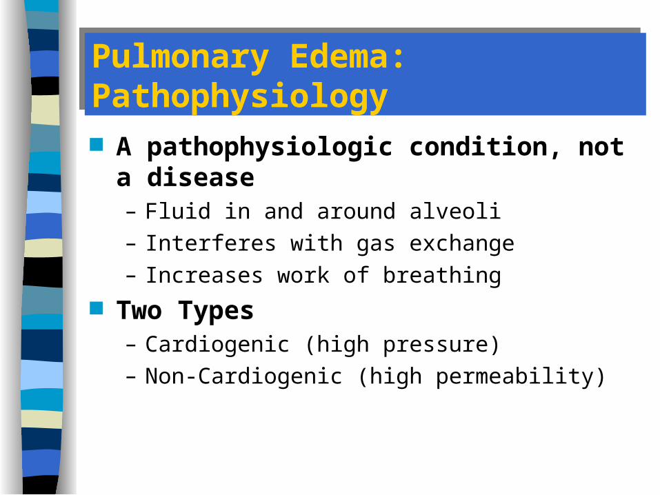
Pulmonary Edema: PathophysiologyPulmonary Edema: Pathophysiology
A pathophysiologic condition, not a disease– Fluid in and around alveoli– Interferes with gas exchange– Increases work of breathing
Two Types– Cardiogenic (high pressure)– Non-Cardiogenic (high permeability)
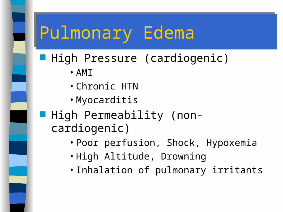
Pulmonary EdemaPulmonary Edema High Pressure (cardiogenic)
• AMI• Chronic HTN• Myocarditis
High Permeability (non-cardiogenic)• Poor perfusion, Shock, Hypoxemia• High Altitude, Drowning• Inhalation of pulmonary irritants

Cardiogenic Pulmonary Edema: EtiologyCardiogenic Pulmonary Edema: Etiology
Left ventricular failure Valvular heart disease
– Stenosis– Insufficiency
Hypertensive crisis (high afterload) Volume overload
Increased Pressure in Pulmonary Vascular Bed

Pulmonary EdemaPulmonary Edema
High Permeability– Disrupted alveolar-capillary membrane– Membrane allows fluid to leak into the interstitial
space– Widened interstitial space impairs diffusion

Non-Cardiogenic Pulmonary Edema: EtiologyNon-Cardiogenic Pulmonary Edema: Etiology
Toxic inhalation Near drowning Liver disease Nutritional deficiencies Lymphomas High altitude pulmonary edema Adult respiratory distress syndrome
Increased Permeability of Alveolar-Capillary Walls
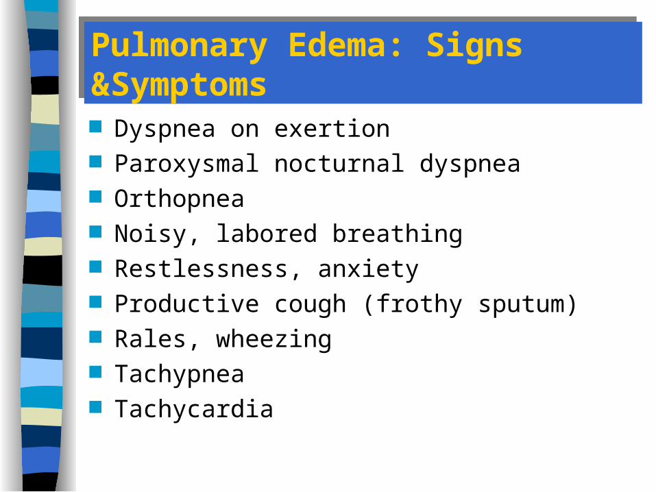
Pulmonary Edema: Signs &SymptomsPulmonary Edema: Signs &Symptoms
Dyspnea on exertion Paroxysmal nocturnal dyspnea Orthopnea Noisy, labored breathing Restlessness, anxiety Productive cough (frothy sputum) Rales, wheezing Tachypnea Tachycardia

Management of Non-Cardiogenic Pulmonary EdemaManagement of Non-Cardiogenic Pulmonary Edema Position Oxygen PPV / Intubation
– CPAP– PEEP
IV Access; Minimal fluid administration Treat the underlying cause
– Diuretics usually not helpful; May be harmful Transport

Adult Respiratory Distress SyndromeAdult Respiratory Distress Syndrome
AKA: Non-cardiogenic pulmonary edema A complication of:
– Severe Trauma / Shock– Severe infection / Sepsis– Bypass Surgery– Multiple blood transfusions– Drug overdose– Aspiration– Decreased compliance– Hypoxemia
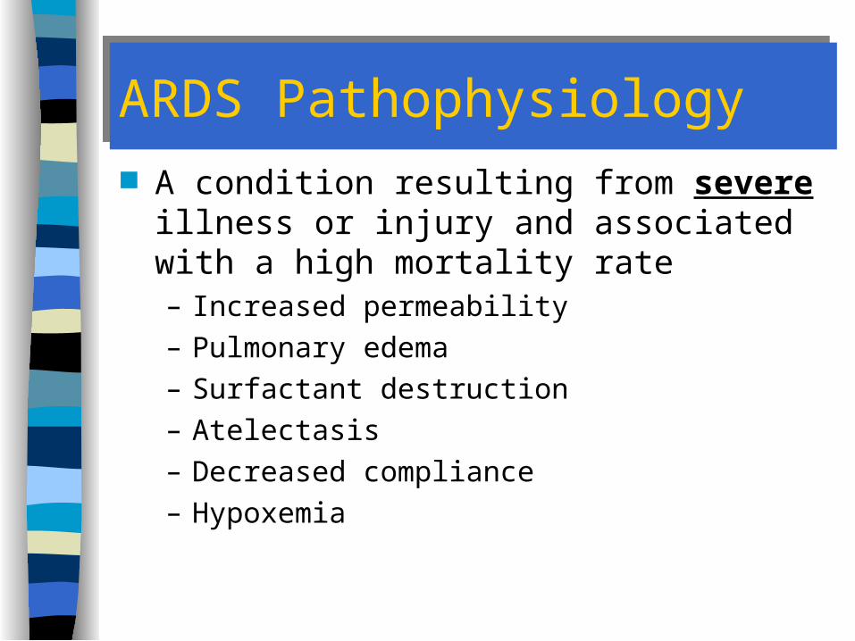
ARDS PathophysiologyARDS Pathophysiology A condition resulting from severe illness or
injury and associated with a high mortality rate– Increased permeability– Pulmonary edema– Surfactant destruction– Atelectasis– Decreased compliance– Hypoxemia

ARDS PresentationARDS Presentation
History– Recent hx of severe illness or injury– Often already being treated for underlying cause
Exam Findings– Dyspnea– Evidence of pulmonary edema– Poor oxygenation– Decreased lung compliance

ARDS ManagementARDS Management
Airway Management– Endotracheal intubation– Suction
Mechanical Ventilation– PEEP
ECG Monitoring Treat underlying cause
– May require vasopressors for shock

Pneumonia Pneumonia

PneumoniaPneumonia
Fifth leading cause of death in US Group of Specific infections Risk factors
– Cigarette smoking– Exposure to cold– Extremes of age
• young • old

PneumoniaPneumonia Inflammation of the bronchioles and alveoli
– Products of inflammation (secretions, pus) add to respiration difficulty
Gas exchange is impaired Work of breathing increases May lead to
– Atelectasis– Sepsis– VQ Mismatch– Hypoxemia
Pneumonia: EtiologyPneumonia: Etiology
Viral Bacterial Fungi Protozoa (pneumocystis) Aspiration

Presentation of PneumoniaPresentation of Pneumonia
Shortness of breath, Dyspnea Fever, chills Pleuritic Chest Pain, Tachycardia Cough
– Green/brown sputum May have crackles, rhonchi or wheezing in
peripheral lung fields– Consolidation– Egophony
Management of PneumoniaManagement of Pneumonia
Treatment mostly based upon symptoms– Oxygen– Rarely is intubation required– IV Access & Rehydration
– B2 agonists may be useful
– Antibiotics (e.g. Rocephin)– Antipyretics

Pneumonia: ManagementPneumonia: Management
MD follow-up for labs, cultures & Rx Transport considerations
– Elderly have significant co-morbidity– Young have difficulty with oral medications– ED vs PMD office/clinic– Transport in position of comfort
Would an anticholinergic like Atrovent be useful in managing
pneumonia?

Pulmonary EmbolismPulmonary Embolism

Pulmonary EmbolismPulmonary Embolism
~ 50,000 deaths / year– ~5% of all sudden deaths– <10% of all PE result in death

Pulmonary Embolism: PathophysiologyPulmonary Embolism: Pathophysiology
Something moving with flow of blood passes through right heart into pulmonary circulation
It reaches an area too narrow to pass through and lodges there
Part of pulmonary circulation is blocked Blood:
– Does not pass alveoli– Does not exchange gases
Pulmonary Embolism (PE)Pulmonary Embolism (PE)
A disorder of perfusion Combination of factors increase probability of
occurrence– Hypercoagulability– Platelet aggregation– Deep vein stasis
Embolus usually originates in lower extremities or pelvis

Pulmonary Embolism (PE)Pulmonary Embolism (PE)
Risk factors– Venostasis or DVT– Recent surgery or trauma
• Long bone fractures (lower)
– Oral contraceptives– Pregnancy– Smoking– Cancer

Pulmonary Embolism: EtiologyPulmonary Embolism: Etiology
Most Common Cause = Blood Clots
Vessel Wall Injury
Hypercoagulability Venous Stasis
Virchow’sTriad
Other causes– Air– Amniotic fluid– Fat particles (long bone fracture)– Particulates from substance abuse– Venous catheter
Pulmonary Embolism: EtiologyPulmonary Embolism: Etiology

Pulmonary Embolism: Signs & SymptomsPulmonary Embolism: Signs & Symptoms
Small Emboli– Rapid Onset– Dyspnea– Tachycardia– Tachypnea– Fever– Episodic = Showers– Evidence or history of thrombophlebitis– Consider early when no other cardiorespiratory
diagnosis fits

Larger Emboli– Small Emboli S/S plus:– Pleuritic pain– Pleural rub– Coughing– Wheezing– Hemoptysis (rare)
Pulmonary Embolism: Signs & SymptomsPulmonary Embolism: Signs & Symptoms
Very Large Emboli– Preceded by S/S of Small & Larger Emboli plus: – Central chest pain– Distended neck veins– Acute right heart failure– Shock– Cardiac arrest
Pulmonary Embolism: Signs & SymptomsPulmonary Embolism: Signs & Symptoms

Pulmonary Embolism: Signs & SymptomsPulmonary Embolism: Signs & Symptoms
There are NO assessment
findings specific to pulmonary embolism

Management based on severity of Sx/Sx Airway & Breathing
– High concentration O2
– Consider assisting ventilations– Early Intubation
Circulation– IV, 2 lg bore sites
• Fluid bolus then TKO; Titrate to BP ~ 90 mm Hg
– Monitor ECG Rapid transport
Pulmonary Embolism: ManagementPulmonary Embolism: Management

PE ManagementPE Management Thrombolytics
– Aspirin & Heparin (questionable if any benefit) Rapid transport to appropriate facility
– Embolectomy or thrombolytics at hospital (rarely effective in severe cases due to time delay)
– Poor prognosis when cardiac arrest follows

Pulmonary EmbolismPulmonary Embolism
If the patient is alive when you get to them, that embolus isn’t going to
kill them.
But the next one they throw might!

PleurisyPleurisy
Inflammation of pleura caused by a friction rub– layers of pleura rubbing together
Commonly associated with other respiratory disease

Presentation of PleurisyPresentation of Pleurisy
Sharp, sudden and intermittent chest pain with related dyspnea– Possibly referred to shoulder– May or with respiration
Pleural “friction rub” may be audible” May have effusion or be dry

PleurisyPleurisy
Management– Based upon severity of presentation– Mostly supportive

Pulmonary Diseases & DisordersPulmonary Diseases & Disorders
Problems with Airway Obstructions

Obstructive Airway Diseases
Obstructive Airway DiseaseObstructive Airway Disease
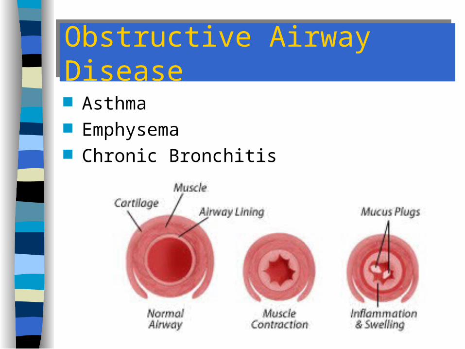
Asthma Emphysema Chronic Bronchitis

Obstructive Airway DiseasesObstructive Airway Diseases
Asthma experienced by ~ 4 - 5 % of US population– Mortality rate increasing
Factors leading to Obstructive Airway Diseases– Smoking– Exposure to environmental agents– Genetic predisposition
How does this differ from “COPD”?

Obstructive Airway DiseaseObstructive Airway Disease
Exacerbation Factors– Intrinsic
• Stress (especially in adults)• URI• Exercise
– Extrinsic• Cigarette Smoke• Allergens• Drugs• Occupational hazards

Obstructive Airway DiseaseObstructive Airway Disease
General Pathophysiology– Specific pathophysiology varies by disease– Obstruction in bronchioles
• Smooth muscle spasm (beta)• Mucous accumulation• Inflammation
– Obstruction may be reversible or irreversible

Obstructive Airway DiseaseObstructive Airway Disease
General Pathophysiology– Obstruction results in air trapping
• Bronchioles usually dilate on inspiration• Dilation allows air to enter even in presence of
“obstruction”• Bronchioles tend to constrict on expiration• Air becomes trapped distal to obstruction

Lower Airway Disease

Chronic Obstructive Pulmonary Disease
Emphysema
Chronic Bronchitis
(Rarely Asthma may result in COPD)

COPD: EpidemiologyCOPD: Epidemiology
Most common chronic lung disease 14.8 million cases in U.S. 4th leading cause of death 110,000 deaths annually

Emphysema
Type A COPD

Emphysema: Definition Emphysema: Definition
Destruction of alveolar walls
Distention of pulmonary air spaces
Loss of elastic recoil Destruction of gas
exchange surface

Emphysema: IncidenceEmphysema: Incidence
Male > females Urban area > rural areas Age usually > 55

Emphysema:EtiologyEmphysema:Etiology Smoking
– 90% of all cases– Smokers 10x more likely to die of COPD than
non-smokers Environmental factors Alpha – 1 antitrypsin deficiency
– hereditary– 50,000 to 100,000 cases– mostly people of northern European descent

Emphysema: PathophysiologyEmphysema: Pathophysiology
Decreased surface area leads to decreased gas exchange with blood
Loss of pulmonary capillaries & hypercapnia lead to – increased resistance to blood flow which leads to
• pulmonary HTN• right heart failure (cor pulmonale)

Emphysema: PathophysiologyEmphysema: Pathophysiology
Loss of elastic recoil leads to increased residual volume and CO2 retention– Air Trapping– Hyperinflation– Hypercapnia -> pulmonary vasoconstriction ->
V/Q mismatch

Emphysema: Signs and SymptomsEmphysema: Signs and Symptoms
Increasing dyspnea on exertion Non-productive cough Malaise Anorexia, Loss of weight Hypertrophied respiratory accessory muscles

Emphysema: Signs and SymptomsEmphysema: Signs and Symptoms
Increased Thoracic AP Diameter (Barrel Chest)
Decreased lung/heart sounds
Hyperresonant chest
Emphysema: Signs and SymptomsEmphysema: Signs and Symptoms Lip pursing on exhalation Clubbed fingertips Altered blood gases
– Normal or decreased PaO2
– Elevated CO2
Cyanosis occurs LATE in course of disease
PINK PUFFER

Chronic Bronchitis
Type B COPD

Chronic Bronchitis: DefinitionChronic Bronchitis: Definition
Increased mucus production for > 3 months for > 2 consecutive years
Recurrent productive cough

Chronic Bronchitis: IncidenceChronic Bronchitis: Incidence
Males > females Urban areas > rural areas Age usually > 45

Chronic Bronchitis: EtiologyChronic Bronchitis: Etiology
Smoking Environmental irritants

Chronic Bronchitis: PathophysiologyChronic Bronchitis: Pathophysiology
Mucus plugging/inflammatory edema Increased airflow resistance leads to
alveolar hypoventilation Alveolar hypoventilation leads to
– hypercarbia– hypoxemia

Chronic Bronchitis: PathophysiologyChronic Bronchitis: Pathophysiology
Hypoxemia leads to– increased RBC’s w/o oxygen which leads to
• cyanosis
Hypercarbia leads to– pulmonary vascular constriction which leads to
• increased right ventricular work which leads to• right heart failure which may progress to• cor pulmonale

Chronic Bronchitis: Signs and SymptomsChronic Bronchitis: Signs and Symptoms Increasing dyspnea on exertion Frequent colds of increasing duration Productive cough Weight gain, edema (right heart failure) Rales, rhonchi, wheezing Bluish-red skin color (polycythemia) Headache, drowsiness (increased CO2)

Chronic Bronchitis: Signs and SymptomsChronic Bronchitis: Signs and Symptoms Decreased intellectual ability Personality changes Abnormal blood gases
– Hypercarbia– Hypoxia
Cyanosis EARLY in course of disease
BLUE BLOATER
COPD Assessment FindingsCOPD Assessment Findings
Chronic condition acute episode S&S of work of breathing and/or hypoxemia
– Use of accessory muscles– Increased expiratory effort– Tachycardia, AMS, Cyanosis– Wheezing, Rhonchi, LS– Thin, red/pink appearance
Saturation usually normal in emphysema

COPD: ManagementCOPD: Management
Causes of Decompensation– Respiratory infection (increased mucus
production)– Chest trauma (pain discourages coughing or deep
breathing)– Sedation (depression of respirations and
coughing)– Spontaneous pneumothorax– Dehydration (causes mucus to dry out)

COPD: ManagementCOPD: Management
Airway and Breathing– Sitting position or position of comfort– Calm & Reassure– Encourage cough– Avoid exertion
Oxygen– Don’t withhold
– Maintain O2 saturation above 90 %
TRUE HYPOXIC DRIVE IS VERY RARE

COPD: ManagementCOPD: Management
Ventilation– Avoid intubation unless absolutely necessary
• near respiratory failure• exhaustion
Circulation– IV TKO– Titrate fluid to degree of dehydration
• 250 cc trial bolus
– Excessive fluid may precipitate CHF– Monitor ECG

COPD: ManagementCOPD: Management
Drug Therapy– Obtain thorough medication history– Nebulized Beta 2 agonists
• Albuterol• Terbutaline• Metaproterenol• Isoetharine

COPD: Management
REMEMBER
All bronchodilators are potentially arrhythmogenic

COPD: ManagementCOPD: Management
Drug Therapy– Ipratropium (anticholinergic) by SVN– Terbutaline (beta-2 agonist) by MDI, SQ or IV– Corticosteroids (anti-inflammatory agent) by IV

COPD: ManagementCOPD: Management
Drug Therapy– Aminophylline (methylxanthine)
• Little evidence of benefit in acute management• Is arrhythmogenic• Produces toxicity easily• 2 to 3 hours to peak effect
– Magnesium sulfate• Also with little supportive evidence
– Antibiotics
COPD: ManagementCOPD: Management
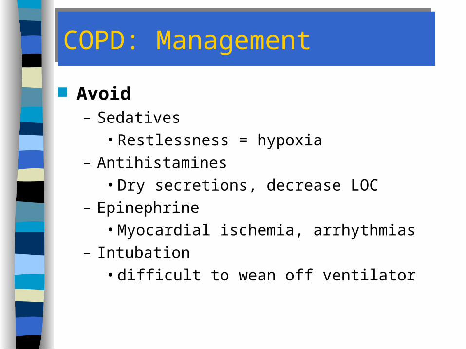
Avoid– Sedatives
• Restlessness = hypoxia– Antihistamines
• Dry secretions, decrease LOC– Epinephrine
• Myocardial ischemia, arrhythmias– Intubation
• difficult to wean off ventilator

Reversible Obstructive Airway Disease
Asthma

Asthma: DefinitionAsthma: Definition
Lower airway hyper-responsiveness to a variety of stimuli
Diffuse reversible airway obstruction or narrowing
Airway inflammation

Asthma: IncidenceAsthma: Incidence
50% onset before age 10 33% before age 30 “Asthma” in older patients suggests other
obstructive pulmonary diseases Risk Factors
– Family history of asthma– Perinatal exposure to airborne allergens and
irritants– Genetic hypersensitivity to environmental allergens
(Atopy)

AsthmaAsthma
Diagnosis
– H&P, Spirometry– Hx or presence of episodic symptoms of
airflow obstruction– airflow obstruction is at least partially
reversible– alternative diagnoses are excluded

AsthmaAsthma
Commonly misdiagnosed in children as– Chronic bronchitis– Recurrent croup– Recurrent URI– Recurrent pneumonia

AsthmaAsthma
Often triggered by:– Cold temperature– Respiratory Infections– Vigorous exercise– Emotional Stress– Environmental allergens or irritants
Exacerbation– Extrinsic common in children– Intrinsic common in adults
Asthma PathophysiologyAsthma Pathophysiology
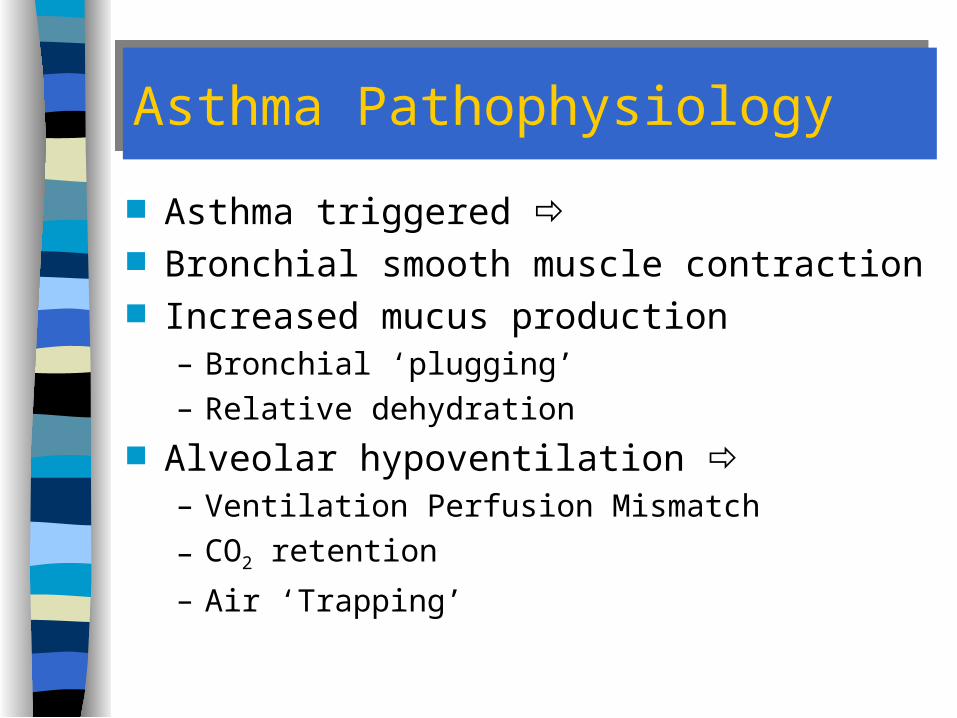
Asthma triggered Bronchial smooth muscle contraction Increased mucus production
– Bronchial ‘plugging’– Relative dehydration
Alveolar hypoventilation – Ventilation Perfusion Mismatch
– CO2 retention
– Air ‘Trapping’

Asthma: PathophysiologyAsthma: Pathophysiology
Bronchospasm
Bronchial Edema Increased MucusProduction

Asthma: PathophysiologyAsthma: Pathophysiology

Asthma: PathophysiologyAsthma: Pathophysiology
Cast of airway produced by asthmatic mucus plugs

Asthma: PathophysiologyAsthma: Pathophysiology
Difficulty exhaling– chest hyperinflation
Poor gas exchange– hypoxia– hypercarbia
Increased respiratory water loss– dehydration
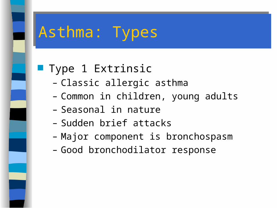
Asthma: TypesAsthma: Types
Type 1 Extrinsic– Classic allergic asthma– Common in children, young adults– Seasonal in nature– Sudden brief attacks– Major component is bronchospasm– Good bronchodilator response

Asthma: TypesAsthma: Types
Type 2 Extrinsic Asthma– Adults < 35– Long term exposure to irritants– More inflammation than Type 1 Extrinsic– Does not respond well to bronchodilators– Needs treatment with corticosteroids
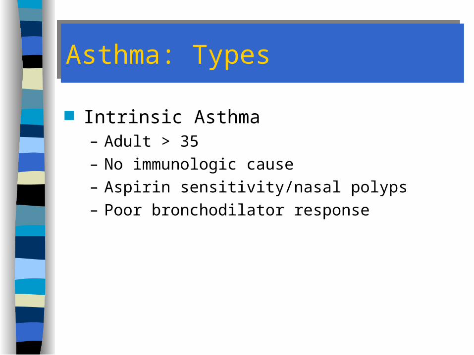
Asthma: TypesAsthma: Types
Intrinsic Asthma– Adult > 35– No immunologic cause– Aspirin sensitivity/nasal polyps– Poor bronchodilator response
Asthma: Signs and SymptomsAsthma: Signs and Symptoms
Onset of attacks associated with “triggers” Dyspnea Non-productive cough Tachypnea Expiratory wheezing Accessory muscle use Retractions

Asthma: Signs and Symptoms
Absence of wheezing
IMPENDING RESPIRATORY ARREST!

Asthma: Signs and SymptomsAsthma: Signs and Symptoms
Tachycardia Pulsus paradoxus in severe attacks Anxiety, restlessness (hypoxia) progressing
to drowsiness, confusion (hypercarbia)

Asthma: Signs and Symptoms
Lethargy, confusion, suprasternal retractions RESPIRATORY FAILURE

Asthma: Signs and SymptomsAsthma: Signs and Symptoms Early Blood Gas Changes
– Decreased PaO2
– Decreased PaCO2
WHY?
Asthma: Signs and SymptomsAsthma: Signs and Symptoms
Later Blood Gases– Decreased PaO2
– Normal PaCO2
IMPENDING RESPIRATORY
FAILURE
Asthma: Signs and SymptomsAsthma: Signs and Symptoms
Still Later Blood Gases– Decreased PaO2
– Increased PaCO2
RESPIRATORY FAILURE

Asthma: Risk AssessmentAsthma: Risk Assessment Prior ICU admissions Prior intubation >3 ED visits in past year >2 hospital admissions in past year >1 bronchodilator canister used in past month Use of bronchodilators > every 4 hours Chronic use of steroids Progressive symptoms in spite of aggressive Rx

Asthma: ManagementAsthma: Management
Airway Breathing
– Sitting position or position of comfort
– Humidified O2 by NRB mask
• Dry O2 dries mucus, worsens plugs
– Encourage coughing– Consider intubation, assisted ventilation
• Impending respiratory failure• Avoid if at all possible

Asthma: ManagementAsthma: Management
Circulation– IV TKO– Assess for dehydration– Titrate fluid administration to severity of
dehydration• Trial bolus of 250 cc
– Monitor ECG, Pulse Oximetry

Asthma: ManagementAsthma: Management
Obtain medication history Consider
– Overdose– Dysrhythmias

Asthma: ManagementAsthma: Management
Nebulized Beta-2 agents– Albuterol – Terbutaline – Metaproterenol– Isoetharine
Nebulized anticholinergics– Ipratropium– Atropine
IV Corticosteroid– Methylprednisolone
Asthma: ManagementAsthma: Management
Rarely used– Questionable efficacy, Potential Complications
– Magnesium Sulfate (IV)– Methylxanthines
• Aminophylline (IV)

Asthma: ManagementAsthma: Management
Subcutaneous beta agents– Epinephrine 1:1000 q 30 minutes up to 3 doses
• Adult – 0.3 to 0.5 mg SQ• Pediatric – 0.1 to 0.3 mg SQ
– Terbutaline• Adult - 0.25 mg SQ q 30 minutes up to 2 doses• Pediatric -SQ or IV infusion usually begun @
0.17 mcg/kg/min
POSSIBLE BENEFIT IN PATIENTS WITH VENTILATORY FAILURE

Asthma: ManagementAsthma: Management
Use EXTREME caution in giving two sympathomimetics or two doses to same patient
Monitor ECG

Asthma: ManagementAsthma: Management
Avoid– Sedatives
• Depress respiratory drive– Antihistamines
• Decrease LOC, dry secretions– Aspirin
• High incidence of allergy

Asthma: ManagementAsthma: Management
Continuous Monitoring & Frequent Reassessment
Need for transport? Destination?
Asthma: ManagementAsthma: Management
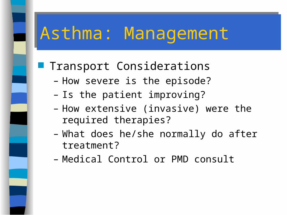
Transport Considerations– How severe is the episode?– Is the patient improving?– How extensive (invasive) were the required
therapies?– What does he/she normally do after treatment?– Medical Control or PMD consult

Drug Delivery Methods: ReviewDrug Delivery Methods: Review
MDI vs. MDI w/ spacer vs. SVNvs. SQ injection

Status Asthmaticus
Asthma unresponsive to beta-2 adrenergic agents

Status AsthmaticusStatus Asthmaticus Oxygen (humidified if possible) Nebulized beta-2 agents Nebulized Ipratropium Corticosteroids IV or SQ terbutaline or epinephrine Aminophylline (controversial) Magnesium sulfate (controversial) Intubation
– Caution with PPV
Golden RuleGolden Rule
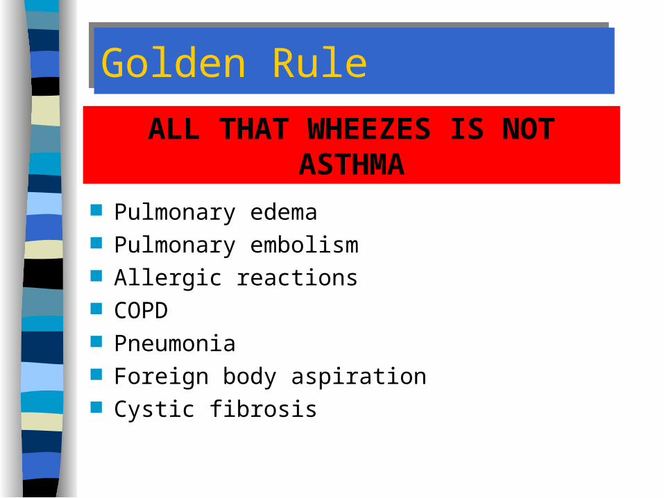
Pulmonary edema Pulmonary embolism Allergic reactions COPD Pneumonia Foreign body aspiration Cystic fibrosis
ALL THAT WHEEZES IS NOT ASTHMA

Lower Airway Disease
Cystic Fibrosis

Cystic Fibrosis: DefinitionCystic Fibrosis: Definition
Inherited metabolic disease of exocrine glands and sweat glands
Primarily affects digestive, respiratory systems
Begins in infancy

Cystic Fibrosis: EtiologyCystic Fibrosis: Etiology
Autosomal recessive gene Both parents must be carriers Incidence
– Caucasians--1:2000– Blacks--1:17,000– Asians--very rare

Cystic Fibrosis: PathophysiologyCystic Fibrosis: Pathophysiology
Obstruction of pancreatic, intestinal gland, bile ducts
Over-secretion by airway mucus glands– mucous plugs
Excess loss of sodium chloride in sweat

Cystic Fibrosis: RecognitionCystic Fibrosis: Recognition
History Airway obstruction, chronic cough
– Recurrent respiratory infections– May be oxygen-dependent
Diffuse Wheezing Frequent, foul-smelling stools Salty taste on skin Intolerance of hot environments

Cystic Fibrosis: ManagementCystic Fibrosis: Management
Position of comfort Oxygen Suctioning Nebulized Beta agonists
– May not be very helpful but worth attempting if absence of contraindications
Assisted ventilation

Lower Airway Disease
Neoplasms of the Lung

Neoplasms of the LungNeoplasms of the Lung
150,000 cases Usually occurs between ages of 55 and 65 Most die within one year 20% only local lung involved 25% spread to lymphatic system 55% result in distant metastatic cancer

Neoplasms of the LungNeoplasms of the Lung
Prevention– Centered on prevention of smoking in youths– Then, cessation in current smokers– Avoid environmental hazards (e.g. asbestos)

Neoplasms of the LungNeoplasms of the Lung
Presentation– Respiratory Difficulty progressing to Distress– Cough, Hemoptysis– Hoarseness or voice change– Dysphagia

Management of Neoplasms of the LungManagement of Neoplasms of the Lung Supportive care based upon presentation
– Oxygen– Consider presence of advance directives or DNR
• Patient’s wishes• Family discussions• MD prognosis
– If appropriate• Assist ventilations or Intubate• IV access & rehydration• Bronchodilators • Analgesia for pain (small, slow doses)
Hyperventilation Syndrome

Hyperventilation SyndromeHyperventilation Syndrome
Brady Textbook Correction, Vol. 3, p. 57– Table 1-4: These are NOT Causes of
hyperventilation syndrome A diagnosis of EXCLUSION!!! An increased ventilatory rate that
– DOES NOT have a pathologic origin– Results from anxiety
Remains a real problem for the patient

Hyperventilation Syndrome: PathophysiologyHyperventilation Syndrome: Pathophysiology
Tachypnea or hyperpnea secondary to anxiety
Decreased PaCO2
Respiratory alkalosis
Vasoconstriction Hypocalcemia Decreased O2 Release to
Tissues

Hyperventilation Syndrome:Signs & SymptomsHyperventilation Syndrome:Signs & Symptoms Symptoms
– Light-headedness, giddiness, anxiety– Numbness, paresthesias of:
• Hands• Feet• Circumoral area
– Cold hands, feet– Carpopedal spasms– Dyspnea– Chest pain

Hyperventilation Syndrome:Signs & SymptomsHyperventilation Syndrome:Signs & Symptoms Signs
– Rapid breathing– Cool & possibly pale skin– Carpopedal spasm– Dysrhythmias
• Sinus Tachycardia• SVT• Sinus arrhythmia
– Loss of consciousness and seizures (late & rare)
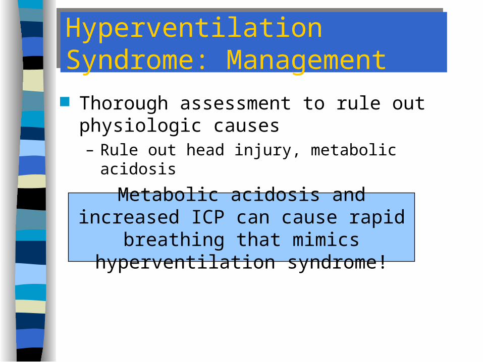
Hyperventilation Syndrome: ManagementHyperventilation Syndrome: Management Thorough assessment to rule out
physiologic causes– Rule out head injury, metabolic acidosis
Metabolic acidosis and increased ICP can cause rapid breathing that mimics
hyperventilation syndrome!

Hyperventilation Syndrome:ManagementHyperventilation Syndrome:Management
Oxygen based upon presentation Reassurance & Patience
– Coach breathing rate– CAUTION: Rebreathing into bag or NRB
Monitoring– ECG– Pulse oximetry
Hyperventilation Syndrome: ManagementHyperventilation Syndrome: Management Educate patient & family
– Consider possible psychopathology especially in “repeat customers”
Transport occasionally required– If loss of consciousness, carpopedal spasm,
muscle twitching, or seizures occur:• Monitor EKG• IV TKO• Transport

Hyperventilation Syndrome
Hyperventilation itself can be serious
Serious diseases can mimic hyperventilation

Pulmonary Infectious Diseases

Laryngotracheobronchitis (Croup)Laryngotracheobronchitis (Croup)
Common syndrome of infectious upper airway obstruction
Viral infection– parainfluenza virus
Subglottic Edema– larynx, trachea,
mainstem bronchi
Usually 3 months to 4 years of age

Croup: Signs & SymptomsCroup: Signs & Symptoms
Gradual onset (several days)– Often begins with Sx of URI– May begin with only low grade fever
Hoarseness Cough
– “Seal Bark Cough”– “Brassy Cough”
Nocturnal episodes of increased dyspnea and stridor

Croup: Signs & SymptomsCroup: Signs & Symptoms
Evidence of respiratory distress– Tracheal tugging– Substernal/intercostal retractions– Accessory muscle use
Inspiratory stridor or respiratory distress may develop slowly or acutely

Croup: ManagementCroup: Management
Usually requires little out of home treatment
Calm & Prevent agitation!!! Moist cool air - mist Humidified O2 by mask or blowby Do Not Examine Upper Airways!!!

Croup: ManagementCroup: Management
If in respiratory distress:– Racemic epinephrine via nebulizer
• Decreases subglottic edema (temporarily)• Necessitates transport for observation for rebound
– IV TKO - ONLY if severe respiratory distress– Transport

BronchiolitisBronchiolitis Pathophysiology
– Viral Disease resulting in inflammation of the lower airways
– Usually caused by RSV Typically affects children 6 - 18 months old
(15% of all children < 2 years old) Usually occurs in the winter or early spring
Bronchiolitis: PresentationBronchiolitis: Presentation Usually
– less than 18 months– during the winter or early spring– wheezing– mild to moderate respiratory difficulty– no asthma history– associated with other viral symptoms
• runny nose• sneezing• cough• low grade fever

Bronchiolitis: ManagementBronchiolitis: Management Usually require little out of home treatment Oxygen, mask or blowby Nebulized Bronchodilators if respiratory
distress– May not respond well or at all
Transport

EpiglottitisEpiglottitis
Bacterial infection (Hemophilus influenza )
Edema of epiglottis (supraglottic)– partial upper airway
obstruction Typically affects 3-7
year olds

Epiglottitis: PresentationEpiglottitis: Presentation Age: 3-7 years of age
– can occur in adults– can occur in infants
Rapid onset & progression– Fever– Severe sore throat– Dysphagia– Muffled voice– Drooling

Epiglottitis: PresentationEpiglottitis: Presentation Respiratory difficulty
– Stridor– Usually in an upright, sitting, tripod position
Child may go to bed asymptomatic and awaken during the night with– sore throat– painful swallowing– respiratory difficulty
Epiglottitis: ManagementEpiglottitis: Management
Do NOT attempt to visualize airway Allow child to assume position of comfort
– AVOID agitation of the child!!!– AVOID anxiety of the healthcare providers!!!
O2 by high concentration mask
Immediate life threat (8-12% die from airway obstruction)

Epiglottitis: ManagementEpiglottitis: Management
If respiratory failure is eminent:– IV TKO ONLY if eminent or respiratory arrest– Be prepared to take control of airway
• Intubation equipment with smaller sized tubes• Needle cricothyrotomy & jet ventilation equipment
Rapid but calm transport– Appropriate facility

Upper Respiratory InfectionUpper Respiratory Infection
Common illness Rarely life-threatening Often exacerbates underlying pulmonary
conditions May become more significant in some
patients– Immunosuppressed– Elderly– Chronic pulmonary disease

Upper Respiratory InfectionUpper Respiratory Infection
Prevention– Avoidance is nearly impossible
• Too many potential causes• Temporarily impaired immune system
– Best prevention strategy is handwashing• Covering of mouth during sneezing and coughing also
helpful

Pathophysiology of URIPathophysiology of URI
Wide variety of bacteria and viruses are causes– Normal immune system response results in
presentation 20-30% are Group A streptococci Most are self-limiting diseases

Presentation of URIPresentation of URI
Symptoms– Sore throat– Fever– Chills– HA
Signs– Cervical adenopathy– Erythematous pharynx– Positive throat culture (bacterial)

Management of URIManagement of URI
Usually requires no intervention Oxygen if underlying condition has been
exacerbated Rarely, pharmacologic interventions are
required– Bronchodilators– Corticosteroid
Occasionally, transport required– Key question: Destination?
Central Respiratory DepressionCentral Respiratory Depression

Respiratory Depression: CausesRespiratory Depression: Causes
Head trauma CVA Depressant drug toxicity
– Narcotics– Barbiturates– Benzodiazepines– ETOH

Respiratory Depression: RecognitionRespiratory Depression: Recognition
Decreased respiratory rate (< 12/min) Decreased tidal volume Decreased LOC
Look, Listen, Feel
Use Your Stethoscope
If you can’t tell whether a patient is breathing
adequately...
THEY PROBABLY
AREN’T
Respiratory Depression: ManagementRespiratory Depression: Management
Airway– Open, clear, maintain– Consider endotracheal intubation
The need to VENTILATE is not the same as the need to INTUBATE

Respiratory Depression: ManagementRespiratory Depression: Management
Breathing– Oxygenate, ventilate– Restore normal rate, tidal volume
Oxygen alone is INSUFFICIENT if Ventilation is INADEQUATE
Respiratory Depression: ManagementRespiratory Depression: Management Circulation
– Obtain vascular access– Monitor EKG (Silent MI may present as CVA)
Manage Cause– Check Blood Sugar– Consider Narcan 2mg IV push if S/S suggest
narcotic overdose Intubate if can not find or treat cause
Thoracic Bellows MalfunctionThoracic Bellows Malfunction
Pickwickian Syndrome Guillian-Barre Syndrome Myasthenia Gravis

Pickwickian SyndromePickwickian Syndrome
Results from extreme obesity– form of sleep apnea
Decreased excursion of chest wall, diaphragm causes– hypoventilation
– CO2 retention

Pickwickian SyndromePickwickian Syndrome
Signs and Symptoms– Headache– Drowsiness– Inappropriate sleepiness– Sleep apnea
Treat symptomatically– Assist ventilations as needed

Guillian-Barre´ SyndromeGuillian-Barre´ Syndrome
Autoimmune disease– Leads to inflammation and degeneration of
sensory and motor nerve roots (de-myelination)
Progressive ascending paralysis– Progressive tingling and weakness– Moves from extremities then proximally– May lead to respiratory paralysis (25%)

Guillian-Barre´ SyndromeGuillian-Barre´ Syndrome
Self-Limiting– Recovery is spontaneous and complete in
95% of cases– In good outcomes, symptoms clear in 15 to 20
days– Often takes weeks or months

Guillian-Barre´ Syndrome ManagementGuillian-Barre´ Syndrome Management Treatment based on severity of symptoms
– Control airway– Support ventilation– Oxygen– Transport in cases of respiratory depression,
distress or arrest
Myasthenia GravisMyasthenia Gravis
Autoimmune disease Causes loss of ACh receptors at
neuromuscular junction– Attacks the ACh transport mechanism at the
NMJ Episodes of extreme skeletal muscle
weakness Can cause loss of control of airway,
respiratory paralysis
Myasthenia Gravis PresentationMyasthenia Gravis Presentation
Gradual onset of muscle weakness– Face and throat– Extreme muscle weakness
Respiratory weakness -> paralysis Inability to process mucus

Myasthenia Gravis ManagementMyasthenia Gravis Management
Treat symptomatically Watch for aspiration May require assisted ventilations Assess for Pulmonary infection Transport based upon severity of
presentation
Pulmonary Diseases & DisordersPulmonary Diseases & Disorders
Other Causes of Respiratory Emergencies

Angioneurotic EdemaAngioneurotic Edema
Allergic reaction– Edema of tongue, pharynx, larynx– NOT the SAME as anaphylaxis
Common Causes– Food (seafood or nuts)– Drugs (penicillin or sulfa)– Hymenoptera sting (ants, bees, wasps)

Angioneurotic EdemaAngioneurotic Edema
Signs and Symptoms– Itching in palate– “Lump in throat”– Hoarseness– Stridor– Coughing– Dyspnea– Urticaria (hives)
Angioneurotic Edema: ManagementAngioneurotic Edema: Management Based upon severity of presentation
– Establish airway
– O2 via NRB
– IV lg bore TKO– Epinephrine
• 1:1000 0.3 - 0.5mg SQ• repeat after 20 minutes if needed

Angioneurotic Edema: ManagementAngioneurotic Edema: Management Based upon severity of presentation (cont)
– Diphenhydramine 25 to 50mg IM/IV– In severe cases, Consider
• Positive pressure ventilation• Endotracheal intubation• Surgical airway

Spontaneous PneumothoraxSpontaneous Pneumothorax
Low incidence Many are well tolerated Risk Factors
– Males– Younger age– Thin body mass
• Marfan’s syndrome
– History of Obstructive Airway Disease

Presentation of Spontaneous PneumothoraxPresentation of Spontaneous Pneumothorax Symptoms
– Sudden SOB– Sudden pleuritic CP
Signs– Mild pallor, tachycardia, tachypnea– Decreased lung sounds
• usually very localized Increasing pneumothorax presents with more
severe S/S

Management of Simple PneumothoraxManagement of Simple Pneumothorax
Oxygen based on severity of S/S Assisted ventilation and intubation as needed
– May worsen pneumothorax– Rarely needed
IV access if severe symptoms are present Position of comfort Transport

Case Studies

Case OneCase One
It is 1430 hrs. You are called to a business for a “possible stroke.” The patient is a 20-year-old female complaining of dizziness and of numbness around her mouth and fingertips.
What would you like to include in your initial differential diagnosis?

Case OneCase One
Initial Assessment– Airway: Open, maintained by patient– Breathing: Rapid, deep, regular; no accessory
muscle use or retractions– Circulation: Radial pulses present, rapid, full; Skin
warm, dry; capillary refill < 2 seconds– Disability: Awake, alert, anxious
What therapies, if any, would you like to begin?

Case OneCase One Vital Signs
– P: 126 strong, regular– R: 26 deep, regular– BP: 130/82
Physical Exam– Chest: BS present, equal bilaterally; no
adventitious sounds– Extremities: Equal movement in all
extremities; no weakness; hands cool– Oxygen saturation: 98%
Would you like to make any Changes to your therapies or Diff Dx?

Case OneCase One
History– Allergies: NKA– Medications: Birth control pills– Past History: No significant past history; no
history of smoking– Last Meal: Lunch 2 hours ago– Events: S/S began suddenly after argument
with supervisor

Case OneCase One
What problem do you now suspect? How would you manage this patient?

Case TwoCase Two
It is 0530 hours. You are called to a residence to see a child with “a very high fever and difficulty breathing.” The patient is a 6-old-female. Mother says the child woke up crying about 2 hours ago.
What would you like to include in your differential diagnosis?

Case TwoCase Two Initial Assessment
– Airway: Inspiratory stridor audible– Breathing: Rapid, shallow, labored– Circulation: Radial pulses present, rapid, weak;
skin pale, hot, diaphoretic; capillary refill is 2 seconds
– Disability: Awake, alert, obviously frightened and in acute distress
What therapies, if any, would you like to begin now?

Case TwoCase Two Vital Signs
– P: 130 weak, regular– R: 32 shallow, regular with stridor– BP: 110/70
Physical Exam– HEENT: Flaring of nostrils; accessory muscle
use on inspiration; drooling present– Chest: BS present, equal bilaterally; no
adventitious sounds– Oxygen saturation: 92%
Would you like to make any Changes to your therapies or Diff Dx?

Case TwoCase Two History
– Allergies: NKA– Medications: None– Past History: No significant past history– Last Meal: Dinner at about 1800 hours– Events: Awakened with severe sore throat. Has
experienced increasing difficulty breathing. Will not eat or drink. Says it hurts to swallow

Case TwoCase Two
What problem do you now suspect? How would you manage this patient?
Case ThreeCase Three
At 2330 hrs you are called to a residence to see a child with “difficulty breathing.” The patient is a 3 year old male.
How narrow a Differential Diagnosis can you compile at this point?

Case ThreeCase Three Initial Assessment
– Airway: Open, maintained by patient, mild stridor audible
– Breathing: Rapid, shallow, labored– Circulation: Radial pulses present, weak, regular;
Skin pale, warm, moist; Capillary refill <2 seconds– Disability: Awake, sitting up in bed, looks tired and
miserable

Case ThreeCase Three Vital Signs
– P: 100 weak, regular– R: 30 shallow, labored with stridor– BP: 90/50
Physical Exam– HEENT: Use of accessory muscles present; no
drooling– Chest: BS present, equal bilaterally with no
adventitious sounds. Auscultation difficult because of stridor and barking cough
Now you can narrow your Diff Dx? To what?

Case ThreeCase Three History
– Allergies: NKA– Medication: Tylenol for fever before bedtime– Past history: No significant past history– Last meal: Dinner around 1800 hours– Events: Patient has had “cold” for about 3 days.
Reasonably well during day. Awakens around midnight with high-pitched cough that sounds like a dog barking

Case ThreeCase Three
What problem do you suspect? How would you manage this patient?

Case FourCase Four At 1945 hours you are dispatched to a
“breathing difficulty” at Long John Silver’s. The patient is a 26-year-old female complaining of strange feeling in her mouth and difficulty swallowing.
What is your differential diagnosis?

Case FourCase Four
Initial Assessment– Airway: Open, maintained by patient, difficulty
swallowing, voice is hoarse– Breathing: Rapid, labored– Circulation: Radial pulses present, strong, regular;
Skin “flushed”; Capillary refill < 2 seconds– Disability: Awake, alert, very anxious

Case FourCase Four Vital Signs
– P: 120 strong, regular– R: 26 regular, slightly labored– BP: 118/90
Physical Exam– HEENT: Puffiness around eyes; Lips appear swollen;
Mild accessory muscle use– Chest: BS present, equal bilaterally; No adventitious
sounds– Urticaria on upper chest, extremities– Oxygen saturation: 94%
What therapies do you want to initiate?

Case FourCase Four History
– Allergies: No drug allergies; Has experienced itching previously when eating shrimp
– Medications: None– Past history: No significant past history; no
history of smoking– Last meal: In progress at time of call– Events: Began to experience itching and
difficulty swallowing after eating “fish and chips”

Case FourCase Four
What problem do you suspect? How would you manage this patient?
The patient begins to have increased difficulty swallowing, increased anxiety, and increased
difficulty breathing. What do you want to do now?

Case FiveCase Five At 0130 you are dispatched to an
“unconscious person--police on location.” The patient is a 27-year-old male who is apparently unconscious. The police report they found him lying in an alleyway while they were on routine patrol. He is known to live “on the streets”.

Case FiveCase Five Initial Assessment
– Airway: Controllable with manual positioning– Breathing: Very slow, shallow– Circulation: Radial pulses present, weak; Skin
pale, cool, moist; Capillary refill 3 seconds– Disability: Unconscious, unresponsive to painful
stimuli
What therapies would you like to begin?

Case FiveCase Five Vital Signs
– P: 70 regular, weak– R: 4 shallow, regular; alcohol odor on breath– BP: 100/70
Physical Exam– HEENT: Pupils pinpoint, non-reactive– Chest: BS present, equal bilaterally– Abdomen: Soft, non-tender– Extremities: Needle tracks present– Blood glucose: 40 mg/dl

Case FiveCase Five
What problem or problems do you suspect?
How would you manage this patient?