eMedicine Specialties > Dermatology > Diseases of the Oral ... Fissuratum .pdfemedicine.medscape.com...
9
emedicine.medscape.com eMedicine Specialties > Dermatology > Diseases of the Oral Mucosa Epulis Fissuratum Diane Stern, DDS, Clinical Professor, Department of Surgery, Section of Oral and Maxillofacial Surgery, University of Miami; Clinical Professor, Nova Southeast University School of Dental Medicine Updated: Jul 6, 2009 Introduction Background Epulis fissuratum is a mucosal hyperplasia that results from chronic low-grade trauma induced by a denture flange. 1 Epulis fissuratum is analogous to acanthoma fissuratum of skin. Pathophysiology Epulis fissuratum arises in association with denture flanges. Consequently, epulis fissuratum is usually observed in the maxillary or mandibular vestibule. Mortality/Morbidity Significant morbidity does not occur with epulis fissuratum. Race Most cases of epulis fissuratum are observed in whites. This, no doubt, relates to the predominance of whites as denture wearers. Sex Most studies indicate a clear predilection for epulis fissuratum in females. 2 The fact that women are more likely than men to wear their dentures for prolonged periods because of their reluctance to be seen without them probably plays a significant role. In addition, more women than men wear dentures and are more likely to seek treatment. Possibly, atrophic epithelial changes secondary to menopause may influence an increased reaction to trauma in older females. Age Epulis fissuratum occurs in greatest numbers in the fifth, sixth, and seventh decades, but it can be observed at almost any age. Epulis fissuratum has been described in children. The fact that the lesions are related to denture wear and chronicity of an irritative process explains the higher incidence in older individuals.
Transcript of eMedicine Specialties > Dermatology > Diseases of the Oral ... Fissuratum .pdfemedicine.medscape.com...
emedicine.medscape.com
eMedicine Specialties > Dermatology > Diseases of the Oral Mucosa
Epulis Fissuratum Diane Stern, DDS, Clinical Professor, Department of Surgery, Section of Oral and Maxillofacial Surgery, University of Miami; Clinical Professor, Nova Southeast University School of Dental Medicine Updated: Jul 6, 2009
Introduction Background
Epulis fissuratum is a mucosal hyperplasia that results from chronic low-grade trauma induced by a denture
flange.1 Epulis fissuratum is analogous to acanthoma fissuratum of skin.
Pathophysiology
Epulis fissuratum arises in association with denture flanges. Consequently, epulis fissuratum is usually observed in
the maxillary or mandibular vestibule.
Mortality/Morbidity
Significant morbidity does not occur with epulis fissuratum.
Race
Most cases of epulis fissuratum are observed in whites. This, no doubt, relates to the predominance of whites as
denture wearers.
Sex
Most studies indicate a clear predilection for epulis fissuratum in females.2 The fact that women are more likely than
men to wear their dentures for prolonged periods because of their reluctance to be seen without them probably plays
a significant role. In addition, more women than men wear dentures and are more likely to seek treatment. Possibly,
atrophic epithelial changes secondary to menopause may influence an increased reaction to trauma in older females.
Age
Epulis fissuratum occurs in greatest numbers in the fifth, sixth, and seventh decades, but it can be observed at
almost any age. Epulis fissuratum has been described in children. The fact that the lesions are related to denture
wear and chronicity of an irritative process explains the higher incidence in older individuals.
Clinical History
• Epulis fissuratum develops slowly over a prolonged period of time in patients with ill-fitting dentures. It is
associated with a denture flange that may be either a full or partial denture.3
• Typically, patients with epulis fissuratum are asymptomatic.4
Physical
• Examination of an epulis fissuratum patient typically reveals folds of hyperplastic mucosa, which
encompass the border of the denture flange. The edge of the denture usually fits in a groove between the
folds. The lesions are most frequently observed at the facial aspect of the denture. The occurrence of this
on the lingual surface is unusual. They are more often observed in the anterior portion of the jaws;
however, a predilection for the maxilla or the mandible does not seem to exist.
• The surface of the epulis fissuratum mass tends to be smooth; however, occasionally, it is ulcerated (most
often within the depth of the groove) or papillary.
• The size of the epulis fissuratum lesion is variable; some lesions are small, but they can be extensive and
involve the entire length of the vestibule.5
• Although frequently of normal mucosal color, erythema may be associated with inflammation. Some
lesions have a more pyogenic granuloma –like appearance because of capillary proliferation.

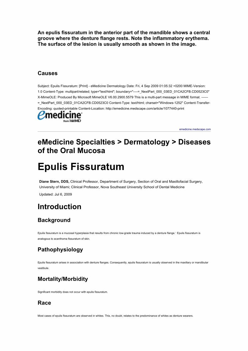
An epulis fissuratum in the anterior part of the mandible shows a central groove where the denture flange rests. Note the inflammatory erythema. The surface of the lesion is usually smooth as shown in the image.
Causes
Subject: Epulis Fissuratum: [Print] - eMedicine Dermatology Date: Fri, 4 Sep 2009 01:05:32 +0200 MIME-Version:
1.0 Content-Type: multipart/related; type="text/html"; boundary="----=_NextPart_000_03ED_01CA2CFB.CD0523C0"
X-MimeOLE: Produced By Microsoft MimeOLE V6.00.2900.5579 This is a multi-part message in MIME format. ------
=_NextPart_000_03ED_01CA2CFB.CD0523C0 Content-Type: text/html; charset="Windows-1252" Content-Transfer-
Encoding: quoted-printable Content-Location: http://emedicine.medscape.com/article/1077440-print
emedicine.medscape.com
eMedicine Specialties > Dermatology > Diseases of the Oral Mucosa
Epulis Fissuratum Diane Stern, DDS, Clinical Professor, Department of Surgery, Section of Oral and Maxillofacial Surgery, University of Miami; Clinical Professor, Nova Southeast University School of Dental Medicine
Updated: Jul 6, 2009
Introduction Background
Epulis fissuratum is a mucosal hyperplasia that results from chronic low-grade trauma induced by a denture flange.1 Epulis fissuratum is
analogous to acanthoma fissuratum of skin.
Pathophysiology
Epulis fissuratum arises in association with denture flanges. Consequently, epulis fissuratum is usually observed in the maxillary or mandibular
vestibule.
Mortality/Morbidity
Significant morbidity does not occur with epulis fissuratum.
Race
Most cases of epulis fissuratum are observed in whites. This, no doubt, relates to the predominance of whites as denture wearers.
Sex
Most studies indicate a clear predilection for epulis fissuratum in females.2 The fact that women are more likely than men to wear their dentures
for prolonged periods because of their reluctance to be seen without them probably plays a significant role. In addition, more women than men
wear dentures and are more likely to seek treatment. Possibly, atrophic epithelial changes secondary to menopause may influence an increased
reaction to trauma in older females.
Age
Epulis fissuratum occurs in greatest numbers in the fifth, sixth, and seventh decades, but it can be observed at almost any age. Epulis
fissuratum has been described in children. The fact that the lesions are related to denture wear and chronicity of an irritative process explains
the higher incidence in older individuals.
Clinical History
• Epulis fissuratum develops slowly over a prolonged period of time in patients with ill-fitting dentures. It is associated with a denture
flange that may be either a full or partial denture.3
• Typically, patients with epulis fissuratum are asymptomatic.4
Physical
• Examination of an epulis fissuratum patient typically reveals folds of hyperplastic mucosa, which encompass the border of the
denture flange. The edge of the denture usually fits in a groove between the folds. The lesions are most frequently observed at the
facial aspect of the denture. The occurrence of this on the lingual surface is unusual. They are more often observed in the anterior
portion of the jaws; however, a predilection for the maxilla or the mandible does not seem to exist.
• The surface of the epulis fissuratum mass tends to be smooth; however, occasionally, it is ulcerated (most often within the depth of
the groove) or papillary.
• The size of the epulis fissuratum lesion is variable; some lesions are small, but they can be extensive and involve the entire length
of the vestibule.5
• Although frequently of normal mucosal color, erythema may be associated with inflammation. Some lesions have a more pyogenic
granuloma –like appearance because of capillary proliferation.
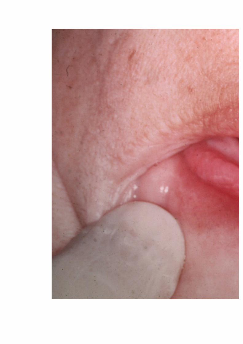

An epulis fissuratum in the anterior part of the mandible shows a central groove where the denture flange rests. Note the inflammatory erythema. The surface of the lesion is usually smooth as shown in the image.
Causes
• The cause of epulis fissuratum is chronic low-grade irritation from an ill-fitting denture. Frequently, this is the consequence of
resorption of the alveolar ridge so that the denture moves further into the vestibular mucosa, creating an inflammatory fibrous
hyperplasia that proliferates over the flange.3
Differential Diagnoses Metastatic Neoplasms to the Oral Cavity
Pyogenic Granuloma (Lobular Capillary Hemangioma)
Squamous Cell Carcinoma
Workup Procedures
• Surgically excise and microscopically examine the epulis fissuratum.
Histologic Findings
Epulis fissuratum is a hyperplastic reactive lesion, often with inflammatory and reparative phases. The histologic picture can be variable.6 Most
frequently, a dense fibrous hyperplasia occurs, often with varying degrees of inflammation and vascularity. Because capillary proliferation is
considerable, an overlap with pyogenic granuloma occurs. Mucous glands are often present in the specimen and may show a chronic
sialadenitis. Occasionally, the glands may have an associated lymphoid hyperplasia and papillary ductal hyperplasia. The epithelium may be
atrophic or hyperplastic and occasionally shows a pseudoepitheliomatous hyperplasia. Ulceration can occur. Infrequently, chondroid or osseous
metaplasia can develop within the mass.
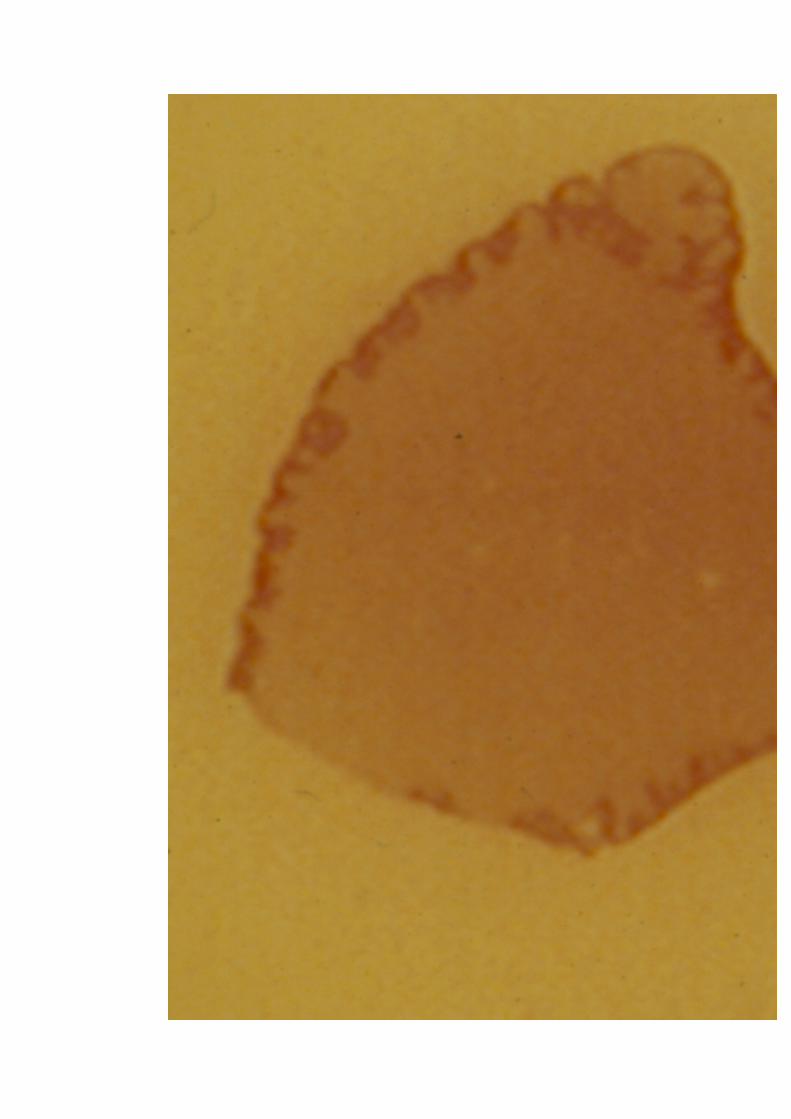
A view of a whole mount of a tissue section taken from an epulis fissuratum shows that it is essentially a fibrous hyperplasia. The central groove can be observed, and, in this patient, papillary hyperplasia is present in some areas.
Treatment Surgical Care
Surgically excise the epulis fissuratum because even removal of the offending stimulus (ie, denture) will not result in complete resolution. In
addition, correct the denture; otherwise, the lesion will recur. Either make a new denture or reline the old denture. The use of laser therapy is
discussed in recent studies.7
Consultations
• Oral and maxillofacial surgeon for excision
• General dentist or prosthodontist for correction of the denture
Follow-up Deterrence/Prevention
• Regular dental care can prevent epulis fissuratum. Patients who wear dentures frequently believe that they no longer require care,
and, under these circumstances, dentures lose their correct fit and become the source of irritation.
Prognosis
• With correction of the poorly fitting denture, the prognosis for epulis fissuratum is excellent.
Patient Education
• Instruct the patient that regular dental care is necessary and that the oral tissues are changing constantly. This means that dentures
are not permanent and need adjustments over time.
Miscellaneous Medicolegal Pitfalls
• The potential problem is that masses that occur in the vestibular area and are associated with dentures tend to be dismissed as
epulis fissuratum. Unfortunately, on rare occasions, these masses prov

![Pemphigus Vulgaris [Print] - eMedicine Dermatology Vulgaris .pdf · emedicine.medscape.com eMedicine Specialties > Dermatology > Bullous Diseases Pemphigus Vulgaris Bassam Zeina,](https://static.fdocuments.in/doc/165x107/5c984ab609d3f21c3a8b874e/pemphigus-vulgaris-print-emedicine-vulgaris-pdf-emedicinemedscapecom.jpg)


![Neuroblastoma_ [Print] - eMedicine Pediatrics_ General Medicine](https://static.fdocuments.in/doc/165x107/551f4c48497959335b8b4ddb/neuroblastoma-print-emedicine-pediatrics-general-medicine.jpg)


![ptosis [emedicine]](https://static.fdocuments.in/doc/165x107/577cdd4a1a28ab9e78acb3ee/ptosis-emedicine.jpg)