Elucidating mechanistic insights into drug action for atopic …€¦ · · 2018-02-07biology...
12
RESEARCH ARTICLE Open Access Elucidating mechanistic insights into drug action for atopic dermatitis: a systems biology approach Indhupriya Subramanian, Vivek K. Singh * and Abhay Jere Abstract Background: Topical Betamethasone (BM) and Pimecrolimus (PC) are widely used drugs in the treatment of atopic dermatitis (AD). Though the biomolecules and biological pathways affected by the drugs are known, the causal inter-relationships among these pathways in the context of skin is not available. We aim to derive this insight by using transcriptomic data of AD skin samples treated with BM and PC using systems biology approach. Methods: Transcriptomic datasets of 10 AD patients treated with Betamethasone and Pimecrolimus were obtained from GEO datasets. We used a novel computational platform, eSkIN (www.persistent.com/eskin), to perform pathway enrichment analysis for the given datasets. eSkIN consists of 35 skin specific pathways, thus allowing skin-centric analysis of transcriptomic data. Fisher’s exact test was used to compute the significance of the pathway enrichment. The enriched pathways were further analyzed to gain mechanistic insights into the action of these drugs. Results: Our analysis highlighted the molecular details of the mechanism of action of the drugs and corroborated the known facts about these drugs i.e. BM is more effective in triggering anti-inflammatory response but also causes more adverse effect on skin barrier than PC. In particular, eSkIN helped enunciate the biological pathways activated by these drugs to trigger anti-inflammatory response and its effect on skin barrier. BM suppresses pathways like TNF and TLRs, thus inhibiting NF-κB while PC targets inflammatory genes like IL13 and IL6 via known calcineurin-NFAT pathway. Furthermore, we show that the reduced skin barrier function by BM is due to the suppression of activators like AP1 transcription factors, CEBPs. Conclusion: We thus demonstrate the detailed mechanistic insight into drug action of AD using a novel computational approach. Keywords: Atopic dermatitis, Computational approach, Transcriptomic data analysis Background Atopic dermatitis (AD) is one of the most common dis- orders of skin that affects approximately 20% of children and 3% adults worldwide [1]. The pathophysiology of AD includes breakdown of the skin barrier, which in turn, initiates immunological response and inflammation [1]. The current treatment for AD involves topical appli- cation of corticosteroids or calcineurin inhibitors [2]. Betamethasone valerate (BM) and Pimecrolimus (PC) are two of the most commonly used drugs for the treat- ment of atopic dermatitis. BM, a corticosteroid, is known to suppress the inflam- mation, but fails to adequately restore the damaged skin barrier which subsequently leads to secondary skin infec- tions. BM binds to its corticosteroid receptor in skin and perturbs various biomolecules in keratinocytes involved in processes like inflammation, keratinocyte differentiation, proliferation and cellular adhesion [3]. On the other hand, PC, a topical calcineurin inhibitor (TCI), causes mild sup- pression of inflammation, but is more efficient in restoring the skin barrier. PC is known to mediate its action through NFAT signaling pathways [2]. US FDA issues TCI drugs with a boxed warning owing to a potential risk of malignancy, in spite of various stud- ies disproving any such association [4]. This favors the use * Correspondence: [email protected] LABS, Persistent Systems Limited, 9A/12, Erandwane, Pune, Maharashtra 411004, India © The Author(s). 2018 Open Access This article is distributed under the terms of the Creative Commons Attribution 4.0 International License (http://creativecommons.org/licenses/by/4.0/), which permits unrestricted use, distribution, and reproduction in any medium, provided you give appropriate credit to the original author(s) and the source, provide a link to the Creative Commons license, and indicate if changes were made. The Creative Commons Public Domain Dedication waiver (http://creativecommons.org/publicdomain/zero/1.0/) applies to the data made available in this article, unless otherwise stated. Subramanian et al. BMC Dermatology (2018) 18:3 https://doi.org/10.1186/s12895-018-0070-4
-
Upload
truongkhuong -
Category
Documents
-
view
213 -
download
1
Transcript of Elucidating mechanistic insights into drug action for atopic …€¦ · · 2018-02-07biology...

RESEARCH ARTICLE Open Access
Elucidating mechanistic insights into drugaction for atopic dermatitis: a systemsbiology approachIndhupriya Subramanian, Vivek K. Singh* and Abhay Jere
Abstract
Background: Topical Betamethasone (BM) and Pimecrolimus (PC) are widely used drugs in the treatment ofatopic dermatitis (AD). Though the biomolecules and biological pathways affected by the drugs are known, thecausal inter-relationships among these pathways in the context of skin is not available. We aim to derive thisinsight by using transcriptomic data of AD skin samples treated with BM and PC using systems biology approach.
Methods: Transcriptomic datasets of 10 AD patients treated with Betamethasone and Pimecrolimus were obtainedfrom GEO datasets. We used a novel computational platform, eSkIN (www.persistent.com/eskin), to perform pathwayenrichment analysis for the given datasets. eSkIN consists of 35 skin specific pathways, thus allowing skin-centricanalysis of transcriptomic data. Fisher’s exact test was used to compute the significance of the pathway enrichment.The enriched pathways were further analyzed to gain mechanistic insights into the action of these drugs.
Results: Our analysis highlighted the molecular details of the mechanism of action of the drugs and corroborated theknown facts about these drugs i.e. BM is more effective in triggering anti-inflammatory response but also causes moreadverse effect on skin barrier than PC. In particular, eSkIN helped enunciate the biological pathways activated by thesedrugs to trigger anti-inflammatory response and its effect on skin barrier. BM suppresses pathways like TNF and TLRs,thus inhibiting NF-κB while PC targets inflammatory genes like IL13 and IL6 via known calcineurin-NFAT pathway.Furthermore, we show that the reduced skin barrier function by BM is due to the suppression of activators like AP1transcription factors, CEBPs.
Conclusion: We thus demonstrate the detailed mechanistic insight into drug action of AD using a novelcomputational approach.
Keywords: Atopic dermatitis, Computational approach, Transcriptomic data analysis
BackgroundAtopic dermatitis (AD) is one of the most common dis-orders of skin that affects approximately 20% of childrenand 3% adults worldwide [1]. The pathophysiology ofAD includes breakdown of the skin barrier, which inturn, initiates immunological response and inflammation[1]. The current treatment for AD involves topical appli-cation of corticosteroids or calcineurin inhibitors [2].Betamethasone valerate (BM) and Pimecrolimus (PC)are two of the most commonly used drugs for the treat-ment of atopic dermatitis.
BM, a corticosteroid, is known to suppress the inflam-mation, but fails to adequately restore the damaged skinbarrier which subsequently leads to secondary skin infec-tions. BM binds to its corticosteroid receptor in skin andperturbs various biomolecules in keratinocytes involved inprocesses like inflammation, keratinocyte differentiation,proliferation and cellular adhesion [3]. On the other hand,PC, a topical calcineurin inhibitor (TCI), causes mild sup-pression of inflammation, but is more efficient in restoringthe skin barrier. PC is known to mediate its actionthrough NFAT signaling pathways [2].US FDA issues TCI drugs with a boxed warning owing
to a potential risk of malignancy, in spite of various stud-ies disproving any such association [4]. This favors the use
* Correspondence: [email protected], Persistent Systems Limited, 9A/12, Erandwane, Pune, Maharashtra411004, India
© The Author(s). 2018 Open Access This article is distributed under the terms of the Creative Commons Attribution 4.0International License (http://creativecommons.org/licenses/by/4.0/), which permits unrestricted use, distribution, andreproduction in any medium, provided you give appropriate credit to the original author(s) and the source, provide a link tothe Creative Commons license, and indicate if changes were made. The Creative Commons Public Domain Dedication waiver(http://creativecommons.org/publicdomain/zero/1.0/) applies to the data made available in this article, unless otherwise stated.
Subramanian et al. BMC Dermatology (2018) 18:3 https://doi.org/10.1186/s12895-018-0070-4
of topical corticosteroids as an alternate treatment for AD,even though it suffers from impaired skin barrier and riskof secondary skin infections as side effects. This raises theneed to have a complete mechanistic insight into the ac-tion of these drugs, which can then be used to understandthe factors responsible for side effects and develop bettertreatment for AD.A central piece for gaining mechanistic insight into drug
action is to understand the biomolecular interactions andpathways that are impacted by the drug, which in turn, de-termine the therapeutic efficiency and adverse effects. Inthis study, we have used eSkIN, a novel systems biologybased computational platform specially designed to aidskin omics research. eSkIN contains a comprehensivemodel of skin with 35 manually-curated skin-specificpathways and 2600+ genes. This allows skin-centric ana-lysis and interpretation of omics data, which to the best ofour knowledge, is not available in other commonly usedsoftware applications (e.g. DAVID [5, 6] and GSEA [7]).We present the detailed mechanistic analysis of BM and
PC highlighting the biomolecular interactions and pathwaysinvolved in their mechanism of action and adverse effects.Publicly available transcriptomic data from patients treatedwith BM and PC [2] were used for this study and the datawere analyzed using eSkIN platform. We report that distinctpathways are affected by these drugs to bring about theirtherapeutic effect, and we also further elucidate the import-ance of these pathways in the context of skin physiology.
MethodsTranscriptomic dataTranscriptomic data from lesional AD skin samples of10 patients, before and after topical treatment of BMand PC twice daily for three weeks, were used in thisstudy [2]. The data was downloaded from NCBI GEO(Gene Expression Omnibus) database using followingaccession number: GSE32473.
Normalization and quality checkThe datasets of BM and PC were analyzed separately. Allthe samples were normalized as per Jensen et al., [2].Briefly, each sample was normalized with 50th percentile(median) of that sample. To ensure quality of the inputdata, only probe sets with present or marginal calls in atleast 70% of samples per analysis group were considered.Median expression values of probes were assigned to gene.
Data analysiseSkIN (www.persistent.com/eskin) was used to performskin-centric analysis of the transcriptomic data owing tothe availability of 35 manually-curated skin-specific path-ways (Additional file 1: Table S1) and 2600+ genes in thisplatform. The 35 pathways represent following functionalcategories of skin physiology: Basic skin physiology,
Epidermal formation, Pigmentation and Stress response.The eSkIN pathways include molecular interactions thatdetail the roles played by various biomolecules (e.g. genes,proteins and small molecules) in a particular pathway.We computed the Log2 fold change of the genes with re-
spect to baseline (before topical treatment) samples of re-spective drug. Two fold change was used as a threshold toidentify differentially expressed genes i.e. up-regulatedgenes ≥ + 1 Log2 fold change and down-regulatedgenes ≤ − 1 Log2 fold change. Sensitivity analysis offold change cutoff was performed by increasing and de-creasing one fold change of the default value to understandits effect on our analysis (see Additional files 2, 3 and 4). Asobserved, the key pathways contributing towards the drugaction are captured by all the three fold change cutoffs.Hence, for further analysis we used the default cutoff(Log2 fold change = 1).
Pathway enrichment analysis using eSkINPathway enrichment analysis is based on the assumptionthat behavior of the genes involved in the same bio-logical pathways is correlated. Using statistical methods,it helps to identify the most perturbed pathways basedon an input set of genes [8]. Such analysis is widely usedto gain insight into functional roles of differentiallyexpressed genes [9, 10]. Statistical methods like Fisher’stest, hypergeometric, binomial, bayesian and chi-squaredare widely used in pathway enrichment analysis [6].We used the skin-centric knowledge-base of eSkIN as
the backend database for performing pathway enrichmentanalysis. This facilitates the identification of skin-specificpathways that are perturbed due to the treatment and thus,helps in understanding the skin-centric effects of the treat-ment. eSkIN uses Fisher’s exact test for computing the sig-nificance of the enrichment of pathways. Fisher’s exact testwith following parameters is used for computing p-value.
p ¼aþ ba
� �cþ dc
� �‘
naþ c
� �
¼ aþ bð Þ! cþ dð Þ! aþ cð Þ! bþ dð Þ!a!b!c!d!n!
ð1Þ
In Eq. (1), a = number of unique differentiallyexpressed genes (DEGs) in a pathway in eSkINknowledge-base, b = number of unique DEGs in eSkINknowledge-base excluding DEGs in that pathway, c =number of unique non-DEGs in that pathway, d = num-ber of unique genes in eSkIN knowledge-base that arenon-DEG and not part of that pathway, n = a + b + c + d,and ðnkÞ represents binomial coefficient. The p-value
Subramanian et al. BMC Dermatology (2018) 18:3 Page 2 of 12
from Fisher’s exact test is a measure of the chance ofrandom association between differentially expressedgenes and a pathway. Smaller the p-value, lower is therandom chance, and thus, higher is the likelihood that apathway is significantly enriched.Furthermore, eSkIN eliminates the need to average out
sample level information as it allows analysis of multiplesamples simultaneously. The enriched pathways (i.e.eSkIN p-value < 0.05) in BM and PC treated samples werefurther analyzed by overlaying the transcriptomic data onthese pathways using Gene Expression Overlay feature ofeSkIN. This feature of eSkIN allows pathway enrichmentanalysis and visualization of transcriptomic data in thecontext of skin related pathways. The genes are coloredbased on their expression levels, and thus, helps in explor-ation of the enriched pathways in the context of their mo-lecular interactions. This provides insight into the varioussignaling events triggered by the drugs that are discussedin the Results and Discussion sections.
Comparative pathway enrichment analysis using DAVIDFor comparing our results with DAVID (https://david.n-cifcrf.gov/) [5], we assigned median expression values ofthe biological replicates (samples) to the genes. Our de-fault cutoff i.e. fold change of 2 (Log2 fold change = 1) wasused to identify the differentially expressed genes.We performed DAVID enrichment analysis using GO
biological processes (GO_BP_FAT) and KEGG path-ways as the annotation datasets. Significantly enrichedprocesses based on similar criterion to that of eSkIN(i.e. p-value < 0.05), were considered for comparison.
ResultsTo gain mechanistic insight into the action of BM andPC, skin-centric transcriptomic data analysis was per-formed using eSkIN (Refer Methods). It is well-knownthat BM is more effective in curbing inflammatory ef-fects of AD but also known to cause more adverse ef-fects especially on skin barrier formation as compared toPC [2]. Our analysis provides new insights in the formof detailed account of the pathways that explains themolecular perturbations after drug treatments. Tables 1and 2 provide a brief account of our key findings thatadds value to the previously reported findings by Jensenet al., 2012 [2]. The findings are further discussed elab-orately in this section.
BM causes large-scale perturbations in inflammatoryresponse as compared to PCAs evident from Fig. 1a, the total number of differen-tially expressed genes (DEG) is higher in BM samples(approximately 1000–2000 genes) than that in PC sam-ples (approximately 500–1000 genes), thus, indicatingthat BM has more profound effect on skin processesthan PC. Similar trend is evident for Inflammation andKeratinocyte Differentiation pathways (see Fig. 1b-c).Our pathway enrichment analysis shows that the path-
ways associated with inflammation (e.g. Inflammation,Immune Response and Chemokine Signaling) and skinbarrier (e.g. Keratinocyte Differentiation, Wound Healingand Barrier Formation) are enriched (see Fig. 2), thus, cor-roborating the already reported findings. It is interestingto note that same set of pathways are enriched by both
Table 1 Key findings from our analysis: Genes and pathways perturbed by BM
Gene/Pathway Role in skin pathways Impact derived from eSkIN
TNF pathway TNF pathway via NF-κB regulates the transcriptionof inflammatory cytokines, adhesionmolecules, MMP9 and SELE.
TNF and its receptors are downregulated after treatment with BM, thuseffecting anti-inflammatory effect.
TLRs TLRs play important role in inflammation byactivating NF-κB, which in turn, activatesinflammatory cytokines.
Downregulated after treatment with BM, thus bringing aboutanti-inflammatory effect.
IL4 pathway Involved in T-cell and eosinophil chemotaxis Downregulated after treatment with BM, thus contributes towardsanti-inflammatory effect.
LOR, FLG, TGM5 andCDSN
Important skin barrier proteins Upregulated after treatment with BM; contributes towards restoration ofskin barrier functions.
IVL, Keratins, LCEs,desmocollins anddesmogleins
Important skin barrier proteins Downregulated after treatment with BM representing the damage toskin barrier; CD44, AKT1, PKC-δ, HRAS and MAP2K3 involved in pathwayleading to transcriptional activation of barrier proteins are alsodownregulated in BM samples.
S100 family proteins Important anti-microbial peptides that helpin protecting the skin from infections.
Downregulated after treatment with BM, thus leading to impaired barrierfunction.
VEGF Wound healing and cell migration, vascularpermeability, angiogenesis, cell invasion andcoagulation
Downregulated after treatment with BM, thus affecting wound healingand other cellular processes through PLC-γ and MAPK cascade.
H2AFX, RAD51, BRCA2,MCM3, DHFR, HMOX1,GINS1 and PCNA
Genes involved in DNA repair Downregulated after treatment with BM, thus affecting DNA repairprocesses.
Subramanian et al. BMC Dermatology (2018) 18:3 Page 3 of 12

BM and PC, however, the detailed analysis clearly differen-tiates the mechanisms with which these drugs act on skin.Additionally, pathways such as Basal Layer Formation
and cellular pathways like Cell Migration, Cell Adhesion,Proteasomal Degradation, Autophagy, DNA Damage andRepair, Lipid Synthesis and ROS (Reactive Oxygen Species)Generation were also enriched in certain BM and PCsamples.
BM mediates its anti-inflammatory effect via TNF, TLRs andIL4 pathwaysBM, a corticosteroid, is known to activate glucocorticoidreceptors (NR3C1) on skin, which in turn, inhibit the ac-tivity or transcription of various triggers of inflammation
like IL1-β, IL4, IL11, TNF-α, TGF-β, MMPs (MMP1, 2and 9), IFN-γ and VEGF [3]. Below, we present theinflammation-specific biomolecular interactions of thesetriggers in the context of transcriptomic data of BMtreated samples, and relate them to key components ofinflammatory response including the activation of T-cell,B-cell, eosinophils and monocytes.Our analysis of Inflammation pathway shows that
NR3C1 can inhibit TNF pathway, albeit with an unclearmechanism [11]. Its involvement in anti-inflammatoryresponse of BM is evident from the fact that TNF, its re-ceptor TNFRSF1A and TRADD are downregulated(Additional file 5: Figure S2). TNF pathway is known toplay a major role in activating NF-κB (NFKB1) [11], and
Table 2 Key findings from our analysis: Genes and pathways perturbed by PC
Gene/Pathway Role in skin pathways Impact derived from eSkIN
TGF-β Plays important role in inflammation via SMADs, and regulates IFNG, IL2, CCL4,CXCL2 and MMP2 that are involved in T-cell chemotaxis and B-cell maturation
Downregulated after treatment with PC, thuscontributes towards anti-inflammatory effect.
IL13 receptor(IL13RA2)
Important regulator of chemokines through JAK-STAT pathway Downregulated after treatment with PC, thuscontributes towards anti-inflammatory effect.
LOR, FLG,TGM5 and CDSN
Important skin barrier proteins Upregulated after treatment with PC, thuscontributes towards restoration of barrierfunctions.
Fig. 1 Differential gene expression in BM and PC samples. Differential gene expression with respect to (a) complete BM and PC datasets; (b)effect on Inflammation pathway; and (c) effect on Keratinocyte Differentiation pathway
Subramanian et al. BMC Dermatology (2018) 18:3 Page 4 of 12

thus, its inhibition results in deactivation of NF-κB(Fig. 3a).NF-κB is one of the important factors in the transcrip-
tion of inflammatory cytokines, and its deactivation af-fects inflammation by downregulating the expression offollowing genes: (i) inflammatory markers: CXCL1,CXCL9, CXCL10, IL18, CCL2, CCL5, CCL13 and CD86;(ii) adhesion molecules: ICAM1 and VCAM1; (iii)matrix metalloproteinases responsible for degradation ofcollagen: MMP9; and (iv) factors causing accumulationof leukocytes at the site of inflammation: SELE [12, 13].Although NF-κB is significantly down regulated only in2 out of 10 samples, it is indeed down regulated in allthe other samples albeit in lower magnitude (see Fig. 3band Additional file 6: Figure S3). However, as reportedby Chen et al., a small change in expression level of NF-κB can result in significant change in the expression ofits target genes [14], which is also observed in our study.Thus, this implicates NF-κB dependent anti-inflammatory effect as an important mechanism bywhich BM brings about its anti-inflammatory effect.Moreover, it is interesting to note that NF-κB also regu-lates the expression of TNF with the help of SMAD4[12], thereby adding further to its anti-inflammatoryeffect.
eSkIN also shows that TLRs (TLR1 and TLR2), whichare known to promote atopic dermatitis [15], are down-regulated by BM. TLRs activate NF-κB through IRAK4,and thus downregulation of TLRs lead to suppression ofinflammation by diminishing NF-κB dependent inflam-mation, as discussed above (Additional file 7: Figure S4)[16]. Moreover, eSkIN depicts that NR3C1 can also dir-ectly inhibit the activity of NF-κB through activation ofIκB (IKBKB) [3]. However, IκB did not show significantchange in its expression level in BM samples(Additional file 8: Figure S5), thus indicating that this isnot a likely route taken by BM to bring about its anti-inflammatory effect.eSkIN also depicts a route for NR3C1 mediated inhib-
ition of IL4 pathway and IFN-γ (IFNG) pathway. Al-though, the transcriptomic data indicated downregulationof IL4 and its receptors in BM samples, IFN-γ pathwayseems to be unaffected (Additional file 9: Figure S6a).Thus, implicating IL4 pathway as an effector of BM drugaction. Our analysis indicate that IL4 pathway throughJAK-STAT mechanism can regulate the transcription ofCXCL6, CXCL16, CCL8, CCL24, CCL25 and CCL26which play a major role in T-cell and eosinophil chemo-taxis (Additional file 9: Figure S6b) [17]. The chemokinesand cytokines downstream of this pathway are down
Fig. 2 Significantly enriched eSkIN pathways in BM and PC samples. The length of the bars denote the number of samples in which the pathways areenriched. Pathways with eSkIN p-value < 0.05 are considered to be significantly enriched
Subramanian et al. BMC Dermatology (2018) 18:3 Page 5 of 12
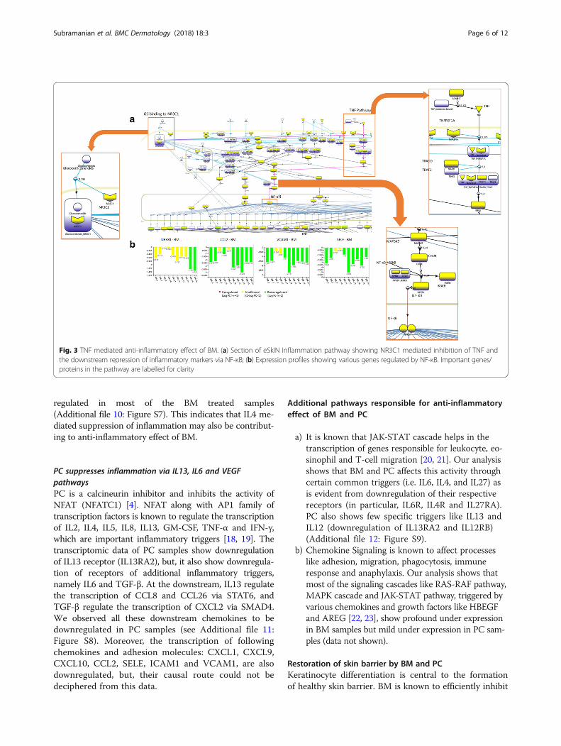
regulated in most of the BM treated samples(Additional file 10: Figure S7). This indicates that IL4 me-diated suppression of inflammation may also be contribut-ing to anti-inflammatory effect of BM.
PC suppresses inflammation via IL13, IL6 and VEGFpathwaysPC is a calcineurin inhibitor and inhibits the activity ofNFAT (NFATC1) [4]. NFAT along with AP1 family oftranscription factors is known to regulate the transcriptionof IL2, IL4, IL5, IL8, IL13, GM-CSF, TNF-α and IFN-γ,which are important inflammatory triggers [18, 19]. Thetranscriptomic data of PC samples show downregulationof IL13 receptor (IL13RA2), but, it also show downregula-tion of receptors of additional inflammatory triggers,namely IL6 and TGF-β. At the downstream, IL13 regulatethe transcription of CCL8 and CCL26 via STAT6, andTGF-β regulate the transcription of CXCL2 via SMAD4.We observed all these downstream chemokines to bedownregulated in PC samples (see Additional file 11:Figure S8). Moreover, the transcription of followingchemokines and adhesion molecules: CXCL1, CXCL9,CXCL10, CCL2, SELE, ICAM1 and VCAM1, are alsodownregulated, but, their causal route could not bedeciphered from this data.
Additional pathways responsible for anti-inflammatoryeffect of BM and PC
a) It is known that JAK-STAT cascade helps in thetranscription of genes responsible for leukocyte, eo-sinophil and T-cell migration [20, 21]. Our analysisshows that BM and PC affects this activity throughcertain common triggers (i.e. IL6, IL4, and IL27) asis evident from downregulation of their respectivereceptors (in particular, IL6R, IL4R and IL27RA).PC also shows few specific triggers like IL13 andIL12 (downregulation of IL13RA2 and IL12RB)(Additional file 12: Figure S9).
b) Chemokine Signaling is known to affect processeslike adhesion, migration, phagocytosis, immuneresponse and anaphylaxis. Our analysis shows thatmost of the signaling cascades like RAS-RAF pathway,MAPK cascade and JAK-STAT pathway, triggered byvarious chemokines and growth factors like HBEGFand AREG [22, 23], show profound under expressionin BM samples but mild under expression in PC sam-ples (data not shown).
Restoration of skin barrier by BM and PCKeratinocyte differentiation is central to the formationof healthy skin barrier. BM is known to efficiently inhibit
Fig. 3 TNF mediated anti-inflammatory effect of BM. (a) Section of eSkIN Inflammation pathway showing NR3C1 mediated inhibition of TNF andthe downstream repression of inflammatory markers via NF-κB; (b) Expression profiles showing various genes regulated by NF-κB. Important genes/proteins in the pathway are labelled for clarity
Subramanian et al. BMC Dermatology (2018) 18:3 Page 6 of 12

the primary cause of AD i.e. inflammation, but it also af-fects the skin barrier leading to secondary complicationslike skin atrophy and infection.Our analysis corroborates the fact that BM and PC up-
regulate several key skin barrier formation genes such asLoricrin (LOR), Filaggrin (FLG), TGM5 and CDSN(Additional file 13: Figure S10a) as a measure to restorebarrier functions [2]. However, it is also observed thatmost of the other barrier formation proteins like involu-crin (IVL), LCEs (LCE3D), TGM1, TGM3 and DSG3 areunder expressed in BM samples while they are mostlyunaffected by PC (Additional file 13: Figure S10b).
BM adversely affects the synthesis of barrier proteinsthrough AP1 family and CEBPsWe explored the possible causative pathway that couldlead to the impairment of skin barrier by BM. It isknown that calcium acts as a key trigger for epidermaldifferentiation through PLC-γ (PLCG1), which regulatesthe transcription of barrier proteins. Our analysis depictsanother, calcium independent route, to activate PLC-γthrough AKT1-PKN2 complex which is activated byCD44 [24] (Fig. 4a). This pathway further activates sev-eral transcriptional regulators including AP1 family oftranscription factors like JUN and FOS, CEBP-β(CEBPB), SP1 and HSPB1. These regulators play a majorrole in the transcription of barrier proteins like keratins,transglutaminases, loricrin, involucrin, filaggrin, LCEs,desmogleins, desmocollins, family of S100 proteins [25].The molecular details feature of eSkIN shows that thegenes involved in this pathway like CD44, AKT1, PKC-δ,HRAS and MAP2K3 are mostly downregulated in BM
samples, thus, implying the role of this pathway in BMtriggered skin barrier impairment (see Fig. 4b).Other transcriptional regulators involved in the activa-
tion of skin barrier proteins like JUND and FOSL1, arealso found to be downregulated. Furthermore, CEBP-α(CEBPA), an important transcription factor involved inthe transcription of IVL and desmocollins [26], is alsounder expressed in BM samples (Additional file 14:Figure S11). TGFA pathway mediates this transcriptionalregulation of CEBP-α [27]. The proliferation markerslike MYC and ERK (MAPK1) present in this pathwayare also under expressed in BM samples (data notshown).Keratinocyte Differentiation pathway also indicates that
the anti-microbial peptides S100A7, S100A8 and S100A9are under expressed in BM samples (Additional file 15:Figure S12). While S100A7 transcription is mediated byJUN [28], S100A8 and S100A9 transcription is mediatedby STAT3 via IL4 pathway (Additional file 15: Figure S12)[29]. This further explains the compromised skin barrierfunctions upon treatment with BM, leading to skin infec-tion [2].On the contrary, PC has mild effect on barrier pro-
teins and their regulation, and thus, helps in restoringskin barrier in AD patients.In addition to the profile of skin barrier proteins ob-
served in Keratinocyte Differentiation pathway, BarrierFormation genes like CTTN, CDC42, GRHL3 and AHRwhich are involved in the formation of tight junctions[30] are under expressed in BM samples. PC samplesdoes not show significant enrichment of this pathway inmost of the samples (Additional file 16: Figure S13).
Fig. 4 AP1 and CEBP mediated skin barrier impairment by BM. (a) eSkIN Keratinocyte Differentiation pathway depicting the effect of BM on synthesisof skin barrier proteins; (b) Expression profiles of key genes in this pathway. Important genes/proteins in the pathway are labelled for clarity
Subramanian et al. BMC Dermatology (2018) 18:3 Page 7 of 12

Additional pathways responsible for effect on skin barrierby BM and PC
a) Wound Healing pathway of eSkIN shows that VEGF(VEGFA), an important trigger for cellular processeslike cell migration, vascular permeability,angiogenesis, cell invasion and coagulation, issignificantly more under expressed in BM than inPC (Additional file 17: Figure S14). VEGFA activatesvarious pathways like PI3K (PIK3CA), PLC-γ andMAPK cascade, thus, impacting the normal physio-logical processes that are responsible for woundhealing [31]. Fibronectin (FN1), another protein in-volved in wound healing, is downregulated as well inBM samples [32].
b) Similarly, Basal Layer Formation pathway shows thatproteins like integrins, LAMA5 and collagens likeCOL4A1 [33] are under expressed in BM samples.On the contrary, most of these proteins show anupregulated trend in PC dataset (Additional file 18:Figure S15).
This further illustrates that BM affects the skin barrierformation by influencing the synthesis of barrier pro-teins, antimicrobial proteins and basal layer proteins.Such behavior is not observed in PC samples.
Cellular functions affected by BM and PCLipid Synthesis pathway is of interest as lipids are an inte-gral part of skin barrier. Application of BM is known toimpair the fatty acids and lipid content of skin [34]. LipidSynthesis pathway shows that lipid transporters likeLDLR and ABCA12 [35, 36] show downregulatedtrend (Additional file 19: Figure S16a), which mighthinder the transportation of cholesterol and otherlipids to skin. Enzymes like SPTLC2, SGMS2 andSMPD2 involved in the conversion of fatty acids toglucosylceramides [36] are also downregulated (Add-itional file 19: Figure S16b). Moreover, genes involvedin the synthesis of fatty acids or lipids like LXR(NR1H2), SCD, FASN and HMGCR [37, 38] aredownregulated (Additional file 20: Figure S17a), whilegenes involved in the metabolism of lipids like LPLand APOC1 [39] are showing an upregulated trend(Additional file 20: Figure S17b).DNA Damage and Repair is differentially affected in BM
and PC dataset. While most of the genes are unaffected byPC, genes involved in DNA repair like H2AFX, RAD51,BRCA2, MCM3, DHFR, HMOX1, GINS1 and PCNA[40–42] are under expressed in BM (Additional file 21:Figure S18). Similarly, CDK1, CDKN1A, CCNB1 and E2Ffamily of proteins, involved in cell cycle progression [43]show a downregulated trend in BM samples.
Though Autophagy and Proteasomal Degradation path-ways are enriched in most of the BM and PC samples, wecould not establish any relevance of this to the action ofthese drugs.
Comparison of eSkIN results with DAVIDTo evaluate the performance of eSkIN pathway enrichmentanalysis, we compared the results with the most widelyused functional enrichment tool, DAVID (https://david.n-cifcrf.gov/) [5]. BM and PC samples were analyzed separ-ately in DAVID. We obtained 409 differentially expressedgenes in BM and 49 genes in PC datasets that were used asinput for DAVID analysis (refer Methods for details).
Comparison of pathway enrichment for BM datasetThe enriched processes for BM dataset yielded 781 GOBiological Processes (GO BP) terms and 24 KEGGpathways (see Additional file 22). We observe that Im-mune response, Defense response, Inflammatory re-sponse, Keratinocyte differentiation, Skin developmentand Keratinization are enriched amongst the GO terms.Amongst KEGG pathways, Drug metabolism, Steroidhormone biosynthesis, Chemokine signaling pathwayand NF-kappa B signaling pathways are enriched.eSkIN uses a comprehensive manually curated skin cen-
tric knowledge-base for its analysis, and thus we observe alimited set of only relevant pathways (26 pathways) to beenriched. The pathways related to skin physiology, whichare enriched in DAVID analysis are also obtained usingeSkIN analysis (see Table 3), thus, corroborating the cap-ability of eSkIN to perform skin-centric pathway enrich-ment analysis.However, GO BP terms cannot be further explored in
terms of the molecular interactions between these genes(or their protein products) to derive mechanistic insightsinto the action of the drugs. On the other hand, eSkIN al-lows further exploration of the enriched pathways interms of the molecular interactions in them, and thus,helps in deriving mechanistic insights into drug action(see Table 1). Though KEGG Pathways can be explored insimilar context, the enriched KEGG pathways (Drug me-tabolism, Steroid hormone biosynthesis Chemokine sig-naling pathway and NF-kappa B signaling pathways) arenot directly relevant to our analysis.
Comparison of pathway enrichment for PCThe enriched processes for PC dataset yielded 62 GO BPterms and 0 KEGG pathways (see Additional file 22). Weobserve that skin related processes like Epidermis devel-opment, Skin development, Keratinocyte differentiation,Immune response and Defense response are enrichedamongst the GO terms. As is evident from Table 3, thecorresponding pathways are also enriched in eSkIN.Moreover, owing to the capability of eSkIN to highlight
Subramanian et al. BMC Dermatology (2018) 18:3 Page 8 of 12
molecular interactions of the enriched pathways, insightsinto mechanistic action of PC were obtained (see Table 2).
DiscussionIn this study, we analyzed transcriptomic datasets of ADpatients treated with Betamethasone and Pimecrolimus.We used a novel systems biology based approach to
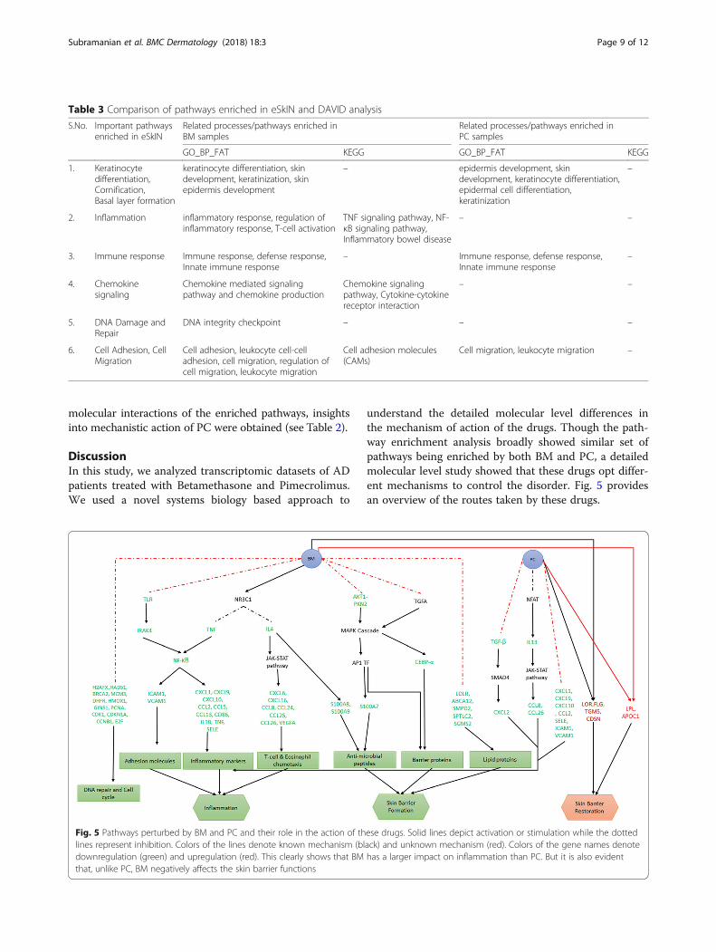
understand the detailed molecular level differences inthe mechanism of action of the drugs. Though the path-way enrichment analysis broadly showed similar set ofpathways being enriched by both BM and PC, a detailedmolecular level study showed that these drugs opt differ-ent mechanisms to control the disorder. Fig. 5 providesan overview of the routes taken by these drugs.
Table 3 Comparison of pathways enriched in eSkIN and DAVID analysis
S.No. Important pathwaysenriched in eSkIN
Related processes/pathways enriched inBM samples
Related processes/pathways enriched inPC samples
GO_BP_FAT KEGG GO_BP_FAT KEGG
1. Keratinocytedifferentiation,Cornification,Basal layer formation
keratinocyte differentiation, skindevelopment, keratinization, skinepidermis development
– epidermis development, skindevelopment, keratinocyte differentiation,epidermal cell differentiation,keratinization
–
2. Inflammation inflammatory response, regulation ofinflammatory response, T-cell activation
TNF signaling pathway, NF-κB signaling pathway,Inflammatory bowel disease
– –
3. Immune response Immune response, defense response,Innate immune response
– Immune response, defense response,Innate immune response
–
4. Chemokinesignaling
Chemokine mediated signalingpathway and chemokine production
Chemokine signalingpathway, Cytokine-cytokinereceptor interaction
– –
5. DNA Damage andRepair
DNA integrity checkpoint – – –
6. Cell Adhesion, CellMigration
Cell adhesion, leukocyte cell-celladhesion, cell migration, regulation ofcell migration, leukocyte migration
Cell adhesion molecules(CAMs)
Cell migration, leukocyte migration –
Fig. 5 Pathways perturbed by BM and PC and their role in the action of these drugs. Solid lines depict activation or stimulation while the dottedlines represent inhibition. Colors of the lines denote known mechanism (black) and unknown mechanism (red). Colors of the gene names denotedownregulation (green) and upregulation (red). This clearly shows that BM has a larger impact on inflammation than PC. But it is also evidentthat, unlike PC, BM negatively affects the skin barrier functions
Subramanian et al. BMC Dermatology (2018) 18:3 Page 9 of 12

We highlighted the role of TLRs, TNF and IL4 to triggerthe anti-inflammatory response and AKT1-PKN2 andTGFA via MAPK cascade to affect the skin barrier pro-teins in AD patients, when treated with BM. Stojadinovicet al. have shown the role of glucocorticoids in various cel-lular processes like inflammation, innate immunity, cellmigration, tissue remodeling, cell differentiation and celldeath in keratinocytes [3]. Our analysis further adds valueto the above mentioned finding by elaborating on thepathways that trigger various cellular responses, particu-larly in AD patients. We also depict the impact of BM onlipids and DNA damage and repair proteins thus leadingto an impaired barrier function.Under PC treatment, we show that TGF- and IL13
could be the plausible pathways involved in anti-inflammatory response. Through our comparative studyof the two drugs, we show that the molecular signaturesof barrier proteins under PC clearly contributes towardsrestoration of barrier.
Novel insight into disease manifestation during drugtreatmentThe mechanistic analysis of transcriptomic data of BMand PC treated samples also allowed us to understandthe manifestation of disease during the treatment bythese drugs. This provides new avenue towards futuredirection of drug discovery efforts in this area.In particular, EDN1 (Endothelin 1), which is positively
correlated with AD clinical severity [44, 45], is observed
to be mostly upregulated in BM while PC shows mildupregulation. This implies a risk of disease manifestationeven after treatment by BM or PC. By leveraging themolecular interaction maps of eSkIN, we elucidate thatin the downstream, EDN1 activates PLCB2 [46], whichin turn, triggers the MAPK cascade involved in cell mi-gration and inflammation (see Fig. 6a). We believe thattreatment by BM or PC, if supplemented with a drugtargeting EDN1 or downstream proteins, can bring syn-ergistic therapeutic effect in AD care.Furthermore, it is interesting to note that the suppres-
sors of cytokine family (SOCS), SOCS1 and SOCS3 aredownregulated even after the treatment with BM andPC. SOCS proteins are known to contribute towards dis-ease manifestation in psoriatic skin [47]. Our analysisshows that SOCS3 inhibits STAT3 activation via JAK1[48] (Fig. 6b). However, their role in AD manifestation isnot very well studied. Based on our analysis using eSkIN,it appears that downregulation of SOCS could be con-tributing towards the manifestation of AD, and thus, itmay be worthwhile to explore it as a drug target for fu-ture drug discovery efforts.
ConclusionOur study suggested the causal biomolecular inter-relationships involved in the action of BM and PC onhuman skin, apart from highlighting the existing evi-dence on these drugs in the context of skin-associatedfunctional networks. It is evident that BM downregulates
Fig. 6 Factors implicated in manifestation of disease during drug treatment. (a) EDN1 mediated; (b) SOCS3 mediated. Important genes/proteinsin the pathway are labelled for clarity
Subramanian et al. BMC Dermatology (2018) 18:3 Page 10 of 12

molecules involved in inflammation, T-cell & eosinophilchemotaxis, adhesion, DNA repair and cell cycle, whilePC targets a smaller section of inflammatory genes. Also,both BM and PC upregulate few important barrier pro-teins to restore the skin barrier functions. However, BMalso downregulates many other barrier componentswhich is largely unaffected by PC, thus, accounting forthe impaired skin barrier due to the treatment with BM.It should be noted that the results reported in this studyare based on 10 AD patients’ data that were available inNCBI GEO database, and needs further validation in alarger cohort.
Additional files
Additional file 1: Table S1. List of 35 pathways of eSkIN and theircategorization. (DOCX 13 kb)
Additional file 2: Sensitivity analysis to evaluate the effect of foldchange cutoff on pathway enrichment analysis. (DOCX 12 kb)
Additional file 3: Pathway enrichment analysis for different fold changecutoffs. Table and charts of pathway enrichment analysis for different foldchange cutoffs. (XLSX 30 kb)
Additional file 4: Figure S1. Comparison of enriched pathways at threedifferent fold change cutoffs (Log2 fold change (FC) = 0.5, 1 and 1.5).(TIFF 3928 kb)
Additional file 5: Figure S2. Expression profile of genes involved inTNF pathway in BM samples. (TIFF 4333 kb)
Additional file 6: Figure S3. Expression profile of genes regulated byNF-κB in BM samples. (TIFF 12640 kb)
Additional file 7: Figure S4. Section of eSkIN Inflammation pathwayshowing TLR mediated activation of NF-κB and the expression profile ofgenes involved in this pathway. (TIFF 12017 kb)
Additional file 8: Figure S5. Section of eSkIN Inflammation pathwayshowing NR3C1 mediated inhibition of NF-κB through IκB (highlighted inpink) and the expression profile of IκB. (TIFF 7299 kb)
Additional file 9: Figure S6. Sections of eSkIN Inflammation pathwayshowing: (a) activation of JAK-STAT pathway by IL4 and IFN-γ and theirexpression profiles (b) genes regulated by STAT6. (TIFF 9845 kb)
Additional file 10: Figure S7. Expression profile of genes activated byIL4 via JAK-STAT pathway. (TIFF 5004 kb)
Additional file 11: Figure S8. Expression profile of inflammatory genesthat show downregulation in PC samples. (TIFF 10440 kb)
Additional file 12: Figure S9. Section of eSkIN Immune Responsepathway showing various activators of JAK-STAT pathway and theirexpression profile in BM and PC samples. (TIFF 10437 kb)
Additional file 13: Figure S10. Expression profile of skin barrierproteins in BM and PC samples. (a) Expression profile of genes that areupregulated in both BM and PC in order to restore barrier functions; (b)Expression profile of skin barrier genes that show treatment specificdifference in their expressions. (TIFF 8378 kb)
Additional file 14: Figure S11. Expression profile of importanttranscription factors of barrier proteins in BM. (TIFF 5538 kb)
Additional file 15: Figure S12. Expression profile of anti-microbialpeptides in BM samples and a section of eSkIN pathway showingtheir transcriptional regulation by IL4. (TIFF 5747 kb)
Additional file 16: Figure S13. Expression profile of junction proteinsthat show treatment specific difference in their expressions. (TIFF 6474 kb)
Additional file 17: Figure S14. Section of eSkIN Wound Healingpathway showing VEGF mediated activation of cellular functions like focaladhesion, actin remodeling, cell migration, vascular permeability,
angiogenesis, degradation of collagen, cell invasion and blood coagulation,and expression profile of VEGF in BM and PC samples. (TIFF 12210 kb)
Additional file 18: Figure S15. Expression profile of basal layer genesthat show treatment specific difference in their expressions. (TIFF 12463 kb)
Additional file 19: Figure S16. Sections of eSkIN Lipid Synthesispathway showing: (a) lipid transporters and their expression profiles inBM samples (b) enzymes involved in fatty acid conversion and theirexpression profiles in BM samples. (TIFF 5067 kb)
Additional file 20: Figure S17. Sections of eSkIN Lipid Synthesispathway showing: (a) the genes involved in the synthesis of lipids and fattyacids and their expression profiles in BM samples (b) genes involved in lipidmetabolism and their expression profiles in BM samples. (TIFF 7877 kb)
Additional file 21: Figure S18. Section of eSkIN DNA Damage andRepair pathway showing the genes involved in DNA repair mechanismsand their expression profiles in BM samples. (TIFF 10956 kb)
Additional file 22: Pathway enrichment analysis using DAVID. Tables ofenriched pathways (p-value < 0.05) using GO biological process(GO_BP_FAT) and KEGG pathways as annotation datasets in DAVID.BM and PC datasets were analyzed separately. (XLSX 168 kb)
AbbreviationsAD: Atopic dermatitis; BM: Betamethasone valerate; DEG: Differentiallyexpressed genes; GEO: Gene Expression Omnibus; PC: Pimecrolimus;ROS: Reactive oxygen species; TCI: Topical calcineurin inhibitor; US FDA: USFood and Drug Administration
AcknowledgementsThe authors acknowledge the eSkIN team for making eSkIN available for thepresent study. We thank Council of Scientific and Industrial Research (CSIR)for funding eSkIN project.
FundingNot applicable
Availability of data and materialseSkIN is available at www.persistent.com/eskin. The dataset analyzed isdownloaded from NCBI GEO (Gene Expression Omnibus) database usingfollowing accession number: GSE32473.
Authors’ contributionsAJ, VS and IS conceived and designed the study. IS and VS performed theexperiments. All authors contributed in analyzing the results and writing themanuscript. All authors have given approval to the final version of themanuscript.
Ethics approval and consent to participateThis study does not involve humans or animals.
Consent for publicationNot applicable
Competing interestsAll authors are employees of Persistent Systems Limited. However, this doesnot alter the authors’ adherence to the journal policies on sharing data andmaterials.
Publisher’s NoteSpringer Nature remains neutral with regard to jurisdictional claims inpublished maps and institutional affiliations.
Received: 11 July 2017 Accepted: 30 January 2018
References1. Nutten S. Atopic dermatitis: global epidemiology and risk factors. Ann Nutr
Metab. 2015;66(Suppl 1):8–16.2. Jensen JM, et al. Gene expression is differently affected by pimecrolimus and
betamethasone in lesional skin of atopic dermatitis. Allergy. 2012;67(3):413–23.
Subramanian et al. BMC Dermatology (2018) 18:3 Page 11 of 12

3. Stojadinovic O, et al. Novel genomic effects of glucocorticoids in epidermalkeratinocytes: inhibition of apoptosis, interferon-gamma pathway, andwound healing along with promotion of terminal differentiation. J BiolChem. 2007;282(6):4021–34.
4. Siegfried EC, Jaworski JC, Hebert AA. Topical calcineurin inhibitors andlymphoma risk: evidence update with implications for daily practice. Am JClin Dermatol. 2013;14(3):163–78.
5. Huang DW, Sherman BT, Lempicki RA. Systematic and integrative analysis oflarge gene lists using DAVID bioinformatics resources. Nat Protoc. 2009;4(1):44–57.
6. Huang DW, Sherman BT, Lempicki RA. Bioinformatics enrichment tools:paths toward the comprehensive functional analysis of large gene lists.Nucleic Acids Res. 2009;37(1):1–13.
7. Subramanian A, et al. Gene set enrichment analysis: a knowledge-basedapproach for interpreting genome-wide expression profiles. Proc Natl AcadSci U S A. 2005;102(43):15545–50.
8. J.-H. Hung, “Gene set/pathway enrichment analysis,” in Data Mining forSystems Biology, H. Mamitsuka, C. DeLisi, and M. Kanehisa, Eds. HumanaPress, 2013, pp. 201–213.
9. P. Zhao et al., “Identification of differentially expressed genes in pituitaryadenomas by integrating analysis of microarray data,” Int J Endocrinol, 2015.Available: https://www.hindawi.com/journals/ije/2015/164087/. Accessed 17Oct 2017.
10. Wu X, et al. Network expansion and pathway enrichment analysis towardsbiologically significant findings from microarrays. J Integr Bioinforma. 2016;9(2):113–25.
11. Bradley JR. TNF-mediated inflammatory disease. J Pathol. 2008;214(2):149–60.12. Miller LS. Toll-like receptors in skin. Adv Dermatol. 2008;24:71–87.13. Murphy JE, Robert C, Kupper TS. Interleukin-1 and cutaneous inflammation:
a crucial link between innate and acquired immunity. J. Invest. Dermatol.2000;114(3):602–8.
14. Chen Y, et al. Microarray analysis reveals the inhibition of nuclear factor-kappaB signaling by aristolochic acid in normal human kidney (HK-2) cells. ActaPharmacol Sin. 2010;31(2):227–36.
15. Kaesler S, et al. Toll-like receptor 2 ligands promote chronic atopicdermatitis through IL-4-mediated suppression of IL-10. J Allergy ClinImmunol. 2014;134(1):92–9.
16. Hari A, Flach TL, Shi Y, Mydlarski PR. Toll-like receptors: role indermatological disease. Mediat Inflamm. 2010;2010:437246.
17. Bao L, Zhang H, Chan LS. The involvement of the JAK-STAT signalingpathway in chronic inflammatory skin disease atopic dermatitis. JAK-STAT.2013;2(3):e24137.
18. Im S-H, Rao A. Activation and deactivation of gene expression by Ca2+/calcineurin-NFAT-mediated signaling. Mol Cells. 2004;18(1):1–9.
19. Medyouf H, Ghysdael J. The calcineurin/NFAT signaling pathway: a noveltherapeutic target in leukemia and solid tumors. Cell Cycle Georget Tex.2008;7(3):297–303.
20. Murata T, Husain SR, Mohri H, Puri RK. Two different IL-13 receptor chains areexpressed in normal human skin fibroblasts, and IL-4 and IL-13 mediate signaltransduction through a common pathway. Int Immunol. 1998;10(8):1103–10.
21. Wittmann M, Zeitvogel J, Wang D, Werfel T. IL-27 is expressed in chronichuman eczematous skin lesions and stimulates human keratinocytes.J Allergy Clin Immunol. 2009;124(1):81–9.
22. New DC, Wong YH. CC chemokine receptor-coupled signalling pathways. ShengWu Hua Xue Yu Sheng Wu Wu Li Xue Bao (Shanghai). 2003;35(9):779–88.
23. Pastore S, Mascia F, Mariotti F, Dattilo C, Mariani V, Girolomoni G. ERK1/2regulates epidermal chemokine expression and skin inflammation.J Immunol Baltim Md 1950. 2005;174(8):5047–56.
24. Bourguignon LYW, Singleton PA, Diedrich F. Hyaluronan-CD44 interactionwith Rac1-dependent protein kinase N-gamma promotes phospholipaseCgamma1 activation, ca(2+) signaling, and cortactin-cytoskeleton functionleading to keratinocyte adhesion and differentiation. J Biol Chem. 2004;279(28):29654–69.
25. Eckert RL, et al. Regulation of involucrin gene expression. J. Invest. Dermatol.2004;123(1):13–22.
26. Efimova T, Deucher A, Kuroki T, Ohba M, Eckert RL. Novel protein kinase Cisoforms regulate human keratinocyte differentiation by activating a p38delta mitogen-activated protein kinase cascade that targets CCAAT/enhancer-binding protein alpha. J Biol Chem. 2002;277(35):31753–60.
27. Reynolds NJ, et al. Differential induction of phosphatidylcholine hydrolysis,diacylglycerol formation and protein kinase C activation by epidermal
growth factor and transforming growth factor-alpha in normal human skinfibroblasts and keratinocytes. Biochem J. 1993;294(Pt 2):535–44.
28. Emberley ED, et al. The S100A7-c-Jun activation domain binding protein 1pathway enhances prosurvival pathways in breast cancer. Cancer Res. 2005;65(13):5696–702.
29. Uto-Konomi A, et al. Dysregulation of suppressor of cytokine signaling 3 inkeratinocytes causes skin inflammation mediated by interleukin-20 receptor-related cytokines. PLoS One. 2012;7(7):e40343.
30. Jamora C, Fuchs E. Intercellular adhesion, signalling and the cytoskeleton.Nat Cell Biol. 2002;4(4):E101–8.
31. Olsson A-K, Dimberg A, Kreuger J, Claesson-Welsh L. VEGF receptor signalling -in control of vascular function. Nat Rev Mol Cell Biol. 2006;7(5):359–71.
32. Zambruno G, et al. Transforming growth factor-beta 1 modulates beta 1and beta 5 integrin receptors and induces the de novo expression of thealpha v beta 6 heterodimer in normal human keratinocytes: implications forwound healing. J Cell Biol. 1995;129(3):853–65.
33. Fleischmajer R, et al. Initiation of skin basement membrane formation at theepidermo-dermal interface involves assembly of laminins through bindingto cell membrane receptors. J Cell Sci. 1998;111(Pt 14):1929–40.
34. Jensen J-M, et al. Different effects of pimecrolimus and betamethasone onthe skin barrier in patients with atopic dermatitis. J Allergy Clin Immunol.2009;123(5):1124–33.
35. Zhang L, Reue K, Fong LG, Young SG, Tontonoz P. Feedback regulation ofcholesterol uptake by the LXR-IDOL-LDLR axis. Arterioscler Thromb VascBiol. 2012;32(11):2541–6.
36. Oda Y, Uchida Y, Moradian S, Crumrine D, Elias PM, Bikle DD. Vitamin Dreceptor and coactivators SRC2 and 3 regulate epidermis-specificsphingolipid production and permeability barrier formation. J. Invest.Dermatol. 2009;129(6):1367–78.
37. Yokoyama A, et al. Induction of SREBP-1c mRNA by differentiation and LXRligand in human keratinocytes. J. Invest. Dermatol. 2009;129(6):1395–401.
38. Harris IR, et al. Parallel regulation of sterol regulatory element bindingprotein-2 and the enzymes of cholesterol and fatty acid synthesis but notceramide synthesis in cultured human keratinocytes and murine epidermis.J Lipid Res. 1998;39(2):412–22.
39. Jiang ZG, Robson SC, Yao Z. Lipoprotein metabolism in nonalcoholic fattyliver disease. J Biomed Res. 2013;27(1):1–13.
40. Malewicz M, et al. Essential role for DNA-PK-mediated phosphorylation ofNR4A nuclear orphan receptors in DNA double-strand break repair. GenesDev. 2011;25(19):2031–40.
41. Yoshida K, Miki Y. Role of BRCA1 and BRCA2 as regulators of DNA repair,transcription, and cell cycle in response to DNA damage. Cancer Sci. 2004;95(11):866–71.
42. Meng X, Yuan Y, Maestas A, Shen Z. Recovery from DNA damage-inducedG2 arrest requires actin-binding protein filamin-a/actin-binding protein 280.J Biol Chem. 2004;279(7):6098–105.
43. Sulaimon SS, Kitchell BE. The basic biology of malignant melanoma:molecular mechanisms of disease progression and comparative aspects.J Vet Intern Med. 2003;17(6):760–72.
44. Aktar MK, Kido-Nakahara M, Furue M, Nakahara T. Mutual upregulation ofendothelin-1 and IL-25 in atopic dermatitis. Allergy. 2015;70(7):846–54.
45. Tsybikov NN, Petrisheva IV, Kuznik BI, Magen E. Plasma endothelin-1 levelsduring exacerbation of atopic dermatitis. Allergy Asthma Proc. 2015;36(4):320–4.
46. Bouallegue A, Daou GB, Srivastava AK. Endothelin-1-induced signalingpathways in vascular smooth muscle cells. Curr Vasc Pharmacol. 2007;5(1):45–52.
47. Sonkoly E, et al. MicroRNAs: novel regulators involved in the pathogenesisof psoriasis? PLoS One. 2007;2(7):e610.
48. Yamasaki K, et al. Suppressor of cytokine signaling 1/JAB and suppressor ofcytokine signaling 3/cytokine-inducible SH2 containing protein 3 negativelyregulate the signal transducers and activators of transcription signalingpathway in normal human epidermal keratinocytes. J Invest Dermatol. 2003;120(4):571–80.
Subramanian et al. BMC Dermatology (2018) 18:3 Page 12 of 12