
Elisabete Fernandes biomarkers -...
20
3-Multiplexed detection of ischemic stroke (ICTUS) related biomarkers Invennta Project, with Santiago de Compostela Hospital biomarkers Elisabete Fernandes Requirement: fast analysis (< 1h) of patient to select treatment
Transcript of Elisabete Fernandes biomarkers -...

3-Multiplexed detection of ischemic stroke (ICTUS) related biomarkers
Invennta Project, with Santiago de Compostela Hospital
biomarkers
Elisabete Fernandes
Requirement: fast analysis (< 1h) of patient to select treatment

magnetic label (FeOx)
probe antibody
Labeling antibody
proteic biomarker
2.5 x 40 um2
Sandwich immunoassay using MNP labels and magnetoresistive detection
MR sensor
Biotinilated
antibody

Matrix Metallopeptidase 9 detection
70 µV
100 µV
170 µV
MMP9 present detection limit <1ng/mL
Fibronection < 0.5 ug/mL

-0.005
0.000
0.005
0.010
0.015
0.020
DV
bin
din
g/V
sen
sor
Probe antibody
Fibro S100 Angio PDGF Serpin
SPINTRONIC INTEGRATED MICROFLUIDIC PLATFORMS FOR ENVIRONMENTAL AND BIOMEDICAL
APPLICATIONS
Group: NANOELECTRONIS
Detection of Fibronectin in a complex sample matrix
1
1
21
11
21
21
21
1
1
21
11
12
1
121
1
1
2
1
1
1
Complex Sample
(multiple targets)

New projects already started
( GAIN funding)
-detecting biomarkers for colo-rectal cancer
In feces ( with J Cubiella, Sergas, Ourense)
-detecting biomarkers for peritoneal fibrosis
( with Africa Gonzalez, CINBIO, U Vigo)

• Detection & Counting of
Circulating Tumor Cells
(CTCs) as a means of studying
the process of metastasis,
using an electronic,
automated platform
• CTCs phenotype
identification:
EpCAM (+) vs EpCAM (-)
Invasive CTCs were identified as cells exhibiting CAM
invasion (CAM+) and expressing standard epithelial markers
(Epi+). Clin Chem. 2011 Sep;57(9):1242-55. doi: 10.1373/clinchem.2011.165068. Epub 2011 Jul 22.
4-Integrated CTC’s detection in bloodA. Chicharo, L.Dieguez, M.Oliveira, S.Cardoso, J.PiteiraR. Lopez, M.Abal, Clotilde INL, INESC MN, IDIS, Hospital Santiago Compostela

Previous work: detecting labeled cells in flow(INESC MN)

With C Fermon, and M Pannetier
APL 2009
Detecting 1 um magn. beads

Cell detection – Kg1a cells
cell=5-10m
Cells marked with 50nm
FeOx particles
Lab on Chip ( 2011)

Estimate
#of cells
Pipetted
Initial #of
cells
counted
parallel
Traped
#of cells in
the filter
Recovered
#cells
Trapping
efficiency
Recover
y
effiency
Run #1 50 48 34 40 71% 83%
Run #2 50 44 35 31 80% 70%
Run #3 50 62 49 43 79% 69%
Result
s 70% 66% 0%
20%
40%
60%
80%
100%
PBS
Eff
icie
ncy
Trapping efficiency
• 50 cells SW480 labelled
with magnetic beads
• Loaded in the microfluidic
device
• Cells are suspended in
PBS-BSA2%
Micron-sized
posts
Flow
100 µm
Trapping cancer cells (CTCs) in microfluidic device

- Baseline
- Sample of beads with control Antibody
- 500 Sw480 cells spiked in buffy coat+ beads with EpCam Antibody
Baseline
Beads + Ab
Cells + Beads + Ab
Beads + Ab
Baseline
613
0
100
200
300
400
500
600
700
Sensor 1
Num
ber
of
pea
ks
peak
valey
At a flow rate of 50l/min: Amplitude Time-of-flight
Bead 120Vpp 2-3 ms
Cell 240Vpp 15-30ms
By direct measurements: the sample’s velocity
is 12.5mm/s
Bead CellAlexandre Chicharo, INL/IST
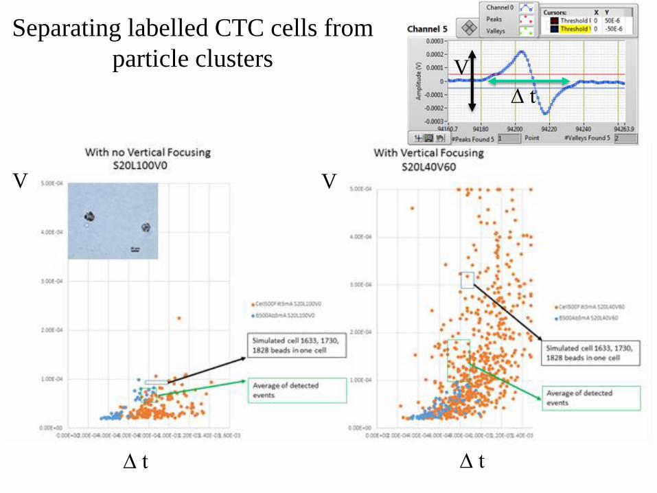
Separating labelled CTC cells from
particle clusters
VV
∆ t ∆ t
∆ t
V

Neuronal magnetic fields: In-vivo experiments
Stimuli: 100ms or 500ms
Light pulses, with inter-stimulus
Time around 1s
Laure Caruso1, Thomas Wunderle2, Christopher M. Lewis2, Joao Valadeiro3, Vincent Trauchessec1,
Josué Trejo Rosillo1, José Pedro Amaral3, Jianguang Ni2, Claude Fermon1, Susana Cardoso3, Paulo
Freitas3, Pascal Fries2,4, Myriam Pannetier-Lecoeur1*.
http://dx.doi.org/10.1101/092569 Neuron 95, 1–9, September 13, 2017
2014-2016

Sensors
200 µm
thick Si
Problems: shield sensor from light ( affecting Si substrate)

Signal modulation and
demodulation
Magnetic signal
Electrical signal
(capacitive coupling)
Total gain
500-1000

6 months later

Comparison of magnetic and electrical
signals: ERF and ERP
W electrodeMR sensor

Stimulation electrode
Recording electrode
Rat hippocampus
Synaptic current monitoring with high
Spatial resolution ( with A.Sebastiao, IMM, V.Santos, ICVS)
INESC MN and IMM
MAGNETRODES, FP7 (2013-2016)

Recording
electrodeStimulus
electrode
CA
1
CA
3
MR sensor
(sharp probe)Stimulus
electrode
Rat brain
slice
Recording Chamber
Setup facilities at IMM, Lisbon
Electrical
Stimulus:
Frequency : 0.5 Hz
Amplitude: 300 A
Duration : 0.3 ms
MR sensor
(sharp probe)
Stimulus
electrode
300 m
550 m
Hippocampus Brain
Slice dimensions:
- thickness: 300- 400 m
- width: 1.6 mm
- length: 3 mm
In collaboration with A Sebastiao, IMM, Lisboa
INESC MN

Measurement setup
Reference Input
x2
Computer
500 W+100W
500 W500 W
Signal Generator
(Sine wave 20kHz;
500mV amplitude)DAQ
Electronic
Board
AC measurement with modulation and demodulation
Wheatstone bridge:
• two fixed resistances (500 W)
• one adjustable resistance (fixed resistance 500 W in series with a potentiometer of 100 W)
• one variable resistance (Magnetoresistive Sensor)
Total gain = 200 000
ac measurements
220 Hz


