ELISA Kit ab108870 – IL-10 Mouse€™s IL-10 Mouse in vitro ELISA (enzyme-linked immunosorbent...
27
Version 4 Last Updated 8 October 2018 Instructions for Use For the quantitative measurement of mouse IL-10 in plasma, serum, tissue extracts and cell culture supernatants. This product is for research use only and is not intended for diagnostic use. ab108870 – IL-10 Mouse ELISA Kit
Transcript of ELISA Kit ab108870 – IL-10 Mouse€™s IL-10 Mouse in vitro ELISA (enzyme-linked immunosorbent...

Version 4 Last Updated 8 October 2018
Instructions for Use
For the quantitative measurement of mouse IL-10 in plasma, serum, tissue extracts and cell culture supernatants.
This product is for research use only and is not intended for diagnostic use.
ab108870 – IL-10 Mouse ELISA Kit

Discover more at www.abcam.com 1
Table of Contents
INTRODUCTION1. BACKGROUND 22. ASSAY SUMMARY 33. PRECAUTIONS 4
GENERAL INFORMATION4. STORAGE AND STABILITY 45. MATERIALS SUPPLIED 46. MATERIALS REQUIRED, NOT SUPPLIED 57. LIMITATIONS 58. TECHNICAL HINTS 6
ASSAY PREPARATION9. REAGENT PREPARATION 710. STANDARD PREPARATIONS 1011. SAMPLE PREPARATION 1312. PLATE PREPARATION 14
ASSAY PROCEDURE13. ASSAY PROCEDURE 15
DATA ANALYSIS14. CALCULATIONS 1715. TYPICAL DATA 1816. TYPICAL SAMPLE VALUES 1917. ASSAY SPECIFICITY 20
RESOURCES18. TROUBLESHOOTING 2119. NOTES 23
Discover more at www.abcam.com 2
INTRODUCTION
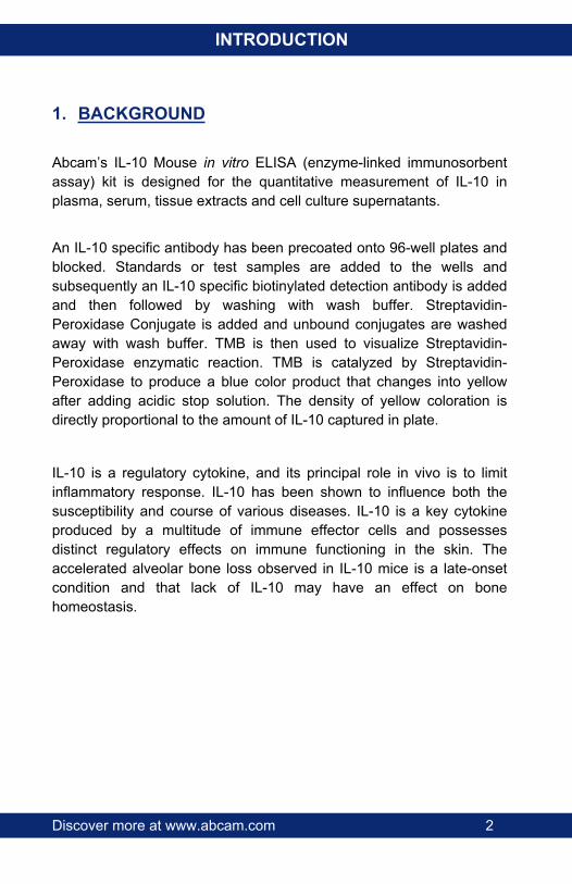
1. BACKGROUND
Abcam’s IL-10 Mouse in vitro ELISA (enzyme-linked immunosorbent assay) kit is designed for the quantitative measurement of IL-10 in plasma, serum, tissue extracts and cell culture supernatants.
An IL-10 specific antibody has been precoated onto 96-well plates and blocked. Standards or test samples are added to the wells and subsequently an IL-10 specific biotinylated detection antibody is added and then followed by washing with wash buffer. Streptavidin-Peroxidase Conjugate is added and unbound conjugates are washed away with wash buffer. TMB is then used to visualize Streptavidin-Peroxidase enzymatic reaction. TMB is catalyzed by Streptavidin-Peroxidase to produce a blue color product that changes into yellow after adding acidic stop solution. The density of yellow coloration is directly proportional to the amount of IL-10 captured in plate.
IL-10 is a regulatory cytokine, and its principal role in vivo is to limit inflammatory response. IL-10 has been shown to influence both the susceptibility and course of various diseases. IL-10 is a key cytokine produced by a multitude of immune effector cells and possesses distinct regulatory effects on immune functioning in the skin. The accelerated alveolar bone loss observed in IL-10 mice is a late-onset condition and that lack of IL-10 may have an effect on bone homeostasis.

Discover more at www.abcam.com 3
INTRODUCTION
2. ASSAY SUMMARY
Prepare all reagents, samples and standards as instructed.
Add standard or sample to each well used. Incubate at room temperature.
Wash and add prepared biotin antibody to each well. Incubate at room temperature.
Wash and add prepared Streptavidin-Peroxidase Conjugate. Incubate at room temperature.
Add Chromogen Substrate to each well. Incubate at room temperature. Add Stop Solution to each well. Read immediately.
Discover more at www.abcam.com 4
GENERAL INFORMATION
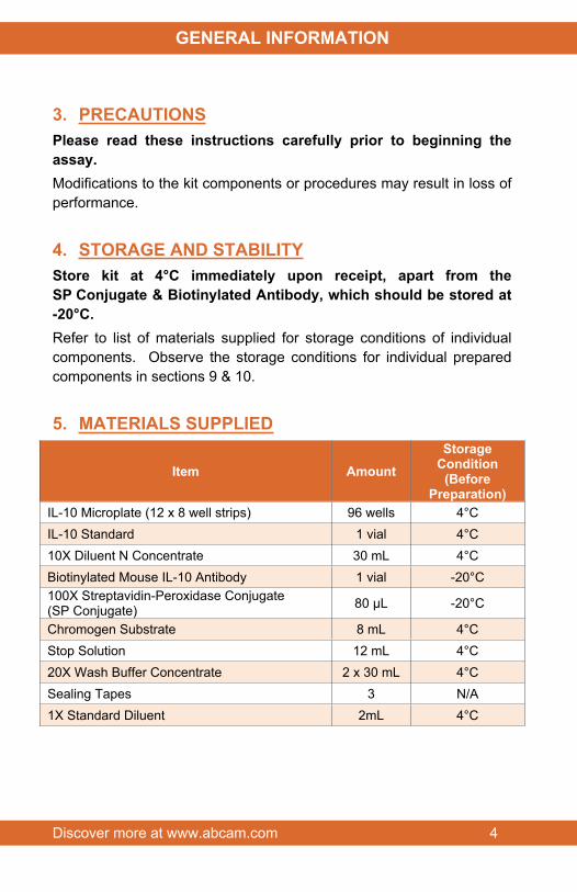
3. PRECAUTIONSPlease read these instructions carefully prior to beginning the assay.Modifications to the kit components or procedures may result in loss of performance.
4. STORAGE AND STABILITYStore kit at 4°C immediately upon receipt, apart from the SP Conjugate & Biotinylated Antibody, which should be stored at -20°C.Refer to list of materials supplied for storage conditions of individual components. Observe the storage conditions for individual prepared components in sections 9 & 10.
5. MATERIALS SUPPLIED
Item AmountStorage
Condition(Before
Preparation)IL-10 Microplate (12 x 8 well strips) 96 wells 4°CIL-10 Standard 1 vial 4°C10X Diluent N Concentrate 30 mL 4°CBiotinylated Mouse IL-10 Antibody 1 vial -20°C100X Streptavidin-Peroxidase Conjugate (SP Conjugate) 80 µL -20°C
Chromogen Substrate 8 mL 4°CStop Solution 12 mL 4°C20X Wash Buffer Concentrate 2 x 30 mL 4°CSealing Tapes 3 N/A1X Standard Diluent 2mL 4°C

Discover more at www.abcam.com 5
GENERAL INFORMATION
6. MATERIALS REQUIRED, NOT SUPPLIEDThese materials are not included in the kit, but will be required to successfully utilize this assay:
1 Microplate reader capable of measuring absorbance at 450 nm.
Precision pipettes to deliver 1 µL to 1 mL volumes.
Adjustable 1-25 mL pipettes for reagent preparation.
100 mL and 1 liter graduated cylinders.
Absorbent paper.
Distilled or deionized water.
Log-log graph paper or computer and software for ELISA data analysis.
8 tubes to prepare standard or sample dilutions.
7. LIMITATIONS Do not mix or substitute reagents or materials from other kit lots or
vendors.
Discover more at www.abcam.com 6
GENERAL INFORMATION
8. TECHNICAL HINTS Samples generating values higher than the highest standard
should be further diluted in the appropriate sample dilution buffers.
Avoid foaming or bubbles when mixing or reconstituting components.
Avoid cross contamination of samples or reagents by changing tips between sample, standard and reagent additions.
Ensure plates are properly sealed or covered during incubation steps.
Complete removal of all solutions and buffers during wash steps.
This kit is sold based on number of tests. A ‘test’ simply refers to a single assay well. The number of wells that contain sample, control or standard will vary by product. Review the protocol completely to confirm this kit meets your requirements. Please contact our Technical Support staff with any questions.
Discover more at www.abcam.com 7
ASSAY PREPARATION
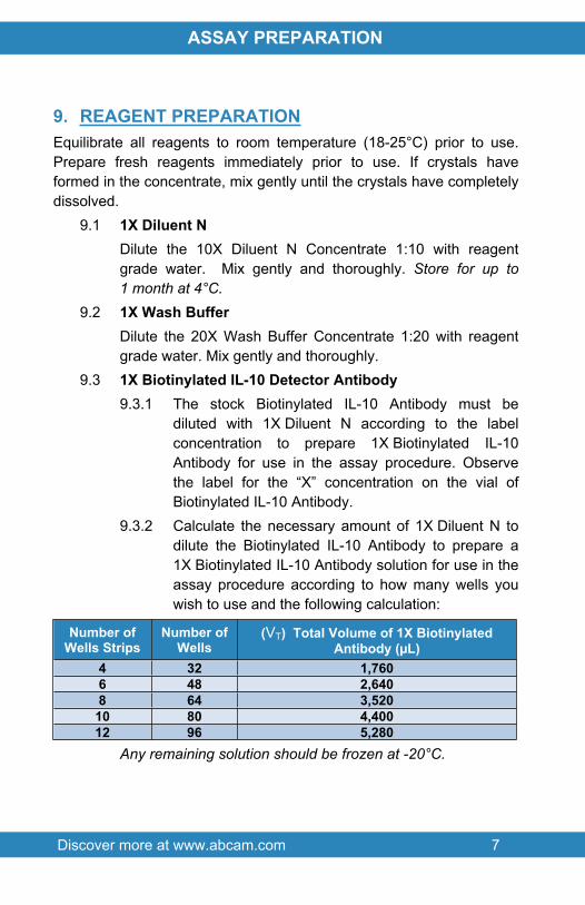
9. REAGENT PREPARATIONEquilibrate all reagents to room temperature (18-25°C) prior to use. Prepare fresh reagents immediately prior to use. If crystals have formed in the concentrate, mix gently until the crystals have completely dissolved.
9.1 1X Diluent NDilute the 10X Diluent N Concentrate 1:10 with reagent grade water. Mix gently and thoroughly. Store for up to 1 month at 4°C.
9.2 1X Wash BufferDilute the 20X Wash Buffer Concentrate 1:20 with reagent grade water. Mix gently and thoroughly.
9.3 1X Biotinylated IL-10 Detector Antibody9.3.1 The stock Biotinylated IL-10 Antibody must be
diluted with 1X Diluent N according to the label concentration to prepare 1X Biotinylated IL-10 Antibody for use in the assay procedure. Observe the label for the “X” concentration on the vial of Biotinylated IL-10 Antibody.
9.3.2 Calculate the necessary amount of 1X Diluent N to dilute the Biotinylated IL-10 Antibody to prepare a 1X Biotinylated IL-10 Antibody solution for use in the assay procedure according to how many wells you wish to use and the following calculation:
Number of Wells Strips
Number of Wells
(VT) Total Volume of 1X Biotinylated Antibody (µL)
4 32 1,7606 48 2,6408 64 3,520
10 80 4,40012 96 5,280
Any remaining solution should be frozen at -20°C.

Discover more at www.abcam.com 8
ASSAY PREPARATION
Where:CS = Starting concentration (X) of stock Biotinylated IL-10 Antibody
(variable)CF = Final concentration (always = 1X) of 1X Biotinylated IL-10
Antibody solution for the assay procedureVT = Total required volume of 1X Biotinylated IL-10 Antibody solution
for the assay procedureVA = Total volume of (X) stock Biotinylated IL-10 AntibodyVD = Total volume of 1X Diluent N required to dilute (X) stock
Biotinylated IL-10 Antibody to prepare 1X Biotinylated IL-10 solution for assay procedures
Calculate the volume of (X) stock Biotinylated Antibody required for the given number of desired wells:
(CF / CS) x VT = VA
Calculate the final volume of 1X Diluent N required to prepare the 1X Biotinylated IL-10 Antibody:
VT - VA = VD
Example:NOTE: This example is for demonstration purposes only. Please remember to check your antibody vial for the actual concentration of antibody provided.CS = 50X Biotinylated IL-10 Antibody stockCF = 1X Biotinylated IL-10 Antibody solution for use in the assay
procedureVT = 3,520 µL (8 well strips or 64 wells)
(1X/50X) x 3,520 µL = 70.4 µL3,520 µL - 70.4 µL = 3,449.6 µL
VA = 70.4 µL total volume of (X) stock Biotinylated IL-10 Antibody required
VD = 3,449.6 µL total volume of 1X Diluent N required to dilute the 50X stock Biotinylated Antibody to prepare 1X Biotinylated IL-10 Antibody solution for assay procedures

Discover more at www.abcam.com 9
ASSAY PREPARATION
9.3.3 First spin the Biotinylated IL-10 Antibody vial to collect the contents at the bottom.
9.3.4 Add calculated amount VA of stock Biotinylated IL-10 Antibody to the calculated amount VD of 1X Assay Diluent N. Mix gently and thoroughly.
9.4 1X SP Conjugate Spin down the 100X Streptavidin-Peroxidase Conjugate (SP Conjugate) briefly and dilute the desired amount of the conjugate 1:100 with 1X Diluent N. Any remaining solution should be stored at -20°C.

Discover more at www.abcam.com 10
ASSAY PREPARATION
10.STANDARD PREPARATIONS Prepare serially diluted standards immediately prior to use.
Always prepare a fresh set of standards for every use. Any remaining standard should be stored at -20°C after
reconstitution and used within 30 days. This procedure prepares sufficient standard dilutions for
duplicate wells.10.1 Reconstitute of the IL-10 Standard using Standard diluent
to prepare 2 ng/mL IL-10 Standard #110.1.1 First consult the IL-10 Standard vial to determine
the mass of protein in the vial.10.1.2 Calculate the appropriate volume of 1X Standard
Diluent to add when resuspending the IL-10 Standard vial to produce a 2 ng/mL IL-10 Standard #1 by using the following equation:
CS = Starting mass of IL-10 Standard (see vial label) (ng)CF = 2 ng/mL IL-10 Standard #1 final required concentrationVD = Required volume of 1X Standard Diluent for reconstitution (µL)
Calculate total required volume 1X Standard Diluent for resuspension:
(CS/ CF) x 1,000 = VD
Example:NOTE: This example is for demonstration purposes only. Please remember to check your standard vial for the actual amount of standard provided.
CS = 2 ng of IL-10 Standard in vialCF = 2 ng/mL IL-10 Standard #1 final concentration VD = Required volume of 1X Standard Diluent for reconstitution
(2 ng / 2 ng/mL) x 1,000 = 1,000 µL
Discover more at www.abcam.com 11
ASSAY PREPARATION
10.1.3 First briefly spin the IL-10 Standard Vial to collect the contents on the bottom of the tube.
10.1.4 Reconstitute the IL-10 Standard vial by adding the appropriate calculated amount VD of 1X Standard Diluent to the vial to generate the 2 ng/mL IL-10 Standard #1. Mix gently and thoroughly.
10.2 Allow the reconstituted 2 ng/mL IL-10 Standard #1 to sit for 10 minutes with gentle agitation prior to making subsequent dilutions
10.3 Label seven tubes #2 – 8.10.4 Add 120 µL of 1X Diluent N to tube #2 – 8. 10.5 To prepare Standard #2, add 120 μL of the Standard #1
into tube #2 and mix gently.10.6 To prepare Standard #3, add 120 μL of the Standard #2
into tube #3 and mix gently.10.7 Using the table below as a guide, prepare subsequent
serial dilutions.10.8 1X Diluent N serves as the zero standard, 0 ng/mL
(tube #8)

Discover more at www.abcam.com 12
ASSAY PREPARATION
Standard Dilution Preparation Table
Standard #
Volume to
Dilute(µL)
Volume Diluent
N(µL)
Total Volume
(µL)
StartingConc.
(ng/mL)Final Conc.
(ng/mL)
1 Step 10.1 2.0002 120 120 240 2.000 1.0003 120 120 240 1.000 0.5004 120 120 240 0.500 0.2505 120 120 240 0.250 0.1256 120 120 240 0.125 0.0637 120 120 240 0.063 0.0318 - 120 120 - 0
Discover more at www.abcam.com 13
ASSAY PREPARATION
11.SAMPLE PREPARATION11.1 Plasma
Collect plasma using one-tenth volume of 0.1 M sodium citrate as an anticoagulant. Centrifuge samples at 3,000 x g for 10 minutes and assay. Store samples at -20°C or below for up to 3 months. Avoid repeated freeze-thaw cycles.
11.2 Cell Culture SupernatantsCentrifuge cell culture media at 1500 rpm for 10 minutes at 4°C to remove debris. Collect supernatants and assay. Store samples at -80°C. Avoid repeated freeze-thaw cycles.
11.3 SerumSamples should be collected into a serum separator tube. After clot formation, centrifuge samples at 3,000 x g for 10 minutes. Remove serum and assay. Store samples at -20°C or below for up to 3 months. Avoid repeated freeze-thaw cycles.
11.4 TissueExtract tissue samples with 0.1 M Tris-buffered saline (pH 7.4) containing 0.5% Triton X-100 and centrifuge at 14,000 x g for 30 min. Collect the supernatant and measure the protein concentration. Dilute the tissue extract if necessary and assay. The undiluted samples can be stored at -80°C. Avoid repeated freeze-thaw cycles.

Discover more at www.abcam.com 14
ASSAY PREPARATION
12.PLATE PREPARATION The 96 well plate strips included with this kit are supplied ready to
use. It is not necessary to rinse the plate prior to adding reagents.
Unused well plate strips should be returned to the plate packet and stored at 4°C.
For statistical reasons, we recommend each sample should be assayed with a minimum of two replicates (duplicates).
Well effects have not been observed with this assay. Contents of each well can be recorded on the template sheet included in the Resources section.

Discover more at www.abcam.com 15
ASSAY PROCEDURE
13.ASSAY PROCEDURE
● Equilibrate all materials and prepared reagents to room temperature (18 - 25°C) prior to use.
● It is recommended to assay all standards, controls and samples in duplicate.
13.1 Prepare all reagents, working standards and samples as instructed. Equilibrate reagents to room temperature before use. The assay is performed at room temperature (18-25°C).
13.2 Remove excess microplate strips from the plate frame and return them immediately to the foil pouch with desiccant inside. Reseal the pouch securely to minimize exposure to water vapor and store in a vacuum desiccator.
13.3 Add 50 μL of IL-10 standard or sample per well. Cover wells with a sealing tape and incubate for two hours. Start the timer after the last sample addition.
13.4 Wash five times with 200 μL of 1X Wash Buffer manually. Invert the plate each time and decant the contents; tap it 4-5 times on absorbent paper towel to completely remove the liquid. If using a machine wash six times with 300 μL of 1X Wash Buffer and then invert the plate, decant the contents; tap it 4-5 times on absorbent paper towel to completely remove the liquid.
13.5 Add 50 μL of 1X Biotinylated IL-10 Antibody to each well and incubate for two hours.
13.6 Wash microplate as described above.13.7 Add 50 μL of 1X SP Conjugate to each well and incubate
for 30 minutes. Turn on the microplate reader and set up the program in advance.
13.8 Wash microplate as described above.13.9 Add 50 μL of Chromogen Substrate per well and incubate
for about 25 minutes or till the optimal blue colour density

Discover more at www.abcam.com 16
ASSAY PROCEDURE
develops. Gently tap plate to ensure thorough mixing and break the bubbles in the well with pipette tip.
13.10 Add 50 μL of Stop Solution to each well. The color will change from blue to yellow.
13.11 Read the absorbance on a microplate reader at a wavelength of 450 nm immediately. If wavelength correction is available, subtract readings at 570 nm from those at 450 nm to correct optical imperfections. Otherwise, read the plate at 450 nm only. Please note that some unstable black particles may be generated at high concentration points after stopping the reaction for about 10 minutes, which will reduce the readings.

Discover more at www.abcam.com 17
DATA ANALYSIS
14.CALCULATIONSCalculate the mean value of the triplicate readings for each standard and sample. To generate a Standard Curve, plot the graph using the standard concentrations on the x-axis and the corresponding mean 450 nm absorbance on the y-axis. The best-fit line can be determined by regression analysis using log-log or four-parameter logistic curve-fit. Determine the unknown sample concentration from the Standard Curve and multiply the value by the dilution factor.

Discover more at www.abcam.com 18
DATA ANALYSIS
15.TYPICAL DATATYPICAL STANDARD CURVE – Data provided for demonstration purposes only. A new standard curve must be generated for each assay performed.

Discover more at www.abcam.com 19
DATA ANALYSIS
16.TYPICAL SAMPLE VALUESSENSITIVITY –The minimum detectable dose of IL-10 is typically ~0.02 ng/mL.This assay recognizes both natural and recombinant mouse IL-10.
RECOVERY – Standard Added Value: 0.1 – 1 ng/mLRecovery %: 90 - 112Average Recovery %: 97
REFERENCE VALUESNormal mouse IL-10 plasma level is <20 pg/mL
LINEARITY OF DILUTION –
Plasma Dilution Average % Expected ValueNo Dilution 94
1:2 991:4 105
Serum Dilution Average % Expected ValueNo Dilution 93
1:2 991:4 102
PRECISION –
Intra-Assay
Inter-Assay
% CV 5.1 9.8

Discover more at www.abcam.com 20
DATA ANALYSIS
17.ASSAY SPECIFICITY
Species % Cross ReactivityCanine None
Bovine None
Monkey NoneMouse 100
Rat None
Swine None
Rabbit None
Discover more at www.abcam.com 21
RESOURCES
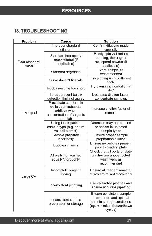
18.TROUBLESHOOTING
Problem Cause SolutionImproper standard
dilutionConfirm dilutions made
correctly
Standard improperly reconstituted (if
applicable)
Briefly spin vial before opening; thoroughly resuspend powder (if
applicable)
Standard degraded Store sample as recommended
Poor standard curve
Curve doesn't fit scale Try plotting using different scale
Incubation time too short Try overnight incubation at 4oC
Target present below detection limits of assay
Decrease dilution factor; concentrate samples
Precipitate can form in wells upon substrate
addition when concentration of target is
too high
Increase dilution factor of sample
Using incompatible sample type (e.g. serum
vs. cell extract)
Detection may be reduced or absent in untested
sample types
Low signal
Sample prepared incorrectly
Ensure proper sample preparation/dilution
Bubbles in wells Ensure no bubbles present prior to reading plate
All wells not washed equally/thoroughly
Check that all ports of plate washer are unobstructed
wash wells as recommended
Incomplete reagent mixing
Ensure all reagents/master mixes are mixed thoroughly
Inconsistent pipetting Use calibrated pipettes and ensure accurate pipetting
Large CV
Inconsistent sample preparation or storage
Ensure consistent sample preparation and optimal
sample storage conditions (eg. minimize freeze/thaws
cycles)

Discover more at www.abcam.com 22
RESOURCES
Problem Cause Solution
Wells are insufficiently washed
Wash wells as per protocol recommendations
Contaminated wash buffer Make fresh wash buffer
Waiting too long to read plate after adding STOP
solution
Read plate immediately after adding STOP solution
Improper storage of ELISA kit
Store all reagents as recommended. Please
note all reagents may not have identical storage
requirements.
High background/
Low sensitivity
Using incompatible sample type (e.g. Serum
vs. cell extract)
Detection may be reduced or absent in untested
sample types

Discover more at www.abcam.com 23
RESOURCES
19.NOTES

Discover more at www.abcam.com 24
RESOURCES

Discover more at www.abcam.com 25
RESOURCES

RESOURCES 26
UK, EU and ROWEmail: [email protected] | Tel: +44-(0)1223-696000
AustriaEmail: [email protected] | Tel: 019-288-259
FranceEmail: [email protected] | Tel: 01-46-94-62-96 GermanyEmail: [email protected] | Tel: 030-896-779-154 SpainEmail: [email protected] | Tel: 911-146-554 SwitzerlandEmail: [email protected] Tel (Deutsch): 0435-016-424 | Tel (Français): 0615-000-530
US and Latin AmericaEmail: [email protected] | Tel: 888-77-ABCAM (22226)
CanadaEmail: [email protected] | Tel: 877-749-8807
China and Asia Pacific Email: [email protected] | Tel: 108008523689 (中國聯通) JapanEmail: [email protected] | Tel: +81-(0)3-6231-0940
www.abcam.com | www.abcam.cn | www.abcam.co.jp
Copyright © 2018 Abcam, All Rights Reserved. The Abcam logo is a registered trademark.
All information / detail is correct at time of going to print.