ELECTROSPINNING OF PECTIN-BASED …...Figure 2. Electrospinning Apparatus Setup. 2.4 Crosslinking of...
7
Proceedings of The National Conference On Undergraduate Research (NCUR) 2017 University of Memphis, Memphis Tennessee April 6 – 8, 2017 ELECTROSPINNING OF PECTIN-BASED NANOFIBERS FOR BIOLOGICAL AND MEDICAL APPLICATIONS Romare Antrobus Biochemistry Department Lawrence University 711 E. Boldt Way Appleton, WI 54911 USA Biomolecular Engineering Program Milwaukee School of Engineering 1025 N. Broadway Ave. Milwaukee, WI 53202 USA Faculty Advisor: Dr. Wujie Zhang Abstract Pectin is a natural biopolymer that can often be found in the primary cell walls of terrestrial plants. It is biodegradability, biocompatibility, and easy availability showing great potential when applied to many biomedical applications such as tissue engineering, drug delivery and the food industry. During this study, biocompatible pectin nanofibers were fabricated via an electrospinning method utilizing pectin-based mixtures prepared with the carrier polymer Polyethylene oxide (PEO) and non-toxic surfactant Pluronic® F127. Electrospinning parameters such as solution properties (viscosity, surface tension), voltage, spinning distance, needle tip size, and solution flow rate were adjusted to: 1) optimize the electrospinning process and 2) ensure the fabrication of uniform nanofibers. The pectin-PEO- Pluronic® F127 ratio was varied in order to control surface tension and hydrogen bonding to study their electrospinning ability. Pectin- based nanofibers were successfully prepared under optimal parameters. Keywords: Pectin, Electrospinning, Natural Biopolymer, Nanofiber 1. INTRODUCTION Many synthetic and natural polymers have been successfully electrospun to fabricate microfibers and successively, nanofibers. Due to the variability in fiber structure there is a high demand for nanofibers to be used in many applications within biomedical research, industry, and the military. This includes applications for drug delivery, tissue engineering, the textile industry, ballistic (clothing) protection, and wound dressing purposes. 1 However, in general, synthetic polymers are not biocompatible. 2 Therefore, there is a high demand for the fabrication of nanofibers by using natural biopolymers, that can be applied to biomedical and tissue engineering purposes without toxic effects. Pectin, a natural biopolymer with a heteropolysaccharide linear structure (Figure 1), is found in and between the primary cell walls of terrestrial plant cells (carrots, apricots, apples, etc.) to provide shape and structural support. The complex mix of polysaccharides are mainly composed of D- galacturonic acid units connected by α-(1,4) glycosidic linkages. 3 The carboxyl and hydroxyl side chains contribute to its hydrophilic nature. In the presence of calcium ions (Ca 2+ ) or oligochitosan, pectin molecules crosslink with each other and produce a hydrogel. Because it is a natural product, pectin is more biodegradable and biocompatible than common synthetic polymers. 4 Nanofibrous pectin mats potentially allow for the fibers to mimic the extracellular matrix (ECM) in structure, size,
Transcript of ELECTROSPINNING OF PECTIN-BASED …...Figure 2. Electrospinning Apparatus Setup. 2.4 Crosslinking of...
Proceedings of The National Conference On Undergraduate Research (NCUR) 2017
University of Memphis, Memphis Tennessee April 6 – 8, 2017
ELECTROSPINNING OF PECTIN-BASED NANOFIBERS FOR BIOLOGICAL AND MEDICAL APPLICATIONS
Romare Antrobus
Biochemistry Department Lawrence University
711 E. Boldt Way Appleton, WI 54911 USA
Biomolecular Engineering Program Milwaukee School of Engineering
1025 N. Broadway Ave. Milwaukee, WI 53202 USA
Faculty Advisor: Dr. Wujie Zhang
Abstract
Pectin is a natural biopolymer that can often be found in the primary cell walls of terrestrial plants. It is biodegradability, biocompatibility, and easy availability showing great potential when applied to many biomedical applications such as tissue engineering, drug delivery and the food industry. During this study, biocompatible pectin nanofibers were fabricated via an electrospinning method utilizing pectin-based mixtures prepared with the carrier polymer Polyethylene oxide (PEO) and non-toxic surfactant Pluronic® F127. Electrospinning parameters such as solution properties (viscosity, surface tension), voltage, spinning distance, needle tip size, and solution flow rate were adjusted to: 1) optimize the electrospinning process and 2) ensure the fabrication of uniform nanofibers. The pectin-PEO- Pluronic® F127 ratio was varied in order to control surface tension and hydrogen bonding to study their electrospinning ability. Pectin- based nanofibers were successfully prepared under optimal parameters. Keywords: Pectin, Electrospinning, Natural Biopolymer, Nanofiber 1. INTRODUCTION
Many synthetic and natural polymers have been successfully electrospun to fabricate microfibers and successively, nanofibers. Due to the variability in fiber structure there is a high demand for nanofibers to be used in many applications within biomedical research, industry, and the military. This includes applications for drug delivery, tissue engineering, the textile industry, ballistic (clothing) protection, and wound dressing purposes.1 However, in general, synthetic polymers are not biocompatible.2 Therefore, there is a high demand for the fabrication of nanofibers by using natural biopolymers, that can be applied to biomedical and tissue engineering purposes without toxic effects. Pectin, a natural biopolymer with a heteropolysaccharide linear structure (Figure 1), is found in and between the primary cell walls of terrestrial plant cells (carrots, apricots, apples, etc.) to provide shape and structural support. The complex mix of polysaccharides are mainly composed of D- galacturonic acid units connected by α-(1,4) glycosidic linkages.3 The carboxyl and hydroxyl side chains contribute to its hydrophilic nature. In the presence of calcium ions (Ca2+) or oligochitosan, pectin molecules crosslink with each other and produce a hydrogel. Because it is a natural product, pectin is more biodegradable and biocompatible than common synthetic polymers.4 Nanofibrous pectin mats potentially allow for the fibers to mimic the extracellular matrix (ECM) in structure, size,
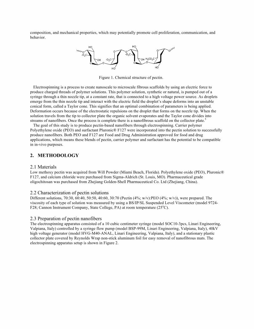
composition, and mechanical properties, which may potentially promote cell proliferation, communication, and behavior.
Figure 1. Chemical structure of pectin. Electrospinning is a process to create nanoscale to microscale fibrous scaffolds by using an electric force to produce charged threads of polymer solutions. This polymer solution, synthetic or natural, is pumped out of a syringe through a thin nozzle tip, at a constant rate, that is connected to a high voltage power source. As droplets emerge from the thin nozzle tip and interact with the electric field the droplet’s shape deforms into an unstable conical form, called a Taylor cone. This signifies that an optimal combination of parameters is being applied. Deformation occurs because of the electrostatic repulsions on the droplet that forms on the nozzle tip. When the solution travels from the tip to collector plate the organic solvent evaporates and the Taylor cone divides into streams of nanofibers. Once the process is complete there is a nanofibrous scaffold on the collector plate.5 The goal of this study is to produce pectin-based nanofibers through electrospinning. Carrier polymer Polyethylene oxide (PEO) and surfactant Pluronic® F127 were incorporated into the pectin solution to successfully produce nanofibers. Both PEO and F127 are Food and Drug Administration approved for food and drug applications, which means these blends of pectin, carrier polymer and surfactant has the potential to be compatible in in-vivo purposes. 2. METHODOLOGY
2.1 Materials Low methoxy pectin was acquired from Will Powder (Miami Beach, Florida). Polyethylene oxide (PEO), Pluronic® F127, and calcium chloride were purchased from Sigma-Aldrich (St. Louis, MO). Pharmaceutical grade oligochitosan was purchased from Zhejiang Golden-Shell Pharmaceutical Co. Ltd (Zhejiang, China). 2.2 Characterization of pectin solutions Different solutions, 70:30, 60:40, 50:50, 40:60, 30:70 (Pectin (4%; w/v):PEO (4%; w/v)), were prepared. The viscosity of each type of solution was measured by using a BS/IP/SL Suspended Level Viscometer (model 9724-F28; Cannon Instrument Company, State College, PA) at room temperature (25oC). 2.3 Preparation of pectin nanofibers The electrospinning apparatus consisted of a 10 cubic centimeter syringe (model SOC10-3pcs, Linari Engineering, Valpiana, Italy) controlled by a syringe flow pump (model BSP-99M, Linari Engineering, Valpiana, Italy), 40kV high voltage generator (model HVG-M40-ANAL, Linari Engineering, Valpiana, Italy), and a stationary plastic collector plate covered by Reynolds Wrap non-stick aluminum foil for easy removal of nanofibrous mats. The electrospinning apparatus setup is shown in Figure 2.
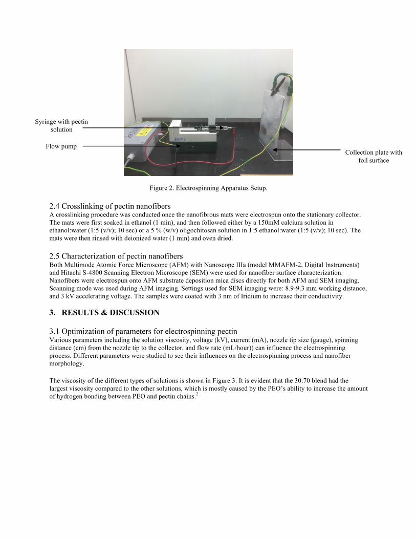
Figure 2. Electrospinning Apparatus Setup. 2.4 Crosslinking of pectin nanofibers A crosslinking procedure was conducted once the nanofibrous mats were electrospun onto the stationary collector. The mats were first soaked in ethanol (1 min), and then followed either by a 150mM calcium solution in ethanol:water (1:5 (v/v); 10 sec) or a 5 % (w/v) oligochitosan solution in 1:5 ethanol:water (1:5 (v/v); 10 sec). The mats were then rinsed with deionized water (1 min) andoven dried. 2.5 Characterization of pectin nanofibers Both Multimode Atomic Force Microscope (AFM) with Nanoscope IIIa (model MMAFM-2, Digital Instruments) and Hitachi S-4800 Scanning Electron Microscope (SEM) were used for nanofiber surface characterization. Nanofibers were electrospun onto AFM substrate deposition mica discs directly for both AFM and SEM imaging. Scanning mode was used during AFM imaging. Settings used for SEM imaging were: 8.9-9.3 mm working distance, and 3 kV accelerating voltage. The samples were coated with 3 nm of Iridium to increase their conductivity. 3. RESULTS & DISCUSSION 3.1 Optimization of parameters for electrospinning pectin Various parameters including the solution viscosity, voltage (kV), current (mA), nozzle tip size (gauge), spinning distance (cm) from the nozzle tip to the collector, and flow rate (mL/hour)) can influence the electrospinning process. Different parameters were studied to see their influences on the electrospinning process and nanofiber morphology. The viscosity of the different types of solutions is shown in Figure 3. It is evident that the 30:70 blend had the largest viscosity compared to the other solutions, which is mostly caused by the PEO’s ability to increase the amount of hydrogen bonding between PEO and pectin chains.2
Collection plate with foil surface
Flow pump
Syringe with pectin solution
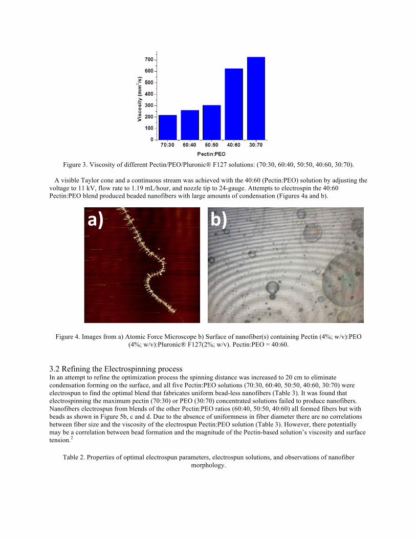
Figure 3. Viscosity of different Pectin/PEO/Pluronic® F127 solutions: (70:30, 60:40, 50:50, 40:60, 30:70).
A visible Taylor cone and a continuous stream was achieved with the 40:60 (Pectin:PEO) solution by adjusting the voltage to 11 kV, flow rate to 1.19 mL/hour, and nozzle tip to 24-gauge. Attempts to electrospin the 40:60 Pectin:PEO blend produced beaded nanofibers with large amounts of condensation (Figures 4a and b).
Figure 4. Images from a) Atomic Force Microscope b) Surface of nanofiber(s) containing Pectin (4%; w/v):PEO (4%; w/v):Pluronic® F127(2%; w/v). Pectin:PEO = 40:60.
3.2 Refining the Electrospinning process In an attempt to refine the optimization process the spinning distance was increased to 20 cm to eliminate condensation forming on the surface, and all five Pectin:PEO solutions (70:30, 60:40, 50:50, 40:60, 30:70) were electrospun to find the optimal blend that fabricates uniform bead-less nanofibers (Table 3). It was found that electrospinning the maximum pectin (70:30) or PEO (30:70) concentrated solutions failed to produce nanofibers. Nanofibers electrospun from blends of the other Pectin:PEO ratios (60:40, 50:50, 40:60) all formed fibers but with beads as shown in Figure 5b, c and d. Due to the absence of uniformness in fiber diameter there are no correlations between fiber size and the viscosity of the electrospun Pectin:PEO solution (Table 3). However, there potentially may be a correlation between bead formation and the magnitude of the Pectin-based solution’s viscosity and surface tension.2
Table 2. Properties of optimal electrospun parameters, electrospun solutions, and observations of nanofiber morphology.
a) b)
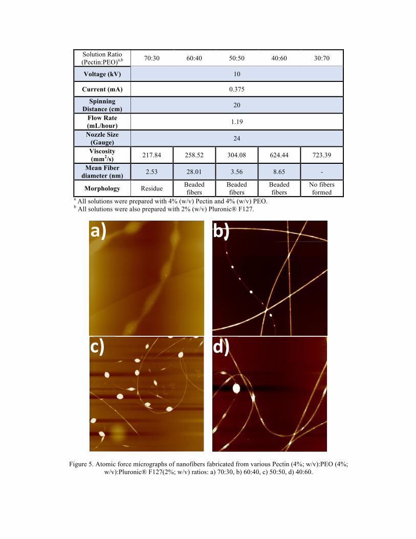
Solution Ratio (Pectin:PEO)a,b 70:30 60:40 50:50 40:60 30:70
Voltage (kV) 10
Current (mA) 0.375
Spinning Distance (cm) 20
Flow Rate (mL/hour) 1.19
Nozzle Size (Gauge) 24
Viscosity (mm2/s) 217.84 258.52 304.08 624.44 723.39
Mean Fiber diameter (nm) 2.53 28.01 3.56 8.65 -
Morphology Residue Beaded fibers
Beaded fibers
Beaded fibers
No fibers formed
a All solutions were prepared with 4% (w/v) Pectin and 4% (w/v) PEO. b All solutions were also prepared with 2% (w/v) Pluronic® F127.
Figure 5. Atomic force micrographs of nanofibers fabricated from various Pectin (4%; w/v):PEO (4%; w/v):Pluronic® F127(2%; w/v) ratios: a) 70:30, b) 60:40, c) 50:50, d) 40:60.
b) a)
c) d)
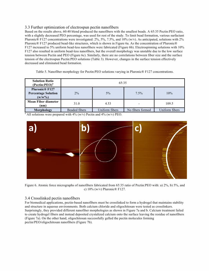
3.3 Further optimization of electrospun pectin nanofibers Based on the results above, 60:40 blend produced the nanofibers with the smallest beads. A 65:35 Pectin:PEO ratio, with a slightly decreased PEO percentage, was used for rest of the study. To limit bead formation, various surfactant Pluronic® F127 concentrations were investigated: 2%, 5%, 7.5%, and 10% (w/v). As anticipated, solutions with 2% Pluronic® F127 produced bead-like structures, which is shown in Figure 6a. As the concentration of Pluronic® F127 increased to 5% uniform bead-less nanofibers were fabricated (Figure 6b). Electrospinning solutions with 10% F127 also resulted in uniform bead-less nanofibers, but the overall morphology was unstable due to the low surface tension between Pectin and PEO (Figure 6c). Similarly, there are no correlations between fiber size and the surface tension of the electrospun Pectin:PEO solutions (Table 3). However, changes in the surface tension effectively decreased and eliminated bead formation.
Table 5. Nanofiber morphology for Pectin:PEO solutions varying in Pluronic® F127 concentrations.
Solution Ratio (Pectin:PEO)a 65:35
Pluronic® F127 Percentage Solution
(w/w%) 2% 5% 7.5% 10%
Mean Fiber diameter (nm) 31.0 4.53 - 109.5
Morphology Beaded fibers Uniform fibers No fibers formed Uniform fibers a All solutions were prepared with 4% (w/v) Pectin and 4% (w/v) PEO.
Figure 6. Atomic force micrographs of nanofibers fabricated from 65:35 ratio of Pectin:PEO with: a) 2%, b) 5%, and
c) 10% (w/v) Pluronic® F127. 3.4 Crosslinked pectin nanofibers For biomedical applications, pectin-based nanofibers must be crosslinked to form a hydrogel that maintains stability and structure in aqueous environments. Both calcium chloride and oligochitosan were tested as crosslinkers. Surprisingly, they provided different nanofiber morphologies as shown in Figure 7a and b. Calcium treatment failed to create hydrogel fibers and instead deposited crystalized calcium onto the surface leaving the residue of nanofibers (Figure 7a). On the other hand, oligochitosan successfully gelled the pectin molecules forming pectin/PEO/oligochitosan nanofibers (Figure 7b).
a) b) c)
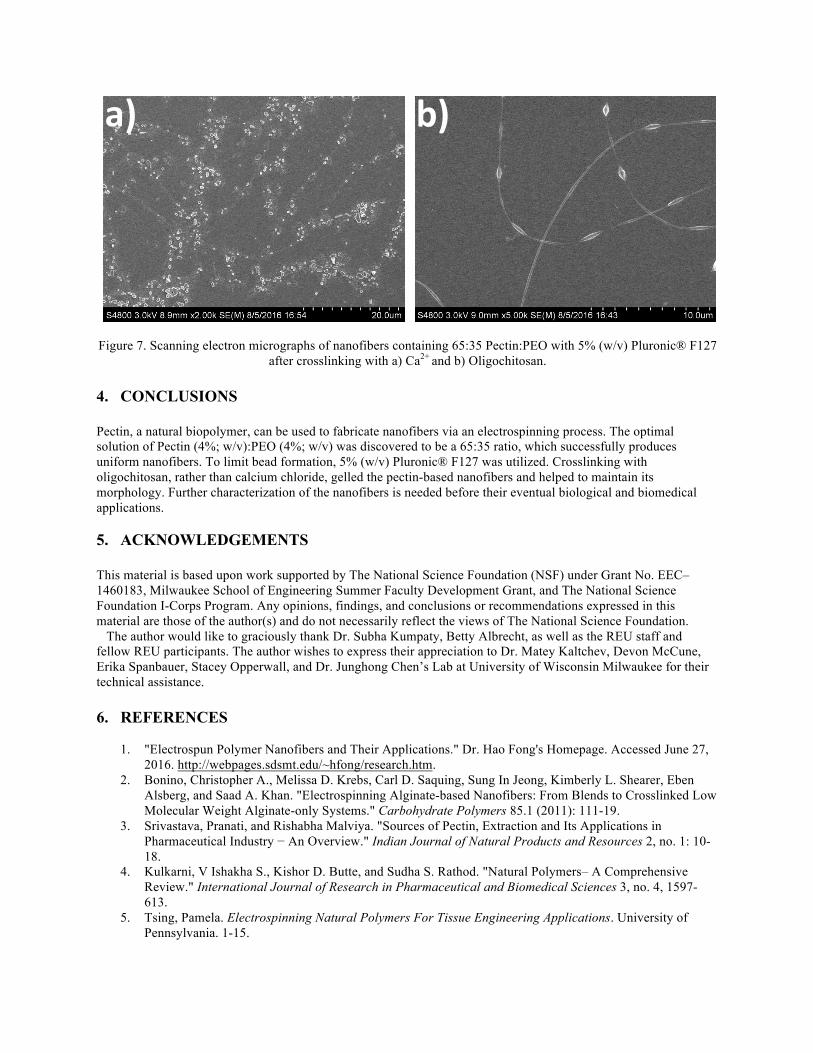
Figure 7. Scanning electron micrographs of nanofibers containing 65:35 Pectin:PEO with 5% (w/v) Pluronic® F127 after crosslinking with a) Ca2+ and b) Oligochitosan.
4. CONCLUSIONS Pectin, a natural biopolymer, can be used to fabricate nanofibers via an electrospinning process. The optimal solution of Pectin (4%; w/v):PEO (4%; w/v) was discovered to be a 65:35 ratio, which successfully produces uniform nanofibers. To limit bead formation, 5% (w/v) Pluronic® F127 was utilized. Crosslinking with oligochitosan, rather than calcium chloride, gelled the pectin-based nanofibers and helped to maintain its morphology. Further characterization of the nanofibers is needed before their eventual biological and biomedical applications. 5. ACKNOWLEDGEMENTS
This material is based upon work supported by The National Science Foundation (NSF) under Grant No. EEC–1460183, Milwaukee School of Engineering Summer Faculty Development Grant, and The National Science Foundation I-Corps Program. Any opinions, findings, and conclusions or recommendations expressed in this material are those of the author(s) and do not necessarily reflect the views of The National Science Foundation. The author would like to graciously thank Dr. Subha Kumpaty, Betty Albrecht, as well as the REU staff and fellow REU participants. The author wishes to express their appreciation to Dr. Matey Kaltchev, Devon McCune, Erika Spanbauer, Stacey Opperwall, and Dr. Junghong Chen’s Lab at University of Wisconsin Milwaukee for their technical assistance. 6. REFERENCES
1. "Electrospun Polymer Nanofibers and Their Applications." Dr. Hao Fong's Homepage. Accessed June 27, 2016. http://webpages.sdsmt.edu/~hfong/research.htm.
2. Bonino, Christopher A., Melissa D. Krebs, Carl D. Saquing, Sung In Jeong, Kimberly L. Shearer, Eben Alsberg, and Saad A. Khan. "Electrospinning Alginate-based Nanofibers: From Blends to Crosslinked Low Molecular Weight Alginate-only Systems." Carbohydrate Polymers 85.1 (2011): 111-19.
3. Srivastava, Pranati, and Rishabha Malviya. "Sources of Pectin, Extraction and Its Applications in Pharmaceutical Industry − An Overview." Indian Journal of Natural Products and Resources 2, no. 1: 10-18.
4. Kulkarni, V Ishakha S., Kishor D. Butte, and Sudha S. Rathod. "Natural Polymers– A Comprehensive Review." International Journal of Research in Pharmaceutical and Biomedical Sciences 3, no. 4, 1597-613.
5. Tsing, Pamela. Electrospinning Natural Polymers For Tissue Engineering Applications. University of Pennsylvania. 1-15.
a) b)