Electrophysiology of Muscle Skeletal & Cardiac excitation & contraction Mike Clark, M.D.
37
Electrophysiology of Muscle Skeletal & Cardiac excitation & contraction Mike Clark, M.D.
-
Upload
kyree-henington -
Category
Documents
-
view
231 -
download
3
Transcript of Electrophysiology of Muscle Skeletal & Cardiac excitation & contraction Mike Clark, M.D.
Electrophysiology of MuscleSkeletal & Cardiac excitation & contraction
Mike Clark, M.D.
NOTE
Prior to reading this PowerPoint presentation – students may want to review my PowerPoint presentation General Electrophysiology – which explains the Resting Membrane Potential and Action Potentials in general.

Neuron Action Potential Skeletal Muscle Action Potential
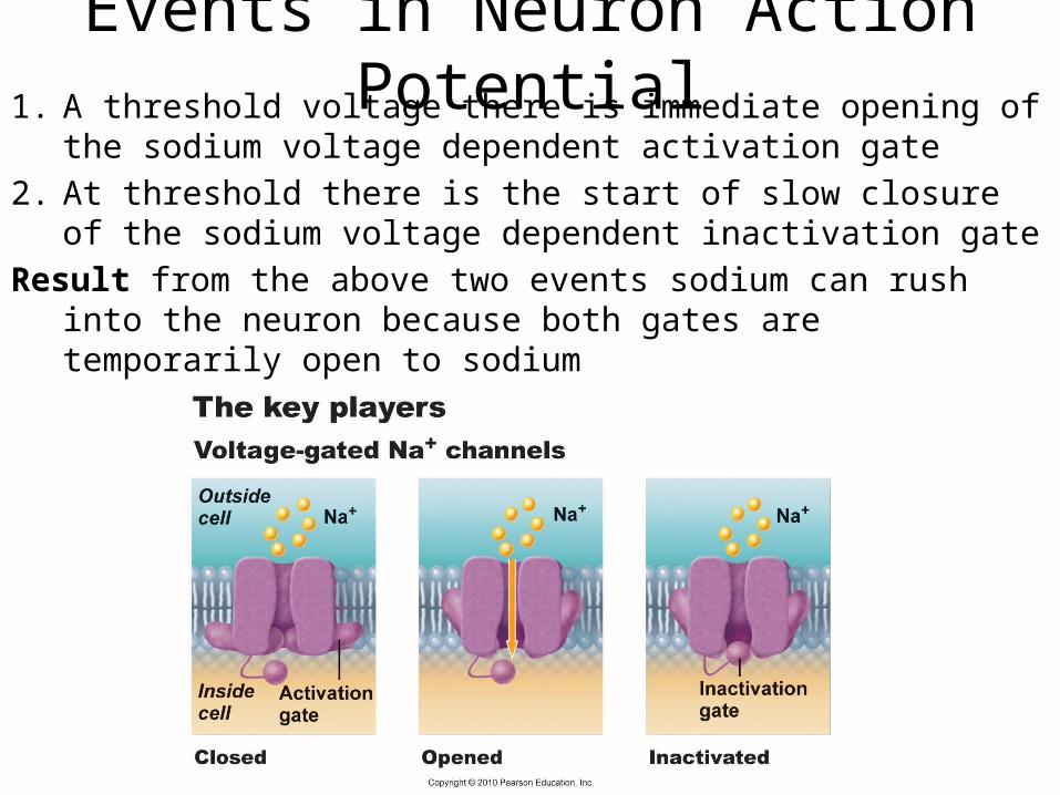
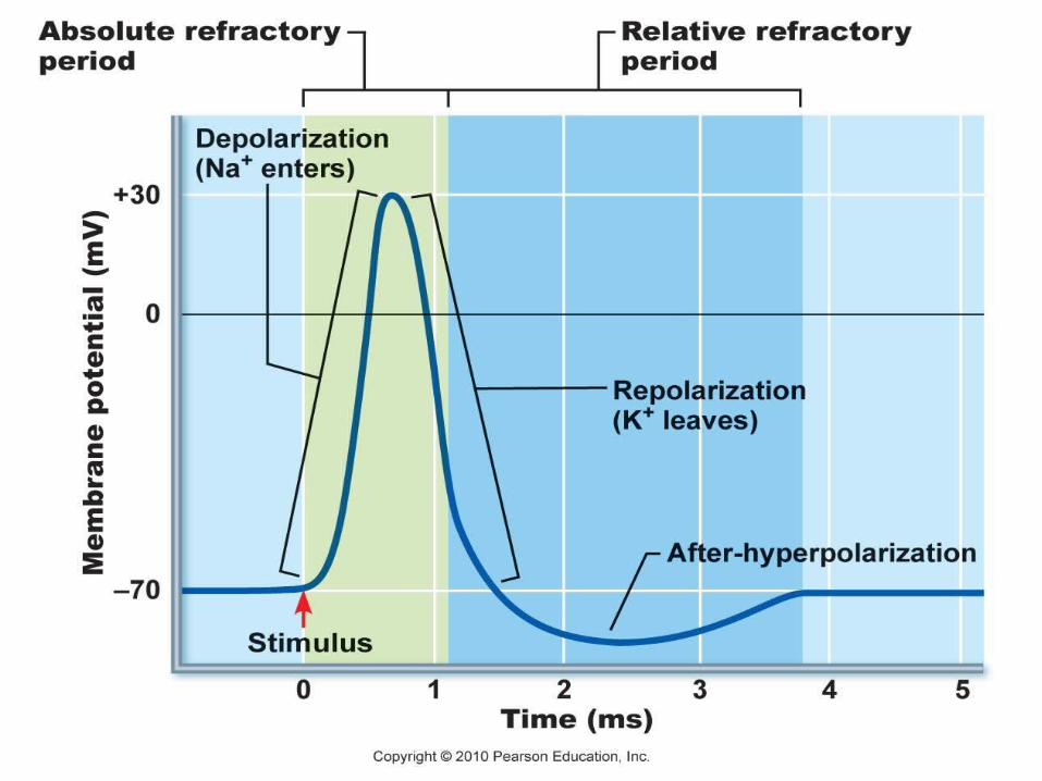
Events in Neuron Action Potential1. A threshold voltage there is immediate opening of the sodium
voltage dependent activation gate2. At threshold there is the start of slow closure of the sodium
voltage dependent inactivation gateResult from the above two events sodium can rush into the
neuron because both gates are temporarily open to sodium
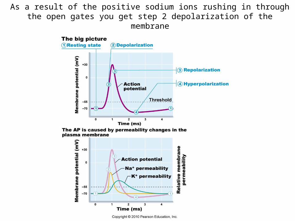
As a result of the positive sodium ions rushing in through the open gates you get step 2 depolarization of the membrane

3. When the slow sodium inactivation gate closes the positive sodium ions stop rushing in and the membrane depolarizes no further – the up-shoot stops.
4. The same voltage that operated the sodium gates also is the same voltage to initiate action of the potassium gates – however the potassium gates are very slow so they do not open till around the time that the sodium inactivation gate is
closing – thus since no further sodium is rushing In and now
positive potassium is rushing out the inside of the neuron again begins to become More negative – Repolarization.
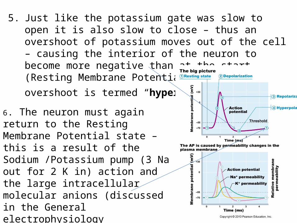
5. Just like the potassium gate was slow to open it is also slow to close – thus an overshoot of potassium moves out of the cell – causing the interior of the neuron to become more negative than at the start (Resting Membrane Potential). This overshoot is
termed “hyperpolarization.”
6. The neuron must again return to the Resting Membrane Potential state – this is a result of the Sodium /Potassium pump (3 Na out for 2 K in) action and the large intracellular molecular anions (discussed in the General electrophysiology PowerPoint).
Refractory Periods• Absolute Refractory Period – a time in which the
same area of the neuron cell membrane cannot be re-excited (fire another action potential). It is time it takes for the sodium gates to fully reset.
• Relative Refractory Period – a time immediately after the absolute refractory period in which the same area of the neuron cell membrane can be re-excited but requires a higher voltage higher than the usual threshold voltage. During this time the potassium channels are still open – thus positive potassium is rushing out making the interior of the neuron more negative thus harder to reach the voltage of threshold.
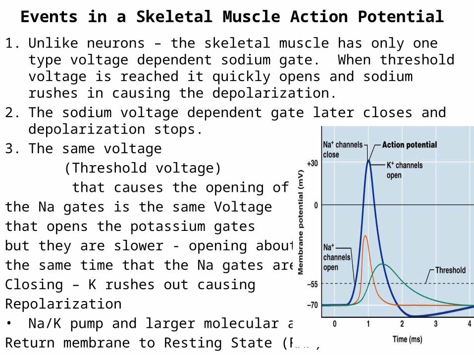
Events in a Skeletal Muscle Action Potential 1. Unlike neurons – the skeletal muscle has only one type voltage
dependent sodium gate. When threshold voltage is reached it quickly opens and sodium rushes in causing the depolarization.
2. The sodium voltage dependent gate later closes and depolarization stops.
3. The same voltage (Threshold voltage) that causes the opening of the Na gates is the same Voltage that opens the potassium gatesbut they are slower - opening aboutthe same time that the Na gates areClosing – K rushes out causing Repolarization• Na/K pump and larger molecular anions Return membrane to Resting State (RMP)
Excitation- Contraction Coupling• Neurons can illicit conductivity (sometimes referred to
as excitation)• This conductivity is in the form of action potentials• Muscle can illicit the charge activity of conductivity as
well as the mechanical activity of contractility• The charge movement of conductivity (action
potentials) leads to the mechanical contraction – thus the excitation must be coupled to the contraction
• Since charge activity is faster than mechanical activity – the action potentials initiate first followed fairly quickly by the initiation of the mechanical activity of contraction.
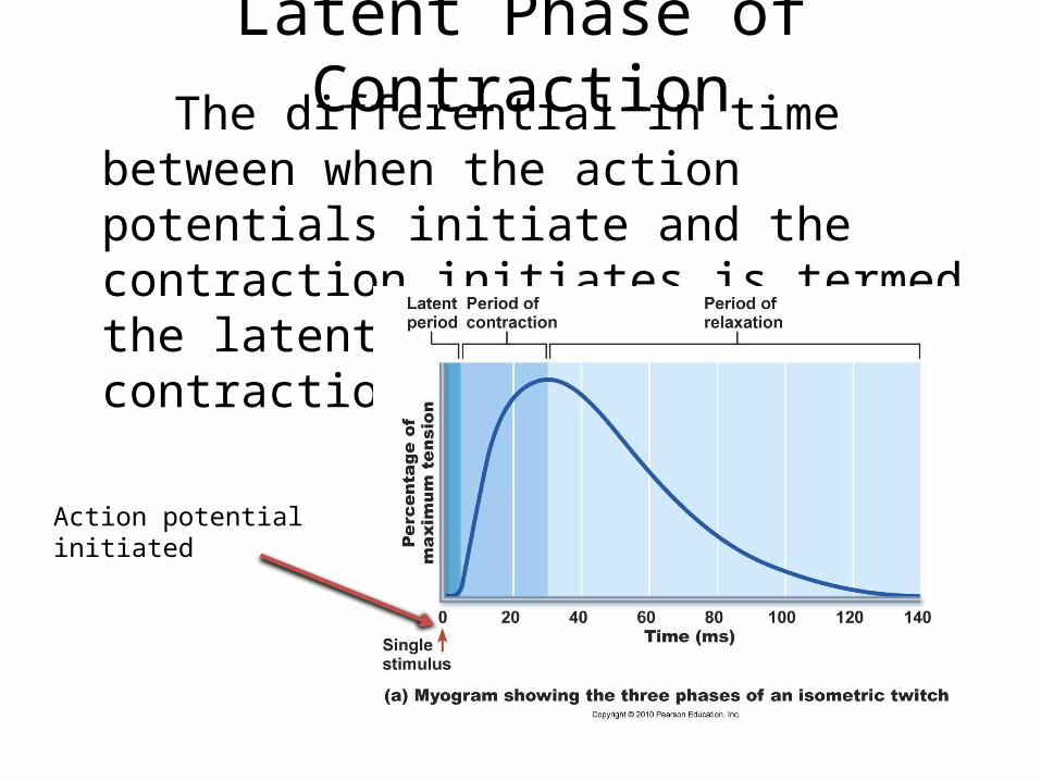
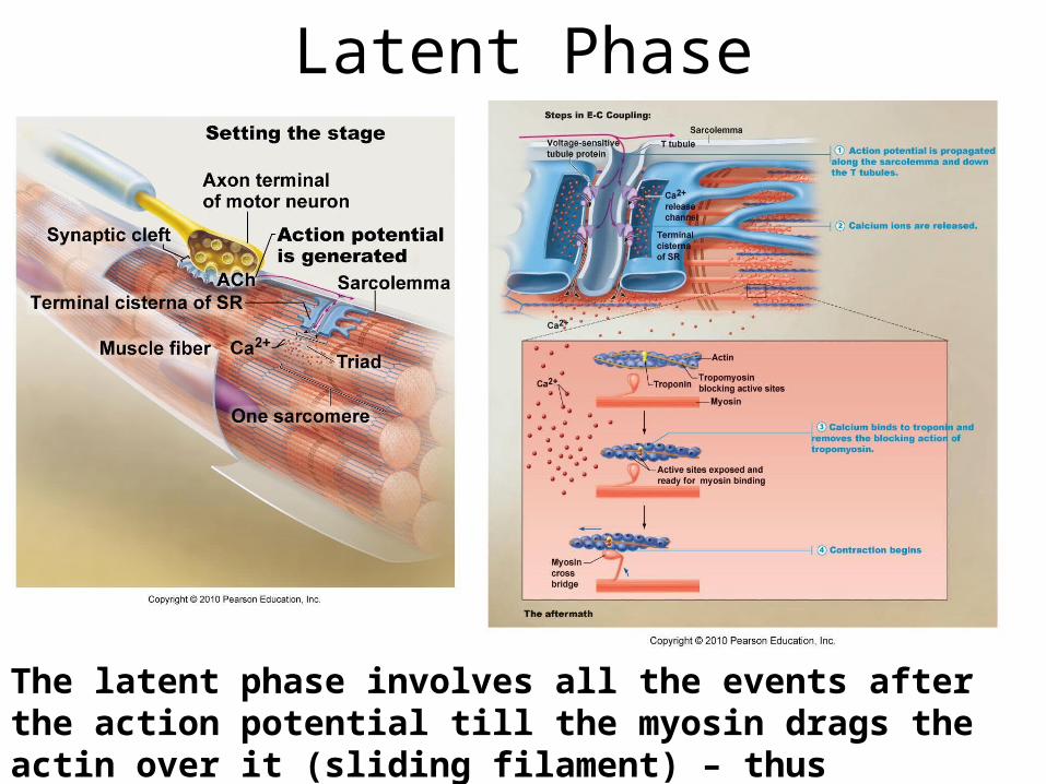
Latent Phase of Contraction The differential in time between when the
action potentials initiate and the contraction initiates is termed the latent phase of muscle contraction
Action potential initiated
Latent Phase
The latent phase involves all the events after the action potential till the myosin drags the actin over it (sliding filament) – thus causing the initiation of the muscle contraction
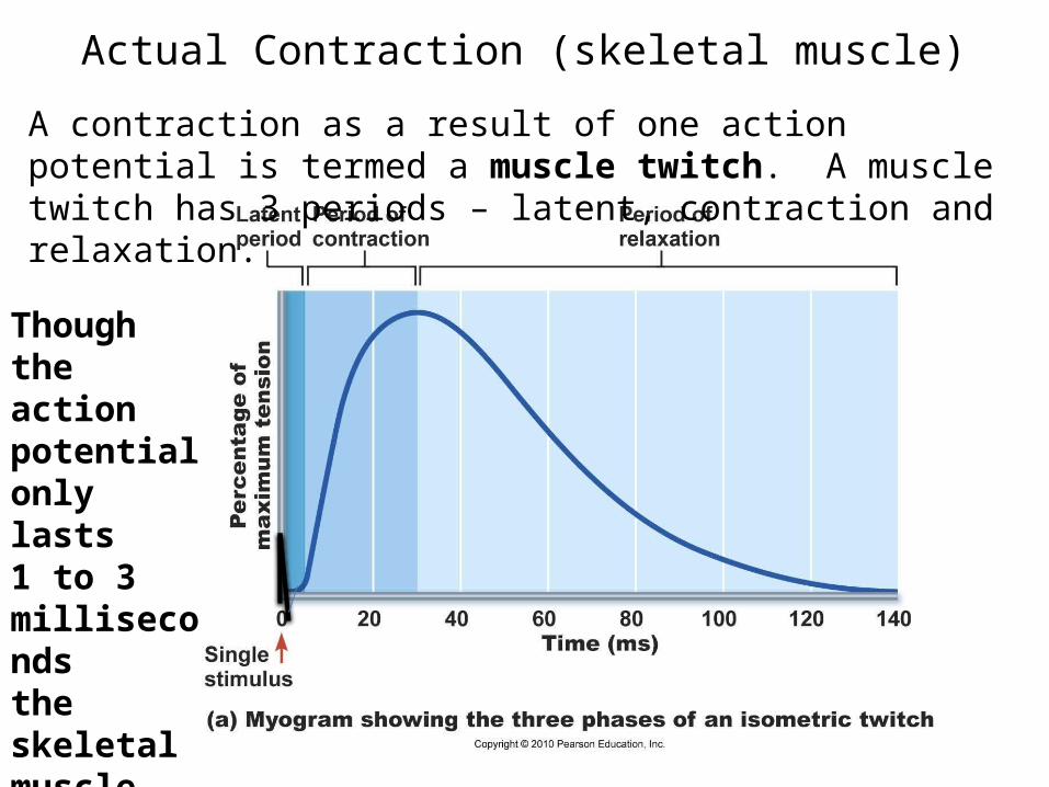
Actual Contraction (skeletal muscle)
Though the actionpotential only lasts1 to 3 millisecondsthe skeletalmuscle contractionlasts over 100 ms
A contraction as a result of one action potential is termed a muscle twitch. A muscle twitch has 3 periods – latent, contraction and relaxation.
100 milliseconds is 1/10 of a second• 100 milliseconds (1/10th of a second) is not a long time
at all – we could not do anything muscle wise in that time frame – thus we must add (summate) together these isolated contractions (muscle twitch) – to make a longer useful contraction. The adding together of muscle twitches is termed “tetany.”
• Tetany occurs by delivering to the muscle cell- a rapid continuous set of action potentials.
• The action potentials can occur fast but how close together they can occur is limited by the action potentials “absolute refractory period.”
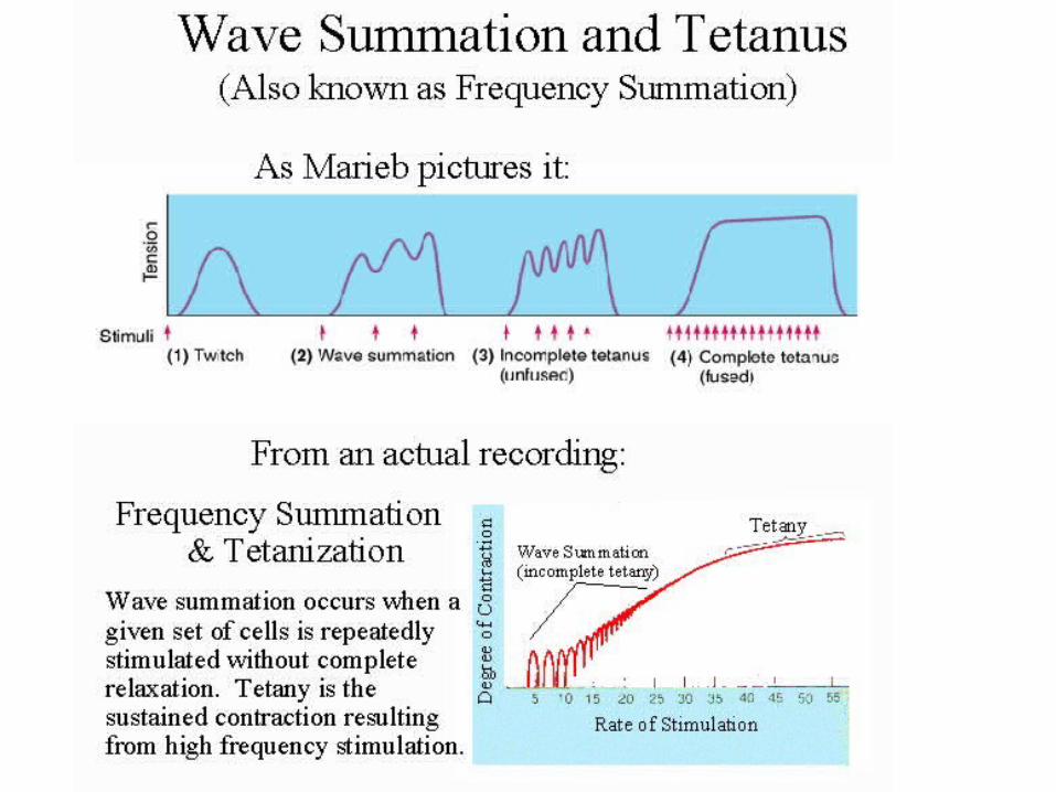
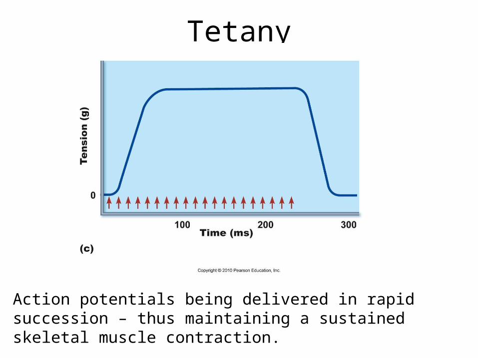
Tetany
Action potentials being delivered in rapid succession – thus maintaining a sustained skeletal muscle contraction.
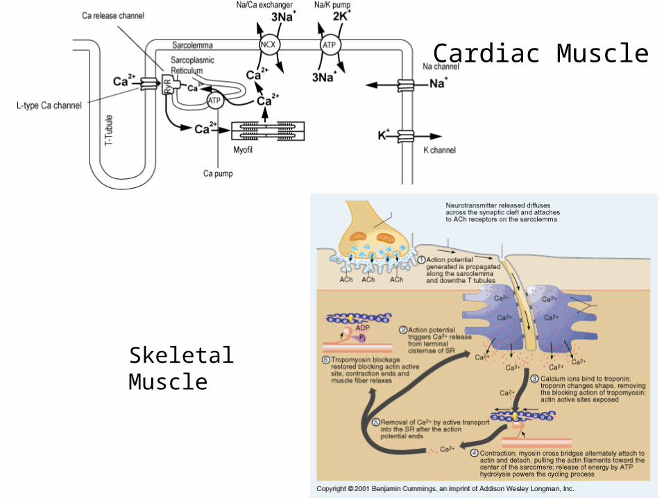
The events causing heart muscle contraction differ slightly from those of skeletal muscle
• Cardiac muscle – just as skeletal muscle- require calcium to allow actin to bind to myosin – which is the main component of the sliding filament model of muscle contraction.
• All the calcium required for skeletal muscle contraction is located on the inside of the skeletal muscle cell in the sarcoplasmic reticulum – however cardiac muscle requires some calcium to enter from outside of the muscle cell.
• in cardiac muscle some (10 -20%) extracellular calcium is required for contraction. This calcium enters through voltage dependent calcium channels known as L channels or sometimes referred to as slow calcium channels. release In fact the 10 -20% of calcium coming from the outside of the cell not only goes to troponin C to assist directly with contraction – but it also assists in the opening of the calcium release channels in the cardiac muscle cell sarcoplasmic reticulum for the other 80 – 90% to be released.
Cell Membrane Calcium Channels• In cardiac muscle cells some (10 -20%) of extracellular
calcium is required for contraction. This calcium enters through voltage dependent calcium channels known as L (Long lasting) channels or sometimes referred to as slow calcium channels.
• The 10 -20% of calcium coming from the outside of the cell not only goes to troponin C to assist directly with cardiac muscle contraction – but it also assists in the opening of the calcium release channels in the cardiac muscle cell sarcoplasmic reticulum for the other 80 – 90% to be released.
• This influx of calcium from the outside of the cell occurs during the cardiac muscle action potential – part of the mechanism of the action potential – especially in the plateau phase. Thus even though the calcium enters as part of the action potential mechanism–it is also important for the contraction action.
• The entering calcium is important for excitation and contraction.• Since (1) the amount of calcium available to troponin C is
important to the strength of contraction in both skeletal muscle and cardiac muscle and (2) some of the calcium for cardiac muscle contraction must come from the extracellular fluid – the blood (extracellular fluid) calcium level is more important to cardiac muscle than skeletal muscle.
• Since cardiac muscle has functional L calcium channels – and skeletal muscle cells do not – a calcium channel blocker drug will affect cardiac muscle and not skeletal muscle (example Verapamil) . The medication will decrease the force of contractility (inotropic effect).
Cardiac Muscle
Skeletal Muscle

The Heart Action Potentials• The heart has two types of action potentials – one type
that is the heart muscle action potential
and another type that is the pacemaker (SA node, AV node, etc. ) action potential
NO TETANY• The heart muscle should never go into tetany. A heart
muscle cell should do one muscle twitch then relax for a while before it embarks on another twitch.
• A heart chamber only fills with blood when it is relaxed (diastole) – thus tetany would cause a heart chamber to have a sustained contraction- not relax and be unable to fill with blood.
• If the heart chamber cannot fill – it has no blood to empty into the next chamber or into the next blood vessels.
• If heart muscle goes into tetany – that is what is called cardiac flutter and fibrillation. Fibrillation can lead to death.
Prevention of Cardiac muscle tetany• Since one initiated action potential can cause
one muscle contraction (muscle twitch) – and a rapid succession of action potentials on a muscle cell can cause tetany – then make the action potentials get further apart – so they cannot summate as easily.
How can this be done?• Prolong the absolute refractory period.
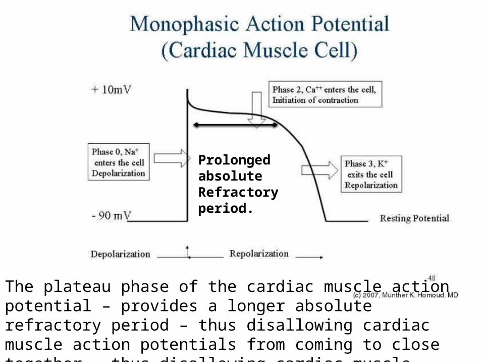
Prolonged absoluteRefractory period.
The plateau phase of the cardiac muscle action potential – provides a longer absolute refractory period – thus disallowing cardiac muscle action potentials from coming to close together – thus disallowing cardiac muscle tetany.

In addition to the fact that the cardiac muscle action potentials cannot come to close together there is another fact of interest – the absolute refractory period lasts over 200 msec which is almost the entire time of one cardiac muscle twitch (one contraction). Thus by the time the action potential is fully over – so is the muscle contraction – thus no tetany.
The red graphis the contractioncurve and the blue is the actionpotential curve
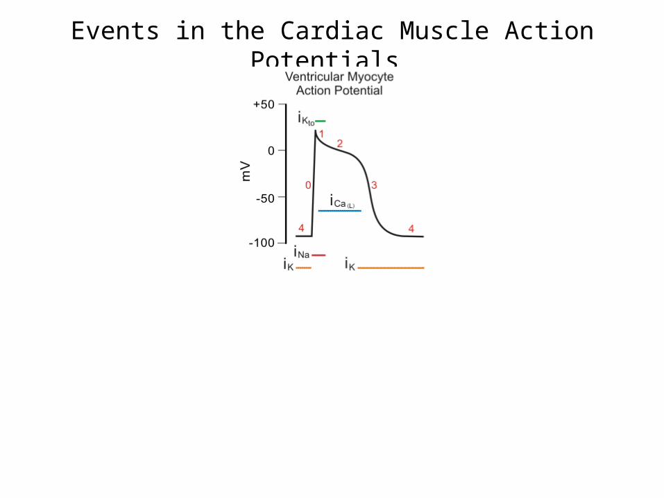
Events in the Cardiac Muscle Action Potentials

Events in the Cardiac Muscle Action Potential
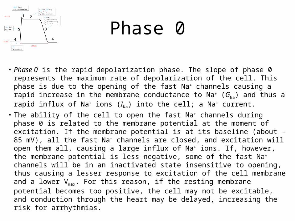
Phase 0
• Phase 0 is the rapid depolarization phase. The slope of phase 0 represents the maximum rate of depolarization of the cell. This phase is due to the opening of the fast Na+ channels causing a rapid increase in the membrane conductance to Na+ (GNa) and thus a rapid influx of Na+ ions (INa) into the cell; a Na+ current.
• The ability of the cell to open the fast Na+ channels during phase 0 is related to the membrane potential at the moment of excitation. If the membrane potential is at its baseline (about -85 mV), all the fast Na+ channels are closed, and excitation will open them all, causing a large influx of Na+ ions. If, however, the membrane potential is less negative, some of the fast Na+ channels will be in an inactivated state insensitive to opening, thus causing a lesser response to excitation of the cell membrane and a lower Vmax. For this reason, if the resting membrane potential becomes too positive, the cell may not be excitable, and conduction through the heart may be delayed, increasing the risk for arrhythmias.
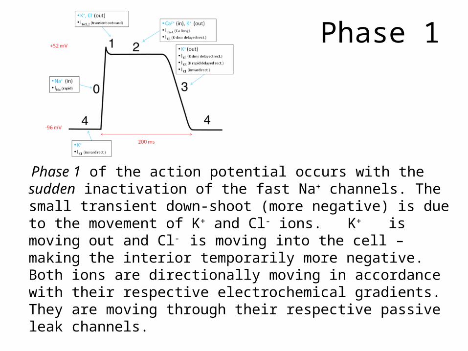
Phase 1
Phase 1 of the action potential occurs with the sudden inactivation of the fast Na+ channels. The small transient down-shoot (more negative) is due to the movement of K+ and Cl- ions. K+ is moving out and Cl- is moving into the cell – making the interior temporarily more negative. Both ions are directionally moving in accordance with their respective electrochemical gradients. They are moving through their respective passive leak channels.
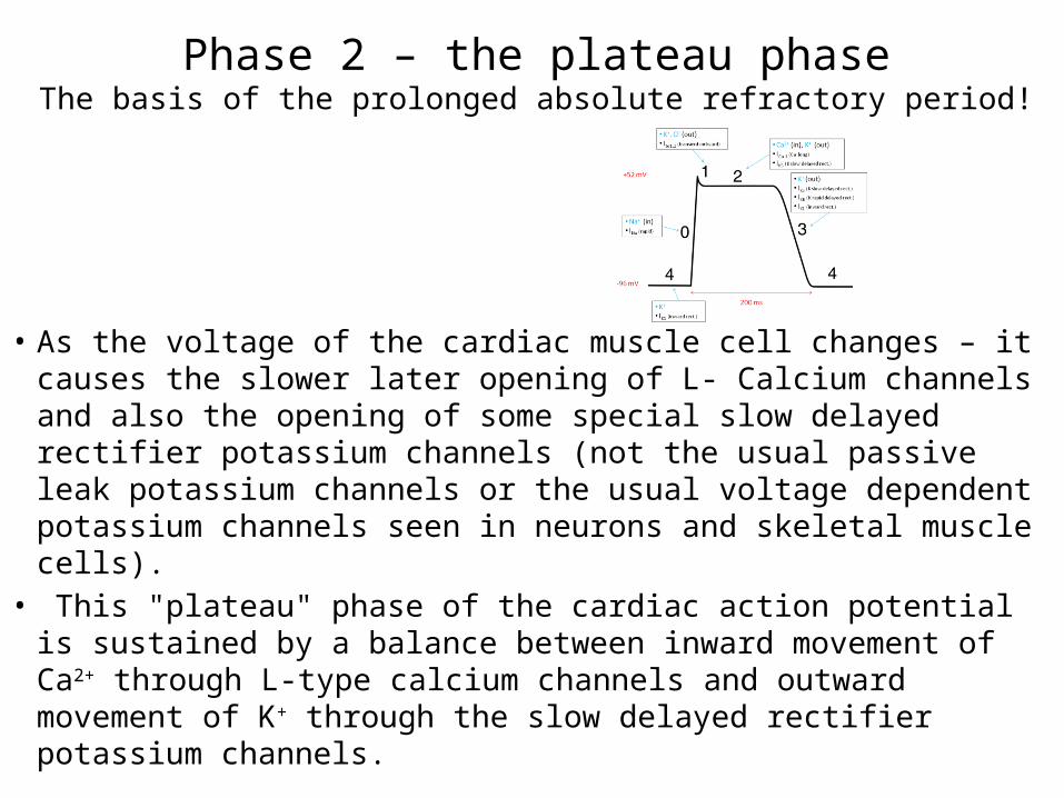
Phase 2 – the plateau phaseThe basis of the prolonged absolute refractory period!
• As the voltage of the cardiac muscle cell changes – it causes the slower later opening of L- Calcium channels and also the opening of some special slow delayed rectifier potassium channels (not the usual passive leak potassium channels or the usual voltage dependent potassium channels seen in neurons and skeletal muscle cells).
• This "plateau" phase of the cardiac action potential is sustained by a balance between inward movement of Ca2+ through L-type calcium channels and outward movement of K+ through the slow delayed rectifier potassium channels.

Phase 3
• During phase 3 (the "rapid repolarization" phase) of the action potential, the L-type Ca2+ channels close, while the slow delayed rectifier K+ channels remain open. This ensures a net outward current, corresponding to negative change in membrane potential, thus allowing more types of K+ channels to open. These are primarily the rapid delayed rectifier K+ channels.
• This net outward, positive current (equal to loss of positive charge from the cell) causes the cell to repolarize. The delayed rectifier K+ channels close when the membrane potential is restored to about -80 to -85 mV. The Na/K pump helps restore the RMP – Phase 4.
Phase 4 is the usual Resting Membrane Potential

Cardiac Pacemaker Action Potential

Pacemaker cells• The pacemaker cells have automatic rhythm – they
spontaneously in and of themselves can fire action potentials.
• Each pacemaker structure has a different automatic firing rate.
• SA node – 60 – 80 natural pacemaker of the heart because it has the fastest firing frequency
• AV node – 40 – 60 spontaneous action potentials per minute
• Bundle of HIS – 20 – 40 action potentials per minute
• Bundle Branches – 10 – 20 action potentials per minute
• It is the pacemaker cells that cause the adjacent cardiac muscle cells to reach threshold voltage – thus initiating the cardiac muscle cell action potential
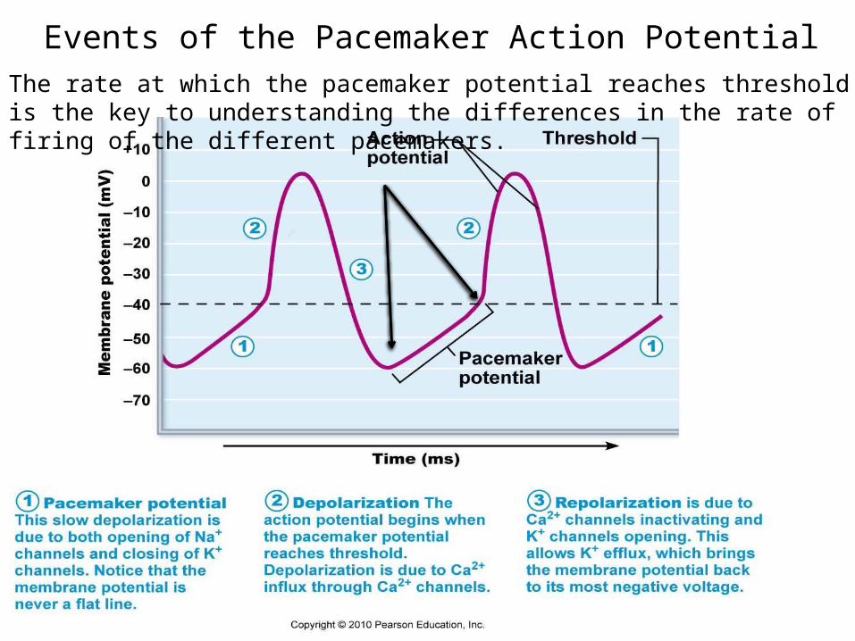
Events of the Pacemaker Action PotentialThe rate at which the pacemaker potential reaches threshold is the key to understanding the differences in the rate of firing of the different pacemakers.
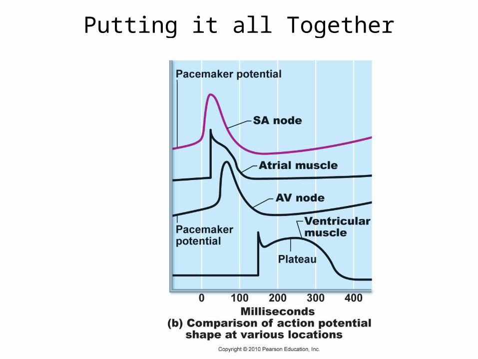
Putting it all Together

Threshold
• Subthreshold stimulus—weak local depolarization that does not reach threshold
• Threshold stimulus—strong enough to push the membrane potential toward and beyond threshold
• AP is an all-or-none phenomenon—action potentials either happen completely, or not at all


![A mathematical model for active contraction in healthy and failing ...xl/PLOS-myocytes.pdf · important ion regulating myocardial excitation-contraction coupling [16]. Continuous](https://static.fdocuments.in/doc/165x107/5f8068044621df3bd9599405/a-mathematical-model-for-active-contraction-in-healthy-and-failing-xlplos-.jpg)