
ElectrophysiologicalandMorphologicalCharacteristicsand...
18
Behavioral/Systems/Cognitive Electrophysiological and Morphological Characteristics and Synaptic Connectivity of Tyrosine Hydroxylase-Expressing Neurons in Adult Mouse Striatum Osvaldo Iba ´n ˜ez-Sandoval, Fatuel Tecuapetla, Bengi Unal, Fulva Shah, Tibor Koo ´s, and James M. Tepper Center For Molecular and Behavioral Neuroscience, Rutgers, The State University of New Jersey, Newark, New Jersey 07102 Whole-cell recordings were obtained from tyrosine hydroxylase-expressing (TH ) neurons in striatal slices from bacterial artificial chromosome transgenic mice that synthesize enhanced green fluorescent protein (EGFP) selectively in neurons expressing TH transcrip- tional regulatory sequences. Stereological cell counting indicated that there were 2700 EGFP–TH neurons/striatum. Whole-cell recordings in striatal slices demonstrated that EGFP–TH neurons comprise four electrophysiologically distinct neuron types whose electrophysiological properties have not been reported previously in striatum. EGFP–TH neurons were identified in retrograde tracing studies as interneurons. Recordings from synaptically connected pairs of EGFP–TH interneurons and spiny neurons showed that the interneurons elicited GABAergic IPSPs/IPSCs in spiny neurons powerful enough to significantly delay evoked spiking. EGFP–TH interneurons responded to local or cortical stimulation with glutamatergic EPSPs. Local stimulation also elicited GABA A IPSPs, at least some of which arose from identified spiny neurons. Single-cell reverse transcription-PCR showed expression of VMAT1 in EGFP–TH interneurons, consistent with previous suggestions that these interneurons may be dopaminergic as well as GABAergic. All four classes of interneurons were medium sized with modestly branching, varicose dendrites, and dense, highly varicose axon collateral fields. These data show for the first time that there exists in the normal rodent striatum a substantial population of TH /GABAergic interneurons comprising four electrophysiologically distinct subtypes whose electrophysiological properties differ significantly from those of previ- ously described striatal GABAergic interneurons. These interneurons are likely to play an important role in striatal function through fast GABAergic synaptic transmission in addition to, and independent of, their potential role in compensation for dopamine loss in experi- mental or idiopathic Parkinson’s disease. Introduction The neostriatum is composed almost entirely of GABAergic neu- rons. The vast majority of these, commonly assumed to total 95% of striatal neurons in rodents (Graveland and DiFiglia, 1985; Gerfen and Wilson, 1996), are medium-sized spiny projection neurons (SPNs) that represent both the major input and the only output from the striatum. The remaining neurons consist of large aspiny cholinergic interneurons, and at least three electrophysiologically distinct types of GABAergic interneurons (Kawaguchi, 1993; Kawaguchi et al., 1995; Tepper and Bolam, 2004). Despite being present in very small numbers, striatal interneurons have a profound effect on striatal functioning. Cholinergic interneurons exert a criti- cally important neuromodulatory role in SPNs (Nishi et al., 1990; Kitai and Surmeier, 1993; Calabresi et al., 2000). In contrast, GABAergic interneurons, in particular the parvalbumin-immuno- reactive (PV ) fast-spiking (FSI) and low-threshold Ca 2 spiking (LTS) interneurons, have been shown to be the principal source of strong, fast synaptic inhibition in striatum (Koo ´ s and Tepper, 1999; Koo ´ s et al., 2004; Tepper et al., 2004, 2008; Taverna et al., 2007), whereas the spiny cell axon collaterals, once believed to comprise a large and powerful lateral inhibitory network (Groves, 1983), exert surprisingly weak synaptic effects at the level of the SPN somata (Tunstall et al., 2002; Koo ´ s et al., 2004; Tecuapetla et al., 2009) (but see Wickens et al., 2007) but likely play key roles in regulating den- dritic events and overall spiny cell excitability. Classically, there are three subtypes of GABAergic interneu- rons in the neostriatum that can be distinguished neurochemi- cally. One expresses the peptides somatostatin (SOM) and neuropeptide Y (NPY) and the enzymes NADPH diaphorase and nitric oxide synthase (NOS). The other two express the calcium binding proteins PV or calretinin (CR) (Kawaguchi, 1993; Kawaguchi et al., 1995). Together, these GABAergic interneurons comprise 2% of the rodent neostriatal cell population (Rymar et al., 2004). However, Dubach et al. (1987) described a population of ty- rosine hydroxylase-immunoreactive (TH ) neurons adult mon- key striatum. Subsequently, striatal TH neurons were found in several other species, including rat (Tashiro et al., 1989a,b; Meredith et al., 1999; O’Byrne et al., 2000), mouse (Mao et al., 2001; Petroske et al., 2001), monkey (Betarbet et al., 1997; Received Dec. 3, 2009; revised March 23, 2010; accepted March 26, 2010. This research was supported, in part, by National Institutes of Health Grants NS034865 (J.M.T.) and NS052370 (T.K.), Consejo Nacional de Ciencia y Tecnología (O.I.-S.), and Rutgers University. We thank Drs. Elizabeth D. Aber- crombie and Denis Pare ´ for generously allowing us the use of their microscopes and image acquisition and analysis software for the cell counting and colocalization experiments, and Harry Xenias for comments on this manuscript. Correspondence should be addressed to Dr. James M. Tepper, Center For Molecular and Behavioral Neuroscience, Rutgers, The State University of New Jersey, 197 University Avenue, Newark NJ 07102. E-mail: jtepper@ andromeda.rutgers.edu. DOI:10.1523/JNEUROSCI.5996-09.2010 Copyright © 2010 the authors 0270-6474/10/306999-18$15.00/0 The Journal of Neuroscience, May 19, 2010 • 30(20):6999 –7016 • 6999
Transcript of ElectrophysiologicalandMorphologicalCharacteristicsand...

Behavioral/Systems/Cognitive
Electrophysiological and Morphological Characteristics andSynaptic Connectivity of Tyrosine Hydroxylase-ExpressingNeurons in Adult Mouse Striatum
Osvaldo Ibanez-Sandoval, Fatuel Tecuapetla, Bengi Unal, Fulva Shah, Tibor Koos, and James M. TepperCenter For Molecular and Behavioral Neuroscience, Rutgers, The State University of New Jersey, Newark, New Jersey 07102
Whole-cell recordings were obtained from tyrosine hydroxylase-expressing (TH �) neurons in striatal slices from bacterial artificialchromosome transgenic mice that synthesize enhanced green fluorescent protein (EGFP) selectively in neurons expressing TH transcrip-tional regulatory sequences. Stereological cell counting indicated that there were �2700 EGFP–TH � neurons/striatum. Whole-cellrecordings in striatal slices demonstrated that EGFP–TH � neurons comprise four electrophysiologically distinct neuron types whoseelectrophysiological properties have not been reported previously in striatum. EGFP–TH � neurons were identified in retrograde tracingstudies as interneurons. Recordings from synaptically connected pairs of EGFP–TH � interneurons and spiny neurons showed that theinterneurons elicited GABAergic IPSPs/IPSCs in spiny neurons powerful enough to significantly delay evoked spiking. EGFP–TH �
interneurons responded to local or cortical stimulation with glutamatergic EPSPs. Local stimulation also elicited GABAA IPSPs, at leastsome of which arose from identified spiny neurons. Single-cell reverse transcription-PCR showed expression of VMAT1 in EGFP–TH �
interneurons, consistent with previous suggestions that these interneurons may be dopaminergic as well as GABAergic. All four classesof interneurons were medium sized with modestly branching, varicose dendrites, and dense, highly varicose axon collateral fields. Thesedata show for the first time that there exists in the normal rodent striatum a substantial population of TH �/GABAergic interneuronscomprising four electrophysiologically distinct subtypes whose electrophysiological properties differ significantly from those of previ-ously described striatal GABAergic interneurons. These interneurons are likely to play an important role in striatal function through fastGABAergic synaptic transmission in addition to, and independent of, their potential role in compensation for dopamine loss in experi-mental or idiopathic Parkinson’s disease.
IntroductionThe neostriatum is composed almost entirely of GABAergic neu-rons. The vast majority of these, commonly assumed to total �95%of striatal neurons in rodents (Graveland and DiFiglia, 1985; Gerfenand Wilson, 1996), are medium-sized spiny projection neurons(SPNs) that represent both the major input and the only outputfrom the striatum. The remaining neurons consist of large aspinycholinergic interneurons, and at least three electrophysiologicallydistinct types of GABAergic interneurons (Kawaguchi, 1993;Kawaguchi et al., 1995; Tepper and Bolam, 2004). Despite beingpresent in very small numbers, striatal interneurons have a profoundeffect on striatal functioning. Cholinergic interneurons exert a criti-cally important neuromodulatory role in SPNs (Nishi et al., 1990;Kitai and Surmeier, 1993; Calabresi et al., 2000). In contrast,GABAergic interneurons, in particular the parvalbumin-immuno-
reactive (PV�) fast-spiking (FSI) and low-threshold Ca2� spiking(LTS) interneurons, have been shown to be the principal source ofstrong, fast synaptic inhibition in striatum (Koos and Tepper, 1999;Koos et al., 2004; Tepper et al., 2004, 2008; Taverna et al., 2007),whereas the spiny cell axon collaterals, once believed to comprise alarge and powerful lateral inhibitory network (Groves, 1983), exertsurprisingly weak synaptic effects at the level of the SPN somata(Tunstall et al., 2002; Koos et al., 2004; Tecuapetla et al., 2009) (butsee Wickens et al., 2007) but likely play key roles in regulating den-dritic events and overall spiny cell excitability.
Classically, there are three subtypes of GABAergic interneu-rons in the neostriatum that can be distinguished neurochemi-cally. One expresses the peptides somatostatin (SOM) andneuropeptide Y (NPY) and the enzymes NADPH diaphorase andnitric oxide synthase (NOS). The other two express the calciumbinding proteins PV or calretinin (CR) (Kawaguchi, 1993;Kawaguchi et al., 1995). Together, these GABAergic interneuronscomprise �2% of the rodent neostriatal cell population (Rymaret al., 2004).
However, Dubach et al. (1987) described a population of ty-rosine hydroxylase-immunoreactive (TH�) neurons adult mon-key striatum. Subsequently, striatal TH� neurons were found inseveral other species, including rat (Tashiro et al., 1989a,b;Meredith et al., 1999; O’Byrne et al., 2000), mouse (Mao et al.,2001; Petroske et al., 2001), monkey (Betarbet et al., 1997;
Received Dec. 3, 2009; revised March 23, 2010; accepted March 26, 2010.This research was supported, in part, by National Institutes of Health Grants NS034865 (J.M.T.) and NS052370
(T.K.), Consejo Nacional de Ciencia y Tecnología (O.I.-S.), and Rutgers University. We thank Drs. Elizabeth D. Aber-crombie and Denis Pare for generously allowing us the use of their microscopes and image acquisition and analysissoftware for the cell counting and colocalization experiments, and Harry Xenias for comments on this manuscript.
Correspondence should be addressed to Dr. James M. Tepper, Center For Molecular and Behavioral Neuroscience,Rutgers, The State University of New Jersey, 197 University Avenue, Newark NJ 07102. E-mail: [email protected].
DOI:10.1523/JNEUROSCI.5996-09.2010Copyright © 2010 the authors 0270-6474/10/306999-18$15.00/0
The Journal of Neuroscience, May 19, 2010 • 30(20):6999 –7016 • 6999

Mazloom and Smith, 2006), and man (Cossette et al., 2005). Inmany reports, the TH� neurons were also shown to express thedopamine transporter (DAT), leading some authors to infer thatthese neurons were dopaminergic (Betarbet et al., 1997; Porritt etal., 2000, 2006; Palfi et al., 2002; Cossette et al., 2005; Huot et al.,2007; San Sebastian et al., 2007). Because these neurons havenever been recorded from or intracellularly labeled, almost noth-ing is known about their synaptic connectivity or detailed soma-todendritic or axonal morphology. We used a bacterial artificialchromosome (BAC) transgenic mouse strain that expresses en-hanced green fluorescent protein (EGFP) (Gong et al., 2003) con-trolled by endogenous TH regulatory factors to identify theseneurons in adult brain slices and obtain whole-cell recordings.Our results show that there are four electrophysiologically dis-tinct types of striatal TH� neurons that are well integrated intothe functional synaptic organization of the neostriatum.
Parts of this work have been reported previously in abstractform (Ibanez-Sandoval et al., 2007, 2008).
Materials and MethodsSubjects. All subjects were Tg(Th–EGFP)1Gsat/MNmnc transgenic mice(hereafter referred to as EGFP–TH � mice), bred in our colony at Rutgersfrom stock obtained from Mutant Mouse Regional Resource Centers atUniversity of California, Los Angeles from mice originally derived fromGENSAT (for Gene Expression Nervous System Atlas; http://www.mmrrc.org/strains/292/0292.html). These mice contain multiple copies of amodified BAC containing the TH gene in which the TH mRNA andprotein coding sequences were replaced by sequences encoding the EGFPreporter gene. The modified BAC was injected into pronuclei of FVB/Nfertilized oocytes (Gong et al., 2007). Hemizygous progeny were mated toFVB mice each generation thereafter. All offspring were genotyped fromblood samples obtained from the saphenous vein, and only those ex-pressing the EGFP transgene were used in these experiments.
All procedures were performed with the approval of the Rutgers Uni-versity Institutional Animal Care and Use Committee and in accordancewith the National Institutes of Health Guide to the Care and Use of Lab-oratory Animals.
Preparation of brain slices. All experiments were performed on brainslices obtained from adult EGFP–TH � mice ranging in age from 28 d to5 months. Most of the slices were taken from mice between 2 and 4months of age. Mice were deeply anesthetized with 150 mg/kg ketamineand 30 mg/kg xylazine intraperitoneally and transcardially perfused withice-cold, modified Ringer’s solution containing the following (in mM):248 sucrose, 2.5 KCl, 7 MgCl2, 23 NaHCO3, 1.2 NaH2PO4, 7 glucose, 1ascorbate, and 3 pyruvate, pH 7.3 (bubbled with 95% O2 and 5% CO2).The brain was quickly removed into a beaker containing ice-cold oxy-genated Ringer’s solution and trimmed to a block containing the stria-tum. Coronal or parahorizontal sections (250 –350 �m) were cut in thesame medium using a Vibratome 3000 and immediately transferred tonormal Ringer’s solution [containing in mM: 124 NaCl, 2.5 KCl, 1.2NaH2PO4, 26 NaHCO3, 1.3 MgCl2, 2 CaCl2, 10 glucose, 1 ascorbate, 3pyruvate, and 0.4 myo-inositol, pH 7.3 (heated to 34 �C and continu-ously bubbled with 95% O2 and 5% CO2)] for 1 h before recording andthen maintained at room temperature until use. Slices were transferred tothe recording chamber and submerged in continuously flowing oxygen-ated buffer (2– 4 ml/min), which was heated to �34°C.
Fluorescence and differential interference contrast imaging and record-ing. Slices were initially visualized under epifluorescence illuminationwith a high-sensitivity digital frame transfer camera (Cooke SensiCam)mounted on an Olympus BX50-WI epifluorescence microscope and a40� long working distance water-immersion lens. Once an EGFP–TH �
neuron was identified, visualization was switched to infrared differentialinterference contrast (DIC) microscopy for the actual patching of theneuron, usually performed under current clamp.
Micropipettes for whole-cell recording were constructed from 1.5 mmouter diameter borosilicate pipettes (WPI) on a Narishige PP-83 verticalpuller. The standard internal solution for whole-cell current-clamp re-
cording contained the following (in mM): 130 K-gluconate, 10 NaCl, 2MgCl2, 10 HEPES, 3 Na2ATP, 0.3 GTP, and 1 EGTA, pH 7.3–7.4 (plus0.1– 0.3% biocytin). These pipettes had a direct current impedance of4 – 6 M�. E[Cl] � �58 mV at 35°C (Koos and Tepper, 1999). In someexperiments, for recording of synaptically connected neuron pairs,K-gluconate was replaced by CsMeSO4 in the internal for the postsynap-tic neuron that also contained 25 �M Alexa 594 (Invitrogen) to helpvisualize the axonal and/or dendritic arborization of presynaptic and/orpostsynaptic neurons.
For most voltage-clamp recordings, the internal solution containedthe following (in mM): 125 CsMeSO4, 16 K-gluconate, 2 MgCl2, 10HEPES, 3 Na2ATP, 0.3 Na3GTP, 1 EGTA, 0.1 CaCl2, and usually 5 mM
QX-314 [2(triethylamino)-N-(2,6-dimethylphenyl) acetamine] (Sigma),pH 7.3–7.4. This had the effect of minimizing the effects of synapselocation on the size of the IPSC and made small synaptic responses instriatal SPNs approximately three times larger and consequently easier todetect (Koos et al., 2004). Biocytin (0.1– 0.5%) was also added to theinternal solution in most cases. Current-clamp recordings were madewith a Neurodata IR-283 current-clamp amplifier whose output wasdigitized at 20 – 40 kHz with a CED Micro 1401 Mk II and a personalcomputer running Signal version 4 or 5 (Cambridge Electronic Design).
Voltage-clamp recordings were obtained with 3.5– 6 M� pipettes anddigitized with a Multiclamp 700B (Molecular Devices) running Axo-graph 1.2 or an EPC-9 (HEKA) and transferred to a Macintosh PowerPC(Apple Computers). The membrane potential of the postsynaptic neu-ron was clamped at �70 mV. Series resistance compensation of 30 – 60%was used, and recordings were filtered online with a second-order Besselfilter at 1 kHz and digitized at 10 kHz. During paired recording experi-ments, presynaptic neurons were stimulated at 0.5 Hz with single pulses,double pulses (20 –50 ms interpulse interval), or short trains of 10 pulsesat 10 –50 Hz.
At the completion of the experiments, slices containing biocytin-injected neurons were fixed by immersion in 4% paraformaldehyde–0.5% glutaraldehyde for 30 min at room temperature or microwaved to60°C for 10 s and stored overnight at 4°C.
Extracellular stimulation. Stimulating electrodes consisted of bipolarenamel/nylon-coated 100-�m-diameter stainless steel wires (CaliforniaFine Wire). The tips of the electrodes were stripped of insulation andseparated by 100 –500 �m. For local striatal stimulation, electrodes wereplaced onto the surface of the slice within 500 �m of the recorded cell(s).For cortical stimulation, the electrodes were placed onto the surface ofthe deep layers of the cortex just rostral to the anterior pole of the neos-triatum. Stimuli consisted of single square-wave pulses (typically 0.01–1mA, 0.1– 0.3 ms duration at 0.1– 0.5 Hz) or brief trains consisting of 5–10pulses delivered at 50 –100 Hz and were generated by a Winston A-65timer and SC-100 constant-current stimulus isolation unit (Iribe et al.,1999; Lee and Tepper, 2007).
Biocytin histochemistry. Slices containing biocytin-filled neurons weretransferred into 4% paraformaldehyde overnight. In some cases, thethick sections were resectioned on a vibratome at 60 �m. Sections werewashed three times for 10 min in 0.1 M phosphate buffer (PB), followedby 10% methanol and 3% H2O2 for 15 min, and incubated with avidin–biotin–peroxidase complex (1:200; Vector Laboratories) and 0.1% Tri-ton X-100 overnight at 4°C. After washing six times for 10 min in 0.1 M
PB, the sections were reacted with 3,–3�-diaminobenzidine (DAB)(0.025%) and H2O2 (0.0008%) in PB. In some cases, nickel intensifica-tion (Adams, 1981) was used (2.5 mM nickel ammonium sulfate and 7mM ammonium chloride in the DAB and H2O2 incubation). The sectionswere then postfixed in osmium tetroxide (0.1% in PB) for 30 min, dehy-drated through a graded series of ethanol, followed by propylene oxide,and infiltrated overnight with a mixture of propylene oxide and epoxyresin (Durcupan; Fluka). The sections were then transferred to freshresin mixture for several hours, flat embedded between glass slides andcoverslips, and polymerized at 60°C for 24 h.
Intrastriatal colchicine injection. To increase the intensity of TH im-munolabeling in striatal neurons, three mice were anesthetized withisoflurane (one EGFP–TH � and two EGFP–TH �) and mounted in astereotaxic apparatus. Subcutaneous bupivacaine (0.5%; Henry Schein)was injected under the scalp and an incision was made. Small burr holes
7000 • J. Neurosci., May 19, 2010 • 30(20):6999 –7016 Ibanez-Sandoval et al. • Physiology and Anatomy of Mouse Striatal TH� Neurons

were drilled in the skull overlying the striatum. Glass micropipettes werepulled from 1.2 mm capillary tubing, and the tips were broken back to adiameter of �50 �m under microscopic control. Pipettes were filled10 –25 mg/ml colchicine (catalog #C9754; Sigma-Aldrich) in 0.9% sterilesaline. A total volume of 0.5–1 �l of colchicine (final dose of 0.3– 0.85mg/kg) was pressure injected at 20 psi over 20 min per site at two to threedifferent coordinates per striatum (respectively, from bregma: �1.3,�0.9, �0.5 mm anterior; 1.5, 1.8, 2.3 mm mediolateral; and 2.3, 2.5, 2.8mm ventral from the dura). After completion of injections, the micropi-pettes were left in place for an additional 5 min to allow diffusion of thetoxin before being slowly retracted. The incision on the scalp was suturedusing surgical silk, and the wound was infiltrated with bupivicaine andbacitracin ointment. Animals were allowed to recover under a heat lampand closely monitored. Animals were supplemented with 0.2 ml of lac-tated Ringer’s solution subcutaneously once daily after surgery, untildecapitation.
Three to 7 d after injection, an overdose of ketamine/xylazine wasadministered, and mice were perfused intracardially with ice-cold artifi-cial CSF ringer buffer, followed by ice-cold 4% paraformaldehyde in 0.1M sodium phosphate buffer, pH 7.4. Mice were decapitated, and thebrains were removed and left in fixative overnight before sectioning on avibratome at 60 �m. Sections were then pretreated with 1% sodiumborohydride, followed by 10% methanol and 3% H2O2 in PBS beforeincubation in blocking solution containing 10% normal donkey serum(NDS), 3% bovine serum albumin (BSA), and 0.5% Triton X-100 for 4 –5h at room temperature.
Immunocytochemistry. Sections were pretreated with 1% sodiumborohydride, followed by 10% methanol and 3% H2O2 in PBS beforeincubation in 10% normal goat serum, 2% bovine serum albumin, and0.5% Triton X-100 for 1 h. Sections were then incubated in a solutioncontaining 1:1000 rabbit anti-PV (catalog #24428; ImmunoStar) or
anti-CR polyclonal (catalog #AB5054; Millipore Corporation) antibody,1:1000 goat anti-neuronal NOS (nNOS) polyclonal antibody (catalog#ab1376; Abcam), or 1:1500 rabbit anti-tyrosine hydroxylase polyclonalantibody (catalog #AB152; Millipore Corporation) along with 1% NDS,1% BSA, and 0.1% Triton X-100 in PBS for 48 h, first 24 h at roomtemperature and then later 24 h at 4°C. After washing three times for 10min each in PBS, sections were transferred to a solution containing 1:500(PV, CR) donkey anti-rabbit IgG or 1:500 (NOS) donkey anti-goat IgGconjugated with Alexa 594 (#A21207; Invitrogen), in PBS at room tem-perature for 4 h or 4°C overnight. After three washes for 10 min in PBS,sections were mounted in antifade solution, Vectashield hardest mount-ing medium (catalog #H1400; Vector Laboratories), and observed usingan Olympus BX60 microscope equipped with epifluorescence.
Retrograde labeling. Three adult EGFP–TH � adult mice were anesthe-tized with isoflurane and placed into a stereotaxic frame. Glass micropi-pettes were pulled from 1.2 mm capillary tubing. Tips were broken backunder microscopic control to a diameter of �50 �m and tip filled withrhodamine microspheres (Lumafluor). A volume of 0.1 �l of the rhoda-mine microspheres was pressure injected (Picospritzer; Intracel) over 15min bilaterally into the globus pallidus (GP) (0.4 mm posterior and 1.7mm lateral with respect to bregma and 3.5 mm ventral from the dura)and into substantia nigra pars reticulata (3.4 mm posterior and 1.4 mmlateral with respect to bregma and 4.5 mm ventral from the dura). Afterthe completion of each injection, the pipette was left in place for 3 minand then slowly withdrawn.
Four to 7 d after injection, animals were administered an overdose ofketamine/xylazine and transcardially perfused with chilled Ringer’s so-lution, followed by 2% paraformaldehyde in 0.1 M PB. Brains were keptovernight in 2% paraformaldehyde at 4°C, subsequently transferred intoPBS, and stored at 4°C until sectioned. Sixty micrometers frozen coronalsections containing striatum and the injection sites were cut on a sliding
Figure 1. Electrophysiological properties of neostriatal and substantia nigra pars compacta neurons from BAC transgenic EGFP–TH � mice. A, Typical striatal SPN. Note the extremely low inputresistance, strong inward rectification, lack of spontaneous activity, and depolarizing ramp in response to depolarizing current pulse leading to delayed spiking (red trace). B, FSI characterized byrelatively low input resistance, lack of spontaneous activity, nonlinear spiking in response to depolarizing current pulses, high maximal firing frequency, and large spike afterhyperpolarizations.C, Cholinergic interneurons were characterized by their spontaneous activity (F, top trace and red trace in C) and marked time-dependent sag in response to hyperpolarizing current injectionsattributable to activation of HCN channels. D, PLTS interneurons exhibited high input resistance and both low-threshold spike and plateau potential (arrow) at the offset of negative currentinjections. None of these neurons was EGFP �, and all exhibited characteristics identical to those described previously for electrophysiologically and neurochemically identified neurons in striatalslices from rat and mouse. E, Typical responses of a substantia nigra pars compacta (SNc) dopaminergic neuron, fluorescent for EGFP–TH � (red arrow in inset). EGFP–TH � nigral dopaminergicneurons exhibit slow regular spontaneous activity (F, bottom trace), long-duration action potentials, and a large time-dependent sag in response to hyperpolarizing current injections attributableto HCN. G, Single action potential from a SPN used to illustrate methods for measurements of action potential threshold (1), action potential duration at half-amplitude (2), afterhyperpolarization(3), and action potential amplitude (4). Scale bar in inset and Figs. 3– 6, 20 �m.
Ibanez-Sandoval et al. • Physiology and Anatomy of Mouse Striatal TH� Neurons J. Neurosci., May 19, 2010 • 30(20):6999 –7016 • 7001

microtome (Microm), mounted on glass slidesin Vectashield HardSet mounting medium(Vector Laboratories), and coverslipped. Sec-tions were examined for colocalization ofEGFP and rhodamine beads, and microphoto-graphs were obtained on an Olympus BX-50fluorescent microscope with a Retiga 2000RCCD camera (Q-Imaging) controlled by StereoInvestigator (MBF Bioscience).
Cell counting. A separate group of EGFP–TH � mice (n � 3) underwent the same fixa-tion, sectioning, and mounting proceduresdescribed above in preparation for unbiasedstereological cell counting (West, 2002) usingStereo Investigator version 9 using the optical-fractionator method (MBF Bioscience). Thefirst section was randomly determined by thedefault workflow of the software, and everyfifth section was traced at 4� magnification forstriatal volume estimation. A grid with a singleframe size of 500 � 900 �m was superimposedover the traced region of interest, and countingframes were placed randomly by the software.Counting was performed with a 20� objectivein which the counting frame size was set at200 � 200 �m with an interframe distance of300 �m, which yielded 10 counting sites persection on average. Disector height was set at30 �m with a 5% guard zone on top and bot-tom of the sampled volume of tissue. StriatalEGFP–TH � cell bodies falling within thecounting frame borders and not touching theexclusion borders were marked under epiflu-orescence illumination at 485 nm. Markedstriatal EGFP–TH � somata were further in-vestigated for colocalization of retrogradely transported rhodamine mi-crospheres from globus pallidus and substantia nigra and substantianigra pars reticulata under epifluorescence illumination at 530 nm.These data were used to estimate the total number of EGFP–TH � onlycells and double-labeled EGFP–TH �/rhodamine � cells in striatum.
Single-cell multiplex reverse transcription-PCR analysis. In 10 cases atthe completion of the recording experiment, the cytoplasm of the re-corded neuron was aspirated into the recording micropipette, and theresulting material was used for multiplex reverse transcription (RT)-PCRanalysis as described previously (Monyer and Lambolez, 1995; Surmeieret al., 1996) with modifications, including the addition of GTP and ATPto the recording solution as described in detail by Tecuapetla et al.(2009). Briefly, electrode holders and other relevant equipment wereautoclaved or rinsed with RNase away (Invitrogen) and 100% ethanol, andpipette glass was baked at 200°C for �4 h. Intracellular recording solutionswere prepared carefully in RNase-free environment with RNase-free chem-icals dedicated to this purpose and DEPC-treated water and had the samecomposition as for regular whole-cell recordings. Neuronal mRNA was col-lected by harvesting the cytoplasm by aspiration. The pipette contents wereexpelled into RNase-free 0.25 ml PCR tubes containing 5�l of DEPC-treatedH20, 1 �l of Superase RNase inhibitor (Ambion), 1 �l of 0.1 M dithiothreitol,and 1 �l of oligo-dT12–18 primer (Invitrogen), briefly centrifuged, and storedat�20°C. Reverse transcription was performed as described by Tecuapetla etal. (2009). Multiplex PCR was performed with two rounds of amplificationas described by Cauli et al. (1997) with the modification of including thefirst-round PCR pairs for VMAT1 and VMAT2 in the first PCR round andamplifying these two signals separately in the second round with the corre-sponding nested primer sets. The nested PCR strategy for vesicularmonoamine transporter (VMAT) isoforms was chosen to increase spec-ificity. The VMAT1 and VMAT2 primer pairs have been described andcharacterized by Graff et al. (2001). The PCR amplicons were analyzedwith agarose gel electrophoresis.
Statistical analysis. Most numerical values are reported as the mean SEM. Data were analyzed by using ANOVA with Prism (GraphPad Soft-
ware), followed with Bonferroni’s post hoc tests to compare individual group(type) means. Differences were considered to be significant if p 0.05.
ResultsElectrophysiological properties of previously describedstriatal and nigral neuron types in EGFP–TH � BACtransgenic miceIn whole-cell recordings from striatal slices from adult EGFP–TH� mice, neurons exhibiting the electrophysiological charac-teristics of SPNs, FSIs, cholinergic interneurons, and persistentlow-threshold spiking (PLTS) interneurons described previouslyin normal mice and rats were readily encountered. Analysis of asubset of these neurons, none of which were EGFP–TH�, re-vealed that SPNs (n � 9) exhibited an input resistance of 57.3 6.6 M�, a strongly hyperpolarized resting membrane potential of�91.7 1.47 mV, an action potential threshold of �43.1 1.0mV, an action potential duration of 0.88 0.02 ms (measured athalf-amplitude), and an action potential amplitude of 104.4 1.7 mV. These neurons showed delayed spiking to depolarizingcurrent pulses, a marked inward rectification, did not fire spon-taneously, and were indistinguishable in all respects from SPNsreported previously in normal adult wild-type mice and rats (Kitaet al., 1984; Nisenbaum and Wilson, 1995; Wilson and Kawagu-chi, 1996; Gertler et al., 2008). Similarly, FSIs (n � 8) exhibited aninput resistance of 71.7 13.0 M�, a more depolarized restingmembrane potential of �78.6 1.3 mV but no spontaneousactivity, an action potential threshold of �41.3 1.08 mV, anaction potential duration of 0.39 0.02 ms at half-amplitude,and an action potential amplitude of 78.2 2.0 mV as reportedpreviously (Kawaguchi, 1993; Koos and Tepper, 1999). Cholinergicinterneurons (n � 8) exhibited an input resistance of 69 3.1 M�,
Figure 2. Four different types of EGFP–TH � neurons in mouse striatum. A, Selected two-dimensional scatter plots of variouselectrophysiological parameters reveal the separation of striatal EGFP–TH � neurons into four distinct groups, termed Types I–IV.AP, Action potential. B, Clustering of four distinct cell types in one representative three-dimensional scatter plot. C, Averagedaction potentials from cell Types I–IV clearly show differences in multiple spike waveform parameters. D, Histogram showing thedistribution of the four EGFP–TH � cell types.
7002 • J. Neurosci., May 19, 2010 • 30(20):6999 –7016 Ibanez-Sandoval et al. • Physiology and Anatomy of Mouse Striatal TH� Neurons
a depolarized resting membrane potential of �55.3 1.28 mV,and fired spontaneously between �61 to �48 mV at 1.9 0.4 Hz.Cholinergic interneurons exhibited an action potential threshold of�44.4 1.15 mV, a spike duration of 1.77 0.12 ms, and a markedtime-dependent sag in response to hyperpolarizing current injec-
tions (Kawaguchi, 1993; Kawaguchi et al., 1995; Bennett et al.,2000). PLTS interneurons (n � 3) exhibited an input resistanceof 562 30 M�, a resting membrane potential of �56 1 mV,an action potential threshold of 41 1 mV, a spike duration of1.02 0.04 ms, a marked time-dependent sag in response tohyperpolarizing current injections, and a rebound persistent low-threshold spike and/or a prolonged plateau potential after thetermination of hyperpolarizing current injections (Kawaguchi,1993; Kawaguchi et al., 1995).
In substantia nigra pars compacta, dopaminergic neuronsidentified by EGFP–TH� fluorescence exhibited an input resis-
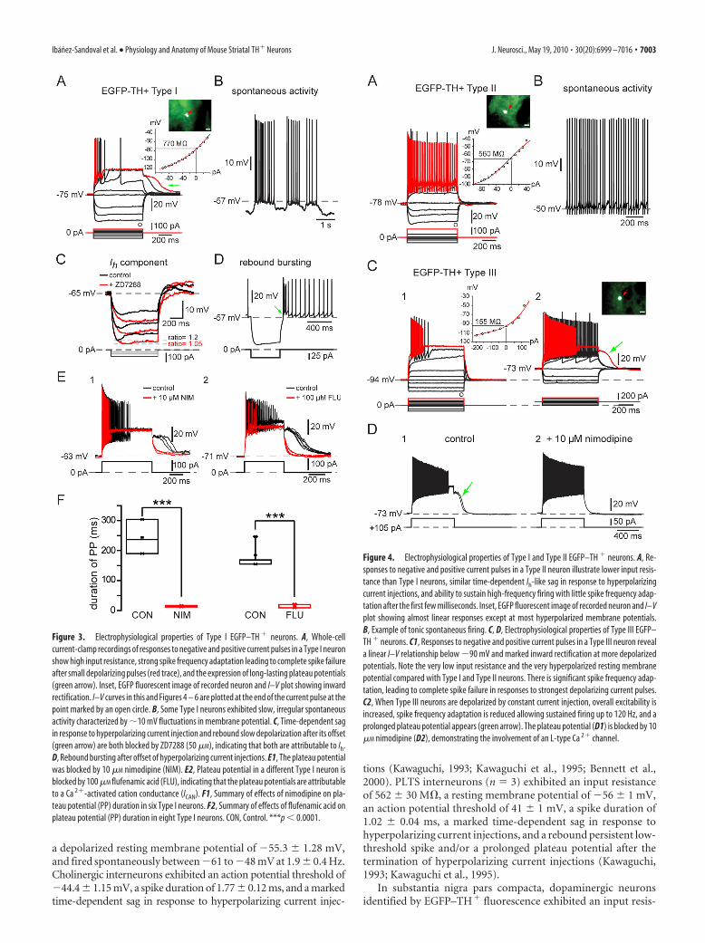
Figure 3. Electrophysiological properties of Type I EGFP–TH � neurons. A, Whole-cellcurrent-clamp recordings of responses to negative and positive current pulses in a Type I neuronshow high input resistance, strong spike frequency adaptation leading to complete spike failureafter small depolarizing pulses (red trace), and the expression of long-lasting plateau potentials(green arrow). Inset, EGFP fluorescent image of recorded neuron and I–V plot showing inwardrectification. I–V curves in this and Figures 4 – 6 are plotted at the end of the current pulse at thepoint marked by an open circle. B, Some Type I neurons exhibited slow, irregular spontaneousactivity characterized by �10 mV fluctuations in membrane potential. C, Time-dependent sagin response to hyperpolarizing current injection and rebound slow depolarization after its offset(green arrow) are both blocked by ZD7288 (50 �M), indicating that both are attributable to Ih.D, Rebound bursting after offset of hyperpolarizing current injections. E1, The plateau potentialwas blocked by 10 �M nimodipine (NIM). E2, Plateau potential in a different Type I neuron isblocked by 100 �M flufenamic acid (FLU), indicating that the plateau potentials are attributableto a Ca 2�-activated cation conductance (ICAN). F1, Summary of effects of nimodipine on pla-teau potential (PP) duration in six Type I neurons. F2, Summary of effects of flufenamic acid onplateau potential (PP) duration in eight Type I neurons. CON, Control. ***p 0.0001.
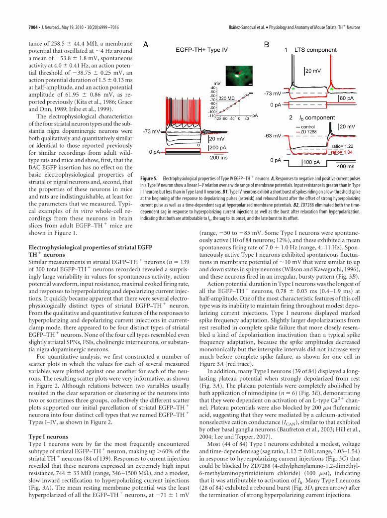
Figure 4. Electrophysiological properties of Type I and Type II EGFP–TH � neurons. A, Re-sponses to negative and positive current pulses in a Type II neuron illustrate lower input resis-tance than Type I neurons, similar time-dependent Ih-like sag in response to hyperpolarizingcurrent injections, and ability to sustain high-frequency firing with little spike frequency adap-tation after the first few milliseconds. Inset, EGFP fluorescent image of recorded neuron and I–Vplot showing almost linear responses except at most hyperpolarized membrane potentials.B, Example of tonic spontaneous firing. C, D, Electrophysiological properties of Type III EGFP–TH � neurons. C1, Responses to negative and positive current pulses in a Type III neuron reveala linear I–V relationship below �90 mV and marked inward rectification at more depolarizedpotentials. Note the very low input resistance and the very hyperpolarized resting membranepotential compared with Type I and Type II neurons. There is significant spike frequency adap-tation, leading to complete spike failure in responses to strongest depolarizing current pulses.C2, When Type III neurons are depolarized by constant current injection, overall excitability isincreased, spike frequency adaptation is reduced allowing sustained firing up to 120 Hz, and aprolonged plateau potential appears (green arrow). The plateau potential (D1) is blocked by 10�M nimodipine (D2), demonstrating the involvement of an L-type Ca 2� channel.
Ibanez-Sandoval et al. • Physiology and Anatomy of Mouse Striatal TH� Neurons J. Neurosci., May 19, 2010 • 30(20):6999 –7016 • 7003
tance of 258.5 44.4 M�, a membranepotential that oscillated at �4 Hz arounda mean of �53.8 1.8 mV, spontaneousactivity at 4.0 0.41 Hz, an action poten-tial threshold of �38.75 0.25 mV, anaction potential duration of 1.5 0.13 msat half-amplitude, and an action potentialamplitude of 61.95 0.86 mV, as re-ported previously (Kita et al., 1986; Graceand Onn, 1989; Iribe et al., 1999).
The electrophysiological characteristicsof the four striatal neuron types and the sub-stantia nigra dopaminergic neurons wereboth qualitatively and quantitatively similaror identical to those reported previouslyfor similar recordings from adult wild-type rats and mice and show, first, that theBAC EGFP insertion has no effect on thebasic electrophysiological properties ofstriatal or nigral neurons and, second, thatthe properties of these neurons in miceand rats are indistinguishable, at least forthe parameters that we measured. Typi-cal examples of in vitro whole-cell re-cordings from these neurons in brainslices from adult EGFP–TH � mice areshown in Figure 1.
Electrophysiological properties of striatal EGFPTH � neuronsSimilar measurements in striatal EGFP–TH� neurons (n � 139of 300 total EGFP–TH� neurons recorded) revealed a surpris-ingly large variability in values for spontaneous activity, actionpotential waveform, input resistance, maximal evoked firing rate,and responses to hyperpolarizing and depolarizing current injec-tions. It quickly became apparent that there were several electro-physiologically distinct types of striatal EGFP–TH� neuron.From the qualitative and quantitative features of the responses tohyperpolarizing and depolarizing current injections in current-clamp mode, there appeared to be four distinct types of striatalEGFP–TH� neurons. None of the four cell types resembled evenslightly striatal SPNs, FSIs, cholinergic interneurons, or substan-tia nigra dopaminergic neurons.
For quantitative analysis, we first constructed a number ofscatter plots in which the values for each of several measuredvariables were plotted against one another for each of the neu-rons. The resulting scatter plots were very informative, as shownin Figure 2. Although relations between two variables usuallyresulted in the clear separation or clustering of the neurons intotwo or sometimes three groups, collectively the different scatterplots supported our initial parcellation of striatal EGFP–TH�
neurons into four distinct cell types that we named EGFP–TH�
Types I–IV, as shown in Figure 2.
Type I neuronsType I neurons were by far the most frequently encounteredsubtype of striatal EGFP–TH� neuron, making up �60% of thestriatal TH� neurons (84 of 139). Responses to current injectionrevealed that these neurons expressed an extremely high inputresistance, 744 33 M� (range, 346 –1500 M�), and a modest,slow inward rectification to hyperpolarizing current injections(Fig. 3A). The mean resting membrane potential was the leasthyperpolarized of all the EGFP–TH� neurons, at �71 1 mV
(range, �50 to �85 mV. Some Type I neurons were spontane-ously active (10 of 84 neurons; 12%), and these exhibited a meanspontaneous firing rate of 7.0 � 1.0 Hz (range, 4 –11 Hz). Spon-taneously active Type I neurons exhibited spontaneous fluctua-tions in membrane potential of �10 mV that were similar to upand down states in spiny neurons (Wilson and Kawaguchi, 1996),and these neurons fired in an irregular, bursty pattern (Fig. 3B).
Action potential duration in Type I neurons was the longest ofall the EGFP–TH� neurons, 0.78 0.03 ms (0.4 –1.9 ms) athalf-amplitude. One of the most characteristic features of this celltype was its inability to maintain firing throughout modest depo-larizing current injections. Type I neurons displayed markedspike frequency adaptation. Slightly larger depolarizations fromrest resulted in complete spike failure that more closely resem-bled a kind of depolarization inactivation than a typical spikefrequency adaptation, because the spike amplitudes decreasedmonotonically but the interspike intervals did not increase verymuch before complete spike failure, as shown for one cell inFigure 3A (red trace).
In addition, many Type I neurons (39 of 84) displayed a long-lasting plateau potential when strongly depolarized from rest(Fig. 3A). The plateau potentials were completely abolished bybath application of nimodipine (n � 6) (Fig. 3E), demonstratingthat they were dependent on activation of an L-type Ca 2� chan-nel. Plateau potentials were also blocked by 200 �M flufenamicacid, suggesting that they were mediated by a calcium-activatednonselective cation conductance (ICAN), similar to that exhibitedby other basal ganglia neurons (Baufreton et al., 2003; Hill et al.,2004; Lee and Tepper, 2007).
Most (44 of 84) Type I neurons exhibited a modest, voltageand time-dependent sag (sag ratio, 1.12 0.01; range, 1.03–1.54)in response to hyperpolarizing current injections (Fig. 3C) thatcould be blocked by ZD7288 (4-ethylphenylamino-1,2-dimethyl-6-methylaminopyrimidinium chloride) (100 �M), indicatingthat it was attributable to activation of Ih. Many Type I neurons(28 of 84) exhibited a rebound burst (Fig. 3D, green arrow) afterthe termination of strong hyperpolarizing current injections.
Figure 5. Electrophysiological properties of Type IV EGFP–TH � neurons. A, Responses to negative and positive current pulsesin a Type IV neuron show a linear I–V relation over a wide range of membrane potentials. Input resistance is greater than in TypeIII neurons but less than in Type I and II neurons. B1, Type IV neurons exhibit a short burst of spikes riding on a low-threshold spikeat the beginning of the response to depolarizing pulses (asterisk) and rebound burst after the offset of strong hyperpolarizingcurrent pulse as well as a time-dependent sag at hyperpolarized membrane potentials. B2, ZD7288 eliminated both the time-dependent sag in response to hyperpolarizing current injections as well as the burst after relaxation from hyperpolarization,indicating that both are attributable to Ih, the sag to its onset, and the late burst to its offset.
7004 • J. Neurosci., May 19, 2010 • 30(20):6999 –7016 Ibanez-Sandoval et al. • Physiology and Anatomy of Mouse Striatal TH� Neurons

Figure 6. Retrograde labeling. A, Medium-magnification fluorescence micrograph of striatum after injections of rhodamine beads into both SN and GP under the EGFP filter. Eleven fluorescentEGFP–TH � neurons (arrows) are visible. B, Same field as in A but under the rhodamine filter showing numbers fluorescent retrogradely labeled SPNs. C, Merged image of A and B. Note that thereis no colocalization of EGFP and rhodamine. D, Rhodamine injection sites in SN and GP for three animals. E, Higher-magnification merged image of the area shown in the white square in C shows thatnone of the retrogradely labeled neurons express EGFP.
Table 1. Electrophysiological properties of striatal EGFP–TH � interneurons in adult mice
Parameter Type I (84) Type II (18) Type III (8) Type IV (29)
Input resistance (M�) 744 33a,b,c (346 to 1500) 429 39 (234 to 758) 180 8f (150 to 205) 471 30 (235 to 821)n � 78 n � 17 n � 8 n � 29
Resting membrane potential (mV) �71 1a,b,c (�50 to �85) �77 1d (�62 to �82) �89 1 (�86 to �94) �74 1f (�55 to �82)n � 79 n � 18 n � 8 n � 29
AP threshold (mV) �43 0.3a (�38 to �50) �47 0.5e (�42 to �50) �45 0.6 (�42 to �47) �44 0.5 (�39 to �48)n � 81 n � 17 n � 8 n � 29
AP width 50% (ms) 0.78 0.03a,b,c (0.4 to 1.9) 0.43 0.01 (0.3 to 0.53) 0.46 0.01 (0.4 to 0.5) 0.5 0.02 (0.4 to 0.85)n � 81 n � 17 n � 8 n � 29
AP amplitude (mV) 76 1.3a (52 to 101) 94 2d,e (86 to 116) 79 2 (70 to 88) 80 1 (63 to 91)n � 81 n � 17 n � 8 n � 29
AHP amplitude (mV) 16 0.4a (7 to 24) 20 0.6d,e (16 to 25) 14 0.3 (13 to 16) 15 0.5 (10 to 24)n � 81 n � 17 n � 8 n � 29
Maximum number of AP (150 –200 pA) 7 0.6a,b,c (2 to 22) 53 2d,e (45 to 68) 14 1f (9 to 17) 28 1 (18 to 45)n � 53 n � 14 n � 8 n � 23
Maximum frequency (Hz) 114 9a,c (32 to 256) 194 10d,e (137 to 265) 98 6f (66 to 124) 321 6 (276 to 386)n � 53 n � 14 n � 8 n � 23
Spontaneous activity 10 of 84 4 of 18 0 of 8 1 of 29Plateau potential duration (ms) 255 31 (46 to 1250) 150 116 (15 to 381) 159 41 (119 to 200) 0 of 26
39 of 84 3 of 12 2 of 8LTS component 0 of 84 4 of 18 0 of 8 29 of 29Ih ratio 1.12 0.01 (1.03 to 1.54) 1.12 0.02 (1.05 to 1.2) 1.13 0.02 (1.02 to 1.3)
44 of 84 11 of 18 0 of 8 17 of 29
All values are means SEM. Numbers in parentheses refer to the range of values. n refers to number of neurons from which the measurements were obtained. AP, Action potential; AHP, afterhyperpolarization. One-way ANOVA revealeda significant difference effect of type for all parametric measurements. Bonferroni’s post hoc comparisons were run to determine specific type to type differences.aType I versus Type II; bType I versus Type III; cType I versus Type IV; dType II versus Type III; eType II versus Type IV; fType III versus Type IV.a,b,c,d,e,fp 0.001.a,b,c,d,e,fp 0.05.
Ibanez-Sandoval et al. • Physiology and Anatomy of Mouse Striatal TH� Neurons J. Neurosci., May 19, 2010 • 30(20):6999 –7016 • 7005

The Type I neuron was clearly distinctfrom the PLTS interneuron described byKawaguchi (1993). The NPY-containingPLTS neuron exhibits plateaus during de-polarizing current injections and a re-bound low-threshold spike after the offsetof a hyperpolarizing pulse, whereas theType I neuron exhibits neither of thesebut rather exhibits a long-lasting plateaupotential after the offset of depolarizingcurrent pulse as shown in Figure 3.
Type II neuronsType II neurons were clearly distinctfrom Type I neurons. They exhibited amuch lower input resistance (429 39M�; range, 234 –758 M�) and a relativelylinear I–V curve except for a slight inwardrectification at very hyperpolarized mem-brane potentials. Spike duration was brief,0.43 0.01 ms (range, 0.30 – 0.53 ms),with a spike amplitude of 94 2 mV(range, 86 –116 mV). Type II neuronswere readily classifiable as fast-spikingneurons and responded to depolarizingcurrent injection with a mean sustainedfiring of 194 10 Hz (range, 137–265Hz), as illustrated for one typical examplein Figure 4A. Although unquestionably atype of fast-spiking neuron, Type II neu-rons were clearly distinct from PV� FSIsby virtue of their much higher input im-pedance and lack of intermittent/burstfiring in response to depolarizing currentpulses. Type II neurons accounted for13% (18 of 139) of the striatal EGFP–TH� neurons and were the only othersubtype of striatal EGFP–TH� neuronthat exhibited spontaneous activity (4 of18 neurons; 22%), with a mean firing rateof 10.4 8 Hz (range, 2 to 24 Hz), asshown in Figure 4B. The majority of theseneurons (11 of 18) exhibited a time-dependent sag in response to hyperpolar-izing current injection similar to that ofType I neurons.
Type III neuronsType III neurons exhibited the most hyperpolarized membranepotentials of the striatal EGFP–TH� neurons (resting membranepotential, 89 1 mV; range, �86 to �94 mV) and exhibited thelowest input resistance (180 8 M�; range, 150 –205 M�), pri-marily because of a strong inward rectification at membrane po-tentials more hyperpolarized than �80 mV (Fig. 4C). Type IIIneurons made up �6% of the striatal EGFP–TH� neurons andwere not spontaneously active. Type III neurons did not firespikes in response to depolarizing current injections adminis-tered from rest that depolarized the resting membrane potentialas high as �50 mV. Above this, however, a small increment in theintensity of the injected current resulted in a short burst of actionpotentials that exhibited strong spike frequency adaptation, ac-commodation and reduction in spike amplitude, followed by anabrupt cessation in spiking, similar to that seen in Type I neurons.
Additional increases in the amplitude of the injected current in-creased the maximum firing rate and accelerated the adaptationand accommodation and shortened the latency to the cessation ofspiking.
However, when identical depolarizing current pulses were in-jected into neurons depolarized to �73 mV by constant currentinjection, Type III neurons exhibited a more linear I–V relation-ship, exhibited a lowered threshold for spiking, and were capableof sustained spiking throughout the current pulse at a muchhigher firing rate than could be achieved with even larger currentinjections applied from rest. With higher intensity stimuli, firingwas also blocked in these neurons, but the period of sustainedfiring was considerably lengthened compared with what was seenwhen stimuli were applied at the resting membrane potential.Strong depolarizing current pulses delivered to depolarized TypeIII neurons also elicited the expression of a plateau potentialsimilar to that seen in Type I and Type II cells under appropriate
Figure 7. Neurochemical identification of striatal EGFP–TH � interneurons. A–C, Low-magnification double fluorescence mi-crographs of striatum showing EGFP–TH (green) simultaneously with PV immunofluorescence (A), NOS immunofluorescence (B),and CR immunofluorescence (C). Higher-magnification photomicrographs of the areas outlined in the white boxes are shown inA1–A3 (EGFP–TH, PV, and merged), B1–B3 (EGFP–TH, NOS, and merged), and C1–C3 (EGFP–TH, CR, and merged). Note thatnone of the EGFP–TH interneurons (white arrows in A1–C1) colocalize PV, NOS, or CR (red arrows). D1–D3, Colocalization of THimmunofluorescence with EGFP–TH 3 d after colchicine injection. D1, EGFP–TH � neurons (white arrows). D2, ImmunoreactiveTH � neurons (red arrows). D3, Merge of D1 and D2 showing neurons expressing both EGFP and TH (yellow arrows), EGFP but notTH (green arrow), and TH but not EGFP (red arrow). Str, Striatum; Ctx, cortex.
7006 • J. Neurosci., May 19, 2010 • 30(20):6999 –7016 Ibanez-Sandoval et al. • Physiology and Anatomy of Mouse Striatal TH� Neurons

conditions and, like those plateau potentials, was readily blockedby application of L-type Ca 2� channel antagonists (Fig. 4D,green arrow).
Type IV neuronsType IV neurons were the second most abundant of the EGFP–TH�
neurons, comprising 21% (29 of 139) of the striatal EGFP–TH�
neurons. Type IV neurons exhibited a linear I–V relationship andan input resistance similar to that of Type II neurons (471 30M�; range, 235– 821 M�). Type IV neurons exhibited a sag inresponse to hyperpolarizing current injections of similar timecourse and amplitude to that seen in Type I and II cells that couldalso be blocked by ZD7288 (Fig. 5B2), indicating that it was me-diated by a similar Ih. An example of a typical Type IV EGFP–TH� neuron is illustrated in Figure 5.
The most characteristic feature of the Type IV neuron was theexpression of a slow depolarization that closely resembled a low-threshold spike, with a burst of fast sodium–potassium spikesriding on top (Fig. 5B1). All Type IV neurons exhibited this low-threshold spike/burst, which could be elicited by a depolarizingcurrent pulse delivered to the cell at rest (�73 mV) as well as atthe termination of a strong hyperpolarizing current injection.
Unlike Type I, II, and III neurons, which each exhibited anensemble of electrophysiological properties unlike those of anycell type previously identified in striatum, the Type IV cell resem-bled in many ways the LTS interneuron described by Koos andTepper (1999). That neuron and the Type IV EGFP–TH� neu-ron differed from the NPY/vasoactive intestinal peptide/SOM�
PLTS neuron described by Kawaguchi and colleagues (Kawaguchi,1993; Kawaguchi et al., 1995) by the absence of plateau potentialsriding on the response to depolarizing current injections, aslightly lower input resistance, and a significantly shorter dura-tion action potential.
Means and SEMs of the electrophysiological properties of stri-atal EGFP–TH� neurons measured are reported in Table 1.ANOVA followed by Bonferroni’s multiple comparisons and be-tween the four cell types show that, consistent with our interpre-tation from the scatter plots, each of the EGFP–TH� cell types isstatistically different from one or more of the other cell types inseveral different electrophysiological parameters.
EGFP–TH � cell counting and retrograde labelingRetrograde labeling data and EGFP–TH� cell counts were ob-tained from six striatal hemispheres from three mice in whichrhodamine beads were injected bilaterally into both the globuspallidus and substantia nigra. A total of 203 EGFP–TH� neuronswere counted in the six hemispheres. The total number of EGFP–TH� neurons in the mouse striatum was estimated using threedifferent methods; optical fractionator, mean measured sectionthickness, and number weighted section thickness. All threemethods generated similar numbers, but the number weightedsection thickness approach consistently gave the smallest num-bers and thus was the most conservative estimate. These are thenumbers we report here.
The estimated number of EGFP–TH� neurons per striatumwas 2684 1216 (n � 6) in which the mean Gundersen’s coeffi-cient of error was 0.12. This is significantly greater than mostestimates of the abundance of striatal neurons labeled by THimmunocytochemistry described in studies in the normal mouseor rat striatum (Tashiro et al., 1989b; Mao et al., 2001; Busceti etal., 2008). This may be attributable in part to several factors,including the increased sensitivity of the BAC transgenic ap-proach as a result of incorporation of multiple copies of the EGFP
gene (Gong et al., 2003), the relatively increased brightness of thestriatal EGFP–TH� somata compared with either bright-field ordark-field immunocytochemical labeling, as well as other factorsdiscussed below.
Of the 203 EGFP–TH� neurons counted in the six hemi-spheres, none were also labeled with rhodamine despite verydense retrograde labeling in the immediate areas of the EGFP–TH� neurons (Fig. 6). The failure to find any retrogradely labeledEGFP–TH� neurons cannot, of course, completely rule out thepossibility that a small fraction of these neurons does project toSN or GP, but it is clear that the vast majority of the striatalEGFP–TH� neurons cannot be striatonigral or striatopallidalSPNs and must therefore be interneurons.
Immunocytochemical characterization of striatalEGFP–TH � neuronsWe tested striatal sections from EGFP–TH� mice for PV, NOS,or CR immunofluorescence to determine the extent of colocaliza-tion, if any, of these substances in striatal EGFP–TH� neurons. Asshown in Figure 7, although numerous brightly fluorescent PV�,NOS�, and CR� neurons were observed, colocalization withEGFP was never observed. Thus, consistent with the electrophys-iological data that showed physiological characteristics for eachof the EGFP–TH� neurons clearly distinct from those reportedpreviously for striatal fast-spiking or PLTS interneurons, the im-munocytochemical data also show that the striatal EGFP–TH�
neurons are neurochemically distinct from previously describedstriatal interneurons.
Because the expression of the EGFP–TH� reporter is not adirect demonstration of the abundance of TH mRNA or THprotein but instead indicates rates of transcription of the TH gene(Gong et al., 2003), we performed TH immunostaining on stria-tal and midbrain sections from EGFP–TH� mice. To our sur-prise, although the majority of substantia nigra and ventral
Figure 8. Results of single-cell RT-PCR performed on four EGFP–TH � neurons. A, Typicalresponses of a Type I neuron to depolarizing current injection. B, Agarose gel from the cellshown in A plus another Type I neuron showing that both express the amplicon for VMAT1 butnot VMAT2. C, Typical responses of a Type IV neuron to depolarizing current injection. D, Gelsfrom the neuron shown in C plus a second Type IV neuron show that Type IV neurons also expressVMAT1. MW, Molecular weight.
Ibanez-Sandoval et al. • Physiology and Anatomy of Mouse Striatal TH� Neurons J. Neurosci., May 19, 2010 • 30(20):6999 –7016 • 7007

tegmental area EGFP–TH� neurons werealso immunoreactive for TH (although aminority did not stain for TH and someTH-immunopositive neurons did not ap-pear to express EGFP; data not shown),such double labeling of striatal neuronswas almost nonexistent; in fact, very fewstriatal TH� neurons were found in con-trol or transgenic mice even with highconcentrations of primary antibody andlong primary incubation times. We sus-pected that perhaps this was attributableto the striatal EGFP–TH� neurons ex-pressing much lower levels of TH thanmidbrain dopaminergic neurons and/orefficient axonal transport of the enzyme tothe terminals, similar to what was foundto be the case in studies of GABA andGAD immunocytochemistry in certainstriatal interneurons (Bolam et al., 1985;Kita and Kitai, 1988; Chesselet and Robbins,1989). Therefore, we injected colchicine in-trastriatally in three EGFP–TH� mice.
There was significant nonspecific dam-age in the injected striata so we analyzedthe contralateral striata for TH� immu-noreactivity and colocalization of TH andEGFP fluorescent labeling. In contrast tocontrol striata, we found an abundantnumber of TH� immunofluorescent so-mata in the striata of colchicine-treatedmice, similar to the number of EGFP–TH� neurons observed in normal EGFP–TH� transgenic mice. Figure 7 illustratesone representative section from the stria-tum of an EGFP–TH� mouse 3 d aftercolchicine injection. Thus, a majority ofstriatal EGFP–TH� neurons express TH,albeit in much lower amounts than in themesencephalic dopaminergic neurons.
Single-cell RT-PCR phenotyping ofstriatal TH � neuronsIn 10 EGFP–TH� neurons that were recorded in whole-cellmode for short periods just long enough to identify the subtype ofthe EGFP–TH� neuron, the cytoplasm was aspirated and RT-PCR was used to probe for the expression of VMAT1 andVMAT2. Although none of the neurons expressed amplicons forVMAT2, the transporter expressed by mesencephalic dopami-nergic neurons, all 10 EGFP–TH� neurons exhibited robust ex-pression of VMAT1 mRNA. Examples of gels from two Type Iand two Type IV neurons, all of which showed expression ofVMAT1 but not VMAT2, are shown in Figure 8.
Synaptic connections of striatal EGFP–TH � interneuronsLocal electrical stimulation with single pulses evoked depolariz-ing synaptic potentials (DPSPs) in all striatal EGFP–TH� inter-neurons tested (n � 5). The DPSPs were composed of twocomponents that reversed at different membrane potentials. Theinhibitory component was an IPSP that exhibited a reversal po-tential at approximately �65 mV (Fig. 9B) (64 1.5 mV; n � 4)and could be blocked by bicuculline (10 �M), indicating that itwas mediated principally by GABAA receptors. Blockade of the
IPSP revealed a pure EPSP that was completely abolished by 10�M DNQX, suggesting that it was mediated by non-NMDA re-ceptors, consistent with the hyperpolarized membrane potentialsand recording conditions, as shown for one representative TypeI and II interneuron in Figure 9, B and C. Note that, in Figure 9,B3 and C3, combined application of bicuculline and CNQX to-tally abolished the synaptic responses, demonstrating that 10 �M
bicuculline resulted in a 100% block of the IPSP and, similarly, 10�M DNQX resulted in a 100% block of the EPSP.
Striatal Type I EGFP–TH� interneurons also responded tostimulation of the cortex with DPSPs (Fig. 9D). Unlike the re-sponses to local stimulation, bicuculline had no effect on thecortically evoked DPSPs, indicating that they were pure EPSPs.As with local stimulation, bath application of 10 �M DNQX com-pletely abolished the EPSP, indicating that it was mediated pre-dominantly or exclusively by non-NMDA receptors.
Like Type I interneurons, Type II interneurons were well in-tegrated into the synaptic network of the striatum. Local stimu-lation elicited EPSPs and IPSPs blocked by DNQX andbicuculline, just like in the Type I interneurons (Fig. 9C).
Figure 9. Synaptic input to EGFP–TH � neurons. A, Experimental design. Bipolar stimulating electrodes were placed onto thesurface of the deep layers of the cortex (1) and in striatum to evoke responses in striatal EGFP–TH � neurons. CPu, Caudateputamen. B1, In a representative Type I cell, local stimulation evoked a biphasic response consisting of an overlapping EPSP andIPSP (control). B2, DNQX (10 �M) blocked the EPSP component, leaving a pure IPSP with an apparent reversal potential of�65 mV(B4 ). B3, The IPSP was blocked by bicuculline (10 �M), indicating mediation by GABAA receptors. C, Same experiment performedwith a Type II neuron but reversing the order of the antagonist application. C1, Local stimulation evokes biphasic response.C2, The inhibitory component was blocked by 10 �M bicuculline, showing that it was mediated by a GABAA receptor.C3, The remaining EPSP is completely eliminated by addition of DNQX (10 �M). D1, Cortical stimulation evokes short-latency monosynaptic DPSP in a Type I neuron. D2, The DPSP is not affected by 10 �M bicuculline. D3, The AMPA/kainatechannel blocker DNQX (10 �M) completely eliminates the DPSP, showing that it is a glutamatergic EPSP. D4, Time courseof drug effects on the EPSP.
7008 • J. Neurosci., May 19, 2010 • 30(20):6999 –7016 Ibanez-Sandoval et al. • Physiology and Anatomy of Mouse Striatal TH� Neurons
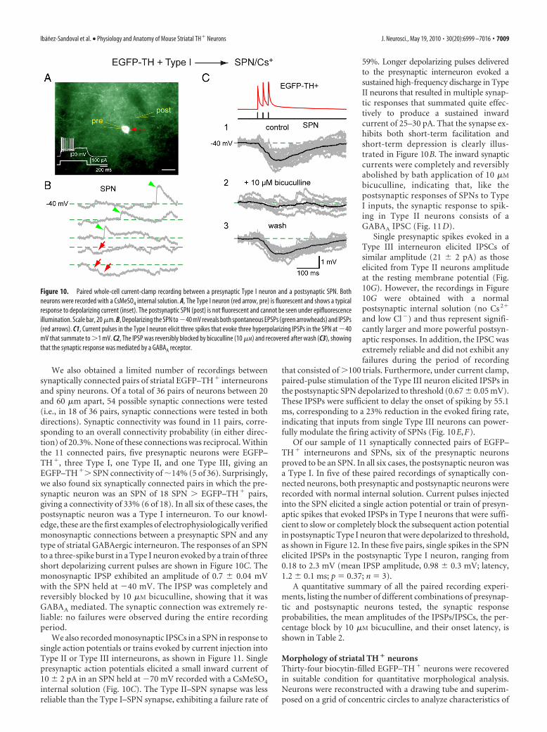
We also obtained a limited number of recordings betweensynaptically connected pairs of striatal EGFP–TH� interneuronsand spiny neurons. Of a total of 36 pairs of neurons between 20and 60 �m apart, 54 possible synaptic connections were tested(i.e., in 18 of 36 pairs, synaptic connections were tested in bothdirections). Synaptic connectivity was found in 11 pairs, corre-sponding to an overall connectivity probability (in either direc-tion) of 20.3%. None of these connections was reciprocal. Withinthe 11 connected pairs, five presynaptic neurons were EGFP–TH�, three Type I, one Type II, and one Type III, giving anEGFP–TH�� SPN connectivity of �14% (5 of 36). Surprisingly,we also found six synaptically connected pairs in which the pre-synaptic neuron was an SPN of 18 SPN � EGFP–TH� pairs,giving a connectivity of 33% (6 of 18). In all six of these cases, thepostsynaptic neuron was a Type I interneuron. To our knowl-edge, these are the first examples of electrophysiologically verifiedmonosynaptic connections between a presynaptic SPN and anytype of striatal GABAergic interneuron. The responses of an SPNto a three-spike burst in a Type I neuron evoked by a train of threeshort depolarizing current pulses are shown in Figure 10C. Themonosynaptic IPSP exhibited an amplitude of 0.7 0.04 mVwith the SPN held at �40 mV. The IPSP was completely andreversibly blocked by 10 �M bicuculline, showing that it wasGABAA mediated. The synaptic connection was extremely re-liable: no failures were observed during the entire recordingperiod.
We also recorded monosynaptic IPSCs in a SPN in response tosingle action potentials or trains evoked by current injection intoType II or Type III interneurons, as shown in Figure 11. Singlepresynaptic action potentials elicited a small inward current of10 2 pA in an SPN held at �70 mV recorded with a CsMeSO4
internal solution (Fig. 10C). The Type II–SPN synapse was lessreliable than the Type I–SPN synapse, exhibiting a failure rate of
59%. Longer depolarizing pulses deliveredto the presynaptic interneuron evoked asustained high-frequency discharge in TypeII neurons that resulted in multiple synap-tic responses that summated quite effec-tively to produce a sustained inwardcurrent of 25–30 pA. That the synapse ex-hibits both short-term facilitation andshort-term depression is clearly illus-trated in Figure 10B. The inward synapticcurrents were completely and reversiblyabolished by bath application of 10 �M
bicuculline, indicating that, like thepostsynaptic responses of SPNs to TypeI inputs, the synaptic response to spik-ing in Type II neurons consists of aGABAA IPSC (Fig. 11 D).
Single presynaptic spikes evoked in aType III interneuron elicited IPSCs ofsimilar amplitude (21 2 pA) as thoseelicited from Type II neurons amplitudeat the resting membrane potential (Fig.10G). However, the recordings in Figure10G were obtained with a normalpostsynaptic internal solution (no Cs 2�
and low Cl�) and thus represent signifi-cantly larger and more powerful postsyn-aptic responses. In addition, the IPSC wasextremely reliable and did not exhibit anyfailures during the period of recording
that consisted of �100 trials. Furthermore, under current clamp,paired-pulse stimulation of the Type III neuron elicited IPSPs inthe postsynaptic SPN depolarized to threshold (0.67 0.05 mV).These IPSPs were sufficient to delay the onset of spiking by 55.1ms, corresponding to a 23% reduction in the evoked firing rate,indicating that inputs from single Type III neurons can power-fully modulate the firing activity of SPNs (Fig. 10E,F).
Of our sample of 11 synaptically connected pairs of EGFP–TH� interneurons and SPNs, six of the presynaptic neuronsproved to be an SPN. In all six cases, the postsynaptic neuron wasa Type I. In five of these paired recordings of synaptically con-nected neurons, both presynaptic and postsynaptic neurons wererecorded with normal internal solution. Current pulses injectedinto the SPN elicited a single action potential or train of presyn-aptic spikes that evoked IPSPs in Type I neurons that were suffi-cient to slow or completely block the subsequent action potentialin postsynaptic Type I neuron that were depolarized to threshold,as shown in Figure 12. In these five pairs, single spikes in the SPNelicited IPSPs in the postsynaptic Type I neuron, ranging from0.18 to 2.3 mV (mean IPSP amplitude, 0.98 0.3 mV; latency,1.2 0.1 ms; p � 0.37; n � 3).
A quantitative summary of all the paired recording experi-ments, listing the number of different combinations of presynap-tic and postsynaptic neurons tested, the synaptic responseprobabilities, the mean amplitudes of the IPSPs/IPSCs, the per-centage block by 10 �M bicuculline, and their onset latency, isshown in Table 2.
Morphology of striatal TH � neuronsThirty-four biocytin-filled EGFP–TH� neurons were recoveredin suitable condition for quantitative morphological analysis.Neurons were reconstructed with a drawing tube and superim-posed on a grid of concentric circles to analyze characteristics of
Figure 10. Paired whole-cell current-clamp recording between a presynaptic Type I neuron and a postsynaptic SPN. Bothneurons were recorded with a CsMeSO4 internal solution. A, The Type I neuron (red arrow, pre) is fluorescent and shows a typicalresponse to depolarizing current (inset). The postsynaptic SPN (post) is not fluorescent and cannot be seen under epifluorescenceillumination. Scale bar, 20 �m. B, Depolarizing the SPN to�40 mV reveals both spontaneous EPSPs (green arrowheads) and IPSPs(red arrows). C1, Current pulses in the Type I neuron elicit three spikes that evoke three hyperpolarizing IPSPs in the SPN at �40mV that summate to �1 mV. C2, The IPSP was reversibly blocked by bicuculline (10 �M) and recovered after wash (C3), showingthat the synaptic response was mediated by a GABAA receptor.
Ibanez-Sandoval et al. • Physiology and Anatomy of Mouse Striatal TH� Neurons J. Neurosci., May 19, 2010 • 30(20):6999 –7016 • 7009

the dendritic and axonal arborizations(Sholl, 1953). Examples of the recon-structed neurons and the resulting Shollplots are shown in Figure 13. Becausethere were no significant morphometricdifferences among the four cell types inthe quantitative measurements, the mea-surements from all 34 neurons werepooled and are presented in Table 3.
Striatal EGFP–TH� interneurons weremedium sized, with the long axis of thesoma ranging from 11.4 to 24 �m and itsperpendicular ranging from 7.1 to 13.2�m. ANOVA followed by Bonferroni’spost hoc tests revealed that none of the celltypes differed from any of the other celltypes in terms of somatic area, perimeter,length of long or short axes, or number ofprimary dendrites.
Somata were most frequently round orovoid (28 of 34; 77%). The remaining six(23%), all of which were Type I interneu-rons, were pyramidal shaped. The dendritesof most of the EGFP–TH� interneurons ofall four subtypes were mostly or completelyaspiny and varicose (30 of 34; 88%). Thedegree of dendritic varicosity varied fromslight, as illustrated for a Type II neuron inFigure 13B, to considerable, as illustrated bythe Type IV neuron in Figure 13D.
Four of the stained neurons appearedmarkedly different from the rest. All of thesecells were Type I interneurons. The den-drites of these cells exhibited fairly dense,thick stick-like appendages that appearedto lack distinct spine heads. Nevertheless,these neurons were clearly not SPNs (Fig.12A,C). Low- and high-magnificationmicrographs of a biocytin-filled spinyType I EGFP–TH� interneuron areshown in Figure 14 along with a represen-tative biocytin-filled SPN, also from thestriatum of an EGFP–TH� mouse. Sev-eral differences between the two neuronaltypes are readily apparent. The Type Ineuron is slightly larger than the SPN,and the density of spines covering thedendrites of the SPN is an order of mag-nitude greater than that of the Type Ineuron. High-magnification inspectionof dendrites of the two neuronal typesreveals that, compared with the spineson the SPN neuron, the dendritic spe-cializations on the Type I neuron oftenseem to lack a spine head, and many arelonger than the spines on the SPNs.
Figure 11. Spiking in Type II and Type III neurons elicits IPSCs in SPNs and delays firing in response to current injection.A, Connected pair consisting of a presynaptic EFGP � Type II neuron (top left) and a postsynaptic SPN (bottom left). The insets arehigh-magnification micrographs showing an aspiny Type II (pre) dendrite and a spiny SPN dendrite (post) after both were patchedin whole-cell mode and stained with Alexa 594. B, Top, Current pulses injected into the Type II neuron elicited a train of presynapticspikes that evoked IPSCs in the postsynaptic SPN. Both short-term depression and early facilitation were evident. C, Two spikesevoked by two 5 ms pulses 50 ms apart in the Type II neuron result in modest paired-pulse facilitation in the same SPN. Black tracesare averages, and gray traces are individual trials. D, The IPSCs were reversibly blocked by bicuculline (10 �M) showing that theywere mediated by a GABAA receptor. E, Connected pair consisting of a presynaptic Type III neuron and a postsynaptic SPN (inset).Two presynaptic spikes separated by 50 ms (bottom trace) delay the occurrence of depolarization induced spiking in the postsyn-aptic SPN (top black trace) by almost 100 ms (top red trace). F, Entire dataset from which the traces in E were selected. Depolar-ization evoked spiking in SPN under control conditions (black traces) and in the presence of two spikes in the presynaptic neuron
4
(red traces). Note the reliability of this connection. G, I–V plotfor this synaptic response showing a reversal potential consis-tent with mediation by Cl � ions and a synaptic conductancenear 1 nS.
7010 • J. Neurosci., May 19, 2010 • 30(20):6999 –7016 Ibanez-Sandoval et al. • Physiology and Anatomy of Mouse Striatal TH� Neurons

The axonal arborization of the spinyType I neurons is also qualitatively differ-ent from that of the local SPNs. This is alsoillustrated in Figure 14. Whereas the localcollaterals of SPNs are by and largesmooth, with intermittently spaced vari-cose segments, the axonal arborization ofthe Type I EGFP–TH� interneuron con-sists of a dense plexus of highly branchedthin processes, heavily beaded with large(�1 �m), round varicosities, most ofwhich are en passant.
It was not possible to discriminateamong the aspiny Type II, III, and IVinterneurons in our sample based onqualitative or quantitative morphologicalcriteria. For all subtypes, the axon emergedfrom the soma or a proximal dendrite andbranched almost immediately, forming adense local axon collateral plexus that oc-cupied a volume coextensive with andsometimes extending beyond the den-dritic tree of the issuing neuron. The col-laterals were highly branched and studdedwith large numbers of varicosities. Noneof the filled cells exhibited a single axonalbranch that was larger than the rest or thatcould be clearly identified as the mainaxon, and in none of the cells was thereevidence of a single long axon that pro-jected away from the immediate locale ofthe parent cell, consistent with the retro-grade labeling data that identified them asinterneurons.
DiscussionThere are four distinct subtypes ofEGFP–TH � neurons in mouse striatumStriatal EGFP–TH� neurons could be re-liably divided into four subtypes based ona number of electrophysiological charac-
Figure 12. Whole-cell recordings of two connected pairs in which the presynaptic neuron was an SPN and the postsynapticneuron was a Type I neuron. A–D, Presynaptic and postsynaptic neurons recorded with 140 mM CsMeSO4 internal solution. A,Postsynaptic EGFP–TH � Type I neuron (red arrow, post) is fluorescent and shows typical response to depolarizing current pulses.Presynaptic SPN are not fluorescent and cannot be seen (pre) and were patched under DIC visualization. B1, Depolarizing currentpulse evokes three spikes in a Type I neuron (top black trace). B2, Single action potential elicited in the SPN (red trace) causes anIPSP (�45 mV) in the Type I neuron held at�48 mV (black trace) that was sufficient to completely block the second of three spikesevoked in the Type I neuron. Mean IPSP amplitude, 0.58 0.14 mV; latency, 2.6 0.13 ms; p � 100%. C, Single spike (top redtrace) in the SPN evokes a hyperpolarizing IPSP in a Type I neuron held at �46 mV. Black trace is average, and gray traces areindividual trials. Note that the IPSP failure rate is zero. The IPSP is reversed at�80 mV (middle) and is completely blocked by 10 �M
4
bicuculline (bottom), indicating a GABAA IPSP. D, Comparisonof a spontaneous IPSP and one evoked from the SPN showsthem to have identical amplitudes and time courses, suggest-ing that some of the spontaneous IPSPs in Type I neurons arisefrom surrounding SPNs. E–G, Another connected pair in whichthe presynaptic neuron was an SPN and the postsynaptic neu-ron was a Type I. Both presynaptic and postsynaptic neuronsrecorded with normal internal solution. E, Postsynaptic Type Ineuron (red arrow, post) is fluorescent. Presynaptic SPN is notfluorescent and cannot be seen (pre) and was patched underDIC visualization. F, Train of spikes in the presynaptic SPN elic-ited by a current pulse (red traces) elicit IPSPs (green arrows) inthe postsynaptic Type I neuron (black traces). The IPSPs delaythe onset of the subsequent spike. G, Single depolarization-evoked spikes in the SPN elicit IPSPs that delay the occurrenceof the subsequent spike in the postsynaptic Type I neuron de-polarized to threshold by current injection. H2, Bicuculline at10 �m blocks the IPSPs and the subsequent postsynapticspikes occur with a decreased latency. H3, The IPSP returnsafter washout, and the latency to the subsequent postsynapticspike increases. Scale bars: A, E, 20 �m.
Ibanez-Sandoval et al. • Physiology and Anatomy of Mouse Striatal TH� Neurons J. Neurosci., May 19, 2010 • 30(20):6999 –7016 • 7011

teristics. The majority of the cells wereType I, and Type I and Type IV cells to-gether accounted for �80% of all the stri-atal EGFP–TH� neurons.
With the exception of the Type IV neu-ron, which resembled the LTS GABAergicinterneuron described by Koos and Tep-per (1999) in several respects, each of theother EGFP–TH� cell types displayed anelectrophysiological profile that was dif-ferent from that described previously forany striatal cell type (Tepper and Bolam,2004). Types II and IV, capable of sus-tained firing over 200 Hz, are clearly sub-types of fast-spiking neuron, but none ofthese neurons expressed PV (or CR orNOS). Thus, there are (at least) two ad-ditional types of fast-spiking neuron instriatum other than the well knownPV � FSI.
Striatal EGFP–TH � neurons areGABAergic interneuronsIdentification of striatal TH� neurons as in-terneurons or projection neurons has beencontroversial, with some authors arguingthat these cells are interneurons (Dubach etal., 1987; Betarbet et al., 1997; Cossette et al.,2005; Mazloom and Smith, 2006; Huotand Parent, 2007), whereas others haveargued that they are SPNs (Tashiro et al.,1989a,b; Darmopil et al., 2008). Our ret-rograde tracing experiments failed to labelany EGFP–TH� neurons. Furthermore,none of the biocytin-labeled EGFP–TH�
neurons exhibited the distinctive electro-physiological or morphological profiles ofSPNs. Thus, our conclusions are consis-tent with that showing that the over-whelming majority of striatal EGFP–TH�
neurons are interneurons.Data from synaptically connected pairs
of EGFP–TH� interneurons and SPNsshowed that spikes in EGFP–TH� inter-neurons resulted in GABAA IPSPs/IPSCsin SPNs, consistent with previous studiesthat showed that striatal TH� neuronsalso express GAD (Betarbet et al., 1997; Mura et al., 2000; Cos-sette et al., 2005; Mazloom and Smith, 2006; Tande et al., 2006;Huot and Parent, 2007). Thus, striatal EGFP–TH � interneu-
rons represent four novel subtypes of striatal GABAergicinterneurons.
Single-cell RT-PCR on 10 EGFP–TH� interneurons revealedthat all expressed VMAT1, normally associated with large dense
Figure 13. Drawing tube reconstructions of EGFP–TH � neurons filled with biocytin after recording, superimposed on concentriccircles for Sholl analyses. Striatal EGFP–TH � neurons had three to five primary dendrites that branched between 15 and 50 �m from thesoma and gave rise to a modest arborization of secondary and tertiary varicose dendrites. Dendrites are plotted in black and axons in red inthe Sholl plots. The quantitative morphometric results are given in Table 3. All Type II–IV EGFP–TH �neurons were aspiny, but 6 of 26 TypeI neurons expressed sparse spine-like processes on their higher-order dendrites (e.g., A and C; see Fig. 14). The axonal arborizations weresometimes quite dense (A–C), reaching a maximum density between 50 and 120 �m from the soma (red circles in Sholl plots) and werestudded throughout with small varicosities (A–D; deep red dots on the axons), presumably synaptic boutons. In these reconstructions, thesoma and dendrites are shown in black, and the axon are shown in red and varicosities in dark red.
Table 2. Summary of pairs recording of striatal EGFP–TH � interneurons in adult mice
n (pairs)Synapticconnections
% of pairsrecorded
Synapsetype
p (%) connections andrecording condition
Amplitude ofconnection (mV/pA)
Inhibition bybicuculline (%)
Onsetlatency (ms)
32 3 9.4 Type I to SPN (1) p � 100% (Cs �) 0.7 0.04 mV at �40 mV 82 1.6 0.1(2) p � 17% (Cl �) 9 2 pA at HP � 50 mV 1.1 0.3
2 1 50 Type II to SPN p � 41.6% (Cs �) 10 2 pA at HP � �70 mV 100 1.2 0.072 1 50 Type III to SPN p � 100% (Cl �) 21 2 pA at HP � �70 mV 0.6 0.04
18 6 33.3 SPN to Type I (1) p � 100% (Cs �) 1.8 0.11 mV at �44 mV 100 1.6 0.13(5) p � 37 8% (Cl �) 0.98 0.3 mV at �45 mV 100 1.2 0.1
Summary of paired recording experiments. �Synaptic connections� refers to the number of connections observed in the direction specified by �Synapse type.� p refers to the probability that a spike in the presynaptic neuron resulted in anIPSP/IPSC in the postsynaptic neuron. Numbers in parentheses refer to the n of connections that each p value is based on. �Onset latency� refers to delay between the peak of the presynaptic spike and the onset of the IPSC. HP,Hyperpolarization.
7012 • J. Neurosci., May 19, 2010 • 30(20):6999 –7016 Ibanez-Sandoval et al. • Physiology and Anatomy of Mouse Striatal TH� Neurons

core vesicles, but not VMAT2, which is the transporter expressedby all midbrain dopaminergic neurons (Weihe et al., 1996). It isnot immediately clear what this difference means, but it could berelated to the fact that striatal TH� neurons are reported to in-crease in number after 6-OHDA or 1-methyl-4-phenyl-1,2,3,6-tetrahydropyridine (MPTP) treatment, whereas midbraindopaminergic neurons are destroyed by these toxins (O’Byrne etal., 2000; Tande et al., 2006; Huot and Parent, 2007). Regardless,
the expression of VMAT1 by the striatalEGFP–TH� interneurons fits with previ-ous reports claiming DAT expression instriatal TH� neurons cited above and isconsistent with the possibility that theEGFP–TH� interneurons may be dopa-minergic as well as GABAergic.
Striatal EGFP–TH � interneurons areimportant components of striatalintrinsic circuitrySynaptic responses of striatal EGFP–TH�
interneurons resemble those of other stri-atal neurons. Local stimulation elicitsnearly simultaneous monosynaptic gluta-matergic EPSPs and GABAergic IPSPCsseveral millivolts in amplitude. Corticalstimulation evokes a monosynaptic gluta-matergic EPSP in striatal EGFP–TH� in-terneurons as it does in most striatalneurons. Paired recordings revealed a con-nection probability of �14% for EGFP–TH� to SPN connections and 33% forSPN to EGFP–TH� interneuron connec-tions. Synaptic inputs from SPNs toGABAergic interneurons have not beenreported previously in striatum, and theirexistence suggests that the EGFP–TH�
interneurons may play a role in synapticintegration in striatum distinct from thebetter known GABAergic interneurons.These data reveal that striatal TH� inter-neurons are well integrated into the syn-aptic circuitry of the striatum.
Similar to PV�–SPN synapses, syn-apses between EGFP–TH� interneuronsand SPNs were very reliable. Two pairs (aType I and a Type III) exhibited a failurerate of zero, and, overall, IPSC amplitudeswere greater than those from axon collat-eral synapses. These IPSPs effectively de-layed firing in SPNs. These data suggestthat, similar to other GABAergic inter-neuronal synapses onto SPNs, those orig-inating from EGFP–TH� interneuronsmay represent a particularly important
and powerful source of striatal inhibition.
Relationship between striatal EGFP–TH � interneurons andstriatal TH � neurons previously identified by THimmunostainingIt seems natural to assume that the striatal neurons that expressEGFP in the EGFP–TH� mice described in this study are from
Figure 14. Characteristic anatomy of a typical SPN and a spiny EGFP–TH � Type I neuron filled with biocytin after whole-cellrecording from adult mouse slices. A, Low-magnification photomicrograph of an SPN shows a soma �12 �m in diameter thatgives rise to six primary dendrites that are aspiny for the initial 20 �m that then become densely invested with spines and give riseto secondary and higher-order spiny dendrites extending for a radius of �300 �m. 1–5, Higher-magnification images of spinydendrites (A2–A5) and axons (A1, A2) from regions marked with the same numbers in A above. Note the high density of dendriticspines. The mushroom-like appearance of many of the spines is particularly apparent in A4 and A5. The local axon collaterals arevaricose and exhibit intermittent varicosities. A6, Whole-cell current-clamp recording of responses to intracellularly injectedcurrent pulses from this neuron is shown in 6 and is very characteristic of SPNs. B, A spiny Type I neuron shows a slightly larger somaand four thick primary dendrites that branch less frequently and follow a straighter course than the dendrites of an SPN. Thedendrites are invested with spine-like appendages. These spine-like processes are of much lower density than the dendritic spineson the SPN, many of them are quite long (arrows in B), and most appear to lack spine heads (B2). Like the SPN, the axonalarborization of the Type I neuron is quite dense. The intervaricose segments appear thinner than those of the SPN, and the entireaxonal arborization is studded with larger and more prominent varicosities than that of the SPN (B1). B3, The whole-cell current-clamp recording from this neuron is typical for Type I neurons and completely distinct from that of a typical SPN.
Table 3. Anatomical parameters of striatal EGFP–TH � interneurons in adult mice
Somatic area(�m 2)
Perimeter(�m)
Soma size (�m)Number ofprimarydendrites
Processes on dendrites
Sholl analysis
Width Height Varicose Spine BothNumber of dendritictips
Mean dendritic fielddiameter (�m)
Mean axonal fielddiameter (�m)
n � 34 112.7 7.6(65–266)
37.7 1.5(27– 60)
15.2 0.6(9 –24)
10.8 0.4(7–18)
3.3 0.1(2– 4)
28 of 34 4 of 34a 2 of 34 16.2 1.9 (5–32),n � 17
173 10 (140 –220),n � 7
267 38 (170 – 460),n � 7
Mean morphometric parameters from all EGFP–TH � neurons intracellularly labeled with biocytin after recording. All values are given as mean SEM.aAll four spiny neurons were Type I.
Ibanez-Sandoval et al. • Physiology and Anatomy of Mouse Striatal TH� Neurons J. Neurosci., May 19, 2010 • 30(20):6999 –7016 • 7013

the same population of neurons previously identified on the basisof TH immunocytochemical labeling, because EGFP expressionis limited to neurons that express the endogenous TH transcrip-tional regulatory sequences (Heintz, 2001). Attempts to comparethe morphology of striatal TH� neurons identified by immuno-cytochemistry and those identified by EGFP–TH� expressionand then stained with biocytin are complicated somewhat by thedifferent staining techniques and the contradictory nature ofsome of the previous reports. However, in general, the majority ofprevious studies converge to a picture of striatal TH� neurons assmall- to medium-sized aspiny neurons, most likely interneu-rons, with varicose dendrites (Dubach et al., 1987; Tashiro et al.,1989a,b; Betarbet et al., 1997; Cossette et al., 2005; Huot andParent, 2007), consistent with the present findings.
Biocytin-labeled Type I interneurons were medium sized witha pyramidal soma and varicose dendrites. A subset of Type Ineurons exhibited slightly larger somata and were fairly denselyinvested with elongated, stick-like dendritic appendages. Despitethe spine-like specializations, these cells were clearly not SPNsand could easily be distinguished from them morphologically(Fig. 13) as well as electrophysiologically. Nevertheless, it may bethat cells of this type are responsible, at least in part, for some ofthe claims that striatal TH� neurons are spiny projection neu-rons (Darmopil et al., 2008). Although exhibiting quite distinctelectrophysiological characteristics, Types II–IV could not be distin-guished from one another morphologically. All exhibited mediumround or ovoid somata from which issued three to four aspiny, var-icose primary dendrites that branched sparingly. Neurons with thismorphology have been described often in Golgi studies (DiFiglia etal., 1976; Chang et al., 1982; Bolam et al., 1983; Groves, 1983).
One fact difficult to reconcile with the assumption that striatalEGFP–TH� interneurons represent the same population of cellsthat were stained by TH immunocytochemistry in previous stud-ies is the difference in neuron number. We found �2700 EGFP–TH� neurons/striatal hemisphere, a figure approximately oneorder of magnitude greater than that of striatal TH� neurons innormal rodents determined in most previous studies (Tashiro etal., 1989b; Mao et al., 2001; Busceti et al., 2008). Indeed, someinvestigators fail to find any TH� neurons in control striatum ofrat (Meredith et al., 1999; Lopez-Real et al., 2003; Darmopil et al.,2008) or primate (Mazloom and Smith, 2006), although suchneurons were readily detected after dopamine denervation with6-OHDA or MPTP.
Furthermore, although EGFP expression and TH immunofluo-rescence were colocalized in a large majority of pars compacta neu-rons in EGFP–TH� mice, the vast majority of EGFP–TH� neuronsin striatum did not exhibit TH immunofluorescence in control an-imals. However, after intrastriatal colchicine treatment, the numberof TH� neurons immunolabeled in control mice was greatly in-creased, as was colocalization of EGFP and TH immunofluorescencein the transgenic animals.
These findings are likely a result of the much greater sensitivityof the BAC EGFP labeling versus immunocytochemistry foridentification of TH-expressing neurons. The EGFP–TH� neu-rons contain multiple copies of the EGFP transgene, and the levelof EGFP expression is related to the rate of TH transcriptionrather than to the level of TH itself (Gong et al., 2003). Thus,striatal EGFP–TH� interneurons do express TH but at a signifi-cantly lower level than midbrain dopaminergic neurons. Fur-thermore, much of the TH that is synthesized in striatal neuronsseems to be transported to the terminals, thus accounting for theeffects of colchicine. Thus, it seems likely that the striatal TH�
neurons identified by immunocytochemistry in normal, unle-
sioned animals represent a small subset of striatal TH� neuronsthat have the highest levels of TH expression, whereas the major-ity of the EGFP–TH� neurons constitute cells with lower levels ofTH expression that are usually below the threshold for detectionby immunocytochemistry.
Summary and conclusionsIn primates at least, several different laboratories have suggestedthat striatal TH� neurons also express DAT (Betarbet et al., 1997;Palfi et al., 2002; Tande et al., 2006; Huot and Parent, 2007) andsuggested that they are, in fact, dopaminergic neurons. Our ex-periments revealed that striatal TH� interneurons also expressVMAT1, a finding consistent with this possibility. Other studiesin rodents have shown that L-amino acid decarboxylase, the en-zyme responsible for converting L-3,4-dihydroxyphenylalanineinto dopamine, is present in a significant number of striatal TH�
neurons (Meredith et al., 1999; Lopez-Real et al., 2003), strength-ening the intriguing possibility that striatal TH� neurons may becapable of synthesizing and releasing dopamine. In the presentstudy, we have shown that four electrophysiologically distincttypes of striatal TH � interneurons exist in significant num-bers in the untreated mouse striatum and that these neuronsmake afferent and efferent GABAergic synaptic connectionswith SPNs. Whether these neurons can also release dopamine,and under what conditions, remain important questions forfuture experiments.
ReferencesAdams JC (1981) Heavy metal intensification of DAB-based HRP reaction
product. J Histochem Cytochem 29:775.Baufreton J, Garret M, Rivera A, de la Calle A, Gonon F, Dufy B, Bioulac B,
Taupignon A (2003) D5 (not D1) dopamine receptors potentiate burst-firing in neurons of the subthalamic nucleus by modulating an L-typecalcium conductance. J Neurosci 23:816 – 825.
Bennett BD, Callaway JC, Wilson CJ (2000) Intrinsic membrane propertiesunderlying spontaneous tonic firing in neostriatal cholinergic interneu-rons. J Neurosci 20:8493– 8503.
Betarbet R, Turner R, Chockkan V, DeLong MR, Allers KA, Walters J, LeveyAI, Greenamyre JT (1997) Dopaminergic neurons intrinsic to the pri-mate striatum. J Neurosci 17:6761– 6768.
Bolam JP, Clarke DJ, Smith AD, Somogyi P (1983) A type of aspiny neuronin the rat neostriatum accumulates [ 3H]gamma-aminobutyric acid: com-bination of Golgi-staining, autoradiography, and electron microscopy.J Comp Neurol 213:121–134.
Bolam JP, Powell JF, Wu JY, Smith AD (1985) Glutamate decarboxylase-immunoreactive structures in the rat neostriatum: a correlated light andelectron microscopic study including a combination of Golgi impregna-tion with immunocytochemistry. J Comp Neurol 237:1–20.
Busceti CL, Biagioni F, Mastroiacovo F, Bucci D, Lenzi P, Pasquali L,Trabucco A, Nicoletti F, Fornai F (2008) High number of striatal dopa-minergic neurons during early postnatal development: correlation anal-ysis with dopaminergic fibers. J Neural Transm 115:1375–1383.
Calabresi P, Centonze D, Gubellini P, Pisani A, Bernardi G (2000)Acetylcholine-mediated modulation of striatal function. Trends Neurosci23:120 –126.
Cauli B, Audinat E, Lambolez B, Angulo MC, Ropert N, Tsuzuki K, Hestrin S,Rossier J (1997) Molecular and physiological diversity of cortical non-pyramidal cells. J Neurosci 17:3894 –3906.
Chang HT, Wilson CJ, Kitai ST (1982) A Golgi study of rat neostriatal neu-rons: light microscopic analysis. J Comp Neurol 208:107–126.
Chesselet MF, Robbins E (1989) Characterization of striatal neurons ex-pressing high levels of glutamic acid decarboxylase messenger RNA. BrainRes 492:237–244.
Cossette M, Levesque D, Parent A (2005) Neurochemical characterizationof dopaminergic neurons in human striatum. Parkinsonism Relat Disord11:277–286.
Darmopil S, Muneton-Gomez VC, de Ceballos ML, Bernson M, Moratalla R(2008) Tyrosine hydroxylase cells appearing in the mouse striatum after
7014 • J. Neurosci., May 19, 2010 • 30(20):6999 –7016 Ibanez-Sandoval et al. • Physiology and Anatomy of Mouse Striatal TH� Neurons

dopamine denervation are likely to be projection neurones regulated byl-DOPA. Eur J Neurosci 27:580 –592.
DiFiglia M, Pasik P, Pasik T (1976) A Golgi study of neuronal types in theneostriatum of monkeys. Brain Res 114:245–256.
Dubach M, Schmidt R, Kunkel D, Bowden DM, Martin R, German DC(1987) Primate neostriatal neurons containing tyrosine hydroxylase: im-munohistochemical evidence. Neurosci Lett 75:205–210.
Gerfen CR, Wilson CJ (1996) The basal ganglia. In: Handbook of chemicalneuroanatomy, Ed 12 (Swanson LW, Bjorklund A, Hokfelt T, eds), pp371– 468. Amsterdam: Elsevier Science.
Gertler TS, Chan CS, Surmeier DJ (2008) Dichotomous anatomical prop-erties of adult striatal medium spiny neurons. J Neurosci 28:10814 –10824.
Gong S, Zheng C, Doughty ML, Losos K, Didkovsky N, Schambra UB, NowakNJ, Joyner A, Leblanc G, Hatten ME, Heintz N (2003) A gene expressionatlas of the central nervous system based on bacterial artificial chromo-somes. Nature 425:917–925.
Gong S, Doughty M, Harbaugh CR, Cummins A, Hatten ME, Heintz N,Gerfen CR (2007) Targeting Cre recombinase to specific neuron popu-lations with bacterial artificial chromosome constructs. J Neurosci27:9817–9823.
Grace AA, Onn SP (1989) Morphology and electrophysiological propertiesof immunocytochemically identified rat dopamine neurons recorded invitro. J Neurosci 9:3463–3481.
Graff L, Castrop F, Bauer M, Hofler H, Gratzl M (2001) Expression of ve-sicular monoamine transporters, synaptosomal-associated protein 25and syntaxin1: a signature of human small cell lung carcinoma. CancerRes 61:2138 –2144.
Graveland GA, DiFiglia M (1985) The frequency and distribution ofmedium-sized neurons with indented nuclei in the primate and rodentneostriatum. Brain Res 327:307–311.
Groves PM (1983) A theory of the functional organization of the neostria-tum and the neostriatal control of voluntary movement. Brain Res286:109 –132.
Heintz N (2001) BAC to the future: the use of bac transgenic mice for neu-roscience research. Nat Rev Neurosci 2:861– 870.
Hill K, Benham CD, McNulty S, Randall AD (2004) Flufenamic acid is apH-dependent antagonist of TRPM2 channels. Neuropharmacology47:450 – 460.
Huot P, Parent A (2007) Dopaminergic neurons intrinsic to the striatum.J Neurochem 101:1441–1447.
Huot P, Levesque M, Parent A (2007) The fate of striatal dopaminergicneurons in Parkinson’s disease and Huntington’s chorea. Brain130:222–232.
Ibanez-Sandoval O, Abercrombie ED, Koos T Tepper, JM (2007) Electro-physiology and morphology of a striatal dopaminergic neuron in mouse.Soc Neurosci Abstr 33:515.22.
Ibanez-Sandoval O, Tecuapetla F, Shah F, Altinbilek B, Koos T, Tepper, JM(2008) Synaptic connections of striatal dopaminergic neurons. Soc Neu-rosci Abstr 34:179.16.
Iribe Y, Moore K, Pang KC, Tepper JM (1999) Subthalamic stimulation-induced synaptic responses in nigral dopaminergic neurons in vitro.J Neurophysiol 82:925–933.
Kawaguchi Y (1993) Physiological, morphological, and histochemical char-acterization of three classes of interneurons in rat neostriatum. J Neurosci13:4908 – 4923.
Kawaguchi Y, Wilson CJ, Augood SJ, Emson PC (1995) Striatal interneu-rones: chemical, physiological and morphological characterization.Trends Neurosci 18:527–535.
Kita H, Kitai ST (1988) Glutamate decarboxylase immunoreactive neuronsin rat neostriatum: their morphological types and populations. Brain Res447:346 –352.
Kita T, Kita H, Kitai ST (1984) Passive electrical membrane properties of ratneostriatal neurons in an in vitro slice preparation. Brain Res300:129 –139.
Kita T, Kita H, Kitai ST (1986) Electrical membrane properties of rat sub-stantia nigra compacta neurons in an in vitro slice preparation. Brain Res372:21–30.
Kitai ST, Surmeier DJ (1993) Cholinergic and dopaminergic modulation ofpotassium conductances in neostriatal neurons. Adv Neurol 60:40 –52.
Koos T, Tepper JM (1999) Inhibitory control of neostriatal projection neu-rons by GABAergic interneurons. Nat Neurosci 2:467– 472.
Koos T, Tepper JM, Wilson CJ (2004) Comparison of IPSCs evoked byspiny and fast-spiking neurons in the neostriatum. J Neurosci 24:7916 –7922.
Lee CR, Tepper JM (2007) A calcium-activated nonselective cation conduc-tance underlies the plateau potential in GABAergic neurons of the ratsubstantia nigra. J Neurosci 27:6531– 6541.
Lopez-Real A, Rodriguez-Pallares J, Guerra MJ, Labandeira-Garcia JL (2003)Localization and functional significance of striatal neurons immunoreac-tive to aromatic L-amino acid decarboxylase or tyrosine hydroxylase in ratParkinsonian models. Brain Res 969:135–146.
Mao L, Lau YS, Petroske E, Wang JQ (2001) Propound astrogenesis in thestriatum of adult mice following nigrostriatal dopaminergic lesion byrepeated MPTP administration. Brain Res Dev Brain Res 131:57– 65.
Mazloom M, Smith Y (2006) Synaptic microcircuitry of tyrosine hydro-xylase-containing neurons and terminals in the striatum of 1-methyl-4-phenyl-1,2,3,6-tetrahydropyridine-treated monkeys. J Comp Neurol495:453– 469.
Meredith GE, Farrell T, Kellaghan P, Tan Y, Zahm DS, Totterdell S (1999)Immunocytochemical characterization of catecholaminergic neurons inthe rat striatum following dopamine-depleting lesions. Eur J Neurosci11:3585–3596.
Monyer H, Lambolez B (1995) Molecular biology and physiology at thesingle-cell level. Curr Opin Neurobiol 5:382–387.
Mura A, Linder JC, Young SJ, Groves PM (2000) Striatal cells containingaromatic L-amino acid decarboxylase: an immunohistochemical com-parison with other classes of striatal neurons. Neuroscience 98:501–511.
Nisenbaum ES, Wilson CJ (1995) Potassium currents responsible for in-ward and outward rectification in rat neostriatal spiny projection neu-rons. J Neurosci 15:4449 – 4463.
Nishi K, Atkins PT, Surmeier DJ, Kitai ST (1990) Muscarinic regulation ofcyclic AMP metabolism in rat neostriatal cultures. Brain Res 534:111–116.
O’Byrne MB, Bolam JP, Hanley JJ, Tipton KF (2000) Tyrosine-hydroxylaseimmunoreactive cells in the rat striatum following treatment with MPP�.Adv Exp Med Biol 483:369 –374.
Palfi S, Leventhal L, Chu Y, Ma SY, Emborg M, Bakay R, Deglon N, HantrayeP, Aebischer P, Kordower JH (2002) Lentivirally delivered glial cell line-derived neurotrophic factor increases the number of striatal dopaminer-gic neurons in primate models of nigrostriatal degeneration. J Neurosci22:4942– 4954.
Petroske E, Meredith GE, Callen S, Totterdell S, Lau YS (2001) Mousemodel of Parkinsonism: a comparison between subacute MPTP andchronic MPTP/probenecid treatment. Neuroscience 106:589 – 601.
Porritt MJ, Batchelor PE, Hughes AJ, Kalnins R, Donnan GA, Howells DW(2000) New dopaminergic neurons in Parkinson’s disease striatum. Lan-cet 356:44 – 45.
Porritt MJ, Kingsbury AE, Hughes AJ, Howells DW (2006) Striatal dopami-nergic neurons are lost with Parkinson’s disease progression. Mov Disord21:2208 –2211.
Rymar VV, Sasseville R, Luk KC, Sadikot AF (2004) Neurogenesis and ste-reological morphometry of calretinin-immunoreactive GABAergic inter-neurons of the neostriatum. J Comp Neurol 469:325–339.
San Sebastian W, Guillen J, Manrique M, Belzunegui S, Ciordia E, Izal-Azcarate A, Garrido-Gil P, Vazquez-Claverie M, Luquin MR (2007)Modification of the number and phenotype of striatal dopaminergic cellsby carotid body graft. Brain 130:1306 –1316.
Sholl DA (1953) Dendritic organization in the neurons of the visual andmotor cortices of the cat. J Anat 87:387– 406.
Surmeier DJ, Song WJ, Yan Z (1996) Coordinated expression of dopaminereceptors in neostriatal medium spiny neurons. J Neurosci 16:6579 –6591.
Tande D, Hoglinger G, Debeir T, Freundlieb N, Hirsch EC, Francois C(2006) New striatal dopamine neurons in MPTP-treated macaques re-sult from a phenotypic shift and not neurogenesis. Brain 129:1194 –1200.
Tashiro Y, Sugimoto T, Hattori T, Uemura Y, Nagatsu I, Kikuchi H, MizunoN (1989a) Tyrosine hydroxylase-like immunoreactive neurons in thestriatum of the rat. Neurosci Lett 97:6 –10.
Tashiro Y, Kaneko T, Sugimoto T, Nagatsu I, Kikuchi H, Mizuno N (1989b)Striatal neurons with aromatic L-amino acid decarboxylase-like immu-noreactivity in the rat. Neurosci Lett 100:29 –34.
Taverna S, Canciani B, Pennartz CM (2007) Membrane properties and syn-aptic connectivity of fast-spiking interneurons in rat ventral striatum.Brain Res 1152:49 –56.
Ibanez-Sandoval et al. • Physiology and Anatomy of Mouse Striatal TH� Neurons J. Neurosci., May 19, 2010 • 30(20):6999 –7016 • 7015

Tecuapetla F, Koos T, Tepper JM, Kabbani N, Yeckel MF (2009) Differentialdopaminergic modulation of neostriatal synaptic connections of striato-pallidal axon collaterals. J Neurosci 29:8977– 8990.
Tepper JM, Bolam JP (2004) Functional diversity and specificity of neostria-tal interneurons. Curr Opin Neurobiol 14:685– 692.
Tepper JM, Koos T, Wilson CJ (2004) GABAergic microcircuits in the neos-triatum. Trends Neurosci 27:662– 669.
Tepper JM, Wilson CJ, Koos T (2008) Feedforward and feedback inhibitionin neostriatal GABAergic spiny neurons. Brain Res Rev 58:272–281.
Tunstall MJ, Oorschot DE, Kean A, Wickens JR (2002) Inhibitory interac-tions between spiny projection neurons in the rat striatum. J Neuro-physiol 88:1263–1269.
Weihe E, Tao-Cheng JH, Schafer MK, Erickson JD, Eiden LE (1996) Visu-alization of the vesicular acetylcholine transporter in cholinergic nerveterminals and its targeting to a specific population of small synaptic ves-icles. Proc Natl Acad Sci U S A 93:3547–3552.
West MJ (2002) Design-based stereological methods for counting neurons.Prog Brain Res 135:43–51.
Wickens JR, Arbuthnott GW, Shindou T (2007) Simulation of GABA func-tion in the basal ganglia: computational models of GABAergic mecha-nisms in basal ganglia function. Prog Brain Res 160:313–329.
Wilson CJ, Kawaguchi Y (1996) The origins of two-state spontaneous mem-brane potential fluctuations of neostriatal spiny neurons. J Neurosci 16:2397–2410.
7016 • J. Neurosci., May 19, 2010 • 30(20):6999 –7016 Ibanez-Sandoval et al. • Physiology and Anatomy of Mouse Striatal TH� Neurons