ELECTROPHYSIOLOGICAL AND MORPHOLOGICAL PROPERTIES OF … et al., 2002 Celltypes of the NCL... ·...
15
ELECTROPHYSIOLOGICAL AND MORPHOLOGICAL PROPERTIES OF CELL TYPES IN THE CHICK NEOSTRIATUM CAUDOLATERALE S. KRO º NER, a * K. GOTTMANN, b H. HATT b and O. GU º NTU º RKU º N a a AE Biopsychologie, Fakulta «t fu « r Psychologie, Ruhr-Universita «t Bochum, 44780 Bochum, Germany b Lehrstuhl fu « r Zellphysiologie, Fakulta «t fu « r Biologie, Ruhr-Universita «t Bochum, 44780 Bochum, Germany AbstractöThe neostriatum caudolaterale, in the chick also referred to as dorsocaudal neostriatal complex, is a polymodal associative area in the forebrain of birds that is involved in sensorimotor integration and memory processes. We have used whole-cell patch-clamp recordings in chick brain slices to characterize the principal cell types of the neostriatum caudolaterale. Electrophysiological properties distinguished four classes of neurons. The morphological characteristics of these classes were examined by intracellular injection of Lucifer Yellow. Type I neurons characteristically ¢red a brief burst of action potentials. Morphologically, type I neurons had large somata and thick dendrites with many spines. Type II neurons were characterized by a repetitive ¢ring pattern with conspicuous frequency adaptation. Type II neurons also had large somata and thick dendrites with many spines. There was no clear morphological distinction between type I and type II neurons. Type III neurons showed high-frequency ¢ring with little accommodation and a prominent time-depen- dent inward recti¢cation. They had thin, sparsely spiny dendrites and extensive local axonal arborizations. Electro- physiological and morphological properties indicated them as being interneurons. Type IV neurons had a longer action potential duration, a larger input resistance, and a longer membrane time constant than the other classes. Type IV neurons had small somata and short dendrites with few spines. The long axon collaterals of neurons in all spiny cell classes (types I, II, IV) followed similar patterns, suggesting that neurons from all these types can contribute to the projections of the neostriatum caudolaterale to sensory, limbic and motor areas. The electrophysiological and anatomical characterization of the major classes of neurons in the caudal forebrain of the chick provides a framework for the investigation of sensorimotor integration and learning at the cellular level in birds. ß 2002 IBRO. Published by Elsevier Science Ltd. All rights reserved. Key words: forebrain, whole-cell recordings, immunocytochemistry, sensorimotor integration. The neostriatum caudolaterale (NCL), in the chick also referred to as dorsocaudal neostriatal complex (Metzger et al., 1998), is a multimodal association area in the fore- brain of birds (Leutgeb et al., 1996; Metzger et al., 1998; Kro «ner and Gu «ntu «rku «n, 1999). In the chick it has mainly been studied as part of a network responsible for early learning, speci¢cally imprinting (Bock et al., 1997; Metzger et al., 1998; Bock and Braun, 1999a,b), whereas in the pigeon most work has focused on tasks which in mammals invoke ‘frontal’ executive functions, e.g. work- ing memory (Mogensen and Divac, 1982, 1993; Gagliardo et al., 1996, 1997; Gu « ntu « rku «n, 1997; Kalt et al., 1999), reversal learning (Hartmann and Gu «ntu «rku «n, 1998; Diekamp et al., 2000), response inhibition (Gu «ntu «rku «n, 1997; Aldavert-Vera et al., 1999), and spa- tial orientation (Gagliardo and Divac, 1993). Anatomi- cally, the NCL seems to be particularly well suited for the association between external stimuli and the animal’s behavior, as it integrates information from all modalities and exerts in£uence over motor and limbic structures (Metzger et al., 1998, in chicks; Leutgeb et al., 1996; Kro «ner and Gu « ntu « rku «n, 1999, in pigeons). In the chick, the NCL has therefore been postulated to be a polymodal associative part of the ‘imprinting pathway’, in which the various sensory and emotional components of natural imprinting objects are integrated during both the learning process and memory recall (Braun et al., 1999). With regard to short-term memory and executive func- tions recent electrophysiological studies have begun to unravel the cellular mechanisms that underlie these func- tions of the NCL. Single-unit recordings in awake pigeons performing a delayed Go/No-Go task have dem- onstrated the involvement of NCL neurons in response inhibition and working memory (Kalt et al., 1999). The maintenance of information ‘on-line’ during short inter- vals is an essential component of working memory. In standard delayed response tasks domestic chickens show a similar performance as pigeons when short delays (1.5 s) are used (Foster et al., 1995). However, this inter- val can be drastically extended when social stimuli instead of food reinforcement are used. Five-day-old chicks accustomed to follow an imprinted object can 459 *Correspondence to: S. Kro « ner, Department of Neuroscience, Uni- versity of Pittsburgh, 446 Crawford Hall, Pittsburgh, PA 15260, USA. Tel.: +1-412-624-4567; fax: +1-412-624-9198. E-mail address : [email protected] (S. Kro « ner). Abbreviations : AHP, afterhyperpolarization ; ANOVA, analysis of variance ; EGTA, ethylene glycol-bis(2-aminoethyl-ether)- N,N,NP,NP-tetraacetic acid ; HEPES, N-(2-hydroxyethyl)pipera- zine-NP-(2-ethanesulfonic acid); LY, Lucifer Yellow; NCL, neo- striatum caudolaterale ; Nd, neostriatum dorsale ; PBS, phos- phate-bu¡ered saline; RMP, resting membrane potential. NSC 5345 11-3-02 Cyaan Magenta Geel Zwart www.neuroscience-ibro.com Neuroscience Vol. 110, No. 3, pp. 459^473, 2002 ß 2002 IBRO. Published by Elsevier Science Ltd All rights reserved. Printed in Great Britain PII:S0306-4522(01)00506-1 0306-4522 / 02 $22.00+0.00
Transcript of ELECTROPHYSIOLOGICAL AND MORPHOLOGICAL PROPERTIES OF … et al., 2002 Celltypes of the NCL... ·...

ELECTROPHYSIOLOGICAL AND MORPHOLOGICAL PROPERTIES OFCELL TYPES IN THE CHICK NEOSTRIATUM CAUDOLATERALE
S. KROë NER,a* K. GOTTMANN,b H. HATTb and O. GUë NTUë RKUë Na
aAE Biopsychologie, Fakulta«t fu«r Psychologie, Ruhr-Universita«t Bochum, 44780 Bochum, GermanybLehrstuhl fu«r Zellphysiologie, Fakulta«t fu«r Biologie, Ruhr-Universita«t Bochum, 44780 Bochum, Germany
AbstractöThe neostriatum caudolaterale, in the chick also referred to as dorsocaudal neostriatal complex, is a polymodalassociative area in the forebrain of birds that is involved in sensorimotor integration and memory processes. We haveused whole-cell patch-clamp recordings in chick brain slices to characterize the principal cell types of the neostriatumcaudolaterale. Electrophysiological properties distinguished four classes of neurons. The morphological characteristics ofthese classes were examined by intracellular injection of Lucifer Yellow. Type I neurons characteristically ¢red a briefburst of action potentials. Morphologically, type I neurons had large somata and thick dendrites with many spines. TypeII neurons were characterized by a repetitive ¢ring pattern with conspicuous frequency adaptation. Type II neurons alsohad large somata and thick dendrites with many spines. There was no clear morphological distinction between type I andtype II neurons. Type III neurons showed high-frequency ¢ring with little accommodation and a prominent time-depen-dent inward recti¢cation. They had thin, sparsely spiny dendrites and extensive local axonal arborizations. Electro-physiological and morphological properties indicated them as being interneurons. Type IV neurons had a longeraction potential duration, a larger input resistance, and a longer membrane time constant than the other classes. TypeIV neurons had small somata and short dendrites with few spines. The long axon collaterals of neurons in all spiny cellclasses (types I, II, IV) followed similar patterns, suggesting that neurons from all these types can contribute to theprojections of the neostriatum caudolaterale to sensory, limbic and motor areas.
The electrophysiological and anatomical characterization of the major classes of neurons in the caudal forebrain of thechick provides a framework for the investigation of sensorimotor integration and learning at the cellular level inbirds. ß 2002 IBRO. Published by Elsevier Science Ltd. All rights reserved.
Key words: forebrain, whole-cell recordings, immunocytochemistry, sensorimotor integration.
The neostriatum caudolaterale (NCL), in the chick alsoreferred to as dorsocaudal neostriatal complex (Metzgeret al., 1998), is a multimodal association area in the fore-brain of birds (Leutgeb et al., 1996; Metzger et al., 1998;Kro«ner and Gu«ntu«rku«n, 1999). In the chick it has mainlybeen studied as part of a network responsible for earlylearning, speci¢cally imprinting (Bock et al., 1997;Metzger et al., 1998; Bock and Braun, 1999a,b), whereasin the pigeon most work has focused on tasks which inmammals invoke `frontal' executive functions, e.g. work-ing memory (Mogensen and Divac, 1982, 1993;Gagliardo et al., 1996, 1997; Gu«ntu«rku«n, 1997; Kalt etal., 1999), reversal learning (Hartmann and Gu«ntu«rku«n,1998; Diekamp et al., 2000), response inhibition(Gu«ntu«rku«n, 1997; Aldavert-Vera et al., 1999), and spa-tial orientation (Gagliardo and Divac, 1993). Anatomi-
cally, the NCL seems to be particularly well suited forthe association between external stimuli and the animal'sbehavior, as it integrates information from all modalitiesand exerts in£uence over motor and limbic structures(Metzger et al., 1998, in chicks; Leutgeb et al., 1996;Kro«ner and Gu«ntu«rku«n, 1999, in pigeons). In thechick, the NCL has therefore been postulated to be apolymodal associative part of the `imprinting pathway',in which the various sensory and emotional componentsof natural imprinting objects are integrated during boththe learning process and memory recall (Braun et al.,1999).
With regard to short-term memory and executive func-tions recent electrophysiological studies have begun tounravel the cellular mechanisms that underlie these func-tions of the NCL. Single-unit recordings in awakepigeons performing a delayed Go/No-Go task have dem-onstrated the involvement of NCL neurons in responseinhibition and working memory (Kalt et al., 1999). Themaintenance of information `on-line' during short inter-vals is an essential component of working memory. Instandard delayed response tasks domestic chickens showa similar performance as pigeons when short delays(1.5 s) are used (Foster et al., 1995). However, this inter-val can be drastically extended when social stimuliinstead of food reinforcement are used. Five-day-oldchicks accustomed to follow an imprinted object can
459
*Correspondence to: S. Kro«ner, Department of Neuroscience, Uni-versity of Pittsburgh, 446 Crawford Hall, Pittsburgh, PA 15260,USA. Tel. : +1-412-624-4567; fax: +1-412-624-9198.E-mail address: [email protected] (S. Kro«ner).Abbreviations: AHP, afterhyperpolarization; ANOVA, analysis of
variance; EGTA, ethylene glycol-bis(2-aminoethyl-ether)-N,N,NP,NP-tetraacetic acid; HEPES, N-(2-hydroxyethyl)pipera-zine-NP-(2-ethanesulfonic acid); LY, Lucifer Yellow; NCL, neo-striatum caudolaterale; Nd, neostriatum dorsale ; PBS, phos-phate-bu¡ered saline; RMP, resting membrane potential.
NSC 5345 11-3-02 Cyaan Magenta Geel Zwart
www.neuroscience-ibro.com
Neuroscience Vol. 110, No. 3, pp. 459^473, 2002ß 2002 IBRO. Published by Elsevier Science Ltd
All rights reserved. Printed in Great BritainPII: S 0 3 0 6 - 4 5 2 2 ( 0 1 ) 0 0 5 0 6 - 1 0306-4522 / 02 $22.00+0.00

remember it's location over delay periods of up to 180 s(Vallortigara et al., 1998).
Similar to the modules of mammalian cortex (Douglasand Martin, 1992), the avian forebrain might be con-structed from a relatively small number of canonical cir-cuits, that are repeated in large quantities to achieveparallel computing power. Thus, to further extend ourunderstanding of the functions of the NCL on a cellularlevel, it is important to characterize its intrinsic neuronalorganization and the cell types that might be involved inthe processing of di¡erent aspects of information. In thisreport, we have used in vitro whole-cell patch-clamprecording in combination with intracellular staining tocharacterize the principal cell types in the chick NCLelectrophysiologically and morphologically.
EXPERIMENTAL PROCEDURES
Preparation of slices and electrophysiological recording
Fertilized eggs were obtained from a commercial supplier(So«rries Trockels, Mo«hnesee, Germany) and chicks (Gallus gal-lus) were hatched and kept in small groups on a 12:12 h dark/light cycle. All e¡orts were made to minimize both the su¡eringand number of animals used. Treatment of the animals con-formed to German guidelines and was approved by a reviewcommittee of the State of North Rhine-Westphalia, Germany(Az 23.8720^27.7). A total of 43 chicks (7^11 days post-hatch,mean 9.1 days) were decapitated and the brains were transferredto iced extracellular solution. Coronal slices (350 Wm) of thecaudal telencephalon were cut on a vibratome. Slices were incu-bated in extracellular solution consisting of (in mM): 119 NaCl,2.5 KCl, 1 NaH2PO4, 26.2 NaHCO3, 10 D-glucose, 3.5 CaCl2,and 1.3 MgCl2, saturated with 95% O2/5% CO2, pH 7.3. Sliceswere allowed to recover for at least 1 h, before being transferredto the recording chamber. Recordings were made at room tem-perature (22^24³C) in a submerged slice chamber perfused withextracellular solution.
Whole-cell patch-clamp recordings were obtained in the cau-dolateral subventricular region of the forebrain (cf. Fig. 1),using the blind-patch technique. In current-clamp experimentsrecording electrodes (4^7 M6 resistance) were ¢lled with anintracellular solution consisting of (in mM): 135 K-gluconate,20 KCl, 2 MgCl2, 10 HEPES, 0.1 EGTA, 4 Na2-ATP, and 0.5Na2-GTP, and adjusted to pH 7.3 with KOH. In some record-ings 3 mg/ml of the dipotassium salt Lucifer Yellow (LY;Sigma, Deisenhofen, Germany) was added to the intracellularsolution and neurons were ¢lled by di¡usion during 30^90 minof recording. Recordings were made using a HEKA(Lambrecht, Pfalz, Germany) EPC-7 patch-clamp ampli¢er, ¢l-tered at 3 kHz, and sampled at 20 kHz. The voltage drop acrossthe pipette (which was usually about 8^10 mV) could not bebridge-balanced. Sampling was done with a Digidata 1200 inter-face using pClamp 6.0 software (Axon Instruments, Foster City,CA, USA), and data were stored on computer for o¡-line anal-ysis with the CLAMPFIT module of pClamp. Input resistancewas determined from the voltage de£ection induced by a hyper-polarizing current pulse in the linear range of the current^volt-age relation. Membrane time constants were calculated by ¢ttinga single exponential function to the voltage response to injec-tions of hyperpolarizing currents in the linear range of the volt-age response. The percentage sag that occurred in some neuronsin response to hyperpolarizing and depolarizing current pulseswas calculated as 100U(Vmax3Vend)/Vmax, where Vmax was thepeak voltage de£ection and Vend the voltage at the end of thecurrent pulse. Resting membrane potentials (RMPs) wereassessed in current-clamp mode 3^5 min after establishing thewhole-cell con¢guration. Spike duration was measured at the
half-maximal amplitude from threshold, and the amplitudeand time-to-peak value for the afterhyperpolarization (AHP)which followed the action potential were measured from theequipotential point on the repolarizing phase.
Histological procedures
Following recording, slices were ¢xed in 4% paraformalde-hyde and 0.2% glutaraldehyde in 0.12 M phosphate bu¡er(4³C, pH 7.4) for about 15 h. They were then transferred to asolution of 30% sucrose in phosphate bu¡er containing 0.9%NaCl (phosphate-bu¡ered saline (PBS); pH 7.4) and 0.01%NaN3 as a preservative. Slices were resectioned at 70 Wm on afreezing microtome and collected in PBS. For immunohisto-chemistry of LY, endogenous peroxidases were blocked by pre-incubating slices in 0.5% H2O2. Slices were washed in PBS and£oating sections were incubated overnight at 4³C in biotinylatedanti-LY from rabbit (Molecular Probes, Leiden, The Nether-lands; 1:200 working dilution) in PBS containing 0.3% TritonX-100 (Sigma). After washing, slices were incubated for 2 h inthe avidin^biotin complex (Vector Laboratories, Burlingame,CA, USA; 1:100 in PBS with 0.3% Triton X-100). Washes inPBS were followed by additional washes in 0.12 M acetate bu¡-er (pH 6). Staining was achieved by the 3,3P-diaminobenzidinetechnique with heavy-metal ampli¢cation by addingH8N2NiO8S2 (2.5 g/100 ml), NH4Cl and CoCl2 (both 40 mg/100 ml). After 20 min of preincubation the reaction was cata-lyzed using 0.3% H2O2. Rinsing the tissue in acetate bu¡er andPBS stopped the reaction. Slices were then mounted, dehydratedand coverslipped.
Quantitative measures of cell morphology were made using aLeica DMR (Leica, Wetzlar, Germany) and the analySIS soft-ware package (Soft-Imaging Software, Mu«nster, Germany).These analyses were limited to those cells in which the majorityof the dendritic arbor was preserved after resectioning. Den-dritic length was measured at U25 or U50 magni¢cation andspines were counted at U125 magni¢cation. Measurementsfor the area covered by a neuron's dendrites and the dendritic¢eld diameter were obtained by aligning images of all sectionsand subsequently `connecting' the tips of the dendrites. Thisproved valid, especially as most neurons ^ with the exceptionof few type IV neurons ^ had symmetrical dendritic ¢elds. Forsome cells camera lucida reconstructions were drawn using aLeitz BioMed with a drawing tube at U12.5 or U50 magni¢-cation.
Data analysis. Data are presented as means þ S.E.M. Statis-tical analyses were done using the SPSS 8.0 software package.The di¡erences among cell classes for the various parameterswere compared by analysis of variance (ANOVA) and the sta-tistical signi¢cance of the di¡erences (P6 0.05) was examinedwith a Sche¡e test for multiple comparisons.
RESULTS
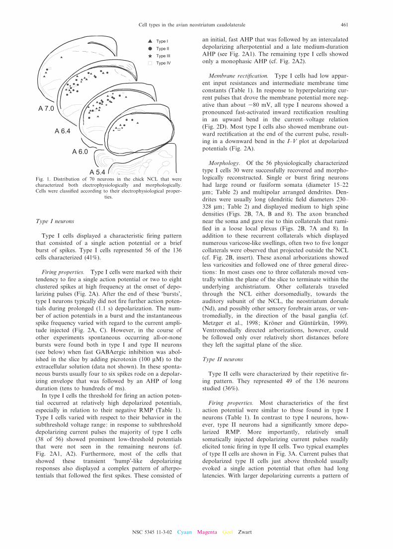
Stable whole-cell recordings were obtained from 136cells in the NCL. Neurons were selected only if theyexhibited a RMP more negative than 355 mV and anovershooting action potential. Neurons were classi¢edinto four distinct types according to qualitative di¡eren-ces in their intrinsic ¢ring properties and in the voltageresponses to hyperpolarizing current pulses. For 70 ofthe neurons thus characterized basic morphological fea-tures were analyzed after ¢lling with LY. The two maincell types (types I and II) could not be distinguished withregard to their morphological characteristics. The distri-bution of cell types did not vary throughout the NCL(Fig. 1).
NSC 5345 11-3-02
S. Kro«ner et al.460
Type I neurons
Type I cells displayed a characteristic ¢ring patternthat consisted of a single action potential or a briefburst of spikes. Type I cells represented 56 of the 136cells characterized (41%).
Firing properties. Type I cells were marked with theirtendency to ¢re a single action potential or two to eightclustered spikes at high frequency at the onset of depo-larizing pulses (Fig. 2A). After the end of these `bursts',type I neurons typically did not ¢re further action poten-tials during prolonged (1.1 s) depolarization. The num-ber of action potentials in a burst and the instantaneousspike frequency varied with regard to the current ampli-tude injected (Fig. 2A, C). However, in the course ofother experiments spontaneous occurring all-or-nonebursts were found both in type I and type II neurons(see below) when fast GABAergic inhibition was abol-ished in the slice by adding picrotoxin (100 WM) to theextracellular solution (data not shown). In these sponta-neous bursts usually four to six spikes rode on a depolar-izing envelope that was followed by an AHP of longduration (tens to hundreds of ms).
In type I cells the threshold for ¢ring an action poten-tial occurred at relatively high depolarized potentials,especially in relation to their negative RMP (Table 1).Type I cells varied with respect to their behavior in thesubthreshold voltage range: in response to subthresholddepolarizing current pulses the majority of type I cells(38 of 56) showed prominent low-threshold potentialsthat were not seen in the remaining neurons (cf.Fig. 2A1, A2). Furthermore, most of the cells thatshowed these transient `hump'-like depolarizingresponses also displayed a complex pattern of afterpo-tentials that followed the ¢rst spikes. These consisted of
an initial, fast AHP that was followed by an intercalateddepolarizing afterpotential and a late medium-durationAHP (see Fig. 2A1). The remaining type I cells showedonly a monophasic AHP (cf. Fig. 2A2).
Membrane recti¢cation. Type I cells had low appar-ent input resistances and intermediate membrane timeconstants (Table 1). In response to hyperpolarizing cur-rent pulses that drove the membrane potential more neg-ative than about 380 mV, all type I neurons showed apronounced fast-activated inward recti¢cation resultingin an upward bend in the current^voltage relation(Fig. 2D). Most type I cells also showed membrane out-ward recti¢cation at the end of the current pulse, result-ing in a downward bend in the I^V plot at depolarizedpotentials (Fig. 2A).
Morphology. Of the 56 physiologically characterizedtype I cells 30 were successfully recovered and morpho-logically reconstructed. Single or burst ¢ring neuronshad large round or fusiform somata (diameter 15^22Wm; Table 2) and multipolar arranged dendrites. Den-drites were usually long (dendritic ¢eld diameters 230^328 Wm; Table 2) and displayed medium to high spinedensities (Figs. 2B, 7A, B and 8). The axon branchednear the soma and gave rise to thin collaterals that rami-¢ed in a loose local plexus (Figs. 2B, 7A and 8). Inaddition to these recurrent collaterals which displayednumerous varicose-like swellings, often two to ¢ve longercollaterals were observed that projected outside the NCL(cf. Fig. 2B, insert). These axonal arborizations showedless varicosities and followed one of three general direc-tions: In most cases one to three collaterals moved ven-trally within the plane of the slice to terminate within theunderlying archistriatum. Other collaterals traveledthrough the NCL either dorsomedially, towards theauditory subunit of the NCL, the neostriatum dorsale(Nd), and possibly other sensory forebrain areas, or ven-tromedially, in the direction of the basal ganglia (cf.Metzger et al., 1998; Kro«ner and Gu«ntu«rku«n, 1999).Ventromedially directed arborizations, however, couldbe followed only over relatively short distances beforethey left the sagittal plane of the slice.
Type II neurons
Type II cells were characterized by their repetitive ¢r-ing pattern. They represented 49 of the 136 neuronsstudied (36%).
Firing properties. Most characteristics of the ¢rstaction potential were similar to those found in type Ineurons (Table 1). In contrast to type I neurons, how-ever, type II neurons had a signi¢cantly xmore depo-larized RMP. More importantly, relatively smallsomatically injected depolarizing current pulses readilyelicited tonic ¢ring in type II cells. Two typical examplesof type II cells are shown in Fig. 3A. Current pulses thatdepolarized type II cells just above threshold usuallyevoked a single action potential that often had longlatencies. With larger depolarizing currents a pattern of
Fig. 1. Distribution of 70 neurons in the chick NCL that werecharacterized both electrophysiologically and morphologically.Cells were classi¢ed according to their electrophysiological proper-
ties.
NSC 5345 11-3-02 Cyaan Magenta Geel Zwart
Cell types in the avian neostriatum caudolaterale 461
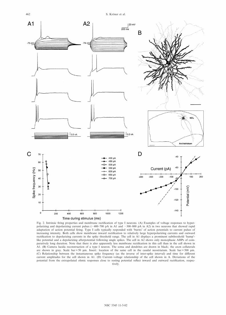
Fig. 2. Intrinsic ¢ring properties and membrane recti¢cation of type I neurons. (A) Examples of voltage responses to hyper-polarizing and depolarizing current pulses (3400^700 pA in A1 and 3300^800 pA in A2) in two neurons that showed rapidadaptation of action potential ¢ring. Type I cells typically responded with `bursts' of action potentials to current pulses ofincreasing intensity. Both cells show membrane inward recti¢cation to relatively large hyperpolarizing currents and outwardrecti¢cation to depolarizing currents in the spike threshold range. The cell in A1 displays a prominent subthreshold `hump'-like potential and a depolarizing afterpotential following single spikes. The cell in A2 shows only monophasic AHPs of com-paratively long duration. Note that there is also apparently less membrane recti¢cation in this cell than in the cell shown inA1. (B) Camera lucida reconstruction of a type I neuron. The soma and dendrites are drawn in black; the axon collateralsare shown in gray. Scale bar = 50 Wm. Insert : location of the same cell in the caudal neostriatum. Scale bar = 500 Wm.(C) Relationship between the instantaneous spike frequency (as the inverse of inter-spike interval) and time for di¡erentcurrent amplitudes for the cell shown in A1. (D) Current^voltage relationship of the cell shown in A. Deviations of thepotential from the extrapolated ohmic responses close to resting potential re£ect inward and outward recti¢cation, respec-
tively.
NSC 5345 11-3-02
S. Kro«ner et al.462
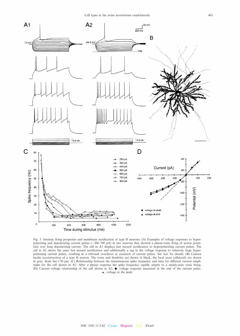
Fig. 3. Intrinsic ¢ring properties and membrane recti¢cation of type II neurons. (A) Examples of voltage responses to hyper-polarizing and depolarizing current pulses (3300^700 pA) in two neurons that showed a phasic-tonic ¢ring of action poten-tials over long depolarizing current. The cell in A1 displays fast inward recti¢cation to hyperpolarizing current pulses. Thecell in A2 shows the same fast inward recti¢cation and additionally a sag in the voltage response to relatively large hyper-polarizing current pulses, resulting in a rebound overshoot at cessation of current pulses. See text for details. (B) Cameralucida reconstruction of a type II neuron. The soma and dendrites are drawn in black, the local axon collaterals are shownin gray. Scale bar = 50 Wm. (C) Relationship between the instantaneous spike frequency and time for di¡erent current ampli-tudes for the cell shown in A2. After a phasic response the spike frequency rapidly adapts to a steady-state tonic ¢ring.(D) Current^voltage relationship of the cell shown in A2. b, voltage response measured at the end of the current pulse;
R, voltage at the peak.
NSC 5345 11-3-02 Cyaan Magenta Geel Zwart
Cell types in the avian neostriatum caudolaterale 463
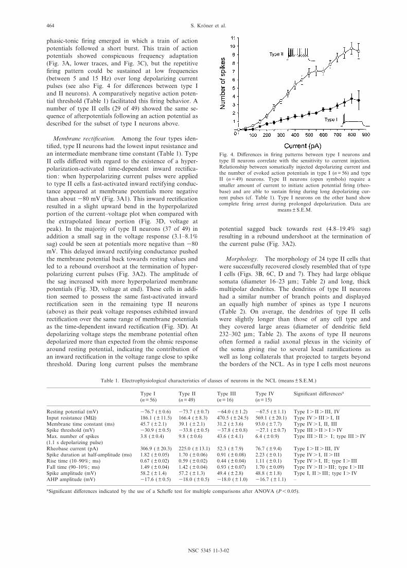
phasic-tonic ¢ring emerged in which a train of actionpotentials followed a short burst. This train of actionpotentials showed conspicuous frequency adaptation(Fig. 3A, lower traces, and Fig. 3C), but the repetitive¢ring pattern could be sustained at low frequencies(between 5 and 15 Hz) over long depolarizing currentpulses (see also Fig. 4 for di¡erences between type Iand II neurons). A comparatively negative action poten-tial threshold (Table 1) facilitated this ¢ring behavior. Anumber of type II cells (29 of 49) showed the same se-quence of afterpotentials following an action potential asdescribed for the subset of type I neurons above.
Membrane recti¢cation. Among the four types iden-ti¢ed, type II neurons had the lowest input resistance andan intermediate membrane time constant (Table 1). TypeII cells di¡ered with regard to the existence of a hyper-polarization-activated time-dependent inward recti¢ca-tion: when hyperpolarizing current pulses were appliedto type II cells a fast-activated inward rectifying conduc-tance appeared at membrane potentials more negativethan about 380 mV (Fig. 3A1). This inward recti¢cationresulted in a slight upward bend in the hyperpolarizedportion of the current^voltage plot when compared withthe extrapolated linear portion (Fig. 3D, voltage atpeak). In the majority of type II neurons (37 of 49) inaddition a small sag in the voltage response (3.1^8.1%sag) could be seen at potentials more negative than 380mV. This delayed inward rectifying conductance pushedthe membrane potential back towards resting values andled to a rebound overshoot at the termination of hyper-polarizing current pulses (Fig. 3A2). The amplitude ofthe sag increased with more hyperpolarized membranepotentials (Fig. 3D, voltage at end). These cells in addi-tion seemed to possess the same fast-activated inwardrecti¢cation seen in the remaining type II neurons(above) as their peak voltage responses exhibited inwardrecti¢cation over the same range of membrane potentialsas the time-dependent inward recti¢cation (Fig. 3D). Atdepolarizing voltage steps the membrane potential oftendepolarized more than expected from the ohmic responsearound resting potential, indicating the contribution ofan inward recti¢cation in the voltage range close to spikethreshold. During long current pulses the membrane
potential sagged back towards rest (4.8^19.4% sag)resulting in a rebound undershoot at the termination ofthe current pulse (Fig. 3A2).
Morphology. The morphology of 24 type II cells thatwere successfully recovered closely resembled that of typeI cells (Figs. 3B, 6C, D and 7). They had large obliquesomata (diameter 16^23 Wm; Table 2) and long, thickmultipolar dendrites. The dendrites of type II neuronshad a similar number of branch points and displayedan equally high number of spines as type I neurons(Table 2). On average, the dendrites of type II cellswere slightly longer than those of any cell type andthey covered large areas (diameter of dendritic ¢eld232^302 Wm; Table 2). The axons of type II neuronsoften formed a radial axonal plexus in the vicinity ofthe soma giving rise to several local rami¢cations aswell as long collaterals that projected to targets beyondthe borders of the NCL. As in type I cells most neurons
Fig. 4. Di¡erences in ¢ring patterns between type I neurons andtype II neurons correlate with the sensitivity to current injection.Relationship between somatically injected depolarizing current andthe number of evoked action potentials in type I (n = 56) and typeII (n = 49) neurons. Type II neurons (open symbols) require asmaller amount of current to initiate action potential ¢ring (rheo-base) and are able to sustain ¢ring during long depolarizing cur-rent pulses (cf. Table 1). Type I neurons on the other hand showcomplete ¢ring arrest during prolonged depolarization. Data are
means þ S.E.M.
Table 1. Electrophysiological characteristics of classes of neurons in the NCL (means þ S.E.M.)
Type I(n = 56)
Type II(n = 49)
Type III(n = 16)
Type IV(n = 15)
Signi¢cant di¡erencesa
Resting potential (mV) 376.7 ( þ 0.6) 373.7 ( þ 0.7) 364.0 ( þ 1.2) 367.5 ( þ 1.1) Type Is IIs III, IVInput resistance (M6) 186.1 ( þ 11.5) 166.4 ( þ 8.3) 470.5 ( þ 24.5) 569.1 ( þ 20.1) Type IVs IIIs I, IIMembrane time constant (ms) 45.7 ( þ 2.1) 39.1 ( þ 2.1) 31.2 ( þ 3.6) 93.0 ( þ 7.7) Type IVs I, II, IIISpike threshold (mV) 330.9 ( þ 0.5) 333.8 ( þ 0.5) 337.8 ( þ 0.8) 327.1 ( þ 0.7) Type IIIs IIs Is IVMax. number of spikes(1.1 s depolarizing pulse)
3.8 ( þ 0.4) 9.8 ( þ 0.6) 43.6 ( þ 4.1) 6.4 ( þ 0.9) Type IIIs IIs I; type IIIs IV
Rheobase current (pA) 306.9 ( þ 20.3) 225.0 ( þ 13.1) 52.3 ( þ 7.9) 76.7 ( þ 9.4) Type Is IIs III, IVSpike duration at half-amplitude (ms) 1.82 ( þ 0.05) 1.70 ( þ 0.06) 0.91 ( þ 0.08) 2.23 ( þ 0.1) Type IVs I, IIs IIIRise time (10^90% ; ms) 0.67 ( þ 0.02) 0.59 ( þ 0.02) 0.44 ( þ 0.04) 1.11 ( þ 0.1) Type IVs I, II ; type Is IIIFall time (90^10% ; ms) 1.49 ( þ 0.04) 1.42 ( þ 0.04) 0.93 ( þ 0.07) 1.70 ( þ 0.09) Type IVs IIs III; type Is IIISpike amplitude (mV) 58.2 ( þ 1.4) 57.2 ( þ 1.3) 49.4 ( þ 2.8) 48.8 ( þ 1.8) Type I, IIs III; type Is IVAHP amplitude (mV) 317.6 ( þ 0.5) 318.0 ( þ 0.5) 318.0 ( þ 1.0) 316.7 ( þ 1.1) ^
aSigni¢cant di¡erences indicated by the use of a Sche¡e test for multiple comparisons after ANOVA (P6 0.05).
NSC 5345 11-3-02
S. Kro«ner et al.464

sent one to three descending collaterals towards thearchistriatum but also had additional arborizations thatseemed to travel ventromedially and dorsomedially (cf.Fig. 7, insert), probably making numerous contacts withother cells within NCL on their way.
Type III neurons
The key features of type III neurons were the ability to¢re action potentials at a high frequency and a promi-nent sag in the response to hyperpolarizing current
Fig. 5. Intrinsic ¢ring properties and membrane recti¢cation of a type III cell. (A) Voltage responses to hyperpolarizing anddepolarizing current pulses (3120^240 pA). There is a large sag in response to strong hyperpolarizing current pulses, whichresults in a rebound overshoot that is large enough to trigger spikes. Small depolarizing currents elicit continuous ¢ring atcomparatively high frequencies. (B) Camera lucida reconstruction of a type III neuron. Scale bar = 50 Wm. (C) Relationshipbetween the instantaneous spike frequency and time for di¡erent current amplitudes for the cell shown in A. There is somefrequency adaptation after the ¢rst few spikes, but the cell maintains a high-frequency ¢ring without further attenuation overa long current pulse. (D) Current^voltage relationship of the cell shown in A. b, voltage response measured at the end of the
current pulse; R, voltage at the peak.
NSC 5345 11-3-02 Cyaan Magenta Geel Zwart
Cell types in the avian neostriatum caudolaterale 465
pulses, as well as a short action potential duration. TypeIII cells represented 16 of the 136 cells (12%) character-ized.
Firing properties. In response to small suprathresholddepolarizing current pulses all neurons designated astype III responded with regular tonic ¢ring that showedonly relatively little frequency adaptation (Fig. 5). Thus,type III cells were able to initially ¢re action potentials atfrequencies of about 100 Hz. After several spikes the
¢ring rate usually accommodated to some degree, butsustained ¢ring over long depolarizing current pulsescontinued at frequencies much higher than found inany other cell class (Fig. 5C). These cells also showedthe most positive RMPs, while generally having themost negative threshold for the initiation of an actionpotential (Table 1). The duration of action potentialsin type III cells was by far the shortest among all classes,which was also re£ected by short rise and fall times(Table 1). The action potential was followed by an
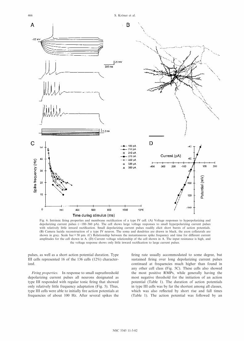
Fig. 6. Intrinsic ¢ring properties and membrane recti¢cation of a type IV cell. (A) Voltage responses to hyperpolarizing anddepolarizing current pulses (3180^360 pA). The cell shows large voltage responses to small hyperpolarizing current pulseswith relatively little inward recti¢cation. Small depolarizing current pulses readily elicit short bursts of action potentials.(B) Camera lucida reconstruction of a type IV neuron. The soma and dendrites are drawn in black, the axon collaterals areshown in gray. Scale bar = 50 Wm. (C) Relationship between the instantaneous spike frequency and time for di¡erent currentamplitudes for the cell shown in A. (D) Current^voltage relationship of the cell shown in A. The input resistance is high, and
the voltage response shows only little inward recti¢cation to large current pulses.
NSC 5345 11-3-02
S. Kro«ner et al.466
AHP that was large in amplitude compared to the rela-tively small amplitude of the spike (Table 1).
Membrane recti¢cation. Type III cells had compara-tively high input resistances but short membrane timeconstants (Table 1). In response to hyperpolarizing cur-
rent pulses (membrane potential more negative than 380mV) a prominent sag in the voltage response (9^21.5%sag) occurred that was followed by a rebound depolari-zation at cessation of the current pulse. The time-depen-dent inward recti¢cation and the rebound overshootincreased as the hyperpolarizing current pulses increased.
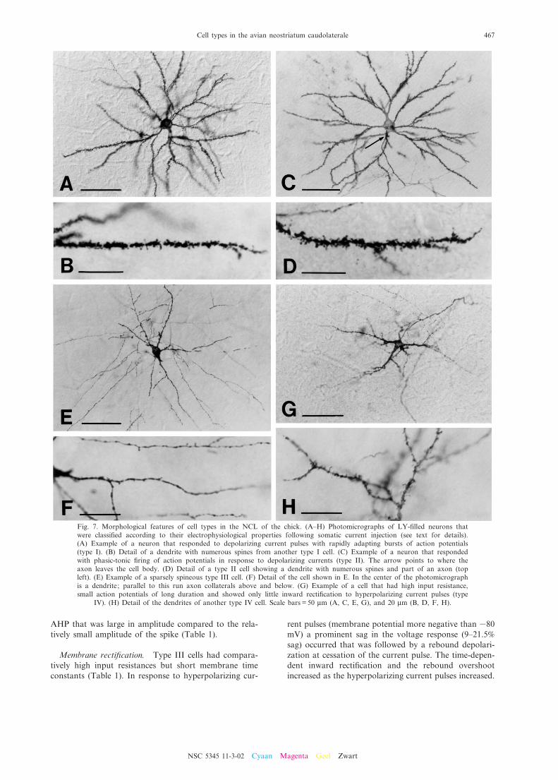
Fig. 7. Morphological features of cell types in the NCL of the chick. (A^H) Photomicrographs of LY-¢lled neurons thatwere classi¢ed according to their electrophysiological properties following somatic current injection (see text for details).(A) Example of a neuron that responded to depolarizing current pulses with rapidly adapting bursts of action potentials(type I). (B) Detail of a dendrite with numerous spines from another type I cell. (C) Example of a neuron that respondedwith phasic-tonic ¢ring of action potentials in response to depolarizing currents (type II). The arrow points to where theaxon leaves the cell body. (D) Detail of a type II cell showing a dendrite with numerous spines and part of an axon (topleft). (E) Example of a sparsely spineous type III cell. (F) Detail of the cell shown in E. In the center of the photomicrographis a dendrite; parallel to this run axon collaterals above and below. (G) Example of a cell that had high input resistance,small action potentials of long duration and showed only little inward recti¢cation to hyperpolarizing current pulses (type
IV). (H) Detail of the dendrites of another type IV cell. Scale bars = 50 Wm (A, C, E, G), and 20 Wm (B, D, F, H).
NSC 5345 11-3-02 Cyaan Magenta Geel Zwart
Cell types in the avian neostriatum caudolaterale 467
In response to larger negative current pulses the rebounddepolarization could initiate action potentials (Fig. 5A).Type III neurons also appear to possess an additionalfast-activated inward recti¢er, as the peak voltageresponses exhibited inward recti¢cation over the samerange of membrane potentials as the time-dependentinward recti¢cation (Fig. 5D).
Morphology. Type III cells (n = 7) were characterizedby small fusiform somata (diameter 10^14 Wm; Table 2)and thin, aspiny or sparsely spiny dendrites (Figs. 5B,7E, F and 8). In fact, in three out of seven neuronsthat were successfully reconstructed the dendrites wereso thin that they could not be clearly distinguishedfrom the extensive local axonal rami¢cations. Theremaining three cells had relatively few long multipolardendrites (diameter of dendritic ¢eld 227^267 Wm). Thedendrites showed a low number of total branch pointsand especially few higher order (v fourth branches)branchings (Table 2). The axon appeared to have alarge number of varicosities, and arborized extensivelyto form a dense plexus of terminals in the vicinity ofthe soma (Figs. 7E and 8). Several long protrudingaxon collaterals formed symmetrical round or oval axo-nal ¢elds with diameters of about 545^736 Wm. Axons oftype III neurons could never be followed beyond theborders of the NCL.
Type IV neurons
The key features of type IV cells are a high input re-sistance and long membrane time constant, as well aslong action potential duration. Of the 136 cells charac-terized, 15 (11%) were found to belong to this class.
Firing properties. Among the classes identi¢ed heretype IV cells had an intermediate RMP and a highspike threshold. Their bursting spike pattern resembled
that of type I cells (Fig. 6), but type IV neurons wereable to initially ¢re a larger number of action potentials(Table 1). However, in response to large depolarizingcurrents the ability of type IV neurons to ¢re repetitivelywas markedly attenuated (Fig. 6C). A characteristic ofcells in this class was the long action potential duration,which was also re£ected in the signi¢cantly longest riseand fall times (Table 1). Furthermore, the action poten-tials of type IV cells showed a prominent progressivespike broadening during repetitive ¢ring. Type IV cellshad comparatively small monophasic AHPs (Table 1).
Membrane recti¢cation. The key feature required fortype IV cells was a long membrane time constant (s 65ms). They also had the highest input resistance (s 400M6) among all classes of cells (Table 1) and showedsteep current^voltage relationships (Fig. 6C). Only atmembrane potentials more negative than 395 mV inmost neurons a weak, fast-activated inward recti¢cationbecame evident (Fig. 6D). All type IV cells showed aprominent membrane outward recti¢cation at depolar-ized potentials but no sag.
Morphology. Type IV cells (n = 9) were characterizedby small, oblique somata (diameter 12^16.5 Wm; Table 2)and multipolar dendrites that possessed few thin spines.Usually, proximal dendrites were short and thin, butsometimes two or three main dendrites were seen thathad thick stems but tapered considerably with distancefrom the soma (Figs. 6B and 7G). In these instances theusual spherical form of the dendritic ¢eld (diameter 180^225 Wm) was tilted by these dendrites. The dendritic treeof type IV neurons showed relatively few branches andcovered the smallest area among the four cell types(Table 2). Axonal arborizations of type IV cells werenot as extensive as in type I or type II cells, but projec-tions followed the same pattern seen in the other classesof spiny neurons. One to three main collaterals
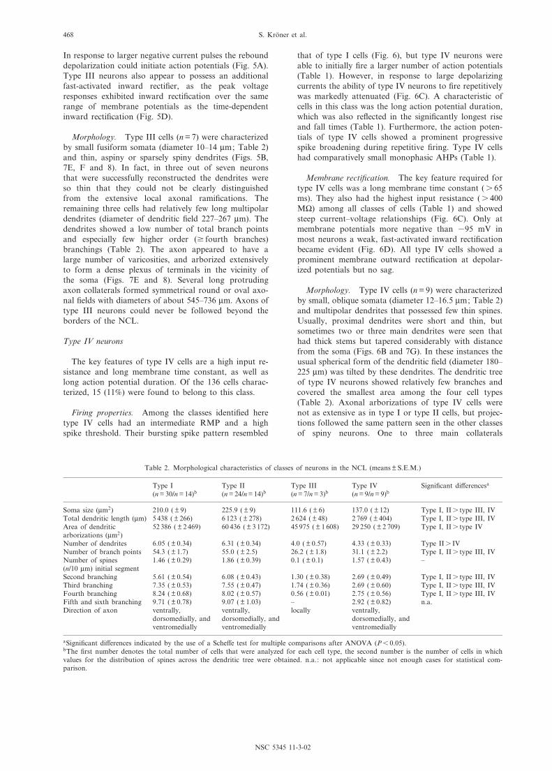
Table 2. Morphological characteristics of classes of neurons in the NCL (means þ S.E.M.)
Type I(n = 30/n = 14)b
Type II(n = 24/n = 14)b
Type III(n = 7/n = 3)b
Type IV(n = 9/n = 9)b
Signi¢cant di¡erencesa
Soma size (Wm2) 210.0 ( þ 9) 225.9 ( þ 9) 111.6 ( þ 6) 137.0 ( þ 12) Type I, IIs type III, IVTotal dendritic length (Wm) 5 438 ( þ 266) 6 123 ( þ 278) 2 624 ( þ 48) 2 769 ( þ 404) Type I, IIs type III, IVArea of dendriticarborizations (Wm2)
52 386 ( þ 2 469) 60 436 ( þ 3 172) 45 975 ( þ 1 608) 29 250 ( þ 2 709) Type I, IIs type IV
Number of dendrites 6.05 ( þ 0.34) 6.31 ( þ 0.34) 4.0 ( þ 0.57) 4.33 ( þ 0.33) Type IIs IVNumber of branch points 54.3 ( þ 1.7) 55.0 ( þ 2.5) 26.2 ( þ 1.8) 31.1 ( þ 2.2) Type I, IIs type III, IVNumber of spines(n/10 Wm) initial segment
1.46 ( þ 0.29) 1.86 ( þ 0.39) 0.1 ( þ 0.1) 1.57 ( þ 0.43) ^
Second branching 5.61 ( þ 0.54) 6.08 ( þ 0.43) 1.30 ( þ 0.38) 2.69 ( þ 0.49) Type I, IIs type III, IVThird branching 7.35 ( þ 0.53) 7.55 ( þ 0.47) 1.74 ( þ 0.36) 2.69 ( þ 0.60) Type I, IIs type III, IVFourth branching 8.24 ( þ 0.68) 8.02 ( þ 0.57) 0.56 ( þ 0.01) 2.75 ( þ 0.56) Type I, IIs type III, IVFifth and sixth branching 9.71 ( þ 0.78) 9.07 ( þ 1.03) ^ 2.92 ( þ 0.82) n.a.Direction of axon ventrally,
dorsomedially, andventromedially
ventrally,dorsomedially, andventromedially
locally ventrally,dorsomedially, andventromedially
aSigni¢cant di¡erences indicated by the use of a Sche¡e test for multiple comparisons after ANOVA (P6 0.05).bThe ¢rst number denotes the total number of cells that were analyzed for each cell type, the second number is the number of cells in whichvalues for the distribution of spines across the dendritic tree were obtained. n.a. : not applicable since not enough cases for statistical com-parison.
NSC 5345 11-3-02
S. Kro«ner et al.468
descended ventrally in the direction of the archistriatum,while other collaterals traveled dorsomedially and/orventromedially within the NCL.
DISCUSSION
We have shown the existence of at least four cell typeswithin the NCL of chicks based on their intrinsic electro-physiological and morphological properties. In general,
there was a good correspondence of physiological andmorphological criteria, although the two main types ofprojection neurons, types I and II, could not be di¡er-entiated by morphological criteria.
Cell types
Type I. Type I neurons possess a depolarized actionpotential threshold despite a relatively negative restingpotential, and a prominent outward recti¢cation in
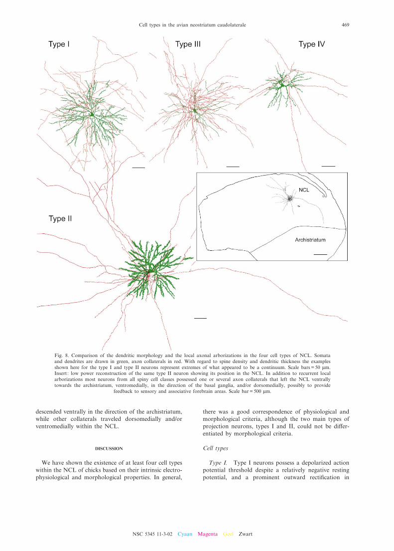
Fig. 8. Comparison of the dendritic morphology and the local axonal arborizations in the four cell types of NCL. Somataand dendrites are drawn in green, axon collaterals in red. With regard to spine density and dendritic thickness the examplesshown here for the type I and type II neurons represent extremes of what appeared to be a continuum. Scale bars = 50 Wm.Insert : low power reconstruction of the same type II neuron showing its position in the NCL. In addition to recurrent localarborizations most neurons from all spiny cell classes possessed one or several axon collaterals that left the NCL ventrallytowards the archistriatum, ventromedially, in the direction of the basal ganglia, and/or dorsomedially, possibly to provide
feedback to sensory and associative forebrain areas. Scale bar = 500 Wm.
NSC 5345 11-3-02 Cyaan Magenta Geel Zwart
Cell types in the avian neostriatum caudolaterale 469
response to depolarizing current pulses. These propertiesindicate that strong, temporally or spatially integratedexcitatory inputs are necessary for type I neurons to¢re. If su¤ciently depolarized, cells in this class prefer-entially responded with bursts of action potentials. Func-tionally, a neuron's ability to ¢re bursts of actionpotentials will probably enhance its integrative rolewithin a network and aide the plasticity of synaptic con-nections: burst ¢ring may increase the probability ofdriving the post-synaptic cell beyond spike threshold(Snider et al., 1998). The high-frequency ¢ring of actionpotentials during a burst is thus thought to amplify aneural signal and to synchronize the activity in a popu-lation of post-synaptic cells, both temporarily and spa-tially (Snider et al., 1998; Williams and Stuart, 1999). Ina set of type I and type II neurons single action poten-tials or bursts were followed by three distinct afterpoten-tials, consisting of an initial fast, and a late slowafterhyperpolarization, separated by an intercalateddepolarizing afterpotential. Depolarizing afterpotentialsappear to be related to a neuron's ability to ¢re burstsof action potentials (Chagnac-Amitai et al., 1990; Mageeand Carruth, 1999). In mammals, the expression of theafterpotential and the emergence of a tripartite AHPhave been shown to depend on changes during post-natal development, and similarly, burst ¢ring does notdevelop before the third post-natal week (Kasper et al.,1994). In contrast, chicks are already well developed athatch; accordingly, we observed evoked bursts in record-ings from very young animals (2 days post-hatch, datanot shown).
Type II. An initial tonic ¢ring and a relatively hyper-polarized action potential threshold may indicate thatthe ¢ring of type II cells is readily elicited by weak exci-tatory inputs. The phasic-tonic ¢ring pattern elicited withlarge depolarizing currents indicates that type II neuronsrespond strongly but transiently to a brief input, yetproduce a sustained response to a prolonged input; apattern that favors the augmentation of synaptic connec-tions (Thomson, 2000). Functionally, the ability to gen-erate a tonic ¢ring mode could also enable type II cells toretain information of their input for a short time period.Pharmacological blockade or lesions of the NCL inpigeons cause speci¢c impairments in a variety of delaytasks (Mogensen and Divac, 1982, 1993; Gagliardo etal., 1996, 1997; Gu«ntu«rku«n, 1997; Diekamp et al.,2000; Gu«ntu«rku«n and Durstewitz, 2000). In these experi-ments, the animal has to hold on-line speci¢c informa-tion provided during a previous cue period to perform acorrect response after the end of the delay. In mammals,neurons in the prefrontal cortex show enhancement intheir ¢ring rate during the delay (e.g. Funahashi et al.,1989). The sustained delay activity of these neurons mayprovide the animal with the ability to hold an internalrepresentation of relevant aspects of the external worldthat are needed for the organization of subsequentresponses (Goldman-Rakic, 1996). Neurons with similardelay activities to those recorded from rat or primateprefrontal cortex have been observed in the pigeon'sNCL during a delayed Go/No-Go task (Kalt et al.,
1999). The ability of type II neurons to sustain ¢ringover long periods of time makes them likely candidatesto participate in the maintenance of sustained delayactivity within the NCL.
The majority of type II neurons furthermore showed asmall transient sag in the voltage response to hyperpolar-izing currents, which was never seen in type I or IVneurons. This inward recti¢cation is likely to be mediatedby a voltage- and time-dependent hyperpolarization-acti-vated mixed cationic current called Ih (Pape, 1996; seealso discussion of type III below). It has been shown thatneurons with a time-dependent-inward recti¢er ¢reaction potentials preferentially to rhythmic (oscillatory)inputs (Hutcheon et al., 1996). Recently, neurons havebeen described in the dorsal forebrain of zebra ¢nches,which participate in the perception of song pattern, andwhich share a number of ¢ring properties and morpho-logical features with the type II neurons in the presentstudy (Dutar et al., 1998; Kubota and Taniguchi, 1998;Mooney, 2000). Thus, type II neurons may representmembers of a common class of spiny projection neuronsin the dorsal forebrain of birds that can respond prefer-entially to input of a speci¢c pattern.
Type III. The electrophysiological and morphologi-cal characteristics of the type III cells resemble those ofGABA-containing inhibitory interneurons found in themammalian telencephalon (McCormick et al., 1985;Kawaguchi, 1995; Gupta et al., 2000), i.e. little or noaccommodation of spike frequency, and beaded aspinydendrites. Type III cells are capable of sustained ¢ring ofaction potentials at a wide range of frequencies. They arethus able to perform a very reliable input^output con-version, retaining the temporal pattern of their synapticinputs. These ¢ring characteristics are facilitated by theshort rise and fall times of the action potential (Table 1).In addition, the relatively pronounced AHPs of type IIIcells may also enable sustained high-frequency ¢ring ofaction potentials in that they reduce the accumulation ofdepolarization-dependent Na� channel inactivation(Hamill et al., 1991; Erisir et al., 1999). Large AHPswill also reduce the in£ux of Ca2� into the cell, thusdiminishing the e¡ects of Ca2�-activated K� channels,which otherwise might lead to prolonged hyperpolariza-tion and spike frequency adaptation (Storm, 1990).Another prominent feature in type III cells is the exis-tence of the strong inward rectifying current Ih that ini-tiates slow depolarization if the membrane potential hasbecome negative. In central neurons of mammals Ih hasbeen implicated in the determination of the restingpotential and the generation of `pacemaker' potentials.It also serves to decrease the propagation of subthresh-old voltage potentials in dendritic trees, thereby regu-lating the integration of synaptic inputs (Magee, 1998,1999). In mammalian cortex GABAergic interneuronshave long been recognized for controlling the spread ofactivity (Chagnac-Amitai and Connors, 1989), and thesynchronization of adjacent projection neurons via inhib-itory phasing (Cobb et al., 1995; Benardo, 1997), whichmight impose a rhythm on the activity of the principalneurons (Buzsaki and Chrobak, 1995). With respect to
NSC 5345 11-3-02
S. Kro«ner et al.470
the extensive local axonal arborizations of type III neu-rons these putative interneurons of the NCL appear tobe able to control a large number of adjacent spiny `prin-cipal' neurons. In summary, given their intrinsic electro-physiological and morphological properties type IIIneurons of the NCL are equipped to play a similarlyimportant role for signal integration as their mammaliancounterparts.
Type IV. Type IV neurons are characterized by highinput resistances, long time constants and a long dura-tion of action potentials. A high input resistance andlong membrane time constant could increase the magni-tude and duration of a cell's response to imposed synap-tic currents. These electrophysiological properties mostlikely result from the morphology of type IV neurons,which possesses small somata, and short, sparsely spine-ous dendrites with few branchings. This morphologyshould render type IV neurons electrotonically compact,and reduce cable attenuation of distal dendritic synapticinputs. This could compensate for the comparativelysmall number of synaptic inputs that these neuronsreceive. Similarly, the long duration of action potentialsseen in type IV neurons may serve to facilitate their out-put: activity-dependent spike broadening due to a reduc-tion in speci¢c K� currents has been correlated withincreased e¤cacy of synaptic transmission onto thepost-synaptic target cell (see Byrne and Kandel, 1996for review). Similarly, the signi¢cantly longer actionpotentials of developing neurons might result in anenhancement of synaptic transmission onto their post-synaptic cells. It should also be noted that a high inputresistance, long time constant and a long duration ofaction potentials are also characteristics of developingcortical neurons in mammals (McCormick and Prince,1987; Kasper et al., 1994) and possibly birds (Kubotaand Taniguchi, 1998). It is thus also conceivable thattype IV neurons represent a class of late maturing neu-rons.
Connectivity and sensorimotor integration
The pattern of axonal arborizations described for thefour cell types here may provide further insight into theanatomical arrangements that underlie (a) the integrationof various sensory modalities within the NCL, and (b)the control of motor and limbic areas via the NCL'sdescending projections, as they have been indicatedfrom the results of tracing studies in vivo (Metzger etal., 1998; Kro«ner and Gu«ntu«rku«n, 1999). Overlap inthe termination areas of sensory a¡erents indicates thatin large areas of the NCL single neurons might receive
multimodal input. In agreement with this, single-unitrecordings in vivo show that neurons in the NCL of thepigeon can encode aspects of a memory task acrossmodalities (Kalt et al., 1999). The present data indicatethat individual cells, or a few interconnected neurons,can provide for stimulus comparison across most sensorycompartments of the NCL (cf. Metzger et al., 1998;Kro«ner and Gu«ntu«rku«n, 1999), either through theirextensive local axonal arborizations or the long collater-als that span the whole mediolateral and/or dorsoventralextent of the NCL. It must be noted, however, that thedetermination of the rostrocaudal extent of these intra-NCL connections remains a critical topic, which wasconstrained by the thickness of the slices used here.
The long axon collaterals of NCL neurons of all typesshowed similar projection patterns, thus suggesting thatthey innervate common targets. The trajectories of thesee¡erents followed general patterns outlined in previoustracing studies in vivo (Metzger et al., 1998; Kro«ner andGu«ntu«rku«n, 1999). Individual projection neurons of theNCL thus appear to provide feedback to sensory areasand/or send parallel e¡erent copies to the limbic andmotor regions in the archistriatum and the basal ganglia,respectively. The latter ¢nding is also noteworthy withregard to comparisons with the organization of the cau-dal forebrain in other birds. Based on similarities in thepattern of connections and the location in the dorsocau-dal forebrain it has previously been suggested that theNd, the auditory subunit of the NCL, may be related tothe HVC (used as the proper name) of songbirds (Wild,1994; Metzger et al., 1998; Kro«ner and Gu«ntu«rku«n,1999). The HVC is involved in the production of songand gives rise to two pathways that terminate in a motornucleus of the archistriatum and the so called area X ofthe basal ganglia (Nottebohm et al., 1982; Fortune andMargoliash, 1995). In the HVC of zebra ¢nches theseprojections arise from two separate populations of neu-rons which resemble type I and type II neurons of thepresent study, respectively (Dutar et al., 1998; Kubotaand Taniguchi, 1998). However, as pointed out above,our anatomical ¢ndings make it unlikely that a similardistinction between cell types exists in the chick fore-brain. These di¡erences might re£ect the specializationof the HVC relevant for singing and the need to exertcontrol over speci¢c aspects of singing-related motorbehavior. In summary, our ¢ndings provide a frameworkfor further study into sensorimotor integration and asso-ciative learning in chicks and possibly other birds.
AcknowledgementsöThis work was supported by Grants of theDFG (GU 227/5-1 to O.G., and SFB 509 to K.G. and H.H.).
REFERENCES
Aldavert-Vera, L., Costa-Miserachs, D., Divac, I., Delius, J.D., 1999. Presumed `prefrontal cortex' lesions in pigeons: e¡ects on visual discrim-ination performance. Behav. Brain Res. 102, 165^170.
Benardo, L.S., 1997. Recruitment of GABAergic inhibition and synchronization of inhibitory interneurons in rat neocortex. J. Neurophysiol. 77,3134^3144.
Bock, J., Braun, K., 1999a. Blockade of N-methyl-D-aspartate receptor activation suppresses learning-induced synaptic elimination. Proc. Natl.Acad. Sci. USA 96, 2485^2490.
NSC 5345 11-3-02 Cyaan Magenta Geel Zwart
Cell types in the avian neostriatum caudolaterale 471
Bock, J., Braun, K., 1999b. Filial imprinting in domestic chicks is associated with spine pruning in the associative area, dorsocaudal neostriatum.Eur. J. Neurosci. 11, 2566^2570.
Bock, J., Schnabel, R., Braun, K., 1997. Role of the dorso-caudal neostriatum in ¢lial imprinting of the domestic chick: a pharmacological andautoradiographical approach focused on the involvement of NMDA-receptors. Eur. J. Neurosci. 9, 1262^1272.
Braun, K., Bock, J., Metzger, M., Jiang, S., Schnabel, R., 1999. The dorsocaudal neostriatum of the domestic chick: a structure serving higherassociative functions. Behav. Brain Res. 98, 211^218.
Buzsaki, G., Chrobak, J.J., 1995. Temporal structure in spatially organized neuronal ensembles: a role for interneuronal networks. Curr. Opin.Neurobiol. 5, 504^510.
Byrne, J.H., Kandel, E.R., 1996. Presynaptic facilitation revisited: state and time dependence. J. Neurosci. 16, 425^435.Chagnac-Amitai, Y., Connors, B.W., 1989. Horizontal spread of synchronized activity in neocortex and its control by GABA mediated inhibition.
J. Neurophysiol. 61, 747^758.Chagnac-Amitai, Y., Luhmann, H.J., Prince, D.A., 1990. Burst generating and regular spiking layer 5 pyramidal neurons of rat neocortex have
di¡erent morphological features. J. Comp. Neurol. 296, 598^613.Cobb, S.R., Buhl, E.H., Halasy, K., Paulsen, O., Somogyi, P., 1995. Synchronization of neuronal activity in hippocampus by individual
GABAergic interneurons. Nature 378, 75^78.Diekamp, B., Kalt, T., Ruhm, A., Koch, M., Gu«ntu«rku«n, O., 2000. Impairment in a discrimination reversal task after D1 receptor blockade in the
pigeon `prefrontal cortex'. Behav. Neurosci. 114, 1145^1155.Douglas, R.J., Martin, K.A.C., 1992. In search of the canonical microcircuits of neocortex. In: Lent, R. (Ed.), The Visual System from Genesis to
Maturity. Birkha«user, Boston, MA, pp. 213^232.Dutar, P., Vu, H.M., Perkel, D.J., 1998. Multiple cell types distinguished by physiological, pharmacological, and anatomic properties in nucleus
HVc of the adult zebra ¢nch. J. Neurophysiol. 80, 1828^1838.Erisir, A., Lau, D., Rudy, B., Leonard, C.S., 1999. Function of speci¢c K� channels in sustained high-frequency ¢ring of fast-spiking neocortical
interneurons. J. Neurophysiol. 82, 2476^2489.Fortune, E.S., Margoliash, D., 1995. Parallel pathways and convergence onto HVc and adjacent neostriatum of adult zebra ¢nches (Taeniopygia
guttata). J. Comp. Neurol. 360, 413^441.Foster, T.M., Temple, W., MacKenzie, C., Demello, L.R., Poling, A., 1995. Delayed matching-to-sample performance of hens. E¡ects of sample
duration and response requirements during the sample. J. Exp. Anal. Behav. 64, 19^31.Funahashi, S., Bruce, C.J., Goldman-Rakic, P.S., 1989. Mnemonic coding of visual space in the monkey's dorsolateral prefrontal cortex.
J. Neurophysiol. 61, 331^349.Gagliardo, A., Divac, I., 1993. E¡ects of ablation of the presumed equivalent of the mammalian prefrontal cortex on pigeon homing. Behav.
Neurosci. 107, 280^288.Gagliardo, A., Bonadonna, F., Divac, I., 1996. Behavioural e¡ects of ablations of the presumed `prefrontal cortex' or the corticoid in pigeons.
Behav. Brain Res. 78, 155^162.Gagliardo, A., Mazzotto, M., Divac, I., 1997. Memory of radial maze behavior in pigeons after ablations of the presumed equivalent of
mammalian prefrontal cortex. Behav. Neurosci. 111, 955^962.Goldman-Rakic, P.S., 1996. Regional and cellular fractionation of working memory. Proc. Natl. Acad. Sci. USA 93, 13473^13480.Gu«ntu«rku«n, O., 1997. Cognitive impairments after lesions of the neostriatum caudolaterale and its thalamic a¡erent in pigeons: Functional
equivalencies to the mammalian prefrontal system? J. Brain Res. 1, 133^144.Gu«ntu«rku«n, O., Durstewitz, D., 2000. Multimodal areas in the avian forebrain ^ blueprints for cognition? In: Roth, G., Wullimann, M. (Eds.),
Brain Evolution and Cognition. Spektrum Akademischer Verlag, pp. 431^450.Gupta, A., Wang, Y., Markram, H., 2000. Organizing principles for a diversity of GABAergic interneurons and synapses in the neocortex. Science
287, 273^278.Hamill, O.P., Huguenard, J.R., Prince, D.A., 1991. Patch-clamp studies of voltage-gated currents in identi¢ed neurons of the rat cerebral cortex.
Cereb. Cortex 1, 48^61.Hartmann, B., Gu«ntu«rku«n, O., 1998. Selective de¢cits in reversal learning after neostriatum caudolaterale lesions in pigeons ^ possible behavioral
equivalencies to the mammalian prefrontal system. Behav. Brain Res. 96, 125^133.Hutcheon, B., Miura, R.M., Puil, E., 1996. Subthreshold membrane resonance in neocortical neurons. J. Neurophysiol. 76, 683^697.Kalt, T., Diekamp, B., Gu«ntu«rku«n, O., 1999. Single unit activity during a Go/NoGo task in the `prefrontal cortex' of pigeons. Brain. Res. 839,
263^278.Kasper, E.M., Larkman, A.U., Lu«bke, J., Blakemore, C., 1994. Pyramidal neurons in layer 5 of the rat visual cortex. II. Development of
electrophysiological properties. J. Comp. Neurol. 339, 475^494.Kawaguchi, Y., 1995. Physiological subgroups of nonpyramidal cells with speci¢c morphological characteristics in layer II/III of rat frontal cortex.
J. Neurosci. 15, 2638^2655.Kro«ner, S., Gu«ntu«rku«n, O., 1999. A¡erent and e¡erent connections of the caudolateral neostriatum in the pigeon (Columba livia) : A retro- and
anterograde pathway tracing study. J. Comp. Neurol. 407, 228^260.Kubota, M., Taniguchi, I., 1998. Characteristics of classes of neuron in the HVc of the zebra ¢nch. J. Neurophysiol. 80, 914^923.Leutgeb, S., Husband, S., Riters, L.V., Shimizu, T., Bingman, V.P., 1996. Telencephalic a¡erents to the caudolateral neostriatum of the pigeon.
Brain Res. 730, 173^181.Magee, J.C., 1998. Dendritic hyperpolarization-activated currents modify the integrative properties of hippocampal CA1 pyramidal neurons.
J. Neurosci. 18, 7613^7762.Magee, J.C., 1999. Dendritic Ih normalizes temporal summation in hippocampal CA1 neurons. Nat. Neurosci. 2, 508^514.Magee, J.C., Carruth, M., 1999. Dendritic voltage-gated ion channels regulate the action potential ¢ring mode of hippocampal CA1 pyramidal
neurons. J. Neurophysiol. 82, 1895^1901.McCormick, D.A., Prince, D.A., 1987. Post-natal development of electrophysiological properties of rat cerebral cortical pyramidal neurones.
J. Physiol. 393, 743^762.McCormick, D.A., Connors, B.W., Lighthall, J.W., Prince, D.A., 1985. Comparative electrophysiology of pyramidal and sparsely spiny stellate
neurons of the neocortex. J. Neurophysiol. 54, 782^806.Metzger, M., Jiang, S., Braun, K., 1998. Organization of the dorsocaudal neostriatal complex: a retrograde and anterograde tracing study in the
domestic chick with special emphasis on pathways relevant to imprinting. J. Comp. Neurol. 395, 380^404.Mogensen, J., Divac, I., 1982. The prefrontal `cortex' in the pigeon: Behavioral evidence. Brain Behav. Evol. 21, 60^66.Mogensen, J., Divac, I., 1993. Behavioural e¡ects of ablation of the pigeon-equivalent of the mammalian prefrontal cortex. Behav. Brain Res. 55,
101^107.Mooney, R., 2000. Di¡erent subthreshold mechanisms underlie song selectivity in identi¢ed HVc neurons of the zebra ¢nch. J. Neurosci. 20, 5420^
5436.
NSC 5345 11-3-02
S. Kro«ner et al.472

Nottebohm, F., Kelley, D.B., Paton, J.A., 1982. Connections of vocal control nuclei in the canary telencephalon. J. Comp. Neurol. 207, 344^357.Pape, H.C., 1996. Queer current and pacemaker: the hyperpolarization-activated cation current in neurons. Annu. Rev. Physiol. 58, 299^327.Snider, R.K., Kabara, J.F., Roig, B.R., Bonds, A.B., 1998. Burst ¢ring and modulation of functional connectivity in cat striate cortex.
J. Neurophysiol. 80, 730^744.Storm, J.F., 1990. Potassium currents in hippocampal pyramidal cells. Prog. Brain Res. 83, 161^187.Thomson, A.M., 2000. Facilitation, augmentation and potentiation at central synapses. Trends Neurosci. 23, 305^312.Vallortigara, G., Regolin, L., Rigoni, M., Zanforlin, M., 1998. Delayed search for a concealed imprinted object in the domestic chick. Anim. Cog.
1, 17^24.Wild, J.M., 1994. Visual and somatosensory inputs to the avian song system via nucleus uvaeformis (Uva) and a comparison with the projections
of a similar thalamic nucleus in a nonsongbird (Columba livia). J. Comp. Neurol. 349, 512^535.Williams, S.R., Stuart, G.J., 1999. Mechanisms and consequences of action potential burst ¢ring in rat neocortical pyramidal neurons. J. Physiol.
521, 467^482.
(Accepted 27 September 2001)
NSC 5345 11-3-02 Cyaan Magenta Geel Zwart
Cell types in the avian neostriatum caudolaterale 473