Electrophysiological analysis of the perception of passive ... 12 - 2011.pdf ·...
6
Neuroscience Letters 501 (2011) 61–66 Contents lists available at ScienceDirect Neuroscience Letters jou rn al h om epage: www.elsevier.com/locate/neulet Electrophysiological analysis of the perception of passive movement Jose Inacio Salles a,e , Heloisa Alves a , Filipe Costa a , Victor Cunha-Cruz b , Mauricio Cagy b,d , Roberto Piedade b , Pedro Ribeiro b,c,∗ a National Institute of Traumatology and Orthopaedics (NITO), Rio de Janeiro, Brazil b Brain Mapping and Sensory Motor Integration, Institute of Psychiatry of the Federal University of Rio de Janeiro (IPUB/UFRJ), Brazil c School of Physical Education, Bioscience Department (EEFD/UFRJ), Brazil d Division of Epidemiology and Biostatistics, Institute of Community Health, Federal Fluminense University (UFF), Rio de Janeiro, Brazil e Brazilian Volleyball Confederation a r t i c l e i n f o Article history: Received 28 February 2011 Received in revised form 29 April 2011 Accepted 1 May 2011 Keywords: qEEG Proprioception Passive movement Perception a b s t r a c t The goal of the present study was to determine the electrophysiological correlate of the threshold of perception of passive motion (TPPM) in a group of healthy individuals. We expect a different pattern of activation over the frontoparietal network produced by the conscious perception of the passive move- ment. Ten right-handed male volunteers, between 20 and 30 years of age, were submitted to the threshold of perception of passive motion (TPPM) task in a proprioception testing device (PTD). The device was designed to passively move the arm in internal and external rotations about the shoulder joint. Partici- pants were instructed to press a hand-held switch every time movement of the shoulder was detected. Electromyographic (EMG) and electroencephalographic (EEG) activities were acquired during the task. Passive movement of the shoulder joint was followed by a clear and prolonged decrease in the signal mag- nitude of the electroencephalogram. The electrophysiological correlate of the TPPM was characterized by the establishment of a frontoparietal network, during the processing of somatosensory information. © 2011 Elsevier Ireland Ltd. All rights reserved. The somatosensory system processes multiple sensations from the body, including pain, pressure, temperature, and proprioception [23]. Proprioception is a specialized form of the sense that encom- passes the ability to detect movement (kinesthesia) [21]. Moreover, proprioception is the sensation of the relative position of neigh- bouring parts of the body. Contrarily, the exteroceptive senses, by which we perceive the outside environment, and interocep- tive senses, by which we perceive the pain and movement of internal organs, proprioception is a third distinct sensory modality that provides feedback solely on the status of the body internally. The assessment of kinesthesia has been traditionally conducted by measuring the threshold of perception of passive motion (TPPM) [7,15,21]. In this context, given that proprioceptive signals con- tribute to the formation of a conscious (i.e., having an awareness of one’s sensations) perception of joint position and motion [8,14,24], the TPPM quantifies one’s ability to consciously detect joint move- ment. Electrophysiology has also been employed in the study of pro- prioception. Specifically, electrophysiological studies have investi- gated the cortical representation of passive motion [1,2,30–32,36]. ∗ Corresponding author at: Brain Mapping and Sensory Motor Integration, Institute of Psychiatry of the Federal University of Rio de Janeiro (IPUB/UFRJ), Rua José Luiz Ferraz 200/1110, 22790-587, Brazil. E-mail address: [email protected] (P. Ribeiro). However, these studies aimed exclusively at contrasting patterns of brain activation between disabled individuals and healthy control subjects, without considering the perception of limb movement. In this sense, there are still gaps to be filled in our understanding of the patterns of neural activity related to somatosensory perception [9,29]. The general goal of the present study was to develop an experimental paradigm that would combine EEG activity and the TPPM, taking a psychophysical approach to the investiga- tion of proprioception. The specific aim was to determine the electrophysiological, event-related spectral perturbation (ERSP), correlate of the threshold of perception of passive motion (TPPM) in a group of healthy individuals, using an attention-demanding task, and examining the cortical representation (i.e., quantitative electroencephalogram—qEEG) of different stages of propriocep- tive information processing, from motion perception until motor response. The present study is relevant because propose a novel psychophysical approach to the study of kinesthetic perception by developing an experimental paradigm that integrates EEG acqui- sition and a proprioceptive task. Moreover, we expect that the conscious perception of the passive movement could generate a new pattern of the frontoparietal network’s activation. The sample consisted of 10 male volunteers, with ages ranging from 20 to 30 years (mean = 25; SD = 1.92). All participants were healthy, free of cognitive deficits, and were not using any medica- tion or psychoactive substance at the time of the test. Participants 0304-3940/$ – see front matter © 2011 Elsevier Ireland Ltd. All rights reserved. doi:10.1016/j.neulet.2011.05.005
Transcript of Electrophysiological analysis of the perception of passive ... 12 - 2011.pdf ·...

E
JRa
b
c
d
e
a
ARRA
KqPPP
Tb[ppbbtitTm[totm
pg
IR
0d
Neuroscience Letters 501 (2011) 61– 66
Contents lists available at ScienceDirect
Neuroscience Letters
jou rn al h om epage: www.elsev ier .com/ locate /neule t
lectrophysiological analysis of the perception of passive movement
ose Inacio Sallesa,e, Heloisa Alvesa, Filipe Costaa, Victor Cunha-Cruzb, Mauricio Cagyb,d,oberto Piedadeb, Pedro Ribeirob,c,∗
National Institute of Traumatology and Orthopaedics (NITO), Rio de Janeiro, BrazilBrain Mapping and Sensory Motor Integration, Institute of Psychiatry of the Federal University of Rio de Janeiro (IPUB/UFRJ), BrazilSchool of Physical Education, Bioscience Department (EEFD/UFRJ), BrazilDivision of Epidemiology and Biostatistics, Institute of Community Health, Federal Fluminense University (UFF), Rio de Janeiro, BrazilBrazilian Volleyball Confederation
r t i c l e i n f o
rticle history:eceived 28 February 2011eceived in revised form 29 April 2011ccepted 1 May 2011
eywords:
a b s t r a c t
The goal of the present study was to determine the electrophysiological correlate of the threshold ofperception of passive motion (TPPM) in a group of healthy individuals. We expect a different pattern ofactivation over the frontoparietal network produced by the conscious perception of the passive move-ment. Ten right-handed male volunteers, between 20 and 30 years of age, were submitted to the thresholdof perception of passive motion (TPPM) task in a proprioception testing device (PTD). The device was
EEGroprioceptionassive movementerception
designed to passively move the arm in internal and external rotations about the shoulder joint. Partici-pants were instructed to press a hand-held switch every time movement of the shoulder was detected.Electromyographic (EMG) and electroencephalographic (EEG) activities were acquired during the task.Passive movement of the shoulder joint was followed by a clear and prolonged decrease in the signal mag-nitude of the electroencephalogram. The electrophysiological correlate of the TPPM was characterized
front
by the establishment of ahe somatosensory system processes multiple sensations from theody, including pain, pressure, temperature, and proprioception23]. Proprioception is a specialized form of the sense that encom-asses the ability to detect movement (kinesthesia) [21]. Moreover,roprioception is the sensation of the relative position of neigh-ouring parts of the body. Contrarily, the exteroceptive senses,y which we perceive the outside environment, and interocep-ive senses, by which we perceive the pain and movement ofnternal organs, proprioception is a third distinct sensory modalityhat provides feedback solely on the status of the body internally.he assessment of kinesthesia has been traditionally conducted byeasuring the threshold of perception of passive motion (TPPM)
7,15,21]. In this context, given that proprioceptive signals con-ribute to the formation of a conscious (i.e., having an awareness ofne’s sensations) perception of joint position and motion [8,14,24],he TPPM quantifies one’s ability to consciously detect joint move-
ent.
Electrophysiology has also been employed in the study of pro-rioception. Specifically, electrophysiological studies have investi-ated the cortical representation of passive motion [1,2,30–32,36].
∗ Corresponding author at: Brain Mapping and Sensory Motor Integration,nstitute of Psychiatry of the Federal University of Rio de Janeiro (IPUB/UFRJ),ua José Luiz Ferraz 200/1110, 22790-587, Brazil.
E-mail address: [email protected] (P. Ribeiro).
304-3940/$ – see front matter © 2011 Elsevier Ireland Ltd. All rights reserved.oi:10.1016/j.neulet.2011.05.005
oparietal network, during the processing of somatosensory information.© 2011 Elsevier Ireland Ltd. All rights reserved.
However, these studies aimed exclusively at contrasting patterns ofbrain activation between disabled individuals and healthy controlsubjects, without considering the perception of limb movement. Inthis sense, there are still gaps to be filled in our understanding ofthe patterns of neural activity related to somatosensory perception[9,29].
The general goal of the present study was to develop anexperimental paradigm that would combine EEG activity andthe TPPM, taking a psychophysical approach to the investiga-tion of proprioception. The specific aim was to determine theelectrophysiological, event-related spectral perturbation (ERSP),correlate of the threshold of perception of passive motion (TPPM)in a group of healthy individuals, using an attention-demandingtask, and examining the cortical representation (i.e., quantitativeelectroencephalogram—qEEG) of different stages of propriocep-tive information processing, from motion perception until motorresponse. The present study is relevant because propose a novelpsychophysical approach to the study of kinesthetic perception bydeveloping an experimental paradigm that integrates EEG acqui-sition and a proprioceptive task. Moreover, we expect that theconscious perception of the passive movement could generate anew pattern of the frontoparietal network’s activation.
The sample consisted of 10 male volunteers, with ages rangingfrom 20 to 30 years (mean = 25; SD = 1.92). All participants werehealthy, free of cognitive deficits, and were not using any medica-tion or psychoactive substance at the time of the test. Participants

62 J.I. Salles et al. / Neuroscience Letters 501 (2011) 61– 66
assive
itatwiEeiwIe
wLstaaacrsdtsm
tmpmaL
ta
s2itii
passive movement and press the button-switch after stimulationonset.
The software (Delphi 5.0) used to acquire the EEG signal wasdeveloped at the Brain Mapping and Sensorimotor Integration Lab-
Fig. 1. The apparatus and participant position. The device was designed to p
ncluded in the study had no previous history of musculoskele-al injuries or shoulder pathologies and had not participated inny systematic long-term activities to improve upper limb abili-ies. To identify and exclude from the experiment any participantho could contaminate future results, a questionnaire was admin-
stered and a neuropsychiatrist performed a clinical evaluation. Thedinburgh Handedness Inventory was used to assess laterality andxclude left-handed individuals from the experiment [27]. Partic-pants signed a consent form, where the experimental condition
as thoroughly described. The ethics committee of the Psychiatricnstitute of the Federal University of Rio de Janeiro approved thexperiment (N0 55-liv.02/08).
A motor-driven, proprioception-testing device (PTD), whichas developed at the Brain Mapping and Sensorimotor Integration
aboratory, was used to assess the threshold of perception of pas-ive motion (TPPM). The PTD comprised: (1) a motor reducer driverhat included an electric motor (12 V, 12 W, 7 A, 14 N m torque) and
reducer; (2) synchronized pulleys and belt that moved a leverrm; (3) an u-shaped lever arm for limb placement; (4) air splintsround the lever arm and the participant’s arm to provide uniformompression within the device, stabilize the upper extremity, andeduce cues from cutaneous mechanoreceptors [3]; (5) a button-witch for the participant to indicate a response. The device wasesigned to passively move the arm in internal and external rota-ions about the shoulder joint. A potentiometer connected to thehaft of the lever arm and interfaced with a PC converted angularovement into electric signals that were stored on the computer.Electroencephalographic (EEG) activity was acquired during
he task with a 20-channel Braintech-3000 (EMSA-Medical Instru-ents, Brazil). The International 10/20 System [18] for electrode
lacement (referenced to linked earlobes) was used and the 20onopolar electrodes were arranged in a nylon cap, which was
lso developed at the Brain Mapping and Sensorimotor Integrationaboratory.
Electromyographic (EMG) activity of the infraspinatus and pec-oralis major muscles was recorded concurrently with the EEG byn EMG device (Lynx-EMG1000) (Fig. 1).
A light and sound-attenuated room was prepared for data acqui-ition, which was always performed in the afternoon (between
p.m. and 5 p.m.). Participants were comfortably seated on a reclin-
ng chair attached to the PTD, with the dominant arm abducted athe shoulder by 90◦ and rotated forward by 30◦ so that the arm wasn the same plane as the scapula. The elbow was flexed at 90◦. Partic-pants were instructed to relax and to avoid imagining movemently move the arm in internal and external rotations about the shoulder joint.
of the shoulder during the task. The task involved internal andexternal rotations of the upper arm about its longitudinal axis.Auditory and visual cues were attenuated using ear-plugs and ablindfold. The initial test position, established at 80◦ of the exter-nal rotation, was chosen to avoid extreme positioning of the joint.Each trial started with 15 s of EEG and EMG recording withoutmotor movement. After these baseline measurements, the motorshaft was engaged to rotate the shoulder at the rate of 0.4◦/s in thedirection of the internal rotation. Participants were instructed torespond by pressing the hand-held switch when movement of theshoulder was detected. Participants performed five blocks of fourtrials each. Threshold of perception of passive motion was mea-sured by recording the response latency, i.e., the time to detect the
Fig. 2. ICA component 7 corresponding to the moment of motor response. Maximumactivation was observed in Broadman area 6 (precentral gyrus), during movementexecution (x = 50, y = −3, z = 50).

J.I. Salles et al. / Neuroscience Letters 501 (2011) 61– 66 63
area 6
oi1twaEE
ppvra
pt0gatrfotwrmtipatmlrTstWa
epmm
aeq
Fig. 3. Activation of Broadman
ratory. Visual inspection was used to detect and eliminate artifactsn the recordings. The data acquired had total amplitude of less than00 �V. The EEG signal was amplified with a gain of 22,000 and digi-ally filtered with a 60-Hz notch filter. Eye-movement (EOG) artifactas monitored with a bipolar electrodes (9-mm diameter) attached
bove and on the external canthus of the right eye. Impedance forEG and EOG electrodes were under 5 k� and 20 k�, respectively.MG activity was sampled at 1,000 Hz.
Data collected during the experiment (EEG and EMG) wererocessed with Matlab 5.3. The EEG epochs were aligned to theressing of the button-switch (trigger). Root mean square (RMS)alue for the EMG was calculated [20] and used to assess possibleesistance to the motion by the participant. The presence of EMGctivity was used as an exclusion criterion.
The data were first averaged for each participant and then acrossarticipants. Continuous EEG data were epoched in 9-s windowsime-locked to the trigger. The baseline was set between −2 s and
s, the analysis period of interest between 0 and 7 s, and the trig-er at 3 s. The data were filtered between 0.10 and 50 Hz to removertifacts, such as those due to eye movements. A visual inspec-ion and independent component analysis (ICA) were applied toemove as many sources of artifacts produced by the task. Datarom individual electrodes exhibiting loss of contact with the scalpr high impedances (>10 k�) were deleted and data from single-rial epochs exhibiting excessive movement artifacts (±100 �V)ere also deleted. ICA was then applied to identify and remove any
emaining artifacts after the initial visual inspection. ICA is an infor-ation maximization algorithm that is derived from spatial filters
hrough the blind source separation of EEG signals into temporallyndependent and spatially fixed components. Independent com-onents resembling eye-blink or muscle artifacts were removednd the remaining components were then back-projected ontohe scalp electrodes by multiplying the input data by the inverse
atrix of the spatial filter coefficients derived from ICA using estab-ished procedures. The ICA-filtered data was then re-inspected foresidual artifacts using the same rejection criteria described above.hen, a classic estimator was applied for the power spectral den-ity (PSD), or directly from the square modulus of the FT (Fourierransform), which was performed by MATLAB 5.3 (Matworks, Inc.).
e removed 12% of the trials, all of them were contaminated withrtifacts.
To assess event-related changes in data recorded from the 20lectrode derivations, EEGLAB employed custom spectral decom-osition techniques for the following event-related time/frequencyeasure: event-related spectral perturbation (ERSP), indicating theean event-related changes in the power spectrum [11].
ERSP was calculated by computing the power spectrum oversliding window and then averaging across trials. The color ofach image pixel indicates signal magnitude (in dB) at a given fre-uency and latency relative to the trigger. Typically, for n trials, if
during movement execution.
Fk(f,t) is the spectral estimate of trial k at frequency f and time t:
ERSP(f, t) = 1n
n∑
k=1
∣∣Fk(f, t)∣∣2
Fk(f,t) was computed by using a short-time Fourier transform thatprovides a specified time and frequency resolution. To visualizepower changes across the frequency range, the mean baselinelog power spectrum was subtracted from each spectral esti-mate, producing the baseline-normalized ERSP. Significance ofdeviations from baseline power was assessed using a bootstrapmethod.
Response latencies were averaged across trials for each partici-pant individually and then across participants.
Mean response latency and standard deviation for the sampleof the study were 3.18 s and 1.84 s, respectively.
Source localization was applied using sLORETA (standardizedlow resolution electromagnetic tomography). Maximum activationwas observed in Broadman area 6 (precentral gyrus), during move-ment execution (x = 50, y = −3, z = 50). Figs. 2 and 3 illustrate ICAcomponent 7, corresponding to the moment of motor response(pressing of the button-switch), and the areas activated around 3 s.
Neural activity related to the stages of somatosensory informa-tion processing was observed. Event-related responses compriseboth induced and evoked potentials. Fig. 4 illustrates mean event-related changes in signal magnitude over time in a broad frequencyrange. Only significant changes in power (p ≤ 0.01) are displayed.
Approximately 3 s before pressing of the button-switch, shoul-der movement was accompanied, at all electrode sites, by a clearand prolonged decrease in signal magnitude. This pattern becamemore evident and intense at 3 s, i.e., the moment of the motorresponse, and lasted for a few seconds. The observed activity at F4,C3, and C4 occurred mainly in the alpha and beta frequency bands(8–30 Hz). In contrast, the pattern of suppression at F3 and P3 wasmore restricted to the alpha band.
Fig. 5 provides topographic maps in different latencies at 8 Hz.It illustrates the activation of different cortical areas during passivemovement of the shoulder until pressing of the button-switch at 3 s.During passive movement, there was a decrease in signal magni-tude that was localized to the parietal region of the left hemisphere,and this was followed by a pronounced decrease in the bilateralfrontal area (1.7 s before the trigger). A lateralized decrease in sig-nal magnitude was observed in the right hemisphere between 1.5and 1.3 s before the trigger. From 1.0 s until 0.7 s before the motorresponse, there was a decrease in signal magnitude in the frontal
and parietal areas in the left hemisphere. Around 0.6 s before thetrigger, the right-central area was activated. About 0.5 s and 0.3 s,parietal activation in the left hemisphere was reduced and left-frontal and right-central activities became more prominent. Just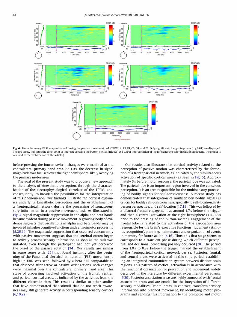
64 J.I. Salles et al. / Neuroscience Letters 501 (2011) 61– 66
F PM) iT ger) atr
bcmt
ticoiasFbdi[wtitinhtwsadtn[
ig. 4. Time–frequency ERSP maps obtained during the passive movement task (TPhe red arrow indicates the time-point of interest: pressing the button-switch (trigeferred to the web version of the article.)
efore pressing the button switch, changes were maximal at theontralateral primary hand area. At 3.0 s, the decrease in signalagnitude was focused over the right hemisphere, likely overlying
he primary motor area.The goal of the present study was to propose a new approach
o the analysis of kinesthetic perception, through the character-zation of the electrophysiological correlate of the TPPM, and,onsequently, to broaden the possibilities for the interpretationf this phenomenon. Our findings illustrate the cortical dynam-cs underlying kinesthetic perception and the establishment of
frontoparietal network during the processing of somatosen-ory information in a passive movement task. As illustrated inig. 4, signal magnitude suppression in the alpha and beta bandsecame evident during passive movement. A growing body of evi-ence suggests that oscillations in alpha and beta bands may be
nvolved in higher cognitive functions and sensorimotor processing5,26,29]. The magnitude suppression that occurred concurrentlyith passive movement suggests that the cerebral cortex began
o actively process sensory information as soon as the task wasnitiated, even though the participant had not yet perceivedhe onset of the passive rotation [34]. Our results are similarn some sense with [25] that found instantly after the begin-ing of the Functional electrical stimulation (FES) movement, aigh up ERD was seen, followed by a beta ERS comparable tohat observed after active or passive wrist actions. Both changesere maximal over the contralateral primary hand area. This
tage of processing involved activation of the frontal, central,nd parietal cortical areas, as indicated by the activities from the
ifferent electrode sites. This result is similar to other studieshat have demonstrated that stimuli that do not reach aware-ess may still generate activity in corresponding sensory cortices6,10,22].n F3, F4, C3, C4, and P3. Only significant changes in power (p ≤ 0.01) are displayed. 3 s. (For interpretation of the references to color in this figure legend, the reader is
Our results also illustrate that cortical activity related to theperception of passive motion was characterized by the forma-tion of a frontoparietal network, as indicated by the simultaneousactivation of specific cortical areas (as seen in Fig. 5). Approxi-mately 3 s before motor response, the parietal lobe was activated.The parietal lobe is an important region involved in the consciousperception. It is an area responsible for the multisensory process-ing of bodily signals for self-consciousness. A recent study hasdemonstrated that integration of multisensory bodily signals iscrucial for bodily self-consciousness, specially to self-location, first-person perspective, and self-location [17,19]. This was followed bya bilateral frontal engagement at around 1.7 s before the triggerand then a central activation at the right hemisphere (1.5–1.3 sprior to the pressing of the button-switch). Engagement of thefrontal lobe is related to the activation of the association arearesponsible for the brain’s executive functions: judgment (stimu-lus recognition), planning, maintenance and organization of eventsin memory for future action [4,16]. Thus, this first stage seems tocorrespond to a transient phase during which different percep-tual and decisional processing possibly occurred [28]. The periodfrom 1.0 s to 0.3 s before the trigger marked the establishmentof the frontoparietal cortical network per se. Posterior, frontal,and central areas were activated in this time period, establish-ing an integrated communication system between distinct brainregions. This pattern of cortical activation is in accordance withthe functional organization of perception and movement widelydescribed in the literature by different experimental paradigms[6,29]. Posterior association areas are highly connected with frontal
association areas and are crucial for the integration of differentsensory modalities. Frontal areas, in contrast, transform sensoryinformation into planned movement, by identifying motor pro-grams and sending this information to the premotor and motor
J.I. Salles et al. / Neuroscience Letters 501 (2011) 61– 66 65
F esentao ented
caItoamtmt
ipnasshpftd
tttmtl
ig. 5. Maps of cortical activation during the passive movement task. Maps are reprf the study. Time length observed varies between 1000 and 3000 ms, and the pres
ortices to be implemented. Thus, the integration between frontalnd posterior association areas is crucial for directed behaviors [33].n this context, we believe that the establishment of such fron-oparietal network characterizes the electrophysiological correlatef the TPPM. In other words, the co-activation of frontal and parietalreas marks the moment awareness is reached and, therefore, theoment of motion perception. As expected, 0.1 s before pressing of
he button switch, and then at 3.0 s, a region overlying the primaryotor cortex was activated, contralateral to the hand that executed
he movement, characterizing the moment of motion response.Our findings corroborate the finding of previous electrophys-
ological and functional imaging studies that showed that therocessing of consciously perceived stimuli involves a widespreadetwork that includes the primary sensory cortex, as well as severalreas higher in the processing hierarchy [6,13,22,35]. Specifically,tudies that investigated the neural mechanisms underlying con-cious perception employing different experimental paradigmsave shown that attention and working memory are essential com-onents of conscious perception and entail the activation of arontoparietal network [12,22]. Overall, studies have demonstratedhat awareness is a product of large-scale interactions betweenifferent regions of the brain.
Our experiment proposes a novel psychophysical approach tohe study of kinesthetic perception by developing an experimen-al paradigm that integrates EEG acquisition and a proprioceptive
ask. The results indicated that conscious perception of passiveovement is related to a specific pattern of cortical activationhat involves the formation of a frontoparietal network. Particu-arly, the approach adopted in the present study proposes that such
tive of the whole sample of the study. Maps are representative of the whole samplefrequency was 8 Hz.
network is what ultimately defines the TPPM. Although distinctmethodologies yield specific results, a key difference between thepresent study and others using the TPPM is the attentional engage-ment embedded in the paradigm. In this sense, the associationbetween EEG, passive movement and the attentional componentof the paradigm enables a broader understanding of the mecha-nisms involved in kinesthetic awareness [31,32]. Future studies arestill necessary to further explore the issue of cortical representationof kinesthetic perception, especially in populations where sensorydeficits are present.
References
[1] F. Alary, B. Doyon, C. Carel, K. Boulanouar, J.P. Ranjeva, P. Celsis, F. Chollet, Event-related potentials elicited by passive movements in humans: characterization,source analysis, and comparison to FMRI , Neuroimage 8 (1998) 377–390.
[2] M. Alegre, A. Labarga, I.G. Gurtubay, J. Iriarte, A. Malanda, J. Artieda, Beta elec-troencephalograph changes during passive movements: sensory afferencesattribute to beta event-related desynchronization in humans , Neurosci. Lett.331 (2002) 29–32.
[3] T. Aydin, Y. Yildiz, I. Yanmis, C. Yildiz, T. Kalyon, Shoulder proprioception:a comparison between the shoulder joint in healthy and surgically repairedshoulders , Arch. Orthop. Trauma Surg. 121 (2001) 422–425.
[4] V. Bachmann, M.H. Fischer, H.P. Landolt, P. Brugger, Asymmetric prefrontal cor-tex functions predict asymmetries in number space , Brain Cogn. 74 (December(3)) (2010) 306–311.
[5] M. Bauer, R. Oostenveld, M. Peeters, P. Fries, Tactile spatial attention enhancesgamma-band activity in somatosensory cortex and reduces low-frequency
activity in parieto-occipital areas , J. Neurosci. 26 (2006) 490–501.[6] D.M. Beck, G. Rees, C.D. Frith, N. Lavie, Neural correlates of change detectionand change blindness , Nat. Neurosci. 4 (2001) 645–650.
[7] K. Browne, J. Lee, P.A. Ring, The sensation of passive movement at metatar-sophalangeal joint of the great toe in man , J. Physiol. 126 (1954) 448–458.

6 ence L
[
[
[
[
[
[
[
[
[
[
[
[
[
[
[
[
[
[
[
[
[
[
[
[
[
[
345–352.
6 J.I. Salles et al. / Neurosci
[8] P.R. Burgess, J.Y. Wei, F.J. Clark, J. Simon, Signaling of kinesthetic informationby peripheral sensory receptors , Annu. Rev. Neurosci. 5 (1982) 171–187.
[9] P.J. Cordo, V.S. Gurfinkel, Y. Levik, Position sense during imperceptibly slowmovements , Exp. Brain Res. 132 (2000) 1–9.
10] S. Dehaene, l. Naccache, l. Cohen, D.L. Bihan, J.F. Mangin, J.B. Poline, Cerebralmechanisms of word masking and unconscious repetition priming , Nat. Neu-rosci. 4 (2001) 752–758.
11] A. Delorme, S. Makeig, EEGLAB: an open source toolbox for analysis of single-trial EEG dynamics , J. Neurosci. Methods 134 (2004) 9–21.
12] J. Driver, P. Vuilleumier, Perceptual awareness and its loss in unilateral neglectand extinction , Cognition 79 (2001) 39–88.
13] J. Feinstein, M. Stein, G. Castillo, M. Paulus, From sensory processes to consciousperception , Conscious. Cogn. 13 (2004) 323–335.
14] S.C. Gandevia, D. Burke, Does the nervous system depend on kinestheticinformation to control natural limb movements? Behav. Brain Sci. 15 (1992)614–632.
15] G.M. Goodwin, D.I. McCloskey, P.C. Mattews, The persistence of appreciablekinesthesia after paralyzing joint afferents but preserving muscle afferents ,Brain Res. 37 (1972) 326–329.
16] G.S. Halford, W.H. Wilson, S. Phillips, Relational knowledge: the foundation ofhigher cognition , Trends Cogn. Sci. 14 (November (11)) (2010) 497–505.
17] L. Heydrich, S. Dieguez, T. Grunwald, M. Seeck, O. Blanke, Illusory own body per-ceptions: case reports and relevance for bodily self-consciousness , Conscious.Cogn. 19 (3) (2010) 702–710.
18] H. Jasper, The ten-twenty electrode system of international federation , Elec-troencephalogr. Clin. Neurophysiol. 10 (1958) 371–375.
19] R. Kanai, B. Bahrami, G. Rees, Human parietal cortex structure predicts individ-ual differences in perceptual rivalry , Curr. Biol. 20 (18) (2010) 1626–1630.
20] A. Kawczynski, H. Nie, A. Jaskólska, A. Jaskólski, L. Arendt-Nielsen, P. Madeleine,Mechanomyography and electromyography during and after fatiguing shoul-der eccentric contractions in males and females , Scand. J. Med. Sci. Sports 17(2007) 172–179.
21] S.M. Lephart, J.B. Myers, The role of the sensorimotor system in athletic shoulder, J. Athl. Train. 35 (2000) 351–363.
22] E.D. Lumer, K.J. Friston, G. Rees, Neural correlates of perceptual rivalry in the
human brain , Science 280 (1998) 1930–1934.23] E.L. Marieb, Human Anatomy and Physiology , Benjamin/Cumming, Menlo Park,CA, 1998.
24] P.B.C. Matthews, Where does Sherrington’s “muscular sense” originate? Mus-cles, joints, corollary discharges? Annu. Rev. Neurosci. 5 (1982) 189– 218.
[
etters 501 (2011) 61– 66
25] G.R. Müller, C. Neuper, R. Rupp, C. Keinrath, H.J. Gerner, G. Pfurtscheller, Event-related beta EEG changes during wrist movements induced by functionalelectrical stimulation of forearm muscles in man , Neurosci. Lett. 340 (April(2)) (2003) 143–147.
26] C. Neuper, M. Wortz, G. Pfurtscheller, ERD/ERS patterns reflecting sen-sorimotor activation and deactivation , Prog. Brain Res. 159 (2006)211–222.
27] R.C. Oldfield, The assessment and analysis of handedness: the Edinburgh inven-tory , Neuropsychologia 9 (1971) 97–113.
28] R. Pally, How the brain actively constructs perceptions , Int. J. Psycho Anal. 78(1997) 1021–1030.
29] S. Palva, K. Linkenkaer-Hansen, R. Näätänen, J.M. Palva, Early neural cor-relates of conscious somatosensory perception , J. Neurosci. 25 (2005)5248–5258.
30] D. Restuccia, M. Valeriani, C. Barba, D. Le Pera, A. Bentivoglio, A. Albanese, M.Rubino, P. Tonali, Abnormal gating of somatosensory inputs in essential tremor, Clin. Neurophysiol. 114 (2003) 120–129.
31] E. Seiss, C.W. Hesse, S. Drane, R. Oortenveld, A.M. Wing, P. Praamstra,Proprioception-related evoked potentials: origin and sensitivity to movementparameters , Neuroimage 17 (2002) 461–468.
32] E. Seiss, P. Praamstra, C.W. Hesse, H. Rickards, Proprioceptive sen-sory function in Parkinson’s disease and Huntington’s disease: evidencefrom proprioception-related EEG potentials , Exp. Brain Res. 148 (2003)308–319.
33] D.J. Serrien, R.J. Fisher, P. Brown, Transient increases of synchronized neu-ral activity during movement preparation: influence of cognitive constraints, Brain Exp. Res. 53 (2003) 27–34.
34] J. Steinbeck, J. Bruntrup, O. Greshake, W. Potzl, T. Filler, U. Liljenqvist, Neuro-histological examination of inferior glenohumeral of the shoulder , J. Orthop.Res. 21 (2003) 250–255.
35] K.M. Stephan, M.H. Thaut, G. Wunderlich, W. Schicks, B. Tian, L. Tell-mann, T. Schmitz, H. Herzog, G.C. McIntosh, R.J. Seitz, V. Homberg,Conscious and subconscious sensorimotor synchronization—prefrontalcortex and the influence of awareness , Neuroimage 15 (2002)
36] W. Szurhaj, P. Derambure, E. Labyt, F. Cassim, J. Bourriez, J. Isnard, J. Guien, F.Mauguière, Basic mechanism of central rhythms reactivity to preparation andexecution of a voluntary movement: a stereoelectro-encephalographic study ,Clin. Neurophysiol. 114 (2003) 107–119.