ELECTROPHORESIS - endrikawidyastuti · PDF filesebagai kation(-) mobility increasing with...
41
ELECTROPHORESIS FOOD ANALYSIS AND BIOCHEMISTRY- PRACTICE ENDRIKA WIDYASTUTI MOCH. NURCHOLIS FOOD AND SCIENCE TECHNOLOGY UNIVERSITY OF BRAWIJAYA 2012
Transcript of ELECTROPHORESIS - endrikawidyastuti · PDF filesebagai kation(-) mobility increasing with...
ELECTROPHORESIS
FOOD ANALYSIS AND BIOCHEMISTRY-
PRACTICE
ENDRIKA WIDYASTUTI MOCH. NURCHOLIS
FOOD AND SCIENCE TECHNOLOGY UNIVERSITY OF BRAWIJAYA
2012
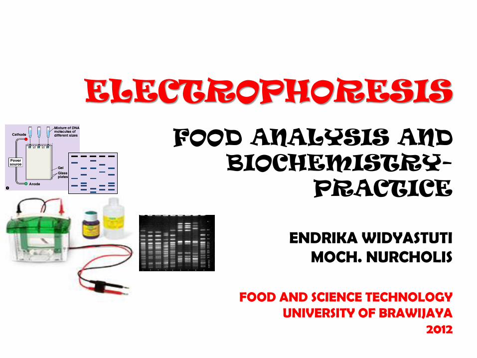
Electrophoresis
• Separation into bands due to friction through the gel and charge on protein.
• Magnitude of charge and voltage will also determine how far the protein will travel in the electrical field.
• Smaller proteins tend to move faster
Why Electrophoresis ???
• Quantiative analysis and fractination of biological fluids
• Characterization of purified components
• Detection and characterization of macromolecular interactions
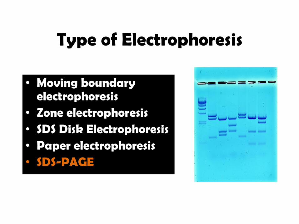
Type of Electrophoresis
• Moving boundary electrophoresis
• Zone electrophoresis • SDS Disk Electrophoresis • Paper electrophoresis • SDS-PAGE
Electrophoresis- SDS Page
Separation based on size. – Protein berikatan dengan SDS to become
negatively charged.
– SDS = sodium dodecyl sulfate => anionic detergent (negative charge)
– Proteins move through gel matrix to the anode (electrical pole with a positive charge).
– The RATE they move is based on size.
– Good for determining protein composition, purity, and estimation of molecular weight.

• Prinsip Analisis :
Suatu metode untuk memisahkan makromolekul seperti asam nukleat dan protein berdasarkan ukuran, muatan listrik dan ciri fisik.
• Tujuan :
• Mengetahui prinsip dasar pemisahan protein dengan metode elektroforesis
• Menentukan berat molekul kasein
ELEKTROFORESIS SDS-PAGE
ELEKTROFORESIS SDS-PAGE
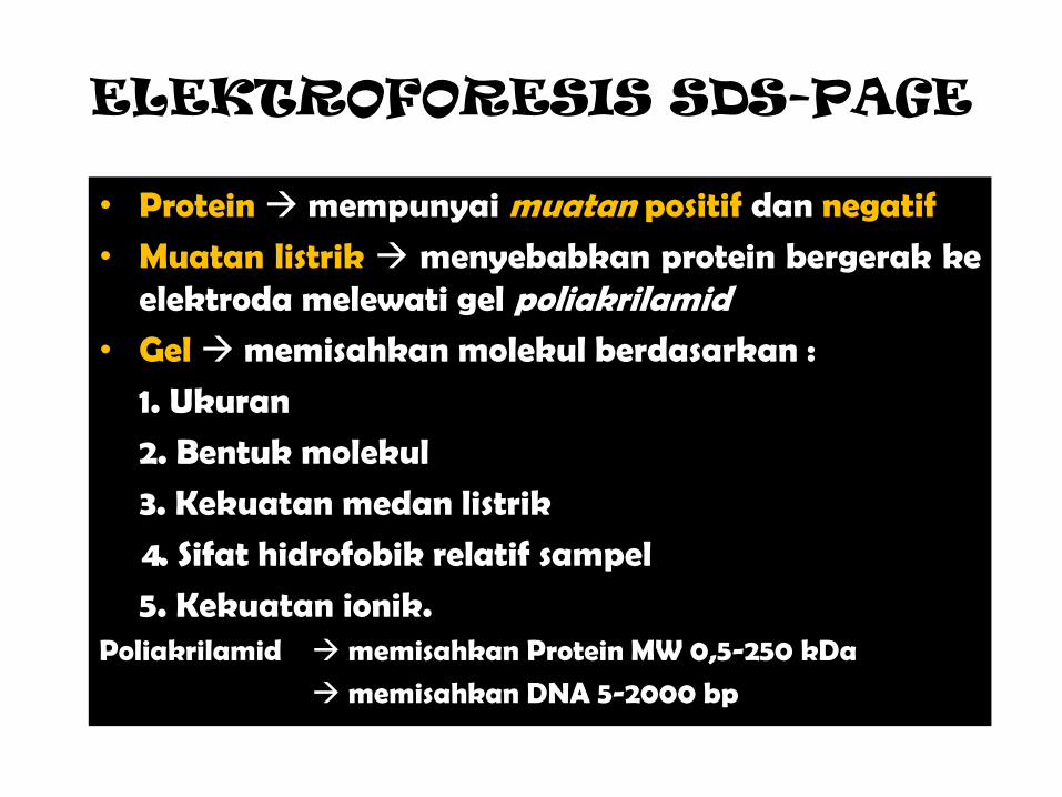
• Protein mempunyai muatan positif dan negatif
• Muatan listrik menyebabkan protein bergerak ke elektroda melewati gel poliakrilamid
• Gel memisahkan molekul berdasarkan :
1. Ukuran
2. Bentuk molekul
3. Kekuatan medan listrik
4. Sifat hidrofobik relatif sampel
5. Kekuatan ionik. Poliakrilamid memisahkan Protein MW 0,5-250 kDa
memisahkan DNA 5-2000 bp
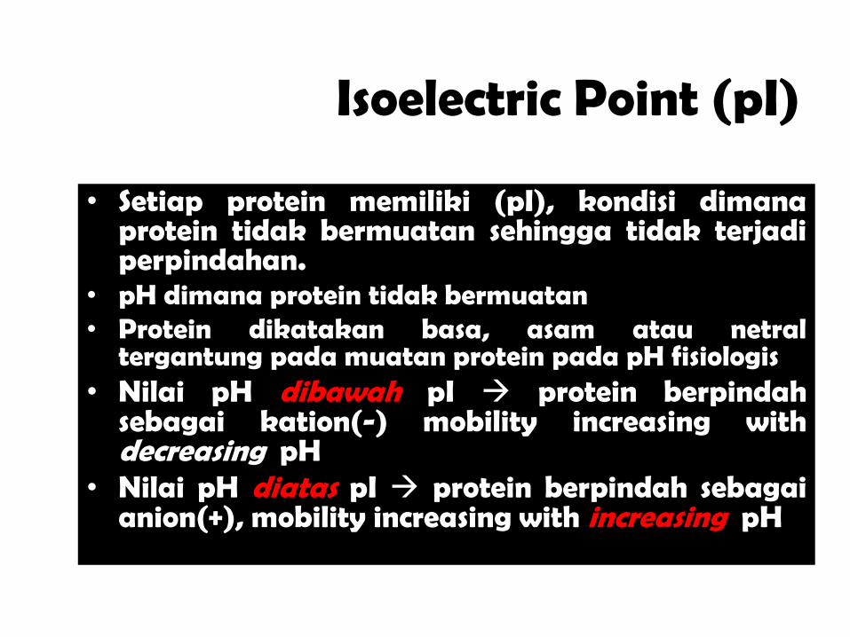
Isoelectric Point (pI)
• Setiap protein memiliki (pI), kondisi dimana protein tidak bermuatan sehingga tidak terjadi perpindahan.
• pH dimana protein tidak bermuatan • Protein dikatakan basa, asam atau netral
tergantung pada muatan protein pada pH fisiologis • Nilai pH dibawah pI protein berpindah
sebagai kation(-) mobility increasing with decreasing pH
• Nilai pH diatas pI protein berpindah sebagai anion(+), mobility increasing with increasing pH
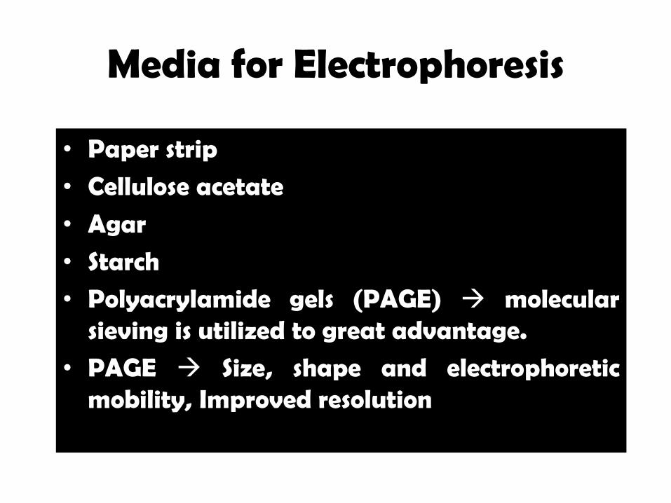
Media for Electrophoresis
• Paper strip
• Cellulose acetate
• Agar
• Starch
• Polyacrylamide gels (PAGE) molecular sieving is utilized to great advantage.
• PAGE Size, shape and electrophoretic mobility, Improved resolution
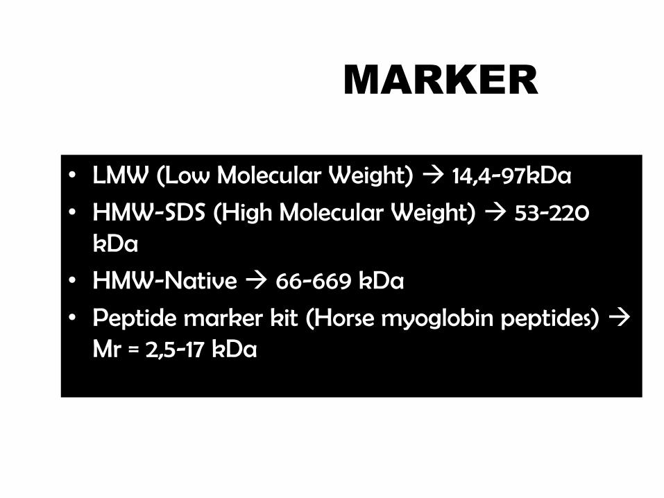
MARKER
• LMW (Low Molecular Weight) 14,4-97kDa
• HMW-SDS (High Molecular Weight) 53-220 kDa
• HMW-Native 66-669 kDa
• Peptide marker kit (Horse myoglobin peptides) Mr = 2,5-17 kDa
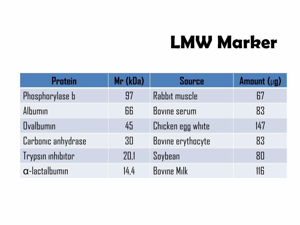
LMW Marker
Protein Mr (kDa) Source Amount (µg)
Phosphorylase b 97 Rabbit muscle 67
Albumin 66 Bovine serum 83
Ovalbumin 45 Chicken egg white 147
Carbonic anhydrase 30 Bovine erythocyte 83
Trypsin inhibitor 20,1 Soybean 80
α-lactalbumin 14,4 Bovine Milk 116
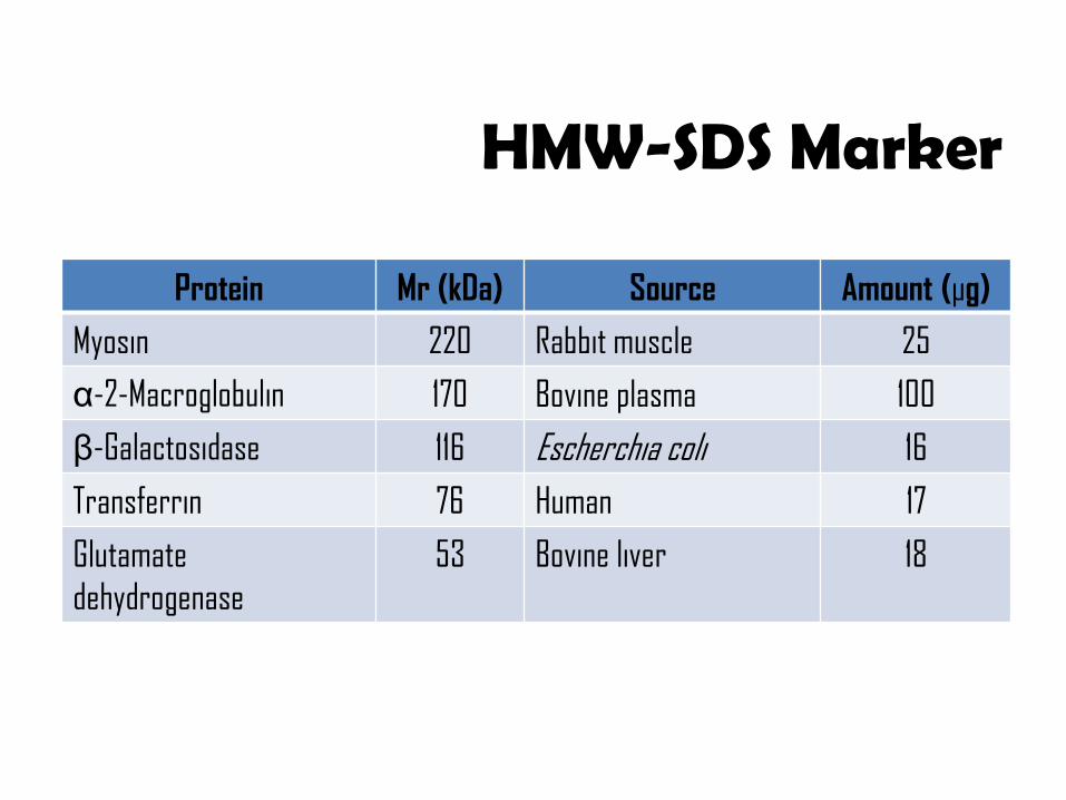
HMW-SDS Marker
Protein Mr (kDa) Source Amount (µg)
Myosin 220 Rabbit muscle 25
α-2-Macroglobulin 170 Bovine plasma 100
β-Galactosidase 116 Escherchia coli 16
Transferrin 76 Human 17
Glutamate
dehydrogenase
53 Bovine liver 18
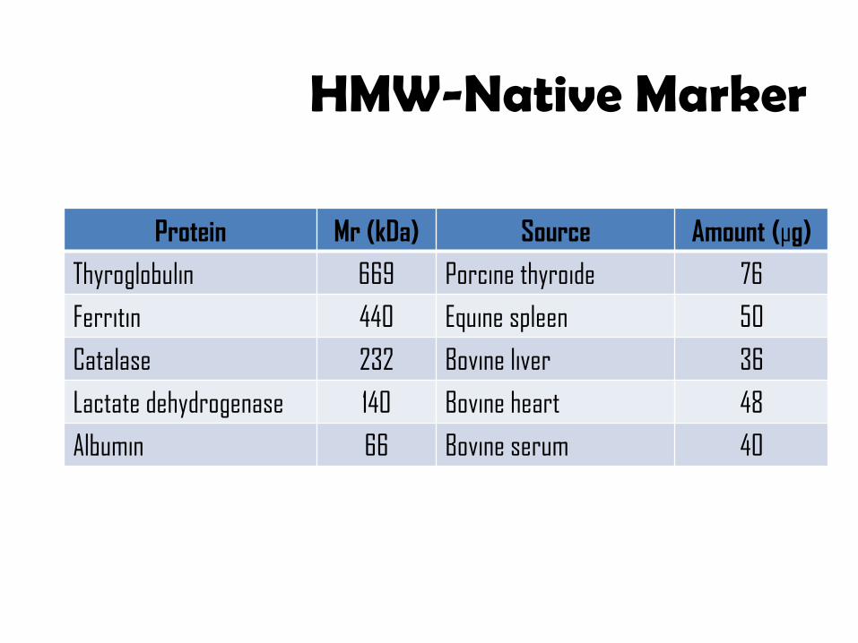
HMW-Native Marker
Protein Mr (kDa) Source Amount (µg)
Thyroglobulin 669 Porcine thyroide 76
Ferritin 440 Equine spleen 50
Catalase 232 Bovine liver 36
Lactate dehydrogenase 140 Bovine heart 48
Albumin 66 Bovine serum 40
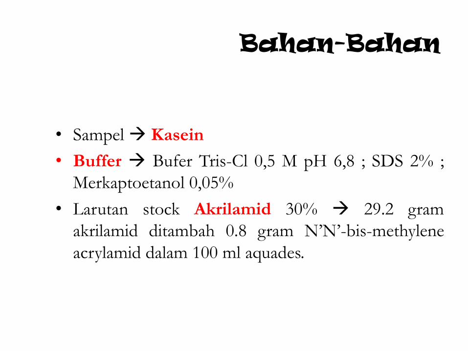
Bahan-Bahan
• Sampel Kasein
• Buffer Bufer Tris-Cl 0,5 M pH 6,8 ; SDS 2% ;
Merkaptoetanol 0,05%
• Larutan stock Akrilamid 30% 29.2 gram
akrilamid ditambah 0.8 gram N’N’-bis-methylene
acrylamid dalam 100 ml aquades.
Bahan-Bahan
• Larutan SDS 10 %
• Amonium persulfat (APS) 10% (di buat setiap akan
digunakan)
• TEMED
• Larutan Pewarna (Staining) 0.1 % commasie blue dalam
larutan metanol : air : asam asetat (5:5:2)
• Larutan Pembilas (destaining) metanol : air : asam
asetat (5:5:2)
• Aquades
Alat
• Seperangkat alat elektroforesis
• Mikropipet
• Tip
• Beker glass 100 ml
• Beker glas 50 ml
• Eppendorf
• Shaker
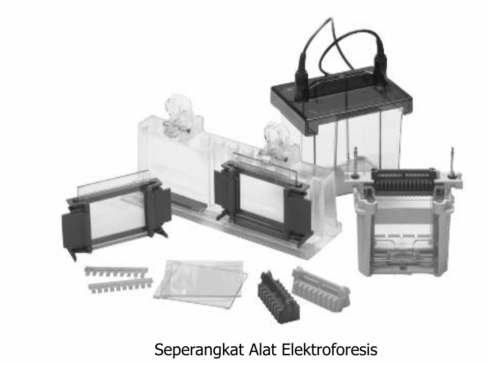
Seperangkat Alat Elektroforesis
Prosedur Kerja
Pembuatan gel :
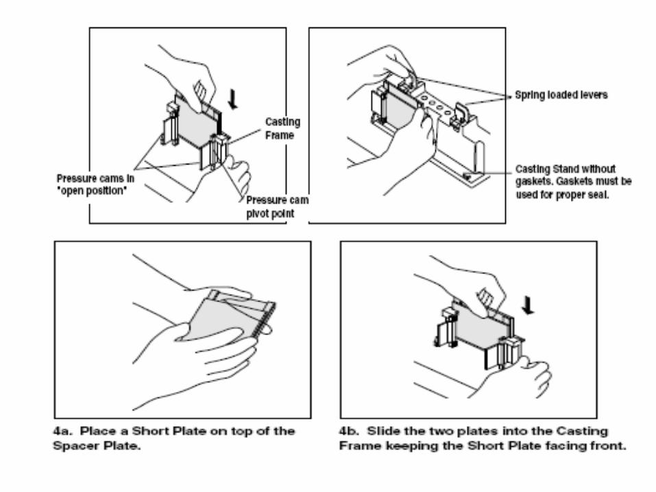
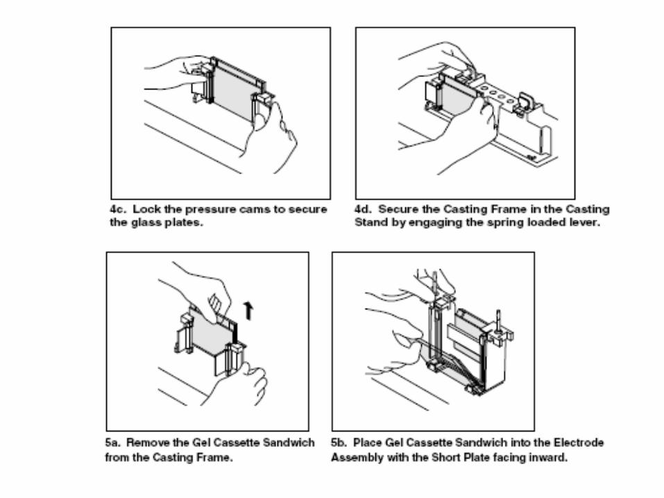
• Pasanglah alat gelas untuk mencetak gel ke tempat yang disediakan (seperti gambar 4.)
• Untuk membuat 20 ml gel 20% campurkan 13.3 ml larutan stok akrilamid 30%, 5 ml buffer Tris-HCl 0.5 M, pH 6.8, 0.2 ml SDS 10%, 1.5 ml aquades.
• Tambahkan segera 100 µl APS 10% dan TEMED 10 µl
• Aduk hingga tercampur merata
• Tuangkan ke dalam cetakan gel dengan menggunakan mikropipet hingga tinggi yang dikehendaki. Beri sisa tempat untuk stacking gel di bawah area peletakan gigi sisir.
• Biarkan gel terpolimerisasi selama 15-30 menit dalam suhu ruang.
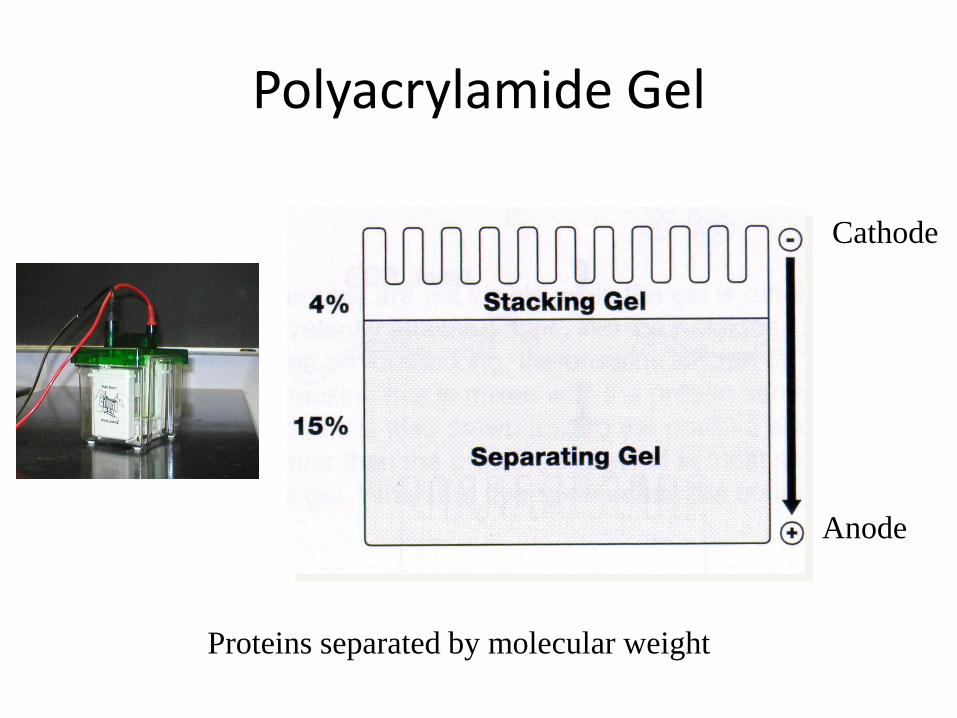
Polyacrylamide Gel
Cathode
Anode
Proteins separated by molecular weight
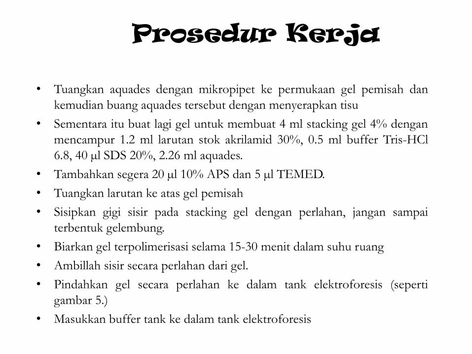
Prosedur Kerja
• Tuangkan aquades dengan mikropipet ke permukaan gel pemisah dan
kemudian buang aquades tersebut dengan menyerapkan tisu
• Sementara itu buat lagi gel untuk membuat 4 ml stacking gel 4% dengan
mencampur 1.2 ml larutan stok akrilamid 30%, 0.5 ml buffer Tris-HCl
6.8, 40 µl SDS 20%, 2.26 ml aquades.
• Tambahkan segera 20 µl 10% APS dan 5 µl TEMED.
• Tuangkan larutan ke atas gel pemisah
• Sisipkan gigi sisir pada stacking gel dengan perlahan, jangan sampai
terbentuk gelembung.
• Biarkan gel terpolimerisasi selama 15-30 menit dalam suhu ruang
• Ambillah sisir secara perlahan dari gel.
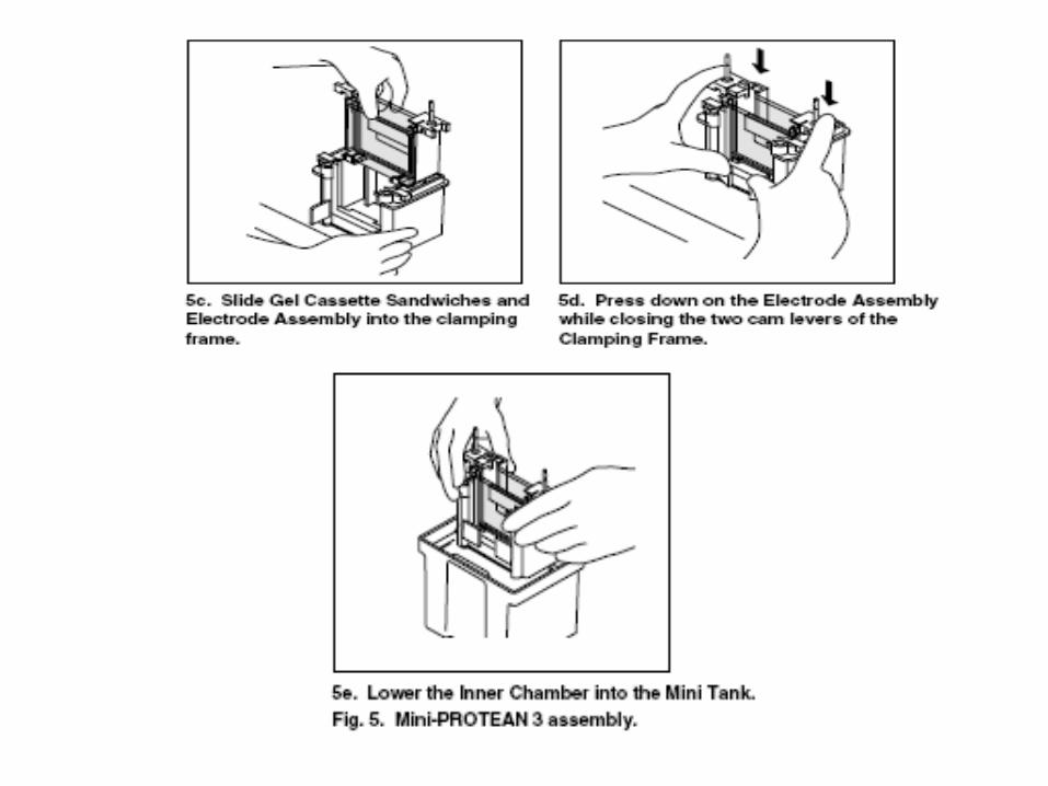
• Pindahkan gel secara perlahan ke dalam tank elektroforesis (seperti
gambar 5.)
• Masukkan buffer tank ke dalam tank elektroforesis
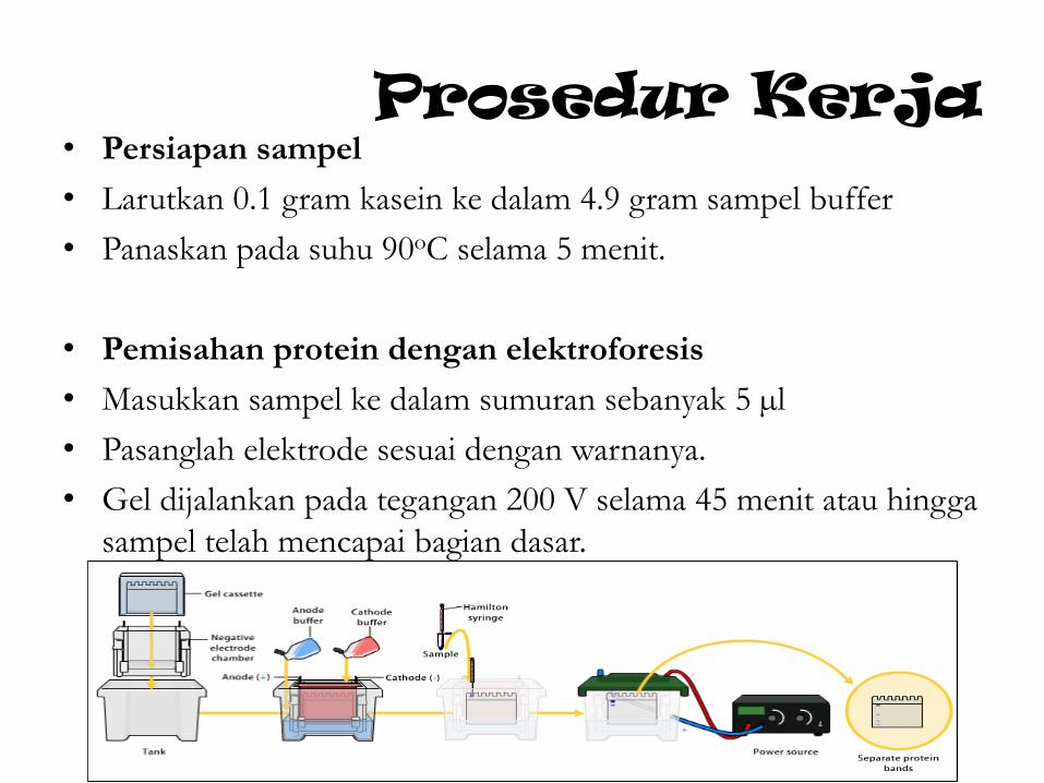
Prosedur Kerja • Persiapan sampel
• Larutkan 0.1 gram kasein ke dalam 4.9 gram sampel buffer
• Panaskan pada suhu 90oC selama 5 menit.
• Pemisahan protein dengan elektroforesis
• Masukkan sampel ke dalam sumuran sebanyak 5 µl
• Pasanglah elektrode sesuai dengan warnanya.
• Gel dijalankan pada tegangan 200 V selama 45 menit atau hingga
sampel telah mencapai bagian dasar.
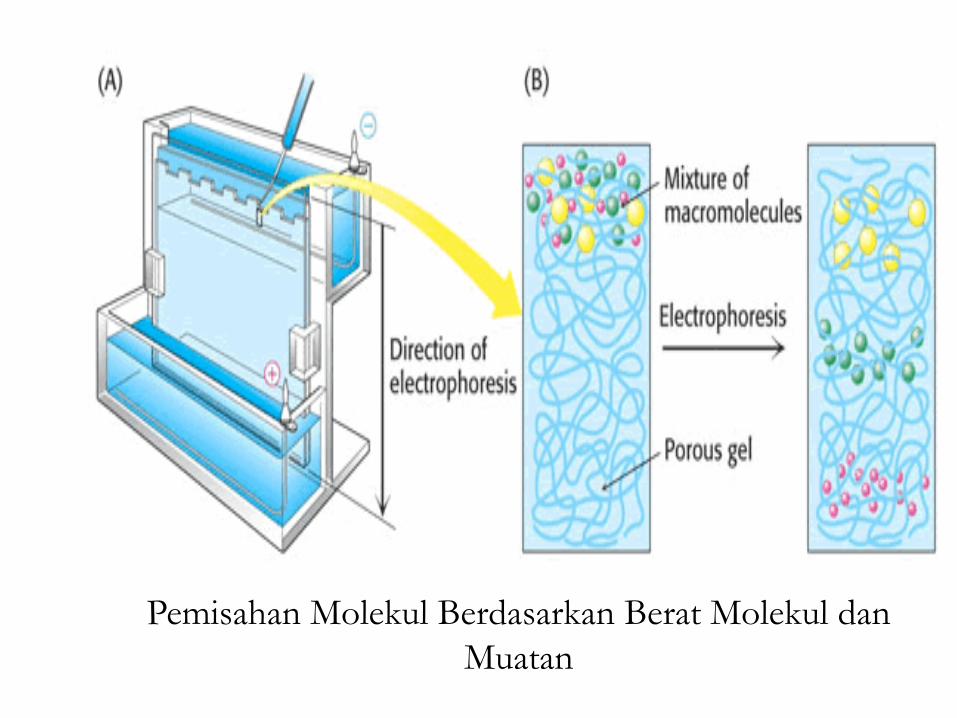
Pemisahan Molekul Berdasarkan Berat Molekul dan
Muatan
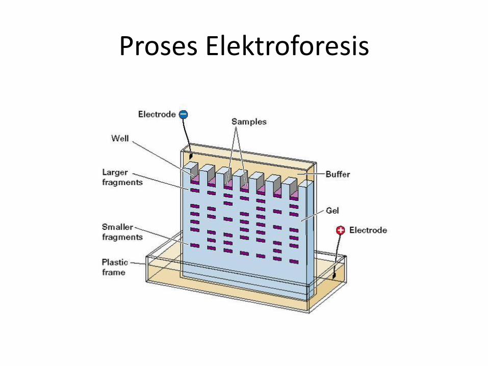
Proses Elektroforesis
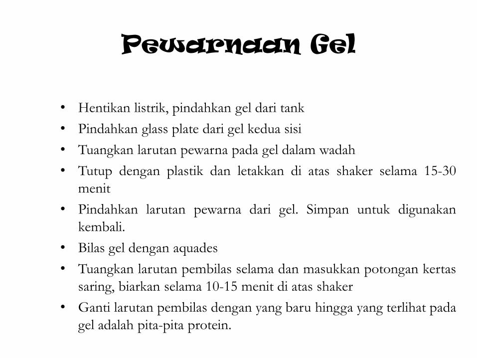
Pewarnaan Gel
• Hentikan listrik, pindahkan gel dari tank
• Pindahkan glass plate dari gel kedua sisi
• Tuangkan larutan pewarna pada gel dalam wadah
• Tutup dengan plastik dan letakkan di atas shaker selama 15-30
menit
• Pindahkan larutan pewarna dari gel. Simpan untuk digunakan
kembali.
• Bilas gel dengan aquades
• Tuangkan larutan pembilas selama dan masukkan potongan kertas
saring, biarkan selama 10-15 menit di atas shaker
• Ganti larutan pembilas dengan yang baru hingga yang terlihat pada
gel adalah pita-pita protein.
Pengamatan
Amati pita-pita yang terbentuk pada gel elektroforesis.
Cari dalam literatur berat molekul masing-masing
komponen penyusun kasein dan tentukan letak
komponen tersebut pada pita gel elektroforesis.
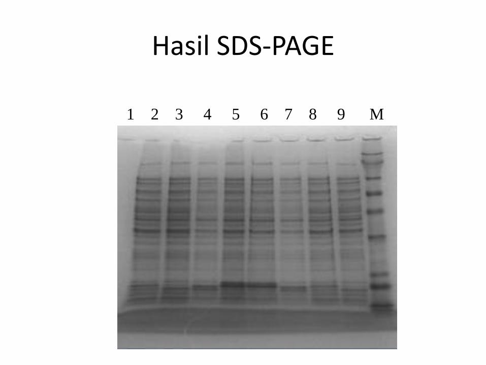
Hasil SDS-PAGE
1 2 3 4 5 6 7 8 9 M

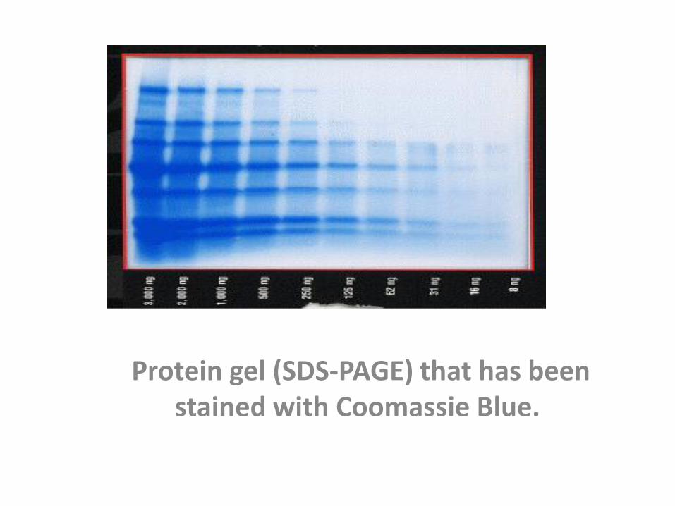
Protein gel (SDS-PAGE) that has been stained with Coomassie Blue.

MATERI TAMBAHAN
SDS-PAGE (PolyAcrylamide Gel Electrophoresis)
• SDS-PAGE, sodium dodecyl sulfate polyacrylamide gel electrophoresis, is a technique widely used in biochemistry,
forensics, genetics and molecular biology:
• to separate proteins according to their electrophoretic mobility (a function of length of polypeptide chain or molecular weight).
• to separate proteins according to their size, and no other physical feature.

Fig.1Before SDS: Protein (pink line) incubated with the denaturing detergent SDS showing negative and
positive charges due to the charged R-groups in the protein. The large H's represent hydrophobic domains where nonpolar R-groups have collected in an attempt to get
away from the polar water that surrounds the protein. After SDS: SDS disrupt hydrophobic areas (H's) and coat proteins with many negative charges which
overwhelms any positive charges the protein had due to positively charged R-groups. The resulting protein has been denatured by SDS (reduced to its primary structure-aminoacid sequence) and
as a result has been linearized.
..SDS
• SDS (the detergent soap) breaks up hydrophobic areas and coats proteins with negative charges thus overwhelming positive charges in the protein.
• The detergent binds to hydrophobic regions in a constant ratio of about 1.4 g of SDS per gram of protein.
..SDS
• Therefore, if a cell is incubated with SDS, the membranes will be dissolved, all the proteins will be solubalized by the detergent and all the proteins will be covered with many negative charges.
PAGE •
If the proteins are denatured and put into an electric field (only), they will all move towards the positive pole at the same rate, with no separation by size.
• However, if the proteins are put into an environment that will allow different sized proteins to move at different rates.
• The environment is polyacrylamide.
• the entire process is called polyacrylamide gel electrophoresis (PAGE).
..PAGE
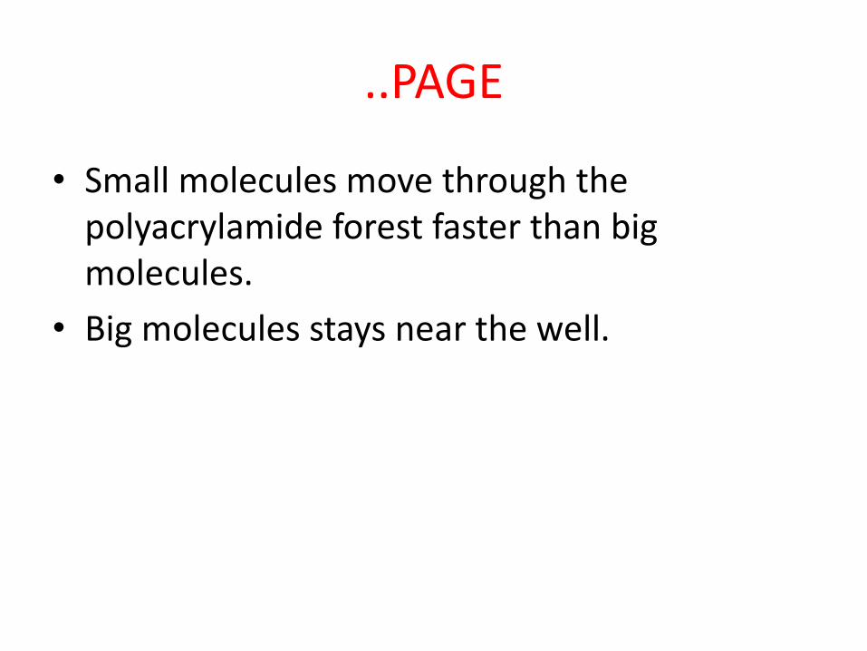
• Small molecules move through the polyacrylamide forest faster than big molecules.
• Big molecules stays near the well.
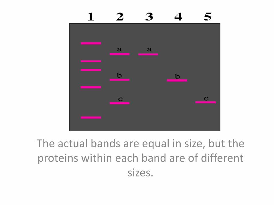
The actual bands are equal in size, but the proteins within each band are of different
sizes.
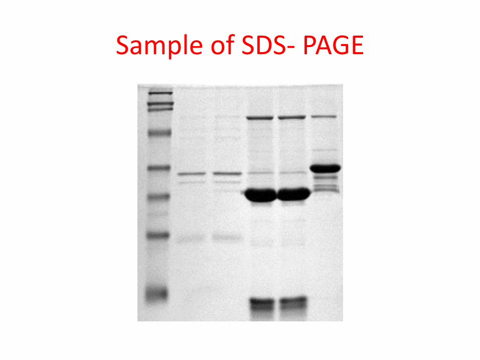
Sample of SDS- PAGE


![jNewSouthWales KATION PTY LTD v LAMRU PTY … · KATION PTY LTD v LAMRU PTY LTD; LEWIS v NORTEX PTY LTD (In liq) [2009] NSWCA 145 (12 June 2009) Last Updated: 15 June 2009 NEW SOUTH](https://static.fdocuments.in/doc/165x107/5b8894267f8b9aaf728df1c1/jnewsouthwales-kation-pty-ltd-v-lamru-pty-kation-pty-ltd-v-lamru-pty-ltd-lewis.jpg)