Electronic excitation processes of photoactive organic molecules2184551/2184551.pdf · ass. prof....
40
VILNIUS UNIVERSITY CENTER FOR PHYSICAL SCIENCES AND TECHNOLOGY STEPAS TOLIAUTAS ELECTRONIC EXCITATION PROCESSES OF PHOTOACTIVE ORGANIC MOLECULES Summary of doctoral dissertation Physical sciences, physics (02 P) Vilnius, 2014
Transcript of Electronic excitation processes of photoactive organic molecules2184551/2184551.pdf · ass. prof....

VILNIUS UNIVERSITY
CENTER FOR PHYSICAL SCIENCES AND TECHNOLOGY
STEPAS TOLIAUTAS
ELECTRONIC EXCITATION PROCESSES
OF PHOTOACTIVE ORGANIC MOLECULES
Summary of doctoral dissertation
Physical sciences, physics (02 P)
Vilnius, 2014

Dissertation was prepared during years 2009 – 2013 at Vilnius university.
Scientific supervisor:
ass. prof. dr. Juozas Šulskus (Vilnius University, physical sciences, physics – 02 P).
Dissertation will be defended in the Council of Physics of Vilnius University:
Chairman – prof. dr. Valdas Šablinskas
(Vilnius University, physical sciences, physics – 02 P).
Members:
prof. habil. dr. Sigitas Tamulevičius
(Kaunas University of Technology, physical sciences, physics – 02 P);
prof. dr. Paulius Miškinis
(Vilnius Gediminas Technical University, physical sciences, physics – 02 P);
dr. Valdas Jonauskas (Vilnius University, physical sciences, physics – 02 P);
dr. Olegas Eicher-Lorka (Center for Physical Sciences and Technology,
Institute of Chemistry, physical sciences, chemistry – 03 P).
Opponents:
ass. prof. dr. Kęstutis Aidas (Vilnius University, physical sciences, physics – 02 P);
prof. habil. dr. Gediminas Niaura (Center for Physical Sciences and Technology,
Institute of Chemistry, physical sciences, chemistry – 03 P).
The official defense of the dissertation will be held at 3 PM on 25th of September 2014
in 207 auditorium at the Faculty of Physics.
Address: Saulėtekio ave. 9-III, Vilnius, Lithuania.
Summary of the dissertation was distributed on 22nd of August 2014.
The dissertation is available for preview at the libraries of Vilnius University and of
Center for Physical Sciences and Technology.

VILNIAUS UNIVERSITETAS
FIZINIŲ IR TECHNOLOGIJOS MOKSLŲ CENTRAS
STEPAS TOLIAUTAS
ELEKTRONINIO SUŽADINIMO PROCESAI
FOTOAKTYVIOSE ORGANINĖSE MOLEKULĖSE
Daktaro disertacijos santrauka
Fiziniai mokslai, fizika (02 P)
Vilnius, 2014 metai

Disertacija rengta 2009 – 2013 metais Vilniaus universitete.
Mokslinis vadovas:
doc. dr. Juozas Šulskus (Vilniaus universitetas, fiziniai mokslai, fizika – 02 P).
Disertacija ginama Vilniaus universiteto Fizikos mokslo krypties taryboje:
Pirmininkas – prof. dr. Valdas Šablinskas
(Vilniaus universitetas, fiziniai mokslai, fizika – 02 P).
Nariai:
prof. habil. dr. Sigitas Tamulevičius
(Kauno technologijos universitetas, fiziniai mokslai, fizika – 02 P);
prof. dr. Paulius Miškinis
(Vilniaus Gedimino technikos universitetas, fiziniai mokslai, fizika – 02 P);
dr. Valdas Jonauskas (Vilniaus universitetas, fiziniai mokslai, fizika – 02 P);
dr. Olegas Eicher-Lorka (Fizinių ir technologijos mokslų centro
Chemijos institutas, fiziniai mokslai, chemija – 03 P).
Oponentai:
doc. dr. Kęstutis Aidas (Vilniaus universitetas, fiziniai mokslai, fizika – 02 P);
prof. habil. dr. Gediminas Niaura (Fizinių ir technologijos mokslų centro
Chemijos institutas, fiziniai mokslai, chemija – 03 P).
Disertacija bus ginama viešame Fizikos mokslų krypties tarybos posėdyje 2014 m.
rugsėjo mėn. 25 d. 15 val. Fizikos fakulteto 207 auditorijoje.
Adresas: Saulėtekio al. 9-III, Vilnius, Lietuva.
Disertacijos santrauka išsiuntinėta 2014 m. rugpjūčio mėn. 22 d.
Disertaciją galima peržiūrėti Vilniaus universiteto bei Fizinių ir technologijos mokslų
centro bibliotekose.

Acknowledgment
My decision to pursue theoretical physics was mostly influenced by two persons:
scientific advisor Juozas Šulskus and the head of the Department of Theoretical Physics,
Leonas Valkūnas. J. Šulskus took many roles – those of lecturer and instructor, study
program curator, academic supervisor, work colleague and, ultimately, scientific advisor
– to impart the current knowledge of science and to motivate me to seek new insights.
L. Valkūnas provided the perfect environment for realization of the emerging ideas and
helped to establish a proper scientific context for the obtained results. Without them the
“dissertation” would probably end there.
The never-ending research and documentation cycle has not yet rolled down to the
proverbial rut thanks to the lively colleagues at the Department of Theoretical Physics
that manage to keep me interested. I'd like to expressly thank Andrius, Jevgenijus, Sveta
and a few Vytautai for the assistance in my attempts to make this text clearer to the
prospective readers as well as to myself.
I thank Kate Stevens, Sven Stafström, Andrea Tindiani and Michael Ruggenthaler
for being in the right place at the right time. Finally, I thank Justė for making sure that
the process of writing this dissertation comes to an END.

Table of contents
1. Introduction..................................................................................................................7
1.1. Electronic excitation processes.....................................................................7 1.2. Main goal of the dissertation........................................................................9 1.3. Novelty and importance of the studies........................................................10 1.4. Propositions for the defense........................................................................10
2. Parameters of the potential energy surfaces................................................................11
2.1. Jablonski diagrams and the potential energy surfaces.................................11 2.2. Main parameters of the potential energy surfaces.......................................12
3. Solvent effect on the proton-transfer processes in MIEP............................................13
3.1. Structural model and computational methods.............................................14 3.2. Deformation pathway of MIEP in vacuum.................................................14 3.3. Changes of MIEP properties in solvent......................................................15 3.4. Comparison to MS1....................................................................................17
4. Photochromic properties of indolo-benzoxazine........................................................17
4.1. Structural model and computational methods.............................................18 4.2. Reaction coordinates of IB compound........................................................19 4.3. Relaxation pathways of the IB compound..................................................20
5. Efficient phosphorescence of PBMSi polymer...........................................................22
5.1. Structural model and computational methods.............................................23 5.2. Charge-transfer state of MSi15-BP.............................................................23 5.3. Excited-state relaxation of MSi15-BP........................................................24 5.4. Energy conversion processes in PBMSi polymer.......................................25
6. Phosphorescence of the organometallic iridium complex...........................................26
6.1. Structural model and computational methods.............................................27 6.2. Properties of the absorption spectra............................................................27 6.3. Structure and properties of the (pbt)2Ir(acac) complex..............................28
7. Conclusions................................................................................................................30
8. The approbation of the conducted research................................................................31
About the author..............................................................................................................33
References.......................................................................................................................34
6

1. Introduction
1.1. Electronic excitation processes
Photoactive, or light-sensitive molecules are found in most living organisms. For
example, plants and some bacteria employ such molecules in the process of
photosynthesis, that is, conversion of light energy to chemical energy. Many animals also
have visual sensors that extract information about their surroundings from the collected
light, and photoactive molecules form the basis of these visual sensors. Such molecules
are known as pigments (light-sensitive parts of the protein), chromophores (molecules
that determine the color of the material) or photochromes (materials that change their
color upon light-excitation) in the literature. The interaction of the molecule with the
photon (quantum of light) results in the change of the molecular energy. Since the energy
delivered by visible light induces changes in the electronic subsystem of the molecule
first, this type of interaction is called the electronic excitation. To distinguish the
interaction with visible light, the term “optical excitation” is sometimes used as well.
Electronic excitation of the molecule usually signals the start of one of the various
energy-related processes within the molecule. Such process may be irreversible; for
example, the provided energy might break the molecule into several parts or enable the
chemical reaction with the surrounding material, resulting in the reaction products. On
the other hand, excited molecule might be able to dissipate the excess energy through the
different conversion channels and return to the ground state, resulting in either the initial
compounds or the isomers of slightly different geometric structure. The repeated process
of the excitation and the energy dissipation or relaxation constitutes the excitation cycle.
Excitation cycles are observed during many physical, chemical and biological
phenomena. In many biological systems, such as the proteins within plants and bacteria,
photon absorption induces a chain of non-radiative interactions known as the photocycle
[1,2]. Such proteins are the main elements of the above-mentioned light sensors and
energy-harvesting units. The other example of the light-induced reversible reaction is the
optomechanical cycle – the structural alteration of the molecule or its fragment that can
7

be used to carry out mechanical work [3,4]. Such compounds are expected to be used in
the construction of molecular-scale devices and the miniaturization of existing
technology. Other relaxation processes are not cyclic; for example, the working principle
of the organic optoelectronics devices – solar elements and OLEDs – is the light-to-
electrical energy conversion [5]. In case of OLED, charge carriers are transported into
the photoactive material, which is then used to emit photons. It is clear from the provided
examples that the investigation of the electronic excitation processes is very relevant for
the research of both the natural organisms and the novel artificial systems.
The main difficulty of theoretical modeling of the electronic excitation processes
in molecules is solving the time-dependent Schrodinger equation, which is currently
computationally too expensive for systems of practical importance. Direct computations
are restricted to small molecules or generalized systems described by a few energy levels
and couplings. However, it is possible to circumvent this problem by gradually changing
structural parameters of the system and repeatedly solving time-independent Schrodinger
equation. Since each set of structural parameters corresponds in principle to a dynamic
state at a certain point in time (although not necessarily reachable by the process under
investigation), the potential energy surfaces (PES) obtained this way qualitatively
describe the behavior of the excited molecular system and can be used to explain, f. e.,
the time-dependent spectra without directly solving the dynamic equations.
The widely used group of molecular investigation methods comprises the
computational methods of electronic structure, based on the molecular theory. The
review of the theoretical methods can be found in Chapter 2 of the dissertation. The
optimized algorithms of the electronic structure methods are gathered into program
packages and made available for the wide circle of scientists – physicists, chemists and
biologists, experimentalists and theoreticians alike. Most popular packages include
Gaussian [6], GAMESS-US [7,8], NWChem [9]. Together with the implementations of
electronic structure methods such packages usually contain structure optimization
algorithms, molecular mechanics and molecular dynamics methods, calculation routines
of thermodynamic parameters etc. The area of computational molecular science covered
by the above-mentioned methods is sometimes called computational chemistry, and the
8

quantum-mechanical subset of the theories is known as quantum chemistry. Combined
use of stationary electronic structure methods and molecular structure optimization
algorithms enables researchers to model electronic excitation processes by studying
molecular ground- and excited-state potential energy surfaces. The main parameters of
the potential energy surfaces of the molecular system are described in Chapter 3 of the
dissertation. The PES-based models are constructed and used to describe the processes
occurring in the investigated molecular compounds. Dissertation chapters 4–7 contain
the descriptions of the studies of four photoactive compounds and the constructed
theoretical models. The conclusions that validate the defense propositions are formulated
in Chapter 8. The last Chapter lists the scientific articles and conferences where the
results of these studies were presented.
1.2. Main goal of the dissertation
The main goal of this dissertation is to study processes caused by the electronic
excitation in photoactive organic molecules using computational electronic structure
methods and to create models based on the potential energy surfaces that describe the
energy relaxation in the investigated molecules. The constructed models are then used to
solve the following problems:
1. To investigate the possible proton transfer pathway in the excited 2-(N-methyl-α-
iminoethyl)-phenol (MIEP) and to determine the effect of the polar solvent
(water) for the energy relaxation of MIEP.
2. To model the structural changes in the excited indolo-benzoxazine (IB) compound
after the opening of the oxazine ring and to characterize the effect of such changes
on its photochromic properties.
3. To explain the cause of the efficient phosphorescence in poly[biphenyl(methyl)-
silylene] (PBMSi) polymer.
4. To determine the character of the absorption and the phosphorescence in bis(2-
phenylbenzothiazole)-iridium-acetylacetonate ((pbt)2Ir(acac)) and to estimate the
effect of carbazole groups on the optical properties of the investigated complex.
9

1.3. Novelty and importance of the studies
• This work features four new potential energy surface models for the description of
the energy relaxation in various photoactive organic compounds.
• Study of the MIEP molecule reveals the properties of the Schiff base – a functio-
nal group in the photoactive center of the bacteriorhodopsin protein, performing
trans-membrane proton transfer – in the polar environment.
• The PES model of the IB compound explains observed processes of the different
timescales within the photochromic molecule that, due to its fatigue resistance, is
proposed to be used for molecular-scale switches.
• The model of the phosphorescence in PBMSi polymer provides evidence of the
newly-observed physical phenomenon – enhanced intersystem crossing in the
conjugated polymer without involving heavy-metal effect. Such materials are
promising for non-coherent up-conversion and white-light OLED applications.
• The (pbt)2Ir(acac) complex model reveals the structural properties of the complex
and its interaction with additional functional groups.
1.4. Propositions for the defense
• The energy relaxation process of the excited MIEP molecule in water is governed
by the formation of the hydrogen-bonded molecular complex consisting of the
MIEP and the solvent molecules adjacent to the Schiff base of the compound.
• Photochromic properties of the excited IB compound are determined by several
relaxation pathways that are related to the movement of the nitrophenol group
after the opening of the oxazine ring.
• The efficient phosphorescence of the PBMSi polymer is caused by the presence of
charge-transfer state between the electronic systems of the silicon chain and
biphenyl groups of the material.
• The excitation of the (pbt)2Ir(acac) complex has only small effect on its structure
and is not affected by the addition of the functional carbazole groups.
10

2. Parameters of the potential energy surfaces
2.1. Jablonski diagrams and the potential energy surfaces
Energy conversion processes in chemistry and chemical physics are commonly
depicted as Jablonski diagrams [10]. Nowadays, the Jablonski diagram is understood as a
broad tool, assuming different forms for the different uses [11]. However, the transitions
between states with the different molecular structure are decidedly vague in Jablonski
diagrams. By including the information about the reaction coordinate or coordinates in
the x axis, Jablonski diagram becomes the potential energy curve or surface diagram
(Fig. 2.1) which may have many special points on it – transition/ saddle points, excited-
state minima, intersection points etc.
Figure 2.1. Potential energy curves of the photoactive molecule
11
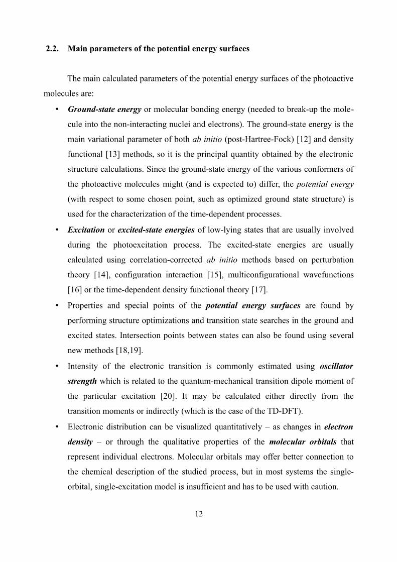
2.2. Main parameters of the potential energy surfaces
The main calculated parameters of the potential energy surfaces of the photoactive
molecules are:
• Ground-state energy or molecular bonding energy (needed to break-up the mole-
cule into the non-interacting nuclei and electrons). The ground-state energy is the
main variational parameter of both ab initio (post-Hartree-Fock) [12] and density
functional [13] methods, so it is the principal quantity obtained by the electronic
structure calculations. Since the ground-state energy of the various conformers of
the photoactive molecules might (and is expected to) differ, the potential energy
(with respect to some chosen point, such as optimized ground state structure) is
used for the characterization of the time-dependent processes.
• Excitation or excited-state energies of low-lying states that are usually involved
during the photoexcitation process. The excited-state energies are usually
calculated using correlation-corrected ab initio methods based on perturbation
theory [14], configuration interaction [15], multiconfigurational wavefunctions
[16] or the time-dependent density functional theory [17].
• Properties and special points of the potential energy surfaces are found by
performing structure optimizations and transition state searches in the ground and
excited states. Intersection points between states can also be found using several
new methods [18,19].
• Intensity of the electronic transition is commonly estimated using oscillator
strength which is related to the quantum-mechanical transition dipole moment of
the particular excitation [20]. It may be calculated either directly from the
transition moments or indirectly (which is the case of the TD-DFT).
• Electronic distribution can be visualized quantitatively – as changes in electron
density – or through the qualitative properties of the molecular orbitals that
represent individual electrons. Molecular orbitals may offer better connection to
the chemical description of the studied process, but in most systems the single-
orbital, single-excitation model is insufficient and has to be used with caution.
12

3. Solvent effect on the proton-transfer processes in MIEP
Light-induced proton transfer is an important part of the processes in biological
systems. Since such processes are very sensitive to the effects of the environment [21],
the proton transfer may be enabled [22], enhanced or suppressed [23], depending on the
surroundings of the photoactive complex. For example, proton transfer in the active
center of the bacteriorhodopsin protein is caused by the light absorption in the retinal
chromophore, which triggers movement of the attached Schiff base (Fig. 3.1) [24]. The
active center is also known to contain several “trapped” water molecules, effect of which
for the proton transfer is not entirely clear [25].
Various factors that may influence the processes in the active center of bacterio-
rhodopsin and the relative importance of said factors are investigated by studying various
structural groups of the active center (e. g. Schiff base), both together with their surroun-
dings and as separate model compounds [26]. The unit of phenol ring and Schiff base,
which share the proton between them, forms the basis of the aromatic anil group of
molecules that exhibit many interesting properties, such as intramolecular proton transfer
[27] and related photochromism [28]. Experiments on one such compound, N-(triphenyl-
methyl)-salicydenimine (MS1) in ethanol, showed that the energy relaxation of the
excited compound is strongly influenced by the solvent molecules [29]. The solvent
effect on the properties of 2-(N-methyl-α-iminoethyl)-phenol (MIEP) in the electronic
ground state was also modeled earlier [30]. The main task of the current study was to
investigate possible proton transfer pathway in excited MIEP molecule in water and to
determine the (polar) solvent effect for the energy relaxation of the compound.
Figure 3.1. Molecular structure of MIEP in vacuum
13
Schiff base
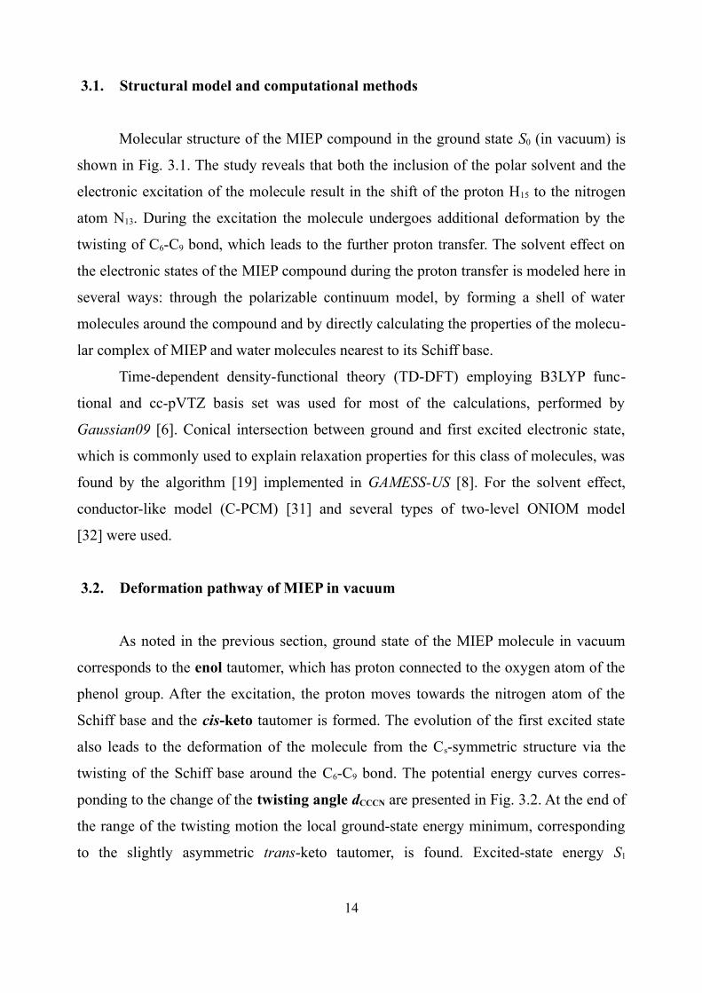
3.1. Structural model and computational methods
Molecular structure of the MIEP compound in the ground state S0 (in vacuum) is
shown in Fig. 3.1. The study reveals that both the inclusion of the polar solvent and the
electronic excitation of the molecule result in the shift of the proton H15 to the nitrogen
atom N13. During the excitation the molecule undergoes additional deformation by the
twisting of C6-C9 bond, which leads to the further proton transfer. The solvent effect on
the electronic states of the MIEP compound during the proton transfer is modeled here in
several ways: through the polarizable continuum model, by forming a shell of water
molecules around the compound and by directly calculating the properties of the molecu-
lar complex of MIEP and water molecules nearest to its Schiff base.
Time-dependent density-functional theory (TD-DFT) employing B3LYP func-
tional and cc-pVTZ basis set was used for most of the calculations, performed by
Gaussian09 [6]. Conical intersection between ground and first excited electronic state,
which is commonly used to explain relaxation properties for this class of molecules, was
found by the algorithm [19] implemented in GAMESS-US [8]. For the solvent effect,
conductor-like model (C-PCM) [31] and several types of two-level ONIOM model
[32] were used.
3.2. Deformation pathway of MIEP in vacuum
As noted in the previous section, ground state of the MIEP molecule in vacuum
corresponds to the enol tautomer, which has proton connected to the oxygen atom of the
phenol group. After the excitation, the proton moves towards the nitrogen atom of the
Schiff base and the cis-keto tautomer is formed. The evolution of the first excited state
also leads to the deformation of the molecule from the Cs-symmetric structure via the
twisting of the Schiff base around the C6-C9 bond. The potential energy curves corres-
ponding to the change of the twisting angle dCCCN are presented in Fig. 3.2. At the end of
the range of the twisting motion the local ground-state energy minimum, corresponding
to the slightly asymmetric trans-keto tautomer, is found. Excited-state energy S1
14

minimum and conical intersection point between S0-S1 states are located in the middle of
the motion range, albeit with the slight changes in the structure of the Schiff base. The
excited-state minimum and conical intersection point share many similarities in their
wavefunction character, which indicates that the MIEP molecule is likely to have two
competing energy relaxation pathways, manifesting as fluorescence from the excited
state and the non-radiative relaxation through the intersection point.
Figure 3.2. Potential energy curves of MIEP in vacuum
3.3. Changes of MIEP properties in solvent
Inclusion of the continuum solvent model brings the first change to the excitation
properties of MIEP, namely, that the most stable conformation in the ground state S0 now
corresponds to the cis-keto tautomer. This is in line with the earlier study [30]. On the
other hand, the potential energy curves retain their shape. To examine whether these
properties are also retained after direct inclusion of the solvent effects on the fairly small
molecule, the calculations of the MIEP in the solvent shell were performed. It was
15

noticed that several molecules nearest to the Schiff base of the compound form strong
(less than 2 Å) hydrogen bonds with MIEP (Fig. 3.3a), while all other solvent molecules
remain more distant (more than 2.5 Å). Due to this finding it was decided to perform
direct calculations of the electronic properties of the molecular complex of MIEP and
four water molecules (Fig. 3.3b).
a) b)Figure 3.3. MIEP molecule in water shell (a) and as a supramolecular complex (b)
Figure 3.4. Potential energy curves of MIEP-4H2O complex
16

Resulting potential energy surfaces are presented in Fig. 3.4. It is clear that the
inclusion of nearby water molecules considerably changes the shape of the ground-state
energy surface; instead of the single saddle point, it is now almost completely flat, with
several shallow minima. Moreover, the trans-keto tautomer does not belong to the one of
the minima as it did before. Excited-state energy surfaces are not affected as much,
although the conical intersection point is now somewhat higher than the excited-state
minimum, hinting at the possible strengthening of the radiative emission channel.
3.4. Comparison to MS1
The experimental studies of the photochromism of MIEP have not been performed
as of yet. However, the identified properties of the compound are in very good agree-
ment with the findings of the investigation of similar MS1 compound in another polar
solvent (ethanol). The excitation of MS1 generates intermediate structures and long-
living photoproducts shifted by 30 nm to the lower energies. The excited-state pathways
and the flat ground-state surface of MIEP would result in a similar observed behavior.
Additionally, neither MIEP in water nor MS1 in ethanol exhibit the photoproduct
corresponding to the trans-keto tautomer. Therefore, the observed relaxation properties
of the excited aromatic anil molecules are most probably caused by the direct interaction
of the compounds with the polar solvent molecules via the hydrogen-bonding.
▼▼▼
4. Photochromic properties of indolo-benzoxazine
The change of the optical properties of photochromic materials after excitation is
usually linked to the reversible structural deformation of the molecules [3]. Since such
deformation may include the spatial movement of the chemically active group or the
formation of controlled intermediate structure, the photochromic molecules are proposed
to be used as molecular-scale building blocks, for example, in high-density data storage
17

[33]. Among such molecules, spiropyrans [34] and coumarins [35] are known for their
excellent resistance during many excitation cycles. 5a,6-dihydro-12h-indolium[2,1-b]
[1,3]benzoxazine (indolobenzoxazine, IB) is one such compound, which exhibits ultra-
fast light-induced ring opening and formation of distinct chromophoric groups [36].
While the experimental results of the compound are tentatively ascribed to the excitation
dynamics of the two constituent moieties, 3H-indolium and 4-nitrophenol, the precise
dynamics is quite complicated and cannot be easily interpreted. The main task of the
theoretical investigation presented in this chapter was to determine the structure of the
electronic states of the IB compound and its constituent moieties and model the
evolution of the compound after the oxazine ring opening.
Figure 4.1. Molecular structure of IB compound
4.1. Structural model and computational methods
The IB compound in the ground state is shown in Fig. 4.1 together with the mar-
kings of the 3H-indolium (Ind) and 4-nitrophenol (pNph) chromophoric groups. The
joining site of the chromophoric groups constitutes a deformed oxazine ring. During
light excitation the O1-C2 bond is cleaved, resulting in the ring opening. Several local
energy minima corresponding to the open-ring structures are found during the investi-
gation. To describe the energy relaxation of the excited compound through the local
minima, two simplified reaction coordinates are used.
18
pNph(−)
Ind(+)
Oxazine ring

Time-dependent density-functional theory (TD-DFT), as implemented in
Gaussian03 [6] and GAMESS-US [8] packages, was used for the calculations. Positions
of ground-state energy minima were found using B3LYP functional and 6-311++(2d,p)
basis set, while the excited-state spectra for all structures were obtained using LC-BOP
functional and cc-pVDZ basis set. Polarizable continuum model [31] was used to include
the effects of acetonitrile solvent used in experiments [36].
4.2. Reaction coordinates of IB compound
In addition to the ground-state structure M0, two local energy minima of the IB
compound were located. The minima, M1 and M2, correspond to the open-ring form of
the compound, obtained by twisting 4-nitrophenol group around the N3-C4 bond in
different directions. Two minima of the first excited state S1, E1a and E1b, were also
located near the ground-state minima. Additionally, the excited-state energy minimum of
the S2 state, E2, was found near the optimal ground-state structure. While the twisting
angle of the N3-C4 bond qualitatively describes the changes in the 4-nitrophenol group,
additional coordinate was needed to indicate the evolution of the 3H-indolium; out of all
oxazine ring-related coordinates (Table 4.1), angle of the N3 atom to the symmetry plane
of 3H-indolium was selected. Ground-state potential energy surface corresponding to the
selected reaction coordinates is presented in Fig. 4.2. Ground- and excited-state minima
are marked on the resulting surface.
Table 4.1. Geometric parameters of the oxazine ring within IB complex for the various points on the potential energy surfaces. Values in italic represent the broken chemical bond
M1 E1a T01 M0 E2 T02 M2 E1b
r(O1-C2), Å 2.943 2.780 2.436 1.479 1.509 2.524 3.209 3.189
d(C8-C7-N3-C4), deg 172.1 172.5 161.9 155.4 154.7 165.6 175.1 153.9
a(N3-C4-C29), deg 111.6 110.5 108.6 111.2 110.6 116.0 113.4 108.8
d(N3-C4-C29-C34), deg −61.1 −55.8 −50.1 −17.1 −20.2 33.5 49.2 54.1
a(C4-C29-C34), deg 117.8 117.3 118.5 119.0 117.8 120.6 116.9 118.3
19

Figure 4.2. Potential energy surface of the ground state of IB compound
4.3. Relaxation pathways of the IB compound
The ground- and excited-state potential energy curves, as well as relaxation
pathways of the optical excitation, are shown in Fig. 4.3. Comparing calculation results
with the spectral measurements of IB compound [36], the following conclusions can be
drawn:
• The absorption peak of the IB compound is mainly due to the absorption of the
4-nitrophenol group.
• Cleavage of the O1-C2 bond results in the immediate formation of the partially-
charged 3H-indolium group. This is confirmed by the calculated partial charges of
the chromophoric groups within the compound that are equal to about ±0.5e for
all open-ring structures.
• The delayed formation of the deprotonated pNphe- group is caused by the confor-
mational changes, namely, the twisting of the 4-nitrophenol group around the
N3-C4 bond. Additionally, the formation yield is not 100 %, because there is a
20

relaxation channel through the excited state E2 which does not result in the
significant deformation of the compound.
• Long-lasting spectral signals of the IB compound can be explained by the
existence of the local ground- and excited-state minima. Ground-state minima M1
and M2 are reached though the excited-state minimum structures E1a and E1b,
formed by the interaction between 3H-indolium and 4-nitrophenol groups. IB
compound is able to revert from the local minima to the initial state by
thermalization due to the shallow potential energy surface and low barriers
between different states.
It has to be noted that the recent experimental studies of the derivative IB
compound [37] confirm the presence of the open-ring structures formed via the first
excited state, as well as the short-lived cleaved-bond state without the significant charge
separation.
Figure 4.3. Potential energy curves and relaxation pathways of IB compound
21
5. Efficient phosphorescence of PBMSi polymer
Most popular OLED emitter materials used today are almost invariably based on
the spin-orbit interaction phenomenon that allows non-radiative conversion between
singlet and triplet excited states, as well as emission from the lowest triplet state to the
singlet ground state [38]. This is achieved by doping the materials with triplet emitter
complexes formed around the heavy-metal atom, such as iridium or platinum [39,40].
The charge-carrier properties of the material can be enhanced by adding functional
groups, such as carbazoles [41], to the complexes. The main drawback of the
organometallic complexes is the discrepancy in efficiency of red-green emitters and
blue-emitting materials [42]. Therefore new materials with higher triplet states and better
charge-transfer properties are heavily searched for.
Organic semiconductors without the presence of heavy-metal atoms ordinarily
exhibit only extremely weak (in the order of 10−4) phosphorescence. However, experi-
ments on silicon-based PBMSi polymer have established extremely efficient phospho-
rescence in this polymer, with quantum yield of up to 15 %. The emission is believed to
originate from the biphenyl side-groups, even when only the Si chain is excited. The
main task of the following study was to determine the electronic state structure of the
PBMSi-comparable model compound and explain the cause of the observed emission of
the polymer.
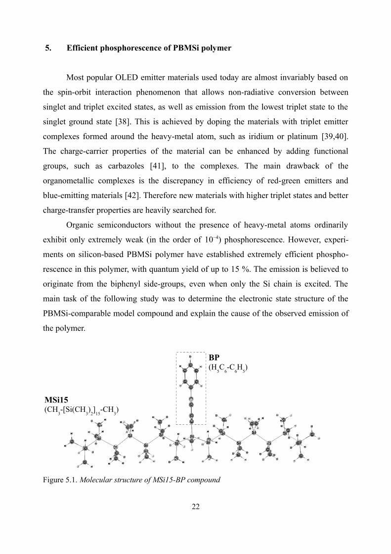
Figure 5.1. Molecular structure of MSi15-BP compound
22
MSi15(CH
3-[Si(CH
3)
2]
15-CH
3)
BP(H
5C
6-C
6H
5)

5.1. Structural model and computational methods
Due to the obvious difficulties of calculating the properties of 40- to 50-element
long polymer with side-groups, PBMSi was modeled by joining methylated Si-chain and
the single biphenyl group in the center of the chain. The structure of the resulting
compound, MSi15-BP, is shown in Fig. 5.1.
Time-dependent density-functional theory (TD-DFT) employing B3LYP functio-
nal and the 6-31G(d,p) basis set was used for the most calculations performed by the
Gaussian09 package [6]. Additional GAMESS-US [8] calculations by the GMC-QDPT
method [43] were performed for the validation of the TD-DFT wavefunction character.
5.2. Charge-transfer state of MSi15-BP
Calculated parameters of the excited electronic states of MSi15-BP compound are
summarized in Table 5.1. According to both TD-DFT and GMC-QDPT calculations, the
lowest excited singlet state S1 is of a charge-transfer character, corresponding to the
charge redistribution from the Si chain onto the biphenyl group (Figure 5.2, 1-1'). Second
singlet excited state S2 corresponds to the Si chain excitation, while the absorption of the
biphenyl group is located significantly higher. Lowest (emitting) triplet state of the
compound T1 shows the character of the biphenyl group.
Table 5.1. Calculated parameters of the excited electronic states of MSi15-BP compound
TD-DFT GMC-QDPT
No. Et, eV fosc Transition No. Et, eV fosc Transition
T1 3.169 - 3-1' BP (π-π*) T1 3.235 - 2-1' BP (π-π*)
T2 3.726 - 1-2' Si (σ-σ*) T2 4.456 - 1-2' Si (σ-σ*)
T3 3.815 - 1-1' CT S1 4.836 0.405 1-1' CT
S1 3.930 0.113 1-1' CT S2 5.530 1.568 1-2' Si (σ-σ*)
T4 4.019 - 1-1' CT T3 5.687 - 1-3' CT
S2 4.063 2.510 1-2' Si (σ-σ*) T4 5.925 - 1-3' CT
S6 4.684 0.281 3-1' BP (π-π*) S4 6.424 0.434 2-1' BP (π-π*)
23

4 1'
2 2'
1 3'
Figure 5.2. Frontier molecular orbitals of the MSi15-BP compound (GMC-QDPT results)
While the charge-transfer state is only slightly lower than the Si-chain absorption
state for the ground-state structure of the MSi15-BP, the character of this state can be
validated by other means. First, optimized structure of charge-transfer state shows fairly
large gap between the two states. Second, the energy of the charge-transfer state is
expected to be overestimated due to the environment interaction effects that are missing
from the calculations of limited-length chain in vacuum. Lastly, the calculated lifetime of
the S1-S0 transition (0.89 ns) is in good agreement with the PBMSi fluorescence lifetime
(1.2 ns) which is itself unusual for this class of materials [44].
5.3. Excited-state relaxation of MSi15-BP
The relaxation scheme of MSi15-BP compound is presented in Fig. 5.3. It
contains fast internal conversion between Si-chain absorption and charge-transfer states
(corroborated by the located intersection point in the vicinity of the absorption structure),
enhanced intersystem crossing between singlet and triplet CT states [45] and subsequent
triplet conversion followed by emission from the lowest state of the biphenyl group.
24
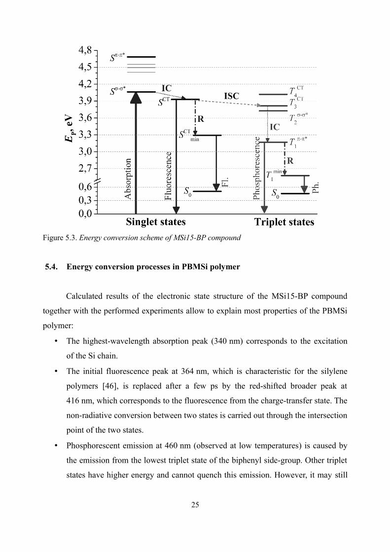
Figure 5.3. Energy conversion scheme of MSi15-BP compound
5.4. Energy conversion processes in PBMSi polymer
Calculated results of the electronic state structure of the MSi15-BP compound
together with the performed experiments allow to explain most properties of the PBMSi
polymer:
• The highest-wavelength absorption peak (340 nm) corresponds to the excitation
of the Si chain.
• The initial fluorescence peak at 364 nm, which is characteristic for the silylene
polymers [46], is replaced after a few ps by the red-shifted broader peak at
416 nm, which corresponds to the fluorescence from the charge-transfer state. The
non-radiative conversion between two states is carried out through the intersection
point of the two states.
• Phosphorescent emission at 460 nm (observed at low temperatures) is caused by
the emission from the lowest triplet state of the biphenyl side-group. Other triplet
states have higher energy and cannot quench this emission. However, it may still
25

be quenched by the external impurities and therefore might be not detectable at
room temperature.
• The position of the charge-transfer state in the electronic spectrum, namely small
positive singlet-triplet CT splitting, explains the striking efficiency of the
phosphorescence [45]. The similar effect was recently observed for the inter-
molecular polaron pairs in CuPC:PCBM blends [47]. The internal conversion of
triplet states takes place of the conventional polaron recombination in this case.
Therefore this study suggests a new molecular-level engineering approach for the
enhancement of the ISC, enabling efficient conversion of primary excited singlets into
triplets in conjugated polymers without involving heavy atom effect.
▼▼▼
6. Phosphorescence of the organometallic iridium complex
Even despite the drawback mentioned in the previous chapter, organometallic
complexes are the most widely used triplet emitters for the organic semiconductor
applications [48], because the triplet states can be efficiently populated by electric charge
carriers and then emit photons with internal quantum efficiency close to 100 % [49].
However, despite the multitude of experimental and theoretical studies, technology of
organic semiconductors is still young and has not reached its limiting potential [50].
Of the organometallic complexes used, the most popular are the iridium-based
compounds because of the effective room-temperature emission [39] as well as long-
lived excitations [51]. On the other hand, the excitation lifetime poses some difficulties
as well, because in pure materials excitations might be quenched due to the annihilation
processes [52]. To avoid this, the organometallic complexes are “diluted” by adding
stable charge-carrier groups, for example, carbazoles [53]. The main task of the
following study was to investigate the electronic spectrum of the organometallic
iridium(III) complex, bis(phenylbenzothiazole)-Ir-acetylacetonate ((pbt)2Ir(acac)), with
and without the added carbazole groups.
26

6.1. Structural model and computational methods
a)
NSIr
O
O
2 b)
NS
N OO
OHOHN N
NN
Figure 6.1. Chemical structure of the investigated compounds: a) (pbt)2Ir(acac) complex,b) pbt ligand with added carbazole groups
Chemical structure of the (pbt)2Ir(acac) compound and pbt ligand with added
carbazole groups is shown in Fig. 6.1. The actual molecular structure of the compounds
is more complicated (cf. sections 6.2, 6.3).
Time-dependent density-functional theory employing B3LYP functional and
Lanl2DZ [54]/ 6-31G(d) basis sets was used for all calculations performed by
Gaussian03 package [6]. Continuum solvent model IEF-PCM [55] was used to include
the effects of tetrahydrofuran solvent.
6.2. Properties of the absorption spectra
Theoretical absorption spectrum of the pbt ligand is presented in Fig. 6.2a, while
the corresponding molecular orbitals are shown in Fig. 6.3. It is clear from the figures
that the addition of the carbazole groups results in the red shift of the main absorption
peak of the ligand because of the enlarged π system.
Addition of the charge-carrier groups to the entire photoactive complex changes
only the position and height of the peak attributed to the separate ligand excitation
(Fig. 6.2b), but not the lowest excitation of the complex, which includes the iridium
atom. This indicates that the charge-carrier groups do not affect the optical properties of
this compound.
27

a) b)Figure 6.2. The absorption spectrum of a) the 2-phenylbenzothiazole, b) the (pbt)2Ir(acac) complex, with and without the carbazole groups
1
pbt
1
pbt+4cz1'
a)
1'
b)Figure 6.3. Frontier molecular orbitals of the 2-phenylbenzothiazole (a) and with added carbazole groups (b)
6.3. Structure and properties of the (pbt)2Ir(acac) complex
The geometric parameter optimization of the (pbt)2Ir(acac) compound results in
the complicated structure shown in Fig. 6.4. The pbt ligands and acetylacetonate bridge
surround the central iridium atom. Similar structures have been reported elsewhere [56].
This structure is very rigid; it does not change significantly between the optimized
ground-state structure S0 and the phosphorescent structure T1. The main changes are
observed in the parameters of one of the ligands. The changes break the symmetry of the
28

complex, thereby inducing separation of the previously almost-degenerate energy levels
(Fig. 6.5). As mentioned previously, addition of the carbazole groups does not have
much influence on the properties of the lowest-lying excited states of the compound. On
the other hand, the appearance of dark excited states corresponding to the carbazole-
ligand electronic transitions indicates the possible mechanism of excitation transfer to
the photoactive complex.
Figure 6.4. Molecular structure of the (pbt)2Ir(acac) complex
Figure 6.5. Energy level diagram of the (pbt)2Ir(acac) complex
29
pbt a pbt bpbt apbt b
acac acac

7. Conclusions
MIEP molecule
• MIEP molecule possesses two competing energy relaxation channels: excited-
state emission and non-radiative relaxation through the intersection point on the
potential energy surface.
• The polar solvent model predicts significant changes in the ground-state energy
surface and is more favorable to the radiative conversion than the vacuum model.
• The main solvent effects are seen only when treating MIEP and adjacent solvent
molecules as a supramolecular complex.
IB compound
• The chromophoric groups of the excited IB compound are formed in the different
timescales due to the time needed for the conformational changes of the
nitrophenol group.
• There exists an additional relaxation pathway without the significant structural
changes that quenches the formation of the negatively-charged nitrophenolate.
• Shallow surface of the ground-state energy surface and low transition barriers are
responsible for the thermal reversion of the IB compound to the initial state.
PBMSi polymer
• PBMSi polymer has low-lying singlet and triplet charge-transfer excited states
that correspond to the electron density redistribution from the Si chain to the
biphenyl group.
• The position of charge-transfer state is extremely favorable for the enhanced
internal conversion and intersystem crossing in the material, leading to the
efficient triplet population in the PBMSi polymer without heavy-metal effect.
(pbt)2Ir(acac) complex
• Differences between the absorption and phosphorescence states in (pbt)2Ir(acac)
complex arise due to the symmetry-breaking structural changes in the ligands.
• The addition of the carbazole groups has no significant effect to the optical
properties of the lowest-lying electronic states of the (pbt)2Ir(acac) complex.
30

8. The approbation of the conducted research
The results of the studies presented in the dissertation have been published in the
following scientific papers:
1. S. Toliautas, J. Sulskus, L. Valkunas, M. Vengris, “Quantum chemical studies of
photochromic properties of benzoxazine compound,” Chemical Physics 404, 64–
73, 2012.
2. S. Toliautas, M. Macernis, J. Sulskus, L. Valkunas, “Solvent effect on the photo-
induced proton transfer in 2-(N-methyl-α-iminoethyl)-phenol,” Chemical Physics
Letters 591, 52–57, 2014.
3. A. Kadashchuk, Yu. Skryshevski, A. Vakhnin, S. Toliautas, J. Sulskus, R. Augulis,
V. Gulbinas, S. Nespurek, J. Genoe, L. Valkunas, “Highly efficient intrinsic
phosphorescence from a σ-conjugated poly(silylene) polymer,” The Journal of
Physical Chemistry C, 2014 (under review).
The studies have been also announced in the following scientific conferences:
1. S. Toliautas, J. Šulskus, M. Mačernis, M. Vengris, L. Valkūnas, “Modelling of
photochromic properties of benzoxazine compound by means of quantum
chemical methods,” ERPOS 12: Electronic and Related Properties of Organic
Systems, Vilnius, 2011 (poster).
2. S. Toliautas, J. Šulskus, K. Kazlauskas, S. Juršėnas, V. Getautis, L. Valkūnas,
“Quantum-chemical calculations of novel phosphorescent iridium complexes,”
ERPOS 12: Electronic and Related Properties of Organic Systems, Vilnius, 2011
(poster).
3. S. Toliautas, J. Šulskus, L. Valkūnas, „Benzoksazino junginio potencinės
energijos paviršiai šviesa indukuoto sužadinimo metu“, 39th Lithuanian National
Physics Conference, Vilnius, 2011 (poster).
4. S. Toliautas, J. Šulskus, L. Valkūnas, “Modelling of structural and excitation
properties of novel light-sensitive organic complexes,” Vilnius Workshop on
Nonlinear Spectroscopy and Open Quantum Systems, Vilnius, 2011 (oral).
31

5. S. Toliautas, J. Šulskus, K. Kazlauskas, S. Juršėnas, V. Getautis, L. Valkūnas,
“Modelling of excited-state properties of novel phosphorescent iridium
complexes,” 9th International Conference on Nanosciences & Nanotechnologies
(NN12), Thessaloniki, 2012 (poster).
6. S. Toliautas, “Potential energy surface analysis of photochromic compound based
on quantum-chemical computation,” Vilnius Workshop on Nonlinear
Spectroscopy and Open Quantum Systems, Vilnius, 2012 (oral).
7. S. Toliautas, M. Mačernis, J. Šulskus, L. Valkūnas, „Tirpiklio poveikis
fotoindukuotai protono pernašai 2-(N-metil-α-iminoetil)-fenolio molekulėje“,
40th Lithuanian National Physics Conference, Vilnius, 2013 (poster).
8. S. Toliautas, J. Šulskus, A. Kadashchuk, Yu. Skryshevski, A. Vakhnin, R. Augulis,
V. Gulbinas, S. Nespurek, J. Genoe, L. Valkūnas, “Highly efficient intrinsic
phosphorescence from a σ-conjugated poly(silylene) polymer,” 11th Nordic
Femtochemistry Conference, Vilnius, 2014 (poster).
Additional presentations by A. Kadashchuk:
9. A. Kadashchuk, Yu. Skryshevski, S. Toliautas, J. Sulskus, L. Valkunas,
S. Nespurek, “Highly efficient intrinsic phosphorescence from a σ-conjugated
polysilane polymer,” Baltic Polymer Symposium 2013, Trakai, 2013 (poster).
10. A. Kadashchuk, S. Toliautas, J. Sulskus, R. Augulis, V. Gulbinas, S. Nespurek,
L. Valkunas, “Highly efficient intrinsic phosphorescence from a sigma-conjugated
poly(silylene) polymer,” ERPOS 13: Electrical and Related Properties of
Organic Solids, Swieradow Zdroj, 2014 (invited lecture).
32
About the author
Stepas Toliautas was born in 1985 in Vilnius. He attended S. Stanevičius secon-
dary school. In 2003 he graduated the Vilnius Lyceum and entered the Faculty of Physics
at the Vilnius University. There he obtained bachelor of physics and master of physics
academic degrees (see below). In 2006, S. Toliautas studied for 5 months at the Linko-
ping University in Sweden through the Erasmus student exchange program.
From 2007, S. Toliautas works at the Faculty of Physics, Department of Theore-
tical Physics as engineer. He is part of a research team at the department's Process
Modeling Laboratory. He actively participates in preparation and teaching courses of
bachelor study program “Computational Physics”. S. Toliautas has also worked as data-
base system engineer and mobile games developer.
Education
1999–2003 Vilnius Lyceum
2003–2007 Bachelor studies, Faculty of Physics at the Vilnius University
(study program “Computational Physics”)
2007–2009 Master studies, Faculty of Physics at the Vilnius University
(study program “Theoretical Physics and Astrophysics”)
2009–2013 Doctoral studies in field of physics at the Vilnius University (02P)
33

References
1. J. K. Lanyi, “Proton transfers in the bacteriorhodopsin photocycle,” Biochimica et Biophysica Acta, 1757 (8), 1012–1018, 2006.
2. V. Molina, M. Merchán, “On the absorbance changes in the photocycle of the photoactive yellow protein: a quantum-chemical analysis,” Proceedings of the National Academy of Sciences of the United States of America, 98 (8), 4299–4304, 2001.
3. T. Hugel, N. B. Holland, A. Cattani, L. Moroder, M. Seitz, H. E. Gaub, “Single-molecule optomechanical cycle,” Science, 296 (5570), 1103–1106, 2002.
4. Z. S. Wang, “Optimally controlled optomechanical work cycle for a molecular locomotive,” Journal of Physics. Condensed Matter : an Institute of Physics journal, 17 (47), S3767–3782, 2005.
5. M. Pope, C. E. Swenberg, Electronic Processes in Organic Crystals and Polymers, 2nd ed., Oxford University Press (Oxford), 1999.
6. M. J. Frisch, G. W. Trucks, H. B. Schlegel, G. E. Scuseria, M. A. Robb, J. R. Cheeseman, G. Scalmani, V. Barone, B. Mennucci, G. A. Petersson, H. Nakatsuji, M. Caricato, X. Li, H. P. Hratchian, A. F. Izmaylov, J. Bloino, G. Zheng, J. L. Sonnenberg, M. Hada, M. Ehara, K. Toyota, R. Fukuda, J. Hasegawa, M. Ishida, T. Nakajima, Y. Honda, O. Kitao, H. Nakai, T. Vreven, J. A. Montgomery Jr., J. E. Peralta, F. Ogliaro, M. J. Bearpark, J. J. Heyd, E. Brothers, K. N. Kudin, V. N. Staroverov, R. Kobayashi, J. Normand, K. Raghavachari, A. Rendell, J. C. Burant, S. S. Iyengar, J. Tomasi, M. Cossi, N. Rega, N. J. Millam, M. Klene, J. E. Knox, J. B. Cross, V. Bakken, C. Adamo, J. Jaramillo, R. Gomperts, R. E. Stratmann, O. Yazyev, A. J. Austin, R. Cammi, C. Pomelli, J. W. Ochterski, R. L. Martin, K. Morokuma, V. G. Zakrzewski, G. A. Voth, P. Salvador, J. J. Dannenberg, S. Dapprich, A. D. Daniels, Ö. Farkas, J. B. Foresman, J. V. Ortiz, J. Cioslowski, D. J. Fox, Gaussian09. Gaussian, Inc., Wallingford, CT, 2013.
7. M. W. Schmidt, K. K. Baldridge, J. A. Boatz, S. T. Elbert, M. S. Gordon, J. H. Jensen, S. Koseki, N. Matsunaga, K. A. Nguyen, S. Su, T. L. Windus, M. Dupuis, J. A. Montgomery Jr., “General atomic and molecular electronic structure system,” Journal of Computational Chemistry, 14 (11), 1347–1363, 1993.
8. M. S. Gordon, M. W. Schmidt, “Advances in electronic structure theory: GAMESS a decade later,” in Theory and Applications of Computational Chemistry: the first forty years, C. E. Dykstra, G. Frenking, K. S. Kim, and G. E. Scuseria, Eds. Elsevier (Amsterdam), 2005, 1167–1189.
9. M. Valiev, E. J. Bylaska, N. Govind, K. Kowalski, T. P. Straatsma, H. J. J. Van Dam, D. Wang, J. Nieplocha, E. Apra, T. L. Windus, W. A. de Jong, “NWChem: A comprehensive and scalable open-source solution for large scale molecular simulations,” Computer Physics Communications, 181 (9), 1477–1489, 2010.
34

10. A. Jabłoński, “Über den Mechanismus der Photolumineszenz von Farbstoffphosphoren,” Zeitschrift für Physik, 94 (1–2), 38–46, 1935.
11. J. Zimmermann, A. Zeug, B. Röder, “A generalization of the Jablonski diagram to account for polarization and anisotropy effects in time-resolved experiments,” Physical Chemistry Chemical Physics, 5 (14), 2964–2969, 2003.
12. J. Hinze, “Developments in the calculation of electronic wavefunctions for molecules: MCSCF, CI, and numerical SCF for molecules,” International Journal of Quantum Chemistry, 20 (S15), 69–90, 1981.
13. R. G. Parr, W. Yang, Density-Functional Theory of Atoms and Molecules, Oxford University Press (Oxford), 1989.
14. R. J. Bartlett, G. D. Purvis, “Many-body perturbation theory, coupled-pair many-electron theory, and the importance of quadruple excitations for the correlation problem,” International Journal of Quantum Chemistry, 14 (5), 561–581, 1978.
15. J. A. Pople, R. Seeger, R. Krishnan, “Variational configuration interaction methods and comparison with perturbation theory,” International Journal of Quantum Chemistry, 12 (S11), 149–163, 1977.
16. M. W. Schmidt, M. S. Gordon, “The construction and interpretation of MCSCF wavefunctions,” Annual Review of Physical Chemistry, 49, 233–266, 1998.
17. E. Runge, E. K. U. Gross, “Density-functional theory for time-dependent systems,” Physical Review Letters, 52 (12), 997–1000, 1984.
18. B. G. Levine, J. D. Coe, T. J. Martínez, “Optimizing conical intersections without derivative coupling vectors: application to multistate multireference second-order perturbation theory (MS-CASPT2),” The journal of physical chemistry. B, 112 (2), 405–413, 2008.
19. S. Maeda, K. Ohno, K. Morokuma, “Updated branching plane for finding conical intersections without coupling derivative vectors,” Journal of Chemical Theory and Computation, 6 (5), 1538–1545, 2010.
20. R. C. Hilborn, “Einstein coefficients, cross sections, f values, dipole moments, and all that,” American Journal of Physics, 50 (11), 982–986, 2002.
21. M. Mačernis, “Aplinkos poveikis fotoindukuotiems reiškiniams organinėse molekulėse,” Vilniaus universitetas / Fizinių ir technologijos mokslų centras, 2011.
22. C. Tanner, C. Manca, S. Leutwyler, “Probing the threshold to H atom transfer along a hydrogen-bonded ammonia wire,” Science, 302 (5651), 1736–1739, 2003.
23. C. Reichardt, T. Welton, Solvents and Solvent Effects in Organic Chemistry, 4th ed., Wiley (Weinheim), 2011.
24. S. Hayashi, E. Tajkhorshid, K. Schulten, “Molecular dynamics simulation of bacteriorhodopsin’s photoisomerization using ab initio forces for the excited chromophore,” Biophysical Journal, 85 (3), 1440–1449, 2003.
35

25. K. Murata, Y. Fujii, N. Enomoto, M. Hata, T. Hoshino, M. Tsuda, “A study on the mechanism of the proton transport in bacteriorhodopsin: the importance of the water molecule,” Biophysical Journal, 79 (2), 982–991, 2000.
26. I. Król-Starzomska, A. Filarowski, M. Rospenk, A. Koll, S. M. Melikova, “Proton transfer equilibria in Schiff bases with steric repulsion,” The Journal of Physical Chemistry A, 108 (11), 2131–2138, 2004.
27. A. Filarowski, T. Głowiaka, A. Koll, “Strengthening of the intramolecular O-H-N hydrogen bonds in Schiff bases as a result of steric repulsion,” Journal of Molecular Structure, 484 (1–3), 75–89, 1999.
28. S. Mitra, N. Tamai, “A combined experimental and theoretical study on the photochromism of aromatic anils,” Chemical Physics, 246 (1–3), 463–475, 1999.
29. R. Karpicz, V. Gulbinas, A. Lewanowicz, M. Mačernis, J. Šulskus, L. Valkūnas, “Relaxation pathways of excited N-(triphenylmethyl)salicylidenimine in solutions,” The Journal of Physical Chemistry A, 115 (10), 1861–1868, 2011.
30. M. Mačernis, B. P. Kietis, J. Šulskus, S. H. Lin, M. Hayashi, L. Valkūnas, “Triggering the proton transfer by H-bond network,” Chemical Physics Letters, 466 (4–6), 223–226, 2008.
31. M. Cossi, N. Rega, G. Scalmani, V. Barone, “Energies, structures, and electronic properties of molecules in solution with the C-PCM solvation model,” Journal of Computational Chemistry, 24 (6), 669–681, 2003.
32. S. Dapprich, I. Komáromi, K. S. Byun, K. Morokuma, M. J. Frisch, “A new ONIOM implementation in Gaussian98. Part I. The calculation of energies, gradients, vibrational frequencies and electric field derivatives,” Journal of Molecular Structure: THEOCHEM, 461–462, 1–21, 1999.
33. M. Irie, “Photochromism: memories and switches - introduction,” Chemical Reviews, 100 (5), 1683–1684, 2000.
34. C. B. McArdle, Ed., Applied Photochromic Polymer Systems, Blackie (Glasgow), 1992.
35. T. Sakata, Y. Kawashima, H. Nakano, “Low-lying excited states of C120 and C151: a multireference perturbation theory study,” The Journal of Physical Chemistry A, 114 (47), 12363–12368, 2010.
36. M. Barkauskas, V. Martynaitis, A. Šačkus, R. Rotomskis, V. Sirutkaitis, M. Vengris, “Ultrafast dynamics of photochromic compound based on oxazine ring opening,” Lithuanian Journal of Physics, 48 (3), 231–242, 2008.
37. K. Redeckas, V. Voiciuk, R. Steponavičiūtė, V. Martynaitis, A. Šačkus, M. Vengris, “Ultrafast spectral dynamics of structurally modified photochromic indolo[2,1-b][1,3]benzoxazines,” Journal of Photochemistry and Photobiology A: Chemistry, 285, 7–15, 2014.
36

38. C. Adachi, M. A. Baldo, M. E. Thompson, S. R. Forrest, “Nearly 100% internal phosphorescence efficiency in an organic light-emitting device,” Journal of Applied Physics, 90 (10), 5048–5051, 2001.
39. S. Lamansky, P. Djurovich, D. Murphy, F. Abdel-Razzaq, H.-E. Lee, C. Adachi, P. E. Burrows, S. R. Forrest, M. E. Thompson, “Highly phosphorescent bis-cyclometalated iridium complexes: synthesis, photophysical characterization, and use in organic light emitting diodes,” Journal of the American Chemical Society, 123 (18), 4304–4312, 2001.
40. A. Köhler, J. S. Wilson, R. H. Friend, M. K. Al-Suti, M. S. Khan, A. Gerhard, H. Bässler, “The singlet–triplet energy gap in organic and Pt-containing phenylene ethynylene polymers and monomers,” The Journal of Chemical Physics, 116 (21), 9457–9463, 2002.
41. Y. Xuan, D. Pan, N. Zhao, X. Ji, D. Ma, “White electroluminescence from a poly-(N-vinylcarbazole) layer doped with CdSe/CdS core–shell quantum dots,” Nanotechnology, 17 (19), 4966–4969, 2006.
42. M. Roberts, N. Akino, K. Asada, P. Benzie, H. Hamamatsu, M. Hatcher, S. King, E. W. Snedden, A. Stevens, S. Tanaka, J. Toner, R. Wilson, W. Young, T. Yamada, “Progress in polymer OLED efficiency,” in 9th International Conference on Electroluminescence & Organic Optoelectronics (ICEL2012), 2012.
43. M. Miyajima, Y. Watanabe, H. Nakano, “Relativistic quasidegenerate perturbation theory with four-component general multiconfiguration reference functions,” The Journal of Chemical Physics, 124 (4), 044101/1–9, 2006.
44. Y. Kanemitsu, K. Suzuki, S. Kyushin, H. Matsumoto, “Visible photoluminescence from silicon-backbone polymers,” Physical Review B, 51 (19), 13103–13110, 1995.
45. S. Difley, D. Beljonne, T. Van Voorhis, “On the singlet-triplet splitting of geminate electron-hole pairs in organic semiconductors,” Journal of the American Chemical Society, 130 (11), 3420–3427, 2008.
46. S. Nešpůrek, A. Kadashchuk, Y. Skryshevski, A. Fujii, K. Yoshino, “Origin of broad visible luminescence in poly[methyl(phenyl)silylene] thin films,” Journal of Luminescence, 99 (2), 131–140, 2002.
47. E. W. Snedden, A. P. Monkman, F. B. Dias, “Photophysics of the geminate polaron-pair state in copper phthalocyanine organic photovoltaic blends: evidence for enhanced intersystem crossing,” Advanced Materials (Deerfield Beach, Fla.), 25 (13), 1930–1938, 2013.
48. M. A. Baldo, D. F. O’Brien, Y. You, A. Shoustikov, S. Sibley, M. E. Thompson, S. R. Forrest, “Highly efficient phosphorescent emission from organic electroluminescent devices,” Nature, 395, 151–154, 1998.
49. X. Yang, D. Neher, “Polymer electrophosphorescence devices,” in Organic Light Emitting Devices, K. Müllen and U. Scherf, Eds. Wiley (Weinheim), 2006, 333–368.
37

50. J. R. Sheats, “Manufacturing and commercialization issues in organic electronics,” Journal of Materials Research, 19 (07), 1974–1989, 2004.
51. S. Lamansky, P. Djurovich, D. Murphy, F. Abdel-Razzaq, R. Kwong, I. Tsyba, M. Bortz, B. Mui, R. Bau, M. E. Thompson, “Synthesis and characterization of phosphorescent cyclometalated iridium complexes,” Inorganic Chemistry, 40 (7), 1704–1711, 2001.
52. Y. Wang, N. Herron, V. V. Grushin, D. LeCloux, V. Petrov, “Highly efficient electroluminescent materials based on fluorinated organometallic iridium compounds,” Applied Physics Letters, 79 (4), 449–451, 2001.
53. L. Yang, J.-K. Feng, A.-M. Ren, J.-Z. Sun, “The electronic structure and optical properties of carbazole-based conjugated oligomers and polymers: a theoretical investigation,” Polymer, 47 (4), 1397–1404, 2006.
54. P. J. Hay, W. R. Wadt, “Ab initio effective core potentials for molecular calculations. Potentials for the transition metal atoms Sc to Hg,” The Journal of Chemical Physics, 82 (1), 270–283, 1985.
55. E. Cances, B. Mennucci, J. Tomasi, “A new integral equation formalism for the polarizable continuum model: theoretical background and applications to isotropic and anisotropic dielectrics,” The Journal of Chemical Physics, 107 (8), 3032–3041, 1997.
56. K. Swiderek, P. Paneth, “Modeling excitation properties of iridium complexes,” Journal of Physical Organic Chemistry, 22 (9), 845–856, 2009.
38
Stepas Toliautas
Elektroninio sužadinimo procesai fotoaktyviose organinėse molekulėse
Fotoaktyvios molekulės randamos beveik visose gyvosiose sistemose ir dalyvauja
tokių reiškinių kaip fotosintezė bei rega metu. Be to, grįžtami fotoaktyvių molekulių
struktūros pakitimai suteikia galimybę tokias molekules naudoti ypač mažo dydžio prie-
taisų konstravimui, o elektros ir šviesos energijos virsmai išnaudojami tobulinant orga-
ninės optoelektronikos elementus – organinius šviestukus bei saulės elementus. Minėti
energijos virsmai teoriškai aprašomi kaip molekulės elektroninio sužadinimo dinamika,
dėl kurios molekulė gali keisti struktūrą, spinduliuoti įvairaus bangos ilgio šviesą, suda-
ryti tarpinius tos pačios cheminės sudėties, tačiau skirtingų savybių fotoproduktus. Elek-
troninio sužadinimo dinamikos ypatybes įmanoma nustatyti tiesiogiai sprendžiant nenuo-
stoviąją Šriodingerio lygtį elektroninei posistemei, tačiau toks metodas reikalauja ypač
didelių skaičiavimo resursų ir šiandien tinka tik nedidelėms (kelių ar keliolikos atomų)
molekulėms arba smarkiai apibendrintoms kvantinėms sistemoms. Kita vertus, sužadi-
nimą bei tolesnius energijos virsmus įmanoma kokybiškai aprašyti sprendžiant nuosto-
viąją Šriodingerio lygtį skirtingoms molekulės branduolių padėtims ir nagrinėjant gautus
pagrindinės bei sužadintų elektroninių būsenų potencinės energijos paviršius. Tokiu būdu
galima tirti įvairaus dydžio ir struktūros molekulines sistemas bei gana skirtingos kilmės
reiškinius. Tyrimų, pateikiamų daktaro disertacijoje, tikslas buvo elektroninės struktūros
skaičiavimų metodais ištirti elektroninio sužadinimo sukeltus procesus keturiose foto-
aktyviose organinėse molekulėse ir joms sudaryti sužadinimo relaksaciją apibūdinančius
potencinės energijos paviršių modelius.
Pirmoji molekulė, MIEP, buvo tiriama siekiant modeliuoti bakteriorodopsino
baltymo funkcinės grupės – Šifo bazės – optines savybes bei molekulei priklausančio
protono pernašos eigą poliniame tirpiklyje. Tyrimo metu nustatyta, jog polinis tirpiklis
pakeičia molekulės enol ir cis-keto atmainų (besiskiriančių protono padėtimi) stabilumą
ir yra palankesnis cis-keto atmainai, kurios protonas prisijungęs prie Šifo bazės. Be to,
po sužadinimo molekulės Šifo bazė deformuojasi ir sukelia tolesnę erdvinę protono
pernašą. Nustatyta, jog tiesioginis tirpiklio molekulių įskaitymas deformacijos metu
39

smarkiai pakeičia pagrindinės molekulės būsenos paviršių; tai nestebima naudojant
elektrostatinį tirpiklio modelį. Gauti rezultatai kokybiškai atitinka panašaus junginio,
MS1, etanolyje eksperimentais stebėtas savybes ir leidžia paaiškinti šios klasės mole-
kulių ypatybes poliniuose tirpikliuose.
Antrasis tirtas junginys, indolo-benzoksazinas, pasižymi sudėtingomis fotochro-
minėmis savybėmis ir dėl atsparumo sužadinimo ciklų metu yra tinkamas molekulinių
jungiklių kūrimui. Tyrimo metu buvo paaiškinti eksperimentiniai rezultatai ir nustatyta,
jog junginio fotochromizmą lemia keli sužadinimo relaksacijos keliai, susiję su nitro-
fenolio grupės judėjimu indolo grupės atžvilgiu bei sąveika tarp grupių. Sukurtas mode-
lis puikiai atitinka neseniai skelbtus kito išvestinio indolo-benzoksazino junginio ekspe-
rimentinių tyrimų rezultatus.
Trečiasis junginys yra silicio polimeras su prijungtomis organinėmis grupėmis.
Šis junginys yra bene pirmas organinis konjuguotas polimeras, kuriame stebima naši
žematemperatūrė fosforescencija nesant sunkiųjų metalų kompleksų ar kitų priemaišų.
Bendradarbiaujant su keliomis eksperimentinėmis grupėmis nustatyta, kad už šią fosfo-
rescenciją yra atsakinga nedidelės energijos krūvio pernašos būsena tarp polimero silicio
grandinėlės ir bifenilo šoninės grupės. Teorinis tyrimas pademonstravo, jog singuletinės
ir tripletinės sužadintų krūvio pernašos būsenų padėtis energijos lygmenų spektre yra
palanki ypač sparčiai interkombinacinei konversijai medžiagoje, dėl kurios efektyviai
užpildomos tripletinės būsenos, iš kurių vyksta spinduliuotė.
Galiausiai buvo tirtas tradicinis metaloorganinis iridžio-ligandų kompleksas ir
nagrinėta jo sąveika su funkcinėmis krūvininkų pernašos grupėmis. Nustatyta sudėtinga,
nelanksti fotoaktyviojo komplekso struktūra, kuri sugerties metu yra beveik simetriška
dviejų ligandų atžvilgiu, o fosforescencijos metu stebimi nedideli vieno iš ligandų struk-
tūriniai pakitimai. Taip pat parodyta, jog prie ligando prijungtos karbazolių grupės šiek
tiek sumažina ligando sugerties energiją, tačiau praktiškai nekeičia iridžio komplekso
mažiausios energijos optiškai aktyvių elektroninių būsenų ypatybių.
40