ELECTROIMPEDANCE MAMMOGRAPHY - Úvod - VPL.sk prezentacie pdf/sala 1/2 piatok... · MAMMOGRAPHY...
30
ELECTROIMPEDANCE MAMMOGRAPHY Ing. Peter Jusko Slovak Republic www.onkocet.eu
Transcript of ELECTROIMPEDANCE MAMMOGRAPHY - Úvod - VPL.sk prezentacie pdf/sala 1/2 piatok... · MAMMOGRAPHY...
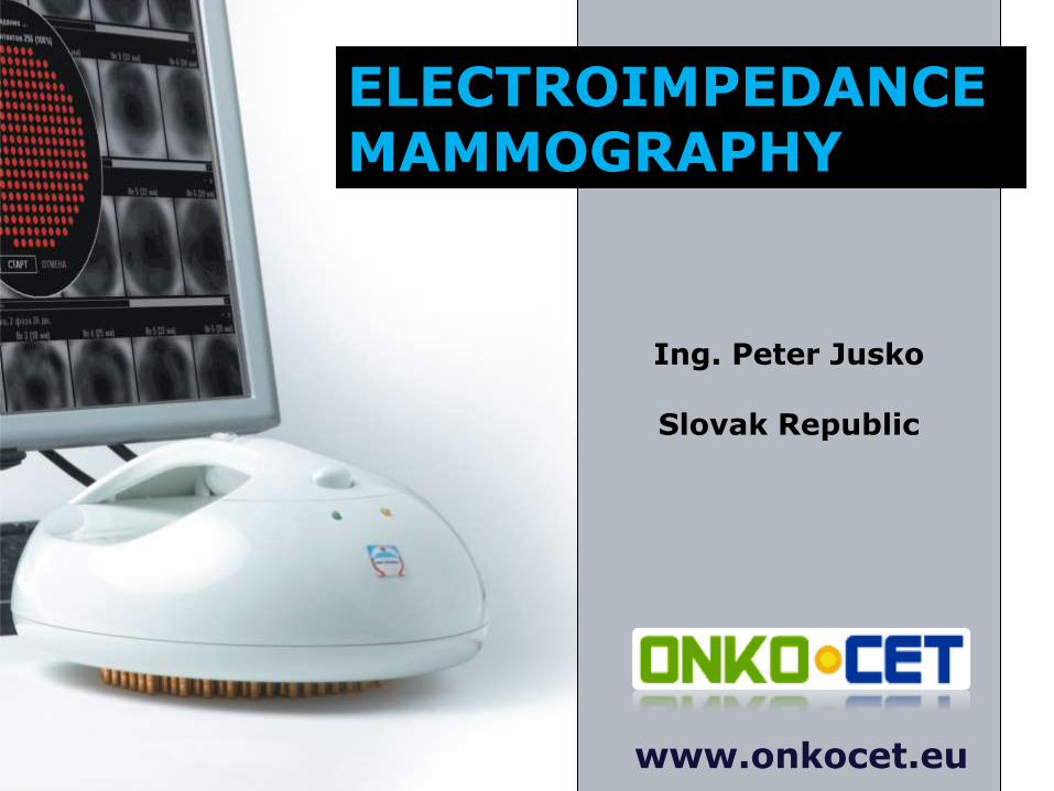
ELECTROIMPEDANCE MAMMOGRAPHY
Ing. Peter Jusko
Slovak Republic
www.onkocet.eu
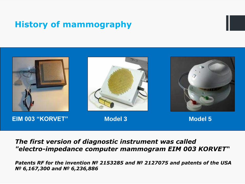
History of mammography
The first version of diagnostic instrument was called "electro-impedance computer mammogram EIM 003 KORVET“ Patents RF for the invention № 2153285 and № 2127075 and patents of the USA № 6,167,300 and № 6,236,886
EIM 003 “KORVET” Model 3 Model 5

Simple electrical model of a cell

Electrical characterisation of the materials
Dielectric properties

Electroconductivity map

Diagnostic set


Operating characteristics
sensitivity - 92%
specificity - 99%
the prognostic value of positive result - 73%
the prognostic value of negative result - 99%
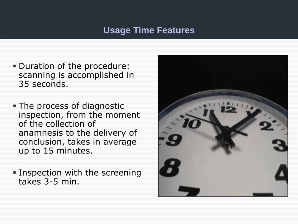
Usage Time Features
Duration of the procedure: scanning is accomplished in 35 seconds.
The process of diagnostic inspection, from the moment of the collection of anamnesis to the delivery of conclusion, takes in average up to 15 minutes.
Inspection with the screening takes 3-5 min.

Identification of pathologies using EI mammograph
Cancer
Cyst
Fibroadenoma
Calcificate
Scar
Inflammation

Breast cancer; type I.a
Visual and quantitative assessment data of the electroimpedance image of the patients’
mammary glands
Patient 66 years old, 10 years after menopause

Breast cancer; type I.a
Visual and quantitative assessment data of the electroimpedance image of the patients’ mammary glands
Patient 74 years old, 21 years after menopause

Breast cancer; type I.b
Visual and quantitative assessment data of the electroimpedance image of the patients’ mammary glands
Patient 45 years old, histology T2 N0 M0


Breast cancer; type I.b
Before surgery After surgery
Histogy: Invasive ductal CA with modular features G3, pT2pN0MX

Breast cancer; type Ic.
Patient 65 years old

Breast cancer; type Ic.
Patient 45 years old

Atypical breast cancer - haemangioma
A cancerous tumor which is hard, translucent, of a gray or
bluish color, and emits a creaking sound when incised.
Patient 44 years old


Mammary gland visualization - cysts
Patient 48 years old

Mammary gland visualization - cysts
Patient 53 years old, mammary gland 3 years after aspiration of a cyst

Mammary gland visualization - cysts
Patient 53 years old, mammary gland 3 years after aspiration of a cyst

Fibroadenoma Fibroadenoma
44 years old patient; Fibroadenoma bilat., lumpectomia bilat.

Fibroadenoma
Left breast:
fibroadenoma mammae I.
sin., extirpated 35 x 25 x
20 mm non capsulated
lobular lesion,
Histopathologically
demonstrated:
Phylloides tumor (PT),
(cystosarcoma phyllodes,
sometimes called „giant
fibroadenomas)

Fibroadenoma
Right breast:
fibroadenoma mammae I.
dx. extirpated 40 x 35 x
30 mm adipose tissue
fibrosis
Histopathologically
demonstrated:
fibrocystic parenchym of
right breast

Mammary gland
visualization - mastitis
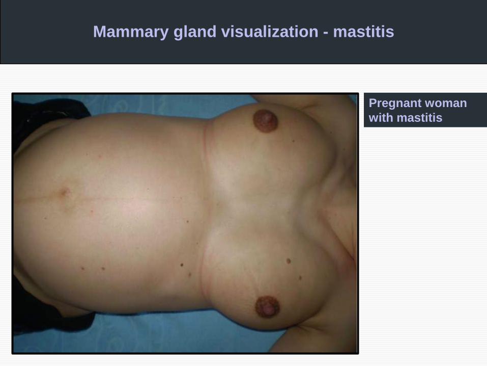
Mammary gland visualization - mastitis
Pregnant woman
with mastitis

Diagnostics of men for breast cancer
Patient 45 years old

Diagnostics of men
for breast cancer
Patient 45 years old
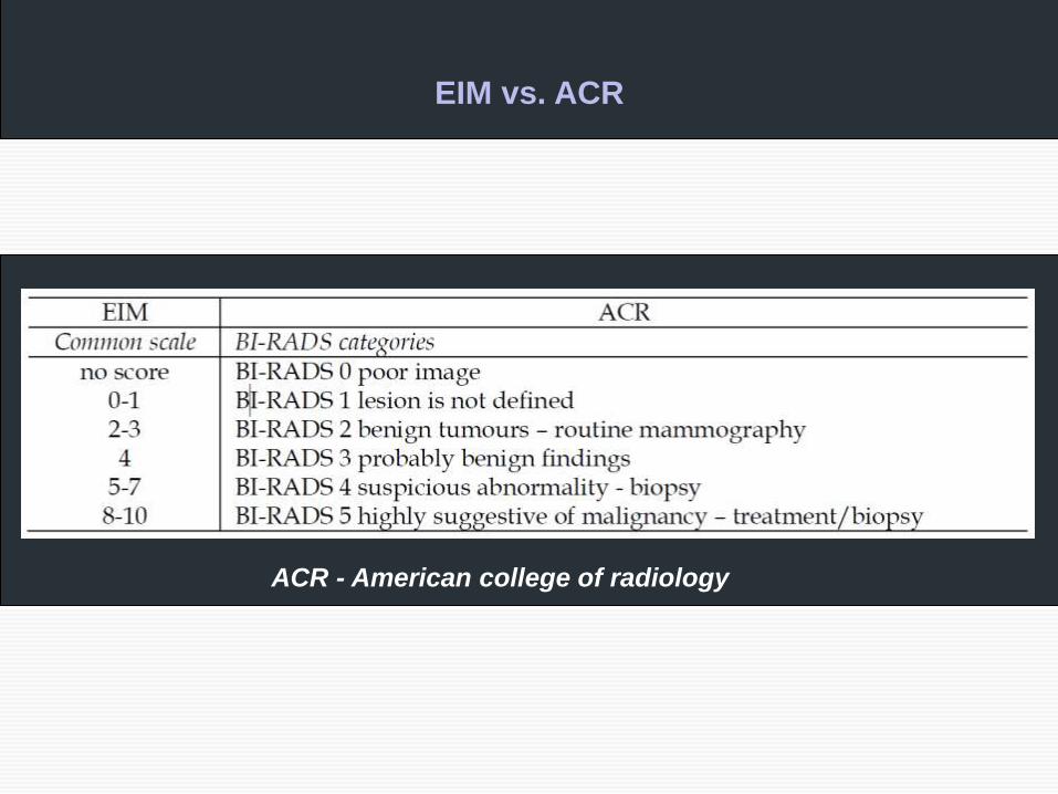
EIM vs. ACR
ACR - American college of radiology