Electrode selection matrix - SDU
42
University of Southern Denmark Consensus for experimental design in electromyography (CEDE) project Electrode selection matrix Besomi, Manuela; Hodges, Paul W; Van Dieën, Jaap; Carson, Richard G; Clancy, Edward A; Disselhorst-Klug, Catherine; Holobar, Aleš; Hug, François; Kiernan, Matthew C; Lowery, Madeleine; McGill, Kevin; Merletti, Roberto; Perreault, Eric; Søgaard, Karen; Tucker, Kylie; Besier, Thor; Enoka, Roger; Falla, Deborah; Farina, Dario; Gandevia, Simon; Rothwell, John C; Vicenzino, Bill; Wrigley, Tim Published in: Journal of Electromyography & Kinesiology DOI: 10.1016/j.jelekin.2019.07.008 Publication date: 2019 Document version: Accepted manuscript Document license: CC BY-NC-ND Citation for pulished version (APA): Besomi, M., Hodges, P. W., Van Dieën, J., Carson, R. G., Clancy, E. A., Disselhorst-Klug, C., Holobar, A., Hug, F., Kiernan, M. C., Lowery, M., McGill, K., Merletti, R., Perreault, E., Søgaard, K., Tucker, K., Besier, T., Enoka, R., Falla, D., Farina, D., ... Wrigley, T. (2019). Consensus for experimental design in electromyography (CEDE) project: Electrode selection matrix. Journal of Electromyography & Kinesiology, 48, 128-144. https://doi.org/10.1016/j.jelekin.2019.07.008 Go to publication entry in University of Southern Denmark's Research Portal Terms of use This work is brought to you by the University of Southern Denmark. Unless otherwise specified it has been shared according to the terms for self-archiving. If no other license is stated, these terms apply: • You may download this work for personal use only. • You may not further distribute the material or use it for any profit-making activity or commercial gain • You may freely distribute the URL identifying this open access version If you believe that this document breaches copyright please contact us providing details and we will investigate your claim. Please direct all enquiries to [email protected] Download date: 14. Feb. 2022
Transcript of Electrode selection matrix - SDU
University of Southern Denmark
Consensus for experimental design in electromyography (CEDE) project
Electrode selection matrixBesomi, Manuela; Hodges, Paul W; Van Dieën, Jaap; Carson, Richard G; Clancy, Edward A;Disselhorst-Klug, Catherine; Holobar, Aleš; Hug, François; Kiernan, Matthew C; Lowery,Madeleine; McGill, Kevin; Merletti, Roberto; Perreault, Eric; Søgaard, Karen; Tucker, Kylie;Besier, Thor; Enoka, Roger; Falla, Deborah; Farina, Dario; Gandevia, Simon; Rothwell, JohnC; Vicenzino, Bill; Wrigley, TimPublished in:Journal of Electromyography & Kinesiology
DOI:10.1016/j.jelekin.2019.07.008
Publication date:2019
Document version:Accepted manuscript
Document license:CC BY-NC-ND
Citation for pulished version (APA):Besomi, M., Hodges, P. W., Van Dieën, J., Carson, R. G., Clancy, E. A., Disselhorst-Klug, C., Holobar, A., Hug,F., Kiernan, M. C., Lowery, M., McGill, K., Merletti, R., Perreault, E., Søgaard, K., Tucker, K., Besier, T., Enoka,R., Falla, D., Farina, D., ... Wrigley, T. (2019). Consensus for experimental design in electromyography (CEDE)project: Electrode selection matrix. Journal of Electromyography & Kinesiology, 48, 128-144.https://doi.org/10.1016/j.jelekin.2019.07.008
Go to publication entry in University of Southern Denmark's Research Portal
Terms of useThis work is brought to you by the University of Southern Denmark.Unless otherwise specified it has been shared according to the terms for self-archiving.If no other license is stated, these terms apply:
• You may download this work for personal use only. • You may not further distribute the material or use it for any profit-making activity or commercial gain • You may freely distribute the URL identifying this open access versionIf you believe that this document breaches copyright please contact us providing details and we will investigate your claim.Please direct all enquiries to [email protected]
Download date: 14. Feb. 2022

Accepted Manuscript
Consensus for Experimental Design in Electromyography (CEDE) project:Electrode selection matrix
Manuela Besomi, Paul W Hodges, Jaap Van Dieën, Richard G Carson, EdwardA. Clancy, Catherine Disselhorst-Klug, Aleš Holobar, François Hug, MatthewC Kiernan, Madeleine Lowery, Kevin McGill, Roberto Merletti, Eric Perreault,Karen Søgaard, Kylie Tucker, Thor Besier, Roger Enoka, Deborah Falla, DarioFarina, Simon Gandevia, John C Rothwell, Bill Vicenzino, Tim Wrigley
PII: S1050-6411(19)30128-2DOI: https://doi.org/10.1016/j.jelekin.2019.07.008Reference: JJEK 2338
To appear in: Journal of Electromyography and Kinesiology
Received Date: 11 April 2019Revised Date: 8 July 2019Accepted Date: 17 July 2019
Please cite this article as: M. Besomi, P.W. Hodges, J. Van Dieën, R.G. Carson, E.A. Clancy, C. Disselhorst-Klug,A. Holobar, F. Hug, M.C. Kiernan, M. Lowery, K. McGill, R. Merletti, E. Perreault, K. Søgaard, K. Tucker, T.Besier, R. Enoka, D. Falla, D. Farina, S. Gandevia, J.C. Rothwell, B. Vicenzino, T. Wrigley, Consensus forExperimental Design in Electromyography (CEDE) project: Electrode selection matrix, Journal ofElectromyography and Kinesiology (2019), doi: https://doi.org/10.1016/j.jelekin.2019.07.008
This is a PDF file of an unedited manuscript that has been accepted for publication. As a service to our customerswe are providing this early version of the manuscript. The manuscript will undergo copyediting, typesetting, andreview of the resulting proof before it is published in its final form. Please note that during the production processerrors may be discovered which could affect the content, and all legal disclaimers that apply to the journal pertain.

1
Consensus for Experimental Design in Electromyography (CEDE) project: Electrode selection matrix
Manuela Besomi1, Paul W Hodges1*, Jaap Van Dieën2, Richard G Carson3,4,5, Edward A. Clancy6, Catherine Disselhorst-Klug7, Aleš Holobar8, François Hug1,9,10, Matthew C Kiernan11, Madeleine Lowery12, Kevin McGill13, Roberto Merletti14, Eric Perreault15,16, Karen Søgaard17, Kylie Tucker1,18, Thor Besier19, Roger Enoka20, Deborah Falla21, Dario Farina22, Simon Gandevia23, John C Rothwell24, Bill Vicenzino1, Tim Wrigley25
Corresponding author*:Professor Paul W. HodgesSchool of Health and Rehabilitation Sciences, The University of Queensland, Brisbane, Qld 4072, Australiae-mail: [email protected]: +61 404 854 589
Institutions: 1School of Health and Rehabilitation Sciences, The University of Queensland, Brisbane, Australia.2Department of Human Movement Sciences, Vrije Universiteit Amsterdam, Amsterdam Movement Sciences, Amsterdam, Netherlands.3Trinity College Institute of Neuroscience, School of Psychology, Trinity College Dublin, Dublin, Ireland. 4School of Psychology, Queen’s University Belfast, Belfast, UK.5School of Human Movement and Nutrition Sciences, The University of Queensland, Australia.6Worcester Polytechnic Institute, Worcester, MA, USA.7Department of Rehabilitation and Prevention Engineering, Institute of Applied Medical Engineering, RWTH Aachen University, Aachen, Germany.8Faculty of Electrical Engineering and Computer Science, University of Maribor, Koroška cesta 46, Maribor, Slovenia.9Faculty of Sport Sciences, Laboratory "Movement, Interactions, Performance" (EA 4334), University of Nantes, Nantes, France.10Institut Universitaire de France (IUF), Paris, France11Brain and Mind Centre, University of Sydney, Sydney, Australia; Department of Neurology, Royal Prince Alfred Hospital, Sydney, Australia. 12UCD School of Electrical and Electronic Engineering, University College Dublin, Belfield, Dublin, Ireland. 13US Department of Veterans Affairs.14LISiN, Department of Electronics and Telecommunications, Politecnico di Torino, Torino, Italy. 15Northwestern University, Evanston, IL, USA.16Shirley Ryan AbilityLab, Chicago, IL, USA.17Department of Clinical Research and Department of Sports Sciences and Clinical Biomechanics, University of Southern Denmark, Odense, Denmark. 18School of Biomedical Sciences, The University of Queensland, Brisbane, Australia.19Auckland Bioengineering Institute and Department of Engineering Science, University of Auckland, Auckland, New Zealand. 20Department of Integrative Physiology, University of Colorado Boulder, CO, USA. 21Centre of Precision Rehabilitation for Spinal Pain (CPR Spine), School of Sport, Exercise and Rehabilitation Sciences, University of Birmingham, UK.22Department of Bioengineering, Imperial College London, London, UK. 23Neuroscience Research Australia, University of New South Wales, Sydney, Australia.24Sobell Department of Motor Neuroscience and Movement Disorders, UCL Institute of Neurology, London, UK. 25Centre for Health, Exercise and Sports Medicine, Department of Physiotherapy, University of Melbourne, Parkville, Australia.
2
Abstract
The Consensus for Experimental Design in Electromyography (CEDE) project is an
international initiative which aims to guide decision-making in recording, analysis, and
interpretation of electromyographic (EMG) data. The quality of the EMG recording, and
validity of its interpretation depend on many characteristics of the recording set-up and
analysis procedures. Different electrode types (i.e., surface and intramuscular) will influence
the recorded signal and its interpretation. This report presents a matrix to consider the best
electrode type selection for recording EMG, and the process undertaken to achieve
consensus. Four electrode types were considered: 1) conventional surface electrode, 2)
surface matrix or array electrode, 3) fine-wire electrode, and 4) needle electrode. General
features, pros, and cons of each electrode type are presented first. This information is
followed by recommendations for specific types of muscles, the information that can be
estimated, the typical representativeness of the recording and the types of contractions for
which the electrode is best suited. This matrix is intended to help researchers when selecting
and reporting the electrode type in EMG studies.
Key words: Electromyography; Recording; Electrode types; Consensus; Reporting
3
1. Introduction
The quality of electromyography (EMG) recordings and the validity of the
interpretation of the data depend on many characteristics of the recording set-up and analysis
procedures. The optimal features differ between applications based on the question to be
addressed and the muscle under investigation. There are many issues to consider and the
purpose of the Consensus for Experimental Design in Electromyography (CEDE) project is to
provide expert consensus opinion of optimal features of recording set-up and analysis to
address a range of experimental questions. EMG electrodes are the interface between the
tissues and the recording system. The properties of the signal depend on their type (surface or
intramuscular), configuration (e.g. bipolar, matrix), and materials/construction (e.g.
Ag/AgCL, conductive ink). Selection of the appropriate type of electrode requires careful
consideration of the signal that is to be recorded, and the way in which the recording is to be
interpreted.
Electrodes for recording electromyography can be broadly defined as surface or
intramuscular. Surface electrodes are often applied as a conventional surface electrode pair or
an array (linear or matrix). Intramuscular electrodes can be either fine-wire or needles. In
both cases, multiple versions are available, that vary in their design characteristics and
recording properties. Less conventional surface electrodes (e.g., anal and vaginal probes)
(Keshwani & McLean, 2015; Merletti, 2016; Mesin et al., 2009), and some innovative new
electrodes that are currently under development/investigation (e.g. tattoo electrodes,
high‐adhesion stretchable electrodes, wearable high-resolution facial array) (Ferrari et al.,
2018; Inzelberg et al., 2018; Liu et al., 2017), generally share properties with those attached
to the skin (i.e. conventional surface or matrix electrodes), but have different types of fixation
and configuration, for specific contexts/situations. These can be considered according to the
principles described for conventional surface or matrix electrodes.

4
a. Conventional surface electrode. Typically consists of a single recording channel using
differential amplification of pairs of electrodes (either applied separately or as a pair
integrated into a single device) placed on the skin overlying a targeted muscle. Other
applications may involve more than two electrodes whose signals are combined to
produce a single output channel (e.g. double differential amplification, Laplacian).
[Merletti et al., 2016; Merletti et al., 2009].
b. Linear array or matrix surface electrode (also known as electrode arrays or grids,
multi-channel surface EMG, high-density surface EMG). Based on a multichannel
detection system arranged in one- (1-D) or two-dimensional (2-D) electrode arrays
[Merletti et al., 2016; Merletti et al., 2009].
c. Fine-wire electrode. Consists of fine diameter insulated wire(s) placed in the muscle
via a hypodermic needle. The insulation is removed from the tip (the length of which
is a determinant of the electrode’s receptive area) and is bent to maintain its
placement in the muscle, and the needle is withdrawn [Merletti & Farina, 2009].
d. Needle electrode. For these electrodes, the needle remains in the muscle.
Configurations involve either the needle shaft or tip (electrically insulated from the
remaining shaft) acting as a recording surface, or the recording surfaces can be
mounted on the shaft or tip of the needle. Several configurations are available with
different characteristics. These electrodes are commonly used to assess
neurophysiological characteristics of neuromuscular disorders [Merletti & Farina,
2009].
Not all electrodes are suitable for all applications, and electrodes must be carefully
selected with specific attention to the question to be answered and the desired properties of
the recording [Mesin, et al., 2009]. The decision to use an electrode type depends on the

5
characteristics of the muscle under investigation and the purpose of the study. Each type of
electrode has advantages and disadvantages that require consideration [Turker, 1993].
Recommendations have been made for bipolar surface EMG including electrode shape and
size, electrode placement, inter-electrode distance, electrode material, and sensor construction
[Hermens et al., 2000], and have been recently updated [Afsharipour et al., 2019]. Other
factors require consideration when selecting electrodes for a specific application. These
factors include the nature of the task (dynamic vs. static; maximal vs. submaximal), location
of the muscle innervation zones, potential for crosstalk, potential sources of noise (e.g.
motion artefact, electromagnetic radiation), depth of the muscle and thickness of the
subcutaneous fat tissue (as the signal is attenuated in the subcutaneous tissues when recorded
from surface EMG – an effect that differs between individuals). Specific issues relate to the
recorded muscle (architecture, location and size), and the information to be estimated and
interpreted (e.g. EMG amplitude vs. discrimination of single motor unit action potentials)
[Merletti et al., 2016; Disselhorst-Klug et al., 2009; Farina et al., 2014; Kuiken et al., 2003;
Merlo & Campanini, 2016; Staudenmann et al., 2010].
Given the complexity, diversity, and variability of EMG research and the growth in
research applications of EMG, recommendations to guide decision-making in recording, data
analysis, and reporting of EMG studies are crucial for accurate interpretation of findings.
This paper presents a guide to decision-making that can be used when selecting the most
appropriate electrode for a proposed EMG application and the process undertaken to achieve
consensus in developing these guidelines.
2. Methods
2.1 Project overview

6
The CEDE project is an international initiative which aims to develop consensus-
based matrices to guide decision-making in recording, analysis, and interpretation of EMG
data. Each design matrix considers specific study design features and the issues that need to
be considered when designing and interpreting the results from an EMG experiment/study.
The aim is to guide high-quality EMG research that enables valid and consistent
interpretation of findings, to aid the review of research using EMG, and to provide an
educational resource. The matrix for electrode selection was developed using a three-step
process: (1) development of draft content by a steering committee from CEDE project team
members; (2) general comments by the CEDE project team, and (3) a Delphi process for
refinement and endorsement of content. Approval for this project was obtained from the
Human Research Ethics Committee of The University of Queensland, Australia. Participants
of the Delphi process are among the co-authors.
2.2 CEDE team
The CEDE project team is composed of 21 researchers with expertise in the field of EMG
and a project coordinator. The details and the selection criteria of the expert panel can be
found elsewhere [Hodges et al., 2019].
2.3 Development of draft content by the steering committee from the CEDE project team
Draft content for the matrix was developed by the steering committee (MB, PWH) and
selected CEDE project members. Content was prepared with consideration of the major pros
and cons of each electrode type and experimental questions that influence the selection of
electrode type for an EMG experiment. The matrix was presented to the CEDE project team
at a face-to-face meeting to obtain broad feedback on the proposed design and content

7
features of the initial draft. This process was followed by refinement of the content and
further development before progressing to phase two.
The agreed general format for the matrix was a presentation of the content in six sections:
general design features and considerations for each type of electrode; pros and cons of each
method; and four clusters of recommendations based on common experimental questions.
These clusters were: 1) What muscles can be recorded; 2) What type of information can be
estimated; 3) Are the recordings representative of the entire muscle; 4) What types of
contractions can yield relevant data? For each experimental context, a recommendation of the
appropriateness of an electrode type for a specific application was provided as “yes”,
“caution”, “generally no”, or “no” (see Table 1 for definitions), along with an explanation.
2.4 General comments by broad CEDE project team
After the initial broad consultation and subsequent refinement and organization of the
content, the draft matrix was sent via email to all experts for further detailed feedback of
content. Comments were collated and integrated for refinement of the matrix. Nine team
members were contacted to provide detailed feedback related to the analysis of EMG
amplitude/frequency. The revised content of these sections of the matrix was re-sent to the
relevant individuals to confirm the accuracy of the integration of changes.
2.5 Delphi process for refinement and endorsement of content
An online Delphi approach was used to reach consensus among experts. This approach is
a widely accepted method to achieve consensus and is used as a decision-making method
[Waggoner et al., 2016]. The Delphi technique uses multiple rounds of questionnaires that
can involve allocation of ratings and/or open-ended answers [von der Gracht, 2012]. In round
one, the entire matrix was sent to the experts along with the instructions and timeline for

8
completion. A reminder was emailed after two weeks. The same approach and timeline were
used for subsequent rounds. For the assessment of satisfaction level and
agreement/disagreement among participants, a nine-point Likert scale was used [Fitch et al.,
2001] that asked contributors to indicate that they considered that content was “appropriate”
(score 7–9), “uncertain” (score 4–6) or “inappropriate” (score 1–3). Participants rated their
agreement for each cell of the matrix and were invited to provide comments to highlight
aspects that were not agreeable. Consensus was considered to be reached if >70% of
contributors provided scores between 7–9 [appropriate] and <15% of contributors provided
scores between 1–3 [inappropriate] [Williamson et al., 2012]. As a further criterion, an
interquartile range (IQR) < 2 units on a nine-unit scale was necessary to consider that
consensus had been reached among Delphi panelists [von der Gracht, 2012]. For cells that did
reach consensus, any contributor’s comments that were recorded were considered and
implemented if they improved the content (as judged by the steering committee).
Based on the results of round one, items with an insufficient consensus were refined by
the steering committee by integrating feedback and re-sent to the experts who had provided
ratings scores <7. Changes or new information proposed by contributors were highlighted in
the second-round questionnaire. The same process was followed for subsequent rounds. All
contributors reviewed the final document for endorsement and were included as authors. For
this matrix, 20 experts participated in the Delphi process. The lead investigator (PH) and the
coordinator (MB) did not participate in that process, but in addition to developing the initial
content, they oversaw the project and collected/integrated all the responses.
All data were entered into Microsoft Excel and processed using the statistical package
STATA/IC (version 14). The number and percentage of participants rating each outcome as
appropriate (score 7-9), uncertain (score 4-6) and inappropriate (score 1-3) were calculated,
as well as the median and IQR for each item.

9
3. Results
After phase 1, thirteen experts (65%) provided additional comments regarding the
content and format of the matrix. Four experts (out of nine) provided additional feedback on
the section related to the analysis of EMG amplitude/frequency content for final refinement
of the matrix.
From the 20 experts who agreed to participate in the Delphi process, 18 (80%) replied
to the first-round questionnaire. After round one, five sections were ranked with insufficient
consensus. Appendix 1 shows the median, IQR, and percentages of “appropriate” (scores 7–
9) and “inappropriate” (scores 1–3) from round one.
For round two, the content of the four sections was refined according to the
suggestions made by respondents and re-sent to experts who had rated an item lower than 7
points (n=13). Of those, 10 experts (76.9%) completed the second-round questionnaire. Two
out of four sections reached consensus in this round (sections 2.6 and 4.2). The remaining
two followed a third round for consensus. Appendix 2 shows the sections that were re-rated
along with the individual responses, median, IQR and percentages of “appropriate” (scores 7–
9) and “inappropriate” (scores 1–3) from round two.
For round three, the sections with insufficient consensus (2.3 and 2.5) were re-sent
following the same process and criteria as previous rounds. The final two sections reached
consensus after this round. Appendix 3 shows the sections that were re-rated along with the
individual responses, median, IQR and percentages of “appropriate” (scores 7–9) and
“inappropriate” (scores 1–3) from round three.
The final electrode selection matrix is presented in Table 2. Additionally, a checklist
(Table 3) is provided to guide and facilitate the reporting of EMG data based on the content
of the matrix.

10
4. Discussion
The presented matrix represents the current state-of-art consensus for the selection of
electrodes for EMG recording. Four electrode types were considered; conventional surface
electrodes, array or matrix surface electrodes, fine-wire electrodes, and needle electrodes.
This matrix is designed to aid decisions regarding the appropriateness of specific electrode
types for specific applications, data analyses, and interpretations. This matrix includes
general features for surface and intramuscular electrodes, design features or properties that
should be reported when describing the method used, and the pros and cons of each electrode
type. This information is followed by sections related to decisions for electrode selection:
three consider the muscles to be recorded (from 1.1 to 1.4); nine consider the type of
information that can be estimated (from 2.1 to 2.9); one considers the representativeness of
the recording with respect to the whole muscle (3.1); and three consider the types of
contractions/tasks that can yield relevant data (from 4.1 to 4.3). In each context, a
recommendation is provided with different levels of certainty. Consideration of electrode
type should be combined with consideration of other issues that relate to the treatment of
EMG data such as signal processing and normalization method. A checklist (Table 3) is
provided in a format ready for use when preparing or reviewing a manuscript that includes
EMG.
4.1 Strengths
There are several strengths to this decision matrix. First, it represents a clear and
concise overview of issues related to electrode selection and provides a summary of expert
opinion as to whether they are appropriate or inappropriate for specific situations. Second, the
matrix is organized in a manner that relates to common questions that arise in an

11
experimental context. The objective is that this format will help researchers (especially in
early career stages) to select the most appropriate method, or when this is not possible, to
report the potential limitations of the method that is employed. Third, the matrix has been
developed with input from experts with a diverse range of expertise (Hodges, 2019).
4.2 Limitations
There are some limitations of this matrix. First, not all recommendations are based on
empirical studies, as in many cases the requisite data are not available. Instead, some
recommendations are based on logical and theoretical considerations. Confidence in the
interpretation offered is provided by the consensus process that was followed to ensure the
agreement of the panel. Second, the content and recommendations provided will change over
time as new empirical evidence emerges, and new methods of recording EMG are developed.
The matrix will need to be updated accordingly. History suggests that advances in
technology, such as the development of new types of electrodes (e.g., tattoo electrodes,
high‐adhesion stretchable nanopile electrodes, wearable high-resolution electrodes) (Ferrari
et al., 2018; Inzelberg et al., 2018; Liu et al., 2017), will provide solutions that more closely
approximate the “ideal” in some contexts. Considering the current state of knowledge, this
matrix represents a comprehensive summary of one set of considerations that should be
addressed when planning an experiment that utilizes EMG. The matrix was organized to
provide guidance for most typical use of EMG to aid the reader to distinguish between the
most common types of electrodes. However, we acknowledge that EMG is used in a wide
array of applications and contexts (Keshwani & McLean, 2015; Lichter et al., 2010).
Although the mode of application may be diverse, similar principles and recommendations
will apply to whether the EMG data is used for interpretation of EMG experiments or for
specific applications, such as biofeedback and rehabilitation (Doğan-Aslan et al., 2012),
12
driving prosthetics or assistive devices (Parker et al, 2006), and clinical applications
(Lamontagne, 2001). For instance, intra-anal probes, with an array of electrodes equally
spaced along a circumference, have been used to investigate the innervation pattern of the
anal sphincter (Merletti, 2016), which is governed by the principles described for “matrix”
electrodes. In some contexts, the combination of electrode types might be a reasonable way to
improve the quality of EMG recordings. In that case, a combination of recommendations
provided in the matrix can be used and an appropriate justification of the method selected and
potential limitations should be reported.
Third, an issue that was highlighted during the process of preparation of the matrix
was that there exists some confusion in the field as a whole that relates to the use of
terminologies, such as “EMG amplitude” and the “level of muscle activation.” These terms
are not interchangeable as EMG amplitude relates to the signal analysis of the recorded
signal, whereas the level of muscle activation refers to the number of active muscle fibers and
their discharge rates and represents a physiological characteristic of the muscle. The term
"EMG amplitude" was also suggested to be vague as it does not refer to the exact
feature/quality that is calculated (e.g., root mean square, mean absolute value, rectify and
low-pass filter), but it is generally accepted as being informative as a general umbrella term
covering any specific measurement of amplitude, and was used in this context in the matrix.
There is the potential for further research projects to seek consensus in relation to the
definition and scope of common EMG terminologies.
5. Conclusion
In summary, the aim of the electrode selection matrix, developed by the CEDE
project team, is to improve the quality of EMG recordings and enhance the validity of the
interpretations drawn on the basis of these recordings. The authors wish to underline that the
matrix is not intended to replace formal training or education for EMG practice, as this
13
remains necessary. Rather, it may be used as a reference when planning studies, and when
reporting (and justifying) the decisions that are made in selecting electrodes for use in EMG
studies or grant applications.
Statements
Funding: This research was funded by the National Health and Medical Research Council (NHMRC)
of Australia (Program Grant: APP1091302). PWH is supported by an NHMRC Senior Principal
Research Fellowship (APP1102905). MB is supported by the University of Queensland Research
Training Scholarship. MCK was supported by the NHMRC Program Grant (APP1132524),
Partnership Project (APP1153439) and Practitioner Fellowship (APP1156093). AH is supported by
Slovenian Research Agency (projects J2-7357 and L7-9421 and Programme funding P2-0041).
Conflict of interest: None declared.

14
References
Afsharipour, B., Soedirdjo, S., Merletti, R. Two-dimensional surface EMG: The effects of
electrode size, interelectrode distance and image truncation. Biomedical Signal Processing
and Control. 2019;49:298-307.
Disselhorst-Klug, C., Schmitz-Rode, T., Rau, G. Surface electromyography and muscle force:
limits in sEMG-force relationship and new approaches for applications. Clin Biomech
(Bristol, Avon). 2009;24(3):225-35.
Doğan-Aslan, M., Nakipoğlu-Yüzer, G. F., Doğan, A., Karabay, İ., & Özgirgin, N. The
Effect of Electromyographic Biofeedback Treatment in Improving Upper Extremity
Functioning of Patients with Hemiplegic Stroke. J Stroke Cerebrovasc Dis. 2012;21(3):187-
92.
Farina, D., Merletti, R., Enoka, R.M. The extraction of neural strategies from the surface
EMG: an update. J Appl Physiol. 2014;117(11):1215-30.
Ferrari, L. M., Sudha, S., Tarantino, S., Esposti, R., Bolzoni, F., Cavallari, P., Cipriani, C.,
Mattoli, V., Greco, F. Ultraconformable Temporary Tattoo Electrodes for Electrophysiology.
Adv. Sci. 2018;5(3), 1700771.
Fitch, K., Bernstein, S.J., Aguilar, M.D., Burnand, B., LaCalle, J.R., Lazaro, P., van het Loo,
M., McDonnell, J., Kahan, JP. The Rand/UCLA appropriateness method user's manual. Santa
Monica, CA: RAND Corporation; 2001.

15
Hermens, H.J., Freriks, B., Disselhorst-Klug, C., Rau, G. Development of recommendations
for SEMG sensors and sensor placement procedures. J Electromyogr Kinesiol.
2000;10(5):361-74.
Hodges, P.W. Editorial: Consensus for Experimental Design in Electromyography (CEDE)
project [under review]. J Electromyogr Kinesiol. 2019.
Inzelberg, L., Rand, D., Steinberg, S., David-Pur, M., Hanein, Y. A Wearable High-
Resolution Facial Electromyography for Long Term Recordings in Freely Behaving Humans.
Scientific Reports. 2018;8(1):2058.
Keshwani, N., & McLean, L. State of the art review: Intravaginal probes for recording
electromyography from the pelvic floor muscles. Neurourology and Urodynamics.
2015;34(2):104-12.
Kuiken, T.A., Lowery, M.M., Stoykov, N.S. The effect of subcutaneous fat on myoelectric
signal amplitude and cross-talk. Prosthet Orthot Int. 2003;27(1):48-54.
Lamontagne, M. (2001). Application of Electromyography in Sport Medicine. In G. Puddu,
A. Giombini, & A. Selvanetti (Eds.), Rehabilitation of Sports Injuries: Current Concepts.
2011, pp:31-42.

16
Lichter, P. A., Lange, E. H., Riehle, T. H., Anderson, S. M., Hedin, D. S. (2010).
Rechargeable wireless EMG sensor for prosthetic control. Conf Proc IEEE Eng Med Biol
Soc. 2010:5074-6.
Liu, Z., Wang, X., Qi, D., Xu, C., Yu, J., Liu, Y., Jiang, Y., Liedberg, B., Chen, X. High-
Adhesion Stretchable Electrodes Based on Nanopile Interlocking. Adv. Mater. 2017;29:
1603382.
Merletti, R., Botter, A., Barone, U. Detection and Conditioning of Surface EMG Signals. In
Merletti, R., Farina. D., editors. Surface Electromyography: Physiology, Engineering, and
Applications. 2016, Chapter 3: pp.1-37. Available from:
https://onlinelibrary.wiley.com/doi/book/10.1002/9781119082934
Merletti, R. Applications in Proctology and Obstetrics. In Merletti, R., Farina. D., editors.
Surface Electromyography: Physiology, Engineering, and Applications. 2016, Chapter 14:
pp.392-407. Available from:
https://onlinelibrary.wiley.com/doi/10.1002/9781119082934.ch14
Merletti, R., Botter, A., Troiano, A., Merlo, E., Minetto, M.A. Technology and
instrumentation for detection and conditioning of the surface electromyographic signal: State
of the art. Clin Biomech. 2009;24(2):122-34.
Merletti, R., Farina, D. Analysis of intramuscular electromyogram signals. Phil. Trans. R.
Soc. A. 2009;367(1887):357-68.

17
Merlo, A., Campanini, I. Applications in Movement and Gait Analysis. In: Merletti, R.,
Farina, D., editors. Surface Electromyography: Physiology, Engineering, and Applications.
2016, Chapter 16: p.440-59. Available from:
https://onlinelibrary.wiley.com/doi/book/10.1002/9781119082934
Mesin, L., Gazzoni, M., Merletti, R. Automatic localisation of innervation zones: a
simulation study of the external anal sphincter. J Electromyogr Kinesiol. 2009;19(6):e413-21.
Mesin, L., Merletti, R., Rainoldi, A. Surface EMG: The issue of electrode location. J
Electromyogr Kinesiol. 2009;19(5):719-26.
Mu, L., Sanders, I. Sihler's whole mount nerve staining technique: a review. Biotechnic &
Histochemistry. 2010;85(1):19-42.
Parker, P., Englehart, K., & Hudgins, B. Myoelectric signal processing for control of
powered limb prostheses. J Electromyogr Kinesiol. 2006;16(6):541-48.
Staudenmann, D., Roeleveld, K., Stegeman, D.F., van Dieen, J.H. Methodological aspects of
SEMG recordings for force estimation--a tutorial and review. J Electromyogr Kinesiol.
2010;20(3):375-87.
Turker, K.S. Electromyography: some methodological problems and issues. Phys Ther.
1993;73(10):698-710.

18
von der Gracht, H.A. Consensus measurement in Delphi studies: Review and implications for
future quality assurance. Technological Forecasting and Social Change. 2012;79(8):1525-36.
Waggoner, J., Carline, J.D., Durning, S.J. Is There a Consensus on Consensus Methodology?
Descriptions and Recommendations for Future Consensus Research. Acad Med.
2016;91(5):663-8.
Williamson, P.R., Altman, D.G., Blazeby, J.M., Clarke, M., Devane, D., Gargon, E.,
Tugwell, P. Developing core outcome sets for clinical trials: issues to consider. Trials.
2012;13(1):132.

19
Table 1. Descriptors used to identify the appropriateness of an electrode type.
Descriptor Definition
YES High probability that it is appropriate
CAUTION Might be appropriate but with consideration of specific issues
GENERALLY NO Generally not appropriate, but may be accepted with consideration of specific issues
NO High probability that it is inappropriate

20
Table 2. EMG electrode selection matrix.
Electrodes placed on the skin Intramuscular electrodesGeneral features Non-invasive procedure that only requires skin preparation to reduce impedance.
Requires precise understanding of anatomy.Recording quality influenced by subcutaneous tissue (fat).
Data quality can be poor in some populations (e.g. high body mass index).
Invasive procedure that requires training and supervision by expert, and may require formal certification for new users.
May be restricted to some professions, some participant groups and some contexts.
Sterilization procedures required.Risks - bruising, fainting, trauma to structures (e.g. blood vessels), pain or
discomfort, infection, wire breakage.Requires precise understanding of anatomy of muscles and other structures that
might be injured.1. Conventional surface electrode 2. Matrix surface electrode 3. Fine-wire electrode 4. Needle electrode- Equipment and electrode model- Pre-amplification of signal at electrode
- Electrode recording size- Dry (e.g. stainless steel) or wet (Ag/AgCl electrodes with an electrolytic gel to form a conductive path between skin and electrode)- Inter-electrode spacing (fixed vs. modifiable)- Number of electrodes- Recording montage (bipolar, monopolar, double differential)- Active vs. passive electrode- Grounding- Anatomical location on the muscle - Alignment relative to fascicle direction / electrode orientation
Design/ properties that should be reported
- Material (e.g. Silver/Silver Chloride) - Spatial configuration (linear, array, custom).
- Wire type and properties (e.g. diameter, wire and insulation material, single or multistrand)- Electrode construction (e.g. needle type used for insertion, length of bent tips, wire length, etc.)- Length of exposed conductor (wire).- Separation between electrodes and how this is controlled (glued pair, staggered pair, monopolar with respect to surface)- Insertion guidance method- Recording montage (bipolar, monopolar)
- Type of needle (monopolar, concentric, bipolar, quadrifilar, tungsten)- Position of insertion.
Example electrodes
- Bar/circular electrode with fixed inter-electrode distance pair- Disposable ECG-type electrodes
- Linear array- Matrix
- Bent tip wire electrodes- Subcutaneous branched electrode- Tri or quadrifilar
- Monopolar- Concentric- Quadrifilar

21
General considerations
PROS- Non-invasive, minimal discomfort and free movements for the participant. - Simple to apply.- General measure of activation of muscle.- Detection/recording area is the largest of all electrode types (depending on electrode size and inter-electrode distance).- Strong contractions are not limited by the electrode (no discomfort).- Many suppliers for electrodes and recording systems.
PROS- Non-invasive, minimal discomfort and free movements for the participant. - Provides information about activation of large area of muscle.- Can be used to identify innervation zone.- Enables measurement of action potential propagation along muscle fibers and estimation of features such as propagation velocity.- Enables evaluation of distribution of activity between regions of muscle.- Strong contractions are not limited by the electrode (no discomfort).- Enables the non-invasive detection of the single motor unit activity.- With appropriate decomposition software, it is possible to identify and track recruitment of multiple motor units.
PROS- Selective recording from small area of muscle (However, electrode cannot be relocated once inserted).- Limited crosstalk from adjacent muscles/muscle regions due to smaller recording zone.- Moves with muscle which enables recording from the same muscle throughout a large range of motion (however, orientation of the electrode to muscle fibres will change, which will alter the recording - i.e. the amplitude of the signal may change without a change in the muscle activation level)- Allows the detection of single motor unit activation- Can be used for strong contractions (However, may have some discomfort).- Enables recording of deep and small muscles.
PROS- Selective recording (which can be modified and optimized via feedback and manipulation of needle).- Limited crosstalk from adjacent muscles/muscle regions due to smaller recording zone.- Allows the detection of single motor unit activation.- Can be moved to record from multiple muscle regions.- Enables recording of deep and small muscles.- Standard method used for diagnosis (e.g. neuromuscular disorders) in clinical neurophysiology examination.
Note: Issues denoted with “*” may be considered as Pros of the electrode type if this feature is consistent with the purpose of the recording.
CONS- Primarily records from superficial regions of a muscle*.- Depth/area of recording zone can be limited if size and inter-electrode distance is small (may be increased by increasing these parameters if possible)*.- Muscle may move with respect to the electrode. Recording zone will change if muscle length changes with contraction and joint angle changes.- Prone to crosstalk from adjacent/ overlying and underlying muscles.- Electrode size, inter-electrode distance and electrode location are critical determinants of the recording.
CONS- Primarily records from superficial regions (depending on the inter-electrode distance).*- Muscle may move with respect to the electrode.- More complex to apply than conventional surface EMG with more sophisticated hardware and software requirements.- Utility in freely performed movements has been questioned (e.g. gait analysis).- Repeatability of measurements may be poor if location and recording parameters are not consistent.
CONS- Electrode cannot be moved to a new location in the muscle once it has been inserted (minor changes in depth can be made by slight withdrawal of the wire).- Recording represents activity of small region of muscle that may not be representative of whole muscle (recording zone can be increased by greater size of recording surface [removal of more insulation from wire], greater separation between recording areas, or use of separate wires placed at a distance to each other).*
CONS- Contraction intensity may be limited by discomfort.- Unstable (motion artefact) with dynamic tasks.- Other “Cons” as described for fine-wire electrodes.

22
- Repeatability of measurements may be poor if location and recording parameters are not consistent.- Environmental (e.g., room temperature, humidity), individual (e.g., skin temperature, sweating) and electrode-related (e.g., gel changes over time) factors may affect the quality of the recording by changing the electrode/skin relation (e.g., changes in impedance, movement). Some gels may be better than others because of greater conductivity, smaller direct current potential, slower drying, and more stable impedance.- Commercial electrodes are limited to an expiration date, therefore is recommended to pay attention to these dates.
- Recording is strongly dependent on properties of tissues interposed between the muscle and the electrodes (e.g. subcutaneous fat tissue). - Environmental (e.g., room temperature, humidity), individual (e.g., skin temperature, sweating) and electrode-related (e.g., gel changes over time) factors may affect the quality of the recording by changing the electrode/skin relation (e.g., changes in impedance, movement). Some gels may be better than others because of greater conductivity, smaller direct current potential, slower drying, and more stable impedance.- Commercial electrodes are limited to an expiration date, therefore is recommended to pay attention to these dates. -Matrix surface electrodes are applied to the skin using double adhesive layers or tape with holes that are filled with gel. The double adhesive material must not absorb sweat or gel that would create conductive bridges between electrodes and the user must pay attention to avoiding such bridges when the gel is applied.
- Unlikely to record same region in separate sessions. Repeatability of data is limited.- Mild discomfort possible during insertion.- Potential risk of infection or sepsis – sterilization procedures are required. - Occasionally some discomfort may be experienced once in situ in the muscle (this depends on the muscle/fascial layers penetrated by the wire and the body region) and might affect activation of the muscle. - Possible risk of tissue injury – specific consideration for some anatomical locations (e.g. chest wall – lung; anterior hip – femoral vascular structures) and some conditions (e.g. blood clotting disorder).- Might be less feasible/acceptable for some participant groups (e.g. children; needle phobia; etc.).- Minor risk of fainting/light headedness.- Clear understanding of anatomy and potential anatomical variation is required (e.g. may need to check region with ultrasound imaging to determine location of nerve/vascular bundles).- Fine-wire electrodes may require custom fabrication and with appropriate sterilization.
1. What muscles can be recorded?1.1 Measurement/ recording of
Yes.EXPLANATION: Although conventional surface electrodes are mainly appropriate for superficial
Yes.EXPLANATION: For each recording point, the matrix will behave in the same way as a single electrode system
Yes.EXPLANATION: Recordings of superficial muscles can be made with this type of electrode and may be
Yes.EXPLANATION: As for fine-wire electrodes. Recordings can be made

23
superficial muscle
muscles, recording may be impacted by crosstalk from muscles that are located adjacent or deep to the intended muscle.
(crosstalk, etc.). The size of the electrode grid may exceed size of muscle region.
preferable to surface electrodes if crosstalk from adjacent or deeper muscles is critical to avoid. As for all fine-wire recordings, the size of recording area will be determined by size of exposed area of wire and electrode separation. Electrode properties should be optimized to balance the representativeness of the recording and the potential for crosstalk.
from multiple sites by moving the needle.
1.2 Measurement/ recording of deep muscle that has overlying muscle
No.EXPLANATION: Although activation of the deep muscle will likely contribute to the recorded signal, conventional analysis does not enable discrimination of the component that arises from the deep source separately from the overlying muscle. Pick-up volume is limited to the recording area.
No.EXPLANATION: Similar consideration as for conventional surface electrodes. The small size and separation of individual electrodes generally limits recording to superficial regions.
Yes.EXPLANATION: Electrode can be placed in deep muscle but method for guidance/confirmation (e.g. ultrasound imaging) of location may be required.
Yes.EXPLANATION: As for fine-wires electrodes.
1.3 Measurement/ recording of small and thin superficial muscle or specific muscle region
Caution.EXPLANATION: Underlying and adjacent muscles can greatly affect the recorded signal (high potential for crosstalk). Depth/area of recording will be smaller with smaller electrodes and smaller inter-electrode spacing.
Caution.EXPLANATION: For each recording point, the matrix will behave in the same way as the single electrode system (crosstalk, etc.). Depth/area of recording will be smaller with smaller electrodes and smaller inter-electrode spacing. The size of the electrode grid may exceed size of muscle region. Care must be taken to only consider electrode pairs with a recording zone, which aligns with the target muscle/muscle region.
Yes. EXPLANATION: Recordings of small and thin muscles can be made with this type of electrode and a method for guidance/confirmation (e.g. ultrasound imaging) of location may be required. May be preferable to surface electrodes if crosstalk from adjacent or deeper muscles is critical to avoid. As for all fine-wire recordings, the size of recording area will be determined by size of exposed area of wire and electrode separation. Electrode properties should be optimized to balance the representativeness of the recording and the potential for crosstalk.
Yes. EXPLANATION: As for fine-wire electrodes. Recordings can be made from multiple sites by moving the needle. Possible to adjust position once inserted by moving the needle.

24
1.4 Measurement/ recording of muscles with fibers that are oblique to the skin surface (e.g. muscles that have a superficial aponeurosis, such as biceps femoris)
Caution.EXPLANATION: Requires consideration for electrode placement (anatomy of muscle fibers). Location of the muscle may move relative to the electrodes especially during dynamic contractions (see 4.2).
Caution. EXPLANATION: It may not be possible to accurately detect propagation of action potentials as this requires placement of electrodes along a muscle fiber and angulation may introduce error.
Yes.EXPLANATION: Recordings can be made with this type of electrode. As for all fine-wire recordings, the size of recording area will be determined by size of exposed area of wire and electrode separation. Electrode properties should be optimized to balance the representativeness of the recording and the potential for crosstalk.
Yes.EXPLANATION: As for fine-wire electrodes. Recordings can be made from multiple sites by moving the needle.
2. What type of information can be estimated?
2.1 Physiological: Estimation of the level of muscle activation
Considerations for the measurement of EMG amplitude – EMG amplitude refers to the signal analysis of the recorded signal. The selected electrode influences the characteristics of the signal for several reasons;
A. Amplitude can be estimated from motor unit firings and using signal voltage measures over a specified time window (e.g. Root mean square, average rectified value), but with caution (see D below) can be inferred from single motor unit discharge properties (e.g. discharge rate)
B. EMG recordings are affected by superimposed action potentials. In general, the power of the signal is preserved for any level of interference (summation of action potentials of multiple recorded motor units) if the level of synchrony between motor units is negligible, but instead of summing perfectly, the single-fiber potentials exhibit interference (they sometimes cancel each other out or superimpose positively) in the case of uncorrelated (unsynchronized) motor unit firings. Amplitude estimates may be unstable for very selective recordings of activity of a small number of motor units because;
1. When few motor units are present in a recording, amplitude estimates will be strongly affected by the number of individual single motor unit action potentials within the analyzed time window.
2. The size/amplitude of single motor unit action potentials depends on proximity to the electrode (closer motor units will have larger action potentials than distant motor units) and the motor unit size (motor units with more muscle fibers within the detection volume of the electrode will have larger action potentials than those with few muscle fibers), thus changes in motor unit recruitment will produce variable changes in amplitude.
C. Amplitude depends on the number of recorded motor units/muscle fibers, thus greater recording volume will produce a more representative estimate of muscle activation if it does not include other muscles.
Considerations for the relationship between EMG amplitude and the level of muscle activation – The term “activation” is used inconsistently in the literature. By definition, activation is the fraction of neural drive to a muscle that considers both recruitment and rate modulation of motor units. Muscle activation level is usually estimated in terms of the intensity of the EMG signal or the rates of the motor-unit discharges and these measures are affected by the choice of electrode. Amplitude does not always increase linearly with level of activation (although it may be almost linear in some situations). How the characteristics of the EMG signal can be

25
interpreted in terms of muscle activation (that is, the fraction of the muscle’s full force-generating capacity – “percent of maximal voluntary contraction (%MVC),” “contraction intensity”) depends on the electrode type.
D. The ideal estimate of muscle activation level would consider the number of single-muscle-fiber discharges that occur throughout the entire muscle within a given interval of time. As this resolution is difficult to achieve activation is estimated from the summed amplitude of action potentials (which depends on the detection area) or the number/frequency of motoneurone discharges (which differs from the number of single-muscle-fiber discharges because each motor unit innervates a group of muscle fibers and the number differs between motor units and muscles).
E. Repeatability of activation estimates depends on ability to replicate recordings from the same sample of motor units.F. Relationship between amplitude and activation may change if the muscle fibers move relative to the electrode locationG. Small area of detection is unlikely to provide a representative estimate of level of activationH. Estimates of level of activation may be affected by pain, fatigue, crosstalk from adjacent muscles, or changes in signal amplitude unrelated to level of activation
(e.g. caused by changes in the electrode-tissue interface, electrode movement, noise particularly at low force contractions, etc.).
Caution.EXPLANATION: Considerations for the measurement of amplitudeA. Various measures of EMG amplitude can be estimated from the voltage signal recorded.B. Not limited by problems of very selective recordings.C. EMG amplitude can be measured for a large area of muscle, but primarily superficial.
Considerations for the relationship between amplitude and level of muscle activation
Caution.EXPLANATION: Considerations for the measurement of amplitudeA. Amplitude could be estimated for each individual electrode/electrode pair (and identify amplitude of specific regions of activity) but an average of amplitude estimates aggregated across all channels may result in a more robust estimate. B. As matrix electrodes generally involve small electrodes with small inter-electrode distances, recordings will be more selective with a smaller detection volume (may be more sensitive to activity of local motor units) than conventional surface electrodes. C. Matrix electrodes enable recording amplitude from a large area of muscle.
Considerations for the relationship between amplitude and level of muscle activationD. Measures of summed amplitude and measures of motor unit discharge rates
Caution.EXPLANATION:Consideration for the measurement of amplitudeA. Amplitude can be estimated from signal voltage measures and motor unit discharge properties, but estimates of power can be poor because of insufficient interference (see B).B. Very selective recordings may provide unstable estimates of EMG amplitude (number of recorded motor units will be greater if the detection area of the wires and the separation between electrodes is larger).C. Amplitude is measured for a small population of the motor units in the immediate vicinity of the electrode (Detection area depends on configuration [separation] and size of the exposed area)
Considerations for the relationship between amplitude and level of muscle activationD. Accurate estimate of muscle activation would require recording
Caution.EXPLANATION: Considerations for the measurement of amplitude
A, B & C. As for fine-wire electrodes
Considerations for the relationship between amplitude and level of muscle activationD, E & G. As for fine-wire electrode

26
D. Measures of summed amplitude but not motor unit properties are possible to estimate level of activation.E. Repeatability is likely to be good as long as electrode placement is standardized, and skin preparation is appropriate. F. Muscle shortening or lengthening may change the relationship between the muscle and the electrode placed on the skin.G. Estimate of level of activation represents a large area of muscle, but primarily superficial.H. No discomfort. Crosstalk from adjacent muscles requires consideration (see 1.2/1.3)
are possible, but the latter represents only a limited number of superficial motor units, and for the electrode type, this will only be possible for superficial motor units.E, F & H. As for conventional surface electrode.G. Estimate of level of activation represents a large area of muscle depending on the area of the matrix (caution at boundaries of muscle, which may not be easily identified), but recording is limited to superficial muscle regions.
from a large number of motor units (multiple recording sites or large exposed area/inter-electrode distance) and this needs to be balanced with the potential for crosstalk with adjacent muscles.E. Repeatability between recordings sessions is likely to be poor because of limited possibility to record from the same sample of motor units with electrode reinsertion. Electrode movement during a contraction will reduce repeatability F. Fine wire electrodes can move with the shortening or lengthening muscle. Hooked wire ends may help maintain this relationship, but not the orientation to muscle fibers, as they change their pennation and length (both will affect amplitude of motor unit action potential recordings).G. Selective recordings will represent a poor estimate of global muscle activity. H. Although, less than needle electrodes, fine wire electrodes may be painful during strong contractions and limit level of activation. Less potential for crosstalk from adjacent muscles than surface electrodes.
F. Solid needle electrodes may move relative to the muscle fibers, particularly during strong contraction. H. Electrodes may be painful during strong contractions and limit level of activation. Less potential for crosstalk from adjacent muscles than surface electrodes.
2.2 Physiological: Detection of temporal eventsof EMG
Considerations for the measurement of temporal events of EMG – How does the selected electrode influence the detection of temporal events of the signal?Temporal events of the EMG signal refer to the detection of time at which specific events are detected such as onset, offset, peak amplitude, etc. The detection of timing of these events from the EMG signal will be influenced by the selected electrode for several reasons;
A. Proximity/distance to the innervation zone: the distance between neuromuscular junction (innervation zone, of which there are generally more than one in a muscle) and the EMG electrode can induce a delay in some components of the action potential (e.g. peak), which is determined (in part) by the conduction velocity of the action potential along the muscle fiber (range: 2-6 m/s). This could introduce delay in detection of EMG events with an order of a magnitude of a few milliseconds, which may or may not be important depending on the application. The detection of EMG onset is determined by volume conduction (i.e., instantaneous) if the innervation zone/endplate is within the recording area.

27
B. Signal-to-noise ratio (SNR): SNR refers to the amplitude of the EMG signal relative to the recording noise. Accuracy of detection of EMG onset/offset is better when SNR is high. This is relevant for visual detection or any statistical criterion of EMG onset detection and when threshold measures (e.g. relative to baseline) are used.
C. Spatial filtering: the amplitude of the EMG signal is attenuated by distance between the electrode and the muscle. This may delay the detection of EMG events because of the effect of change in size and shape of the action potentials and affect the ability to distinguish an action potential from noise.
D. Low pass filtering by electrode dimension in the direction of MUAP propagation: larger/wider spaced electrodes will induce greater filtering (low pass – higher frequency are attenuated).
E. Artefacts: movement artefacts and electrocardiograms (ECG) can interfere with the detection of temporal events, particularly onset and offset times.
*EMG recordings are affected by superimposed action potentials, which sometimes cancel each other out. This can affect all electrode types and may introduce error in detection of EMG events.
Considerations for the relationship between temporal events of EMG and muscle activation properties – How should the temporal events identified from the EMG signal be interpreted?
F. Crosstalk: crosstalk from adjacent/nearby muscles is problematic as it will not be possible to discriminate the source of the signal from which the temporal event is identified (e.g. origin of first active motor unit). Crosstalk is described in sections 1.1, 1.2 and 1.3.
G. Representativeness: as recordings can only reflect temporal events of myoelectric activity within the recording site, selective recordings will optimize localization of motor units in close proximity, but may not be representative of the whole muscle region. The earliest or last recruited motor units may not be within the recording area. For peak measures, large motor units in close proximity to an electrode may dominate estimation of timing.
H. Movement of the electrode relative to the muscle: movement of the electrode relative to the muscle may change the position of the electrode relative to the innervation zone, distorting motor unit shapes and timing.
Caution.EXPLANATION:Considerations for the measurement of temporal events of EMGA. The detection of EMG events in an EMG signal could potentially be influenced by electrode position relative to the innervation zone. However, because of a large detection area (larger than fine wire/needle electrodes) the proximity to the innervation zone has limited impact for this electrode type. This situation can make this electrode type a preferred option but must be weighed against issues of crosstalk and signal-to-noise ratio (*note: the innervation zone should be avoided if
Caution.EXPLANATION:Considerations for the measurement of temporal events of EMGA. If the array of electrode is placed over the innervation zone, the onset of activation of motor units (superficial) can be detected.B. SNR depends on amplification type. Monopolar recordings are often used for array electrodes and these are generally sensitive to other contamination (e.g., ECG). Digital calculation of bipolar signal could increase SNR.C. Potential for spatial filtering by distance between electrodes and muscle is high.
Caution.EXPLANATION:Considerations for the measurement of temporal events of EMGA. Detection of temporal events can be sensitive to the location relative to the innervation zone when this is not included in the recording area. B. SNR is generally good for fine-wire recordings as there is less spatial filtering. This feature can make this electrode type preferable for some applications.C. Spatial filtering is low.D. Low pass filtering is low for small electrodes with small inter-electrode distance.
Caution. EXPLANATION:Considerations for the measurement of temporal events of EMG. A. As for fine wire, except that multiple sites can be sampled by moving the electrode to new sites in the muscle. B-E. As for fine-wire electrode.

28
amplitude measurement is also required (see 2.1)). B. SNR is usually less ideal than fine-wire/needle EMG because of spatial filtering by interposed tissue.C. Potential for spatial filtering by distance between electrodes and muscle is high.D. Low pass filtering is high for large or widely spaced electrodes.E. Artefacts due to ECG are common with surface electrode recordings of trunk muscles. Movement artefacts can occur with dynamic tasks and if high-frequency components are present these can be difficult to distinguish from muscle activation.
Considerations for the relationship between temporal events of EMG and muscle activation propertiesF. Crosstalk from underlying and adjacent muscles is likely and may bias the estimation of onset/offset times. G. Recordings are representative of a large area of muscle (less selective), which can include deep muscles (with larger electrodes and wider inter-electrode distance). Must be balanced with potential for crosstalk from adjacent muscles. H. Location of surface electrodes on the skin may change relative to the muscle and change latency to detection of temporal events
D. Low pass filtering is low for small electrodes with small inter-electrode distance.E. Similar to conventional surface electrodes but particularly for monopolar recordings (see B).
Considerations for the relationship between temporal events of EMG and muscle activation propertiesF. Due to the typically smaller inter-electrode spacing, crosstalk from underlying and adjacent muscles is less likely in bipolar recordings than for conventional surface electrodes. G. Recordings are representative of a large superficial area of the muscle (macro level), but can also reflect specific superficial regions (micro level).H. Movement of muscle relative to the electrodes is less problematic than for conventional surface electrodes because such movement can be tracked (e.g., by observing shifts of the innervation zone).
E. ECG is less commonly a problem than for surface electrodes, but movement artefact can be high. Because C and D, distinguishing action potentials from noise is possible.
Considerations for the relationship between temporal events of EMG and muscle activation propertiesF. Less potential for crosstalk, but must be balanced with representativeness of the recording. Fine-wire electrodes are preferable (or the only viable option) when potential for crosstalk is high or impossible to exclude from surface electrodes recordings because of the muscle location (e.g., deep muscle).G. The size of the recording area will be determined by the size of the exposed area of the wire and electrode separation. Due to high selectivity, recordings might not represent the behavior of the whole muscle.H. Movement of the muscle relative to the electrode is less than for surface electrodes if the electrode tip is bent to fixate the electrode within the muscle.
Considerations for the relationship between temporal events of EMG and muscle activation propertiesF-G. As for fine-wire electrodeH. Movement of the muscle relative to the electrode is more likely than for fine-wire electrode.
* Note: this electrode type is generally not used for detection of temporal events because of selectivity of the recordings and problems with movement.

29
Considerations for the measurement of characteristics of EMG frequency content/action potential propagation – Frequency content of EMG depends on action potential shape and is higher in intramuscular than surface EMG due greater filtering effect (smoothing) of the surface signal (as location of the electrode is far from the muscle fiber). Changes in the signal frequency content during contractions are mediated by changes in the action potential propagation velocity and action potential shape. How does the selected electrode influence the characteristics of the signal?
A. Frequency content depends on inter-electrode distance, distance between the muscle fibers and electrode, and orientation of the electrode to the muscle fibers and physiological factors that modify the action potential shape (e.g. fatigue [see below]).
B. Small area of detection is unlikely to provide a representative estimate of frequency characteristics for an entire muscle.
Considerations for the relationship between characteristics of EMG frequency content/action potential propagation and muscle activation properties – How should the characteristics of the EMG signal be interpreted?
C. Fatigue: has been related to changes in frequency content (e.g. change in mean or median frequency) which is determined by propagation velocity of action potentials and the waveform of the intracellular action potentials.
D. Action potential propagation velocity/Conduction velocity: Accuracy of estimates depend on the relative proportion of end of fiber components in the signal (depends on fiber length, subcutaneous tissue thickness, and crosstalk).
2.3 Physiological: EMG frequency content/Action potentialpropagation velocity
Caution.EXPLANATION: Considerations for the measurement of characteristics of EMG frequency content/action potential propagationA. Inter-electrode distance is relatively constant, although this may change with skin stretch. Care must be taken to align to fiber direction and may require consideration of change in muscle fibers relative to the electrode with movement/muscle shortening, particularly for estimation of conduction velocity. Changes in the pennation angle of the fibers can affect the alignment between the electrodes and muscle fibers, especially at lower levels of contraction, and this may influence estimates of frequency characteristics.B. Frequency content can be measured for a large area of muscle, but primarily superficial, depending on the electrode configuration.
Caution.EXPLANATION: Considerations for the measurement of characteristics of EMG frequency content/action potential propagationA & B. As for conventional surface electrode.
No. EXPLANATION: Considerations for the measurement of characteristics of EMG frequency content/action potential propagationA. Frequency content of intra-muscular recordings depends not only on conduction velocity, but also on inter-electrode distance and orientation to muscle fibers (which are difficult to control with intramuscular recordings) and the shape of the action potentials, which is influenced by multiple factors such as polyphasic potentials. Thus, frequency content can be measured, but is likely to be highly variable, as these features cannot be easily controlled. This also applies to surface EMG, but is more problematic for fine-wire recording because of the greater selectivity of the recordings (less interference). B. Represents only a small area of muscle.
No. EXPLANATION: Considerations for the measurement of characteristics of EMG frequency content/action potential propagationA & B. As for fine-wire electrodes, except that most recordings are monopolar and cannot assess action potential propagation.

30
Considerations for the relationship between characteristics of EMG frequency content/action potential propagation and muscle activation propertiesC & D. Multiple electrode pairs are generally required to generate an accurate measure conduction velocity that represents the whole muscle. Two pairs could be used, but this would limit the representativeness of the estimate. A single differential recording can be used to estimating conduction velocity using a spectral dip method, but this represents a highly variable estimate that should be avoided. Frequency characteristics can be derived from a monopolar recording as long as the signal is stationary and maintain a constant position relative to the muscle fibers. This technique may not be optimal because they are susceptible to artefact and have a large detection area; thus, recordings are susceptible to crosstalk.
Considerations for the relationship between characteristics of EMG frequency content/action potential propagation and muscle activation propertiesC & D. As for conventional surface electrode, except that stable measures are likely to be achieved with identification of optimally aligned electrode pairs (i.e., most accurate estimate if muscle fibers are parallel to electrodes) that are identified with post processing. An array of at least 4 electrodes is usually required for conduction velocity measurements.
Considerations for the relationship between characteristics of EMG frequency content/action potential propagation and muscle activation propertiesC. For reasons mentioned in above (A), the estimate of fatigue is unlikely to be accurate. Main limitations for monitoring fatigue are the small pick-up volume and high selectivity of the electrodes.D. Conduction velocity (of the fastest motor units) could be estimated from delay relative to electrical intramuscular stimuli if the distance of the electrode relative to neuromuscular junction is known.
Considerations for the relationship between characteristics of EMG frequency content/action potential propagation and muscle activation propertiesC & D. Myoelectric manifestations of fatigue and conduction velocity cannot be estimated.
Single motor units are discriminated on the basis of morphology and timing of the action potential recorded with EMG.Single motor unit discrimination is required for estimation of; motor unit discharge rate; motor unit synchronization; motor unit recruitment threshold; number of active motor unit; motor unit interspike interval.
2.4 Physiological: Discrimination of single motor unit action potentials (SMUAP)
Generally no.EXPLANATION: Only for very low-level contractions of superficial muscles.
Caution.EXPLANATION: Greater potential to discriminate single motor unit action potentials (SMUAP) than conventional surface electrodes as each is represented in multiple recordings. High level of signal processing may be required, such as editing of recorded signals. Generally, this method will be limited to superficial motor units. Method has the advantage of
Yes. EXPLANATION: Accuracy of discrimination will be affected by the number of individual motor units represented in the recording (depends on electrode properties that determine the pick-up area (exposed area of wire and separation) and the contraction intensity). Single motor unit action potentials (SMUAP) morphology in the
Yes. EXPLANATION: Same issues as described for fine wire electrodes. SMUAP may change shape/size if electrode moves with respect to the motor unit during recording, which may be more likely with needle electrode. Needle can include multiple recording electrode pairs (e.g. quadrifilar electrode) to increase

31
constant location of the electrode with respect to motor unit over time (except when placement of electrode relative to muscle is changed by movement of the muscle under the skin), and decomposition is improved by spatial information.Different techniques are being compared and debated.
recording may change shape/size if electrode moves with respect to the motor unit during recording. Will only detect the sample of motor units within the pick-up area.
discrimination accuracy as the single motor unit action potentials (SMUAP) represented in multiple separate recordings to improve identification accuracy.
Neural drive to the muscle refers to the ensemble of action potential trains (reflecting the number of single motor units activated and their discharge rate) from the pool of α-motoneurones innervating a muscle. It is important to note that the number of active motor units/motoneurones is different to the number of individual muscle fibers that are activated (which depends on the number of muscle fibers within each motor unit). For this reason, EMG amplitude, which is determined by all of the recorded action potentials from muscle fibers in the detection area, does not provide a direct measure of neural drive.
2.5 Physiological:Estimation of neural drive to the muscle (number of single motor units activated)
Caution.EXPLANATION: As estimation of neural drive requires measurement of discharge properties of a relatively large proportion of the active motor units, this will be limited to the situations in which single motor units can be discriminated from surface EMG recordings – i.e. low force contractions, superficial motor units. If neural drive is estimated from EMG amplitude rather than discharge properties of individual motor units, then surface electrodes would likely provide a recording of a higher number of activated motor units than intramuscular electrode types, providing a reasonable estimate of the total neural drive. However, the measurement provides only a rough estimation of the neural drive as the action potentials from the muscle fibers are filtered by the volume conductor (fat/cutaneous/ subcutaneous tissues) between the muscle and the
Caution.EXPLANATION: As estimation of neural drive requires measurement of discharge properties of a relatively large proportion of the active motor units, this will be limited to the situations in which single motor units can be discriminated from surface EMG recordings – i.e. superficial motor units (discrimination of motor unit action potentials in higher force contractions can be possible [see 2.3]). Completeness of sampling of motor unit population depends of muscle size and thickness. Representativeness depends on the size of the array relative to the size of the muscle. Cannot be used for deep muscles. If neural drive is estimated from EMG amplitude rather than discharge properties of individual motor units then surface electrodes would likely provide a recording of a higher number of activated motor units that intramuscular electrode types,
Caution.EXPLANATION: As estimation of neural drive requires measurement of discharge properties of a relatively large proportion of the active motor units, estimation of neural drive from fine-wire recordings will be limited, as they will generally represent a small part/fraction of the whole muscle. However, multiple recordings can be made by placement of multiple fine-wires in the muscle. Use of multiple insertions requires consideration. Multi-wire array electrodes systems may solve this issue, but success will depend on the size of the muscle.
Caution.EXPLANATION: As estimation of neural drive requires measurement of discharge properties of a relatively large proportion of the active motor units, estimation of neural drive from needle electrode recordings will be limited, as they will generally represent a small part/fraction of the whole muscle. Multiple recordings can be made by moving the needle to different muscle sites or insert multiple needles in the muscle. However, the spatial representation of the recordings may be difficult to control, and it can be difficult to confirm whether the motor units recorded at each site are the same or different. Use of multiple insertions requires consideration.

32
surface electrodes and will be influenced by electrode position, inter-electrode distance, amplitude superimposition (subtraction or summation) and crosstalk.
providing a reasonable estimate of the total neural drive.
Measures include:- firing rates - firing rate variability- motor unit synchronization- neuromuscular junction stability- single-fiber and motor-unit action potential morphology- propagation velocity fluctuation- satellite potentials- myotonic discharges- motor-unit architecture- motor-unit remodeling
*Caution as some measures cannot be made for all type of electrodes.
2.6 Physiological: detailed analysis of properties of single motor unit discharge/ function
No.EXPLANATION: Measures are limited as it is not possible to identify single motor units except in the limited contexts, i.e. generally only in very low-level contractions.
Caution.EXPLANATION: Matrices enable discrimination of motor units. Single motor unit architecture (anatomy) can be at least partially estimated (direction of muscle fibers, innervate zone location etc.) by decomposition based on timing (and not shape). Appropriate for evaluation of general features of motor unit discharge. Compared to other electrode types, matrix electrodes provide the best measurement of motor unit architecture. Less ideal that intramuscular electrodes for evaluation of action potential morphology (e.g. shape) due to low pass filtering by interposed soft tissues.
Caution.EXPLANATION: This electrode type can be used as they enable discrimination of single motor unit action potentials, but measures will be limited to the small population of motor units that are sampled, unless multiple electrodes are used.Appropriate for evaluation of general features of motor unit discharge. Not possible to evaluate motor-unit architecture. Can provide some information of neurophysiological characteristics of neuromuscular disorders.
Caution.EXPLANATION: This electrode type is conventionally used for these analyses as it enables accurate identification of single motor unit discharges and because of the possibility to sample from multiple recording sites by movement of the electrode to new recording sites. Measures will be limited to the small population of motor units that are sampled.Appropriate for evaluation of general features of motor unit discharge. Not possible to evaluate motor-unit architecture. Ideal for detailed evaluation of neurophysiological characteristics of neuromuscular disorders.

33
2.7 Anatomical:Motor unit number estimation (MUNE)
Yes.EXPLANATION: Appropriate for methods that rely on compound muscle action potential (CMAP). These include - incremental stimulation, multi-point stimulation, statistical methods. MUNE methods have an inherent large error, independent on the method for recording. MUNE will be limited to the volume detected by the single electrode pair.
Yes.EXPLANATION: Consideration of methods that rely in CMAP as for convectional surface electrodes. Could be appropriate for methods that rely on recordings of single motor unit action potentials, but only for superficial motor units.
Yes.EXPLANATION: Appropriate for methods that rely on recordings of single motor unit action potentials. These include spike triggered averaging, decomposition spike trigged averaging methods.Limited by sampling area of the electrode and inability to move the electrode.
Yes.EXPLANATION: As for fine-wire electrodes, except the electrode can be moved to sample from multiple areas within the muscle. Requires repositioning of the needle many times.
2.8 Anatomical:Estimation of motor unit territories
No.EXPLANATION: Requires capacity to compare activity between regions (and preferably at different depths), which is not possible with a single electrode pair.
Caution. EXPLANATION: Requires capacity to compare activity between regions, which can be done using a two dimensional array. Although this estimate is not without limitation (e.g. limited to superficial muscle region).
Generally No.EXPLANATION: Requires multiple electrodes inserted at different sites in the muscle and known detection area of the electrode, which is difficult to estimate.
Caution.EXPLANATION: Requires multiple electrodes inserted at different sites in the muscle. Needle can be moved through a muscle to estimate motor unit size. Accurate estimate depends on more complex methods with systematic movement of the electrode to map the territory.
2.9 Anatomical: Identification of location of muscle innervation zone
* Staining techniques suggest that innervation zones are not as discreet at those suggested by electrophysiological recordings [Mu & Sanders, 2010].
Generally No. EXPLANATION: Requires multiple recording sites with known separation and orientation relative to each other, and this can be difficult to control with individually applied electrodes.
Yes. EXPLANATION: Using a two dimensional array, innervation zone can be identified from propagation of motor unit action potentials between matrix electrode pairs for motor units that are sufficiently superficial to be recorded by the electrodes. Requires considerations about anatomical structure of the muscle and is more accurate for parallel than pennate muscles fibers.
Generally No. EXPLANATION: Requires multiple recording sites with known separation and orientation relative to each other and this is generally not possible to control for intramuscular electrodes.
Generally No. EXPLANATION: As for fine-wire electrodes.

34
3. Are the recordings representative of the entire muscle?3.1 Activation of whole muscle
CautionEXPLANATION: Electrode placement (location and separation) may enable recording from large area of muscle, but may be impacted by crosstalk from adjacent muscles, and is not necessarily representative for the whole muscle.
Caution.EXPLANATION: Consider muscle size relative to array size, but in most cases the recording is unlikely to represent activation of the whole muscle, and might be impacted by crosstalk from adjacent muscles. However, this is less problematic than conventional surface electrodes, as matrix electrodes are composed of small electrodes with small inter-electrode distances. Representativeness may be increased by combination of surface and intramuscular array electrodes, but crosstalk remains an issue.
No.EXPLANATION: Size of recording area will be determined by size of exposed area of wire and electrode separation. Pick-up area is unlikely to be sufficient to represent a major portion of a large muscle, but may be possible for small muscles or if multiple electrodes are used. This method might be appropriate if a small region of the muscle is known to be representative of whole muscle.
No.EXPLANATION: Size of recording area will be determined by size of exposed area of the needle electrode. The low inter-electrode distance restricts the measurement volume to nearby motor units. Sampled population of motor units is larger, but still limited, using electrodes with multiple recording sites.
4. What types of contractions can yield relevant data?4.1 Activation during isometric contractions
Yes. EXPLANATION: muscle fiber position remains relatively constant during isometric contraction, but might move slightly relative to the electrodes, because of elasticity of tendons, etc.; consider for placement relative to the innervation zone.
Yes. EXPLANATION: As for conventional surface electrodes.
Yes. EXPLANATION: Wires can move with contracting muscle. This can depend on the wire construction (material; length of bent tip).
Caution.EXPLANATION: Depends on contraction intensity. Can be uncomfortable with high intensity contractions. Possible to record activation in multiple regions of the muscle by repositioning of the needle.
Potential for motion artefact requires specific consideration. This is possible with all electrode types but is reduced by careful skin preparation and careful fixation of the electrodes and cables.
4.2 Activation during dynamic task Caution.
EXPLANATION: Although conventional surface electrodes are generally appropriate, the location of the muscle may move relative to the electrodes placed on the skin (geometrical changes/artefact) and this may change the orientation relative to the
Caution EXPLANATION: The location of the muscle may move relative to the electrodes placed on the skin (geometrical changes/artefact) and this may change the orientation relative to the direction of muscle fibers, the distance between the electrode and fibers, the
Caution. EXPLANATION: Wires can move with contracting muscle, this can be beneficial to maintain recording for consistent region of muscle, but may introduce motion artefacts. Although bent wire tips can fixate the electrode in the muscle this can be variable.
Caution.EXPLANATION: Location of electrode unlikely to be stable in dynamic task, as the needle moves with the muscle. However, it might be suitable for small movements and low contraction intensity. Likely to be uncomfortable, particularly for

35
direction of muscle fibers, the distance between the electrode and fibers, the fiber length, and the electrode placement relative to the innervation zone. These features are less problematic if EMG is detected/sampled at same point during dynamic tasks. As action potentials propagate from the innervation zone, if this region lies or moves within the detection area of an electrode pair this would induce problems with differential amplification. Motion artefact likely, but can be reduced with good electrode fixation, use of telemetered systems (removing cables as a source of artefact) and appropriate filtering.
fiber length, and the electrode placement relative to the innervation zone. These features are less problematic if EMG is detected/sampled at same point during dynamic tasks. Motion artefact likely, but can be reduced with good electrode and cable fixation, and appropriate filtering.
Shape and amplitude of single motor unit action potentials can change because of changed orientation of the electrodes relative to the muscle fibers without necessarily a change in muscle activation and must be considered when interpreting the recordings. Wire-type may need to be considered, as some can be fragile and unsuitable.
inflexible solid needles (e.g. concentric needle electrodes). Shape and amplitude of single motor unit action potentials can change because of changed orientation of the electrodes relative to the muscle fibers without necessarily a change in muscle activation and must be considered when interpreting the recordings.
4.3 Activation during maximum contractions
Yes.EXPLANATION: Considerations depend on type of maximum contraction (isometric or dynamic task). See sections 4.1 and 4.2
Yes.EXPLANATION: As for conventional surface electrodes.
Caution.EXPLANATION: Depending on the subcutaneous structures penetrated and the excursion of the muscle under the skin, it is possible that wires could be moved relative to the muscle fibers during very strong contractions changing the position of the recording area or may damage the wire. Participants might experience discomfort or be hesitant to produce maximal contractions.
No.EXPLANATION: Solid needle electrodes are likely to be uncomfortable with maximal contractions. May damage the needle. Participants might be hesitant to produce maximal contractions.

36
Table 3. Checklist* for EMG electrode selection matrix.
Section/topic Item Description of item Reported (yes/no)
Considerations/limitations reported on page #
1 Are the design/properties of each electrode type reported?
2 Is the selected electrode type suitable to record from the muscles of interest? (see sections 1.1–1.4)
If not, is the reason for selecting the electrode justified, and are the limitations outlined in the paper?
3 Is the selected electrode type suitable for the type of information that is estimated? (see section 2.1–2.9)
If not, is the reason for selecting the electrode justified, and are the limitations outlined in the paper?
4 Is the selected electrode type suitable to represent activation of the whole muscle? (see section 3.1)
If not, is the reason for selecting the electrode justified, and are the limitations outlined in the paper?
Electrode selection matrix
5 Is the selected electrode type suitable for the type of contraction recorded? (see sections 4.1–4.3)
If not, is the reason for selecting the electrode justified, and are the limitations outlined in the paper?
*This checklist is formatted for use in preparing and reviewing manuscripts that include EMG.
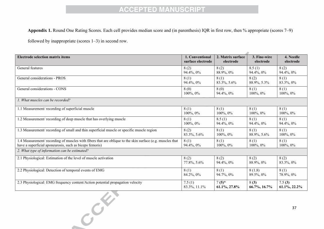
37
Appendix 1. Round One Rating Scores. Each cell provides median score and (in parenthesis) IQR in first row, then % appropriate (scores 7–9)
followed by inappropriate (scores 1–3) in second row.
Electrode selection matrix items 1. Conventional surface electrode
2. Matrix surface electrode
3. Fine-wire electrode
4. Needle electrode
General features 8 (2)94.4%, 0%
8 (2)88.9%, 0%
8.5 (1)94.4%, 0%
8 (2)94.4%, 0%
General considerations - PROS 8 (1)94.4%, 0%
8 (1)83.3%, 5.6%
8 (2)88.9%, 5.3%
8 (1)83.3%, 0%
General considerations - CONS 8 (0)100%, 0%
8 (0)94.4%, 0%
8 (1)100%, 0%
8 (1)100%, 0%
1. What muscles can be recorded?
1.1 Measurement/ recording of superficial muscle 8 (1)100%, 0%
8 (1)100%, 0%
8 (1)100%, 0%
8 (1)100%, 0%
1.2 Measurement/ recording of deep muscle that has overlying muscle 8 (1)100%, 0%
8.5 (1)94.4%, 0%
8 (1)94.4%, 0%
8 (1)94.4%, 0%
1.3 Measurement/ recording of small and thin superficial muscle or specific muscle region 8 (2)83.3%, 5.6%
8 (1)100%, 0%
8 (1)88.9%, 5.6%
8 (1)100%, 0%
1.4 Measurement/ recording of muscles with fibers that are oblique to the skin surface (e.g. muscles that have a superficial aponeurosis, such as biceps femoris)
8 (1)94.4%, 0%
8 (1)100%, 0%
8 (1)100%, 0%
8 (1)100%, 0%
2. What type of information can be estimated?
2.1 Physiological: Estimation of the level of muscle activation 8 (2)77.8%, 5.6%
8 (2)94.4%, 0%
8 (2)88.9%, 0%
8 (2)83.3%, 0%
2.2 Physiological: Detection of temporal events of EMG 8 (1)84.2%, 0%
8 (1)94.7%, 0%
8 (1.8)89.5%, 0%
8 (1)78.9%, 0%
2.3 Physiological: EMG frequency content/Action potential propagation velocity 7.5 (1)83.3%, 11.1%
7 (5)*61.1%, 27.8%
8 (3)66.7%, 16.7%
7.5 (3)61.1%, 22.2%

38
*Numbers in bold represent items that did not reach consensus.
2.4 Physiological: Discrimination of single motor unit action potentials (SMUAP) 8.5 (1)100%, 0%
8 (1)100%, 0%
8 (1)94.4%, 0%
8 (1)94.4%, 0%
2.5 Physiological: Estimation of neural drive to the muscle (number of single motor units activated) 8 (3)72.2%, 0%
8 (3)72.2%, 5.6%
8 (2)83.3%, 0%
8 (2)83.3%, 0%
2.6 Physiological: detailed analysis of properties of single motor unit discharge/ function 8 (2)77.8%, 5.6%
7 (3)61.1%, 16.7%
8 (2)77.8%, 0%
8 (2)88.9%, 0%
2.7 Anatomical: Motor unit number estimation (MUNE) 7.5 (1)83.3%, 0%
8 (1)88.9%, 0%
8 (1)88.9%, 0%
8 (1)88.9%, 0%
2.8 Anatomical: Estimation of motor unit territories 8 (1)94.4%, 5.6%
8 (1)94.4%, 5.6%
8 (1)94.4%, 5.6%
8 (1)94.4%, 5.6%
2.9 Anatomical: Identification of location of muscle innervation zone 8 (1)88.9%, 5.6%
8 (1)94.4%, 0%
8.5 (1)88.9%, 5.6%
8.5 (1)94.4%, 0%
3. Are the recordings representative of the entire muscle?
3.1 Activation of whole muscle 8 (1)94.4%, 0%
8 (2)94.4%, 0%
8 (1)94.4%, 5.6%
8 (1)94.4%, 0%
4. What types of contractions can yield relevant data?
4.1 Activation during isometric contractions 8 (1)88.9%, 5.6%
8 (1)88.9%, 5.6%
8 (1)88.9%, 5.6%
8 (1)94.4%, 0%
4.2 Activation during dynamic task 7.5 (3)72.2%, 5.6%
8 (1)77.8%, 11.1%
8 (1)83.3%, 0%
8 (2)88.9%, 11.1%
4.3 Activation during maximum contractions 8.5 (1)88.9%, 5.6%
8 (1)88.9%, 5.6%
8 (1)100%, 0%
8 (1)88.9%, 0%

39
Appendix 2. Round Two Rating Scores. Each cell provides individual responses in first row, median score and (in parenthesis) IQR in second
row, then % appropriate (scores 7–9) followed by inappropriate (scores 1–3) in third row.
*Numbers in bold represent items that did not reach consensus.
Electrode selection matrix items 1. Conventional surface electrode
2. Matrix surface electrode
3. Fine-wire electrode
4. Needle electrode
2. What type of information can be estimated?
2.3 Physiological: EMG frequency content/Action potential propagation velocity(n = 6/9)
5 7 7 8 8 8 7.5 (1)83.3%, 0%
7 8 8 9 9 9 8.5 (1)100%, 0%
5 6 6 7 7 8 6.5 (1)50%, 0%
5 6 6 7 8 9 6.5 (2)50%, 0%
2.5 Physiological: Estimation of neural drive to the muscle (number of single motor units activated)(n = 5/6)
3 5 6 8 8 6 (3)40%, 20%
4 6 7 8 8 7 (2)60%, 0%
8 8 8 9 9 8 (1)100%, 0%
8 8 8 9 9 8 (1)100%, 0%
2.6 Physiological: detailed analysis of properties of single motor unit discharge/ function(n = 6/8)
5 8 8 8 9 9 8 (1)83.3%, 0%
7 8 8 8 8 9 8 (0)100%, 0%
7 8 8 8 8 9 8 (0)100%, 0%
7 8 8 8 8 9 8 (0)100%, 0%
4. What types of contractions can yield relevant data?
4.2 Activation during dynamic task(n = 9/10)
4 7 7 7 8 8 8 9 9 8 (1)88.9%, 11.1%
4 7 8 8 8 8 8 9 9 8 (0)88.9%, 11.1%
7 7 8 8 8 8 9 9 98 (1)100%, 0%
7 8 8 8 8 9 9 9 98 (1)100%, 0%

40
Appendix 3. Round Three Rating Scores. Each cell provides individual responses in first row, median score and (in parenthesis) IQR in second
row, then % appropriate (scores 7–9) followed by inappropriate (scores 1–3) in third row.
Electrode selection matrix items 1. Conventional surface electrode
2. Matrix surface electrode
3. Fine-wire electrode
4. Needle electrode
2. What type of information can be estimated?
2.3 Physiological: EMG frequency content/Action potential propagation velocity(n = 3/3)
7 8 88 (1)100%, 0%
8 8 98 (1)100%, 0%
8 8 98 (1)100%, 0%
7 8 97 (2)100%, 0%
2.5 Physiological: Estimation of neural drive to the muscle (number of single motor units activated)(n = 3/3)
7 8 87 (0)100%, 0%
8 8 88 (0)100%, 0%
8 9 98 (0)100%, 0%
9 9 99 (0)100%, 0%