Molecularly imprinted polymers as selective sorbents for ...

HAL Id: hal-02974725https://hal.archives-ouvertes.fr/hal-02974725
Submitted on 25 Nov 2020
HAL is a multi-disciplinary open accessarchive for the deposit and dissemination of sci-entific research documents, whether they are pub-lished or not. The documents may come fromteaching and research institutions in France orabroad, or from public or private research centers.
L’archive ouverte pluridisciplinaire HAL, estdestinée au dépôt et à la diffusion de documentsscientifiques de niveau recherche, publiés ou non,émanant des établissements d’enseignement et derecherche français ou étrangers, des laboratoirespublics ou privés.
Electrochemical sensors based on molecularly imprintedchitosan: A review
Fares Zouaoui, Saliha Bourouina-Bacha, Mustapha Bourouina, NicoleJaffrezic-Renault, Nadia Zine, Abdelhamid Errachid
To cite this version:Fares Zouaoui, Saliha Bourouina-Bacha, Mustapha Bourouina, Nicole Jaffrezic-Renault, Nadia Zine, etal.. Electrochemical sensors based on molecularly imprinted chitosan: A review. Trends in AnalyticalChemistry, Elsevier, 2020, 130, pp.115982. �10.1016/j.trac.2020.115982�. �hal-02974725�
1
Electrochemical sensors based on molecularly imprinted chitosan: A review 2
Fares ZOUAOUI1,2, Saliha BOUROUINA-BACHA2, Mustapha BOUROUINA2, Nicole JAFFREZIC-RENAULT1*, Nadia ZINE1, 3 Abdelhamid ERRACHID1,* 4 5 1 University of Lyon, Institute of Analytical Sciences, UMR 5280, CNRS, 69100 Villeurbanne, France. 6 2 University of Bejaia, Laboratory of Environmental Engineering, Bejaia, Algeria. 7 8 Corresponding authors’ email address: Nicole JAFFREZIC-RENAULT [email protected] 9 Abdelhamid ERRACHID [email protected] 10 11 ABSTRACT 12 Despite the enormous development of instruments for analyzing a wide variety of compounds, applied in different fields such as 13 health, environment or quality control, the demand for increasingly sensitive and selective techniques continues to grow. In this 14 regard, efforts have been made to highlight more appropriate techniques. Electrochemical sensors based on molecularly imprinted 15 polymers (MIPs) offer an interesting alternative since they allow reaching high sensitivity and selectivity and they are inexpensive 16 and easily adaptable to miniaturization. The choice of the functional monomer in the synthesis of MIPs is based on its capacity to 17 provide complementary interactions with the target molecules. The various excellent properties of chitosan, as a biosourced polymer, 18 make it a promising alternative to conventional functional monomers. This review reports on the principle of the MIPs technique 19 describing the different possible approaches in their synthesis. It aims to provide an overview of the value of using chitosan as a 20 functional monomer by highlighting its applications in electrochemical sensors. 21
Keywords: Molecularly imprinted polymers, Chitosan, Electrochemical sensors, Electrodepostion, Coating. 22
23
Introduction 24
Conventional analytical techniques (chromatography, Nuclear Magnetic Resonance (NMR), mass spectrometry (MS), etc.) used for 25 monitoring in different fields such as medicine, clinical biology, the food industry and the quality control of our environment (monitoring 26 industrial or domestic discharges) are generally complex, expensive, bulky and often difficult and time-consuming to implement, and 27 they require highly qualified personnel. In addition, conventional procedures consist in taking samples from the monitored 28 environments and analyzing them in the laboratory. The steps of sample preparation, incubation, and processing of results often 29 greatly increase the total analysis time [1]. In order to carry out these analyses, there is a need to set up rapid, simple, inexpensive, 30 selective techniques and to design compact monitoring tools operating on site, in real time. 31
Among the devices capable of responding to this demand, sensors present the essential function of detection or even quantification 32 of a physical, biological or even of a chemical phenomenon in a measurable signal and directly usable by humans. Electrochemical 33 sensors are a promising choice because of their simplicity, sensitivity, ease of implementation and generally low measurement costs, 34 but also for their varied applications in the field of health, of the environment or of quality control of food products. Electrochemical 35 sensors are based on different electrochemical techniques: potentiometry, voltammetry, amperometry, impedimetry [2]. 36
Biological elements (antibodies, enzymes, microorganisms, DNA, etc.) are generally used for recognition in order to increase 37 sensitivity and specificity, which are generally lacking in chemical sensors. However, as they are biomolecules, their use is limited to 38 mild conditions of pH, temperature and nature of the medium in order to avoid denaturating the protein. In addition, using these 39 biological elements is not so simple. Sourcing them and immobilizing them on the electrode surface require considerable skill [3,4]. 40
Due to the need to overcome these limitations, an alternative has been developed which consists of using molecularly imprinted 41 polymers (MIPs). MIPs have been used as an artificial recognition material whose binding site size and shape are complementary to 42 the template molecules for their specific recognition. This approach was established in 1972 by Wulff and Sarhan [5]. To generate 43 MIPs with selective cavities, a monomer is simply polymerized in the presence of the target molecule and a crosslinking agent leading 44 to the formation of a highly cross-linked functional monomer/template complex. The formation of the complex has been theoretically 45 studied in several articles [6,7]. Cavities specific to the target molecule are then obtained after extracting the template from the 46 polymer. Template/monomer and crosslinking agent/monomer ratios play an important role in the synthesis of MIPs improving the 47 efficiency [8,9]. MIPs are characterized by very interesting properties such as physical resistance, robustness, resistance to high 48 pressures and temperatures and high inertia towards various chemicals [10], and are produced at low cost. In addition, MIPs have a 49 high affinity and selectivity towards target molecules; they are comparable to natural receptors [11]. MIPs are widely used in various 50 fields, such as liquid chromatography, capillary electrochromatography, solid phase extraction, therapy, preparation of artificial 51 antibodies and enzymes, catalysts and syntheses, sensors, etc. [12,13]. 52
The choice of the functional monomer in the synthesis of MIPs is a primordial step given its capacity to provide complementary 53 interactions with the matrix molecules [14]. Chitosan (CS) is a natural polysaccharide obtained by deacetylation of chitin which is the 54

most abundant non-toxic, biodegradable and biocompatible natural amino polysaccharide [15]. Chitosan has three types of reactive 55 functional groups, one amine group and two primary and secondary hydroxyl groups. This structure thus allows numerous chemical 56 modifications, and can support a succession of chemical reactions such as the reaction with aldehydes and acetones, grafting, H 57 bond with polar atoms, crosslinking, etc. The functional properties of chitosan films can be improved when combined with other 58 materials [16]. Due to the positive charges on its amine groups, the cationic biopolymer can interact with anionic molecules. These 59 unique properties make CS suitable for a wide range of applications in the food, cosmetic and pharmaceutical, sensor and biosensor 60 fields [17,18]. The presence of reactive functional groups on the polysaccharide chain of CS gives it flexibility, excellent film-forming 61 capacity, good adhesion, biocompatibility, high mechanical resistance, the possibility of undergoing structural modifications, which 62 makes it suitable for preparing MIPs and building electrochemical sensors [19,20]. It is well known that the behavior of chitosan can 63 be affected by several parameters such as pH and humidity [21,22]. Treatment of chitosan with crosslinking agents or by chemical 64 modification are common strategies to improve its physico-chemical characteristics [23]. Chitosan has been used to make 65 membranes, thin films and three-dimensional structures. These forms are obtained by different methods such as electrodeposition 66 [24] and drop-coating [25]. Electrodeposition is very convenient because it is a simple, fast and low-cost technique, but also it makes 67 it possible to obtain controlled films [26]. 68
This review describes the principle of MIPs and the approaches used to synthesize them emphasizing the different criteria for 69 choosing the reagents and the limitations of this technique. In addition, it accounts for the advantage of using chitosan as a functional 70 monomer by highlighting its applications in electrochemical sensors. 71
72 I. Natural origin of chitosan 73
74 I.1. Structure and sources of Chitin 75
Chitin, first identified in 1884, is the second most abundant biopolymer in nature after cellulose. Its chemical structure consists of N-76 acetylglucosamine units. Chitin’s physical structure includes three allomorphs, due to the arrangement of macromolecular chains, α, 77 β and γ-chitin (figure 1). α-chitin is one of the most abundant allomorphs in which polymer chains are arranged in the same direction 78 and parallel. β-chitin is less frequent in nature and presents anti-parallel chains. The γ-chitin, according to the studies, is a variant of 79 the α or β- family [27]. 80
81
82
83
84
85
86
87
88
89
Figure 1: Allomorph types of chitin. 90
91
Chitin is present in co-products in the form of chitin-proteins-calcium carbonate complexes [15]. Chitin can be obtained from various 92 sources (Table 1), it is mainly found in exoskeletons, mollusks and various microorganisms [15,28]. 93
94
95
Table 1: Main sources of chitin. 96
α-chitin β-chitin
Fungi, yeast cell walls, krill, lobster , crab, tendons, shrimp shells, insect cuticle.
Squid pens, tube synthesized by pogonophoran and vestimentifera worms, lorica built by some seaweeds
97
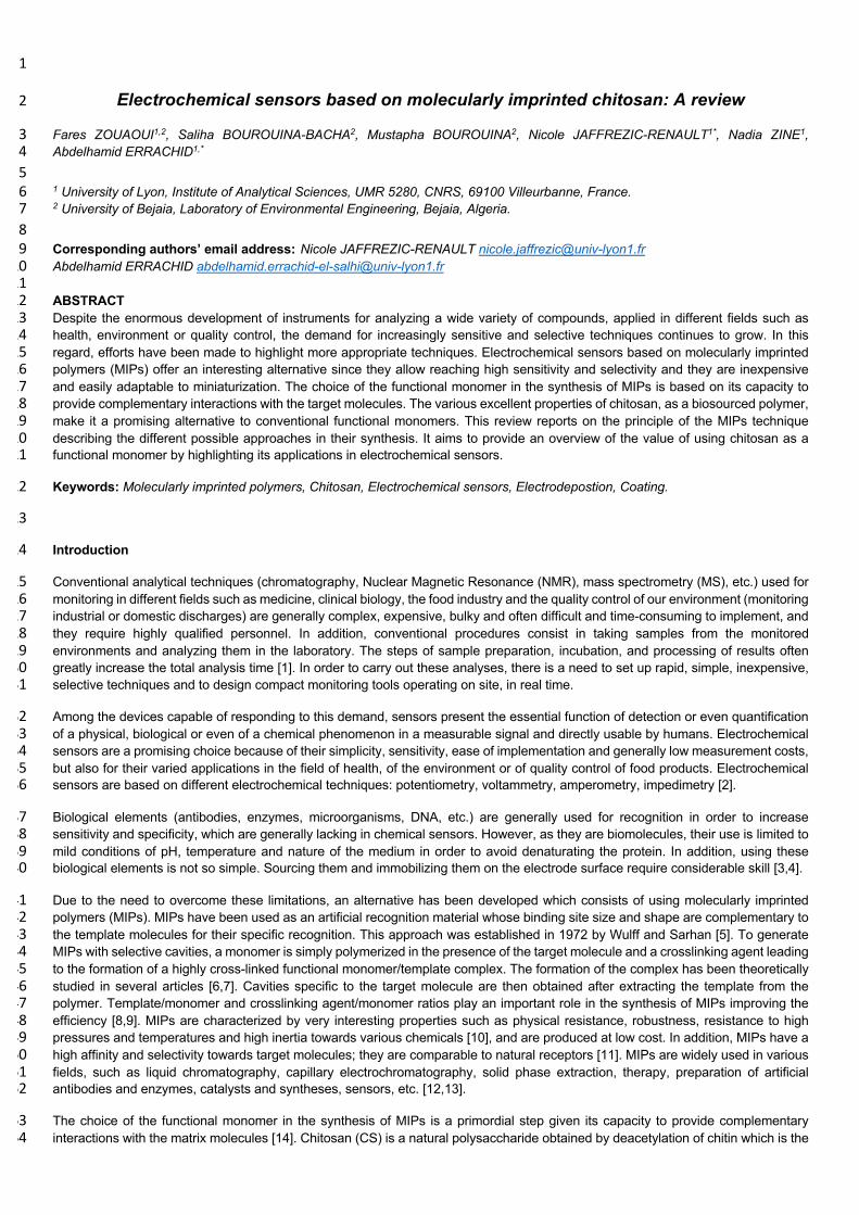
I.2. Extraction of chitin 98
Usually, two types of extraction process are used to obtain pure chitin: chemical and biological methods (Fig.2). Chemical extraction 99 is based on three stages; between these different stages, washing operations are necessary [27]. 100
Ø Demineralization: by hydrochloric acid to eliminate minerals. This consists in the elimination of the calcium carbonate and 101 calcium chloride. 102
Ø Deproteinization: by sodium hydroxide to eliminate proteins. 103 Ø Bleaching: by sodium hypochlorite to remove pigments. 104
Biological treatments offer an alternative way to extract chitin, to avoid acidic and alkaline treatments. Lactic acid-producing bacteria 105 and proteases from bacteria have been used for the demineralization and deproteinization steps respectively [29]. Chitin is highly 106 hydrophobic and insoluble in water and most organic solvents are due to the presence of acetyl groups [15], so it can be used in 107 more applications when transformed into chitosan. 108
I.3. Deacetylation of chitin 109
The deacetylation step consists in converting chitin to chitosan by the removal of acetyl groups and leads to partial deacetylation. 110 The border between chitin and chitosan corresponds to a degree of deacetylation (DD). This makes the degree of deacetylation an 111 important property in the production of chitosan since it determines the free amino groups in the polysaccharides. Indeed, it affects 112 the physicochemical properties and thus determines its appropriate applications. DD is an important criterion for distinguishing 113 between chitin and chitosan, defined as the fraction of glucosamine /N-acetyl glucosamine: note that: 114
Ø for a DD <60%, the polymer is named chitin. 115 Ø for a DD> 60%, the polymer is named chitosan [30]. 116
The degree of acetylation controls the proportion of amine and acetyl groups in the carbonic chain and is directly related to the 117 solubility and acid-base behavior of the polymer [31]. Many methods have been developed to determine DD, such as the 118 conductimetric assay [32,33], potentiometric titration [34], UV spectroscopy, IR spectroscopy, 1H NMR spectroscopy, HPLC analysis 119 [35], etc. Deacetylation can occur either chemically or enzymatically. 120
- Chemical deacetylation 121
Sodium or potassium hydroxides are usually used at high temperature [27]. The alkaline treatment can be repeated to achieve 122 complete deacetylation [36]. It has been reported that the DD can be affected by temperature, processing time and alkali 123 concentration. Chemical deacetylation is used more commonly because of its low cost; however, the negative points are high energy 124 cost, waste of concentrated alkaline solution which leads to an increase in environmental pollution, the fact that it is not easily 125 controlled and lead to a broad and heterogeneous range of products [37]. 126
- Enzymatic deacetylation 127
Chitin deacetylase for the preparation of chitosan polymers is used. It partially catalyses the hydrolysis of N-acetamido bonds in chitin 128 to produce chitosan [37,38]. Enzymatic deacetylation is a sustainable process and offers the possibility of controlling and predicting 129 the bioactivities of chitosan. Therefore, the resulting chitosan has a more regular deacetylation pattern than a chitosan treated with 130 hot alkali [39,40]. 131

132
Figure 2: Preparation of chitin and chitosan from raw material 133
134
II. Properties of chitosan 135 136
II.1. Structure 137
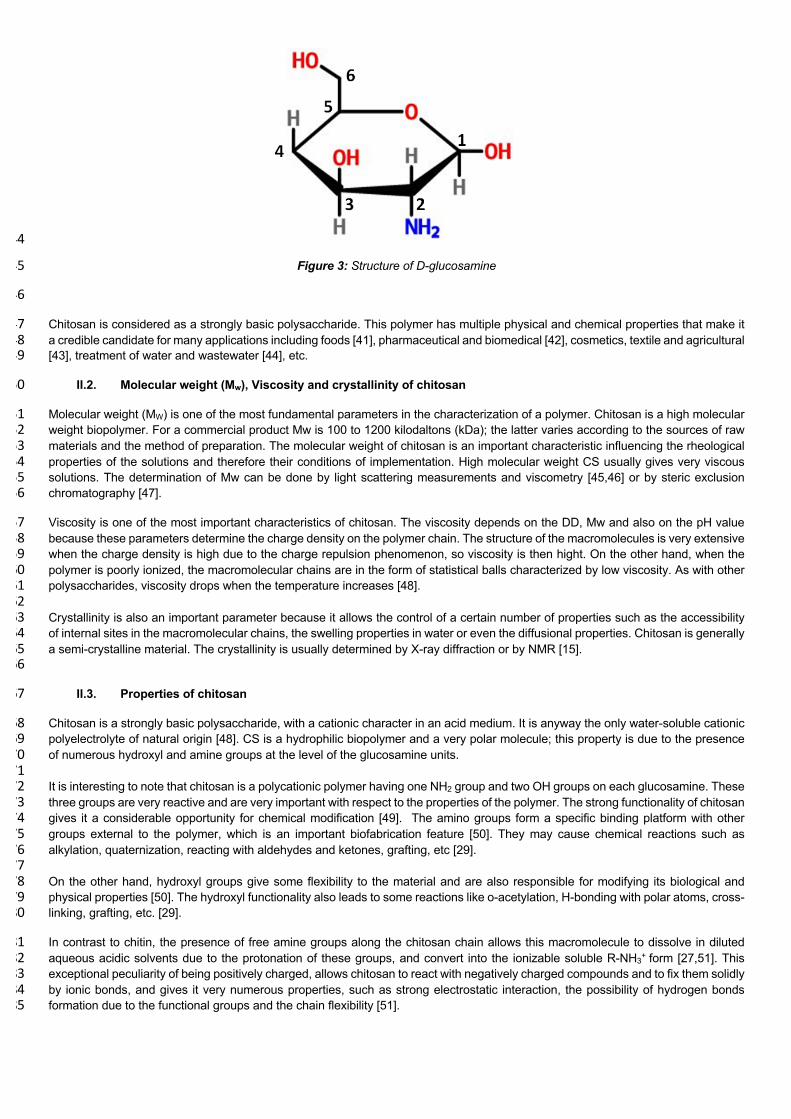
Chitosan (CS) is the name given to chitin after the deacetylation process (Figure 2). CS is an amino sugar with a molar mass of 179 138 g/mol glucosamine. It is a family of linear polysaccharides with a molar fraction DD of D-glucosamine and a fraction (1-DD) of N-139 acetylglucosamine [28]. Chitosan is thus considered as a copolymer with three types of reactive functional groups, a primary amino 140 group, and one primary and two secondary hydroxyl groups (one primary amine group (C2) and two hydroxyl groups (C3, C6) per-141 repeat unit) (figure 3) 142
143
144
Figure 3: Structure of D-glucosamine 145
146
Chitosan is considered as a strongly basic polysaccharide. This polymer has multiple physical and chemical properties that make it 147 a credible candidate for many applications including foods [41], pharmaceutical and biomedical [42], cosmetics, textile and agricultural 148 [43], treatment of water and wastewater [44], etc. 149
II.2. Molecular weight (Mw), Viscosity and crystallinity of chitosan 150
Molecular weight (MW) is one of the most fundamental parameters in the characterization of a polymer. Chitosan is a high molecular 151 weight biopolymer. For a commercial product Mw is 100 to 1200 kilodaltons (kDa); the latter varies according to the sources of raw 152 materials and the method of preparation. The molecular weight of chitosan is an important characteristic influencing the rheological 153 properties of the solutions and therefore their conditions of implementation. High molecular weight CS usually gives very viscous 154 solutions. The determination of Mw can be done by light scattering measurements and viscometry [45,46] or by steric exclusion 155 chromatography [47]. 156
Viscosity is one of the most important characteristics of chitosan. The viscosity depends on the DD, Mw and also on the pH value 157 because these parameters determine the charge density on the polymer chain. The structure of the macromolecules is very extensive 158 when the charge density is high due to the charge repulsion phenomenon, so viscosity is then hight. On the other hand, when the 159 polymer is poorly ionized, the macromolecular chains are in the form of statistical balls characterized by low viscosity. As with other 160 polysaccharides, viscosity drops when the temperature increases [48]. 161 162 Crystallinity is also an important parameter because it allows the control of a certain number of properties such as the accessibility 163 of internal sites in the macromolecular chains, the swelling properties in water or even the diffusional properties. Chitosan is generally 164 a semi-crystalline material. The crystallinity is usually determined by X-ray diffraction or by NMR [15]. 165 166
II.3. Properties of chitosan 167
Chitosan is a strongly basic polysaccharide, with a cationic character in an acid medium. It is anyway the only water-soluble cationic 168 polyelectrolyte of natural origin [48]. CS is a hydrophilic biopolymer and a very polar molecule; this property is due to the presence 169 of numerous hydroxyl and amine groups at the level of the glucosamine units. 170 171 It is interesting to note that chitosan is a polycationic polymer having one NH2 group and two OH groups on each glucosamine. These 172 three groups are very reactive and are very important with respect to the properties of the polymer. The strong functionality of chitosan 173 gives it a considerable opportunity for chemical modification [49]. The amino groups form a specific binding platform with other 174 groups external to the polymer, which is an important biofabrication feature [50]. They may cause chemical reactions such as 175 alkylation, quaternization, reacting with aldehydes and ketones, grafting, etc [29]. 176 177 On the other hand, hydroxyl groups give some flexibility to the material and are also responsible for modifying its biological and 178 physical properties [50]. The hydroxyl functionality also leads to some reactions like o-acetylation, H-bonding with polar atoms, cross-179 linking, grafting, etc. [29]. 180
In contrast to chitin, the presence of free amine groups along the chitosan chain allows this macromolecule to dissolve in diluted 181 aqueous acidic solvents due to the protonation of these groups, and convert into the ionizable soluble R-NH3+ form [27,51]. This 182 exceptional peculiarity of being positively charged, allows chitosan to react with negatively charged compounds and to fix them solidly 183 by ionic bonds, and gives it very numerous properties, such as strong electrostatic interaction, the possibility of hydrogen bonds 184 formation due to the functional groups and the chain flexibility [51]. 185

Chitosan can interact with other chemical substances (ions, molecules, macromolecules) thanks to its particular chemical structure 186 which makes it possible to glimpse capacities to interact, complex or absorb [52]. Chitosan can link functional derivatives on the 187 macromolecular chains either by covalent, semi-covalent or non-covalent bonds (Figure 4). 188
189
Figure 4: Different approaches to complexing chitosan. 190
At a pH < pKa, the amino functions of glucosamine are protonated. These can then develop ionic interactions such as the fixation of 191 metallic anions, anionic dyes, and adhesion to negatively charged surfaces. These interactions also give to chitosan an excellent 192 film-forming property [53]. At a basic pH, the amine functions are not in ionized form and can then form hydrogen bonds or covalent 193 amide or imine bonds with activated carboxylic acids and aldehydes, respectively. Concerning the semi-covalent approach, the amine 194 groups are positively charged for pH values < 6.5 and can form ionic bonds with the carboxylic acid groups [54]. The alcohol functions 195 of glucosamine can mainly develop hydrogen bonds or covalent bonds. To develop each of these approaches, it was necessary to 196 take into account the acid-base constraints of glucosamine allowing the establishment of these specific bonds. 197
Also, these reactive groups offer to chitosan polymers many advantages and relevant properties (e.g. mucoadhesive properties, 198 absorption enhancer) ; they are easily processed into gels, films, membranes, nanofibers and nanofibrils, scaffolds, beads, sponge 199 forms, microparticles and nanoparticles [29,49]. In addition, chitosan possesses many more properties such as biocompatibility, 200 biodegradability, low toxicity and good antimicrobial and antioxidant activities leading to numerous applications [28]. Table 2 201 summarizes the main properties of chitosan [28,52]. 202
Table 2: Properties of chitosan 203
Physical and chemical properties Biological properties
Linear amino polysaccharide
Reactive amino and hydroxyl groups
Chelates many transitional metal ions
Rigid D-glucosamine structure
High hydrophilicity, crystallinity
Deprotonated amino group
Form hydrogen bonds intermolecularly
High viscosity
Soluble in water and organic solvents
Soluble in dilute aqueous acidic solutions
Polyelectrolytes (at acidic pH)
Biocompatibility
Natural polymer
Bioadhesivity
Bioactivity
Nontoxic
Biodegradability
Antimicrobial activity
Antioxidant activity
Antiacid, antiulcer and antitumoral
Blood anticoagulants
Hypolipidemic activity

Flocculating agent
Entrapment, filtration, separation and adsorption properties
Renewable
Film forming ability
Hydrating agent
204
II.4. Chitosan solubility 205
Chitin is insoluble in many organic solvents due to the presence of acetyl group with a hydrophobic nature [55]. Chitosan is the only 206 commercial water-soluble cationic biopolymer and it is soluble in dilute acidic solutions at pH values above 6.5 but insoluble in neutral 207 and alkaline aqueous solutions [43,55]. Table 3, shows some solutions used for the solubilization of chitosan. 208
Table 3: Some solutions used for the solubilization of chitosan 209
Soluble Insoluble
Acetic acid 1% [36]
Formic acid 0.2-100% [36]
Hydrochloric acid 1% [36]
Dilute nitric, succinic, lactic and malic acids [43]
Water [56]
Sulfuric and phosphoric acids [36]
Any neutral and alkaline pH aqueous solutions [43]
210
One of the most important properties of this polymer is the presence of the amine group with a pKa value of 6.5 in the carbonic chain. 211 With this termination, chitosan indeed has an acid-base behavior in solution, as shown in Figure 5. It is reported that the pKa value 212 increases with the ionic strength, thus widening the pH range in which chitosan is soluble [57]. 213
214
Figure 5: Behavior of chitosan according to the pH of the solution 215
216
So, at pH <pKa the amino groups are positively charged which makes the chitosan a water-soluble cationic polyelectrolyte, and at 217 pH> pKa the biopolymer loses its charge and becomes insoluble [36]. Indeed, at low pH, the chain is polycationic. Repulsive 218 electrostatic forces prevent the monomer associations which make chitosan soluble in solution. When pH increases, the chitosan 219 loses its positive charge, the balance between the forces of attraction and repulsion is then disturbed [31,58]. As discussed previously, 220 the solubility of chitosan in acidic medium is explained by the cationic character of its amine functions when they are protonated 221 providing sufficient electrostatic energy to break the numerous hydrogen bonds and considerably improve the hydration of the chains. 222 Therefore, the solubility of chitosan also depends on the distribution of the acetyl groups along the biopolymer chain and the DD 223 value. 224 225
II.5. Chitosan stability 226
Chitosan is very sensitive to different experimental parameters that affect its stability; degree of deacetylation, molecular weight, 227 polymer concentration, type of acid and its concentration are parameters to take into account in the experiments [57]. 228
- Temperature and pH effect 229
The main factors affecting the behavior of CS and its physical properties are pH and temperature. pH affect the polymer properties 230 due to the protonation and deprotonation of the amino groups [59]. High temperature (>500 °K) may cause thermal degradation of 231 CS [60]. Also, it has been reported that heat treatment can change the way the chains of the polymer react; this is attributed to the 232 influence of the ionic association of amine groups with alkanoic acids, such as formic, acetic, propionic, and butyric acid [61,62]. The 233 mechanism may involve an initial depolymerization followed by interchain cross-linking when the amount of heat assimilated is 234 sufficiently high [63]. 235
236 - Humidity effect 237
Humidity is one of the factors affecting the behavior of chitosan. Studies show that exposing thin layers of chitosan to moist air causes 238 considerable swelling of the film. After heating and then cooling to the ambient temperature of these layers, a dramatic decrease in 239 thickness is recorded. These changes are not due to the oxidation or the thermal degradation of the films but it is suggested that the 240 heating irreversibly releases the trapped water [64]. 241
Furthermore, studies on chitosan-based sensors show that, probably, in an airy medium, oxygen molecules are chemically absorbed 242 by chitosan (Equation 1). Once the film is subjected to a potential, the absorbed oxygen molecules react with the free electrons 243 forming ionic species such as O2- and O- (Equation 2-3), causing an increase in resistance to charge transfer until the oxygen 244 molecules are saturated. In the presence of water, the molecules in contact with chitosan react with O- to produce O2 and release 245 the electrons participating in the modification of the response of the sensor (Equation 4). 246
O2 O2 (absorbed) (1) 247
O2 (absorbed) + e- O2- (2) 248
O2- + e- 2O- (3) 249
CS-NH2 + 2O- + H2O CS-NH2 + O2 + H2O + 2e- (4) 250
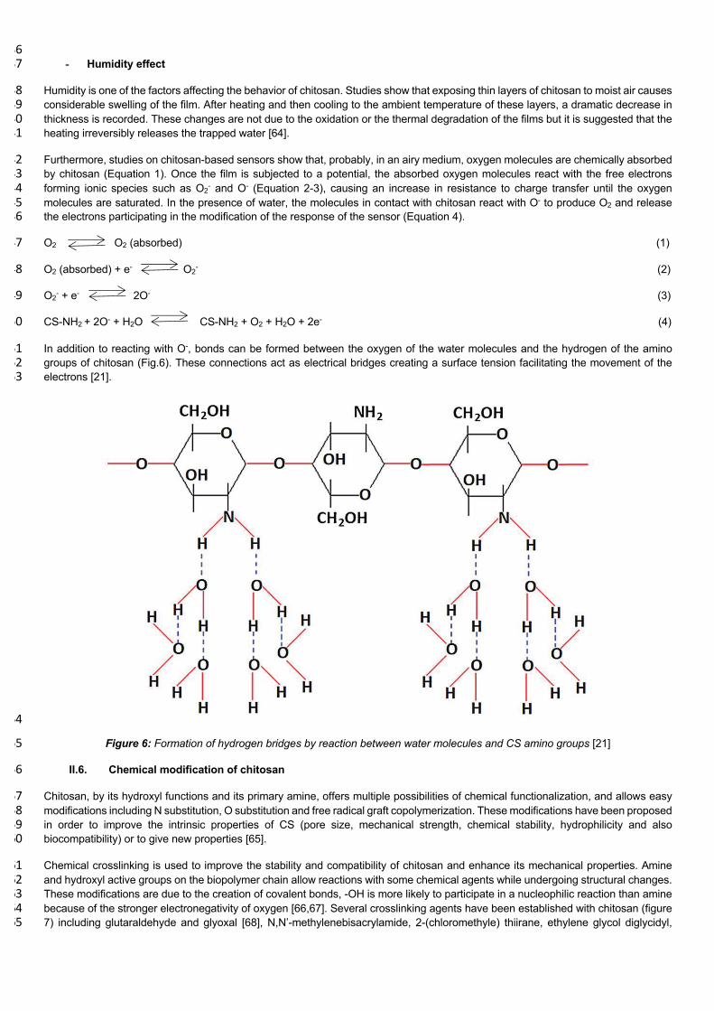
In addition to reacting with O-, bonds can be formed between the oxygen of the water molecules and the hydrogen of the amino 251 groups of chitosan (Fig.6). These connections act as electrical bridges creating a surface tension facilitating the movement of the 252 electrons [21]. 253
254
Figure 6: Formation of hydrogen bridges by reaction between water molecules and CS amino groups [21] 255
II.6. Chemical modification of chitosan 256
Chitosan, by its hydroxyl functions and its primary amine, offers multiple possibilities of chemical functionalization, and allows easy 257 modifications including N substitution, O substitution and free radical graft copolymerization. These modifications have been proposed 258 in order to improve the intrinsic properties of CS (pore size, mechanical strength, chemical stability, hydrophilicity and also 259 biocompatibility) or to give new properties [65]. 260
Chemical crosslinking is used to improve the stability and compatibility of chitosan and enhance its mechanical properties. Amine 261 and hydroxyl active groups on the biopolymer chain allow reactions with some chemical agents while undergoing structural changes. 262 These modifications are due to the creation of covalent bonds, -OH is more likely to participate in a nucleophilic reaction than amine 263 because of the stronger electronegativity of oxygen [66,67]. Several crosslinking agents have been established with chitosan (figure 264 7) including glutaraldehyde and glyoxal [68], N,N’-methylenebisacrylamide, 2-(chloromethyle) thiirane, ethylene glycol diglycidyl, 265
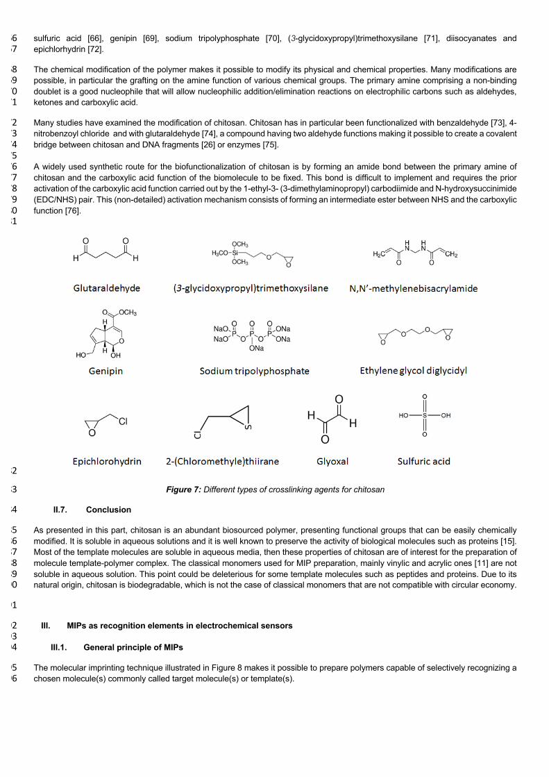
sulfuric acid [66], genipin [69], sodium tripolyphosphate [70], (3-glycidoxypropyl)trimethoxysilane [71], diisocyanates and 266 epichlorhydrin [72]. 267
The chemical modification of the polymer makes it possible to modify its physical and chemical properties. Many modifications are 268 possible, in particular the grafting on the amine function of various chemical groups. The primary amine comprising a non-binding 269 doublet is a good nucleophile that will allow nucleophilic addition/elimination reactions on electrophilic carbons such as aldehydes, 270 ketones and carboxylic acid. 271
Many studies have examined the modification of chitosan. Chitosan has in particular been functionalized with benzaldehyde [73], 4-272 nitrobenzoyl chloride and with glutaraldehyde [74], a compound having two aldehyde functions making it possible to create a covalent 273 bridge between chitosan and DNA fragments [26] or enzymes [75]. 274 275 A widely used synthetic route for the biofunctionalization of chitosan is by forming an amide bond between the primary amine of 276 chitosan and the carboxylic acid function of the biomolecule to be fixed. This bond is difficult to implement and requires the prior 277 activation of the carboxylic acid function carried out by the 1-ethyl-3- (3-dimethylaminopropyl) carbodiimide and N-hydroxysuccinimide 278 (EDC/NHS) pair. This (non-detailed) activation mechanism consists of forming an intermediate ester between NHS and the carboxylic 279 function [76]. 280 281
282
Figure 7: Different types of crosslinking agents for chitosan 283
II.7. Conclusion 284
As presented in this part, chitosan is an abundant biosourced polymer, presenting functional groups that can be easily chemically 285 modified. It is soluble in aqueous solutions and it is well known to preserve the activity of biological molecules such as proteins [15]. 286 Most of the template molecules are soluble in aqueous media, then these properties of chitosan are of interest for the preparation of 287 molecule template-polymer complex. The classical monomers used for MIP preparation, mainly vinylic and acrylic ones [11] are not 288 soluble in aqueous solution. This point could be deleterious for some template molecules such as peptides and proteins. Due to its 289 natural origin, chitosan is biodegradable, which is not the case of classical monomers that are not compatible with circular economy. 290
291
III. MIPs as recognition elements in electrochemical sensors 292 293
III.1. General principle of MIPs 294
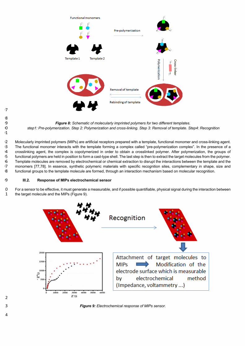
The molecular imprinting technique illustrated in Figure 8 makes it possible to prepare polymers capable of selectively recognizing a 295 chosen molecule(s) commonly called target molecule(s) or template(s). 296
297
298 Figure 8: Schematic of molecularly imprinted polymers for two different templates. 299
step1: Pre-polymerization. Step 2: Polymerization and cross-linking. Step 3: Removal of template. Step4: Recognition 300 301
Molecularly imprinted polymers (MIPs) are artificial receptors prepared with a template, functional monomer and cross-linking agent. 302 The functional monomer interacts with the template forming a complex called “pre-polymerization complex”. In the presence of a 303 crosslinking agent, the complex is copolymerized in order to obtain a crosslinked polymer. After polymerization, the groups of 304 functional polymers are held in position to form a cast-type shell. The last step is then to extract the target molecules from the polymer. 305 Template molecules are removed by electrochemical or chemical extraction to disrupt the interactions between the template and the 306 monomers [77,78]. In essence, synthetic polymeric materials with specific recognition sites, complementary in shape, size and 307 functional groups to the template molecule are formed, through an interaction mechanism based on molecular recognition. 308
III.2. Response of MIPs electrochemical sensor 309
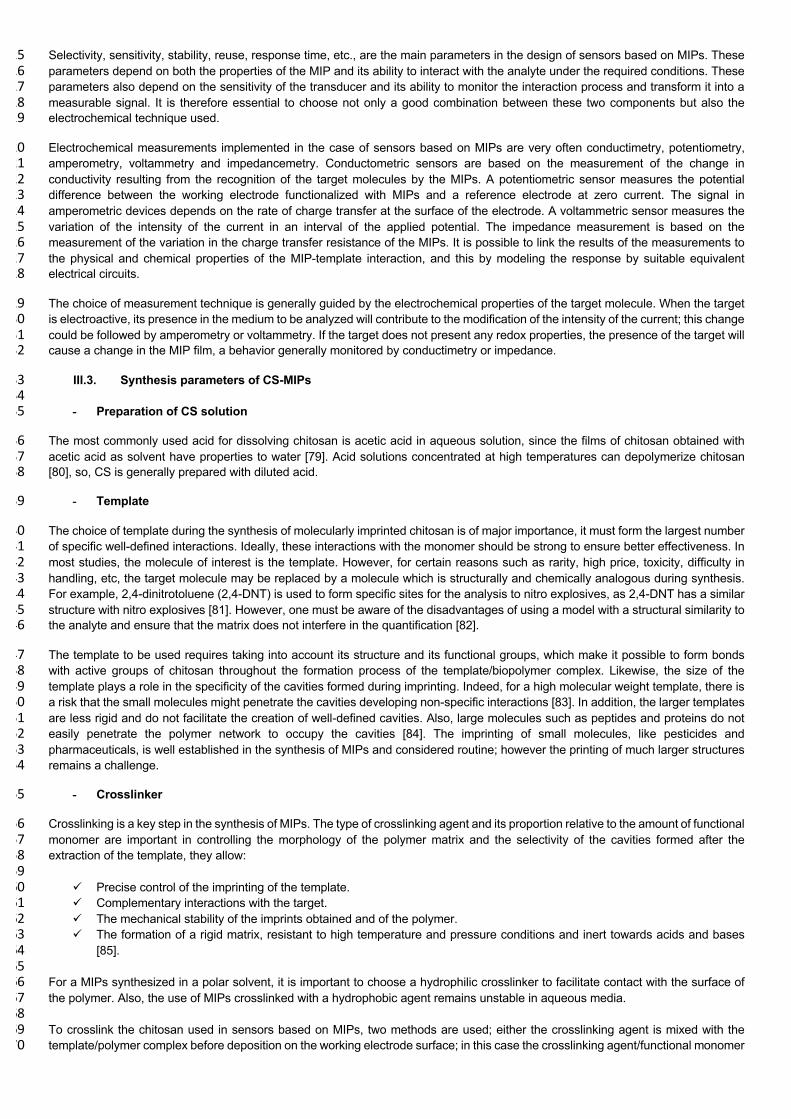
For a sensor to be effective, it must generate a measurable, and if possible quantifiable, physical signal during the interaction between 310 the target molecule and the MIPs (Figure 9). 311
312
Figure 9: Electrochemical response of MIPs sensor. 313
314
Selectivity, sensitivity, stability, reuse, response time, etc., are the main parameters in the design of sensors based on MIPs. These 315 parameters depend on both the properties of the MIP and its ability to interact with the analyte under the required conditions. These 316 parameters also depend on the sensitivity of the transducer and its ability to monitor the interaction process and transform it into a 317 measurable signal. It is therefore essential to choose not only a good combination between these two components but also the 318 electrochemical technique used. 319
Electrochemical measurements implemented in the case of sensors based on MIPs are very often conductimetry, potentiometry, 320 amperometry, voltammetry and impedancemetry. Conductometric sensors are based on the measurement of the change in 321 conductivity resulting from the recognition of the target molecules by the MIPs. A potentiometric sensor measures the potential 322 difference between the working electrode functionalized with MIPs and a reference electrode at zero current. The signal in 323 amperometric devices depends on the rate of charge transfer at the surface of the electrode. A voltammetric sensor measures the 324 variation of the intensity of the current in an interval of the applied potential. The impedance measurement is based on the 325 measurement of the variation in the charge transfer resistance of the MIPs. It is possible to link the results of the measurements to 326 the physical and chemical properties of the MIP-template interaction, and this by modeling the response by suitable equivalent 327 electrical circuits. 328
The choice of measurement technique is generally guided by the electrochemical properties of the target molecule. When the target 329 is electroactive, its presence in the medium to be analyzed will contribute to the modification of the intensity of the current; this change 330 could be followed by amperometry or voltammetry. If the target does not present any redox properties, the presence of the target will 331 cause a change in the MIP film, a behavior generally monitored by conductimetry or impedance. 332
III.3. Synthesis parameters of CS-MIPs 333 334
- Preparation of CS solution 335
The most commonly used acid for dissolving chitosan is acetic acid in aqueous solution, since the films of chitosan obtained with 336 acetic acid as solvent have properties to water [79]. Acid solutions concentrated at high temperatures can depolymerize chitosan 337 [80], so, CS is generally prepared with diluted acid. 338
- Template 339
The choice of template during the synthesis of molecularly imprinted chitosan is of major importance, it must form the largest number 340 of specific well-defined interactions. Ideally, these interactions with the monomer should be strong to ensure better effectiveness. In 341 most studies, the molecule of interest is the template. However, for certain reasons such as rarity, high price, toxicity, difficulty in 342 handling, etc, the target molecule may be replaced by a molecule which is structurally and chemically analogous during synthesis. 343 For example, 2,4-dinitrotoluene (2,4-DNT) is used to form specific sites for the analysis to nitro explosives, as 2,4-DNT has a similar 344 structure with nitro explosives [81]. However, one must be aware of the disadvantages of using a model with a structural similarity to 345 the analyte and ensure that the matrix does not interfere in the quantification [82]. 346
The template to be used requires taking into account its structure and its functional groups, which make it possible to form bonds 347 with active groups of chitosan throughout the formation process of the template/biopolymer complex. Likewise, the size of the 348 template plays a role in the specificity of the cavities formed during imprinting. Indeed, for a high molecular weight template, there is 349 a risk that the small molecules might penetrate the cavities developing non-specific interactions [83]. In addition, the larger templates 350 are less rigid and do not facilitate the creation of well-defined cavities. Also, large molecules such as peptides and proteins do not 351 easily penetrate the polymer network to occupy the cavities [84]. The imprinting of small molecules, like pesticides and 352 pharmaceuticals, is well established in the synthesis of MIPs and considered routine; however the printing of much larger structures 353 remains a challenge. 354
- Crosslinker 355
Crosslinking is a key step in the synthesis of MIPs. The type of crosslinking agent and its proportion relative to the amount of functional 356 monomer are important in controlling the morphology of the polymer matrix and the selectivity of the cavities formed after the 357 extraction of the template, they allow: 358 359
ü Precise control of the imprinting of the template. 360 ü Complementary interactions with the target. 361 ü The mechanical stability of the imprints obtained and of the polymer. 362 ü The formation of a rigid matrix, resistant to high temperature and pressure conditions and inert towards acids and bases 363
[85]. 364 365
For a MIPs synthesized in a polar solvent, it is important to choose a hydrophilic crosslinker to facilitate contact with the surface of 366 the polymer. Also, the use of MIPs crosslinked with a hydrophobic agent remains unstable in aqueous media. 367 368 To crosslink the chitosan used in sensors based on MIPs, two methods are used; either the crosslinking agent is mixed with the 369 template/polymer complex before deposition on the working electrode surface; in this case the crosslinking agent/functional monomer 370
ratio is very important [86]. The second method is based on the deposition of the template/polymer complex on the electrode and 371 then impregnating the modified electrode in the cross-linker solution. Here, the concentration of the crosslinking solution and contact 372 time define the properties of the prepared MIPs [87]. 373
374
III.4. Principle of the electrodeposition of chitosan 375
The ability to create devices like sensors and biosensors requires easy methods to precisely control surfaces. A variety of techniques 376 can be used to produce the desired structures. The integration of a chitosan-MIP with the transducer area is a crucial step in the 377 fabrication of the sensor. The biopolymer material must adhere well to this surface without dispersing or detaching under any 378 conditions. The properties of chitosan have been used to make membranes, thin films and three-dimensional structures by various 379 methods such as drop coating [88], microcontact, complexation with anionic polyelectrolytes [26]. 380 381 The pH-sensitive solubility and properties of chitosan have also been used to make membranes, thin films and three-dimensional 382 structures. Chitosan changes from a soluble state in acid solution to a precipitated hydrogel state when the pH of the solution 383 increases. CS can be easily poured, producing membranes and films which can be converted into insoluble networks by 384 neutralization. The cast chitosan membranes/films can be made insoluble over the entire pH range by covalently crosslinking the 385 chitosan chains by amide and imine interactions. 386
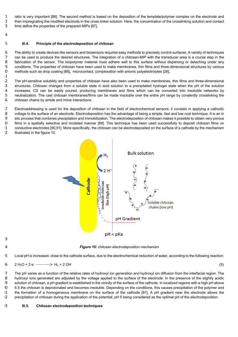
Electroaddressing is used for the deposition of chitosan in the field of electrochemical sensors; it consists in applying a cathodic 387 voltage to the surface of an electrode. Electrodeposition has the advantage of being a simple, fast and low cost technique. It is an in 388 situ process that combines precipitation and immobilization. The electrodeposition of chitosan makes it possible to obtain very porous 389 films in a spatially selective and modeled manner [89]. This technique has been used successfully to deposit chitosan films on 390 conductive electrodes [90,91]. More specifically, the chitosan can be electrodeposited on the surface of a cathode by the mechanism 391 illustrated in the figure 10. 392
393
Figure 10: chitosan electrodeposition mechanism 394
Local pH is increased, close to the cathode surface, due to the electrochemical reduction of water, according to the following reaction: 395
2 H2O + 2 e- H2 + 2 OH- (5) 396
The pH varies as a function of the relative rates of hydroxyl ion generation and hydroxyl ion diffusion from the interfacial region. The 397 hydroxyl ions generated are adjusted by the voltage applied to the surface of the electrode. In the presence of the slightly acidic 398 solution of chitosan, a pH gradient is established in the vicinity of the surface of the cathode. In localized regions with a high pH above 399 6.5 the chitosan is deprotonated and becomes insoluble. Depending on the conditions, this causes precipitation of the polymer and 400 the formation of a homogeneous membrane on the surface of the cathode [91]. A pH gradient near the electrode allows the 401 precipitation of chitosan during the application of the potential, pH 5 being considered as the optimal pH of the electrodeposition. 402
III.5. Chitosan electrodeposition techniques 403
The two most commonly used techniques for the electrodeposition of chitosan are cyclic voltammetry (CV) and chronoamperometry 404 (CA). 405
a- Electrodeposition by CV 406
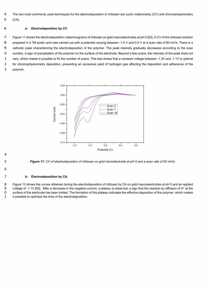
Figure 11 shows the electrodeposition voltammograms of chitosan on gold macroelectrodes at pH 5 [92]. A CV of the chitosan solution 407 prepared in 0.1M acetic acid was carried out with a potential varying between -1.5 V and 0.5 V at a scan rate of 80 mV/s. There is a 408 cathodic peak characterizing the electrodeposition of the polymer. The peak intensity gradually decreases according to the scan 409 number, a sign of precipitation of the polymer on the surface of the electrode. Beyond a few scans, the intensity of the peak does not 410 vary, which makes it possible to fix the number of scans. This test shows that a constant voltage between -1.3V and -1.1V is optimal 411 for chronoamperometry deposition, preventing an excessive yield of hydrogen gas affecting the deposition and adherence of the 412 polymer. 413
414
Figure 11: CV of electrodeposition of chitosan on gold microelectrode at pH 5 and a scan rate of 80 mV/s. 415
416
b- Electrodeposition by CA 417
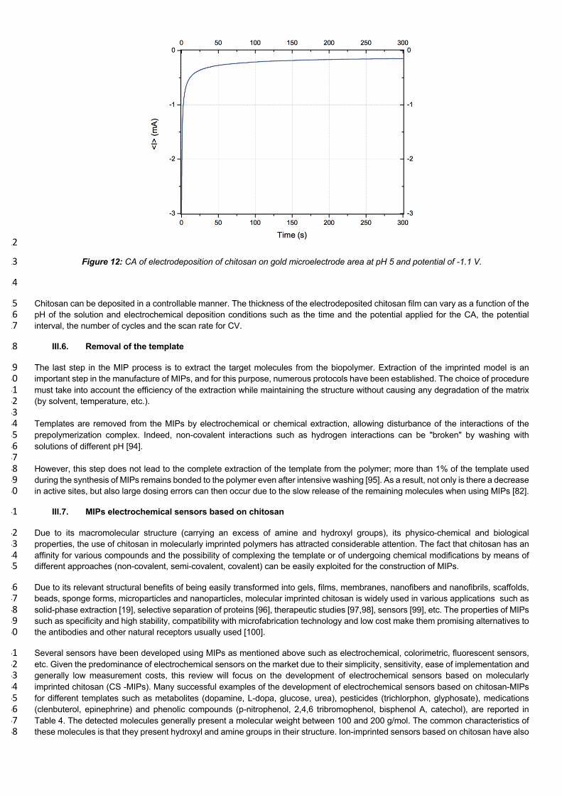
Figure 12 shows the curves obtained during the electrodeposition of chitosan by CA on gold macroelectrodes at pH 5 and an applied 418 voltage of -1.1V [93]. After a decrease in the negative current, a plateau is observed, a sign that the reaction by diffusion of H+ at the 419 surface of the electrode has been limited. The formation of this plateau indicates the effective deposition of the polymer, which makes 420 it possible to optimize the time of the electrodeposition. 421
-1,5 -1,0 -0,5 0,0 0,5-0,10
-0,08
-0,06
-0,04
-0,02
0,00
0,02
Cur
rent
(nA)
Potential (V)
Scan 2 Scan 7 Scan 16
422
Figure 12: CA of electrodeposition of chitosan on gold microelectrode area at pH 5 and potential of -1.1 V. 423
424
Chitosan can be deposited in a controllable manner. The thickness of the electrodeposited chitosan film can vary as a function of the 425 pH of the solution and electrochemical deposition conditions such as the time and the potential applied for the CA, the potential 426 interval, the number of cycles and the scan rate for CV. 427
III.6. Removal of the template 428
The last step in the MIP process is to extract the target molecules from the biopolymer. Extraction of the imprinted model is an 429 important step in the manufacture of MIPs, and for this purpose, numerous protocols have been established. The choice of procedure 430 must take into account the efficiency of the extraction while maintaining the structure without causing any degradation of the matrix 431 (by solvent, temperature, etc.). 432 433 Templates are removed from the MIPs by electrochemical or chemical extraction, allowing disturbance of the interactions of the 434 prepolymerization complex. Indeed, non-covalent interactions such as hydrogen interactions can be "broken" by washing with 435 solutions of different pH [94]. 436 437 However, this step does not lead to the complete extraction of the template from the polymer; more than 1% of the template used 438 during the synthesis of MIPs remains bonded to the polymer even after intensive washing [95]. As a result, not only is there a decrease 439 in active sites, but also large dosing errors can then occur due to the slow release of the remaining molecules when using MIPs [82]. 440
III.7. MIPs electrochemical sensors based on chitosan 441
Due to its macromolecular structure (carrying an excess of amine and hydroxyl groups), its physico-chemical and biological 442 properties, the use of chitosan in molecularly imprinted polymers has attracted considerable attention. The fact that chitosan has an 443 affinity for various compounds and the possibility of complexing the template or of undergoing chemical modifications by means of 444 different approaches (non-covalent, semi-covalent, covalent) can be easily exploited for the construction of MIPs. 445
Due to its relevant structural benefits of being easily transformed into gels, films, membranes, nanofibers and nanofibrils, scaffolds, 446 beads, sponge forms, microparticles and nanoparticles, molecular imprinted chitosan is widely used in various applications such as 447 solid-phase extraction [19], selective separation of proteins [96], therapeutic studies [97,98], sensors [99], etc. The properties of MIPs 448 such as specificity and high stability, compatibility with microfabrication technology and low cost make them promising alternatives to 449 the antibodies and other natural receptors usually used [100]. 450
Several sensors have been developed using MIPs as mentioned above such as electrochemical, colorimetric, fluorescent sensors, 451 etc. Given the predominance of electrochemical sensors on the market due to their simplicity, sensitivity, ease of implementation and 452 generally low measurement costs, this review will focus on the development of electrochemical sensors based on molecularly 453 imprinted chitosan (CS -MIPs). Many successful examples of the development of electrochemical sensors based on chitosan-MIPs 454 for different templates such as metabolites (dopamine, L-dopa, glucose, urea), pesticides (trichlorphon, glyphosate), medications 455 (clenbuterol, epinephrine) and phenolic compounds (p-nitrophenol, 2,4,6 tribromophenol, bisphenol A, catechol), are reported in 456 Table 4. The detected molecules generally present a molecular weight between 100 and 200 g/mol. The common characteristics of 457 these molecules is that they present hydroxyl and amine groups in their structure. Ion-imprinted sensors based on chitosan have also 458
been developed for the detection of chromium (VI), cadmium and copper, due to the ion coordination with hydroxyl and amine groups 459 in the glucosamine units. 460
Electrochemical sensors combined with molecularly imprinted chitosan showed good performance. The proposed sensors exhibited 461 a good linear response towards several targets with a low detection limit, good selectivity, reproducibility, stability and reusability. 462 The preparation procedures presented are relatively simple, convenient and provided rapid and economical methods [101–103]. 463
Pulse Differential Voltammetry (DPV) is the most used electrochemical technique in MIP-based sensors. The results obtained show 464 low detection limits, reaching 10-12 M for the target molecule urea[104], and a slightly higher LOD in the case of dopamine (10-11M) 465 [77]. LOD is in the order of 10-7 to 10-9M with phenolic compounds [56,78,101,105]. The majority of these sensors can be reused up 466 to 9 or 10 times [78,102–104,106], and others less for example 5 times with the target molecules 2,4,6-tribromophenol [105] and 467 copper ions [107]. 468
CA and CV are used in several studies. The detection limits are between 10-5 and 10-9 M [24,25,108,109]. Some sensors have been 469 regenerated 9 times [108,110], and others less, for example 7 times in the case of bisphenol A [109], 3 times with catechol [24,25]. 470 The lowest detection limit recorded is 10-21M. The technique used was Square wave voltammetry (SWV) for the detection of bisphenol 471 A [111]. Another, of 5.9x10⁻¹⁸ M, has been described using electrochemical impedance spectroscopy, for the detection of glyphosate 472 (EIS) [92]. 473
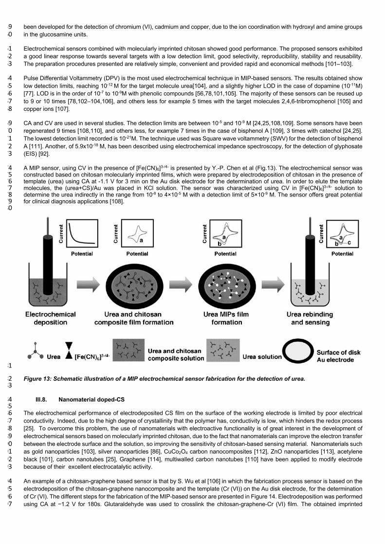
A MIP sensor, using CV in the presence of [Fe(CN)6]3-/4- is presented by Y.-P. Chen et al (Fig.13). The electrochemical sensor was 474 constructed based on chitosan molecularly imprinted films, which were prepared by electrodeposition of chitosan in the presence of 475 template (urea) using CA at -1.1 V for 3 min on the Au disk electrode for the determination of urea. In order to elute the template 476 molecules, the (urea+CS)/Au was placed in KCl solution. The sensor was characterized using CV in [Fe(CN)6]3-/4- solution to 477 determine the urea indirectly in the range from 10-8 to 4×10⁻⁵ M with a detection limit of 5×10⁻⁹ M. The sensor offers great potential 478 for clinical diagnosis applications [108]. 479 480
481
Figure 13: Schematic illustration of a MIP electrochemical sensor fabrication for the detection of urea. 482 483
III.8. Nanomaterial doped-CS 484 485 The electrochemical performance of electrodeposited CS film on the surface of the working electrode is limited by poor electrical 486 conductivity. Indeed, due to the high degree of crystallinity that the polymer has, conductivity is low, which hinders the redox process 487 [25]. To overcome this problem, the use of nanomaterials with electroactive functionality is of great interest in the development of 488 electrochemical sensors based on molecularly imprinted chitosan, due to the fact that nanomaterials can improve the electron transfer 489 between the electrode surface and the solution, so improving the sensitivity of chitosan-based sensing material. Nanomaterials such 490 as gold nanoparticles [103], silver nanoparticles [86], CuCo2O4 carbon nanocomposites [112], ZnO nanoparticles [113], acetylene 491 black [101], carbon nanotubes [25], Graphene [114], multiwalled carbon nanotubes [110] have been applied to modify electrode 492 because of their excellent electrocatalytic activity. 493
An example of a chitosan-graphene based sensor is that by S. Wu et al [106] in which the fabrication process sensor is based on the 494 electrodeposition of the chitosan-graphene nanocomposite and the template (Cr (VI)) on the Au disk electrode, for the determination 495 of Cr (VI). The different steps for the fabrication of the MIP-based sensor are presented in Figure 14. Electrodeposition was performed 496 using CA at −1.2 V for 180s. Glutaraldehyde was used to crosslink the chitosan-graphene-Cr (VI) film. The obtained imprinted 497

electrode was successively rinsed with acetone/water to remove the template. The detection was performed using DPV in a linear 498 range from 10−9 to 10−5 mol/L, with a low detection limit of 6.4×10⁻¹⁰ mol/L. The proposed sensor was successfully applied to the 499 detection of Cr(VI) ions in tap water and river water. 500 501 502
503 504 Figure 14: Schematic representation of the fabrication process of chitosan-graphene MIP sensor for Cr (VI) detection. 505 506 507 In the work of A. Fatoni et al (Fig.15), a MIP sensor was prepared by dropping onto the gold rod electrode the mixture of CS, 508 acrylamide, N,N’-methylenebisacrylamide, graphene, ferrocene and microalbumin as template. The template was removed from the 509 modified electrode by soaking in acetic acid-sodium dodecylsulfate solution. The specific imprinted surface was used to detect 510 microalbumin from 1.5×10⁻⁹ to 1.5×10⁻⁷ M via a redox mediator (ferrocene), entrapped in the cryogel, using differential pulse 511 voltammetry (DPV). The limit of detection was 7.7×10¹³ M and the excellent performance of the biosensor was confirmed by analyzing 512 microalbumin in urine samples [102]. 513 514

515
Figure 15: Schematic representation of the fabrication process of MIP sensor chitosan grafted polyacrylamide MIP cryogel 516 with graphene and entrapped ferrocene for the detection of microalbumin. 517
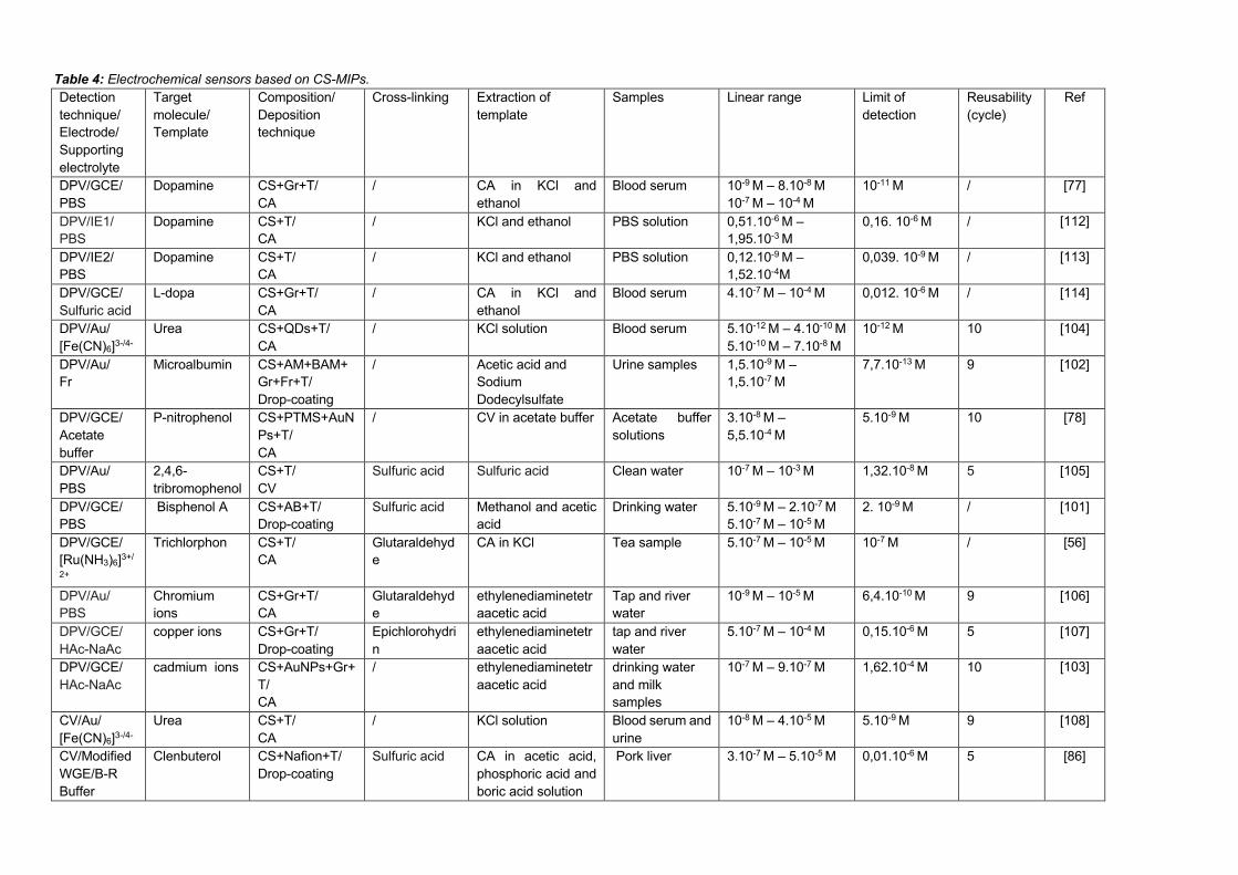
Table 4: Electrochemical sensors based on CS-MIPs. Detection
technique/
Electrode/
Supporting
electrolyte
Target
molecule/
Template
Composition/
Deposition
technique
Cross-linking
Extraction of
template
Samples Linear range Limit of
detection
Reusability
(cycle)
Ref
DPV/GCE/
PBS
Dopamine CS+Gr+T/
CA
/
CA in KCl and
ethanol
Blood serum 10-9 M – 8.10-8 M
10-7 M – 10-4 M
10-11 M
/ [77]
DPV/IE1/
PBS
Dopamine CS+T/
CA
/ KCl and ethanol PBS solution 0,51.10-6 M –
1,95.10-3 M
0,16. 10-6 M / [112]
DPV/IE2/
PBS
Dopamine CS+T/
CA
/ KCl and ethanol PBS solution 0,12.10-9 M –
1,52.10-4M
0,039. 10-9 M / [113]
DPV/GCE/
Sulfuric acid
L-dopa CS+Gr+T/
CA
/ CA in KCl and
ethanol
Blood serum 4.10-7 M – 10-4 M 0,012. 10-6 M / [114]
DPV/Au/
[Fe(CN)6]3-/4- Urea CS+QDs+T/
CA
/ KCl solution Blood serum 5.10-12 M – 4.10-10 M
5.10-10 M – 7.10-8 M
10-12 M 10 [104]
DPV/Au/
Fr
Microalbumin CS+AM+BAM+
Gr+Fr+T/
Drop-coating
/ Acetic acid and
Sodium
Dodecylsulfate
Urine samples 1,5.10-9 M –
1,5.10-7 M
7,7.10-13 M 9 [102]
DPV/GCE/
Acetate
buffer
P-nitrophenol
CS+PTMS+AuN
Ps+T/
CA
/ CV in acetate buffer
Acetate buffer
solutions
3.10-8 M –
5,5.10-4 M
5.10-9 M 10 [78]
DPV/Au/
PBS
2,4,6-
tribromophenol
CS+T/
CV
Sulfuric acid Sulfuric acid Clean water 10-7 M – 10-3 M
1,32.10-8 M
5 [105]
DPV/GCE/
PBS
Bisphenol A CS+AB+T/
Drop-coating
Sulfuric acid Methanol and acetic
acid
Drinking water 5.10-9 M – 2.10-7 M
5.10-7 M – 10-5 M
2. 10-9 M / [101]
DPV/GCE/
[Ru(NH3)6]3+/
2+
Trichlorphon CS+T/
CA
Glutaraldehyd
e
CA in KCl Tea sample 5.10-7 M – 10-5 M 10-7 M / [56]
DPV/Au/
PBS
Chromium
ions
CS+Gr+T/
CA
Glutaraldehyd
e
ethylenediaminetetr
aacetic acid
Tap and river
water
10-9 M – 10-5 M
6,4.10-10 M
9 [106]
DPV/GCE/
HAc-NaAc
copper ions CS+Gr+T/
Drop-coating
Epichlorohydri
n
ethylenediaminetetr
aacetic acid
tap and river
water
5.10-7 M – 10-4 M 0,15.10-6 M 5 [107]
DPV/GCE/
HAc-NaAc
cadmium ions CS+AuNPs+Gr+
T/
CA
/ ethylenediaminetetr
aacetic acid
drinking water
and milk
samples
10-7 M – 9.10-7 M 1,62.10-4 M
10 [103]
CV/Au/
[Fe(CN)6]3-/4-
Urea CS+T/
CA
/ KCl solution Blood serum and
urine
10-8 M – 4.10-5 M
5.10-9 M 9 [108]
CV/Modified
WGE/B-R
Buffer
Clenbuterol
CS+Nafion+T/
Drop-coating
Sulfuric acid CA in acetic acid,
phosphoric acid and
boric acid solution
Pork liver 3.10-7 M – 5.10-5 M 0,01.10-6 M 5 [86]
CV/ABPE/
PBS
BisphenolA CS+T/
Drop-coating
Sulfuric acid Sulfuric acid Plastic samples 8.10-8 M – 10-5 M 60.10-9 M 7 [109]
CV/BDD/
PBS
Catechol CS+AuNPs+MW
CNT+T/
Drop-coating
Glutaraldehyd
e
KCl solution wine sample 7,5.10-5 M – 10-3 M 3,7.10-5 M 3 [25]
CV/BDD/
PBS
Catechol CS+T/
CA
/ KCl solution wine sample 5.10-5 M – 7,5.10-5
M
6,9.10-7 M 3 [24]
SWV/Au/
[Fe(CN)6]3-/4-
bisphenol A CS+T/
CA
Glutaraldehyd
e
Ethanol and acetic
acid
drinking water 10-3 M – 10-21 M 0,67.10-21 M 10 [111]
LSV/ABPE/
PBS
Bisphenol A CS+Gr+T/
Drop-coating
Sulfuric acid CV in HCl Drinking water
and canned
beverages
8.10-9 M – 10-6 M
10-6 M – 20.10-6 M
6.10-9 M 10 [87]
CA/NiOE/
KOH
Glucose CS+T/
CA
/ water elution KOH solution 10-5 M – 20.10-5 M 2.10-6 M / [115]
CA/IE2/
PBS
Dopamine CS+T/
CA
/ KCl and ethanol PBS solution 0,12. 10-9 M –
152. 10-6 M
0.039.10-9 M / [113]
CA/ITO/
PBS
Epinephrine CS+PIL+MWCN
T+T/
CA
/ phosphate buffer
solution
phosphate
buffer solution
0,2. 10-6 M –
0,67.10-3 M
60.10-9 M 9 [110]
EIS/Au/
[Fe(CN)6]3-/4-
Glyphosate CS+T/
CV
Sulfuric acid Ethanol and acetic
acid
River water 1,83. 10-15 M –
3.10-10 M
5,9. 10-18 M 2 [92]
DPV: Pulse Differential Voltammetry
CA: Chronoamperometry
SWV: Square wave voltammetry
CV: Cyclic Voltammetry
LSV: Linear Sweep Voltammetry
EIS: Electrochemical Impedance Spectroscopy
Au: Gold electrode
GCE: Glass Carbon Electrode IE1: Integrated electrode CuCo2O4@carbon nanocoposites/Three-dimensional kenaf stem-derived marcoporous carbon IE2: Integrated electrode ZnO@carbon nanocoposites/Three-dimensional kenaf stem-derived marcoporous carbon Modified WGE: electrode Nano-silver/Quercetin/Working graphite ABPE: Acetylene Black Paste Electrode BDD: Bored-Doped Diamond electrode NiOE: Nickel Oxide Electrode PBS: Phosphate Buffer Solution
B-R Buffer: Britton-Robinson Buffer
HAc-NaAc: Acetic acid-Sodium Acetate T: Template Gr: Graphene QDs: Quantum Dots AM: Acrylamide BAM: N,N’-methylenebisacrylamide

Fr: Ferrocene PTMS: Phenyltrimethoxysilane AuNPs: Gold Nanoparticles AB: Acetylene Black MWCNT: Multiwalled carbon nanotubes

Conclusion and perspectives
Molecular imprinted polymers are a very good choice which has been applied in several fields. Their relevant properties make them very attractive for replacing biological entities in various applications. Chitosan has been considered as an interesting biopolymer for the development of MIPs because of its excellent properties.
Chitosan-MIPs have been successfully integrated into the transducer area for the development of electrochemical sensors. The physicochemical characteristics of chitosan have been exploited to manufacture membranes, thin films and three-dimensional structures by various methods. Electrodeposition is a simple, easy, fast and low-cost technique. It allows the deposition of very porous chitosan films in a controlled manner.
The imprinting of small molecules like pesticides and pharmaceuticals is well established in the synthesis of MIPs and considered routine; however the imprinting of much larger structures remains a challenge. The behavior of chitosan is interrupted by several parameters which undoubtedly affects the sensitivity of the sensors developed. The properties of chitosan should allow the introduction of chemical modifications which can collectively lead to major performance improvements. In the future, multi-imprinting of several templates could be performed on the CS, which can open the door to a fascinating range of fundamental experiments.
Acknowledgements: The authors acknowledge the financial support of the EU H2020 research and innovation program entitled KardiaTool grant #768686 and from CAMPUS FRANCE program under grant agreement PROFAS B+ and PHC MAGHREB #39382RE.
Declaration of interests: The authors declare that they have no known competing financial interests or personal relationships that could have appeared to influence the work reported in this paper. References: [1] M.-P.N. Bui, J. Brockgreitens, S. Ahmed, A. Abbas, Dual detection of nitrate and mercury in water using disposable
electrochemical sensors, Biosensors and Bioelectronics. 85 (2016) 280–286. https://doi.org/10.1016/j.bios.2016.05.017. [2] U. Guth, W. Vonau, J. Zosel, Recent developments in electrochemical sensor application and technology—a review,
Measurement Science and Technology. 20 (2009) 042002. https://doi.org/10.1088/0957-0233/20/4/042002. [3] P. Yáñez-Sedeño, S. Campuzano, J.M. Pingarrón, Electrochemical sensors based on magnetic molecularly imprinted
polymers: A review, Analytica Chimica Acta. 960 (2017) 1–17. https://doi.org/10.1016/j.aca.2017.01.003. [4] F.W. Scheller, X. Zhang, A. Yarman, U. Wollenberger, R.E. Gyurcsányi, Molecularly imprinted polymer-based electrochemical
sensors for biopolymers, Current Opinion in Electrochemistry. 14 (2019) 53–59. https://doi.org/10.1016/j.coelec.2018.12.005. [5] G. Wulff, The use of polymers with enzyme-analogous structures for the resolution of racemates, Angrew. Chem. Internat.
Edit. 11 (1972) 341. [6] J. O’Mahony, B.C.G. Karlsson, B. Mizaikoff, I.A. Nicholls, Correlated theoretical, spectroscopic and X-ray crystallographic
studies of a non-covalent molecularly imprinted polymerisation system, The Analyst. 132 (2007) 1161. https://doi.org/10.1039/b706258c.
[7] K. Singh, S. Balasubramanian, B.E. Amitha Rani, Computational and Experimental Studies of Molecularly Imprinted Polymers for Organochlorine Pesticides Heptachlor and DDT, Current Analytical Chemistry. 8 (2012) 562–568. https://doi.org/10.2174/157341112803216807.
[8] G.Z. Kyzas, N.K. Lazaridis, D.N. Bikiaris, Optimization of chitosan and β-cyclodextrin molecularly imprinted polymer synthesis for dye adsorption, Carbohydrate Polymers. 91 (2013) 198–208. https://doi.org/10.1016/j.carbpol.2012.08.016.
[9] H. Yu, Z. Chen, Y. Fu, L. Kang, M. Wang, X. Du, Synthesis and optimization of molecularly imprinted polymers for quercetin, Polymer International. 61 (2012) 1002–1009. https://doi.org/10.1002/pi.4172.
[10] F. Qiao, H. Sun, H. Yan, K.H. Row, Molecularly Imprinted Polymers for Solid Phase Extraction, Chromatographia. 64 (2006) 625–634. https://doi.org/10.1365/s10337-006-0097-2.
[11] K. Haupt, K. Mosbach, Molecularly Imprinted Polymers and Their Use in Biomimetic Sensors, Chemical Reviews. 100 (2000) 2495–2504. https://doi.org/10.1021/cr990099w.
[12] J. Orozco, A. Cortés, G. Cheng, S. Sattayasamitsathit, W. Gao, X. Feng, Y. Shen, J. Wang, Molecularly Imprinted Polymer-Based Catalytic Micromotors for Selective Protein Transport, Journal of the American Chemical Society. 135 (2013) 5336–5339. https://doi.org/10.1021/ja4018545.
[13] A. Nezhadali, M. Mojarrab, Computational study and multivariate optimization of hydrochlorothiazide analysis using molecularly imprinted polymer electrochemical sensor based on carbon nanotube/polypyrrole film, Sensors and Actuators B: Chemical. 190 (2014) 829–837. https://doi.org/10.1016/j.snb.2013.08.086.
[14] E. Turiel, A. Martín-Esteban, Molecularly imprinted polymers for sample preparation: A review, Analytica Chimica Acta. 668 (2010) 87–99. https://doi.org/10.1016/j.aca.2010.04.019.
[15] P.K. Dutta, J. Dutta, V. Tripathi, Chitin and chitosan: Chemistry, properties and applications, (2004). [16] R. Jayakumar, M. Prabaharan, R.L. Reis, J.F. Mano, Graft copolymerized chitosan—present status and applications,
Carbohydrate Polymers. 62 (2005) 142–158. https://doi.org/10.1016/j.carbpol.2005.07.017. [17] A. Grenha, C.I. Grainger, L.A. Dailey, B. Seijo, G.P. Martin, C. Remuñán-López, B. Forbes, Chitosan nanoparticles are
compatible with respiratory epithelial cells in vitro, European Journal of Pharmaceutical Sciences. 31 (2007) 73–84. https://doi.org/10.1016/j.ejps.2007.02.008.
[18] M.-C. Li, J.K. Lee, U.R. Cho, Synthesis, characterization, and enzymatic degradation of starch-grafted poly(methyl methacrylate) copolymer films, Journal of Applied Polymer Science. 125 (2012) 405–414. https://doi.org/10.1002/app.35620.
[19] Y. Wang, E. Wang, Z. Wu, H. Li, Z. Zhu, X. Zhu, Y. Dong, Synthesis of chitosan molecularly imprinted polymers for solid-phase extraction of methandrostenolone, Carbohydrate Polymers. 101 (2014) 517–523. https://doi.org/10.1016/j.carbpol.2013.09.078.

[20] X. Wang, M. Wu, W. Tang, Y. Zhu, L. Wang, Q. Wang, P. He, Y. Fang, Simultaneous electrochemical determination of ascorbic acid, dopamine and uric acid using a palladium nanoparticle/graphene/chitosan modified electrode, Journal of Electroanalytical Chemistry. 695 (2013) 10–16. https://doi.org/10.1016/j.jelechem.2013.02.021.
[21] T.I. Nasution, I. Nainggolan, S.D. Hutagalung, K.R. Ahmad, Z.A. Ahmad, The sensing mechanism and detection of low concentration acetone using chitosan-based sensors, Sensors and Actuators B: Chemical. 177 (2013) 522–528. https://doi.org/10.1016/j.snb.2012.11.063.
[22] P.M. Claesson, B.W. Ninham, pH-dependent interactions between adsorbed chitosan layers, Langmuir. 8 (1992) 1406–1412. https://doi.org/10.1021/la00041a027.
[23] V. Tangpasuthadol, N. Pongchaisirikul, V.P. Hoven, Surface modification of chitosan films., Carbohydrate Research. 338 (2003) 937–942. https://doi.org/10.1016/s0008-6215(03)00038-7.
[24] C. Salvo-Comino, I. Rassas, S. Minot, F. Bessueille, M.L. Rodriguez-Mendez, A. Errachid, N. Jaffrezic-Renault, Voltammetric sensor based on electrodeposited molecularly imprinted chitosan film on BDD electrodes for catechol detection in buffer and in wine samples, Materials Science and Engineering: C. 110 (2020) 110667. https://doi.org/10.1016/j.msec.2020.110667.
[25] C. Salvo-Comino, I. Rassas, S. Minot, F. Bessueille, M. Arab, V. Chevallier, M.L. Rodriguez-Mendez, A. Errachid, N. Jaffrezic-Renault, Voltammetric Sensor Based on Molecularly Imprinted Chitosan-Carbon Nanotubes Decorated with Gold Nanoparticles Nanocomposite Deposited on Boron-Doped Diamond Electrodes for Catechol Detection, Materials. 13 (2020) 688. https://doi.org/10.3390/ma13030688.
[26] H. Yi, L.-Q. Wu, W.E. Bentley, R. Ghodssi, G.W. Rubloff, J.N. Culver, G.F. Payne, Biofabrication with Chitosan, Biomacromolecules. 6 (2005) 2881–2894. https://doi.org/10.1021/bm050410l.
[27] I. Aranaz, M. Mengibar, R. Harris, I. Panos, B. Miralles, N. Acosta, G. Galed, A. Heras, Functional Characterization of Chitin and Chitosan, Current Chemical Biology. 3 (2009) 203–230. https://doi.org/10.2174/187231309788166415.
[28] I. Younes, M. Rinaudo, Chitin and Chitosan Preparation from Marine Sources. Structure, Properties and Applications, Marine Drugs. 13 (2015) 1133–1174. https://doi.org/10.3390/md13031133.
[29] H. El Knidri, R. Belaabed, A. Addaou, A. Laajeb, A. Lahsini, Extraction, chemical modification and characterization of chitin and chitosan, International Journal of Biological Macromolecules. 120 (2018) 1181–1189. https://doi.org/10.1016/j.ijbiomac.2018.08.139.
[30] A. Domard, M. Domard, Chitosan: structure-properties relationship and biomedical applications, Polymeric Biomaterials. 2 (2001) 187–212.
[31] P. Sorlier, C. Rochas, I. Morfin, C. Viton, A. Domard, Light Scattering Studies of the Solution Properties of Chitosans of Varying Degrees of Acetylation, Biomacromolecules. 4 (2003) 1034–1040. https://doi.org/10.1021/bm034054n.
[32] L. Raymond, F.G. Morin, R.H. Marchessault, Degree of deacetylation of chitosan using conductometric titration and solid-state NMR, Carbohydrate Research. 246 (1993) 331–336. https://doi.org/10.1016/0008-6215(93)84044-7.
[33] Z.M. dos Santos, A.L.P.F. Caroni, M.R. Pereira, D.R. da Silva, J.L.C. Fonseca, Determination of deacetylation degree of chitosan: a comparison between conductometric titration and CHN elemental analysis, Carbohydrate Research. 344 (2009) 2591–2595. https://doi.org/10.1016/j.carres.2009.08.030.
[34] X. Jiang, L. Chen, W. Zhong, A new linear potentiometric titration method for the determination of deacetylation degree of chitosan, Carbohydrate Polymers. 54 (2003) 457–463. https://doi.org/10.1016/j.carbpol.2003.05.004.
[35] M.R. Kasaai, Determination of the degree of N-acetylation for chitin and chitosan by various NMR spectroscopy techniques: A review, Carbohydrate Polymers. 79 (2010) 801–810. https://doi.org/10.1016/j.carbpol.2009.10.051.
[36] V. Zargar, M. Asghari, A. Dashti, A Review on Chitin and Chitosan Polymers: Structure, Chemistry, Solubility, Derivatives, and Applications, ChemBioEng Reviews. 2 (2015) 204–226. https://doi.org/10.1002/cben.201400025.
[37] I. Tsigos, A. Martinou, D. Kafetzopoulos, V. Bouriotis, Chitin deacetylases: new, versatile tools in biotechnology, Trends in Biotechnology. 18 (2000) 305–312. https://doi.org/10.1016/S0167-7799(00)01462-1.
[38] S. Kim, N. Rajapakse, Enzymatic production and biological activities of chitosan oligosaccharides (COS): A review, Carbohydrate Polymers. 62 (2005) 357–368. https://doi.org/10.1016/j.carbpol.2005.08.012.
[39] Y. Zhao, R.-D. Park, R.A.A. Muzzarelli, Chitin Deacetylases: Properties and Applications, Marine Drugs. 8 (2010) 24–46. https://doi.org/10.3390/md8010024.
[40] S. Naqvi, S. Cord-Landwehr, R. Singh, F. Bernard, S. Kolkenbrock, N.E. El Gueddari, B.M. Moerschbacher, A Recombinant Fungal Chitin Deacetylase Produces Fully Defined Chitosan Oligomers with Novel Patterns of Acetylation, Appl. Environ. Microbiol. 82 (2016) 6645–6655. https://doi.org/10.1128/AEM.01961-16.
[41] H.K. No, S.P. Meyers, W. Prinyawiwatkul, Z. Xu, Applications of Chitosan for Improvement of Quality and Shelf Life of Foods: A Review, J Food Science. 72 (2007) R87–R100. https://doi.org/10.1111/j.1750-3841.2007.00383.x.
[42] R. Cheung, T. Ng, J. Wong, W. Chan, Chitosan: An Update on Potential Biomedical and Pharmaceutical Applications, Marine Drugs. 13 (2015) 5156–5186. https://doi.org/10.3390/md13085156.
[43] I. Hamed, F. Özogul, J.M. Regenstein, Industrial applications of crustacean by-products (chitin, chitosan, and chitooligosaccharides): A review, Trends in Food Science & Technology. 48 (2016) 40–50. https://doi.org/10.1016/j.tifs.2015.11.007.
[44] A. Bhatnagar, M. Sillanpää, Applications of chitin- and chitosan-derivatives for the detoxification of water and wastewater — A short review, Advances in Colloid and Interface Science. 152 (2009) 26–38. https://doi.org/10.1016/j.cis.2009.09.003.
[45] R.A.A. Muzzarelli, Potential of chitin/chitosan-bearing materials for uranium recovery: An interdisciplinary review, Carbohydrate Polymers. 84 (2011) 54–63. https://doi.org/10.1016/j.carbpol.2010.12.025.
[46] M. Hasegawa, A. Isogai, F. Onabe, Preparation of low-molecular-weight chitosan using phosphoric acid, Carbohydrate Polymers. 20 (1993) 279–283. https://doi.org/10.1016/0144-8617(93)90100-I.
[47] W.A. Bough, W.L. Salter, A.C.M. Wu, B.E. Perkins, Influence of manufacturing variables on the characteristics and effectiveness of chitosan products. I. Chemical composition, viscosity, and molecular-weight distribution of chitosan products, Biotechnol. Bioeng. 20 (1978) 1931–1943. https://doi.org/10.1002/bit.260201208.
[48] J. Pa, T.L. Yu, Light scattering study of chitosan in acetic acid aqueous solutions, Macromolecular Chemistry and Physics. 202 (2001) 985–991.
[49] I. Aranaz, R. Harris, A. Heras, Chitosan amphiphilic derivatives. Chemistry and applications, Current Organic Chemistry. 14 (2010) 308–330.
[50] M. Dash, F. Chiellini, R.M. Ottenbrite, E. Chiellini, Chitosan—A versatile semi-synthetic polymer in biomedical applications, Progress in Polymer Science. 36 (2011) 981–1014. https://doi.org/10.1016/j.progpolymsci.2011.02.001.
[51] T. Ahmed, B. Aljaeid, Preparation, characterization, and potential application of chitosan, chitosan derivatives, and chitosan metal nanoparticles in pharmaceutical drug delivery, Drug Design, Development and Therapy. (2016) 483. https://doi.org/10.2147/DDDT.S99651.
[52] M. Rinaudo, Chitin and chitosan: Properties and applications, Progress in Polymer Science. 31 (2006) 603–632. https://doi.org/10.1016/j.progpolymsci.2006.06.001.
[53] C. Tual, E. Espuche, M. Escoubes, A. Domard, Transport properties of chitosan membranes: influence of crosslinking, Journal of Polymer Science Part B: Polymer Physics. 38 (2000) 1521–1529.
[54] B. Sellergren, L. Andersson, Molecular recognition in macroporous polymers prepared by a substrate analog imprinting strategy, The Journal of Organic Chemistry. 55 (1990) 3381–3383.
[55] J. Zhang, W. Xia, P. Liu, Q. Cheng, T. Tahi, W. Gu, B. Li, Chitosan Modification and Pharmaceutical/Biomedical Applications, Marine Drugs. 8 (2010) 1962–1987. https://doi.org/10.3390/md8071962.
[56] J. Chen, H. Lian, X. Sun, B. Liu, Development of a chitosan molecularly imprinted electrochemical sensor for trichlorphon determination, International Journal of Environmental Analytical Chemistry. 92 (2012) 1046–1058. https://doi.org/10.1080/03067319.2010.496054.
[57] P. Sorlier, A. Denuzière, C. Viton, A. Domard, Relation between the degree of acetylation and the electrostatic properties of chitin and chitosan, Biomacromolecules. 2 (2001) 765–772.
[58] C. Schatz, C. Pichot, T. Delair, C. Viton, A. Domard, Static Light Scattering Studies on Chitosan Solutions: From Macromolecular Chains to Colloidal Dispersions, Langmuir. 19 (2003) 9896–9903. https://doi.org/10.1021/la034410n.
[59] A.S. Carreira, F.A.M.M. Gonçalves, P.V. Mendonça, M.H. Gil, J.F.J. Coelho, Temperature and pH responsive polymers based on chitosan: Applications and new graft copolymerization strategies based on living radical polymerization, Carbohydrate Polymers. 80 (2010) 618–630. https://doi.org/10.1016/j.carbpol.2009.12.047.
[60] T. Wanjun, W. Cunxin, C. Donghua, Kinetic studies on the pyrolysis of chitin and chitosan, Polymer Degradation and Stability. 87 (2005) 389–394. https://doi.org/10.1016/j.polymdegradstab.2004.08.006.
[61] G.C. Ritthidej, T. Phaechamud, T. Koizumi, Moist heat treatment on physicochemical change of chitosan salt films, International Journal of Pharmaceutics. 232 (2002) 11–22. https://doi.org/10.1016/S0378-5173(01)00894-8.
[62] A. Toffey, W.G. Glasser, Chitin derivatives III Formation of amidized homologs of chitosan, Cellulose. 8 (2001) 35–47. [63] L. Lim, E. Khor, C. Ling, Effects of dry heat and saturated steam on the physical properties of chitosan, Journal of Biomedical
Materials Research: An Official Journal of The Society for Biomaterials, The Japanese Society for Biomaterials, and The Australian Society for Biomaterials. 48 (1999) 111–116.
[64] C.A. Murray, J.R. Dutcher, Effect of Changes in Relative Humidity and Temperature on Ultrathin Chitosan Films, Biomacromolecules. 7 (2006) 3460–3465. https://doi.org/10.1021/bm060416q.
[65] J. Wang, L. Wang, H. Yu, Zain-ul-Abdin, Y. Chen, Q. Chen, W. Zhou, H. Zhang, X. Chen, Recent progress on synthesis, property and application of modified chitosan: An overview, International Journal of Biological Macromolecules. 88 (2016) 333–344. https://doi.org/10.1016/j.ijbiomac.2016.04.002.
[66] L. Xu, Y.-A. Huang, Q.-J. Zhu, C. Ye, Chitosan in Molecularly-Imprinted Polymers: Current and Future Prospects, International Journal of Molecular Sciences. 16 (2015) 18328–18347. https://doi.org/10.3390/ijms160818328.
[67] E. Szymańska, K. Winnicka, Stability of Chitosan—A Challenge for Pharmaceutical and Biomedical Applications, Marine Drugs. 13 (2015) 1819–1846. https://doi.org/10.3390/md13041819.
[68] K. Gupta, F. Jabrail, Effects of degree of deacetylation and cross-linking on physical characteristics, swelling and release behavior of chitosan microspheres, Carbohydrate Polymers. 66 (2006) 43–54. https://doi.org/10.1016/j.carbpol.2006.02.019.
[69] Y. Yuan, B.M. Chesnutt, G. Utturkar, W.O. Haggard, Y. Yang, J.L. Ong, J.D. Bumgardner, The effect of cross-linking of chitosan microspheres with genipin on protein release, Carbohydrate Polymers. 68 (2007) 561–567. https://doi.org/10.1016/j.carbpol.2006.10.023.
[70] M.S. Chiou, H.Y. Li, Adsorption behavior of reactive dye in aqueous solution on chemical cross-linked chitosan beads, Chemosphere. 50 (2003) 1095–1105. https://doi.org/10.1016/S0045-6535(02)00636-7.
[71] Y.-L. Liu, Y.-H. Su, J.-Y. Lai, In situ crosslinking of chitosan and formation of chitosan–silica hybrid membranes with using γ-glycidoxypropyltrimethoxysilane as a crosslinking agent, Polymer. 45 (2004) 6831–6837. https://doi.org/10.1016/j.polymer.2004.08.006.
[72] J.D. Schiffman, C.L. Schauer, Cross-Linking Chitosan Nanofibers, Biomacromolecules. 8 (2007) 594–601. https://doi.org/10.1021/bm060804s.
[73] F.S. Pereira, D.L. da Silva Agostini, A.E. Job, E.R.P. González, Thermal studies of chitin–chitosan derivatives, J Therm Anal Calorim. 114 (2013) 321–327. https://doi.org/10.1007/s10973-012-2835-z.
[74] A.O. Martins, E.L. da Silva, E. Carasek, N.S. Gonçalves, M.C.M. Laranjeira, V.T. de Fávere, Chelating resin from functionalization of chitosan with complexing agent 8-hydroxyquinoline: application for metal ions on line preconcentration system, Analytica Chimica Acta. 521 (2004) 157–162. https://doi.org/10.1016/j.aca.2004.06.033.
[75] P.R. Solanki, A. Kaushik, A.A. Ansari, A. Tiwari, B.D. Malhotra, Multi-walled carbon nanotubes/sol–gel-derived silica/chitosan nanobiocomposite for total cholesterol sensor, Sensors and Actuators B: Chemical. 137 (2009) 727–735. https://doi.org/10.1016/j.snb.2008.12.044.
[76] T.W. Chung, J. Yang, T. Akaike, K.Y. Cho, J.W. Nah, S.I. Kim, C.S. Cho, Preparation of alginate/galactosylated chitosan scaffold for hepatocyte attachment, Biomaterials. 23 (2002) 2827–2834. https://doi.org/10.1016/S0142-9612(01)00399-4.
[77] B. Liu, H.T. Lian, J.F. Yin, X.Y. Sun, Dopamine molecularly imprinted electrochemical sensor based on graphene–chitosan composite, Electrochimica Acta. 75 (2012) 108–114. https://doi.org/10.1016/j.electacta.2012.04.081.
[78] S. Li, D. Du, J. Huang, H. Tu, Y. Yang, A. Zhang, One-step electrodeposition of a molecularly imprinting chitosan/phenyltrimethoxysilane/AuNPs hybrid film and its application in the selective determination of p-nitrophenol, Analyst. 138 (2013) 2761. https://doi.org/10.1039/c3an36497f.
[79] C. Caner, P.J. Vergano, J.L. Wiles, Chitosan Film Mechanical and Permeation Properties as Affected by Acid, Plasticizer, and Storage, Journal of Food Science. 63 (2006) 1049–1053. https://doi.org/10.1111/j.1365-2621.1998.tb15852.x.
[80] G.A.F. Roberts, J.G. Domszy, Determination of the viscometric constants for chitosan, International Journal of Biological Macromolecules. 4 (1982) 374–377. https://doi.org/10.1016/0141-8130(82)90074-5.
[81] M. Lopez-Nogueroles, S. Lordel-Madeleine, A. Chisvert, A. Salvador, V. Pichon, Development of a selective solid phase extraction method for nitro musk compounds in environmental waters using a molecularly imprinted sorbent, Talanta. 110 (2013) 128–134. https://doi.org/10.1016/j.talanta.2013.02.023.
[82] R. Lorenzo, A. Carro, C. Alvarez-Lorenzo, A. Concheiro, To Remove or Not to Remove? The Challenge of Extracting the Template to Make the Cavities Available in Molecularly Imprinted Polymers (MIPs), International Journal of Molecular Sciences. 12 (2011) 4327–4347. https://doi.org/10.3390/ijms12074327.
[83] E. Turiel, A. Martín-Esteban, P. Fernández, C. Pérez-Conde, C. Cámara, Molecular Recognition in a Propazine-imprinted Polymer and Its Application to the Determination of Triazines in Environmental Samples, Analytical Chemistry. 73 (2001) 5133–5141. https://doi.org/10.1021/ac0105538.
[84] H. Yan, K. Row, Characteristic and Synthetic Approach of Molecularly Imprinted Polymer, International Journal of Molecular Sciences. 7 (2006) 155–178. https://doi.org/10.3390/i7050155.
[85] J. Svenson, I.A. Nicholls, On the thermal and chemical stability of molecularly imprinted polymers, Analytica Chimica Acta. 435 (2001) 19–24. https://doi.org/10.1016/S0003-2670(00)01396-9.
[86] C. Zhao, G.-P. Jin, L.-L. Chen, Y. Li, B. Yu, Preparation of molecular imprinted film based on chitosan/nafion/nano-silver/poly quercetin for clenbuterol sensing, Food Chemistry. 129 (2011) 595–600. https://doi.org/10.1016/j.foodchem.2011.04.072.
[87] P. Deng, Z. Xu, Y. Kuang, Electrochemical determination of bisphenol A in plastic bottled drinking water and canned beverages using a molecularly imprinted chitosan–graphene composite film modified electrode, Food Chemistry. 157 (2014) 490–497. https://doi.org/10.1016/j.foodchem.2014.02.074.
[88] H. Li, C. Xie, X. Fu, Electrochemiluminescence sensor for sulfonylurea herbicide with molecular imprinting core–shell nanoparticles/chitosan composite film modified glassy carbon electrode, Sensors and Actuators B: Chemical. 181 (2013) 858–866. https://doi.org/10.1016/j.snb.2013.02.094.
[89] X. Pang, I. Zhitomirsky, Electrodeposition of composite hydroxyapatite–chitosan films, Materials Chemistry and Physics. 94 (2005) 245–251. https://doi.org/10.1016/j.matchemphys.2005.04.040.
[90] L.-Q. Wu, H. Yi, S. Li, G.W. Rubloff, W.E. Bentley, R. Ghodssi, G.F. Payne, Spatially Selective Deposition of a Reactive Polysaccharide Layer onto a Patterned Template, Langmuir. 19 (2003) 519–524. https://doi.org/10.1021/la026518t.
[91] R. Fernandes, L.-Q. Wu, T. Chen, H. Yi, G.W. Rubloff, R. Ghodssi, W.E. Bentley, G.F. Payne, Electrochemically Induced Deposition of a Polysaccharide Hydrogel onto a Patterned Surface, Langmuir. 19 (2003) 4058–4062. https://doi.org/10.1021/la027052h.
[92] F. Zouaoui, S. Bourouina-Bacha, M. Bourouina, I. Abroa-Nemeir, H. Ben Halima, J. Gallardo-Gonzalez, N. El Alami El Hassani, A. Alcacer, J. Bausells, N. Jaffrezic-Renault, N. Zine, A. Errachid, Electrochemical impedance spectroscopy determination of glyphosate using a molecularly imprinted chitosan, Sensors and Actuators B: Chemical. 309 (2020) 127753. https://doi.org/10.1016/j.snb.2020.127753.
[93] S. Boudjaoui, Développement et optimisation de capteurs de gaz à base de silicium: analyse des composés organiques volatils contenus dans l’air expiré par les patients atteints d’insuffisance cardiaque, (2018).
[94] C. Algieri, E. Drioli, L. Guzzo, L. Donato, Bio-Mimetic Sensors Based on Molecularly Imprinted Membranes, Sensors. 14 (2014) 13863–13912. https://doi.org/10.3390/s140813863.
[95] A. Ellwanger, L. Karlsson, P.K. Owens, C. Berggren, C. Crecenzi, K. Ensing, S. Bayoudh, P. Cormack, D. Sherrington, B. Sellergren, Evaluation of methods aimed at complete removal of template from molecularly imprinted polymers, The Analyst. 126 (2001) 784–792. https://doi.org/10.1039/b009693h.
[96] T.-Y. Guo, Y.-Q. Xia, J. Wang, M.-D. Song, B.-H. Zhang, Chitosan beads as molecularly imprinted polymer matrix for selective separation of proteins, Biomaterials. 26 (2005) 5737–5745. https://doi.org/10.1016/j.biomaterials.2005.02.017.
[97] D. Teng, J. Hou, X. Zhang, X. Wang, Z. Wang, C. Li, Glucosamine-carrying temperature- and pH-sensitive microgels: Preparation, characterization, and in vitro drug release studies, Journal of Colloid and Interface Science. 322 (2008) 333–341. https://doi.org/10.1016/j.jcis.2008.03.014.
[98] H.L. Zhao, T.Y. Guo, Y.Q. Xia, M.D. Song, Hemoglobin-imprinted polymer gel prepared using modified glucosamine as functional monomer, Chinese Chemical Letters. 19 (2008) 233–236. https://doi.org/10.1016/j.cclet.2007.10.045.
[99] G. Liu, T. Li, X. Yang, Y. She, M. Wang, J. Wang, M. Zhang, S. Wang, F. Jin, M. Jin, H. Shao, Z. Jiang, H. Yu, Competitive fluorescence assay for specific recognition of atrazine by magnetic molecularly imprinted polymer based on Fe 3 O 4 -chitosan, Carbohydrate Polymers. 137 (2016) 75–81. https://doi.org/10.1016/j.carbpol.2015.10.062.
[100] W. Guo, F. Pi, H. Zhang, J. Sun, Y. Zhang, X. Sun, A novel molecularly imprinted electrochemical sensor modified with carbon dots, chitosan, gold nanoparticles for the determination of patulin, Biosensors and Bioelectronics. 98 (2017) 299–304. https://doi.org/10.1016/j.bios.2017.06.036.
[101] Y. Tan, J. Jin, S. Zhang, Z. Shi, J. Wang, J. Zhang, W. Pu, C. Yang, Electrochemical Determination of Bisphenol A Using a Molecularly Imprinted Chitosan-acetylene Black Composite Film Modified Glassy Carbon Electrode, Electroanalysis. 28 (2016) 189–196. https://doi.org/10.1002/elan.201500533.
[102] A. Fatoni, A. Numnuam, P. Kanatharana, W. Limbut, P. Thavarungkul, A novel molecularly imprinted chitosan–acrylamide, graphene, ferrocene composite cryogel biosensor used to detect microalbumin, Analyst. 139 (2014) 6160–6167. https://doi.org/10.1039/C4AN01000K.
[103] S. Wu, K. Li, X. Dai, Z. Zhang, F. Ding, S. Li, An ultrasensitive electrochemical platform based on imprinted chitosan/gold nanoparticles/graphene nanocomposite for sensing cadmium (II) ions, Microchemical Journal. 155 (2020) 104710. https://doi.org/10.1016/j.microc.2020.104710.
[104] H.-T. Lian, B. Liu, Y.-P. Chen, X.-Y. Sun, A urea electrochemical sensor based on molecularly imprinted chitosan film doping with CdS quantum dots, Analytical Biochemistry. 426 (2012) 40–46. https://doi.org/10.1016/j.ab.2012.03.024.
[105] L. Huang, Y. Lu, Z. Wu, M. Li, S. Xiang, X. Ma, Z. Zhang, A Facile Approach to Preparing Molecularly Imprinted Chitosan for Detecting 2,4,6-Tribromophenol with a Widely Linear Range, Environments. 4 (2017) 30. https://doi.org/10.3390/environments4020030.
[106] S. Wu, X. Dai, T. Cheng, S. Li, Highly sensitive and selective ion-imprinted polymers based on one-step electrodeposition of chitosan-graphene nanocomposites for the determination of Cr(VI), Carbohydrate Polymers. 195 (2018) 199–206. https://doi.org/10.1016/j.carbpol.2018.04.077.
[107] P. Wei, Z. Zhu, R. Song, Z. Li, C. Chen, An ion-imprinted sensor based on chitosan-graphene oxide composite polymer modified glassy carbon electrode for environmental sensing application, Electrochimica Acta. 317 (2019) 93–101. https://doi.org/10.1016/j.electacta.2019.05.136.
[108] Y.-P. Chen, B. Liu, H.-T. Lian, X.-Y. Sun, Preparation and Application of Urea Electrochemical Sensor Based on Chitosan Molecularly Imprinted Films, Electroanalysis. 23 (2011) 1454–1461. https://doi.org/10.1002/elan.201000693.
[109] P. Deng, Z. Xu, J. Li, Y. Kuang, Acetylene black paste electrode modified with a molecularly imprinted chitosan film for the detection of bisphenol A, Microchim Acta. 180 (2013) 861–869. https://doi.org/10.1007/s00604-013-1001-z.

[110] Y. Wu, X. Feng, S. Zhou, H. Shi, H. Wu, S. Zhao, W. Song, Sensing epinephrine with an ITO electrode modified with an imprinted chitosan film containing multi-walled carbon nanotubes and a polymerized ionic liquid, Microchimica Acta. 180 (2013) 1325–1332. https://doi.org/10.1007/s00604-013-1063-y.
[111] H. Chakroun Galai, P. Namour, A. Bonhomme, F. Bessueille, S. Besbes Hentati, N. Jaffrezic-Renault, Elaboration of an Imprinted Polymer Film Based on Chitosan Electrodeposition for the Voltammetric Detection of BPA, Journal of The Electrochemical Society. 167 (2020) 027507. https://doi.org/10.1149/1945-7111/ab6283.
[112] L. Wang, H. Yang, L. Xu, C. Peng, Y. Song, A novel popamine-imprinted chitosan/CuCo2O4@carbon/three-dimensional macroporous carbon integrated electrode, Journal of Alloys and Compounds. 817 (2020) 152771. https://doi.org/10.1016/j.jallcom.2019.152771.
[113] Y. Song, J. Han, L. Xu, L. Miao, C. Peng, L. Wang, A dopamine-imprinted chitosan Film/Porous ZnO NPs@carbon Nanospheres/Macroporous carbon for electrochemical sensing dopamine, Sensors and Actuators B: Chemical. 298 (2019) 126949. https://doi.org/10.1016/j.snb.2019.126949.
[114] L. Lin, H.-T. Lian, X.-Y. Sun, Y.-M. Yu, B. Liu, An L-dopa electrochemical sensor based on a graphene doped molecularly imprinted chitosan film, Analytical Methods. 7 (2015) 1387–1394.
[115] H.X. Li, W. Yao, Q. Wu, W.S. Xia, Glucose Molecularly Imprinted Electrochemical Sensor Based on Chitosan and Nickel Oxide Electrode, Advanced Materials Research. 1052 (2014) 215–219. https://doi.org/10.4028/www.scientific.net/AMR.1052.215.