Electrocardiogram manifestations in hyperkalemia
7
World Journal of Cardiovascular Diseases, 2012, 2, 57-63 WJCD http://dx.doi.org/10.4236/wjcd.2012.22010 Published Online April 2012 (http://www.SciRP.org/journal/wjcd/ ) Electrocardiogram manifestations in hyperkalemia Ronny Cohen 1* , Rhadames Ramos 1 , Christine A. Garcia 2 , Sohail Mehmood 1 , Yoojin Park 2 , Anthony Divittis 1 , Brooks Mirrer 1 1 Woodhull Medical Center, New York University School of Medicine, New York, USA 2 St. George’s University School of Medicine, St. George’s, Grenada Email: * [email protected] Received 31 October 2011; revised 16 December 2011; accepted 23 December 2011 ABSTRACT Hyperkalemia is defined as serum potassium level of more than 5 mmol/L. Prompt identification of hyper- kalemia and appropriate management are critical, since severe hyperkalemia can lead to lethal cardiac dysrhythmias. There is a wide range of electrocardio- gram (EKG) changes associated with hyperkalemia. The sequence of EKG changes has been previously described with limited information to correlate the level of potassium to a particular change in the EKG. This study aims to describe a correlation between the level of potassium and EKG changes in the presence or absence of certain diagnoses, to determine which EKG finding in the context of level of hyperkalemia, should be considered life-threatening and prompt emergency intervention. If a relationship between se- rum levels of potassium and EKG changes is signifi- cant, clinicians may be able to better monitor and treat hyperkalemic patients. This paper reviews the literature on hyperkalemia, potassium homeostasis and EKG changes attributed to elevated potassium. Keywords: EKG; ECG; Hyperkalemia; Electrocardiogram 1. INTRODUCTION Extracellular potassium concentration is normally main- tained between 4 and 4.5 mmol/L. Hyperkalemia is de- fined as serum potassium level greater than 5.0 mmol/L. It is a common metabolic disturbance with potentially life-threatening consequences. Since severe hyperkalemia can lead to lethal cardiac dysrhythmias, prompt identifi- cation of hyperkalemia and appropriate management are crucial. 1.1. Normal Potassium Homeostasis The serum potassium concentration is determined by the relationship between potassium intake, the distribution of potassium in the cells and the extra cellular fluid, and urinary potassium excretion. In normal subjects, dietary potassium is largely excreted in the urine, a process that involves the potassium secretion in the principal cells in the cortical collecting tubule. The degree of potassium secretion is primarily stimulated by three factors: an in- crease in the serum concentration, a rise in the plasma aldosterone concentration and enhanced delivery of so- dium and water to the distal secretory site. Under normal circumstances, renal excretion accounts for approximately 90% of daily potassium elimination. Distal tubule urine flow may influence potassium secre- tion because increased flow increases the concentration gradient and therefore, potassium loss. As renal function declines, a greater fraction of the filtered load is excreted, and plasma potassium levels generally do not rise until renal function declines to less than 25% of normal [1-3]. 1.2. Causes of Hyperkalemia There are numerous causes of hyperkalemia. Elevated serum potassium levels can be due to either true hyper- kalemia or pseudohyperkalemia. Pseudohyperkalemia is artificially elevated plasma potassium [K + ] in a blood specimen due to potassium movement out of the cells. The major cause of pseudohyperkalemia is mechanical trauma during venipuncture (hemolytic) and prolonged tourniquet placement during venipuncture. This may result in potassium release from cells and adds characteristic reddish tint to serum due to concomitant release of he- moglobin from cells. True hyperkalemia occurs as a result of transcellular shift, increased potassium intake and decreased potas- sium excretion by kidneys. Transcellular shift can result from Diabetic Ketoacidosis (DKA). The acidemia in DKA causes hydrogen ions (H + ) to move into cells and K + ions to move out of cells to maintain electroneutrality, result- ing in subsequent hyperkalemia. Insulin deficiency and hyperosmolar state also contribute to the shift of potas- sium from intracellular fluid to extracellular fluid. Other * Corresponding author. OPEN ACCESS
Transcript of Electrocardiogram manifestations in hyperkalemia
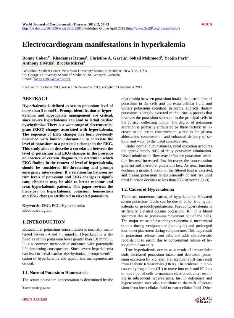
World Journal of Cardiovascular Diseases, 2012, 2, 57-63 WJCD http://dx.doi.org/10.4236/wjcd.2012.22010 Published Online April 2012 (http://www.SciRP.org/journal/wjcd/)
Electrocardiogram manifestations in hyperkalemia
Ronny Cohen1*, Rhadames Ramos1, Christine A. Garcia2, Sohail Mehmood1, Yoojin Park2, Anthony Divittis1, Brooks Mirrer1
1Woodhull Medical Center, New York University School of Medicine, New York, USA 2St. George’s University School of Medicine, St. George’s, Grenada Email: *[email protected] Received 31 October 2011; revised 16 December 2011; accepted 23 December 2011
ABSTRACT
Hyperkalemia is defined as serum potassium level of more than 5 mmol/L. Prompt identification of hyper- kalemia and appropriate management are critical, since severe hyperkalemia can lead to lethal cardiac dysrhythmias. There is a wide range of electrocardio- gram (EKG) changes associated with hyperkalemia. The sequence of EKG changes has been previously described with limited information to correlate the level of potassium to a particular change in the EKG. This study aims to describe a correlation between the level of potassium and EKG changes in the presence or absence of certain diagnoses, to determine which EKG finding in the context of level of hyperkalemia, should be considered life-threatening and prompt emergency intervention. If a relationship between se- rum levels of potassium and EKG changes is signifi- cant, clinicians may be able to better monitor and treat hyperkalemic patients. This paper reviews the literature on hyperkalemia, potassium homeostasis and EKG changes attributed to elevated potassium. Keywords: EKG; ECG; Hyperkalemia; Electrocardiogram
1. INTRODUCTION
Extracellular potassium concentration is normally main- tained between 4 and 4.5 mmol/L. Hyperkalemia is de- fined as serum potassium level greater than 5.0 mmol/L. It is a common metabolic disturbance with potentially life-threatening consequences. Since severe hyperkalemia can lead to lethal cardiac dysrhythmias, prompt identifi- cation of hyperkalemia and appropriate management are crucial.
1.1. Normal Potassium Homeostasis
The serum potassium concentration is determined by the
relationship between potassium intake, the distribution of potassium in the cells and the extra cellular fluid, and urinary potassium excretion. In normal subjects, dietary potassium is largely excreted in the urine, a process that involves the potassium secretion in the principal cells in the cortical collecting tubule. The degree of potassium secretion is primarily stimulated by three factors: an in- crease in the serum concentration, a rise in the plasma aldosterone concentration and enhanced delivery of so- dium and water to the distal secretory site.
Under normal circumstances, renal excretion accounts for approximately 90% of daily potassium elimination. Distal tubule urine flow may influence potassium secre-tion because increased flow increases the concentration gradient and therefore, potassium loss. As renal function declines, a greater fraction of the filtered load is excreted, and plasma potassium levels generally do not rise until renal function declines to less than 25% of normal [1-3].
1.2. Causes of Hyperkalemia
There are numerous causes of hyperkalemia. Elevated serum potassium levels can be due to either true hyper- kalemia or pseudohyperkalemia. Pseudohyperkalemia is artificially elevated plasma potassium [K+] in a blood specimen due to potassium movement out of the cells. The major cause of pseudohyperkalemia is mechanical trauma during venipuncture (hemolytic) and prolonged tourniquet placement during venipuncture. This may result in potassium release from cells and adds characteristic reddish tint to serum due to concomitant release of he-moglobin from cells.
True hyperkalemia occurs as a result of transcellular shift, increased potassium intake and decreased potas- sium excretion by kidneys. Transcellular shift can result from Diabetic Ketoacidosis (DKA). The acidemia in DKA causes hydrogen ions (H+) to move into cells and K+ ions to move out of cells to maintain electroneutrality, result-ing in subsequent hyperkalemia. Insulin deficiency and hyperosmolar state also contribute to the shift of potas-sium from intracellular fluid to extracellular fluid. Other *Corresponding author.
OPEN ACCESS

R. Cohen et al. / World Journal of Cardiovascular Diseases 2 (2012) 57-63 58
examples include non-selective beta-blockers, digitalis overdose, tumor lysis syndrome, rhabdomyolysis, et al. Increased potassium intake is usually iatrogenic due to over replenishing of potassium in the presence of renal impairment. It may occur slowly if the kidney cannot excrete potassium (e.g. renal failure, aldosterone antago- nists), and it may occur rapidly if potassium is being dumped from cells into the blood (e.g. crushing injuries, necrotic tissue, acidosis). The failing kidney can adapt by increasing potassium excretion per functioning nephron is low potassium diet is maintained, until oliguria ensues [4-6]. Any cause of decreased aldosterone release can decrease the efficiency of potassium excretion. Drugs affecting aldosterone include potassium-sparing diuretics, NSAIDs, heparin and pentamidine. The Transtubular Po- tassium Gradient (TTKG) can be useful when suspecting the diagnosis of hypoaldosteroneism. The TTKG in nor- mal subjects on a regular diet are 8 - 9, and rises to 11 with a potassium load, indicating increased potassium secretion. A value below 7, particularly below 5 in a hy- perkalemic patient is highly suggestive of hypoaldos- teronism. The diagnosis can be confirmed with the serum level of plasma aldosterone.
Renal tubular acidosis (RTA) is a disease that occurs when the kidneys fail to excrete acids into the urine, which causes a person’s blood to remain too acidic. There are several types of RTA. Type 4 is called hyper- kalemic RTA and is caused by a generalized transport abnormality of the distal tubule. The transport of elec- trolytes such as sodium, chloride, and potassium that normally occurs in the distal tubule is impaired. Type 4 RTA occurs when blood levels of the hormone aldoster- one are low or when the kidneys do not respond to it. Drugs that may cause type 4 RTA include diuretics, an- giotensin-converting enzyme (ACE) inhibitors and an- giotensin receptor blockers (ARBs), trimethoprim, pen- tamidine, heparin, nonsteroidal anti-inflammatory drugs (NSAIDs) and some immunosuppressive medications. Type 4 RTA may also result from diseases that alter kid- ney structure and function such as diabetic nephropathy, HIV/AIDS, Addison’s disease, sickle cell disease, uri- nary tract obstruction, lupus, amyloidosis, removal or destruction of both adrenal glands, and kidney transplant rejection [7].
1.3. Clinical Manifestations of Hyperkalemia
Hyperkalemia can be difficult to diagnose clinically since complaints may be vague. With non-specific findings, hyperkalemia often is an incidental laboratory finding. Clinical manifestations depend on both the severity as well as the rate of increase in potassium concentration. Patients with hyperkalemia may present as asymptomatic or may report weakness, generalized fatigue, paresthesias,
paralysis or even cardiac arrest [3,8,9]. Patient history is most valuable in identifying conditions that may predis- pose hyperkalemia.
1.4. EKG Changes with Hyperkalemia
Hyperkalemia is not always expressed with EKG changes, therefore EKG is not a reliable indicator of the severity of hyperkalemia. Profound hyperkalemia can be seen in absence of classic EKG changes, but the presence of EKG changes mandates treatment [10,11]. The EKG changes from hyperkalemia can range from peaked T waves, loss of P waves, prolonged QRS complex, ST segment elevation, extopic beats and escape rhythm, pro- gressive widening of QRS complex, sine wave, ventricu- lar fibrillation, asystole, axis deviation, bundle branch block and fascicular blocks. EKG alterations are also associated with sinus node depression, appearance of escape rhythms and malignant ventricular arrhythmias [12-14].
Hyperkalemia can cause several characteristic EKG abnormalities that are often progressive. Initially the T wave becomes tall, symmetrically peaked and tented. Widening of the QRS complex with an intraventricular conduction delay then occurs. Additional elevation of serum potassium leads to a decrease in the amplitude of the P wave and its eventual disappearance from the EKG. Rarely, ST segment elevation mimicking myocardial in- farction, described as “pseudoinfarction” pattern, is pre- sent. Further progression of hyperkalemia leads to a sine wave appearance of EKG and eventual asystole. These progressive changes in EKG have not been previously studied in respect to the corresponding serum K+ levels.
1.5. Therapeutic Options for Hyperkalemia
Hyperkalemia is one of the most fatal electrolyte abnor- malities; it is also the most treatable. American Heart Association defines hyperkalemia as greater than 5 mEq/L, and moderate (6 - 7 mEq/L) and severe (>7 mEq/L) are life threatening and require immediate therapy. Thera- peutic options aim to antagonize the effects of hyper- kalemia at the cellular level (calcium salts, decrease se- rum potassium levels by promoting the influx of potas- sium into cells of the body (insulin and sodium bicar- bonate), and finally, remove potassium from the body (resins, loop diuretics, hemodialysis) [6,14].
Calcium salts, such as calcium chloride and calcium gluconate, antagonize cardiac toxicity by increasing the threshold potential and reestablishing cardiac excitability but have no direct effect on potassium levels. Calcium administration shifts the threshold potential shifts to a less negative value (that is, from –75 mV to –65 mV), so that the initial difference between resting and threshold potentials of 15 mV can be restored. Calcium therapy for
Copyright © 2012 SciRes. OPEN ACCESS
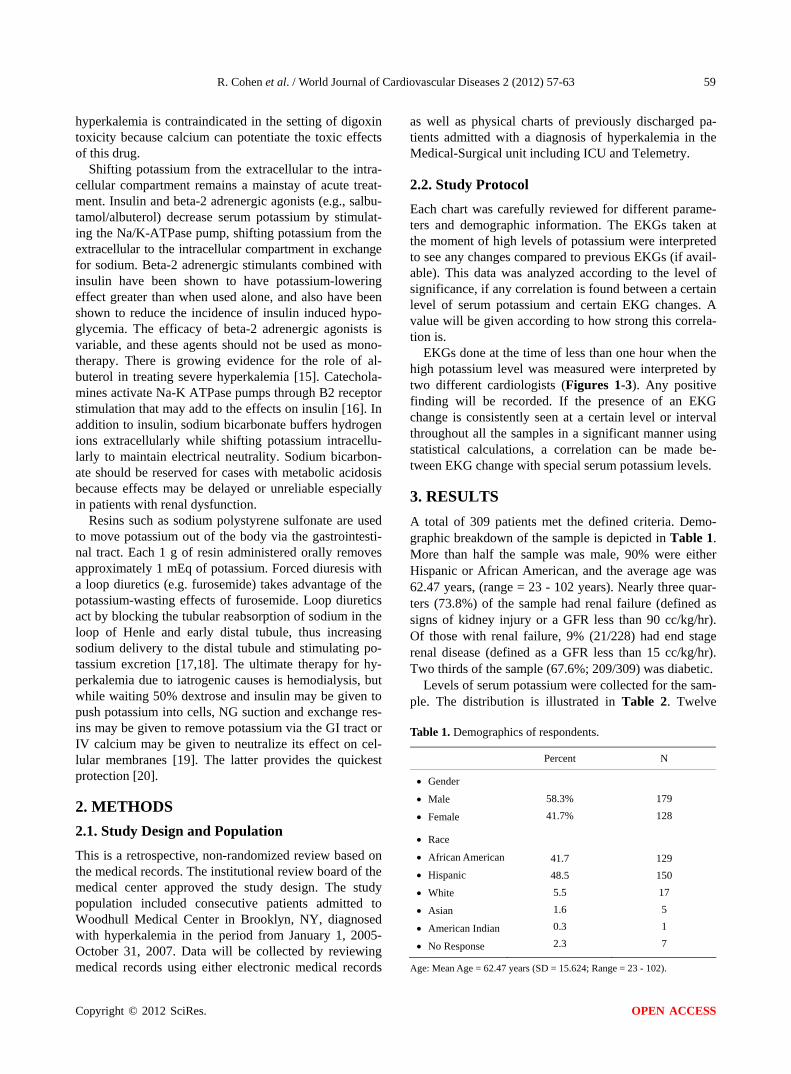
R. Cohen et al. / World Journal of Cardiovascular Diseases 2 (2012) 57-63 59
hyperkalemia is contraindicated in the setting of digoxin toxicity because calcium can potentiate the toxic effects of this drug.
Shifting potassium from the extracellular to the intra- cellular compartment remains a mainstay of acute treat- ment. Insulin and beta-2 adrenergic agonists (e.g., salbu- tamol/albuterol) decrease serum potassium by stimulat- ing the Na/K-ATPase pump, shifting potassium from the extracellular to the intracellular compartment in exchange for sodium. Beta-2 adrenergic stimulants combined with insulin have been shown to have potassium-lowering effect greater than when used alone, and also have been shown to reduce the incidence of insulin induced hypo- glycemia. The efficacy of beta-2 adrenergic agonists is variable, and these agents should not be used as mono- therapy. There is growing evidence for the role of al- buterol in treating severe hyperkalemia [15]. Catechola- mines activate Na-K ATPase pumps through B2 receptor stimulation that may add to the effects on insulin [16]. In addition to insulin, sodium bicarbonate buffers hydrogen ions extracellularly while shifting potassium intracellu- larly to maintain electrical neutrality. Sodium bicarbon- ate should be reserved for cases with metabolic acidosis because effects may be delayed or unreliable especially in patients with renal dysfunction.
Resins such as sodium polystyrene sulfonate are used to move potassium out of the body via the gastrointesti- nal tract. Each 1 g of resin administered orally removes approximately 1 mEq of potassium. Forced diuresis with a loop diuretics (e.g. furosemide) takes advantage of the potassium-wasting effects of furosemide. Loop diuretics act by blocking the tubular reabsorption of sodium in the loop of Henle and early distal tubule, thus increasing sodium delivery to the distal tubule and stimulating po- tassium excretion [17,18]. The ultimate therapy for hy- perkalemia due to iatrogenic causes is hemodialysis, but while waiting 50% dextrose and insulin may be given to push potassium into cells, NG suction and exchange res- ins may be given to remove potassium via the GI tract or IV calcium may be given to neutralize its effect on cel- lular membranes [19]. The latter provides the quickest protection [20].
2. METHODS
2.1. Study Design and Population
This is a retrospective, non-randomized review based on the medical records. The institutional review board of the medical center approved the study design. The study population included consecutive patients admitted to Woodhull Medical Center in Brooklyn, NY, diagnosed with hyperkalemia in the period from January 1, 2005- October 31, 2007. Data will be collected by reviewing medical records using either electronic medical records
as well as physical charts of previously discharged pa-tients admitted with a diagnosis of hyperkalemia in the Medical-Surgical unit including ICU and Telemetry.
2.2. Study Protocol
Each chart was carefully reviewed for different parame- ters and demographic information. The EKGs taken at the moment of high levels of potassium were interpreted to see any changes compared to previous EKGs (if avail- able). This data was analyzed according to the level of significance, if any correlation is found between a certain level of serum potassium and certain EKG changes. A value will be given according to how strong this correla-tion is.
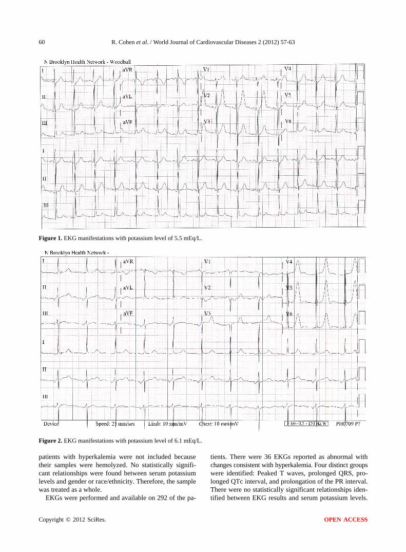
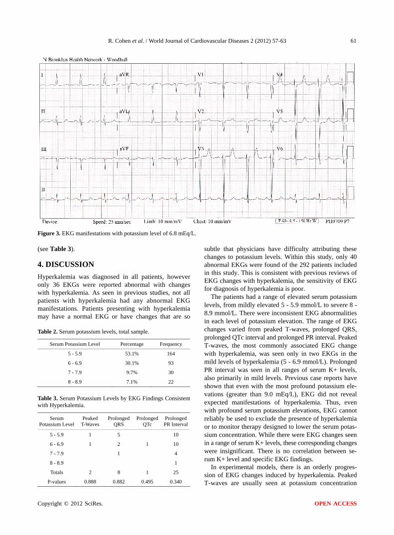
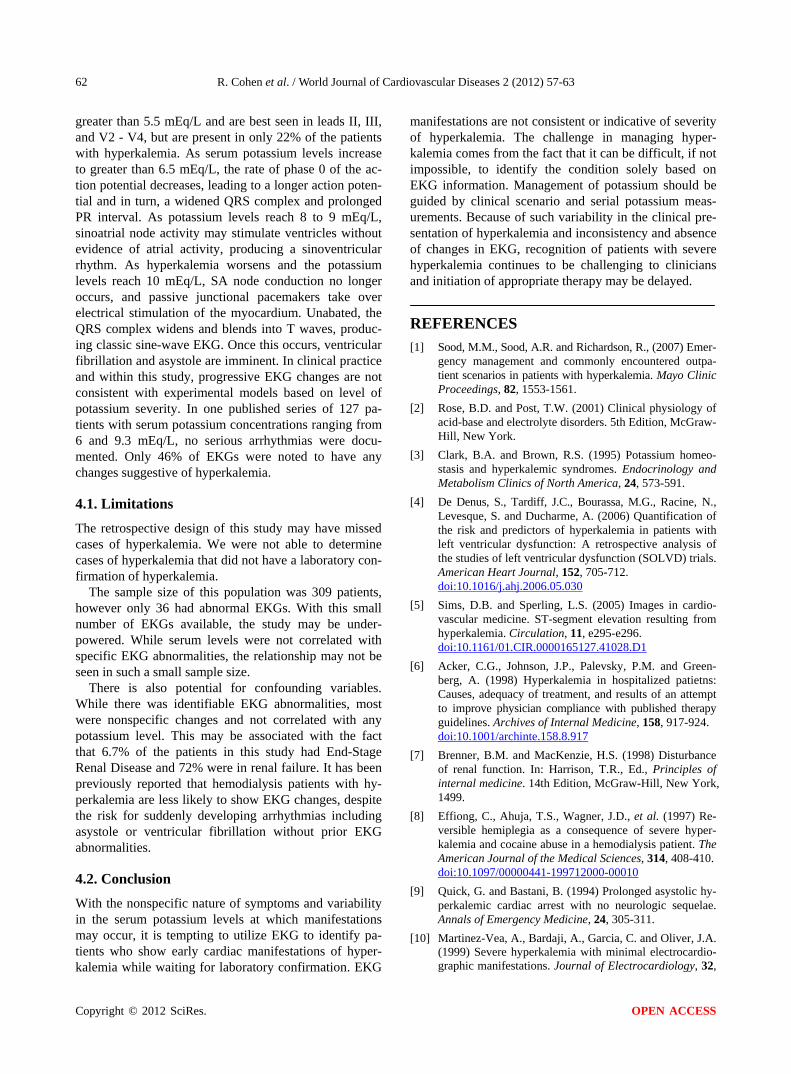
EKGs done at the time of less than one hour when the high potassium level was measured were interpreted by two different cardiologists (Figures 1-3). Any positive finding will be recorded. If the presence of an EKG change is consistently seen at a certain level or interval throughout all the samples in a significant manner using statistical calculations, a correlation can be made be-tween EKG change with special serum potassium levels.
3. RESULTS
A total of 309 patients met the defined criteria. Demo- graphic breakdown of the sample is depicted in Table 1. More than half the sample was male, 90% were either Hispanic or African American, and the average age was 62.47 years, (range = 23 - 102 years). Nearly three quar- ters (73.8%) of the sample had renal failure (defined as signs of kidney injury or a GFR less than 90 cc/kg/hr). Of those with renal failure, 9% (21/228) had end stage renal disease (defined as a GFR less than 15 cc/kg/hr). Two thirds of the sample (67.6%; 209/309) was diabetic.
Levels of serum potassium were collected for the sam- ple. The distribution is illustrated in Table 2. Twelve
Table 1. Demographics of respondents.
Percent N
Gender
Male
Female
58.3%
41.7%
179
128
Race
African American
Hispanic
White
Asian
American Indian
No Response
41.7
48.5
5.5
1.6
0.3
2.3
129
150
17
5
1
7
A ge: Mean Age = 62.47 years (SD = 15.624; Range = 23 - 102).
Copyright © 2012 SciRes. OPEN ACCESS
R. Cohen et al. / World Journal of Cardiovascular Diseases 2 (2012) 57-63
Copyright © 2012 SciRes.
60
Figure 1. EKG manifestations with potassium level of 5.5 mEq/L.
Figure 2. EKG manifestations with potassium level of 6.1 mEq/L.
patients with hyperkalemia were not included because their samples were hemolyzed. No statistically signifi- cant relationships were found between serum potassium levels and gender or race/ethnicity. Therefore, the sample was treated as a whole.
EKGs were performed and available on 292 of the pa-
tients. There were 36 EKGs reported as abnormal with changes consistent with hyperkalemia. Four distinct groups were identified: Peaked T waves, prolonged QRS, pro- longed QTc interval, and prolongation of the PR interval. There were no statistically significant relationships iden- ified between EKG results and serum potassium levels. t
OPEN ACCESS
R. Cohen et al. / World Journal of Cardiovascular Diseases 2 (2012) 57-63 61
Figure 3. EKG manifestations with potassium level of 6.8 mEq/L.
(see Table 3).
4. DISCUSSION
Hyperkalemia was diagnosed in all patients, however only 36 EKGs were reported abnormal with changes with hyperkalemia. As seen in previous studies, not all patients with hyperkalemia had any abnormal EKG manifestations. Patients presenting with hyperkalemia may have a normal EKG or have changes that are so
Table 2. Serum potassium levels, total sample.
Serum Potassium Level Percentage Frequency
5 - 5.9 53.1% 164
6 - 6.9 30.1% 93
7 - 7.9 9.7% 30
8 - 8.9 7.1% 22
Table 3. Serum Potassium Levels by EKG Findings Consistent with Hyperkalemia.
Serum Potassium Level
Peaked T-Waves
Prolonged QRS
Prolonged QTc
Prolonged PR Interval
5 - 5.9 1 5 10
6 - 6.9 1 2 1 10
7 - 7.9 1 4
8 - 8.9 1
Totals 2 8 1 25
P-values 0.888 0.882 0.495 0.340
subtle that physicians have difficulty attributing these changes to potassium levels. Within this study, only 40 abnormal EKGs were found of the 292 patients included in this study. This is consistent with previous reviews of EKG changes with hyperkalemia, the sensitivity of EKG for diagnosis of hyperkalemia is poor.
The patients had a range of elevated serum potassium levels, from mildly elevated 5 - 5.9 mmol/L to severe 8 - 8.9 mmol/L. There were inconsistent EKG abnormalities in each level of potassium elevation. The range of EKG changes varied from peaked T-waves, prolonged QRS, prolonged QTc interval and prolonged PR interval. Peaked T-waves, the most commonly associated EKG change with hyperkalemia, was seen only in two EKGs in the mild levels of hyperkalemia (5 - 6.9 mmol/L). Prolonged PR interval was seen in all ranges of serum K+ levels, also primarily in mild levels. Previous case reports have shown that even with the most profound potassium ele- vations (greater than 9.0 mEq/L), EKG did not reveal expected manifestations of hyperkalemia. Thus, even with profound serum potassium elevations, EKG cannot reliably be used to exclude the presence of hyperkalemia or to monitor therapy designed to lower the serum potas- sium concentration. While there were EKG changes seen in a range of serum K+ levels, these corresponding changes were insignificant. There is no correlation between se- rum K+ level and specific EKG findings.
In experimental models, there is an orderly progres- sion of EKG changes induced by hyperkalemia. Peaked T-waves are usually seen at potassium concentration
Copyright © 2012 SciRes. OPEN ACCESS
R. Cohen et al. / World Journal of Cardiovascular Diseases 2 (2012) 57-63 62
greater than 5.5 mEq/L and are best seen in leads II, III, and V2 - V4, but are present in only 22% of the patients with hyperkalemia. As serum potassium levels increase to greater than 6.5 mEq/L, the rate of phase 0 of the ac- tion potential decreases, leading to a longer action poten- tial and in turn, a widened QRS complex and prolonged PR interval. As potassium levels reach 8 to 9 mEq/L, sinoatrial node activity may stimulate ventricles without evidence of atrial activity, producing a sinoventricular rhythm. As hyperkalemia worsens and the potassium levels reach 10 mEq/L, SA node conduction no longer occurs, and passive junctional pacemakers take over electrical stimulation of the myocardium. Unabated, the QRS complex widens and blends into T waves, produc- ing classic sine-wave EKG. Once this occurs, ventricular fibrillation and asystole are imminent. In clinical practice and within this study, progressive EKG changes are not consistent with experimental models based on level of potassium severity. In one published series of 127 pa- tients with serum potassium concentrations ranging from 6 and 9.3 mEq/L, no serious arrhythmias were docu- mented. Only 46% of EKGs were noted to have any changes suggestive of hyperkalemia.
4.1. Limitations
The retrospective design of this study may have missed cases of hyperkalemia. We were not able to determine cases of hyperkalemia that did not have a laboratory con- firmation of hyperkalemia.
The sample size of this population was 309 patients, however only 36 had abnormal EKGs. With this small number of EKGs available, the study may be under- powered. While serum levels were not correlated with specific EKG abnormalities, the relationship may not be seen in such a small sample size.
There is also potential for confounding variables. While there was identifiable EKG abnormalities, most were nonspecific changes and not correlated with any potassium level. This may be associated with the fact that 6.7% of the patients in this study had End-Stage Renal Disease and 72% were in renal failure. It has been previously reported that hemodialysis patients with hy- perkalemia are less likely to show EKG changes, despite the risk for suddenly developing arrhythmias including asystole or ventricular fibrillation without prior EKG abnormalities.
4.2. Conclusion
With the nonspecific nature of symptoms and variability in the serum potassium levels at which manifestations may occur, it is tempting to utilize EKG to identify pa- tients who show early cardiac manifestations of hyper- kalemia while waiting for laboratory confirmation. EKG
manifestations are not consistent or indicative of severity of hyperkalemia. The challenge in managing hyper-kalemia comes from the fact that it can be difficult, if not impossible, to identify the condition solely based on EKG information. Management of potassium should be guided by clinical scenario and serial potassium meas- urements. Because of such variability in the clinical pre- sentation of hyperkalemia and inconsistency and absence of changes in EKG, recognition of patients with severe hyperkalemia continues to be challenging to clinicians and initiation of appropriate therapy may be delayed.
REFERENCES
[1] Sood, M.M., Sood, A.R. and Richardson, R., (2007) Emer- gency management and commonly encountered outpa- tient scenarios in patients with hyperkalemia. Mayo Clinic Proceedings, 82, 1553-1561.
[2] Rose, B.D. and Post, T.W. (2001) Clinical physiology of acid-base and electrolyte disorders. 5th Edition, McGraw- Hill, New York.
[3] Clark, B.A. and Brown, R.S. (1995) Potassium homeo- stasis and hyperkalemic syndromes. Endocrinology and Metabolism Clinics of North America, 24, 573-591.
[4] De Denus, S., Tardiff, J.C., Bourassa, M.G., Racine, N., Levesque, S. and Ducharme, A. (2006) Quantification of the risk and predictors of hyperkalemia in patients with left ventricular dysfunction: A retrospective analysis of the studies of left ventricular dysfunction (SOLVD) trials. American Heart Journal, 152, 705-712. doi:10.1016/j.ahj.2006.05.030
[5] Sims, D.B. and Sperling, L.S. (2005) Images in cardio- vascular medicine. ST-segment elevation resulting from hyperkalemia. Circulation, 11, e295-e296. doi:10.1161/01.CIR.0000165127.41028.D1
[6] Acker, C.G., Johnson, J.P., Palevsky, P.M. and Green- berg, A. (1998) Hyperkalemia in hospitalized patietns: Causes, adequacy of treatment, and results of an attempt to improve physician compliance with published therapy guidelines. Archives of Internal Medicine, 158, 917-924. doi:10.1001/archinte.158.8.917
[7] Brenner, B.M. and MacKenzie, H.S. (1998) Disturbance of renal function. In: Harrison, T.R., Ed., Principles of internal medicine. 14th Edition, McGraw-Hill, New York, 1499.
[8] Effiong, C., Ahuja, T.S., Wagner, J.D., et al. (1997) Re- versible hemiplegia as a consequence of severe hyper- kalemia and cocaine abuse in a hemodialysis patient. The American Journal of the Medical Sciences, 314, 408-410. doi:10.1097/00000441-199712000-00010
[9] Quick, G. and Bastani, B. (1994) Prolonged asystolic hy- perkalemic cardiac arrest with no neurologic sequelae. Annals of Emergency Medicine, 24, 305-311.
[10] Martinez-Vea, A., Bardaji, A., Garcia, C. and Oliver, J.A. (1999) Severe hyperkalemia with minimal electrocardio- graphic manifestations. Journal of Electrocardiology, 32,
Copyright © 2012 SciRes. OPEN ACCESS
R. Cohen et al. / World Journal of Cardiovascular Diseases 2 (2012) 57-63
Copyright © 2012 SciRes.
63
OPEN ACCESS
45-49. doi:10.1016/S0022-0736(99)90020-1
[11] Yu, A.S. (1996) Atypical electrocardiographic changes in severe hyperkalemia. The American Journal of Cardi- ology, 77, 906-908. doi:10.1016/S0002-9149(97)89197-7
[12] Lin, J. and Huang, C. (1990) Successful initiation of hemo- dialysis during cardiopulmonary resuscitation due to le- thal hyperkalemia. Critical Care Medicine, 18, 342-343. doi:10.1097/00003246-199003000-00022
[13] Dittrich, K.I. and Walls, R.M. (1986) Hyperkalemia: ECG manifesttations and considerations. The Journal of Emer- gency Medicine, 4, 449-55. doi:10.1016/0736-4679(86)90174-5
[14] Carbalhana, V., Burry, L. and Lapinsky, S.E. (2006) Man- agement of severe hyperkalemia without hemodialysis: Case report and literature review. Journal of Critical Care, 21, 316-321. doi:10.1016/j.jcrc.2005.10.001
[15] Parham, W., Mehdirad, A., Biermann, A. and Friedman, C. (2006) Hyperkalemia revisited. Texas Heart Institute Journal, 33, 40-47.
[16] Reardon, L.C. and MacPherson, D.S. (1998) Hyperkalemia
in outpatients using angiotensin-converting enzyme in- hibitors. Archives of Internal Medicine, 158, 26-32. doi:10.1001/archinte.158.1.26
[17] Ahee, P. and Crowe, A.V. (2000) The management of hy- perkalemia in the emergency department. Journal of Ac- cident & Emergency Medicine, 17, 188-191. doi:10.1136/emj.17.3.188
[18] Blumberg, A., Weidmann, P., Shaw, S. and Gnadinger, M. (1988) Effect of various therapeutic approaches on plasma potatssium and major regulating factors in terminal renal failure. American Journal of Medicine, 85, 507-512. doi:10.1016/S0002-9343(88)80086-X
[19] Lin, J. and Huang, C. (1990) Successful initiation of hemo- dialysis during cardiopulmonary resuscitation due to le- thal hyperkalemia. Critical Care Medicine, 18, 342-343. doi:10.1097/00003246-199003000-00022
[20] Mahoney, B.A., Smith, W.A.D., Lo, D.S., et al. (2005) Emergency interventions for hyperkalemia. John Wiley & Sons, Ltd., New York.


![Treatment of Hyperkalemia [Read-Only]](https://static.fdocuments.in/doc/165x107/62e53ff26c7a3007180b6201/treatment-of-hyperkalemia-read-only.jpg)