Electrical Stimulation-Based Sensory Feedback in Phantom ...movements of the phantom limb, e.g.,...
43
Electrical Stimulation-Based Sensory Feedback in Phantom Limb Pain Treatment
Transcript of Electrical Stimulation-Based Sensory Feedback in Phantom ...movements of the phantom limb, e.g.,...
Electrical Stimulation-BasedSensory Feedback inPhantom Limb Pain
Treatment

Electrical Stimulation-BasedSensory Feedback inPhantom Limb Pain
Treatment
PhD Thesis by
Bo Geng
Center for Sensory-Motor Interaction,Department of Health Science and Technology,
Aalborg University, Denmark

ISBN 978-87-93102-42-2 (e-book)
Published, sold and distributed by:River PublishersNiels Jernes Vej 109220 Aalborg ØDenmark
Tel.: +45369953197www.riverpublishers.com
Copyright for this work belongs to the author, River Publishers have the soleright to distribute this work commercially.
All rights reserved c© 2013 Bo Geng.
No part of this work may be reproduced, stored in a retrieval system, or trans-mitted in any form or by any means, electronic, mechanical, photocopying,microfilming, recording or otherwise, without prior written permission fromthe Publisher.
2
TABLE OF CONTENTS
PREFACE ........................................................................................................................................................... 3
ENGLISH SUMMARY ......................................................................................................................................... 4
DANSK RESUME ............................................................................................................................................... 5
1. BACKGROUND .............................................................................................................................................. 6
1.1 NON-PAINFUL PHANTOM SENSATION .............................................................................................................. 6
1.2 PHANTOM LIMB PAIN (PLP) .......................................................................................................................... 7
1.2.1 Characteristics.................................................................................................................................... 7
1.2.2 Neurobiological mechanisms ............................................................................................................. 8
1.2.3 State-of-the-art treatments for PLP ................................................................................................. 10
1.3 SENSORY FEEDBACK ................................................................................................................................... 11
1.3.1 Sensory feedback for PLP treatment ................................................................................................ 11
1.3.2 Means of providing sensory feedback.............................................................................................. 12
1.3.3 Measurement of sensory feedback .................................................................................................. 14
1.4 THE EU TIME PROJECT .............................................................................................................................. 15
2. OVERVIEW OF THE PHD WORK .................................................................................................................. 17
2.1 HYPOTHESIS AND QUESTIONS ....................................................................................................................... 17
2.2 STUDIES AND CORRESPONDING PUBLICATIONS ................................................................................................ 18
Study 1: Investigation of the impact of selected stimulation parameters on the perception threshold ......... 20
Study 2: Evaluation of the impact of selected stimulation parameters on evoked sensations ....................... 21
Study 3: Examination of the sensory identification ability of able-bodied subjects ...................................... 22
Study 4: Exploration of the effect of sensory identification training on PLP and cortical reorganization ... 23 Study 5: Design and development of a computerized tool to control and evaluate multi-channel electrical
stimulation-based sensory feedback .............................................................................................................. 24
3. DISCUSSION AND CONCLUSION ................................................................................................................. 25
3.1 DISCUSSION OF MAIN FINDINGS.................................................................................................................... 25
3.2 METHODOLOGICAL CONSIDERATIONS ............................................................................................................ 29
3.3 CONCLUSIONS .......................................................................................................................................... 31
REFERENCES ................................................................................................................................................... 32
3
Preface
The research presented in this Ph.D. thesis was carried out at the center for Sensory Motor
Interaction (SMI) in the Department of Health Science and Technology of Aalborg University
between 2008 and 2011. This research was financially supported by the EU-funded ‘TIME’
project: Transverse Intrafasicular Multichannel Electrode System, grant no. CP-FP-INFSO
224012. Completion of this thesis would not have been possible without the support of both
the SMI and TIME project team.
I would like to express my sincere thanks to my supervisor, Winnie Jensen, who supported
and encouraged me throughout my Ph.D. work. She gave me a chance to pursue a research
topic that was new for me and provided guidance and constructive feedback. I also want to
extend my thanks to Ken Yoshida for his many valuable contributions to my work. I have
indeed benefited from close collaboration with him.
I would also like to thank my colleagues, who have significantly contributed to the research
presented in this thesis. I would especially like to thank Laura Petrini for providing advice on
psychophysical methods, Line Lindhardt Egsgaard for her help with the EEG data analysis,
and Knud Larsen for his assistance in solving technical problems in the software development.
Lastly, I thank all the members of the NPI lab for participating in discussions on all aspects of
this research, which helped shape my view on what research really is.
Special thanks go to my husband Ming for his patience and endless support and to my parents
and brother, who always support me. The thesis is dedicated to our lovely daughter, Joyce,
whose arrival added so much delight to my Ph.D. life.
4
English summary
Following amputation, up to 80% of amputees perceive pain in the missing part of the arm or
leg, known as phantom limb pain (PLP). PLP can be extremely intractable, and there are no
effective, long-lasting treatments currently available. Reorganization in the primary
sensorimotor cortex has been found to be closely associated with PLP. Therefore, an approach
targeting reversal of cortical reorganization may hold promise for PLP relief. The present
thesis hypothesizes that providing sensory feedback through electrocutaneous stimulation of
the residual limb may reverse cortical reorganization and consequently suppress PLP.
To address the hypothesis, five studies were conducted. Studies 1 and 2 examined the impact
of stimulation parameters on the perception threshold and the evoked sensation, respectively.
The stimulation location and pulse number were found to significantly affect the magnitude
and quality of the perceived sensation. These two parameters were then considered to be able
to effectively convey sensory information. Study 3 investigated human ability in sensory
discrimination of the two identified parameters, in which satisfactory performance was
obtained with able-bodied subjects. Based on the findings of the first three studies, the
hypothesis was tested in study 4. An upper-limb amputee was trained in sensory identification
to evaluate the effects on PLP and cortical reorganization. The results showed no changes in
PLP and cortical reorganization, although the volunteer’s identification accuracy improved
over the course of the training period. As part of the EU-funded project ‘TIME’, a
computerized tool was developed for evaluation of sensory feedback in a multi-channel
intrafascicular stimulation system in study 5. This tool was used to assist in identifying
optimal stimulation patterns that can evoke natural sensory feedback referred to the phantom
limb. Sensory feedback using direct nerve stimulation may be an alternative treatment for
PLP awaiting further clinical evaluation in future work.
5
Dansk resume
Efter amputation føler op til 80% af de amputerede personer smerter i den manglende del af
armen eller benet; dette kaldes også fantomsmerter. Fantomsmerter kan være ekstremt
voldsomme og der findes ingen effektive, langvarige behandlingsmetoder mod disse smerter.
Reorganisering i primær sensorisk-motorisk cortex har vist sig at være tæt forbundet med
fantomsmerter. Derfor kan en metode, der er rettet imod ændring af den kortikale
reorganisation vise sig at være lovende i behandlingen af fantomsmerter. Denne afhandling
antager som udgangspunkt, at frembringelse af sensorisk feedback gennem elektrokutan
stimulation af det resterende lem kan ændre den kortikale reorganisering og som følge heraf
dæmpe fantomsmerterne.
For at underbygge denne hypotese blev der udført fem studier. Studie 1 og 2 undersøgte
effekten af stimulationsparametre på henholdsvis perceptionstærsklen og den fremkaldte
fornemmelse. Det blev fundet, at stimulationssted og antallet af impulser væsentligt påvirker
den opfattede fornemmelses styrke og kvalitet. De to parametre blev herefter bestemt til
effektivt at kunne viderebringe sensoriske informationer. Studie 3 undersøgte menneskets
evne til sensorisk diskrimination af de to identificerede parametre, hvor tilfredsstillende
resultater blev opnået med raske forsøgspersoner. Baseret på resultaterne af de tre første
studier blev hypotesen undersøgt i studie 4. Træning af sensorisk identifikation blev
undersøgt hos en person med amputation i overekstremiteten for at undersøge effekten på
fantomsmerter og kortikal reorganisering. Resultaterne viste ingen ændringer i hverken
fantomsmerter eller kortikal reorganisering, selvom identifikationsevnen forbedredes i
træningsperioden. Som en del af det EU-finansierede projekt ‘TIME’ blev der i studie 5
udviklet et computerværktøj til vurdering af sensorisk feedback i et multi-kanal
intrafascikulært stimulationssystem. Dette blev anvendt til hjælp til identifikation af optimale
stimulationsmønstre, som kan fremkalde naturlig sensorisk feedback, som refereres til
fantomlemmet. Sensorisk feedback ved hjælp af direkte nervestimulation kan være en
alternativ behandling for fantomsmerter, hvilket dog afventer nærmere fremtidig klinisk
vurdering.
6
1. Background
Surgical limb amputations are typically due to peripheral vascular diseases, cancer, or
diabetes (Nikolajsen and Jensen, 2001). Motor vehicle accidents, wartime conflicts, terrorist
attacks and landmine explosions can also necessitate traumatic limb amputation in otherwise
healthy people (Lacoux et al., 2002). In the USA, limb loss affects nearly 1.6 million
individuals (Ephraim et al., 2005), and a recent study estimated that the number of people
living with a limb loss will more than double by the year 2050 (Ziegler-Graham et al., 2008).
1.1 Non-painful phantom sensation
Virtually all amputees experience non-painful phantom sensations, i.e., the feeling that the
removed limb still exists. The qualities of the phantom sensations include specific
somatosensory experiences such as touch, cold, warmth, itching and other paraesthesias
(Kooijman et al., 2000). In addition, patients often claim they can perceive kinesthetic
features, such as the size, shape, and position of the missing limb, and even voluntary
movements of the phantom limb, e.g., reaching out to grab an object, making a fist or moving
their fingers individually (Weinstein, 1998). Involuntary phantom movements are also
common, e.g., suddenly moving to occupy a new posture or suddenly developing a clenching
spasm of the fingers (Ramachandran and Hirstein, 1998).
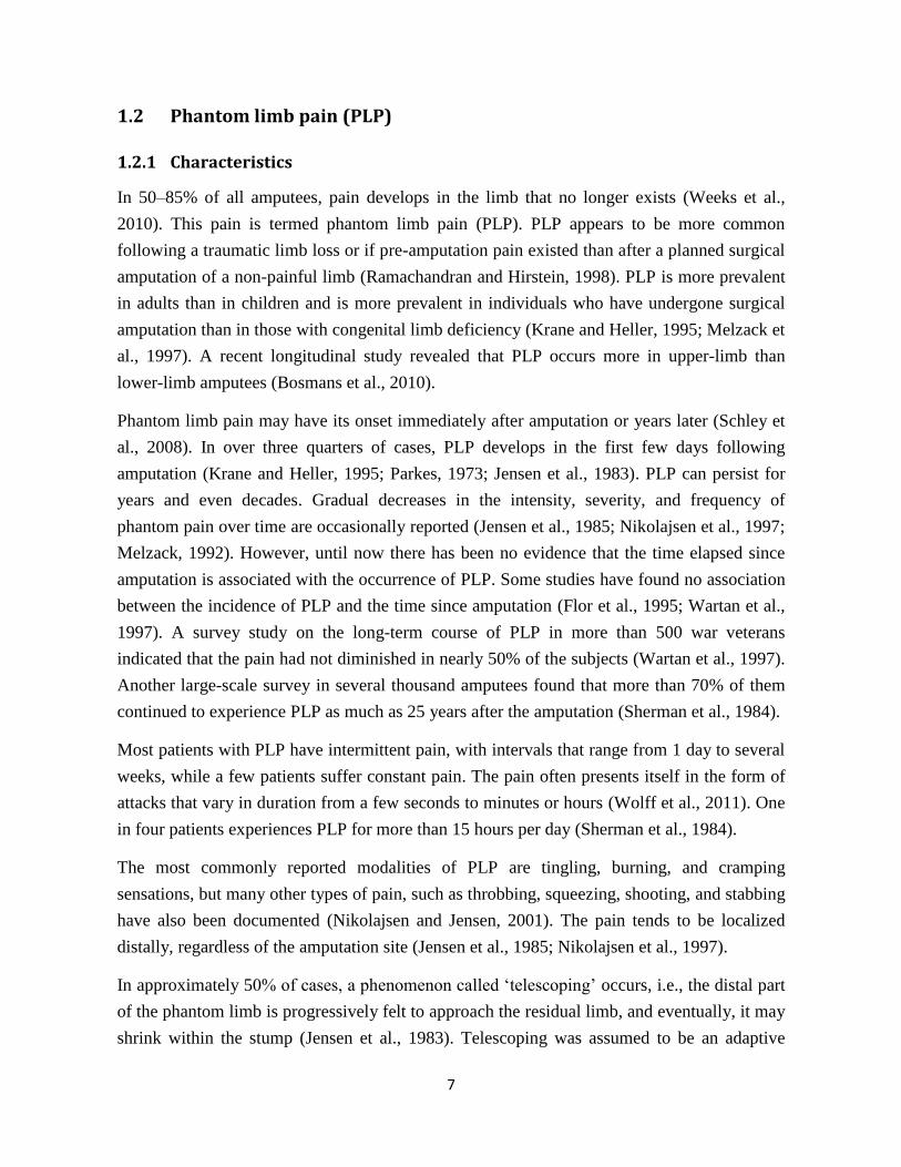
Phantom sensations may be evoked by applying stimulation on the ipsilateral face or the
stump (Figure 1). These phantom sensations, usually called referred sensations, i.e.,
sensations perceived as originating from a body site other than the one stimulated, have been
described frequently following amputation (Ramachandran and Hirstein, 1998; Grüsser et al.,
2001; Ramachandran et al., 1995).
Figure 1. The distribution of referred phantom sensation in patient D. S., whose left arm was amputated 6
cm above the elbow joint due to injuries from a car accident. (A) Topographic arrangement of digits of the
phantom hand on the ipsilateral face 6 months after amputation. (B) Topographic mapping of phantom
digits in the region of the deltoid muscle on the stump (from Ramachandran et al. 1998).
(A) (B)
7
1.2 Phantom limb pain (PLP)
1.2.1 Characteristics
In 50–85% of all amputees, pain develops in the limb that no longer exists (Weeks et al.,
2010). This pain is termed phantom limb pain (PLP). PLP appears to be more common
following a traumatic limb loss or if pre-amputation pain existed than after a planned surgical
amputation of a non-painful limb (Ramachandran and Hirstein, 1998). PLP is more prevalent
in adults than in children and is more prevalent in individuals who have undergone surgical
amputation than in those with congenital limb deficiency (Krane and Heller, 1995; Melzack et
al., 1997). A recent longitudinal study revealed that PLP occurs more in upper-limb than
lower-limb amputees (Bosmans et al., 2010).
Phantom limb pain may have its onset immediately after amputation or years later (Schley et
al., 2008). In over three quarters of cases, PLP develops in the first few days following
amputation (Krane and Heller, 1995; Parkes, 1973; Jensen et al., 1983). PLP can persist for
years and even decades. Gradual decreases in the intensity, severity, and frequency of
phantom pain over time are occasionally reported (Jensen et al., 1985; Nikolajsen et al., 1997;
Melzack, 1992). However, until now there has been no evidence that the time elapsed since
amputation is associated with the occurrence of PLP. Some studies have found no association
between the incidence of PLP and the time since amputation (Flor et al., 1995; Wartan et al.,
1997). A survey study on the long-term course of PLP in more than 500 war veterans
indicated that the pain had not diminished in nearly 50% of the subjects (Wartan et al., 1997).
Another large-scale survey in several thousand amputees found that more than 70% of them
continued to experience PLP as much as 25 years after the amputation (Sherman et al., 1984).
Most patients with PLP have intermittent pain, with intervals that range from 1 day to several
weeks, while a few patients suffer constant pain. The pain often presents itself in the form of
attacks that vary in duration from a few seconds to minutes or hours (Wolff et al., 2011). One
in four patients experiences PLP for more than 15 hours per day (Sherman et al., 1984).
The most commonly reported modalities of PLP are tingling, burning, and cramping
sensations, but many other types of pain, such as throbbing, squeezing, shooting, and stabbing
have also been documented (Nikolajsen and Jensen, 2001). The pain tends to be localized
distally, regardless of the amputation site (Jensen et al., 1985; Nikolajsen et al., 1997).
In approximately 50% of cases, a phenomenon called ‘telescoping’ occurs, i.e., the distal part
of the phantom limb is progressively felt to approach the residual limb, and eventually, it may
shrink within the stump (Jensen et al., 1983). Telescoping was assumed to be an adaptive
8
process beneficial to PLP (Cronholm, 1951; Katz, 1992). However, recent evidence suggests
telescoping and PLP are positively related (Grüsser et al., 2001; Montoya et al., 1997).
PLP may be elicited or worsened by a range of physical factors (e.g., weather change or
pressure on the residual limb) or psychological factors (e.g., emotional stress) (Sherman et al.,
1989; Arena et al., 1990). Cognitive factors also play a part in the modulation of PLP.
Patients who have personality traits characterized by passive coping are more affected by the
pain and report more interference (Richardson et al., 2007).
In summary, PLP can be extremely intractable and disabling. It is sufficiently severe to
hamper prosthetic training (Carabelli and Kellerman, 1985) and reduce amputees’ likelihood
of employment and social activities (Millstein et al., 1985). Apart from its negative impacts
on patients’ functioning and well-being (Pezzin et al., 2000; Ehde et al., 2000), PLP also
poses a significant problem for health care systems worldwide (Hanley et al., 2006). Thus,
successful intervention for treatment of PLP is greatly needed.
1.2.2 Neurobiological mechanisms
PLP is usually classified as neuropathic pain, which involves multiple pathophysiological
changes, both in the peripheral nervous system (PNS) and in the central nervous system (CNS)
(Flor, 2002a; Costigan et al., 2009; Navarro et al., 2007). After surgical removal of a limb, the
complete truncation of peripheral nerves and the neural damage initiate a cascade of changes
that lead to and sustain phantom pain, which might be the manifestation of maladaptive
plasticity in the nervous system (Di Pino et al., 2009). No single mechanism appears to be
able to explain the development of PLP independently. Hence, it is currently believed that
PLP should be considered a complex syndrome and that multiple mechanisms at different
levels in the nervous system contribute to the occurrence of PLP.
Peripheral mechanisms
Following truncation of peripheral nerves, a neuroma is universally formed as a result of
aberrant sprouting of regenerating axons. Ectopic discharge from a stump neuroma has been
postulated as one important peripheral mechanism. Such neuromas show spontaneous
discharge or hyper-excitability following mechanical and chemical stimulation (Devor et al.,
1993). In addition, similar abnormal activity and sensitivity occurs in dorsal root ganglia
(DRG), where the discharge coming from the residual limb can be amplified (Devor, Seltzer,
1999). In addition, abnormal sympathetic activity may increase the amount of circulating
epinephrine, which can trigger and exacerbate neuronal activity from neuroma (Devor et al.,
1994). These aberrant nociceptive impulses may be interpreted by the brain as pain.
9
One of the strongest arguments for a peripheral cause of phantom limb pain is its positive
correlation with stump pain. Phantom pain occurs significantly more frequently in amputees
with chronic stump pain than in those without stump pain (Sherman and Sherman, 1983).
Nevertheless, peripheral factors alone cannot entirely explain the occurrence of PLP. In some
cases, PLP occurs immediately after amputation when there are not yet neuromas in the
residual limb (Carlen et al., 1978). Congenital amputees occasionally also report PLP (Alviar
et al., 2011). These findings indicate that central factors must be involved as well.
Central mechanisms
While spinal plasticity, such as sensitization of low-threshold sensory receptors (Cervero,
2009) and central sensitization (Costigan et al., 2009; Baron, 2006), have been discussed as
processes involved in PLP or neuropathic pain, cortical reorganization has gained a great deal
of attention and is considered a plausible explanation for PLP. Furthermore, cortical
reorganization may partly account for why stimulation on the face, stump or surrounding
regions elicit sensations referred to the phantom limb (Flor et al., 2006).
Extensive empirical studies have demonstrated that sensorimotor cortices undergo massive
neuroplasticity changes in people with extremity amputation (Flor et al., 1995; Ramachandran
et al., 1992; Hall et al., 1990; Chen et al., 2002). The cortical area that formerly represented
the amputated extremity has been found to be taken over by its neighboring mouth and chin
representation zone in the primary somatosensory cortex (S1) of amputees (Elbert et al., 1994;
Yang et al., 1994). Further studies have found that cortical reorganization also develops in the
motor cortex (M1) (Lotze et al., 2001; Karl et al., 2001; Karl et al., 2004).
Furthermore, the extent of cortical reorganization has been found to be closely associated with
the severity of phantom pain and the size of the deafferented region. A series of studies have
revealed a positive relation between the magnitude of PLP and the amount of reorganization
in the sensorimotor cortices (Lotze et al., 2001; Karl et al., 2001; Karl et al., 2004; Florence et
al., 2000). Cortical remapping may be related to the incongruence of motor intentions and
sensory feedback, based on maladaptive plastic changes in the brain (Diers et al., 2010).
Neuroplasticity has also been observed in the thalamus and has shown close relation to the
perception of phantom limbs and phantom pain, according to the results of thalamic
stimulation and recordings in human amputees (Davis et al., 1998). Experiments in monkeys
have shown that the changes can be relayed from the spinal and brainstem level (Florence and
Kaas, 1995), but those on the subcortical levels may also originate in the cortex, which has
strong efferent connections to the thalamus and lower structures (Ergenzinger et al., 1998).
10
1.2.3 State-of-the-art treatments for PLP
Commonly used treatments for PLP can be categorized as pharmacological, surgical,
anesthetic, psychological, or other methods (Flor, 2002a). The following is a brief description
of each category.
Pharmacological
Numerous pharmacological interventions have been reported, such as N-methyl D-aspartate
receptor antagonists, antidepressants, anticonvulsants, neuroleptics, β-blockers, and muscle
relaxants (Wolff et al., 2011; Alviar et al., 2011). Despite many drugs or combination of
drugs tried over decades, mixed results have been obtained, with some studies showing
positive outcomes and others showing no efficacy (Alviar et al., 2011).
Surgical
Conventional surgical methods include stump revision, neurectomy, rhizotomy, cordotomy
sympathectomy, tractotomy etc (Flor, 2002a). In general, these treatments have shown
unfavorable results for decades and have been most abandoned, as the surgical procedures
may carry a risk of further nerve damage (Nikolajsen and Jensen, 2001).
Neurostimulation based on surgery is also grouped in this category. Deep brain stimulation
(DBS) of the periventricular grey (PVG) and sensory thalamus has shown promise as an
effective treatment for peripheral neuropathic pain and PLP (Bittar et al., 2005; Owen et al.,
2007; Ray et al., 2009; Nguyen et al., 2011). Relief of PLP has also been achieved by spinal
cord stimulation (SCS) and motor cortex stimulation (MCS) (Katayama et al., 2002; Sol et al.,
2002). While neurostimulation appears to be promising, it is currently difficult to assess its
effectiveness because of the lack of long-term controlled studies.
Anesthetic
Many studies have examined the effectiveness of epidural anesthesia but unfortunately have
not been consistent in their experimental designs and have yielded inconsistent results
(Gehling and Tryba, 2003; Lambert et al., 2001). Other anesthetic methods, including nerve
blocks, sympathetic blocks, and local anesthesia, have also been used, while no well-
controlled studies have demonstrated long-lasting favorable effects on diminishing PLP.
Psychological
Several studies have suggested that temperature and electromyography (EMG) biofeedback
may be helpful in alleviating burning and cramping PLP sensations (Belleggia and Birbaumer,
2001; Dougherty, 1980; Sherman et al., 1979). However, there is no evidence to match
11
specific types of phantom pain with specific biofeedback techniques (Harden et al., 2005).
Hypnosis has also been anecdotally reported as being effective in PLP relief (Chan, 2006).
Other approaches
Transcutaneous electrical nerve stimulation (TENS) has been recommended as a treatment
option for phantom pain and stump pain (Black et al., 2009). In multiple placebo-controlled
trials and epidemiologic surveys (Wartan et al., 1997; Baron, 2006; Halbert et al., 2002),
desensitization resulting from TENS application has been reported to be capable of relieving
PLP. However, its long-term effectiveness remains unclear. Some studies have suggested that
PLP reductions after 1 year of TENS treatment are comparable to those achieved using
placebos (Sherman 2002).
Mirror therapy appears to be a promising treatment option. In mirror therapy, patients are
given the visual illusion that they can use their missing limbs again (Ramachandran and
Rodgers-Ramachandran, 1996; Chan et al., 2007). Recently, a randomized, sham-controlled,
crossover study of mirror therapy indicated that it achieved a significant decrease in pain
intensity, whereas two control groups did not show satisfactory treatment outcomes (Chan et
al., 2007). Despite the success of this therapy, the underlying mechanism accounting for the
success remains to be elucidated, and more experiments are needed to replicate the results.
Many other treatments for PLP, such vibration, acupuncture, mental imagery, ultrasound,
massage, electroconvulsive therapy, electromagnetic fields, and far infrared rays, have been
reported in small-sample-size studies and case reports (Chan, 2006; Lundeberg, 1985; Mannix
et al., 2013; MacIver et al., 2008; Rasmussen and Rummans, 2000; Huang et al., 2009;
Bókkon et al., 2011). Although many therapies for PLP have been attempted or are currently
in use, most appear ineffective or limited in their effectiveness. It is thus critical to develop
effective treatments for PLP.
1.3 Sensory feedback
1.3.1 Sensory feedback for PLP treatment
Behaviorally relevant interventions that provide feedback to the brain may modify the cortical
mapping in brain areas such as the primary somatosensory cortex (S1). In the adult owl
monkey, several weeks of tactile discrimination training of individual fingers led to an
expansion of the cortices representing the trained fingers in the S1 zone, whereas cortical
alteration was not observed after passive stimulation (Jenkins et al., 1990). Change in the
12
topographic organization of the hand representation zone was also observed after training in a
frequency discrimination task (Recanzone et al., 1992).
Phantom limb pain has been found to be related to reorganization in S1. There is a significant
correlation between the severity of PLP and the amount of cortical reorganization (Grüsser et
al., 2001; Knecht et al., 1996). It therefore has been postulated that interventions designed to
reverse somatosensory cortical reorganization may be valuable alternative treatments for PLP
and neuropathic pain (Flor, 2002b). Providing cognitive behavior-relevant sensory feedback
may be able to address the incongruence between motor intention and sensory feedback and
consequently relieve phantom pain through normalization of cortical somatosensory
representation maps.
Several studies have demonstrated the beneficial effect of enhancing somatosensory feedback
on PLP and cortical reorganization. In a study on the effect of sensory discrimination training
on PLP, amputee patients were asked to perform the task of discriminating among the
locations and frequencies of the surface electrical stimuli applied to the residual limb. After
two weeks of daily training, all five patients in the training group experienced significant
decreases in PLP, compared with a control group that received regular treatments. Their
discrimination ability was also improved, and the amount of cortical reorganization was
reduced (Flor et al., 2001). In another study, intensive use of myoelectric prosthesis led to
significant reduction in PLP (Lotze et al., 1999). It has also been reported that training for
control of a robotic hand with a limited amount of sensory feedback significantly reduced
PLP in a human amputee implanted with four intra-fascicular electrodes in the nerve stump.
The reduction in PLP lasted several weeks after removal of the electrodes, and changes in
sensorimotor cortex topography were observed (Rossini et al., 2010). A recent study found
that usage of a prosthesis that provides somatosensory feedback on the grip strength was
effective in alleviating PLP (Dietrich et al., 2012). Furthermore, tactile discrimination, rather
than passive stimulation, relieved pain and improved tactile acuity in patients with chronic
pain (Moseley et al., 2008). These findings suggest the therapeutic benefit of somatosensory
feedback in the treatment of PLP and chronic pain.
1.3.2 Means of providing sensory feedback
Although sensory feedback has been proposed for the treatment of PLP, it was first
recognized as being greatly needed for better control of prosthetic devices and improving
body awareness of artificial limbs. Artificial sensory feedback is intended to provide users
who have lost their sensory functions with regained tactile and kinesthetic sensibilities. For
decades, the development of artificial sensory inputs to a sensory feedback system for
13
prostheses has been mainly based on mechanical stimulation, electrocutaneous stimulation,
and direct nerve stimulation.
Mechanical stimulation
Mechanical stimulation, which can be applied in two ways, by vibration or by static pressure,
conveys sensory information by activation of mechanoreceptors in the skin using an actuator
(Kaczmarek et al., 1991). Mechanical sensory feedback systems generally have higher
universal psychological acceptance than electrocutaneous systems because the vibration and
pressure sensation feel more natural. While mechanical transducers have occasionally been
criticized as being bulky, heavy, moving, and power-consuming, a comparative study
suggested that it is capable of yielding performance comparable to electrocutaneous
stimulation (Shannon, 1976). In several studies, different types of small, low-power motors
were evaluated successfully for application in shoulder pad displays (Toney et al., 2003) and
sensory feedback in prosthetic systems (Pylatiuk et al., 2006; Witteveen et al., 2012).
Electrocutaneous stimulation
In electrocutaneous stimulation, electrical current flows through the skin and evokes
sensations by directly activating afferent nerve fibers (Szeto and Saunders, 1982). It has also
been suggested that small electrodes (1 mm2) activate receptors or end organs in the dermis
(Pfeiffer, 1968). Subjects describe the qualities of the sensations evoked by electrocutaneous
stimulation as tingles, itches, vibrations, buzzes, touches, pressure, pinches, and sharp and
burning pain. The sensations originate in the skin but are not necessarily confined to a small
region of skin when deeper nerve bundles are stimulated. The sensation evoked is a function
of many factors, including the stimulating voltage, the current, the waveform, the electrode
size, the material, the location on the skin, the thickness, and the degree of hydration
(Kaczmarek et al., 1991).
The use of electrocutaneous stimulation to generate sensory feedback has attracted great
attention because of its ability to provide densely packed information and produce a sensation
whose frequency and intensity can be reliably controlled (Szeto and Saunders, 1982). Unlike
mechanical vibrators, cutaneous electrodes usually have no moving parts and maintain
constant contact with the skin. In addition, they are efficient in terms of power consumption
and are simple to fabricate (Szeto and Saunders, 1982). A series of studies have shown that
sensory feedback employing electrocutaneous stimulation improves the level of a subject’s
confidence in using a hand prosthesis and facilitates the incorporation of the prosthesis into
body image (Scott et al., 1980; Prior et al., 1976; Shannon, 1979; Schmidl, 1977; Beeker et al.,
1967).
14
Intra-neural stimulation
When electrical stimulation is applied directly to the nerve in a residual limb, possible
activation of small clusters of sensory neurons at a subfascicular level may evoke more
natural, meaningful sensations. This was demonstrated in a study in human amputees with
micro-fabricated longitudinal intra-fascicular electrodes (LIFE) implanted in their
median/ulnar nerve stumps. The stimulations generated discrete, graded sensations of
touch/pressure, joint position or movement of the phantom hand, although the proprioceptive
perceptions were reported to be vague, and the interpretation was therefore difficult (Dhillon
et al., 2005). A recent study used newer-generation, thin-film LIFEs with more stimulating
channels implanted in the median/ulnar nerves of an amputee volunteer for four weeks. The
results showed that during the initial experiments, the patient reported various sensations after
stimulation at low to moderate levels, although tactile sensations could only be generated
within the first 10 days (Rossini et al., 2010). Although many problems, including
development of biochemically resistant electrodes with high selectivity and evaluation of
long-term effects, remain to be investigated, intra-neural stimulation might be another viable
means of artificially providing sensory feedback in the future.
1.3.3 Measurement of sensory feedback
Cutaneous sensory feedback is essentially the transmission of sensory information from the
skin to the brain. Electrocutaneous stimulation activates sensory neurons in or under the skin.
Neural signals passing via sensory nerves to the brain evoke subjective experience of the
stimulus and produce a sensation. Psychophysical methods can be used to quantitatively
investigate the relation between electrical stimuli and subjective sensations (Gescheider,
1997).
Measurement of the perception threshold is usually the first step in design of a sensory
feedback scheme because the stimulus current amplitude between the perception threshold
and the upper limit should be carefully determined. Since the perception threshold is a
function of the electric charge in the context of electrocutaneous stimulation, a lower
threshold is preferred for its better energy efficiency (Szeto and Saunders, 1982). Among the
three classical psychophysical methods for perception threshold measurement (i.e., the
method of adjustment, the method of limits, and the method of constant stimuli), the method
of constant stimuli is generally considered to provide the most reliable estimate of the
threshold because a random presentation of stimuli can efficiently eliminate possible bias
from a subject’s anticipation (Ehrenstein and Ehrenstein, 1999).
15
In a sensory feedback scheme, evoked sensation ideally should be strong but without
discomfort. Measurement of a sensation involves evaluation of the sensation quality (or type)
and estimation of the sensation intensity.
The qualities of the sensations evoked by cutaneous electrical stimulation have been
documented in the literature mainly in the forms of descriptive words reported by subjects,
such as touch, pressure, tingling, and vibration. As a general rule in psychophysics, questions
that are precise and simple enough to obtain convincing answer should be formulated
(Ehrenstein, 1999). Therefore, a question that provides subjects with a group of descriptors
covering possibly elicited sensation types may be used to evaluate the quality of sensation.
The intensity of sensation can be quantitatively estimated using a scaling method by assigning
numbers to the perceptual event such as sensation (Stevens, 1957). A visual analogue scale
(VAS) can be used to measure a sensation, with 0 representing ‘no sensation’ and 10
representing ‘the upper limit of the sensation or pain.’ There are also a number of other types
of linear scales, such as the Likert scale and the Borg scale, which may outperform others in
specific circumstances (Grant et al., 1999).
1.4 The EU TIME project
Part of this PhD research was involved in the EU consortium’s TIME project. The goal of the
TIME project was to develop an implantable neural prosthesis system with sufficient
stimulation selectivity to manipulate phantom sensations and explore the possibility of using
the method as a potentially effective treatment for PLP. Given sufficient control over a large
set of afferent fibers and fiber types, stimulation via a neural interface is able to artificially
evoke sensations of touch, vibration, heat, and illusions of limb/ finger/joint position and
movement. In the case of amputees, precise activation of the intact part of the transacted
sensory fibers through a selective multi-channel electrode may elicit vivid sensations in the
phantom limb. The TIME project hypothesizes that manipulating phantom sensations using
selective stimulation of the nerve stump may mitigate PLP and reverse cortical reorganization.
The hypothesis is illustrated in Figure 3.
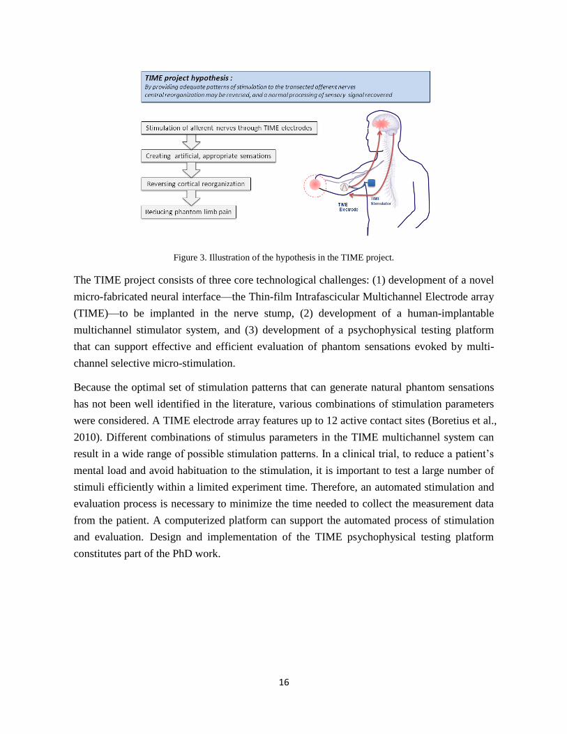
16
Figure 3. Illustration of the hypothesis in the TIME project.
The TIME project consists of three core technological challenges: (1) development of a novel
micro-fabricated neural interface—the Thin-film Intrafascicular Multichannel Electrode array
(TIME)—to be implanted in the nerve stump, (2) development of a human-implantable
multichannel stimulator system, and (3) development of a psychophysical testing platform
that can support effective and efficient evaluation of phantom sensations evoked by multi-
channel selective micro-stimulation.
Because the optimal set of stimulation patterns that can generate natural phantom sensations
has not been well identified in the literature, various combinations of stimulation parameters
were considered. A TIME electrode array features up to 12 active contact sites (Boretius et al.,
2010). Different combinations of stimulus parameters in the TIME multichannel system can
result in a wide range of possible stimulation patterns. In a clinical trial, to reduce a patient’s
mental load and avoid habituation to the stimulation, it is important to test a large number of
stimuli efficiently within a limited experiment time. Therefore, an automated stimulation and
evaluation process is necessary to minimize the time needed to collect the measurement data
from the patient. A computerized platform can support the automated process of stimulation
and evaluation. Design and implementation of the TIME psychophysical testing platform
constitutes part of the PhD work.
17
2. Overview of the PhD work
2.1 Hypothesis and questions
As stated in the previous section, the severity of PLP was found to be positively associated
with the amount of cortical alteration (Grüsser et al., 2001). Because cortical changes in the
brain may result from the incongruence of motor intention and impaired sensory feedback due
to transection of periphery nerves (Diers et al., 2010), artificially providing adequate feedback
of the phantom limb may assist in addressing the incongruence. The scientific hypothesis of
the present PhD research was the following:
Providing sensory feedback through electrocutaneous stimulation of a residual limb may
reverse cortical alteration and consequently reduce PLP.
To test the hypothesis, three questions were formulated:
Question 1. How do stimulus parameters influence sensory responses?
Question 2. Does sensory identification training have a therapeutic benefit for PLP?
Question 3. How may sensory feedback be evaluated efficiently in a multi-channel stimulation setting?
The sensation elicited by electrocutaneous stimulation is a function of the stimulation
parameters. The stimulus parameters selected to be modulated to convey sensory information
play important roles in determining the effectiveness of a sensory feedback scheme. It is thus
important to examine how stimulus parameters influence sensory responses (Question 1).
Furthermore, training patients in discriminating among different stimulus parameters has been
proven to have a positive effect on PLP (Flor et al., 2001). Sensory identification is a
moderately more complex behaviorally relevant task that is assumed to involve more
sophisticated sensory processing. Does sensory identification training have a therapeutic
benefit for PLP (Question 2)? Given the multichannel, intra-neural stimulation system
developed in the TIME project, how may sensory feedback be evaluated effectively and
efficiently (Question 3)?
Electrocutaneous stimulation, rather than mechanical stimulation, was chosen as the means of
delivering sensory feedback, mainly for the following reasons: (1) it is likely to produce more
types of sensation, which may involve more sensory processing and thus act on the sensory
cortices more effectively; (2) stimulation parameters are more controllable, which partly
ensures the reliability of the sensory feedback; and (3) many product options for electrodes
are commercially available.
18
2.2 Studies and corresponding publications
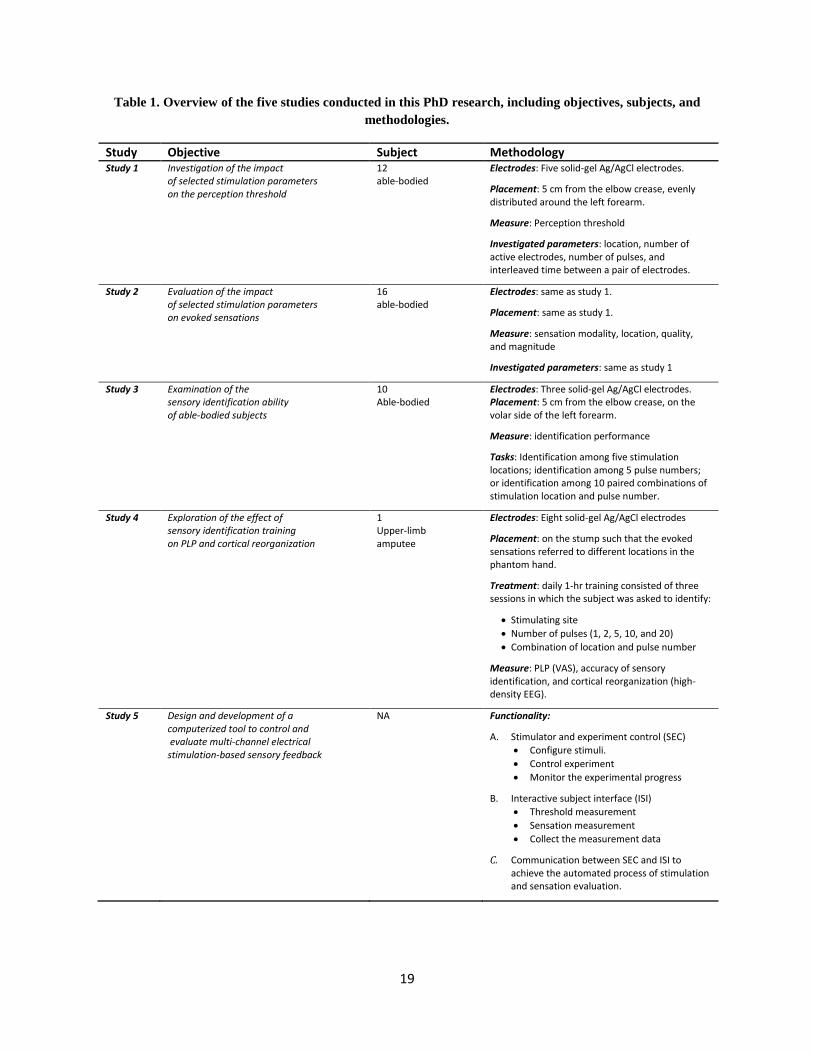
To address the three questions raised, five studies were conducted in this PhD research.
Question 1 was addressed through examination of the impact of stimulation parameters on the
perception threshold in Study 1 and the impact on evoked sensation magnitude, quality, and
location in Study 2. Question 2 was addressed in Studies 3 and 4. Study 3 investigated human
ability to identify the stimulation location and pulse number in behaviorally relevant tasks
with able-bodied subjects. In study 4, an upper-limb amputee volunteer with PLP was trained
in sensory identification to examine the effect of training on identification performance, the
intensity of PLP, and cortical reorganization. Question 3 was addressed by design and
development of a computerized, automated psychophysical test platform in Study 5. An
overview of the objectives, subjects, and methodologies of the five specific studies is
presented in Table 1.
19
Table 1. Overview of the five studies conducted in this PhD research, including objectives, subjects, and
methodologies.
Study Objective Subject Methodology Study 1
Investigation of the impact of selected stimulation parameters on the perception threshold
12 able-bodied
Electrodes: Five solid-gel Ag/AgCl electrodes.
Placement: 5 cm from the elbow crease, evenly distributed around the left forearm.
Measure: Perception threshold
Investigated parameters: location, number of active electrodes, number of pulses, and interleaved time between a pair of electrodes.
Study 2
Evaluation of the impact of selected stimulation parameters on evoked sensations
16 able-bodied
Electrodes: same as study 1.
Placement: same as study 1.
Measure: sensation modality, location, quality, and magnitude
Investigated parameters: same as study 1
Study 3
Examination of the sensory identification ability of able-bodied subjects
10 Able-bodied
Electrodes: Three solid-gel Ag/AgCl electrodes. Placement: 5 cm from the elbow crease, on the volar side of the left forearm.
Measure: identification performance
Tasks: Identification among five stimulation locations; identification among 5 pulse numbers; or identification among 10 paired combinations of stimulation location and pulse number.
Study 4
Exploration of the effect of sensory identification training on PLP and cortical reorganization
1 Upper-limb amputee
Electrodes: Eight solid-gel Ag/AgCl electrodes
Placement: on the stump such that the evoked sensations referred to different locations in the phantom hand.
Treatment: daily 1-hr training consisted of three sessions in which the subject was asked to identify:
Stimulating site
Number of pulses (1, 2, 5, 10, and 20)
Combination of location and pulse number
Measure: PLP (VAS), accuracy of sensory identification, and cortical reorganization (high-density EEG).
Study 5 Design and development of a computerized tool to control and evaluate multi-channel electrical stimulation-based sensory feedback
NA Functionality:
A. Stimulator and experiment control (SEC)
Configure stimuli.
Control experiment
Monitor the experimental progress
B. Interactive subject interface (ISI)
Threshold measurement
Sensation measurement
Collect the measurement data
C. Communication between SEC and ISI to achieve the automated process of stimulation and sensation evaluation.
20
Study 1: Investigation of the impact of selected stimulation parameters on
the perception threshold
Published in: Journal of Neuroengineering Rehabilitation 2011, 8:9.
Impacts of selected stimulation patterns on the perception threshold in electrocutaneous
stimulation
Bo Geng1, Ken Yoshida
2, Winnie Jensen
1
Affiliations:
1 Center for Sensory-Motor Interaction, Dept. Health Science and Technology, Aalborg University,
Denmark; 2Biomedical Engineering Department, Indiana University-Purdue University Indianapolis,
Indianapolis, USA
21
Study 2: Evaluation of the impact of selected stimulation parameters on
evoked sensations
Published in: Journal of Rehabilitation Research and Development 2012, 49(2): 297-308.
Evaluation of sensation evoked by electrocutaneous stimulation on forearm in nondisabled
subjects
Bo Geng1, Ken Yoshida
2, Laura Petrini
1, 3, Winnie Jensen
1
Affiliations:
1 Center for Sensory-Motor Interaction, Dept. Health Science and Technology, Aalborg University,
Denmark; 2Biomedical Engineering Department, Indiana University-Purdue University Indianapolis,
Indianapolis, USA; 3Department of Communication and Psychology, Aalborg University, Denmark
22
Study 3: Examination of the sensory identification ability of able-bodied
subjects
Under preparation, to be submitted to Journal of Neuroengineering and Rehabilitation.
Human ability in identification of location and pulse number for electrocutaneous stimulation
applied on the forearm
Bo Geng1, Winnie Jensen
1
Affiliations:
1 Center for Sensory-Motor Interaction, Dept. Health Science and Technology, Aalborg University,
Denmark
23
Study 4: Exploration of the effect of sensory identification training on PLP
and cortical reorganization
Published in Annual meeting of Society for Neuroscience, Neuroscience 2011.
A case study on phantom sensation and sensory discrimination induced by electrocutaneous
stimulation
Bo Geng1, Ken Yoshida
2, Winnie Jensen
1
Affiliations:
1 Center for Sensory-Motor Interaction, Dept. Health Science and Technology, Aalborg University,
Denmark; 2Biomedical Engineering Department, Indiana University-Purdue University Indianapolis,
Indianapolis, USA
24
Study 5: Design and development of a computerized tool to control and
evaluate multi-channel electrical stimulation-based sensory feedback
Under preparation, to be submitted to International Journal of Computer Human Interaction.
Computerized tool to control and evaluate multi-channel electrical stimulation based sensory
feedback—example of use for phantom limb pain treatment
Bo Geng1, Ken Yoshida
2, Winnie Jensen
1
Affiliations:
1 Center for Sensory-Motor Interaction, Dept. Health Science and Technology, Aalborg University,
Denmark; 2Biomedical Engineering Department, Indiana University-Purdue University Indianapolis,
Indianapolis, USA;

25
3. Discussion and Conclusion
Five studies were performed to address the hypothesis and the three formulated questions.
The main outcomes and related issues of the Ph.D. research have been discussed here,
whereas a more detailed discussion of specific studies can be found in relevant papers. Some
perspectives pertaining to this research topic are also discussed, as appropriate.
3.1 Discussion of main findings
An overview of the main findings of each study is outlined in Table 2.
Table 2. Major outcomes for each study.
Overall hypothesis: Providing sensory feedback through electrocutaneous stimulation of a residual limb may reverse cortical alteration and consequently reduce PLP.
Q1: How do stimulation parameters influence sensory input?
Q2: Does sensory identification training have therapeutic benefit for PLP?
Q3: How may sensory
feedback be evaluated
efficiently in a multi-
channel stimulation
setting?
Study 1: Investigation of the impact of selected stimulation parameters on the perception threshold
Ventral side has a lower perception threshold than dorsal side of the forearm.
There is an inverse relationship between the perception threshold and the number of pulses in a pulse train.
Study 2: Evaluation of the impact of selected stimulation parameters on evoked sensations
Volar and ulnar side of the forearm can perceive more consistent tactile sensation.
The number of pulses in a pulse train significantly influences the intensity of evoked sensations.
Study 3: Examination of the sensory identification ability of able-bodied subjects
The average performance in sensory identification of location and pulse number was promising (92.2% and 90.8%).
Study 4: Exploration of the effect of sensory identification training on PLP and cortical reorganization
The sensory identification accuracy improved over the training period, but there was no decreases in PLP and no change in cortical plasticity.
Study 5: Design and development of a computerized tool to control and evaluate multi-channel electrical stimulation-based sensory feedback
The platform was utilized for threshold and sensation measurement with intra-fasicular multi-channel electrodes implanted in the nerve stump in an amputee volunteer.
26
In summary, the ventral forearm had a lower perception threshold than the dorsal forearm.
The stimulation location on the forearm significantly influenced the sensory responses. The
number of pulses also had a significant impact on the perceived magnitude of sensation.
When the stimulation location and pulse number were varied in identification tasks,
satisfactory identification performance was achieved by able-bodied subjects. However,
training in the same sensory identification tasks neither relieved PLP nor reversed cortical
reorganization in the amputee patient, although the amputee patient’s identification
performance improved over the training period. In addition, a computerized sensory feedback
evaluation platform developed for the TIME project made a tool for further studies on PLP
treatment by providing patients with more intuitive, natural sensory feedback using selective
intra-neural stimulation. Each study is discussed below in relation to the three questions.
Study 1: Effect of stimulation parameters on the perception threshold (Q1)
By examining the relation between stimulation parameters and the perception threshold, study
1 provided useful information for optimization of a stimulation protocol in a sensory feedback
scheme. The ventral aspect of the forearm was found to have a lower perception threshold
than the dorsal aspect. Therefore, use of the ventral forearm is recommended for receiving
electrocutaneous sensory feedback, due to the better energy efficiency and potentially higher
information transfer capacity that it provides.
Sensory feedback should be consistent to gain users' confidence in interpreting artificial
sensory input. However, consistency cannot be guaranteed because of the non-linear relation
between the stimulation parameters and sensory responses (Pfeiffer, 1968). This is confirmed
by the results of study 1. The four parameters investigated—stimulation location, number of
stimulating channels, number of pulses, and time delay in interleaved stimulation—were all
shown to be related to the perception threshold in a non-linear way.
The stimulation parameters investigated were chosen on the basis of a literature review and
the need for sensory feedback in amputee patients. Some parameters, such as body locus,
pulse duration, waveform, and frequency, have been investigated in previous studies (Girvin
et al., 1982; Kantor et al., 1994; Palmer et al., 1999). Those studies mainly concerned single-
channel stimulation using different types of surface electrodes at various body sites, whereas
study 1 investigated the parameters in relation to multi-channel stimulation on the forearm
with solid-gel electrodes.
Study 2: Effect of stimulation parameters on evoked sensation (Q1)
Study 2 provided information used in the choice of the optimal stimulation parameters to be
used for sensory modulation. Varying a stimulation parameter can modulate the sensation by
27
which information is encoded and transmitted to human subjects (Szeto and Saunders, 1982).
If the sensation (e.g., intensity or modality) can be easily and reliably modulated by varying a
stimulation parameter, the coding scheme was assumed to be effective.
In study 2, stimulation of the ventral forearm was found to more easily evoke tactile and less
pricking sensation than stimulation of the dorsal side. This finding is important to ensure the
comfort of electrocutaneous sensory feedback, as electrical stimulation is often associated
with discomfort and occasionally pain, which can be a limiting factor in users’ acceptance
(Chae and Hart, 1998; Garnsworthy et al., 1988). Other elements, such as electrode size,
waveform, and frequency, may also have an impact on the comfort of electrocutaneous
stimulation (Kuhn et al., 2010; Naaman et al., 2000; Gracanin and Trnkoczy, 1975).
Furthermore, the number of pulses has a significant effect on the perceived magnitude,
implying that varying the number of pulses may be an effective means of sensory modulation.
This finding is consistent with those of a recent study (Van Der Heide et al., 2009). An early
study derived a specific relation for pulse number growing as the 1.8 power of the perceived
magnitude in stimulation applied on the abdomen at 30 Hz (Sachs et al., 1980).
Study 3: Sensory identification in able-bodied subjects (Q2)
In study 3, the sensory identification ability of able-bodied subjects was evaluated. Based on
the findings of studies 1 and 2, electrodes were placed on the ventral forearm. The stimulation
location and pulse number were varied for purposes of sensory modulation.
Modulation of the two parameters yielded satisfactory identification performance. The overall
accuracy was 92.2% for spatial identification and 90.8% for the identification of the pulse
number. Performance worsened when the two dimensions were required to be distinguished at
the same time. The results provide an opportunity to directly compare the identification
performance of able-bodied subjects and amputees, which can assist in translating the
findings to clinical application. However, to the best of my knowledge, there have been no
such studies on sensory identification with upper-limb amputees. The only related work has
been on the effect of sensory discrimination training on PLP. In that study, amputee subjects
were trained in sensory discrimination of stimulation location and frequency. Discriminability
was shown to be improved over the two weeks of the training period, but the accuracy
remained below 60% for both location and frequency discrimination (Flor et al., 2001).
Study 4: Sensory feedback training in amputee patients (Q2)
In study 4, an amputee patient was trained in sensory identification to investigate the effect of
training on PLP. The stimulation location and pulse number were again varied for purposes of
28
sensory modulation. It was expected that the identification ability of amputees would be
better than that of able-bodied subjects because stimulation of the residual limb can evoke
sensations not only locally but also referred to the phantom limb (Ramachandran et al., 1992),
implying that the somatosensory cortex representing the forearm may be expanded and thus
that sensory acuity may be improved in amputees. However, the amputee patient did not
achieve good identification performance in the first days of training. Over the training period,
the amputee patient’s accuracy improved and was eventually comparable to that of the able-
bodied subjects. The unexpected low identification ability of the amputee patient at the
beginning of the training period was likely due to subject-to-subject variance in learning rates.
Research into the identification abilities of more amputees is needed to allow for statistical
comparison between the two groups.
No reduction in PLP and no changes in cortical reorganization were observed after ten days of
training. Nevertheless, the failure of sensory identification training in PLP treatment should
be interpreted with caution. First of all, the training might not have been extensive enough, or
the stimulus intensity might not have been sufficiently strong. A similar study on the effect of
feedback-guided sensory training on PLP was performed with ten daily 90-min training
sessions and a stimulus level of 0.1 mA below the individual pain threshold (Flor et al., 2001).
In that study, all five amputee patients experienced reductions in PLP and reversal of cortical
reorganization. A comparative study is needed to validate the training regimen. Furthermore,
cortical reorganization alone might not be the primary factor in the development of PLP in
this patient. Providing sensory feedback of the phantom hand with the aim of counteracting
cortical reorganization might thus be ineffective for him. This may further confirm that
various mechanisms account for the causation and maintenance of PLP in individual patients
and that no one treatment is likely to be effective for all amputee patients (Flor, 2002a).
Appropriate selection of interventions that can address the underlying problems will be
important for effective PLP treatment.
A follow-up study with a larger sample size and perhaps a higher stimulation intensity is
needed to further evaluate the effect of sensory identification training on PLP. Additionally,
in what subgroup of amputee patients, sensory feedback-based intervention is effective
remains to be investigated. To what extent the number of training sessions and the stimulus
intensity would affect the treatment outcome also needs to be determined.
Study 5: Computerized psychophysical testing platform (Q3)
Recently, peripheral intra-neural interfaces with multiple channels have been developed for
direct nerve stimulation with high selectivity, which could be used to record motor signals in
29
bionic hand control (Rossini et al., 2010; Boretius et al., 2010; Dhillon and Horch, 2005).
Using the same electrodes to selectively activate sensory fibers in the nerve bundle provides
the possibility to manipulate phantom sensation (Dhillon and Horch, 2005) and counteract
phantom pain by enabling sensory processing that is missing subsequent to amputation.
To efficiently evaluate the phantom sensation evoked by a wide range of stimulation patterns
in a multi-channel setting, a computerized psychophysical testing platform was developed.
The platform was designed to collect the data from the threshold and sensation measure
experiments. However, to counteract cortical alterations and consequently relieve PLP, it was
expected that repeated stimulation sessions with one subject would need to be carried out.
Hence, selection of a subset of optimal stimulation patterns was required for each subject,
based on the results of the sensation measurements. Optimal stimulation patterns were defined
as those that elicited clear, distinct types of sensations referred to the phantom limb. Therefore,
a module that can automatically select optimal stimulation patterns should be considered in
future development.
3.2 Methodological considerations
Studies with able-bodied subjects
The first three studies were conducted with able-bodied subjects. It is noteworthy that the
sample population is not representative of amputee patients with PLP. Therefore, the results
ought to be carefully interpreted before implementation in amputee patients, because when
stimulating damaged limbs, the perceptual experience may differ from those in nondisabled
people. It will be of interest to see whether similar results can be obtained with subjects with
amputations in future work.
Choice of stimulation parameters
Throughout studies 1 to 4, a biphasic waveform was used because the dermis accumulates
electrochemical changes from monophasic pulses. In addition, biphasic pulse pairs produce
less long-term reddening and a more comfortable sensation than monophasic pulses (Szeto
and Saunders, 1982). A pulse duration of 200 μs was chosen because it produced the least
‘pricking’ sensation in our pilot experiment, in which pulse durations of 100 μs, 200 μs and
500 μs were tested. A frequency of 20 Hz was used (Szeto et al., 1979) because this may be
the optimal frequency for sensory communication, as the maximum frequency discrimination
occurs near 20 Hz (Szeto et al., 1979).
Stimulation parameters related to dual channel stimulation were investigated in studies 1 and
2 because incorporation of a second channel introduces additional variables that can affect the
30
efficacy of sensory feedback (Szeto and Saunders, 1982). As a limitation, only a selected set
of stimulation parameters (i.e., the stimulation site, the number of stimulating channels, the
number of pulses, and the time delay between two channels) were examined in studies 1 and 2.
Sensory identification
Both animal and human experiments have revealed that training in sensory discrimination,
rather than passive stimulation, can result in cortical remodeling (Recanzone et al., 1992; Flor
et al., 2001). In discrimination tasks, a subject needs to determine whether there is a
detectable difference between the presented stimulus and the reference stimulus. In
identification tasks, the subject needs to identify the presented stimulus among several
different stimuli. Sensory identification requires not only detecting the difference between
two stimuli presented but also determining which stimulus was presented. As such, it is
assumed that sensory identification involves more advanced cognitive activity, such as
attention and memory, than does sensory discrimination or passive stimulation and
consequently facilitates cortex remapping. This assumption led to studies 3 and 4, in which
sensory identification was chosen as the means of providing sensory feedback to the subjects.
Measurement of cortical reorganization
In the study 4 on the effect of sensory identification training on PLP, cortical reorganization
was assessed before and after training. Neuroelectrical source analysis of high-density EEG
recording was used to localize the cortical activity. Light superficial pressure stimulation was
applied to the corner of the lower lip because the cortical area representing the mouth was
found to take over the former hand area (Ramachandran et al., 1992; Elbert et al., 1994). The
patient’s phantom pain and the observation of the shift of the lip area to the hand area on the
amputation side confirmed the earlier proposed theory that cortical reorganization is related to
PLP (Grüsser et al., 2001; Flor et al., 2001; Birbaumer, 1997).
However, the PLP was not reduced after the sensory identification training, and the cortical
shift was not reversed. From the perspective of the technology employed, although EEG
source analysis is occasionally used to localize brain activity, it suffers from the limitation of
poor spatial resolution. The accuracy of EEG source analysis is affected by many practical
factors, such as the electrode position on the scalp, the choice of reference, the interpolation
algorithm chosen, the treatment of artifact-contaminated channels due to poor electrode–scalp
contact or amplifier malfunction, and the head model and mathematical inverse model chosen
(Michel et al., 2004). In this regard, other imaging methods, such as fMRI, might be better
suited to localization of brain activity and ensure the validity of the results.
Electrocutaneous vs. intra-neural sensory feedback
31
Sensory feedback employing electrocutaneous stimulation has been shown to be somewhat
successful in PLP treatment (Flor et al., 2001; Dietrich et al., 2012). Its noninvasiveness is
also attractive. However, electrocutaneous stimulation can only elicit somatic sensation (e.g.,
touch, tingling) locally or in the phantom limb, which greatly limits the utilization of sensory
feedback. Performing behaviorally relevant discrimination and identification tasks is then
used as an alternative means of providing meaningful sensory feedback. Incorporation of
other sensory modality feedback (e.g., visual, audio) may enhance its effectiveness in
counteracting the cortical reorganization underlying PLP.
Direct nerve stimulation made it possible to evoke more types of meaningful sensory
feedback (e.g., finger movement, joint position, wrist movement) (Dhillon et al., 2005).
Moreover, it has the advantage of using the same set of implanted electrodes for both bionic
hand control and sensory feedback. In particular, the patient who participated in study 4 was
later treated by providing sensory feedback using intrafascicular stimulation with implanted
TIME electrodes, and PLP was temporarily alleviated within the implantation period (results
not published). This is an exciting clue that encourages further exploration of this type of
sensory feedback in PLP treatment.
3.3 Conclusions
This PhD thesis has presented five studies to address the hypothesis that providing sensory
feedback through electrocutaneous stimulation of a residual limb may reverse cortical
alteration and consequently reduce PLP in amputee patients. The impact of stimulation
parameters on the perception threshold and the evoked sensation was examined. The
stimulation location and pulse number were identified to be able to effectively convey sensory
information. Human ability in sensory identification of the two parameters was then
investigated, and satisfactory performance was obtained in able-bodied subjects. On the basis
of these findings, the hypothesis was tested by providing sensory identification training to an
upper-limb amputee volunteer. However, PLP and cortical reorganization in this patient did
not show significant changes. Direct afferent nerve stimulation of the residual limb makes it
possible to evoke natural sensations referred to the phantom limb. As part of the EU-funded
TIME project, a computerized tool was developed for efficient sensory feedback evaluation in
a multi-channel direct nerve stimulation system that may be used to provide more meaningful
sensory feedback to amputee patients and consequently reduce PLP. Using sensory feedback
to treat PLP or other chronic pain is still at an early stage of development. Multiple sensory
modality feedback could also be considered as the focus of future work.
32
References
Alviar MJ, Hale T, Dungca M (2011) Pharmacologic interventions for treating phantom limb
pain. Cochrane Database of Systematic Reviews (Online) 12: CD006380.
Arena JG, Sherman RA, Bruno GM, Smith JD (1990) The relationship between situational
stress and phantom limb pain: Cross-lagged correlational data from six month pain logs. J
Psychosom Res 34:71-77.
Baron R (2006) Mechanisms of disease: Neuropathic pain - A clinical perspective. Nature
Clinical Practice Neurology 2:95-106.
Beeker TW, During J, Den Hertog A (1967) Artificial touch in a hand-prosthesis. Med & Biol
Engng 5:47-49.
Belleggia G, Birbaumer N (2001) Treatment of phantom limb pain with combined EMG and
thermal biofeedback: A case report. Applied Psychophysiology Biofeedback 26:141-146.
Birbaumer N (1997) Effects of regional anesthesia on phantom limb pain are mirrored in
changes in cortical reorganization. Journal of Neuroscience 17:5503-5508.
Bittar RG, Otero S, Carter H, Aziz TZ (2005) Deep brain stimulation for phantom limb pain.
Journal of Clinical Neuroscience 12:399-404.
Black LM, Persons RK, Jamieson B (2009) What is the best way to manage phantom limb
pain? J Fam Pract 58:155-158.
Bókkon I, Till A, Grass F, Erdöfi Szabó A (2011) Phantom pain reduction by low-frequency
and low-intensity electromagnetic fields. Electromagnetic Biology and Medicine 30:115-127.
Boretius T, Badia J, Pascual-Font A, Schuettler M, Navarro X, Yoshida K, Stieglitz T (2010)
A transverse intrafascicular multichannel electrode (TIME) to interface with the peripheral
nerve. Biosensors and Bioelectronics 26:62-69.
Bosmans JC, Geertzen JHB, Post WJ, Van Der Schans CP, Dijkstra PU (2010) Factors
associated with phantom limb pain: A 3 1/2-year prospective study. Clin Rehabil 24:444-453.
Carabelli RA, Kellerman WC (1985) Phantom limb pain: Relief by application of TENS to
contralateral extremity. Arch Phys Med Rehabil 66:466-467.
Carlen PL, Wall PD, Nadvorna H, Steinbach T (1978) Phantom limbs and related phenomena
in recent traumatic amputations. Neurology 28:211-217.
Chae J, Hart R (1998) Comparison of discomfort associated with surface and percutaneous
intramuscular electrical stimulation for persons with chronic hemiplegia. American Journal of
Physical Medicine and Rehabilitation 77:516-522.
Chan BL, Witt R, Charrow AP, Magee A, Howard R, Pasquina PF, Heilman KM, Tsao JW
(2007) Mirror therapy for phantom limb pain [15]. N Engl J Med 357:2206-2207.
Chan R (2006) Hypnosis and phantom limb pain. Australian Journal of Clinical and
Experimental Hypnosis 34:55-64.
Chen R, Cohen LG, Hallett M (2002) Nervous system reorganization following injury.
Neuroscience 111:761-773.
33
Costigan M, Scholz J, Woolf CJ (2009) Neuropathic pain: A maladaptive response of the
nervous system to damage. Annual Review of Neuroscience 32:1-32.
CRONHOLM B (1951) Phantom limbs in amputees; a study of changes in the integration of
centripetal impulses with special reference to referred sensations. Acta Psychiatrica Et
Neurologica Scandinavica.Supplementum 72:1-310.
Davis KD, Kiss ZHT, Luo L, Tasker RR, Lozano AM, Dostrovsky JO (1998) Phantom
sensations generated by thalamic microstimulation. Nature 391:385-387.
Devor M, Janig W, Michaelis M (1994) Modulation of activity in dorsal root ganglion
neurons by sympathetic activation in nerve-injured rats. J Neurophysiol 71:38-47.
Devor M, Govrin-Lippmann R, Angelides K (1993) Na+ channel immunolocalization in
peripheral mammalian axons and changes following nerve injury and neuroma formation.
Journal of Neuroscience 13:1976-1992.
Dhillon GS, Horch KW (2005) Direct neural sensory feedback and control of a prosthetic arm.
IEEE Transactions on Neural Systems and Rehabilitation Engineering 13:468-472.
Dhillon GS, Krüger TB, Sandhu JS, Horch KW (2005) Effects of short-term training on
sensory and motor function in severed nerves of long-term human amputees. J Neurophysiol
93:2625-2633.
Di Pino G, Guglielmelli E, Rossini PM (2009) Neuroplasticity in amputees: Main
implications on bidirectional interfacing of cybernetic hand prostheses. Prog Neurobiol
88:114-126.
Diers M, Christmann C, Koeppe C, Ruf M, Flor H (2010) Mirrored, imagined and executed
movements differentially activate sensorimotor cortex in amputees with and without phantom
limb pain. Pain 149:296-304.
Dietrich C, Walter-Walsh K, Preißler S, Hofmann GO, Witte OW, Miltner WHR, Weiss T
(2012) Sensory feedback prosthesis reduces phantom limb pain: Proof of a principle.
Neurosci Lett 507:97-100.
Dougherty J (1980) Relief of phantom limb pain after EMG biofeedback assisted relaxation:
A case report. Behav Res Ther 18:355-357.
Ehde DM, Czerniecki JM, Smith DG, Campbell KM, Edwards WT, Jensen MP, Robinson LR
(2000) Chronic phantom sensations, phantom pain, residual limb pain, and other regional pain
after lower limb amputation. Arch Phys Med Rehabil 81:1039-1044.
Ehrenstein WH, Ehrenstein A (1999) Psychophysical methods. In: Modern techniques in
neuroscience research (Windhorst U, Johansson H, eds), pp1211-1241. Berlin: Springer.
Elbert T, Flor H, Birbaumer N, Knecht S, Hampson S, Larbig W, Taub E (1994) Extensive
reorganization of the somatosensory cortex in adult humans after nervous system injury.
Neuroreport 5:2593-2597.
Ephraim PL, Wegener ST, MacKenzie EJ, Dillingham TR, Pezzin LE (2005) Phantom pain,
residual limb pain, and back pain in amputees: Results of a national survey. Arch Phys Med
Rehabil 86:1910-1919.
34
Ergenzinger ER, Glasier MM, Hahm JO, Pons TP (1998) Cortically induced thalamic
plasticity in the primate somatosensory system. Nat Neurosci 1:226-229.
Flor H (2002a) Phantom-limb pain: Characteristics, causes, and treatment. Lancet Neurology
1:182-189.
Flor H (2002b) The modification of cortical reorganization and chronic pain by sensory
feedback. Applied Psychophysiology Biofeedback 27:215-227.
Flor H, Nikolajsen L, Jensen TS (2006) Phantom limb pain: A case of maladaptive CNS
plasticity? Nat Rev Neurosci 7:873-881.
Flor H, Denke C, Schaefer M, Grüsser S (2001) Effect of sensory discrimination training on
cortical reorganisation and phantom limb pain. Lancet 357:1763-1764.
Flor H, Elbert T, Knecht S, Wienbruch C, Pantev C, Birbaumer N, Larbig W, Taub E (1995)
Phantom-limb pain as a perceptual correlate of cortical reorganization following arm
amputation. Nature 375:482-484.
Florence SL, Kaas JH (1995) Large-scale reorganization at multiple levels of the
somatosensory pathway follows therapeutic amputation of the hand in monkeys. Journal of
Neuroscience 15:8083-8095.
Florence SL, Hackett TA, Strata F (2000) Thalamic and cortical contributions to neural
plasticity after limb amputation. J Neurophysiol 83:3154-3159.
Garnsworthy RK, Gully RL, Kenins P, Westerman RA (1988) Transcutaneous electrical
stimulation and the sensation of prickle. J Neurophysiol 59:1116-1127.
Gehling M, Tryba M (2003) Prophylaxis of phantom pain: Is regional analgesia ineffective?
Schmerz 17:11-19.
Gescheider GA (1997) Classical psychophysical theory. In: Psychophysics: The fundamentals
Classical psychophysical theory. Mahwah, NJ: Lawrence Erlbaum Associates.
Girvin JP, Marks LE, Antunes JL (1982) Electrocutaneous stimulation I. the effects of
stimulus parameters on absolute threshold. Percept Psychophys 32:524-528.
Gracanin F, Trnkoczy A (1975) Optimal stimulus parameters for minimum pain in the
chronic stimulation of innervated muscle. Arch Phys Med Rehabil 56:243-249.
Grant S, Aitchison T, Henderson E, Christie J, Zare S, McMurray J, Dargie H (1999) A
comparison of the reproducibility and the sensitivity to change of visual analogue scales, borg
scales, and likert scales in normal subjects during submaximal exercise. Chest 116:1208-1217.
Grüsser SM, Winter C, Mühlnickel W, Denke C, Karl A, Villringer K, Flor H (2001) The
relationship of perceptual phenomena and cortical reorganization in upper extremity amputees.
Neuroscience 102:263-272.
Halbert J, Crotty M, Cameron ID (2002) Evidence for the optimal management of acute and
chronic phantom pain: A systematic review. Clin J Pain 18:84-92.
Hall EJ, Flament D, Fraser C, Lemon RN (1990) Non-invasive brain stimulation reveals
reorganised cortical outputs in amputees. Neurosci Lett 116:379-386.
35
Hanley MA, Ehde DM, Campbell KM, Osborn B, Smith DG (2006) Self-reported treatments
used for lower-limb phantom pain: Descriptive findings. Arch Phys Med Rehabil 87:270-277.
Harden RN, Houle TT, Green S, Remble TA, Weinland SR, Colio S, Lauzon J, Kuiken T
(2005) Biofeedback in the treatment of phantom limb pain: A time-series analysis. Applied
Psychophysiology Biofeedback 30:83-93.
Huang C-, Yang R-, Kuo T-, Hsu K- (2009) Phantom limb pain treated by far infrared ray.
Proceedings of the 31st Annual International Conference of the IEEE Engineering in
Medicine and Biology Society: Engineering the Future of Biomedicine, EMBC 2009 1589-
1591.
Jenkins WM, Merzenich MM, Ochs MT, Allard T, Guic-Robles E (1990) Functional
reorganization of primary somatosensory cortex in adult owl monekys after behaviorally
controlled tactile stimulation. J Neurophysiol 63:82-104.
Jensen TS, Krebs B, Nielsen J, Rasmussen P (1985) Immediate and long-term phantom limb
pain in amputees: Incidence, clinical characteristics and relationship to pre-amputation limb
pain. Pain 21:267-278.
Jensen TS, Krebs B, Nielsen J, Rasmussen P (1983) Phantom limb, phantom pain and stump
pain in amputees during the first 6 months following limb amputation. Pain 17:243-256.
Kaczmarek KA, Webster JG, Bach-y-Rita P, Tompkins WJ (1991) Electrotactile and
vibrotactile displays for sensory substitution systems. IEEE Transactions on Biomedical
Engineering 38:1-16.
Kantor G, Alon G, Ho HS (1994) The effects of selected stimulus waveforms on pulse and
phase characteristics at sensory and motor thresholds. Phys Ther 74:951-962.
Karl A, Mühlnickel W, Kurth R, Flor H (2004) Neuroelectric source imaging of steady-state
movement-related cortical potentials in human upper extremity amputees with and without
phantom limb pain. Pain 110:90-102.
Karl A, Birbaumer N, Lutzenberger W, Cohen LG, Flor H (2001) Reorganization of motor
and somatosensory cortex in upper extremity amputees with phantom limb pain. Journal of
Neuroscience 21:3609-3618.
Katayama Y, Yamamoto T, Kobayashi K, Kasai M, Oshima H, Fukaya C (2002) Motor
cortex stimulation for phantom limb pain: Comprehensive therapy with spinal cord and
thalamic stimulation. Stereotact Funct Neurosurg 77:159-162.
Katz J (1992) Psychophysiological contributions to phantom limbs. Canadian Journal of
Psychiatry 37:282-298.
Knecht S, Henningsen H, Elbert T, Flor H, Höhling C, Pantev C, Taub E (1996)
Reorganizational and perceptional changes after amputation. Brain 119:1213-1219.
Kooijman CM, Dijkstra PU, Geertzen JHB, Elzinga A, Van Der Schans CP (2000) Phantom
pain and phantom sensations in upper limb amputees: An epidemiological study. Pain 87:33-
41.
Krane EJ, Heller LB (1995) The prevalence of phantom sensation and pain in pediatric
amputees. J Pain Symptom Manage 10:21-29.
36
Kuhn A, Keller T, Lawrence M, Morari M (2010) The influence of electrode size on
selectivity and comfort in transcutaneous electrical stimulation of the forearm. IEEE
Transactions on Neural Systems and Rehabilitation Engineering 18:255-262.
Lacoux PA, Crombie IK, Macrae WA (2002) Pain in traumatic upper limb amputees in sierra
leone. Pain 99:309-312.
Lambert AW, Dashfield AK, Cosgrove C, Wilkins DC, Walker AJ, Ashley S (2001)
Randomized prospective study comparing preoperative epidural and intraoperative perineural
analgesia for the prevention of postoperative stump and phantom limb pain following major
amputation. Reg Anesth Pain Med 26:316-321.
Lotze M, Flor H, Grodd W, Larbig W, Birbaumer N (2001) Phantom movements and pain an
fMRI study in upper limb amputees. Brain 124:2268-2277.
Lotze M, Grodd W, Birbaumer N, Erb M, Huse E, Flor H (1999) Does use of a myoelectric
prosthesis prevent cortical reorganization and phantom limb pain? [2]. Nat Neurosci 2:501-
502.
Lundeberg T (1985) Relief of pain from a phantom limb by peripheral stimulation. J Neurol
232:79-82.
MacIver K, Lloyd DM, Kelly S, Roberts N, Nurmikko T (2008) Phantom limb pain, cortical
reorganization and the therapeutic effect of mental imagery. Brain 131:2181-2191.
Mannix SM, O'Sullivan C, Kelly GA (2013) Acupuncture for managing phantom-limb
syndrome: A systematic review. Medical Acupuncture 25:23-42.
Melzack R (1992) Phantom limbs. Sci Am 266:120-126.
Melzack R, Israel R, Lacroix R, Schultz G (1997) Phantom limbs in people with congenital
limb deficiency or amputation in early childhood. Brain 120:1603-1620.
Michel CM, Murray MM, Lantz G, Gonzalez S, Spinelli L, Grave De Peralta R (2004) EEG
source imaging. Clinical Neurophysiology 115:2195-2222.
Millstein S, Bain D, Hunter GA (1985) A review of employment patterns of industrial
amputees: Factors influencing rehabilitation. Prosthet Orthot Int 9:69-78.
Montoya P, Larbig W, Grulke N, Flor H, Taub E, Birbaumer N (1997) The relationship of
phantom limb pain to other phantom limb phenomena in upper extremity amputees. Pain
72:87-93.
Moseley GL, Zalucki NM, Wiech K (2008) Tactile discrimination, but not tactile stimulation
alone, reduces chronic limb pain. Pain 137:600-608.
Naaman SC, Stein RB, Thomas C (2000) Minimizing discomfort with surface neuromuscular
stimulation. Neurorehabil Neural Repair 14:223-228.
Navarro X, Vivó M, Valero-Cabré A (2007) Neural plasticity after peripheral nerve injury
and regeneration. Prog Neurobiol 82:163-201.
Nguyen J-, Nizard J, Keravel Y, Lefaucheur J- (2011) Invasive brain stimulation for the
treatment of neuropathic pain. Nature Reviews Neurology 7:699-709.
Nikolajsen L, Jensen TS (2001) Phantom limb pain. Br J Anaesth 87:107-116.
37
Nikolajsen L, Ilkjær S, Krøner K, Christensen JH, Jensen TS (1997) The influence of
preamputation pain on postamputation stump and phantom pain. Pain 72:393-405.
Owen SLF, Green AL, Nandi DD, Bittar RG, Wang S, Aziz TZ (2007) Deep brain
stimulation for neuropathic pain. Acta Neurochirurgica, Supplementum 111-116.
Palmer ST, Martin DJ, Steedman WM, Ravey J (1999) Alteration of interferential current and
transcutaneous electrical nerve stimulation frequency: Effects on nerve excitation. Arch Phys
Med Rehabil 80:1065-1071.
Parkes CM (1973) Factors determining the persistence of phantom pain in the amputee. J
Psychosom Res 17:97-108.
Pezzin LE, Dillingham TR, MacKenzie EJ (2000) Rehabilitation and the long-term outcomes
of persons with trauma-related amputations. Arch Phys Med Rehabil 81:292-300.
Pfeiffer EA (1968) Electrical stimulation of sensory nerves with skin electrodes for research,
diagnosis, communication and behavioral conditioning: A survey. Med & Biol Engng 6:637-
651.
Prior RE, Lyman J, Case PA, Scott CM (1976) Supplemental sensory feedback for the
VA/NU myoelectric hand. background and preliminary designs. Bull Prosthet Res 10:170-
191.
Pylatiuk C, Kargov A, Schulz S (2006) Design and evaluation of a low-cost force feedback
system for myoelectric prosthetic hands. Journal of Prosthetics and Orthotics 18:57-61.
Ramachandran VS, Hirstein W (1998) The perception of phantom limbs. the D. O. hebb
lecture. Brain 121:1603-1630.
Ramachandran VS, Rodgers-Ramachandran D (1996) Synaesthesia in phantom limbs induced
with mirrors. Proceedings of the Royal Society B: Biological Sciences 263:377-386.
Ramachandran VS, Rogers-Ramachandran D, Cobb S (1995) Touching the phantom limb [3].
Nature 377:489-490.
Ramachandran VS, Rogers-Ramachandran D, Stewart M, Pons TP (1992) Perceptual
correlates of massive cortical reorganization. Science 258:1159-1160.
Rasmussen KG, Rummans TA (2000) Electroconvulsive therapy for phantom limb pain. Pain
85:297-299.
Ray NJ, Jenkinson N, Kringelbach ML, Hansen PC, Pereira EA, Brittain JS, Holland P,
Holliday IE, Owen S, Stein J, Aziz T (2009) Abnormal thalamocortical dynamics may be
altered by deep brain stimulation: Using magnetoencephalography to study phantom limb
pain. Journal of Clinical Neuroscience 16:32-36.
Recanzone GH, Merzenich MM, Jenkins WM, Grajski KA, Dinse HR (1992) Topographic
reorganization of the hand representation in cortical area 3b of owl monkeys trained in a
frequency-discrimination task. J Neurophysiol 67:1031-1056.
Richardson C, Glenn S, Horgan M, Nurmikko T (2007) A prospective study of factors
associated with the presence of phantom limb pain six months after major lower limb
amputation in patients with peripheral vascular disease. Journal of Pain 8:793-801.
38
Rossini PM, Micera S, Benvenuto A, Carpaneto J, Cavallo G, Citi L, Cipriani C, Denaro L,
Denaro V, Di Pino G, Ferreri F, Guglielmelli E, Hoffmann K-, Raspopovic S, Rigosa J,
Rossini L, Tombini M, Dario P (2010) Double nerve intraneural interface implant on a human
amputee for robotic hand control. Clinical Neurophysiology 121:777-783.
Sachs RM, Miller JD, Grant KW (1980) Perceived magnitude of multiple electrocutaneous
pulses. Percept Psychophys 28:255-262.
Schley MT, Wilms P, Toepfner S, Schaller H-, Schmelz M, Konrad CJ, Birbaumer N (2008)
Painful and nonpainful phantom and stump sensations in acute traumatic amputees. Journal of
Trauma - Injury, Infection and Critical Care 65:858-864.
Schmidl H (1977) The importance of information feedback in prostheses for the upper limbs.
Prosthet Orthot Int 1:21-24.
Scott RN, Caldwell RR, Brittain RH, Cameron AB, Dunfield VA (1980) Sensory-feedback
system compatible with myoelectric control. Medical and Biological Engineering and
Computing 18:65-69.
Shannon GF (1979) A myoelectrically-controlled prosthesis with sensory feedback. Medical
and Biological Engineering and Computing 17:73-80.
Shannon GF (1976) A comparison of alternative means of providing sensory feedback on
upper limb prostheses. MED.BIOL.ENGINEERING 14:289-294.
Sherman RA, Sherman CJ (1983) Prevalence and characteristics of chronic phantom limb
pain among american veterans. results of a trial survey. Am J Phys Med 62:227-238.
Sherman RA, Sherman CJ, Parker I (1984) Chronic phantom and stump pain among american
veterans: Results of a survey. Pain 18:83-95.
Sherman RA, Gall N, Gormly J (1979) Treatment of phantom limb pain with muscular
relaxation training to disrupt the pain-anxiety-tension cycle. Pain 6:47-55.
Sherman RA, Arena JG, Sherman CJ, Ernst JL (1989) The mystery of phantom pain:
Growing evidence for psychophysiological mechanisms. Biofeedback Self 14:267-280.
Sol JC, Casaux J, Roux FE, Lotterie JA, Bousquet P, Verdié JC, Mascott C, Lazorthes Y
(2002) Chronic motor cortex stimulation for phantom limb pain: Correlations between pain
relief and functional imaging studies. Stereotact Funct Neurosurg 77:172-176.
Stevens SS (1957) On the psychophysical law. Psychol Rev 64:153-181.
Szeto AYJ, Lyman J, Prior RE (1979) Electrocutaneous pulse rate and pulse width
psychometric functions for sensory communications. Hum Factors 21:241-249.
Szeto A, Saunders FA (1982) ELECTROCUTANEOUS STIMULATION FOR SENSORY
COMMUNICATION IN REHABILITATION ENGINEERING. IEEE Trans Biomed Eng
BME-29:300-308.
Toney A, Dunne L, Thomas BH, Ashdown SP (2003) A shoulder pad insert vibrotactile
display. Proceedings - International Symposium on Wearable Computers, ISWC 35-44.
Van Der Heide EM, Buitenweg JR, Marani E, Rutten WLC (2009) Single pulse and pulse
train modulation of cutaneous electrical stimulation: A comparison of methods. Journal of
Clinical Neurophysiology 26:54-60.
39
Wartan SW, Hamann W, Wedley JR, Mccoll I (1997) Phantom pain and sensation among
british veteran amputees. Br J Anaesth 78:652-659.
Weeks SR, Anderson-Barnes VC, Tsao JW (2010) Phantom limb pain: Theories and therapies.
Neurologist 16:277-286.
Weinstein SM (1998) Phantom limb pain and related disorders. Neurol Clin 16:919-935.
Witteveen HJB, Rietman JS, Veltink PH (2012) Grasping force and slip feedback through
vibrotactile stimulation to be used in myoelectric forearm prostheses. Proceedings of the
Annual International Conference of the IEEE Engineering in Medicine and Biology Society,
EMBS 2969-2972.
Wolff A, Vanduynhoven E, Van Kleef M, Huygen F, Pope JE, Mekhail N (2011) 21.phantom
pain. Pain Practice 11:403-413.
Yang TT, Gallen C, Schwartz B, Bloom FE, Ramachandran VS, Cobb S (1994) Sensory maps
in the human brain [13]. Nature 368:592-593.
Ziegler-Graham K, MacKenzie EJ, Ephraim PL, Travison TG, Brookmeyer R (2008)
Estimating the prevalence of limb loss in the united states: 2005 to 2050. Arch Phys Med
Rehabil 89:422-429.

A part of this thesis has been removed from this publication due tocopyright.