ELECTRICAL INJURIES
31
ELECTRICAL INJURIES • a unique form of trauma • clinical picture of cellular damage syndrome vs specific injury
-
Upload
stacy-trujillo -
Category
Documents
-
view
27 -
download
0
description
ELECTRICAL INJURIES. a unique form of trauma clinical picture of cellular damage syndrome vs specific injury. High vs Low Tension Injury Dividing Line is 1000 volts Low voltage injuries: thermal burns, zone of injury from surface extending into the tissue. High-voltage injury - PowerPoint PPT Presentation
Transcript of ELECTRICAL INJURIES

ELECTRICAL INJURIES
• a unique form of trauma• clinical picture of cellular damage
syndrome vs specific injury

• High vs Low Tension Injury Dividing Line is 1000 volts
• Low voltage injuries: thermal burns, zone of injury from surface extending into the tissue

• High-voltage injury Varying degrees of cutaneous burn combined
with ‘hidden’ destruction of deep tissueElectrical insult results in progressive tissue
necrosis in excess of the originally apparent trauma
Resembles injury of crush trauma

PATHOPHYSIOLOGY
• Historically, thought to be HEAT• Passage of electrical current through a solid
conductor results in conversion of electrical energy into heat, the joule effect
• Amount of heat: determined by the Ohm’s law and Joule Effect

OHM’s Law
• the current travelling through tissue is equal to the ratio of voltage and resistance
• V/R

Joule Effect
J = I RT• J: heat production, I: current, R: tissue
resistance, T: time of contact• Extent of injury depends on the type of
current, pathway of flow, local tissue resistance and duration of contact

Joule Effect Theory 1
Differences in tissue resistance to current flow explain the observed changes following electrical injury (Baxter)

BoneFatTendonSkinMuscleBlood Vessels Nerve
RESISTANCE
Bone having the greatest resistance generates the most heat

Implications
• greater necrosis in the periosseous tissues• current travels along lines of lesser resistance,
i.e. blood vessels• vessels are injured but not immediately
thrombosed • progressive muscular necrosis: delayed
arterial occlusion and progressive ischemic necrosis

Joule Effect Theory 2
Body acts like a volume conductor of a single resistance not of tissues of varying resistance (Hunt et al)

PATHOPHYSIOLOGY
• With the onset of current flow, amperage and temperature rise parallel through out the limbs
• By the time of current arcing, both muscle and bone temperatures are equal
TBONE = TMUSCLE

PATHOPHYSIOLOGY
• It takes bone longer to dissipate the heat accounts for periosseous core of necrotic muscle
• Involved muscle and vessels sustained irreversible damage at the time of current passage with immediate microscopic muscular coagulation necrosis and small nutrient artery thrombosis

PATHOPHYSIOLOGY
• Progressive small vessel thrombosis and deterioration of the microvasculature could convert these areas of patchy necrosis into complete tissue loss

PATHOPHYSIOLOGY
• The entire pathophysiology responsible for the syndrome of electrical injury cannot be attributed to heat
• Lee and Kolodney: tissue of lower conductivity and higher resistance would generate the greatest amount of heat (similar to Baxter), however, most clinical situations would place the tissues in parallel

PATHOPHYSIOLOGY
• They suggested that muscle would generate the greatest intensity of joule heating and the adjacent bone would be heated secondarily
• The bone heated secondarily would dissipate the heat quite slowly (corroborating Hunt et al)
• Showed that often the amount of heat is not sufficient to explain tissue damage sites distant from current contact

PATHOPHYSIOLOGY
• Electrical fields which are stronger than the mechanical response of a membrane can rupture bilaminate lipid membranes
PATHOPHYSIOLOGY
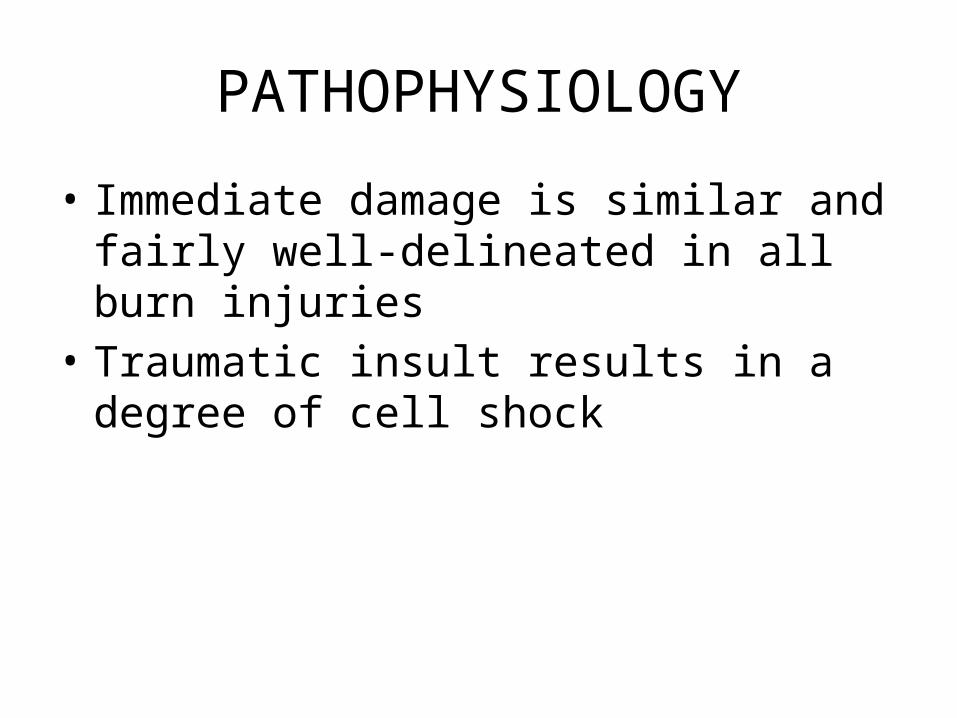
• Immediate damage is similar and fairly well-delineated in all burn injuries
• Traumatic insult results in a degree of cell shock

PATHOPHYSIOLOGY
• The intracellular energy system changes stored adenosine triphosphate (ATP) to cyclic adenosine monophosphate (AMP) and the sodium pump becomes ineffective as the cell membrane becomes permeable to sodium and calcium ions
ATPCyclic AMP

Loss of Phospholipids
Loss of Phospholipids
MembraneDamage
MembraneDamage
Release ofEnzymes
Release ofEnzymes
Elevated“Markers”
ex CK, CKMB
Elevated“Markers”
ex CK, CKMB
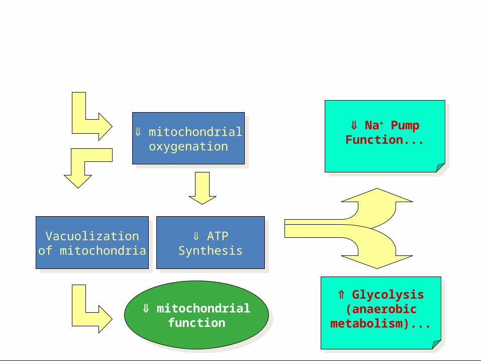
mitochondrialoxygenation
mitochondrialoxygenation
ATPSynthesis
ATPSynthesis
Vacuolizationof mitochondria
Vacuolizationof mitochondria
mitochondrialfunction
mitochondrialfunction
Na+ PumpFunction...
Na+ PumpFunction...
Glycolysis(anaerobic
metabolism)...
Glycolysis(anaerobic
metabolism)...

intracellular Na+
extracellular K+
intracellular Ca++
intracellular Na+
extracellular K+
intracellular Ca++
intracellularH2O
intracellularH2O
Cellular Swelling& rupture
Cellular Swelling& rupture
E.R.Dilation
E.R.Dilation
proteinsynthesis
proteinsynthesis
cellularfunction!
cellularfunction!
Na+ PumpFunction...
Na+ PumpFunction...
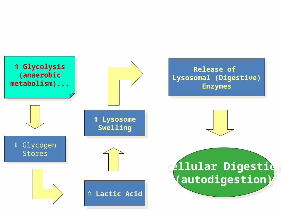
Hypoxic Injury (pathway 2)
Glycolysis(anaerobic
metabolism)...
Glycolysis(anaerobic
metabolism)...
GlycogenStores
GlycogenStores
Lactic Acid Lactic Acid
LysosomeSwelling
LysosomeSwelling
Release of Lysosomal (Digestive)
Enzymes
Release of Lysosomal (Digestive)
Enzymes
Cellular Digestion(autodigestion)
Cellular Digestion(autodigestion)

PATHOPHYSIOLOGY
• Proteases are released and can trigger the coagulation cascade and complement degradation
• C5a begin the immunologic cascade and soft tissue respond by increased vascular permeability and chemotaxis
• The mast cells produces more more cyclic AMP

PATHOPHYSIOLOGY
• Injury also activates phospholipase A which cleaves the phospholipids that are bound to cholesterol and triglycerides in cell membranes
• This results in the metabolism of arachidonic acid which when released helps to activate platelet plug


PATHOPHYSIOLOGY
• The respiratory burst of leukocyte produces more arachidonic acid and produces free radicals necessary for the initial control of bacteria at the site of injury
• All traumatic soft tissue injury results in these immediate effects but these immediate effects do not result in tissue necrosis

PATHOPHYSIOLOGY
• The major causes of tissue lost in the various clinical presentations of the electrical syndrome are due to delayed effects
• Progressive tissue lost in burns and frost bite was due to progressive dermal ischemia mediated by various metabolites of arachidonic acid

PATHOPHYSIOLOGY
• Cellular intergrity depends on a homeostatic relationship between PGE2 and PGE2α
• Injury to cell disrupts this balance and a shunt in arachidonic acid metabolism in favor of thromboxane production
• In large amounts causes vasoconstriction, thrombosis, progressive ischemic necrosis and further thromboxane production

PATHOPHYSIOLOGY
• The demonstration of arachidonic acid metabolites in increasing amounts in tissue following electrical trauma allows a unifying concept for various theories regarding the pathophysiology of these injuries
• Both joule heating and cell membrane disruption can activate the arachidonic acid cascade

PATHOPHYSIOLOGY
• Once initiated, the various immediate effects of trauma produce more mediators
• Inflammatory mediators can cause vasoconstriction of microcirculation
• This would decrease perfusion of an anatomical part and marked decreased in cooling

PATHOPHYSIOLOGY
• Elevated thromboxane production of injured cellscould easily provide the impetus for converting patchy necrosis into areas of complete tissue loss