Ehab Kotb Abd Elghany Elmahallawy › tesisugr › 26071459.pdf · Ehab Kotb Abd Elghany...
217
University of Granada Faculty of Medicine Department of Microbiology Epidemiological Studies on Zoonotic Leishmaniasis and New Trials for Studying the Effect of Melatonin on the Parasite Ehab Kotb Abd Elghany Elmahallawy A thesis submitted for the fulfillment of the degree of International Doctor of Philosophy (PhD) in Clinical Medicine and Public Health Granada, 2015
Transcript of Ehab Kotb Abd Elghany Elmahallawy › tesisugr › 26071459.pdf · Ehab Kotb Abd Elghany...

University of Granada
Faculty of Medicine
Department of Microbiology
Epidemiological Studies on Zoonotic Leishmaniasis
and New Trials for Studying the Effect of Melatonin
on the Parasite
Ehab Kotb Abd Elghany Elmahallawy
A thesis submitted for the fulfillment of the degree of
International Doctor of Philosophy (PhD) in Clinical Medicine
and Public Health
Granada, 2015

Editor: Universidad de Granada. Tesis Doctorales Autor: Ehab Kotb Abd Elghany Elmahallawy ISBN: 978-84-9125-544-4 URI: http://hdl.handle.net/10481/42676

Epidemiological Studies on Zoonotic Leishmaniasis
and New Trials for Studying the Effect of Melatonin
on the Parasite
Instituto de
Neurociencias Facultad de Medicina Universidad de Granada
Hospital virgen de las Nieves Departamento de Departamento de
- Granada Farmacología Microbiología

D. Ahmad Mhmad Agil Abdalla, Profesor Titular de farmacología de la Facultad de medicina
Universidad de Granada, D. Jose Gutiérrez-Fernández, Catedrático de Microbiología de la
Facultad de medicina Universidad de Granada y D. Javier Rodriguez-Granger, doctor del
servicio de Microbiología y Parasitología, Hospital Universitario Virgen de las Nieves Granada
certifica que: Don. Ehab Kotb Abd Elghany Elmahallawy, que ha realizado bajo mi dirección
en el Departamento de Farmacología y Departamento de Microbiología de la Facultad de
medicina Universidad de Granada y Servicio de Microbiología y Parasitología, Hospital
Universitario Virgen de las Nieves, Granada, el presente trabajo de investigación, titulado:
‘’Epidemiological Studies on Zoonotic Leishmaniasis and New Trials for Studying the
Effect of Melatonin on the Parasite‘’, que ha sido objeto de su tesis doctoral, reuniendo las
condiciones necesarias para optar al grado de doctor.
Granada, 11 Septiembre de 2015
Dirigida por los Directores:
Fdo.: Ahmad Agil Abdalla
Fdo.: Jose Gutiérrez-Fernández
Fdo.: Javier Rodriguez-Granger
El interesado: Fdo.: Ehab Kotb Abd Elghany Elmahallawy

Dr. Ahmad Mhmad Agil Abdalla, Professor of Pharmacology, Faculty of Medicine University
of Granada, Dr. Jose Gutierrez-Fernandez, Professor of Microbiology, Faculty of Medicine
University of Granada and Dr. Javier Rodriguez-Granger, doctor service Microbiology and
Parasitology, University Hospital Virgen de las Nieves Granada certify that: Mr. Ehab Kotb Abd
Elghany Elmahallawy, who has done under my direction in the Department of Pharmacology
and Department of Microbiology, Faculty of Medicine and University of Granada Department of
Microbiology and Parasitology, University Hospital Virgen de las Nieves, Granada, the present
research entitled: '' Epidemiological Studies on Zoonotic leishmaniasis and New Trials for
Studying the Effect of Melatonin on the Parasite '' which has been the subject of his doctoral
thesis, gathering the necessary conditions for the degree of doctor.
Granada, 11 Septiembre de 2015
Directed by Directors:
Signed: Ahmad Agil Abdalla
Signed: Jose Gutiérrez-Fernández
Signed: Javier Rodriguez-Granger
Interested author: Ehab Kotb Abd Elghany Elmahallawy

El doctorando Ehab Kotb Abd Elghany Elmahallawy y los directores de la tesis Ahmad
Mhmad Agil Abdalla, Jose Gutiérrez-Fernández, y Javier Rodriguez-Granger,
garantizamos, al firmar esta tesis doctoral, que el trabajo ha sido realizado por el doctorando bajo
la dirección de los directores de la tesis y hasta donde nuestro conocimiento alcanza, en la
realización del trabajo, se han respetado los derechos de otros autores a ser citados, cuando se
han utilizado sus resultados o publicaciones.
Granada, 11 Septiembre de 2015
Dirigida por los Doctores:
Fdo.: Ahmad Agil Abdalla
Fdo.: Jose Gutiérrez-Fernández
Fdo.: Javier Rodriguez-Granger
El interesado:
Fdo.: Ehab Kotb Abd Elghany Elmahallawy

The doctoral candidate Ehab Kotb Abd Elghany Elmahallawy and directors of the thesis
Ahmad Mhmad Agil Abdalla, Jose Gutierrez-Fernandez and Javier Rodriguez-Granger,
guarantee, by signing this document that the work has been performed by the doctoral student
under the direction of thesis directors and as far as our knowledge reaches, in the performance of
work, have respected the rights of others to be mentioned when the results have been used or
publications.
Granada, 11 Septiembre de 2015
Directed by Directors:
Signed: Ahmad Agil Abdalla
Signed: Jose Gutiérrez-Fernández
Signed: Javier Rodriguez-Granger
Interested author:
Signed: Ehab Kotb Abd Elghany Elmahallawy

Declaration by author
This thesis is composed of my original work, and contains no material previously published or
written by another person except where due reference has been made in the text. I have clearly
stated the contribution by others to jointly-authored works that I have included in my thesis.
I have clearly stated the contribution of others to my thesis as a whole, including statistical
assistance, survey design, data analysis, significant technical procedures, professional editorial
advice, and any other original research work used or reported in my thesis. The content of my
thesis is the result of work I have carried out since the commencement of my research higher
degree candidature and does not include a substantial part of work that has been submitted to
qualify for the award of any other degree or diploma in any university or other tertiary
institution. I have clearly stated which parts of my thesis, if any, have been submitted to qualify
for another award.
I acknowledge that copyright of all material contained in my thesis resides with the copyright
holder(s) of that material. Where appropriate I have obtained copyright permission from the
copyright holder to reproduce material in this thesis.
Granada, 11 Septiembre de 2015
Directed by Directors:
Signed: Ahmad Agil Abdalla
Signed: Jose Gutiérrez-Fernández
Signed: Javier Rodriguez-Granger
Interested author:
Signed: Ehab Kotb Abd Elghany Elmahallawy

Dedication
This work is dedicated to Soul of my father, my
mother and my sisters and all family, and to the
people who dedicate their lives to seek for the
truth…

Acknowledgement

First of all, I wish to express my sincere gratitude and all thanks to our God for giving me
everything I need in my life, easiness the difficulties and supporting me to finish this work.
Praise be to our God, the Cherisher and Sustainer of the worlds, who with his grace this work
has been done...
This thesis would have not been possible without the support of my supervisors, colleagues,
family and friends who in different ways encouraged me and shared this long way. To all of you,
I express my grateful acknowledgment and sincerely thank for bringing this thesis to a successful
end. I would like to express about my deep thanking for my supervisors for their generous time
and effort while pushing me to strive for excellence as a Ph.D. student, a scientist and a person.
I would like to thank them for pushing me to the limits on this thesis, for spending hours on my
drafts and for ceaselessly providing me with feedback and discussion. They have always made
time regardless of their busy workload. I could not have done it without them. They share my
success and I will be forever grateful for their support.
I wish to express my cordial gratitude and my deepest thank to Prof. Ahmad Agil, for his
continuous helpful advice, encouragement, stimulating indispensable supervision, guidance and
valuable criticism. Without his mentorship and guidance, this work would not have been
possible. With all my feelings of gratitude I gladly acknowledge the great effort, excellent help
and valuable opinions of Prof. Jose Gutiérrez-Fernández, for his guidance and continuous
support during the work which compelled me to pursue further education in this field.Special
thanks are due to Dr. Javier Rodriguez-Granger, for his keen supervision, continuous
professional guidance and encouragement in preparing the subjects of this work.
My sincere gratitude also goes to Dr. Antonio Sampedro-Martínez, for her tireless effortsy
during the serological study. There are not enough words to express my gratitude and
appreciation to Prof. Dr. Ezio Ferroglio, I wish to offer my warmest thanks for his continuous
support and patience.

I cannot express enough my admiration of my previous and current amazing lab mates with
whom I have interacted during this work including: Aroa, Stefania Zanet, Anna Trisciuoglio,
Javier, and Maria Ortiz-Ruiz. I would like to pass my word of thanks to the entire staff of the
Departments of pharmacology and Microbiology, university of Granada for providing full
support during my stay. Special thanks are due to all my colleagues in the Department of
Veterinary Sciences, University of Turin, Italy, who supported me completely during my
research stay in Italy.
Without my family, I could not be who I am today. My father passed away but he was always so
proud of me. Thank you for that, Dad. I miss you too much. To my mother, my sisters and my
brother, for giving me unlimited support. From the bottom of my heart, thank you my mother. To
my future wife, thanks for your love, companionship, and support. To the two lovely roses, my
sisters Marwa and Omima, whom have decorated my life and made it full of happiness and joy.
All of them, they were my other pillar of support.
Special recognition should also go to Erasmus Mundus team for financial support towards my
studies and giving me the chance to pursue my studies in Granada. For that I am truly grateful.
It has been one of the best journeys of my life thus far.
Lastly, I would like to give my heartfelt thanks and blessing to all my friends and all of those who
supported me in any respect during the completion of this work as well as expressing my
apologies that I could not mention them personally one by one.

Funding and publications

I. Funding
The doctoral student received an Erasmus Mundus scholarship. The study was also supported in
part by CTS-109 group from the Junta de Andalucía (Spain).
II. Scientific publications:
The present work has been developed in the laboratories of the following departments,
department of pharmacology, department of Microbiology of school of medicicne, university of
Granada, Granada, Spain, as well as part has been carried out in service of microbiology and
parasitology. Another part from the present work has been done in the laboratory of parasitology,
department of veterinary sciences, University of Turin, Italy. Parts from the results of the present
works have been published in the following scientific journals:
El presente trabajo se ha desarrollado en los laboratorios del Departmento de Faramcologia,
departmento de de Microbiologia de la facultad de medicina, del Universidad de Granada y en el
servicio de Microbiología y Parasitología del Hospital Universitario Virgen de las Nieves -
Universidad de Granada, España. Parte del presente trabajo se se ha desarrollado en el
Laboratorio del parasitología, departamento de ciencias veterinarias de la Universidad de Turín,
Italia. Partes de los resultados de este trabajo han sido publicados en las siguentes revistas:
Authors: Ehab Kotb Elmahallawy, Aroa Jiménez-Aranda, Antonio Sampedro Martínez, Javier
Rodriguez-Granger, Miguel Navarro-Alarcón, José Gutiérrez-Fernández and Ahmad Agil
Title: Activity of melatonin against Leishmania infantum promastigotes by mitochondrial
dependent pathway.
Reference: Chemico-Biological Interactions Journal. 2014; 220C:84-93.
Authors: Ehab Kotb Elmahallawy, Antonio Sampedro Martinez, Javier Rodriguez Granger,
Yannick Hoyos-Mallecot, Ahamd Agil, Jose Mari Navarro Mari and Jose Gutierrez Fernandez
Title: Diagnosis of Leishmaniasis.
Reference: The Journal of Infection in Developing Countries. 2014 8(8):961-72.

Authors: Ehab Kotb Elmahallawy and Ahmad Agil.
Title: Treatment of Leishmaniasis: A Review and Assessment of recent research.
Reference: Current Pharmaceutical Design. 2015; 21(18):2259-75.
Authors: Ehab Kotb Elmahallawy; Elena Cuadros-Moronta; Mª del Carmen Liébana Martos;
Antonio Sampedro-Martínez, Javier Rodriguez-Granger, Ahmad Agil, José M Navarro-Marí and
José Gutierrez-Fernández
Title: Seroprevalence of Leishmania infection among asymptomatic renal transplant recipients
from southern Spain.
Reference: Transplant Infectious Disease. 2015 Aug 19. doi: 10.1111/tid.12444.
Authors: Ehab Kotb Elmahallawy, Javier Ortega Luque, Abdelkarim saleh Aloweidi, José
Gutiérrez Fernández, Antonio Sampedro Martínez, Javier Rodriguez-Granger, Abdullah Kaki,
and Ahmad Agil.
Title: Potential relevance of melatonin against some infectious agents: A Review and
Assessment of Recent Research.
Reference: current medicinal chemistry. 2015 Aug 26 Accepted.
Authors: Ehab Kotb Elmahallawy, Marco Poggi, Gabriele Cieri, Ahmad Agil, Stefania Zanet,
Anna Trisciuoglio and Ezio Ferroglio.
Title: Cross-sectional Epidemiological survey of Leishmania infantum in cats from an endemic
region in Northwestern Italy.
Reference: Acta Tropica-S-15-00778. 2015, under review.

III. Communications and Congresses
Por otro lado, partes de los resultados de este trabajo han sido publicados en las siguientes
reuniones y conferencias:
1. Elmahallawy EK ,Jiménez-Aranda A, Velasco-Pérez, L, Ruiz, OM, Rodriguez-Granger J,
Gutiérrez Fernández J, Agil A. In vitro antileishmanial activity of melatonin. V Reunión de
Jóvenes Farmacólogos de Andalucía. 9 July 2013, Departamento de Farmacología Universidad
de Malaga, Spain.
2. Ehab Kotb Elmahallawy, Javier Rodriguez-Granger, Jose Gutiérrez Fernández, Ahmad Agil.
Possible involvement of mitochondrial complex II in melatonin-induced apoptosis in
Lesihmania. V Programa Jornadas Neurociencias. 12 March 2014, Instituto de Neurociencias,
Universidad de Granada, Spain.
3. Elmahallawy EK, Martínez A, Rodriguez-Granger J, Fernández JG, and Agil A. Melatonin-
induced apoptosis and mitochondrial alterations in Leishmania infantum promastigotes. VI
Reunión de Jóvenes Farmacólogos de Andalucía. 12 June 2014, Departamento de Farmacología
Universidad de Sevilla, Spain (Best oral presentation award by the Spanish Society of
Pharmacology).
IV. Collaboration
Por otro lado, el doctorando estaba Colaborando con nuestra grupo de investigación. Asi que
partes de los resultados de este trabajo han sido publicados en las siguientes revistas, reuniones y
conferencias:
Authors: Ahmad Agil, Ehab Kotb Elmahallawy, José Mauel Rodríguez-Ferrer, Abdu Adem,
Salim M Bastaki, Ibrahim Al-abbadi, Yazmin Anamaría Fino Solano, Miguel Navarro-Alarcon.
Title: Melatonin increases intracellular calcium in liver, muscle, white adipose tissues and
pancreas of diabetic obese rats.
Reference: Food & Function 2015 Aug 5;6(8):2671-8.

1. Velasco-Pérez L., Jiménez-Aranda A., Elmahallawy EK. , Ortiz-Ruiz M., Kaki A., Agil A.
Chronic melatonin administration has beneficial effects in neuropathic pain in Zucker diabetic
(ZDF) fatty rats. V Reunión de Jóvenes Farmacólogos de Andalucía. 9 July 2013, Departamento
de Farmacología Universidad de Malaga, Spain.
2. Velasco-Pérez L., Jiménez-Aranda A., Elmahallawy EK. , Ortiz-Ruiz M., Kaki A., Agil A.
Chronic melatonin administration has beneficial effects in neuropathic pain in Zucker diabetic
(ZDF) fatty rats. V Reunión de Jóvenes Farmacólogos de Andalucía. 9 July 2013, Departamento
de Farmacología Universidad de Malaga, Spain.
3. Elmahallawy EK, Ortiz-Ruiz M and Agil A. Involvement of mitochondrial membrane
permeability transition pore opening in melatonin hepato-protective effects in Zucker diabetic
fatty (ZDF) rats. VI Reunión de Jóvenes Farmacólogos de Andalucía. 12 June 2014,
Departamento de Farmacología Universidad de Sevilla, Spain.
4. Ortiz-Ruiz M, Elmahallawy EK, Agil A. Chronic melatonin administration improves the
mitocondrial stress in liver of Zucker diabetic fatty (ZDF) rats. VI Reunión de Jóvenes
Farmacólogos de Andalucía. 12 June 2014, Departamento de Farmacología Universidad de
Sevilla, Spain.
V. Awards
4. Best oral presentation award on VI Reunión de Jóvenes Farmacólogos de Andalucía. 12 June
2014, Departamento de Farmacología Universidad de Sevilla, Spain on his presentation entitled
‘’Melatonin-induced apoptosis and mitochondrial alterations in Leishmania infantum
promastigotes’’.

List of abbreviations

List of abbreviations
Abbreviation Mean
2-ME
HEPES
AIDS
ALT
ALP
ACL
AVL
Ab
Ag
APC
AST
BSA
Ca2+
CDC
CF
CHOL
CFT
IC 50
CREA
CSA
CL
CSA
DPI
◦C
Dnase I
DNA
DMSO
DAT
2- mercaptoethanol
4-(2-Hydroxyethyl)piperazine-1-ethanesulfonic acid
Acquired immune deficiency syndrome
alanine transaminase
alkaline phosphatase
anthroponotic cutaneous leishmaniasis
anthroponotic visceral leishmaniasis
Antibody
Antigen
antigen presenting cells
aspartate aminotransferase
Bovine serum albumin
calcium
Center of Disease control
Central nervous system
cholesterol
Complement fixation test
concentration of inhibition of parasite growth by 50%
creatinine
Crude soluble antigen
Cutaneous leishmaniasis
Cyclosporin A
Days Post Infection
degree Celsius
Deoxyribonuclease I
Deoxyribonucleic acid
Dimethyl Sulfoxide
Direct agglutination test

Na2 HPO4
D.W
ETC
ER
E.
ELISA
EOS
EDTA
FAST
FIV
FeLV
CFC
FDA
GLU
GST
HCT
Hb
HAART
H
h
HIV
H2O2
IgG
ICT
IFAT
IL
IU
I.P
kDa
rK
kDNA
Di-sodium hydrogen Phosphate
Distilled water
electron transport chain
Endoplasmic reticulum
Entamoeba
Enzyme Linked Immuno-sorbant Assay
eosinophils
Ethylene diamine tetraacetic acid
fast agglutination screening test
Feline immunodeficiency virus
Feline leukemia virus
final concentration of control culture
Food and Drug Administration
glucose
glutathione-S transferase
hematocrit
hemoglobin
Highly Active Anti-Retroviral Therapy
histones
hour
Human Immunodeficiency Virus
Hydrogen peroxide
Immune globulin G
Immunochromatographic test
immunofluorescence assay
interleukin
international unit
Intra- peritoneal
kilodalton
kinesin-related protein recombinant antigen
kinetoplast DNA

LAT
L.
L
LYM
Mφ
MCL
MHC
μM
MCM
μg
mM
mL
mm
medRNA min
MEM
min
MON
M
MCL
nm
nM
NK
NEU
NAD(P)
NOS/ iNOS
NF-κB
OPD
PP
Sbv
PPI%
P.
latex agglutination test
Leishmania
Liter
lymphocytes
Macrophage
Magnesium chloride
major histocompatibility complex
micro molar
micro-culture method
microgram
mille Mole
milliliter
millimeter
miniexon-derived RNA
Minimum Essential culture Medium
minutes
monocytes
Mole
Mucocutaneous leishmaniasis
nanometers
nanomolar
Natural Killer
neutrophils
nicotinamide adenine dinucleotide (phosphate)
nitric oxide synthase
nuclear factor kappa-light-chain-enhancer of activated B cells
Ortho-Phenylene-diamine
Patent Period
Pentavalent Antimonial
percentage of parasite inhibition
Phlebotomus

PBS
PBS-T
PHOS
P.
PAGE
PCR
p.i.
PKDL
K
KCL
K H2PO4
PP
P- Value
PLT
RDT
RBC
ROT
RNA
rRNA
SSC
SAG
NaHCO3
Na2CO3
Na CL
STE
SDS
Na OH
SSG
Sol
SPF
SD
Phosphate Buffer Saline
Phosphate Buffer Saline-Tween-20
phosphorus
Plasmodium
Polyacrylamide gel electrophoresis
polymerase chain reaction
Post Infection
Post Kala Azar Dermal Leishmaniasis
potassium
Potassium Chloride
Potassium di-hydrogen phosphate
Pre-Patent Period
Probability value
platelets count
Rapid Diagnostic Test
red-blood cell count
rhoptry protein
ribonucleic acid
ribosomal RNA
saline sodium citrate buffer
Sodium antimony gluconate
Sodium bicarbonate
Sodium carbonate
Sodium chloride
Sodium Chloride-Tris-EDTA buffer
Sodium dodecyl sulfate
Sodium hydroxide
Sodium stibogluconate
Solution
specific pathogen free
Standard deviation

S. c.
H2SO4
Th
3' UTR
TP
T.gondi
T.
TNF-α
VL
VL/HIV
WB
WBC
WHO
ZCL
ZVL
Subcutaneous
Sulfuric acid
T helper cell
three prime untranslated region
total protein
Toxoplasma gondii
Trypanosoma
Tumor Necrosis Factor-α
Visceral Leishmaniasis
Visceral Leishmaniasis HIV co infection
Western blotting
white-blood cell count
World Health Organization
zoonotic cutaneous leishmaniasis
zoonotic visceral leishmaniasis

Index

Index
No. Title Page
N.
(1) Summary/Resumen 1
(2) Introduction
1. Background
2. Diagnosis of Leishmaniasis
3. Treatment of Leishmaniasis
4. Potential relevance of melatonin against some infectious agents
8
21
30
51
(3) Hypothesis and objectives
1. Hypothesis
2. Objectives
64
(4)
Material and Methods
Experimental Design
1. Diagnostic part:
1.1. Determination of Leishmania seroprevalence among renal transplant
recipients
1.1.1. Study area and population
1.1.2. Sample collection and screening using IFAT.
1.2. Epidemiological survey of Leishmania infantum in cats
1.2.1. Study population and preparation of serum samples
1.2.2. Clinical and laboratory diagnosis
1.2.3. Western blotting and SDS- polyacrylamide gel electrophoresis
1.2.4. Preparation of blood samples and extraction of DNA
1.2.5. Qualitative PCR for detection of Leishmania and Mycoplasma DNA
1.2.6. Detection of FELV antigen and FIV antibody
2. Melatonin treatment part:
2.1. Materials
2.2. Leishmania cells and culture conditions
2.3. Drug assessment on promastigote growth inhibition assay
68
71

2.4. Mitochondrial studies
2.4.1. Mitochondrial Isolation
2.4.2. Protein concentration measurement
2.4.3. Calcium Retention Capacity
2.4.4. Determination of Mitochondrial Nitrites
2.4.5. Determination of Mitochondrial Superoxide Dismutase Activity
2.4.6. Spectrophotometric Assays of Individual Respiratory Chain Complexes
3. Statistical analysis
75
(5) Results
1. Seroprevalence of Leishmania infection among asymptomatic renal transplant
recipients from southern Spain
2. Cross-sectional Epidemiological survey of Leishmania infantum in cats from an
endemic region in Northwestern Italy
3. Activity of melatonin against Leishmania infantum promastigotes by
mitochondrial dependent pathway
77
79
82
(6) Discussion
1. General aspects
2. Leishmaniasis among renal transplant patients
3. Role of cats in L.infantum epidimiology
4. Melatonin and parasites
87
(7) Conclusion/ Conclusión 101
(8) Bibliography 104

SUMMARY

Summary
- 1 -
Summary
Leishmaniasis remains an important public health problem caused by protozoan of genus
Leishmania and transmitted by the bite of a female phlebotomine sand fly. Humans, rodents, and
some animal species are considered reservoir for the disease. Among other animal species, the
dogs are the most important reservoirs in a domestic environment, maintaining the endemic
focus of the parasite. The disease has been also linked to tropical and subtropical regions besides
being an endemic disease in the Mediterranean basin and South America. Depending on the
infecting parasite species and host immune response, three forms of the disease are known:
cutaneous, mucocutaneous, and disseminated visceral leishmaniasis of fatal prognosis.
The recent years have witnessed extraordinary potential progress and ever growing in organ
transplantation worldwide as a consequence of sustained economic growth and the higher
investments in tertiary healthcare policies in many developing countries. Spain is widely known
as one of the countries with the highest transplant rates. As result of lack of routine serology for
blood or organ donors in areas of high endemicity, transplanted recipients are susceptible to a
broad spectrum infectious agents resulting in different symptoms. Leishmania is considered one
of opportunistic infections but it is not common disease among transplanted patients, however,
the growing pool of transplant survivors and high migration dynamics steadily increases the
numbers of infected cases among transplant recipients especially among renal transplanted
recipients mainly in southern Europe, particularly with visceral leishmanisis (VL).
Despite several leishmanial researches, many questions are still unanswered. Early case detection
followed by adequate treatment represents the key to the control of the disease that may improve
the prognosis and can reduce transmission. Diagnosis of Leishmania infection is still somewhat
controversial due to absence of gold standard technique that appears mandatory to establish
effective strategic programmes. Current tools of diagnosis are several and they rely mainly on
parasite detection by microscopic examination or by molecular biology-based assays for
detecting parasite DNA (PCR) but none of these methods have become popular in field
diagnosis. The conventional parasitological techniques are also risky, time consuming, invasive
for the patient and require skilled personnel. The molecular methods also require the availability
of software, probes and primers, which cannot easily applied especially in field settings and still
not affordable for many clinical and scientific laboratories in developing countries.

Summary
- 2 -
Interestingly, serological diagnosis using several tests is an alternative tool for the parasitological
diagnosis; can be used for a large-scale and decentralized diagnosis, however, all the serological
techniques share many drawbacks like that related to sensitivity or specificity have been
reported.
On the other hand, the presence of different Leishmania species and various manifestations also
complicates the therapeutic approach, especially in immunocompromised patients. A limited
number of effective antileishmanial agents are available for chemotherapy, and many of them are
expensive with severe side effects or have a markedly reduced effectiveness due to the
development of drug resistance. Based upon several published works, plants have different
biologically active compounds in their organs, which can be pharmacologically studied.
Melatonin is an indoleamine synthesized and released by several organs. Several studies have
included the relationship between melatonin and many parasitic or viral diseases.
Given above information, there is a genuine need to develop a novel effective and less toxic
antileishmanial drug for amelioration of patient’s life quality besides the necessity for surveying
strategy using a rapid and reliable diagnostic test in one of animal reservoirs in certain endemic
area (cats).
The scheduling of the thesis has been divided into several phases: the first part includes reviews
of the literature about current status of epidemiology, development in diagnosis and treatment of
the disease for better understanding the gaps in disease management. The second part of the
thesis, the experimental part, includes two phases; the first phase of the present work included a
serological study about Leishmania infection among transplanted organ recipients from southern
Spain followed by assessment of the occurrence of Leishmania infantum in domestic cats from
an endemic region in North-western Italy, by the association of both serological and molecular
tests. In the second phase of our work, we have studied the effect of melatonin against the
parasite in vitro.
Our results provide an evidence that a relatively high prevalence of L.infantum was recorded
among kidney transplanted recipients, where 30 (4.08%) samples were positive for L. infantum
out of 625 examined serum samples. Regarding the cross sectional survey in cats, we have found

Summary
- 3 -
that 33 samples (13.12%) were positive for L. infantum out of 250 examined serum samples from
cats, whereas of the 282 blood samples, 80 (28.37%) were positive.
In accordance with therapeutic trials, melatonin not only demonstrated a significant
antileishmanial activity in vitro but was also accompanied by an alteration of the several
mitochondrial parameters, including calcium homeostasis and by changes in some mitochondrial
parameters critical to parasite survival.
These multiple results suggest that 1) the routine serological testing for VL should be initially
considered before undergoing transplantation for both donor and recipient transplant patients
living or traveling in endemic areas to prevent such serious post-transplantation infection, 2)
High prevalence of L.infantum among cats in the studied area which show the importance of cats
not only as reservoir for the disease, but also the need for further future research for accurate
diagnosis of this zoonosis, 3) Western blot and PCR would be a novel potential tools in diagnosis
of Leishmania infection in cat, and 4) melatonin may be a potent antileishmanial agent, and
therefore further research is warranted to elucidate the effects of melatonin in vivo and in
association with other antileishmanial drugs combined with examination the role of melatonin
receptors in these effects and their underlying mechanisms. These observations together could be
of special attention for the people to identify the risk factors of transmission of such protozoan
and would be helpful in earlier detection, treatment and as a consequence in eradication of this
neglected disease.

Resumen
- 4 -
Resumen
La leishmaniasis es a día de hoy un conjunto de enfermedades de interés internacional causadas
por protozoos del género Leishmania y transmitido por la picadura de un flebotomo femenino.
Los seres humanos, roedores, y algunas otras especies animales se consideran reservorio de
dicha enfermedad. Entre otras especies animales, los perros son los reservorios más importantes
en el entorno doméstico, manteniendo el foco endémico del parásito. La enfermedad se ha
vinculado también a regiones tropicales y subtropicales, además de ser una enfermedad
endémica en la cuenca del Mediterráneo y América del Sur. Dependiendo de la especie causante
de la enfermedad, así como de la respuesta inmunitaria del huésped, se conocen tres formas de la
enfermedad: cutánea, mucocutánea, y visceral, ésta última diseminada y en ausencia de
tratamiento suele resultar fatal. Los últimos años han sido testigos de avances de extraordinario
potencial y de crecientes casos de transplantes de órganos en todo el mundo como consecuencia
de un crecimiento económico continuado y una mayor inversión en políticas de salud terciarias
en muchos países en desarrollo. España es ampliamente conocida como uno de los países con las
tasas más altas de trasplante. Como resultado de la falta de serología rutinaria de sangre o de
órganos donantes en áreas de alta endemicidad, los receptores trasplantados son susceptibles a un
amplio espectro de agentes infecciosos que resultan en síntomas diferentes. La Leishmania es
considerada un agente infeccioso oportunista, pero no es una enfermedad común entre los
pacientes trasplantados. Sin embargo, el creciente número de supervivientes de trasplante y la
alta tasa de migración aumenta constantemente el número de casos de infección entre los
receptores de trasplante, especialmente entre los receptores de transplantes renales y en el sur de
Europa, y en particular a la leishmaniasis visceral (LV).
A pesar de las varias investigaciones en Leishmania, muchas preguntas siguen sin respuesta. La
detección temprana de los casos, seguida de un tratamiento adecuado, representan la clave para
el control de la enfermedad con lo que se podría mejorar el pronóstico y reducir la transmisión.
En los últimos años se ha producido un extraordinario avance en el diagnóstico de la infección
por Leishmania, pero el principal reto sigue siendo la falta de técnica de referencia, que parece
obligatoria para establecer programas estratégicos eficaces para controlar y erradicar la
enfermedad. Las herramientas actuales de diagnóstico son variadas y se basan principalmente en
la detección de parásitos mediante examen microscópico o mediante ensayos basados en la

Resumen
- 5 -
biología molecular para la detección de ADN del parásito (PCR), pero ninguno de estos métodos
se han hecho populares en el diagnóstico de campo. Las técnicas parasitológicas convencionales
también son arriesgadas, lentas, invasivas para el paciente y requieren personal cualificado. Los
métodos moleculares también requieren de la disponibilidad de software, sondas y cebadores,
que no son fácilmente aplicables, sobre todo en el campo, y todavía no son asequibles para
muchos laboratorios clínicos y científicos en los países en desarrollo. El diagnóstico serológico
es una herramienta alternativa para el diagnóstico parasitológico; puede ser utilizado para una
gran escala y el diagnóstico descentralizado, sin embargo, casi todas las técnicas serológicas
comparten muchos inconvenientes como los relacionados con la sensibilidad o especificidad
entre diferentes ubicaciones. De hecho, la presencia de diferentes especies de Leishmania y
diversas manifestaciones también complican el enfoque terapéutico, especialmente en pacientes
inmunodeprimidos, ya que los rendimientos de quimioterapia convencionales ofrecen pobres
resultados e inconsistentes. Solo un número limitado de agentes eficaces anti-Leishmania están
disponibles para la quimioterapia, y muchos de ellos son caros y o bien tienen efectos
secundarios graves, o presentan una eficacia marcadamente reducida debido al desarrollo de
resistencia a los medicamentos. Sobre la base de varios trabajos publicados, las plantas tienen
diferentes compuestos biológicamente activos en sus órganos, que pueden ser estudiados
farmacológicamente. La melatonina es un indolamina sintetizada y liberada principalmente por
la glándula pineal durante la oscuridad. Varios estudios han incluido la relación entre la
melatonina y muchas enfermedades parasitarias o virales.
Basándonos en esta información, existe una auténtica necesidad de desarrollar un nuevo fármaco
anti-Leishmania, eficaz y menos tóxico, para la mejora de la calidad de vida del paciente junto a
la necesidad de conseguir una prueba de diagnóstico rápida y fiable para el diagnóstico de la
infección por Leishmania, ya sea en humanos o en los principales animales reservorios. La
programación de la tesis se ha dividido en varias fases: la primera parte incluye una revisión de
la literatura sobre el estado actual epidemiologíco, desarrollo en el diagnostic, y el tratamiento de
la enfermedad para evaluar las deficiencias en la gestión de la leishmaniasis. La segunda parte de
la tesis, que es la parte experimental, incluye dos fases; la primera fase de este trabajo incluyó un
estudio serológico sobre la infección causada por Leishmania entre los receptores de órganos
trasplantados del sur de España, siguió por un estudio epidemiológico sobre la presencia de

Resumen
- 6 -
Leishmania infantum en los gatos (domésticos) de una región endémica en el norte-oeste de
Italia (Liguria), por la asociación de ambas pruebas serológicas (western blotting) y moleculares
(PCR). En la segunda fase de nuestro trabajo, hemos estudiado el efecto de la melatonina contra
el parásito in vitro.
Nuestros resultados evidencian que una prevalencia relativamente alta de L. infantum se registró
entre los receptores de trasplante de riñón, donde 30 (4.08%) muestras fueron positivas para L.
infantum de 626 muestras de suero examinadas. En cuanto a la encuesta epdimiological en los
gatos, también hemos encontrado que 33 muestras (13.12%) fueron positivos de 250 muestras de
suero examinadas para L. infantum con el western blotting, mientras que de las 282 muestras de
sangre, 80 (28.37%) fueron positivas. Por otra parte con respecto a los ensayos terapéuticos, la
melatonina no sólo demostró una actividad significativa contra Leishmania in vitro, sino también
fue acompañada por una alteración de varios parámetros mitocondriales, incluyendo la
homeostasis del calcio y por cambios en algunos parámetros mitocondriales críticos para la
supervivencia del parásito.
Estos múltiples resultados sugieren que 1) las pruebas serológicas de rutina para LV deben ser
tenidas en cuenta inicialmente a la hora de realizarse un trasplante, habiendo de llevarse a
cabo en el donante y en el receptor si éstos viven o viajan a áreas endémicas para evitar la grave
infección post-trasplante, 2 ) la melatonina puede ser un agente anti-leishmania potente, y por lo
tanto es necesaria más investigación para dilucidar los efectos de la melatonina in vivo y en
asociación con otros fármacos contra leishmania, combinados con el examen del papel de los
receptores de melatonina en estos efectos y sus mecanismos subyacentes, 3) alta prevalencia de
L. infantum entre los gatos en el área del estudio que muestran la importancia de los gatos no
sólo como reservorio de la enfermedad, sino también la necesidad de una mayor investigación en
el futuro para el diagnosis esta zoonosis, y 4) Western blot y PCR podrían ser unas potencial
herramientas en el diagnóstico de la infección por Leishmania en muestras en los gatos. En
conclusion, estas observaciones en conjunto podrían ser de especial interés para identificar los
factores de riesgo de transmisión a las personas de dichos protozoos y serían útiles para la
detección temprana, el tratamiento y, como consecuencia, para la erradicación de esta
enfermedad olvidada.

- 7 -

Introduction
- 8 -
1. Background
General remarks
The term leishmaniasis refers to a heterogeneous group of diseases with various clinical
syndromes, caused by obligate intracellular protozoa of the genus Leishmania. This protozoan
parasite causes a widespread disease in tropical and subtropical regions [1-4]. The disease is
mainly transmitted to humans by the bite of infected female phlebotomine sand fly [4], resulting
in one or more of a spectrum of manifestations known as leishmaniasis. Three main forms of the
disease are known: cutaneous, mucocutaneous, or visceral leishmaniasis [4]. This group of
diseases is found in 98 countries around the world and affects a total of 12 million people [1, 3,
5], whereas approximately 350 million people are at risk for infection, in addition to an estimated
500 000- 2 000 000 million new cases and 20 000 to 50 000 deaths occur annually [1, 3, 5-7].
Among other forms, cutaneous leishmaniasis (CL) is the most common form of the disease,
which characterized by skin lesions like ulcers can heal spontaneously but may result in
disfiguring scars [5]. About 95% of the total cases of CL occur in the Mediterranean basin,
Americas, and the Middle East and Central Asia while over two-third of CL new cases occur in
six countries: Afghanistan, Algeria, Brazil, Colombia, Iran and the Syrian Arab Republic [4]. It
is also estimated that 0.7 - 1.3 million new cases occur worldwide annually [4]. Mucocutaneous
leishmaniasis (MCL) is another form of the disease, which results in partial or complete
destruction of mucous membranes of the nose, mouth and throat. Several studies have been
reported that 90% of MCL cases occur in the Plurinational State of Bolivia, Brazil and Peru [4].
Visceral Leishmaniasis (VL) is the most fatal form, which is associated with 200 000-400 000
new annual cases worldwide. The disease (VL) starts with skin ulcers and later accompanied
with irregular bouts of fever, low red blood cells, anaemia, weight loss, enlarged spleen and liver
and often fatal in absence of proper intervention [5, 8, 9]. High endemicity of this form was
reported in the Indian subcontinent and East Africa; particularly, more 90% of new cases occur
in six countries: Bangladesh, Brazil, Ethiopia, India, South Sudan, and Sudan [4, 10].
It should be stressed that the clinical features of VL can be mistaken with some diseases such as
malaria, tuberculosis, typhoid fever and leprosy [11]. Hence, the reliable laboratory methods
become mandatory for accurate diagnosis and early case detection followed by adequate
treatment, together is the key to the control of the disease.

Introduction
- 9 -
Regarding the causative protozoan, there are more than 20 species of Leishmania infect humans,
and they are classified mainly by geographical distribution (Table 1) and the clinical
characteristics of the disease [4, 6, 10]. Importantly, the aggressiveness of the individual species,
their organ preference and the host immune status are the principal determinant of disease
course. In fact, HIV/Leishmania co-infection represents a distinct challenge to leishmaniasis
approach with particular epidemiological features, diagnosis and treatment in
immunocompromised patients [10, 12, 13]. Risk factors include: poverty, malnutrition,
deforestation, and urbanization increase the threating [4].
CL is caused by L. major, L. tropica, L. aethiopica (Old World CL), L. infantum, L. chagasi
(Mediterranean and Caspian Sea region CL), L. amazonensis, L. mexicana, L. braziliensis, L.
panamesis, L. peruviana, and L. guayanensis (New World CL) [6, 14-16]. MCL or espundia, is
caused by L. brazilensis, L. panamensis, L. guyanensis in the New World (Western Hemisphere,
specifically the Americas), whereas in the Old World (Estern Hemisphere, mainly Africa, Asia,
and Europe), it is mainly caused by L. infantum and L. donovani [6, 9, 10, 14].
Clinical forms of the
Disease
Causative agents Region
CL
L. tropica Mediterranean and countries, Afghanistan
L. major Middle East, Western and Northern Africa, Kenya
L. aethiopica Ethiopia
L. Mexicana Central America, Amazon regions
MCL L. braziliensis Brazil, Peru, Ecuador, Columbia, Venezuela
VL L. donovani China, India, Iran, Sudan, Kenya, Ethiopia
L. infantum Mediterranean countries (Spain)
L. chagasi Brazil, Columbia, Venezuela, Argentina
Table 1: Different forms of leishmaniasis with their distribution, adapted from: [17].

Introduction
- 10 -
On the other hand, VL, which also known as kala-azar black fever or Dumdum fever, is caused
by L. donovani species complex that consist mainly of L. infantum, L. donovani, and L. chagasi
[5, 6, 10, 14, 18]. Taken into account, these species occasionally may cause other forms of
disease [5].
Post-kala-azar dermal leishmaniasis (PKDL) is a complication of VL and characterized by skin
lesions such as erythematous, maculo-papular, or nodular rash on the face, trunk, or other part of
the body; those lesions is usually 6 months or several years after initial treatment of VL. This
form is commonly noticed in L. donovani infection in Sudan and Indian subcontinent where it
follows 50-60% and 10% of treated VL cases, respectively [19, 20]. Importantly, those infected
people with chronic PKDL are considered important reservoir host. The following sections will
review some general remarks about the epidemiological aspects of the disease, aiming for getting
more information about the disease [21, 22].
Systematic and Morphology of the parasite
The first report about the disease and its etiological agents (Leishmania) was in 1903 by the
Scottish pathologist William Boog Leishman and Charles Donovan, independently of each other
[23]. Leishmania are protozoan hemoflagellates (2-5μm large) belonging to the order of
Kinetoplastida (Figure 1). They are unicellular eukaryotes having a well-defined nucleus and
other cell organelles including flagella and a deeply staining structure at the end of a flagellum
called kinetoplasts [24]. A single locomotory flagellum, free or attached to a pellicle as an
undulating membrane, originates from the kinetoplast [24].
It is widely known that all parasites species undergo obligate changes, altering their morphology
and propagations in their respective invertebrate host. Leishmania exists in two stages on its
lifecycle; the amastigote form which is an intracellular and non-motile form, devoid of external
flagella, however, a short flagellum is embedded at the anterior end without projecting out. This
form of the parasite is oval in shape, and measures 3–6 µm in length and 1–3 µm in breadth. It is
found in the mononuclear phagocytes and circulatory systems of mammalian hosts including
humans. On the other hand, the promastigote form is an extracellular motile form, found in the
alimentary tract of sandflies; it is larger and highly elongated than amastigote form, measuring
15-30 µm in length and 5 µm in width. Promastigote has spindle shape, with long flagellum
projected externally at the anterior end and the nucleus lies at the center.

Introduction
- 11 -
Among 35 species of genus Leishmania, more than 20 of them can be transmitted to humans [25,
26], and therefore the taxonomy of Leishmania organisms is complex. On the basis of the
location of the parasite within the insect gut, species of the genus Leishmania are classified into
two subgenera; Leishmania and Viannia. The subgenus Leishmania includes pathogenic
medically important vectors of the Old World species: L. tropica, L. aethiopica, L. major, L.
infantum and L. donovani, while the medical important species of the L. mexicana group
comprise L. mexicana, L. amazonensis, L. venezuelensis and L. pifanoi belong the New World.
In subgenus Viannia, the parasite develops in the hind gut of the insect and is found only in
Central and South America. The most important species have public health importance for this
subgenus include: L. braziliensis, L. guyanensis, L. panamensis, L. infantum and L. peruviana
[27, 28]. It should be emphasized that the hybrids might be involved as reported in Brazil
between Leishmania (V.) guyanensis and Leishmania (V.) shawi shawi [29].
Vectors, Transmission and Reservoirs
The parasite is naturally transmitted by the bite of 30 species of infected female sandflies belong
the genus Phlebotomous in the Old World and Lutzomyia in the New World [30, 31]. Taken into
account, the sand fly are very active from dusk to dawn while they are less active during the
hottest time of the day. The majority of people might not realize the presence of sand flies as
they are very small (2–3 mm), and therefore, they can pass through the holes netting.
Three mechanisms for transmission of the disease by the bite of an infected sand fly are known:
(1) regurgitation of metacyclic promastigotes from the thoracic midgut into the skin during the
blood meal (common) [32]; (2) precipitation of the infective stage from the proboscis into the
skin [33]; (3) inoculation of metacyclic promastigotes from the salivary glands into the skin with
the saliva [34]. However, it should be borne into mind that in some countries, such as Spain,
besides the traditional epidemiological pattern involved in a zoonotic infection, namely infected
dog –sandfly vector- human infection [35, 36].
Among others, Leishmania can also be transmitted to human by contaminated sharing among
intravenous drug abusers [37], by blood transfusion [38], by venereal infections [39-41],
congenitally from mother to infant [42], and by direct contact from animal to animal as reported
in infected dogs that have co-habited with Leishmania-positive dogs [43].

Introduction
- 12 -
In fact, these later routes of transmission are very rare versus the vector-borne transmission.
Humans and rodents are considered reservoir for the disease, however, the dogs are the most
important reservoirs in a domestic environment [4, 44]. Therefore, it is not surprising to state that
dogs are the main peridomestic reservoirs for zoonotic leishmaniasis in the Mediterranean area
[45-48], where the disease is referred as canine leishmaniosis (CanL) and occurs as cutaneous or
cutaneous-visceral form with several clinical manifestations. The most common clinical signs
appear in the form of generalized lymphadenomegaly, splenomegaly, a pale mucous membrane
and weight loss and the disease is often associated with skin abnormalities with dry exfoliative
dermatitis, ulcers, periorbital or diffuse alopecia and onychogryphosis. Likewise, the most
important laboratory findings represent by an increase in gammaglobulins, hypoalbuminaemia,
hyperproteinemia and anaemia. Some dogs show no systemic clinical signs but can have severe
renal failure [49-51].
As previously mentioned, rodents, in particular, Rattus rattus and Rattus norvegicus, and Mus
musculus, also play an important role as a possible reservoir of leishmania in some regions like
Italy, Spain, Yugoslavia , and Saudi Arabi [22, 44, 45, 52-54]. Wild canids including wolves,
foxes, jackals, hoary zorros, bush dogs, sloths and armadillos are also involved in the
epidemiological cycle as sylvatic reservoirs in some countries [55-58]. Bats and wild mammals
are also reported to play a role in transmission of the infection [59-61].
The role of cats as reservoirs of Leishmania is still controversial, however, cats remain secondary
reservoirs, rather than accidental ones [57, 62]. Few reports about leishmaniosis in cats (FL) are
available, however, in some immunosuppressed conditions like feline virus infection and tumor
may accelerate the contraction of infection [63-65]. L. infantum and L donovani are the most
common Leishmania species in domesticated cats in the Mediterranean area and Middle East
[66], while L. chagasi, L. braziliensis, L. amazonensis and L. mexicana are the major species in
Central and South America [67-70].
Other domestic animal species such as pigs, goats, cattle and horses may act as incidental
reservoirs but they do not play a major significant role in transmission and only rare cases of CL
have been reported in Africa and South America [71-79]. Few sporadic cases of equine
leishmaniosis (EquL) have been also reported in Europe, in the form of self-healing, skin-
dwelling disease [57, 80, 81]. The affected domestic animals showed body weight loss, skin

Introduction
- 13 -
lesions and swollen lymph nodes and sometimes lameness and recurrent fever [82], but VL has
not been reported in equines. The role of the domestic chicken (Gallus gallus) in the
epidemiology of VL in Brazil was reported in 2002 [83]. Actually, chickens represent the link
between the sylvatic and domestic transmission cycles as they are very attractive to
phlebotomine sandflies and also to reservoir hosts of L. infantum infection, such as the crab-
eating fox Cerdocyon thous [83]. Hence, chickens must be considered as a risk factor for the
presence of vectors and reservoir hosts of Leishmania next to human habitations in some regions
[83]. Some studies have also concluded that raising pigs, chickens, or other livestock in the
adjacency of urban dwellings significantly increases the risk of CanL as chicken blood provides
a nutritive meal for sand fly [83-86].
Pathogen Life Cycle
As previously mentioned, Leishmania is a protozoan parasite transmitted by the bite of female
phlebotomine sand flies (Phlebotomus and Lutzomyia species) to the mammalian host [4]. The
female’s sandflies inject their saliva that contains metacyclic promastigotes (infective stage) into
the skin during blood meals, causing some allergic reactions (Fig. 1). In the mammalian host,
promastigotes are phagocytized by macrophages and occupy an intracellular niche then
transform into amastigotes, mainly within macrophages of the host where they multiply in
phagolysosomes of phagocytes as amastigotes which exists within a parasitophorous vacuole and
unaffected by cellular digestive enzymes. Amastigotes multiply by simple division and proceed
to infect new mononuclear phagocytic cells and affect different tissues, depending in part on
which Leishmania species is involved. Parasite, host, and other factors affect whether the
infection becomes symptomatic and whether cutaneous or visceral forms.
When a female sand fly bites an infected vertebrate host, they ingest macrophages infected with
amastigotes with the blood meal, and then a chitin-based peritrophic membrane is synthesized
around the blood-meal. In the sand fly's midgut (in the hindgut for Viannia subgenus) and over
4-25 days, the amastigote differentiates by binary fission into elongated, motile procyclic
promastigotes, which multiply, differentiate into metacyclic promastigotes (flagellated forms),
which are resistant to the digestive enzymes, migrate to the proboscis and are finally transmitted
to the next host during the next blood meal [27, 87, 88].

Introduction
- 14 -
Figure 1: Life cycle and Morphological Stages of leishmania, adopted from CDC website
(http://www.cdc.gov/parasites/leishmaniasis/biology.html).
Manifestations of Disease
Depending on the causative species and the form involved, the disease is manifested in humans
in several clinical forms: CL, MCL, VL, and PKDL [4, 89]. These forms are usually developed
after vector bite but the latency may occur from a week to years [90, 91]. Therefore, the
characterization of the disease mainly depends on the infecting species and the immunological
responses of the host [90, 92].

Introduction
- 15 -
The cutaneous form of the disease (CL) is very common and characterized by skin lesions which
develop within weeks or months after being bitten and these lesions may be either localized or
diffused [4, 89, 93]. However, it should be borne in mind that some people don’t develop any
clincal signs [94]. The localized cutaneous form manifested as crusted papules or ulcers on
exposed skin where lesions may be associated with sporotrichotic spread, while the disseminated
form appears in the form of multiple, widespread non-tender, non-ulcerating cutaneous papules
and nodules; similar to lepromatous leprosy lesions. These lesions are common on uncovered
areas of the skin that are such as hands, the face, or lower legs in the form of nodular plaques,
and eventually lead to open sores with a raised border and central crater [90, 93]. They are
usually painless but can be painful, particularly after become secondary infection with bacteria.
Some people may develop satellite lesions and swollen glands near the sores (regional
lymphadenopathy), and nodular lymphangitis. These previously mentioned lesions may resolve
quickly after two to three months without treatment or become chronic, lasting months or years
[90, 92, 95]. This form of the disease (CL) is of great potential concern, especially with some
species of the Leishmania in South and Central America, where the infection may progress from
the skin to the mucosal surfaces of the nose or mouth and cause sores [90, 93].
Mucocutaneous form (MCL) is manifested as ulcerative or granulomatous lesions of the nasal,
oral and pharyngeal mucosa and may result in disfiguration and ulcerative tissue destruction that
may results in perforation of the nasal septum [92, 96, 97]. Presence of granulation, erosion, and
ulceration of the palate, lips, pharynx, and larynx, with sparing of the bony structures; may be
occur. Gingivitis, periodontitis, localized lymphadenopathy, optical and genital mucosal
involvement was also reported in severe cases [91, 98-101]. In fact, this form is less common but
was reported in travelers and expatriates whose cases of CL were inadequately treated or absence
of treatment at all, and mostly in Latin America [90, 102-106].
As previously mentioned, VL is the most life-threatening form of leishmaniasis, with very high
rate (95 %) of mortality in absence of treatment [107, 108]. In this form, the parasite spreads into
the following organs: lymph nodes, kidneys, spleen, bone marrow, liver, pancreas, testicles,
lungs, eyes, joints and bones; and therefore, all these organs may develop a granulomatous
reaction with variable numbers of amastigotes. VL may also behave the chronic way and
characterized by fever, lymphadenopathy, hepatosplenomegaly, anaemia, leukopenia,
Introduction
- 16 -
thrombocytopenia, weight loss, besides progressive emaciation and weakness; which is mainly
resulted from proliferation of the parasite in macrophages and organs associated with the
reticuloendothelial system [4, 57, 82, 109-113]. Also, silent or latent infection has been reported
[94, 108].
Advances in the epidemiology and the geographical distribution of Leishmaniasis
To our knowledge, the disease is widely known as the second-largest parasitic killer in the world
affecting a variety of settings including rain forests, deserts, rural and peri-urban areas [4, 93].
Regarding the zoonotic aspect of the disease, leishmaniasis has two zoonotic forms in Europe:
zoonotic cutaneous leishmaniasis (ZCL) and zoonotic visceral leishmaniasis (ZVL). Zoonotic
visceral Leishmaniasis (ZVL), caused by L. infantum, occurs in most countries of the
Mediterranean region, while zoonotic cutaneous leishmaniasis (ZCL), is caused by L. major and
occurs in Afghanistan, Egypt, Iran, Iraq, Jordan, Libya, Morocco, Palestine, Pakistan, Saudi
Arabia, Sudan, Syria, Tunisia and Yemen [114].
ZCL may also occurs in the same areas endemic for VL but it seems that there are many
subclinical cases than those registered [115, 116]. These two forms are usually associated with L.
infantum, being the most common etiological agent of leishmaniasis in Europe [117]. This
parasite species might result in a latent public health threat but some immunosuppressive cases
[117], such as human immunodeficiency virus (HIV) co- infection, organ transplanted patients
[118] or immunosuppressive therapies, reactivate the latent infection [119]. However, isolation
of parasites from patients with CL in France, Spain, Italy and Malta revealed only one species of
L. infantum [120-122], among other species.
Leishmaniasis may also occur as an anthroponotic cutaneous Leishmaniasis (ACL) which is
usually caused by L. tropica and sporadically occurs in of the western Mediterranean, Spain
and Greece [2, 121, 123, 124], however, this form is mainly found in Afghanistan, Iran, Iraq,
Morocco, Pakistan, Saudi Arabia, Syria and Yemen [114], whereas Anthroponotic visceral
leishmaniasis (AVL), is another form caused by L. donovani and mainly occurs in Sudan and
Somalia [114]. In fact, the epidemiology of leishmaniasis depends on several factors such as the
parasite species, local ecologic settings of the transmission sites, current and past exposure of the
human population to the parasite and human behavior. It seems that the disease is found with

Introduction
- 17 -
higher tendency in the tropics, subtropics, where conditions are favorable for its vector. The
ecological characteristics range from rainy forests in Central and South America to deserts in
western Asia and the Middle East, and therefore, climate and other environmental changes have
the potential to expand the geographic range of the sand fly vectors and the distribution of
leishmaniasis in the world. Taken into account, leishmaniasis usually is more common in rural
than in urban areas, however, its presence in the outskirts of some cities explains the hypothesis
state that the disease is a climate-sensitive disease and is strongly affected by changes in rainfall,
atmospheric temperature and humidity. Such epidemiological aspects gave the patchy
distribution of the disease with discrete transmission foci all over the world, depending on
microecological conditions [125-127]. This geographic expansion of the disease has led to the
necessity of establishing more effective control measures which is mainly based on accurate
diagnostic tools.
As previously mentioned, leishmaniasis is a climate-sensitive disease. Therefore, global
warming, changes and land degradation together strongly influence the incidence and
epidemiology of leishmaniasis via several mechanisms [52, 128-130]. The changes in the
environment a part from urbanization, domestication of the transmission cycle and the incursion
of agricultural farms and settlements into forested areas, markedly involve the epidemiological
pattern [131-134]. Additional occupational activities such as deforestation and hunting may also
influence the risk of infection by increasing. Therefore, the infection rate is often high among
people living at the edge of natural foci, close to the sylvatic cycle [135-142]. In this regard,
geographical information systems (GIS) may provide a valuable tool for basic and operational
research on the epidemiology of disease, including epidemiological surveys [143-145]. The
history of previous exposure seems an important aspect as large proportion of the adult
population will gain acquired immunity to the parasite. Nevertheless, the immunosuppressed
individuals who enter an endemic area are highly at risk for the disease. Hence, leishmaniasis is
also often associated with migration and movement of individuals into endemic areas or enzootic
transmission cycles as reported in military activities and conflict areas [131, 132, 146, 147].
Regarding the age, it is mainly depend on the parasite species, the history of population exposure
and the immune status of the individual. In endemic foci, where the causative parasite is L.
infantum, the median age of clinical VL patients tends to be younger (usually younger than 5

Introduction
- 18 -
years) than that of L. donovani [148, 149]. Moreover, young age with malnutrition seems to
predispose humans to VL in South American [150, 151]. Indeed, it seems that the young adults
are more susceptible to the infection than the other ages, however, some studies on VL in Brazil
for dogs have not evidenced sexual, racial or age range related to the infection [136, 138, 139].
In concern with the sex as epidemiological aspect, several studies have reported more cases in
male than female, however, the sex ratio can be accurately ascertained only in community-based
studies [136, 138]. The breeds may also involve the epidemiological pattern since it has been
reported that miniature breeds are less affected because they usually live inside the houses [152-
154]. Another important epidemiological aspect is the poverty which increases the risk for
leishmaniasis though different mechanisms [136]. Poor housing and peridomestic sanitary
conditions may increase sandfly breeding and resting sites, and therefore, their access to humans
[136]. Overcrowding into a small space may also attract sandflies by providing a large biomass
for blood-meals, and therefore, large outbreaks in highly populated cities also occur, especially
during war and population migration. The host genetic background may play a role in disease
manifestations [155-157].
In concern with the geographic distribution, leishmaniasis is generally found on all inhibited
continents except Australia and Antarctica; hence, the disease afflicts approximately 350 million
people in 98 countries or territories around the world [4, 158, 159]. As previously mentioned, at
least 20 morphologically indistinguishable species of Leishmania can infect humans and mainly
classified by geographical distribution and the clinical characteristics of the disease they afflict
[6]. The HIV/Leishmania co-infection represents additional challenge to the approach to
leishmaniasis with diagnosis and treatment as the clinical course of the disease is even less
specific and can be masked by other associated opportunistic infection [12, 159, 160].
Among 20 well-recognized Leishmania species, 13 species have zoonotic importance, which
results in different forms among the Old and the New Worlds [57]. In the Old World, throughout
the Mediterranean Basin of North Africa, the Middle East and Southern Europe, the CL form
occur predominantly and is also partly endemic in sub-Sahara Africa, Southern Asia, the western
parts of India and China [114]. The most severe form, VL, affects poor populations in remote
areas across South Central Asia and China, East Africa, South America, Middle East and the

Introduction
- 19 -
Mediterranean region. In this concern, more than 90% of the world's cases of VL occur in rural
and suburban areas of India, Sudan, Bangladesh, Nepal, and Brazil [4, 21, 57, 161, 162].
The African continent, in particular the East and North, is considered the origin of leishmaniasis,
from where the disease is spreading to Southern Europe [163]. In East Africa, there are frequent
outbreaks of VL in the northern Acacia–Balanite savanna, southern savanna and forest areas
where sandflies live. On the other hand, CL is found in Ethiopia and other places in East Africa,
where increased human–fly contact occurs in villages built on rock hills or river banks. In the
Mediterranean Basin, VL is the main form of the disease which is more common in rural areas in
villages and mountainous regions. Leishmaniasis is also found across much of Asia, and Middle
East. In South-East Asia, VL is the main form of the disease and the transmission of the disease
is generally found in rural areas with a heavy annual rainfall and abundant vegetation. Among
other Asian countries, leishmaniasis occurs commonly in Afghanistan[164], where Kabul is
estimated as the largest focus of CL in the world, with around 67,500 cases in 2004, which is
partly presumed to bad sanitation.
The epidemiology of CL in the Americas is somewhat complex, with variations in transmission
cycles, Leishmania species, reservoir hosts, sandfly vectors, clinical manifestations and response
to therapy even in the same geographical area. The disease can be found from Texas (USA) to
Argentina in South America. On the other hand, MCL mostly occurs in the New World (Brazil
and Central America), while the cases of leishmaniasis evaluated in the United States reflect
travel and immigration patterns [103]. For example, many cases of VL and CL have been
reported by U.S. troops stationed in Saudi Arabia and Iraq since the Gulf War of 1990, and also,
travelers have been acquired the disease in tourist destinations in Latin America [103, 165].
Current situation of the disease in Europe
As previously mentioned, leishmaniasis is found endemic in Europe, particularly in
Mediterranean basin. Several autochthonous cases have been reported but the first case of L.
donovani in Europe have been detected in Cyprus [116, 166], and later it has been estimated that
around 700 new cases of leishmaniasis occur in southern European countries [2, 21, 104]. The
World Health Organization (WHO) has estimated approximately 410–620 VL cases each year

Introduction
- 20 -
during 2003 to 2008 in only nine countries of the European Union (EU), in particular Bulgaria,
Macedonia, Greece, Croatia, Italy, Southern parts of France and the Iberian Peninsula [167].
WHO data also revealed that co-infection cases with Leishmania/HIV are higher in Spain,
compared with other Mediterranean countries [160], more specifically, one third of patients
hospitalized with leishmaniasis have been described to be co-infected with HIV, which has
alarming effect about this zoonosis [22, 35, 168-170]. For example, Madrid was recently
reported to be the largest community with outbreak of leishmaniasis in Europe with 446 cases in
the period from 2009 to 2012 [35]. This high prevalence of human infections could be attributed
to two main reasons, including humans are frequently bitten by sandflies and infection is
widespread in a highly susceptible host (dogs) [124, 171]. As a result of absence of systematic
analyses of leishmaniasis in travellers visiting endemic areas in Europe [105, 172], a large series
at the north European countries with natural transmission of VL and CL have reported that
mainly acquired the infection during holidays in southern Europe [104, 169, 173-177].
In accordance with the vector, the disease is mainly transmitted in Europe by sand flies of the
genus Phlebotomus but rodents and canines are important common reservoirs [2, 124]. The
seroprevalence of canine Leishmaniasis (CanaL) ranged from 5–30% in some countries of
southern EU, which means that infection rates may reach values of 40–80% [2, 178, 179].
Wildlife and hares were recently shown to play an important role in transmission in some
countries like Spain [180, 181].
Five major species of the subgenus Larroussius (P. ariasi, P. neglectus, P. perfiliewi, P.
perniciosus and P. tobbi) are considered the competent vectors in the Mediterranean area of
Europe [124]. They are not equally distributed and more than one species of this subgenus may
transmit the parasite at the same place [120]. In Spain, P. ariasi, P. perniciosus and P. langeroni
are the main vectors but P. papatasi, P. sergenti and P. longicuspis may play a role in
transmission of the disease [182-184], while in Greece, P. sergenti is the main vector of L.
tropica [120, 185-187]. The clinical course of the disease in Europe, like other countries, is slow
chronic course and may results in deaths due to inappropriate management of VL cases,
however, these cases are rare and more frequent among VL/ HIV co-infection cases or, in case of
young children, malnutrition associated with late diagnosis.

Introduction
- 21 -
2. Diagnosis of Leishmaniasis
Adapted from a previous review article published by the same author as a part from the
thesis: Ehab Kotb Elmahallawy, Antonio Sampedro Martínez, Javier Rodriguez-Granger, et al.
Diagnosis of Leishmaniasis. Review Article. J Infect Dev Ctries 2014; 8(8):961-972.
Diagnosis of visceral leishmaniasis
In developing countries where the disease is not prevalent, the existence of laboratory facilities
enables an adequate and efficient follow-up of the disease. However, in developing countries
with large numbers of patients in rural areas, simple diagnostic tools are necessary for field use
[188]. Laboratory diagnosis of VL includes microscopic observation and culture from adequate
samples, antigen detection, serological tests, and detection of parasite DNA.
Culture and microscopic observation
Definitive VL diagnosis is supported by direct demonstration of parasites in clinical specimens
and specific molecular methods [189-191]. The commonly used samples are splenic or bone
marrow aspirates. The presence of amastigotes can also be determined in other samples such as
liver biopsies, lymph nodes, and buffy coats of peripheral blood. The sensitivity of the bone
marrow stained with Giemsa is about 60% to 85%. In splenic aspirates, the sensitivity is higher
(93%)[192], but sampling is associated with a risk of fatal hemorrhage in inexperienced hands.
To increase the sensitivity, fluorescent dye-conjugated antibodies can be used [193].
The sensitivity in peripheral blood smears is low, especially in individuals with low parasitemia.
In addition, results are dependent on technical expertise and the quality of prepared slides.
Culture of the parasite can improve diagnostic sensitivity, but is tedious, time-consuming, and
expensive, and thus seldom used for clinical diagnosis. There are new culture methods that
improve sensitivity, such as the micro-culture method (MCM); recent modifications of this
method involve using the buffy coat and peripheral blood mononuclear cells [194, 195].
The culture media used may be biphasic and may include Novy-MacNeal-Nicolle medium and
Tobie’s medium (conversion of amastigotes to promastigotes and monophasic medium),
Schneider's insect medium, M199, and Grace's insect medium (amplifying parasite number)
[193].

Introduction
- 22 -
Antigen detection in urine
Several studies have demonstrated leishmanial antigen in the urine of VL patients. In 1995, De
Colmenares et al. reported two polypeptide fractions of 72–75 kDa and 123 kDa in the urine of
kala-azar patients [196]. In 2002, Sarkari et al. described a urinary 5–20 kDa carbohydrate-
based, heat stable antigen of VL patients[197]. A latex agglutination test (KAtex, Kalon
Biological, UK) for detection of this antigen in urine samples was evaluated using samples from
confirmed cases and controls from endemic and non-endemic regions. This test showed good
specificity (82% to 100%), but had low to moderate sensitivity that ranged from 47% to 95%
[198-202]. Nowadays, this method is useful for the diagnosis of disease in cases with deficient
antibody production.
In this respect, this method has reported 100% sensitivity and 96% specificity in
immunocompromised patients [203]. In another study, 87% specificity and 85% sensitivity were
obtained for primary VL in HIV-co-infected patients, and the method had predictive capabilities
in the follow-up of treatment and detection of subclinical infection in Leishmania/HIV co-
infected cases[204]. Another urinary leishmanial antigen, a low-molecular weight, heat-stable
carbohydrate has been detected in the urine of VL patients by an agglutination test with 60% to
71% sensitivity and 79% to 94% specificity [205]. In summary, the latex agglutination test is
simple, easy to perform, inexpensive, rapid, and can be used as a screening test. Efforts are being
made to improve the performance of this technique, because it promises to be a test of cure in
populations of developing areas [192].
Serological diagnosis
Specific serological diagnosis is based on the presence of a specific humoral response. Current
serological tests are based on four formats: indirect fluorescent antibody (IFA), enzyme-linked
immunosorbent assay (ELISA), western blot, and direct agglutination test (DAT). The sensitivity
depends upon the assay and its methodology, but the specificity depends on the antigen rather
than the serological format used. It has been noted that all antibody detection tests share the same
drawbacks; the antibodies remain positive for many months after the patient has been cured and
do not differentiate between current and past infection. In endemic regions, asymptomatically
infected persons can also be positive in these tests. Table 2 presents a summary of all the
serological and antigen detection techniques commonly used for leishmaniasis diagnosis.

Introduction
- 23 -
Indirect fluorescent antibody (IFA) test
The IFA test shows acceptable sensitivity (87%–100%) and specificity (77%–100%) [205, 206].
Promastigote forms should be the antigens of choice for diagnosis of visceral leishmaniasis by
the IFA test because they minimize cross-reactivity with trypanosomal sera [207]. The antibody
response is detectable very early in infection and becomes undetectable six to nine months after
cure; hence, if the antibodies persist in low titers, it is a good indication of a probable relapse
[208]. The need for a sophisticated laboratory with a fluorescence microscope restricts use of the
IFA test to reference laboratories.
Enzyme-linked immunosorbent assay (ELISA)
ELISA is the preferred laboratory test for serodiagnosis of VL. The technique is highly sensitive,
but its specificity depends upon the antigen used. Moreover, this assay can be performed easily
and is adaptable for use with several antigenic molecules. One of the antigens used in the ELISA
test is a crude soluble promastigote antigen (CSA) that is obtained by freezing and thawing live
promastigotes. The sensitivity of ELISA using CSA ranges from 80% to 100%, and specificity
ranges from 84% to 95% [209, 210]. Cross-reactivity with sera from patients with tuberculosis,
trypanosomiasis, and toxoplasmosis has been reported [211-215]. When selective antigenic
molecules were used (with molecular weights of 116 kDa, 72 kDa, and 66 kDa), the specificity
approached 100%, while the sensitivity was very low (37%) [195, 216]. Other purified antigens
used were a 36-kDa glycoprotein [217], metabolic antigens released by L. donovani [218], and
A2 proteins implicated in the development of visceral disease[219-221].
A conserved portion of a kinesin-related protein recombinant antigen from a cloned protein of L.
chagasi, called rK39, has been reported to be highly reactive to sera from human and canine VL
cases when run in an ELISA format[222, 223]. Using this recombinant antigen, 99% specificity
and sensitivity were reported in immunocompetent patients with clinical VL. In India, this
antigen was reported with a sensitivity of 98% and a specificity of 89%[224]; however, a report
from Sudan and other countries revealed that this antigen showed low sensitivity (75%) and
specificity (70%)[225]. In HIV-positive patients, rK39-ELISA showed higher sensitivity (82%)
than the IFA test (54%), with higher predictive value for detecting VL [226]. With successful

Introduction
- 24 -
therapy, antibody titers declined steeply at the end of treatment and during follow-up; in contrast,
patients who relapsed showed increased titers of antibodies to rK39. This suggests the possible
application of rK39-ELISA in monitoring drug therapy and detecting relapse of VL [216]. In
another study, two hydrophilic antigens of Leishmania chagasi were used (rk9 and rk26), leading
to an increase in the list of available antigens for serodiagnosis of VL [227]. Another kinesin
recombinant related protein used in ELISA assay is rKE16. The use of this antigen for VL
diagnosis has been very sensitive and specific as rK39 when tested in patients from China,
Pakistan, and Turkey [228].
Another new assay, based on the detection of the K28 fusion protein in studies performed in
Sudan and Bangladesh with 96% sensitivity in Sudan and 98% in Bangladesh, has been
developed [229]. Moreover, heat shock proteins HPS70 or histones proteins H2A, H2B, H3, and
H4 may have potential use for serodiagnosis of VL [230, 231]; furthermore, lipid-binding
proteins (LBPs) as antigens have shown high levels of sensitivity and an absence of cross-
reactions with the sera of patients with other diseases [232]. The ELISA test, due to the
requirement of skilled personnel, sophisticated equipment, and electricity, is not used in endemic
regions for the diagnosis of VL.
Immunoblotting (western blot)
For this type of testing, promastigotes are cultured to log phase, lysed, and the proteins are
separated on SDS-PAGE. The separated proteins are electrotransferred onto a nitrocellulose
membrane and probed with serum from the patient. The western blot technique provides detailed
antibody responses to various leishmanial antigens[233, 234], and has been found to be more
sensitive than the IFA test and ELISA, especially in co-infected HIV patients with VL [235-
237], but the drawbacks of the technique (equipment and time requirements, cumbersomeness,
and cost) limit its use to research laboratories.
Direct agglutination test (DAT)
The DAT is based on direct agglutination of Leishmania promastigotes that react specifically
with anti-Leishmania antibodies in the serum specimen, resulting in agglutination of the
promastigotes. Whole, trypsinized, coomassie-stained promastigotes can be used either as a
suspension or in freeze-dried form that can be stored at room temperature for at least two years,
facilitating its use in the field [238, 239]. Chappuis et al. [240], in a meta-analysis that included

Introduction
- 25 -
thirty studies evaluating DAT, found that the DAT had sensitivity and specificity estimates of
94.8% (95% confidence intervals [CI], 92.7–96.4) and 97.1% (95% CI, 93.9–98.7), respectively.
Moreover, in settings where parasitological confirmation is not feasible, the freeze-dried DAT
together with classical clinical features of VL can be used for diagnosis at a cut-off for positive
DAT, which is 1:12,800 as in endemic areas. The DAT is simpler than many other tests but
presents severe problems in terms of reproducibility of results, which depends on antigen
elaboration[241]. A new antigen elaboration method, the EasyDAT described in 2003, shows the
same sensitivity, specificity, and durability as the traditional DAT antigen method but offers the
additional advantages of cost reduction and standardization [242].
Although the DAT for the serodiagnosis of visceral leishmaniasis has high sensitivity and
specificity, it still has some limitations; among these are the relative long incubation time (18
hours) and the serial dilutions of the samples that must be made. In order to circumvent these
problems, Schoone et al. in 2001 [243] developed a fast agglutination screening test (FAST).
The FAST utilizes only one serum dilution (qualitative result) and requires three hours of
incubation. This makes the test very suitable for the screening of large populations. The
sensitivity and specificity of FAST were found to be 91.1%–95.4% and 70.5%–88.5%,
respectively [244, 245]. Anti-Leishmania antibodies may persist for years as a result of previous
VL infection, so titers measured by DAT may remain positive for up to five years after recovery
in > 50% of VL patients, which may limit the DAT’s widespread applicability in regions of
endemicity. Although the DAT is the first real field test, it remains the serological test of choice
as well as the first antibody detection test for VL used in field settings, particularly in many
developing countries and in Leishmania/HIV co-infections [237, 246, 247].
Immunochromatographic assay (IC)
The immunochromatographic test using rK39 antigen (39 amino-acid-repeat recombinant
leishmanial antigen from L. chagasi) has become popular in recent years. It is a qualitative
membrane-based immunoassay with nitrocellulose strips impregnated with recombinant K39
Leishmania antigen. A drop of blood or serum is smeared over the pad of the strips and dipped in
a small amount of buffer; the results are ready within a few minutes. In clinical cases of VL, the
rK39 IC showed variation in the sensitivity and specificity among different populations.

Introduction
- 26 -
The rK39 IC showed 100% sensitivity and 93%–98% specificity in India [248, 249], 90%
sensitivity and 100% specificity in Brazil [250], and 100% sensitivity and specificity in the
Mediterranean area [251]. In other reports in southern Europe, the rK39 IC test was positive in
only 71.4% of the cases of VL [252]; in Sudan, rK39 IC showed a sensitivity of 67% [225].
These differences in sensitivity may be due to differences in the antibody responses observed in
different ethnic groups. The rK39 IC assay has proven to be versatile in predicting acute
infection, and it is the only available format for diagnosis of VL with acceptable sensitivity and
specificity levels. It is also easy to use in the field, rapid (15–20 minutes), cheap, and gives
reproducible results. Like the DAT assay, IC is positive in a significant proportion of healthy
individuals in endemic regions and for long periods after cure; hence, this limits its usefulness in
persons with a previous history of VL who present with recurrence of fever and splenomegaly,
as these tests cannot discriminate between a case of VL relapse and other pathologies.
Latex agglutination test (LAT)
The LAT is one of the recently developed rapid diagnostic tests for the rapid detection of anti-
Leishmania antibodies against the A2 antigen derived from the amastigote form as well as those
against crude antigens derived from the promastigote form of an Iranian strain of L. infantum. In
a comparative study with the DAT, the sensitivity of tested human sera from DAT-confirmed
patients yielded 88.4% sensitivity, while the specificity was 93.5% on A2-LAT amastigote, with
a higher degree of similarity in accuracy to the DAT [253, 254].
Molecular methods: polymerase chain reaction (PCR)
Although different molecular methods have successively been evaluated for leishmaniasis
diagnosis, PCR-based assays are the main molecular diagnostic tools, especially in
immunosupressed patients [255-258]. PCR protocols to detect Leishmania DNA in VL diagnosis
have used a variety of samples, including spleen, lymph node, and bone marrow aspirates, whole
blood, and buffy coat [256, 259-265]. There are different target sequences used, which include
ribosomal RNA genes, kinetoplast DNA (kDNA), miniexon-derived RNA (medRNA), and the
β-tubulina gene region[266].Many PCR-based methods for diagnosis of VL have been described
with different specificities and sensitivities; PCR assay sensitivity depends on the sample used.
Sensitivity is highest (near 100%) in spleen or bone marrow samples [257, 260, 267]. The ideal
sample is peripheral blood due to its non-invasive character. Using peripheral blood, the
sensitivity ranges described vary from 70% to 100% [257, 266, 267].

Introduction
- 27 -
A comparative clinical study between conventional microbiologic techniques and a leishmania
species-specific PCR assay in HIV-co-infected and HIV-uninfected patients has shown the
sensitivity of the Leishmania species-specific PCR to be 95.7% for bone marrow and 98.5% for
peripheral blood samples; the sensitivity in HIV-co-infected and non-HIV-co-infected adults
was 100% [258]. A PCR-ELISA was used to diagnose VL in HIV-negative patients; peripheral
blood was used and yielded a high sensitivity [268]. A similar PCR-based technique was applied
in the diagnosis of VL in HIV patients, with good results [189]. Recently, an evaluation of an
oligochromatography-PCR for diagnosis of VL, cutaneous leishmaniasis (CL), and post kala-
azar dermal leishmaniasis (PKDL), showed a high sensitivity (> 95%) on lymph, blood, and
bone marrow samples from confirmed VL patients [269].
Another interesting approach is a rapid fluorogenic PCR technique. Wortmann et al. used a
fluorescent DNA probe for a conserved rRNA gene that is amplified using flanking primers; this
technique using clinical samples showed great sensitivity and specificity [262]. The real-time
PCR has the advantage of being quantitative, which could be useful in the follow-up of
treatment, allowing for the assessment of the parasite burden [270, 271]. However, PCR
techniques remain complex and expensive, and in most VL-endemic countries, they are
restricted to a few teaching hospitals and research centers.
Diagnosis of VL-HIV co-infection
According to the World Health Organization (WHO)[272], an estimated 35 million people
worldwide are living with HIV. Immunosuppression may reactivate latent Leishmania infection
in asymptomatic patients and among HIV/AIDS patients. It is known that Leishmania has
emerged as an opportunistic disease among HIV patients in endemic areas [22, 273-275].
Moreover, it has been noted that the risks of clinical VL in HIV patients increased by 100 to
1,000 times [274]. Although cases of co-infection have so far been reported in 33 countries
worldwide, most of the cases have been found in sub-Saharan African countries, especially in
East Africa. In Humera (in northwest Ethiopia), the proportion of VL patients co-infected with
HIV increased from 18.5% in 1998–99 to 40% in 2006 [276].
VL in HIV patients has atypical clinical presentation; only 75% of HIV-infected patients, as
opposed to 95% of non-HIV-infected patients, exhibit the characteristic clinical pattern –
namely, fever, splenomegaly, hepatomegaly, and gastrointestinal involvement [277-280]. The

Introduction
- 28 -
diagnostic principles remain the same as those for non-HIV-infected patients. The presence of
Leishmania amastigotes in the bone marrow can often be demonstrated in buffy coat preparation
and in unusual locations (stomach, colon, or lungs) [281], but it has lower sensitivity in VL-HIV
patients. For HIV patients, the sensitivity of antibody-based immunological tests such as the IFA
test and ELISA is low. Serological tests have limited diagnostic value because over 40% of co-
infected individuals have no detectable specific antibody levels against Leishmania [12]. In their
meta-analysis, Cota et al. [237] summarized the accuracy of different serological techniques
used for diagnosing HIV-co-infected persons. The estimated sensitivities using random effect
models and their respective 95% confidence intervals for the other tests were: IFA test, 51%;
ELISA, 66% (40% to 88%); DAT, 81% (61% to 95%); and immunoblotting, 84% (75% to 91%).
The estimated specificity using random effect models and their respective confidence intervals
for the following tests were: immunoblotting, 82% (65% to 94%); ELISA, 90% (77% to 98%);
IFA test, 93% (81% to 99%); and DAT, 90% (66% to 100%).
Thus, due to the low sensitivity of the serological tests for VL diagnosis in HIV-infected
patients, at least two different serological tests should be used for each patient to increase the
sensitivity of antibody detection [160]. The detection of polypeptide fractions of 72–75 kDa and
123 kDa of Leishmania antigen in the urine of patients with VL was 96% sensitive and 100%
specific; furthermore, these antigens were not detectable after three weeks of treatment,
suggesting a good prognostic value [196]. In conclusion, serology should not be used to rule out
a diagnosis of VL among HIV-infected patients; an additional specially recommended
serological test and/or molecular or parasitological methods may be necessary if the results of
serological tests are negative.

Introduction
- 29 -
Serological Test Sensitivity
(%)
Specificity
(%)
Advantages Disadvantages Ref.
(A) Antibody detection
1) IFA test
77–100
Positive in the
early stages of
infection and
undetectable six
to nine months
after cure.
Requires sophisticated
laboratory
Not applicable in the
field.
The possible a possibility of cross
reaction with Trypanosoma sera.
Lower recorded sensitivity which
ranged from 28 to 98%.
[205-207]
2) ELISA with:
CSA
Fucose-mannose
rK39 antigen
96
84-95
96
98-99
86-100
84 – 95
95-100
99-100
Can be used to screen a
large
scale of epidemiologic
studies
Sensitivity and specificity is greatly
influenced by the antigen used.
Requires skilled personnel,
sophisticated equipment, and
electricity.
[209, 210, 217,
222, 223]
3) Western blot
90–98
88 – 100
Provides detailed
antibody responses to
various leishmanial
antigens.
Time consuming, technically
cumbersome, and expensive
[233-237]
4) DAT
85 – 100
91 - 100
Rapid test, applicable
in the field.
Limited to use in regions of
endemicity.
Long incubation time is needed.
Unavailability of commercial source
of the positive antigen and fragility of
is aqueous form.
[238, 240, 241,
243]
5) IC test with rK39
90-100
93–100
Inexpensive, rapid,
simple, and can be
performed by untrained
person
Low sensitivity and specificity
reported in Sudan (67%) and southern
Europe (71%).
[248-250, 252]
(B) Antigen detection
Latex agglutination test in
urine (Katex)
79–100
60–100
Simple, easy
performing,
Useful in diagnosis of
disease
immunocompromised
patients.
Difficult to distinguish weakly
positive from negative results and the
urine must be boiled to avoid false-
positive reactions
[197, 198, 200]
Table 2: Serological and antigen detection techniques commonly used for leishmaniasis diagnosis [15].

Introduction
- 30 -
3. Treatment of Leishmaniasis: A Review and Assessment of recent research
Adapted from a previous review article published by the same author as a part from his
thesis: Ehab Kotb Elmahallawy and Ahmad Agil. Current Pharmaceutical Design. 2015;
21(18):2259-75.
General remarks
Over the past couple of decades, research on Leishmania has made a significant progress in
various directions but many questions have not been answered so far. Treatment remains the
mind-boggling question, presumably due to the existence of various species of Leishmania and
different clinical manifestations which complicate the treatment strategy.
Treatment of CL is fraught with controversy, the lesions could resolve spontaneously, but
sometimes may evolve into MCL [282]. The aim of the treatment in CL cases is to prevent the
development of mucosal forms of the disease and to limit tissue damage, especially on the face,
and therefore the patients should be given systemic treatment. Leishmania species are
temperature sensitive that makes local treatment with heat, infrared heat therapy or even carbon
dioxide laser (CO2 laser) may hasten the resolution of Old and New World CL lesions [283,
284]. Cryotherapy, using Liquid nitrogen application, has also been successful in uncomplicated
lesions of L. tropica and L. major infections with 69% cure rate, but this strategy can be painful
[285]. Topical applications, using intralesional injection of some antileishmanial agents, have
been known to be useful in the treatment of some cases of CL, depending on the infecting
species and the risk of serious complications [286]. Trials with 15% paromomycin and 12%
methylbenzethonium application have been used successfully with CL infected cases in the
Middle East and in Latin America [287].
Immunotherapy represents an additional method of treatment [288, 289]. In this regard, a
combination of heat-killed Leishmania and live bacille Calmette–Guérin (BCG), defined that
recombinant interferon-gamma (IFN-γ) or granulocyte-macrophage colony-stimulating factor
together with pentavalent antimony; has been used with higher efficacy for the treatment of CL,
MCL and even VL in some cases, but this method consumes a longer period time [290-295].
MCL is successfully treated with systemic drugs with the combination of pentavalent antimony
and pentoxifylline [293, 294].

Introduction
- 31 -
To date, the reliable human vaccine together with the ideal strategy to control parasite vectors
and reservoirs is still out of reach. Treatment of VL or PKDL is mainly based on leishmanicidal
drugs in order to cure the patient and to prevent the development of resistance and relapse.
Around 25 compounds and formulations are available for the treatment of leishmaniasis in
humans, but not all are in use. These agents are associated with toxicity and/or drug resistance
problems, making the development of an effective drug against all forms of Leishmaniasis
indispensable [296, 297]. The treatment strategy principally consists of pentavalent antimonials
that have been in use since the early 20th century, especially for VL [152, 298, 299]. In the
recent years, however, there has been an alarming increase in reports of primary resistance,
irregular effectiveness, serious side effects, and relapse [300, 301]. The second line of treatment
includes liposomal amphotericin B and miltefosine [299, 302]. Liposomal amphotericin B is
highly effective antiparasitic agent, but it is costly and associated with serious adverse effects
[303]. Miltefosine was the first oral drug approved against VL, but it is associated with
teratogenicity and the efficacy is highly dependent on the Leishmania species infecting the
patient [14, 282, 304]. Confronted with resistant leishmaniasis, new antileishmanial agents have
been developed such as tamoxifen and paramomycin [18, 305].
As previously mentioned, many of available antileishmanial drugs share the same adverse effects
of high toxicity, high cost issues, treatment for long duration, and poor patient compliance [297,
306]. Moreover, the efficacy of these agents is impaired by the development of drug resistance
and frequent cases of relapse [6, 306]. Drug combinations have been introduced in the treatment
strategy and demonstrated positive results with a short-term solution and high efficacy which
seems promising [13, 307, 308]. Importantly, treatment of VL in co-infected persons with HIV
or AIDS represents an additional challenge. The intention of this review is not to summarize in
detail all of the knowledge in this field, but rather to highlight several facts about the current
status of available antileishmanial agents and their adverse effects (Table 3).
Pentavalent Antimony Compounds (SbV)
Despite SbV compounds being used since the mid of 1930s for the treatment all forms of
leishmaniasis [309], they still remain to be recommended candidates for the treatment of VL and
CL in many areas, especially in developing countries [310, 311].

Introduction
- 32 -
SbV is the inactive form of the drug, which converts in macrophages to the active form (Sb
III)[312]. The bioavailability and efficacy of SbV against VL and CL differ according to the
available compounds. Two commercial SbV compounds are available: branded meglumine
antimoniate (Glucantime, Sanofi-Aventis) contains approximately 8.5% Sbv and mainly used in
English-speaking countries, whereas in Latin America and French-speaking parts of Africa and
Europe; generic sodium stibogluconate (SSG, Pentostam, GlaxoSmithKline) is largely used and
contains approximately 10% Sbv [313, 314]. The generic form was produced in India and found
to be safer, cheaper and more effective than the branded version for the treatment of VL [315,
316].
The exact mechanism of action of SbV compounds remains unknown. It was reported that SbV
compounds inhibit amastigote glycolytic activity and fatty acid oxidation with influencing
macrophage parasiticidal activity [317]. This inhibition is accompanied by a dose-dependent
inhibition in net formation of adenosine triphosphate (ATP) and guanosine triphosphate (GTP)
[318, 319].
As mentioned above, SbV compunds enter the host cells, cross the phagolysosomal membrane
and convert into trivalent antimony (Sb III). Then, Sb III act against amastigotes by
compromising the cells thiol redox potential through induction of intracellular thiols efflux, in
consequence, inhibit trypanothione reductase (TR) that protects the parasite from oxidant
damage and toxic heavy metals, which in turn leads to initiation of apoptosis [319]. Also, this
reduction can be non-enzymatic under acidic conditions, such as those found in the
phagolysosome (e.g. glutathione (GSH), glycylcysteine and trypanothione), or enzymatic by
thiol-dependent reductase (TDR1) or antimonite reductase (ACR2) which increase the sensitivity
of Leishmania to SbV [320]. SbV can also kill the parasite by indirect mechanisms, such as
increasing cytokine levels [321]. Additionally, antimonials may directly affect the molecular
processes of the parasite at the DNA level, which in turn induce DNA damage in vivo [322] and
inhibit DNA topoisomerase I [323].
VL patients seem to tolerate SbV better than CL or MCL patients. SSG is considered the drug of
choice for persistent PKDL in Sudan, but requires prolonged hospitalization with daily injections
[324, 325]. Likewise, SbV drugs remain the standard for the treatment of CL in America, but this
is counteracted by some adverse effects such as toxicity, poor tolerability, high potential
recurrence rate (20–45%), and development of occasional resistance [311, 313, 326]. The cure

Introduction
- 33 -
rate was 60–70% using Sbv compounds with L. braziliensis in patients from Brazil and
Colombia, while it was higher (95%) in US troops infected with L. panamensis in Panama, but
the later was associated with some drawbacks [313]. SbV drugs are also safe drugs for the
treatment of ML caused by L. braziliensis with 75% cure rate for mild cases, however, it was
only 10-63% in more advanced cases in Brazil with 30% relapse rate [311, 313, 327, 328].
The potential adverse effects of Sbv compounds were nausea, vomiting, abdominal pain,
myalgia, arthralgia, malaise, neutropenia, and thrombocytopenia associated with hypoplasia of
the bone marrow [282, 329-331]. Clinically apparent pancreatitis has been recorded in many
cases [332, 333]. Reversible elevations of hepatocellular and pancreatic enzymes can also
develop [332, 334]. Among others, renal tubular acidosis, nephrotoxicity and cardiotoxicity are
considered the most serious issues [335, 336]. SbV compounds also cause dose-dependent
electrocardiographic changes, including prolonged Q-T intervals, nonspecific ST-T-wave and T-
wave flattening or inversion effects on the electrocardiogram [297, 307, 337]. Serious toxicity
and Sudden death have been observed with higher doses, especially with generic SbV
preparations in patients who concomitantly possess cardiac disease, renal failure, liver disease,
advanced HIV infection or advanced age and pregnant women [282]. The death in such cases is
presumably due to cardiac arrhythmias and acute renal failure; therefore, special attention should
be given to those patients prior to therapy. The emergence of resistance of resistant strains of
Leishmania to SbV compounds, in both CL and VL, has been reported in some endemic areas,
such as India with efficacy less than 40% [326, 338-341].
Despite these drawbacks, SbV remain efficacious and widely considered to be one of the safest
parental drugs when given at appropriate doses in some areas [311]. Several recent attempts have
been reported in order to reduce the toxicity and improve delivery of antimonies [301], including
liposome- based formulations for VL treatment [342] and cyclodextrin-based formulation for
oral delivery [343].
Amphotericin B
It is antifungal macrolide originally extracted from Streptomyces nodosus, a filamentous
bacterium, to treat systemic Mycoses [344]. Presently, a high cure rate (90-95%) can be achieved
against leishmaniasis using the drug [296, 345, 346]. Two forms of amphotericin are available:
conventional amphotericin B deoxycholate and Liposomal amphotericin B [346].

Introduction
- 34 -
The action of amphotericin against leishmaniasis resulted from formation of complexes with 24-
substituted sterols of the biological membrane such as ergosterol, causing alteration of ion
balance and cell death [347, 348]. A previous study has revealed that increased calcium
permeability is not responsible for the rapid lethal effects of amphotericin on Leishmania [349],
however, a more recent study concluded that amphotericin produces osmotic cell lysis by the
formation of aqueous pores in the membrane of promastigotes, establishing the role of Ca2+
in
amphotericin-induced apoptosis through osmotic alteration [303, 350].
Since there was a steady increase of resistance to antimonials, amphotericin B deoxycholate was
successfully introduced in the 1990s as a second-line drug for MCL and resistant cases of VL,
particularly in India with 90-100% cure rate [302, 351, 352]. Although amphotericin
deoxycholate is effective and less expensive than liposomal amphotericin B, the former drug is
associated with the following side effects: frequent and severe fever, chills and thrombophlebitis,
muscle and joint pain, gastro-intestinal cramps, cardiac arrhythmias, liver disorders, peripheral
neuropathy and convulsions, anemia and hypokalemia, and occasionally developed resistance or
toxicity in many cases, and hence administration of amphotericin deoxycholate requires careful
and slow i.v. injection, and should be avoided in elderly patients or pregnant women [18, 353-
355]. Additionally, renal impairment represents one of the major side effects of amphotericin B.
Accordingly, careful monitoring of renal function and hospitalization during the administration
(4–5 weeks) seems mandatory, but increases the cost of therapy. The previous adverse effects of
amphotericin deoxycholate discourage the use of the drug in the treatment of VL or CL [356].
Lipid formulations of amphotericin B include: liposomal amphotericin B (AmBisome),
amphotericin B lipid complex, and amphotericin B cholesteryl sulphate, have been developed to
improve the pharmacokinetic characters of the drug, the tolerability in the patient, and to
minimize the side effects [344, 356-358]. Liposomes are artificial lipid particles which are
rapidly assimilated then target the mononuclear phagocyte system where L.donovani amastigotes
reside and develop [311, 359]. Importantly, the smaller liposomes stay in the blood stream longer
than the free drug, which improve the drug bioavailability within shorter courses of therapy
[358]. AmBisome has been approved as therapeutic agent for VL with low toxicity and high cure
rate (90-95%) [360-362]. Accordingly, LAmB (Ambisome, Gilead Sciences, USA) is considered
the only antileishmanial drug approved in 1997 by the Food and Drug Administration (FDA) for
treatment of MCL, VL and PKDL in all regions [359, 363-365].

Introduction
- 35 -
It is also considered the first-line treatment against MCL, VL and PKDL in most non-endemic
countries in all ages, but the drug response was mixed with scares studies in CL [359, 363-365].
Interestingly, liposomal Amphotericin was 100% effective in Leishmania / HIV co-infection
cases [366]. The lack of stability at high temperature and high cost, represent the main
limitations of amphotericin forms which limit their practical value in less developed countries
[367, 368]. In fact, the high cost of liposomal amphotericin B has been reduced for use in
endemic countries, but more drastic reduction is still needed [369]. According to a recent study,
a much cheaper and alternative therapy has been developed using conventional amphotericin B
diluted in a commercially available lipid emulsion and has achieved high efficacy against VL in
both immunocompetent and immunocompromised patients [370]. The other lipid-associated
amphotericin B preparations also appear to be effective, but have not been sufficiently studied
[356, 371].
Paromomycin (Monomycin or Aminosidine)
Paromomycin is a parenteral antibacterial aminoglycoside, designed against cryptosporidiosis
and amoebiasis [372]. Among other properties, paromomycin showed good efficacy against
many infectious diseases; particularly protozoa like Leishmania [373, 374]. It is also considered
the second choice for the treatment of leishmanisis alone or in association with SbV with
variable results, presumably due to the species of Leishmania involved, the geographical
location, and the drug combination [375-377]. Paromomycin was introduced in the 1960s as an
ointment (15% paromomycin) for the treatment of CL and showed 87% cure rate [378]. It was
also very effective ointment for the treatment of CL in Peru, especially the uncomplicated cases
with 90-95% cure rate, however, in Brazil and by parenteral administration of paromomycin
sulfate, the cure rate was low (48 - 67 %) [142, 379, 380]. In fact, topical paromomycin was
effective against CL caused by L. major and L. mexicana because these species do not tend to
cause visceral or mucocutaneous disease [355]. On the other hand, the efficacy of the drug
against L. tropica infection cases has been disappointing; skin rashes and local pruritus have also
been encountered; therefore, topical therapy is not recommended for the treatment of New World
species [355, 381].
Paromomycin was introduced in 1990s for the treatment of VL with high efficacy to treat Sbv-
resistant VL in India through an IV administration [382, 383] and it is poorly absorbed into

Introduction
- 36 -
systemic circulation after oral administration than intramuscular injection [384]. The patients
treated with paromomycin in phase II in India achieved response rates of 77–97% versus 66 %
for antimonials [375]. In a phase III trial in Bihar (India), the final cure rate was 94.6% against
VL, simulating that of amphotericin B deoxycholate, while it was 94.2 % in phase IV in the
same region [373, 385]. The therapeutic efficacy of paromomycin in Africa varied substantially
between geographical areas, where the cure rate was 14.3–46.7 %, 80 %, and 75–96.6 % in
Sudan, Kenya, and Ethiopia, respectively [386]. There are no reliable data on the efficacy of
paromomycin for the Mediterranean and Latin America [387].
The mechanism of action of paromomycin in Leishmania is not yet precisely understood,
however, it has been reported to bind to the 30S ribosomal subunit, and therefore interfers with
initiation of protein synthesis by fixing the 30S-50S ribosomal complex at the start codon of
mRNA, in turn leads to accumulation of abnormal initiation complex [377, 388, 389]. Other
mechanisms were suggested such as alterations in membrane fluidity and lipid metabolism
following low Mg2+
concentration, and impairment the mitochondrial membrane potential, which
in turn leads to dysfunction in the respiratory systems [390, 391].
High efficacy (94.6%), excellent tolerability, and the very cheap price (US$ 10–20) are the major
advantages of paromomycin [373, 392]. On the other hand, it showed a lower incidence of side
effects, including reversible high-tone ototoxicity and nephrotoxicity, vestibular instability,
increase in hepatic transaminases and long duration of injection [373, 393]. Paromomycin
resistant isolates would probably be developed clinically [394], following a 60 day parenteral
course for treatment of two L. aethiopica cases, isolates taken from relapsing patients were three
to five fold less sensitive to paromomycin after treatment than the isolates taken before treatment
in an amastigote-macrophage assay [394, 395].
Pentamidine
Pentamidine, an antimicrobial drug, has good clinical activity in treating leishmaniasis since
1980s and mostly used as second-line therapeutic option in antimony-resistant VL treatment in
India and Sudan with a high cure rate (93%) [48, 396, 397]. By 1990, the cure rate has been
drastically reduced to 70-80% in certain areas in India [318, 351, 398]. With respect to CL and
MCL, pentamidine is the drug of choice for treatment L. viannia guyanensis in French Guiana, L.
panamensis in Colombia, and L. braziliensis in Brazil, and achieved response rates of 90-93%,

Introduction
- 37 -
therefore, the Pan-American Health Organization (PAHO) recommended pentamidine as a
second-line drug against leishmania [399-401]. However, a low cure rate was reported (35%) in
patients infected with L. braziliensis in Peru [402, 403].
The mechanism of action of pentamidine is unknown, however, there are some evidences about
the involvement of the drug on mitochondrial function or through selective modification of
ubiquitin [404, 405]. Pentamidine acts on parasite genome by hampering replication and
transcription at the mitochondrial level. Polyamines are substituted at nuclei acid binding sites,
which preferentially bind to kinetoplast DNA [296]. Likewise, pentamidine can be taken up by
purine receptors as in Trypansoma brucei, hence, the parasite is unable to synthesize their own
adenine, in turn triggers parasite death by inhibiting the enzymes and interacting with DNA
[406].
The drug can be given as an intramuscular injection or intravenous with a 5-15 and 54 minutes
half-life, respectively [355, 384]. However, pentamidine is widely known in combination therapy
[326], some serious adverse effects have been reported in the treatment of VL patients, including
fall in blood pressure with intravenous injection, low efficacy resulted from development of drug
resistance, toxicity hazards due to vascular collapse, pancreatitis, and reversible renal
dysfunction [387, 407]. The most serious drawback is life-threatening hypoglycemia that may be
followed by persistent diabetes mellitus or even the diabetic cases getting worse [297, 318, 407].
Such adverse effects combined with drug resistance and few data about pentamidine efficacy in
the Mediterranean area and Latin America might potentiate the complete desertion of the drug in
some countries [387, 408].
Miltefosine (Hexadecylphosphocholine)
Miltefosine is an alkylphosphocholin, was originally developed as antineoplastic drug then
implicated as the first orally effective antileishmanial agent [302, 409-412]. Miltefosine yielded
cure rate of 94–97% for the treatment of Kala-azar in India [413]. It is well-tolerated for the
treatment of all forms of leishmaniasis [298, 413, 414], therfore, it is considered ‘’the first line’’
treatment of VL in India, Nepal and Bangladesh among all ages, especially children’s [415, 416].
Phase 2 and 3 drug studies showed high efficacy (93-100%) in patients infected with VL in India
[417-419]. On the other hand, the final cure rate was only 82-85% in phase 4 trial, however, it is
still imperative to prevent the emergence of resistance [420, 421].

Introduction
- 38 -
Although the exact mechanism of drug activity against the parasite is not fully understood, some
studies have reported the direct involvement of a putative miltefosine transporter (LdMT), B-
subunit LdRos3 and P-type ATPase in miltefosine and phospholipids translocation in
Leishmania [422]. On the basis of the other published scientific researches, impairment of lipid
metabolism and alteration of the plasma membrane permeability could be involved, resulting in
fast drug metabolism and efflux of the drug, in turn trigger parasite apoptosis [422, 423].
Miltefosine also stimulates IFN-gamma-dominated antileishmanial immune response via
production of inducible nitric oxide synthetase 2 (iNOS2) of the host cell, and therefore catalyzes
the generation of nitric oxide (NO) which kills the parasite within the macrophage [424]. It has
been hypothesized that miltefosine causes programmed cell death in Leishmania via
mitochondrial dependent pathway through its interaction with membrane constituents, affecting
cell signaling pathways that cause modulation of cell surface receptors, inositol metabolism,
inhibition of protein kinase C and other mitogenic pathways, and consequently trigger the
apoptotic pathway [423, 425, 426].
Miltefosine is effective against CL and seems promising for the treatment of ML in Bolivian
patients [409, 427]. High doses of miltefosine reported 91-94% cure rate in CL caused by L.
panamensis in Colombian patients and 82% for L. braziliensis in Bolivian patients, respectively,
while the efficacy was only 53% in curing of infections caused by L. viannia braziliensis in
Guatemala [409, 411, 428]. The initial cure rate was 96% with PKDL [413, 429].
Miltefosine toxicity is not very common, however, it is associated with teratogenicity and
gastrointestinal side-effects [297, 392]. Skin allergy, elevated hepatic transaminase
concentrations, high blood urea nitrogen and creatinine have been also observed but these effects
decrease with continuation of the drug. The relatively high cost of miltefosine and a long half-
life (approximately 152 hours), represent additional challenges [416, 419, 430-432].
Miltefosine administered biweekly did not prevent visceral relapse in patients co-infected with
HIV, however, it remains effective when retreatment is needed but with lower initial response
(64%) [393]. For this reason, miltefosine should not be used alone either in the treatment of
patients co-infected with VL and HIV or in pregnant women or who could become pregnant
within 3 months after treatment [282, 420, 433]. The geographic location, causative species,
immune status, and age, seem to be mandatory factors in the response to miltefosine treatments
[428, 431, 432].

Introduction
- 39 -
Despite these limitations, miltefosine is now recommended in combination with AmBisome to
prevent the drug resistance [410]. Further studies are mandatory to elucidate the cure rate for
miltefosine, especially in Latin America and Mediterranean region due to lack of reliable data
[434].
Azole Drugs
Metronidazole and other imidazole-derived antifungal medications, including Ketoconazole,
miconazole, cotrimazole, posaconazole, itraconazole, fluconazole and terbinafine, have been
studied as antileishmanial agents [282, 435-438]. They have been reported to be effective in the
treatment of some VL cases in India, either alone or in association with another antileishmanial
agent [302, 439-441]. A combination of allopurinol and ketoconazole reported good results in
renal transplant patients with rare cases of nephrotoxicity [442]. Both ketoconazole and
itraconazole have also shown some effectiveness with CL and MCL, but yielded variable cure
rate (23-80%), depending on the species and the region [443-447].
In this regard, oral ketoconazole and Fluconazole have been used against L. major and L.
mexicana in Old World to hasten CL healing and acheived curing rate of 79-89% in
uncomplicated cases [287, 435]. Itraconazole has been used in Old and New World, but the
studies consistently demonstrated a gradient of reduced efficacy [448]. Although, ketoconazole
was more effective than antimony in treatment of localized CL caused by L. mexicana (89% cure
rate), it was less effective in L. viannia braziliensis (30% cure rate) infections [446, 449, 450].
Simulating to other azole antifungals, azole drugs, exert their effects through inhibition of some
enzymes like cytochrome P450 14-alpha-demethylase (P45014DM) and 14 alpha-demethylase, a
key enzymes in the synthesis of ergosterol [451-454].
The major drawbacks of azole derivatives are low or failure of efficacy against some parasite
species such as L. tropica, L. aethiopica, and L. braziliensis, and reports of toxicity represent,
hence, the routine blood tests for liver function in the patients on long-term azole therapy seem
to be mandatory [296, 311, 324, 439, 455-457]. Less common side effects were observed with
ketoconazole such as endocrine dysfunction, reduction in cortisol levels, hyponatremia, and
adrenal insufficiency [311, 439, 458]. Itraconazole is better tolerated than ketoconazole,
however, it elevates alanine aminotransferase levels, few cases develop congestive heart failure,
and of low efficacy in India (66%) [459].

Introduction
- 40 -
Fluconazole has also been associated with QT interval prolongation, which may lead to serious
cardiac arrhythmias or lethal hepatotoxicity, and therefore contraindicated in hypersensitive
patients to azole derivatives, pregnant and lactating mothers [460, 461]. Furthermore, the
efficacy of treatment with azole derivatives was based on uncontrolled studies with conflicting
results [439, 462].
Sitamaquine
Sitamaquine is a potential orally active 8-aminoquinoline analog discovered by the Walter Reed
Army Institute, in collaboration with GlaxoSmithKline, for the potential treatment of VL [463,
464]. It was first synthesized as an antimalarial agent in the US [465]. Later on, it is considered
the second oral medication against VL, even for compassionate use in HIV/ VL co- infections
[307, 408]. On the other hand, topical sitamaquine was not active for the treatment of
experimental CL caused by L. major in BALB/c mice [466]. This 8-aminoquinolone was first
introduced in March 2002 [467] and was shown to be a promising candidate for the treatment of
VL with 27-87% cure rate in preliminary clinical studies (Phase II trials) in Africa, Brazil, and
India [463, 468-470].
The antileishmanial action of sitmaquine metabolites is not fully understood, however, the
intracellular targets of the drug are mitochondria and acidocalcisomes [471-473]. It has been
suggested that the alteration of Leishmania morphology [474, 475], or the greater affinity of the
drug for negative phospholipids are the possible mechanisms through which sitmaquine act
[475]. Sitamaquine also interacts with anionic phospholipids, then with phospholipid acyl chains
to insert within biological membranes and accumulates in Leishmania cytosol via sterol-
independent process [476]. Indeed, sitamaquine efflux is related to energy-dependent mechanism
[475]. It was noticed that high amount of protein in the medium could potentiate the activity of
sitamaquine on L. donovani promastigotes, possibly suggesting that interaction occurs between
proteins and the antileishmanial action of sitamaquine [475-477]. A more recent study showed
that sitamaquine targets succinate dehydrogenase, resulting in oxidative stress and apoptosis in
L. donovani treatment of CL and visceral experimental models of leishmaniasis [305, 478, 479].
Tamoxifen exert its effects against the parasite through induction of a rapid colonization of
parasitophorous vacuoles harboring Leishmania [480].

Introduction
- 41 -
On the other hand, the use of tamoxifen in the treatment of leishmaniasis has encountered the
following risks: impairment of bone development in children or childbearing age women, and
associated with endometrial cancer in women [481, 482].
Imiquimod, a topical imidazole quinolone, is a novel immune response modifier that results in an
induction of macrophage activation with the production of pro-inflammatory cytokines, and
therefore potentiate the beneficial effect of the drug in combination with pentavalent antimony in
relapsed CL caused by L. chagasi and L. tropica [483, 484]. Imiquimod may be effective in
antimony-refractory cases but sever irritation at the site of application could be resulted [484].
promastigotes [485].
Sitamaquine administration route is generally well tolerated by VL patients. Besides its
pharmacokinetics data in humans, sitamaquine showed short elimination half-life (26 hour) than
miltefosine (150-200 hour) [486], that weigh in favor of improving the bioavailability and a low
probability of resistance emergence.
Some adverse effects have been pointed out during the selection of a sitamaquine-resistant clone
of L. donovani in laboratory, with low efficacy after treatment in phase II clinical [463]. The
resistance to sitamaquine was also reported in vitro [487]. Other effects such as abdominal pains,
cyanosis, a recognized glucose-6- phosphate deshydrogenase (G6PD) deficiency, and renal
adverse effects, were reported [463, 466, 473]. Therfore, sitamaquine alone remains insufficient
for the treatment.
Allopurinol
Allopurinol is a purine analog; It was first synthesized as antineoplastic drug [488]. It is widely
used today for the treatment of CL, MCL, and PKDL [489-491]. The drug also showed activity
against canine Leishmaniasis (CanaL) with high efficacy for maintaining clinical remission with
long-term intermittent administration [492]. It was shown to be effective against VL, especially
among transplanted organ patients either alone or in combination with other drugs such as
fluconazole [493-495].
The suggested mechanism of action of allopurinol is through purine salvage pathway as
alternative substrate for hypoxanthine guanine phosphoribosyl transferase (HGPRTase), well-
known xanthine oxidase inhibitor, which results in incorporation of allopurinol riboside into
RNA, then inhibit protein synthesis in the parasite [451].

Introduction
- 42 -
Though, allopurinol was reported to be inexpensive and orally effective against CL, the drug did
not show activity in placebo-controlled double-blinded trial in colombian patients with CL
caused by L. panamensis or L. braziliensis, even when combined with probenecid [311, 496-
498]. Allopurinol was reported to be associated with a higher incidence of relapse (56%) and
lower efficacy versus pentavalent antimony in treatment of VL in HIV-infected patients [297,
499]. The experience with allopurinol in treatment of VL is out of date due to dissimilar results
and the majority of the studies based upon combination therapy of allopurinol with other drugs
[489, 500].
Other Alternative Drugs:
A number of other drugs, include some secondary metabolites from plants, are under clinical
evaluation or reported to have antileishmanial activity based upon relatively small studies either
in vitro and/or in vivo [302].
Tamoxifen is a nonsteroidal triphenylethylene derivative and exhibits in vitro antileishmanial
activity against several Leishmania species [478, 501]. It was effective in the treatment of CL
and visceral experimental models of leishmaniasis [305, 478, 479]. Tamoxifen exert its effects
against the parasite through induction of a rapid colonization of parasitophorous vacuoles
harboring Leishmania [480]. On the other hand, the use of tamoxifen in the treatment of
leishmaniasis has encountered the following risks: impairment of bone development in children
or childbearing age women, and associated with endometrial cancer in women [481, 482].
Imiquimod, a topical imidazole quinolone, is a novel immune response modifier that results in
an induction of macrophage activation with the production of pro-inflammatory cytokines, and
therefore potentiate the beneficial effect of the drug in combination with pentavalent antimony in
relapsed CL caused by L. chagasi and L. tropica [483, 484]. Imiquimod may be effective in
antimony-refractory cases but sever irritation at the site of application could be resulted [484].
Azithromycin has shown in vitro and in vivo activities against CL and MCL [502, 503], but
failure of treatment in Brazil and a very low cure rate (45.5 %) in treatment of CL caused by L.
braziliensis in Argentina was also recorded [498, 504].
Melatonin, in a recent work, we have shown for the first time that exogenous melatonin,
neurohormone, could lower the number of viable promastigotes of L. infantum, accompanied by
impairment of some mitochondrial parameters which target the parasite viability [505]. Further

Introduction
- 43 -
research is warranted to elucidate the mechanisms and adverse effects of these alternative drugs,
either alone or in association with other antileishmanial drugs, in an attempt to develop a novel
chemotherapeutic strategy.
Combination Therapy
As previously mentioned, the available drugs being alone (monotherapy) for the treatment of
leishmaniasis share some drawbacks such as field administration difficulties, toxicity and
resistance issues. Hence, combination therapy appears to be more effective and promising
alternative strategy which has recently been implemented in many endemic areas [308, 506]. In
this form of treatment, two or more drugs are combined together to give a better efficacy and
decrease the emergence of resistance. Several studies have included the major advantages of
antileishmanial drug combination versus monotherapy, especially lower cost, shorter treatment
period, the lower overall dose of drugs which reduce the toxicities and improve compliance [308,
408, 507]. The combination strategy should be applied with care to avoid development of
resistance and consequently loss of efficacy [282, 308].
Several drug combinations were examined with encouraging results. Currently, the most
successful combination is sodium stibogluconate (SSG) plus paromomycin for 17 days which
achieved higher cure rate of VL than antimonials alone [508]. The efficacy of this combination
was 93% in India and East Africa, while there were no differences in the efficacy of the
combination treatment versus monotherapy in Ethiopia, Kenya, and Sudan, presumably due to
regional variation [509-511]. A combination of antimonials plus allopurinol, ketoconazole,
fluconazole, or itrakonazole has been used, but with insufficient evidences to make a
recommendation [512]. The cure rate was 98–99% in India using three separate combinations:
liposomal amphotericin B (LAmB) plus miltefosine (oral) or liposomal amphotericin B plus
paromomycin (intramuscular) or co-administration of miltefosine plus paromomycin
(intramuscular); this strategy was cheap, safe, and effective within a short duration (7- 10 days)
to treat antimony-resistant VL infection [307, 369, 410, 513-515].
Interestingly, several studies have emphasized the benefits of a combination of low-dose or short
course of effective antileishmanial drugs with immunomodulator products as a successful
approach for effective treatment of leishmaniasis [516-518]. The main purpose of the
immunomodulator is to enhance the immune response through activation of macrophages and

Introduction
- 44 -
enhancement of NO production to eliminate the infection [326, 519-521]. In this regard, several
clinical trials using rIFN-γ with Sbv were useful in treating severe or Sb refractory VL in Brazil
[522, 523]. Likewise, low-dose or short course of amphotericin B with Tuftsin can induce the
effector immune response by stimulating white blood cells, in addition to a wide spectrum of
biological activities like enhancement of phagocytosis [524, 525]. A combination of miltefosine
with tuftsin or picroliv was also found to augment the efficacy of miltefosine against
experimental VL than miltefosine alone [516, 526].
A combination of allopurinol, Imiquimod, itraconazole or oral pentoxifylline plus meglumine
antimoniate may be a highly effective regime (86% cure rate) against CL, especially in its
chronic form [294, 447, 527, 528]. Though, a combination of intravenous stibogluconate and
allopurinol showed higher efficacy against CL caused by L. viannia panamensis compared to
stibogluconate alone, this effect was not recorded in the treatment of MCL [446]. Taken into
consideration, combination strategy is limited only to the approved drugs for short-term which
complicates understanding the mechanisms underlying the effects and may couple with risk;
nevertheless, this regime offers major advantages over monotherapy and remains a priority.
Synergistic role of immune system in the therapeutic efficacy of some antileishmanial drugs
Understanding the immune pathways of leishmaniasis is critical to identify novel drug targets
and therapeutic strategies that may exhibit their effects through the immunological cross-wires of
pathogenic cross-talk [299]. The cell-mediated immunity is essential for parasite clearance in
many cases of leishmanial infections, which represented by effective mechanisms of macrophage
to decimate intracellular pathogens (amastigotes) via generation of toxic nitrogen and oxygen
metabolites like NO and reactive oxygen species (ROS) [529]. It seems that immune system
plays an important synergistic role in the therapeutic efficacy of many of antileishmanial drugs
[517, 530, 531]. This synergetic interaction is classically associated with a depression of Th1
cells and preferential expansion of Th2 cells, by skewing T cells toward Th1 responses [94, 299,
532-534].
The process of parasite clearance is suppressed by the infection itself, which down-regulates the
requisite of pro-inflammatory signaling pathways between macrophage and T cell such as the
interleukin (IL) 12, major histocompatibility complex (MHC) presentation, rIFN-γ, and
persistent tumor necrosis factor-alpha (TNF-a), in whole or in part, explain the beneficial role of

Introduction
- 45 -
Th1 inflammation to the host [535-538]. On the other hand, Th2 response has controversial
involvement in disease susceptibility, mainly conducted by the major cytokines such as IL-4, IL-
17, and IL-22 [532, 539, 540]. These cytokines have an important regulatory function during
Leishmania infection though their action on neutrophils [532, 539, 540]. It is therefore not
surprising that T cell compartment is necessary for the action of some antileishmanial agents
[424, 517, 541].
In fact, the most commonly used drugs are known to have direct or indirect immunomodulatory
effects [517, 541, 542]. In this regard, the efficacy of antimonials is contingent on host T-cell
function and underlying immune bias [543, 544]. Sodium antimony gluconate (SAG) can
activate both innate and adaptive immune compartments through its role in reticuloendothelial
system [545]. This action was confirmed by the loss of effectiveness of antimonial therapy in
mice without a functional T-cell response or without key Th1 effector cytokines such as rIFN-γ
[546]. Stibogluconate also inhibits host cell tyrosine phosphatases, which in turn leads to an
increase in secretion of cytokines [321]. Moreover, SAG has been reported to induce both ROS
and NO production in murine macrophages and promote two waves of killing of L. donovani
amastigotes [547].
Several previous reports have shown in vitro and in vivo immunomodulation by miltefosine in
Leishmania-infected animals [424, 541, 548]. Indeed, miltefosine did not induce a humoral
response, natural killer (NK) cell activation or cytotoxic spleen cells in host cells, but induced
immunological and inflammatory effects on isolated mononuclear cells and macrophages [548-
550]. Miltefosine also activates intracellular signaling pathways through the production of
cytokines, NO, TNF-a, and promotes IFN-gamma-dominated anti-leishmanial immune response,
which could mediate the immunomodulation of the host cell and kill the parasite within the
macrophage [548, 551]. The combination of paromomycin and miltefosine also targets Toll-like
receptors (TLR4) in the host defense, which in turn induce Th-1-biased immunomodulation for
parasite killing in VL [552, 553]. Recently, Ghosh and co-authors have studied the host cell-
dependent mechanism of four antileishmanial drugs (SAG, miltefosine, amphotericin B and
paromomycin) for killing of intracellular parasites and their cytotoxic effect on macrophages
[554]. Indeed, any alteration on macrophage membrane fluidity has a bearing effect on T cell-
stimulation ability [555].

Introduction
- 46 -
Ghosh and co-authors have observed that in vitro and in vivo treatment of macrophages with
certain doses of miltefosine, SAG or paromomycin enhanced T cell-stimulating ability, which
was evidenced by a significantly high IL-2 level and generation of ROS, nitrite, interleukin-12
and TNF-α versus the untreated macrophages [554]. In contrast, amphotericinB-macrophages
failed to show any appreciable increase in IL-2 production from a T cell hybridoma [554]. It is
therefore hypothesized that most antileishmanial drugs target the immune system directly or
indirectly.
Leishmania–HIV Co-Infection
The evidence for treatment of CL and MCL/ HIV co-infection cases is still very limited,
however, some cases of ML have successfully been treated with local treatment, while other
cases may argue in favor of systemic treatment due to the risk of subsequent visceralization
[556].
The treatment of VL/ HIV co-infection cases is similar to that of the VL patients without HIV
infection with some precautions due to low CD4 counts (90-100 cells/mm³), combined with high
frequency of leukopenia, lymphocytopenia, and thrombocytopenia [557]. These co-infection
cases are usually associated with low cure rate, high relapse rate, and treatment-associated
mortality, particularly with antimonials [142, 315, 558-560].
WHO recommended liposomal amphotericin B as a preferred therapy [282, 359, 365]. In fact,
the validity and dose of the amphotericin may differ, according to the geographical zone of
infection. The majority of studies were undertaken to date in Europe where amphotericin B is
considered a valid resource for the treatment of VL-HIV co-infection, unlike the developing
countries due to high cost of the drug [282, 561].
The use of antimonials compounds in the treatment of co-infection cases yielded unsteady
results, depending on the region. In this concern, meglumine antimoniate reported lower cure
rate (58%) in VL/ HIV patients from Ethiopia and having many side effects, including
pancreatitis and drug resistance, then the efficacy lowered to 35% in Bihar (India) [562, 563].
Relatively high rates of fatal toxicity were also recorded with antimonials and long duration of
treatment (30–60 days) which requires close monitoring, hence these compounds are not
tolerated and not always successful [558, 564, 565]. The response to antimonial compounds in
Europe ranged from 33-82%, with frequent cases of relapse [560, 566, 567].

Introduction
- 47 -
Most studies recommended liposomal amphotericin B, because of its safety profile and well-
tolerated by the patients [359, 568]. The relapse rate (1-year) was reduced to 50% in the patients
who received prophylactic amphotericin B lipid complex, while the relapse rate was 78% in
patients without prophylaxis [569].
The second-line treatment options for VL/ HIV-coinfected patients include miltefosine and
paromomycin. Miltefosine had an acceptable safety profile, and the drug-related toxicities were
manageable with moderate efficacy, however, this efficacy was lower than SSG with percentage
of mortalities in east Africa [564, 570-572]. Moreover, miltefosine-treated patients were
proposed to relapse the infection with stoppage of treatment [573].
It should be emphasized that effective highly active antiretroviral treatment (HAART) may be
beneficial to VL/ HIV-coinfected patients, as it was proved to reduce the development of the
disease and increase the survival of the patients, but of partial effect on relapse [142, 381, 574].
The development of PKDL was also reported in HIV/AIDs infected cases undergoing HAART
as a manifestation of immune reconstitution syndrome [566, 572].
The previously mentioned shortcomings support the necessity of multidrug combinational
therapy over the prolonged period of treatment in immunosuppressed patients infected with
Leishmania [575]. There is no data about drug combination in VL/HIV Co-infected patients and
the available data originated from a reports of Ethiopian patients treated with drug combination
such as miltefosine-SSG combinations [308, 558].
Another combination of Sbv compounds with allopurinol or azole derivatives or rIFN-γ have
used, but with insufficient evidences to make a recommendation [512]. Novel combination
regimens of intravenous pentamidine and oral fluconazole were also reported to be effective
against VL/HIV co-infected patients, but with several cases of relapses after failure of treatment
using the other antileishmanial agents [576].

Introduction
- 48 -
Drug Formulation and
route of
administration
Regimen of administration Cure rate (%) Potential adverse
effects
Comments References
Pentavalent
antimony (SbV)
Sodium stibogluconate
(i.v. or i.m.)
Meglumine
antimonite (i.v. or i.m.)
For CL: 20 mg Sb/kg daily for 20 days.
For MCL and VL: 20 mg Sb/kg daily for 28–30 days.
For east African PKDL: 20 mg Sb/kg
daily for 60 days.
10–95%, according to the region,
species infected,
and form of the infection.
Nausea, abdominal pain, myalgia,
arthralgia,
pancreatitis, renal tubular acidosis,
nephrotoxicity and
cardiotoxicity.
Emergence of Clinical resistance of Leishmania
could be noted.
Shouldn’t be used in patients
with cardiac disease, renal
failure, liver disease, advanced HIV infection, and pregnant
women.
[149, 311, 313, 326-
333, 336, 338-341]
Amphotericin B
Amphotericin B deoxycholate (i.v.)
Liposomal
amphotericin (i.v.)
For VL: 0.5–1 mg/kg/day daily or every other day for up to 8 weeks.
For PKDL: the same dose of VL daily or
on alternate days for up to 4 months.
For CL and MCL: the same VL regime
but continues for 20–30 days in CL and for MCL is up to 45 days.
For VL in immunocopetent patients: 3.0
mg/kg body weight on days 1 through 5, 14, and 21.
For VL in immunocompromised
patients: 4.0 mg/kg body weight on days 1 through 5, 10, 17, 24, 31, and 38.
CL and MCL: 3 mg/kg/day up to ten doses, given within a 21-day period.
90-100% for CL, VL, and MCL.
90-100% for VL
while 84% in CL.
Renal impairment, thrombophlebitis,
gastrointestinal
cramps, cardiac arrhythmias, liver
disorders, peripheral
neuropathy, and convulsions. Highly
toxic, development of
resistance, anaphylactic
reactions.
Very little experience in Eastern Africa.
High cost.
Not indicated in elder patients or pregnant women.
Contraindicated in patients who
develop hypersensitivity
reactions.
Further studies concerning its effect against CL and MCL
are mandatory.
[18, 302, 351-359]
[350, 358-362, 364-
371]
Paromomycin
Paromomycin
sulfate
(i.v. /i.m. or oral )
For VL: 15–20mg/kg/day for 21 days.
For CL: topically in a 15% ointment
together with 12% methyl
benzothonium chloride daily for 20
days.
For CL (Parenterally): IM at a dose of 12–18 mg/kg/day for 14 day.
For MCL: 16 mg/kg/day for 20 days.
In VL: 77-94%
In CL: 90-95% cure
rate for topical
application and
47-90% for
parenteral administration.
MCL: 0-67%.
Ototoxicity,
vestibular instability and nephrotoxicity.
Mild pain at the
injection site, and long duration of
injection.
Drug resistance could develop
especially with CL and Inadequate data regarding its
use in pregnancy.
Therapy is not recommended
for treatment for New World species to avoid the progress
to mucocutaneous disease.
No reliable data concerning its use in the Mediterranean area
[350, 355, 370, 373,
375-378, 385, 387,
394, 395, 577]

Introduction
- 49 -
and Latin America.
Drug Formulation and
route of
administration
Regimen of administration Cure rate (%) Potential adverse
effects
Comments References
Pentamidine
Pentamidine
isethionate
Pentam – 300
(i.m. or i.v.)
For VL/MCL: 4 mg/kg three times
weekly for 15–30 week.
For CL: 3–4 mg/kg/day every other
day, for a total of 4–10 injections.
In VL: 93 % but in recent studies in
India, it was 70-
80%.
In CL and MCL: 90-
93%.
Life-threatening
hypoglycemia,
persistent diabetes
mellitus.
Development of drug
resistance, lower
efficacy, toxicity
hazards, pancreatitis,
and reversible renal
dysfunction.
Liver enzyme
abnormalities, bone
marrow effects,
nephrotoxicity and
cardiotoxicity.
Little literature about use of
pentamidine in the
Mediterranean area and Latin
America against VL.
First-line CL option in
Pregnancy.
A low cure rate was reported
(35%) in patients infected with
L. braziliensis.
Patients on this drug require
careful observation.
[48, 297, 318, 387,
396-398, 403, 407]
Miltefosine
Hexadecylphosph
ocholine
(Impavido,
Zentaris) per oral
For VL: up to 150 mg for adult for 28
days.
For CL: 2.5 mg/kg/day 28 days.
For MCL: doses of 2.5–3.3 mg/kg/day
for 28 days.
For PKDL: 2.5 mg/kg/day for 12 weeks.
VL: 93-100% while
in phase IV and
Ethiopia was 85%
and 64%,
respectively.
CL: 53-100%.
MCL: 58-83%.
PKDL: 96%.
Many cases of
relapse, drug
resistance
development, and
teratogenic effect.
Should not be used in
pregnant women or who could
become pregnant within 3
months after treatment.
Should not be used alone in
treatment of patients co-
infected with VL and HIV.
Recommended in childhood
against VL and considered the
first line of treatment of VL in
India.
[297, 392, 393, 411-
413, 428-434, 534]

Introduction
- 50 -
Drug Formulation and
route of
administration
Regimen of administration Cure rate (%) Potential adverse
effects
Comments References
Azole Derivatives
Ketoconazole,
fluconazole, and
itraconazole (per
oral)
Miconazole or
clotrimazole
(topical
application)
For CL: Ketoconazole: 600 mg/day for
4-6 weeks for the adult while for
children’s, the dose is 200mg/day for
28-30 day.
Fluconazole: 200- 400 mg/day for 6
weeks.
Itraconazole 200 mg bid for 28 days.
2 % miconazole or 1 % clotrimazole
cream applied on the lesion twice a day
for 30 days.
For MCL: doses of 4 mg/Kg/day from
oral drugs for 6-24 weeks.
For VL: itraconazole 600 mg/day orally
for up to 24 months.
Ketoconazole and
Fluconazole
yielded 79 -
89% cure rate
against L. major
and L. mexicana
in Old World.
CL and MCL: 23-80%.
Higher efficacy
against VL without any relapses.
Failures of response,
ineffective against
some species of the
parasite such as L.
tropica, L.
aethiopica, and L.
braziliensis or toxic.
Reduction in cortisol
levels, adrenal
insufficiency, and
hypoadrenalism.
Itraconazole elevates
alanine
aminotransferase
levels; some cases
develop congestive
heart failure and
hepatotoxicity.
A variable or low efficacy of
Itrakonazole and ketoconazole
Against L. viannia braziliensis
(30%).
Require routine blood tests for
liver function.
Fluconazole is contra-
indicated in pregnant and
lactating mothers.
Few data about the efficacy of
azole derivatives against VL.
[287, 302, 311, 324,
439-450, 455, 456,
458, 461]
Table 3: The available antileishmanial agents combined with regimen of administration, cure rate, and potential adverse effects [578].

Introduction
- 51 -
4. Potential relevance of melatonin against some infectious agents: A Review
and Assessment of Recent Research
Adapted from a previous review article published by the same author as a part from his
thesis: Ehab Kotb Elmahallawy, Javier Ortega Luque, Abdelkarim saleh Aloweidi, et al. Curr
Med Chem. 2015 Aug 26.
Contents
1. Melatonin synthesis, mammalian subtypes, precursors and their role in cell biology.
2. Physiological and therapeutic effects of melatonin
2.1 Effects of melatonin on immune system
2.2 Antioxidant action of melatonin
2.3 Melatonin in bacterial and viral infection
2.4 Melatonin and parasites
2.4.1 Melatonin and Apicomplexa
2.4.1.1 Melatonin and malaria
2.4.1.2 Melatonin and toxoplasmosis
2.4.2 Melatonin and trypanosomasis
2.4.3 Melatonin and schistosomiasis
2.4.4 Melatonin and amoebiasis
2.4.5 Melatonin and Leishmaniasis
1. Melatonin synthesis, mammalian subtypes, precursors and their role in cell biology
Melatonin, N-acetyl-5-methoxytryptamine, is an indoleamine released by the pineal gland with
peak concentrations at night and is thought to participate in regulation of circadian rhythms in
many eukaryotes, including vertebrates, invertebrates, higher plants and dinoflagellates [579,
580]. Taken into account, the secretion of this natural hormone is not confined exclusively to the
pineal gland, but other peripheral organs and tissues including retina, gastrointestinal tract,
Harderian gland, skin, leukocytes, thymus and bone marrow cells also produce melatonin but not
extrapineal melatonin retains the chronobiotic properties [581, 582].
Melatonin is synthesized from tryptophan and converted into serotonin in the circulatory system
[583]. Serotonin is transformed into N-acetylserotonin via arylalkylamine-N-acetyl transferase
enzyme which is then metabolized into melatonin by hydroxyindole-O-methyltransferase

Introduction
- 52 -
enzyme [583]. Melatonin is released immediately into the blood capillaries and rapidly
distributed throughout the body tissues with high affinity in the cerebrospinal fluid [584], due to
its amphiphilic nature which enables it to cross all biologic barriers and gets free access to all
cellular compartments, especially nucleus and mitochondria [585].
To our knowledge, there are two mammalian subtypes of G protein-coupled receptor (GPCR)
binds to melatonin receptors; MT1 (Mel1a) and MT2 (Mel1b), which mediate most of the
regulatory functions of melatonin [586-588]. Though these receptors mainly expressed in central
nervous system (CNS), they also present in peripheral organs [589]. Taken into account, both
MT1 (Mel1a) and MT2 receptors are of similar binding properties, however, human MT2
receptor has shown a lower affinity to melatonin versus human MT1 receptor [590]. The
previously mentioned receptors (MT1 and MT2) seem to be extremely important in regulation of
cell cycle in some infectious agents like parasites [591, 592]. Also, melatonin binds to other
cellular targets such as calmodulin (CaM), calreticulin, quinone reductase 2 (MT3 binding site),
and tubulin, explaining that some effects of melatonin are independent of the activation of
membrane-bound receptors [593, 594].
Besides its great role in circadian rhythm, melatonin been implicated in a wide array on the
plethora of processes of cell biology and physiological functions in many infectious agents [595-
602]. Importantly, the underlying mechanisms of these effects are various and may involve
intracellular antioxidant enzymes, receptor-mediated and receptor-independent actions [603].
The following explanation will discuss some physiological and therapeutic implications of
melatonin and their potential relevance against some infectious agents (Figure: 2).

Introduction
- 53 -
Figure 2: Summarize the physiological and therapeutic implications of melatonin and their
potential relevance against some infectious agents [592, 597, 604-621].
2. Physiological and therapeutic effects of melatonin
2.1 Effects of melatonin on immune system
Melatonin has been recognized as neuroendocrine–immunological network modulator due to its
affinity to T-lymphocytes (CD4+) and innate immunity [622-625]. Several previous studies have
reported the immunomodulatory effect in both animals and humans as it enhances innate and
acquired immunity through activation of natural killer (NK) cells and antibody-dependent cell-
mediated cytotoxicity and subsequently increases T cells proliferation and production of
cytokines [605, 606, 622, 626, 627]. This may justify the immunotherapeutic potential of
melatonin which counteracts the induced-immunosuppression by acute stress, ageing, bacterial
and viral infections [607, 608, 623].

Introduction
- 54 -
As previously mentioned, leukocytes, bone marrow cells, thymocytes and epithelial cells have
been reported to produce melatonin [581, 609, 628]. Even more, cultured human lymphocytes
were able to release large amount of melatonin which has autocrine, endocrine, intracrine, and/or
paracrine effects, and therefore coordinates immune response [626]. The presence of melatonin’s
receptors (especially MT1) in different immune cells of thymus and spleen also implicates the
modulatory and anti-inflammatory effects of melatonin [585, 629]. These effects are mainly
mediated through its effect on certain receptors in immune organs and immunocompetent cells of
many mammals, as well as human [630-632]. The involvement of receptors MT2 in melatonin’s
modulatory effects have been explored in mice through enhanced splenocyte and lymphocytes
proliferation, while this effect was blocked by the MT2 antagonist luzindole [609, 622, 633-635].
Melatonin also regulates hematopoiesis indirectly through its action on certain receptors located
on bone marrow cells and via the induction of T-helper-cell-derived opioid cytokines [636], or
directly through its action on some progenitor cells such as NK cells, pre-B cells, and monocytes
[610, 637]. Hence, the anticancer action of melatonin may be attributed to activation of
lymphocytes, monocytes and macrophages which also prevents tumor development [638, 639].
Likewise, activation of melatonin receptors has been reported to enhance the secretion of
cytokines by T-helper cell Type 1 (Th1), like interleukin-2 (IL-2) and gamma-interferon (IFNc)
[605, 609, 636]. Interestingly, activation of T-helper cells type 1, monocytes, and/or monocyte-
derived cells by melatonin was found to enhance the production IL-1, IL-6, IL-12, IFN-γ, and
macrophagecolony stimulating factor (M-CSF), which together act through binding to nuclear
RZR/ROR receptors subfamily belongs retinoic acid receptor and membrane MT1 and MT2
receptors [587, 610-612, 640]. These previously mentioned cytokines may counteract stress-
induced immunosuppression in several infectious cases [610], besides their role in
immunomodulation process [624].
Melatonin also promotes the expression of major histocompatibility complex (MHC) class II and
transforming growth factor (TGF)-β in antigen-presenting cells (APC) [605, 641]. In addition to
promotion or suppression of Th-2 responses in some cases, melatonin involved in down-
regulation of cyclooxygenase expression in macrophages and 5-lipoxygenase which antagonizes
prostaglandin synthesis [581, 607, 608, 623].
On the other hand, the role of melatonin in other autoimmune diseases is still controversial and
the mechanism of action is poorly understood, however, some studies related such effects to the

Introduction
- 55 -
balance between Th1/Th2 and others suggest its contribution to the immune system homeostasis
[610, 642]. In this regard, this compound has pro-inflammatory action in rheumatoid arthritis
patients where a high plasma level of melatonin was found and the synovial macrophages of the
patients respond to exogenous melatonin with an increased production of IL-12 and nitric oxide
(NO)[641, 643]. Hence, melatonin antagonists may achieve therapeutic effects in such cases and
further studies are necessary to understanding the underlying mechanisms of action [642].
2.2 Antioxidant action of melatonin
Oxidative stress is a common term refers to the disturbance in the balance between the reactive
oxygen species (ROS) and the antioxidant defense [644] which accompanied several
pathological conditions such as parasitic infection and aging [645, 646]. Based upon several
published works, melatonin has shown a potential antioxidant effect resulted from both
hydrophilic and hydrophobic features of this indolamine that allow it to cross several body
barriers [613]. Also, melatonin indirectly regularizes the activity of several antioxidant enzymes,
increases the efficiency of mitochondrial bioenergetics, and reduces the electron leakage from
the mitochondria, which in turns lowers the free radical generation and augments the efficiency
of other antioxidants [614, 615, 647]. Additionally, melatonin has a great scavenging activity for
the free radicals, including hydroxyl radicals like hydroperoxyl radical, NO, singlet oxygen or
peroxynitrite anion (ONOO−), which explains the role of melatonin as a potent scavenger of
mutagenic and carcinogenic hydroxyl radical (OH-•) [585, 614]. Interestingly, this anti-
inflammatory action of melatonin results from its inhibitory effect on inducible nitric oxide
synthase (NO synthase), which consequently reduces the oxidative damage and protects from
NO-mediated mitochondrial blockade under acute or chronic conditions [609, 628, 648-650].
Hence, some clinical trials suggested that melatonin can contribute efficiently to several
metabolic functions [651].
2.3 Role of melatonin in bacterial and viral infection
Several published works have reported the beneficial effect of melatonin in bacterial and viral
infections [607, 625, 652, 653]. Indeed, administration of melatonin was found to be effective in
controlling chlamydial infection and bacterial infection caused by Mycobacterium tuberculosis,
Helicobacter pylori, and Dichelobacter nodosus, in addition to many viral infections such as
Equine encephalomyelitis virus and Ebola virus disease [607, 653-659]. The activity of
melatonin in these cases is mainly attributed to its free radical scavenger activity, regulation of

Introduction
- 56 -
bacterial growth, depletion of some intracellular substrates like iron, and/or immunomodulatory-
adjuvant activities [657, 660-662].
2.4 Melatonin and parasites
General remarks about role of calcium homeostasis in biology of some parasites and its
relation to anti- parasitic effects of melatonin
In fact, the cell function in parasites is coordinated using a second messenger signaling cascades
involving cyclic adenosine monophosphate (cAMP) and calcium (Ca2+
) [663-673], which
control many critical events including host cell invasion, gliding motility, parasite differentiation
and egress [674-682]. Even more, calcium binding proteins such as CaM and calcium-dependent
protein kinase (CDPK) genes play critical roles in protein secretion, host cell invasion and
parasite differentiation [679, 683]. Calmodulin (CaM), the ubiquitous intracellular calcium
binding natural regulator, has been identified in American and African trypanosomes,
Leishmania braziliensis, Leishmania tropica and Leishmania donovani [684-686]. It shared 99%
amino acid sequence identity between trypanosomatids [684-686] and related to various
functions in trypanosomatids like cAMP-dependent phosphodiestherase stimulation in
Trypanosoma cruzi (T. cruzi) [687-689], Ca2+/
calmodulin (Ca2+/
CaM)-dependent protein kinase
(TcCaM K) [690, 691], and transduction mechanisms of the cGMP-nitric oxide pathway in T.
cruzi [692-694]. These events allowed CaM to act as: a mediator of Ca2+
functions, calcium
sensor, and signal transducer to many proteins which are able to bind to CaM and unable to bind
calcium [99].
Moreover, some scientists have reported other important functions of Ca2+
in regulation of
cellular differentiation, cAMP levels in T. brucei [695-697], and cAMP phosphodiesterase in T.
cruzi [688, 698]. It was also proposed that an inositol 1, 4, 5-trisphosphate (InsP3)- dependent
calcium response in Plasmodium species (spp.) and T. gondii [699]. Calcium is also considered
the main controller of protein secretion, invasion, motility, and egress of Toxoplasma [700, 701],
while it is very critical for developmental regulation and cyclic nucleotide signaling in
Plasmodium with involvement of many stages of invasion and motility of the parasite including
erythrocyte invasion stage by merozoites [702], besides its important role in the sexual
multiplication in the mosquito vector [703-708].

Introduction
- 57 -
Similar to eukaryotic cells, intracellular Ca2+
is finely regulated in trypanosomatids by various
organelles [675, 694, 709], including mitochondria, endoplasmic reticulum, Golgi and
acidocalcisomes which are known as major calcium storage sites [694, 710]. Moreover,
trypanosomatids possess acidocalcisomes which involved in bioenergetics besides a single
mitochondrion which represents 12% of the parasite volume and capable for accumulation large
amounts of polyphosphates together with Ca2+
ions [711-713]. Accordingly, any fluctuations in
cytosolic Ca2+
level ([Ca2+]
i) could control many cellular functions in such organisms [676, 679].
In this regard, many of available antiprotozoal agents exert their effects through alteration of
Ca2+
homeostasis in the parasite and/or through impairment of the activity some mitochondrial
parameters [714, 715].
According to the latest publications, melatonin has shown a wide range of activity against
various parasites [653, 716]. To our knowledge, the signal transduction mechanisms of
melatonin for its receptors are different among the various tissues and cell types [601, 717, 718].
It has been reported that melatonin exerted its effects in such pathological conditions through its
influence on some intracellular proteins like CaM [616, 719], calreticulin [720], or tubulin [721],
antagonizing the binding of Ca2+
to CaM [722]. As mentioned above, there are two mammalian
subtypes of G protein coupled heterodimers participate in signaling pathways, leading to
downstream effects on Ca2+
channels, Ca2+
signaling and changes in extracellular-signal-
regulated kinases which give melatonin and its derivatives a pleiotropic nature [581, 723, 724].
MT1 melatonin receptor could also mediate adenylyl cyclase inhibition and phospholipase C
beta (β) activation through its coupling to different G proteins. Therefore, activation of MT1
receptor may activate a large variety of G proteins which inhibit the cyclic adenosine
monophosphate (cAMP) signal transduction cascade and their accumulation decrease the activity
of protein kinase A and cAMP response element binding CREB) [617, 618, 725]. Melatonin
(MT1) receptors also regulate ion fluxes besides its influence on calcium-activated potassium
channels [726-728]. Likewise, several studies have reported a numerous safeguarding
mitochondrial effects of melatonin, which is mainly attributed to its role on respiratory electron
flux [585, 626] or through its unique effect in alteration of Ca2+
-induced mitochondrial
permeability transition pore (mPTP), which is found to be a gatekeeper of apoptotic and necrotic
cell death [581, 592].

Introduction
- 58 -
Taken together, these previous events have a strong influence on the control of some infectious
agents, especially the parasitic type, since disruption of Ca2+
homeostasis may result in cell death
[137], however, it should be borne in mind that the suggested mechanisms underlying this
activity seem to be different among these parasites (Figure 3). The following section will
highlight several facts about the potential activity of melatonin against several global infectious
diseases caused by a group of parasites.
Figure 3: Summarize the action of melatonin with Plasmodium, Toxoplasma gondii,
Trypansoma cruzi, Entamoeba histolytica, and Leishmania infantum promastigotes [597, 600,
602, 604, 653, 729-738]

Introduction
- 59 -
2.4.1 Melatonin and Apicomplexa
2.4.1.1 Melatonin and malaria
Malaria is mosquito-borne infectious disease of humans and other animals caused by protozoan
of genus Plasmodium and mainly transmitted via the bites of infected mosquitoes [739]. More
than 220 million cases of malarial infections are reported every year, and the disease kills
between 473,000 and 789,000 people worldwide, mainly in Africa [611, 739, 740]. Plasmodium
falciparum (P. falciparum), Plasmodium malariae (P. malariae), Plasmodium vivax (P. vivax),
Plasmodium knowlesi (P. knowlesi) and Plasmodium ovale (P. ovale) are the main causative
species [739, 740]. The parasite multiplies in the liver of human, and then infects red blood cells
(RBCs); this stage (erythrocytic) occurs after 48 hours in P. falciparum and consists of ring,
trophozoite, schizonts and ultimately give rise to merozoites that release into the blood stream at
a specific time of the day-night cycle [640].
Melatonin and its precursors are widely known as a nocturnal signal can regulate the cell
physiology of the parasite, besides their critical role in the synchronization of maturation of the
parasite and its survival in the host [598, 612, 628, 729, 730]. In this regard, several studies have
reported that melatonin drives a temporal regulation in some species of Plasmodium either in
vivo or in vitro [592, 741].
Importantly, melatonin drives as a second messenger through modulation of Ca2+
and cyclic-
AMP pathways, besides its role in activation of Protein kinase A (PKA), a class of cAMP-
dependent enzymes which modulates the cell cycle [15, 730]. The level of extracellular calcium
is a critical event for the invasion of the parasite into RBCs, exflagellation process as a step of
sexual stage of the life cycle, and Plasmodium kinases [742-744].
Indeed, melatonin and its derivatives promote [Ca2+
]I increase by mobilizing it from internal
stores either by direct uncaging (Photolytically) of InsP3 within the intraerythrocytic stage of the
parasite or by increasing parasite inositol phosphate formation, which subsequently modulate the
P. falciparum cell cycle [602, 732, 745]. Therefore, they could regulate and modulate the life
cycle of human malaria parasite, P. chabaudi and P. falciparum, in vivo and in vitro [602, 730],
by mobilization of Ca2+
from internal Ca2+
pools of parasite trophozoite, augmenting the
proportion of schizonts and cytosolic free Ca2 [729].

Introduction
- 60 -
Furthermore, recent studies have revealed that melatonin up regulates the genes related to
ubiquitin-proteosome-protein system (UPS) which involved in specific functions related to
pathogenesis and virulence of P. falciparum [600, 746]. It should be also pointed out that
exogenous melatonin remarkably prevents development of mitochondrial pathology and
mitochondrial oxidative stress in hepatocytes, which in turns prevents hepatic cell damage
resulting from malaria infection [730, 747, 748].
On the other hand, melatonin is known as a potent antioxidant agent protects malarial parasites
from ROS attacks in the oxygen rich environment at erythrocytic stage [14, 748]. Arguably, the
blockade of melatonin’s nocturnal action on malaria parasite growth or the circadian changes in
the melatonin levels of the host using common melatonin antagonists or some derivatives seems
extremely important in combating this disease [713, 745]. Taken these facts together, melatonin
and its derivatives exhibit potent antimalarial effects.
2.4.1.2 Melatonin and toxoplasmosis
Toxoplasmosis is a worldwide parasitic zoonotic disease caused by protozoan of genus
Toxoplasma gondii, which is considered a causative agent of death in the United States [749].
Most warm-blooded animals can be infected, including humans, but the primary host is family
Felidae [749].
Several studies have investigated the effect of artificial supplementation of melatonin and/or zinc
on the response of immune system to T. gondii. Melatonin has shown an important role in
activation of cellular immunity by stimulating CD4+
and CD8+
production [619, 653, 750, 751].
Furthermore, NO levels increase in Toxoplasma infection, particularly in the chronic phase of the
infection in Sprague-Dawley rats, which increases in melatonin deficiency.
Hence, melatonin reduces the activity inducible nitric oxide synthase (iNOS) activity which
enhance the immune system by the activation of Astrocytes and HUVEC cells, resulting in NO
release in the presence of the parasite and the later might be beneficial to the host, as it
normalizes nitrites (NO2−) levels [731, 752]. Taken together, melatonin could be an adjunctive
therapy for treatment of Toxoplasma retinochoroiditis, especially in immunosuppressed
individuals.
2.4.2 Melatonin and trypanosomasis
Trypanosomiasis is a group of parasitic diseases of vertebrates, mainly caused by protozoan
parasite of genus Trypanosoma. The parasite has three different stages: trypomastigote,

Introduction
- 61 -
amastigote, and epimastigote [753]; the transformation of epimastigote form into the metacyclic
trypomastigotes is mainly occurred during darkness period [754].
There are two main forms of Trypanosomiasis; Human African Trypanosomiasis which is
common disease in 36 countries of sub-Saharan Africa with more than 60 million people at risk
[755] and is caused by Trypanosoma bruceigambiense or Trypanosoma bruceirhodesiense while
tsetse flies are responsible for transmission of the disease to human [756].
The other form, American trypanosomiasis (Chagas disease), is caused by Trypanosoma cruzi
(T. cruzi) and transmitted mostly by insects known as Triatominae [757], resulting in in 21,000
cases of deaths annually, mainly in Latin America [757]. Sudden death in acute patients may be
resulted from congestive heart failure associated with myocarditis or meningoencephalitis [758],
while most of the patients develop the chronic form of the disease [759].
Several studies have reported the significant contribution of melatonin in controlling T. cruzi
multiplication in vivo and in vitro [760-762]. In this regard, melatonin treatment (5 mg/kg
orally), prior to experimental infection or during the infection, resulted in reduction of the levels
of IL-10, IL- 4, tumor growth factor-β and NO, while it increased the number of macrophages
and enhance the release of IL-12, IL-2, TNF-α and IFN-γ [597, 620, 733, 763]. In such cases,
melatonin up-regulates Th-1 immune response and suppressed Th-2 response [597, 734, 764],
which promotes a reduction in blood and tissue parasites, and therefore reduce the parasitemia
combined with the blockade of prostaglandin E2 synthesis [599, 765].
Administration of melatonin during the acute phase of infection with the parasite may possess a
dual effect (promoting and inhibitory) on T. Cruzi life cycle, based upon the period of exposure
and the concentration used [736, 765]. In this regard, melatonin administered during the acute
phase of T. cruzi infection resulted in reduction of the parasitemia [762], inhibition of parasite
propagation or killing the parasite through its action on the immune system, as it activated the
macrophages as a result of enhanced NO production. This later product is considered the major
effectors’ molecule of T. cruzi intracellular amastigote killing [735, 760, 766].
It has also been proposed that ROS and oxidative stress play an important role in expansion of
the systemic complications of Chagas, especially cardiomyopathy [767-770]. As consequences,
mitochondrial functional decline, combined with loss of the scavenger activity for ROS,
resulting in sustained oxidative stress during infection [768, 769]. Furthermore, NO
accumulation was found to slow down the electron transport chain, which inhibits the production

Introduction
- 62 -
of Adenosine triphosphate (ATP), higher ROS production, and in turn increases the
susceptibility of cell death [771]. Indeed, melatonin could protect mitochondria by counteracting
the oxidative damage and prevent the development of heart damage [772].
During the chronic phase of the disease, melatonin could be beneficial for combating the disease
progression [768, 773]. It could reduce the oxidative stress accompanying the myocardial
damage, which represented by reduction in the number of trypomastigotes, fewer amastigote
carriage, lower tissue disorganization in the heart, and higher number of leucocytes resulted from
activation of Th-1 inflammatory response [597, 736, 762].
Therefore, administration of melatonin agonist like the MT1/MT2 agonist (ramelteon) in
Chagas´ disease during the acute phase may enhance the immune response without impairment
in NO production, while high doses of melatonin during the chronic course of the disease lowers
the oxidative stress, preserves the mitochondria and prevents the development of
cardiomyopathy [773]. These findings prove that melatonin either alone or in association with
other drugs such as meloxicam could be helpful therapy in American trypanosomiasis [597,
762].
2.4.3 Melatonin and schistosomiasis
Schistosomiasis (Bilharzia) is neglected disease caused by parasitic worms of genus
Schistosoma [774]. The disease affects almost 210 million people worldwide [774] and is
considered the second devastating parasitic disease after malaria, especially in poor societies
with unclean water and inadequate sanitation [775].
Melatonin enhanced the protective immune response against Schistosoma mansoni in hamster
infected with Schistosoma mansoni using cercarial and soluble worm antigens [596]. Indeed,
melatonin has been postulated to be protective against the pathological changes in Schistosoma
mansoni-infected mice, which may be resulted from its antioxidant and free radical scavenging
activity that reduces the oxidative damage and increases the survival rate [621].
2.4.4 Melatonin and amoebiasis
Amebiasis is a parasitic infection of the large intestine, sometimes involving the liver caused
by Entamoeba histolytica, and estimated to cause 70,000-100,000 deaths per year worldwide
[776, 777].
França-Botelho and co-authors have studied the effect of melatonin administration (15 mg/kg
body weight subcutaneously) in experimental amoebiasis (in vivo and in vitro) and on the

Introduction
- 63 -
relationship between trophozoites of the virulent strain HM1-IMSS of E. histolytica and human
blood cells [737]. They have noticed a marked decrease in the amoebic necrotic areas in liver
infiltrated with large quantities of mononuclear inflammatory cells, which explains the enhanced
adherence of the parasite trophozoites to mononuclear and polymorph nuclear leukocytes (PMN)
[653, 737]. Therefore, it is suggested that melatonin administration resulted in induction of Th1
responses and could establish its role as an adjuvant therapeutic agent in amebiasis [653, 737].
2.4.5 Melatonin and leishmaniasis
Leishmaniasis is a group of neglected diseases, caused by infection by flagellate protozoa of the
genus Leishmania and present in all inhabited continents with a clear endemicity in tropic and
subtropics areas [15, 778].
Despite several leishmanial researches, a limited number of effective and less toxic
antileishmanial agents are available which is mainly encountered by the development of drug
resistance [779, 780]. As previously mentioned, Leishmania is a member of the
trypanosomatidae family possess a large mitochondrion which represents 12% of the parasite
volume and can accumulate large amounts of Ca2+
[713], besides its role as in important target
for many of the available antileishmanial agents [714, 781, 782]. Recently, melatonin has shown
antileishmanial activity against the promastigotes phase of Leishmania infantum, together with
marked alteration in parasite mitochondrial calcium level and significant alteration in some
mitochondrial parameters, and therefore target parasite survival [738].

- 64 -
Hypothesis and Objectives

Hypothesis and Objectives
- 65 -
1. Hypothesis
The last decade has witnessed an extraordinary progress in the spread of the disease to urban
areas which led to the necessity for more surveying and search for novel drug targets due to
absence the drug of choice, aiming to construct effective control measures adopted to eliminate
transmission and prevent new epidemics. The working hypothesis may switch on the light for the
surveillance of such diseases in human and feline using some diagnostic methods such as IFAT,
WB and PCR. The working hypothesis also proposed in this study is that exogenous melatonin,
as a potent natural antioxidant would target the parasite survival and thereby, may be play a
future role as antileishmanial agent.
2. Objectives
General Objective
The main objective was to get more information about epidemiology of Leishmaniasis through a
serological study about Leishmania infection among kidney transplanted patients from southern
Spain combined with assessment the prevalence of L. infantum infection among domestic cats
from highly endemic area, named Liguria which is a coastal region of north-western Italy. The
other part was mainly undertaken to assess the effects of melatonin against the parasite in vitro.
The expected outcomes of our study is better understanding the incidence of the disease among
asymptomatic renal recipients populations and in cats from endemic area in Italy, besides the
involvement of new diagnostic technique both WB and PCR in diagnosis of the infection to
identify the risk factors of transmission of such protozoan and the potential reservoir for the
infection in this area. The study also tested the association of Leishmania infection in cats to
some infectious agents like haemotropic Mycoplasma species and reoviruse including Feline
immunodeficiency virus (FIV) and Feline leukemia virus (FeLV) and the possible association
between these agents and the anemia status of the possible examined animal. On the other hand,
the other present work outcome was the activity of melatonin against the parasite. We further
hope to provide new diagnostic and /or therapeutic perspectives for combating this neglected
disease.

Hypothesis and Objectives
- 66 -
2.1. Specific aims:
2.1.1. To assess the Seroprevalence of Leishmania infection among asymptomatic renal
transplant recipients from southern Spain.
2.1.2. To evaluate the occurrence of Leishmania infantum in cats from an endemic region in
Northwestern Italy.
2.1.3. To investigate the effects of melatonin against L. infantum and, if so, to study the possible
underlying mechanisms of this activity.

- 67 -
Material and Methods

Material and Methods
- 68 -
Experimental Design
1. Diagnostic part:
1.1. Determination of Leishmania seroprevalence among renal transplant recipients
1.1.1. Study area and population: Serum samples from 625 individual renal transplant patients
were collected for one year (April 2013-April 2014). Age, gender, the cause of the transplant, the
survival time after transplantation and the type of immunosuppressive therapy were controlled.
Any individuals with fever or infectious disease were excluded from the study. All patients were
living in neighbouring Granada provinces for at least the last ten years, for which our hospital is
a referral centre for renal and liver transplantation. The study was approved by the Ethical
Committee of Research of the University Hospital Virgen de las Nieves (Granada, Spain), before
initiation of data collection.
1.1.2. Sample collection and screening using IFAT: To investigate Leishmania infection,
peripheral blood was collected into sterile Vacutainer™ tubes. Samples were centrifuged at 4ºC
and stored at -80°C until serological tests were performed. All patients had a low level of
immunosuppression at the time of blood sampling.
Serum samples were screened for leishmaniasis with indirect fluorescent antibody test (IFAT).
The IFAT for determination of antibodies against L. infantum (Leishmania IFI IgG Kit, Vircell,
Santa Fe, Granada, Spain) was performed according to the manufacturer’s instructions.
When samples were positive with a titer of 1:80, a twofold serum dilution was done until reached
1:1280 dilution. As shown in the kit prescription IFAT is considered positive with promastigotes
fluorescence with a cutoff dilution of 1:40 due to the endemicity of the area of study. This result
is considered as indeterminate in this assay.
1.2. Epidemiological survey of Leishmania infantum in cats
1.2.1. Study population and preparation of serum samples
A total number of 346 domestic cats (n=250 serum and n=282 blood samples) from Liguria, a
coastal region located in Northwestern Italy, were routinely sampled between 2005 and 2013

Material and Methods
- 69 -
during veterinary clinic visit and underwent screening. To investigate Leishmania infection, 2mL
of blood was collected with Ethylene diamine tetraacetic acid (EDTA) and without anticoagulant
(through jugular vein puncture), from the animal in clean sterile tube. Serum samples were
centrifuged and stored at 80°C until testing. The tests were performed in the Department of
Veterinary Sciences, division of Parasitology.
2.1.2.2. Clinical and laboratory diagnosis
Cats were examined clinically for any lesion together with routine hematochemical examination.
For the complete blood count (CBC), Pentra60 Horiba ABXTM used to count the blood cells,
whereas for a biochemical MIRA Plus Horiba ABXTM (chemical liquid) and for a serum protein
electrophoresis, SAE-NT of Chemetron was used. The haematochemical parameters included the
following: white-blood cell count(WBC), monocytes (MON), neutrophils (NEU), eosinophils
(EOS), lymphocytes (LYM), red-blood cell count (RBC), hemoglobin (Hb), hematocrit (HCT),
platelets count (PLT); albumin (ALB), total protein (TP), creatinine (CREA), blood urea
nitrogen (BUN), alanine transaminase (ALT), aspartate aminotransferase (AST), alkaline
phosphatase (ALP), cholesterol (CHOL), triglycerides (TG), glucose (GLU), calcium (Ca),
potassium (K) and phosphorus (PHOS). Serum samples were also serologically screened using
L. infantum promastigotes as antigen with western blotting. The molecular detection was carried
out using PCR using blood samples.
2.1.2.3. Western blotting and SDS- polyacrylamide gel electrophoresis
Serum samples were screened by WB using L. infantum promastigotes as antigen according to
the method described elsewhere [783, 784] with slight modifications using horseradish
peroxidase labelled Goat Anti-Cat IgG (H+L) Antibody, diluted 1:8000 (Novex by Life
Tecnologies, USA). The resulting bands were compared using as marker Prestained Protein
Molecular Weight Marker (Fermentas International Inc, Ontario, Canada) and Biotinylated SDS
PAGE Standards broad range (BioRad Laboratories, Hercules, California, USA). Samples were
considered positive by WB when at least two bands of 169, 115, 66, or 33 kDa could be detected
[784].

Material and Methods
- 70 -
2.1.2.4. Preparation of blood samples and extraction of DNA
Total genomic DNA from was extracted from 200 μL of whole blood using the commercial kit
GenomeElute under conditions suggested by the manufacturer (Sigma–Aldrich). The DNA was
then stored in sterile DNAse- and RNAse-free microtubes and kept at –20°C.
2.1.2.5. Qualitative polymerase chain reaction (PCR) for detection of Leishmania and
Mycoplasma DNA
We adapted a previously described PCR protocol [785] that uses primers mRV1 5’
CTTTTCTGGTCCCGCGGGTAGG-3’ and mRV2 (5’-CCACCTGGCCTATTTTACACCA-3’)
to amplify a 145 bp fragment present on the highly reiterated kDNA minicircle of L. infantum.
The PCR reaction mixture (25 μl) contained ≈ 100 ng of DNA template, 2.5 μl 10X PCR buffer,
5 μl of Q Buffer, 2.5 UI of HotStarTaq DNA Polymerase (Qiagen, Milan, Italy), 0.5 μl of dNTPs
mix (10 mM of each dNTP, Sigma-Aldrich, St. Louis,MO, USA), and 12.5 pmol of each primer.
An initial denaturation step of 15 min at 95°C was followed by 35 repeats of 15 min at 95°C, 1
min at 60°C, and 1 min at 72°C, and a final elongation step of 10 min at 72°C.
To detect the presence of haemotropic mycoplasmas, we developed a multiplex PCR protocol
targeting the 16s rRNA gene. Species specific forward primers (MhfF 5’-
TCTTTGGTTTCGGCCAAAGAT-3’, MhmF 5’-GCTTGATAGGAAATGATTAAGC-3’, and
MtcF 5’-TCCTCCATCAGACAGAAGGGGGA-3’ for Mycoplasma haemofelis, Candidatus
Mycoplasma haemominutum, and Candidatus Mycoplasma turicensis, respectively) were used
together with a common reverse primer MycR 5’-GGGTATCTAATCCCATTGC-3’.
The multiplex PCR was optimized in a final volume of 25 µL, using Promega PCR Master Mix
(Promega Corporation, WI, USA), together with 1µM of primers MhfF and MtcF, 0.5µM of
MhmF, 2 µM of the reverse primer MycR and ≈100 ng of DNA template.
The amplification included a 5 min denaturation step at 95°C followed by 40 repeats of 1 min at
95°C, 30 s at 60°C, and 3 min at 72°C and a final extension at 72°C for 10 min. PCR fragment
size was estimated by comparison with two molecular weight standards: PCR 100 pb Low
Ladder and pBR 322 HaeIII Digest (Sigma–Aldrich) after electrophoresis on a 2% agarose gel.

Material and Methods
- 71 -
Gels were stained with MegaFluor kit (Euroclone, Milano, Italy) performed under the conditions
suggested by the manufacturer and photographed on a Gel-Doc System (Bio-Rad).
Fifteen randomly selected positive PCR products were sequenced (Macrogen Inc., The
Netherlands) and the resulting sequences were compared to those available in GenBank, to
confirm PCR specificity.
2.1.2.6. Detection of FELV antigen and FIV antibody
To test the correlation between Retroviruses and Leishmania infection, 87 samples were tested
for the presence of FeLV antigen and 89 samples for FIV antibody. Detection of FeLV antigen
(p27) and FIV antibody was performed using immunochromatographic BVTTM until 2006.
From 2007 onwards, ELISA Test Snap IDEXXTM commercial assay kit (SNAP® FIV
Antibody/FeLV Antigen Combo Test; IDEXX Laboratories, Westbrook, ME) was used.
2. Melatonin treatment part
2.1. Materials: Melatonin, amphotericin B, and Dimethyl Sulfoxide (DMSO) were obtained
from Sigma Chemicals (Madrid, Spain), while Alamar blue was obtained from Invitrogen™ of
Life Technologies (Catalog N. DAL1025). Minimum Essential culture Medium (MEM; Sigma-
Aldrich Corporation, St Louis, MO, USA), supplemented with 10% fetal calf serum (GIBCO,
Invitrogen, NY, USA), 100 IU/mL penicillin and 100 μg/mL streptomycin (Sigma-Aldrich
Corporation) was used in a growth inhibition assay to test the effect of melatonin on parasites.
The required materials for following subcellular fractionation techniques such as Oregon green
were obtained from Panreac (Madrid, Spain) and Invitrogen (Madrid, Spain). All the other
remaining chemicals and reagents were purchased from Sigma-Aldrich (Madrid, Spain). All
materials were of analytical grade.
2.2. Leishmania cells and culture conditions
L. infantum MHOM/Fr/LEM75 (PB75) strain was used in a preliminary screening test of the
effects of melatonin. L. infantum Promastigotes were grown in 25-mL culture flasks using MEM
at 26°C supplemented with 10% fetal calf serum,100 IU/mL penicillin, and 100 μg/mL
streptomycin [786, 787]. The parasites were maintained by weekly transfer from previous culture
into new medium.

Material and Methods
- 72 -
2.3. Drug assessment on promastigote growth inhibition assay
The susceptibility of promastigote to melatonin was assessed using the method described by
Carrio and co-authors in three independent assays [788]. Briefly, a 5-day-old culture (log phase)
was centrifuged at 1500-2000×g for 10 min at 4°C. The pellet was then resuspended in fresh
MEM supplemented with 10% fetal calf serum, 100 IU/mL penicillin and 100 μg/mL
streptomycin at 26°C, and the promastigote count was adjusted to 1 ×10⁶
cells/mL.
Tested agents were: melatonin at different concentrations (1, 10, 25, and 50 nanomolar [nM]),
and 0.1 μM amphotericin B dissolved in 0.1% DMSO. DMSO at this concentration is known to
have no inhibitory effects on parasite growth [789, 790].
100 μL of log-phase promastigotes of L. infantum (1 ×10⁶ cells /mL) were seeded in 96-well
culture plate (Nunc, Denmark) containing 100 μL of MEM medium per well treated with
melatonin (1, 10, 25, or 50 nM) or 0.1 μM amphotericin B (reference drug) for 72 h at 26°C.
The Parasite survival and viability were monitored by direct counting of parasites using
Neubauer hemocytometer. The results were expressed as the percentage of parasite inhibition
(PPI) for each concentration used while the number of parasites counted in wells without drug
was set as 100% parasite survival (control negative) [791].
Where CFC is the final concentration of control culture (cells /mL), and CFP is the final
concentration of treated culture (cells /mL).
The leishmanicidal effect was expressed as the concentration inhibiting parasite growth by 50%
(IC 50) which was calculated from the sigmoidal dose response curve. Results were verified by
using the Alamar blue colorimetric assay, which has proven to be more accurate for estimation
of cell growth and viability [792, 793].
The assay was carried out on microtiter plates by adding 20 μL Alamar blue 8 h before the end
of the incubation period and then measuring absorbance of the treated samples and medium
blank with a spectrophotometer at 570 nm and 600 nm.
The inhibition assays were repeated three times in triplicate wells. For mitochondrial and
subcellular fractionation, a parallel technique for incubation of promastigote cultures was carried
out in 25 mL culture flasks under the same treatment and incubation conditions. The experiment
was repeated at least three times in three independent assays.

Material and Methods
- 73 -
2.4. Mitochondrial studies
2.4.1. Mitochondrial Isolation
The mitochondrion of treated and untreated promastigote cultures was isolated according to a
previously reported protocol [794, 795] with slight modifications. Cells were harvested by
centrifugation at 1000 x g for 10 min, washed three times in saline-sodium citrate buffer (SSC),
and then resuspended in Sodium Chloride-Tris-EDTA (STE) buffer, homogenized by passage
through a 26 gauge needle, and centrifuged at 700 x g for 10 min. This centrifugation step was
repeated until all cells were removed from the supernatant, which was then centrifuged at 8000 x
g for 10 min. The pellet obtained was resuspended in STE buffer and incubated with 200 mg/mL
of DNase I at 37 °C for 30 min in the presence of 7-mM MgCl2. DNase I was then removed by
washing thrice with 40-mM EDTA. The resulting fractions were then resuspended in 0.15- M
NaCl, 0.1- M EDTA, and 0.05-M Na2HPO4.
2.4.2. Protein concentration measurement
Protein concentration of the isolated mitochondrion was measured according to Bradford method
[796] using bovine serum albumin (BSA) as reference. Protein quantification was performed in
mitochondrial suspension volumes of equal weight and volume (0.2 mL) to standardize the
procedure.
2.4.3. Calcium Retention Capacity
The sensitivity of the mitochondrial Permeability Transition Pore (mPTP) opening to calcium
ions and the calcium retention capacity of isolated mitochondria from treated and untreated
cultures were assessed in the presence of 0.25 μM Oregon green using a fluorimetric assay.
Pulses of 250 μM Ca2+
were injected until pore opening. One μM Cyclosporine A (CsA), the
standard inhibitor of PTP, was added to evaluate the specificity [797]. mPTP opening was
calculated from the area under the curve (AUC) of fluorescence signals over time.
2.4.4. Determination of Mitochondrial Nitrites
Determination of mitochondrial nitrites was performed using the Griess diazotization reaction, in
which nitric oxide (NO) is spontaneously oxidized under physiological conditions and the
resulting nitrate is measured spectrophotometrically [798, 799]. Measurements were performed
at 548 nm on isolated mitochondria using a Griess Reagent Kit [Molecular Probes, G-7921],
according to the manufacturer’s instructions.
2.4.5. Determination of Mitochondrial Superoxide Dismutase Activity

Material and Methods
- 74 -
Manganese (Mn) and Copper/Zinc (Cu/Zn) superoxide Dismutase (SOD) activities of isolated
mitochondria were determined using a SOD Assay Kit-WST [Sigma-Aldrich, 19160,
Switzerland], according to the manufacturer’s instructions. Results were expressed as a
percentage of inhibition of SOD activity.
2.4.6. Spectrophotometric Assays of Individual Respiratory Chain Complexes
Subsequent assays of respiratory enzymes activity were performed as described below,
expressing the specific activity as nmol·min/mg protein.
Complex I. Submitochondrial fractions were incubated for 5 min in a medium containing 250
mM sucrose, 50 mM potassium-phosphate, 1 mM potassium cyanide (KCN), and 0.5 mM
decylubiquinone, pH 7.4. The reaction was initiated by the addition of NADH. The specific
activity was determined by the decrease in absorbance as a result of the oxidation of NADH at
340 nm [800].
Complex II. Submitochondrial fractions were added to the reaction buffer containing 1 M
potassium phosphate, 1 M succinate, 0.1 % DCIP, 1 mM EDTA and Triton x-100, pH 7.4. The
reaction was initiated by adding 0.5 %decylubiquinone (in ethanol) was added. The specific
activity represented by the reduction in 2, 6-dichlorophenolindophenol was determined at 600
nm [801].
Complex III. Specific activity of isolated submitochondrial fractions was measured by
monitoring the reduction in cytochrome c at 550 nm, through mixing of a submitochondrial
fractions [0.03 mg/mL], with a reaction medium of 0.1 M potassium phosphate, 15 mM
decylubiquinone, 0.1 M Sodium azide (NaN3) and 10% BSA [pH 7.5]. The reaction was
initiated by adding 1% oxidized Cytochrome c [802].

Material and Methods
- 75 -
3. Statistical analysis
Regarding the cross sectional survey in cats, we used generalized linear mixed models in which
the result of the PCR and WB was the dichotomous response variable to identify possible
associations between L.infantum prevalence and seroprevalence. The potential explanatory
variables (covariates) considered were: individual factors (breed, age, sex), and hemato-
biochemical parameters (WBC, MON, NEU, EOS, LYM, RBC, Hb, HCT, PLT, ALB, TP,
CREA, BUN, ALT, AST, ALP, CHOL, TG, GLU, Ca, K and PHOS). Concurrent infection with
FeLV, FIV and Mycoplasma were also considered as potential risk factors. The Variance
inflation Factor (VIF) was used to test and avoid multicollinearity among predictors [803]. All
the putative covariates were screened in univariate regression models. Variables identified as
significant factors (p≤0.05) in the first univariate analysis were selected further tested by
multivariate linear regression. Best model selection was performed using AIC (Akaike
information Criterion), while the goodness-of-fit of the final model was assessed by computing
the area under the curve (AUC) of the receiver operating characteristic plots. All statistical
analysis were performed using R [804]. In case of testing the efficacy of melatonin against
promastigotes culture of Leishmani infantum, values were expressed as means ± SEM. The data
were analyzed using the Statistical Package for the Social Sciences (SPSS), Version 17.0 (IBM
Inc, Michigan, IL, USA). Mean values were compared between treated and untreated
promastigote cultures by using one-way analysis of variance (ANOVA) followed by the Tukey
post-hoc test. The significance level was * P < 0.05, **p < 0.01 and ***p < 0.001.

- 76 -
Results

Results
- 77 -
1. Seroprevalence of Leishmania infection among asymptomatic renal transplant
recipients from southern Spain
Ehab Kotb Elmahallawy; Elena Cuadros-Moronta; Mª del Carmen Liébana Martos; et al..
Seroprevalence of Leishmania infection among asymptomatic renal transplant recipients from
southern Spain. Transplant Infectious Disease. 2015 Aug 19. doi: 10.1111/tid.12444.
The aim of this study was undertaken to assess the seroprevalence of Leishmania infection
among asymptomatic renal transplant recipient patients in Granada (Southern Spain) while they
are tracking transplant.
During the study period, 625 patients with a mean time of ten years from transplantation to the
last review were recruited at their transplant consultation. In terms of age distribution median age
was 49 years, (range: 11-81 years). According to gender, 225 (36%) were female and 400 (64%)
were male. Regarding the distribution by race, the caucasian race is predominant 621 (99.4%)
within which 17 were romani ethnicity, and 4 (0.6%) were black. The etiology of renal failure
that caused the transplant was: glomerular disease 174, (27. 8%), unknown 116 (18.6%), cystic
disease 98 (15.7%), interstitial disease 84 (13, 4%), vascular disease 81 (13%), diabetes 27
(4.3%) and 45 other causes (6.1%). IFA test was performed on 625 serum samples. 30 samples
were positive, 13 showed titers of 1:80, 15 showed titers of 1:160; 2 showed titers of 1:320. 14
additional samples showed titers of 1:40, considered as indeterminate. The characteristics of
patients with Leishmania antibodies in relation to the variable studied are shown in the following
table (table 4) and the prevalence was 4.8%. The immunosuppressive treatments after transplants
for patients with positive serology included following regimens: thymoglobulin (administered
only during the first three months after transplantation) plus mycophenolate mofetil plus
prednisone in 10 patients (30,3%); tacrolimus plus mycophenolate mofetil plus prednisone in 8
patients (2.6%); prednisone plus mycophenolate mofetil plus basiliximab in 5 patients (16.6%);
advagraf (tacrolimus once daily) plus basiliximab plus mycophenolate mofetil plus prednisone
sodium in 4 patients (13.3%) and basiliximab plus cyclosporine plus mycophenolate mofetil plus
prednisone in 3 patients (10%). All patients had a low level of immunosuppression at the time of
blood sampling. There were no differences between the immunosuppressive treatment among
patients with negative serology and positive serology. No patient developed leishmaniasis
disease during the study period and to date.

Results
- 78 -
Table 4: Characteristics of patiens (n=30) grouped by antibody titer against Leishmania
infantum.
* All patients had a low level of immunosuppression.
Antibody titer (n) 1:80 (n= 13) 1:160 (n= 15) 1:320 (n= 2)
Age in years (range) 44.15 (29-66) 49.33 (29-67) 45.50 (43-48)
Sex
Man 6 (46.2) 9 (60) 2 (100)
Woman 7 (53.8) 6 (40) 0 (0)
Race
Mediterranean white 13 (100) 14 (93.3) 2 (100)
Nordic white - 1 (6.7) -
Renal failure type
Unknown 4 (30.8) 3 (20) 1 (50)
Glomerular 5 (38.5) 3 (20) -
Vascular 2 (15.4) 1 (6.7) -
Systemic 2 (15.4) - -
Interstitial - 2 (13.3) 1 (50)
Cystic - 3 (20) -
Diabetes - 2 (13.3) -
Survival time in years (range) 2.64 (1.58-3.72) 2.20 (0.3-3.67) 1.52 (0.44-2.61)

Results
- 79 -
2. Cross-sectional Epidemiological survey of Leishmania infantum in cats from an
endemic region in Northwestern Italy
Ehab Kotb Elmahallawy, Marco Poggi, Gabriele Cieri, et al.,.Cross-sectional Epidemiological
survey of Leishmania infantum in cats from an endemic region in Northwestern Italy. Submitted.
Acta Tropica-S-15-00778. 2015.
The present study was undertaken to assess the prevalence and current situation of L. infantum
infection among domestic cats from a highly endemic area in northwestern Italy. We also aimed
to identify the risk factors related to infection with Leishmania infantum protozoa. We tested the
association between Leishmania infection and infectious agents like haemotropic Mycoplasma
species and Retrovirus including Feline immunodeficiency virus (FIV) and Feline leukemia virus
(FeLV). Hematobiochemical alterations were also considered as variables associated to infection.
Of the 250 sera tested with WB, 33 samples tested positive (P=13.20%; CI95% 9.56%-17.96%)
for L. infantum, while of 282 blood samples tested with PCR, 80 samples were positive for L.
infantum with a prevalence of 28.37% (CI95% 23.43%-33.89%). It was possible to test by both
PCR and WB a total of 186 cats. Of these, 10 tested positive by both PCR and WB (Table 5).
WB
PCR Negative positive Total
Negative 118 17 135
Positive 41 10 51
Total 159 27 186
Table 5: A total of 186 samples were tested for L. infantum by both polymerase chain reaction
(PCR) and Western Blotting (WB), the results of both tests are summarized in the table.
Sequencing confirmed the specificity of the protocol used as all the sequenced amplicons were
identified as L. infantum (identity ≥98% to GenBank accession number: AB678348).
On the other hand, 17 out of the 167 samples tested with PCR for various species of Mycoplasma
(P=10.18%; CI95% 6.45%-15.70%) were positive, including 8 which were also positive for
Leishmania using PCR and other 3 were positive in WB and PCR.The prevalence of

Results
- 80 -
Mycoplasma in PCR and WB positive samples was 7.2% compared to 22% in PCR and WB
negative samples (P 0.002).
Out of the 89 also tested samples for FIV, 31 (P=34.83%; CI95% 25.75%-45.17%) were
positive, whereas of 87 test FeLV performed, 22 (P=25.29%; IC95% 17.33%-35.33%) sample
tested positive. PCR positivity for L. infantum resulted positively associated with higher values
of BUN, NEU and with concomitant infection with FIV (p<0.05), while there was a significant
negative correlation with low values of HCT, RBC and Hb (p<0.05). The best fitting generalized
linear model included FIV, BUN and RBC (AUC= 0.82, AIC= 181.75).
For WB our data evidenced only a direct significant correlation between Mycoplasma infection
and the presence of anti-L. infantum antibodies. The characteristics of those variables which
were significantly associated to PCR or WB results are summarized in table 6.

Results
- 81 -
Table 6: L. infantum PCR results were found to be significantly associated to Feline Immunodeficiency Virus (FIV) infection, to
higher values of Blood Urea Nitrogen (BUN) and Neutrophils (NEUTR), and to low values of Hematocrit (HCT), Red blood cells
(RBC), and Hemoglobin (Hb). Western blot (WB) positivity was instead positively associated to concurrent Mycoplasma spp.
infection. The data of each covariate are summarized in the table.
WB associate covariates
Qualitative variables Number of WB positive
cats Number of WB negative cats P OR CI 95%
Mycoplasma spp. Pos 4 5
0.076 4.39 0.78 - 23.16 Neg 15 84
PCR associated covariates
Qualitative variables Number of PCR positive
cats Number of PCR negative cats P OR CI 95%
FIV Pos 23 18
0.006 3.77 1.43 - 10.43 Neg 12 36
Quantitative
variables Mean value (min-max) P OR CI 95%
BUN PCR pos 119 (18-600)
0.047 1.78 0.85-6.27 PCR neg 93 (5.59-583)
NEUTR PCR pos 10.84 (0.21-37.5)
0.050 2.01 1.03-9.58 PCR neg 8.30 (0.33-22.12)
HCT PCR pos 18.37 (4.6-43.4)
0.032 0.86 0.05-1.46 PCR neg 24.94 (4.2-73.4)
RBC PCR pos 4.04 (0.95-10.12)
0.0480 0.78 0.01-10.45 PCR neg 5.34 (1.04-11.65)
Hb PCR pos 6.15 (1.6-14.3)
0.050 0.81 0.44-7.43 PCR neg 8.18 (1.7-27.1)
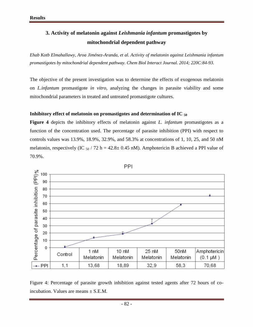
Results
- 82 -
3. Activity of melatonin against Leishmania infantum promastigotes by
mitochondrial dependent pathway
Ehab Kotb Elmahallawy, Aroa Jiménez-Aranda, et al. Activity of melatonin against Leishmania infantum
promastigotes by mitochondrial dependent pathway. Chem Biol Interact Journal. 2014; 220C:84-93.
The objective of the present investigation was to determine the effects of exogenous melatonin
on L.infantum promastigote in vitro, analyzing the changes in parasite viability and some
mitochondrial parameters in treated and untreated promastigote cultures.
Inhibitory effect of melatonin on promastigotes and determination of IC 50
Figure 4 depicts the inhibitory effects of melatonin against L. infantum promastigotes as a
function of the concentration used. The percentage of parasite inhibition (PPI) with respect to
controls values was 13.9%, 18.9%, 32.9%, and 58.3% at concentrations of 1, 10, 25, and 50 nM
melatonin, respectively (IC 50 / 72 h = 42.8± 0.45 nM). Amphotericin B achieved a PPI value of
70.9%.
Figure 4: Percentage of parasite growth inhibition against tested agents after 72 hours of co-
incubation. Values are means ± S.E.M.

Results
- 83 -
Alteration of Calcium Retention Capacity and sensitivity of mPTP opening
Figure 5 shows the effects of treatment with melatonin (at 1, 10, 25, or 50 nM) or with
amphotericin on calcium-induced mPTP opening of the isolated mitochondrial fractions in
comparison to negative and positive (CsA-treated) controls. Addition of Ca2+
or melatonin
stimulated the release of accumulated Ca2+
in all isolated mitochondrial fractions, and this
release was hindered by CsA. The AUC value was significantly higher for treatments with 25 or
50 nM melatonin or amphotericin than for treatments with lower melatonin concentrations or for
controls, indicating a greater mPTP opening, given that the mPTP opens earlier with lower Ca2+
retention capacity and vice versa.
Figure 5: Calcium-induced permeability transition pore (PTP) opening of control (untreated) and
amphotericin- and melatonin-treated promastigote cultures (1-50nM). Pulses of 250 μM Ca2+
were injected in the presence of 0.25 μM Oregon green until pore opening. 1 μM Cyclosporine A
(CsA) was added in vitro to the isolated mitochondria. PTP opening was calculated from the
Area under the Curve (AUC) of fluorescence signals versus time. Values are means ± S.E.M.
Superscript letters refer to significant differences measured using one-way ANOVA followed by
the Tukey post-hoc test. (* P < 0.05 in 25 nM melatonin-treated cultures; ** P < 0.01 in
amphotericin-treated cultures; and *** P <0.001 in CSA-treated mitochondria and 50 nM
melatonin-treated cultures, all versus control cultures.

Results
- 84 -
Melatonin increased Mitochondrial Nitrites Levels
As shown in Figure 6 A, the level of mitochondrial nitrites was significantly higher in the
cultures treated with the highest melatonin concentrations (25 or 50 nM) than in the untreated
cultures, reaching a similar level to that in the amphotericin-treated culture.
Melatonin non-significantly reduced SOD Activity
Figure 6 B depicts the mitochondrial SOD activity in treated and untreated cultures. Although
SOD activity was lower in cultures treated with melatonin (especially 50 nM) than in controls,
no significant difference was found with any melatonin concentration, whereas a significant
reduction was observed with amphotericin.
Melatonin impaired activities of respiratory complexes I, II and III
Clearly, the promastigote cultures treated with 25 or 50 nM melatonin or with amphotericin
showed significantly impaired complex I, II, and III activities in comparison to controls (Figures
7 A, B and C).

Results
- 85 -
Figure 6 A. Nitrites concentration in mitochondria isolated from control (untreated),
amphotericin, and melatonin-treated cultures.
Figure 6 B. Superoxide dismutase (SOD) activity in mitochondria isolated from control
(untreated), amphotericin-, and melatonin-treated cultures. Values are means ± S.E.M.
Superscript letters refer to significant differences measured using one-way ANOVA followed by
the Tukey post-hoc test (*P <0.05 in 25 nM melatonin-treated cultures compared with control;
*** P < 0.001 amphotericin and 50 nM melatonin-treated cultures versus control cultures).

Results
- 86 -
Figure 7 A, B, C. Activities of the electron transport chain complexes I, II, and III respectively, in
mitochondria isolated from untreated and treated promastigote cultures. Values are the means ±
S.E.M. Superscript letters refer to significant differences measured using one-way ANOVA
followed by the Tukey post-hoc test (Complex I : *** P < 0.001 for amphotericin- and 25 and 50
nM melatonin-treated cultures versus control cultures; complex II: * P <0.05 melatonin treated
cultures at concentrations of 25 and 50 nM compared with control cultures; and complex III: **
P < 0.01 for 25 nM melatonin-treated cultures; *** P < 0.001 for amphotericin- and 50 nM
melatonin-treated cultures versus control cultures).

- 87 -
Discussion

Discussion
- 88 -
1. General aspects
Leishmaniasis refers to a group of vector-borne diseases caused by an opportunistic intracellular
protozoan parasite of the genus Leishmania, belonging to the family Trypanasomatidae, order
Kinetoplastida [1, 282, 805]. The epidemiological profile of the disease include humans, rodents,
domestic and wild animals but the dogs are the most important reservoirs in a domestic
environment [44]. This clinically heterogeneous syndrome encompasses subclinical, localized
(skin lesion), and disseminated (cutaneous, mucocutaneous, and visceral) infection and the
spectrum of manifestations depends on the immune status of the host, on the parasite, and on
immunoinflammatory responses. The impact of the disease on public health has been increased
due to its zoonotic importance. Among 15 well-recognized species of subgenus Leishmania, 13
species are zoonotic [57, 282]. Furthermore, the distribution of disease has traditionally been
linked to tropical and subtropical regions besides being endemic in many areas worldwide such
as Mediterranean basin, East Africa and South America [1, 282].
The last decade has witnessed an extraordinary progress in the spread of the infection to new
areas, in particular due to global change and/or increases in global movements of hosts and
vectors [1, 167, 806-809]. This resulted in the appearance of new endemic disease foci together
with increasing reports of new reservoirs and the new transmission routes. On the other hand,
over the past decade, new formulations of standard drugs have become available and registered
for use in many countries. However, although around 25 compounds and formulations are
available to treat leishmaniasis in humans [373, 791], all are associated with toxicity and/or drug
resistance problems, and there is an urgent need to develop an effective drug against all forms of
leishmaniasis [299, 810]. In fact, many available antileishmanial agents can exert their effects
through the disruption of Ca2+
homeostasis in the parasite and /or through changes in different
mitochondrial parameters [714, 782].
With this background, the disease has spurred the interest of the scientific research towards the
role of several animal species as potential reservoirs for the infection and as consequence led to
the necessity for more surveying using a rapid and reliable diagnostic test and search for novel
less toxic antileishmanial drug, aiming to construct effective control measures adopted to
eliminate transmission and prevent new epidemics [1, 15, 780].

Discussion
- 89 -
In this regard, our intial goal was to assess the included a seroloprevalence of Leishmania
infection among transplanted organ recipients from southern Spain followed by investigation the
role of domestic cats from an endemic region in North-western Italy in Leishmania infantum
epidemiology, by the association of both serological and molecular tests. In the second phase of
our work, we have studied the effect of melatonin against the promastigote phase of the parasite
in vitro. The first results showed a relatively high prevalence of L.infantum was recorded among
kidney transplanted recipients. These novel findings in this area suggest that routine serological
testing for VL should be done before undergoing transplantation for both donor and recipient
transplant patients living or traveling in endemic areas.
Furthermore, we have found high prevalence of L.infantum among cats in the studied,
confirming the importance of cats not only as reservoir for the disease, but also the need for
further future research for accurate diagnosis of this zoonosis. In accordance with therapeutic
trials, melatonin not only demonstrated a significant antileishmanial activity against the
promastigote phase of the parasite in vitro but was also accompanied by an alteration of the
several mitochondrial parameters, including calcium homeostasis and by changes in some
mitochondrial parameters critical to parasite survival. In conclusion, the findings presented in
these papers represent the begining of novel strategy for surveying and combating leishmanisis.
2. Leishmaniasis among renal transplanted patients
As previously mentioned, VL is considered to be a well-established zoonotic disease in the area
of Mediterranean basin, especially in southwestern Europe, where approximately 700 cases per
year appear and the clinical form is frequently associated to immunosuppression[2, 277]. The
actual prevalence of leishmaniasis in Spain is not completely known [168], however, in our
geographical area, southern Spain, there are studies in Alpujarra Region (Granada province) and
Axarquia region (Malaga province) of rural population, using the leishmanin skin test (LST) (test
cell immunity), published in 1996 with average positivity rates exceeding 40% in both studies
[811, 812]. Nationwide, more recent studies have been made in areas close to ours, such as the
study performed on blood donors from Ibiza (Balearic Islands, Spain) in 2004 which indicated a
high rate of asymptomatic infection on the island and another study in 2008 from Balearic
Islands that confirms this high prevalence [813, 814]. Apart from these studies mentioned
previously, the most studied groups of population have been HIV infected individuals and

Discussion
- 90 -
intravenous drug users in our environment (southern Spain) [261, 815, 816]. There are not many
publications about the known prevalence of Leishmania in special groups such as transplant
recipients, oncological or other immunodeficient non HIV groups in Spain and the
Mediterranean region.
Leishmaniasis is not a common disease among transplant recipient patients but with this growing
pool of transplant survivors and increasing migration dynamics, the numbers of infected cases
among transplant recipients are in a steady increase especially among renal transplant recipients
mainly in southern Europe [817], which is considered to be an additional challenge [818, 819].
Transmission of VL to this specific group of population occurs through different routes:
Leishmania transmission by sandflies after transplantation when the specific group of population
is living in an endemic area or makes a short stay in an endemic area represents a route of
transmission [820, 821], also it may have originated from a donor with an undiagnosed infection,
or a recrudescence or to a latent infection in previously infected recipient during a period of
immunosuppression. Infection can also be acquired through blood transfusion [822].
Although VL is a rare disease among transplant patients, the current situation has been changed
and the number of published cases has increased in recent years[823]. More than 100 VL cases
following transplantation have been reported worldwide, predominantly described in renal
transplantation [819, 823, 824]. The prevalence of asymptomatic infections of Leishmania spp
varied from 0.6% to 71% in endemic areas [117]. To detect asymptomatic carriers, direct
method (PCR, culture, microscopic examination) or indirect methods (IFA test, direct
agglutination test, ELISA with single or crude Leishmania antigens, Western blot,
immunochromatographic techniques) can be used but there is not a gold standard test able to
identify asymptomatic infection with high sensitivity and specificity [117, 825].
The prevalence of Leishmania infection in our study is in agreement with previous studies in
different geographical areas of our country, Balearic Islands and Castilla León, with results of
prevalence of 3.1 % and 4.9% in blood donors and general population respectively [814, 826].
More recent studies, like the one published in the province of Seville on a small population that
included intravenous drug users and non-drug injectors, where the frequency of Leishmania–
seropositive was high, showed prevalence of infection by Leishmania of 24% using PCR-
ELISA. This study showed that a remarkable proportion of asymptomatic Leishmania

Discussion
- 91 -
seropositive individuals at risk for parenterally transmited infections carry Leishmania kDNA in
blood [261]. Some studies have been made in southern Spain in asymptomatic HIV population
diagnosing by amplification of Kinetoplast DNA (kDNA) from peripheral blood showing a
prevalence of 30.8% (28/92) infection by Leishmania. No patients showed positive results by
other techniques like ELISA, WB or leishmanin skin test. Conversely, patients with a negative
PCR result showed prevalence values respectively of 3.5%, 2.4% and 4.3% when tested by
ELISA , WB and LST [816] . In other endemic zones for L. infantum like Brazil, studies have
recently been published, which raise interest in the detection of asymptomatic infection in
transplant population living in the endemic areas as well as the need to use more sensitive
techniques for screening this population group, such as the research of Clemente et al. (2014) on
asymptomatic Leishmania infection among liver donors and recipients. The results of this study
show a prevalence of Leishmania infection of 1.5% using serological techniques which increased
to 7.5 %, 8.9 % and 5.9 % respectively in blood samples, liver samples and splenic samples
using molecular methods [824].
The results obtained in our study (4.8%) using a unique serological technique may be
underestimated, because IFAT has less sensibility than other indirect assays (ELISA)[117]. The
use of two serological methods could slightly increase the proportion of asymptomatic carriers
detected [827, 828]. On the other hand, antibody detection may not reflect a chronic infection but
more a recent contact with parasite followed by cure; PCR assay would have greater sensitivity
[829] than the indirect method and reflects actual parasitism rather than a previous infection by
Leishmania. Different studies highlight the added value of using a combination of tests
(molecular and serological) to increase the capacity to detect asymptomatic Leishmania
infections [117, 825]. It is at present not possible to predict exactly who and when among the
asymptomatically infected people will develop VL disease [830]. Reasons causing an infection
to remain asymptomatic or progress to VL are likely the result of a complex interaction between
environment, parasite and host related factors [831].
Studies of the risk of progression from infection to disease have yielded contradictory results
based on serological status, the data revealed strong associations between the magnitude of
positive serology and risk of progressing to symptomatic VL [832].

Discussion
- 92 -
International guidelines for the management of transplant recipients recommend specific
serology in donors and recipients from endemic areas, regardless of laboratory test limitations
and availability [833]. Given that patients such as transplant recipients are susceptible to develop
leishmaniasis, and the clinical manifestations in these patients can be serious, it seems advisable
to perform a screening of serological status in this population group and a more intense
monitoring of patients with positive serology with high titers in order to prevent possible
development of disease.
3. Role of cat in Leishmania infantum epidemiology
Our data shows that cats play an important role as L.infantum host, represented by higher
prevalence of Leishmania infantum among the examined cats either serologically using WB or
molecularly using PCR. This survey is the first epidemiological investigation performed about
leishmaniasis in cats in this endemic area in North-western Italy (Liguria). Likewise, it gives us
more evidences about the correlation between infection of cats with L. infantum as widely known
opportunistic pathogen and concomitant infection with some pathogens like FIV, FeLV and three
feline hemoplasma species that include: Mycoplasma haemofelis, Mycoplasma haemominutum,
and Mycoplasma turicensis.
As previously mentioned, cats rarely show clinical signs of VL and usually limited to skin ulcers.
A scant clinical cases appear with typical signs of cutaneous forms, including ulcer crusted
dermatitis and nodular lesions on the nose, lips, ears, and eyelids [834]. Other groups may
develop chronic ulceration, located particularly on the head and limbs [834]. In rare reported
cases of VL, the infected cases showed visceral involvement of liver, spleen, lymph nodes and
kidneys. Also, they are accompanied, almost always, with cutaneous manifestations [57, 63, 835,
836]; and there may also be local or generalized lymphadenopathy, ocular lesions, acute renal
failure, epistaxis, lameness, anemia, hepatosplenomegaly, icterus, lymphoplasmocytic
gastroenteritis with abundant Leishmania parasites, and membranous glomerulonephritis [834-
837]. Despite this previous fact stats that leishmaniasis is not common in cats, some
previous studies have examined the occurrence of feline leishmaniasis worldwide, especially in
several countries where zoonotic leishmaniasis is present like America, Brazil and Iran [66-70,
838, 839].

Discussion
- 93 -
In Europe, several clinical cases of feline leishmaniasis have been described since 1911 to date
in France, Greece, Switzerland, Spain, Italy, and Portugal, with very controversial results among
serological and molecular methods [63, 66, 67, 840-844]. In southern Europe where the disease
is endemic, in particular Italy, several sero-epidemiological studies have shown a prevalence of
L.infantum in cats ranged from 0.6% to 68%, which is differing among countries [62, 66, 839-
842, 845-850], while the positivity to PCR ranging from 3% to 61% [841, 851]. This may
explain the influence of geographical location on the epidemiological pattern of the disease and
as consequences variation in the efficiency of serological test used in each area.
Among other serological tests, western blotting is used to detect and quantify proteins that react
with a specific antibody. As reported in some previous studies with Leishmania, this test has
shown a good sensitivity, greater than that IFAT and ELISA, which make it recommended
mainly in doubtful cases besides its high specificity and sensitivity [852, 853]. Regarding our
results, we have reported positivity of 28.37% using PCR which is nearly similar with a previous
study using the same method [840]. Nevertheless, the reported prevalence rate, these findings
highlights the role played by cats in the transmission of disease, which has been confirmed
through sandflies infection contraction from cats naturally infected with Leishmania [854],
suggesting that cats could play an important role as secondary reservoir host for L. infantum.
In the present study, the prevalence of infection using WB yielded 13.20% which is either lower
or higher than previous reports in the same country or even worldwide; this may be attributed to
the difference of serological technique used as the majority of studies have used either ELISA
[62], IFAT [69, 840, 841, 845, 850] or even DAT [848], and may explain the role of WB as an
accurate test to diagnose the clinical disease than to detect the infection. Furthermore, a previous
study in Liguria and Tuscany has recorded much lower seroprevalence (0.9%) than our study
using IFAT [845], which could be attributed to lower sensitivity of IFAT compared to WB and
explains the possibility of other wild reservoirs increase [180, 855, 856] or even habit changes
and expansion range of sandfly vectors which resulted in appearance of new endemic foci for the
disease [806-809, 857]. As well, nearly similar results have been reported in some studies in the
same country using different serological technique [847]. Beside the difference in the serological
tests used, the difference in the prevalence rate between our study and the previous studies could
be attributed to the geographic location which affects the degree of endemicity of the disease.

Discussion
- 94 -
Taken into account, several serological studies have revealed that Leishmania-infected cats often
develop a low level of humoral response or remain seronegative [841, 845].
To our knowledge, the polymerase chain reaction (PCR) is widely accepted as a molecular tool
in epidemiological studies for identification and quantification of Leishmania spp. in various
tissues and body fluids of reservoir or hosts [69, 785, 858]. Therefore, several studies have been
recommended PCR as a highly efficient non-invasive tool for diagnosis and follow-up of the
disease [258, 859, 860]. Our results reported either lower, higher or even nearly similar
prevalence rate of L. infantum using PCR than in several previous studies [840, 841], which may
be also attributed to difference of the geographic location which affect the degree of endemicity
of the disease [1, 21, 57].
The role of some other pathogens like viral infection like FeLV or FIV or even Mycoplasma and
its association to Leishmania infection is still unclear [845]. Based upon our going results, there
was a statistical correlation between and positivity of sample to L. infantum using PCR and both
FIV, anemic status of animal, blood urea nitrogen level and some other hematological
parameters, whereas using WB, there was just association between positivity and Mycoplasma
infection; which is similar to associations previously reported between these parameters and
Leishmania infection [836, 845, 850, 861, 862]. This may suggest the role played some viruses
like FIV as a retroviral infection in development of some opportunistic pathogens like
Leishmania which is widely accepted [15]. However, it should be borne into mind that the only
presence of FIV is not sufficient to demonstrate an immunodeficiency status and this needs
additional immunological tests. On the other side, there was no a clear relationship between
infected cats with FeLV to contract the infection by Leishmania, which is consistent with several
previous results [62, 841, 842, 847, 849].
As shown, the use of direct methods (PCR) and indirect (WB) yielded discrepant results, which
has allowed to show that the prevalence of infection in cats with double direct method than the
serology. The explanation for this is not in the different sensitivity of the two methods, but rather
in the fact that in species resistant to infection as the cat, the immune response activated is
particularly of cell mediated than humoral immunity [861]. The data collected in our present
work confirms the hypothesis suggesting that the cat predominantly a Th1 response, as
evidenced by the high number infections (positivity to PCR) than the number of subjects with

Discussion
- 95 -
circulating antibodies (positive to WB) and the limited non-specific clinical signs occurrence in
animals in question [861, 863]. Also, our present data give more information about the
association between Leishmania infection and both retrooviruses and hemoplasmosis, which
seem to play a role in pathogenesis of disease or even contraction of the infection.
In conclusion, our study reports higher prevalence of L.infantum in cats which could be an
important parameter when further epidemiological studies show xenodiagnosis. This means the
infected cats are able to transmit the infection to the other reservoirs or vectors of the disease.
Based upon these findings, our study might suggest including a routine serological testing for
leishmaniasis in cats especially in endemic area. This contributes to the hypothesis that cats may
be a Leishmania reservoir. Further studies are also needed to evaluate the role of cats in the
epidemiology of L. infantum in the Mediterranean area. Other alternative prophylactic strategies
may be also essential to reduce the risk of infection and identification for new models of
leishmanial transmission.
4. Activity of melatonin against L. infantum promastigote
Our present work shows that exogenous melatonin reduces the number of viable L. infantum
promastigote accompanied by alteration of some mitochondrial parameters that are target for
parasite viability. This antileishmanial effect was associated with enhanced mPTP opening,
suggesting alterations in intracellular Ca2+
distribution, and also an increase in mitochondrial
nitrite levels and respiratory chain complex impairment. All of these effects may indirectly
compromise parasite survival, leading to cell death.
To our knowledge, no published data are available on the potential biocidal activity of melatonin
against Leishmania. Melatonin is known to regulate important physiological processes, including
pubertal development, sleep wake cycle and seasonal adaptation [730]. Various studies have
shown that melatonin has an antioxidant effect, a good pharmacological safety profile, and
represents a potentially useful tool for stand- alone or adjuvant therapy for many degenerative
and infectious diseases, including Chagas’s disease and several immune disorders [610, 627,
716, 772, 864-871].
Among other properties, melatonin has also shown biocidal activity against a wide range of
pathogenic agents including parasites [602, 660, 737]. As mentioned above, melatonin exert its

Discussion
- 96 -
effects against some pathogenic agents through interaction through its interaction with
intracellular proteins such as calreticulin [720], tubulin [721], or Calmodulin (CaM),
antagonizing the binding of Ca2+
to CaM [722, 736]. Thus, MT1 and MT2 are transmembrane G-
protein-coupled heterodimers whose signaling pathways lead to downstream effects on Ca2+
channels, Ca2+
signaling, and changes in mitogen-activated protein kinases and extracellular-
signal-regulated kinases [724], explaining the role of melatonin in the regulation of Ca2+
ion
fluxes in cells. However, it should be borne in mind that melatonin is also a very powerful free
radical scavenger and antioxidant, and these actions do not require a receptor [716, 800].
As noted above, trypanosomatids possess a single mitochondrion that occupies 12% of the total
volume of the parasite and has been recognized as drug target in many of these parasites [713].
In such cases, the antiparasitic agents target mitochondria through one or more of the following
programmed cell death mechanisms: intracellular Ca2+
levels alteration [782, 872]; loss of
membrane potential and ATP levels [771]; and increase of hydrogen peroxide (H2O2) and
superoxide radical generation (O2−) [873]. The mitochondrion is known to be an essential
organelle involved to be operative in the apoptotic pathway in Leishmania, but the main
mechanisms by which the protozoal mitochondrion is involved in parasite apoptosis are not yet
precisely understood [872]. Evidences have been published linking many leishmanicidal drugs
with mitochondrial dysfunction, and the mitochondrion has become established as a target for
antileishmanial drugs as in miltefosine and pentamidine [781, 874], which exert their effects via
multiple pathways.
Ca2+
homeostasis is a crucial matter in all organisms [875]. In common with most eukaryotes,
cell function in parasites is coordinated using second messenger signaling cascades involving
cyclic adenosine monophosphate (cAMP) and Ca2+
[676, 876]. Moreover, in the family
trypanosomatidae, Ca2+
is also involved in microtubule assembly, and differentiation processes,
among numerous other functions [675]. Calcium antagonists, such as miltefosine, have shown
antileishmanial activity by inducing a large increase in intracellular Ca2+
, thereby disrupting the
parasite’s intracellular Ca2+
homeostasis [781]. Other effective antileishmanial drugs against
promastigote and amastigote phase, such as tafenoquine and amiodarone, were found to be partly
localized in acidocalcisomes, important Ca2+
reservoirs in Leishmania; increasing the free
cytosolic Ca2+
level mobilized from intracellular stores; disrupting Ca2+
homeostasis, resulting in
mitochondrial damage [714, 782].

Discussion
- 97 -
Thus, fluctuations of cytosolic free Ca2+
concentrations may regulate a various cellular functions
in all eukaryotes. The lytic effect of trypanocidal drugs against Leishmania and African
trypanosomiasis was attributed to the alteration of calcium homeostasis produced by Ca2+
mobilization from intracellular stores during the progression of apoptosis [676, 877]. It has also
been proposed that cell damage results from the formation of reactive oxygen species (ROS)
induced by Ca2+
alterations [878]. The oxidative phosphorylation process produces an
accumulation of ROS such as H2O2 and O2− which are considered toxic products of
mitochondrial respiration and have been implicated in altered Ca2+
homeostasis in parasites [879,
880]. Accumulation of these products in in the mitochondrion mediates the oxidation of critical
thiol groups of adenine nucleotide translocase. This results in opening of the mPTP, which
causes collapse of mitochondrial membrane potential (ΔΨ m), loss of matrix solute,
mitochondrial membrane swelling, and indirectly enhance the release of death factors into the
cytosol, leading to cell death [881-883].
As shown in our results, a significantly higher mPTP opening were observed in melatonin-
treated cultures of L.infantum promastigotes, especially at a dose of 50 nM, than in control or
amphotericin-treated cultures, signifying a reduced Ca2+
retention capacity with an earlier PTP
opening. As expected; CsA had a blocking effect on mPTP opening. Interestingly, these findings
confirms the phenomenon stats that mPTP opening involved in respiratory chain inhibitor-
induced dissipation of ΔΨ m and corroborates with previous observations that PTP opening
induces ΔΨ m collapse through a proton dissipation pathway [884].
Clearly, our data also support the hypothesis that Leishmania parasite utilizes intracellular Ca2+
based signaling during maturation and suggests that melatonin may modulate the cell cycle of
Leishmania through its action on mPTP opening, calcium retention capacity, and the release of
intracellular Ca2+
, disrupting the Ca2+
homeostasis of the parasite [675]. This is considered the
most striking effect of melatonin treatment in our work as stimulation of the Ca2+
-induced
mPTP in mitochondria isolated from melatonin-treated cultures, which either alone or in
association with the other mitochondrial parameters, indirectly acts as an initiator of induced cell
death [883, 885-888]. Since mPTP is widely accepted be the most notorious of all the inner
membrane megachannels and is considered the gatekeeper of apoptotic and necrotic cell death
[883, 884, 888-892]. A previous study reported that increased calcium permeability is not
responsible for the rapid lethal effects of amphotericin on Leishmania [349]; however, a more

Discussion
- 98 -
recent study concluded that amphotericin produces osmotic cell lysis by formation of aqueous
pores in in the membrane of promastigotes [350], establishing the role of Ca2+
in amphotericin-
induced apoptosis through osmotic alteration, consistent with our results [303].
In accordance with mitochondrial nitrites (NO2−) level, results for the level of NO2
− suggest a
potential role for the mitochondrion in regulating nitrite concentrations, which are considered an
indicator for the stable breakdown product of NO in mitochondria. Several studies have revealed
a major role of NO in controlling L. major and T. cruzi multiplication in vivo and in vitro [760,
761]. Melatonin administered to T.cruzi-infected animals was found to increase NO production,
inhibiting parasite propagation or killing the parasite [736]. NO accumulation was also reported
to inhibit the mitochondrial respiratory chain, resulting in inhibition of ATP production, greater
ROS production, and increased the susceptibility to cell death. This cytotoxicity was preceded by
a decrease in ΔΨ m followed by an increase in O2− and peroxynitrite levels [893].
Our results are in the same line, with a significant increase in nitrate level in cultures treated with
25 or 50 nM melatonin or amphotericin versus the control cultures. At 1 or 10 nM melatonin,
however, the nitrite levels were non-significantly higher than those of control of control cultures.
These non-significant variations may be attributable to the antioxidant effect of melatonin at
these lower concentrations, hindering free radical production, given reports that melatonin and
its metabolites are effective direct scavengers of nitric oxide free radical (NO·) and peroxynitrite
anion (ONOO-) [894]. It should also be taken also into account that the induced nitrite levels in
the previous studies were in vivo and therefore mainly based on the immune system, in which the
activated macrophages were reported to synthesize NO, considered the major effector molecule
of T. cruzi intracellular amastigote killing [735, 760].
Several studies have reported that Leishmania species are susceptible to ROS and reactive
nitrogen species [895]. Three types of SOD have been identified: Cu/Zn-SOD, Mn-SOD, and Fe-
SOD [896]. They are considered one of defense enzymes, protecting the parasites from
mitochondrial-derived ROS damage and programmed cell death by detoxifying superoxide into
hydrogen peroxide and oxygen, thereby maintaining the integrity of the mitochondrion [897].
Mitochondrial Fe-SOD has been shown to protect the Leishmania parasite from oxidative stress,
and inhibit programmed cell death, as observed in miltefosine-induced cytotoxicity in L.
donovani [897, 898]. However, we found non-significantly lower Cu/Zn-SOD activities in
melatonin-treated cultures (all concentrations) than in control cultures. This lack of a significant

Discussion
- 99 -
difference may be attributable to the modest effect of Cu/Zn-SOD in Leishmania, the antioxidant
or protective effect of melatonin at lower concentrations, the parasite strain, among other
possibilities. Consistent with these findings, a recent study reported the negligible inhibition of
human Cu/Zn-SOD with pyrazole-based benzo[g]phthalazine derivatives on L. infantum and L.
braziliensis [899].
It is noteworthy that many available antileishmanial agents act by impairing of mitochondrial
complexes. Complexes I, III and IV are known to function as H+ ions (protons) pumps,
generating a proton electrochemical gradient that drives ATP synthesis, explaining why
mitochondrial complexes are the primary source of endogenous ROS [900]. Thus, inhibition of
these complexes by antileishmanial agents interferes with electron transport processes, resulting
in increased ROS from diverted electron flow, as in the case of antimycin A and tafenoquine
against L. donovani [782, 901].
In the present study, melatonin produced a significant inhibition of mitochondrial complexes I, II
and III, which was marked at the highest concentrations of melatonin (25 and 50 mM). However,
the activity of complex I is very low in Leishmania, and only the enzyme corresponding to
rotenone-insensitive NADH: quinone oxidoreductase has been observed in Trypanosoma and
Leishmania [902]. Therefore, these parasites possess only one NADH dehydrogenase gene in
their total DNA that may be essential for parasite survival; explaining its appeal as a drug target
[902]. Inhibition of complex I would result in generation of ROS and therefore leads to
mitochondrial dysfunction, which in turn triggers the apoptotic mitochondrial pathway [782,
902, 903]. The present results are in agreement with the previous findings on the antileishmanial
effect of luteolin resulted from reduction in the activities of electron transport chain complexes I,
II, III, and IV [904].
According to results, a drastically lowered activity of complex II on melatonin-treated cultures
versus control cultures was observed. This result correlates with report of induced death in
Leishmania using new Benzophenone-Derived Bisphosphonium Salts, which inhibit complex II
[905].
Several studies have described complex III as an attractive target for antiprotozoal drugs [904].
Tafenoquine was found to cause a mitochondrial dysfunction in Leishmania by complex III
(cytochrome c reductase) inhibition [782]. In these cases, the stoppage of electron transfer may
reduce the oxygen consumption rate, causing an imbalance of Ca2+
homeostasis and dissipating

Discussion
- 100 -
the ΔΨ m that is accompanied by production of O2−, triggering death of the parasite [782, 895,
901, 906]. It should be stressed that inhibition of Complexes I , II and III results in an
intracellular increase of Ca2+
that intimately associated with ROS generation, while the inhibition
of complex II markedly alters Ca2+
[895, 905].
These are highly promising findings, given that complex I inhibition may result on mitochondrial
hyperpolarization that is usually preceded by increased O2− production, while inhibition of
complexes II and III might cause dissipation of ΔΨ m. Both hyperpolarization and loss of ΔΨ m
might target the viability of the promastigotes [907, 908]. The inhibitory effect of melatonin on
these mitochondrial complexes support the proposition that Leishmania mitochondria respond
strongly to the inhibition of respiratory chain complexes [909].
The role of mitochondria in our findings may be initiated by the effects of melatonin on mPTP, a
key implication, through disruption of Ca2+
homeostasis which indirectly drives to lethal effects
resulting in cell death by apoptosis or necrosis [888]. As shown in the results, melatonin-treated
cultures depleted some respiratory components, probably due to the opening of mPTP.
Remarkably, impairment of activity of mitochondrial respiratory enzymes in treated cultures
may push the mitochondria towards mitochondrial oxidative stress expressed by inhibition of
electron transfer, together with inhibition of SOD and the resulted nitrosative stress which may
act collectively as a major inducer of mPTP opening, thereby indirectly drives to lethal effects by
apoptosis or necrosis [878, 910]. All of the above impressive changes in melatonin-treated
cultures are in part attributable to alterations in mPTP opening, accompanied by higher
mitochondrial nitrite levels and inhibition of the activity of mitochondrial complexes especially
II and III, and these events may indirectly trigger death of the parasite by apoptosis or necrosis.
The present results indicate that melatonin exerts in vitro activity on promastigote phase of
L.infantum, producing mitochondrial alterations in Ca2+
distribution, nitrite concentrations, and
complexes I, II and III, thereby targeting the parasite survival. Further research is warranted to
elucidate the in vivo effects of melatonin alone and in association with other antileishmanial
drugs in order to develop a novel chemotherapeutic strategy against the disease. There is also a
need to examine the mechanisms underlying these effects and the role of melatonin receptors.

- 101 -
Conclusions

Conclusions
- 102 -
Conclusions
1. According to the recorded prevalence in this study, transplant renal recipients are susceptible
population to develop Leishmaniasis which emphasizes the impact of the disease in our area.
Clearly, it is suggested to perform a dequete and intense monitoring in this type of patients with
high titers in order to prevent the development of the serious and fatal forms of leishmaniasis.
2. Given the limitations of serological techniques, a combination of serological tests together with
molecular techniques could of higher efficacy in such type of patients.
3. High prevalence of L.infantum in cats could be an important parameter when further
epidemiological studies show xenodiagnosis. This means the infected cats are able to transmit
the infection to the other reservoirs or vector of the disease.
4. Further efforts should be done to control feline leishmaniasis and the human disease in endemic
areas and other alternative prophylactic strategies may be also essential to reduce the risk of
infection and identification for new models of leishmanial transmission.
5. Melatonin exerts an interesting antileishmanial activity against the parasite in vitro. This
observation together with its high pharmacological safety profile make melatonin a potentially
useful tool for stand-alone or adjunct therapy for leishmaniasis. Hence, further studies are
suggested to investigate the effects of melatonin in vivo in association with other available
antileishmanial drugs, followed by investigation the role of receptores underlying these effects.

Conclusions
- 103 -
Conclusiones
1. En función de la prevalencia encontrada en este estudio, los receptores de trasplante renal son
una población susceptible de desarrollar leishmaniasis, siendo por tanto importante en nuestra
área llevar a cabo esta determinación en este tipo de pacientes y realizar una vigilancia más
adecuada en aquellos pacientes con títulos mas altos, para prevenir la posible aparición de
formas graves de leishmnia.
2. Dadas las limitaciones de las técnicas serológicas, el uso combinado de estas técnicas, junto con
técnicas de biología molecular podría mejorar el diagnóstico de leishmaniasis en este tipo de
pacientes.
3. La alta prevalencia de L. infantum en gatos podría ser un importante parámetro cuando estudios
epidemiológicos muestran xenodiagnosis. Esto significa que los gatos infectados pueden
transmitir la infección hacia otros reservorios o vectore de la enfermedad.
4. Deben realizarse más esfuerzos para controlar la leishmaniosis felina y la enfermedad humana en
las zonas endémicas y otras estrategias profilácticas alternativas puede ser también necesarias
para reducir el riesgo de infección y la identificación de nuevos modelos de transmisión de
Leishmania.
5. La melatonina ejerce una actividad anti-leishmaniosis interesante contra el parásito in vitro. Esta
observación, junto con su alto perfil de seguridad farmacológica hace a la melatonina una
herramienta potencialmente útil para la administración por sí sola o terapia adjunta para la
leishmaniasis. Por lo tanto es necesario más investigación en los efectos de la melatonina in
vivo y en asociación con otros fármacos contra Leishmania, seguidos del papel de los receptores
de melatonina en estos efectos y sus mecanismos subyacentes.

- 104 -

- 105 -

Bibliography
- 105 -
Bibliography
1. Clem A: A current perspective on leishmaniasis. J Glob Infect Dis 2010, 2(2):124-126.
2. Ready PD: Leishmaniasis emergence in Europe. Euro Surveill 2010, 15(10):19505.
3. Control of the leishmaniases. World Health Organization technical report series
2010(949):xii-xiii, 1-186, back cover.
4. WHO: Leishmaniasis Fact sheet N°375". World Health Organization. January 2014.
Retrieved 17 February 2014.
5. Barrett MP, Croft SL: Management of trypanosomiasis and leishmaniasis. British medical
bulletin 2012, 104:175-196.
6. Herwaldt BL: Leishmaniasis. Lancet 1999, 354(9185):1191-1199.
7. Lozano R, Naghavi M, Foreman K, Lim S, Shibuya K, Aboyans V, Abraham J, Adair T,
Aggarwal R, Ahn SY et al: Global and regional mortality from 235 causes of death for 20 age
groups in 1990 and 2010: a systematic analysis for the Global Burden of Disease Study 2010.
Lancet 2012, 380(9859):2095-2128.
8. Rabkin SW, Klassen GA: Session on sodium and calcium in the management of hypertension
summary of remarks of discussant and chairman. Can J Physiol Pharmacol 1986, 64(6):784-
785.
9. Chang and Bray R: Human Parasitic Diseases: Leishmaniasis, Elsevier Science Publishers,
Oxford, 1985.
10. Desjeux P: Human leishmaniases: epidemiology and public health aspects. World health
statistics quarterly Rapport trimestriel de statistiques sanitaires mondiales 1992, 45(2-3):267-
275.
11. Machado CM, Martins TC, Colturato I, Leite MS, Simione AJ, Souza MP, Mauad MA,
Colturato VR: Epidemiology of neglected tropical diseases in transplant recipients. Review of
the literature and experience of a Brazilian HSCT center. Revista do Instituto de Medicina
Tropical de Sao Paulo 2009, 51(6):309-324.
12. Alvar J, Canavate C, Gutierrez-Solar B, Jimenez M, Laguna F, Lopez-Velez R, Molina R,
Moreno J: Leishmania and human immunodeficiency virus coinfection: the first 10 years.
Clin Microbiol Rev 1997, 10(2):298-319.

Bibliography
- 106 -
13. Alvar J, Yactayo S, Bern C: Leishmaniasis and poverty. Trends in parasitology 2006,
22(12):552-557.
14. Desjeux P: Leishmaniasis: current situation and new perspectives. Comp Immunol Microbiol
Infect Dis 2004, 27(5):305-318.
15. Elmahallawy EK, Sampedro Martinez A, Rodriguez-Granger J, Hoyos-Mallecot Y, Agil A,
Navarro Mari JM, Gutierrez Fernandez J: Diagnosis of leishmaniasis. Journal of infection in
developing countries 2014, 8(8):961-972.
16. Chang K.P. B, R.S. : Human Parasitic Diseases: Leishmaniasis, Elsevier Science Publishers,
Oxford, 1985.
17. Cheng CY, Mruk DD: The blood-testis barrier and its implications for male contraception.
Pharmacological reviews 2012, 64(1):16-64.
18. Chappuis F, Sundar S, Hailu A, Ghalib H, Rijal S, Peeling RW, Alvar J, Boelaert M: Visceral
leishmaniasis: what are the needs for diagnosis, treatment and control? Nat Rev Microbiol
2007, 5(11):873-882.
19. Thakur CP, Kumar K: Post kala-azar dermal leishmaniasis: a neglected aspect of kala-azar
control programmes. Ann Trop Med Parasitol 1992, 86(4):355-359.
20. Das VN, Pandey K, Verma N, Lal CS, Bimal S, Topno RK, Singh D, Siddiqui NA, Verma
RB, Das P: Short report: Development of post-kala-azar dermal leishmaniasis (PKDL) in
miltefosine-treated visceral leishmaniasis. The American journal of tropical medicine and
hygiene 2009, 80(3):336-338.
21. Dujardin JC, Campino L, Canavate C, Dedet JP, Gradoni L, Soteriadou K, Mazeris A, Ozbel
Y, Boelaert M: Spread of vector-borne diseases and neglect of Leishmaniasis, Europe. Emerg
Infect Dis 2008, 14(7):1013-1018.
22. Gil-Prieto R, Walter S, Alvar J, de Miguel AG: Epidemiology of leishmaniasis in Spain based
on hospitalization records (1997-2008). Am J Trop Med Hyg 2011, 85(5):820-825.
23. Leishman WB: On the possibility of the occurrence of trypanomiasis in India". The British
Medical Journal; Donovan, C. (1903). "Memoranda: On the possibility of the occurrence of
trypanomiasis in India". . The British Medical Journal 1903.
24. Pulvertaft RJ, Hoyle GF: Stages in the life-cycle of Leishmania donovani. Transactions of the
Royal Society of Tropical Medicine and Hygiene 1960, 54:191-196.

Bibliography
- 107 -
25. Franco AM, Grimaldi G, Jr.: Characterization of Endotrypanum (Kinetoplastida:
Trypanosomatidae), a unique parasite infecting the neotropical tree sloths (Edentata).
Memorias do Instituto Oswaldo Cruz 1999, 94(2):261-268.
26. Momen H, Cupolillo E: Speculations on the origin and evolution of the genus Leishmania.
Memorias do Instituto Oswaldo Cruz 2000, 95(4):583-588.
27. Killick-Kendrick R: The life cycles of Leishmania in the sand fly and transmission of
leishmaniosis by bite. In: Canine leishmaniosis: moving towards a solution. . Proceedings of
the Second International Canine Leishmaniosis Forum Sevilla, Spain 2002, pp. 57-68.
28. Schuster FL, Sullivan JJ: Cultivating of clinically significant hemoflagellates. . Clinical
Microbiology Review, 2002, 15 (3):374-389.
29. Jennings YL, de Souza AA, Ishikawa EA, Shaw J, Lainson R, Silveira F: Phenotypic
characterization of Leishmania spp. causing cutaneous leishmaniasis in the lower Amazon
region, western Para state, Brazil, reveals a putative hybrid parasite, Leishmania (Viannia)
guyanensis x Leishmania (Viannia) shawi shawi. Parasite 2014, 21:39.
30. Myler P, f FN: Leishmania: After The Genome. Caister Academic Press. p. [page needed].
ISBN = 978-1-904455-28-8. 2008.
31. Harms G, U. B: Leishmaniosen-importierte Krankheiten. . Dtsch Arztebl 2000, 31/32:1589–
1592.
32. Schlein Y, Jacobson RL, Messer G: Leishmania infections damage the feeding mechanism of
the sandfly vector and implement parasite transmission by bite. Proceedings of the National
Academy of Sciences of the United States of America 1992, 89(20):9944-9948.
33. Killick-Kendrick R: Biology of Leishmania in phlebotomine sandflies. In: Biology of the
Kinetoplastida. W.H. R. Lumsden, D.A. Evans (eds), . New York, Academic Press 1979,
2:395-460.
34. Killick-Kendrick R: Leishmaniosis – an English disease of the future? . Bull Trop Med Int
Health 4, 5 1996.
35. Arce A, Estirado A, Ordobas M, Sevilla S, Garcia N, Moratilla L, de la Fuente S, Martinez
AM, Perez AM, Aranguez E et al: Re-emergence of leishmaniasis in Spain: community
outbreak in Madrid, Spain, 2009 to 2012. Euro surveillance : bulletin Europeen sur les
maladies transmissibles = European communicable disease bulletin 2013, 18(30):20546.

Bibliography
- 108 -
36. Baneth G, Koutinas AF, Solano-Gallego L, Bourdeau P, Ferrer L: Canine leishmaniosis - new
concepts and insights on an expanding zoonosis: part one. Trends in parasitology 2008,
24(7):324-330.
37. Cruz I, Morales MA, Noguer I, Rodriguez A, Alvar J: Leishmania in discarded syringes from
intravenous drug users. Lancet 2002, 359(9312):1124-1125.
38. Owens SD, Oakley DA, Marryott K, Hatchett W, Walton R, Nolan TJ, Newton A, Steurer F,
Schantz P, Giger U: Transmission of visceral leishmaniasis through blood transfusions from
infected English foxhounds to anemic dogs. J Am Vet Med Assoc 2001, 219(8):1076-1083.
39. Symmers WS: Leishmaniasis acquired by contagion: a case of marital infection in Britain.
Lancet 1960, 1(7116):127-132.
40. Chatterjee K: Dermal leishmanoid. In: Chatterjee KD Editor Parasitology, 12th edition. .
Calcutta: Chatterjee Medical Publishers 1980, 64-65.
41. Silva FL, Oliveira RG, Silva TM, Xavier MN, Nascimento EF, Santos RL: Venereal
transmission of canine visceral leishmaniasis. Veterinary parasitology 2009, 160(1-2):55-59.
42. Rosypal AC, Troy GC, Zajac AM, Frank G, Lindsay DS: Transplacental transmission of a
North American isolate of Leishmania infantum in an experimentally infected beagle. J
Parasitol 2005, 91(4):970-972.
43. Slappendel RJ, Teske E: 1999. A review of canine leishmaniosis presenting outside endemic
areas. In: Canine Leishmaniosis: an update. Proceeding of the International Canine
Leishmaniosis Forum, Killick-Kendrick (ed.), . Wiesbaden 1999: pp,54-59.
44. Gradoni L, Pozio E, Gramiccia M, Maroli M, Bettini S: Leishmaniasis in Tuscany (Italy): VII.
Studies on the role of the black rat, Rattus rattus, in the epidemiology of visceral
leishmaniasis. Transactions of the Royal Society of Tropical Medicine and Hygiene 1983,
77(4):427-431.
45. WHO: The leishmaniases. Report of a WHO Expert Committee. World Health Organization
technical report series 1984, 701:1-140.
46. Solano-Gallego L, Rodriguez-Cortes A, Trotta M, Zampieron C, Razia L, Furlanello T,
Caldin M, Roura X, Alberola J: Detection of Leishmania infantum DNA by fret-based real-
time PCR in urine from dogs with natural clinical leishmaniosis. Veterinary parasitology
2007, 147(3-4):315-319.

Bibliography
- 109 -
47. Miro G, Montoya A, Mateo M, Alonso A, Garcia S, Garcia A, Caballero MJ, Molina R: A
leishmaniosis surveillance system among stray dogs in the region of Madrid: ten years of
serodiagnosis (1996-2006). Parasitology research 2007, 101(2):253-257.
48. Zijlstra EE, Musa AM, Khalil EA, el-Hassan IM, el-Hassan AM: Post-kala-azar dermal
leishmaniasis. The Lancet Infectious diseases 2003, 3(2):87-98.
49. Oliva G, Foglia Manzillo V, Pagano A: [Canine leishmaniasis: evolution of the
chemotherapeutic protocols]. Parassitologia 2004, 46(1-2):231-234.
50. Ciaramella P, Oliva G, Luna RD, Gradoni L, Ambrosio R, Cortese L, Scalone A, Persechino
A: A retrospective clinical study of canine leishmaniasis in 150 dogs naturally infected by
Leishmania infantum. The Veterinary record 1997, 141(21):539-543.
51. Moritz A, Steuber S, Greiner M: Clinical follow-up examination after treatment of canine
leishmaniasis. The Tokai journal of experimental and clinical medicine 1998, 23(6):279-283.
52. Ready PD: Leishmaniasis emergence and climate change. Rev Sci Tech 2008, 27(2):399-412.
53. Petrovic Z, Borjoski A, Savin Z: Les resultats de recherches sur le réservoir de Leishmania
donovani dans une région endémique du Kala-azar. . Proc Europ Multicoll Parasti Trogir
1975. , 2, :97-98.
54. Di Bella C, Vitale F, Russo G, Greco A, Milazzo C, Aloise G, Cagnin M: Are rodents a
potential reservoir for Leishmania infantum in Italy? J Med Ecol 2003. , 7 (Suppl.) 125-129.
55. Beck A, Beck R, Kusak J, Gudan A, Martinkovic F, Artukovic B, Hohsteter M, Huber D,
Marinculic A, Grabarevic Z: A case of visceral leishmaniosis in a gray wolf (Canis lupus)
from Croatia. Journal of wildlife diseases 2008, 44(2):451-456.
56. Gibson-Corley KN, Hostetter JM, Hostetter SJ, Mullin K, Ramer-Tait AE, Boggiatto PM,
Petersen CA: Disseminated Leishmania infantum infection in two sibling foxhounds due to
possible vertical transmission. The Canadian veterinary journal La revue veterinaire
canadienne 2008, 49(10):1005-1008.
57. Gramiccia M, Gradoni L: The current status of zoonotic leishmaniases and approaches to
disease control. International journal for parasitology 2005, 35(11-12):1169-1180.
58. Ashford RW, Bettini S: Ecology and epidemiology: Old World. In: The biology of
Leishmaniases in Biology and Medicine. W. Peters, R. Killick-Kendrick (eds.), . 1987. , 1,
:365-424.

Bibliography
- 110 -
59. De Lima H, Rodriguez N, Barrios MA, Avila A, Canizales I, Gutierrez S: Isolation and
molecular identification of Leishmania chagasi from a bat (Carollia perspicillata) in
northeastern Venezuela. Memorias do Instituto Oswaldo Cruz 2008, 103(4):412-414.
60. Dedet JP, Gay F, Chatenay G: Isolation of Leishmania species from wild mammals in French
Guiana. Transactions of the Royal Society of Tropical Medicine and Hygiene 1989,
83(5):613-615.
61. Sobrino R, Ferroglio E, Oleaga A, Romano A, Millan J, Revilla M, Arnal MC, Trisciuoglio
A, Gortazar C: Characterization of widespread canine leishmaniasis among wild carnivores
from Spain. Veterinary parasitology 2008, 155(3-4):198-203.
62. Solano-Gallego L, Rodriguez-Cortes A, Iniesta L, Quintana J, Pastor J, Espada Y, Portus M,
Alberola J: Cross-sectional serosurvey of feline leishmaniasis in ecoregions around the
Northwestern Mediterranean. The American journal of tropical medicine and hygiene 2007,
76(4):676-680.
63. Mancianti F: [Feline leishmaniasis: what's the epidemiological role of the cat?].
Parassitologia 2004, 46(1-2):203-206.
64. Petersen CA: Leishmaniasis, an emerging disease found in companion animals in the United
States. Topics in companion animal medicine 2009, 24(4):182-188.
65. Grevot A, Jaussaud Hugues P, Marty P, Pratlong F, Ozon C, Haas P, Breton C, Bourdoiseau
G: Leishmaniosis due to Leishmania infantum in a FIV and FelV positive cat with a squamous
cell carcinoma diagnosed with histological, serological and isoenzymatic methods. Parasite
2005, 12(3):271-275.
66. Diakou A, Papadopoulos E, Lazarides K: Specific anti-Leishmania spp. antibodies in stray
cats in Greece. J Feline Med Surg 2009, 11(8):728-730.
67. Pennisi MG, Venza M, Reale S, Vitale F, Lo Giudice S: Case report of leishmaniasis in four
cats. Vet Res Commun 2004, 28 Suppl 1:363-366.
68. Figueiredo FB, Bonna IC, Nascimento LD, Costa T, Baptista C, Pacheco TM, Amendoeira
MR, Madeira Mde F: [Serological evaluation for detection of anti-Leishmania antibodies in
dogs and cats in the district of Santa Rita de Cassia, municipality of Barra Mansa, State of Rio
de Janeiro]. Revista da Sociedade Brasileira de Medicina Tropical 2009, 42(2):141-145.

Bibliography
- 111 -
69. Braga AR, Langoni H, Lucheis SB: Evaluation of canine and feline leishmaniasis by the
association of blood culture, immunofluorescent antibody test and polymerase chain reaction.
J Venom Anim Toxins Incl Trop Dis 2014, 20(1):5.
70. Trainor KE, Porter BF, Logan KS, Hoffman RJ, Snowden KF: Eight cases of feline cutaneous
leishmaniasis in Texas. Veterinary pathology 2010, 47(6):1076-1081.
71. Muller N, Welle M, Lobsiger L, Stoffel MH, Boghenbor KK, Hilbe M, Gottstein B, Frey CF,
Geyer C, von Bomhard W: Occurrence of Leishmania sp. in cutaneous lesions of horses in
Central Europe. Veterinary parasitology 2009, 166(3-4):346-351.
72. Williams AO, Mutinga J, Rodgers M: Leishmaniasis in a domestic goat in Kenya. Molecular
and cellular probes 1991, 5(5):319-325.
73. Mutinga MJ, Kihara SM, Lohding A, Mutero CM, Ngatia TA, Karanu F: Leishmaniasis in
Kenya: description of leishmaniasis of a domestic goat from Transmara, Narok District,
Kenya. Tropical medicine and parasitology : official organ of Deutsche Tropenmedizinische
Gesellschaft and of Deutsche Gesellschaft fur Technische Zusammenarbeit 1989, 40(2):91-96.
74. van der Lugt JJ, Carlyon JF, de Waal DT: Cutaneous leishmaniasis in a sheep. Journal of the
South African Veterinary Association 1992, 63(2):74-77.
75. Moraes-Silva E, Antunes FR, Rodrigues MS, da Silva Julião F, Dias-Lima AG, Lemos-de-
Sousa V, de Alcantara AC, Reis EA, Nakatani M, Badaró R et al: Domestic swine in a
visceral leishmaniasis endemic area produce antibodies against multiple Leishmania infantum
antigens but apparently resist to L. infantum infection. . Acta Trop 2006, 98(2)::176-182.
76. Anjili CO, Ngichabe CK, Mbati PA, Lugalia RM, Wamwayi HM, Githure JI: Experimental
infection of domestic sheep with culture-derived Leishmania donovani promastigotes.
Veterinary parasitology 1998, 74(2-4):315-318.
77. Dubey JP, Bwangamoi O, Courtney SP, Fritz DL: Leishmania-like protozoan associated with
dermatitis in cattle. The Journal of parasitology 1998, 84(4):865-867.
78. Brazil RP, Desterro MD, Nascimento SB, Macau RP: [Natural infection of a pig (Sus scrofa)
by Leishmania in a recent focus of cutaneous leishmaniasis on the Island of Sao Luis,
Maranhao]. Memorias do Instituto Oswaldo Cruz 1987, 82(1):145.
79. Mukhtar MM, Sharief AH, el Saffi SH, Harith AE, Higazzi TB, Adam AM, Abdalla HS:
Detection of antibodies to Leishmania donovani in animals in a kala-azar endemic region in

Bibliography
- 112 -
eastern Sudan: a preliminary report. Transactions of the Royal Society of Tropical Medicine
and Hygiene 2000, 94(1):33-36.
80. Vedovello Filho D, Jorge FA, Lonardoni MV, Teodoro U, Silveira TG: American cutaneous
leishmaniasis in horses from endemic areas in the north-central mesoregion of Parana state,
Brazil. Zoonoses and public health 2008, 55(3):149-155.
81. Lopes AP, Sousa S, Dubey JP, Ribeiro AJ, Silvestre R, Cotovio M, Schallig HD, Cardoso L,
Cordeiro-da-Silva A: Prevalence of antibodies to Leishmania infantum and Toxoplasma
gondii in horses from the north of Portugal. Parasites & vectors 2013, 6:178.
82. Quinnell RJ, Courtenay O: Transmission, reservoir hosts and control of zoonotic visceral
leishmaniasis. Parasitology 2009, 136(14):1915-1934.
83. Alexander B, de Carvalho RL, McCallum H, Pereira MH: Role of the domestic chicken
(Gallus gallus) in the epidemiology of urban visceral leishmaniasis in Brazil. Emerging
infectious diseases 2002, 8(12):1480-1485.
84. Sant'anna MR, Nascimento A, Alexander B, Dilger E, Cavalcante RR, Diaz-Albiter HM,
Bates PA, Dillon RJ: Chicken blood provides a suitable meal for the sand fly Lutzomyia
longipalpis and does not inhibit Leishmania development in the gut. Parasites & vectors
2010, 3(1):3.
85. Moreira ED, Jr., de Souza VM, Sreenivasan M, Lopes NL, Barreto RB, de Carvalho LP:
Peridomestic risk factors for canine leishmaniasis in urban dwellings: new findings from a
prospective study in Brazil. The American journal of tropical medicine and hygiene 2003,
69(4):393-397.
86. Noguera P, Rondon M, Nieves E: Effect of blood source on the survival and fecundity of the
sandfly Lutzomyia ovallesi Ortiz (Diptera: Psychodidae), vector of Leishmania. Biomedica :
revista del Instituto Nacional de Salud 2006, 26 Suppl 1:57-63.
87. Sacks D, Kamhawi S: Molecular aspects of parasite-vector and vector-host interactions in
leishmaniasis. Annual review of microbiology 2001, 55:453-483.
88. Schuster FL, Sullivan JJ: Cultivation of clinically significant hemoflagellates. Clinical
microbiology reviews 2002, 15(3):374-389.
89. Prevention) CCfDCa: Parasites home: leishmaniasis. Epidemiology & risk factors. Available
at http://www.cdc.gov/parasites/leishmaniasis/epi.html. Accessed April 11, 2014.

Bibliography
- 113 -
90. Murray HW, Berman JD, Davies CR, Saravia NG: Advances in leishmaniasis. Lancet 2005,
366(9496):1561-1577.
91. Cardo LJ, Rentas FJ, Ketchum L, Salata J, Harman R, Melvin W, Weina PJ, Mendez J, Reddy
H, Goodrich R: Pathogen inactivation of Leishmania donovani infantum in plasma and
platelet concentrates using riboflavin and ultraviolet light. Vox sanguinis 2006, 90(2):85-91.
92. Lawyer P.G., P.V. P: Leishmaniasis and trypanosomiasis. In: B.F. Eldridge and J.D. Edman
(eds). Medical Entomology. pp 231-298. Kluwer, The Netherlands. 2004.
93. World Health O: Research priorities for Chagas disease, human African trypanosomiasis and
leishmaniasis. World Health Organization technical report series 2012(975):v-xii, 1-100.
94. Bogdan C: Mechanisms and consequences of persistence of intracellular pathogens:
leishmaniasis as an example. Cellular microbiology 2008, 10(6):1221-1234.
95. Neuber H: Leishmaniasis. Journal der Deutschen Dermatologischen Gesellschaft = Journal
of the German Society of Dermatology : JDDG 2008, 6(9):754-765.
96. World Health O: Control of the leishmaniasis: report of a meeting of the WHO Expert
Committee on the Control of Leishmaniases, Geneva, World Health Organization (WHO)
(22–26 March 2010) Contract No.: no. 9492010.
97. Organization WH: Research priorities for Chagas disease, human African trypanosomiasis
and leishmaniasis. World Health Organization technical report series 2012(975):v-xii, 1-100.
98. McHugh CP, Melby PC, LaFon SG: Leishmaniasis in Texas: epidemiology and clinical
aspects of human cases. The American journal of tropical medicine and hygiene 1996,
55(5):547-555.
99. Lian LY, Pandalaneni SR, Todd PA, Martin VM, Burgoyne RD, Haynes LP: Demonstration
of binding of neuronal calcium sensor-1 to the cav2.1 p/q-type calcium channel. Biochemistry
2014, 53(38):6052-6062.
100. Organization. WH: Leishmaniasis: the disease and its epidemiology. Available at
http://www.who.int/leishmaniasis/disease_epidemiology/en/. Accessed April 10, 2014.
101. Prevention. CfDCa: Parasites home: leishmaniasis. Epidemiology & risk factors. Available at
http://www.cdc.gov/parasites/leishmaniasis/epi.html. Accessed April 11, 2014).
102. Ahluwalia S, Lawn SD, Kanagalingam J, Grant H, Lockwood DN: Mucocutaneous
leishmaniasis: an imported infection among travellers to central and South America. BMJ
2004, 329(7470):842-844.

Bibliography
- 114 -
103. Herwaldt BL, Stokes SL, Juranek DD: American cutaneous leishmaniasis in U.S. travelers.
Annals of internal medicine 1993, 118(10):779-784.
104. Ehehalt U, Schunk M, Jensenius M, van Genderen PJ, Gkrania-Klotsas E, Chappuis F,
Schlagenhauf P, Castelli F, Lopez-Velez R, Parola P et al: Leishmaniasis acquired by
travellers to endemic regions in Europe: a EuroTravNet multi-centre study. Travel medicine
and infectious disease 2014, 12(2):167-172.
105. Emeis M, Liesenfeld O, Henze G, Meredith S, Schoone G, Stuck B: [Imported visceral
leishmaniasis (kala azar) in a German infant]. Klinische Padiatrie 1995, 207(6):347-349.
106. Gautret P, Cramer JP, Field V, Caumes E, Jensenius M, Gkrania-Klotsas E, de Vries PJ,
Grobusch MP, Lopez-Velez R, Castelli F et al: Infectious diseases among travellers and
migrants in Europe, EuroTravNet 2010. Euro surveillance : bulletin Europeen sur les
maladies transmissibles = European communicable disease bulletin 2012, 17(26).
107. Choi CM, Lerner EA: Leishmaniasis as an emerging infection. The journal of investigative
dermatology Symposium proceedings / the Society for Investigative Dermatology, Inc [and]
European Society for Dermatological Research 2001, 6(3):175-182.
108. van Griensven J, Diro E: Visceral leishmaniasis. Infectious disease clinics of North America
2012, 26(2):309-322.
109. Lubbert C, Opitz BM, Harms-Zwingenberger G, Nietsch HH: [Fever, pancytopenia, and
splenomegaly 8 months after a trip to Majorca Island (Spain)]. Medizinische Klinik 2008,
103(1):29-35.
110. Schmid MB, Leichsenring M, Keller C, Hegasy G: [Pancytopenia, fever, and splenomegaly in
a 2-year-old boy]. Deutsche medizinische Wochenschrift 2009, 134(24):1274-1277.
111. Lainson R, Shaw JJ, Silveira FT, Fraiha H: Leishmaniasis in Brazil. XIX: visceral
leishmaniasis in the Amazon Region, and the presence of Lutzomyia longipalpis on the Island
of Marajo, Para State. Transactions of the Royal Society of Tropical Medicine and Hygiene
1983, 77(3):323-330.
112. Lainson R: The American leishmaniases: some observations on their ecology and
epidemiology. Transactions of the Royal Society of Tropical Medicine and Hygiene 1983,
77(5):569-596.
113. Aoun K, Amri F, Chouihi E, Haouas N, Bedoui K, Benikhlef R, Ghrab J, Babba H, Chahed
MK, Harrat Z et al: [Epidemiology of Leishmania (L.) infantum, L. major and L. killicki in

Bibliography
- 115 -
Tunisia: results and analysis of the identification of 226 human and canine isolates]. Bulletin
de la Societe de pathologie exotique 2008, 101(4):323-328.
114. Postigo JA: Leishmaniasis in the World Health Organization Eastern Mediterranean Region.
International journal of antimicrobial agents 2010, 36 Suppl 1:S62-65.
115. Lachaud L, Dedet JP, Marty P, Faraut F, Buffet P, Gangneux JP, Ravel C, Bastien P, Working
Group for the Notification of Human Leishmanioses in F: Surveillance of leishmaniases in
France, 1999 to 2012. Euro surveillance : bulletin Europeen sur les maladies transmissibles =
European communicable disease bulletin 2013, 18(29):20534.
116. Schantz PM, Steurer FJ, Duprey ZH, Kurpel KP, Barr SC, Jackson JE, Breitschwerdt EB,
Levy MG, Fox JC: Autochthonous visceral leishmaniasis in dogs in North America. Journal
of the American Veterinary Medical Association 2005, 226(8):1316-1322.
117. Michel G, Pomares C, Ferrua B, Marty P: Importance of worldwide asymptomatic carriers of
Leishmania infantum (L. chagasi) in human. Acta tropica 2011, 119(2-3):69-75.
118. Elmahallawy E.K, Cuadros-Moronta E, María Carmen Liébana-Martos MC, Rodriguez-
Granger JM, Sampedro-Martínez A, Agil A, Navarro-Marí JM, Gutiérrez-Fernández J, J. B-S:
Seroprevalence of Leishmania infection among asymptomatic renal transplant recipients from
southern Spain. JMM 2015.
119. Zanger P, Gabrysch S: Leishmaniasis in the era of tumor necrosis factor alpha antagonist
therapy--a research agenda for Europe. Euro surveillance : bulletin Europeen sur les maladies
transmissibles = European communicable disease bulletin 2013, 18(30):20542.
120. Killick-Kendrick R: Phlebotomine vectors of the leishmaniases: a review. Medical and
veterinary entomology 1990, 4(1):1-24.
121. Gradoni L: Epidemiological surveillance of leishmaniasis in the European Union: operational
and research challenges. Euro surveillance : bulletin Europeen sur les maladies
transmissibles = European communicable disease bulletin 2013, 18(30):20539.
122. Pace D: Leishmaniasis. The Journal of infection 2014, 69 Suppl 1:S10-18.
123. Aguado M, Espinosa P, Romero-Mate A, Tardio JC, Cordoba S, Borbujo J: Outbreak of
cutaneous leishmaniasis in Fuenlabrada, Madrid. Actas dermo-sifiliograficas 2013,
104(4):334-342.
124. Antoniou M, Gramiccia M, Molina R, Dvorak V, Volf P: The role of indigenous
phlebotomine sandflies and mammals in the spreading of leishmaniasis agents in the

Bibliography
- 116 -
Mediterranean region. Euro surveillance : bulletin Europeen sur les maladies transmissibles
= European communicable disease bulletin 2013, 18(30):20540.
125. Mauricio IL, Stothard JR, Miles MA: The strange case of Leishmania chagasi. Parasitology
today 2000, 16(5):188-189.
126. Singh S: Changing trends in the epidemiology, clinical presentation, and diagnosis of
Leishmania-HIV co-infection in India. International journal of infectious diseases : IJID :
official publication of the International Society for Infectious Diseases 2014, 29:103-112.
127. Mollalo A, Alimohammadi A, Shirzadi MR, Malek MR: Geographic information system-
based analysis of the spatial and spatio-temporal distribution of zoonotic cutaneous
leishmaniasis in Golestan Province, north-east of Iran. Zoonoses and public health 2015,
62(1):18-28.
128. Confalonieri UE, Margonari C, Quintao AF: Environmental change and the dynamics of
parasitic diseases in the Amazon. Acta tropica 2014, 129:33-41.
129. Bounoua L, Kahime K, Houti L, Blakey T, Ebi KL, Zhang P, Imhoff ML, Thome KJ, Dudek
C, Sahabi SA et al: Linking climate to incidence of zoonotic cutaneous leishmaniasis (L.
major) in pre-Saharan North Africa. International journal of environmental research and
public health 2013, 10(8):3172-3191.
130. Fischer D, Thomas SM, Beierkuhnlein C: Temperature-derived potential for the establishment
of phlebotomine sandflies and visceral leishmaniasis in Germany. Geospatial health 2010,
5(1):59-69.
131. Desjeux P: The increase in risk factors for leishmaniasis worldwide. Trans R Soc Trop Med
Hyg 2001, 95(3):239-243.
132. Desjeux P: Worldwide increasing risk factors for leishmaniasis. Medical microbiology and
immunology 2001, 190(1-2):77-79.
133. Gonzalez C, Paz A, Ferro C: Predicted altitudinal shifts and reduced spatial distribution of
Leishmania infantum vector species under climate change scenarios in Colombia. Acta
tropica 2014, 129:83-90.
134. Gonzalez C, Wang O, Strutz SE, Gonzalez-Salazar C, Sanchez-Cordero V, Sarkar S: Climate
change and risk of leishmaniasis in north america: predictions from ecological niche models
of vector and reservoir species. PLoS neglected tropical diseases 2010, 4(1):e585.

Bibliography
- 117 -
135. Trotz-Williams L, Gradoni L: Disease risks for the travelling pet Leishmaniasis.. In Practice
2003, 25,: 190–197.
136. al. BRe: Social epidemiology of leishmaniasis in the central jungle of Peru. In: Walton BC,
Wijeyaratne PM, Moddaber F, eds. Research on control strategies for the leishmaniases.
Proceedings of an international workshop held in Ottawa, Canada, 1-4 June, 1987 Ottawa,
Canada, International Development Research Centre (IDRC), 1988 1987.
137. Netto EM, Cuba CC, Costa JL, Barreto AC, Rosa C, Marsden PD: Recurrence of South
American tegumentary leishmaniasis. Lancet 1986, 1(8479):501.
138. Gomes Ade C, Yamamoto YI, Capinzaiki AN, Amaral NM, Guimaraes AJ: [Ecological
aspects of American cutaneous leishmaniasis. 9. Prevalence/incidence of the human infection
in the municipality of Pedro de Toledo and Miracatu, Sao Paulo, Brazil]. Revista do Instituto
de Medicina Tropical de Sao Paulo 1992, 34(2):149-158.
139. Tolezano JE: Ecoepidemiological aspects of American cutaneous leishmaniasis in the state of
Sao Paulo, Brazil. Memorias do Instituto Oswaldo Cruz 1994, 89(3):427-434.
140. Pupo Nogueira Neto J, Basso G, Cipoli AP, El Kadre L: American cutaneous leishmaniasis in
the State of Sao Paulo, Brazil--epidemiology in transformation. Annals of agricultural and
environmental medicine : AAEM 1998, 5(1):1-5.
141. da Costa SM, Cechinel M, Bandeira V, Zannuncio JC, Lainson R, Rangel EF: Lutzomyia
(Nyssomyia) whitmani s.l . (Antunes & Coutinho, 1939)(Diptera: Psychodidae:
Phlebotominae): geographical distribution and the epidemiology of American cutaneous
leishmaniasis in Brazil--mini-review. Memorias do Instituto Oswaldo Cruz 2007, 102(2):149-
153.
142. Davies CR, Kaye P, Croft SL, Sundar S: Leishmaniasis: new approaches to disease control.
BMJ 2003, 326(7385):377-382.
143. Tsegaw T, Gadisa E, Seid A, Abera A, Teshome A, Mulugeta A, Herrero M, Argaw D, Jorge
A, Aseffa A: Identification of environmental parameters and risk mapping of visceral
leishmaniasis in Ethiopia by using geographical information systems and a statistical
approach. Geospatial health 2013, 7(2):299-308.
144. Bhunia GS, Kesari S, Jeyaram A, Kumar V, Das P: Influence of topography on the
endemicity of Kala-azar: a study based on remote sensing and geographical information
system. Geospatial health 2010, 4(2):155-165.

Bibliography
- 118 -
145. Shimabukuro PH, da Silva TR, Ribeiro FO, Baton LA, Galati EA: Geographical distribution
of American cutaneous leishmaniasis and its phlebotomine vectors (Diptera: Psychodidae) in
the state of Sao Paulo, Brazil. Parasites & vectors 2010, 3:121.
146. Antinori S, Gianelli E, Calattini S, Longhi E, Gramiccia M, Corbellino M: Cutaneous
leishmaniasis: an increasing threat for travellers. Clinical microbiology and infection : the
official publication of the European Society of Clinical Microbiology and Infectious Diseases
2005, 11(5):343-346.
147. Pavli A, Maltezou HC: Leishmaniasis, an emerging infection in travelers. International
journal of infectious diseases : IJID : official publication of the International Society for
Infectious Diseases 2010, 14(12):e1032-1039.
148. Control of the leishmaniases. Report of a WHO Expert Committee. World Health
Organization technical report series 1990, 793:1-158.
149. World Health O: Control of the leishmaniases. World Health Organization technical report
series 2010(949):xii-xiii, 1-186, back cover.
150. Harrison LH, Naidu TG, Drew JS, de Alencar JE, Pearson RD: Reciprocal relationships
between undernutrition and the parasitic disease visceral leishmaniasis. Rev Infect Dis 1986,
8(3):447-453.
151. Schaefer KU, Kurtzhals JA, Sherwood JA, Githure JI, Kager PA, Muller AS: Epidemiology
and clinical manifestations of visceral and cutaneous leishmaniasis in Baringo District, Rift
Valley, Kenya. A literature review. Tropical and geographical medicine 1994, 46(3):129-133.
152. Guerin PJ, Olliaro P, Sundar S, Boelaert M, Croft SL, Desjeux P, Wasunna MK, Bryceson
AD: Visceral leishmaniasis: current status of control, diagnosis, and treatment, and a proposed
research and development agenda. The Lancet Infectious diseases 2002, 2(8):494-501.
153. Killick-Kendrick R: Breeding places of Phlebotomus ariasi in the cevennes focus of
leishmaniasis in the south of France. Parassitologia 1987, 29(2-3):181-191.
154. Feliciangeli MD: Natural breeding places of phlebotomine sandflies. Medical and veterinary
entomology 2004, 18(1):71-80.
155. Jeronimo SM, Duggal P, Ettinger NA, Nascimento ET, Monteiro GR, Cabral AP, Pontes NN,
Lacerda HG, Queiroz PV, Gomes CE et al: Genetic predisposition to self-curing infection
with the protozoan Leishmania chagasi: a genomewide scan. The Journal of infectious
diseases 2007, 196(8):1261-1269.

Bibliography
- 119 -
156. Jeronimo SM, Holst AK, Jamieson SE, Francis R, Martins DR, Bezerra FL, Ettinger NA,
Nascimento ET, Monteiro GR, Lacerda HG et al: Genes at human chromosome 5q31.1
regulate delayed-type hypersensitivity responses associated with Leishmania chagasi
infection. Genes and immunity 2007, 8(7):539-551.
157. Blackwell JM, Fakiola M, Ibrahim ME, Jamieson SE, Jeronimo SB, Miller EN, Mishra A,
Mohamed HS, Peacock CS, Raju M et al: Genetics and visceral leishmaniasis: of mice and
man. Parasite immunology 2009, 31(5):254-266.
158. Reithinger R, Dujardin JC, Louzir H, Pirmez C, Alexander B, Brooker S: Cutaneous
leishmaniasis. The Lancet Infectious diseases 2007, 7(9):581-596.
159. Petri WA Jr, R. H: Entamoeba species, including Amebiasis. In: Mandell GL, JE Bennett, R
Dolin, eds. Mandell, Douglas, and Bennett's principles and practice of infectious diseases, 7th
ed. Philadelphia: Churchill Livingstone; 2010. p. 3411–25. 2010.
160. Desjeux P, Alvar J: Leishmania/HIV co-infections: epidemiology in Europe. Ann Trop Med
Parasitol 2003, 97 Suppl 1:3-15.
161. (2011). WHO: Elimination visceral leishmaniasis: a multiple pronged approach. Available:
http://www.who.int/tdr/news/2011/vl-elimination/en/. Accessed May 2014.
162. Lawyer PG, Perkins PV: Leishmaniosis and trypanosomiasis. In: Medical Entomology,
Revised Edition, by B. F. Eldridge and J. D. Edman (eds.), Kluver Academic Publishers,
Great Britain, pp. 231-298. Kluver Academic Publishers, Great Britain, pp 231-298 2004.
163. Aoun K, Bouratbine A: Cutaneous leishmaniasis in North Africa: a review. Parasite 2014,
21:14.
164. Kennedy K: "VCS Advocacy in the News: VA May Designate 9 Infectious Diseases as
Related to Gulf War". Veterans for Common Sense. Retrieved 10 February 2011, (2010).
2010.
165. Kennedy K: "VCS Advocacy in the News: VA May Designate 9 Infectious Diseases as
Related to Gulf War". Veterans for Common Sense. Retrieved 10 February 2011. 2010.
166. Antoniou M, Haralambous C, Mazeris A, Pratlong F, Dedet JP, Soteriadou K: Leishmania
donovani leishmaniasis in Cyprus. The Lancet Infectious diseases 2008, 8(1):6-7.
167. Alvar J, Velez ID, Bern C, Herrero M, Desjeux P, Cano J, Jannin J, den Boer M, Team
WHOLC: Leishmaniasis worldwide and global estimates of its incidence. PloS one 2012,
7(5):e35671.

Bibliography
- 120 -
168. Botet Fregola J, Portus Vinyeta M: [Leishmaniasis in peninsular Spain. A historical-
bibliographic review (1912-1985)]. Rev Sanid Hig Publica (Madr) 1993, 67(4):255-266.
169. Monge-Maillo B, Norman FF, Cruz I, Alvar J, Lopez-Velez R: Visceral leishmaniasis and
HIV coinfection in the Mediterranean region. PLoS neglected tropical diseases 2014,
8(8):e3021.
170. Suarez Rodriguez B, Isidoro Fernandez B, Santos Sanz S, Sierra Moros MJ, Molina Moreno
R, Astray Mochales J, Amela Heras C: [Review of the current situation and the risk factors of
Leishmania infantum in Spain]. Revista espanola de salud publica 2012, 86(6):555-564.
171. Maroli M, Feliciangeli MD, Bichaud L, Charrel RN, Gradoni L: Phlebotomine sandflies and
the spreading of leishmaniases and other diseases of public health concern. Medical and
veterinary entomology 2013, 27(2):123-147.
172. Berens-Riha N, Fleischmann E, Pratlong F, Bretzel G, von Sonnenburg F, Loscher T:
Cutaneous leishmaniasis (Leishmania tropica) in a German tourist after travel to Greece.
Journal of travel medicine 2009, 16(3):220-222.
173. Wall EC, Watson J, Armstrong M, Chiodini PL, Lockwood DN: Epidemiology of imported
cutaneous leishmaniasis at the Hospital for Tropical Diseases, London, United Kingdom: use
of polymerase chain reaction to identify the species. The American journal of tropical
medicine and hygiene 2012, 86(1):115-118.
174. Malik AN, John L, Bruceson AD, Lockwood DN: Changing pattern of visceral leishmaniasis,
United Kingdom, 1985-2004. Emerging infectious diseases 2006, 12(8):1257-1259.
175. Weitzel T, Muhlberger N, Jelinek T, Schunk M, Ehrhardt S, Bogdan C, Arasteh K, Schneider
T, Kern WV, Fatkenheuer G et al: Imported leishmaniasis in Germany 2001-2004: data of the
SIMPID surveillance network. European journal of clinical microbiology & infectious
diseases : official publication of the European Society of Clinical Microbiology 2005,
24(7):471-476.
176. Bart A, van Thiel PP, de Vries HJ, Hodiamont CJ, Van Gool T: Imported leishmaniasis in the
Netherlands from 2005 to 2012: epidemiology, diagnostic techniques and sequence-based
species typing from 195 patients. Euro surveillance : bulletin Europeen sur les maladies
transmissibles = European communicable disease bulletin 2013, 18(30):20544.
177. Mansueto P, Seidita A, Vitale G, Cascio A: Leishmaniasis in travelers: a literature review.
Travel medicine and infectious disease 2014, 12(6 Pt A):563-581.

Bibliography
- 121 -
178. Franco AO, Davies CR, Mylne A, Dedet JP, Gallego M, Ballart C, Gramiccia M, Gradoni L,
Molina R, Galvez R et al: Predicting the distribution of canine leishmaniasis in western
Europe based on environmental variables. Parasitology 2011, 138(14):1878-1891.
179. Cortes S, Vaz Y, Neves R, Maia C, Cardoso L, Campino L: Risk factors for canine
leishmaniasis in an endemic Mediterranean region. Veterinary parasitology 2012, 189(2-
4):189-196.
180. Ruiz-Fons F, Ferroglio E, Gortazar C: Leishmania infantum in free-ranging hares, Spain,
2004-2010. Euro surveillance : bulletin Europeen sur les maladies transmissibles =
European communicable disease bulletin 2013, 18(30):20541.
181. Millan J, Ferroglio E, Solano-Gallego L: Role of wildlife in the epidemiology of Leishmania
infantum infection in Europe. Parasitology research 2014, 113(6):2005-2014.
182. Killick-Kendrick R: The biology and control of phlebotomine sand flies. Clinics in
dermatology 1999, 17(3):279-289.
183. Aransay AM, Testa JM, Morillas-Marquez F, Lucientes J, Ready PD: Distribution of sandfly
species in relation to canine leishmaniasis from the Ebro Valley to Valencia, northeastern
Spain. Parasitology research 2004, 94(6):416-420.
184. Martin-Sanchez J, Gallego M, Baron S, Castillejo S, Morillas-Marquez F: Pool screen PCR
for estimating the prevalence of Leishmania infantum infection in sandflies (Diptera:
Nematocera, Phlebotomidae). Transactions of the Royal Society of Tropical Medicine and
Hygiene 2006, 100(6):527-532.
185. Ntais P, Sifaki-Pistola D, Christodoulou V, Messaritakis I, Pratlong F, Poupalos G, Antoniou
M: Leishmaniases in Greece. The American journal of tropical medicine and hygiene 2013,
89(5):906-915.
186. Gkolfinopoulou K, Bitsolas N, Patrinos S, Veneti L, Marka A, Dougas G, Pervanidou D,
Detsis M, Triantafillou E, Georgakopoulou T et al: Epidemiology of human leishmaniasis in
Greece, 1981-2011. Euro surveillance : bulletin Europeen sur les maladies transmissibles =
European communicable disease bulletin 2013, 18(29):20532.
187. Frank C, Hadziandoniou M, Pratlong F, Garifallou A, Rioux JA: Leishmania tropica and
Leishmania infantum responsible for cutaneous leishmaniasis in Greece: sixteen
autochthonous cases. Transactions of the Royal Society of Tropical Medicine and Hygiene
1993, 87(2):184-185.

Bibliography
- 122 -
188. Santarém N, Silva Cd: Diagnosis of visceral leishmaniasis. In: Mendez-Vilas A, editor.
Communicating current research and educational topics and trends in applied microbiology. .
Badajoz: Formatex 2007:839-846.
189. Del Olmo Martinez L, Aller de la Fuente R, Velayos Jimenez B, Fernandez Salazar L,
Gonzalez Hernandez JM: [Visceral leishmaniasis diagnosed by duodenal biopsy]. Rev Esp
Enferm Dig 2009, 101(6):439-440.
190. Artan R, Yilmaz A, Akcam M, Aksoy NH: Liver biopsy in the diagnosis of visceral
leishmaniasis. J Gastroenterol Hepatol 2006, 21(1 Pt 2):299-302.
191. Reus M, Garcia B, Vazquez V, Morales D, Fuster M, Sola J: Visceral leishmaniasis: diagnosis
by ultrasound-guided fine needle aspiration of an axillary node. Br J Radiol 2005,
78(926):158-160.
192. Srivastava P, Dayama A, Mehrotra S, Sundar S: Diagnosis of visceral leishmaniasis. Trans R
Soc Trop Med Hyg 2011, 105(1):1-6.
193. Zijlstra EE, Ali MS, el-Hassan AM, el-Toum IA, Satti M, Ghalib HW, Kager PA: Kala-azar:
a comparative study of parasitological methods and the direct agglutination test in diagnosis.
Trans R Soc Trop Med Hyg 1992, 86(5):505-507.
194. Allahverdiyev AM, Bagirova M, Uzun S, Alabaz D, Aksaray N, Kocabas E, Koksal F: The
value of a new microculture method for diagnosis of visceral leishmaniasis by using bone
marrow and peripheral blood. Am J Trop Med Hyg 2005, 73(2):276-280.
195. Maurya R, Mehrotra S, Prajapati VK, Nylen S, Sacks D, Sundar S: Evaluation of blood agar
microtiter plates for culturing leishmania parasites to titrate parasite burden in spleen and
peripheral blood of patients with visceral leishmaniasis. J Clin Microbiol 2010, 48(5):1932-
1934.
196. De Colmenares M, Portus M, Riera C, Gallego M, Aisa MJ, Torras S, Munoz C: Short report:
detection of 72-75-kD and 123-kD fractions of Leishmania antigen in urine of patients with
visceral leishmaniasis. Am J Trop Med Hyg 1995, 52(5):427-428.
197. Sarkari B, Chance M, Hommel M: Antigenuria in visceral leishmaniasis: detection and partial
characterisation of a carbohydrate antigen. Acta tropica 2002, 82(3):339-348.
198. Attar ZJ, Chance ML, el-Safi S, Carney J, Azazy A, El-Hadi M, Dourado C, Hommel M:
Latex agglutination test for the detection of urinary antigens in visceral leishmaniasis. Acta
tropica 2001, 78(1):11-16.

Bibliography
- 123 -
199. El-Safi SH, Abdel-Haleem A, Hammad A, El-Basha I, Omer A, Kareem HG, Boelaert M,
Chance M, Hommel M: Field evaluation of latex agglutination test for detecting urinary
antigens in visceral leishmaniasis in Sudan. East Mediterr Health J 2003, 9(4):844-855.
200. Rijal S, Boelaert M, Regmi S, Karki BM, Jacquet D, Singh R, Chance ML, Chappuis F,
Hommel M, Desjeux P et al: Evaluation of a urinary antigen-based latex agglutination test in
the diagnosis of kala-azar in eastern Nepal. Tropical medicine & international health : TM &
IH 2004, 9(6):724-729.
201. Diro E, Techane Y, Tefera T, Assefa Y, Kebede T, Genetu A, Kebede Y, Tesfaye A, Ergicho
B, Gebre-Yohannes A et al: Field evaluation of FD-DAT, rK39 dipstick and KATEX (urine
latex agglutination) for diagnosis of visceral leishmaniasis in northwest Ethiopia. Trans R Soc
Trop Med Hyg 2007, 101(9):908-914.
202. Malaei S, Mohebali M, Akhoundi B, Z Z: Evaluation of latex agglutination test (Katex) for
the detection of urinary antigens in human visceral leishmaniasis. . J Tehran Un Med Sci
2006, 4:: 101-108.
203. Vilaplana C, Blanco S, Dominguez J, Gimenez M, Ausina V, C TU, Munoz C: Noninvasive
method for diagnosis of visceral leishmaniasis by a latex agglutination test for detection of
antigens in urine samples. J Clin Microbiol 2004, 42(4):1853-1854.
204. Boelaert M, El-Safi S, Hailu A, Mukhtar M, Rijal S, Sundar S, Wasunna M, Aseffa A, Mbui
J, Menten J et al: Diagnostic tests for kala-azar: a multi-centre study of the freeze-dried DAT,
rK39 strip test and KAtex in East Africa and the Indian subcontinent. Trans R Soc Trop Med
Hyg 2008, 102(1):32-40.
205. Boelaert M, Rijal S, Regmi S, Singh R, Karki B, Jacquet D, Chappuis F, Campino L, Desjeux
P, Le Ray D et al: A comparative study of the effectiveness of diagnostic tests for visceral
leishmaniasis. The American journal of tropical medicine and hygiene 2004, 70(1):72-77.
206. Iqbal J, Hira PR, Saroj G, Philip R, Al-Ali F, Madda PJ, Sher A: Imported visceral
leishmaniasis: diagnostic dilemmas and comparative analysis of three assays. Journal of
clinical microbiology 2002, 40(2):475-479.
207. Badaro R, Reed SG, Carvalho EM: Immunofluorescent antibody test in American visceral
leishmaniasis: sensitivity and specificity of different morphological forms of two Leishmania
species. The American journal of tropical medicine and hygiene 1983, 32(3):480-484.

Bibliography
- 124 -
208. Singh S, Sivakumar R: Recent advances in the diagnosis of leishmaniasis. J Postgrad Med
2003, 49(1):55-60.
209. Ryan JR, Smithyman AM, Rajasekariah GH, Hochberg L, Stiteler JM, Martin SK: Enzyme-
linked immunosorbent assay based on soluble promastigote antigen detects immunoglobulin
M (IgM) and IgG antibodies in sera from cases of visceral and cutaneous leishmaniasis.
Journal of clinical microbiology 2002, 40(3):1037-1043.
210. Rajasekariah GH, Ryan JR, Hillier SR, Yi LP, Stiteler JM, Cui L, Smithyman AM, Martin
SK: Optimisation of an ELISA for the serodiagnosis of visceral leishmaniasis using in vitro
derived promastigote antigens. Journal of immunological methods 2001, 252(1-2):105-119.
211. Smrkovski LL, Larson CL: Antigenic cross-reactivity between Mycobacterium bovis (BCG)
and Leishmania donovani. Infect Immun 1977, 18(2):561-562.
212. Choudhry A, Guru PY, Saxena RP, Tandon A, Saxena KC: Enzyme-linked immunosorbent
assay in the diagnosis of kala-azar in Bhadohi (Varanasi), India. Trans R Soc Trop Med Hyg
1990, 84(3):363-366.
213. Kumar R, Pai K, Pathak K, Sundar S: Enzyme-linked immunosorbent assay for recombinant
K39 antigen in diagnosis and prognosis of Indian visceral leishmaniasis. Clin Diagn Lab
Immunol 2001, 8(6):1220-1224.
214. Bray R: Immunodiagnosis of leishmaniasis. In Chang KP, Bray RS, editors. Leishmaniasis.
Amsterdam: Elsevier 1985:177-181.
215. Bray R: Immunodiagnosis of leishmaniasis. In Cohen S, Sadun EH, editors. Immunology of
parasitic infections. Oxford: Blackwell. . 1976:65-76.
216. Vinayak VK, Mahajan D, Sobti RC, Singla N, Sundar S: Anti-66 kDa antileishmanial
antibodies as specific immunodiagnostic probe for visceral leishmaniasis. Indian J Med Res
1994, 99:109-114.
217. Palatnik-de-Sousa CB, Gomes EM, Paraguai-de-Souza E, Palatnik M, Luz K, Borojevic R:
Leishmania donovani: titration of antibodies to the fucose-mannose ligand as an aid in
diagnosis and prognosis of visceral leishmaniasis. Transactions of the Royal Society of
Tropical Medicine and Hygiene 1995, 89(4):390-393.
218. Martin SK, Thuita-Harun L, Adoyo-Adoyo M, Wasunna KM: A diagnostic ELISA for
visceral leishmaniasis, based on antigen from media conditioned by Leishmania donovani
promastigotes. Ann Trop Med Parasitol 1998, 92(5):571-577.

Bibliography
- 125 -
219. Carvalho FA, Charest H, Tavares CA, Matlashewski G, Valente EP, Rabello A, Gazzinelli
RT, Fernandes AP: Diagnosis of American visceral leishmaniasis in humans and dogs using
the recombinant Leishmania donovani A2 antigen. Diagn Microbiol Infect Dis 2002,
43(4):289-295.
220. McCall LI, Zhang WW, Matlashewski G: Determinants for the development of visceral
leishmaniasis disease. PLoS Pathog 2013, 9(1):e1003053.
221. Rafati S, Hassani N, Taslimi Y, Movassagh H, Rochette A, Papadopoulou B: Amastin
peptide-binding antibodies as biomarkers of active human visceral leishmaniasis. Clin
Vaccine Immunol 2006, 13(10):1104-1110.
222. Burns JM, Jr., Shreffler WG, Benson DR, Ghalib HW, Badaro R, Reed SG: Molecular
characterization of a kinesin-related antigen of Leishmania chagasi that detects specific
antibody in African and American visceral leishmaniasis. Proceedings of the National
Academy of Sciences of the United States of America 1993, 90(2):775-779.
223. Badaro R, Benson D, Eulalio MC, Freire M, Cunha S, Netto EM, Pedral-Sampaio D,
Madureira C, Burns JM, Houghton RL et al: rK39: a cloned antigen of Leishmania chagasi
that predicts active visceral leishmaniasis. The Journal of infectious diseases 1996,
173(3):758-761.
224. Singh DP, Goyal RK, Singh RK, Sundar S, Mohapatra TM: In search of an ideal test for
diagnosis and prognosis of kala-azar. J Health Popul Nutr 2010, 28(3):281-285.
225. Zijlstra EE, Nur Y, Desjeux P, Khalil EA, El-Hassan AM, Groen J: Diagnosing visceral
leishmaniasis with the recombinant K39 strip test: experience from the Sudan. Trop Med Int
Health 2001, 6(2):108-113.
226. Houghton RL, Petrescu M, Benson DR, Skeiky YA, Scalone A, Badaro R, Reed SG, Gradoni
L: A cloned antigen (recombinant K39) of Leishmania chagasi diagnostic for visceral
leishmaniasis in human immunodeficiency virus type 1 patients and a prognostic indicator for
monitoring patients undergoing drug therapy. J Infect Dis 1998, 177(5):1339-1344.
227. Mohapatra TM, Singh DP, Sen MR, Bharti K, Sundar S: Compararative evaluation of rK9,
rK26 and rK39 antigens in the serodiagnosis of Indian visceral leishmaniasis. J Infect Dev
Ctries 2010, 4(2):114-117.
228. Sivakumar R, Sharma P, Chang KP, Singh S: Cloning, expression, and purification of a novel
recombinant antigen from Leishmania donovani. Protein Expr Purif 2006, 46(1):156-165.

Bibliography
- 126 -
229. Pattabhi S, Whittle J, Mohamath R, El-Safi S, Moulton GG, Guderian JA, Colombara D,
Abdoon AO, Mukhtar MM, Mondal D et al: Design, development and evaluation of rK28-
based point-of-care tests for improving rapid diagnosis of visceral leishmaniasis. PLoS Negl
Trop Dis 2010, 4(9).
230. Amorim AG, Carrington M, Miles MA, Barker DC, de Almeida ML: Identification of the C-
terminal region of 70 kDa heat shock protein from Leishmania (Viannia) braziliensis as a
target for the humoral immune response. Cell Stress Chaperones 1996, 1(3):177-187.
231. Perez-Alvarez MJ, Larreta R, Alonso C, Requena JM: Characterisation of a monoclonal
antibody recognising specifically the HSP70 from Leishmania. Parasitol Res 2001,
87(11):907-910.
232. Maache M, Azzouz S, Diaz de la Guardia R, Alvarez P, Gil R, de Pablos LM, Osuna A: Host
humoral immune response to Leishmania lipid-binding protein. Parasite Immunol 2005,
27(6):227-234.
233. Brito ME, Mendonca MG, Gomes YM, Jardim ML, Abath FG: Identification of potentially
diagnostic Leishmania braziliensis antigens in human cutaneous leishmaniasis by immunoblot
analysis. Clinical and diagnostic laboratory immunology 2000, 7(2):318-321.
234. Ravindran R, Anam K, Bairagi BC, Saha B, Pramanik N, Guha SK, Goswami RP, Banerjee
D, Ali N: Characterization of immunoglobulin G and its subclass response to Indian kala-azar
infection before and after chemotherapy. Infection and immunity 2004, 72(2):863-870.
235. Mary C, Lamouroux D, Dunan S, Quilici M: Western blot analysis of antibodies to
Leishmania infantum antigens: potential of the 14-kD and 16-kD antigens for diagnosis and
epidemiologic purposes. The American journal of tropical medicine and hygiene 1992,
47(6):764-771.
236. Santos-Gomes G, Gomes-Pereira S, Campino L, Araujo MD, Abranches P: Performance of
immunoblotting in diagnosis of visceral Leishmaniasis in human immunodeficiency virus-
Leishmania sp.-coinfected patients. Journal of clinical microbiology 2000, 38(1):175-178.
237. Cota GF, de Sousa MR, Demarqui FN, Rabello A: The diagnostic accuracy of serologic and
molecular methods for detecting visceral leishmaniasis in HIV infected patients: meta-
analysis. PLoS neglected tropical diseases 2012, 6(5):e1665.
238. Oskam L, Nieuwenhuijs JL, Hailu A: Evaluation of the direct agglutination test (DAT) using
freeze-dried antigen for the detection of anti-Leishmania antibodies in stored sera from

Bibliography
- 127 -
various patient groups in Ethiopia. Transactions of the Royal Society of Tropical Medicine
and Hygiene 1999, 93(3):275-277.
239. Fakhar M, Motazedian MH, Hatam GR, Asgari Q, Monabati FA, . KM: Comparative
performance of direct agglutination test indirect immunofluorescent antibody test polymerase
chain reaction, bone marrow aspiration method for diagnosis of Mediterranean visceral
leishmaniasis. . African J Microbiol 2012, 6:: 5777-5781.
240. Chappuis F, Rijal S, Soto A, Menten J, Boelaert M: A meta-analysis of the diagnostic
performance of the direct agglutination test and rK39 dipstick for visceral leishmaniasis. Bmj
2006, 333(7571):723.
241. Boelaert M, El Safi S, Mousa H, Githure J, Mbati P, Gurubacharya V, Shrestha J, Jacquet D,
De Muynck A, Le Ray D et al: Multi-centre evaluation of repeatability and reproducibility of
the direct agglutination test for visceral leishmaniasis. Tropical medicine & international
health : TM & IH 1999, 4(1):31-37.
242. Gomez-Ochoa P, Castillo JA, Lucientes J, Gascon M, Zarate JJ, Arbea JI, Larraga V,
Rodriguez C: Modified direct agglutination test for simplified serologic diagnosis of
leishmaniasis. Clin Diagn Lab Immunol 2003, 10(5):967-968.
243. Schoone GJ, Hailu A, Kroon CC, Nieuwenhuys JL, Schallig HD, Oskam L: A fast
agglutination screening test (FAST) for the detection of anti-Leishmania antibodies.
Transactions of the Royal Society of Tropical Medicine and Hygiene 2001, 95(4):400-401.
244. Hailu A, Schoone GJ, Diro E, Tesfaye A, Techane Y, Tefera T, Assefa Y, Genetu A, Kebede
Y, Kebede T et al: Field evaluation of a fast anti-Leishmania antibody detection assay in
Ethiopia. Trans R Soc Trop Med Hyg 2006, 100(1):48-52.
245. Akhoundi B, Mohebali M, Babakhan L, Edrissian GH, Eslami MB, Keshavarz H, Malekafzali
H: Rapid detection of human Leishmania infantum infection: a comparative field study using
the fast agglutination screening test and the direct agglutination test. Travel Med Infect Dis
2010, 8(5):305-310.
246. Pedras MJ, de Gouvea Viana L, de Oliveira EJ, Rabello A: Comparative evaluation of direct
agglutination test, rK39 and soluble antigen ELISA and IFAT for the diagnosis of visceral
leishmaniasis. Trans R Soc Trop Med Hyg 2008, 102(2):172-178.

Bibliography
- 128 -
247. Schallig HD, Schoone GJ, Kroon CC, Hailu A, Chappuis F, Veeken H: Development and
application of 'simple' diagnostic tools for visceral leishmaniasis. Med Microbiol Immunol
2001, 190(1-2):69-71.
248. Sundar S, Reed SG, Singh VP, Kumar PC, Murray HW: Rapid accurate field diagnosis of
Indian visceral leishmaniasis. Lancet 1998, 351(9102):563-565.
249. Sundar S, Pai K, Sahu M, Kumar V, Murray HW: Immunochromatographic strip-test
detection of anti-K39 antibody in Indian visceral leishmaniasis. Annals of tropical medicine
and parasitology 2002, 96(1):19-23.
250. Carvalho SF, Lemos EM, Corey R, Dietze R: Performance of recombinant K39 antigen in the
diagnosis of Brazilian visceral leishmaniasis. The American journal of tropical medicine and
hygiene 2003, 68(3):321-324.
251. Brandonisio O, Fumarola L, Maggi P, Cavaliere R, Spinelli R, Pastore G: Evaluation of a
rapid immunochromatographic test for serodiagnosis of visceral leishmaniasis. Eur J Clin
Microbiol Infect Dis 2002, 21(6):461-464.
252. Jelinek T, Eichenlaub S, Loscher T: Sensitivity and specificity of a rapid
immunochromatographic test for diagnosis of visceral leishmaniasis. European journal of
clinical microbiology & infectious diseases : official publication of the European Society of
Clinical Microbiology 1999, 18(9):669-670.
253. Ghatei MA, Hatam GR, Hossini MH, Sarkari B: Performance of latex agglutination test
(KAtex) in diagnosis of visceral leishmaniasis in Iran. Iran J Immunol 2009, 6(4):202-207.
254. Akhoundi B, Mohebali M, Shojaee S, Jalali M, Kazemi B, Bandehpour M, Keshavarz H,
Edrissian GH, Eslami MB, Malekafzali H et al: Rapid detection of human and canine visceral
leishmaniasis: assessment of a latex agglutination test based on the A2 antigen from
amastigote forms of Leishmania infantum. Exp Parasitol 2013, 133(3):307-313.
255. Piarroux R, Gambarelli F, Dumon H, Fontes M, Dunan S, Mary C, Toga B, Quilici M:
Comparison of PCR with direct examination of bone marrow aspiration, myeloculture, and
serology for diagnosis of visceral Leishmaniasis in immunocompromised patients. J Clin
Microbiol 1994, 32(3):746-749.
256. Pizzuto M, Piazza M, Senese D, Scalamogna C, Calattini S, Corsico L, Persico T, Adriani B,
Magni C, Guaraldi G et al: Role of PCR in diagnosis and prognosis of visceral leishmaniasis

Bibliography
- 129 -
in patients coinfected with human immunodeficiency virus type 1. J Clin Microbiol 2001,
39(1):357-361.
257. Lachaud L, Dereure J, Chabbert E, Reynes J, Mauboussin JM, Oziol E, Dedet JP, Bastien P:
Optimized PCR using patient blood samples for diagnosis and follow-up of visceral
Leishmaniasis, with special reference to AIDS patients. J Clin Microbiol 2000, 38(1):236-
240.
258. Antinori S, Calattini S, Longhi E, Bestetti G, Piolini R, Magni C, Orlando G, Gramiccia M,
Acquaviva V, Foschi A et al: Clinical use of polymerase chain reaction performed on
peripheral blood and bone marrow samples for the diagnosis and monitoring of visceral
leishmaniasis in HIV-infected and HIV-uninfected patients: a single-center, 8-year experience
in Italy and review of the literature. Clin Infect Dis 2007, 44(12):1602-1610.
259. Liarte DB, Mendonca IL, Luz FC, Abreu EA, Mello GW, Farias TJ, Ferreira AF, Millington
MA, Costa CH: QBC for the diagnosis of human and canine american visceral leishmaniasis:
preliminary data. Rev Soc Bras Med Trop 2001, 34(6):577-581.
260. da Silva ES, Gontijo CM, Pacheco Rda S, Brazil RP: Diagnosis of human visceral
leishmaniasis by PCR using blood samples spotted on filter paper. Genet Mol Res 2004,
3(2):251-257.
261. Martin-Sanchez J, Pineda JA, Morillas-Marquez F, Garcia-Garcia JA, Acedo C, Macias J:
Detection of Leishmania infantum kinetoplast DNA in peripheral blood from asymptomatic
individuals at risk for parenterally transmitted infections: relationship between polymerase
chain reaction results and other Leishmania infection markers. Am J Trop Med Hyg 2004,
70(5):545-548.
262. Wortmann G, Sweeney C, Houng HS, Aronson N, Stiteler J, Jackson J, Ockenhouse C: Rapid
diagnosis of leishmaniasis by fluorogenic polymerase chain reaction. Am J Trop Med Hyg
2001, 65(5):583-587.
263. Gangneux JP, Menotti J, Lorenzo F, Sarfati C, Blanche H, Bui H, Pratlong F, Garin YJ,
Derouin F: Prospective value of PCR amplification and sequencing for diagnosis and typing
of old world Leishmania infections in an area of nonendemicity. J Clin Microbiol 2003,
41(4):1419-1422.

Bibliography
- 130 -
264. Otero AC, da Silva VO, Luz KG, Palatnik M, Pirmez C, Fernandes O, Palatnik de Sousa CB:
Short report: occurrence of Leishmania donovani DNA in donated blood from seroreactive
Brazilian blood donors. Am J Trop Med Hyg 2000, 62(1):128-131.
265. Sreenivas G, Ansari NA, Kataria J, Salotra P: Nested PCR assay for detection of Leishmania
donovani in slit aspirates from post-kala-azar dermal Leishmaniasis Lesions. J Clin Microbiol
2004, 42(4):1777-1778.
266. Singh N, Curran MD, Rastogil AK, Middleton D, Sundar S: Diagnostic PCR with Leishmania
donovani specificity using sequences from the variable region of kinetoplast minicircle DNA.
Trop Med Int Health 1999, 4(6):448-453.
267. Osman OF, Oskam L, Zijlstra EE, Kroon NC, Schoone GJ, Khalil ET, El-Hassan AM, Kager
PA: Evaluation of PCR for diagnosis of visceral leishmaniasis. J Clin Microbiol 1997,
35(10):2454-2457.
268. De Doncker S, Hutse V, Abdellati S, Rijal S, Singh Karki BM, Decuypere S, Jacquet D, Le
Ray D, Boelaert M, Koirala S et al: A new PCR-ELISA for diagnosis of visceral
leishmaniasis in blood of HIV-negative subjects. Trans R Soc Trop Med Hyg 2005, 99(1):25-
31.
269. Saad AA, Ahmed NG, Osman OS, Al-Basheer AA, Hamad A, Deborggraeve S, Buscher P,
Schoone GJ, Schallig HD, Laurent T et al: Diagnostic accuracy of the Leishmania OligoC-
TesT and NASBA-Oligochromatography for diagnosis of leishmaniasis in Sudan. PLoS Negl
Trop Dis 2010, 4(8):e776.
270. Bossolasco S, Gaiera G, Olchini D, Gulletta M, Martello L, Bestetti A, Bossi L, Germagnoli
L, Lazzarin A, Uberti-Foppa C et al: Real-time PCR assay for clinical management of human
immunodeficiency virus-infected patients with visceral leishmaniasis. J Clin Microbiol 2003,
41(11):5080-5084.
271. Mary C, Faraut F, Lascombe L, Dumon H: Quantification of Leishmania infantum DNA by a
real-time PCR assay with high sensitivity. J Clin Microbiol 2004, 42(11):5249-5255.
272. WHO: World Health Organization: HIV/AIDS fact sheet nº 360. Available:
http://www.who.int/mediacentre/factsheets/fs360/en/. Accessed 5 May 2014. 2013.
273. Shafiei R, Mohebali M, Akhoundi B, Galian MS, Kalantar F, Ashkan S, Fata A, Farash BR,
Ghasemian M: Emergence of co-infection of visceral leishmaniasis in HIV-positive patients
in northeast Iran: a preliminary study. Travel Med Infect Dis 2014, 12(2):173-178.
Bibliography
- 131 -
274. Sinha PK, van Griensven J, Pandey K, Kumar N, Verma N, Mahajan R, Kumar P, Kumar R,
Das P, Mitra G et al: Liposomal amphotericin B for visceral leishmaniasis in human
immunodeficiency virus-coinfected patients: 2-year treatment outcomes in Bihar, India. Clin
Infect Dis 2011, 53(7):e91-98.
275. ter Horst R, Tefera T, Assefa G, Ebrahim AZ, Davidson RN, Ritmeijer K: Field evaluation of
rK39 test and direct agglutination test for diagnosis of visceral leishmaniasis in a population
with high prevalence of human immunodeficiency virus in Ethiopia. Am J Trop Med Hyg
2009, 80(6):929-934.
276. WHO: World Health Organization (2007) Report of the Fifth Consultative Meeting on
Leishmania /HIV Co-infection Addis Ababa, Ethiopia, 20–22 March 2007. Available:
http://www.who.int/leishmaniasis/resources/Leishmaniasis_hiv_coinfection5.pdf2. Accessed
May 2014. 2014.
277. Fernandez-Guerrero ML, Aguado JM, Buzon L, Barros C, Montalban C, Martin T, Bouza E:
Visceral leishmaniasis in immunocompromised hosts. Am J Med 1987, 83(6):1098-1102.
278. Mondain-Miton V, Toussaint-Gari M, Hofman P, Marty P, Carles M, De Salvador F, Miton F,
Le Fichoux Y, Dellamonica P: Atypical leishmaniasis in a patient infected with human
immunodeficiency virus. Clin Infect Dis 1995, 21(3):663-665.
279. Albrecht H: Leishmaniosis--new perspectives on an underappreciated opportunistic infection.
AIDS 1998, 12(16):2225-2226.
280. Rosenthal E, Marty P, le Fichoux Y, Cassuto JP: Clinical manifestations of visceral
leishmaniasis associated with HIV infection: a retrospective study of 91 French cases. Ann
Trop Med Parasitol 2000, 94(1):37-42.
281. Carnauba D, Jr., Konishi CT, Petri V, Martinez IC, Shimizu L, Pereira-Chioccola VL:
Atypical disseminated leishmaniasis similar to post-kala-azar dermal leishmaniasis in a
Brazilian AIDS patient infected with Leishmania (Leishmania) infantum chagasi: a case
report. Int J Infect Dis 2009, 13(6):e504-507.
282. WHO: Control of the leishmaniases. World Health Organ Tech Rep Ser 2010(949):xii-xiii, 1-
186, back cover.
283. Bumb RA, Prasad N, Khandelwal K, Aara N, Mehta RD, Ghiya BC, Salotra P, Wei L, Peters
S, Satoskar AR: Long-term efficacy of single-dose radiofrequency-induced heat therapy vs.

Bibliography
- 132 -
intralesional antimonials for cutaneous leishmaniasis in India. Br J Dermatol 2013,
168(5):1114-1119.
284. Shamsi Meymandi S, Zandi S, Aghaie H, Heshmatkhah A: Efficacy of CO(2) laser for
treatment of anthroponotic cutaneous leishmaniasis, compared with combination of
cryotherapy and intralesional meglumine antimoniate. J Eur Acad Dermatol Venereol 2011,
25(5):587-591.
285. Negera E, Gadisa E, Hussein J, Engers H, Kuru T, Gedamu L, Aseffa A: Treatment response
of cutaneous leishmaniasis due to Leishmania aethiopica to cryotherapy and generic sodium
stibogluconate from patients in Silti, Ethiopia. Trans R Soc Trop Med Hyg 2012, 106(8):496-
503.
286. Asilian A, Sadeghinia A, Faghihi G, Momeni A: Comparative study of the efficacy of
combined cryotherapy and intralesional meglumine antimoniate (Glucantime) vs. cryotherapy
and intralesional meglumine antimoniate (Glucantime) alone for the treatment of cutaneous
leishmaniasis. Int J Dermatol 2004, 43(4):281-283.
287. Gonzalez U, Pinart M, Reveiz L, Alvar J: Interventions for Old World cutaneous
leishmaniasis. The Cochrane database of systematic reviews 2008(4):CD005067.
288. Garcia Bustos MF, Barrio AB, Parodi Ramoneda CM, Ramos F, Mora MC, Convit J,
Basombrio MA: Immunological correlates of cure in the first American cutaneous
leishmaniasis patient treated by immunotherapy in Argentina. A case report. Invest Clin 2011,
52(4):365-375.
289. Badaro R, Lobo I, Munos A, Netto EM, Modabber F, Campos-Neto A, Coler RN, Reed SG:
Immunotherapy for drug-refractory mucosal leishmaniasis. J Infect Dis 2006, 194(8):1151-
1159.
290. Almeida RP, Brito J, Machado PL, AR DEJ, Schriefer A, Guimaraes LH, Carvalho EM:
Successful treatment of refractory cutaneous leishmaniasis with GM-CSF and antimonials.
Am J Trop Med Hyg 2005, 73(1):79-81.
291. Badaro R, Lobo I, Nakatani M, Muinos A, Netto EM, Coler RN, Reed SG: Successful use of
a defined antigen/GM-CSF adjuvant vaccine to treat mucosal Leishmaniasis refractory to
antimony: A case report. Braz J Infect Dis 2001, 5(4):223-232.
292. Convit J, Ulrich M, Polegre MA, Avila A, Rodriguez N, Mazzedo MI, Blanco B: Therapy of
Venezuelan patients with severe mucocutaneous or early lesions of diffuse cutaneous

Bibliography
- 133 -
leishmaniasis with a vaccine containing pasteurized Leishmania promastigotes and bacillus
Calmette-Guerin: preliminary report. Mem Inst Oswaldo Cruz 2004, 99(1):57-62.
293. Da-Cruz AM, de Oliveira MP, De Luca PM, Mendonca SC, Coutinho SG: Tumor necrosis
factor-alpha in human american tegumentary leishmaniasis. Mem Inst Oswaldo Cruz 1996,
91(2):225-229.
294. Machado PR, Lessa H, Lessa M, Guimaraes LH, Bang H, Ho JL, Carvalho EM: Oral
pentoxifylline combined with pentavalent antimony: a randomized trial for mucosal
leishmaniasis. Clin Infect Dis 2007, 44(6):788-793.
295. Sundar S, Singh VP, Sharma S, Makharia MK, Murray HW: Response to interferon-gamma
plus pentavalent antimony in Indian visceral leishmaniasis. J Infect Dis 1997, 176(4):1117-
1119.
296. Mishra J, Saxena A, Singh S: Chemotherapy of leishmaniasis: past, present and future.
Current medicinal chemistry 2007, 14(10):1153-1169.
297. Sundar S, Chatterjee M: Visceral leishmaniasis - current therapeutic modalities. Indian J Med
Res 2006, 123(3):345-352.
298. Alvar J, Croft S, Olliaro P: Chemotherapy in the treatment and control of leishmaniasis. Adv
Parasitol 2006, 61:223-274.
299. Murray HW: Treatment of visceral leishmaniasis (kala-azar): a decade of progress and future
approaches. Int J Infect Dis 2000, 4(3):158-177.
300. Faraut-Gambarelli F, Piarroux R, Deniau M, Giusiano B, Marty P, Michel G, Faugere B,
Dumon H: In vitro and in vivo resistance of Leishmania infantum to meglumine antimoniate:
a study of 37 strains collected from patients with visceral leishmaniasis. Antimicrob Agents
Chemother 1997, 41(4):827-830.
301. Frezard F, Demicheli C, Ribeiro RR: Pentavalent antimonials: new perspectives for old drugs.
Molecules 2009, 14(7):2317-2336.
302. Sundar S, Chakravarty J: Leishmaniasis: an update of current pharmacotherapy. Expert
opinion on pharmacotherapy 2013, 14(1):53-63.
303. Mbongo N, Loiseau PM, Billion MA, Robert-Gero M: Mechanism of amphotericin B
resistance in Leishmania donovani promastigotes. Antimicrob Agents Chemother 1998,
42(2):352-357.

Bibliography
- 134 -
304. Sundar S, Singh A, Rai M, Prajapati VK, Singh AK, Ostyn B, Boelaert M, Dujardin JC,
Chakravarty J: Efficacy of miltefosine in the treatment of visceral leishmaniasis in India after
a decade of use. Clin Infect Dis 2012, 55(4):543-550.
305. Miguel DC, Zauli-Nascimento RC, Yokoyama-Yasunaka JK, Pereira LI, Jeronimo SM,
Ribeiro-Dias F, Dorta ML, Uliana SR: Clinical isolates of New World Leishmania from
cutaneous and visceral leishmaniasis patients are uniformly sensitive to tamoxifen. Int J
Antimicrob Agents 2011, 38(1):93-94.
306. Garcia-Hernandez R, Manzano JI, Castanys S, Gamarro F: Leishmania donovani develops
resistance to drug combinations. PLoS Negl Trop Dis 2012, 6(12):e1974.
307. Sundar S, Sinha PK, Rai M, Verma DK, Nawin K, Alam S, Chakravarty J, Vaillant M, Verma
N, Pandey K et al: Comparison of short-course multidrug treatment with standard therapy for
visceral leishmaniasis in India: an open-label, non-inferiority, randomised controlled trial.
Lancet 2011, 377(9764):477-486.
308. van Griensven J, Balasegaram M, Meheus F, Alvar J, Lynen L, Boelaert M: Combination
therapy for visceral leishmaniasis. Lancet Infect Dis 2010, 10(3):184-194.
309. Drugs for parasitic infections. Med Lett Drugs Ther 1974, 16(2):5-12.
310. Singh RK, Pandey HP, Sundar S: Visceral leishmaniasis (kala-azar): challenges ahead. Indian
J Med Res 2006, 123(3):331-344.
311. Berman JD: Human leishmaniasis: clinical, diagnostic, and chemotherapeutic developments
in the last 10 years. Clinical infectious diseases : an official publication of the Infectious
Diseases Society of America 1997, 24(4):684-703.
312. Ephros M, Bitnun A, Shaked P, Waldman E, Zilberstein D: Stage-specific activity of
pentavalent antimony against Leishmania donovani axenic amastigotes. Antimicrob Agents
Chemother 1999, 43(2):278-282.
313. Herwaldt BL, Berman JD: Recommendations for treating leishmaniasis with sodium
stibogluconate (Pentostam) and review of pertinent clinical studies. The American journal of
tropical medicine and hygiene 1992, 46(3):296-306.
314. Wortmann G, Miller RS, Oster C, Jackson J, Aronson N: A randomized, double-blind study
of the efficacy of a 10- or 20-day course of sodium stibogluconate for treatment of cutaneous
leishmaniasis in United States military personnel. Clin Infect Dis 2002, 35(3):261-267.

Bibliography
- 135 -
315. Ritmeijer K, Veeken H, Melaku Y, Leal G, Amsalu R, Seaman J, Davidson RN: Ethiopian
visceral leishmaniasis: generic and proprietary sodium stibogluconate are equivalent; HIV co-
infected patients have a poor outcome. Trans R Soc Trop Med Hyg 2001, 95(6):668-672.
316. Moore E, O'Flaherty D, Heuvelmans H, Seaman J, Veeken H, de Wit S, Davidson RN:
Comparison of generic and proprietary sodium stibogluconate for the treatment of visceral
leishmaniasis in Kenya. Bull World Health Organ 2001, 79(5):388-393.
317. Baiocco P, Colotti G, Franceschini S, Ilari A: Molecular basis of antimony treatment in
leishmaniasis. J Med Chem 2009, 52(8):2603-2612.
318. Thakur CP, Kumar M, Pandey AK: Comparison of regimes of treatment of antimony-resistant
kala-azar patients: a randomized study. Am J Trop Med Hyg 1991, 45(4):435-441.
319. Wyllie S, Cunningham ML, Fairlamb AH: Dual action of antimonial drugs on thiol redox
metabolism in the human pathogen Leishmania donovani. J Biol Chem 2004, 279(38):39925-
39932.
320. Ashutosh, Sundar S, Goyal N: Molecular mechanisms of antimony resistance in Leishmania.
J Med Microbiol 2007, 56(Pt 2):143-153.
321. Pathak MK, Yi T: Sodium stibogluconate is a potent inhibitor of protein tyrosine
phosphatases and augments cytokine responses in hemopoietic cell lines. J Immunol 2001,
167(6):3391-3397.
322. Lima MI, Arruda VO, Alves EV, de Azevedo AP, Monteiro SG, Pereira SR: Genotoxic
effects of the antileishmanial drug Glucantime. Arch Toxicol 2010, 84(3):227-232.
323. Torres DC, Adaui V, Ribeiro-Alves M, Romero GA, Arevalo J, Cupolillo E, Dujardin JC:
Targeted gene expression profiling in Leishmania braziliensis and Leishmania guyanensis
parasites isolated from Brazilian patients with different antimonial treatment outcomes. Infect
Genet Evol 2010, 10(6):727-733.
324. el Hassan AM, Ghalib HW, Zijlstra EE, Eltoum IA, Satti M, Ali MS, Ali HM: Post kala-azar
dermal leishmaniasis in the Sudan: clinical features, pathology and treatment. Transactions of
the Royal Society of Tropical Medicine and Hygiene 1992, 86(3):245-248.
325. Musa AM, Khalil EA, Younis BM, Elfaki ME, Elamin MY, Adam AO, Mohamed HA,
Dafalla MM, Abuzaid AA, El-Hassan AM: Treatment-based strategy for the management of
post-kala-azar dermal leishmaniasis patients in the Sudan. J Trop Med 2013, 2013:708391.

Bibliography
- 136 -
326. Croft SL, Coombs GH: Leishmaniasis--current chemotherapy and recent advances in the
search for novel drugs. Trends Parasitol 2003, 19(11):502-508.
327. Lessa HA, Carvalho EM, Marsden PD: Eustachian tube blockage with consequent middle ear
infection in mucosal leishmaniasis. Revista da Sociedade Brasileira de Medicina Tropical
1994, 27(2):103.
328. Franke ED, Wignall FS, Cruz ME, Rosales E, Tovar AA, Lucas CM, Llanos-Cuentas A,
Berman JD: Efficacy and toxicity of sodium stibogluconate for mucosal leishmaniasis. Annals
of internal medicine 1990, 113(12):934-940.
329. Khalil EA, Ahmed AE, Musa AM, Hussein MH: Antimony-induced cerebellar ataxia. Saudi
Med J 2006, 27(1):90-92.
330. Subba Raju BV, Gurumurthy S, Kuhls K, Bhandari V, Schnonian G, Salotra P: Genetic typing
reveals monomorphism between antimony sensitive and resistant Leishmania donovani
isolates from visceral leishmaniasis or post kala-azar dermal leishmaniasis cases in India.
Parasitology research 2012, 111(4):1559-1568.
331. Thakur CP, Kumar K: Efficacy of prolonged therapy with stibogluconate in post kala-azar
dermal leishmaniasis. The Indian journal of medical research 1990, 91:144-148.
332. Gasser RA, Jr., Magill AJ, Oster CN, Franke ED, Grogl M, Berman JD: Pancreatitis induced
by pentavalent antimonial agents during treatment of leishmaniasis. Clin Infect Dis 1994,
18(1):83-90.
333. Shahian M, Alborzi A: Effect of meglumine antimoniate on the pancreas during treatment of
visceral leishmaniasis in children. Medical science monitor : international medical journal of
experimental and clinical research 2009, 15(6):CR290-293.
334. Collin S, Davidson R, Ritmeijer K, Keus K, Melaku Y, Kipngetich S, Davies C: Conflict and
kala-azar: determinants of adverse outcomes of kala-azar among patients in southern Sudan.
Clin Infect Dis 2004, 38(5):612-619.
335. Zaghloul IY, Al-Jasser M: Effect of renal impairment on the pharmacokinetics of antimony in
hamsters. Ann Trop Med Parasitol 2004, 98(8):793-800.
336. Matoussi N, Ameur HB, Amor SB, Fitouri Z, Becher SB: [Cardiotoxicity of n-methyl-
glucamine antimoniate (Glucantime). A case report]. Medecine et maladies infectieuses 2007,
37 Suppl 3:S257-259.

Bibliography
- 137 -
337. Antezana G, Zeballos R, Mendoza C, Lyevre P, Valda L, Cardenas F, Noriega I, Ugarte H,
Dedet JP: Electrocardiographic alterations during treatment of mucocutaneous leishmaniasis
with meglumine antimoniate and allopurinol. Trans R Soc Trop Med Hyg 1992, 86(1):31-33.
338. Ouellette M, Papadopoulou B: Mechanisms of drug resistance in Leishmania. Parasitol Today
1993, 9(5):150-153.
339. Neves LO, Talhari AC, Gadelha EP, Silva Junior RM, Guerra JA, Ferreira LC, Talhari S: A
randomized clinical trial comparing meglumine antimoniate, pentamidine and amphotericin B
for the treatment of cutaneous leishmaniasis by Leishmania guyanensis. Anais brasileiros de
dermatologia 2011, 86(6):1092-1101.
340. Inocencio da Luz R, Romero GA, Dorval ME, Cruz I, Canavate C, Dujardin JC, Van Assche
T, Cos P, Maes L: Drug susceptibility of Leishmania infantum (syn. Leishmania chagasi)
isolates from Brazilian HIV-positive and HIV-negative patients. The Journal of antimicrobial
chemotherapy 2011, 66(3):677-679.
341. Rojas R, Valderrama L, Valderrama M, Varona MX, Ouellette M, Saravia NG: Resistance to
antimony and treatment failure in human Leishmania (Viannia) infection. The Journal of
infectious diseases 2006, 193(10):1375-1383.
342. Schettini DA, Ribeiro RR, Demicheli C, Rocha OG, Melo MN, Michalick MS, Frezard F:
Improved targeting of antimony to the bone marrow of dogs using liposomes of reduced size.
Int J Pharm 2006, 315(1-2):140-147.
343. Demicheli C, Ochoa R, da Silva JB, Falcao CA, Rossi-Bergmann B, de Melo AL, Sinisterra
RD, Frezard F: Oral delivery of meglumine antimoniate-beta-cyclodextrin complex for
treatment of leishmaniasis. Antimicrob Agents Chemother 2004, 48(1):100-103.
344. Lemke A, Kiderlen AF, Kayser O: Amphotericin B. Appl Microbiol Biotechnol 2005,
68(2):151-162.
345. Singh S, Sivakumar R: Challenges and new discoveries in the treatment of leishmaniasis. J
Infect Chemother 2004, 10(6):307-315.
346. Barratt G, Legrand P: Comparison of the efficacy and pharmacology of formulations of
amphotericin B used in treatment of leishmaniasis. Curr Opin Infect Dis 2005, 18(6):527-530.
347. Goad LJ, Holz GG, Jr., Beach DH: Sterols of Leishmania species. Implications for
biosynthesis. Mol Biochem Parasitol 1984, 10(2):161-170.

Bibliography
- 138 -
348. Roberts CW, McLeod R, Rice DW, Ginger M, Chance ML, Goad LJ: Fatty acid and sterol
metabolism: potential antimicrobial targets in apicomplexan and trypanosomatid parasitic
protozoa. Mol Biochem Parasitol 2003, 126(2):129-142.
349. Cohen BE, Benaim G, Ruiz MC, Michelangeli F: Increased calcium permeability is not
responsible for the rapid lethal effects of amphotericin B on Leishmania sp. FEBS Lett 1990,
259(2):286-288.
350. Ramos H, Valdivieso E, Gamargo M, Dagger F, Cohen BE: Amphotericin B kills unicellular
leishmanias by forming aqueous pores permeable to small cations and anions. The Journal of
membrane biology 1996, 152(1):65-75.
351. Mishra M, Biswas UK, Jha DN, Khan AB: Amphotericin versus pentamidine in antimony-
unresponsive kala-azar. Lancet 1992, 340(8830):1256-1257.
352. Thakur CP, Singh RK, Hassan SM, Kumar R, Narain S, Kumar A: Amphotericin B
deoxycholate treatment of visceral leishmaniasis with newer modes of administration and
precautions: a study of 938 cases. Transactions of the Royal Society of Tropical Medicine and
Hygiene 1999, 93(3):319-323.
353. Oliveira LF, Schubach AO, Martins MM, Passos SL, Oliveira RV, Marzochi MC, Andrade
CA: Systematic review of the adverse effects of cutaneous leishmaniasis treatment in the New
World. Acta tropica 2011, 118(2):87-96.
354. Laniado-Laborin R, Cabrales-Vargas MN: Amphotericin B: side effects and toxicity. Rev
Iberoam Micol 2009, 26(4):223-227.
355. Balana-Fouce R, Reguera RM, Cubria JC, Ordonez D: The pharmacology of leishmaniasis.
General pharmacology 1998, 30(4):435-443.
356. Sundar S, Mehta H, Suresh AV, Singh SP, Rai M, Murray HW: Amphotericin B treatment for
Indian visceral leishmaniasis: conventional versus lipid formulations. Clinical infectious
diseases : an official publication of the Infectious Diseases Society of America 2004,
38(3):377-383.
357. Torchilin VP: Lipid-core micelles for targeted drug delivery. Curr Drug Deliv 2005,
2(4):319-327.
358. Yardley V, Croft SL: Activity of liposomal amphotericin B against experimental cutaneous
leishmaniasis. Antimicrobial agents and chemotherapy 1997, 41(4):752-756.

Bibliography
- 139 -
359. Meyerhoff A: U.S. Food and Drug Administration approval of AmBisome (liposomal
amphotericin B) for treatment of visceral leishmaniasis. Clinical infectious diseases : an
official publication of the Infectious Diseases Society of America 1999, 28(1):42-48;
discussion 49-51.
360. Berman JD, Badaro R, Thakur CP, Wasunna KM, Behbehani K, Davidson R, Kuzoe F, Pang
L, Weerasuriya K, Bryceson AD: Efficacy and safety of liposomal amphotericin B
(AmBisome) for visceral leishmaniasis in endemic developing countries. Bull World Health
Organ 1998, 76(1):25-32.
361. Davidson RN, Di Martino L, Gradoni L, Giacchino R, Russo R, Gaeta GB, Pempinello R,
Scott S, Raimondi F, Cascio A et al: Liposomal amphotericin B (AmBisome) in
Mediterranean visceral leishmaniasis: a multi-centre trial. Q J Med 1994, 87(2):75-81.
362. Sundar S, Agrawal G, Rai M, Makharia MK, Murray HW: Treatment of Indian visceral
leishmaniasis with single or daily infusions of low dose liposomal amphotericin B:
randomised trial. Bmj 2001, 323(7310):419-422.
363. Wortmann G, Zapor M, Ressner R, Fraser S, Hartzell J, Pierson J, Weintrob A, Magill A:
Lipsosomal amphotericin B for treatment of cutaneous leishmaniasis. Am J Trop Med Hyg
2010, 83(5):1028-1033.
364. Musa AM, Khalil EA, Mahgoub FA, Hamad S, Elkadaru AM, El Hassan AM: Efficacy of
liposomal amphotericin B (AmBisome) in the treatment of persistent post-kala-azar dermal
leishmaniasis (PKDL). Ann Trop Med Parasitol 2005, 99(6):563-569.
365. Bern C, Adler-Moore J, Berenguer J, Boelaert M, den Boer M, Davidson RN, Figueras C,
Gradoni L, Kafetzis DA, Ritmeijer K et al: Liposomal amphotericin B for the treatment of
visceral leishmaniasis. Clin Infect Dis 2006, 43(7):917-924.
366. Murdaca G, Setti M, Campelli A, Ghio M, Puppo F, Indiveri F: [Liposomal Amphotericin B
for treatment of acute phase and secondary prophylaxis of visceral leishmaniasis in a HIV
positive patient]. Infez Med 2000, 8(4):241-244.
367. Berman J, Dietze R: Treatment of visceral leishmaniasis with amphotericin B colloidal
dispersion. Chemotherapy 1999, 45 Suppl 1:54-66.
368. Sundar S, Goyal AK, Mandal AK, Makharia MK, Singh VP, Murray HW: Amphotericin B
lipid complex in the management of antimony unresponsive Indian visceral leishmaniasis. J
Assoc Physicians India 1999, 47(2):186-188.

Bibliography
- 140 -
369. Sundar S, Goyal AK, More DK, Singh MK, Murray HW: Treatment of antimony-
unresponsive Indian visceral leishmaniasis with ultra-short courses of amphotericin-B-lipid
complex. Ann Trop Med Parasitol 1998, 92(7):755-764.
370. Olliaro P, Sundar S: Anthropometrically derived dosing and drug costing calculations for
treating visceral leishmaniasis in Bihar, India. Tropical medicine & international health : TM
& IH 2009, 14(1):88-92.
371. Pearson RD, Jeronimo SM, Lareau SM: US Food and Drug Administration Approval of
Liposomal Amphotericin B for the Treatment of Visceral Leishmaniasis: A Model for Orphan
Drug Development. Current infectious disease reports 1999, 1(5):415-416.
372. Courtney KO, Thompson PE, Hodgkinson R, Fitzsimmons JR: Paromomycin as a therapeutic
substance for intestinal amebiasis and bacterial enteritis. Antibiot Annu 1959, 7:304-309.
373. Sundar S, Jha TK, Thakur CP, Sinha PK, Bhattacharya SK: Injectable paromomycin for
Visceral leishmaniasis in India. N Engl J Med 2007, 356(25):2571-2581.
374. Ben Salah A, Ben Messaoud N, Guedri E, Zaatour A, Ben Alaya N, Bettaieb J, Gharbi A,
Belhadj Hamida N, Boukthir A, Chlif S et al: Topical paromomycin with or without
gentamicin for cutaneous leishmaniasis. N Engl J Med 2013, 368(6):524-532.
375. Jha TK, Olliaro P, Thakur CP, Kanyok TP, Singhania BL, Singh IJ, Singh NK, Akhoury S,
Jha S: Randomised controlled trial of aminosidine (paromomycin) v sodium stibogluconate
for treating visceral leishmaniasis in North Bihar, India. Bmj 1998, 316(7139):1200-1205.
376. Barrett MP, Mottram JC, Coombs GH: Recent advances in identifying and validating drug
targets in trypanosomes and leishmanias. Trends Microbiol 1999, 7(2):82-88.
377. Sundar S, Chakravarty J: Paromomycin in the treatment of leishmaniasis. Expert Opin
Investig Drugs 2008, 17(5):787-794.
378. El-On J, Livshin R, Even-Paz Z, Hamburger D, Weinrauch L: Topical treatment of cutaneous
leishmaniasis. J Invest Dermatol 1986, 87(2):284-288.
379. Asilian A, Jalayer T, Nilforooshzadeh M, Ghassemi RL, Peto R, Wayling S, Olliaro P,
Modabber F: Treatment of cutaneous leishmaniasis with aminosidine (paromomycin)
ointment: double-blind, randomized trial in the Islamic Republic of Iran. Bull World Health
Organ 2003, 81(5):353-359.

Bibliography
- 141 -
380. Romero GA, Lessa HA, Orge MG, Macedo VO, Marsden PD: [Treatment of mucosal
leishmaniasis with aminosidine sulfate: results of two year follow-up]. Rev Soc Bras Med
Trop 1998, 31(6):511-516.
381. WHO, Organization. WH: Leishmaniasis Fact sheet N°375". January 2014 Retrieved 17
February 2014 2014.
382. Neal RA, Allen S, McCoy N, Olliaro P, Croft SL: The sensitivity of Leishmania species to
aminosidine. J Antimicrob Chemother 1995, 35(5):577-584.
383. Scott JA, Davidson RN, Moody AH, Grant HR, Felmingham D, Scott GM, Olliaro P,
Bryceson AD: Aminosidine (paromomycin) in the treatment of leishmaniasis imported into
the United Kingdom. Trans R Soc Trop Med Hyg 1992, 86(6):617-619.
384. Tracy JW, LT. W: In: Hardman JG, Limbird LE, Eds. The pharmacological basis of
therapeutics. New York: McGraw Hill 2001; pp. 1097. 2001.
385. Sinha PK, Jha TK, Thakur CP, Nath D, Mukherjee S, Aditya AK, Sundar S: Phase 4
pharmacovigilance trial of paromomycin injection for the treatment of visceral leishmaniasis
in India. Journal of tropical medicine 2011, 2011:645203.
386. Hailu A, Musa A, Wasunna M, Balasegaram M, Yifru S, Mengistu G, Hurissa Z, Hailu W,
Weldegebreal T, Tesfaye S et al: Geographical variation in the response of visceral
leishmaniasis to paromomycin in East Africa: a multicentre, open-label, randomized trial.
PLoS Negl Trop Dis 2010, 4(10):e709.
387. Monge-Maillo B, Lopez-Velez R: Therapeutic options for visceral leishmaniasis. Drugs 2013,
73(17):1863-1888.
388. Maarouf M, Lawrence F, Croft SL, Robert-Gero M: Ribosomes of Leishmania are a target for
the aminoglycosides. Parasitol Res 1995, 81(5):421-425.
389. Hirokawa G, Kaji H, Kaji A: Inhibition of antiassociation activity of translation initiation
factor 3 by paromomycin. Antimicrob Agents Chemother 2007, 51(1):175-180.
390. Jhingran A, Chawla B, Saxena S, Barrett MP, Madhubala R: Paromomycin: uptake and
resistance in Leishmania donovani. Mol Biochem Parasitol 2009, 164(2):111-117.
391. Maarouf M, de Kouchkovsky Y, Brown S, Petit PX, Robert-Gero M: In vivo interference of
paromomycin with mitochondrial activity of Leishmania. Exp Cell Res 1997, 232(2):339-348.
392. Sundar S, Chakravarty J: Recent advances in the diagnosis and treatment of kala-azar. Natl
Med J India 2012, 25(2):85-89.

Bibliography
- 142 -
393. Sundar S, Rai M: Treatment of visceral leishmaniasis. Expert Opin Pharmacother 2005,
6(16):2821-2829.
394. Llanos-Cuentas A, Echevarria J, Seas C, Chang E, Cruz M, Alvarez E, Rosales E, Campos P,
Bryceson A: Parenteral aminosidine is not effective for Peruvian mucocutaneous
leishmaniasis. The American journal of tropical medicine and hygiene 2007, 76(6):1128-
1131.
395. Teklemariam S, Hiwot AG, Frommel D, Miko TL, Ganlov G, Bryceson A: Aminosidine and
its combination with sodium stibogluconate in the treatment of diffuse cutaneous
leishmaniasis caused by Leishmania aethiopica. Transactions of the Royal Society of Tropical
Medicine and Hygiene 1994, 88(3):334-339.
396. Nok AJ: Arsenicals (melarsoprol), pentamidine and suramin in the treatment of human
African trypanosomiasis. Parasitol Res 2003, 90(1):71-79.
397. Thakur CP: Epidemiological, clinical and therapeutic features of Bihar kala-azar (including
post kala-azar dermal leishmaniasis). Trans R Soc Trop Med Hyg 1984, 78(3):391-398.
398. Das VN, Siddiqui NA, Pandey K, Singh VP, Topno RK, Singh D, Verma RB, Ranjan A,
Sinha PK, Das P: A controlled, randomized nonblinded clinical trial to assess the efficacy of
amphotericin B deoxycholate as compared to pentamidine for the treatment of antimony
unresponsive visceral leishmaniasis cases in Bihar, India. Ther Clin Risk Manag 2009,
5(1):117-124.
399. PAHO, salud. Opdl: Tratamiento de las enfermedades parasitarias. In Guı´a para el
Tratamiento de las enfermedades infecciosas 2013–2014. Washington, DC: OPS; p 212–3.
400. de Paula CD, Sampaio JH, Cardoso DR, Sampaio RN: [A comparative study between the
efficacy of pentamidine isothionate given in three doses for one week and N-methil-
glucamine in a dose of 20mgSbV/day for 20 days to treat cutaneous leishmaniasis]. Rev Soc
Bras Med Trop 2003, 36(3):365-371.
401. Amato V, Amato J, Nicodemo A, Uip D, Amato-Neto V, Duarte M: [Treatment of
mucocutaneous leishmaniasis with pentamidine isothionate]. Ann Dermatol Venereol 1998,
125(8):492-495.
402. Nacher M, Carme B, Sainte Marie D, Couppie P, Clyti E, Guibert P, Pradinaud R: Influence
of clinical presentation on the efficacy of a short course of pentamidine in the treatment of
cutaneous leishmaniasis in French Guiana. Ann Trop Med Parasitol 2001, 95(4):331-336.

Bibliography
- 143 -
403. Andersen EM, Cruz-Saldarriaga M, Llanos-Cuentas A, Luz-Cjuno M, Echevarria J, Miranda-
Verastegui C, Colina O, Berman JD: Comparison of meglumine antimoniate and pentamidine
for peruvian cutaneous leishmaniasis. The American journal of tropical medicine and hygiene
2005, 72(2):133-137.
404. Nguewa PA, Fuertes MA, Cepeda V, Iborra S, Carrion J, Valladares B, Alonso C, Perez JM:
Pentamidine is an antiparasitic and apoptotic drug that selectively modifies ubiquitin. Chem
Biodivers 2005, 2(10):1387-1400.
405. Sun T, Zhang Y: Pentamidine binds to tRNA through non-specific hydrophobic interactions
and inhibits aminoacylation and translation. Nucleic Acids Res 2008, 36(5):1654-1664.
406. "WHO, Drugs". Who.int. 2011-12-28. Retrieved 2013-03-26.]. . 2013.
407. Berman J: Recent Developments in Leishmaniasis: Epidemiology, Diagnosis, and Treatment.
Curr Infect Dis Rep 2005, 7(1):33-38.
408. Ouellette M, Drummelsmith J, Papadopoulou B: Leishmaniasis: drugs in the clinic, resistance
and new developments. Drug Resist Updat 2004, 7(4-5):257-266.
409. Soto J, Toledo J, Valda L, Balderrama M, Rea I, Parra R, Ardiles J, Soto P, Gomez A,
Molleda F et al: Treatment of Bolivian mucosal leishmaniasis with miltefosine. Clin Infect
Dis 2007, 44(3):350-356.
410. Sundar S, Rai M, Chakravarty J, Agarwal D, Agrawal N, Vaillant M, Olliaro P, Murray HW:
New treatment approach in Indian visceral leishmaniasis: single-dose liposomal amphotericin
B followed by short-course oral miltefosine. Clin Infect Dis 2008, 47(8):1000-1006.
411. Soto J, Arana BA, Toledo J, Rizzo N, Vega JC, Diaz A, Luz M, Gutierrez P, Arboleda M,
Berman JD et al: Miltefosine for new world cutaneous leishmaniasis. Clin Infect Dis 2004,
38(9):1266-1272.
412. Sindermann H, Engel J: Development of miltefosine as an oral treatment for leishmaniasis.
Transactions of the Royal Society of Tropical Medicine and Hygiene 2006, 100 Suppl 1:S17-
20.
413. Sundar S, Jha TK, Thakur CP, Engel J, Sindermann H, Fischer C, Junge K, Bryceson A,
Berman J: Oral miltefosine for Indian visceral leishmaniasis. The New England journal of
medicine 2002, 347(22):1739-1746.

Bibliography
- 144 -
414. Berman J, Bryceson AD, Croft S, Engel J, Gutteridge W, Karbwang J, Sindermann H, Soto J,
Sundar S, Urbina JA: Miltefosine: issues to be addressed in the future. Trans R Soc Trop Med
Hyg 2006, 100 Suppl 1:S41-44.
415. Singh UK, Prasad R, Mishra OP, Jayswal BP: Miltefosine in children with visceral
leishmaniasis: a prospective, multicentric, cross-sectional study. Indian J Pediatr 2006,
73(12):1077-1080.
416. Bhattacharya SK, Jha TK, Sundar S, Thakur CP, Engel J, Sindermann H, Junge K, Karbwang
J, Bryceson AD, Berman JD: Efficacy and tolerability of miltefosine for childhood visceral
leishmaniasis in India. Clin Infect Dis 2004, 38(2):217-221.
417. Sundar S, Jha TK, Thakur CP, Bhattacharya SK, Rai M: Oral miltefosine for the treatment of
Indian visceral leishmaniasis. Trans R Soc Trop Med Hyg 2006, 100 Suppl 1:S26-33.
418. Sundar S, Rosenkaimer F, Makharia MK, Goyal AK, Mandal AK, Voss A, Hilgard P, Murray
HW: Trial of oral miltefosine for visceral leishmaniasis. Lancet 1998, 352(9143):1821-1823.
419. Jha TK, Sundar S, Thakur CP, Bachmann P, Karbwang J, Fischer C, Voss A, Berman J:
Miltefosine, an oral agent, for the treatment of Indian visceral leishmaniasis. N Engl J Med
1999, 341(24):1795-1800.
420. Bhattacharya SK, Sinha PK, Sundar S, Thakur CP, Jha TK, Pandey K, Das VR, Kumar N, Lal
C, Verma N et al: Phase 4 trial of miltefosine for the treatment of Indian visceral
leishmaniasis. J Infect Dis 2007, 196(4):591-598.
421. Rahman M, Ahmed BN, Faiz MA, Chowdhury MZ, Islam QT, Sayeedur R, Rahman MR,
Hossain M, Bangali AM, Ahmad Z et al: Phase IV trial of miltefosine in adults and children
for treatment of visceral leishmaniasis (kala-azar) in Bangladesh. Am J Trop Med Hyg 2011,
85(1):66-69.
422. Perez-Victoria FJ, Gamarro F, Ouellette M, Castanys S: Functional cloning of the miltefosine
transporter. A novel P-type phospholipid translocase from Leishmania involved in drug
resistance. J Biol Chem 2003, 278(50):49965-49971.
423. Paris C, Loiseau PM, Bories C, Breard J: Miltefosine induces apoptosis-like death in
Leishmania donovani promastigotes. Antimicrob Agents Chemother 2004, 48(3):852-859.
424. Wadhone P, Maiti M, Agarwal R, Kamat V, Martin S, Saha B: Miltefosine promotes IFN-
gamma-dominated anti-leishmanial immune response. J Immunol 2009, 182(11):7146-7154.

Bibliography
- 145 -
425. Ruiter GA, Zerp SF, Bartelink H, van Blitterswijk WJ, Verheij M: Anti-cancer alkyl-
lysophospholipids inhibit the phosphatidylinositol 3-kinase-Akt/PKB survival pathway.
Anticancer Drugs 2003, 14(2):167-173.
426. Verma NK, Singh G, Dey CS: Miltefosine induces apoptosis in arsenite-resistant Leishmania
donovani promastigotes through mitochondrial dysfunction. Exp Parasitol 2007, 116(1):1-13.
427. Soto J, Toledo J, Gutierrez P, Nicholls RS, Padilla J, Engel J, Fischer C, Voss A, Berman J:
Treatment of American cutaneous leishmaniasis with miltefosine, an oral agent. Clin Infect
Dis 2001, 33(7):E57-61.
428. Soto J, Rea J, Balderrama M, Toledo J, Soto P, Valda L, Berman JD: Efficacy of miltefosine
for Bolivian cutaneous leishmaniasis. The American journal of tropical medicine and hygiene
2008, 78(2):210-211.
429. Ramesh V, Katara GK, Verma S, Salotra P: Miltefosine as an effective choice in the treatment
of post-kala-azar dermal leishmaniasis. Br J Dermatol 2011, 165(2):411-414.
430. Sundar S, Murray HW: Availability of miltefosine for the treatment of kala-azar in India.
Bulletin of the World Health Organization 2005, 83(5):394-395.
431. Perez-Victoria FJ, Sanchez-Canete MP, Seifert K, Croft SL, Sundar S, Castanys S, Gamarro
F: Mechanisms of experimental resistance of Leishmania to miltefosine: Implications for
clinical use. Drug resistance updates : reviews and commentaries in antimicrobial and
anticancer chemotherapy 2006, 9(1-2):26-39.
432. Soto J, Berman J: Treatment of New World cutaneous leishmaniasis with miltefosine.
Transactions of the Royal Society of Tropical Medicine and Hygiene 2006, 100 Suppl 1:S34-
40.
433. Sundar S, Olliaro PL: Miltefosine in the treatment of leishmaniasis: Clinical evidence for
informed clinical risk management. Therapeutics and clinical risk management 2007,
3(5):733-740.
434. Rijal S, Ostyn B, Uranw S, Rai K, Bhattarai NR, Dorlo TP, Beijnen JH, Vanaerschot M,
Decuypere S, Dhakal SS et al: Increasing failure of miltefosine in the treatment of Kala-azar
in Nepal and the potential role of parasite drug resistance, reinfection, or noncompliance.
Clinical infectious diseases : an official publication of the Infectious Diseases Society of
America 2013, 56(11):1530-1538.

Bibliography
- 146 -
435. Alrajhi AA, Ibrahim EA, De Vol EB, Khairat M, Faris RM, Maguire JH: Fluconazole for the
treatment of cutaneous leishmaniasis caused by Leishmania major. N Engl J Med 2002,
346(12):891-895.
436. Mishra M, Thakur BD, Choudhary M: Metronidazole and Indian kala-azar: results of a
clinical trial. Br Med J (Clin Res Ed) 1985, 291(6509):1611.
437. Al-Abdely HM, Graybill JR, Loebenberg D, Melby PC: Efficacy of the triazole SCH 56592
against Leishmania amazonensis and Leishmania donovani in experimental murine cutaneous
and visceral leishmaniases. Antimicrob Agents Chemother 1999, 43(12):2910-2914.
438. Thakur CP, Sinha PK: Inefficacy of ethambutol, ethambutol plus isoniazid, INH plus
rifampicin, co-trimoxazole and metronidazole in the treatment of kala-azar. J Trop Med Hyg
1989, 92(6):383-385.
439. Wali JP, Aggarwal P, Gupta U, Saluja S, Singh S: Ketoconazole in the treatment of antimony-
and pentamidine-resistant kala-azar. The Journal of infectious diseases 1992, 166(1):215-216.
440. Jha TK, Singh NK, Jha S: Ketoconazole in antimonial unresponsive cases of Kala-azar. The
Journal of the Association of Physicians of India 1995, 43(5):347.
441. Angarano G, Maggi P, Coppola SL, Cavaliere RL: Itraconazole as maintenance therapy for
visceral leishmaniasis in HIV-infected patients. European journal of clinical microbiology &
infectious diseases : official publication of the European Society of Clinical Microbiology
1998, 17(5):365-367.
442. Hueso M, Bover J, Seron D, Gil-Vernet S, Rufi G, Alsina J, Grinyo JM: The renal transplant
patient with visceral leishmaniasis who could not tolerate meglumine antimoniate-cure with
ketoconazole and allopurinol. Nephrology, dialysis, transplantation : official publication of
the European Dialysis and Transplant Association - European Renal Association 1999,
14(12):2941-2943.
443. Boecken G, Sunderkotter C, Bogdan C, Weitzel T, Fischer M, Muller A, Lobermann M,
Anders G, von Stebut E, Schunk M et al: [Diagnosis and therapy of cutaneous and
mucocutaneous Leishmaniasis in Germany]. Journal der Deutschen Dermatologischen
Gesellschaft = Journal of the German Society of Dermatology : JDDG 2011, 9 Suppl 8:1-51.
444. Calvopina M, Guevara AG, Armijos RX, Hashiguchi Y, Davidson RN, Cooper PJ:
Itraconazole in the treatment of New World mucocutaneous leishmaniasis. International
journal of dermatology 2004, 43(9):659-663.

Bibliography
- 147 -
445. Iniguez E, Sanchez A, Vasquez MA, Martinez A, Olivas J, Sattler A, Sanchez-Delgado RA,
Maldonado RA: Metal-drug synergy: new ruthenium(II) complexes of ketoconazole are
highly active against Leishmania major and Trypanosoma cruzi and nontoxic to human or
murine normal cells. Journal of biological inorganic chemistry : JBIC : a publication of the
Society of Biological Inorganic Chemistry 2013, 18(7):779-790.
446. Navin TR, Arana BA, Arana FE, Berman JD, Chajon JF: Placebo-controlled clinical trial of
sodium stibogluconate (Pentostam) versus ketoconazole for treating cutaneous leishmaniasis
in Guatemala. The Journal of infectious diseases 1992, 165(3):528-534.
447. Rodriguez LV, Dedet JP, Paredes V, Mendoza C, Cardenas F: A randomized trial of
amphotericin B alone or in combination with itraconazole in the treatment of mucocutaneous
leishmaniasis. Memorias do Instituto Oswaldo Cruz 1995, 90(4):525-528.
448. Nassiri-Kashani M, Firooz A, Khamesipour A, Mojtahed F, Nilforoushzadeh M, Hejazi H,
Bouzari N, Dowlati Y: A randomized, double-blind, placebo-controlled clinical trial of
itraconazole in the treatment of cutaneous leishmaniasis. Journal of the European Academy of
Dermatology and Venereology : JEADV 2005, 19(1):80-83.
449. Saenz RE, Paz H, Berman JD: Efficacy of ketoconazole against Leishmania braziliensis
panamensis cutaneous leishmaniasis. The American journal of medicine 1990, 89(2):147-155.
450. Ozgoztasi O, Baydar I: A randomized clinical trial of topical paromomycin versus oral
ketoconazole for treating cutaneous leishmaniasis in Turkey. International journal of
dermatology 1997, 36(1):61-63.
451. Monzote L: Antileishmanial patents antileishmanial current drugs and relevant patents. Recent
Pat Antiinfect Drug Discov 2011, 6(1):1-26.
452. Vannier-Santos MA, Urbina JA, Martiny A, Neves A, de Souza W: Alterations induced by
the antifungal compounds ketoconazole and terbinafine in Leishmania. J Eukaryot Microbiol
1995, 42(4):337-346.
453. Andrade-Neto VV, Cicco NN, Cunha-Junior EF, Canto-Cavalheiro MM, Atella GC, Torres-
Santos EC: The pharmacological inhibition of sterol biosynthesis in Leishmania is
counteracted by enhancement of LDL endocytosis. Acta Trop 2011, 119(2-3):194-198.
454. Loose DS, Kan PB, Hirst MA, Marcus RA, Feldman D: Ketoconazole blocks adrenal
steroidogenesis by inhibiting cytochrome P450-dependent enzymes. J Clin Invest 1983,
71(5):1495-1499.

Bibliography
- 148 -
455. Dan M, Verner E, el-On J, Zuckerman F, Michaeli D: Failure of oral ketoconazole to cure
cutaneous ulcers caused by Leishmania braziliensis. Cutis 1986, 38(3):198-199.
456. Khalil EA, Nur NM, Zijlstra EE, El-Hassan AM, Davidson RN: Failure of a combination of
two antifungal drugs, terbinafine plus itraconazole, in Sudanese post kala-azar dermal
leishmaniasis. Transactions of the Royal Society of Tropical Medicine and Hygiene 1996,
90(2):187-188.
457. McGwire BS, Satoskar AR: Leishmaniasis: clinical syndromes and treatment. QJM 2014,
107(1):7-14.
458. Weinrauch L, Livshin R, el-On J: Ketoconazole in cutaneous leishmaniasis. The British
journal of dermatology 1987, 117(5):666-668.
459. Albanese G, Giorgetti P, Santagostino L, Crippa D, Sala G: Cutaneous leishmaniasis.
Treatment with itraconazole. Arch Dermatol 1989, 125(11):1540-1542.
460. Rossi S: Australian Medicines Handbook; 2006. ISBN 0-9757919-2-3 Rossi S, editor.
Australian Medicines Handbook 2006. Adelaide: Australian Medicines Handbook; 2006.
ISBN 0-9757919-2-3
461. Kragie L, Turner SD, Patten CJ, Crespi CL, Stresser DM: Assessing pregnancy risks of azole
antifungals using a high throughput aromatase inhibition assay. Endocrine research 2002,
28(3):129-140.
462. Sundar S, Kumar K, Singh VP: Ketoconazole in visceral leishmaniasis. Lancet 1990,
336(8730):1582-1583.
463. Dietze R, Carvalho SF, Valli LC, Berman J, Brewer T, Milhous W, Sanchez J, Schuster B,
Grogl M: Phase 2 trial of WR6026, an orally administered 8-aminoquinoline, in the treatment
of visceral leishmaniasis caused by Leishmania chagasi. Am J Trop Med Hyg 2001,
65(6):685-689.
464. Kinnamon KE, Steck EA, Loizeaux PS, Hanson WL, Chapman WL, Jr., Waits VB: The
antileishmanial activity of lepidines. Am J Trop Med Hyg 1978, 27(4):751-757.
465. Elderfield R.C., Mertel H.E., Mitch R.T., I.M. W, E.J. W: Synthesis of primaquine and certain
of its analogs. . Journal of the American Chemical Society, 1955, 77, :4816-4819.
466. Garnier T, Brown MB, Lawrence MJ, Croft SL: In-vitro and in-vivo studies on a topical
formulation of sitamaquine dihydrochloride for cutaneous leishmaniasis. J Pharm Pharmacol
2006, 58(8):1043-1054.

Bibliography
- 149 -
467. Yeates C: Sitamaquine (GlaxoSmithKline/Walter Reed Army Institute). Curr Opin Investig
Drugs 2002, 3(10):1446-1452.
468. Wasunna MK, Rashid JR, Mbui J, Kirigi G, Kinoti D, Lodenyo H, Felton JM, Sabin AJ,
Albert MJ, Horton J: A phase II dose-increasing study of sitamaquine for the treatment of
visceral leishmaniasis in Kenya. Am J Trop Med Hyg 2005, 73(5):871-876.
469. Jha TK, Sundar S, Thakur CP, Felton JM, Sabin AJ, Horton J: A phase II dose-ranging study
of sitamaquine for the treatment of visceral leishmaniasis in India. Am J Trop Med Hyg 2005,
73(6):1005-1011.
470. Sherwood JA, Gachihi GS, Muigai RK, Skillman DR, Mugo M, Rashid JR, Wasunna KM,
Were JB, Kasili SK, Mbugua JM et al: Phase 2 efficacy trial of an oral 8-aminoquinoline
(WR6026) for treatment of visceral leishmaniasis. Clin Infect Dis 1994, 19(6):1034-1039.
471. Vercesi AE, Docampo R: Ca2+ transport by digitonin-permeabilized Leishmania donovani.
Effects of Ca2+, pentamidine and WR-6026 on mitochondrial membrane potential in situ.
Biochem J 1992, 284 ( Pt 2):463-467.
472. Vercesi AE, Rodrigues CO, Catisti R, Docampo R: Presence of a Na(+)/H(+) exchanger in
acidocalcisomes of Leishmania donovani and their alkalization by anti-leishmanial drugs.
FEBS Lett 2000, 473(2):203-206.
473. Loiseau PM, Cojean S, Schrevel J: Sitamaquine as a putative antileishmanial drug candidate:
from the mechanism of action to the risk of drug resistance. Parasite 2011, 18(2):115-119.
474. Langreth SG, Berman JD, Riordan GP, Lee LS: Fine-structural alterations in Leishmania
tropica within human macrophages exposed to antileishmanial drugs in vitro. J Protozool
1983, 30(3):555-561.
475. Duenas-Romero AM, Loiseau PM, Saint-Pierre-Chazalet M: Interaction of sitamaquine with
membrane lipids of Leishmania donovani promastigotes. Biochim Biophys Acta 2007,
1768(2):246-252.
476. Coimbra ES, Libong D, Cojean S, Saint-Pierre-Chazalet M, Solgadi A, Le Moyec L, Duenas-
Romero AM, Chaminade P, Loiseau PM: Mechanism of interaction of sitamaquine with
Leishmania donovani. J Antimicrob Chemother 2010, 65(12):2548-2555.
477. Lopez-Martin C, Perez-Victoria JM, Carvalho L, Castanys S, Gamarro F: Sitamaquine
sensitivity in Leishmania species is not mediated by drug accumulation in acidocalcisomes.
Antimicrob Agents Chemother 2008, 52(11):4030-4036.

Bibliography
- 150 -
478. Miguel DC, Zauli-Nascimento RC, Yokoyama-Yasunaka JK, Katz S, Barbieri CL, Uliana SR:
Tamoxifen as a potential antileishmanial agent: efficacy in the treatment of Leishmania
braziliensis and Leishmania chagasi infections. J Antimicrob Chemother 2009, 63(2):365-
368.
479. Miguel DC, Yokoyama-Yasunaka JK, Uliana SR: Tamoxifen is effective in the treatment of
Leishmania amazonensis infections in mice. PLoS Negl Trop Dis 2008, 2(6):e249.
480. Miguel DC, Yokoyama-Yasunaka JK, Andreoli WK, Mortara RA, Uliana SR: Tamoxifen is
effective against Leishmania and induces a rapid alkalinization of parasitophorous vacuoles
harbouring Leishmania (Leishmania) amazonensis amastigotes. J Antimicrob Chemother
2007, 60(3):526-534.
481. Iqbal J, Ginsburg OM, Wijeratne TD, Howell A, Evans G, Sestak I, Narod SA: Endometrial
cancer and venous thromboembolism in women under age 50 who take tamoxifen for
prevention of breast cancer: a systematic review. Cancer Treat Rev 2012, 38(4):318-328.
482. Karimian E, Chagin AS, Gjerde J, Heino T, Lien EA, Ohlsson C, Savendahl L: Tamoxifen
impairs both longitudinal and cortical bone growth in young male rats. J Bone Miner Res
2008, 23(8):1267-1277.
483. Arevalo I, Tulliano G, Quispe A, Spaeth G, Matlashewski G, Llanos-Cuentas A, Pollack H:
Role of imiquimod and parenteral meglumine antimoniate in the initial treatment of cutaneous
leishmaniasis. Clin Infect Dis 2007, 44(12):1549-1554.
484. Arevalo I, Ward B, Miller R, Meng TC, Najar E, Alvarez E, Matlashewski G, Llanos-Cuentas
A: Successful treatment of drug-resistant cutaneous leishmaniasis in humans by use of
imiquimod, an immunomodulator. Clin Infect Dis 2001, 33(11):1847-1851.
485. Carvalho L, Luque-Ortega JR, Lopez-Martin C, Castanys S, Rivas L, Gamarro F: The 8-
aminoquinoline analogue sitamaquine causes oxidative stress in Leishmania donovani
promastigotes by targeting succinate dehydrogenase. Antimicrob Agents Chemother 2011,
55(9):4204-4210.
486. Theoharides AD, Chung H, Velazquez H: Metabolism of a potential 8-aminoquinoline
antileishmanial drug in rat liver microsomes. Biochem Pharmacol 1985, 34(2):181-188.
487. Bories C, Cojean S, Huteau F, Loiseau PM: Selection and phenotype characterisation of
sitamaquine-resistant promastigotes of Leishmania donovani. Biomed Pharmacother 2008,
62(3):164-167.

Bibliography
- 151 -
488. Robins RK: Potential Purine Antagonists. I. Synthesis of Some 4,6-Substituted Pyrazolo \3,4-
d] pyrimidines1. Amer Chem Soc 1956, 78 (4):784
489. Martinez S, Gonzalez M, Vernaza ME: Treatment of cutaneous leishmaniasis with allopurinol
and stibogluconate. Clin Infect Dis 1997, 24(2):165-169.
490. Ramesh V, Kumar J, Kumar D, Salotra P: A retrospective study of intravenous sodium
stibogluconate alone and in combinations with allopurinol, rifampicin, and an
immunomodulator in the treatment of Indian post-kala-azar dermal leishmaniasis. Indian J
Dermatol Venereol Leprol 2010, 76(2):138-144.
491. Reveiz L, Maia-Elkhoury AN, Nicholls RS, Romero GA, Yadon ZE: Interventions for
American cutaneous and mucocutaneous leishmaniasis: a systematic review update. PLoS
One 2013, 8(4):e61843.
492. Ginel PJ, Lucena R, Lopez R, Molleda JM: Use of allopurinol for maintenance of remission
in dogs with leishmaniasis. J Small Anim Pract 1998, 39(6):271-274.
493. Jha TK: Evaluation of allopurinol in the treatment of kala-azar occurring in North Bihar,
India. Trans R Soc Trop Med Hyg 1983, 77(2):204-207.
494. Harzallah K, Belhadj R, Jemli B, Haloues M, Berraies N, Gargouri S, Hmida J, Battikh R,
Manaa J: Visceral leishmaniasis in a renal transplant recipient treated with allopurinol. Saudi
J Kidney Dis Transpl 2010, 21(1):105-108.
495. Colakoglu M, Yaylali GF, Colakoglu NY, Yilmaz M: Successful treatment of visceral
leishmaniasis with fluconazole and allopurinol in a patient with renal failure. Scand J Infect
Dis 2006, 38(2):152-154.
496. Guderian RH, Chico ME, Rogers MD, Pattishall KM, Grogl M, Berman JD: Placebo
controlled treatment of Ecuadorian cutaneous leishmaniasis. Am J Trop Med Hyg 1991,
45(1):92-97.
497. Esfandiarpour I, Alavi A: Evaluating the efficacy of allopurinol and meglumine antimoniate
(Glucantime) in the treatment of cutaneous leishmaniasis. Int J Dermatol 2002, 41(8):521-
524.
498. Velez I, Agudelo S, Hendrickx E, Puerta J, Grogl M, Modabber F, Berman J: Inefficacy of
allopurinol as monotherapy for Colombian cutaneous leishmaniasis. A randomized, controlled
trial. Ann Intern Med 1997, 126(3):232-236.

Bibliography
- 152 -
499. Ribera E, Ocana I, de Otero J, Cortes E, Gasser I, Pahissa A: Prophylaxis of visceral
leishmaniasis in human immunodeficiency virus-infected patients. Am J Med 1996,
100(5):496-501.
500. Torrus D, Boix V, Massa B, Portilla J, Perez-Mateo M: Fluconazole plus allopurinol in
treatment of visceral leishmaniasis. J Antimicrob Chemother 1996, 37(5):1042-1043.
501. Bonano VI, Yokoyama-Yasunaka JK, Miguel DC, Jones SA, Dodge JA, Uliana SR:
Discovery of synthetic leishmania inhibitors by screening of a 2-arylbenzothiophene library.
Chem Biol Drug Des 2014, 83(3):289-296.
502. Sampaio RN, Lucas IC, Costa Filho AV: The use of azythromycin and N-methyl glucamine
for the treatment of cutaneous Leishmaniasis caused by Leishmania (Leishmania)
amazonensis in C57BL6 mice. An Bras Dermatol 2009, 84(2):125-128.
503. Silva-Vergara ML, Silva Lde A, Maneira FR, da Silva AG, Prata A: Azithromycin in the
treatment of mucosal leishmaniasis. Rev Inst Med Trop Sao Paulo 2004, 46(3):175-177.
504. Teixeira AC, Paes MG, Guerra Jde O, Prata A, Silva-Vergara ML: Failure of both
azithromycin and antimony to treat cutaneous leishmaniasis in Manaus, AM, Brazil. Rev Inst
Med Trop Sao Paulo 2008, 50(3):157-160.
505. Elmahallawy EK, Jiménez-Aranda A, AS. M, Rodriguez-Granger J, Navarro –Alarcón M, J.
G-F, Agil A: Activity of melatonin against Leishmania infantum promastigotes by
mitochondrial dependent pathway. Chem Biol Interact 2014, 220(C):84-93.
506. Musa A, Khalil E, Hailu A, Olobo J, Balasegaram M, Omollo R, Edwards T, Rashid J, Mbui
J, Musa B et al: Sodium stibogluconate (SSG) & paromomycin combination compared to
SSG for visceral leishmaniasis in East Africa: a randomised controlled trial. PLoS Negl Trop
Dis 2012, 6(6):e1674.
507. Nwaka S, Hudson A: Innovative lead discovery strategies for tropical diseases. Nat Rev Drug
Discov 2006, 5(11):941-955.
508. Thakur CP, Kanyok TP, Pandey AK, Sinha GP, Zaniewski AE, Houlihan HH, Olliaro P: A
prospective randomized, comparative, open-label trial of the safety and efficacy of
paromomycin (aminosidine) plus sodium stibogluconate versus sodium stibogluconate alone
for the treatment of visceral leishmaniasis. Trans R Soc Trop Med Hyg 2000, 94(4):429-431.

Bibliography
- 153 -
509. Chunge CN, Owate J, Pamba HO, Donno L: Treatment of visceral leishmaniasis in Kenya by
aminosidine alone or combined with sodium stibogluconate. Trans R Soc Trop Med Hyg
1990, 84(2):221-225.
510. Seaman J, Pryce D, Sondorp HE, Moody A, Bryceson AD, Davidson RN: Epidemic visceral
leishmaniasis in Sudan: a randomized trial of aminosidine plus sodium stibogluconate versus
sodium stibogluconate alone. J Infect Dis 1993, 168(3):715-720.
511. Melaku Y, Collin SM, Keus K, Gatluak F, Ritmeijer K, Davidson RN: Treatment of kala-azar
in southern Sudan using a 17-day regimen of sodium stibogluconate combined with
paromomycin: a retrospective comparison with 30-day sodium stibogluconate monotherapy.
Am J Trop Med Hyg 2007, 77(1):89-94.
512. Laguna F, Lopez-Velez R, Soriano V, Montilla P, Alvar J, Gonzalez-Lahoz JM: Assessment
of allopurinol plus meglumine antimoniate in the treatment of visceral leishmaniasis in
patients infected with HIV. J Infect 1994, 28(3):255-259.
513. Omollo R, Alexander N, Edwards T, Khalil EA, Younis BM, Abuzaid AA, Wasunna M,
Njoroge N, Kinoti D, Kirigi G et al: Safety and efficacy of miltefosine alone and in
combination with sodium stibogluconate and liposomal amphotericin B for the treatment of
primary visceral leishmaniasis in East Africa: study protocol for a randomized controlled trial.
Trials 2011, 12:166.
514. Seifert K, Croft SL: In vitro and in vivo interactions between miltefosine and other
antileishmanial drugs. Antimicrob Agents Chemother 2006, 50(1):73-79.
515. Berman JD: U.S Food and Drug Administration approval of AmBisome (liposomal
amphotericin B) for treatment of visceral leishmaniasis. Clin Infect Dis 1999, 28(1):49-51.
516. Shakya N, Sane SA, Gupta S: Antileishmanial efficacy of fluconazole and miltefosine in
combination with an immunomodulator--picroliv. Parasitol Res 2011, 108(4):793-800.
517. Saha P, Mukhopadhyay D, Chatterjee M: Immunomodulation by chemotherapeutic agents
against Leishmaniasis. Int Immunopharmacol 2011, 11(11):1668-1679.
518. Goto H, Lindoso JA: Current diagnosis and treatment of cutaneous and mucocutaneous
leishmaniasis. Expert Rev Anti Infect Ther 2010, 8(4):419-433.
519. Murray HW, Brooks EB, DeVecchio JL, Heinzel FP: Immunoenhancement combined with
amphotericin B as treatment for experimental visceral leishmaniasis. Antimicrob Agents
Chemother 2003, 47(8):2513-2517.
Bibliography
- 154 -
520. Chattopadhyay A, Jafurulla M: A novel mechanism for an old drug: amphotericin B in the
treatment of visceral leishmaniasis. Biochem Biophys Res Commun 2011, 416(1-2):7-12.
521. Banerjee A, De M, Ali N: Complete cure of experimental visceral leishmaniasis with
amphotericin B in stearylamine-bearing cationic liposomes involves down-regulation of IL-10
and favorable T cell responses. J Immunol 2008, 181(2):1386-1398.
522. Squires KE, Rosenkaimer F, Sherwood JA, Forni AL, Were JB, Murray HW:
Immunochemotherapy for visceral leishmaniasis: a controlled pilot trial of antimony versus
antimony plus interferon-gamma. Am J Trop Med Hyg 1993, 48(5):666-669.
523. Badaro R, Johnson WD, Jr.: The role of interferon-gamma in the treatment of visceral and
diffuse cutaneous leishmaniasis. J Infect Dis 1993, 167 Suppl 1:S13-17.
524. Wardowska A, Dzierzbicka K, Mysliwski A: [Tuftsin--new analogues and properties].
Postepy Biochem 2007, 53(1):60-65.
525. Wardowska A, Dzierzbicka K, Trzonkowski P, Mysliwski A: The biological activity of new
tuftsin derivatives--induction of phagocytosis. Acta Pol Pharm 2006, 63(5):378-381.
526. Shakya N, Sane SA, Haq W, Gupta S: Augmentation of antileishmanial efficacy of
miltefosine in combination with tuftsin against experimental visceral leishmaniasis. Parasitol
Res 2012, 111(2):563-570.
527. Llanos-Cuentas A, Echevarria J, Cruz M, La Rosa A, Campos P, Campos M, Franke E,
Berman J, Modabber F, Marr J: Efficacy of sodium stibogluconate alone and in combination
with allopurinol for treatment of mucocutaneous leishmaniasis. Clin Infect Dis 1997,
25(3):677-684.
528. Esfandiarpour I, Dabiri SH: Treatment of cutaneous leishmaniasis recidivans with a
combination of allopurinol and meglumine antimoniate: a clinical and histologic study. Int J
Dermatol 2007, 46(8):848-852.
529. Seifert K, Escobar P, Croft SL: In vitro activity of anti-leishmanial drugs against Leishmania
donovani is host cell dependent. J Antimicrob Chemother 2010, 65(3):508-511.
530. Doenhoff MJ, Modha J, Lambertucci JR, McLaren DJ: The immune dependence of
chemotherapy. Parasitol Today 1991, 7(1):16-18.
531. Berger BJ, Fairlamb AH: Interactions between immunity and chemotherapy in the treatment
of the trypanosomiases and leishmaniases. Parasitology 1992, 105 Suppl:S71-78.

Bibliography
- 155 -
532. Alexander J, Brombacher F: T helper1/t helper2 cells and resistance/susceptibility to
leishmania infection: is this paradigm still relevant? Front Immunol 2012, 3:80.
533. Awasthi A, Mathur RK, Saha B: Immune response to Leishmania infection. Indian J Med Res
2004, 119(6):238-258.
534. Bimal S, Sinha S, Singh SK, Narayan S, Kumar V, Verma N, Ranjan A, Sinha PK, Das VN,
Pandey K et al: Leishmania donovani: CD2 biased immune response skews the SAG
mediated therapy for a predominant Th1 response in experimental infection. Experimental
parasitology 2012, 131(3):274-282.
535. Gombos I, Kiss E, Detre C, Laszlo G, Matko J: Cholesterol and sphingolipids as lipid
organizers of the immune cells' plasma membrane: their impact on the functions of MHC
molecules, effector T-lymphocytes and T-cell death. Immunol Lett 2006, 104(1-2):59-69.
536. Carvalho EM, Johnson WD, Barreto E, Marsden PD, Costa JL, Reed S, Rocha H: Cell
mediated immunity in American cutaneous and mucosal leishmaniasis. J Immunol 1985,
135(6):4144-4148.
537. Silveira FT, Lainson R, De Castro Gomes CM, Laurenti MD, Corbett CE: Immunopathogenic
competences of Leishmania (V.) braziliensis and L. (L.) amazonensis in American cutaneous
leishmaniasis. Parasite Immunol 2009, 31(8):423-431.
538. Sacks D, Noben-Trauth N: The immunology of susceptibility and resistance to Leishmania
major in mice. Nat Rev Immunol 2002, 2(11):845-858.
539. Aseffa A, Gumy A, Launois P, MacDonald HR, Louis JA, Tacchini-Cottier F: The early IL-4
response to Leishmania major and the resulting Th2 cell maturation steering progressive
disease in BALB/c mice are subject to the control of regulatory CD4+CD25+ T cells. J
Immunol 2002, 169(6):3232-3241.
540. Pitta MG, Romano A, Cabantous S, Henri S, Hammad A, Kouriba B, Argiro L, el Kheir M,
Bucheton B, Mary C et al: IL-17 and IL-22 are associated with protection against human kala
azar caused by Leishmania donovani. J Clin Invest 2009, 119(8):2379-2387.
541. Murray HW, Delph-Etienne S: Visceral leishmanicidal activity of hexadecylphosphocholine
(miltefosine) in mice deficient in T cells and activated macrophage microbicidal mechanisms.
J Infect Dis 2000, 181(2):795-799.
542. Hartley MA, Kohl K, Ronet C, Fasel N: The therapeutic potential of immune cross-talk in
leishmaniasis. Clin Microbiol Infect 2013, 19(2):119-130.

Bibliography
- 156 -
543. Li J, Sutterwala S, Farrell JP: Successful therapy of chronic, nonhealing murine cutaneous
leishmaniasis with sodium stibogluconate and gamma interferon depends on continued
interleukin-12 production. Infect Immun 1997, 65(8):3225-3230.
544. Nabors GS, Farrell JP: Successful chemotherapy in experimental leishmaniasis is influenced
by the polarity of the T cell response before treatment. J Infect Dis 1996, 173(4):979-986.
545. Croft SL, Yardley V: Chemotherapy of leishmaniasis. Curr Pharm Des 2002, 8(4):319-342.
546. Haldar AK, Sen P, Roy S: Use of antimony in the treatment of leishmaniasis: current status
and future directions. Mol Biol Int 2011, 2011:571242.
547. Mookerjee Basu J, Mookerjee A, Sen P, Bhaumik S, Banerjee S, Naskar K, Choudhuri SK,
Saha B, Raha S, Roy S: Sodium antimony gluconate induces generation of reactive oxygen
species and nitric oxide via phosphoinositide 3-kinase and mitogen-activated protein kinase
activation in Leishmania donovani-infected macrophages. Antimicrob Agents Chemother
2006, 50(5):1788-1797.
548. Ponte CB, Alves EA, Sampaio RN, Urdapilleta AA, Kuckelhaus Cdos S, Muniz-Junqueira
MI, Kuckelhaus SA: Miltefosine enhances phagocytosis but decreases nitric oxide production
by peritoneal macrophages of C57BL/6 mice. Int Immunopharmacol 2012, 13(1):114-119.
549. Hilgard P, Kampherm E, Nolan L, Pohl J, Reissmann T: Investigation into the immunological
effects of miltefosine, a new anticancer agent under development. J Cancer Res Clin Oncol
1991, 117(5):403-408.
550. Zeisig R, Rudolf M, Eue I, Arndt D: Influence of hexadecylphosphocholine on the release of
tumor necrosis factor and nitroxide from peritoneal macrophages in vitro. J Cancer Res Clin
Oncol 1995, 121(2):69-75.
551. Sau K, Mambula SS, Latz E, Henneke P, Golenbock DT, Levitz SM: The antifungal drug
amphotericin B promotes inflammatory cytokine release by a Toll-like receptor- and CD14-
dependent mechanism. J Biol Chem 2003, 278(39):37561-37568.
552. Aderem A, Ulevitch RJ: Toll-like receptors in the induction of the innate immune response.
Nature 2000, 406(6797):782-787.
553. Das S, Rani M, Pandey K, Sahoo GC, Rabidas VN, Singh D, Das P: Combination of
paromomycin and miltefosine promotes TLR4-dependent induction of antileishmanial
immune response in vitro. J Antimicrob Chemother 2012, 67(10):2373-2378.

Bibliography
- 157 -
554. Ghosh M, Roy K, Roy S: Immunomodulatory effects of antileishmanial drugs. J Antimicrob
Chemother 2013, 68(12):2834-2838.
555. Chakraborty D, Banerjee S, Sen A, Banerjee KK, Das P, Roy S: Leishmania donovani affects
antigen presentation of macrophage by disrupting lipid rafts. J Immunol 2005, 175(5):3214-
3224.
556. Faucher B, Pomares C, Fourcade S, Benyamine A, Marty P, Pratlong L, Faraut F, Mary C,
Piarroux R, Dedet JP et al: Mucosal Leishmania infantum leishmaniasis: specific pattern in a
multicentre survey and historical cases. J Infect 2011, 63(1):76-82.
557. Laguna F, Adrados M, Alvar J, Soriano V, Valencia ME, Moreno V, Polo R, Verdejo J,
Jimenez MI, Martinez P et al: Visceral leishmaniasis in patients infected with the human
immunodeficiency virus. Eur J Clin Microbiol Infect Dis 1997, 16(12):898-903.
558. Alvar J, Aparicio P, Aseffa A, Den Boer M, Canavate C, Dedet JP, Gradoni L, Ter Horst R,
Lopez-Velez R, Moreno J: The relationship between leishmaniasis and AIDS: the second 10
years. Clin Microbiol Rev 2008, 21(2):334-359, table of contents.
559. van Griensven J, Carrillo E, Lopez-Velez R, Lynen L, Moreno J: Leishmaniasis in
immunosuppressed individuals. Clin Microbiol Infect 2014, 20(4):286-299.
560. Laguna F, Lopez-Velez R, Pulido F, Salas A, Torre-Cisneros J, Torres E, Medrano FJ, Sanz J,
Pico G, Gomez-Rodrigo J et al: Treatment of visceral leishmaniasis in HIV-infected patients:
a randomized trial comparing meglumine antimoniate with amphotericin B. Spanish HIV-
Leishmania Study Group. AIDS 1999, 13(9):1063-1069.
561. Foti G: [Treatment of visceral leishmaniasis]. Minerva Med 2001, 92(4):245-249.
562. Sundar S, More DK, Singh MK, Singh VP, Sharma S, Makharia A, Kumar PC, Murray HW:
Failure of pentavalent antimony in visceral leishmaniasis in India: report from the center of
the Indian epidemic. Clin Infect Dis 2000, 31(4):1104-1107.
563. Hailu W, Weldegebreal T, Hurissa Z, Tafes H, Omollo R, Yifru S, Balasegaram M, Hailu A:
Safety and effectiveness of meglumine antimoniate in the treatment of Ethiopian visceral
leishmaniasis patients with and without HIV co-infection. Trans R Soc Trop Med Hyg 2010,
104(11):706-712.
564. Ritmeijer K, Dejenie A, Assefa Y, Hundie TB, Mesure J, Boots G, den Boer M, Davidson
RN: A comparison of miltefosine and sodium stibogluconate for treatment of visceral

Bibliography
- 158 -
leishmaniasis in an Ethiopian population with high prevalence of HIV infection. Clin Infect
Dis 2006, 43(3):357-364.
565. Hurissa Z, Gebre-Silassie S, Hailu W, Tefera T, Lalloo DG, Cuevas LE, Hailu A: Clinical
characteristics and treatment outcome of patients with visceral leishmaniasis and HIV co-
infection in northwest Ethiopia. Trop Med Int Health 2010, 15(7):848-855.
566. Pintado V, Lopez-Velez R: HIV-associated visceral leishmaniasis. Clin Microbiol Infect
2001, 7(6):291-300.
567. Laguna F, Videla S, Jimenez-Mejias ME, Sirera G, Torre-Cisneros J, Ribera E, Prados D,
Clotet B, Sust M, Lopez-Velez R et al: Amphotericin B lipid complex versus meglumine
antimoniate in the treatment of visceral leishmaniasis in patients infected with HIV: a
randomized pilot study. J Antimicrob Chemother 2003, 52(3):464-468.
568. Murray HW: Leishmaniasis in the United States: treatment in 2012. Am J Trop Med Hyg
2012, 86(3):434-440.
569. Lopez-Velez R, Videla S, Marquez M, Boix V, Jimenez-Mejias ME, Gorgolas M, Arribas JR,
Salas A, Laguna F, Sust M et al: Amphotericin B lipid complex versus no treatment in the
secondary prophylaxis of visceral leishmaniasis in HIV-infected patients. J Antimicrob
Chemother 2004, 53(3):540-543.
570. Sindermann H, Engel KR, Fischer C, Bommer W: Oral miltefosine for leishmaniasis in
immunocompromised patients: compassionate use in 39 patients with HIV infection. Clin
Infect Dis 2004, 39(10):1520-1523.
571. Marques N, Sa R, Coelho F, Oliveira J, Saraiva Da Cunha J, Melico-Silvestre A: Miltefosine
for visceral leishmaniasis relapse treatment and secondary prophylaxis in HIV-infected
patients. Scand J Infect Dis 2008, 40(6-7):523-526.
572. Antinori S, Longhi E, Bestetti G, Piolini R, Acquaviva V, Foschi A, Trovati S, Parravicini C,
Corbellino M, Meroni L: Post-kala-azar dermal leishmaniasis as an immune reconstitution
inflammatory syndrome in a patient with acquired immune deficiency syndrome. Br J
Dermatol 2007, 157(5):1032-1036.
573. Troya J, Casquero A, Refoyo E, Fernandez-Guerrero ML, Gorgolas M: Long term failure of
miltefosine in the treatment of refractory visceral leishmaniasis in AIDS patients. Scand J
Infect Dis 2008, 40(1):78-80.

Bibliography
- 159 -
574. Paredes R, Munoz J, Diaz I, Domingo P, Gurgui M, Clotet B: Leishmaniasis in HIV infection.
J Postgrad Med 2003, 49(1):39-49.
575. Grabmeier-Pfistershammer K, Poeppl W, Brunner PM, Rappersberger K, Rieger A: Clinical
Challenges in the Management of Leishmania/HIV Coinfection in a Nonendemic Area: A
Case Report. Case Rep Infect Dis 2012, 2012:787305.
576. Rybniker J, Goede V, Mertens J, Ortmann M, Kulas W, Kochanek M, Benzing T, Arribas JR,
Fatkenheuer G: Treatment of visceral leishmaniasis with intravenous pentamidine and oral
fluconazole in an HIV-positive patient with chronic renal failure--a case report and brief
review of the literature. Int J Infect Dis 2010, 14(6):e522-525.
577. Hailu A, Musa A, Wasunna M, Balasegaram M, Yifru S, Mengistu G, Hurissa Z, Hailu W,
Weldegebreal T, Tesfaye S et al: Geographical variation in the response of visceral
leishmaniasis to paromomycin in East Africa: a multicentre, open-label, randomized trial.
PLoS neglected tropical diseases 2010, 4(10):e709.
578. Elmahallawy EK, Agil A: Treatment of leishmaniasis: a review and assessment of recent
research. Current pharmaceutical design 2015, 21(17):2259-2275.
579. Lerner AB, Case JD, Takahashi Y, Lee TH, Mori N: Isolation of melatonin, a pineal factor
that lightens melanocytes. . J Am Chem Soc 1958., 80, 2587.
580. Cassone VM, Natesan AK: Time and time again: the phylogeny of melatonin as a transducer
of biological time. J Biol Rhythms 1997, 12(6):489-497.
581. Hardeland R, Cardinali DP, Srinivasan V, Spence DW, Brown GM, Pandi-Perumal SR:
Melatonin--a pleiotropic, orchestrating regulator molecule. Prog Neurobiol 2011, 93(3):350-
384.
582. Agil A, Elmahallawy EK, Rodriguez-Ferrer JM, Adem A, Bastaki SM, Al-Abbadi I, Fino
Solano YA, Navarro-Alarcon M: Melatonin increases intracellular calcium in the liver,
muscle, white adipose tissues and pancreas of diabetic obese rats. Food Funct 2015.
583. Axelrod J, Wurtman RJ: Photic and neural control of indoleamine metabolism in the rat pineal
gland. Adv Pharmacol 1968, 6(Pt A):157-166.
584. Cardinali DP, Pevet P: Basic aspects of melatonin action. Sleep Med Rev 1998, 2(3):175-190.
585. Acuna Castroviejo D, Lopez LC, Escames G, Lopez A, Garcia JA, Reiter RJ: Melatonin-
mitochondria interplay in health and disease. Curr Top Med Chem 2011, 11(2):221-240.

Bibliography
- 160 -
586. Reppert S, Weaver D, Godson C: Melatonin receptors step into the light: cloning and
classification of subtypes. Trends Pharmacol Sci 1996, 17((3)):100-102.
587. Dubocovich ML, Delagrange P, Krause DN, Sugden D, Cardinali DP, Olcese J: International
Union of Basic and Clinical Pharmacology. LXXV. Nomenclature, classification, and
pharmacology of G protein-coupled melatonin receptors. Pharmacol Rev 2010, 62(3):343-
380.
588. Legros C, Devavry S, Caignard S, Tessier C, Delagrange P, Ouvry C, Boutin JA, Nosjean O:
Melatonin MT(1) and MT(2) receptors display different molecular pharmacologies only in the
G-protein coupled state. Br J Pharmacol 2014, 171(1):186-201.
589. Zlotos DP, Jockers R, Cecon E, Rivara S, Witt-Enderby PA: MT1 and MT2 melatonin
receptors: ligands, models, oligomers, and therapeutic potential. J Med Chem 2014,
57(8):3161-3185.
590. Dubocovich ML, Markowska M: Functional MT1 and MT2 melatonin receptors in mammals.
Endocrine 2005, 27(2):101-110.
591. Srinivasan V, Ahmad AH, Mohamed M, Zakaria R: Melatonin effects on Plasmodium life
cycle: new avenues for therapeutic approach. Recent Pat Endocr Metab Immune Drug Discov
2012, 6(2):139-147.
592. Srinivasan V, Mohamed M, Zakaria R, Ahmad AH: Malaria, anti malarial drugs and the role
of melatonin. Infect Disord Drug Targets 2012, 12(5):371-379.
593. Pandi-Perumal SR, Trakht I, Srinivasan V, Spence DW, Maestroni GJ, Zisapel N, Cardinali
DP: Physiological effects of melatonin: role of melatonin receptors and signal transduction
pathways. Prog Neurobiol 2008, 85(3):335-353.
594. Boutin JA: [Melatonin binding site MT3 is QR2: state of the art]. J Soc Biol 2007, 201(1):97-
103.
595. Srinivasan V, Zakaria R, Mohamed M, Saleh RM: Effects of melatonin derivatives on human
malaria parasite Plasmodium falciparum. Recent Pat Endocr Metab Immune Drug Discov
2014, 8(2):102-108.
596. Soliman MF, El Shenawy NS, El Arabi SE: Schistosoma mansoni: melatonin enhances
efficacy of cercarial and soluble worm antigens in the induction of protective immunity
against infection in the hamster. Exp Parasitol 2008, 119(2):291-295.

Bibliography
- 161 -
597. Santello FH, Frare EO, Caetano LC, AlonsoToldo MP, do Prado JC, Jr.: Melatonin enhances
pro-inflammatory cytokine levels and protects against Chagas disease. Journal of pineal
research 2008, 45(1):79-85.
598. Reiter RJ, Tan DX, Terron MP, Flores LJ, Czarnocki Z: Melatonin and its metabolites: new
findings regarding their production and their radical scavenging actions. Acta Biochim Pol
2007, 54(1):1-9.
599. Oliveira LG, Kuehn CC, Santos CD, Toldo MP, do Prado JC, Jr.: Enhanced protection by
melatonin and meloxicam combination in experimental infection by Trypanosoma cruzi.
Parasite Immunol 2010, 32(4):245-251.
600. Koyama FC, Ribeiro RY, Garcia JL, Azevedo MF, Chakrabarti D, Garcia CR: Ubiquitin
proteasome system and the atypical kinase PfPK7 are involved in melatonin signaling in
Plasmodium falciparum. J Pineal Res 2012, 53(2):147-153.
601. Furuyama W, Enomoto M, Mossaad E, Kawai S, Mikoshiba K, Kawazu S: An interplay
between 2 signaling pathways: melatonin-cAMP and IP3-Ca2+ signaling pathways control
intraerythrocytic development of the malaria parasite Plasmodium falciparum. Biochem
Biophys Res Commun 2014, 446(1):125-131.
602. Alves E, Bartlett PJ, Garcia CR, Thomas AP: Melatonin and IP3-induced Ca2+ release from
intracellular stores in the malaria parasite Plasmodium falciparum within infected red blood
cells. J Biol Chem 2011, 286(7):5905-5912.
603. Reiter RJ, Tan DX, Manchester LC, Pilar Terron M, Flores LJ, Koppisepi S: Medical
implications of melatonin: receptor-mediated and receptor-independent actions. Adv Med Sci
2007, 52:11-28.
604. Elmahallawy EK, Luque JO, Aloweidi AS, Fernandez JG, Martinez AS, Rodriguez-Granger
J, Kaki A, Agil A: Potential relevance of melatonin against some infectious agents: A Review
and Assessment of Recent Research. Current medicinal chemistry 2015.
605. Carrillo-Vico A, Reiter RJ, Lardone PJ, Herrera JL, Fernandez-Montesinos R, Guerrero JM,
Pozo D: The modulatory role of melatonin on immune responsiveness. Curr Opin Investig
Drugs 2006, 7(5):423-431.
606. Pioli C, Caroleo MC, Nistico G, Doria G: Melatonin increases antigen presentation and
amplifies specific and non specific signals for T-cell proliferation. International journal of
immunopharmacology 1993, 15(4):463-468.

Bibliography
- 162 -
607. Srinivasan V, Mohamed M, Kato H: Melatonin in bacterial and viral infections with focus on
sepsis: a review. Recent Pat Endocr Metab Immune Drug Discov 2012, 6(1):30-39.
608. Ben-Nathan D, Maestroni GJ, Lustig S, Conti A: Protective effects of melatonin in mice
infected with encephalitis viruses. Arch Virol 1995, 140(2):223-230.
609. Carrillo-Vico A, Lardone PJ, Fernandez-Santos JM, Martin-Lacave I, Calvo JR, Karasek M,
Guerrero JM: Human lymphocyte-synthesized melatonin is involved in the regulation of the
interleukin-2/interleukin-2 receptor system. J Clin Endocrinol Metab 2005, 90(2):992-1000.
610. Maestroni GJ: The immunotherapeutic potential of melatonin. Expert Opin Investig Drugs
2001, 10(3):467-476.
611. Guerrero JM, Pozo D, Garcia-Maurino S, Osuna C, Molinero P, Calvo JR: Involvement of
nuclear receptors in the enhanced IL-2 production by melatonin in Jurkat cells. Ann N Y Acad
Sci 2000, 917:397-403.
612. Garcia-Maurino S, Pozo D, Calvo JR, Guerrero JM: Correlation between nuclear melatonin
receptor expression and enhanced cytokine production in human lymphocytic and monocytic
cell lines. J Pineal Res 2000, 29(3):129-137.
613. Johns JR, Platts JA: Theoretical insight into the antioxidant properties of melatonin and
derivatives. Org Biomol Chem 2014, 12(39):7820-7827.
614. Zhang HM, Zhang Y: Melatonin: a well-documented antioxidant with conditional pro-oxidant
actions. J Pineal Res 2014, 57(2):131-146.
615. Reiter RJ, Tan DX, Mayo JC, Sainz RM, Leon J, Czarnocki Z: Melatonin as an antioxidant:
biochemical mechanisms and pathophysiological implications in humans. Acta Biochim Pol
2003, 50(4):1129-1146.
616. Hardeland R: Melatonin: signaling mechanisms of a pleiotropic agent. Biofactors 2009,
35(2):183-192.
617. McNulty S, Ross AW, Barrett P, Hastings MH, Morgan PJ: Melatonin regulates the
phosphorylation of CREB in ovine pars tuberalis. J Neuroendocrinol 1994, 6(5):523-532.
618. Gazarini ML, Beraldo FH, Almeida FM, Bootman M, Da Silva AM, Garcia CR: Melatonin
triggers PKA activation in the rodent malaria parasite Plasmodium chabaudi. J Pineal Res
2011, 50(1):64-70.
619. Avunduk AM, Avunduk MC, Baltaci AK, Mogulkoc R: Effect of melatonin and zinc on the
immune response in experimental Toxoplasma retinochoroiditis. Ophthalmologica Journal

Bibliography
- 163 -
international d'ophtalmologie International journal of ophthalmology Zeitschrift fur
Augenheilkunde 2007, 221(6):421-425.
620. Brazao V, Del Vecchio Filipin M, Santello FH, Caetano LC, Abrahao AA, Toldo MP, do
Prado JC, Jr.: Melatonin and zinc treatment: distinctive modulation of cytokine production in
chronic experimental Trypanosoma cruzi infection. Cytokine 2011, 56(3):627-632.
621. El-Sokkary GH, Omar HM, Hassanein AF, Cuzzocrea S, Reiter RJ: Melatonin reduces
oxidative damage and increases survival of mice infected with Schistosoma mansoni. Free
Radic Biol Med 2002, 32(4):319-332.
622. Carrillo-Vico A, Guerrero JM, Lardone PJ, Reiter RJ: A review of the multiple actions of
melatonin on the immune system. Endocrine 2005, 27(2):189-200.
623. Carrillo-Vico A, Lardone PJ, Alvarez-Sanchez N, Rodriguez-Rodriguez A, Guerrero JM:
Melatonin: buffering the immune system. Int J Mol Sci 2013, 14(4):8638-8683.
624. Reiter RJ, Paredes SD, Manchester LC, Tan DX: Reducing oxidative/nitrosative stress: a
newly-discovered genre for melatonin. Crit Rev Biochem Mol Biol 2009, 44(4):175-200.
625. Calvo JR, Gonzalez-Yanes C, Maldonado MD: The role of melatonin in the cells of the innate
immunity: a review. J Pineal Res 2013, 55(2):103-120.
626. Carrillo-Vico A, Calvo JR, Abreu P, Lardone PJ, Garcia-Maurino S, Reiter RJ, Guerrero JM:
Evidence of melatonin synthesis by human lymphocytes and its physiological significance:
possible role as intracrine, autocrine, and/or paracrine substance. FASEB J 2004, 18(3):537-
539.
627. Agil A, Reiter RJ, Jimenez-Aranda A, Iban-Arias R, Navarro-Alarcon M, Marchal JA, Adem
A, Fernandez-Vazquez G: Melatonin ameliorates low-grade inflammation and oxidative stress
in young Zucker diabetic fatty rats. J Pineal Res 2013, 54(4):381-388.
628. Lardone PJ, Carrillo-Vico A, Molinero P, Rubio A, Guerrero JM: A novel interplay between
membrane and nuclear melatonin receptors in human lymphocytes: significance in IL-2
production. Cell Mol Life Sci 2009, 66(3):516-525.
629. Ishii H, Tanaka N, Kobayashi M, Kato M, Sakuma Y: Gene structures, biochemical
characterization and distribution of rat melatonin receptors. J Physiol Sci 2009, 59(1):37-47.
630. Guerrero JM, Reiter RJ: Melatonin-immune system relationships. Curr Top Med Chem 2002,
2(2):167-179.

Bibliography
- 164 -
631. Calvo JR, Rafii-el-Idrissi M, Pozo D, Guerrero JM: Immunomodulatory role of melatonin:
specific binding sites in human and rodent lymphoid cells. J Pineal Res 1995, 18(3):119-126.
632. Maestroni GJ, Sulli A, Pizzorni C, Villaggio B, Cutolo M: Melatonin in rheumatoid arthritis:
synovial macrophages show melatonin receptors. Ann N Y Acad Sci 2002, 966:271-275.
633. Drazen DL, Bilu D, Bilbo SD, Nelson RJ: Melatonin enhancement of splenocyte proliferation
is attenuated by luzindole, a melatonin receptor antagonist. Am J Physiol Regul Integr Comp
Physiol 2001, 280(5):R1476-1482.
634. Drazen DL, Nelson RJ: Melatonin receptor subtype MT2 (Mel 1b) and not mt1 (Mel 1a) is
associated with melatonin-induced enhancement of cell-mediated and humoral immunity.
Neuroendocrinology 2001, 74(3):178-184.
635. Wei W, Shen YX, Dai M, Chen Q: Effects and mechanisms of melatonin on immune
responses in mice of different months. Acta Pharmacol Sin 2003, 24(7):719-723.
636. Sze SF, Liu WK, Ng TB: Stimulation of murine splenocytes by melatonin and
methoxytryptamine. J Neural Transm Gen Sect 1993, 94(2):115-126.
637. Maestroni GJ, Hertens E, Galli P, Conti A, Pedrinis E: Melatonin-induced T-helper cell
hematopoietic cytokines resembling both interleukin-4 and dynorphin. J Pineal Res 1996,
21(3):131-139.
638. Miller SC, Pandi-Perumal SR, Esquifino AI, Cardinali DP, Maestroni GJ: The role of
melatonin in immuno-enhancement: potential application in cancer. Int J Exp Pathol 2006,
87(2):81-87.
639. Lissoni P: The pineal gland as a central regulator of cytokine network. Neuro Endocrinol Lett
1999, 20(6):343-349.
640. von Gall C, Stehle JH, Weaver DR: Mammalian melatonin receptors: molecular biology and
signal transduction. Cell Tissue Res 2002, 309(1):151-162.
641. Lin GJ, Huang SH, Chen SJ, Wang CH, Chang DM, Sytwu HK: Modulation by melatonin of
the pathogenesis of inflammatory autoimmune diseases. Int J Mol Sci 2013, 14(6):11742-
11766.
642. Kalpakcioglu B, Senel K: The role of melatonin in rheumatic diseases. Infect Disord Drug
Targets 2009, 9(4):453-456.
643. Sanchez-Barcelo EJ, Mediavilla MD, Tan DX, Reiter RJ: Clinical uses of melatonin:
evaluation of human trials. Curr Med Chem 2010, 17(19):2070-2095.

Bibliography
- 165 -
644. Valko M, Morris H, Cronin MT: Metals, toxicity and oxidative stress. Curr Med Chem 2005,
12(10):1161-1208.
645. Pratap Singh K, Zaidi A, Anwar S, Bimal S, Das P, Ali V: Reactive oxygen species regulates
expression of iron-sulfur cluster assembly protein IscS of Leishmania donovani. Free Radic
Biol Med 2014, 75:195-209.
646. Lowes DA, Webster NR, Murphy MP, Galley HF: Antioxidants that protect mitochondria
reduce interleukin-6 and oxidative stress, improve mitochondrial function, and reduce
biochemical markers of organ dysfunction in a rat model of acute sepsis. Br J Anaesth 2013,
110(3):472-480.
647. Mogulkoc R, Baltaci AK, Oztekin E, Aydin L, Sivrikaya A: Melatonin prevents oxidant
damage in various tissues of rats with hyperthyroidism. Life Sci 2006, 79(3):311-315.
648. Ortiz F, Garcia JA, Acuna-Castroviejo D, Doerrier C, Lopez A, Venegas C, Volt H, Luna-
Sanchez M, Lopez LC, Escames G: The beneficial effects of melatonin against heart
mitochondrial impairment during sepsis: inhibition of iNOS and preservation of nNOS. J
Pineal Res 2014, 56(1):71-81.
649. Galley HF, Lowes DA, Allen L, Cameron G, Aucott LS, Webster NR: Melatonin as a
potential therapy for sepsis: a phase I dose escalation study and an ex vivo whole blood model
under conditions of sepsis. J Pineal Res 2014, 56(4):427-438.
650. Joshi N, Biswas J, Nath C, Singh S: Promising Role of Melatonin as Neuroprotectant in
Neurodegenerative Pathology. Mol Neurobiol 2014.
651. Cipolla-Neto J, Amaral FG, Afeche SC, Tan DX, Reiter RJ: Melatonin, energy metabolism,
and obesity: a review. J Pineal Res 2014, 56(4):371-381.
652. Tunon MJ, San-Miguel B, Crespo I, Laliena A, Vallejo D, Alvarez M, Prieto J, Gonzalez-
Gallego J: Melatonin treatment reduces endoplasmic reticulum stress and modulates the
unfolded protein response in rabbits with lethal fulminant hepatitis of viral origin. J Pineal
Res 2013, 55(3):221-228.
653. Vielma JR, Bonilla E, Chacin-Bonilla L, Mora M, Medina-Leendertz S, Bravo Y: Effects of
melatonin on oxidative stress, and resistance to bacterial, parasitic, and viral infections: a
review. Acta Trop 2014, 137:31-38.
Bibliography
- 166 -
654. Regodon S, Ramos A, Morgado S, Tarazona R, Martin-Palomino P, Rosado JA, Miguez Mdel
P: Melatonin enhances the immune response to vaccination against A1 and C strains of
Dichelobacter nodosus. Vaccine 2009, 27(10):1566-1570.
655. Valero N, Nery A, Bonilla E, Espina LM, Chacin-Bonilla L, Anez F, Maldonado M, Melean
E: Antagonistic effect of luzindole in mice treated with melatonin during the infection with
the venezuelan equine encephalomyelitis virus. Neurochem Res 2009, 34(2):268-273.
656. Rahman MA, Azuma Y, Fukunaga H, Murakami T, Sugi K, Fukushi H, Miura K, Suzuki H,
Shirai M: Serotonin and melatonin, neurohormones for homeostasis, as novel inhibitors of
infections by the intracellular parasite chlamydia. J Antimicrob Chemother 2005, 56(5):861-
868.
657. Ozkan E, Yaman H, Cakir E, Deniz O, Oztosun M, Gumus S, Akgul EO, Agilli M, Cayci T,
Kurt YG et al: Plasma melatonin and urinary 6-hydroxymelatonin levels in patients with
pulmonary tuberculosis. Inflammation 2012, 35(4):1429-1434.
658. Tekbas OF, Ogur R, Korkmaz A, Kilic A, Reiter RJ: Melatonin as an antibiotic: new insights
into the actions of this ubiquitous molecule. J Pineal Res 2008, 44(2):222-226.
659. Tan DX, Korkmaz A, Reiter RJ, Manchester LC: Ebola virus disease: potential use of
melatonin as a treatment. J Pineal Res 2014, 57(4):381-384.
660. Bonilla E, Valero N, Chacin-Bonilla L, Medina-Leendertz S: Melatonin and viral infections. J
Pineal Res 2004, 36(2):73-79.
661. Silvestri M, Rossi GA: Melatonin: its possible role in the management of viral infections--a
brief review. Ital J Pediatr 2013, 39:61.
662. Regodon S, Ramos A, Miguez MP, Carrillo-Vico A, Rosado JA, Jardin I: Vaccination
prepartum enhances the beneficial effects of melatonin on the immune response and reduces
platelet responsiveness in sheep. BMC Vet Res 2012, 8:84.
663. McDonough KA, Rodriguez A: The myriad roles of cyclic AMP in microbial pathogens: from
signal to sword. Nat Rev Microbiol 2012, 10(1):27-38.
664. Botsford JL, Harman JG: Cyclic AMP in prokaryotes. Microbiol Rev 1992, 56(1):100-122.
665. Rodriguez A, Martinez I, Chung A, Berlot CH, Andrews NW: cAMP regulates Ca2+-
dependent exocytosis of lysosomes and lysosome-mediated cell invasion by trypanosomes. J
Biol Chem 1999, 274(24):16754-16759.

Bibliography
- 167 -
666. Claustrat B, Brun J, Chazot G: The basic physiology and pathophysiology of melatonin. Sleep
Med Rev 2005, 9(1):11-24.
667. Merckx A, Nivez MP, Bouyer G, Alano P, Langsley G, Deitsch K, Thomas S, Doerig C, Egee
S: Plasmodium falciparum regulatory subunit of cAMP-dependent PKA and anion channel
conductance. PLoS Pathog 2008, 4(2):e19.
668. Read LK, Mikkelsen RB: Comparison of adenylate cyclase and cAMP-dependent protein
kinase in gametocytogenic and nongametocytogenic clones of Plasmodium falciparum. J
Parasitol 1991, 77(3):346-352.
669. Eaton MS, Weiss LM, Kim K: Cyclic nucleotide kinases and tachyzoite-bradyzoite transition
in Toxoplasma gondii. Int J Parasitol 2006, 36(1):107-114.
670. Mol M, Patole MS, Singh S: Signaling networks in Leishmania macrophages deciphered
through integrated systems biology: a mathematical modeling approach. Syst Synth Biol 2013,
7(4):185-195.
671. Swierczewski BE, Davies SJ: A schistosome cAMP-dependent protein kinase catalytic
subunit is essential for parasite viability. PLoS Negl Trop Dis 2009, 3(8):e505.
672. Lopes AH, Gomes MT, Dutra FL, Vermelho AB, Meyer-Fernandes JR, Silva-Neto MAC,
Souto-Padrón T, Vieira DP: Intracellular Signaling Pathways Involved in Cell Differentiation
in Trypanosomatids The Open Parasitology Journal 2010, 4:102-110
673. Beraldo FH, Almeida FM, da Silva AM, Garcia CR: Cyclic AMP and calcium interplay as
second messengers in melatonin-dependent regulation of Plasmodium falciparum cell cycle. J
Cell Biol 2005, 170(4):551-557.
674. Besteiro S, Tonn D, Tetley L, Coombs GH, Mottram JC: The AP3 adaptor is involved in the
transport of membrane proteins to acidocalcisomes of Leishmania. J Cell Sci 2008, 121(Pt
5):561-570.
675. Moreno SN, Docampo R: Calcium regulation in protozoan parasites. Curr Opin Microbiol
2003, 6(4):359-364.
676. Moreno SN, Ayong L, Pace DA: Calcium storage and function in apicomplexan parasites.
Essays Biochem 2011, 51:97-110.
677. Raabe AC, Wengelnik K, Billker O, Vial HJ: Multiple roles for Plasmodium berghei
phosphoinositide-specific phospholipase C in regulating gametocyte activation and
differentiation. Cell Microbiol 2011, 13(7):955-966.

Bibliography
- 168 -
678. Alano P, Billker O: Gametocytes and Gametes. In Molecular Approaches to Malaria, I.W.
Sherman, ed. (Washington, D.C.: ASM Press), pp. 191–219. 2005. .
679. Docampo R, Vercesi AE, Huang G: Mitochondrial calcium transport in trypanosomes. Mol
Biochem Parasitol 2014, 196(2):108-116.
680. Docampo R, Moreno SN, Plattner H: Intracellular calcium channels in protozoa. Eur J
Pharmacol 2014, 739:4-18.
681. Vassella E, Reuner B, Yutzy B, Boshart M: Differentiation of African trypanosomes is
controlled by a density sensing mechanism which signals cell cycle arrest via the cAMP
pathway. J Cell Sci 1997, 110 ( Pt 21):2661-2671.
682. Santos DO, Oliveira MM: Effect of cAMP on macromolecule synthesis in the pathogenic
protozoa Trypanosoma cruzi. Mem Inst Oswaldo Cruz 1988, 83(3):287-292.
683. Racioppi L, Means AR: Calcium/calmodulin-dependent protein kinase kinase 2: roles in
signaling and pathophysiology. J Biol Chem 2012, 287(38):31658-31665.
684. Chin D, Means AR: Calmodulin: a prototypical calcium sensor. Trends Cell Biol 2000,
10(8):322-328.
685. Miranda A, Samudio F, Saldana A, Castillo J, Brandao A, Calzada JE: The calmodulin
intergenic spacer as molecular target for characterization of Leishmania species. Parasit
Vectors 2014, 7:35.
686. Benaim G, Cervino V, Villalobo A: Comparative phosphorylation of calmodulin from
trypanosomatids and bovine brain by calmodulin-binding protein kinases. Comp Biochem
Physiol C Pharmacol Toxicol Endocrinol 1998, 120(1):57-65.
687. Seebeck T, Schaub R, Johner A: cAMP signalling in the kinetoplastid protozoa. Curr Mol
Med 2004, 4(6):585-599.
688. Tellez-Inon MT, Ulloa RM, Torruella M, Torres HN: Calmodulin and Ca2+-dependent cyclic
AMP phosphodiesterase activity in Trypanosoma cruzi. Mol Biochem Parasitol 1985,
17(2):143-153.
689. Ruben L, Haghighat N, Campbell A: Cyclical differentiation of Trypanosoma brucei involves
changes in the cellular complement of calmodulin-binding proteins. Exp Parasitol 1990,
70(2):144-153.

Bibliography
- 169 -
690. Ogueta SB, Macintosh GC, Tellez-Inon MT: Stage-specific substrate phosphorylation by a
Ca2+/calmodulin-dependent protein kinase in Trypanosoma cruzi. J Eukaryot Microbiol
1998, 45(4):392-396.
691. Fujisawa H: Regulation of the activities of multifunctional Ca2+/calmodulin-dependent
protein kinases. J Biochem 2001, 129(2):193-199.
692. Paveto C, Pereira C, Espinosa J, Montagna AE, Farber M, Esteva M, Flawia MM, Torres HN:
The nitric oxide transduction pathway in Trypanosoma cruzi. J Biol Chem 1995,
270(28):16576-16579.
693. Mayer B: Nitric oxide/cyclic GMP-mediated signal transduction. Ann N Y Acad Sci 1994,
733:357-364.
694. Benaim B, Garcia CR: Targeting calcium homeostasis as the therapy of Chagas' disease and
leishmaniasis - a review. Trop Biomed 2011, 28(3):471-481.
695. Voorheis HP, Martin BR: Characteristics of the calcium-mediated mechanism activating
adenylate cyclase in Trypanosoma brucei. Eur J Biochem 1981, 116(3):471-477.
696. Bowles DJ, Voorheis HP: Release of the surface coat from the plasma membrane of intact
bloodstream forms of Trypanosoma brucei requires Ca2+. FEBS Lett 1982, 139(1):17-21.
697. Stojdl DF, Clarke MW: Trypanosoma brucei: analysis of cytoplasmic Ca2+ during
differentiation of bloodstream stages in vitro. Exp Parasitol 1996, 83(1):134-146.
698. Moreno SN, Silva J, Vercesi AE, Docampo R: Cytosolic-free calcium elevation in
Trypanosoma cruzi is required for cell invasion. J Exp Med 1994, 180(4):1535-1540.
699. Passos AP, Garcia CR: Inositol 1,4,5-trisphosphate induced Ca2+ release from chloroquine-
sensitive and -insensitive intracellular stores in the intraerythrocytic stage of the malaria
parasite P. chabaudi. Biochem Biophys Res Commun 1998, 245(1):155-160.
700. Billker O, Lourido S, Sibley LD: Calcium-dependent signaling and kinases in apicomplexan
parasites. Cell Host Microbe 2009, 5(6):612-622.
701. Morlon-Guyot J, Berry L, Chen CT, Gubbels MJ, Lebrun M, Daher W: The Toxoplasma
gondii calcium-dependent protein kinase 7 is involved in early steps of parasite division and is
crucial for parasite survival. Cell Microbiol 2014, 16(1):95-114.
702. Gao X, Gunalan K, Yap SS, Preiser PR: Triggers of key calcium signals during erythrocyte
invasion by Plasmodium falciparum. Nat Commun 2013, 4:2862.

Bibliography
- 170 -
703. Ishino T, Orito Y, Chinzei Y, Yuda M: A calcium-dependent protein kinase regulates
Plasmodium ookinete access to the midgut epithelial cell. Mol Microbiol 2006, 59(4):1175-
1184.
704. Siden-Kiamos I, Ecker A, Nyback S, Louis C, Sinden RE, Billker O: Plasmodium berghei
calcium-dependent protein kinase 3 is required for ookinete gliding motility and mosquito
midgut invasion. Mol Microbiol 2006, 60(6):1355-1363.
705. Baton LA, Ranford-Cartwright LC: How do malaria ookinetes cross the mosquito midgut
wall? Trends Parasitol 2005, 21(1):22-28.
706. Ono T, Cabrita-Santos L, Leitao R, Bettiol E, Purcell LA, Diaz-Pulido O, Andrews LB,
Tadakuma T, Bhanot P, Mota MM et al: Adenylyl cyclase alpha and cAMP signaling mediate
Plasmodium sporozoite apical regulated exocytosis and hepatocyte infection. PLoS Pathog
2008, 4(2):e1000008.
707. Gaur D, Chitnis CE: Molecular interactions and signaling mechanisms during erythrocyte
invasion by malaria parasites. Curr Opin Microbiol 2011, 14(4):422-428.
708. Vaid A, Thomas DC, Sharma P: Role of Ca2+/calmodulin-PfPKB signaling pathway in
erythrocyte invasion by Plasmodium falciparum. J Biol Chem 2008, 283(9):5589-5597.
709. Benaim G, Romero PJ: A calcium pump in plasma membrane vesicles from Leishmania
braziliensis. Biochim Biophys Acta 1990, 1027(1):79-84.
710. Michelangeli F, Ogunbayo OA, Wootton LL: A plethora of interacting organellar Ca2+
stores. Curr Opin Cell Biol 2005, 17(2):135-140.
711. Docampo R, de Souza W, Miranda K, Rohloff P, Moreno SN: Acidocalcisomes - conserved
from bacteria to man. Nat Rev Microbiol 2005, 3(3):251-261.
712. Vercesi AE: Ca2+ homeostasis and mitochondrial bioenergetics in Trypanosoma cruzi. Braz J
Med Biol Res 1994, 27(2):501-503.
713. Fidalgo LM, Gille L: Mitochondria and trypanosomatids: targets and drugs. Pharm Res 2011,
28(11):2758-2770.
714. Serrano-Martin X, Garcia-Marchan Y, Fernandez A, Rodriguez N, Rojas H, Visbal G,
Benaim G: Amiodarone destabilizes intracellular Ca2+ homeostasis and biosynthesis of
sterols in Leishmania mexicana. Antimicrob Agents Chemother 2009, 53(4):1403-1410.
715. Vercesi AE, Hoffmann ME, Bernardes CF, Docampo R: ATP and Ca2+ homeostasis in
Trypanosoma cruzi. Braz J Med Biol Res 1993, 26(4):355-363.

Bibliography
- 171 -
716. Bagnaresi P, Nakabashi M, Thomas AP, Reiter RJ, Garcia CR: The role of melatonin in
parasite biology. Mol Biochem Parasitol 2012, 181(1):1-6.
717. Kilic U, Yilmaz B, Reiter RJ, Yuksel A, Kilic E: Effects of memantine and melatonin on
signal transduction pathways vascular leakage and brain injury after focal cerebral ischemia in
mice. Neuroscience 2013, 237:268-276.
718. Reppert SM, Weaver DR, Godson C: Melatonin receptors step into the light: cloning and
classification of subtypes. Trends Pharmacol Sci 1996, 17(3):100-102.
719. Benitez-King G, Anton-Tay F: Calmodulin mediates melatonin cytoskeletal effects.
Experientia 1993, 49(8):635-641.
720. Macias M, Escames G, Leon J, Coto A, Sbihi Y, Osuna A, Acuna-Castroviejo D:
Calreticulin-melatonin. An unexpected relationship. Eur J Biochem 2003, 270(5):832-840.
721. Melendez J, Maldonado V, Ortega A: Effect of melatonin on beta-tubulin and MAP2
expression in NIE-115 cells. Neurochem Res 1996, 21(6):653-658.
722. Benitez-King G: Melatonin as a cytoskeletal modulator: implications for cell physiology and
disease. J Pineal Res 2006, 40(1):1-9.
723. Chiang RP, Huang CT, Tsai YJ: Melatonin reduces median nerve injury-induced mechanical
hypersensitivity via inhibition of microglial p38 mitogen-activated protein kinase activation in
rat cuneate nucleus. J Pineal Res 2013, 54(2):232-244.
724. Dubocovich ML, Yun K, Al-Ghoul WM, Benloucif S, Masana MI: Selective MT2 melatonin
receptor antagonists block melatonin-mediated phase advances of circadian rhythms. FASEB
J 1998, 12(12):1211-1220.
725. Brydon L, Roka F, Petit L, de Coppet P, Tissot M, Barrett P, Morgan PJ, Nanoff C, Strosberg
AD, Jockers R: Dual signaling of human Mel1a melatonin receptors via G(i2), G(i3), and
G(q/11) proteins. Mol Endocrinol 1999, 13(12):2025-2038.
726. Calamini B, Santarsiero BD, Boutin JA, Mesecar AD: Kinetic, thermodynamic and X-ray
structural insights into the interaction of melatonin and analogues with quinone reductase 2.
Biochem J 2008, 413(1):81-91.
727. Geary GG, Duckles SP, Krause DN: Effect of melatonin in the rat tail artery: role of K+
channels and endothelial factors. Br J Pharmacol 1998, 123(8):1533-1540.

Bibliography
- 172 -
728. Tjong YW, Li MF, Hung MW, Fung ML: Melatonin ameliorates hippocampal nitric oxide
production and large conductance calcium-activated potassium channel activity in chronic
intermittent hypoxia. J Pineal Res 2008, 44(3):234-243.
729. Beraldo FH, Garcia CR: Products of tryptophan catabolism induce Ca2+ release and modulate
the cell cycle of Plasmodium falciparum malaria parasites. J Pineal Res 2005, 39(3):224-230.
730. Srinivasan V, Spence DW, Moscovitch A, Pandi-Perumal SR, Trakht I, Brown GM, Cardinali
DP: Malaria: therapeutic implications of melatonin. Journal of pineal research 2010, 48(1):1-
8.
731. Baltaci AK, Mogulkoc R, Turkoz Y, Bediz CS, Ozugurlu F: The effect of pinealectomy and
zinc deficiency on nitric oxide levels in rats with induced Toxoplasma gondii infection. Swiss
Med Wkly 2004, 134(23-24):359-363.
732. Beraldo FH, Mikoshiba K, Garcia CR: Human malarial parasite, Plasmodium falciparum,
displays capacitative calcium entry: 2-aminoethyl diphenylborinate blocks the signal
transduction pathway of melatonin action on the P. falciparum cell cycle. J Pineal Res 2007,
43(4):360-364.
733. Santello FH, Caetano LC, Filipin Mdel V, Brazao V, Caetano LN, Toldo MP, do Prado JC,
Jr.: Does orchiectomy enhance the immune-stimulatory effects of melatonin during
experimental Chagas' disease? Res Vet Sci 2012, 93(2):819-825.
734. Santello FH, Del Vecchio Filipin M, Caetano LC, Brazao V, Caetano LN, Dos Santos CD,
Alonso Toldo MP, do Prado JC, Jr.: Influence of melatonin therapy and orchiectomy on T cell
subsets in male Wistar rats infected with Trypanosoma cruzi. J Pineal Res 2009, 47(3):271-
276.
735. Brunet LR: Nitric oxide in parasitic infections. Int Immunopharmacol 2001, 1(8):1457-1467.
736. Macias M, Rodriguez-Cabezas MN, Reiter RJ, Osuna A, Acuna-Castroviejo D: Presence and
effects of melatonin in Trypanosoma cruzi. Journal of pineal research 1999, 27(2):86-94.
737. Franca-Botelho AC, Franca JL, Oliveira FM, Franca EL, Honorio-Franca AC, Caliari MV,
Gomes MA: Melatonin reduces the severity of experimental amoebiasis. Parasit Vectors
2011, 4:62.
738. Elmahallawy EK, Jimenez-Aranda A, Martinez AS, Rodriguez-Granger J, Navarro-Alarcon
M, Gutierrez-Fernandez J, Agil A: Activity of melatonin against Leishmania infantum

Bibliography
- 173 -
promastigotes by mitochondrial dependent pathway. Chemico-biological interactions 2014,
220:84-93.
739. WHO: Malaria Fact sheet N°94". . Retrieved 28 August 2014 2014.
740. Beare NA, Lewallen S, Taylor TE, Molyneux ME: Redefining cerebral malaria by including
malaria retinopathy. Future Microbiol 2011, 6(3):349-355.
741. Bagnaresi P, Alves E, da Silva HB, Epiphanio S, Mota MM, Garcia CR: Unlike the
synchronous Plasmodium falciparum and P. chabaudi infection, the P. berghei and P. yoelii
asynchronous infections are not affected by melatonin. Int J Gen Med 2009, 2:47-55.
742. Wasserman M: The role of calcium ions in the invasion of Plasmodium falciparum. Blood
Cells 1990, 16(2-3):450-451.
743. Deligianni E, Morgan RN, Bertuccini L, Kooij TW, Laforge A, Nahar C, Poulakakis N,
Schuler H, Louis C, Matuschewski K et al: Critical role for a stage-specific actin in male
exflagellation of the malaria parasite. Cell Microbiol 2011, 13(11):1714-1730.
744. Glushakova S, Lizunov V, Blank PS, Melikov K, Humphrey G, Zimmerberg J: Cytoplasmic
free Ca2+ is essential for multiple steps in malaria parasite egress from infected erythrocytes.
Malar J 2013, 12:41.
745. Schuck DC, Jordao AK, Nakabashi M, Cunha AC, Ferreira VF, Garcia CR: Synthetic indole
and melatonin derivatives exhibit antimalarial activity on the cell cycle of the human malaria
parasite Plasmodium falciparum. Eur J Med Chem 2014, 78:375-382.
746. Rivo YB, Alkarimah A, Ramadhani NN, Cahyono AW, Laksmi DA, Winarsih S, Fitri LE:
Metabolite extract of Streptomyces hygroscopicus Hygroscopicus inhibit the growth of
Plasmodium berghei through inhibition of ubiquitin - proteasome system. Trop Biomed 2013,
30(2):291-300.
747. Dey S, Guha M, Alam A, Goyal M, Bindu S, Pal C, Maity P, Mitra K, Bandyopadhyay U:
Malarial infection develops mitochondrial pathology and mitochondrial oxidative stress to
promote hepatocyte apoptosis. Free Radic Biol Med 2009, 46(2):271-281.
748. Guha M, Maity P, Choubey V, Mitra K, Reiter RJ, Bandyopadhyay U: Melatonin inhibits free
radical-mediated mitochondrial-dependent hepatocyte apoptosis and liver damage induced
during malarial infection. J Pineal Res 2007, 43(4):372-381.
749. KJ; R, (editors) RC: Sherris Medical Microbiology (4th ed.). . McGraw Hill 2004:pp. 723–
727.

Bibliography
- 174 -
750. Baltaci AK, Mogulkoc R, Bediz CS, Pekel A: Effects of zinc deficiency and pinealectomy on
cellular immunity in rats infected with Toxoplasma gondii. Biol Trace Elem Res 2005,
104(1):47-56.
751. Baltaci AK, Bediz CS, Mogulkoc R, Kurtoglu E, Pekel A: Effect of zinc and melatonin
supplementation on cellular immunity in rats with toxoplasmosis. Biol Trace Elem Res 2003,
96(1-3):237-245.
752. Rozenfeld C, Martinez R, Figueiredo RT, Bozza MT, Lima FR, Pires AL, Silva PM, Bonomo
A, Lannes-Vieira J, De Souza W et al: Soluble factors released by Toxoplasma gondii-
infected astrocytes down-modulate nitric oxide production by gamma interferon-activated
microglia and prevent neuronal degeneration. Infect Immun 2003, 71(4):2047-2057.
753. Hoare CA: [Rationalization of the terminology for the developmental stages of
trypanosomatid flagellates]. Med Parazitol (Mosk) 1971, 40(3):307-309.
754. Noireau F, Diosque P, Jansen AM: Trypanosoma cruzi: adaptation to its vectors and its hosts.
Vet Res 2009, 40(2):26.
755. WHO: "Trypanosomiasis, Human African (sleeping sickness)". Fact sheet N°259. World
Health Organization. Retrieved 25 February 2014. 2014.
756. Henze K, Martin W: Evolutionary biology: essence of mitochondria. Nature 2003,
426(6963):127-128.
757. Rassi A, Jr., Rassi A, Marcondes de Rezende J: American trypanosomiasis (Chagas disease).
Infect Dis Clin North Am 2012, 26(2):275-291.
758. Tanowitz HB, Machado FS, Jelicks LA, Shirani J, de Carvalho AC, Spray DC, Factor SM,
Kirchhoff LV, Weiss LM: Perspectives on Trypanosoma cruzi-induced heart disease (Chagas
disease). Prog Cardiovasc Dis 2009, 51(6):524-539.
759. Cunha Neto E: [Rethinking the pathogenesis of chronic chagasic cardiopathy at the closing of
the millennium]. Medicina (B Aires) 1999, 59(5 Pt 1):496-500.
760. Reyes JL, Terrazas LI, Espinoza B, Cruz-Robles D, Soto V, Rivera-Montoya I, Gomez-
Garcia L, Snider H, Satoskar AR, Rodriguez-Sosa M: Macrophage migration inhibitory factor
contributes to host defense against acute Trypanosoma cruzi infection. Infect Immun 2006,
74(6):3170-3179.

Bibliography
- 175 -
761. Assreuy J, Cunha FQ, Epperlein M, Noronha-Dutra A, O'Donnell CA, Liew FY, Moncada S:
Production of nitric oxide and superoxide by activated macrophages and killing of
Leishmania major. Eur J Immunol 1994, 24(3):672-676.
762. Santello FH, Frare EO, dos Santos CD, Toldo MP, Kawasse LM, Zucoloto S, do Prado JC,
Jr.: Melatonin treatment reduces the severity of experimental Trypanosoma cruzi infection. J
Pineal Res 2007, 42(4):359-363.
763. Oztekin E, Mogulkoc R, Baltaci AK, Tiftik AM: The influence of estradiol and progesterone
and melatonin supplementation on TNF-alpha levels in ovariectomized and pinealectomized
rats. Acta Biol Hung 2006, 57(3):275-281.
764. Santello FH, Frare EO, dos Santos CD, Caetano LC, Alonso Toldo MP, do Prado JC, Jr.:
Suppressive action of melatonin on the TH-2 immune response in rats infected with
Trypanosoma cruzi. J Pineal Res 2008, 45(3):291-296.
765. Zhang S, Li W, Gao Q, Wei T: Effect of melatonin on the generation of nitric oxide in murine
macrophages. Eur J Pharmacol 2004, 501(1-3):25-30.
766. Rodrigues WF, Miguel CB, Chica JE, Napimoga MH: 15d-PGJ(2) modulates acute immune
responses to Trypanosoma cruzi infection. Mem Inst Oswaldo Cruz 2010, 105(2):137-143.
767. Paes MC, Cosentino-Gomes D, de Souza CF, Nogueira NP, Meyer-Fernandes JR: The Role
of Heme and Reactive Oxygen Species in Proliferation and Survival of Trypanosoma cruzi. J
Parasitol Res 2011, 2011:174614.
768. Gupta S, Wen JJ, Garg NJ: [In Process Citation]. Interdiscip Perspect Infect Dis 2009,
2009:190354.
769. de Oliveira TB, Pedrosa RC, Filho DW: Oxidative stress in chronic cardiopathy associated
with Chagas disease. Int J Cardiol 2007, 116(3):357-363.
770. Wen JJ, Vyatkina G, Garg N: Oxidative damage during chagasic cardiomyopathy
development: role of mitochondrial oxidant release and inefficient antioxidant defense. Free
Radic Biol Med 2004, 37(11):1821-1833.
771. Menna-Barreto RF, Salomao K, Dantas AP, Santa-Rita RM, Soares MJ, Barbosa HS, de
Castro SL: Different cell death pathways induced by drugs in Trypanosoma cruzi: an
ultrastructural study. Micron 2009, 40(2):157-168.
772. Cardinali DP, Alvarez CB: Melatonin in Chagas' disease. Possible therapeutic value.
Medicina (B Aires) 2011, 71(5):477-483.

Bibliography
- 176 -
773. Oliveira LG, Kuehn CC, dos Santos CD, Miranda MA, da Costa CM, Mendonca VJ, do Prado
Junior JC: Protective actions of melatonin against heart damage during chronic Chagas
disease. Acta Trop 2013, 128(3):652-658.
774. Fenwick A: The global burden of neglected tropical diseases. Public Health 2012,
126(3):233-236.
775. Thetiot-Laurent SA, Boissier J, Robert A, Meunier B: Schistosomiasis chemotherapy. Angew
Chem Int Ed Engl 2013, 52(31):7936-7956.
776. WHO: Life in the 21st Century: a vision for all. The World Health Report 1998. World Health
Organization, Geneva, Switzerland. 1998.
777. Chacin-Bonilla L: [An update on amebiasis]. Rev Med Chil 2013, 141(5):609-615.
778. WHO: Technical Report Series., 2012. Research Priorities for Chagas Disease, Human
African Trypanosomiasis and Leishmaniasis. Technical Report of the TDR Disease Reference
Group on Chagas Disease, Human African Trypanosomiasis and Leishmaniasis. No. 975.
2012.
779. Sereno D, Maia C, Ait-Oudhia K: Antimony resistance and environment: Elusive links to
explore during Leishmania life cycle. Int J Parasitol Drugs Drug Resist 2012, 2:200-203.
780. Elmahallawy EK, Agil A: Treatment of leishmaniasis: a review and assessment of recent
research. Curr Pharm Des 2015, 21(18):2259-2275.
781. Luque-Ortega JR, Rivas L: Miltefosine (hexadecylphosphocholine) inhibits cytochrome c
oxidase in Leishmania donovani promastigotes. Antimicrob Agents Chemother 2007,
51(4):1327-1332.
782. Carvalho L, Luque-Ortega JR, Manzano JI, Castanys S, Rivas L, Gamarro F: Tafenoquine, an
antiplasmodial 8-aminoquinoline, targets leishmania respiratory complex III and induces
apoptosis. Antimicrob Agents Chemother 2010, 54(12):5344-5351.
783. Ferroglio E, Centaro E, Mignone W, Trisciuoglio A: Evaluation of an ELISA rapid device for
the serological diagnosis of Leishmania infantum infection in dog as compared with
immunofluorescence assay and Western blot. Vet Parasitol 2007, 144(1-2):162-166.
784. Ferroglio E, Trisciuoglio A, Gastaldo S, Mignone W, M. DP: Comparison of ELISA, IFAT
and Western blot for the serological diagnosis of Leishmania infantum infection in dog. .
Parassitologia 2002, 44(Suppl):64.

Bibliography
- 177 -
785. Lachaud L, Marchergui-Hammami S, Chabbert E, Dereure J, Dedet JP, Bastien P:
Comparison of six PCR methods using peripheral blood for detection of canine visceral
leishmaniasis. J Clin Microbiol 2002, 40(1):210-215.
786. Evans D: Handbook on isolation, characterization and cryopreservation of Leishmania.
TDR.UNDP/World Bank/WHO. Special Programme for Research and Training in Tropical
Diseases, 45. 1989.
787. Debrabant A, Joshi MB, Pimenta PF, Dwyer DM: Generation of Leishmania donovani axenic
amastigotes: their growth and biological characteristics. Int J Parasitol 2004, 34(2):205-217.
788. Carrio J, de Colmenares M, Riera C, Gallego M, Arboix M, Portus M: Leishmania infantum:
stage-specific activity of pentavalent antimony related with the assay conditions. Exp
Parasitol 2000, 95(3):209-214.
789. Jesus Corral-Caridad M, Moreno I, Torano A, Dominguez M, Alunda JM: Effect of allicin on
promastigotes and intracellular amastigotes of Leishmania donovani and L. infantum. Exp
Parasitol 2012, 132(4):475-482.
790. Bodley AL, McGarry MW, Shapiro TA: Drug cytotoxicity assay for African trypanosomes
and Leishmania species. J Infect Dis 1995, 172(4):1157-1159.
791. Mesa-Valle CM, Castilla-Calvente J, Sanchez-Moreno M, Moraleda-Lindez V, Barbe J,
Osuna A: Activity and mode of action of acridine compounds against Leishmania donovani.
Antimicrob Agents Chemother 1996, 40(3):684-690.
792. Mikus J, Steverding D: A simple colorimetric method to screen drug cytotoxicity against
Leishmania using the dye Alamar Blue. Parasitol Int 2000, 48(3):265-269.
793. Rampersad SN: Multiple applications of Alamar Blue as an indicator of metabolic function
and cellular health in cell viability bioassays. Sensors (Basel) 2012, 12(9):12347-12360.
794. Pallotti F, Lenaz G: Isolation and subfractionation of mitochondria from animal cells and
tissue culture lines. Methods Cell Biol 2007, 80:3-44.
795. Renger HC, Wolstenholme DR: The form and structure of kinetoplast DNA of Crithidia. J
Cell Biol 1972, 54(2):346-364.
796. Bradford MM: A rapid and sensitive method for the quantitation of microgram quantities of
protein utilizing the principle of protein-dye binding. Anal Biochem 1976, 72:248-254.

Bibliography
- 178 -
797. Morales AI, Detaille D, Prieto M, Puente A, Briones E, Arevalo M, Leverve X, Lopez-Novoa
JM, El-Mir MY: Metformin prevents experimental gentamicin-induced nephropathy by a
mitochondria-dependent pathway. Kidney Int 2010, 77(10):861-869.
798. Stuehr DJ, Nathan CF: Nitric oxide. A macrophage product responsible for cytostasis and
respiratory inhibition in tumor target cells. J Exp Med 1989, 169(5):1543-1555.
799. Githinji EK, Irungu LW, Tonui WK, Rukunga GM, Mutai C, Muthaura CN, Lugalia R,
Gikandi G, Wainaina CW, Ingonga JM et al: In vitro effects of Warburgia ugandensis, Psiadia
punctulata and Chasmanthera dependens on Leishmania major promastigotes. Afr J Tradit
Complement Altern Med 2010, 7(3):264-275.
800. Lopez A, Garcia JA, Escames G, Venegas C, Ortiz F, Lopez LC, Acuna-Castroviejo D:
Melatonin protects the mitochondria from oxidative damage reducing oxygen consumption,
membrane potential, and superoxide anion production. J Pineal Res 2009, 46(2):188-198.
801. Ziegler DM, Doeg KA: Studies on the electron transport system. XLIII. The isolation of a
succinic-coenzyme Q reductase from beef heart mitochondria. Archives of Biochemistry and
Biophysics 1962, 97(1):41–50.
802. Rieske JS: Preparation and properties of reduced coenzyme Q-cytochrome c reductase
(complex III of the respiratory chain). Methods in Enzymology 1967, 10:239-245.
803. Zuur A, Ieno E, Walker N, Saveliev A, Smith G: Mixed effects models and extensions in
ecology with R, 1st edition. Springer, New York. 2009.
804. R Core Team: R: A language and environment for statistical computing. R Foundation for
Statistical Computing, Vienna, Austria. ISBN 3-900051-07-0, URL http://www.R-
project.org/. 2015.
805. Romero HD, Silva Lde A, Silva-Vergara ML, Rodrigues V, Costa RT, Guimaraes SF,
Alecrim W, Moraes-Souza H, Prata A: Comparative study of serologic tests for the diagnosis
of asymptomatic visceral leishmaniasis in an endemic area. Am J Trop Med Hyg 2009,
81(1):27-33.
806. Ferroglio E, Romano A, Trisciuoglio A, Poggi M, Ghiggi E, Sacchi P, Biglino A:
Characterization of Leishmania infantum strains in blood samples from infected dogs and
humans by PCR-RFLP. Trans R Soc Trop Med Hyg 2006, 100(7):636-641.

Bibliography
- 179 -
807. Biglino A, Bolla C, Concialdi E, Trisciuoglio A, Romano A, Ferroglio E: Asymptomatic
Leishmania infantum infection in an area of northwestern Italy (Piedmont region) where such
infections are traditionally nonendemic. J Clin Microbiol 2010, 48(1):131-136.
808. Maroli M, Rossi L, Baldelli R, Capelli G, Ferroglio E, Genchi C, Gramiccia M, Mortarino M,
Pietrobelli M, Gradoni L: The northward spread of leishmaniasis in Italy: evidence from
retrospective and ongoing studies on the canine reservoir and phlebotomine vectors. Trop
Med Int Health 2008, 13(2):256-264.
809. Ferroglio E, Maroli M, Gastaldo S, Mignone W, Rossi L: Canine leishmaniasis, Italy. Emerg
Infect Dis 2005, 11(10):1618-1620.
810. Sereno D, Maia C, Aït-Oudhia K: Antimony resistance and environment: Elusive links to
explore during Leishmania life cycle. Inter J for Parasito: Drugs and Drug Resistance 2012,
2:200–203.
811. Acedo Sanchez C, Martin Sanchez J, Velez Bernal ID, Sanchis Marin MC, Louassini M,
Maldonado JA, Morillas Marquez F: Leishmaniasis eco-epidemiology in the Alpujarra region
(Granada Province, southern Spain). Int J Parasitol 1996, 26(3):303-310.
812. Morillas F, Sanchez Rabasco F, Ocana J, Martin-Sanchez J, Ocana-Wihelmi J, Acedo C,
Sanchiz-Marin MC: Leishmaniosis in the focus of the Axarquia region, Malaga province,
southern Spain: a survey of the human, dog, and vector. Parasitol Res 1996, 82(6):569-570.
813. Riera C, Fisa R, Udina M, Gallego M, Portus M: Detection of Leishmania infantum cryptic
infection in asymptomatic blood donors living in an endemic area (Eivissa, Balearic Islands,
Spain) by different diagnostic methods. Trans R Soc Trop Med Hyg 2004, 98(2):102-110.
814. Riera C, Fisa R, Lopez-Chejade P, Serra T, Girona E, Jimenez M, Muncunill J, Sedeno M,
Mascaro M, Udina M et al: Asymptomatic infection by Leishmania infantum in blood donors
from the Balearic Islands (Spain). Transfusion 2008, 48(7):1383-1389.
815. Pineda JA, Gallardo JA, Macias J, Delgado J, Regordan C, Morillas F, Relimpio F, Martin-
Sanchez J, Sanchez-Quijano A, Leal M et al: Prevalence of and factors associated with
visceral leishmaniasis in human immunodeficiency virus type 1-infected patients in southern
Spain. J Clin Microbiol 1998, 36(9):2419-2422.
816. Garcia-Garcia JA, Martin-Sanchez J, Gallego M, Rivero-Roman A, Camacho A, Riera C,
Morillas-Marquez F, Vergara S, Macias J, Pineda JA: Use of noninvasive markers to detect

Bibliography
- 180 -
Leishmania infection in asymptomatic human immunodeficiency virus-infected patients. J
Clin Microbiol 2006, 44(12):4455-4458.
817. Ma DD, Concannon AJ, Hayes J: Fatal leishmaniasis in renal-transport patient. Lancet 1979,
2(8137):311-312.
818. Antinori S, Schifanella L, Corbellino M: Leishmaniasis: new insights from an old and
neglected disease. Eur J Clin Microbiol Infect Dis 2012, 31(2):109-118.
819. Simon I, Wissing KM, Del Marmol V, Antinori S, Remmelink M, Nilufer Broeders E, Nortier
JL, Corbellino M, Abramowicz D, Cascio A: Recurrent leishmaniasis in kidney transplant
recipients: report of 2 cases and systematic review of the literature. Transpl Infect Dis 2011,
13(4):397-406.
820. Vinhal FA, Afonso-Cardoso SR, Silva AG, Pereira CG, Sousa W, Botelho SM, Souza JB,
Silva WD, Pena JD, Souza MA: A typical presentation of cutaneous leishmaniasis after renal
transplantation. Nephrol Dial Transplant 2007, 22(12):3674.
821. Clemente WT, Couto CA, Ribeiro DD, de Medeiros Chaves Franca M, Sanches MD: An
atypical course of visceral leishmaniasis (Kala-azar) in a liver transplant recipient.
Transplantation 2007, 83(3):368-369.
822. le Fichoux Y, Quaranta JF, Aufeuvre JP, Lelievre A, Marty P, Suffia I, Rousseau D, Kubar J:
Occurrence of Leishmania infantum parasitemia in asymptomatic blood donors living in an
area of endemicity in southern France. J Clin Microbiol 1999, 37(6):1953-1957.
823. Bouchekoua M, Trabelsi S, Ben Abdallah T, Khaled S: Visceral leishmaniasis after kidney
transplantation: report of a new case and a review of the literature. Transplantation reviews
2014, 28(1):32-35.
824. Clemente WT, Rabello A, Faria LC, Peruhype-Magalhaes V, Gomes LI, da Silva TA, Nunes
RV, Iodith JB, Protil KZ, Fernandes HR et al: High prevalence of asymptomatic Leishmania
spp. infection among liver transplant recipients and donors from an endemic area of Brazil.
Am J Transplant 2014, 14(1):96-101.
825. Srivastava P, Gidwani K, Picado A, Van der Auwera G, Tiwary P, Ostyn B, Dujardin JC,
Boelaert M, Sundar S: Molecular and serological markers of Leishmania donovani infection
in healthy individuals from endemic areas of Bihar, India. Trop Med Int Health 2013,
18(5):548-554.

Bibliography
- 181 -
826. Garrote JI, Gutierrez MP, Izquierdo RL, Duenas MA, Zarzosa P, Canavate C, El Bali M,
Almaraz A, Bratos MA, Berbel C et al: Seroepidemiologic study of Leishmania infantum
infection in Castilla-Leon, Spain. Am J Trop Med Hyg 2004, 71(4):403-406.
827. de Gouvea Viana L, de Assis TS, Orsini M, da Silva AR, de Souza GF, Caligiorne R, da Silva
AC, Peruhype-Magalhaes V, Marciano AP, Martins-Filho OA et al: Combined diagnostic
methods identify a remarkable proportion of asymptomatic Leishmania (Leishmania) chagasi
carriers who present modulated cytokine profiles. Trans R Soc Trop Med Hyg 2008,
102(6):548-555.
828. Alborzi A, Pourabbas B, Shahian F, Mardaneh J, Pouladfar GR, Ziyaeyan M: Detection of
Leishmania infantum kinetoplast DNA in the whole blood of asymptomatic individuals by
PCR-ELISA and comparison with other infection markers in endemic areas, southern Iran.
Am J Trop Med Hyg 2008, 79(6):839-842.
829. Mary C, Faraut F, Drogoul MP, Xeridat B, Schleinitz N, Cuisenier B, Dumon H: Reference
values for Leishmania infantum parasitemia in different clinical presentations: quantitative
polymerase chain reaction for therapeutic monitoring and patient follow-up. Am J Trop Med
Hyg 2006, 75(5):858-863.
830. Gidwani K, Kumar R, Rai M, Sundar S: Longitudinal seroepidemiologic study of visceral
leishmaniasis in hyperendemic regions of Bihar, India. Am J Trop Med Hyg 2009, 80(3):345-
346.
831. Singh OP, Hasker E, Sacks D, Boelaert M, Sundar S: Asymptomatic Leishmania infection: a
new challenge for Leishmania control. Clin Infect Dis 2014, 58(10):1424-1429.
832. Hasker E, Malaviya P, Gidwani K, Picado A, Ostyn B, Kansal S, Singh RP, Singh OP,
Chourasia A, Kumar Singh A et al: Strong association between serological status and
probability of progression to clinical visceral leishmaniasis in prospective cohort studies in
India and Nepal. PLoS Negl Trop Dis 2014, 8(1):e2657.
833. Schwartz BS, Mawhorter SD: Parasitic infections in solid organ transplantation. Am J
Transplant 2013, 13 Suppl 4:280-303.
834. Chatzis MK, Leontides L, Athanasiou LV, Papadopoulos E, Kasabalis D, Mylonakis M,
Rallis T, Koutinas AF, Andreadou M, Ikonomopoulos J et al: Evaluation of indirect
immunofluorescence antibody test and enzyme-linked immunosorbent assay for the diagnosis

Bibliography
- 182 -
of infection by Leishmania infantum in clinically normal and sick cats. Experimental
parasitology 2014, 147:54-59.
835. Hervas J, Chacon-Manrique de Lara F, Lopez J, Gomez-Villamandos JC, Guerrero MJ,
Moreno A: Granulomatous (pseudotumoral) iridociclitis associated with leishmaniasis in a
cat. Vet Rec 2001, 149(20):624-625.
836. Hervas J, Chacon MDLF, Sanchez-Isarria MA, Pellicer S, Carrasco L, Castillo JA, Gomez-
Villamandos JC: Two cases of feline visceral and cutaneous leishmaniosis in Spain. J Feline
Med Surg 1999, 1(2):101-105.
837. Marcos R, Santos M, Malhao F, Pereira R, Fernandes AC, Montenegro L, Roccabianca P:
Pancytopenia in a cat with visceral leishmaniasis. Veterinary clinical pathology / American
Society for Veterinary Clinical Pathology 2009, 38(2):201-205.
838. Sarkari B, Hatam GR, Adnani SJ, Asgari Q: Seroprevalence of feline leishmaniasis in areas of
Iran where Leishmania infantum is endemic. Annals of tropical medicine and parasitology
2009, 103(3):275-277.
839. Ayllon T, Tesouro MA, Amusategui I, Villaescusa A, Rodriguez-Franco F, Sainz A:
Serologic and molecular evaluation of Leishmania infantum in cats from Central Spain. Ann N
Y Acad Sci 2008, 1149:361-364.
840. Maia C, Gomes J, Cristovao J, Nunes M, Martins A, Rebelo E, Campino L: Feline
Leishmania infection in a canine leishmaniasis endemic region, Portugal. Vet Parasitol 2010,
174(3-4):336-340.
841. Martin-Sanchez J, Acedo C, Munoz-Perez M, Pesson B, Marchal O, Morillas-Marquez F:
Infection by Leishmania infantum in cats: epidemiological study in Spain. Vet Parasitol 2007,
145(3-4):267-273.
842. Spada E, Proverbio D, Migliazzo A, Della Pepa A, Roberta Perego R, G BDG: Serological
and Molecular Evaluation of Leishmania infantum Infection in Stray Cats in a Nonendemic
Area in Northern Italy. ISRN Parasitology 2013., 2013(Article ID 916376, 6 pages).
843. Miro G, Ruperez C, Checa R, Galvez R, Hernandez L, Garcia M, Canorea I, Marino V,
Montoya A: Current status of L. infantum infection in stray cats in the Madrid region (Spain):
implications for the recent outbreak of human leishmaniosis? Parasit Vectors 2014, 7:112.
844. Rufenacht S, Sager H, Muller N, Schaerer V, Heier A, Welle MM, Roosje PJ: Two cases of
feline leishmaniosis in Switzerland. Vet Rec 2005, 156(17):542-545.
Bibliography
- 183 -
845. Poli A, Abramo F, Barsotti P, Leva S, Gramiccia M, Ludovisi A, Mancianti F: Feline
leishmaniosis due to Leishmania infantum in Italy. Vet Parasitol 2002, 106(3):181-191.
846. Pennisi MG: Leishmaniosis of companion animals in Europe: an update. Vet Parasitol 2015,
208(1-2):35-47.
847. Vita S, Santori D, Aguzzi I, Petrotta E, Luciani A: Feline leishmaniasis and ehrlichiosis:
serological investigation in Abruzzo region. Vet Res Commun 2005, 29 Suppl 2:319-321.
848. Cardoso L, Lopes AP, Sherry K, Schallig H, Solano-Gallego L: Low seroprevalence of
Leishmania infantum infection in cats from northern Portugal based on DAT and ELISA. Vet
Parasitol 2010, 174(1-2):37-42.
849. Duarte A, Castro I, Pereira da Fonseca IM, Almeida V, Madeira de Carvalho LM, Meireles J,
Fazendeiro MI, Tavares L, Vaz Y: Survey of infectious and parasitic diseases in stray cats at
the Lisbon Metropolitan Area, Portugal. J Feline Med Surg 2010, 12(6):441-446.
850. Ayllon T, Diniz PP, Breitschwerdt EB, Villaescusa A, Rodriguez-Franco F, Sainz A: Vector-
borne diseases in client-owned and stray cats from Madrid, Spain. Vector Borne Zoonotic Dis
2012, 12(2):143-150.
851. Tabar MD, Altet L, Francino O, Sanchez A, Ferrer L, Roura X: Vector-borne infections in
cats: molecular study in Barcelona area (Spain). Vet Parasitol 2008, 151(2-4):332-336.
852. Fernandez-Perez FJ, Gomez-Munoz MT, Mendez S, Alunda JM: Leishmania-specific
lymphoproliferative responses and IgG1/IgG2 immunodetection patterns by Western blot in
asymptomatic, symptomatic and treated dogs. Acta Trop 2003, 86(1):83-91.
853. Ozon C, Marty P, Pratlong F, Breton C, Blein M, Lelievre A, Haas P: Disseminated feline
leishmaniosis due to Leishmania infantum in Southern France. Vet Parasitol 1998, 75(2-
3):273-277.
854. Maroli M, Pennisi MG, Di Muccio T, Khoury C, Gradoni L, Gramiccia M: Infection of
sandflies by a cat naturally infected with Leishmania infantum. Vet Parasitol 2007, 145(3-
4):357-360.
855. Millan J, Zanet S, Gomis M, Trisciuoglio A, Negre N, Ferroglio E: An investigation into
alternative reservoirs of canine leishmaniasis on the endemic island of Mallorca (Spain).
Transbound Emerg Dis 2011, 58(4):352-357.

Bibliography
- 184 -
856. Zanet S, Sposimo P, Trisciuoglio A, Giannini F, Strumia F, Ferroglio E: Epidemiology of
Leishmania infantum, Toxoplasma gondii, and Neospora caninum in Rattus rattus in absence
of domestic reservoir and definitive hosts. Vet Parasitol 2014, 199(3-4):247-249.
857. Ferroglio E, Romano A, Dettoni F, Trisciuoglio A: Distribution of Phlebotomus perniciosus
in North-Italy: a study on 18S rDNA of phlebotomine sand flies. Vet Parasitol 2010, 170(1-
2):127-130.
858. keda-Garcia FA, M. M: Métodos de diagnóstico da leishmaniose visceral canina. Rev Clín Vet
2007, 71:34-42.
859. Mortarino M, Franceschi A, Mancianti F, Bazzocchi C, Genchi C, Bandi C: [Quantitative
PCR in the diagnosis of Leishmania]. Parassitologia 2004, 46(1-2):163-167.
860. Gramiccia M: Recent advances in leishmaniosis in pet animals: epidemiology, diagnostics
and anti-vectorial prophylaxis. Vet Parasitol 2011, 181(1):23-30.
861. Slappendel R, Ferrer L: Leishmaniasis. In: Saunders, W.B. (Ed.), Infectious Diseases of the
Dog and Cat. . Philadelphia, 1998, pp. 450–458.
862. Roura X, Peters IR, Altet L, Tabar MD, Barker EN, Planellas M, Helps CR, Francino O,
Shaw SE, Tasker S: Prevalence of hemotropic mycoplasmas in healthy and unhealthy cats and
dogs in Spain. J Vet Diagn Invest 2010, 22(2):270-274.
863. Baneth G: Leishmaniasis. In Infectious Diseases of the Dog and Cat. C.E. Greene, Ed: 685–
698 WB Saunders Philadelphia , PA 2006.
864. Buscemi N, Vandermeer B, Hooton N, Pandya R, Tjosvold L, Hartling L, Vohra S, Klassen
TP, Baker G: Efficacy and safety of exogenous melatonin for secondary sleep disorders and
sleep disorders accompanying sleep restriction: meta-analysis. BMJ 2006, 332(7538):385-
393.
865. Zhdanova IV, Wurtman RJ, Regan MM, Taylor JA, Shi JP, Leclair OU: Melatonin treatment
for age-related insomnia. J Clin Endocrinol Metab 2001, 86(10):4727-4730.
866. Terry PD, Villinger F, Bubenik GA, Sitaraman SV: Melatonin and ulcerative colitis:
evidence, biological mechanisms, and future research. Inflamm Bowel Dis 2009, 15(1):134-
140.
867. Siddiqi NJ, Pandey VC: Studies on hepatic oxidative stress and antioxidant defence systems
during arteether treatment of Plasmodium yoelii nigeriensis infected mice. Mol Cell Biochem
1999, 196(1-2):169-173.

Bibliography
- 185 -
868. Seabra ML, Bignotto M, Pinto LR, Jr., Tufik S: Randomized, double-blind clinical trial,
controlled with placebo, of the toxicology of chronic melatonin treatment. J Pineal Res 2000,
29(4):193-200.
869. Molina-Carballo A, Munoz-Hoyos A, Reiter RJ, Sanchez-Forte M, Moreno-Madrid F, Rufo-
Campos M, Molina-Font JA, Acuna-Castroviejo D: Utility of high doses of melatonin as
adjunctive anticonvulsant therapy in a child with severe myoclonic epilepsy: two years'
experience. J Pineal Res 1997, 23(2):97-105.
870. Bonnefont-Rousselot D, Collin F: Melatonin: action as antioxidant and potential applications
in human disease and aging. Toxicology 2010, 278(1):55-67.
871. Silman RE: Melatonin: a contraceptive for the nineties. Eur J Obstet Gynecol Reprod Biol
1993, 49(1-2):3-9.
872. Arnoult D, Akarid K, Grodet A, Petit PX, Estaquier J, Ameisen JC: On the evolution of
programmed cell death: apoptosis of the unicellular eukaryote Leishmania major involves
cysteine proteinase activation and mitochondrion permeabilization. Cell Death Differ 2002,
9(1):65-81.
873. Getachew F, Gedamu L: Leishmania donovani iron superoxide dismutase A is targeted to the
mitochondria by its N-terminal positively charged amino acids. Mol Biochem Parasitol 2007,
154(1):62-69.
874. Mukherjee A, Padmanabhan PK, Sahani MH, Barrett MP, Madhubala R: Roles for
mitochondria in pentamidine susceptibility and resistance in Leishmania donovani. Mol
Biochem Parasitol 2006, 145(1):1-10.
875. Brini MC, Tito; Ottolini, Denis; Carafoli, Ernesto . : "Chapter 5 Intracellular Calcium
Homeostasis and Signaling". In Banci, Lucia (Ed.). Metallomics and the Cell Metal Ions in
Life Sciences 12 Springer 2013.
876. Doerig C, Baker D, Billker O, Blackman MJ, Chitnis C, Dhar Kumar S, Heussler V, Holder
AA, Kocken C, Krishna S et al: Signalling in malaria parasites. The MALSIG consortium.
Parasite 2009, 16(3):169-182.
877. Clarkson AB, Jr., Amole BO: Role of calcium in trypanocidal drug action. Science 1982,
216(4552):1321-1323.

Bibliography
- 186 -
878. Batandier C, Leverve X, Fontaine E: Opening of the mitochondrial permeability transition
pore induces reactive oxygen species production at the level of the respiratory chain complex
I. J Biol Chem 2004, 279(17):17197-17204.
879. Kroemer G, Reed JC: Mitochondrial control of cell death. Nat Med 2000, 6(5):513-519.
880. Turrens JF: Superoxide production by the mitochondrial respiratory chain. Biosci Rep 1997,
17(1):3-8.
881. Petronilli V, Penzo D, Scorrano L, Bernardi P, Di Lisa F: The mitochondrial permeability
transition, release of cytochrome c and cell death. Correlation with the duration of pore
openings in situ. J Biol Chem 2001, 276(15):12030-12034.
882. Kanno T, Sato EE, Muranaka S, Fujita H, Fujiwara T, Utsumi T, Inoue M, Utsumi K:
Oxidative stress underlies the mechanism for Ca(2+)-induced permeability transition of
mitochondria. Free Radic Res 2004, 38(1):27-35.
883. Green DR, Kroemer G: The pathophysiology of mitochondrial cell death. Science 2004,
305(5684):626-629.
884. Ly JD, Grubb DR, Lawen A: The mitochondrial membrane potential (deltapsi(m)) in
apoptosis; an update. Apoptosis 2003, 8(2):115-128.
885. Green DR, Reed JC: Mitochondria and apoptosis. Science 1998, 281(5381):1309-1312.
886. Ichas F, Mazat JP: From calcium signaling to cell death: two conformations for the
mitochondrial permeability transition pore. Switching from low- to high-conductance state.
Biochim Biophys Acta 1998, 1366(1-2):33-50.
887. Kushnareva YE, Sokolove PM: Prooxidants open both the mitochondrial permeability
transition pore and a low-conductance channel in the inner mitochondrial membrane. Arch
Biochem Biophys 2000, 376(2):377-388.
888. Zhivotovsky B, Orrenius S: Calcium and cell death mechanisms: a perspective from the cell
death community. Cell Calcium 2011, 50(3):211-221.
889. Kroemer G, Galluzzi L, Brenner C: Mitochondrial membrane permeabilization in cell death.
Physiol Rev 2007, 87(1):99-163.
890. Di Lisa F, Bernardi P: Mitochondrial function as a determinant of recovery or death in cell
response to injury. Mol Cell Biochem 1998, 184(1-2):379-391.
891. Kinnally KW, Peixoto PM, Ryu SY, Dejean LM: Is mPTP the gatekeeper for necrosis,
apoptosis, or both? Biochim Biophys Acta 2011, 1813(4):616-622.

Bibliography
- 187 -
892. Dumas JF, Argaud L, Cottet-Rousselle C, Vial G, Gonzalez C, Detaille D, Leverve X,
Fontaine E: Effect of transient and permanent permeability transition pore opening on
NAD(P)H localization in intact cells. J Biol Chem 2009, 284(22):15117-15125.
893. Shiva S: Mitochondria as metabolizers and targets of nitrite. Nitric Oxide 2010, 22(2):64-74.
894. Laothong U, Pinlaor P, Hiraku Y, Boonsiri P, Prakobwong S, Khoontawad J, Pinlaor S:
Protective effect of melatonin against Opisthorchis viverrini-induced oxidative and nitrosative
DNA damage and liver injury in hamsters. J Pineal Res 2010, 49(3):271-282.
895. Das R, Roy A, Dutta N, Majumder HK: Reactive oxygen species and imbalance of calcium
homeostasis contributes to curcumin induced programmed cell death in Leishmania donovani.
Apoptosis 2008, 13(7):867-882.
896. Bannister JV, Bannister WH, Rotilio G: Aspects of the structure, function, and applications of
superoxide dismutase. CRC Crit Rev Biochem 1987, 22(2):111-180.
897. Plewes KA, Barr SD, Gedamu L: Iron superoxide dismutases targeted to the glycosomes of
Leishmania chagasi are important for survival. Infect Immun 2003, 71(10):5910-5920.
898. Getachew F, Gedamu L: Leishmania donovani mitochondrial iron superoxide dismutase A is
released into the cytosol during miltefosine induced programmed cell death. Mol Biochem
Parasitol 2012, 183(1):42-51.
899. Sanchez-Moreno M, Gomez-Contreras F, Navarro P, Marin C, Ramirez-Macias I, Olmo F,
Sanz AM, Campayo L, Cano C, Yunta MJ: In vitro leishmanicidal activity of imidazole- or
pyrazole-based benzo[g]phthalazine derivatives against Leishmania infantum and Leishmania
braziliensis species. J Antimicrob Chemother 2012, 67(2):387-397.
900. Schagger H: Respiratory chain supercomplexes. IUBMB Life 2001, 52(3-5):119-128.
901. de Graaf AO, Meijerink JP, van den Heuvel LP, DeAbreu RA, de Witte T, Jansen JH,
Smeitink JA: Bcl-2 protects against apoptosis induced by antimycin A and bongkrekic acid
without restoring cellular ATP levels. Biochim Biophys Acta 2002, 1554(1-2):57-65.
902. Kitamura Y, Inden M, Miyamura A, Kakimura J, Taniguchi T, Shimohama S: Possible
involvement of both mitochondria- and endoplasmic reticulum-dependent caspase pathways
in rotenone-induced apoptosis in human neuroblastoma SH-SY5Y cells. Neurosci Lett 2002,
333(1):25-28.
903. Vaidya AB: Mitochondrial and plastid functions as antimalarial drug targets. Curr Drug
Targets Infect Disord 2004, 4(1):11-23.

Bibliography
- 188 -
904. Sen N, Das BB, Ganguly A, Banerjee B, Sen T, Majumder HK: Leishmania donovani:
intracellular ATP level regulates apoptosis-like death in luteolin induced dyskinetoplastid
cells. Exp Parasitol 2006, 114(3):204-214.
905. Luque-Ortega JR, Reuther P, Rivas L, Dardonville C: New benzophenone-derived
bisphosphonium salts as leishmanicidal leads targeting mitochondria through inhibition of
respiratory complex II. J Med Chem 2010, 53(4):1788-1798.
906. Sudhandiran G, Shaha C: Antimonial-induced increase in intracellular Ca2+ through non-
selective cation channels in the host and the parasite is responsible for apoptosis of
intracellular Leishmania donovani amastigotes. J Biol Chem 2003, 278(27):25120-25132.
907. Mehta A, Shaha C: Apoptotic death in Leishmania donovani promastigotes in response to
respiratory chain inhibition: complex II inhibition results in increased pentamidine
cytotoxicity. J Biol Chem 2004, 279(12):11798-11813.
908. Chinopoulos C, Tretter L, Adam-Vizi V: Reversible depolarization of in situ mitochondria by
oxidative stress parallels a decrease in NAD(P)H level in nerve terminals. Neurochem Int
2000, 36(6):483-488.
909. Sen N, Majumder HK: Mitochondrion of protozoan parasite emerges as potent therapeutic
target: exciting drugs are on the horizon. Curr Pharm Des 2008, 14(9):839-846.
910. Bernardi P, Krauskopf A, Basso E, Petronilli V, Blachly-Dyson E, Di Lisa F, Forte MA: The
mitochondrial permeability transition from in vitro artifact to disease target. FEBS J 2006,
273(10):2077-2099.