EGYPTIAN , January, 2019 DENTAL JOURNAL I.S.S.N 0070-9484
8
www.eda-egypt.org • Codex : 169/1901 I.S.S.N 0070-9484 Fixed Prosthodontics, Dental materials, Conservative Dentistry and Endodontics EGYPTIAN DENTAL JOURNAL Vol. 65, 579: 586, January, 2019 * Lecturer, Faculty of Oral and Dental Medicine, Cairo University. INTRODUCTION Telescopic crowns are widely used for retaining partial dentures instead of clasps, telescopic partial denture sometimes is a good choice to overcome the esthetic problem of the metallic clasp display, Some studies proved that telescopic partial denture improve the oral health related quality of life, (OHR QOL), patient satisfaction and masticatory efficiency 1 . It also provides retention using friction of parallel surfaces 2 . THE EFFECT OF NUMBER OF TELESCOPIC PEEK CROWNS ON UNILATERAL FREE-END SADDLE SUPPORTING STRUCTURES. A STRAIN GAUGE STRESS ANALYSIS STUDY Sahar A. Ghorab * and Doaa A. Rostom * ABSTRACT The aim of this in vitro study was to evaluate the strain induced on the last natural abutment and the unilateral free end saddle supporting area in case of using two splinted telescopes VS using single telescope with removable partial dentures. Costruction of two epoxy resin casts was done these casts were of Kennedy class II lower with the last standing tooth is the first premolar, simulation of periodontal ligaments and alveolar mucosa was done with rubber, For the first cast PEEK telescopic crowns were constructed on first premolar only while for the second cast it was done on both canine and first premolar. Then chrome cobalt removable partial dentures were done where the secondary coping are attached to its fitting surface. Using two strain gauges fixed on each cast one on the free end saddle area and the other was distal to the first premolar. Using loading machine and strain meter, load was applied on two sites the second premolar and the first molar .collected data were statistically analyzed .It showed significant difference between the two casts, where micro strains were higher in case of using only one telescope where it was less with two telescope .also micro strains were less on the residual ridge than on the distal side of the first premolar in both casts. In conclusion, different designs of RPDs will have different effects on abutments and residual ridges but many studies suggested using telescopic crowns as an appropriate treatment options for such cases as it ensures maximally favorable masticatory force transmission, which always takes place axial to the abutments. This treatment modality can lead to predictable long-term treatment outcomes. The limitations in this study were that it was carried out in vitro without considering the variation in clinical situations of different patients. Thus future clinical trials should be carried out to confirm any concluded results.
Transcript of EGYPTIAN , January, 2019 DENTAL JOURNAL I.S.S.N 0070-9484
www.eda-egypt.org • Codex : 169/1901
I . S . S . N 0 0 7 0 - 9 4 8 4
Fixed Prosthodontics, Dental materials, Conservative Dentistry and Endodontics
EGYPTIANDENTAL JOURNAL
Vol. 65, 579:586, January, 2019
* Lecturer, Faculty of Oral and Dental Medicine, Cairo University.
INTRODUCTION
Telescopic crowns are widely used for retaining partial dentures instead of clasps, telescopic partial denture sometimes is a good choice to overcome the esthetic problem of the metallic clasp display,
Some studies proved that telescopic partial denture improve the oral health related quality of life, (OHR QOL), patient satisfaction and masticatory efficiency1. It also provides retention using friction of parallel surfaces2.
THE EFFECT OF NUMBER OF TELESCOPIC PEEK CROWNS ON UNILATERAL FREE-END SADDLE SUPPORTING STRUCTURES.
A STRAIN GAUGE STRESS ANALYSIS STUDY
Sahar A. Ghorab* and Doaa A. Rostom*
ABSTRACT
The aim of this in vitro study was to evaluate the strain induced on the last natural abutment and the unilateral free end saddle supporting area in case of using two splinted telescopes VS using single telescope with removable partial dentures. Costruction of two epoxy resin casts was done these casts were of Kennedy class II lower with the last standing tooth is the first premolar, simulation of periodontal ligaments and alveolar mucosa was done with rubber, For the first cast PEEK telescopic crowns were constructed on first premolar only while for the second cast it was done on both canine and first premolar. Then chrome cobalt removable partial dentures were done where the secondary coping are attached to its fitting surface. Using two strain gauges fixed on each cast one on the free end saddle area and the other was distal to the first premolar. Using loading machine and strain meter, load was applied on two sites the second premolar and the first molar .collected data were statistically analyzed .It showed significant difference between the two casts, where micro strains were higher in case of using only one telescope where it was less with two telescope .also micro strains were less on the residual ridge than on the distal side of the first premolar in both casts. In conclusion, different designs of RPDs will have different effects on abutments and residual ridges but many studies suggested using telescopic crowns as an appropriate treatment options for such cases as it ensures maximally favorable masticatory force transmission, which always takes place axial to the abutments. This treatment modality can lead to predictable long-term treatment outcomes. The limitations in this study were that it was carried out in vitro without considering the variation in clinical situations of different patients. Thus future clinical trials should be carried out to confirm any concluded results.
(580) Sahar A. Ghorab and Doaa A. RostomE.D.J. Vol. 65, No. 1
A telescopic denture is a prosthesis that consists of a primary coping that is cemented to the abutments in a patient’s mouth and a secondary coping that is attached to the prosthesis and fits on the primary coping. It thereby increases the retention and stability of the prosthesis. According to Glossary of prosthodontic terms, a telescopic denture is also called as an overdenture, which is defined as any removable dental prosthesis that covers and rests on one or more of the remaining natural teeth, on the roots of the natural teeth and/or on the dental implants. It is also called as overlay denture, overlay prosthesis and superimposed prosthesis3.
They were also known as a double crown, a crown and sleeve coping or as Konuskrone, a German term that describes a cone-shaped design. These crowns are an effective means for retaining the Removable partial dentures and dentures. They transfer forces along the long axis of the abutment teeth and provide guidance, support and protection from the movements that dislodge the denture4.
Double crown –retained prostheses also provide guidance, support and protection from dislodging movements. It also transfers bite forces along the long axis of the abutment teeth .Longitudinal follow-up studies of 5-10 years reported that conical crown-retained partial dentures have a lower failure rate compared to those retained with clasps or precision attachments5.
The main advantage of the telescopic overdenture in the present is providing balanced stress distribution between teeth and soft tissues. It decreases the proportion of most traumatic lateral forces and transmits the occlusal forces in the direction of the long axis of the abutment teeth6.
In unilateral free end saddle, the distal abutment exposed to twisting and tilting force called torque, the torqueing is very destructive to the abutment especially when the abutment root is single, round and tapered with heavy occlusal load directed
outside the root, and when the patient have parafaunctional habits7.
Polyetheretherketone (PEEK) is a thermoplastic resin has several uses in the field of dentistry and medicine for several years, its composition offers a unique combination of mechanical and physical properties (SIEWERT &PARRA 2013). PEEK contains about 20% of its composition ceramic filler which provide a modulus of elasticity of around 4 GPa, this degree of elasticity helps reducing any stress that might develop and reduces stress shielding8.
The PEEK can be processed in two ways, Milling from computer aided design/computer aided manufacturing (CAD/CAM) out of blanks or vacuum pressing. In prosthetic dentistry, PEEK has been used as in removable prosthesis, bridges or crowns and show good long-term survival rates and a high degree of patient comfort concerning telescopic crowns9.
In many studies, strain gauge stress analysis was used for evaluating the stress resealed from the natural abutment or implants; the most common type of strain gauge consists of an insulating flexible backing that supports a metallic foil pattern. The gauge is attached to the object by a suitable adhesive. As the object is deformed, the foil is deformed, causing its electrical resistance to change10.
The aim of this in vitro study was to evaluate the strain induced from the last natural abutment and the unilateral free end saddle supporting area in case of using two splinted telescope VS using single telescope with removable partial dentures.
MATERIALS AND METHOD
Model construction
Commercially available rubber mandibular models which have removable acrylic teeth with roots were used*. These casts have unilateral
* Nissin dental products inc. Kyoto,Japan.
THE EFFECT OF NUMBER OF TELESCOPIC PEEK CROWNS ON UNILATERAL (581)
free-end saddle and the last standing tooth is first premolar (Kennedy class-II).
Two Silicone rubber mold for Kennedy class II cast were done, teeth were removed from the model and inserted in its position in the impressions after wrapping the roots with tin foil of 0.3 mm which will represent the periodontal ligaments space. Epoxy resin* was poured into the silicone rubber impression using a mechanical vibrator and was left for polymerization .Then the model was removed from the rubber impression, foil was removed from around the roots.
Light body rubber-base was injected into the sockets then teeth were reinserted in place.
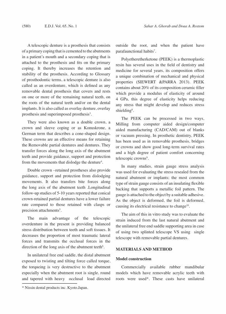
For simulation of the alveolar mucosa, 2 layers of base plate wax were adapted on the free-end saddle area, then plaster index was done over it then wax was removed and application of rubber adhesive on the saddle area was done before rubber base was applied in place and index was repositioned to give the rubber the form and the even thickness. After rubber sets, plaster index was removed.(Fig. 1)
For model 1, preparation of the first premolar only was done to receive the primary telescopic crown and for model 2, the canine and first premolars
were prepared with 6 degree of conversion in both models.
Telescopic crown fabrication
The primary telescopes were waxed up; wax elimination was done then injected into PEEK telescope which was cemented to the abutments. Duplication of the cast with the primary coping was done.
On the duplicate cast, wax pattern for the sec-ondary coping was done. Flasking, wax elimination, then the mold was pressed and injected into PEEK.
Removebale partial denture construction :
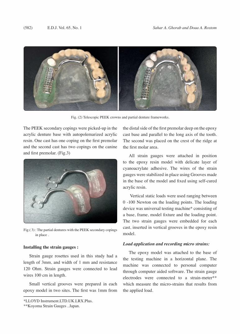
The epoxy resin models with the primary copings cemented in place and the secondary copings seated on their corresponding abutments were duplicated into refractory models, on which the wax pattern for the RPD design was constructed.(Fig.2)
The wax pattern was sprued, invested, and cast into Vitallium, finished and polished. Setting up of acrylic teeth was carried out for both RPDs. The waxed up dentures were flasked, wax was eliminated, heat cured acrylic resin was packed and cured, and then finishing and polishing were carried out following the conventional techniques.
Fig. (1) Model preparation
* Kemapoxy 150JM.CBM International.
(582) Sahar A. Ghorab and Doaa A. RostomE.D.J. Vol. 65, No. 1
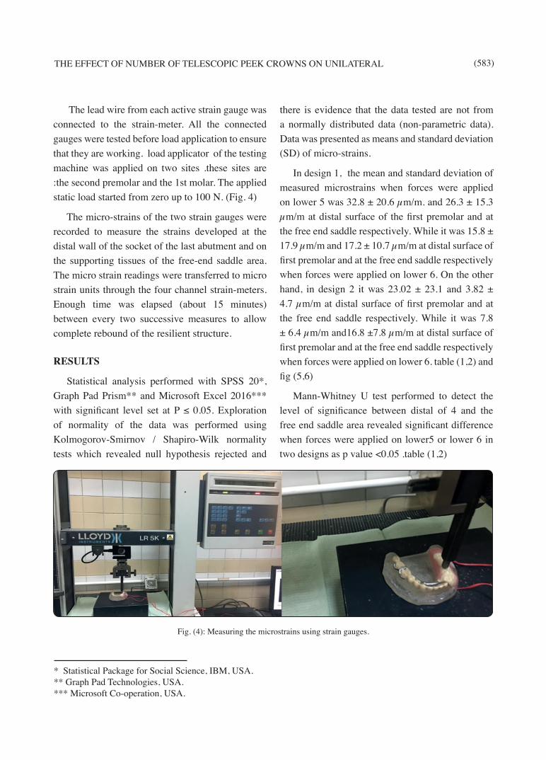
The PEEK secondary copings were picked-up in the acrylic denture base with autopolemarized acrylic resin. One cast has one coping on the first premolar and the second cast has two copings on the canine and first premolar. (Fig.3)
Installing the strain gauges :
Strain gauge rosettes used in this study had a length of 3mm, and width of 1 mm and resistance 120 Ohm. Strain gauges were connected to lead wires 100 cm in length.
Small vertical grooves were prepared in each epoxy model in two sites. The first was 1mm from
the distal side of the first premolar deep on the epoxy cast base and parallel to the long axis of the tooth. The second was placed on the crest of the ridge at the first molar area.
All strain gauges were attached in position to the epoxy resin model with delicate layer of cyanoacrylate adhesive. The wires of the strain gauges were stabilized in place using Grooves made in the base of the model and fixed using self-cured acrylic resin.
Vertical static loads were used ranging between 0 -100 Newton on the loading points. The loading device was universal testing machine* consisting of a base, frame, model fixture and the loading point. The two strain gauges were embedded for each cast, inserted in vertical grooves in the epoxy resin model.
Load application and recording micro strains:
The epoxy model was attached to the base of the testing machine in a horizontal plane. The machine was connected to personal computer through computer aided software. The strain gauge electrodes were connected to a strain-meter** which measure the micro-strains that results from the applied load.
Fig. (2) Telescopic PEEK crowns and partial denture frameworks.
Fig.( 3) : The partial dentures with the PEEK secondary copings in place .
*LLOYD Instrument.LTD.UK.LRX.Plus.**Koyoma Strain Gauges , Japan.
THE EFFECT OF NUMBER OF TELESCOPIC PEEK CROWNS ON UNILATERAL (583)
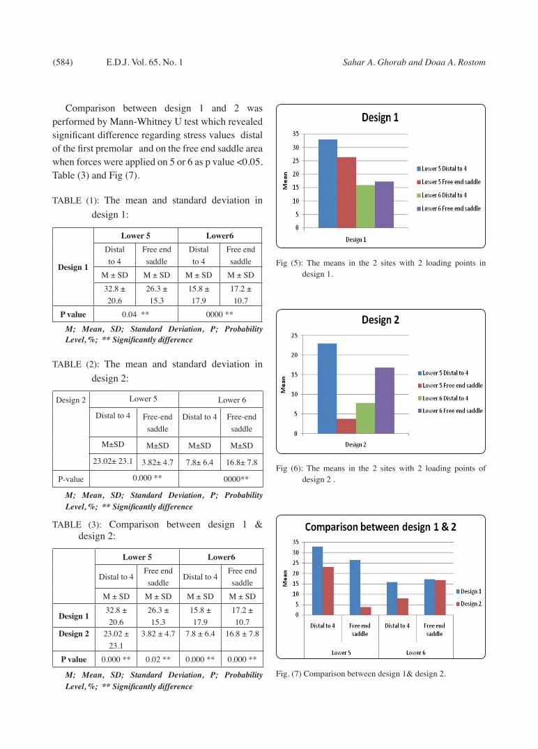
The lead wire from each active strain gauge was connected to the strain-meter. All the connected gauges were tested before load application to ensure that they are working. load applicator of the testing machine was applied on two sites .these sites are :the second premolar and the 1st molar. The applied static load started from zero up to 100 N. (Fig. 4)
The micro-strains of the two strain gauges were recorded to measure the strains developed at the distal wall of the socket of the last abutment and on the supporting tissues of the free-end saddle area. The micro strain readings were transferred to micro strain units through the four channel strain-meters. Enough time was elapsed (about 15 minutes) between every two successive measures to allow complete rebound of the resilient structure.
RESULTS
Statistical analysis performed with SPSS 20*, Graph Pad Prism** and Microsoft Excel 2016*** with significant level set at P ≤ 0.05. Exploration of normality of the data was performed using Kolmogorov-Smirnov / Shapiro-Wilk normality tests which revealed null hypothesis rejected and
there is evidence that the data tested are not from a normally distributed data (non-parametric data). Data was presented as means and standard deviation (SD) of micro-strains.
In design 1, the mean and standard deviation of measured microstrains when forces were applied on lower 5 was 32.8 ± 20.6 µm/m. and 26.3 ± 15.3 µm/m at distal surface of the first premolar and at the free end saddle respectively. While it was 15.8 ± 17.9 µm/m and 17.2 ± 10.7 µm/m at distal surface of first premolar and at the free end saddle respectively when forces were applied on lower 6. On the other hand, in design 2 it was 23.02 ± 23.1 and 3.82 ± 4.7 µm/m at distal surface of first premolar and at the free end saddle respectively. While it was 7.8 ± 6.4 µm/m and16.8 ±7.8 µm/m at distal surface of first premolar and at the free end saddle respectively when forces were applied on lower 6. table (1,2) and fig (5,6)
Mann-Whitney U test performed to detect the level of significance between distal of 4 and the free end saddle area revealed significant difference when forces were applied on lower5 or lower 6 in two designs as p value <0.05 .table (1,2)
Fig. (4): Measuring the microstrains using strain gauges.
* Statistical Package for Social Science, IBM, USA.** Graph Pad Technologies, USA.*** Microsoft Co-operation, USA.
(584) Sahar A. Ghorab and Doaa A. RostomE.D.J. Vol. 65, No. 1
Comparison between design 1 and 2 was performed by Mann-Whitney U test which revealed significant difference regarding stress values distal of the first premolar and on the free end saddle area when forces were applied on 5 or 6 as p value <0.05. Table (3) and Fig (7).
TABLE (1): The mean and standard deviation in design 1:
Design 1
Lower 5 Lower6Distal to 4
Free end saddle
Distal to 4
Free end saddle
M ± SD M ± SD M ± SD M ± SD32.8 ± 20.6
26.3 ± 15.3
15.8 ± 17.9
17.2 ± 10.7
P value 0.04 ** 0000 **
M; Mean, SD; Standard Deviation, P; Probability Level, %; ** Significantly difference
TABLE (2): The mean and standard deviation in design 2:
Design 2 Lower 5 Lower 6
Distal to 4 Free-end saddle
Distal to 4 Free-end saddle
M±SD M±SD M±SD M±SD
23.02± 23.1 3.82± 4.7 7.8± 6.4 16.8± 7.8
P-value 0.000 ** 0000**
M; Mean, SD; Standard Deviation, P; Probability Level, %; ** Significantly difference
TABLE (3): Comparison between design 1 & design 2:
Lower 5 Lower6
Distal to 4Free end saddle
Distal to 4Free end saddle
M ± SD M ± SD M ± SD M ± SD
Design 132.8 ± 20.6
26.3 ± 15.3
15.8 ± 17.9
17.2 ± 10.7
Design 2 23.02 ± 23.1
3.82 ± 4.7 7.8 ± 6.4 16.8 ± 7.8
P value 0.000 ** 0.02 ** 0.000 ** 0.000 **
M; Mean, SD; Standard Deviation, P; Probability Level, %; ** Significantly difference
Fig (5): The means in the 2 sites with 2 loading points in design 1.
Fig (6): The means in the 2 sites with 2 loading points of design 2 .
Fig. (7) Comparison between design 1& design 2.

THE EFFECT OF NUMBER OF TELESCOPIC PEEK CROWNS ON UNILATERAL (585)
DISCUSSION
Use of telescopic crowns removable partial den-ture is a favorable treatment option due to its good esthetic and biomechanical advantage as it direct stresses to the long axis of the abutments and also it minimizes stresses transferred to the residual ridge .
The model used in this study was fabricated in order to simulate as much as possible the natural conditions .The epoxy resin material has an appropriate elastic modulus for a bone analog material11.
Roots were lined with 0.3 mm thickness of light body silicone rubber impression material .while the residual ridges were covered by 2mm thickness of the same material for simulation of mucosa12.
Fixing the rosette of the strain gauge was done on flat prepared surfaces to minimize the possibility of obtaining incremental strain which would be formed due to mounting it on curved surface12, 13.
The normal average bite force is 70 N which was used in the loading device in our study14.
The second premolar and first molar areas were selected as areas of load application because maximum occlusal forces are often exerted in this region where there is maximum contraction of the elevator muscles15.
LLOYD digital loading device was used to deliver ascending load in this study for its digital easy operating method, it also give high degree of accuracy ,rapid data acquisition and full personal computer integration16.
5 minutes interval was given between each reading to give chance to release the stresses and heat from strain gauges17.
It can be assumed that PEEK might be a suitable material for primary crowns regardless of the taper. This could be explained by that PEEK is a soft ductile material that yield and adapt well and easily which resulted in a good marginal fit9.
The results of this study showed higher values of micro-strains in design 1 in both sites (distal to first premolar and on the free-end saddle) than that in design 2 which can be explained by the decreased tooth support .as in design 2 we used 2 telescopic crowns so the torque and stresses are distributed on larger area of teeth surface so the measured micro strains are less .this was in agreement with the clinical findings of many studies which concluded that an increased number of telescopic crowns significantly improved the longevity of the prostheses and their associated abutment teeth in most unilateral partial denture designs18,19,20.
Microstraines were of higher values on the distal side of the first premolar than that on the free-end saddle in both designs in the two loading conditions (the 2nd. Premolar and 1st. molar) this is in agreement with many other studies as within the limitations of this experiment, designs with rigid support as conical crown telescope, produced less damage to the residual ridges. While designs with flexible support were found to create greater stress concentrations on the residual ridges. The minimum stress gradient between the abutments and the residual ridge should be an important issue when fabricating ideal RPDs18, 19,20,21.
The distal abutment always exhibits the highest strain regardless of the loading conditions.
In conclusion, different designs of RPDs will have different effects on abutments and residual ridges but many studies suggested using telescopic crowns as an appropriate treatment options for such cases as it ensures maximally favorable masticatory force transmission, which always takes place axial to the abutments. This treatment modality can lead to predictable long-term treatment outcomes20, 21, 22.
The limitations in this study were that it was carried out in vitro without considering the variation in clinical situations of different patients. Thus future clinical trials should be carried out to confirm any concluded results.
(586) Sahar A. Ghorab and Doaa A. RostomE.D.J. Vol. 65, No. 1
REFERENCES
1- Hakkoum, M. A., & Wazir, G. Telescopic Denture. The Open Dentistry Journal, 12(1), 246–254 (2018).
2- Verma, R., Joda, T., Brägger, U., & Wittneben, J. G. A Systematic Review of the Clinical Performance of Tooth-Retained and Implant-Retained Double Crown Prostheses with a Follow-Up of ≥3 Years. Journal of Prosthodontics, 22(1), 2–12. (2013).
3- Wenz HJ, Hertrampf K, Lehmann KM. Clinical longev-ity of removable partial dentures retained by telescopic crowns outcome of the double crown with clearance fit. Int J Prosthodont; 14:207e13. (2001).
4- Langer Y, Langer A. Tooth-supported telescopic prosthe-ses in compromised dentitions: A clinical report. J Prosthet Dent 2000;84:129-32.
5- Beschnidt, S.M., Chitmongkolsuk, S., Prull, R.: Telescopic crown-retained removable partial dentures: reviewand case report. Compend. Contin. Educ. Dent.22:927-32, (2001).
6- Sahin, V., Akaltan, F., & Parnas, L. Effects of the type and rigidity of the retainer and the number of abutting teeth on stress distribution of telescopic-retained removable partial dentures. Journal of Dental Sciences, 7(1), 7–13. (2012).
7- Bhathal, M., Batra, J., Attresh, G., & Sambyal, S. A re-view on stresses-induced by removable partial dentures. International Journal of Contemporary Dental and Medical Reviews, 5. doi:10.15713/ins.ijcdmr.56(2015).
8- Bechir, E. S., Bechir, A., Gioga, C., Manu, R., & Burcea, A. The Advantages of BioHPP Polymer as Superstructure Material in Oral Implantology. Materials Sciences and Ap-plications, 53(3), 394–398. (2016).
9- Stock, V., Wagner, C., Merk, S., Roos, M., Schmidlin, P. R., Eichberger, M., & Stawarczyk, B.: Retention force of differently fabricated telescopic PEEK crowns with dif-ferent tapers. Dental Materials Journal, 35(4), 594–600 (2016).
10- Karl, M., Dickinson, A., Holst, S., Holst, A.: Biomechani-cal methodsapplied in dentistry :a comparative overview of photoelastic examinations ,strain gauge measurements.finite element analysis and three dimentional deformation analysis . Eur.J.Prosthodont.Restor.Dent.17:50-57,2009.
11- Lee,C.K., Karl, M. Kelly, J.R.: Evaluation of test protocol variables for dental implant fatigue research. Dent. Mater. 25:1419-1425,2009.
12- El-Gendy, A.A.,: Microstrains evaluation of different im-plant positions supporting mandibular bilateral distal ex-tention partial overdenture .Tanta Dental J. 4:39-46, 2007.
13- Alca, K., Cehreli, M.C., Iplikciogl, H.: Acoomparison of three dimentional finite element stress analysis with in vi-tro strain gauge measurements on dental implants .Int.J. Prosthodont.,15:115-121,2002.
14- Fernandes, C.P., Glantz, P.O., Nilner, K.: On the accuracy of some in vitro models for mechanical studies of maxillary removable partial dentures. Dent. Mater.19:127-136,2003.
15- Hegazy, S.A.F., Gebreel,A.A., Emera, R.M.K.: Resilient versus rigid telescopic attachment for two implants as-sisted complete mandibular overdentures :Invitro stress analysis study. 60:725-732,2014.
16- Hegazy, S.A., Elshahawi, I.M., Elmotayam, H.: Stresses induced by mesially and distally placed implants to re-tain a mandibular distal-extension removable partial denture :Acomparative study.Int.J.Oral Maxillofac. Im-plants.28:403-407,2013.
17- Cekic,C.,Akea,K.Cehreli,M.C.:Effects of attachment de-sign on strains around implants supporting overdentures .Quintessence Int.,38:291-297,2007.
18- Saito M, Miura Y, Notani K, Kawasaki T. Stress distribu¬tion of abutments and base displacement with precision at¬tachment and telescopic crown-retained re-movable partial dentures. J Oral Rehabil, 30:482-48,2003.
19- Lee, H.E., Wu, J.H., Wang, C.H., Lan, T.H., Kangdu, J.E.: Biomechanical analysis of distal extension removable par-tial dentures with different retainers. J Dent Sci, 3 : 133-139, 2008.
20- Mohammadi,I.A.A.,Hanafy,S.A.,Awadallah,M.E.:Effect of unilateral mandibular implant-tooth supported telescop-ic prosthesis on the supporting structures (In-vitro study).J. Science &Res.,6:14,2016
21- Dahab, I. A., El-Gendy, A. A., & Eltorky, I. R. :In vitro stress analysis study of different prosthetic options using single posterior implant for management of mandibular unilateral distal extension saddle. Tanta Dental Journal, 12(1), 7–15,2015.
22- Igarashi Y., Ogata A., Kuroiwa A., Wang C. H. Stress dis-tribution and abutment tooth mobility of distal-extension removable partial dentures with different retainers: an in-vivo study. J Oral Rehabil, 26; 111–116,1999.


![EGYPTIAN , October, 2014 DENTAL JOURNAL I.S.S.N 0070-9484 · Cleaning and shaping are the main goals of root canal instrumentation [1]. In the last 20 years, several new instrumentation](https://static.fdocuments.in/doc/165x107/5e861015a8dfab6bda71c90f/egyptian-october-2014-dental-journal-issn-0070-9484-cleaning-and-shaping-are.jpg)