EGS Congress · 13 th ongress Fort asso lorence taly 19/22 May 2018 P1.002 ASTF ® questionnaire: a...
522
13 th EGS Congress Florence, Italy 19/22 May 2018 www.eugs.org Abstract Book
Transcript of EGS Congress · 13 th ongress Fort asso lorence taly 19/22 May 2018 P1.002 ASTF ® questionnaire: a...

13th EGS Congress
Florence, Italy19/22 May 2018
www.eugs.org
Abstra
ct Boo
k

13th EGS CongressFortezza da Basso, Florence, Italy
19/22 May 2018
POSTER SESSION 1
EPIDEMIOLOGY, HEALTH ECONOMICS, VISUAL DISABILITY, QOL,
PATHOGENESIS

13th EGS CongressFortezza da Basso, Florence, Italy
19/22 May 2018
P1.002 FAST® questionnaire: a short and effective tool to assess ocular surface disease in all glaucoma patients M. Misiuk-Hojlo1, J. Clarke2, A. Antón López3, C. Baudouin4 (1Poland, 2United Kingdom, 3Spain, 4France)
P1.003 Experience of the volunteer mission with the new mobile glaucoma unit G. Dalianis, A. Trivli, C. Terzidou (Greece)
P1.005 The results of state screening for glaucoma in Republic of Kazakhstan N. Aldasheva, L. Tashtitova (Kazakhstan)
P1.006 Clinical profile of Polish glaucoma patients- results of a Polish Ophthalmological Society survey conducted among ophthalmologists I. Grabska-Liberek, J. Majszyk-Ionescu, A. Skowyra, M. Rogowska (Poland)
P1.007 Literature review comparing the use of fixed dose combinations versus multidrug treatments in glaucoma patients D. Viriato1, S. Gogna2, R. Jindal2, G. Dasari2, Y.S. Kim, A. Mullins3, B. Sloesen1, J. Banhazi4, A. Realini3
(1Switzerland, 2India, 3USA, 4Ireland)
P1.008 Comparativ analysis of antiglaucoma generic and original eye drugs in Split- Dalmatia County, Croatia V. Rogosic, L. Vanjaka Rogosic, V. Mastnak, M. Gulin, K. Novak Laus (Croatia)
P1.009 Glaucoma in the Northern Ireland Cohort for the Longitudinal Study of Ageing (NICOLA) and Glaucoma within NICOLA (GwNICOLA): rationale and methods P. McCann, R.E. Hogg, I.S. Young, F. Kee, A. Azuara-Blanco (United Kingdom)
P1.010 I still haven’t found what i’m looking for….. Bono, Google and glaucoma awareness C. Lyons (Ireland)
P1.011 The relationship between environmental factors and exfoliation M. Irkec, F. Bezci, T. Bagcibosi, S. Kocabeyoglu (Turkey)
P1.012 Cognitive evaluation of patients with exfoliative glaucoma and primary open-angle glaucoma S. Kocabeyoglu, Y. Deniz, C. Balci, B.B. Yavuz, M. Cankurtaran, M. Irkec (Turkey)
P1.013 The prevalence of undiagnosed glaucomatous and other age-related sight threatening diseases in self-proclaimed healthy individuals S. Lemmens, J. Barbosa-Breda, T. Jacobs, K. Van Keer, R. Van Landeghem, I. Stalmans (Belgium)
P1.014 The prevalence and clinical characteristics of charles bonnet syndrome in Turkish patients with glaucoma G. Arikan, C. Durmaz, E. Yaka, U. Gunenc (Turkey)
P1.015 Streamlining glaucoma outpatient services in Moorfields eye hospital south: results of a quality improvement initiative to increase clinic capacity and quality of care L.-Y. Ngai, E. Gabbott, T. Bader, P. Rai (United Kingdom)
P1.016 Patients factors encompassing glaucoma care R.T. Brady, Á. Ní Mhéalóid, E. Henry, J. Brady, J. Stokes (Ireland)
P1.017 Differences of ocular surface disease signs and symptoms between patients with primary open angle glaucoma and pseudoexfoliative glaucoma A. Matsou, M. Dermenoudi, C. Keskini, P. Brazitikos, E. Anastasopoulos (Greece)
P1.018 Association of visit-to-visit blood pressure variability with normal tension glaucoma M.D. Ahn, N.Y. Lee (Republic of Korea)
P1.019 Association between open-angle glaucoma and hypothyroidism: a meta-analysis G.J. Seong, C.Y. Kim, H.W. Bae, S. Kim (Republic of Korea)
P1.020 Association between open-angle glaucoma and the risk of Alzheimer’s disease and Parkinson’s disease in South Korea: a 10-year nationwide cohort study S.H. Lee, J. Moon, S.J. Jung (Republic of Korea)
P1.021 The Italian primary open-angle glaucoma study: quality of life changes over one-year follow-up I. Riva1, L. Legramandi1, E. Rulli1, A. Katsanos2, A. Iaria1, L. Quaranta1 (1Italy, 2Greece)

13th EGS CongressFortezza da Basso, Florence, Italy
19/22 May 2018
P1.022 ISY (satISfaction surveY): first real-life data of use of a preservative-free multidose glaucoma device (EasyGrip® delivery system) in 5 European countries P. Denis, C. Erb2, P. Puska3, J. Skov4, S. Duch5 (1France, 2Germany, 3Finland, 4Denmark, 5Spain)
P1.023 Association of alcohol consumption and intraocular pressure in men and women: the 5th Korea National Health and Nutritional Examination Survey 2010-2012 Y.C. Yoo, H.K. Kim, K.B. Lee (Republic of Korea)
P1.024 Polymorphism of estrogen receptors and their influence on clinical status of patients with primary open angle glaucoma D. Wróbel-Dudzinska1, M. Sagan1, E. Kosior-Jarecka1, U. Łukasik1, T. Aung2, C.C. Khor2, J. Kocki1, T. Zarnowski1 (1Poland, 2 Singapore)
P1.025 Comparison of relationships between anterior chamber depth and other ophthalmic biometrical ocular factors either at the central and the peripheral anterior chamber portions among (Japan)ese residents K. Kitamura, T. Chiba, J. Tanabe, K. Kashiwagi (Japan)
P1.026 Acute glaucoma: incidence at ophthalmic emergencies at an urgency-emergency general hospital C. Rocha Lauretti, D. Pereira da Silva Felipe Crosta, A. Messias (Brazil)
P1.027 Evaluation of intraocular pressure and central corneal thickness in children O. Balta (Turkey)
P1.028 Vision-related quality of life in patients with congenital glaucoma
A. Miraftabi1, A.L. Coleman2, M. Parsamanesh1, N. Nilforushan1, A.H. Nguyen2, S.A.P. Alemzadeh1
(1Iran, 2USA)
P1.029 The association of posterior embryotoxone and pediatric glaucoma in the Serbia V. Jovanovic, L. Magarasevic, Z. Abazi (Serbia)
P1.030 Ocular hypertension in patients with uveitis R. Kijima, Y. Shinmei, T. Ohguchi, T. Nitta, K. Namba, S. Chin, S. Ishida (Japan)
P1.031 Analysis of exfoliation syndrome and exfoliation glaucoma prevalence in Novosibirsk region
E. Tashlykova, O. Kuleshova, S. Aidagulova, A. Lazareva, V. Dulidova, V. Chernikh (Russia)
P1.032 Risk factors and outcomes of ocular hypertension in patients with uveitis S. Benchekroun, R. El Hadiri, L. El Kaissoumi, Y. El Harrak, O. Cherkaoui (Morocco)
P1.033 Neutrophil-to-Lymphocyte Ratio and systemic diseases in patients with pseudoexfoliation glaucoma M. Lopez-Valladares, A. Suarez-Campo, P. Mascareñas-Pazos, T. Rodriguez-Ares (Spain)
P1.034 Clinical characteristics of drug induced secondary bilateral acute angle-closure crisis J. Jin Jung, Y. Yoo, H.J. Hwang, Y. Gon Lee, J. Geun Jeong (Republic of Korea)
P1.035 Comparing of laminar cribrosa and peripapillary vessels density between branch retinal vein occlusion and normal tension glaucoma with swept source OCT and OCTA C.K. Lee, J. Shin, S. Rho, D.W. Kim, M. Lee (Republic of Korea)
P1.037 RhoA activation inhibited phagocytic activity of trabecular meshwork cells T. Fujimoto, T. Inoue, S. Ohira, M. Inoue, H. Tanihara (Japan)
P1.038 The Effect of histone deacetylase on activity of glial cells in the ischemic mouse retina S.W. Park (Republic of Korea)
P1.039 Relationship between preoperative intraocular pressure and retinal nerve fiber layer thinning after glaucoma surgery K.N. Kim (Republic of Korea)
P1.040 A theoretical study of the role of conformational properties of trans-epithelial ion pump on aqueous humor production D.E. Messenio1, R. Sacco1, L. Sala2, A. Mauri3, G. Guidoboni3, A. Harris3 (1Italy, 2France, 3USA)

13th EGS CongressFortezza da Basso, Florence, Italy
19/22 May 2018
P1.042 The association between haemodialysis and optic neuropathy B.N.K. Choy, N.S.K. Fung, J.S.M. Lai (Hong Kong)
P1.043 Functional analysis of MANF in retinal ganglion cells by oxidative stress J. Ko (Japan)
P1.044 Visual field defects due to cerebrospinal fluid hypovolemia M. Kusajima, Y. Shinmei, A. Takahashi, K. Ishijima, Y. Tagawa, K. Suzuki, T. Yamamoto, K. Nakamura, S. Chin, S. Ishida (Japan)
P1.045 Mapping expression of glaucoma risk genes in the mouse eye
W. Hubens, E. Breddels, T. Gorgels, C. Webers (Netherlands)
P1.046 Glaucoma-on-a-chip: an in vitro model for glaucoma drug discovery based on mimicking the mechanical stress of high eye pressure T.G.M.F. Gorgels, P.A.M.M. Vroemen, R. Sinha, J. Rouwkema, L. Moroni, J. de Boer, C.A.B. Webers (Netherlands)
P1.047 Isolation and culturing of rodent retinal ganglion cells for glaucoma disease modeling P. Vroemen, J. de Boer, C.A.B. Webers, T.G.M.F. Gorgels (Netherlands)
P1.048 Systemic and ocular determinants of mean ocular perfusion pressure in a population-based sample N. Yildirim, E. Atalay, L. Niyaz, S. Gültekin (Turkey)
P1.049 Structural and vascular evaluation of optic nerve and macula using optical coherence tomography angiography in hypertension B. Burgos-Blasco, J. Pascual-Prieto, J.M. Martinez de la Casa, N. Güemes-Villahoz, L. Salazar-Quiñones, M. Avila-Sanchez, M.A. Barbero-Pedraz, F. Saenz-Frances, S. García-Saénz, J. García-Feijoo (Spain)
P1.050 Prevalence of hypotensive episodes in patients with hypertension, with or without glaucoma R. Nesher, M. Mosseri, Y. Keshet, I. Frumkin, O. Kazanovsky, M. Mimouni, E. Golan, S. Ben Shitrit (Israel)
P1.051 A novel tissue engineered biomimetic model of the lamina cribrosa region for glaucoma research D. Clissmann, R. Murphy, R. Low, A. Hopkins, M. Irnaten, D. Wallace, A. Hibbitts, A. Ryan, F. O’Brien, C. O’Brien, D. Brennan (Ireland)
P1.052 Serum homocysteine, vitamin B12 and folic acid levels in different types of open angle glaucoma M. Stojcic, M. Stojkovic, V. Lukic, B. Stojcic, L. Zoric, Z. Bukumiric (Serbia)
P1.053 Proteomic expression profile of the Iris in primary glaucoma: a pilot report A. Narayanaswamy, M. Nongpiur, L. Zhou, T. Wong, T. Aung (Singapore)
P1.054 Changes in the optic nerve head induced by horizontal eye movements W.J. Lee, H.W. Lim (Republic of Korea)
P1.055 Lamina cribrosa cell bioenergetics in glaucoma: role of glycolysis and glutaminolysis D. Hickey1, M. Irnaten1, D. Brennan2, D. Wallace1, A. Clark2, C.J. O’Brien1 (1Ireland, 2USA)
P1.056 Gene expression of TGF beta isoforms and its receptors in the parts of trabecular meshwork in patients with glaucoma - a preliminary report W. Maruszczyk, M. Kimsa, M. Machalska, M. Dorecka, B. Strzalka-Mrozik, U. Mazurek, E. Mrukwa-Kominek (Poland)
P1.057 Rare case of right side horner syndrome and simultaneous pigmentary glaucoma of the left eye K. Moustaklis, S. Gorezis (Greece)
P1.058 Anterior segment circulating neurotoxic cytokines in primary open angle glaucoma patients A. Pantalon, D. Chiselita, C. Feraru (Romania)
P1.059 Deep optic nerve head morphology are associated with pattern of glaucomatous visual field defect in open angle glaucoma J.C. Han, C. Kee, J.H. Choi, E.J. Lee, D.Y. Park (Republic of Korea)
13th EGS CongressFortezza da Basso, Florence, Italy
19/22 May 2018
P1.060 Topical ripasudil prevents retinal ganglion cell death in a mouse model of normal tension glaucoma T. Harada, K. Akaiwa, K. Namekata, Y. Azuchi, X. Guo, A. Kimura, C. Harada, Y. Mitamura (Japan)
P1.061 Features of lamina cribrosa and autonomic nervous system in glaucoma patients with disc hemorrhage J. Kim, C.K. Park, H.-Y.L. Park (Republic of Korea)
P1.062 Relationship between microstructure of peripapillary atrophy and microvasculature in myopic eyes M.S. Sung (Republic of Korea)
P1.063 Racial differences in the extracellular matrix and histone acetylation of the lamina cribrosa and peripapillary sclera H.-Y. Lopilly Park, C.K. Park (Republic of Korea)
P1.064 Different contributions of autophagy to retinal ganglion cell death in the diabetic and glaucomatous retinas C.K. Park, J.H. Kim, H.-Y. Lopilly Park (Republic of Korea)
P1.065 Optical coherence tomography angiography of the optic disc perfusion in glaucoma in Asian eyes - A promising technology D. Aryasingha, S. Pathirana (Sri Lanka)
P1.066 Association between additive effects of genetic variants associated with primary open-angle glaucoma and family history of glaucoma F. Mabuchi, N. Mabuchi, Y. Sakurada, S. Yoneyama, K. Kashiwagi, Z. Yamagata, H. Iijima (Japan)
P1.067 Bioinformatical pathway analyses to discover the molecular pathogenesis of primary open angle glaucoma I. Liesenborghs, M. Kutmon, L.M.T. Eijssen, T.G.M.F. Gorgels, C.T. Evelo, H.J.M. Beckers, C.A.B. Webers,J.S.A.G. Schouten (Netherlands)
P1.068 Bruch´s membrane opening expands in primary open angle glaucoma G. Rebolleda, A. Pérez-Sarriegui, L. Díez-Álvarez, V. De Juan, F.J. Muñoz-Negrete (Spain)
P1.069 Human trabecular meshwork exposure to oxidative stress: two-dimensional vs three-dimensional cultures S.C. Saccà, C.A. Cutolo, T. Sbrana, S. Gandolfi, C.E. Traverso, A.M. Bassi (Italy)
P1.070 The roles of TNF-α-308 gene polymorphism and TNF-α serum concentrations in glaucoma patients M. Trenkic Bozinovic, P. Jovanovic, J. Jocic Djordjevic, M. Petrovic, M. Radenkovic, A. Veselinovic, A.Miljkovic (Serbia)
P1.071 Effect of week day on intraocular pressure variations using continuous intraocular telemetry H.L. Rao (India)
P1.072 Involvement of the trabecular meshwork, distal and proximal layers of sclera in increasing the resistance of the outflow pathways in pseudoexfoliation glaucoma O. Kuleshova, S. Aidagulova, A. Lazareva, E. Pichikova, V. Dulidova (Russia)
P1.073 Is obstructive sleep apnoea associated with glaucoma progression? R. Bourne1, D. Wozniak1, G. Peretz2, J. Kean1, R. Foster1, S. Downes1, I. Smith1 (1United Kingdom, 2Israel)
P1.074 Prevalence of obstructive sleep apnoea in glaucoma: the POSAG study G. Peretz1, D. Wozniak2, J. Kean2, S. Harun2, C. Willshire2, S. Villar2, R. Foster2, S. Downes2, I. Smith2,R. Bourne2 (1Israel, 2United Kingdom)
P1.075 ET-1 effect on functional and structural change in patients with POAG S. Ljaljevic, E. Alimanovic, M. Lika-Pranjic (Bosnia and Herzegovina)
P1.076 Vascular evaluation of the optic disc with OCT-A in primary open-angle glaucoma P. Faria, I. Sobral, C. Azenha, J. Cardoso, J.F. Silva, J. Moura Pereira (Portugal)

13th EGS CongressFortezza da Basso, Florence, Italy
19/22 May 2018
P1.078 Evaluation of lamina cribrosa by swept-source optical coherence tomography in primary open-angle glaucoma I. Pasaoglu, B. Satana, C. Altan, B. Basarir, E. Pasaoglu, T. Yasar, M. Taskapili (Turkey)
P1.079 The association of choroidal thickness with retinal nerve fiber layer thickness in advanced primary open-angle glaucoma F.S. Yilmaz, A. Cakir, G. Ozturan, M. Elcioglu (Turkey)
P1.080 The proteins of mitochondrial dysfunction and integrin signaling change in the rat retina following cerebrospinal fluid pressure reduction F. Yan, F. Yu, Y. Gong, L. Zhang, J. Zhang, S. Wu, H. Deng, N. Wang (China)
P1.081 The role of lysyl oxidase-1 (LOX1) as collagen cross-linker in POAG N. Rahman, M. Irnaten, C. O’Brien (Ireland)
P1.082 Translaminar pressure gradient and ocular perfusion pressure in glaucoma patients with different optic disc sizes N. Kasahara (Brazil)
P1.083 Analysis of MYOC gene in a Czech family with primary open-angle glaucoma L. Rezkova, M. Fichtl, P. Liskova, L. Dudakova, E. Ruzickova (Czech Republic)
P1.084 Short-term observation of retina blood filling changes in rat retina ischemia-reperfusion model established with a novel operative approach and procedure F. Yan, L. Zhang, S. Wu, J. Zhang, K. Liu, S. Li, N. Wang (China)
P1.085 Genetic variants associated with high intraocular pressure are not associated with primary angle closure glaucoma R.S. Chong1, G.S.E. Kiew2, M. Nongpiur1, S. Perera1, L.J. Chen3, S. Sarangapani4, D. Tan5, Y. Azhany6, A.T. Liza-Sharmini6, C. Tham3, N.X. Hiep5, R. George4, C.-C. Khor1, E. Vithana1, T. Aung1 (1Singapore, 2United Kingdom, 3Hong Kong, 4India, 5 Viet Nam, 6Malaysia)
P1.087 Quantitative proteomic analysis of aqueous humour across angle closure disease spectrum M. Nongpiur, L. Zhou, E. Vithana, T. Aung, T. Wong (Singapore)
P1.088 Newborn glaucoma: don’t forget infections! S. Kaushik, G. Joshi, M. Singh, P. Kataria, A. Arora, S.S. Pandav (India)
P1.089 Iris thickness and severity of neovascular glaucoma determined using swept-source anterior-segment optical coherence tomography S. Nakakura, Y. Kobayashi, K. Matsuya, Y. Kiuchi (Japan)
P1.090 Peripapillary vessel density in glaucomatous eyes: comparison between pseudoexfoliation glaucoma and primary open-angle glaucoma J.-H. Park1, C. Yoo1, M. Girard2, J.-M. Mari3, Y.Y. Kim1 (1Republic of Korea, 2Singapore, 3French Polynesia)
P1.091 Comparison of plasma C-reactive protein level in pseudoexfoliation (PXF) glaucoma with normal population R. Zarei (Iran)
P1.092 Possible role of chlamydias in the etiopathogenesis of pseudoexfoliation syndrome A. Yilmaz, S. Gonca, Z. Oksuz Karaarslan, M.S. Serin, O. Oktay (Turkey)
P1.093 Benefits of oral supplementation with docosahexaenoic acid and antioxidants in pseudoexfoliative glaucoma: a 6-month open-label randomized controlled trial S. Romeo Villadoniga, E. Rodriguez Garcia, M.D. Álvarez Diaz, J.C. Domingo Pedrol, O. Sagastagoia (Spain)
P1.094 The role of environmental factors and genetic makeup in the pathogenesis of ocular pseudoexfoliation (pxf) syndrome in Saudi military and their family S. Alshamrani (Saudi Arabia)
P1.095 New stop codon mutatıon of ADAMTS10 gene IN Weıll-Marchesanı syndrome A. Ozcan, B. Ulas (Turkey)
13th EGS CongressFortezza da Basso, Florence, Italy
19/22 May 2018
P1.002 FAST® QUESTIONNAIRE: A SHORT AND EFFECTIVE TOOL TO ASSESS OCULAR SURFACE DISEASE IN ALL GLAUCOMA PATIENTSMarta Misiuk-Hojlo1, Jonathan Clarke2, Alfonso Antón López3, Christophe Baudouin4
1Department of Ophthalmology, Wroclaw Medical University, Wroclaw - Poland2NIHR Biomedical Research Centre at Moorfields Eye Hospital NHS Foundation Trust and UCL Institute of Ophthalmology, London - United Kingdom3Institut Català de Retina, Barcelona - Spain4CHNO des 15-20, Paris - France
Purpose: Despite its common prevalence, Ocular Surface Disease (OSD) remains an uncommonly recognized condition in glaucoma patients. It has been published that OSD may compromise the tolerability of topical therapy and may induce a decrease of compliance that may impact treatment efficacy and therefore progress of visual loss. The identification of OSD must be integrated in glaucoma patients’ management. In order to help the ophthalmologists in OSD diagnosis, the FAST (Fast Assessment of ocular Surface Trouble) questionnaire has been developed.
Methods: A survey, in 7 European countries (BE, FR, IT, PL, SP, CH, UK) has been implemented to evaluate in real life the new and simple FAST questionnaire. It includes 14 short questions for collecting risk factors, symptoms and ocular signs and it highlights abnormal results. It is divided into two parts to collect data from the patient interview (demography, risk factors and symptoms) and from the clinical examination. The objective is to identify correlations between risk factors, symptoms and signs and to produce a shorter and validated questionnaire thanks to the Graded Response Model analysis.
Results: The results were obtained from 928 glaucoma patients from 4 countries: SP (302), FR (88), UK (100) and PL (438). 68% were using at least one preserved glaucoma treatment. At least one risk factor was observed in 64.2% of patients. 84% are using artificial tears or anti-allergic agents; 72% reported at least one ocular sign: 50% reported dry eye symptoms between instillations, 50% reported itching/irritation and 57% reporting burning sensation. Only 45.5% of patients have a tBUT superior to 10s. There was a significant association between the number of preserved glaucoma drops (superior to 1 drop /day) and all the symptoms between instillations and the ocular signs (p < 0.05).
Conclusions: These new results offer interesting insight into the prevalence of OSD and also highlight the simplicity of this tool to report symptoms and OSD. The final validated version of the FAST questionnaire will be useful tool for ophthalmologists in daily practice.

13th EGS CongressFortezza da Basso, Florence, Italy
19/22 May 2018
P1.003EXPERIENCE OF THE VOLUNTEER MISSION WITH THE NEW MOBILE GLAUCOMA UNITGeorge Dalianis1, Alexandra Trivli1, Chryssa Terzidou1
1Department of Ophthalmology, Konstantopouleio General Hospital, N.Ionia, Ahens - Greece
Purpose: To present the set-up and results of the volunteer mission in the remote Greek highlands with the mobile glaucoma unit of our department equipped with portable OCT.
Methods: All the equipment necessary was moved from our department and transported and assembled on location. It included two spare slit-lamps, a portable pachymeter, two goldmann applanation tonometers, two indirect fundus lenses (66 and 90 dioptres), a gonioscopy lens, an EDTRS chart, a portable OCT, various medications (mydriatics, fluorescein, topical anesthetics) and necessities (slit-lamp tables, stools, patient forms). Staff included 4 doctors (two glaucoma specialists, one ophthalmologist and one resident), 1 nurse and 1health care provider. During the two-day mission, 185 patients (106 women and 79 men) with a mean age of 60 years (range 27-91) were examined as following: history, complete slit lamp examination, CCT, IOP evaluation (GAT) and ONH evaluation using (a) Optical Coherence Tomography (RNFL+GCC) and (b) fundus examination after pupil dilation. Anterior segment evaluation using OCT was performed in patients with narrow angle and OCT evaluation of the macula was performed in patients with pathological findings on fundus examination.
Results: 15 patients were diagnosed with glaucoma (8.1%) and were suggested further evaluation and treatment and 14 patients were glaucoma suspects (7.56%) and were suggested visual field examination and proper follow-up. Two patients were diagnosed with ION. During screening fundus examination, in 20 of the patients (10.8%) AMD was observed and NPDR in two patients.
Conclusions: Glaucoma screening is essential in general population, given the asymptomatic nature of the disease. It is especially important for people living in remote regions with difficult access to ophthalmologists and special equipment. Portable OCT proved to be very valuable, not only for ONH evaluation, but also for the evaluation of narrow angles as well as the presence of macular edema in the patients found to have AMD and NPDR. This kind of services must be performed in a volunteer fashion more often, in order to provide better care for this category of patients.

13th EGS CongressFortezza da Basso, Florence, Italy
19/22 May 2018
P1.005THE RESULTS OF STATE SCREENING FOR GLAUCOMA IN REPUBLIC OF KAZAKHSTANNeilya Aldasheva1, Lyailya Tashtitova2
1Deputy Chairman of the board for Science and strategic Development Doctor of medical science, Almaty - Kazakhstan, 2Department of Ophthalmology, Kazakh Scientific-Research Institute of Eye Diseases, Almaty - Kazakhstan
Purpose: To study the effectiveness of state screening for glaucoma at the level of primary healthcare system.
Methods: Screening is subject to the population aged 40-70 years. The screening methodology uses examination of the target group once in 2 years and includes a survey of patients on the presence of risk factors and measurement of intraocular pressure. If patients have an increased IOP and the presence of risk factors, they are referred to a specialized glaucoma room for verification of the diagnosis.
Results: Annually, within the framework of active screening for glaucoma, an average of 1.5 million people undergoes screening. Detectability of glaucoma is 0.27-0.3% in average. The effectiveness and adequacy of screening is confirmed by the trend in the number of patients with diagnosed glaucoma, depending on age. A direct correlation was found through the increase in the percentage of detectable glaucoma in older age groups, which corresponds to the literature data. Number of officially registered patients with glaucoma in 2016 in the Republic of Kazakhstan is 68,195 (0.38% of the total population - 17.8 million). While in 2011, at the beginning of screening, there were only 50,220 registered patients, which was 0.30% of the total population. The number of registered patients from the beginning of screening increased by 46%. The annual number of newly diagnosed cases of glaucoma (for self-reversal and screening) during screening program is 12,000 in average. Since 2012, number of detected glaucoma patients annually grows for an average of 4000 cases.
Conclusions: Thus, the effectiveness of glaucoma screening within the framework of the State Program “Salamatty Kazakhstan” is in average 0.27-0.3%. The screening effectiveness in terms of detecting new cases is 49%.
13th EGS CongressFortezza da Basso, Florence, Italy
19/22 May 2018
P1.006CLINICAL PROFILE OF POLISH GLAUCOMA PATIENTS. RESULTS OF A POLISH OPHTHALMOLOGICAL SOCIETY SURVEY CONDUCTED AMONG OPHTHALMOLOGISTSIwona Grabska - Liberek1, Julita Majszyk - Ionescu1, Agnieszka Skowyra1, Monika Rogowska1
1The Medical Centre of Postgraduate Education, Warsaw - Poland
Purpose: The aim of this study is to learn more about glaucoma patients in Poland: their demographic profile, risk factors of glaucoma development, diagnostic tools and the way of treatment.
Methods: The research program was an observational, non-interventional study. It was conducted in 2016-2017. During its implementation, no additional, non-standard medical procedures were performed. The study was carried out in the field of open treatment (counseling) by specialists in the field of ophthalmology. Each researcher obliged to observe 100 patients. As part of the study, each patient had one visit. Descriptive statistical analysis was performed - calculation of appropriate measurements (mean, modal, quartiles, median), variability (range, variance, standard deviation), asymmetry (skewness) and concentration (kurtosis) for all parameters included in the questionnaire and comparison of groups of patients. Based on a preliminary analysis of the results obtained, appropriate statistical tests were selected to examine potential relationships. In the case of normal distribution, parametric tests were performed, in the case of non-normal distribution, non-parametric tests were used for the calculations.
Results: The number of patients included in the study was 3678. Open angle glaucoma was most often recognized. Over 40% of patients had an initial stage of glaucoma and 1/3 an intermediate stage. The most common risk factors were age (71%), hypertension (45%) and glaucoma in the family (32.4%). Among the previous ophthalmic surgeries, the most common cataract surgery was performed (21%). In more than two-thirds of the patients, the current therapy was continued, and 17.3% changed it. The most commonly used group of drugs were prostaglandin analogs.
Conclusions: A wide population of patients with glaucoma was examined. The study allowed to determine with high accuracy the clinical and therapeutic regimens in patients with glaucoma, in which the use of prostaglandin analogs predominated.
13th EGS CongressFortezza da Basso, Florence, Italy
19/22 May 2018
P1.007LITERATURE REVIEW COMPARING THE USE OF FIXED DOSE COMBINATIONS VERSUS MULTIDRUG TREATMENTS IN GLAUCOMA PATIENTSDaniel Viriato1, Suditi Gogna2, Ramandeep Jindal2, Ganesh Dasari2, Yong Soo Kim1, Anmol Mullins3, Brigitte Sloesen1, Judit Banhazi4, Anthony Realini5
1Novartis Pharma AG, Basel - Switzerland2Novartis Healthcare Pvt. Ltd., Hyderabad - India3Novartis Pharmaceuticals, East Hanover - USA4Novartis Ireland Ltd, Dublin - Ireland5Department of Ophthalmology, West Virginia University, Morgantown - USA
Purpose: Reduction of intraocular pressure (IOP) is the only proven method to reduce the progression of glaucoma. Pharmacologic treatments for glaucoma include topical medications from different therapeutic classes such as prostaglandin analogs, beta-blockers, carbonic anhydrase inhibitors and alpha-adrenergic agonists which can be prescribed as single agents or in fixed-dose combinations (FDC). Frequently, more than one medication is required to achieve adequate control of IOP. The objective of this review was to compare the efficacy, safety, adherence and patient preferences/satisfaction of FDC versus multidrug treatments (MDT) in glaucoma patients.
Methods: A literature search of English publications between January 1998 and September 2017 in MEDLINE and EMBASE was performed. Search terms were variations on ‘glaucoma’, ‘patient preferences’, ‘satisfaction’, ‘adherence’, ‘efficacy and safety outcomes’. All types of clinical and real-world studies were included.
Results: Overall, 19 studies were included assessing efficacy and safety (18), patient preferences (4), satisfaction (1), and adherence (5). Most studies (17) demonstrated a similar efficacy and safety profile for FDC and MDT, in a controlled clinical setting. The only exception was the study conducted by Inoue et al (2014) where patients switched from MDT to FDC and reported a significant decrease in IOP at 36 months (mean IOP; FDC =14.3, MDT = 15.2, p < 0.01). In the four studies assessing the preferences of patients who switched from MDT to FDC the results favored FDC by noteworthy margins (FDC vs MDT: Study-1: 82.1 vs. 11.1%, Study-2: 81.3 vs. 3.2%, Study-3: 54.8 vs. 11.9%, Study-4: FDC1 = 63 vs. 0%, FDC2 = 50 vs. 10%). Patient satisfaction score was significantly improved when patients switched from two separate drugs to FDC (mean satisfaction score; FDC = 7.3; MDT = 6.3, p = 0.0051). In 3 of 5 studies, patients exhibited better adherence with FDC compared to MDT, while remaining were neutral.
Conclusions: With similar efficacy and safety profiles, FDC compared to MDT is the preferred treatment option for glaucoma patients. Ease of administration and reduced number of drops may improve adherence to dosing regimens. Further studies on patient preferences, treatment adherence/satisfaction with FDC for glaucoma are needed to confirm these findings.

13th EGS CongressFortezza da Basso, Florence, Italy
19/22 May 2018
P1.008COMPARATIV ANALYSIS OF ANTIGLAUCOMA GENERIC AND ORIGINAL EYE DRUGS IN SPLIT-DALMATIA COUNTY, CROATIAVeljko Rogosic1, Lucija Vanjaka Rogosic1, Viktor Mastnak1, Marija Gulin1, Katia Novak Laus1Department of Ophthalmology, Clinical Hospital Center Split, Split - Croatia
Purpose: The primary aim of this research in medical antiglaucoma therapy is to find out the total number of antiglaucoma drugs in Split-Dalmatia county in the last 5 years, excpecially with regards to consumption of original antiglaucoma drugs against their generic counterparts and comparison between fixed (combined) and mono therapy. Also we want to show if the prescription of prostaglandin analogues has risen through the years.
Patients and Methods: In this research we used data of the number of antiglaucoma drugs used in Split- Dalmatia County between the years of 2012 and 2016 from glaucoma patients. The data is collected in pharmacies of the county. In this research the data gathered from the pharmacies will be retrospectively statistically and anlytically analysed. The data was processed in Microsoft Excel and Microsoft Word programs.
Results: In our study we showed that of the total amount of consumed antiglaucoma drugs (n = 784,123), the highest amount takes consumption of original mono-component drugs (n = 554,530 or 71%) which makes it 23.7 times more than generic mono-component drugs. The amount of original drugs spent (n = 755,335) in Split- Dalmatia county is 26 times higher compared to generic drugs (n = 28,788). Among original drugs, 73% are monocomponent drugs while 14% are fixed dose combination drugs. Consumption share of the original mono-component drugs shows a decreasing trend from 2012- 2016 while consumption share of the original fixed dose combination drugs shows an increasing trend from 2012- 2016. Also the amount of consumed Latanox, which is a prostaglandin analogue that makes 70% of consumed generic monocomponent drugs, has an increasing trend from 2012-2016 (r = 0.9313; p = 0.007). Its original counterpart Xalatan also shows an increasing trend in prescription from 2012-2016 (r = 0.9359; p = 0.002).
Conclusion: Conclusively we can say that the increasing trend in prescribing prostaglandin analogues, an increasing trend in prescribing fixed-dose combinations and a general much higher sales numbers in original antiglaucoma medications show the tendency in the Split-Dalmatian counties’ ophthalmological community to follow the guidelines of the European Glaucoma Society.
13th EGS CongressFortezza da Basso, Florence, Italy
19/22 May 2018
P1.009GLAUCOMA IN THE NORTHERN IRELAND COHORT FOR THE LONGITUDINAL STUDY OF AGEING (NICOLA) AND GLAUCOMA WITHIN NICOLA (GWNICOLA): RATIONALE AND METHODSPaul McCann1, Ruth E. Hogg1, Ian S. Young1, Frank Kee1, Augusto Azuara-Blanco1
1Centre for Public Health, Queen’s University Belfast, Belfast - United Kingdom
Purpose: To present the rationale and methods of the Glaucoma component of the Ophthalmic Branch of NICOLA and GwNICOLA. The research questions are: 1. What is the prevalence of glaucoma? 2. What are the socio-economic factors associated with glaucoma?3. What is the diagnostic accuracy of circumpapillary retinal nerve fibre layer (cRNFL) thickness
and macular posterior pole asymmetry analysis (PPAA) spectral domain optical coherence tomography (SD-OCT) parameters?
4. Are SD-OCT parameters associated with ocular and demographic factors and cognitive impairment? 5. What are the relationships between structural and functional parameters in GwNICOLA?
Methods: NICOLA: an ongoing longitudinal population-based cohort study comprised of three elements; Computer Assisted Personal Interview (CAPI), Self-Completion Questionnaire (SCQ) and Health Assessment (HA). CAPI recorded self-reported medical history and sociodemographics. SCQ recorded the National Eye Institute Visual Function Questionnaire (NEI-VFQ-9). HA consisted of anthropometric, cardiovascular, cognitive and ophthalmic tests: Best Corrected Visual Acuity (BCVA), Autorefraction, Tonometry and Biomechanics (ORA), Optic Disc Stereophotography and Spectralis SD-OCT (cRNFL and PPAA). GwNICOLA: A cross-sectional study comprised of a glaucoma-related HA: BCVA, Autorefraction, ORA, Humphrey’s Matrix 24-2, Goldmann applanation tonometry (GAT), gonioscopy, biometry, Pentacam, Spectralis SD-OCT (cRNFL and PPAA progression), Spectralis Glaucoma Premium Module Edition (Bruch’s membrane opening-minimum rim width [BMO-MRW], cRNFL and PPAA scans), Spectralis OCT-angiography and retinal oximetry (Oxymap T1). International Society Geographical and Epidemiological Ophthalmology (ISGEO) criteria will be used to define glaucoma. Inclusion criteria: NICOLA participants with VCDR ≥ 0.7 or VCDRA ≥ 0.2 or NRRR ≤ 0.1 or IOP ≥ 25 mmHg.
Results: 8,504 NICOLA participants from randomly sampled addresses undertook CAPI. Optic disc photographs for 3001 participants and SD-OCT scans for 3182 participants were graded. ORA measurements for 5734 eyes of 2906 participants were analysed. 214 NICOLA participants were eligible for GwNICOLA.
Conclusions: These studies will estimate the prevalence of glaucoma, phenotype glaucoma-related parameters and assess the diagnostic accuracy of imaging technologies in a Northern Ireland population-based study.

13th EGS CongressFortezza da Basso, Florence, Italy
19/22 May 2018
P1.010I STILL HAVEN’T FOUND WHAT I’M LOOKING FOR….. BONO, GOOGLE AND GLAUCOMA AWARENESSConor Lyons1
1Department of Ophthalmology, University Hospital Galway, Galway - Ireland
Purpose: The effect of celebrity diagnosis on public awareness of health conditions has already been well documented. In October 2014, Bono, the lead singer with U2, revealed publicly for the first time that he has glaucoma. This study aimed to analyze the impact of Bono’s announcement on public awareness of glaucoma using Google Search trends as an indicator of public interest in the disease.
Methods: Google Trends was used to examine Google Search activity for the term ‘Glaucoma’ between 2009 and 2015 in both Ireland and the United Kingdom. Trend analyses were performed using Microsoft Excel Version 14.3.5.
Results: Google Trends was used to examine Google Search activity for the term ‘Glaucoma’ between 2009 and 2015 in both Ireland and the United Kingdom. Trend analyses were performed using Microsoft Excel Version 14.3.5. Increased Google Search activity for ‘Glaucoma’ in October 2014 was found in both Ireland and the United Kingdom. A five-fold increase from the mean Google Search activity for this term was found in Ireland and a two-fold increase from the mean Google Search activity for this term was found in the United Kingdom. No such increase in Google Search activity occurred during each country’s 2014 Glaucoma Awareness week.
Conclusions: Google Trends is useful in medical research as a means of assessing public awareness of, and/or interest in, health related topics. Current approaches to glaucoma related health promotion in both Ireland and the United Kingdom have failed to yield an increase in on-line Google Search activity. While there was an increase in interest in glaucoma it is unclear whether this led to an increase in health seeking behaviour.

13th EGS CongressFortezza da Basso, Florence, Italy
19/22 May 2018
P1.011THE RELATIONSHIP BETWEEN ENVIRONMENTAL FACTORS AND EXFOLIATIONMurat Irkec1, Figen Bezci1, Tulay Bagcibosi2, Sibel Kocabeyoglu1
1Department of Opthalmology, Hacettepe University, Ankara - Turkey2Department of Public Health, Hacettepe University, Ankara - Turkey
Purpose: The aim of this study was to assessment the effect of socio demographic and environmental factors on exfoliation syndrome (ES) and exfoliation glaucoma(EG).
Methods: A total of 159 patients with ES and 131 patients with EG were enrolled in this cross-sectional study between March 2017-December 2017. As control group, 290 age matched subjects without ES or EG were recruited in the study. All participants underwent a detailed ophthalmologic evaluation and were applied questionnaires, which prepared according to the study objectives. The survey includes the following sections’ socio-demographic characteristics, life style, food frequency, birthplace, place of residence, house heating methods, smoking and using sunglasses habit. For the statistical analysis, Chi square test and One-way ANOVA tests were used to compare the results between three groups. A p value of 0.05 was accepted statistically significant.
Results: There was no significant difference in age between the patients with ES (69.3 ± 8.8 years), EG (68.0 ± 6.5 years), and control group (69.8 ± 8.5 years) (p = 0.131). The average time spent outdoor during the day in summer and winter was found to be different between the ES (6.2 ± 2.7 vs 3.5 ± 1.9), EG (7.1 ± 1.9 vs 4.3 ± 1.6), and control group (3.8 ± 1.7 vs 2.3 ± 1.4) (p < 0.001). Although no association was observed in terms of settlements between the groups (p = 0.701), when the first 12 years of life were questioned, 40.9% of the control group experienced village life while the other group had country life (p < 0.001). In terms of housing characteristics, they mostly lived in apartments, but the rate of living in the village house was higher in the EG group (p = 0.013). The use of stoves as the heating method was not common in all group (5.5 %), but most frequently was detected in the ES group (p < 0.014). Smoking behavior was at least in the control group compared to the ES and EG groups (p< 0.001).
Conclusion: This study on a group of Turkish population showed a significant contribution of environmental factors including the time spent outdoor, settlements, heating method at living area, and smoking on the development of ES and EG.
13th EGS CongressFortezza da Basso, Florence, Italy
19/22 May 2018
P1.012COGNITIVE EVALUATION OF PATIENTS WITH EXFOLIATIVE GLAUCOMA AND PRIMARY OPEN-ANGLE GLAUCOMASibel Kocabeyoglu1, Yagmur Deniz1, Cafer Balci2, Burcu Balam Yavuz2, Mustafa Cankurtaran2, Murat Irkec1
1Department of Opthalmology, Hacettepe University, Ankara - Turkey2Department of Internal Medicine, Hacettepe University, Division of Geriatric Medicine, Ankara - Turkey
Purpose: The purpose of this study was to perform cognitive function assessment of patients with exfoliative glaucoma (XFG) and primary open-angle glaucoma (POAG) and to compare the results with age matched control subjects.
Methods: This prospective and case-control study included 13 healthy control subjects with a mean age of 65.9 ± 7.4 years; 35 patients with POAG (mean age 67.5 ± 9.1 years), and 22 patients with XFG (mean age 71.8 ± 6.1 years). Ophthalmologic examination, age, gender, educational status, glaucoma type and accompanying systemic diseases were recorded. All subjects were assessed using the Mini-Mental State Examination (MMSE), Quick Mild Cognitive Impairment (QMCI) screen and Montreal Cognitive Assessment (MOCA) tests. The test results were compared among the three groups. Statistical analysis was performed by SPSS 15.0. Categorical variables are given as number and percentages, normally distributed numeric variables are given as mean±SD, skew distributed numeric variables are given as median (minimum-maximum). Comparison between groups were performed by ANOVA or Kruskal Wallis test.
Results: There was no statistically difference between groups in terms of total scores of QMCI, MOCA, and MMSE. However, verbal fluency subtest of QMCI was found to be significantly different between POAG and XFG groups compared to the control subjects (p = 0.036). Verbal fluency test scores in XFG group was significantly lower than control group (7.1 ± 2.2, vs. 7.3 ± 1.9; p = 0.036) and POAG group (7.1 ± 2.2, vs. 9.1 ± 3.2; p = 0.044).
Conclusion: These preliminary results of an ongoing study may show a possible association with the cognitive dysfunction and XFG.

13th EGS CongressFortezza da Basso, Florence, Italy
19/22 May 2018
P1.013THE PREVALENCE OF UNDIAGNOSED GLAUCOMATOUS AND OTHER AGE-RELATED SIGHT THREATENING DISEASES IN SELF-PROCLAIMED HEALTHY INDIVIDUALSSophie Lemmens1, João Barbosa-Breda1, Tine Jacobs1, Karel Van Keer1, Ruben Van Landeghem1, Ingeborg Stalmans1
1Department of Ophthalmology, University Hospitals Leuven, Leuven - Belgium
Purpose: To investigate the prevalence of age-related eye diseases, such as glaucoma, age-related macular degeneration (AMD), diabetic retinopathy and cataract, in a cohort of self-proclaimed healthy elderly and thus get a rough estimation of the prevalence of undiagnosed glaucoma in the Flemish population.
Methods: All participants were at least 55 years old and without known history of ocular diseases. All subjects answered a general medical questionnaire and underwent an ophthalmological examination, which included a biomicroscopic examination, intraocular pressure measurement and acquisition of fundus pictures and Optical Coherence Tomography (OCT) scans.
Results: One hundred and two individuals aged 69.8 ± 5.4 years (48 male/ 54 female) were included. Based on the clinical findings, 25 participants (25%) were referred for additional examinations, of which three out of four referrals were due to signs of glaucomatous pathology. In 16 cases (16%) suspicious optic discs were the reason for referral. Three participants (3%) had ocular hypertension (one of which had narrow anterior chamber angles and eventually underwent prophylactic iridotomies), and 2 (2%) were referred due to signs of AMD. Other signs that led to referrals for additional examinations were episcleral/retinal vessel tortuosity, unspecific macular changes, cataract, and posterior capsule opacification. No cases of diabetic retinopathy or exsudative-AMD were observed.
Conclusions: Mass screening for glaucoma and other age-related eye diseases has always been controversial, and diagnosis is often made by opportunistic screening. A routine ophthalmological check-up is already advised on a regular basis from a certain age onwards. This study reinforces such practice by demonstrating that even in a self-proclaimed healthy group, ocular pathology, and especially signs of possible glaucomatous disease, are prevalent and underdiagnosed. This calls for an increase in public awareness and the reassessment of current screening algorithms.(2468)
13th EGS CongressFortezza da Basso, Florence, Italy
19/22 May 2018
P1.014THE PREVALENCE AND CLINICAL CHARACTERISTICS OF CHARLES BONNET SYNDROME IN TURKISH PATIENTS WITH GLAUCOMAGul Arikan1, Ceren Durmaz1, Erdem Yaka2, Uzeyir Gunenc1
1Dokuz Eylul University, School of Medicine, Department of Ophthalmology, Izmir - Turkey2Dokuz Eylul University, School of Medicine, Department of Neurology, Izmir - Turkey
Purpose: To investigate the prevalence, clinical characteristics of Charles Bonnet syndrome (CBS) in patients with diagnosis of glaucoma in a Turkish population
Methods: Two hundred fifty consecutive adult glaucoma patients who were followed in the Glaucoma Unit of our clinic were asked for complex visual hallucinations. Symptoms of CBS were investigated with a standardized questionnaire in the patients who responded positively. Demographic data of all participants were recorded. Randomly selected age and gender matched 30 patients were designated as non-hallucinating patients (NHP). A comprehensive ophthalmologic evaluation including best corrected visual acuity, intraocular pressure, topographic optic nerve analysis and visual field examination were performed in CBS patients and their age and gender matched control group and a comparison was made between them.
Results: There were 104 women (43.3%) and 136 men (56.7%) with a mean age of 63.5 ± 11.2 years (range: 36-88 years) in the non-CBS group and there were 10 patients (4 women, 6 men) in the CBS group with a median age of 62 years (range: 39 years). The prevalence of CBS among glaucoma patients was found to be 4%. The median visual acuity level (Snellen) in the best seeing eye was 0.45 (range: 0.79) in the CBS group and it was lower than NHP with a median visual acuity level of 0.70 (range: 0.90). We found that CBS patients had higher negative mean deviation (MD) values in visual field analysis compared to NHP and the difference was statistically significant (p = 0.04). There was no difference in level of education or living situation between the groups (p > 005.)
Conclusion: CBS is not uncommon in a population with glaucoma, however there is low disclosure by the patients for fear of being labeled as mentally ill. Thus clinicians should be aware of CBS and question the patients appropriately.
13th EGS CongressFortezza da Basso, Florence, Italy
19/22 May 2018
P1.015STREAMLINING GLAUCOMA OUTPATIENT SERVICES IN MOORFIELDS EYE HOSPITAL SOUTH: RESULTS OF A QUALITY IMPROVEMENT INITIATIVE TO INCREASE CLINIC CAPACITY AND QUALITY OF CARELai-Yeung Ngai1, Emma Gabbott2, Tara Bader2, Poornima Rai1
1Department of Glaucoma, Moorfields Eye Hospital, London - United Kingdom2Department of Ophthalmology, Moorfields Hospital South, St Georges Hospital, London - United Kingdom
Purpose: Recent estimates predict a 44% rise in glaucoma clinic numbers over the next 20 years.
The growth in demand has already had an impact, with a backlog causing delays to appointments that should not be tolerated. This project aims to help plan for this growth, with the overall objective of increasing capacity, quality of care and avoidance of adverse outcomes or delayed treatment. We identified that cataract patients place a burden upon the specialist clinic and could be streamlined for reviews by allied health professionals (AHPs). We aimed to assess this demand and provide recommendations on service reorganization within the department.
Methods: 250 cataract operations were performed within the glaucoma service between 1st January-31st July 2017, in Moorfields, St Georges Hospital. The notes were reviewed to determine the reasons for cataract patients being listed for the glaucoma service, the appropriateness of reviews and to assess what proportion of patients could have been reviewed by AHPs.
Results: So far, 48 patients have been analysed. Routine cataracts accounted for 38% of these and 15% were OHT/glaucoma suspects. Only 4% had a combined glaucoma procedure (5FU injection, shunt/stent implantations) with no reported complications. 81% were reviewed by a glaucoma specialist at a mean interval of 2.1 weeks. 50% were deemed ‘justifiable management’ and 62% judged to be suitable for possible AHP follow-up.
Conclusions: Based on our estimates, there are 260 potential uncomplicated/non-glaucoma cataracts on lists of 3 glaucoma consultants in 12 months, with a possible 50% of these being reviewed unnecessarily within the glaucoma service and 160 patients that could have instead been reviewed by an AHP in 12 months. Given these provisional results, we will aim to draft protocols for the postoperative review of cataract patients, to ensure appointments are appropriate for the patient, and risk stratified accordingly. If routine cataracts could be seen outside of the glaucoma clinics, this could lead to significant savings and reduced burden upon the specialist clinics.

13th EGS CongressFortezza da Basso, Florence, Italy
19/22 May 2018
P1.016PATIENTS FACTORS ENCOMPASSING GLAUCOMA CARERobert Thomas Brady1, Áine Ní Mhéalóid1, Emer Henry1, Janice Brady1, John Stokes1
1Department of Ophthalmology, University Hospital Waterford, Waterford - Ireland
Purpose: Glaucoma, an often silent progressive condition, can result in blindness. Early diagnosis and treatment are key to offsetting visual decline. This study investigated a cohort of patients, with glaucoma, attending routine clinic appointments. In addition to patients demographics, we wished to assess patient knowledge of the disease. Furthermore, we evaluated their understanding of disease monitoring, the role of eye drops in their treatment, and aspects surrounding adherence to treatments.
Methods: A focused 15 question survey was designed and distributed to fifty patients attending routine glaucoma clinics. The survey briefly investigates four keys areas: 1. Patient demographics, 2. Patient’s knowledge of glaucoma as a disease entity, 3. Patient’s understanding of treatment and disease monitoring, 4. Technical factors surrounding use of medications.
Results:1. Demographics: 26% of patients had a diagnosis of less than one years duration, with a reasonably
equal gender distribution throughout (56% Male, 44% Female). 74% of patients were above 61 years old.
2. Knowledge: Whilst 56% of those surveyed had some element of visual impairment, 16% of those did not know that glaucoma could result in blindness.
3. Understanding: 70% of patients were unaware of how their glaucoma was monitored and 42% were not aware that the role of their eye drops was to reduce intraocular pressure. 88% of those had been reviewed by a doctor or optician within the last twelve months, with 70% having attended within 6 months or less.
4. Technical factors: When questioned about how frequently they forgot to take their drops, 61.7% said “never”, with 21.3% forgetting once per week. Most patients surveyed had no difficultly using their drops however, 23.4% found them moderately difficult to use.
Conclusions: This study indicates an aging patient demographic as expected. Interestingly, findings indicate most patients had a poor understanding of their condition. In spite of this however, it is positive to note that many are seen on a frequent basis and have excellent self-reported compliance. Assuredly, this study afforded us the opportunity to discuss the role of visual field monitoring, and treatments goals, such as to educate, encourage and empower patients to treat themselves earnestly.

13th EGS CongressFortezza da Basso, Florence, Italy
19/22 May 2018
P1.017DIFFERENCES OF OCULAR SURFACE DISEASE SIGNS AND SYMPTOMS BETWEEN PATIENTS WITH PRIMARY OPEN ANGLE GLAUCOMA AND PSEUDOEXFOLIATIVE GLAUCOMAArtemis Matsou1, Maria Dermenoudi1,2, Christina Keskini1,3, Periklis Brazitikos1,2, Eleftherios Anastasopoulos1,2
1Aristotle University of Thessaloniki, Thessaloniki - Greece2Papageorgiou General Hospital, Thessaloniki - Greece,3AHEPA General Hospital, Thessaloniki - Greece
Purpose: Topical anti-glaucoma medications are widely used in the management of glaucoma but the favorable effects of treatment are sometimes counterbalanced by side effects on ocular surface and tear film function. Pseudo-exfoliation (PEX) has been implicated in more severe impairment of tear secretion due to deposition of the material in the anterior ocular structures including goblet cells and accessory lacrimal glands. The purpose of this prospective case series was to evaluate the prevalence of signs and symptoms of ocular surface disease (OSD) in patients receiving IOP-lowering medication for PEX glaucoma (PEXG) and primary open angle glaucoma (POAG).
Methods: Patients with either POAG or PEXG were prospectively examined for signs of OSD by means of clinical evaluation of severity of eyelid redness, conjunctival hyperemia, fluorescein conjunctival and corneal staining, tear film break-up time (TFBUT) and questioned for symptoms using the OSDI (ocular surface disease index) questionnaire.
Results: One-hundred and eighteen (118) eyes of 118 patients were prospectively enrolled. Mean age was 73 years (95% CI 65.84- 71.01), median duration of glaucoma diagnosis was 62 months (range 2-314 months), type of glaucoma was POAG in 63 cases (53.4%) and PEXG in 55 (46.6%). A significant number of patients in both groups exhibited signs of OSD: conjunctival hyperaemia 88.1%, eyelid redness 82.2%, conjunctival fluorescein staining 83%, corneal fluorescein staining 85.6%, abnormal TFBUT 45.8%. In the univariate analysis, eyelid redness, corneal and conjunctival fluorescein staining exhibited statistically significant differences (p = 0.001, p = 0.014, p < 0.001 respectively) between the two groups. Severity of eyelid redness along with conjunctival fluorescein staining were found to differ significantly in the multivariate logistic regression model adjusted for age, gender and number of preserved drops with PEXG being 8.9 and 4.5 times more likely to present with eyelid redness and conjunctival fluorescein staining respectively compared to POAG cases (p = 0.007, p = 0.044 respectively).
Conclusions: In our case-series, the vast majority of patients in both groups suffered from OSD, with eyelid redness and conjunctival fluorescein staining being objectively the clinical signs that differed significantly between PEX and POAG patients even when adjusted for age, gender and number of preserved drops, with the former exhibiting more severe disease.
13th EGS CongressFortezza da Basso, Florence, Italy
19/22 May 2018
P1.018ASSOCIATION OF VISIT-TO-VISIT BLOOD PRESSURE VARIABILITY WITH NORMAL TENSION GLAUCOMAMyung Douk Ahn1, Na Young Lee2
1Ahn Myung Douk glaucoma clinic, Seoul - Republic of Korea2Department of Ophthalmology, Catholic University of Republic of Korea, Seoul - Republic of Korea
Purpose: In the present study, we evaluated the relationship of visit-to-visit blood pressure (BP) variability and normal tension glaucoma (NTG).
Methods: We recruited NTG patients and normal controls. We used the mean SBPs and DBPs measured at each visit to calculate the standard deviations (SDs) in SBP and DBP over the various visits. Visit-to-visit BP variability is defined using these SDs. Baseline characteristics were compared among the NTG and control groups using the chi-squared and t-tests. Results: NTG patients showed statistically significant difference in SBP SD (p = 0.001) compared with age-matched control group. NTG patients were less likely to have DM (p = 0.026), more likely to be myopia (p = 0.001) and high IOP (p = 0.026), than subjects of the comparison group. No significant between-group difference in terms of history of hypertension, SBP, DBP and DBP SD. Conclusions: Our present showed that SBP variability was a significant predictor of NTG development. Patients exhibiting high visit-to-visit BP variability should be carefully monitored in terms of NTG development.

13th EGS CongressFortezza da Basso, Florence, Italy
19/22 May 2018
P1.019ASSOCIATION BETWEEN OPEN-ANGLE GLAUCOMA AND HYPOTHYROIDISM: A META-ANALYSISGong Je Seong1, Chan Yun Kim1, Hyoung Won Bae1, Sangah Kim1
1Department of Ophthalmology, Severance Hospital, Institute of Vision Research, Yonsei University College of Medicine, Seoul - Republic of Korea
Purpose: To evaluate whether a significant association exists between hypothyroidism and open-angle glaucoma (OAG) by performing a meta-analysis of previous studies.
Methods: A comprehensive search for articels published before 31 December 2015 was performed using PubMed, Embase, and reference lists. The pooled odds ratio (OR) was calculated using the fixed-and random-effects models. Subgroup analysises were also conducted, and publication bias was assessed using a funnel plot and Egger’s regression test.
Results: In total, 8 studies with 353,461 indivisuals met the inclusion criteria. The pooled OR of four population – based studies was 1.26 (95% CI: 0.83-1.92) using the random-effects model. No significant association was also identified in a meta-analysis of three hospital – based studies using random – effects model, which showed a pooled OR of 2.02 (95% CI: 0.76-5.41). For subgroup analyses, using the random-effects model, the pooled OR for untreated hypothyroidism and treated hypothyroidism were 1.05 (95% CI: 0.62-1.79) and 1.23 (95% CI: 0.82-1.83), respectively. No publication bias was detected in either analysis.
Conclusions: The present meta-analysis showed a lack of an association between hypothyroidism and OAG. We also found that the development of OAG was not affected by either untreated or treated hypothyroidism.

13th EGS CongressFortezza da Basso, Florence, Italy
19/22 May 2018
P1.020ASSOCIATION BETWEEN OPEN-ANGLE GLAUCOMA AND THE RISK OF ALZHEIMER’S DISEASE AND PARKINSON’S DISEASE IN SOUTH REPUBLIC OF KOREA: A 10-YEAR NATIONWIDE COHORT STUDYSi Hyung Lee1, Jongyoon Moon2, Sang Joon Jung1
1Department of Ophthalmology, Soonchunhyang University Hospital Bucheon, Bucheon - Republic of Korea2Department of Preventive Medicine, Soonchunhyang University, College of Medicine, Cheonan - Republic of Korea
Purpose: To investigate the risk of neurodegenerative diseases including Alzheimer’s disease, and Parkinson’s disease after diagnosis of open-angle glaucoma (OAG) during 10-year follow-up period using a nationwide cohort.
Methods: This study included 1,025,340 subjects from the Korean National Health Insurance Service National Sample Cohort database, and we performed propensity score-matched retrospective cohort study. The OAG group (n = 1,382) included patients who were initially diagnosed with OAG between January 2004 and December 2007, and the subjects in the comparison group was selected randomly, five subjects per glaucoma patient. Cox proportional hazard regression analyses were performed to investigate the risk of developing neurodegenerative diseases.
Results: The diagnosis of OAG was associated with increased risk of developing neurodegenerative diseases (composite outcome; HR = 2.34, 95% CI 2.15 - 2.54). For each neurodegenerative disease, OAG was significantly associated with increased incidence of Alzheimer’s disease (HR = 1.76, 95% CI 1.53 - 2.02), but not with Parkinson’s disease incidence (HR = 0.63, 95% CI 0.39 - 1.03). In subgroup analyses according to different age groups, participants with OAG in age group of 65 to 74 were more likely to develop subsequent Alzheimer’s disease during 10-year follow-up period.
Conclusions: Patients diagnosed with OAG have higher risk of developing Alzheimer’s disease, but not Parkinson’s disease, and the risk was different according to patients’ age.

13th EGS CongressFortezza da Basso, Florence, Italy
19/22 May 2018
P1.021THE ITALIAN PRIMARY OPEN-ANGLE GLAUCOMA STUDY: QUALITY OF LIFE CHANGES OVER ONE-YEAR FOLLOW-UPIvano Riva1, Lorenzo Legramandi2, Eliana Rulli2, Andreas Katsanos3, Antonio Iaria1, Luciano Quaranta1
1Department of Medical and Surgical Specialties, Radiological Sciences and Public Health, Glaucoma Unit, University of Brescia, Brescia - Italy2IRCCS Istituto di Ricerche Farmacologiche Mario Negri, Milan - Italy3Ophthalmology Department, University of Ioannina, Ioannina - Greece
Purpose: As a progressive condition, glaucoma may impair quality of life (QoL), mainly due to central vision loss and peripheral visual field impairment. The aim of the present study was to assess QoL trend over one-year follow-up, in a cohort of newly diagnosed primary open-angle glaucoma (POAG) patients, and to examine its association with clinical-demographic characteristics.
Methods: Multicentre, prospective, cohort study. POAG patients aged > 45 years were considered eligible. The cohort of newly diagnosed POAG patients was followed-up for 12 months, and evaluated every 6 months. At baseline and at subsequent visits, patients underwent a comprehensive ocular examination, and QoL questionnaires [25-item National Eye Institute Visual Function Questionnaire (NEI-VFQ-25) and Glaucoma Symptom Scale (GSS)] were administered. Statistical analyses were performed using linear mixed-effects models.
Results: One hundred seventy-eight newly-diagnosed POAG patients were longitudinally analyzed. At baseline, mean age was 66.9 years (standard deviation (SD) 12.2), visual field mean deviation was -4.5 dB (SD 5.3) and visual field index was 89.2 (SD 15.4). Baseline NEI-VFQ-25 and GSS total scores were 88.3 (0.8) and 77.8 (1.4), respectively. A significant increase in total NEI-VFQ-25 (0.69 per 6 months; 95%CI: 0.14 - 1.23) and GSS (2.15 per 6 months; 95%CI: 1.12 - 3.18) scores was observed over the one-year follow-up. When the association between clinical-demographic characteristics and QOL trend over time was analyzed, a significant interaction between time and concomitant treatments was observed, for both the NEI-VFQ-25 (p = 0.034) and the GSS (p = 0.028) total scores. A significant interaction between time and diabetes was detected only for the GSS total score (p = 0.035).
Conclusions: Albeit a general increase in QoL scores was observed in this cohort of newly diagnosed glaucoma patients over 1-year follow-up, comorbidities and the presence of diabetes may have a negative impact on QoL change over time.
13th EGS CongressFortezza da Basso, Florence, Italy
19/22 May 2018
P1.022ISY (SATISFACTION SURVEY): FIRST REAL-LIFE DATA OF USE OF A PRESERVATIVE-FREE MULTIDOSE GLAUCOMA DEVICE (EASYGRIP® DELIVERY SYSTEM) IN 5 EUROPEAN COUNTRIESPhilippe Denis1, Carl Erb2, Päivi Puska3, Jesper Skov4, Susana Duch5
1Service d’Ophtalmologie - Hôpital de la Croix-Rousse, Lyon - France2Augenklinik am Wittenbergplatz, Berlin - Germany3Lääkärikeskus Aava Kamppi, Helsinki - Finland4Øjenlæge, Fredericia - Denmark5Glaucoma Unit - Institut Comtal d’Oftalmologia (ICO), Barcelona - Spain
Purpose: Patient assessment of the handiness and the ease of use of the dropper is key to understand the impact on treatment compliance and ultimately the treatment efficacy. The availability of the first Preservative-Free (PF) Dorzolamide/Timolol (DT) Fixed-Combination (FC) in a MultiDose (MD) vial (EasyGrip®) could help to improve patient satisfaction, Quality of Life and treatment adherence.
Methods: ISY is an International, multicentre, observational and cross-sectional study. 1880 glaucoma patients, treated and stabilised for at least 28 days with the PF-MD DTFC (Duokopt®/Dualkopt® - Laboratoires Théa - France), are planned to be recruited in 7 European countries. A two-part questionnaire is completed independently by the ophthalmologist and the patient during one routine visit. The primary endpoint is the prevalence of patients satisfied with the PF-MD vial. Other parameters are: patient age, year of glaucoma diagnosis, IOP, visual acuity, QuickDASH® score [0=very good dexterity to 100=very low dexterity], physical function and symptoms, easiness of use of the vial; ophthalmologist satisfaction; use of tear substitutes and adherence.
Results: The results of the first 493 patients from 5 countries (SP:189, FR:163, GE:110, DK:23, FI:8) are presented. Patients were previously using 70% MD vial and 30% unidose format. 82.2% of patients have a QuichDASH® score from 0 to 25, 14.8% from 25 to 50 and 3.0% from 50 to 75. 97.3% of ophthalmologists are satisfied/very satisfied to prescribe the PF-MD DTFC. 81.7% of patients are satisfied enough to continue with the PF-MD device. Even in patients having moderate to severe vision loss, this percentage of satisfaction is still high (77.5%) for continuing to use it. 82.2% of patients declared the PF-MD vial same or better/much better than their previous eye dropper. If the patient used tear substitutes, 34.1% of them decreased or stopped this use after switching to PF-MD DTFC.
Conclusion: The EasyGrip® PF-MD vial is well accepted even in an elderly population. The patient QoL were improved by the decrease and stop of use of tear substitutes for more than one third of patients. The results confirm the interest of switching to the PF-MD EasyGrip® delivery system whilst keeping the efficacy of DTFC.

13th EGS CongressFortezza da Basso, Florence, Italy
19/22 May 2018
P1.023ASSOCIATION OF ALCOHOL CONSUMPTION AND INTRAOCULAR PRESSURE IN MEN AND WOMEN: THE 5TH REPUBLIC OF KOREA NATIONAL HEALTH AND NUTRITIONAL EXAMINATION SURVEY 2010-2012Young Cheol Yoo1, Hwang Ki Kim1, Kwan Bok Lee1
1Glaucoma service, Kim’s Eye Hospital, Seoul - Republic of Korea
Purpose: To assess the relationship between daily alcohol consumption and intraocular pressure (IOP) in Korean men and women.
Methods: We explored the effect of daily alcohol intake on high IOP in 7,532 adults who participated in the 2010-2012 Korean National Health and Nutritional Examination Survey (KNHANES). Multiple logistic regression analysis was used to assess the relationship between average daily alcohol consumption and an IOP of ≥ 18mmHg after adjusting for age, body mass index, hypertension, diabetes mellitus, and smoking in each sex group.
Results: When adjusted for related factors, the odds of high IOP was 2.57 times (95% confidence interval, 1.239 to 5.314) higher in men with a daily heavy alcohol intake than men with a heavy alcohol intake < 1 per month. However, increased odds of high IOP with daily alcohol consumption were not found among women.
Conclusions: After adjusting for age and other confounders, there was a significant relationship between daily alcohol consumption and high IOP in men, whereas the relationship was not significant in women.
13th EGS CongressFortezza da Basso, Florence, Italy
19/22 May 2018
P1.024POLYMORPHISM OF ESTROGEN RECEPTORS AND THEIR INFLUENCE ON CLINICAL STATUS OF PATIENTS WITH PRIMARY OPEN ANGLE GLAUCOMADominika Wróbel-Dudzinska1, Malgorzata Sagan1, Ewa Kosior-Jarecka1, Urszula Łukasik1, Tin Aung2, Chiea Chuen Khor3, Janusz Kocki4, Tomasz Zarnowski1
1Department of Diagnostics and Microsurgery of Glaucoma, Medical University of Lublin, Poland, Lublin - Poland2Department of Ophthalmology, National University Health System, National University of Singapore, Singapore - Singapore3Singapore Eye Research Institute, Singapore - Singapore4Department of Clinical Genetics, Medical University, Lublin - Poland
Purpose: The aim of this study was to evaluate the frequency of single nucleotide polymorphisms (SNP) of estrogen receptor genes (ESR-1:rs12154178,rs1884054 and ESR-2:rs1268656,rs7159462) and to assess their possible influence on development and progression of primary open angle glaucoma.
Methods: The study included 143 patients with normal-tension glaucoma (NTG), 92 patients with high-tension glaucoma (HTG), and 165 healthy controls. DNA was isolated from peripheral blood, and SNP genotyping was performed using the Real-Time Polymerase Chain Reaction method to analyze the frequency of selected polymorphic variants of estrogen receptor genes. The clinical condition (best-corrected visual acuity, intraocular pressure, visual field results, cup to disc ratio, haemorrhages, notches, peripapillary atrophy, cold extremities) of participants were examined for association with polymorphisms.
Results: A similar frequency of the polymorphic varieties of the studied genes was observed in patients with NTG, HTG and control group. Initial intraocular pressure was the lowest in NTG patients with GG variant of rs1268656.The lowest maximal IOP in HTG patients was observed in CC variant of rs12154178. Patients with HTG and CC variant of ESR-1 polymorphism rs1884054 had the best visual acuity, similar tendency was observed in normal tension glaucoma group. This polymorphic variant of ESR-1 gene in HTG was also related to earlier damage in visual field assessed according to MD values and higher percentage of notches. In rs12154178, homozygotic variant CC was related to earlier glaucoma damage according to MD in HTG patients. In polymorphism rs12154178 disc haemorhages were described only in AC variant. Cold extremities were most frequent in NTG patients with TT variant of rs1268656 comparing to other variants. Notches on optic disc were less frequent in patients with CC variant of rs12154178 of ERS-1 gene.
Conclusions: The studied polymorphic varieties of ESR-1 and ESR-2 genes do have an influence on clinical condition of patients with primary open angle glaucoma.

13th EGS CongressFortezza da Basso, Florence, Italy
19/22 May 2018
P1.025COMPARISON OF RELATIONSHIPS BETWEEN ANTERIOR CHAMBER DEPTH AND OTHER OPHTHALMIC BIOMETRICAL OCULAR FACTORS EITHER AT THE CENTRAL AND THE PERIPHERAL ANTERIOR CHAMBER PORTIONS AMONG JAPANESE RESIDENTSKazuyoshi Kitamura1, Tatsuya Chiba1, Joji Tanabe2, Kenji Kashiwagi1
1Department of Ophthalmology, University of Yamanashi, Chuo - Japan2Tanabe Eye Clinic, Kai - Japan
Purpose: To compare relationships between anterior chamber depth (ACD) and other ophthalmic biometrical ocular factors either at the central and peripheral ACD among Japanese residents.
Methods: An ophthalmic health examination was performed in Chuo, Yamanashi Japan in 2016. This examination included non-contact intraocular pressure (IOP) measurement, axial length, anterior chamber depth at the center and peripheral portions, lens thickness, anterior segment OCT, slit-lamp examination and fundus examination in addition to medical questionnaire survey. ACDs at the central and peripheral were measured by a non-contact optic device or a scanning peripheral anterior chamber depth analyzer (SPAC), respectively. SPAC evaluated the peripheral anterior chamber configuration into 12 grades. The shallowest or deepest anterior chamber depth categorized grade 1 or grade 12, respectively.
Results: A total of 216 Japanese residents participated in this study. Of these, 203 participants (54 males and 149 females, 62.9 ± 13.1 years old), or 365 eyes were subject to the analysis after eliminating eyes matching exclusion criteria. The demographics of enrolled participants were mean IOP of 13.2 ± 2.6 mmHg (6.3-25.3 mmHg), mean central corneal thickness of 520.0 ± 35.1 μm (402-635 μm), mean axial length of 23.8 ± 1.3 mm (20.78-29.41 mm), mean central anterior chamber depth of 3.2 ± 0.4 mm (2.23-4.26 mm), mean lens thickness of 4.5 ± 0.4 mm (3.39-5.57 mm), mean SPAC grade of 7.4 ± 2.0 (grades 2-12), mean trabecular-iris angle (TIA) measured by anterior segment OCT of 32.8 ± 8.1 degree (14-56 degree). Both central and peripheral ACD showed a significant positive correlation with axial length, TIA and a significant negative correlation with lens thickness. Only central ACD showed a significant negative correlation with corneal thickness.
Conclusions: Both peripheral and central ACDs showed significant correlations with some biometrical ocular factors both positive and negative fashions.

13th EGS CongressFortezza da Basso, Florence, Italy
19/22 May 2018
P1.026ACUTE GLAUCOMA: INCIDENCE AT OPHTHALMIC EMERGENCIES AT AN URGENCY-EMERGENCY GENERAL HOSPITALClaudia Rocha Lauretti1, Daniela Pereira da Silva Felipe Crosta1, Andre Messias2
1Clinical Hospital Faculty of Medicin of Ribeirao Preto-Sao Paulo University, Ribeirao Preto - Brazil2Clinical Hospital of Faculty of Medicine Ribeirao Preto USP, Ribeirão Preto - Brazil
Purpose: A survey among ophthalmic emergencies attended during a 12 month-period is presented to find the incidence of acute angle closure glaucoma. Methods: Retrospective analysis of acute angle close glaucoma cases, treated with IV mannitol, VO Diamox and pilocarpine 2%; with goniocoscopy before and after the crises, with 2 hours mean time of resolution (decreased IOP, clear cornea, miosis, no pain, increased visual acuity) and yag laser iridectomy on elective procedure. Results: From the total number of 8.400 new ophthalmic patients admitted in a year; 168 or 2% represented acute angle closure glaucoma. The mean IOP was 48.7 mmHg at arriving and 14 mmHg at resolution. Mean initial Av was movement detecting to resolution mean Av 0.3 . Conclusions: Gonioscopy is a fundamental procedure during diagnosis and treatment of acute angle closure glaucoma, with represents 2% of ophthalmic urgencies; and if not suitable conducted; can unfortunately lead to blindness.

13th EGS CongressFortezza da Basso, Florence, Italy
19/22 May 2018
P1.027EVALUATION OF INTRAOCULAR PRESSURE AND CENTRAL CORNEAL THICKNESS IN CHILDRENOzgur Balta1
1Dr. Nafiz Korez Hospital, Ankara - Turkey
Purpose: To evaluate intraocular pressure (IOP) is associated with refractive error and central corneal thickness (CCT) in children.
Methods: Two hundred children aged 7-10 years old were included in the study. All of the patients underwent a comprehensive ophthalmic examination, IOP measured by Goldmann applanation tonometer, cycloplegic auto refraction and ultrasonic paquimetry. Refractive error was classified into three groups: hyperopia, emmetropia, myopia.
Results: The mean IOP was 15.65 mmHg in children, 15.55 mmHg in emmetropia, 15.61 mmHg in hyperopia, 15.72 mmHg in myopia in children. In this study, the mean central corneal thickness was 536.56 μM in children. It was 536.55, 536.45, 536.75 μM respectively. No differences were observed amoung groups with regard to mean IOP and CCT (p > 0.05).
Conclusıons: There was no correlation between refractive error in children and corneal thickness and IOP.

13th EGS CongressFortezza da Basso, Florence, Italy
19/22 May 2018
P1.028VISION-RELATED QUALITY OF LIFE IN PATIENTS WITH CONGENITAL GLAUCOMAArezoo Miraftabi1, Anne L. Coleman2, Mohammad Parsamanesh1, Naveed Nilforushan1, Andrew Hong Nguyen2, Sayyed Amir Pooya Alemzadeh1
1Eye Research Center,Rasoul Akram Hospital, Iran University of Medical Sciences, Tehran - Iran2Stein Eye Institute, David Geffen School of Medicine, Los Angeles - USA
Purpose: To assess vision related quality of life in adult patient with history of congenital glaucoma in childhood.
Methods: 23 eligible patients were recruited from the outpatient glaucoma services. The inclusion criteria were: 1) primary congenital glaucoma; 2) age more than eighteen. The exclusion criteria were: 1) secondary glaucoma; 2) visual acuity less than LP; 3) another eye problem except glaucoma; 4) monocular patient. All patients underwent a complete ophthalmologic examination. The subjects were requested to answer a Persian approved version of the 25-item National Eye Institute Visual Function Questionnaire (NEI VFQ-25).
Result: A total of 23 congenital glaucoma patients and 13 age and sex matched controls enrolled in this study. In patient group there were 12 women and 11 men with the mean age of 29.21(18-59) years and the mean visual acuity of 0.32 ± 0.27 LogMAR. The mean IOP, in the right eyes was 13.82 (3-23) mmHg with the mean number of medication of 1.1. The mean IOP in the left eyes was 15.28 (10-30) mmHg with mean number of medication of 1.2. General health score in the patients group was 60.86 ± 23.63 and in the control group was 73.07 ± 6.93 (p = 0.42). General vision score was 57.39 ±19.35 and 92.30 ±12 in the patients group and control group respectively (p < 0.001). Near activities score was 54.68 ± 31.58 and 98.06 ± 5.00 in the patients and control group respectively (p < 0.001). Distance activities score in the patients group was 61.91 ± 27.50 and it was 96.78 ± 6.41 in the control group (p < 0.001). Vision specific social function score was 68.65 ± 28.11 and 100.00 ± 0.00 in the patients group and control group respectively (p < 0.001). Vision specific mental health score in the patients group was 53.71 ± 29.71 and in the control group was 94.23 ± 9.00 (p < 0.001).
Conclusion: In all 12 subscales of NEI VFQ-25 the adults with history of congenital glaucoma had significantly lower scores than normal controls.
13th EGS CongressFortezza da Basso, Florence, Italy
19/22 May 2018
P1.029THE ASSOCIATION OF POSTERIOR EMBRYOTOXONE AND PEDIATRIC GLAUCOMA IN THE SERBIAVerica Jovanovic1, Lidija Magarasevic2, Zihret Abazi3
1Optic Profesional Suvajac -Center of glaucoma, Belgarde - Serbia2Department of Ophthalmology, Zvezdara University Medical Center, Belgrade - Serbia3Department of Ophthalmology, Balkan Eye Center, Belgrade - Serbia
Purpose: To determine epidemiological and clinical characteristics of posterior embryotoxone (PE). Indicate to association with other congenital malformations of the anterior segment and the occurrence of pediatric glaucoma.
Methods: Observational analytic study of 1485 children in 6 years of age in Palilula municipality, Belgrade city. The clinically verified PE in the slit lamp, of two different independent ophthalmologists. They were subjected of complete ophthalmological exam and multimodal imaging.
Results: Based on clinical information, 75 patients of this pedigree were considered to have PE. The prevalence in the given population is 5.05% (0-18 years), and in the general population 0.04%. In 11 patients it was confirmed by pediatric glaucoma and in 18 children there are no criteria for glaucoma and they are monitored. The incidence of glaucoma in PE was in our study 11.7%. Congenital anterior segment changes associated with PE were anterior synechiae (23), iris dysgenesis (13) and congenital cataract (5).
Conclusions: Posterior embryotoxon involves a thickened and centrally displaced anterior border ring of Schwalbe and it is visible in 8%-30% of normal eyes as an irregular, opaque ridge 0.5-2.0 mm central to the limbus. In the present study the existence of PE in 65% of children was not of clinical significance. AS-OCT was validated as a non-contact imaging method to evaluate the anterior segment of the pediatric population and useful to clarify diagnosis for clinical atypical manifestations of anterior segment disorders.
13th EGS CongressFortezza da Basso, Florence, Italy
19/22 May 2018
P1.030OCULAR HYPERTENSION IN PATIENTS WITH UVEITISRiki Kijima1, Yasuhiro Shinmei1, Takeshi Ohguchi1, Takuya Nitta2, Kenichi Namba1, Shinki Chin2, Susumu Ishida1
1Department of Ophthalmology Faculty of Medicine and Graduate School of Medicine Hokkaido University, Sapporo - Japan2Kaimeido Ophthalmic and Dental Clinic, Sapporo - Japan
Purpose: To evaluate the prevalence and clinical characteristics of ocular hypertension (OHT) in patients with uveitis.
Methods: A retrospective, observational case review of 354 consecutive patients with uveitis who visited Hokkaido University Hospital between January 2008 and December 2012 was carried out. We observed these patients for two years or more. OHT was defined as an intraocular pressure (IOP) higher than 21mmHg requiring medication to control the high IOP. For patients with bilateral disease, data from one eye was chosen randomly in each patient. The study, conforming to the Declaration of Helsinki, was approved by the institutional review board of Hokkaido University Hospital.
Results: OHT was found in 146 eyes (41.2%) of the 354 patients with uveitis. The prevalence of OHT was 50.0% (54/108) in sarcoidosis, 40.7% (11/27) in Behçet’s disease, and 23.7% (9/38) in Vogt-Koyanagi-Harada disease. Inflammatory OHT amounted to 50.0% (73/146) of OHT, with 20.5% (30/146) showing peripheral anterior synechiae. Steroid-induced OHT was seen in 8.9% (13/146). Surgeries were performed in 28% (41/146) of OHT, including modified 360-degree suture trabeculotomy (S-LOT) in 26 eyes, 120-degree metal trabeculotomy in one eye, trabeculectomy in 12 eyes, peripheral iridectomy in one eye, and narrow angle repairing cataract surgery in one eye. Three eyes needed additional glaucoma surgeries during the follow-up period.
Conclusions: The prevalence and clinical characteristics of OHT varied depending on the type of uveitis. IOP control was mandatory in the management of nearly half of the patients with uveitis, in addition to treating intraocular inflammation.

13th EGS CongressFortezza da Basso, Florence, Italy
19/22 May 2018
P1.031ANALYSIS OF EXFOLIATION SYNDROME AND EXFOLIATION GLAUCOMA PREVALENCE IN NOVOSIBIRSK REGIONEkaterina Tashlykova1, Olga Kuleshova1, Svetlana Aidagulova2, Anastasya Lazareva1, Veronika Dulidova3, Valery Chernikh4
1Surgical Department Microsurgery of Eye, by Fyodorov S., Novosibirsk - Russia2NSMU, Novosibirsk - Russia3Examination Department Microsurgery of Eye, by Fyodorov S., Novosibirsk - Russia4Microsurgery of Eye, by Fyodorov S., Novosibirsk - Russia
Purpose: To study the prevalence of PEX and pseudoexfoliation glaucoma (PXFG) among healthy people, first time patients, and patients under evaluation.
Methods: Epidemiological analysis, ophthalmological examination, statistical analysis.
Results: The prevalence of PEX among healthy people living in Novosibirsk and Novosibirsk region is 19.9%. Among people with various ophthalmological disorders and first time patients the prevalence of PEX is 24%, of which 57.2% is PXFG. Retrospectively, the prevalence of PXFG in patients with PEX is 60.8%. PXFG accounts for 70% of all open angle glaucoma in the Novosibirsk region, which is much higher than in the European part of Russia.
Conclusion: There is a high prevalence of PEX and PXFG in the Novosibirsk region. PXFG treatment with FCBT has good hypotensive efficacy on all stages, even for patients treated with other medication and fixed combinations. Treatment with FCBT provides hypotensive efficacy in refractory cases.

13th EGS CongressFortezza da Basso, Florence, Italy
19/22 May 2018
P1.032RISK FACTORS AND OUTCOMES OF OCULAR HYPERTENSION IN PATIENTS WITH UVEITISSaad Benchekroun1, Reda El Hadiri1, Loubna El Kaissoumi1, Yassine El Harrak1, Ouafae Cherkaoui1
1Department Of Ophtamology Hopital Des Specialités De Rabat, Rabat - Morocco
Purpose: Analyze risk factors and outcomes of ocular hypertension (OHT) in adults with uveitis at the ophthalmology department of the university.
Methods: This is a monocentric retrospective study. Between January 2010 and January 2017, 200 patients (351 eyes) were admitted for uveitis according to the criteria of International Uveitis Study Group were followed-up for at least 18 weeks. Patients with previous history of elevated intraocular pressure (IOP) were excluded. History, physical examination and treatment data were extracted from medical records. IOP was monitored in all cases using Goldmann applanation tonometry. An elevated IOP was considered when IOP ≥ 21 mmHg. Medical and/or non-medical treatment was proposed for all cases with ocular hypertension. The primary outcome was visual acuity improvement at 18 week from hospital admission.
Results: Of 200 cases, 11.11% (39 eyes) had OHT. Statistically significant risk factors for incidence of OHT included: panuveitis (67%); infectious etiology (48%); worse presenting visual acuity (70%); peripheral anterior synechiae (64%); chronic uveitis (73%); and long-term use of corticosteroids (62%). OHT was associated with less visual acuity improvement (65% had a VA less than 20/200). 90% of uveitis patients with OHT were successfully managed either by a beta-blocker-based antihypertensive drug (80%) or by drugs association (10%). 3 eyes underwent laser peripheral iridotomy and 2 eyes had an unsuccessful trabeculectomy.
Conclusion: In the Moroccan context, uveitis is a sight-threatening condition because of the socio-economic characteristics and the severe clinical pictures of the majority of patients. OHT commonly worsens the outcome and therefore must be diagnosed by a systematic surveillance of all cases presenting with uveitis especially those having risk factors of OHT-Uveitis association. A special interest must be attributed to the modifiable risk factors of OHT in this population.

13th EGS CongressFortezza da Basso, Florence, Italy
19/22 May 2018
P1.033NEUTROPHIL-TO-LYMPHOCYTE RATIO AND SYSTEMIC DISEASES IN PATIENTS WITH PSEUDOEXFOLIATION GLAUCOMAMaria Lopez-Valladares1, Andres Suarez-Campo1, Pablo Mascareñas-Pazos2, Teresa Rodriguez-Ares2
1Glaucoma Unit, Department of Ophthalmology, Complexo Hospitalario Universitario, Santiago de Compostela - Spain2Department of Surgery, Ophthalmology, University of Santiago, Santiago de Compostela - Spain
Purpose: The aim of this study was to assess neutrophil-to-lymphocyte ratio (NLR) levels in patients with pseudoexfoliation glaucoma, and to investigate the frequency of systemic disorders in these patients.
Methods: Medical records from a total of 74 patients with pseudoexfoliation glaucoma (PG group) and 68 patients without neither glaucoma nor pseudoexfoliation (control group) were retrospectively examined. Clinical and laboratory data were collected: complete blood count, systemic and ocular diseases and drug use. NLR was calculated as the ratio of total neutrophil count to the total lymphocyte count.
Results: The mean age was 76.4 ± 5 years in the PG group, and 77.8 ± 7 years in the control group (p = 0.258). NLR levels were higher in patients with pseudoexfoliation glaucoma, but this difference did not reach statistical significance (p = 0.07). NLR levels were significantly higher in patients with cardiovascular disease (p = 0.036). We found a higher frequency of Type 2 Diabetes in the control group (p = 0.033), but there were no statistical difference in the incidence of systemic vascular diseases, ischaemic heart disease or neoplasia between the two groups.
Conclusions: NLR has been proposed as a biomarker for pseudoexfoliation glaucoma and pseudoexfoliation syndrome. We did not find significant higher NLR levels in patients with pseudoexfoliation glaucoma. NLR is a nonspecific inflammatory marker, which levels increases with age, and also can be elevated in several systemic disorders and numerous cancers. We do not recommend its use as a pseudoexfoliation marker, since it is nonspecific, and it can be also increased in patients with diseases that have been associated with pseudoexfoliation syndrome.
13th EGS CongressFortezza da Basso, Florence, Italy
19/22 May 2018
P1.034CLINICAL CHARACTERISTICS OF DRUG INDUCED SECONDARY BILATERAL ACUTE ANGLE-CLOSURE CRISISJong Jin Jung1, Youngcheol Yoo1, Hyun Ji Hwang1, Yoon Gon Lee1, Jae Geun Jeong1
1Department of Ophthalmology, Kim’s Eye Hospital, Myung-Gok Eye Research Institute, Seoul - Republic of Korea
Purpose: To describe clinical manifestations, ultrasound biomicroscopy(UBM) findings and treatment outcome of drug induced secondary bilateral acute angle-closure glaucoma.
Methods: A retrospective chart review of patients diagnosed with drug induced secondary bilateral acute angle-closure glaucoma from May 2012 to February 2015, was performed.
Result: Eight female patients & One male patient (16 eyes) were included in the study. 7 patients were taking appetite suppressant to lose weight. 2 patient was taking a cold medicine. The mean age was 30.3 ± 6.6 years old. All the patients complained of decreased visual acuity, and 2 of them complained of ocular pain and a headache, either. UBM revealed supraciliary choroidal effusion causing forward displacement of the lens-iris diaphragm, which resulted in myopia (-6.07 ± 2.5 diopter), anterior chamber shallowing, and increased intraocular pressure (31.6 ± 7.0 mmHg on the right eye, 31.86 ± 8.1 mmHg on the left eye by Goldmann applanation tonometer) by angle-closure. Discontinuation of causative drugs and administration of pressure-lowering agents led to the resolution of the symptoms. Symptoms improved 7.0 ± 3.9 days after initiation of the treatment, and supracilliary choroidal effusion subsided, which was confirmed by UBM findings. No patients required invasive treatment such as laser iridotomy, or developed glaucomatous optic neuropathy.
Conclusions: Drug induced secondary bilateral acute angle-closure glaucoma is likely to respond very well to cessation of causative drugs and application of pressure lowering drugs. There were no patients who needed invasive treatment, or developed glaucomatous optic neuropathy.
13th EGS CongressFortezza da Basso, Florence, Italy
19/22 May 2018
P1.035COMPARING OF LAMINAR CRIBROSA AND PERIPAPILLARY VESSELS DENSITY BETWEEN BRANCH RETINAL VEIN OCCLUSION AND NORMAL TENSION GLAUCOMA WITH SWEPT SOURCE OCT AND OCTAChang Kyu Lee1, Jonghoon Shin2, Seungsoo Rho3, Dai Woo Kim4, Marvin Lee5
1Department of Ophthalmology, Ulsan University Hospital, University of Ulsan College of Medicine, Ulsan - Republic of Korea2Pusan national university yansan hospital, Pusan - Republic of Korea3Department of ophthalmology, CHA Bundang medical center, CHA university, Bundang - Republic of Korea 4Department of ophthalmology, Kyungpook national university hospital, kyungpook national university school of medicine, Daegu - Republic of Korea5Kong eye center, Seoul - Republic of Korea
Purpose: To compare of lamina cribrosa(LC) property and peripapillary vessels density between branch retinal vein occlusion(BRVO) and normal tension glaucoma(NTG) with swept source Optical Coherence Tomography(OCT)and angiography(OCTA).
Design: Retrospective cohort study
Subjects: The study involved 21 eyes of 21 patients with BRVO and 43 eyes of 43 NTG patients from June 2016 to May 2017.Methods: Both group were adjusted with age and mean deviation(MD) of visual field. The authors compared anterior lamina depth length(ALDL) and LC thickness(LCT) at mid-superior, central ,mid-inferior of LC between BRVO group and NTG group. Mean difference of ALDL from mid-supeior to mid-inferior of central lamina cribrosa were also analyzed to check evenness of LC between two group. Moreover the authors compared four section of peripapillary vessel density at level of superficial, deep and choroidal layer between two groups.
Main Outcome Measures: ALDL, LCT, mean difference of ALDL, peripapillary vessels densityResults: ALDL in NTG group at mid-superior, central, mid-inferior portion of LCc were significantly deeper than BRVO’s(P<0.05). LCT in NTG was thinner than BRVO’s, it was not significant though. Moreover, mean difference of ALDL of BRVO was higher than NTG’s(P=0.03). Superficial layer peripapillary vessel density of BRVO was significantly lower than NTG’s at superiortemporal area(P<0.05) and deep, choroidal layer peripapillary vessel density of NTG was lower than BRVO’s at inferotemporal area(P<0.05)
Conclusions: The cupping of LC in BRVO was significantly less than in NTG. However superior portion of LC where commonly affected in vein occlusion was more deformed in BRVO than in NTG. Peripapillary vessel density with superficial layer and affected area was significantly decreased in BRVO comparing to NTG.

13th EGS CongressFortezza da Basso, Florence, Italy
19/22 May 2018
P1.037RHOA ACTIVATION INHIBITED PHAGOCYTIC ACTIVITY OF TRABECULAR MESHWORK CELLSTomokazu Fujimoto1, Toshihiro Inoue1, Saori Ohira1, Miyuki Inoue1, Hidenobu Tanihara1
1Department of Ophthalmology, Faculty of Life Sciences, Kumamoto University, Kumamoto - Japan
Purpose: Trabecular meshwork (TM) cells have phagocytic capacity. Recent studies indicated that phagocytosis of TM cells is regulated by small G proteins, such as RhoG and Rac1. We reported the relationship between RhoA/ROCK signal pathway and aqueous humor outflow previously. However, the relationship between RhoA signaling and phagocytosis is not completely clear. The purpose of this study is to investigate RhoA signaling effects on phagocytosis of TM cells.
Method: L-α-lysophosphatidic acid (LPA) and calpeptin were used as Rho activators. C3 transferase and Y-27632 were used as Rho and ROCK inhibitor, respectively. TM cells were isolated from porcine eyes. TM cells were grown to confluence in 60-mm culture dishes or 6-well culture plates. After starvation for 24 hours, the cells were treated with LPA or calpeptin for 30 min or 1 hour to assess RhoA and Rac1 activities and phagocytic activities. RhoA and Rac1 activities were measured using G-LISA® assay kit (Cytoskeleton Inc.). To assess the phagocytic activity, TM cells were incubated with pHrodo® Red S. aureus bioparticle conjugates (Molecular Probes) for 2 hours, and the fluorescence intensity was measured by cell sorter. The phagocytic activity of RhoA knockdown TM cells by small interfering RNA (siRNA) was also assessed.
Results: LPA and calpeptin activated RhoA at 30 min after treatment. Relative RhoA activities after treatment with 10 μM LPA and 100 μM calpeptin were 1.38 ± 0.026 -fold and 1.47 ± 0.070 -fold compared with control, respectively. LPA and calpeptin didn’t change Rac1 activity. The phagocytic activity of TM cells was decreased by 10 μM LPA (0.67 ± 0.099 -fold) and 100 μM calpeptin (0.56 ± 0.013 -fold) compared with control. By contrast, C3 transferase inhibited the effects of LPA and calpeptin on phagocytosis. The knockdown of RhoA suppressed the effect of LPA on phagocytic activity. Furthermore, Y-27632 inhibited the effect of LPA on phagocytosis.
Conclusion: These results suggest that the RhoA signal pathway may regulate the phagocytic function in TM cells.

13th EGS CongressFortezza da Basso, Florence, Italy
19/22 May 2018
P1.038THE EFFECT OF HISTONE DEACETYLASE ON ACTIVITY OF GLIAL CELLS IN THE ISCHEMIC MOUSE RETINASang Woo Park1
1Department of Ophthalmology, Chonnam National University Medical School and Hospital, Gwangju - Republic of Korea
Purpose: To determine whether histone deacetylase (HDAC)1 and HDAC2 isoforms are associated with glial activation and evaluate the role of HDAC inhibition by trichostatin A (TSA) in a mouse model of retinal ischemic-reperfusion (IR) injury.
Methods: In the first experiment, intravitreal injection of adenoviral solution was performed to induce overexpression of HDAC1 and HDAC2 isoforms 72 hours before retinal ischemia. Transient retinal ischemia was induced by an acute elevation of intraocular pressure in C57BL/6 mice. Glial fibrillary acidic protein (GFAP) expression were assessed 72 hours after IR by real-time polymerase chain reaction. In the second experiment, mice were randomized to control, TSA injection, IR injury, IR injury and TSA injection groups. TSA was injected intraperitoneally 6 hours before retinal ischemia. Expression of GFAP, HDAC1 and HDAC2 were analyzed at day 3 and retinal ganglion cell (RGC) survival was assessed by labeling flat-mounted retinas with Brn3a at day 14.
Results: Adenoviral overexpression of HDAC2 enhanced glial activation following retinal IR injury, whereas HDAC1 did not accelerate GFAP elevation. Gene expression of HDAC1 and HDAC2 were not changed after IR injury and TSA treatment. Our results indicate that HDAC2 activity rather than its expression is associated with glial activation. HDAC inhibition by TSA treatment suppressed glial activation and increased RGC survival after IR injury.
Conclusions: HDAC2 activity is involved in glial activation in a mouse model of IR injury. HDAC inhibition by TSA showed neuroprotective potential by inhibiting glial activation. Our results suggest the potential of HDAC inhibitor as a promising neuroprotective agent.

13th EGS CongressFortezza da Basso, Florence, Italy
19/22 May 2018
P1.039RELATIONSHIP BETWEEN PREOPERATIVE INTRAOCULAR PRESSURE AND RETINAL NERVE FIBER LAYER THINNING AFTER GLAUCOMA SURGERYKyoung nam Kim1
1Department of Ophthalmology, Chungnam National University Hospital, Daejeon - Republic of Korea
Purpose: Several studies reported that the rate of visual field deterioration and retinal nerve fiber layer (RNFL) thinning after glaucoma surgery were slower than before surgery. However, we experienced patients showing significantly decreased RNFL thickness for a few months after surgery, despite well-controlled intraocular pressure (IOP). Therefore, the purpose of this study is to determine the clinical factors affecting changes in RNFL thickness in the early postoperative period after glaucoma surgery.
Methods: The medical records of 60 eyes of 60 patients, who underwent trabeculectomy or Ahmed glaucoma valve implantation for uncontrolled IOP, were reviewed retrospectively. Patients with optic neuropathy other than glaucoma, macular disease, or progressive retinopathy were excluded. The RNFL thickness was measured using spectral domain optical coherence tomography, before and 3-6 months after surgery. Changes in RNFL thickness and associated clinical factors were analyzed.
Results: The mean age of the patients was 55.4 ± 13.6 years. The IOP decreased from 37.4 ± 10.8 mmHg to 14.8 ± 3.5 mmHg. The RNFL thickness was significantly decreased from 75.6 ± 17.7 μm to 70.2 ± 15.8 μm (p < 0.001). The change in RNFL thickness was significantly correlated with preoperative IOP (r = 0.689, p < 0.001). There was no significant correlation between preoperative IOP and RNFL thickness changes in patients with preoperative IOP < 36.5 mmHg, which was the median IOP before surgery (r = 0.175, p=0.354). However, in patients with preoperative IOP > 36.5 mmHg, the RNFL thickness change was significantly associated with the preoperative IOP (r = 0.519, p = 0.003).
Conclusions: A significant decrease in RNFL thickness was detected, although IOP was well-controlled for a few months after glaucoma surgery. A higher preoperative IOP was associated with greater reduction in RNFL thickness.

13th EGS CongressFortezza da Basso, Florence, Italy
19/22 May 2018
P1.040A THEORETICAL STUDY OF THE ROLE OF CONFORMATIONAL PROPERTIES OF TRANS-EPITHELIAL ION PUMP ON AQUEOUS HUMOR PRODUCTIONDario Eugenio Messenio1, Riccardo Sacco2, Lorenzo Sala3, Aurelio Mauri4, Giovanna Guidoboni4, Alon Harris5
1Eye Clinic, Deparment of Clinical Science, Luigi Sacco Hospital, University of Milan, Milan - Italy2Dipartimento di Matematica, Politecnico di Milano, Milan - Italy3Université de Strasburg, CNRS, IRMA, Strasburg - France4Department of Electrical Engineering and Computer Science, College of Engineering, University of Missouri, Columbia (MO) - USA5Ophthalmology, Indiana University of Medicine, Indianapolis (IN) - USA
Purpose: Intraocular pressure, resulting from the balance of aqueous humor (AH) production and drainage, is the only approved treatable risk factor in glaucoma. In this work, we propose a mathematical model to investigate the role of conformational properties of Na+-K+, Ca2+-Na+, Cl--HCO3- and Na+-H+ ion pumps on AH production.
Methods: Ion pump function is modelled by coupling a velocity-extended electrochemical module for ion motion and an electrochemically driven fluid module for AH flow. Time-dependent simulations are conducted to study ion pump features as a function of (1) permanent electric charge density over the channel pump surface; (2) osmotic gradient coefficient; (3) stoichiometric ratio between ion pump currents at channel inlet and outlet.
Results: Steady-state electric potential drop (Fig. 1) is due only to the electric field generated by permanent surface charge density. The predicted trans-membrane potential is for all pumps in good agreement with the range [-2.7, -2.3] mV experimentally measured on monkeys. Fluid motion of AH is due only to electric pressure exerted by the ions (Fig. 2). Model predicts a positive AH flow in all channel length for Na+-K+ and Ca2+-Na+ pumps, a positive AH flow in the central region for Cl--HCO3- pump and AH flow inversion at Z=6.5nm for Na+-H+ pump.
Conclusions: The proposed mathematical model allowed us to simulate the four main ion pumps involved in AH production. Predicted trans-epithelial potential and AH flow are in good agreement with measured data and biophysical intuition. Results support adopting the theoretical tool as a virtual laboratory to verify conjectures, compare different scenarios and complement the indispensable animal model in patient-specific therapy design.
Fig. 1. Spatial distribution of electric potential along channel axis Fig. 2. Spatial distribution of AH velocity along channel axis.

13th EGS CongressFortezza da Basso, Florence, Italy
19/22 May 2018
P1.042THE ASSOCIATION BETWEEN HAEMODIALYSIS AND OPTIC NEUROPATHYBonnie Nga Kwan Choy1, Nicholas Siu Kay Fung2, Jimmy Shiu Ming Lai1
1Department of Ophthalmology, The University of Hong Kong, Hong Kong - Hong Kong2Department of Ophthalmology, Grantham Hospital, Hong Kong - Hong Kong
Purpose: Haemodialysis (HD) causes chronic intermittent haemodynamic disturbance. This study aims to identify the incidence of retinal nerve fibre layer thinning and visual field defect in patients receiving HD.
Methods: Patients receiving at least 6 months of HD at Queen Mary Hospital Hemodialysis Unit between May 2011 and Mar 2015 were included. All patients were assessed at baseline, then annually for at least 3 years. During each visit, assessment of the cup to disc ratio (CDR), intraocular pressure (IOP), best corrected distance visual acuity (VA), central corneal thickness (CCT), Humphrey visual field (VF), optical coherence tomography (OCT) for retinal nerve fibre layer (RNFL) analysis were performed.
Results: Fifty eligible subjects were included at baseline. Sixty-nine eyes in 38 patients with complete data were available for analysis at the final visit. There were worsening in VA, increase in CDR and increase in CCT, but the IOP remained unchanged. There was significant decrease in RNFL at baseline and at final visit in the nasal and inferonasal quadrants. No VF deterioration was noted after at least 3 years of HD.
Conclusions: Our study showed that patients receiving HD developed thinning of RNFL in 2 quadrants over the course of at least 3 years. This is in line with the increased in CDR. Despite the possible structural deficit, there was no functional loss. Therefore, we would consider HD to be safe to the optic nerve although the more long-term effect of HD, especially those who already have compromised optic nerve, should be evaluated in further studies. This is, to the best of our knowledge, the first study to evaluate the risk of optic neuropathy development among patients on HD.
13th EGS CongressFortezza da Basso, Florence, Italy
19/22 May 2018
P1.043FUNCTIONAL ANALYSIS OF MANF IN RETINAL GANGLION CELLS BY OXIDATIVE STRESSJi-Ae Ko1
1Department of Ophthalmology Hiroshima University, Hiroshima - Japan
Purpose: The neuroprotection of retinal ganglion cells is important for development of new drugs for glaucoma. We have now examined the effects of mesencephalic astrocyte derived neutrophic factor (MANF) on the retinal ganglion cells survival by oxidative stress.
Methods: Primary retinal ganglion cells from 4-5days rats after birth were cultured. And, the cells were treated with 500 mM hydrogen peroxide (H2O2), at the same time, stimulated by MANF. After cultivation for indicated time, using RT-PCR and immunoblot analysis, the expression levels of the several survival markers of retinal ganglion cells were studied. Also, the extension of neurites of retinal ganglion cells after culture was examined by Immunofluorescence analysis.
Results: Immunoblot and Immunofluorescence analysis were indicated the down regulated expression levels of the several survival markers of RGC, also extension of neurites were decreased in RGC with 500 mM H2O2. But, in additional MANF, we found blocked down-regulated expression of neural markers and extension of neurites by oxidative stress in RGC. So, the stimulation with MANF was blocked the damage from oxidative stress in RGC.
Conclusions: These results suggest that MANF may play an important role in the survival of RGC with oxidative stress. In brief, it is possible that MANF is important key factor in neuroprotection of RGC.
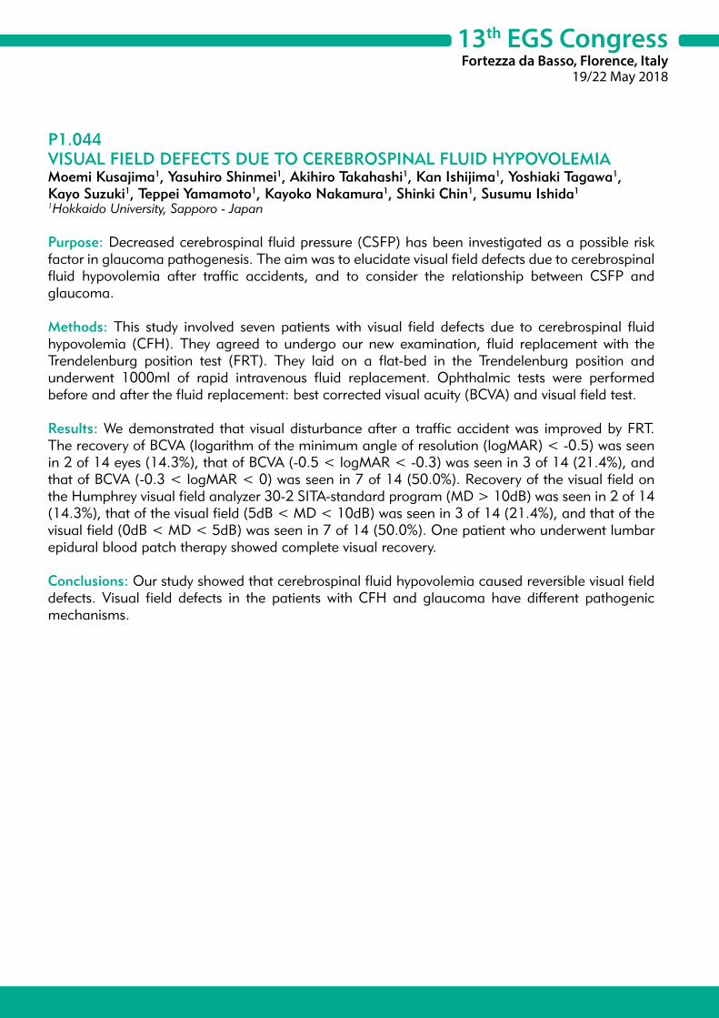
13th EGS CongressFortezza da Basso, Florence, Italy
19/22 May 2018
P1.044VISUAL FIELD DEFECTS DUE TO CEREBROSPINAL FLUID HYPOVOLEMIAMoemi Kusajima1, Yasuhiro Shinmei1, Akihiro Takahashi1, Kan Ishijima1, Yoshiaki Tagawa1, Kayo Suzuki1, Teppei Yamamoto1, Kayoko Nakamura1, Shinki Chin1, Susumu Ishida1
1Hokkaido University, Sapporo - Japan
Purpose: Decreased cerebrospinal fluid pressure (CSFP) has been investigated as a possible risk factor in glaucoma pathogenesis. The aim was to elucidate visual field defects due to cerebrospinal fluid hypovolemia after traffic accidents, and to consider the relationship between CSFP and glaucoma.
Methods: This study involved seven patients with visual field defects due to cerebrospinal fluid hypovolemia (CFH). They agreed to undergo our new examination, fluid replacement with the Trendelenburg position test (FRT). They laid on a flat-bed in the Trendelenburg position and underwent 1000ml of rapid intravenous fluid replacement. Ophthalmic tests were performed before and after the fluid replacement: best corrected visual acuity (BCVA) and visual field test.
Results: We demonstrated that visual disturbance after a traffic accident was improved by FRT. The recovery of BCVA (logarithm of the minimum angle of resolution (logMAR) < -0.5) was seen in 2 of 14 eyes (14.3%), that of BCVA (-0.5 < logMAR < -0.3) was seen in 3 of 14 (21.4%), and that of BCVA (-0.3 < logMAR < 0) was seen in 7 of 14 (50.0%). Recovery of the visual field on the Humphrey visual field analyzer 30-2 SITA-standard program (MD > 10dB) was seen in 2 of 14 (14.3%), that of the visual field (5dB < MD < 10dB) was seen in 3 of 14 (21.4%), and that of the visual field (0dB < MD < 5dB) was seen in 7 of 14 (50.0%). One patient who underwent lumbar epidural blood patch therapy showed complete visual recovery.
Conclusions: Our study showed that cerebrospinal fluid hypovolemia caused reversible visual field defects. Visual field defects in the patients with CFH and glaucoma have different pathogenic mechanisms.
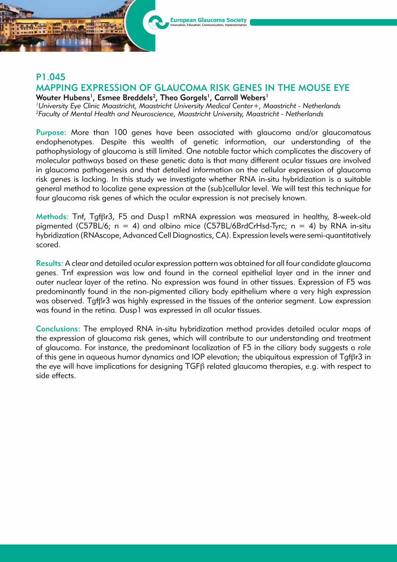
13th EGS CongressFortezza da Basso, Florence, Italy
19/22 May 2018
P1.045MAPPING EXPRESSION OF GLAUCOMA RISK GENES IN THE MOUSE EYEWouter Hubens1, Esmee Breddels2, Theo Gorgels1, Carroll Webers1
1University Eye Clinic Maastricht, Maastricht University Medical Center+, Maastricht - Netherlands2Faculty of Mental Health and Neuroscience, Maastricht University, Maastricht - Netherlands
Purpose: More than 100 genes have been associated with glaucoma and/or glaucomatous endophenotypes. Despite this wealth of genetic information, our understanding of the pathophysiology of glaucoma is still limited. One notable factor which complicates the discovery of molecular pathways based on these genetic data is that many different ocular tissues are involved in glaucoma pathogenesis and that detailed information on the cellular expression of glaucoma risk genes is lacking. In this study we investigate whether RNA in-situ hybridization is a suitable general method to localize gene expression at the (sub)cellular level. We will test this technique for four glaucoma risk genes of which the ocular expression is not precisely known.
Methods: Tnf, Tgfβr3, F5 and Dusp1 mRNA expression was measured in healthy, 8-week-old pigmented (C57BL/6; n = 4) and albino mice (C57BL/6BrdCrHsd-Tyrc; n = 4) by RNA in-situ hybridization (RNAscope, Advanced Cell Diagnostics, CA). Expression levels were semi-quantitatively scored.
Results: A clear and detailed ocular expression pattern was obtained for all four candidate glaucoma genes. Tnf expression was low and found in the corneal epithelial layer and in the inner and outer nuclear layer of the retina. No expression was found in other tissues. Expression of F5 was predominantly found in the non-pigmented ciliary body epithelium where a very high expression was observed. Tgfβr3 was highly expressed in the tissues of the anterior segment. Low expression was found in the retina. Dusp1 was expressed in all ocular tissues.
Conclusions: The employed RNA in-situ hybridization method provides detailed ocular maps of the expression of glaucoma risk genes, which will contribute to our understanding and treatment of glaucoma. For instance, the predominant localization of F5 in the ciliary body suggests a role of this gene in aqueous humor dynamics and IOP elevation; the ubiquitous expression of Tgfβr3 in the eye will have implications for designing TGFβ related glaucoma therapies, e.g. with respect to side effects.

13th EGS CongressFortezza da Basso, Florence, Italy
19/22 May 2018
P1.046GLAUCOMA-ON-A-CHIP: AN IN VITRO MODEL FOR GLAUCOMA DRUG DISCOVERY BASED ON MIMICKING THE MECHANICAL STRESS OF HIGH EYE PRESSURET.G.M.F. Gorgels1, P.A.M.M. Vroemen1, R. Sinha2, J. Rouwkema3, L. Moroni2, J. de Boer2, C.A.B. Webers1
1University Eye Clinic Maastricht, Maastricht University Medical Centre+, Maastricht - Netherlands2MERLN Institute for Technology-Inspired Regenerative Medicine, Maastricht University, Maastricht - Netherlands3Department of Biomechanical Engineering, Universtity of Twente, Enschede - Netherlands
Purpose: The pathology of glaucoma is characterized by optic neuropathy. At the cellular level, there is the loss of retinal ganglion cells (RGCs). Since the loss of these specialized neurons is irreversible, it is urgent to develop treatments that protect these cells. To achieve this, an in vitro glaucoma model would be very useful. The purpose of this study is to develop such a model by culturing RGCs and exposing them to conditions that mimick increased ocular pressure (IOP), the main risk factor of glaucoma.
Methods: Elevated IOP leads to increased hydrostatic pressure in the eye, causing, amongst others, deformation and stretching of cells. To mimick this, we need a device that can apply both hydrostatic pressure and stretch simultaneously to cultured cells. Stretching should be applied dynamically, in order to account for fluctuations in IOP that occur naturally in vivo. To realize this, we modified a recently developed device for stretch and shear stress (Sinha et al. Lab Chip 2015; 15 429-439). We use the immortalized neuronal PC12 cell line to establish the model; later RGCs will be used. Output parameters include cell density, survival and morphology.
Results: A medium-throughput cell culture device has been constructed that can apply hydrostatic pressure via the culture medium (0-90 mmHg). In addition, simultaneously, cells can be dynamically stretched anisotropically and isotropically ranging from 0 to 20% (in 5 steps). All experimental conditions are represented in fourfold on the device. First experiments using PC12 cells indicate that cyclic (1 Hz) stretch (10%) applied for 2 days, results in reduced cell density as compared to control (no stretch). Variation in cell density was large, probably related to variation in seeding density. To cope with this variation, we now also record the initial cell density, before applying mechanical strain, and use this for normalization.
Conclusions: We constructed a cell culture device that can apply pressure and stretch to cultured cells. This glaucoma-on-a-chip will help to identify the molecular mechanisms of mechanically-induced RGC death and help to design neuroprotective treatments. In addition, it can serve to characterize sensitivity of patients cells to mechanical stress.
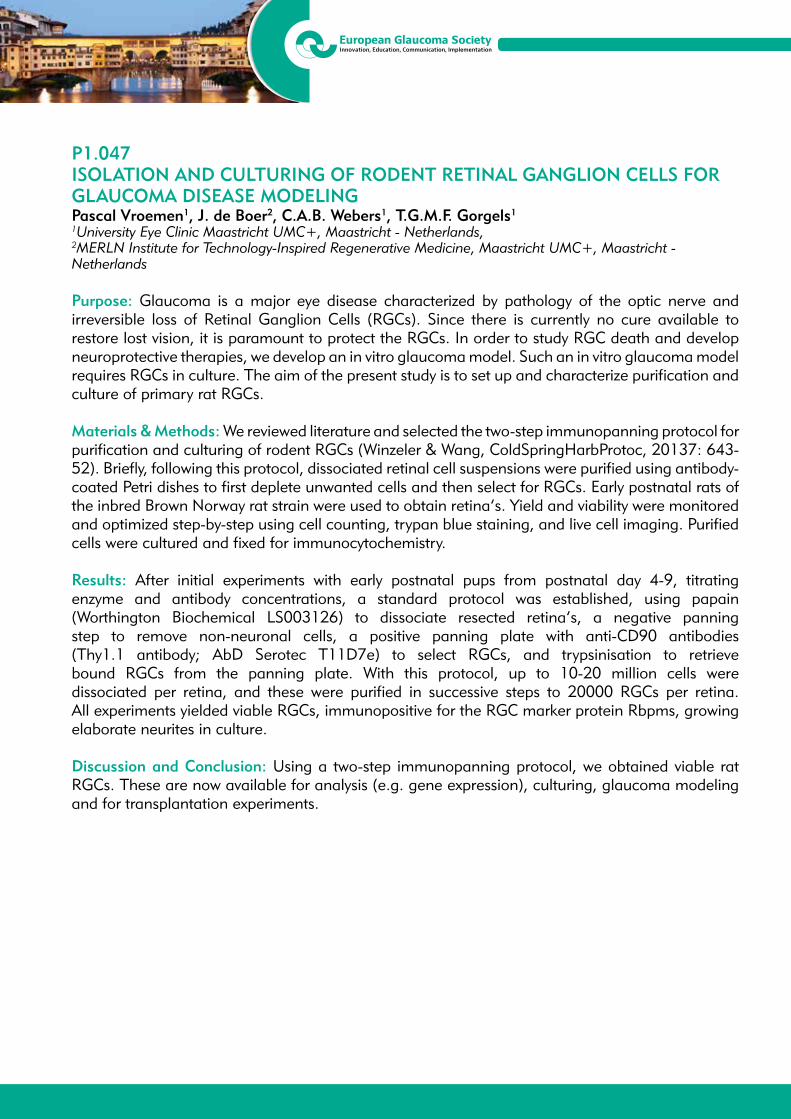
13th EGS CongressFortezza da Basso, Florence, Italy
19/22 May 2018
P1.047ISOLATION AND CULTURING OF RODENT RETINAL GANGLION CELLS FOR GLAUCOMA DISEASE MODELINGPascal Vroemen1, J. de Boer2, C.A.B. Webers1, T.G.M.F. Gorgels1
1University Eye Clinic Maastricht UMC+, Maastricht - Netherlands, 2MERLN Institute for Technology-Inspired Regenerative Medicine, Maastricht UMC+, Maastricht - Netherlands
Purpose: Glaucoma is a major eye disease characterized by pathology of the optic nerve and irreversible loss of Retinal Ganglion Cells (RGCs). Since there is currently no cure available to restore lost vision, it is paramount to protect the RGCs. In order to study RGC death and develop neuroprotective therapies, we develop an in vitro glaucoma model. Such an in vitro glaucoma model requires RGCs in culture. The aim of the present study is to set up and characterize purification and culture of primary rat RGCs.
Materials & Methods: We reviewed literature and selected the two-step immunopanning protocol for purification and culturing of rodent RGCs (Winzeler & Wang, ColdSpringHarbProtoc, 20137: 643-52). Briefly, following this protocol, dissociated retinal cell suspensions were purified using antibody-coated Petri dishes to first deplete unwanted cells and then select for RGCs. Early postnatal rats of the inbred Brown Norway rat strain were used to obtain retina’s. Yield and viability were monitored and optimized step-by-step using cell counting, trypan blue staining, and live cell imaging. Purified cells were cultured and fixed for immunocytochemistry.
Results: After initial experiments with early postnatal pups from postnatal day 4-9, titrating enzyme and antibody concentrations, a standard protocol was established, using papain (Worthington Biochemical LS003126) to dissociate resected retina’s, a negative panning step to remove non-neuronal cells, a positive panning plate with anti-CD90 antibodies (Thy1.1 antibody; AbD Serotec T11D7e) to select RGCs, and trypsinisation to retrieve bound RGCs from the panning plate. With this protocol, up to 10-20 million cells were dissociated per retina, and these were purified in successive steps to 20000 RGCs per retina. All experiments yielded viable RGCs, immunopositive for the RGC marker protein Rbpms, growing elaborate neurites in culture.
Discussion and Conclusion: Using a two-step immunopanning protocol, we obtained viable rat RGCs. These are now available for analysis (e.g. gene expression), culturing, glaucoma modeling and for transplantation experiments.

13th EGS CongressFortezza da Basso, Florence, Italy
19/22 May 2018
P1.048SYSTEMIC AND OCULAR DETERMINANTS OF MEAN OCULAR PERFUSION PRESSURE IN A POPULATION-BASED SAMPLENilgün Yildirim1, Eray Atalay1, Leyla Niyaz2, Saadet Gültekin3
1Department of Ophthalmology, Eskisehir Osmangazi University Faculty of Medicine, Eskisehir - Turkey2Department of Ophthalmology, Ondokuz Mayis University Faculty of Medicine, Samsun - Turkey3Ophthalmology Clinic, Kütahya Evliya Çelebi Egitim Arastirma Hastanesi, Kutahya - Turkey
Purpose: To investigate the associations between mean ocular perfusion pressure (MOPP) and several variables including body mass index (BMI), comorbid medical conditions and various ocular parameters.
Methods: Data of 2091 healthy participants from a population based cross-sectional study were reviewed. Inclusion criteria were adults ≥ 40 years of age who responded to an invitation to undergo screening for glaucoma. During the data collection session, participants were initially interviewed face-to-face using a standard questionnaire which aimed to collect data on the medical history. Height, weight, systolic and diastolic blood pressures (SBP and DBP, respectively) were measured. A basic ocular examination was performed which included intraocular pressure (Tono-Pen XL) and central corneal thickness (Pacline pachymetry; Optikon, Rome, Italy) measurement, slit-lamp examination and non-mydriatic optic disc photography (nonmydα fundus camera, Kowa). MOPP was calculated using the formula [2/3 x (DBP + 1/3(SBP-DBP)]-IOP and low MOPP was defined as MOPP ≤ 45 mmHg.
Results: Mean age of the subjects was 63.04 ± 9.7 years (range: 44 and 99 years) and the majority were female (74.1%, n = 1549). Obesity (BMI ≥ 30) was observed in 35.2% (n = 737) of the sample. Mean MOPP values in normal weight (BMI < 25), overweight (BMI = 25-29.9) and in obese individuals were 46.9 ± 9.0 mmHg, 48.6 ± 9.2 mmHg and 50.7 ± 10.0 mmHg, respectively (p < 0.001, in all pairwise comparisons). Mean MOPP was significantly different across the age groups (p < 0.001, in all pairwise comparisons) with the highest MOPP noted in subjects aged > 70 years (52.0 ± 9.5 mmHg) followed by the age range 56-59 years (49.2 ± 9.4 mmHg) and ≤ 55 years (45.2 ± 8.8 mmHg). In the multivariable logistic regression analysis, migraine and elevated IOP (≥ 21mmHg) were significantly associated with a low MOPP (OR: 1.99 and 6.53, p = 0.01 and < 0.001, respectively). On the contrary, risk of low MOPP was reduced in subjects with hypertension, obesity and with increasing age.
Conclusions: Migraine and elevated IOP increases the risk of low MOPP and this may have a causal relationship with impaired optic nerve head blood flow.

13th EGS CongressFortezza da Basso, Florence, Italy
19/22 May 2018
P1.049STRUCTURAL AND VASCULAR EVALUATION OF OPTIC NERVE AND MACULA USING OPTICAL COHERENCE TOMOGRAPHY ANGIOGRAPHY IN HYPERTENSIONBarbara Burgos-Blasco1, Javier Pascual-Prieto1, Jose Maria Martínez-de-la-Casa1, Noemi Güemes-Villahoz1, Liseth Salazar-Quiñones1, Mario Avila-Sanchez2, Maria Aranzazu Barbero-Pedraz2, Federico Saenz-Frances1, Sofia García-Saénz1, Julian García-Feijoo1
1Department of Ophthalmology, Hospital Clinico San Carlos, Madrid - Spain2Hypertension Unit, Department of Internal Medicine, Hospital Clinico San Carlos, Madrid - Spain
Purpose: To evaluate the differences in the optic nerve head parameters, the nerve fibre layer, the ganglion cell complex and the vascular density in normal subjects and patients with low and very high risk hypertension using Optical Coherence Tomography Angiography (OCTA).
Materials and Methods: An observational study was performed. 73 eyes of 73 normal subjects and hypertensive patients from the Hospital Clinico San Carlos Hypertension Unit were included. Patients from the Hypertension Unit were stratified by cardiovascular risk according to the European Society of Cardiology guidelines. Patients with low and very high risk cardiovascular risk were included in this study. All subjects underwent 4.5 × 4.5 mm OCTA scan centered on the fovea and optic nerve, using an OCT RS-3000 (Nidek Co, Gamagori, Japan). Main outcome measures were macular and peripapillary vascular density, the ganglion cell complex thickness, optic nerve head parameters and retinal nerve fibre layer thickness.
Results: A total of 73 patients were included in this study with a mean age of 61.75 ± 7.5 years. There were 26 (36%), 24 (33%), and 23 (31%) patients in the control, low risk, and very high risk group, respectively.Significant differences were found between groups in vascular density at the macular deep capillary plexus (p = 0.025) and also in the macular superficial complex capillary plexus (p = 0.047). A significant decrease was also observed in the macular thickness in 4 out of 8 examined zones (p = 0.041, p = 0.008, p = 0.007 and p=0.012). No statistically significant differences were observed in the vascular density at the optic nerve. The other parameters analysed did not show statistically significant differences either.
Conclusion: High risk hipertensive patients have a significant decrease in macular vascular density and thickness compared with normal subjects and low risk patients. This differences can be assesed with OCTA in a non-invasive way.
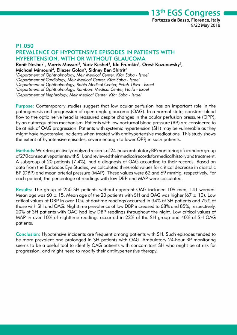
13th EGS CongressFortezza da Basso, Florence, Italy
19/22 May 2018
P1.050PREVALENCE OF HYPOTENSIVE EPISODES IN PATIENTS WITH HYPERTENSION, WITH OR WITHOUT GLAUCOMARonit Nesher1, Morris Mosseri2, Yariv Keshet3, Ido Frumkin1, Orest Kazanovsky2, Michael Mimouni4, Eliezer Golan5, Sidney Ben Shitrit5
1Department of Ophthalmology, Meir Medical Center, Kfar Saba - Israel2Department of Cardiology, Meir Medical Center, Kfar Saba - Israel3Department of Ophthalmology, Rabin Medical Center, Petah Tikva - Israel4Department of Ophthalmology, Rambam Medical Center, Haifa - Israel5Department of Nephrology, Meir Medical Center, Kfar Saba - Israel
Purpose: Contemporary studies suggest that low ocular perfusion has an important role in the pathogenesis and progression of open angle glaucoma (OAG). In a normal state, constant blood flow to the optic nerve head is reassured despite changes in the ocular perfusion pressure (OPP), by an autoregulation mechanism. Patients with low nocturnal blood pressure (BP) are considered to be at risk of OAG progression. Patients with systemic hypertension (SH) may be vulnerable as they might have hypotensive incidents when treated with antihypertensive medications. This study shows the extent of hypotensive episodes, severe enough to lower OPP, in such patients.
Methods: We retrospectively analyzed records of 24-hour ambulatory BP monitoring of a random group of 270 consecutive patients with SH, and reviewed their medical records for medical history and treatment. A subgroup of 20 patients (7.4%), had a diagnosis of OAG according to their records. Based on data from the Barbados Eye Studies, we calculated threshold values for critical decrease in diastolic BP (DBP) and mean arterial pressure (MAP). These values were 62 and 69 mmHg, respectively. For each patient, the percentage of readings with low DBP and MAP were calculated.
Results: The group of 250 SH patients without apparent OAG included 109 men, 141 women. Mean age was 60 ± 15. Mean age of the 20 patients with SH and OAG was higher (67 ± 10). Low critical values of DBP in over 10% of daytime readings occurred in 34% of SH patients and 75% of those with SH and OAG. Nighttime prevalence of low DBP increased to 68% and 85%, respectively. 20% of SH patients with OAG had low DBP readings throughout the night. Low critical values of MAP in over 10% of nighttime readings occurred in 22% of the SH group and 40% of SH-OAG patients.
Conclusion: Hypotensive incidents are frequent among patients with SH. Such episodes tended to be more prevalent and prolonged in SH patients with OAG. Ambulatory 24-hour BP monitoring seems to be a useful tool to identify OAG patients with concomitant SH who might be at risk for progression, and might need to modify their antihypertensive therapy.

13th EGS CongressFortezza da Basso, Florence, Italy
19/22 May 2018
P1.051A NOVEL TISSUE ENGINEERED BIOMIMETIC MODEL OF THE LAMINA CRIBROSA REGION FOR GLAUCOMA RESEARCHDeirdre Clissmann1, Rory Murphy2, Rebecca Low1, Alan Hopkins2, Mustapha Irnaten2, Deborah Wallace3, Alan Hibbitts4, Alan Ryan4, Fergal O’Brien4, Colm O’Brien2, Deirdre Brennan1
1School of Medicine and Medical Science, University College Dublin, Dublin - Ireland2Institute of Ophthalmology, Mater Misericordiae University Hospital, Dublin - Ireland3Clinical Research Centre, Catherine McAuley Centre, School of Medicine, University College Dublin, Dublin - Ireland4Tissue Engineering Research Group (TERG), Dept. of Anatomy, Royal College of Surgeons in Ireland, Dublin - Ireland
Purpose: The lamina cribrosa (LC) region of the optic nerve head (ONH) is a key initial site of glaucomatous damage. Loss of compliance (stiffening) within the LC is thought to play a pivotal role in fibrotic ONH remodeling, governed by the resident LC cells1,2. Traditionally, LC cells have been cultured in-vitro on flat plastic cell culture dishes. However, such 2D models neglect the complex interplay that occurs between the LC cells and their surrounding 3D tissue microenvironment. Thus, the aim of this study was to create a novel, 3D in-vitro model of the stiffened glaucomatous LC region using bioengineered scaffolds.
Methods: Collagen/glycosaminoglycan scaffolds (8mm width, porosity 40-50 μm) were fabricated using a freeze-drying method3. Dehydrothermal and 1-ethyl-3-3-dimethyl aminopropyl carbodimide crosslinking was utilised to produce soft (0.5 kPa, 1 kPa) and stiff (1.5 kPa) unit scaffold compliance reference values. Scaffolds were seeded with primary human LC cells at 3500 cells/mm2 using a dual-seeding method on both top and bottom scaffold surfaces. Cell-seeded scaffolds were subsequently harvested after 2 days in culture and processed for histological evaluation.
Results: The 0.5kPa scaffold proved non-viable, as it experienced extensive shrinkage and loss of mechanical integrity. However, the microporous 3D structure was maintained in both the soft (1 kPa) and stiff (1.5 kPa) scaffolds. Histological evaluation showed that the primary human LC cells successfully integrated into both the 1kPa and 1.5 kPa scaffolds.
Conclusions: In conclusion, primary human LC cells have successfully been grown for the first time on biomimetic ‘soft’ and glaucomatous ‘stiff’ LC scaffolds. This 3D model system is more representative of the in-vivo LC cellular microenvironment by comparison to traditional 2D systems. Creation of this 3D cell model will allow future in-depth investigations of the LC cellular phenotype in response to glaucomatous fibrotic conditions.
References:1. Wallace DM, O’Brien CJ. The role of lamina cribrosa cells in optic nerve head fibrosis in glaucoma.
Exp Eye Res 2016 Jan;142: 102-9.2. Liu B, McNally S, Kilpatrick JI, Jarvis SP, O’Brien CJ. Aging and ocular tissue stiffness in glaucoma.
Surv Ophthalmol 2018 Jan - Feb; 63(1): 56-74. 3. O’Brien FJ, Harley BA, Yannas IV, Gibson L. Influence of freezing rate on pore structure in freeze-
dried collagen-GAG scaffolds. Biomaterials 2004 Mar; 25(6): 1077-86.
13th EGS CongressFortezza da Basso, Florence, Italy
19/22 May 2018
P1.052SERUM HOMOCYSTEINE, VITAMIN B12 AND FOLIC ACID LEVELS IN DIFFERENT TYPES OF OPEN ANGLE GLAUCOMAMilan Stojcic1, Milenko Stojkovic2, Vera Lukic3, Biljana Stojcic4, Lepša Zoric5, Zoran Bukumiric6
1Railway Health Care Institute, Ophthalmology Department, Belgrade - Serbia2University of Belgrade, Medical Faculty, Clinic for Eye Diseases, Belgrade - Serbia3Railway Health Care Institute, Department of Biochemistry, Belgrade - Serbia4Primary Health Center Euromedik, Belgrade - Serbia5Ophthalmology Department, Health Center Kosovska Mitrovica and Faculty of Medicine, University of Pristina/Kosovska Mitrovica, Kosovska Mitrovica - Serbia6Institute for Medical Statistics and Informatics, University of Belgrade, Medical Faculty, Belgrade - Serbia
Purpose: The aim of this study was to compare levels of serum homocysteine (Hcy), vitamin B12 and folic acid in patients with primary open-angle glaucoma (POAG), normal tension glaucoma (NTG), exfoliation glaucoma (XFG) and healthy controls.
Methods: Twenty patients with POAG, 19 with NTG, 20 with XFG and 18 healthy controls who met the inclusion/exclusion criteria were included in this prospective cross-sectional study. Hcy levels, vitamin B12 and folic acid were measured in venous blood samples of all of subjects. The concentrations of serum homocysteine, vitamin B12, and folate are determined by the technique of chemiluminescent micro-cellular immunoassays (CMIA), on the immunochemical analyzer of the Architect i2000 manufacturer Abbott,with the original reagents.
Results: Mean ± standard deviation of Hcy levels in the POAG group was 10.60 ± 2.53 μmol/L, in the NTG was 10.62 ± 3.59 μmol/L, in the XFG was 13.06 ± 3.41 μmol/L, and in the controls was 10.46 ±1.86 μmol/L. The mean Hcy concentration in the XFG group was significantly higher (p = 0.018) as compared to the other groups. Pairwise comparisons between groups show that mean Hcy concentration in the XFG group was significantly different from those in POAG group and controls (p < 0.05), but not in those in NTG group, although this statistical difference was borderline significant (p = 0.056). Mean ± SD of vitamin B12 levels in POAG, NTG, XFG and controls were 252.2 ± 91.05, 257.8 ± 78.5, 266.3 ± 86.8, 254.9 ± 70.6 pmol/L respectively. There were no statistical differences in vitamin B 12 levels among POAG,NTG, XFG and controls (p = 0.955). Mean ± SD of folic acid concentrations in POAG, NTG, XFG and controls were 15.9 ± 5.62, 15.22 ± 6.80, 12.94 ± 6.35, 14.59 ± 4.40 nmol/l respectively. Although folic acid level reduced in XFG group, no statistically significant difference was found between study groups (p = 0.432).
Conclusion: Our study demonstrated a positive correlation between the level of Hcy and XFG and is in accordance with the results of previously published studies. HHcy might play a role in the pathogenesis of XFG.
13th EGS CongressFortezza da Basso, Florence, Italy
19/22 May 2018
P1.053PROTEOMIC EXPRESSION PROFILE OF THE IRIS IN PRIMARY GLAUCOMA: A PILOT REPORTArun Narayanaswamy1, Monisha Nongpiur1, Lei Zhou2, Tina Wong1, Tin Aung1
1Singapore National Eye Centre, Singapore - Singapore2Singapore Eye Research Institute, Singapore - Singapore
Purpose: To report the proteomic expression profile of the iris tissue in primary glaucomas
Methods: Iris tissue specimens were obtained from peripheral surgical iridectomies performed as part of standard phaco- trabeculectomy and cataract procedures. Specimens were obtained from 10 eyes diagnosed with primary open angle glaucoma (POAG), 9 with primary angle closure glaucoma (PACG) and 4 normal eyes. Fresh iris tissue was dissected, immediately frozen on dry ice, and stored at - 80ºC until prior to analysis. The tissues were freeze-pulverized and proteins were sequentially extracted based on solubility. The extracted proteins were digested and labeled with iTRAQ reagents (AB SCIEX). The peptide mixture was then analyzed using nano-liquid chromatography-tandem mass spectrometry (nanoLC-MS/MS). Significant up-regulation and down-regulation were graded using a benchmark of 2-fold change.
Results: A total of 223 proteins were identified (False discovery rate < 1%, peptide confidence level > 95%) in all samples. Of these, 214 were quantifiable. Thirty three proteins were significantly up-regulated in both POAG and PACG, whilst 20 proteins were differentially up regulated in POAG and 24 in PACG. Likewise, 15 proteins were down-regulated in both the subtypes, while 19 and 13 were differentially downregulated in POAG and PACG respectively.
Conclusion: Differential levels of the identified protein bio-markers may provide a better understanding of the pathophysiology of glaucoma and more specifically their sub-types.

13th EGS CongressFortezza da Basso, Florence, Italy
19/22 May 2018
P1.054CHANGES IN THE OPTIC NERVE HEAD INDUCED BY HORIZONTAL EYE MOVEMENTSWon June Lee1, Han Woong Lim2
1Department of Ophthalmology, Seoul National University Hospital, Seoul - Republic of Korea2Department of Ophthalmology, Hanyang University Hospital, Seoul - Republic of Korea
Purpose: To investigate the effect of eye movement on the optic nerve head (ONH) using swept-source optical coherence tomography (SS-OCT), and to measure the degree of ONH changes.
Methods: This is the prospective observational study. Both optic nerve head and macula were imaged simultaneously using wide volume scan of the SS-OCT in the primary and different gaze positions. Horizontal eye movements were used to obtain OCT images in abducted and adducted eyeball positions. Multilateral three-dimensional reconstruction was used to process and analyze the images to measure the degree of ONH changes. We measured the area of optic nerve head change.
Results: Significant morphologic changes were observed in the ONH during both abduction and adduction. In abduction, the overall ONH tissues were elevated, and the mean area of elevation was 115,134 ± 9,424 μm2 (p < 0.001). In adduction, the mean areas from two perspectives, which were nasal or temporal, and peripapillary tissues or optic nerve cupping were 95,277 ± 73,846 μm2, 34,450 ± 44,948 μm2, -108,652 ± 91,246 μm2, and -30,581 ± 46,249 μm2, respectively. Elevation in abduction (overall, nasal cup segment, and temporal cup segment; R = 0.204, 0.195 and 0.225, p = 0.038, 0.047 and 0.021, respectively) and elevation of nasal peripapillary segments in adduction were positively correlated with axial length (R=0.346, p < 0.001).
Conclusions: We found significant morphologic changes in the ONH in both abduction and adduction and these changes were associated with axial length. Considering these morphologic changes as physical properties, it allows a better understanding of the biomechanical characteristics of the optic nerve head.

13th EGS CongressFortezza da Basso, Florence, Italy
19/22 May 2018
P1.055LAMINA CRIBROSA CELL BIOENERGETICS IN GLAUCOMA: ROLE OF GLYCOLYSIS AND GLUTAMINOLYSISDiarmaid Hickey1, Mustapha Irnaten1, Deirdre Brennan2, Deborah Wallace3, Abe Clark4, Colm J. O’Brien1
1Institute of Ophthalmology, Mater Misericordiae University Hospital, Dublin - Ireland2Anatomy, School of Medicine, University College Dublin, Dublin - Ireland3Clinical Research Centre, Catherine McCauley Centre, School of Medicine, University College Dublin, Dublin - Ireland4North Texas Eye Research Institute (NTERI), Health Science Center, University of North Texas, Fort Worth - USA
Purpose: In glaucoma, the lamina cribrosa (LC) region undergoes considerable 3D structural changes relating to significant alterations in the extracellular matrix and associated cupping. Cells mediating this cupping include astrocytes and LC cells. We have previously shown mitochondrial dysfunction and an increased rate of proliferation in glaucomatous LC cells (GLC). Rapidly proliferating cells utilise alternative metabolic pathways to meet energy requirements. Monocarboxylate transporter 1 (MCT1) and glutaminase 2 (GLS2) are known to play an essential role in cancer cell metabolism. Glycolysis (the Warburg effect) results in high lactate levels, increasing the expression of MCT1. Glutamine uptake and metabolism is a key part of biomass accumulation in rapidly proliferating cells. The purpose of our research was to investigate the expression of markers (MCT1 and GLS2) associated with an enhanced glycolysis and glutaminolysis phenotype. Methods: Human primary LC cells derived from normal and glaucomatous age-matched donors were cultured between passages 4-8. At confluence, cells were subject to either RNA extraction or protein isolation. MCT1 and GLS2 expression levels were quantified using quantitative real time (qRT-PCR) and western immunoblotting. Results: The results showed that the PCR transcription level of both MCT1 and GLS2 was significantly elevated in GLC (39.14 ± 3.17 fold change in gene expression) versus normal LC cells (NLC) (31.34 ± 2. 91), (n = 3, p < 0.05) for MCT1 and (35.69 ± 3.15) versus NLC (17.63 ± 2.16) (n = 3, p < 0.02) for GLS2. This was confirmed at the protein expression level, as western immunoblotting analysis showed that the expression of both MCT1 and GLS2 was significantly elevated in GLC (9.41 ± 1.29 a.u) versus NLC (6.04 ± 1.23 a.u), (n = 3, p < 0.05) for MCT1 and (8.67 ± 1.23 a.u) versus NLC (4.95 ± 0.98 a.u) (n = 3, p < 0.05) for GLS2. Conclusion: We found elevated expression of MCT1 and GLS2 both at transcript and protein levels, indicating enhanced glycolysis and glutaminolysis in glaucomatous LC cells. This is new evidence that glaucomatous LC cells utilise alternative metabolic pathways. Blocking these pathological pathways or facilitating physiological pathways (i.e. oxidative phosphorylation) could be a potential therapeutic in glaucoma.

13th EGS CongressFortezza da Basso, Florence, Italy
19/22 May 2018
P1.056GENE EXPRESSION OF TGF BETA ISOFORMS AND ITS RECEPTORS IN THE PARTS OF TRABECULAR MESHWORK IN PATIENTS WITH GLAUCOMA - A PRELIMINARY REPORTWojciech Maruszczyk1, Malgorzata Kimsa2, Malgorzata Machalska3, Mariola Dorecka1, Barbara Strzalka-Mrozik2, Urszula Mazurek2, Ewa Mrukwa-Kominek1
1Department of Ophthalmology, Prof. K. Gibinski University Clinical Centre, School of Medicine in Katowice, Medical University of Silesia in Katowice, Katowice - Poland2Department of Molecular Biology, School of Pharmacy with the Division of Laboratory Medicine in Sosnowiec, Medical University of Silesia in Katowice, Sosnowiec - Poland3Ophthalmology Ward, Prof. K. Gibinski University Clinical Center, University Hospital, Katowice - Poland
Purpose: Glaucoma is chronic ophthalmological disease witch cause irreversible optic disc and retinal ganglion cells damage occurring as a visual field lost. The pathogenesis of this condition is still uncertain. Nowadays, the obstruction on trabecular meshwork level is one of the most potent cause of this disease, in which transforming growth factor beta (TGFβ) may play a role. The aim of this study is to evaluate the gene expression of isoforms of TGFβ and its receptors, at the mRNA level, in the removed parts of the iridocorneal angle (trabeculum) in patients with glaucoma who underwent trabeculectomy with basal iridectomy in The Department of Ophthalmology, Prof. K. Gibinski University Clinical Centre, School of Medicine in Katowice, Medical University of Silesia in Katowice, Katowice - Poland. Study was performed due to Medical University of Silesia in Katowice grants No KNW-1-190/N/4/0 and KNW-2-047/D/4/N.
Methods: The study group included 19 eyes qualified for glaucoma surgery, trabeculectomy with basal iridectomy. Diagnostic material, collected from each patient, included a part of the iridocorneal angle. Tissues were collected into separate tubes with buffer to prevent the degradation of RNA (TRIzol), and then stored at -20stC until further assays. Total RNA extraction was performed using TRIzol reagent according to the manufacturer’s instructions. QRT-PCR reaction was performed (TGFβ1, TGFβ2, TGFβ3, TGFβ1R, TGFβ2R, TGFβ3R and 2 endogenous controls - GAPDH and β-actin. PAGE and fixing of the melting temperature confirmed the specificity of the PCR reactions.
Results: There were statistically significant differences in the gene expression of TGFβ isoforms - TGFβ1: 17418.45; TGFβ2: 6041.48; TGFβ3: 7240.11 [mRNA copies / ug of RNA] (p = 0.0035). There were no statistically significant differences in the gene expression of TGFβ receptors - TGFβ1R: 11388.42; TGFβ2R: 13152.66; TGFβ3R: 5645.16 [mRNA copies / ug of RNA] (p > 0.05).
Conclusions: The study confirmed the presence of TGF beta isoforms and its receptors in the trabeculum in glaucoma patients. Some differences in the expression of the TGF beta isoforms were observed.

13th EGS CongressFortezza da Basso, Florence, Italy
19/22 May 2018
P1.057RARE CASE OF RIGHT SIDE HORNER SYNDROME AND SIMULTANEOUS PIGMENTARY GLAUCOMA OF THE LEFT EYEKonstantinos Moustaklis1, Spyridon Gorezis2
1Private Ophthalmology Office, Ioannina - Greece2Epirus Vision Center, Ioannina - Greece
Purpose: To report a rare case of simultaneous presence of Horner syndrome and pigmentary glaucoma and how this coexistence actually benefits the patient.
Materials and Methods: A 37 years old patient presented to the office for routine periodical examination of his refraction. Slit lamp examination in the right eye was within normal limits while in the left eye it revealed Krukenberg’s spindle and hypertony (36 mmHg). Further clinical examination showed also anisocoria, heterochromia iridum and ptosis of the right eyelid. Finally we were led to the conclusion that the patient suffered from right side Horner’s syndrome and pigmentary glaucoma of the left eye. Neither signs of pigmentary glaucoma nor even pigment dispersion signs were found in the right eye. We expect pigment dispersion that leads to pigmentary glaucoma to be a bilateral condition. However in this case, it seems that the myosis caused to the right eye by the interruption of the sympathetic nerve supply actually protects the right eye from developing pigment dispersion syndrome and high ocular pressure. Structural and morphological changes of the iris are evident in the AC-OCT examinations.
Results: Follow up examinations during a three years’ time period never showed signs of pigment dispersion or intraocular pressure higher than 18 mmHg. The left eye pressure is controlled with prostaglandin analog (latanoprost).
Conclusion: This rare case provides us with insight to the etiology of pigment dispersion syndrome and pigmentary glaucoma and to the role of the sympathetic nervous system in the induction of mechanical friction that releases pigment in eyes with certain anatomical characteristics

13th EGS CongressFortezza da Basso, Florence, Italy
19/22 May 2018
P1.058ANTERIOR SEGMENT CIRCULATING NEUROTOXIC CYTOKINES IN PRIMARY OPEN ANGLE GLAUCOMA PATIENTSAnca Pantalon1, Dorin Chiselita1, Crenguta Feraru1
1“Gr.T.Popa” Univerisity of Medicine and Pharmacy, Ophthalmology Clinic - “Sf. Spiridon” Univerisity Hospital, Iasi - Romania
Typically in glaucoma the injuries in the retinal ganglion cells are irreversible and mostly due to high intraocular pressure. Currently there are accepted pathogenic theories that go beyond high intraocular pressure in the area of neuroinflammation, autoimmunity or vascular dysfunction. AIM of our study was to identify and compare levels of anterior segment circulating inflammatory cytokines such as IL-1Ra (Interleukin-1 receptor antagonist), IL-1α (Interleukin-1α), IL-1β (Interleukin-1β), IL-10 (Interleukin 10) and IFNγ (interferon-gamma) in glaucoma patients.
Material and Method: We included only primary open angle glaucoma cases which were compared to healthy controls, matched for age and sex. For all subjects we recorded: age, best corrected visual acuity (decimal), intraocular pressure (mmHg) by Goldmann tonometry, MD/PSD in Humphrey perimetry 24-2, SITA Standard. Duration of glaucoma, type and number of IOP lowering substances were noted. All glaucoma patients received non-preserved topical medication at the time of inclusion. None of the patients had a history of glaucoma surgery, including laser. Cataract surgery was indicated by the ophthalmologist at a time when there was significant lens opacification. Patients with significant ophthalmological disorders or general that could have influenced the ocular local and systemic inflammation status were excluded.
Results: In the study, we included 40 eyes from 40 patients: 24 eyes in the control group (cataract) and 16 eyes in the study group (POAG). The mean age was 72.33 ± 11.26 years in controls vs 75.69 ± 5.54 years in POAG group, (p > 0.05). The mean IOP in the control group was 14.21 ± 2.68 mmHg, whereas in the study group the mean was significantly higher 18.19 ± 4.3 mmHg (p = 0.000), under 3 ± 0.87 hypotensive drugs. Functional impairment in POAG patients revealed means of 13.59 +/- 9.35 dB for MD and 4.25 ± 4.22 dB for PSD. Significant differences were found for IFNγ (3 ± 0.35 pg/ml in controls vs 3.39 ± 1.63 pg/ml in POAG, p = 0.017), IL-1 (9.51 ± 4.83 pg/ml in controls vs 14.73 ± 5.02 pg/ml in POAG, p = 0.002) and IL-1Ra (656.35 ± 518.31 pg/ml in controls vs 2276 ± 3017.65 pg/ml in POAG, p = 0.01). Age or sex didn’t influence the cytokines levels, but an IOP higher than 18 mmHg increased the level of IFNγ in the POAG group in a significant way, compared to the category of IOP below 18 mmHg (p = 0.003). Same observation was made for IL-1α where an IOP higher than 18mmHg increased the cytokine level in a statistically significant manner (p = 0.000); in this particular case younger patients (< 60 years old) developed a stronger inflammatory response than those with age > 60 years old.
Conclusion: Our results show an increased expression of inflammatory molecules with neurotoxicity capabilities in primary open angle glaucoma patients. Moreover the magnitude of the activation is dependent of the age and IOP levels in POAG patients compared to healthy subjects.
13th EGS CongressFortezza da Basso, Florence, Italy
19/22 May 2018
P1.059DEEP OPTIC NERVE HEAD MORPHOLOGY ARE ASSOCIATED WITH PATTERN OF GLAUCOMATOUS VISUAL FIELD DEFECT IN OPEN ANGLE GLAUCOMAJong Chul Han1, Changwon Kee1, Jae Hwan Choi1, Eun Jung Lee1, Do Young Park1
1Department of Ophthalmology, Samsung Medical Center, Sungkyunkwan University School of Medicine, Seoul - Republic of Korea
Purpose: To investigate the relationship between visual field (VF) defect pattern and deep optic nerve head (ONH) morphology in open angle glaucoma (OAG) eyes using spectral-domain (SD) optical coherence tomography (OCT).
Methods: One hundred sixty-nine OAG eyes with the presence of externally oblique border tissue (EOBT) and 55 OAG eyes without presence of EOBT were included in the analysis. Enhance depth imaging SD-OCT were used to measure the deep ONH parameters such as EOBT length, ONH tilt angle and optic canal (OC) obliqueness. The maximal value of each parameter was defined as the maximum deep ONH parameter. VF defect patterns were classified into two groups according to the dominant VF defect location (superior vs. inferior) and the presence of paracentral scotoma. The extents and the locations of the maximum deep ONH parameters were explored according to VF pattern in OAG eyes.
Results: OAG eyes with presence of EOBT showed longer axial length (AL) and greater cases of superior VF defect compared to OAG eyes without EOBT (p < 0.001, p = 0.02, respectively). Multivariate logistic regression analysis revealed that more inferiorly location of maximum deep ONH parameters were associated with superior hemifield defect dominant in OAG (p < 0.001 in all parameters). When only OAG eyes with AL < 24.5 mm or IOP ≥ 19 mmHg were included, the results exhibited the spatial correspondence between the locations of maximum deep ONH parameter and dominant VF defect. The presence of paracentral scotoma in OAG was associated with worse mean deviation (MD) and relatively inferior location of deep ONH parameters.
Conclusions: The locations of maximum deep ONH parameters such as EOBT length, ONH tilt angle and OC obliqueness were associated with the location of dominant VF defect and the presence of paracentral scotoma in OAG eyes. The present study results suggest that different deep ONH morphology may affect different vulnerability around ONH in OAG eyes.
13th EGS CongressFortezza da Basso, Florence, Italy
19/22 May 2018
P1.060TOPICAL RIPASUDIL PREVENTS RETINAL GANGLION CELL DEATH IN A MOUSE MODEL OF NORMAL TENSION GLAUCOMATakayuki Harada1, Kei Akaiwa2, Kazuhiko Namekata1, Yuriko Azuchi1, Xiaoli Guo1, Atsuko Kimura1, Chikako Harada1, Yoshinori Mitamura2
1Visual Research Project, Tokyo Metropolitan Institute of Medical Science, Tokyo - Japan2Department of Ophthalmology, Institute of Biomedical Sciences, Tokushima University Graduate School, Tokushima - Japan
Purpose: To assess if ripasudil has a neuroprotective effect using mice with excitatory amino acid carrier 1 (EAAC1) deletion (EAAC1 knockout [KO] mice), a mouse model of normal tension glaucoma. Methods: Topical administration (5 μl/day) of two different concentrations of ripasudil (0.4% and 2%) were applied once daily to EAAC1 KO mice for 7 weeks from 5 to 12 weeks old. Optical coherence tomography, multifocal electroretinograms, the measurement of intraocular pressure (IOP), and histopathology analyses were performed at 5, 8, and 12 weeks old. Retrograde labeling of retinal ganglion cells (RGCs), immunoblot and immunohistochemical analyses of phosphorylated p38 mitogen-activated protein kinase (MAPK) in the retina were performed at 8 weeks old. Results: Topical ripasudil ameliorated retinal degeneration and improved visual function in EAAC1 KO mice at both 8 and 12 weeks old. Ripasudil slightly reduced IOP and strongly suppressed the phosphorylation of p38 MAPK that stimulates RGC death in EAAC1 KO mice. Conclusions: These results suggest that, in addition to lowering IOP, ripasudil prevents glaucomatous retinal degeneration by stimulating signaling pathways that leads to neuroprotection.

13th EGS CongressFortezza da Basso, Florence, Italy
19/22 May 2018
P1.061FEATURES OF LAMINA CRIBROSA AND AUTONOMIC NERVOUS SYSTEM IN GLAUCOMA PATIENTS WITH DISC HEMORRHAGEJinu Kim1, Chan Kee Park1, Hae-Young Lopilly Park1
1Department of Ophthalmology, The Catholic University of Republic of Korea, Seoul - Republic of Korea
Purpose: To assess the effect of the structural change of lamina cribrosa (LC) and the status of autonomic nervous system on disc hemorrhage (DH).
Methods: A retrospective study was performed on 68 eyes of 68 patients with primary open angle glaucoma and optic disc hemorrhage. We divided those into two groups using OCT by the presence of the LC hole or defect and compared both groups. We also analyzed the autonomic nervous system function of the patients by heart rate variability test and compared two groups.
Results: The eyes with LC hole or defect were found to have significantly longer axial length then those without them (p = 0.029) and DH tended to be located more proximally (p < 0.001). The significantly larger proportion of the eyes without LC hole or defect had configurational change of optic disc such as focal rim thinning or generalized thinning (p = 0.001). On heart rate variability test, the group without LC hole or defect had significantly higher “low frequency / high frequency ratio” than the other group, and showed remarkable propensity to have DH on the fellow eyes (p = 0.001).
Conclusion: The structural change of LC can cause DH of distinctive features. It is required that DH be classified further by its clinical features and it will help understand the more specific pathogenic mechanism under a patient with DH. Furthermore, when evaluating DH, autonomic system function should be considered which is one of the factors affecting the development of DH.

13th EGS CongressFortezza da Basso, Florence, Italy
19/22 May 2018
P1.062RELATIONSHIP BETWEEN MICROSTRUCTURE OF PERIPAPILLARY ATROPHY AND MICROVASCULATURE IN MYOPIC EYESMi Sun Sung1
1Department of Ophthalmology, Chonnam National University Medical School and Hospital, Gwangju - Republic of Korea
Purpose: To evaluate the clinical characteristics of β-peripapillary atrophy (β-PPA) in myopic eyes and determine whether the microstructure of β-PPA is associated with peripapillary microvasculature using optical coherence tomography (OCT) angiography.
Methods: Ninety-one myopic eyes with β-PPA were included. Spherical equivalent (SE) refractive error, axial length, horizontal tilt angle and area of β-PPA were evaluated. Microstructure of β-PPA and choroidal thickness were evaluated by enhanced depth imaging-OCT and eyes were classified into 3 groups on the basis of the presence of Bruch’s membrane (BM); PPA+BM only group, PPA+BM and PPA-BM group, and PPA-BM only group, respectively. Superficial and deep peripapillary vessel density was measured using OCT angiography. Clinical characteristics were compared among the groups and linear regression analysis was performed to explore the relationship between the microstructure of β-PPA and peripapillary microvasculature.
Results: The mean axial length and SE refractive error of the participants were 26.30 ± 1.35 mm and -5.94 ± 2.88 diopters, respectively. PPA+BM only group have significantly longer axial length (p < 0.001), larger β-PPA (p = 0.001), and lower superficial and deep peripapillary vessel density (p < 0.001 and p = 0.001) compared with those of PPA-BM only group. Eyes with PPA-BM only showed significantly thicker choroid in the subfoveal and peripapillary region (p < 0.001 and p = 0.005). Linear regression analysis showed that larger horizontal tilt (p = 0.030), longer PPA+BM width (p = 0.029), and thinner choroidal thickness (p = 0.045) were independently associated with the deep peripapillary vessel density.
Conclusions: There were significant differences in microvasculature according to the microstructure of β-PPA. PPA+BM width on horizontal scan showed a significant negative correlation with deep peripapillary vessel density. The association between PPA+BM and microvasculature may help understanding the pathogenesis of glaucoma in myopic eyes.

13th EGS CongressFortezza da Basso, Florence, Italy
19/22 May 2018
P1.063RACIAL DIFFERENCES IN THE EXTRACELLULAR MATRIX AND HISTONE ACETYLATION OF THE LAMINA CRIBROSA AND PERIPAPILLARY SCLERAHae-Young Lopilly Park1, Chan Kee Park1
1Department of Ophthalmology and Visual Science, College of Medicine, Seoul - Republic of Korea
Purpose: To investigate differences between Korean and Caucasian eyes in histone acetylation of the lamina cribrosa (LC) and peripapillary sclera (PPS) and related the major extracellular matrix (ECM) components.
Methods: Posterior segment tissues were obtained from 30 Caucasian donors and 42 age and axial length-matched Korean donors. Histone modification was assessed by immunohistochemical staining for histone deacetylase (HDAC) 2, HDAC3, and acetylated histone H3 in LC and PPS tissues. The promoter regions of the major ECM in the LC and PPS were evaluated by chromatin immunoprecipitation (ChIP) assay to find out the target ECM component of epigenetic modification. Protein expression of major ECM components were assessed using western blot analysis and immunohistochemical staining of the LC and PPS.
Results: HDAC2 and HDAC3 expression levels were decreased and acetylated histone H3 was increased in the LC and PPS of Korean eyes than Caucasian eyes. The significantly acetylated promoter regions were in the gene of LOXL2, elastin, and fribrillin-1. Western blot analysis showed increased expression of LOXL2 and elastic fiber components (elastin and fibrillin-1). The quantification of total content of elastic fiber in the LC revealed that it was increased in Korean eyes than Caucasian eyes.
Conclusions: Histone acetylation status differed in the promoter regions of the components of the elastic fiber and LOXL2 enzyme in the LC and PPS according to race. Further study to reveal the association with these findings to the pathogenesis of glaucoma in Korean eyes is needed.
13th EGS CongressFortezza da Basso, Florence, Italy
19/22 May 2018
P1.064DIFFERENT CONTRIBUTIONS OF AUTOPHAGY TO RETINAL GANGLION CELL DEATH IN THE DIABETIC AND GLAUCOMATOUS RETINASChan Kee Park1, Jie Hyun Kim1, Hae-Young Lopilly Park1
1Department of Ophthalmology and Visual Science, Seoul St. Mary’s Hospital, Seoul - Republic of Korea
Purpose: Diabetes mellitus and glaucoma are the two major causes of selective retinal ganglion cell (RGC) death. To determine the role of autophagy in RGC death, we compared autophagy and the related molecular pathways in diabetic and glaucomatous retinas and examined their effect on RGC survival.
Methods: Biochemical analysis of microtubule-associated protein light chain 3 (LC3)-II and beclin-1 were observed. To determine the pathways involved in autophagy induction, adenosine monophosphate-activated protein kinase (AMPK) and the mechanistic target of rapamycin (mTOR) were also explored.
Results: Beclin-1 and the LC3B-II to LC3B-I ratio significantly elevated at 4 and 8 weeks after glaucoma induction; however, only a slight increase was apparent in the diabetic retina. Significant upregulation of phosphorylated AMPK and downregulation of phosphorylated mTOR was evident in the diabetic retina. After autophagy was inhibited with 3-methyladenine (3-MA), apoptosis of RGCs was significantly increased in the diabetic retinas. However, 3-MA inhibition of autophagy decreased the apoptosis of RGCs in glaucomatous retinas.
Conclusions: Therefore, our results suggest that RGC death is differentially regulated by autophagy and that the pathways involved differ depending on the triggering injury.

13th EGS CongressFortezza da Basso, Florence, Italy
19/22 May 2018
P1.065OPTICAL COHERENCE TOMOGRAPHY ANGIOGRAPHY OF THE OPTIC DISC PERFUSION IN GLAUCOMA IN ASIAN EYES - A PROMISING TECHNOLOGYDilruwani Aryasingha1, Saliya Pathirana1
1Department of Ophthalmology, Golden Key Hospitals Limited, Colombo - Sri Lanka
Purpose: Glaucoma is an optic neuropathy characterized by progressive loss of nerve fibres. However its pathogenesis is still not clearly understood. Two principle theories have been postulated, a mechanical and a vascular theory. Mechanical theory considers the intraocular pressure (IOP) to be the cause of optic nerve damage and the vascular theory considers, reduced blood flow to be the cause of the damage. Though controlling IOP is the main stay of treatment, patients still progress despite a very good control. There are many ways to assess the ocular blood flow, such as Fluorescein angiography and Colour Doppler imaging. Optical coherence tomography angiography (OCTA) is a non- invasive technique which can acquire images in less than 3 seconds. Purpose of this study is to compare the optic disc perfusion in glaucomatous eyes and normal subjects using OCTA and to detect optic disc perfusion changes in glaucoma.
Method: This is a prospective observational study. Optic disc region was imaged with OCTA in patients with open angle glaucoma (group 1 - advance glaucoma) and normal controls (group 2). Informed consent was obtained. All had good mydriasis and no media opacities. Had no history of diabetes or other pathologies that can cause optic nerve alterations. Segmentation was performed at superficial and deep plexus. Quantitative evaluation of the microcirculation was performed.
Results: 30 patients were imaged, 15 in each group. Mean Intraocular pressures were 14 mmHg and 16mmHg and average ages were 62 and 51 years in groups 1and2 respectively. Mean vessel density was 10.17 in patients with advance glaucoma compared to 17. 83 in normal controls with a p value of 0.04. The attenuation of the deep plexus was more marked than the superficial plexus.
Conclusion: Study of the optic disc vascularization with OCT angiography provides a non- invasive and reproducible method of detection of microvascular changes in glaucoma. Reduced peripapillary perfusion in glaucomatous group was noted. OCT angiography is a promising technology for both diagnosis and monitoring of progression of glaucoma.
13th EGS CongressFortezza da Basso, Florence, Italy
19/22 May 2018
P1.066ASSOCIATION BETWEEN ADDITIVE EFFECTS OF GENETIC VARIANTS ASSOCIATED WITH PRIMARY OPEN-ANGLE GLAUCOMA AND FAMILY HISTORY OF GLAUCOMAFumihiko Mabuchi1, Nakako Mabuchi1, Yoichi Sakurada1, Seigo Yoneyama1, Kenji Kashiwagi1, Zentaro Yamagata2, Hiroyuki Iijima1
1Department of Ophthalmology, Faculty of Medicine, University of Yamanashi, Yamanashi - Japan2Department of Health Sciences, Faculty of Medicine, University of Yamanashi, Yamanashi - Japan
Purpose: Genetic variants associated with primary open-angle glaucoma (POAG) can be classified into two types. One is a genetic variant associated with intraocular pressure (IOP) elevation (IOP-related genetic variant), while the other is a genetic variant associated with a vulnerability of optic nerve independent of IOP (non-IOP-related genetic variant). This study was conducted to investigate the association between the additive effects of IOP-related or non-IOP-related genetic variants and a family history of glaucoma as an onset risk of POAG.
Methods: Nine variants identified as IOP-related genetic variants, including rs1052990 (near gene: CAV2), rs11656696 (GAS7), rs59072263 (GLCCI1/ICA1), rs2472493 (ABCA1), rs58073046 (ARHGEF12), rs2286885 (FAM125B), rs6445055 (FNDC3B), rs8176743 (ABO), and rs747782 (PTPRJ), and 11 variants considered as non-IOP-related genetic variants, including rs3213787 (SRBD1), rs735860 (ELOVL5), rs1063192 (CDKN2B), rs1900004 (ATOH7), rs10483727 (SIX6), rs4619890 (AFAP1), rs11969985 (GMDS), rs3785176 (PMM2), rs1192415 (TGFBR3), rs2745572 (FOXC1), and rs35934224 (TXNRD2), were genotyped for 505 patients with POAG and 246 control subjects. The total number of risk alleles of the 9 IOP-related or 11 non-IOP-related genetic variants was calculated for each participant as genetic risk scores (GRSs), and the association between these GRSs and a family history of glaucoma was evaluated.
Results: The GRS (15.2 ± 1.5, mean ± standard deviation) of 11 non-IOP-related variants in POAG patients with family history of glaucoma was significantly higher (p = 0.0022) than that (14.6 ± 1.9) in POAG patients without family history of glaucoma. Almost 1.2-fold increased risk with family history of glaucoma was found for the GRS of 11 non-IOP-related variants (p = 0.0028, odds ratio: 1.20 per risk allele) by logistic regression analysis adjusted for age and gender.
Conclusions: Additive effect of non-IOP-related, not IOP-related, genetic variants was associated with family history of glaucoma in POAG, which indicates that non-IOP-related genetic variants (vulnerability of optic nerve) rather than IOP-related genetic variants (IOP elevation) may play an important role for onset of POAG.

13th EGS CongressFortezza da Basso, Florence, Italy
19/22 May 2018
P1.067BIOINFORMATICAL PATHWAY ANALYSES TO DISCOVER THE MOLECULAR PATHOGENESIS OF PRIMARY OPEN ANGLE GLAUCOMAIlona Liesenborghs1, Martina Kutmon1, Lars M.T. Eijssen1, Theo G.M.F. Gorgels1, Chris T. Evelo1, Henny J.M. Beckers1, Carroll A.B. Webers1, Johannes S.A.G. Schouten1
1University Eye Clinic Maastricht, Maastricht - Netherlands
Purpose: Performing pathway analysis to obtain a better understanding of the molecular pathogenesis of primary open angle glaucoma and to identify candidate target genes for new treatments. Methods: A systematic search in gene expression Omnibus and ArrayEx-Press was conducted to select publicly available datasets comparing gene expression profiles in the trabecular meshwork between patients with primary open angle glaucoma and controls. Quality control and pre-processing of the datasets was performed with ArrayAnalysis.org. In total fourteen patients with primary open angle glaucoma and twelve controls were included. Molecular pathway enrichment was performed with PathVisio using a combined pathway collection consisting of the widespread used pathway databases WikiPathways, Reactome, and KEGG. The significantly altered pathways were clustered in functional categories. In addition, Gene Ontology analysis was performed. Results: Five functional pathway categories could be distinguished: extracellular matrix, inflammation, complement activation, senescence, and RHO GTPase signalling. Extracellular matrix includes pathways involved in collagen, actin, and cell-cell and cell-matrix interactions. Inflammation includes pathways entailing NF-κB, interleukin signalling, and arachidonic acid. Network analysis showed overlapping genes between these categories. In addition, GO analysis identified development and corticosteroid related clusters. Conclusions: The analysis performed in this study enabled us to obtain an overview of the molecular processes that are involved in the pathogenesis of primary open angle glaucoma. The interconnected identified pathway categories demonstrate that an interplay of different processes underlies the molecular pathogenesis of the disease, with roles for inflammation, extracellular matrix, and senescence processes. Genes involved in the identified pathways and processes can be further investigated to identify new treatment options. The identification of known points of action for drugs as RHO GTPase, NF-κB and corticosteroids in this study supports the value of this approach in the identification of new targets for new treatment options.

13th EGS CongressFortezza da Basso, Florence, Italy
19/22 May 2018
P1.068BRUCH’S MEMBRANE OPENING EXPANDS IN PRIMARY OPEN ANGLE GLAUCOMAGema Rebolleda1, A. Pérez-Sarriegui1, L. Díez-Álvarez1, V. De Juan1, F.J. Muñoz-Negrete1
1Hospital Universitario Ramón Cajal, IRYCIS Opthahlmology, Madrid - Spain
Aims: To compare the optic nerve head (ONH) morphology among primary open-angle glaucoma (POAG) eyes, non-arteritic anterior ischemic optic neuropathy (NAION) eyes, their fellow healthy eyes and control eyes, using spectral-domain optical coherence tomography (SD-OCT) with enhanced depth imaging (EDI).
Methods: An observational, cross-sectional study was performed, including eighty-eight eyes (68 patients). 23 NAION eyes, 17 fellow unaffected eyes, 25 POAG eyes and 23 age-matched control eyes were included. NAION and POAG eyes were visual field mean deviation (MD)-matched. Peripapillary retinal nerve fiber layer thickness (pRNFLT), Bruch’s membrane opening (BMO), BMO-based optic disk area, optic cup depth, lamina cribrosa depth (LCD) and prelaminar tissue thickness (PTT) were assessed.
Results: BMO distance was significantly larger in POAG eyes than in control eyes (1632.2 ± 157.8 vs 1474.9 ± 167.1 μm ; p = 0.02). LC and disk cup were deeper in eyes with POAG than both control and NAION eyes (p < 0.001). Prelaminar tissue (PT) was significantly thinner in POAG eyes than in NAION eyes (p < 0.001) for the same VF status and RNFLT. LC was shallower in both NAION and unaffected fellow eyes compared to healthy eyes (p < 0.001 and p = 0.04 respectively). No differences were found in the optic disk area among groups.
Conclusions: A significantly larger BMO and a deeper LC was found in POAG eyes compared with control eyes. This issue has important clinical implications and must be explored in future studies, because BMO has been considered a stable reference for disk-related measures.
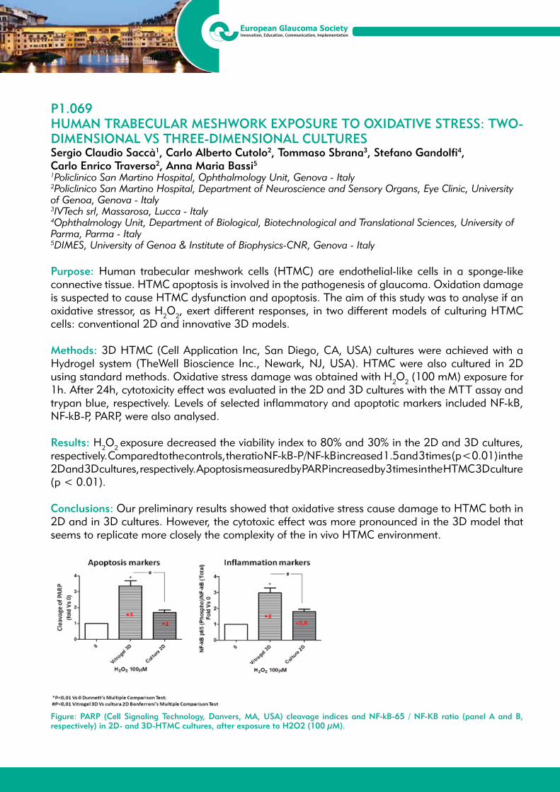
13th EGS CongressFortezza da Basso, Florence, Italy
19/22 May 2018
P1.069HUMAN TRABECULAR MESHWORK EXPOSURE TO OXIDATIVE STRESS: TWO-DIMENSIONAL VS THREE-DIMENSIONAL CULTURESSergio Claudio Saccà1, Carlo Alberto Cutolo2, Tommaso Sbrana3, Stefano Gandolfi4, Carlo Enrico Traverso2, Anna Maria Bassi5
1Policlinico San Martino Hospital, Ophthalmology Unit, Genova - Italy2Policlinico San Martino Hospital, Department of Neuroscience and Sensory Organs, Eye Clinic, University of Genoa, Genova - Italy3IVTech srl, Massarosa, Lucca - Italy4Ophthalmology Unit, Department of Biological, Biotechnological and Translational Sciences, University of Parma, Parma - Italy5DIMES, University of Genoa & Institute of Biophysics-CNR, Genova - Italy
Purpose: Human trabecular meshwork cells (HTMC) are endothelial-like cells in a sponge-like connective tissue. HTMC apoptosis is involved in the pathogenesis of glaucoma. Oxidation damage is suspected to cause HTMC dysfunction and apoptosis. The aim of this study was to analyse if an oxidative stressor, as H2O2, exert different responses, in two different models of culturing HTMC cells: conventional 2D and innovative 3D models.
Methods: 3D HTMC (Cell Application Inc, San Diego, CA, USA) cultures were achieved with a Hydrogel system (TheWell Bioscience Inc., Newark, NJ, USA). HTMC were also cultured in 2D using standard methods. Oxidative stress damage was obtained with H2O2 (100 mM) exposure for 1h. After 24h, cytotoxicity effect was evaluated in the 2D and 3D cultures with the MTT assay and trypan blue, respectively. Levels of selected inflammatory and apoptotic markers included NF-kB, NF-kB-P, PARP, were also analysed.
Results: H2O2 exposure decreased the viability index to 80% and 30% in the 2D and 3D cultures, respectively. Compared to the controls, the ratio NF-kB-P/NF-kB increased 1.5 and 3 times (p < 0.01) in the 2D and 3D cultures, respectively. Apoptosis measured by PARP increased by 3 times in the HTMC 3D culture (p < 0.01).
Conclusions: Our preliminary results showed that oxidative stress cause damage to HTMC both in 2D and in 3D cultures. However, the cytotoxic effect was more pronounced in the 3D model that seems to replicate more closely the complexity of the in vivo HTMC environment.
Figure: PARP (Cell Signaling Technology, Danvers, MA, USA) cleavage indices and NF-kB-65 / NF-KB ratio (panel A and B, respectively) in 2D- and 3D-HTMC cultures, after exposure to H2O2 (100 μM).

13th EGS CongressFortezza da Basso, Florence, Italy
19/22 May 2018
P1.070THE ROLES OF TNF-α-308 GENE POLYMORPHISM AND TNF-α SERUM CONCENTRATIONS IN GLAUCOMA PATIENTSMarija Trenkic Bozinovic1, Predrag Jovanovic1, Jasmina Jocic Djordjevic1, Maja Petrovic2, Marija Radenkovic2, Aleksandar Veselinovic2, Aleksandar Miljkovic3
1University of Nis, Medical Faculty, Department of Ophthalmology, Nis - Serbia2Department of Ophthalmology, University Medical Center Nis, Nis - Serbia3Medical Faculty Novi Sad, University of Novi Sad, Eye Clinic, Novi Sad - Serbia
Purpose: Genetic polymorphism of TNF-α -308 is just one of the genetic factors significant for the development of OAG because of the effects on the TNF-α protein expression. The aim was to determine the genetic polymorphisms distribution of TNF-α-308 G/A gene in OAG patients compared to healthy subjects, and establishing relationships of the gene polymorphism, TNF-α serum concentration and quantitative clinical parameters.
Methods: Distribution of genetic polymorphisms for TNF-α-308 G/A and DNA samples was examined using PCR-RFLP. This study included 328 subjects: 164 patients in the study group (71 HTG, 28 XFG, 65 with senile cataract) and 164 healthy subjects age-sex matched. We examined the frequency of genotypes GG, GA, AA, as well as the A and G alleles, by groups of respondents. The concentrations of circulating TNF-α in plasma were measured by commercial ELISA test.
Results: Genotype GG for the TNF-α-308 was significantly higher in the study compared to control group (p < 0.05), as well as in HTG subjects compared to the control group (p < 0.05). Frequency of genotypes having allele A was statistically significantly higher in control compared to a study group (p < 0.05), as well as in relation to the HTG group (p < 0.05). Visual acuity of HTG and XFG patients (p < 0.05) was higher in the case of GG genotype compared to GA genotype. IOP in both types of glaucoma was higher in the case of the GG genotype. The results showed that serum concentration of TNF-α was significantly higher in patients with glaucoma. The highest serum concentrations of TNF-α was observed in patients with XFG, lower in patients with HTG, and lowest in patients with senile cataract. Concentrations of TNF-α was higher in GA compared to the GG genotype at XFG. No significant association between TNF-α-308 genotypes within HTG and XFG, and TNF-α serum concentration, and investigated clinical parameters was found in OAG.
Conclusions: Serum TNF-α is a powerful cytokine with potent significant role in the pathogenesis of glaucoma and glaucoma neuropathy. Genotype GG TNF-α-308 was significantly higher in OAG than in the healthy subjects group. There is no significant correlation between TNF-α-308 G/A polymorphism with the risk of OAG.
13th EGS CongressFortezza da Basso, Florence, Italy
19/22 May 2018
P1.071EFFECT OF WEEK DAY ON INTRAOCULAR PRESSURE VARIATIONS USING CONTINUOUS INTRAOCULAR TELEMETRYHarsha L. Rao1
1Narayana Nethralaya Hospitals, Bangalore - India
Purpose: A “weekend effect” has been described for some biological parameters. It is not known whether intraocular pressure (IOP) behaves differently by week-day and on weekends. We investigated week-day variations of IOP using intraocular telemetry in glaucoma patients.
Methods: A ring-shaped, ciliary sulcus-placed intraocular sensor (EyeMate, Implandata, Hanover, Germany) for continuous IOP-monitoring was implanted in 22 eyes of patients with primary open angle glaucoma during cataract surgery [1]. Patients obtained measurements using an outside reader unit. Data from patients with ≥ 4 measurements per day were included in the analysis. Analysis of variance (ANOVA) was used to evaluate inter-day variations of IOP by week-day.
Results: Eleven patients fulfilled the criteria 14 days with at least ≥ 4 daily measurements and were included in the study. The average number of days with ≥ 4 measurements was 197.5 ± 186.2 (range, 16 - 614). No difference was found between IOP measurements by week-day and weekend (p = 0.99, ANOVA).
Conclusions: IOP variations were similar during the week and on weekends in patients with glaucoma using continuous IOP monitoring. We found no tendency for IOP increase or decrease by week-day. Inter-day variations of IOP were small and of lower magnitude than intra-day variations. The choice of week-day for IOP measurements may not have a significant impact on glaucoma management.
Reference:1. Koutsonas A, Walter P, Roessler G, Plange N. Implantation of a novel telemetric intraocular
pressure sensor in patients with glaucoma (ARGOS study): 1-year results. Invest Ophthalmol Vis Sci 2015; 56: 1063-9.

13th EGS CongressFortezza da Basso, Florence, Italy
19/22 May 2018
P1.072INVOLVEMENT OF THE TRABECULAR MESHWORK, DISTAL AND PROXIMAL LAYERS OF SCLERA IN INCREASING THE RESISTANCE OF THE OUTFLOW PATHWAYS IN PSEUDOEXFOLIATION GLAUCOMAOlga Kuleshova1, Svetlana Aidagulova2, Anastasiya Lazareva1, Ekaterina Pichikova1, Veronika Dulidova1
1S. Fyodorov Eye Microsurgery Federal State Institution, Novosibirsk - Russia2Novosibirsk State Medical University, Novosibirsk - Russia
Purpose: Determine presence of exfoliation material (EXM) in trabecular tissue, distal and proximal scleral sites, obtained by deep sclerectomy in patients with pseudoexfoliation glaucoma (PEG).
Methods: Complex histological examination of 86 scleral flaps (deep scleral layers 4 x 4 mm), removed during surgery in patients (49 men and 37 women aged 54 to 80 (65.3 ± 3.8) years) with developed stage of PEG with increased intraocular pressure (28.36 ± 2.76 mmHg). Tissue specimens were fixed in 4% solution of paraformaldehyde and examined by light and electron microscopy. Paraffin sections were stained with hematoxylin and eosin in combination with Perls’s reaction, van Gieson with Weigert resorcin-fuchsin-elasticized fibers, with Schick reaction also used. Semithin sections were stained with Schiff’s reagent and 1% solution of azur II. Sections were analyzed with an Axio Scope.A1 microscope and ZEN software (C. Zeiss). Ultrathin sections, stained with saturated alcohol solution of uranyl acetate and lead citrate, were examined using a JEM 1010 electron microscope.
Results: In determining permeability of drainage pathways in PEG, foci of compaction in the form of pigment inclusions, macrophages (melanophages), and a moderate amount of fibroblasts were found in juxtacanalicular tissue (JCT). In sections of sclera proximal to Schlemm’s canal, pigment granules were found throughout the field, but were absent in distal sections. In electron microscopy, EXM in JCT was deposited under the endothelial cell that synthesized it, which led to its degradation. In the proximal sclera, a large number of EXM was located in the collector channel gap, fragments of macrophages with signs of destruction were found. In distal sections, water veins without endothelial lining with a gap supported by high-grade collagen fibrils were determined, EXM was not found.
Conclusion: When creating artificial outflow pathways, it is necessary to activate or prosthetize the most affected intracleral spaces proximally located to Schlemm’s canal to create a reservoir that discharges the aqueous humor into still intact collector canals of the distal sclera into supraciliary and subconjunctival spaces.

13th EGS CongressFortezza da Basso, Florence, Italy
19/22 May 2018
P1.073IS OBSTRUCTIVE SLEEP APNOEA ASSOCIATED WITH GLAUCOMA PROGRESSION?Rupert Bourne1, Dariusz Wozniak2, Gil Peretz3, Jane Kean1, Russell Foster4, Susan Downes5, Ian Smith2
1Anglia Ruskin University, Cambridge - United Kingdom2Papworth Hospital, Cambridge - United Kingdom3Assaf Harofeh medical center,, Tel Aviv - Israel4University of Oxford, Oxford - United Kingdom5Oxford Eye Hospital, Oxford - United Kingdom
Purpose: To determine whether Obstructive Sleep Apnoea (OSA) is a risk factor for progression in patients with primary open-angle glaucoma (POAG).
Methods: Patients with POAG in the POSAG study (Prevalence of Obstructive Sleep Apnoea in Glaucoma, NCT02713152) underwent nocturnal multi-channel respiratory polygraphy, cardiovascular assessment and medical history review. Retrospective data on progression from the diagnosis of POAG to the study visit were collected. Enrolment required 5+ reliable visual fields (VF; Humphrey Visual Field Analyzer 24-2) and 3+ years of follow up data. Patients were categorised as progressors and non-progressors based on European Glaucoma Society guidelines (EGS) and PROGRESSOR pointwise linear regression (Medisoft, Inc). Rate of VF loss was determined by slope of progression for the whole VF (ProgressorSlope) and decline in mean deviation per year (MD/year). For each patient, the eye with the more severe VF defect (worse MD) was selected, unless only one eye was eligible. OSA was diagnosed based on apnoea-hypopnoea index (AHI) ≥5 (Moderate to severe OSA: AHI≥15). Statistics: independent t test, chi-square, binomial & linear regression.
Results: 159 patients with POAG were included (Mean observation period: 9.5 ± 4.3 yrs, mean of 10.6 ± 4.4 VFs per eye. 47.8% of eyes were classified as progressing based on EGS criteria and 51.6% based on PROGRESSOR criteria, with good agreement between the two classifications (κ = 0.61, p = 0.00). Within 5 years leading up the diagnostic test for OSA, 37.3% (EGS) and 49% (PROGRESSOR) eyes progressed. Overall, 56.6% of patients were diagnosed with OSA (moderate to severe in 22% of all patients). OSA was not a significant predictor of glaucoma progression irrespective of the classification used or follow up period: odds ratio = 1.0 (0.53-1.88), p = 0.99 for EGS from baseline, odds ratio = 1.39 (0.74-2.6), p = 0.31 for PROGRESSOR from baseline. Nor were people with moderate and severe OSA at higher risk of progression in comparison with people without OSA: odds ratio = 1.1 (0.72-1.6) for EGS and odds ratio = 0.96 (0.64-1.44), p = 0.83 for PROGRESSOR. AHI score was not a significant predictor of the rate of progression measured by ProgressorSlope (R2 = 0.001, p = 0.78) or MD/year decline (R2 = 0.00, p = 0.87).
Conclusions: OSA was not a significant contributor to glaucoma progression. Patients with OSA and POAG can be reassured they do not appear to be at any extra risk.

13th EGS CongressFortezza da Basso, Florence, Italy
19/22 May 2018
P1.074PREVALENCE OF OBSTRUCTIVE SLEEP APNOEA IN GLAUCOMA: THE POSAG STUDYGil Peretz1, Dariusz Wozniak2, Jane Kean3, Shabbir Harun4, Catherine Willshire4, Sofia Villar5, Russell Foster6, Susan Downes7, Ian Smith2, Rupert Bourne3
1Assaf Harofe Medical Center, Zerifin - Israel2Respiratory Support and Sleep Centre, Papworth Hospital, Cambridge - United Kingdom3Vision & Eye Research Unit, Anglia Ruskin University, Cambridge, United Kingdom, Cambridge - United Kingdom4Department of Ophthalmology, North West Anglia Foundation Trust, Huntingdon - United Kingdom5University of Cambridge School of Clinical Medicine, Cambridge - United Kingdom6Nuffield Department of Clinical Neurosciences, University of Oxford, Oxford - United Kingdom7Oxford Eye Hospital, Oxford - United Kingdom
Purpose: To establish the prevalence of OSA among patients with POAG and determine whether it is higher than in matched people without glaucoma. We also aimed to examine potential associations between markers of OSA and POAG severity.
Methods: Unselected patients with a diagnosis of POAG and their spouses without glaucoma (controls) were invited. A comprehensive ocular examination (visual field tests, ocular coherence tomography, slit-lamp examination, intraocular pressure, optic disc photography) was performed to confirm or exclude glaucoma. A medical interview focused on sleep history and relevant co-morbidities was conducted. All patients underwent nocturnal multi-channel respiratory polygraphy. OSA was diagnosed based on apnoea hypopnoea index (AHI) ≥ 5. Propensity score matching was used to match the groups for any significant imbalances.
Results: Of 795 potentially eligible subjects, 403 participants: 240 POAG patients and 163 controls were enrolled. The prevalence of OSA was 59.2% (95% CI:53-65%) in POAG patients and 55.2% (95% CI:48-63%) in controls. 22.9% (95% CI:18-28%) POAG patients and 16.6% (95% CI:11-22%) controls were diagnosed with moderate to severe OSA (AHI ≥ 15). Among significant predictors of OSA, age and sex differed between the groups. 163 subjects in each group were successfully matched. There was no significant difference in OSA prevalence between the matched groups (p = 0.74 for AHI ≥ 5, p = 0.39 for AHI ≥ 15). The median AHI was 5.9 (IQR 2.2-11.8) in the POAG group and 5.5 (IQR 2.6-11.8) in the control group (p = 0.65). The level of sleepiness assessed by Epworth score was not significantly different (6.3 ± 3.9 vs 6.1 ± 3.7, p = 0.91). In a linear regression, AHI was not a significant predictor of Mean Deviation (p = 0.93), Visual Field Index (p = 0.85) or Retinal Nerve Fibre Layer Thickness (p = 0.27) in people with POAG. Nor were other markers of OSA severity (desaturation index, mean saturation, time with saturation< 90%) correlated with the glaucoma metrics.
Conclusions: This study confirms high prevalence of OSA in people with POAG which is however not higher than in people without glaucoma who otherwise share the same risk factors for OSA. Our findings do not support the hypothesized association between these two conditions.
13th EGS CongressFortezza da Basso, Florence, Italy
19/22 May 2018
P1.075ET-1 EFFECT ON FUNCTIONAL AND STRUCTURAL CHANGE IN PATIENTS WITH POAGSanida Ljaljevic1, Emina Alimanovic1, Merita Lika- Pranjic1
1Eye Clinic University Clinic Center, Sarajevo - Bosnia and Herzegovina
Purpose: To determine the value of ET-1 in the plasma and humor aqueous patients with POAG and control group of patients without glaucoma.To investigate the correlation of plasm ET-1 and humor aqueous (AH ET-1) with the parameters of visual field and OCT in POAG and non glaucomatous group.
Patients and Methods: In this prospective, clinical, manipulative study included 60 patients of both sexes. All patients were hospitalized at the Department of Ophthalmology KCUS, for glaucoma surgery (Trepanotrabeculectomy) or for cataract surgery. Patients were divided into 2 groups of 30 patients: patients with POAG and a control group of patients hospitalized for surgery cataract without glaucoma. Additionally done clinical biochemical and immunological determination of the value of plasm ET-1 and AH ET-1. We followed the parameters of the visual field (Otopus 101), MD and LV, and the diameter of the excavation (C / D) and vertical diameter of excavation using OCT.
Results: There was no SS differences in the plasm ET-1 between POAG and control group, but SS higher values of ET-1 in AH in POAG than in the CG; CG 1,13 pg/ml; POAG 2.80 pg/ml. There was a SS positive correlation between plasm ET-1 and vertical diameter of excavation in the POAG group (rho = 0.448, p < 0.05)and SS positive correlation between AH ET-1 and C/D ratio (rho = 0.551, p < 0.05), and AH ET-1 and vertical diameter of exavation (rho = 0.515, p < 0.05). Positive correlation of AH ET-1 and MD of VF (rho = 0.637, p < 0.01), and LV of VF (rho = 0.644, p < 0.01).
Conclusion: The concentration of ET-1 in aqueous humor is significantly increased in patients with POAG compared to the control group and showed correlation with IOP, parameters of OCT and VF.

13th EGS CongressFortezza da Basso, Florence, Italy
19/22 May 2018
P1.076VASCULAR EVALUATION OF THE OPTIC DISC WITH OCT-A IN PRIMARY OPEN-ANGLE GLAUCOMAPedro Faria1, Isa Sobral1, Cátia Azenha1, João Cardoso1, João Filipe Silva1, José Moura Pereira1
1Serviço de Oftalmologia,Centro Hospitalar e Universitário de Coimbra, Coimbra - Portugal
Purpose: Glaucoma has a multifactorial pathogenesis including vascular factors. OCT-A is an imaging exam that provides information about optic disc (OD) vasculature without contrast. The aim of this study is to evaluate the microstructure and vascularization in patients with primary open-angle glaucoma (POAG).
Methods: Prospective study evaluating the vascularization of OD, retinal nerve fiber layer (RNFL) and ganglion cells complex (GCC) using OCT-A in patients with POAG and controls. Parameters cited were determined using automatic software. Demographic, clinical and visual field parameters were evaluated.
Results: A total of 158 subjects (n = 106 patients with POAG; n = 52 controls) were included. Patients with POAG showed significantly lower flow (p < 0.001) and vascular density (p < 0.001) of OD than the control group. Lower OD vascular density are significantly correlated with lower RNFL (p = 0.007) and GCC (p = 0.001) thicknesses. Similar situation correlated OD flow with RNFL (p = 0.014) and GCC (p = 0.014) thicknesses. The visual acuity in patients with POAG is significantly lower in the cases of lower OD flow (p = 0.049) and vascular density (p = 0.049). We did not document changes with statistical significance between OD vascular parameters and excavation, neuroretinal rim area, MD or PSD.
Conclusions: Patients with POAG have OD vascular indices which are related to visual acuity and structural changes of RNFL and GCC. More studies are needed to clarify the importance of these data in the pathogenesis of glaucoma and its utility in clinical practice.
13th EGS CongressFortezza da Basso, Florence, Italy
19/22 May 2018
P1.078EVALUATION OF LAMINA CRIBROSA BY SWEPT-SOURCE OPTICAL COHERENCE TOMOGRAPHY IN PRIMARY OPEN-ANGLE GLAUCOMAIsil Pasaoglu1, Banu Satana1, Cigdem Altan1, Berna Basarir1, Ercan Pasaoglu2, Tekin Yasar1, Muhittin Taskapili1
1Department of Ophthalmology, Beyoglu Eye Education and Research Hospital, Istanbul - Turkey2Department of Ophthalmology, Yeniyüzyil University, Istanbul - Turkey
Purpose: To compare the thickness and depth measurements of the lamina cribrosa (LC) obtained using a swept-source optical coherence tomography (SS-OCT) device in primary open angle glaucoma (POAG) patients, and healthy subjects.
Methods: This retrospective, cross-sectional observational study included 22 eyes with POAG, and 20 control eyes. The LC measurements with serial horizontal B-scans of the optic nerve head were obtained using SS-OCT (Topcon 3D DRI OCT Triton). The anterior lamina surface (ALS) depth, posterior lamina surface (PLS) depth, and LC thickness measurements were evaluated.
Results: In patients with POAG, the mean ALS depth was 464.18 ± 146.02 μm, the mean PLS depth was 675.72 ± 147.98 μm, and the mean LC thickness was 211.54 ± 47.61 μm. In the control group, the corresponding values were 442.30 ± 91.18 μm, 648.90 ± 81.35 μm, and 206.60 ± 50.41 μm, respectively (p > 0.05).
Conclusıons: Anterior and posterior lamina cribrosa surface depth and also LC thickness can be evaluated using an SS-OCT device. It was not statistically significant but ALS dept and PLS dept were displaced posteriorly in POAG eyes. The assessment of LC level with SS-OCT in POAG cases is a valuable and reproducible adjunctive imaging method in terms of diagnosis and follow-up, but comparative studies with larger series should be performed.
13th EGS CongressFortezza da Basso, Florence, Italy
19/22 May 2018
P1.079THE ASSOCIATION OF CHOROIDAL THICKNESS WITH RETINAL NERVE FIBER LAYER THICKNESS IN ADVANCED PRIMARY OPEN-ANGLE GLAUCOMAFatma Saadet Yilmaz1, Akin Cakir1, Gulcenur Ozturan1, Mustafa Elcioglu1
1Department of Ophthalmalogy, SB Okmeydani Training and Research Hospital, Istanbul - Turkey
Purpose: To investigate the association between macular choroidal thickness (CT) and retinal nerve fiber layer (RNFL) thickness in patients with advanced primary open-angle glaucoma.
Methods: Fifty-four eyes of 54 patients with advanced primary open-angle glaucoma were included in this study. All the participants underwent spectral domain optical coherence tomography (SD-OCT) (Spectralis; Heidelberg Engineering, Heidelberg, Germany) analyzed with the enhanced depth imaging (EDI) mode. CT was measured at the subfoveal location, and at 0.5 mm nasal and temporal to the fovea.
Results: The mean age of the participants was 61.8 ± 8.9 (41-80) years. Thirty-eight (70.4%) subjects were female and 16 (29.6%) were male. All of the participants had a spherical equivalent between ±3.00 diopters. The mean average RNFL thickness was 84.1 ± 13.5 μm. The mean CT was measured as 276.5 ± 49.5 μm at the subfoveal location, 263.2 ± 50.1 μm at nasal, and 263.7 ± 46 μm at temporal to the fovea. The Pearson correlation analysis showed a negative, but not a statistically significant correlation between RNFL thickness and CT at subfoveal, nasal and temporal locations (r: -0.188, p = 0.174; r: -0.051, p = 0.713; r: -0.174, p = 0.208 respectively).
Conclusions: CT in advanced glaucoma patients was found to be not associated with the reduction in RNFL thickness.

13th EGS CongressFortezza da Basso, Florence, Italy
19/22 May 2018
P1.080THE PROTEINS OF MITOCHONDRIAL DYSFUNCTION AND INTEGRIN SIGNALING CHANGE IN THE RAT RETINA FOLLOWING CEREBROSPINAL FLUID PRESSURE REDUCTIONFancheng Yan1, Fudong Yu2, Yiyi Gong3, Lan Zhang4, Jingxue Zhang5, Shen Wu5, Haiteng Deng6, Ningli Wang5
1Beijing Institute of Ophthalmology, Beijing Tongren Eye Center, Beijing Tongren Hospital, Capital Medical University, Beijing Ophthalmology & Visual Sciences Key Laboratory; Aier School of Ophthalmology, Central South University, Beijing - China2Key Laboratory of Reproduction Regulation of NPFPC (SIPPR, IRD, Fudan University), Shanghai - China 3Central Research Laboratory,Peking Union Medical College Hospital, Chinese Academy of Medical Sciences and Peking Union Medical College; MOE Key Laboratory of Bioinformatics, School of Life Sciences, Tsinghua University, Beijing - China4Beijing Tongren Eye Center, Beijing Tongren Hospital, Capital Medical University, Beijing Ophthalmology & Visual Sciences Key Laboratory, Beijing - China5Beijing Institute of Ophthalmology, Beijing Tongren Eye Center, Beijing Tongren Hospital, Capital Medical University, Beijing Ophthalmology & Visual Sciences Key Laboratory, Beijing - China6MOE Key Laboratory of Bioinformatics, School of Life Sciences, Tsinghua University, Beijing - China
Purpose: To examine the influence of cerebrospinal fluid pressure(CSFP) reduction on mitochondrial dysfunction and integrin signaling pathway.
Methods: The experimental study included 5 rats which underwent cerebrospinal fluid pressure reduction for 6 hours and 5 rats in a control group. 24 hours after baseline, the animals were killed and the proteins expression profile of retinas were detected by Tandem Mass Tag (TMT) and Liquid Chromatography-tandem Mass Spectrometry (LC-MS/MS) technology. Ingenuity Pathway Analysis (IPA) was used to analyze the function and signal pathways of the differential expressed proteins.
Results: 28 differentially expressed proteins were detected in mitochondrial dysfunction. 6 cases were up-regulated and 22 cases were down-regulated. There were 5 cases and 12 cases of protein, with up-regulated expression more than 1.5 times and down-regulated more than 50%, respectively. 27 differentially expressed proteins were detected in integrin signaling pathway. 18 cases were up-regulated and 9 cases were down-regulated. There were 6 cases and 3 cases of protein, with up-regulated expression more than 1.5 times and down-regulated more than 50%, respectively.
Conclusions: Experimental models with an acute CSFP showed different protein expression profile changes in the pathway of mitochondrial dysfunction and integrin signaling pathway. It supports the hypothesis that an experimental model with an acute reduction in CSFP may undergo specific pathophysiology in the process of retina damage. It also may present that the pathogeny of the glaucoma patients who have elevated pressure along the optic nerve due to the intracranial pressure reduction has close correlation with the two signaling pathways.

13th EGS CongressFortezza da Basso, Florence, Italy
19/22 May 2018
P1.081THE ROLE OF LYSYL OXIDASE-1 (LOX1) AS COLLAGEN CROSS-LINKER IN POAGNajiha Rahman1, Mustapha Irnaten1, Colm O’Brien1
1Institute of Ophthalmology, Mater Misericordiae University Hospital, Dublin - Ireland
Purpose: Glaucoma is associated with an increase in ocular tissue stiffness, e.g. cornea and trabecular meshwork (TM), which may lead to increased outflow resistance. Abnormal collagen cross-linking may affect the mechanical homeostasis of connective tissue in the TM and lamina cribrosa (LC). Lysyl oxidase is a cross-linking enzyme, which binds collagen and elastin, and the purpose of this study was to examine whether LOX1 levels are increased in patients with primary open angle glaucoma (POAG).
Methods: ELISA analysis kit was used to measure and compare the LOX1 (MyBioSource, USA) levels in aqueous humour (AH) samples, between ten POAG patients (GAq) and ten normal age-matched control subjects (NAq), and also blood/serum levels in POAG and normal controls. We also compared the mRNA expression of LOX1 in LC cells between glaucoma and normal patients (n = 3), using quantitative real-time reverse transcription polymerase chain reaction (qRT-PCR). The average of the results was compared between the two groups using unpaired t-test, with a statistical significance of p-value < 0.05.
Results: Higher LOX1 levels were found in the aqueous of glaucoma patients compared to normal (GAq = 1.55 s 0.76 vs NAq = 1.29 s 0.53 ng/mL, p = 0.39). There was a significantly increased level of LOX1 in the aqueous humour of glaucoma patients compared to the serum levels (GAq = 1.55 s 0.76 vs GSerum = 0.30 s 0.08 ng/mL, p = < 0.0001). qRT-PCR analysis showed that LOX1 transcripts were significantly expressed at higher levels in glaucoma LC cells compared to normal LC cells (p < 0.0001).
Conclusion: We found higher levels of LOX1 in the aqueous humour and LC cells of POAG patients. This could lead to an abnormal collagen cross-linking process and increased tissue stiffness and altered biomechanics within the ECM of the cornea, TM and LC. Therapies that reduce tissue stiffness may be of therapeutic benefit in glaucoma.

13th EGS CongressFortezza da Basso, Florence, Italy
19/22 May 2018
P1.082TRANSLAMINAR PRESSURE GRADIENT AND OCULAR PERFUSION PRESSURE IN GLAUCOMA PATIENTS WITH DIFFERENT OPTIC DISC SIZESNiro Kasahara1
1R. Dr. Cesário Mota Júnior, São Paulo - Brazil
Purpose: The optic disc size varies largely in the population. Clinical variables such as the translaminar pressure gradient (TPG) - difference between intraocular and intracranial pressure - and the ocular perfusion pressure (OPP) play a role in the glaucomatous optic neuropathy. Uncertainty exists as to if these variables differ according to the optic disc size. This was an observational, cross-sectional clinical study to evaluate whether TPG and OPP varies in glaucoma patients with different optic disc sizes.
Methods: The sample comprised 75 patients with open angle glaucoma. All patients underwent an ophthalmic evaluation, clinical measurements (blood pressure, height, and weight), and optical coherence tomography examination. TPG and OPP were calculated for each patient using proxy algorithms to attain indirect surrogate parameter values. Optic disc sizes were stratified according to 3 quantiles and the differences compared with ANOVA test. Data from both eyes were used after using the appropriate correction for inter-eye dependency.
Results: Demographic data are presented on table 1. The TPG did not vary between large and small discs; glaucoma patients with average disc sizes presented with higher TPG; the difference however, failed to reach statistical significance. Glaucoma patients with large disc size presented with higher OPP as compared to average sized discs (post hoc Tukey honest significant difference test p = 0.026).
Conclusion: Glaucoma patients with larger optic discs have higher OPP. The clinical significance of this finding is yet to be determined.
Table 1. Demographic and clinical differences stratified by optic disc size.
1st tertile(n = 33 eyes)
2nd tertile(n = 36 eyes)
3rd tertile(n = 24 eyes)
p value
Age (years) 67.4 ± 9.3 64.8 ± 9.8 69.2 ± 8.7 0.563
Gender M:F 15:18 15:21 11:13 0.994*
Ethnicity 0.969*- white 22 25 15- non-white 11 11 7Disc area (mm2) 2.7 ± 0.3 2.1 ± 0.1 1.7 ± 0.1 <.000
TPG (mmHg) 2.6 ± 5.5 3.0 ± 3.3 2.4 ± 4.4 0.866
OPP (mmHg) 56.0 ± 12.5 50.2 ± 5.8 52.1 ± 7.6 0.032
M: male; F: female; TGP: translaminar pressure gradient; OPP: ocular perfusion pressure.

13th EGS CongressFortezza da Basso, Florence, Italy
19/22 May 2018
P1.083ANALYSIS OF MYOC GENE IN A CZECH FAMILY WITH PRIMARY OPEN-ANGLE GLAUCOMALucie Rezkova1, Marek Fichtl1, Petra Liskova2, Lubica Dudakova3, Eva Ruzickova1
1Department of Ophthalmology, First Faculty of Medicine, Charles University in Prague and General University Hospital, Prague - Czech Republic2Department of Ophthalmology, First Faculty of Medicine, Charles University in Prague and General University Hospital, Research Unit for Rare Diseases; Department of Pediatrics and Adolescent Medicine, First Faculty of Medicine, Charles University and General University Hospital, Prague - Czech Republic 3Research Unit for Rare Diseases; Department of Pediatrics and Adolescent Medicine, First Faculty of Medicine, Charles University and General University Hospital, Prague - Czech Republic
Purpose: To report on the molecular genetic cause of primary open angle glaucoma (POAG) in a Czech family with affected individuals in three generations. Myocilin (MYOC) is known to be implicated in POAG, mutations within this gene contribute to increased aqueous outflow resistance.
Methods: Four family members affected with POAG and one unaffected first degree relative underwent complex ophthalmic examination. Exon 3 of the MYOC gene was directly sequenced in the proband. Next, targeted genetic testing was performed in the other family members.
Results: 51-year old man diagnosed with POAG at the age of 42 years and positive family history was referred to our tertiary care center because of unstable intraocular pressure (IOP) despite the use of multiple topical anti-glaucoma medications. Trabeculectomy with Ologen implant (Aeon Astron Europe B.V.) in both eyes was performed. His son was diagnosed with POAG already at the age of 22 years, and underwent trabeculectomy in both eyes at the age of 24 years. The father as well as the brother of the proband were noted to suffer from POAG in theirs 40’s and underwent anti-glaucoma surgeries bilaterally.Previously reported pathogenic variant c.1099G>A; p.(Gly367Arg) in MYOC was identified and segregated with the disease in the family. Unaffected 25 years old daughter did not inherit the mutation.
Conclusion: In our work, we have demonstrated this pathogenic variant in MYOC gene for the first time in Czech patients. In families with early-onset POAG, monogenic cause of the disease needs to be considered and screening of MYOC recommended. The life-long risk of developing POAG in carriers of p.(Gly367Arg) in MYOC has been estimated to be more than 50 %.

13th EGS CongressFortezza da Basso, Florence, Italy
19/22 May 2018
P1.084SHORT-TERM OBSERVATION OF RETINA BLOOD FILLING CHANGES IN RAT RETINA ISCHEMIA-REPERFUSION MODEL ESTABLISHED WITH A NOVEL OPERATIVE APPROACH AND PROCEDUREFancheng Yan1, Lan Zhang2, Shen Wu3, Jingxue Zhang3, Kegao Liu2, Shuning Li2, Ningli Wang3
1Beijing Institute of Ophthalmology, Beijing Tongren Eye Center, Beijing Tongren Hospital, Capital Medical University, Beijing Ophthalmology & Visual Sciences Key Laboratory; Aier School of Ophthalmology, Central South University, Beijing - China2Beijing Tongren Eye Center, Beijing Tongren Hospital, Capital Medical University, Beijing Ophthalmology & Visual Sciences Key Laboratory, Beijing - China3Beijing Institute of Ophthalmology, Beijing Tongren Eye Center, Beijing Tongren Hospital, Capital Medical University, Beijing Ophthalmology & Visual Sciences Key Laboratory, Beijing - China
Purpose: To develop a novel, reliable, reproducible rat model of retina ischemia-reperfusion through a novel operative approach and procedure and observe the retina blood filling changes in short term.
Methods: Retina ischemia-reperfusion model was established in adult SD rats by directly accessing the area of the retrobulbar optic nerve through the orbital lateral wall of the eyeball. Before the reperfusion of retina vessels in the surgery, the retina ischemia condition lasted for half an hour. Retinal vasculature was examined and photoed by Retinal Imaging System (OPTO-III, Optoprobe Company, Canada) at 15 mins, 30 mins, 60 mins, 1 day, 2 days, 3 days, 4 days, 5 days and 6 days after the surgery. Image processing software (Adobe Photoshop CS6, Adobe Systems Incorporated, USA) is used to quantify and calculate the blood vessels filling areas in the fundus. Quantitative analysis and graphics production were performed with GraphPad Prism 6.0 (GraphPad Software, Inc. USA). The values were presented as mean ± standard deviation (SD) from 6 independent samples. Statistical analysis was conducted by nonparametric one-way analysis of variance. Significance was regarded as p < 0.05.
Results: Compared with preoperative rat retina arteries and veins areas for each group (100%), at 15 mins, 30 mins, 60 mins, 1 day, 2 days, 3 days, 4 days, 5 days and 6 days after the surgery, the arteries area was 89.3% ± 7.8%, 110% ± 6.5%, 101.3% ± 10.1%, 92.8% ± 4.1%, 96.7% ± 0.9%, 99.3% ± 3.6%, 103% ± 6.8%, 101.3% ± 5.2%, 98.3% ± 6.5%, respectively (n = 6). The veins area was 94.3% ± 10.3%, 112% ± 4.6%, 113.3% ± 9%, 89% ± 1.4%, 101.2% ± 2.3%, 106% ± 3.2%, 112.2% ± 2.1%, 98.3% ± 4.9%, 111% ± 2.6%, respectively (n = 6). Within 6 days after surgery, the veins’ total fluctuation range along the time was 2.29 times of the arteries’.
Conclusions: Rat retina ischemia-reperfusion model established by a novel operative approach and procedure is a kind of reproducible and reliable animal model for studying glaucoma or other retina diseases.

13th EGS CongressFortezza da Basso, Florence, Italy
19/22 May 2018
P1.085GENETIC VARIANTS ASSOCIATED WITH HIGH INTRAOCULAR PRESSURE ARE NOT ASSOCIATED WITH PRIMARY ANGLE CLOSURE GLAUCOMARachel Shujuan Chong1, Grace SE Kiew2, Monisha Nongpiur1, Shamira Perera1, Li Jia Chen3, Sripriya Sarangapani4, Do Tan5, Yaakub Azhany6, Ahmad Tajudin Liza-Sharmini6, Clement Tham3, Nguyen Xuan Hiep5, Ronnie George4, Chiea-Chuen Khor1, Eranga Vithana1, Tin Aung1
1Singapore National Eye Centre, Singapore - Singapore2University of Cambridge, Cambridge - United Kingdom3The Chinese University of Hong Kong, Hong Kong - Hong Kong4Sankara Nethralaya Vision Research Foundation, Chennai - India5Vietnam National Institute of Ophthalmology, Ha Noi - Viet Nam6Universiti Sains Malaysia, Kelantan - Malaysia
Purpose: To investigate the association of high intraocular pressure (IOP)-associated single nucleotide polymorphisms (SNPs) with primary angle closure glaucoma (PACG).
Methods: 1850 PACG cases and 10024 controls were recruited across 5 independent collections at ophthalmic hospitals in Asia. We assessed 12 index SNP markers mapping to genetic loci previously reported to show strong association for IOP (the loci are TMCO1, FNDC38, CAV1, ABCA1, RAPSN, NUP160, PTPRJ and GAS7). Single locus analysis was conducted using unconditional logistic regression fitted to test for association between individual SNP genotypes and PACG disease status. Genotype and allele frequencies were calculated for each SNP.
Results: We did not observe evidence of association surpassing the conventional threshold for genome-wide significance (p < 5x10⁻⁸) at any of the 12 SNPs tested. SNPs mapping to ABO and GAS7 had the lowest nominal p-values (ABO, rs8176743, p = 0.001, OR = 0.85; GAS7, rs9913911, p = 0.002, OR = 0.87).
Conclusions: None of the tested SNPs associated with IOP were consistently linked to PACG in our study population. Our results suggest that PACG and POAG may have a divergent genetic basis with relation to IOP.
13th EGS CongressFortezza da Basso, Florence, Italy
19/22 May 2018
P1.087QUANTITATIVE PROTEOMIC ANALYSIS OF AQUEOUS HUMOUR ACROSS ANGLE CLOSURE DISEASE SPECTRUMMonisha Nongpiur1, Lei Zhou1, Eranga Vithana1, Tin Aung1, Tina Wong1
1Singapore Eye Research Institute, Singapore - Singapore
Purpose: The pathogenesis of primary angle closure glaucoma (PACG) and progression from early stages of the disease are not well understood. The aim of this study is to evaluate the aqueous humour proteomic profile across primary angle closure disease spectrum (including primary angle closure suspect (PACS), primary angle closure (PAC) and PACG), to provide an insight into the biological mechanisms associated with the disease.
Methods: Aqueous humour samples were obtained during elective phacoemulsification or combined phacoemulsification and trabeculectomy surgeries from 8 cataract (as controls) and 28 angle closure subjects (including 10 PACS, 9 PAC and 9 PACG). Quantitative proteomic analysis was performed using iTRAQ and liquid chromatography-mass spectrometry (LC-MS/MS). A ratio of > 1.5 and p < 0.05 were considered as cut-off for up-regulation, with the controls pooled to form a common reference.
Results: A total of 549 proteins were identified with false discovery rate (FDR) less than 1% and peptide confidence level more than 95%. Of these, 388 proteins were quantifiable. Nineteen were significantly up-regulated in PACS, 15 in PAC and 14 in PACG; and 5 proteins were differentially up-regulated across the entire angle closure spectrum. Additionally, the levels of 3 proteins were differentially elevated only in PAC and PACG, and 5 proteins (namely, ABCB5, APOA1, NFAT5 KIAA0827 TONEBP, TKT and ZNF541) were exclusively elevated only in the PACG group. These proteins are involved in trans-membrane transport, lipid metabolism, cell differentiation and transcription factor activity.
Conclusions: A distinctive aqueous humour proteomic profile appears to be present across the primary angle closure disease severity spectrum. Validation in an independent cohort is required to further strengthen these findings.
13th EGS CongressFortezza da Basso, Florence, Italy
19/22 May 2018
P1.088NEWBORN GLAUCOMA: DON’T FORGET INFECTIONS!Sushmita Kaushik Kaushik1, Gunjan Joshi1, Mini Singh2, Pankaj Kataria1, Atul Arora1, Surinder Singh Pandav1
1Advanced Eye Centre, Postgraduate Institute of Medical Education and Research, Chandigarh - India2Department of Virology, Postgraduate Institute of Medical Education and Research, Chandigarh, Chandigarh - India
Background: Infants with newborn glaucoma have more severe glaucoma, and carry a worse prognosis than Primary congenital glaucoma (PCG) which characteristically presents in infancy, i.e. between 3 to 9 months of age. It is not established whether it is just a more severe phenotype, or is it a different disease altogether. Intrauterine infections can affect various structures of the developing fetal eye. The impact of intrauterine infections on the incidence of glaucoma presenting at birth has not been widely studied. We report the incidence of Rubella and Cytomegalovirus (CMV) positivity in a cohort of babies with newborn glaucoma.
Methods: Rubella and CMV IgM titres were tested in infants presenting with glaucoma before the age of 4 weeks, between October 2016 and October 2017. Presenting features and early outcome was analyzed in patients who had positive titres compared to those who did not.
Results: 44 eyes of 24 patients were analyzed (12 male; 12 female). 18 babies had bilateral Primary Congenital Glaucoma (PCG), 2 had PCG in one eye and Peters’ anomaly in the other, 2 had unilateral PCG, one had bilateral aniridia, and one had primary hyperplastic primary vitreous (PHPV). 6 patients tested positive for Rubella of which 4 were positive for CMV also. Two others were positive for CMV alone. Rubella positive babies presented significantly earlier (7 ± 6 days vs. 17 ± 12 days; p = 0.02), had significantly opaque corneae (p < 0.001), and shorter eyes(16.6 vs 18.8 mm; p = 0.001) Presenting corneal diameters and intraocular pressures(IOP) were comparable. All infants underwent combined trabeculotomy-trabeculectomy. At 3 months. IOP reduced significantly in all infants. However, the cornea cleared significantly in Rubella negative but not in Rubella positive infants despite comparable IOP reduction.
Conclusion: It is important to recognize these babies early and investigate for intrauterine infections rather than assume they are all primary congenital glaucoma (PCG). Involvement of the cornea, glaucoma, and cataract make management of these babies a major challenge requiring a multi-disciplinary team approach.
13th EGS CongressFortezza da Basso, Florence, Italy
19/22 May 2018
P1.089IRIS THICKNESS AND SEVERITY OF NEOVASCULAR GLAUCOMA DETERMINED USING SWEPT-SOURCE ANTERIOR-SEGMENT OPTICAL COHERENCE TOMOGRAPHYShunsuke Nakakura1, Yui Kobayashi1, Kanae Matsuya1, Yoshiaki Kiuchi2
1Department of Ophthalmology, Sanekai Tsukazaki Hospital, Himeji - Japan2Department of Ophthalmology, Hiroshima University, Hiroshima - Japan
Purpose: To investigate the iris thickness (IT) in neovascular glaucoma (NVG) using swept-source anterior-segment optical coherence tomography (ASOCT).
Patients and Methods: In this retrospective, clinic-based, comparative study, we enrolled 26 NVG patients (15 with 360° angle-closure [AC]-NVG and 11 with NVG without AC) and 14 healthy age-matched controls. Horizontal scanning images of swept-source ASOCT were analyzed using software calipers in temporal and nasal angle areas. ITs at 1 and 2 mm from the pupil edge were measured using ASOCT. Additionally, the effects of intraocular pressure (IOP), intravitreal anti-vascular endothelial growth factor (anti-VEGF) injection, and panretinal photocoagulation (PRP) on IT were evaluated.
Results: The IT was thinner in 360° AC-NVG patients, followed by NVG patients without AC and controls (0.31 vs. 0.49 vs. 0.57 mm at 1 mm and 0.29 vs. 0.43 vs. 0.49 mm at 2 mm; all p < 0.05). In NVG patients, IT was negatively correlated with IOP at examination (-0.36 mm at 1 mm and -0.39 mm at 2 mm; all p < 0.05, Pearson correlation coefficient [r]). Multiple linear regression analysis revealed that IOP (t: -4.51) at 1 mm, IOP (t: -6.10) at 2 mm, and anti-VEGF injection (t: 2.03) were independent predictors of IT; however, PRP was not a predictor of IT.
Conclusions: IT decreases with the progression of the NVG stage, and a higher IOP at examination may be associated with IT. Our study suggests a new morphological feature of NVG.

13th EGS CongressFortezza da Basso, Florence, Italy
19/22 May 2018
P1.090PERIPAPILLARY VESSEL DENSITY IN GLAUCOMATOUS EYES: COMPARISON BETWEEN PSEUDOEXFOLIATION GLAUCOMA AND PRIMARY OPEN-ANGLE GLAUCOMAJi-Hye Park1, Chungkwon Yoo1, Michael Girard2, Jean-Martial Mari3, Yong Yeon Kim1
1Department of Ophthalmology, Republic of Korea University College of Medicine, Seoul - Republic of Korea2Department of Biomedical Engineering, National University of Singapore, Singapore - Singapore3University of French Polynesia, Tahiti - French Polynesia
Purpose: To compare the peripapillary vessel density between eyes with pseudoexfoliation glaucoma (PXG) and eyes with primary open-angle glaucoma (POAG).
Methods: Peripapillary vessel densities in the radial peripapillary capillaries (RPC) were assessed using optical coherence tomography angiography (OCTA), and compared between patients with PXG and those with POAG matched for age and mean deviation (MD) of standard automated perimetry. The vessel density was measured between the optic disc margin and 750 μm from the optic disc margin.
Results: Thirty-nine eyes with PXG were matched to 39 eyes with POAG. Mean untreated intraocular pressure was higher in the PXG group than in the POAG group (21.4 ± 6.7 mmHg vs. 14.9 ± 2.9; p < 0.001), but there was no difference in age, refractive error, visual field MD, or average retinal nerve fiber layer (RNFL) thickness between the two groups. However, the average peripapillary vessel densities were lower in the eyes with PXG than in the eyes with POAG. Moreover, there was a significant correlation between peripapillary vessel density and both visual field MD and RNFL thickness.
Conclusions: Peripapillary vessel density was lower in eyes with PXG than in eyes with POAG of similar severity.
13th EGS CongressFortezza da Basso, Florence, Italy
19/22 May 2018
P1.091COMPARISON OF PLASMA C-REACTIVE PROTEIN LEVEL IN PSEUDOEXFOLIATION (PXF) GLAUCOMA WITH NORMAL POPULATIONReza Zarei1
1Glaucoma Division, Farabi Eye Hospital, Tehran university of medical University, Tehran - Iran
Purpose: Pseudoexfoliation (PXF) glaucoma is a senile disease which affect anterior segment structure of eye. Pseudoexfoliation material has also been found in heart, lung, liver, gall bladder, cerebral meninges, skin, and blood vessel and is thought to be a systemic disorder. C-reactive protein (CRP) is an acute phase reactant found to be an important and sensitive marker of systemic inflammatory states and disorders.The purpose of this study was to be determine the plasma C-reactive protein level in Pseudoexfoliation glaucoma and compare with normal population.
Methods: This case controlled study was performed on 73 cases that reffered to farabi eye hospital in Tehran. Patients were divided in to two groups; 39 cases PXF glaucoma without any other ocular and systemic disorder and 34 controls with no evidence of PXF glaucoma. In both groups, patients with blood pressure, central nervous system and cardiovascular disease were excluded. Plasma CRP levels of all the study participants were determined and compared.
Results: The mean age was 68.4 ± 6.4 years in case group and was 65.3 ± 7.2 years in control group. The mean plasma CRP level in patients was 1.85 ± 2.52 and in normal people was 1.66 ± 1.64. Plasma CRP level were not different in the PXF cases with normal. There was no relationship between CRP level and the disease in male and female groups in different ages.
Conclusion: Our findings suggest that PXF may not be associated with plasma CRP levels and inflammatory causes of PXF glaucoma is debate.
13th EGS CongressFortezza da Basso, Florence, Italy
19/22 May 2018
P1.092POSSIBLE ROLE OF CHLAMYDIAS IN THE ETIOPATHOGENESIS OF PSEUDOEXFOLIATION SYNDROMEAyca Yilmaz1, Serpil Gonca2, Zehra Oksuz Karaarslan2, Mehmet Sami Serin2, Ozgun Oktay1
1Department of Ophthalmology, Faculty of Medicine, Mersin University, Mersin - Turkey2Department of Pharmaceutic Microbiology, Faculty of Pharmacy, Mersin University, Mersin - Turkey
Purpose: Pseudoexfoliation syndrome (PEX) is a kind of unique age-related fibrillopathy characterised by deposition of dandruff-like material over ocular tissues and various organs of the body. This disease is now recognised as a systemic disease with ocular manifestations. Evidence of widespread abnormality in extracellular matrix (ECM) turnover leading to various organ dysfunctions like pelvic prolapse, hypertension and transient ischaemic attacks have been reported. Despite intensive research and medical progress, the exact pathophysiology and etiology of PEX and the glaucoma which is associated with this syndrome still remain obscure. Chlamydia species have been associated with a number of clinically important neurodegenerative and systemic diseases such as atherosclerosis, alzheimer and glaucoma due to their immunopathogenesis. Possible role of Chlamydia in the etiology of PEX has been studied in this study.
Methods: Sera and conjunctival swap samples of 49 PEX cases as patient group and 42 cataract patients as control group were examined by C. trachomatis ELISA (IgG, IgM), C.pneumoniae ELISA (IgG, IgM), IL-6, IL-20, PCR and DNA sequencing.
Results: 3 of 49 patients and 0 of 42 controls were positive for C. trachomatis IgG (p = 0.0515). 2 of 49 patients and 1 of 42 controls were positive for C. trachomatis IgM (p = 0.3253). 28 of 49 patients and 16 of 42 controls were positive for C. pneumoniae IgG (p = 0.0349; p < 0.05). 3 of 49 patients and 2 of 42 controls were positive for C. pneumoniae IgM (p = 0.3882). 0 of 49 patients and 2 of 42 controls were positive for IL-6 (p = 0.0612). 29 of 49 patients and 17 of 42 controls were positive for IL-20 (p = 0.0459; p < 0.05). 8 of 49 patients and 3 of 42 controls were positive for Chlamydia DNA PCR (p = 0.0828). All PCR products were sequensed and found compatible with C. pneumoniae.
Conclusions: C. pneumoniae could have an important role in the etiopathogenesis of PEX development. Further studies should be done to clarify this results.
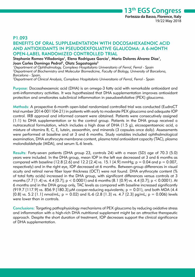
13th EGS CongressFortezza da Basso, Florence, Italy
19/22 May 2018
P1.093BENEFITS OF ORAL SUPPLEMENTATION WITH DOCOSAHEXAENOIC ACID AND ANTIOXIDANTS IN PSEUDOEXFOLIATIVE GLAUCOMA: A 6-MONTH OPEN-LABEL RANDOMIZED CONTROLLED TRIALStephanie Romeo Villadoniga1, Elena Rodriguez Garcia1, Maria Dolores Álvarez Diaz1, Joan Carles Domingo Pedrol2, Olatz Sagastagoia3
1Department of Ophthalmology, Complexo Hospitalario Universitario of Ferrol, Ferrol - Spain2Department of Biochemistry and Molecular Biomedicine, Faculty of Biology, University of Barcelona, Barcelona - Spain, 3Department of Clinical Analysis, Complexo Hospitalario Universitario of Ferrol, Ferrol - Spain
Purpose: Docosahexaenoic acid (DHA) is an omega-3 fatty acid with remarkable antioxidant and anti-inflammatory activities. It was hypothesized that DHA supplementation improves antioxidant protection and ameliorates subclinical inflammation in pseudoexfoliative (PEX) glaucoma.
Methods: A prospective 6-month open-label randomized controlled trial was conducted (EudraCT trial number 2014-001104-21) in patients with early to moderate PEX glaucoma and adequate IOP control. IRB approval and informed consent were obtained. Patients were consecutively assigned (1:1) to DHA supplementation or to the control group. Patients in the DHA group received a nutraceutical formulation, which includes a high dose of DHA (1.5 g), eicosapentaenoic acid, a mixture of vitamins B, C, E, lutein, zeaxanthin, and minerals (3 capsules once daily). Assessments were performed at baseline and at 3 and 6 months. Study variables included ophthalmological examination, DHA erythrocyte membrane content, plasma total antioxidant capacity (TAC), plasma malondialdehyde (MDA), and serum IL-6 levels.
Results: Forty-seven patients (DHA group 23, controls 24) with a mean (SD) age of 70.3 (5.0) years were included. In the DHA group, mean IOP in the left eye decreased at 3 and 6 months as compared with baseline (12.8 [2.6] and 12.2 [2.4] vs. 15.1 [4.9] mmHg; p = 0.04 and p = 0.007, respectively) and in the right eye, IOP decreased at 6 months. Between-group differences in visual acuity and retinal nerve fiber layer thickness (OCT) were not found. DHA erythrocyte content (% of total fatty acids) increased in the DHA group, with significant differences versus controls at 3 months (7.7 [1.4] vs. 4.4 [0.7]; p < 0.0001) and 6 months (8.1 [0.9] vs. 4.4 [0.7]; p < 0.0001). At 6 months and in the DHA group only, TAC levels as compared with baseline increased significantly (919.7 [117.9] vs. 856.9 [180.3] μM cooper-reducing equivalents; p = 0.01), and both MDA (4.4 [0.8] vs. 5.2 [1.1] nmol/mL; p = 0.02) and IL-6 (2.8 [1.3] vs. 4.7 [2.3] pg/mL; p = 0.006) levels were lower than in controls.
Conclusions: Targeting pathophysiology mechanisms of PEX glaucoma by reducing oxidative stress and inflammation with a high-rich DHA nutritional supplement might be an attractive therapeutic approach. Despite the short duration of treatment, IOP decreases support the clinical significance of DHA supplementation.

13th EGS CongressFortezza da Basso, Florence, Italy
19/22 May 2018
P1.094THE ROLE OF ENVIRONMENTAL FACTORS AND GENETIC MAKEUP IN THE PATHOGENESIS OF OCULAR PSEUDOEXFOLIATION (PXF) SYNDROME IN SAUDI MILITARY AND THEIR FAMILYSaad Alshamrani1
1PSMMC, Riyadh - Saudi Arabia
Objective: To find out the role of environmental factors and genetic makeup in pathogenesis of ocular pseudoexfoliation syndrome (PEX) in Saudi Military and their families.
Methods: A total of 1967 military personnel and their families visiting primary care clinics of Prince Sultan Military Medical City were screened for the presence of PEX and effect of environmental factors. Fifty one confirmed cases were studied to determine the role of HLA in the etiology of PEX. HLA-DRBl typing was performed by sequence specific oiigonucleotide primer (PCR-SSOP) method on PEX patients and 101 matched controls.
Results: The prevalence of PEX in studied population was 3.51% and showed an age dependent increase. However, there was no significant difference in the prevalence of PEX in male and female. The majority of patients (62%) have bilateral PEX. PEX was associated with higher intraocular pressure, cataract and poor vision. Chronic exposure to strong UV light causes a change in the cornea known as climatic droplet keratopathy. There was no definitive association between environment/diet/lifestyle and prevalence of PEX in our study. HLA-DRBI typing showed an increased frequencies of DRBI ‘02 (p = 0.047), DRM ‘03 (p = 0.0001) indicating that DRBl ‘02 and DRBI ‘03 may be susceptible to PEX.
Conclusion: There is no definitive association between environment/diet/lifestyle and prevalence of PEX. The presence of HLA-DRBI ‘02 and DRM ‘03 alleles is clearly associated with the susceptibility of PEX in this Saudi population. Further study using large number of subjects is in progress to determine the role of genetic factors in PEX.

13th EGS CongressFortezza da Basso, Florence, Italy
19/22 May 2018
P1.095NEW STOP CODON MUTATION OF ADAMTS10 GENE IN WEILL-MARCHESANI SYNDROMEAltan Ozcan1, Burak Ulas2
1Department of Ophthalmology, University of Cukurova School of Medicine, Adana - Turkey2Department of Ophthalmology, University of Baskent School of Medicine, Adana - Turkey
Purpose: To report new stop codon mutation in a member of the extracellular matrix protease family, the gene encoding ADAMTS10, a disintegrin and metalloprotease with thrombospondin motifs of a rare case of Weill-Marchesani syndrome.
Methods: A 9-year old male patient presented with an intraocular pressure of 45 mmHg in the both eyes. Slit lamp examination through the pupil revealed a dislocated microspherophakic lens causing pupillary block and the anterior chambers were quite shallow. A microspherophakic lens was confirmed by anterior segment optical coherence tomography. Weill-Marchesani syndrome was then diagnosed by ocular examinations, and was accompanied by systemic abnormalities, including brachymorphia and brachydactyly.
Results: The patient subsequently received bilateral YAG laser peripheral iridotomy to control the intraocular pressure. At the 1-year follow-up visit, the patient had well-controlled intraocular pressure, transparent cornea, while the lens remained in place. Molecular analysis was applied and it was revealed that the patient had c.2486G>A (p.W829*) (p.Trp829*) (Homozygocity) in ADAMTS10 gene.
Conclusions: It is known that ADAMTS10 gene plays a major role in Weill-Marchesani synrome. The mutation detected in the analyzes has not been reported previously with the Weill-Marchesani syndrome. The detected mutation was considered to be the most likely cause of the disease being early stop codon. Additional studies will be important to further define the specific function of ADAMTS10 in extracellular matrix remodeling during cell migration and invasion.

13th EGS CongressFortezza da Basso, Florence, Italy
19/22 May 2018
POSTER SESSION 2
DIAGNOSIS
13th EGS CongressFortezza da Basso, Florence, Italy
19/22 May 2018
P2.001 Retinal nerve fibre layer thickness in metabolic syndrome R. Zarei, P. Anvari, G. Fakhraei (Iran)
P2.002 The relationship between macular optic coherence tomography angiography and ganglion cell layer and their combinational index using artificial neural network K. Park (Republic of Korea)
P2.003 Measurement of optic disc cup surface depth using cirrus HD-OCT Y.K. Kim, A. Ha, J.W. Jeoung, K.H. Park (Republic of Korea)
P2.004 Comparision of optic disc margin evaluation by Bruch’s membrane opening and peripapillary retinal nerve fiber layer thickness in spectral domain optic coherence tomography Z. Tuncer, F.S. Erdem (Turkey)
P2.005 Influence of cataract and pupil size on ganglion cell layer and internal plexiform layer measurements by two different OCT devices A. Akman, S. Gür Güngör, S. Cezairlioglu, C. Öztürk, A. Sezenöz, M. Aksoy (Turkey)
P2.006 Correlation between Bruch’s membrane opening-minimum rim width and peripapillary retinal nerve fiber layer thickness according to disc size H.-K. Chop, J.M. Park, C. Kee (Republic of Korea)
P2.007 Hemodynamic profile of glaucomatous eyes and a correlation with mean deviation, visual field index and retinal nerve fiber layer thickness T.T.J.M. Berendschot1, A. Mohan2, M. Jariwala2, S. Sudhakaran2, R.S. Kumar2, R. Shetty2, C.A.B. Webers1
(1Netherlands, 2India)
P2.008 A closed-loop audit: the impact of SIGN 144 guidelines and education on glaucoma referrals R. Lawrie, K. Ah-See, D. Roberts, S. Gillan (United Kingdom)
P2.009 The role of fibrinogen elevated values as the indicator of the acute phase of inflammation with different types of glaucoma M. Lika-Pranjic (Bosnia and Herzegovina)
P2.010 Comparison of glaucoma diagnostic ability of macular ganglion cell imaging in accordance to the range around fovea using swept-source OCT J. Shin, C.K. Lee, K.H. Park (Republic of Korea)
P2.011 Comparison of Korean glaucoma patients identified through health screening versus out-patient-clinic referral Y.J. Song, K.H. Park, Y.K. Kim, H.J. Choi, J.W. Jeoung (Republic of Korea)
P2.012 Who should take MRI when showing visual field defect respecting vertical meridian? S. Kim, H.W. Bae, G.J. Seong, C.Y. Kim (Republic of Korea)
P2.013 Central 30-2 versus 24-2 threshold test measured with humphrey visual field using SITA strategy K. Yi, S.H. Bae (Republic of Korea)
P2.014 Corneal tomographic findings in patients with glaucoma and abnormal corneas J.C. Izquierdo, L. Izquierdo, L.A. Cañola-Ramirez, C. Maldonado, M.A. Henriquez (Peru)
P2.015 Comparison of perimetry for glaucoma using standard size iPad and large screen iPad Pro with Humphrey field analyzer H. Chan1, Y.X.G. Kong1, L. Sahhar1, K.R. Martin2, M. He1, J. Crowston1, A.J. Vingrys1 (1Australia, 2United Kingdom)
P2.016 Clinical efficacy of custom-built database software for early glaucoma detection S. Rho, C.-K. Lee, J. Shin, M. Lee, D.W. Kim, S. Lee, H.Y. Park (Republic of Korea)
P2.017 The location of optic disc grey crescent by optical coherence tomography S. Bang, T.-E. Lee, S.R. Chung (Republic of Korea)
P2.018 Morphofunctional characteristic of the conjunctiva in glaucoma A. Assylbekova (Kazakhstan)

13th EGS CongressFortezza da Basso, Florence, Italy
19/22 May 2018
P2.019 Ganglion-cell complex thickness to total retinal thickness ratio in Caucasian population S. Gür Güngör, A. Akman, A. Sezenöz, C. Öztürk, S. Cezairlioglu, M. Aksoy, M. Çolak (Turkey)
P2.020 Application of the ISNT and IST rules on retinal nerve fiber layer thickness in glaucoma suspect and glaucoma patients S.Y. Cho, Y.Y. Kim, C. Yoo (Republic of Korea)
P2.021 Characteristics of diffuse retinal nerve fiber layer defects in red-free photos as observed in optical coherence tomography en face images Y.Y. Kim, A. Lim, C. Yoo, J.H. Jung (Republic of Korea)
P2.022 Assessment of the biomechanics of the cornea mounted in an innovative bioreactor V. Saunier, E. Crouzet, P. Legout, P. Herbepin, G. Thuret, D. Touboul, P. Gain (France)
P2.023 Comparison of the retinal nerve fiber layer and ganglion cell complex thickness in Korean patients with unilateral exfoliation syndrome and healthy subject S.-H. Lim, T.Y. Lee (Republic of Korea)
P2.024 Hemoglobin measurement the optic nerve head using the Laguna ONhE program: comparison with spectralis OCT and image quality dependency M. Gonzalez-Hernandez, S. Alayon Miranda, M. Gonzalez de la Ros (Spain)
P2.025 Will 24-hour IOP measurements improve patient care and reduce the costs of glaucoma management? F. Meier-Gibbons, M. Töteberg-Harms (Switzerland)
P2.026 Use of ibopamine eye drops in ophthalmology I. Giuffré (Italy)
P2.027 Treatment of refractory glaucoma with two trabecular micro-bypass stents, one suprachoroidal stent, and postoperative topical prostaglandin: five year outcomes I. Singh (USA)
P2.028 Detection of Glaucoma with telemedicine optic disk photos and OCT in a primary care centre A. Anton, G. Fatty, A. Herranz, E. Ayala, A. Morilla, J.J. Gomez Valverde (Spain)
P2.030 Reproducibility of vessel density calculations using different macular imaging methods from two swept-source optical coherence tomography angiography systems T. Shoji, J. Kanno, Y. Yoshikawa, K. Ozaki, H. Ishii, Y. Ichikawa, K. Shinoda (Japan)
P2.031 Looking at the optical disc in the diabetic retinopathy telescreening: is it worth it? D. Lopes, S. Parreira, T. Loureiro, J. Cardoso, I. Machado, N. Campos (Portugal)
P2.032 Optical coherence tomography angiography of optic disc perfusion in glaucoma cases A. Hervás, R. Díaz, E. Sanz, D. Salom, P. Udaondo, S. García-Delpech (Spain)
P2.033 Intraocular pressures in patients with thyroid eye disease A. Kozaki (Japan)
P2.034 Driving status in new glaucoma referrals
C.H.K. Wu, D. Lunt, M. Doherty (United Kingdom)
P2.035 Comparison of structure - Function relationship with contrast modulation and size modulation perimetry G. Montesano1, M. Miranda1, B. Petriti1, H. Zhu2, D. Garway-Heath1 (1United Kingdom, 2China)
P2.036 Accuracy and agreement in detecting disc haemorrhages between Heidelberg retina tomograph and fundus photography in the United Kingdom Glaucoma Treatment Study J. Mohamed-Noriega, C. Gizzi, I. Treesit, T. Togano, C. Schweitzer, K. Yamamoto, D.F. Garway-Heath (United Kingdom)
P2.037 Quantification of RAPD by an automated pupillometer and its relationship to retinal nerve fiber layer thickness and visual fields in glaucoma M. Pillai1, S. Sinha1, P. Aggarwal1, R.D. Ravindran1, C. Privitera2 (1India, 2USA)
13th EGS CongressFortezza da Basso, Florence, Italy
19/22 May 2018
P2.038 New experimental applanation tonometer for myopic patients after refractive laser surgery M. Iglesias, A. Laiseca, B. Kudsieh (Spain)
P2.039 Peripapillary vessel density by OCT-angiography in normal and glaucomatous eyes S. Henriques, A.S. Lopes, D. Silva, M. Mota, M. Lisboa, F. Trancoso Vaz, I. Prieto (Portugal)
P2.040 Morphological structure of lamina cribrosa and peripapillary microvasculature for glaucoma diagnosis, using deep-range imaging R. Silva, P. Camacho, D. Cabral, P. Rodrigues, M. Marques, C. Dias, J. Valadares, J. Segurado, J. Fernandes (Portugal)
P2.041 Interest of the analysis of peri-papillary retinal nerve fibers with different diameters of measurement H. El Chehab, E. Agard, C. Dot (France)
P2.042 Comparison of Cirrus OCT and Spectralis OCT on the ability to measure the peripapillary retinal never fiber layer thickness in concurrent glaucoma and epiretinal membrane M.J. Kim, D.J. Hwang, J.H. Lee, J.H. Na, H.J. Chung, Y.S. Chung, J.Y. Choi (Republic of Korea)
P2.043 Macular morphological parameters in glaucoma diagnosis A. Blasco Alberto, M. González Hernández, V.T. Díaz Alemán (Spain)
P2.044 Evaluation of macular choroidal thickness with spectral domain optic coherence tomography in patients with ocular hypertension Ö. Gürbüz Köz, P. Topcu Yilmaz, A. Yarangümeli, M.N. Alp (Turkey)
P2.045 Optical coherence tomography angiography and optic nerve head drusen. A qualitative and quantitative evaluatio n in relation to automated perimetry defects
A. Diagourtas, K. Andreanos, S. Kandarakis, E. Asimakopoulou, D. Papaconstantinou (Greece)
P2.046 Sensitivity and specificity of RareBit visual field testing for glaucoma in the optometry service J. Przezdziecka-Dolyk, A. Matkowska, E. Fidewicz, J. Szkudlarek-Wodzinska, E. Walek, M. Misiuk-Hojlo (Poland)
P2.047 Inter-observer and inter-device agreement for the assessment of anterior-chamber angle photography with an automated gonioscope R. Scotto, C.A. Cutolo, M. Iester, C.E. Traverso (Italy)
P2.048 Comparison of central corneal thickness with ultrasound pachymetry, non-contact specular microscopy, hand held pachmetry and spectral domain optical coherence tomography G. Demirci, G. Gulkilik, S. Karaman Erdur, C. Tanriverdio, S. Kocabora, M. Ozsutcu (Turkey)
P2.049 Meningioma misdiagnosed as progressing glaucoma - A case report J. Wierzbowska, M. Jacunski (Poland)
P2.050 Relationship between optical coherence tomography angiography vessel density and severity of glaucomatous visual field loss in myopic eyes: comparison with retinal nerve fiber layer thickness S.H. Lee, E.J. Lee, T.-W. Kim (Republic of Korea)
P2.051 2-year results from the HORIZON trial: a randomized study of a Schlemm’s canal microstent for reduction of IOP in primary open angle glaucoma N. Pfeiffer (Germany)
P2.052 Comparison of signal alteration in optic nerve head on 3DT2 MRI between glaucoma and optic atrophy J.Y. Lee, H.J. Park (Republic of Korea)
P2.053 Correlation of the disc damage likelihood scale (DDLS) with retinal nerve fiber layer (RNFL) using optical coherence tomography (OCT) P. Formichella, R. Annoh, F. Zeri, A.J. Tatham (United Kingdom)
P2.054 A comparison of relative diagnostic precision between the Compass fundus perimeter and the Humphrey Field Analyzer L.M. Rossetti1, G. Montesano1, S.R. Bryan2, P. Fogagnolo1, F. Oddone1, A.M. McKendrick3, A. Turpin3, P. Lanzetta1, A. Perdicchi1, C.A. Johnson4, D. Garway-Heath2, D. Crabb2 (1Italy, 2United Kingdom, 3Australia, 4USA)

13th EGS CongressFortezza da Basso, Florence, Italy
19/22 May 2018
P2.055 Peripapillary retinal nerve fiber layer thickness and Bruch’s membrane opening-minimum rim width assessment in nonglaucomatous eyes with large and small discs S. Bayraktar, G. Sultanova, B. Izgi (Turkey)
P2.056 Diagnostic capacity of the retinal vascular density versus the study of the retinal nerve fiber layer thickness and macular ganglion cell complex L. Morales Fernandez, N. Guemes Villahoz, L. Salazar Quiñones, J.M. Martinez de la Casa, F. Saenz Frances, J. Garcia Feijoo (Spain)
P2.057 Does electromagnetic radiation of everyday life gadgets influence the signal of the senor contact lens triggerfish? C. Faschinger, D. Rabensteiner (Austria)
P2.058 Evaluating the pallor at the optic nerve head by retinography colorimetric assessment in glaucoma patients L. Salazar Quiñones, C.D. Méndez Hernández, S. Wang, J. García Feijoó (Spain)
P2.059 Self-measurement with Icare HOME tonometer M. Atanasovska Velkovska, Š. Štunf Pukl, B. Cvenkel (Slovenia)
P2.060 Self-monitoring of intraocular pressure using Icare Home tonometry in clinical practice B. Cvenkel, M. Atanasovska Velkovska, Š. Štunf Pukl (Slovenia)
P2.061 Central visual field defect associated with the location of retinal nerve fibre layer defect H.S. Kim, M.K. Shin, Y.H. Kim, J.A. Bae, J.W. Jung, S.K. Park (Republic of Korea)
P2.062 OCT angiography and color Doppler imaging in glaucoma diagnostics N. Kurysheva (Russia)
P2.063 Correlation among foveal avascular zone (FAZ) detected by OCT angiography and inner retinal thickness, and visual filed defects in POAG/NTG patients T. Fukuchi, R. Igarashi, S. Ochiai, T. Togano, Y. Sakaue, R. Iikawa, A. Suetake (Japan)
P2.064 Correlation between optic nerve head topgraphy and central corneal thickness in patients with primary open-angle glaucoma N. Civcic Kalinic, N. Civcic, V. Civcic (Serbia)
P2.065 Segmental analysis of the inner macular retinal layer thicknesses as early diagnosis of preperimetric open angle glaucoma E.B. Ausin Gonzalez, M. Sanchez Dehesa Saez, J. Iturria Soler, J.M. Ruiz Moreno, A. Royuela (Spain)
P2.066 Using a smart contact lens to identify a new biomarker associated with primary open angle glaucoma K.R. Martin1, K. Mansouri2, R.N. Weinreb3, R. Wasilewicz4, C. Gisler2, J. Hennebert2, D. Genoud2
(1United Kingdom, 2Switzerland, 3USA, 4Poland)P2.067 Clinical assessment of optic disc automatic high definition multicolor stereo vs digital monoscopic
photographs by glaucoma specialists and by general ophthalmologists F. Oddone, C. Carnevale, M. Ferrazza, M. Michelessi, L. Tanga, F. Berardo, I. Riva, L. Agnifili, G. Manni (Italy)
P2.068 AngioOCT peripapillary microvascular density outperforms standard OCT parameters as a discriminant between different glaucoma severity levels J. Barbosa Breda1, D. Andrade de Jesus1, K. Van Keer1, V. Nassiri1, G. Molenberghs1, L. Abegao Pinto2, A. Rocha Sousa2, E. Vandewalle1, I. Stalmans1 (1Belgium, 2Portugal)
P2.069 Effect of macular vascular density on central visual function and macular structure in glaucoma patients S. Jeon, H.-Y. Lopilly Park, C.K. Park (Republic of Korea)
P2.070 Long-term personal experience with second-generation trabecular micro-bypass stents in patients with glaucoma: 3-year outcomes F.H. Hengerer, G.U. Auffarth, I. Conrad-Hengerer (Germany)
P2.071 Implementation of a glaucoma risk factor calculator mobile application (APP) for an early low-tech diagnosis D.R. Mazzacane, P.F. Marino, C. Costagliola (Italy)

13th EGS CongressFortezza da Basso, Florence, Italy
19/22 May 2018
P2.072 The correlation of avascular zone with different OCT parameters in patients with glaucoma E. Kosior-Jarecka, U. Łukasik, D. Wróbel-Dudzinska, J. Wójcikowska-Karska, T. Zarnowski (Poland)
P2.073 Molecular-genetic study of primary open-angle glaucoma in patients from Bashkortostan Republic, Russia A. Zagidullina, B. Aznabaev, S. Lobov, L. Dzhemileva, R. Khasanova, A. Zainitova, E. Khusnutdinova (Russia)
P2.074 Changes in corneal endothelial cell density in patients with different stages of open-angle glaucoma Y. Saldan, N. Kapshuk, Y. Saldan, L. Khrystych (Ukraine)
P2.075 Characteristic of optic nerve head in glaucoma patients A. Miljkovic, M. Trenkic Božinovic, V. Canadanovic, N. Babic (Serbia)
P2.076 Point-wise correlation between visual field and OCT in open angle glaucoma patients P. Cirafici1, G. Maiello2, C. Ancona1, A. Masala1, C.E. Traverso1, M. Iester1 (1Italy, 2Germany)
P2.077 Quality of glaucoma referrals from primary care to the hospital eye service in a teaching hospital - A review L. Rogerson, E. Ridyard, J. Van der Hoek (United Kingdom)
P2.078 Comparison of reproducibility in glaucoma severity evaluation between structural measurement and functional test D. Shiba, S. Adachi, E. Hirano, T. Ono, K. Yuki, K. Tsubota (Japan)
P2.079 The comparision of central macular thickness in cases with having pseudo exfoliation syndrome and ocular
hypertension U. Keklikci (Turkey)
P2.080 Expediency of the automated perimetry using the Goldmann V stimulus size in visually impaired glaucoma patients M. Paolera, N. Kasahara (Brazil)
P2.081 A comparison of retinal nerve fiber layer thickness to visual field sensitivity in patients with glaucoma V. Andreic (Serbia)
P2.082 Expression profile of micro RNAs in tears. A fingerprint for the pre-clinical phase of primary open-angle glaucoma versus ocular hypertension M.D. Pinazo-Durán, V. Zanón-Moreno, J. Raga-Cervera, S.M. Sanz-González, J.J. García-Medina (Spain)
P2.083 Potential metabolic biomarkers in glaucoma patients and their regulation in response to hypoxia M. Kolko1, L.M. Dalgaard1, J.V. Jensen1, L. Hildegaard Bergersen2, N.V. Olsen1, B. Hassel2, F.A. Chaudhry2, R. Vohra1 (1Denmark, 2Norway)
P2.084 Structure-function relationship between SD-OCT multilayered macular analysis and visual field 24-2 and 10-2 defects, in perimetric glaucoma D. Rastelli, S. Giammaria, M. Michelessi, G. Manni (Italy)
P2.085 Color-coded normative classification vs continuous data by Spectral Domain-Optical Coherence Tomography for detecting glaucoma: translating the statistics into clinical practice M. Michelessi, I. Riva, E. Martini, M. Figus, P. Frezzotti, L. Agnifili, G. Manni, L. Quaranta, S. Miglior, C. Posarelli, F. Oddone (Italy)
P2.086 24-hour continuous monitoring of intraocular pressure-related fluctuations in progressing primary open angle glaucoma using a contact lens sensor C. Schweitzer, C. Andrebe, L. Tellouck, S. Roseng, F. Malet, T. Gaboriau (France)
P2.087 The role of phacoemulsification in the management of primary angle-closure glaucoma I. Yalvac, R.B. Kucumen (Turkey)
P2.088 Aphakic glaucoma after congenital cataract surgery H. Marey, A.e.R. El Sebaey (Egypt)
13th EGS CongressFortezza da Basso, Florence, Italy
19/22 May 2018
P2.089 A case of childhood glaucoma with deletion in the 6p25 region containing FOXC1 gene K. Kawase, Y. Niimi, T. Yamamoto, T. Yamamoto, T. Fukao, K. Kurata, N. Tachibana, K. Hosono, Y. Hotta (Japan)
P2.090 Large cup/disc ratio in pediatric age. SD-OCT and OCT-A evaluation D. Hipólito-Fernandes, M.-E. Luis, N. Moura-Coelho, S. Crisóstomo, A. Paixão, C. Brito, A. Toscano (Portugal)
P2.091 The importance of anterior segment imaging in assessing pediatric traumatic intra-ocular hypertension: a case report P. Arede, J. Portelinha, H. Spohr, H. Prior Filipe, S. Patrício (Portugal)
P2.092 Secondary glaucoma with bilateral clear lens subluxation and ciliary staphyloma in achondroplasia: a case
study S. Karkera, S. Deshmukh (India)
P2.093 Schlemm’s canal size and correlation study in a healthy children Caucasian population by optical coherence tomography B. Kudsieh, J.I. Fernandez-Vigo, C. Fernandez-Vigo, L. De Pablo, A. Macarro, I. Almorin, I. Sanchez, J.A. Fernandez-Vigo, J. García Feijóo (Spain)
P2.094 Corneal ring segment in paediatric patients with keratoconus P. Arribas Pardo, C. Mendez Hernandez, D. Puertas Bordallo (Spain)
P2.095 Confocal microscopy as a differential diagnostic tool between Iridocorneo endothelial syndrome and Rieger anomaly A. Burchakchi, A. de Gainza, D. Bakofski (Argentina)
P2.096 Assessment of Iintraocular pressure using ocular response analyzer and goldmann applanation tonometry before and after penetrating keratoplasty A. Wassef, K. Mansour, S. Essmat, H. Magdy (Egypt)
P2.097 Comparison of choroidal macular and peripapilar thickness in patients with pseudoexfoliation glaucoma vs controls using optical coherence tomography A. Figueiredo, D. Dias, D. José, R. Reis, I. Sampaio, M.J. Menéres (Portugal)
P2.098 Corneal biomechanics and compared tonometry after lamellar keratoplasty P. Toro Utrera, C. Mendez Hernandez, R. Sanchez Jean, C. Fernandez Perez, J.M. Benitez Del Castillo, J. Garcia Feijoo (Spain)
P2.099 Secondary glaucoma in neurofibromatosis type 2: a case report M. Wasik, S. Sharma, D. Halliday, A. Parry (United Kingdom)

13th EGS CongressFortezza da Basso, Florence, Italy
19/22 May 2018
P2.001RETINAL NERVE FIBRE LAYER THICKNESS IN METABOLIC SYNDROMEReza Zarei1, Pasha Anvari1, Ghasem Fakhraei1 1Glaucoma Division, Farabi Eye Hospital, Tehran University of Medical Sciences, Tehran - Iran
Purpose: To investigate retinal nerve fiber layer (RNFL) thickness in people with metabolic syndrome (MetS) and healthy controls.
Methods: A cross-sectional study was performed from March 2014 to January 2016. All participants underwent anthropometric and serological biochemical measurements, ophthalmological examination, and spectral-domain optical coherence tomography (SD-OCT). Individuals with elevated intraocular pressure, glaucoma, diabetic retinopathy and other ocular disorders were excluded. T-test, Chi square and general linear models were used to analyses the data.
Results: In total, 278 eyes from 139 participants were investigated [median (interquartile range) age: 37 (32-43) years]. RNFL thickness was lower in the nasal superior (107.8 ± 19.5 μm) and temporal superior (135.7 ± 18.9 μm) sectors in MetS group compared with the control group (114.6 ± 22.4 μm, p = 0.013 and 140.7 ± 18.2 μm, p = 0.027, respectively). After multiple adjustments for age, gender and the side of the examined [right (OD)/left (OS)] eye, MetS was independently associated with a lower RFNL thickness in the nasal superior (β = 0.20, p = 0.009) and temporal superior (β = 0.14, p = 0.048) sectors. RNFL thickness was significantly reduced in participants with higher numbers of metabolic abnormalities, independent of age, gender and the side of the examined eye (p = 0.043).
Conclusion: Our findings demonstrate that MetS is independently associated with reduced RNFL thickness, suggesting that neurodegeneration is implicated in pathogenesis of MetS.

13th EGS CongressFortezza da Basso, Florence, Italy
19/22 May 2018
P2.002THE RELATIONSHIP BETWEEN MACULAR OPTIC COHERENCE TOMOGRAPHY ANGIOGRAPHY AND GANGLION CELL LAYER AND THEIR COMBINATIONAL INDEX USING ARTIFICIAL NEURAL NETWORKKeunheung Park1
1Department of Ophthalmology, Pusan National University College of Medicine, Busan, Republic of Korea, Busan - Republic of Korea
Purpose: To evaluate the relationship between macular ganglion cell-inner plexiform layer thickness (GCIPLT) and vessel density and to compare diagnostic performance of them. Based on these analyses, we attempted to make a new clinically useful single parameter with the aid of artificial neural network which utilize both macular vessel density and GCIPLT.
Methods: A total of 173 subjects were included and divided into major 2 groups, 100 for validation group and 73 for neural net training. Validation group consisted of: 32 healthy, 33 early and 35 advanced glaucoma patients. Training group consisted of 28 healthy and 45 glaucoma patients. Macular GCIPLT and vessel density were measured with Spectralis optical coherence tomography and Topcon swept-source OCT respectively. Linear, quadratic and exponential regression models were used to investigate relationship between GCIPLT and vessel density. To compare model, we used Akaike’s information criterion (AIC). Multilayer neural network with 1 hidden layer was used to make single combined parameter and the diagnostic performance was compared.
Results: Pearson’s correlation coefficients showed significant correlation between macular vessel density and GCIPLT and inferior sectors generally showed stronger correlation than corresponding superior sectors. Comparing various regression analysis, the AIC of linear, quadratic, exponential regression model showed a negligible difference. All three regression models were very similar, and it was almost linear. Both vessel density and GCIPLT were not significantly correlated with central visual field in normal subjects but it was significant in early and advanced glaucoma patients. The diagnostic power of GCIPLT was much better than vessel density in most sectors. However, when vessel density was incorporated into GCIPLT with the aid of neural network, it significantly enhanced diagnostic performance. Especially, in differentiating normal and early glaucoma, artificial neural network showed significant better AUROC than both vessel density and GCIPLT throughout all sectors.
Conclusion: Macular vessel density was significantly decreased in glaucoma patients and almost linearly correlated with GCIPLT. Diagnostic performance of vessel density was much lower than GCIPLT and not enough to use in practice. However, when it was incorporated into GCIPLT using artificial neural network, a single combined parameter showed better performance than GCIPLT alone.

13th EGS CongressFortezza da Basso, Florence, Italy
19/22 May 2018
P2.003MEASUREMENT OF OPTIC DISC CUP SURFACE DEPTH USING CIRRUS HD-OCTYoung Kook Kim1, Annul Ha1, Jin Wook Jeoung1, Ki Ho Park1
1Department of Ophthalmology, Seoul National University College of medicine, Seoul - Republic of Korea
Purpose: To introduce the measurement method of optic disc cup surface depth using spectral-domain optical coherence tomography (SD-OCT) and then evaluate the rates of cup surface depression at three different stages of glaucoma.
Methods: We retrospectively identified 52 eyes with preperimetric glaucoma, 56 with mild-or-moderate glaucoma and 50 with severe glaucoma and followed them for at least 48 months. Eyes were imaged using SD-OCT (Cirrus™ HD-OCT) at 12-month intervals. The mean cup surface depth was calculated using the following formula: Cup volume / (Disc area × Average cup-to-disc ratio2) - 200 μm.
Results: The rates of mean cup surface depression (μm/yr) were significantly greater in mild-or-moderate glaucoma (-7.96 ± 1.03) than in preperimetric (-3.11 ± 0.61) and severe glaucoma (-0.70 ± 0.12; all p < 0.001). The percentile rates of mean cup surface depression (%/yr) were significantly greater than those of average of retinal nerve fiber layer (RNFL) thinning (%/yr) in preperimetric glaucoma (-1.64 ± 0.12 vs -1.11 ± 0.07; p < 0.001) and mild-or-moderate glaucoma (-4.20 ± 0.33 vs -3.14 ± 0.19; p < 0.001); and conversely, in severe glaucoma, mean cup surface depth changed slower than did average RNFL thickness (-0.64 ± 0.06 %/yr vs -0.75 ± 0.08 %/yr; p < 0.001).
Conclusions: In early-to-moderate glaucoma, the cup surface depth changed faster than did the RNFL thickness. These results signify the possibility that SD-OCT-based estimation of cup surface depth might be useful for monitoring of glaucoma development and progression.

13th EGS CongressFortezza da Basso, Florence, Italy
19/22 May 2018
P2.004COMPARISION OF OPTIC DISC MARGIN EVALUATION BY BRUCH’S MEMBRANE OPENING AND PERIPAPILLARY RETINAL NERVE FIBER LAYER THICKNESS IN SPECTRAL DOMAIN OPTIC COHERENCE TOMOGRAPHYZerrin Tuncer1, Fatma Seckin Erdem1
1Bayrampasa Eye Hospital, Istanbul - Turkey
Purpose: Peripapillary retinal nerve fiber layer thickness (RNFLT) measured by conventional optic disc margin-based method is compared with the method using Bruch’s membrane opening (BMO) in spectral domain (SD) optic coherence tomography (OCT) in glaucoma patients.
Methods: 127 eyes of 68 patients of glaucoma or glaucoma suspect were examined prospectively with Heidelberg SD OCT by the same investigator. First, optic discs were analyzed in 3.4 mm diameter by manual method and then the measurements were repeated automatically by BMO method in glaucoma modulus using anatomical positioning system (APS). Average RNFLT of nasal (N), nasal-superior (NS), nasal-inferior (NI), temporal (T), temporal-superior (TS), temporal-inferior (TI), inferior (I) and superior (S) disc are recorded. Records are compared by t-test (parametric) and Wilcoxon Signed Ranks test (nonparametric).
Results: The median age of patients (43 female, 25 male) is 59.42 ± 1.7 years. Average disc diameter is 1.63 ± 0.20 mm. Disc diameter histogram is summarized. There was a statistically significant difference between the two methods, in all quadrants, except temporal.
Conclusion: Glaucoma modulus in SD OCT is an objective method in analyzing optic disc head. In manual method, neuroretinal rim is estimated due to the attention of investigator and differs in repeated measurements. In glaucoma modulus, the distance between BMO and internal limitan membrane (ILM) is applied as reference, fovea center and BMO is determined automatically. So, the automated method represents real anatomical position, eliminates empirical and individual errors. Although it is assumed that the measurements of OCT glaucoma modulus are more realistic, studies with greater groups are needed for exact results.
13th EGS CongressFortezza da Basso, Florence, Italy
19/22 May 2018
P2.005INFLUENCE OF CATARACT AND PUPIL SIZE ON GANGLION CELL LAYER AND INTERNAL PLEXIFORM LAYER MEASUREMENTS BY TWO DIFFERENT OCT DEVICESAhmet Akman1, Sirel Gür Güngör1, Sefik Cezairlioglu1, Caner Öztürk1, Almila Sezenöz1, Mustafa Aksoy1
1Department of Ophthalmology, Baskent University, Ankara - Turkey
Purpose: To investigate and compare the effect of cataract and pupil size on ganglion cell layer-internal plexiform layer (GCL+IPL) measurements using Carl Zeiss Meditec Cirrus HD-optical coherence tomography (OCT) (Cirrus) and Heidelberg Engineering Spectralis OCT (Spectralis).
Methods: Seventeen patients, (11 female, 6 male; mean age: 66.91 ± 9.73 years), were included in the prospective, hospital-based study. All subjects underwent planned phacoemulsification and intraocular lens implantation under topical anesthesia. GCL+IPL measurements were taken before and after dilatation, preoperatively and postoperatively, using Cirrus and Spectralis OCT.
Results: GCL+IPL thickness measurements taken by Spectralis OCT were found to be significantly higher than the ones taken by Cirrus OCT (p = 0.024; p = 0,.012; p = 0.002; p = 0.037, respectively). The postoperative measurements of GCL+IPL thicknesses were found to be higher than the preoperative measurements on both devices. The difference was found to be statistically significant for measurements of Spectralis OCT device (p = 0.007), while that of Cirrus OCT device was not statistically significant (p = 0.145). The GCL+IPL thickness measurements taken before pupil dilatation were found to be higher than measurements taken after pupil dilatation on both devices, however, the differences were not statistically significant (p > 0.05).
Conclusion: While pupil size does not affect GCL+IPL measurements performed by both OCT devices, the presence of cataract seems to have an impact on Spectralis values. Differences in hardware specifications and segmentation algorithms of two devices could be the reason for the influence of cataract on Spectralis measurements.

13th EGS CongressFortezza da Basso, Florence, Italy
19/22 May 2018
P2.006CORRELATION BETWEEN BRUCH’S MEMBRANE OPENING-MINIMUM RIM WIDTH AND PERIPAPILLARY RETINAL NERVE FIBER LAYER THICKNESS ACCORDING TO DISC SIZEHyun-Kyung Chop1, Jong Moon Park1, Changwon Kee2
1Department of Ophthalmology, Gyeongsang National University Changwon Hospital, Gyeongsang National University, School of Medicine, Changwon - Republic of Korea2Department of Ophthalmology, Samsung Medical Center, Sungkyunkwan University School of Medicine, Changwon - Republic of Korea
Purpose: To investigate the correlation between Bruch’s membrane opening minimum rim width (BMO-MRW) and retinal nerve fiber layer (RNFL) thickness according to disc size. And to evaluate the association of RNFL thickness from three different scan circle size.
Design: Retrospective, observational cross-sectional study.
Methods: Glaucoma suspects with no visible localized RNFL defect and standard automated perimetry results of within normal limits, but with suspicious neuroretinal rim thinning were included. Ninety two subjects were divided into three groups according to disc size: group 1 (n = 17), small disc (disc area < 1.63 mm2); group 2 (n = 40), regular size disc (disc area: 1.63 ~ 2.43 mm2); group 3 (n = 35), large disc (disc area > 2.43 mm2). They underwent ophthalmic examinations including confocal scanning laser tomography and Spectralis spectral-domain optical coherence tomography. BMO-MRW values and RNFL thickness from the three circle scans were obtained.
Results: Global BMO-MRW was the thickest in group 1 (317.24 ± 70.60 μm) followed by group 2 (259.03 ± 40.04 μm) and the thinnest in group 3 (236.74 ± 31.21 μm) (p = 0.000, ANOVA). Correlation between global BMO-MRW values and RNFL thickness was the strongest in group 3 (r = 0.647) followed by group 2 (r = 0.570) and none in group 1 (r = 0.176, Pearson correlation). There were no significant differences in global RNFL thickness according to disc size in each of the 3.5 mm, 4.1 mm, and 4.7 mm diameter scan circle (all p > 0.05).
Conclusions: BMO-MRW differed significantly according to disc size. Correlation between BMO-MRW and RNFL thickness may also vary according to disc size.
13th EGS CongressFortezza da Basso, Florence, Italy
19/22 May 2018
P2.007HEMODYNAMIC PROFILE OF GLAUCOMATOUS EYES AND A CORRELATION WITH MEAN DEVIATION, VISUAL FIELD INDEX AND RETINAL NERVE FIBER LAYER THICKNESSTos T.J.M. Berendschot1, Ashwin Mohan2, Manan Jariwala2, Suria Sudhakaran2, Rajesh S. Kumar2, Rohit Shetty2, Carroll A.B. Webers1
1University Eye Clinic Maastricht, Maastricht - Netherlands2Narayana Nethralaya, Bangalore - India
Purpose: To study the hemodynamic profile of glaucomatous eyes and correlate it with the visual field Mean Deviation (MD) and Visual Field Index (VFI), and the Retinal Nerve Fibre Layer (RNFL) thickness.
Methods: Patients with confirmed glaucoma on comprehensive ophthalmic examination were included and subsequently underwent retinal oximetry (Oxymap T1, Oxymap hf, Reykjavik, Iceland) to obtain the arteriolar (SaO2) and venous O2 saturation (SvO2) along with the arterio-venous saturation difference (AVSD). We also measured the MD and in a subset the VFI (N = 43) and RNFL thickness (N = 27).
Results: 53 eyes were analyzed and had an age of 64.9 ± 8.8 years (mean ± standard deviation). SaO2 was 103.2 ± 12.9%, SvO2 was 59.4 ± 9.2%, and AVSD was 44.1 ± 13.1%. Average RNFL thickness was 56.5 ± 14.7 μm, MD was -13.8 ± 9.0 dB, and VFI was 59.1 ± 29.2 . Arteriolar saturation correlated significantly with MD (r = -0.34; p = 0.014). MD correlated significantly with VFI (r = 0.76; p < 0.001) and RNFL thickness (r = 0.48, p = 0.01) and seemed more negative with a higher AVSD, but this was not significant (r = -0.20; p = 0.15).
Conclusions: As the retinal nerve fiber layer thickness decreases and visual field loss worsens, the arteriolar saturation increases. This results in a corresponding increase in AVSD which is contrary to what we would expect in glaucoma where tissue loss is assumed to result in a lower oxygen utilization. However, a healthy Caucasian retina is assumed in the determination of SaO2 and SvO2. Tissue loss will change the underlying model assumptions and cause an apparent artefactual increase in the measured saturations which may explain our findings.
13th EGS CongressFortezza da Basso, Florence, Italy
19/22 May 2018
P2.008A CLOSED-LOOP AUDIT: THE IMPACT OF SIGN 144 GUIDELINES AND EDUCATION ON GLAUCOMA REFERRALSRachel Lawrie1, Kim Ah-See2, David Roberts3, Stewart Gillan3
1University of Dundee School of Medicine1, Dundee - United Kingdom2Princess Alexandra Eye Pavilion, Edinburgh - United Kingdom3Department of Ophthalmology, Ninewells Hospital, Dundee - United Kingdom
Purpose: Approximately 20% of ophthalmology referrals in Scotland are related to glaucoma, the majority of which are provided by community optometrists. The SIGN 144 guidelines for appropriate referrals and safe discharge were launched in March 2015 to streamline the interface between primary and secondary care where glaucoma is concerned. An audit completed in 2016 comparing the “pre-intervention” and ‘post-intervention’ period revealed notable improvement in documentation of risk factors, pachymetry and fundal image. Nevertheless, some areas were consistently under-documented such as repeat tonometry, iridocorneal angle assessment and optic disc size. The results of the audit were discussed with the Local Area Optometric Committee and the importance of provision of efficient eye care delivery was reinforced. Here we evaluate the downstream impact of the SIGN 144 guideline on referrals now that a further intervention has been implemented and the guideline has been in place for at least 18 months.
Methods: The results of the initial audit cycle were discussed with the Local Area Optometric Committee. Electronic and written referral data from 1772 optometry referrals for the period of May to July 2017 were collected and those pertinent to glaucoma were isolated. These referrals were audited against the key criteria as specified in the SIGN guideline and statistical analysis was performed.
Results: The vast majority of referrals in NHS Tayside were consistently of a high standard. The audit data shows that the quality of referrals has improved across most domains. However, it would appear that the number of referrals to secondary care has increased. Comparisons to the first cycle have been made and conclusions will be drawn.
Conclusions: Whist the referrals made to the eye service at NHS Tayside are of a high standard, there are identifiable areas for improvement. There are early signs that the introduction of the guidelines has started to impact positively on the quality of referrals made. It has been shown that feedback from Ophthalmology in terms of this data has raised the bar in terms of streamlining the patient journey in cases where glaucoma is suspected.

13th EGS CongressFortezza da Basso, Florence, Italy
19/22 May 2018
P2.009THE ROLE OF FIBRINOGEN ELEVATED VALUES AS THE INDICATOR OF THE ACUTE PHASE OF INFLAMMATION WITH DIFFERENT TYPES OF GLAUKOMAMerita Lika-Pranjic1
1Clinic for Eye Diseases, KCUS, Sarajevo - Bosnia and Herzegovina
Purpose: The aim of study was to investigate the role of fibrinogen elevated values in serum as a good indicator of the acute phase of inflammation at patients with different types of glaucoma that can be linked to pathogenesis and glaucoma progression.
Methods: The study included 180 eyes glaucoma patients of both genders, ages 40 to 70 years old. Patients were divided into three groups based on the type of glaucoma: 60 eyes with glaucoma simplex (GS), 60 eyes with normotensive glaucoma (NG) and 60 eyes with angular glaucoma (GA). The fourth group, as a control, made 60 eyes with a senile cataract without glaucoma. A detailed ophthalmological examination was performed. The plasma fibrinogen concentration was determined by the method by which the citrred plasma coagulates with increasing thrombin. As reference values of fibrinogen in plasma were 1.8 to 3.8 g/L.
Results: The plasma concentration of fibrinogen in the control group was 3.55 ± 0.13, while in the group of patients with NG it was 3.57 ± 0.15. In the group of patients with GS, the plasma fibrinogen concentration was 3.20 ± 0.10, and with GA the same was 2.10 4.02 ± 0.18. A statistically significant difference in plasma fibrinogen level was observed between patients with GS compared to the control group (p = 0.039), between patients with GS and with NG (p = 0.045), and between patients with GS and with GA p < 0.05). Also statistically significant difference in plasma fibrinogen level was observed between patients with GA compared to the control group (p = 0.041)
Conclusion: There was no statistically significant correlation between the concentration of fibrinogen in plasma and OCT parameters in either glaucoma patients or in the control group. There was a statistically significant positive correlation between plasma fibrinogen concentrations and right eye PNO (optic nerv papille) in patients with GS (rho = 0.375, p < 0.05) (Graph 11) and statistically significant negative correlation between fibrinogen concentration in plasma and MDD values in patients with GA (rho = -0.372, p < 0.05).

13th EGS CongressFortezza da Basso, Florence, Italy
19/22 May 2018
P2.010COMPARISON OF GLAUCOMA DIAGNOSTIC ABILITY OF MACULAR GANGLION CELL IMAGING IN ACCORDANCE TO THE RANGE AROUND FOVEA USING SWEPT-SOURCE OCTJonghoon Shin1, Chang Kyu Lee2, Keun Heung Park3
1Department of ophthalmology, Pusan National University Yangsan Hospital, YangSan - Republic of Korea 2Department of Ophthalmology, Ulsan Univerisity Hospital, Ulsan - Republic of Korea3Department of ophthalmology, Pusan National University Hospital, Busan - Republic of Korea
Purpose: The purpose of the present study was to evaluate the diagnostic ability of GCIPL measurements in approximately the total retina that were obtained using swept-source OCT wide-angle scanning, compared with the automatic GCIPL analysis within a 6- x 6-mm annulus around the fovea.
Methods: This study used a prospective, cross-sectional, and comparative design. Imaging was obtained using the swept-source Deep Range Imaging-OCT (DRI-OCT-1, Topcon). In a wide-angle scan, this software calculates the GCIPL for each 1-mm2 grid square of a 12- X 9-mm scan, and these 108 data were displayed and exported using a built-in program. This study established a boundary of 4- X 4-mm, 6- X 6-mm, and 8- X 8-mm areas centered on the fovea, and each area was divided into 6 sectors. The average, minimum, and 4-sectoral (superotemporal, superonasal, inferotemporal, inferonasal) GCIPL thicknesses of each area were measured and recorded. The determination of the sectoral boundary was referred to as GCA algorithms of the Cirrus HD-OCT software. The automated GCIPL thickness of a 6- X 6-mm scan of macular fixation was measured, and the average, minimum, and 4-sector thicknesses were obtained using segmentation software.
Results: This study enrolled 79 glaucomatous eyes in 61 patients and 75 eyes in 59 normal healthy controls. The sectoral, minimum and average GCIPL measurements in the newly created 4- X 4-mm, 6- X 6-mm, and 8- X 8-mm areas significantly differed among the normal and glaucoma groups or between the glaucoma subgroups (p < 0.001). In the eyes with total glaucoma, the AUROCs for the minimum and inferotemporal GCIPL of the 4- X 4-mm area and the inferotemporal GCIPL thickness of the 6- X 6-mm area (0.941, 0.931, 0.909, respectively); in the eyes with glaucoma with central scotoma (CS), the minimum, inferotemporal, average, and inferonasal GCIPL of the 4- X 4-mm area and the inferotemporal, minimum, and average GCIPL thickness of the 6- X 6-mm area were greater than 0.9 (0.986, 0.978, 0.926, 0.918, 0.944, 0.903, 0.900, respectively). In the eyes with glaucoma without CS, there were no GCIPL parameters greater than 0.9, and the minimum GCIPL of the 4- X 4-mm (0.898) area was the most accurate parameter for discriminating between the patients with glaucoma without CS and the normal patients.
Conclusions: The minimum GCIPL in the 4- x 4-mm area around the fovea is the most accurate parameter of the various GCIPL parameters in this study for glaucoma diagnosis. The GCIPL parameters in the 4- x 4-mm area around the fovea have shown the greater ability for discrimination between normal patients and glaucoma patients than those in the 6- x 6-mm or 8- x 8-mm areas. The ability to diagnosis glaucoma has increased as the area of measuring the GCIPL thickness has narrowed.

13th EGS CongressFortezza da Basso, Florence, Italy
19/22 May 2018
P2.011COMPARISON OF KOREAN GLAUCOMA PATIENTS IDENTIFIED THROUGH HEALTH SCREENING VERSUS OUT-PATIENT-CLINIC REFERRALYong Ju Song1, Ki Ho Park2, Young Kook Kim2, Hyuk Jin Choi3, Jin Wook Jeoung2
1Department of Ophthalmology, Chosun University College of Medicine, Gwangju - Republic of Korea2Department of Ophthalmology, Seoul National University College of Medicine, Seoul - Republic of Korea3Healthcare System Gangnam Center, Seoul National University Hospital, Seoul - Republic of Korea
Purpose: To determine the effectiveness of health screening for early detection of glaucoma.
Methods: The data on 547 patients who had undergone comprehensive glaucoma evaluation at the Seoul National University Hospital Ophthalmologic Center between January 2013 and December 2014 were reviewed. Among the 547 patients, 130 who had already been diagnosed with glaucoma were excluded from the study. The remaining cohort comprised 215 patients who had undergone health screening by non-contact tonometry and non-mydriatic fundus photography (Group A) and 202 who had been referred by other out-patient clinics (Group B). Based on a comprehensive evaluation, the subjects were diagnosed as either glaucoma suspect or definite glaucoma. The patients’ clinical characteristics were then subjected to inter-group comparison.
Results: In Group A (n = 215), 51 patients were diagnosed as definite glaucoma and 164 as glaucoma suspect. In Group B (n = 202), 56 patients were diagnosed as definite glaucoma and 146 as glaucoma suspect. Group A (n = 215) showed, relative to Group B (n = 202), lower intraocular pressure (IOP) (p = 0.037), lower glaucoma severity [average retinal nerve fiber layer thickness (RNFLT), mean deviation (MD), and lower average as well as minimal ganglion cell-inner plexiform layer thickness (GCIPLT)] (p = 0.005, p = 0.004, p = 0.010, p = 0.004). Among the newly diagnosed glaucoma patients (n = 107) moreover, Group A (n = 51) showed, relative to Group B (n = 56), lower IOP (p = 0.008), and lower glaucoma severity (average cup-to-disc ratio, MD, average and minimal GCIPLT) (p = 0.041, p = 0.002, p = 0.026, p = 0.026). In Group A, 47 patients (91%) had lower-tension glaucoma (LTG), and in Group B, 38 patients (68%) did. The proportion of LTG among the glaucoma patients of Group A was 1.3 times that among the glaucoma patients of Group B. Inferior RNFL thickness among the LTG patients of Group A was thicker than among the LTG patients of Group B, significantly (p = 0.037).
Conclusions: The health-screening group (A) showed, relative to the referred group (B), an early stage of glaucoma, among both the newly diagnosed glaucoma patients and the LTG patients. Health screening therefore can be considered to be a useful method for glaucoma diagnosis as well as for early detection of LTG.
13th EGS CongressFortezza da Basso, Florence, Italy
19/22 May 2018
P2.012WHO SHOULD TAKE MRI WHEN SHOWING VISUAL FIELD DEFECT RESPECTING VERTICAL MERIDIAN?Sangah Kim1, Hyoung Won Bae1, Gong Je Seong1, Chan Yun Kim1
1Department of Ophthalmology, Yonsei University College of Medicine, Seoul - Republic of Korea
Purpose: This study aims to investigate the optical coherence tomography (OCT) and visual field (VF) findings of suspected brain lesions in patients with VF defect respecting vertical meridian.
Method: We reviewed the medical records of patients who underwent MRI to identify brain lesions because of hemianopia including complete and partial hemianopia between January 2010 and March 2017. The patients were divided into two groups by MRI findings: brain (+) group (n = 70) and brain (-) group (n = 20). We compared findings of OCT and VF between the two groups to find out the factors that could predict the brain lesion. Based on multivariate logistic model regression analysis, the nomogram was constructed to predict the brain lesion in MRI.
Result: The VF mean deviation (MD) was lower (p = 0.001) and all sectors of peripapillary retinal nerve fiber layer (pRNFL) thickness except the temporal region were thicker in brain (+) group. In contrast, the macular ganglion cell-inner plexiform layer (mGCIPL) thickness tended to be thinner in brain (+) group but not statistically significant. The area under the receiver operating characteristic curve (AUC) of Nomogram showed 0.925 and was well calibrated.
Conclusion: We developed the novel and accurate screening method for detection of the brain lesion in patients with hemianopia. Since this can be seen at a glance with values of OCT and VF, it will be very beneficial when the clinicians are considering whether or not to take MRI easily.

13th EGS CongressFortezza da Basso, Florence, Italy
19/22 May 2018
P2.013CENTRAL 30-2 VERSUS 24-2 THRESHOLD TEST MEASURED WITH HUMPHREY VISUAL FIELD USING SITA STRATEGYKayoung Yi1, Seok Hyun Bae1
1Department of Ophthalmology, Hallym University, College of Medicine, Kangnam Sacred Heart Hospital, Seoul - Republic of Korea
Purpose: To investigate the difference between central 24-2 threshold test and 30-2 threshold test using Humphrey visual field (Carl Zeiss Meditec, Dublin, CA, USA).
Methods: 366 eyes of 183 glaucoma and glaucoma suspect patients who underwent both central 30-2 and 24-2 threshold test using Swedish Interactive Threshold Algorithm(SITA) standard strategy were recruited. We compared test time, fixation loss (%), false positive errors (%), false negative errors (%) which are an index of reliability. Also we compared mean deviation (MD), pattern standard deviation (PSD), and visual field index (VFI).
Results: The mean age was 54.67 ± 14.95. MD, VFI, and fixation loss showed no significant difference between central 30-2 and 24-2 threshold test. However, 24-2 threshold test provided shorter time from 7.52 ± 1.39 minutes to 5.65 ± 1.10 minutes (p < .001). Their difference was 24.51 ± 9.44%. False positive errors was significantly higher, but false negative errors was lower in 24-2 threshold test (p = .009, p < .001). PSD was lower in 24-2 threshold test than 30-2 (p < .001).
Conclusions: VFI and MD are comparable in both tests. But in other aspects, two tests are not identical. Compared with 30-2 threshold test, 24-2 can be more efficient examination for the patients in terms of test time. It could be better to individualize visual field test according to the patients.

13th EGS CongressFortezza da Basso, Florence, Italy
19/22 May 2018
P2.014CORNEAL TOMOGRAPHIC FINDINGS IN PATIENTS WITH GLAUCOMA AND ABNORMAL CORNEASJuan Carlos Izquierdo1, Luis Izquierdo1, Laura A. Cañola-Ramirez1, Carmen Maldonado1, Maria A. Henriquez1
1Instituto de Ojos Oftalmosalud (Research Department), Lima - Peru
Purpose: To evaluate tomographic parameters from Scheimpflug analysis in patients diagnosed with glaucoma and abnormal cornea.
Methods: Cross-sectional retrospective case series study that included 36 eyes of 18 patients, with glaucoma diagnosis at Institute of Eyes, Oftalmosalud, Lima, Peru, between January 2012 and December 2012 and presented abnormal corneas from Scheimpflug analysis. All patients underwent SD-OCT (Spectral Domain Optical Coherence Tomography; Carl Ziess Meditec, Dublin, CA), HRT (Heidelberg Retina Tomograph; Heidelberg Engineering GmbH, Heidelberg, Germany), CV (Visual Field; Humphreys Visual Field Analyzer, Carl Ziess Meditec, Dublin, CA) and Pentacam (Oculus Inc, Wetzlar, Germany); we evaluated visual acuity without correction (AVSC) and intraocular pressure. Abnormal cornea was defined if the cornea meet 3 or more of the suggested cutoff values for subclinical keratoconus. The statistical analysis was performed with SPSS Version 20.
Results: 18 patients with an age range between 34.0 to 74.0 years (58.00 ± 12.78) were diagnosed with glaucoma. Mean BCVA was 0.39 ± 0.64 LogMAR and mean IOP was 15.11 ± 2.8. Mean OCT relation cup/disc was 0.67 ± 0.13. Mean HRT area C / D was 0.45 ± 0.13, mean ring area was 1.56 ± 0.50, mean RNFL was 0.23 ± 0.16 and in the classification Moorfields was 9 (25%) eyes were reported as Borderline and 27 (75%) as Normal. Mean Visual Field the MD was -3.68 ± 3.96. In the analysis Scheimfplflug mean pachymetry at the thinnest point was 559.69 ± 33.92, mean Kmax was 5.22 ± 2.67, mean K1 Later was -6.13 ± 0.20 and mean K2 Later was -6.58 ± 0.37. 11.11% of the eyes had more than 3 parameters considerer for subclinical keratoconus, 8.33% had more than 5 and 8.33% had more than 6. 13.88% of the eyes had more than 3 parameters considerer for keratoconus, 8.33% had more than 5 and 8.33% had more than 6.
Conclusion: This study demonstrates for the first time, evidence between glaucoma and topographic abnormal corneas. Further longitudinal, prospective study are necessary to evaluate the prevalence of this find in glaucoma patients.
Abstract presented to ARVO 2018

13th EGS CongressFortezza da Basso, Florence, Italy
19/22 May 2018
P2.015COMPARISON OF PERIMETRY FOR GLAUCOMA USING STANDARD SIZE IPAD AND LARGE SCREEN IPAD PRO WITH HUMPHREY FIELD ANALYZERHelen Chan1, Yu Xiang George Kong1, Lukas Sahhar1, Keith R. Martin2, Mingguang He3, Jonathan Crowston3, Algis J. Vingrys4
1Royal Victorian Eye and Ear Hospital, Melbourne - Australia2Department of Ophthalmology, Cambridge University Hospitals NHS Foundation Trust, Cambridge - United Kingdom3Centre of Eye Research Australia, Melbourne - Australia Department of Optometry & Vision Sciences, University of Melbourne, Melbourne - Australia
Purpose: To compare perimetric outcomes of the iPad perimetry application Melbourne Rapid Fields (MRF) using the standard-sized 9.7 inch iPad (MRF-9.7) and the larger 12.5 inch iPad Pro (MRF-12.5) with Humphrey Field Analyzer 24-2 Swedish Interactive Threshold Algorithm-Standard (HFA-SS).
Methods: 44 eyes of 44 patients with acuity better than 6/12 (20/40) were recruited (9 normal controls, 6 glaucoma suspect, 10 mild glaucoma and 19 moderate-severe glaucoma). Patients received visual field threshold assessment with HFA-SS followed by MRF-9.7 and MRF-12.5 using a 66-point radial test pattern. The order of presentation of MRF-9.7 and MRF-12.5 were randomized. MRF-9.7 required four changes in fixation to complete visual field assessment, while the larger screen MRF-12.5 required only one. Patients were advised to fixate and follow the fixation target as instructed by a voice over. Exclusion criteria included visual acuity worse than 6/12, intraocular surgery in the preceding six months and poor reliability indices on HFA-SS.
Results: MRF-9.7 and MRF-12.5 test times were both significantly faster than HFA-SS (MRF-9.7: 5.29 ± 1.23 min, MRF-12.5: 4.10 ± 1.07 min, HFA-SS: 6.56 ± 2.23 min, p < 0.01). MRF-12.5 was on average 1.18 ± 0.60 minutes faster per eye compared to MRF-9.7 and 2.51 ± 2.04 minutes faster than HFA-SS. Similar to our previous published study, Pearson’s correlation of perimetry results between MRF-9.7 and HFA-SS were strong for Mean Deviation (MD) (r2 = 0.85) and Pattern Deviation (PD) (r2 = 0.75). Correlations were similarly strong for MRF-12.5 with HFA-SS for MD (r2 = 0.86) and PD (r2 = 0.84). The 95% Limits of Agreement between MRF-9.7 and MRF-12.5 for MD is [-4.41 dB, 6.23 dB] and for PD is [-5.97 dB, 2.30 dB].
Conclusion: Perimetry results using MRF-12.5 had strong correlations to MRF-9.7 and HFA-SS. Testing time for MRF-12.5 was faster than MRF-9.7 (1.2 min faster) and HFA-SS (2.5 min faster), making the large screen iPad Pro an efficient method of visual field assessment. This is especially important in clinical settings where portability and high throughput is required.
13th EGS CongressFortezza da Basso, Florence, Italy
19/22 May 2018
P2.016CLINICAL EFFICACY OF CUSTOM-BUILT DATABASE SOFTWARE FOR EARLY GLAUCOMA DETECTIONSeungsoo Rho1, Chang-Kyu Lee2, Jonghoon Shin3, Marvin Lee4, Dai Woo Kim5, Seunghyun Lee1, Hong Young Park6
1Department of Ophthalmology, CHA University, CHA Medical Center, Seongnam - Republic of Korea2Department of Ophthalmology, Ulsan University Hospital, University of Ulsan College of Medicine, Ulsan - Republic of Korea3Department of Ophthalmology, Pusan National University Yangsan Hospital, Pusan - Republic of Korea4Kong Eye Center, Seoul - Republic of Korea5Department of Ophthalmology, Kyungpook National University Hospital, Kyungpook National University School of Medicine, Daegu - Republic of Korea6Department of Ophthalmology, Gwang-ju Veterans Hospital, Gwangju - Republic of Korea
Purpose: To assess the clinical efficacy for early glaucoma detection using custom-built image software visualizing translucent retinal nerve fiber layer thickness (RNFLT) graph based on normative database.
Methods: This study was conducted using normative database constructed on RNFLT data of 151 Korean healthy eyes. The reference lines of the mean, the lower 5% and the lower 1% limit were visualized as a translucent RNFL thickness graph produced by our software after an input of each subject’s major retinal artery position and overlaid on the RNFLT measurement. Fifty additional healthy control and 50 early glaucoma eyes were collected for validation group. If a subject’s RNFLT graph was caught by the reference line of the lower 1% limit, it was defined as abnormal. Kappa value was utilized to determine the difference of glaucoma detection ability by between built-in software and our software (agreement with standard answer).
Results: The accuracy of our custom-built software was significantly higher than manufacturer’s database (kappa of 0.980 vs 0.320; sensitivity of 1.000 vs 0.320, p < 0.05) maintaining high specificity (1.000 vs 0.980).
Conclusions: The custom-built imaging software with constructed RNFLT normative database showed high clinical efficiency for early glaucoma detection with negligible user-related variability.
13th EGS CongressFortezza da Basso, Florence, Italy
19/22 May 2018
P2.017THE LOCATION OF OPTIC DISC GREY CRESCENT BY OPTICAL COHERENCE TOMOGRAPHYSora Bang1, Tae-Eun Lee1, Sae Rom Chung1
1Department of Ophthalmology, Chonbuk National University Medical School, Jeonju - Republic of Korea
Purpose: The optic disc grey crescent is defined as pigmentation on or within the neuroretinal rim. We evaluated the location of the grey crescent based on the margin of Bruch’s membrane using optical coherence tomography (OCT).
Methods: Overall, 1,042 eyes of 529 subjects were evaluated for the grey crescent using fundus photographs. These photographs were manually overlaid to en-face the OCT images. Based on the margin of Bruch’s membrane using OCT, we evaluated whether the crescent is located inside the disc margin or not.
Results: Fundus photographs of sufficient quality for analysis were available for 972 eyes. The grey crescent was found in 179 eyes (18.4%); of these, 131 eyes were available for OCT analysis. In most eyes, the crescent was located outside the disc margin (122 eyes, 93.1%), with only nine eyes (6.9%) having it inside the disc margin.
Conclusions: The grey crescent was considered that may interfere with adequate assessment of the neuroretinal rim. In this study, we found that most of the grey crescent is located outside the disc margin.
13th EGS CongressFortezza da Basso, Florence, Italy
19/22 May 2018
P2.018MORPHOFUNCTIONAL CHARACTERISTIC OF THE CONJUNCTIVA IN GLAUCOMAAssel Assylbekova1
1Kazakh Eye Research Institute, Almaty - Kazakhstan
Purpose: The studying morphofunctional characteristics of the conjunctiva for various types of glaucoma without drops and on the background of local antihypertensive therapy.
Methods: A confocal conjunctival microscopy was performed in 48 eyes of patients with primary glaucoma. Patients were divided into 2 groups. The 1 group included 20 eyes with first diagnosed primary glaucoma who had not received medical treatment: 1a - 10 eyes with initial and developed stages of glaucoma, 1b - 10 eyes with far-advanced and terminal stages of glaucoma. The second group included 28 eyes with glaucoma which received local antihypertensive therapy, of which 2a - 12 eyes with the prescription of local drug therapy for up to 6 months, 2b - 10 eyes with the prescription of instillation of hypotensive drops from 6 months to 1 year, 2B - 6 eyes with prescription of instillations of drops over 1 year. The examination of the conjunctiva was carried out using confocal microscopy on a Heidelberg retinal tomograph HRT III.
Results: In consequence of conducted confocal microscopy of bulbar conjunctiva was determined that there was no structural change in patients with primary glaucoma who did not receive local antihypertensive therapy (group 1a). Patients with developed stages of glaucoma who did not receive LAT (1b group) showed mild degenerative changes in the conjunctiva in the form of subepithelial infiltration of the conjunctiva with lymphocytes and macrophages, poor discernibility of cell borders, which indicates metabolic disturbances in the anterior segment of the eyeball in primary glaucoma. Patients with primary glaucoma, who used antihypertensive drugs in instillations for no more than 6 months (group 2a), there have been similar changes. As the periods of instillations of LAT preparations (groups 2b and 2c) increase, in addition to the aforecited changes,an increase in the size and the scene of desquamation of the nucleus of the epithelial cells of the conjunctiva was revealed. Conclusions: Thus, the study showed the presence of degenerative changes in the conjunctiva in patients with glaucoma who used local antihypertensive drugs and in patients with far-advansed and terminal stages of glaucoma without antihypertensive drugs.
13th EGS CongressFortezza da Basso, Florence, Italy
19/22 May 2018
P2.019GANGLION-CELL COMPLEX THICKNESS TO TOTAL RETINAL THICKNESS RATIO IN CAUCASIAN POPULATIONSirel Gür Güngör1, Ahmet Akman1, Almila Sezenöz1, Caner Öztürk1, Sefik Cezairlioglu1, Mustafa Aksoy1, Meriç Çolak2
1Department of Ophthalmology, Baskent University, Ankara - Turkey2Health Care Management, Baskent University, Faculty of Medicine, Ankara - Turkey
Purpose: Recently a new parameter was introduced, the ratio of macular ganglion cell complex (GCC) thickness to the corresponding total retinal thickness (G/T ratio). G/T ratio was found to be decreased in early stages of glaucoma, and the area under receiver operating characteristics curve (AUROC) of the G/T ratio was determined to be significantly higher than that of circumpapillary retinal nerve fiber layer thickness (RNFLT) in Japanese population. However same parameter was found to be lower than that of average RNFLT in white Europeans. The aim of this study was to evaluate the diagnostic accuracy of macular G/T ratio in Caucasian population.
Methods: A total of 86 patients were enrolled in the cross-sectional study. Macular GCC thickness, total retinal thickness and RNFLT parameters of 1 randomly selected eye of the patients (9 healthy, 18 patients with ocular hypertension, 28 preperimetric glaucoma and 31 early glaucoma) were measured with Heidelberg Engineering Spectralis Optical Coherence Tomography (OCT) (Spectralis). The ability of each parameter to diagnose glaucoma was examined by analyzing the area under the receiver operating characteristics curve (AUROC) and the sensitivity at fixed specificity.
Results: The macular GCC thickness, total retinal thickness, RNFLT, and G/T ratio decreased respectively in the following groups; healthy patients, patients with ocular hypertension, preperimetric glaucoma and early glaucoma. All comparisons in between the groups were significant with respect to these measurements (p < 0.001 for all). In the normal versus early glaucoma separation, the AUROC values were 0.921, 0.912, 0.887, and 0.812 for average GCC, RNFLT, total retinal thickness, and G/T ratio respectively. Similarly for the other separations (normal vs. ocular hypertension eyes; and normal vs. preperimetric glaucoma eyes) average RNFLT, average GCC, total retinal thickness showed consistently higher AUROC than G/T ratio.
Conclusions: Although a decrease in the G/T ratio was determined in patients with early glaucoma, it did not improve separation of glaucomatous and healthy eyes in our patients.

13th EGS CongressFortezza da Basso, Florence, Italy
19/22 May 2018
P2.020APPLICATION OF THE ISNT AND IST RULES ON RETINAL NERVE FIBER LAYER THICKNESS IN GLAUCOMA SUSPECT AND GLAUCOMA PATIENTSSoon Young Cho1, Yong Yeon Kim2, Chungkwon Yoo2
1Department of Ophthalmology, Dongguk University College of Medicine, Gyeongju - Republic of Korea 2Department of Ophthalmology, Republic of Korea University College of Medicine, Seoul - Republic of Korea
Purpose: We determined the applicability of inferior > superior > nasal > temporal (ISNT) and inferior > superior > temporal (IST) rules on retinal nerve fiber layer (RNFL) thickness using spectral domain optical coherence tomography (SD-OCT) in glaucoma, glaucoma suspect and healthy eyes.
Method: A cross sectional study which included 40 eyes of 40 normal subjects, 40 eyes of 40 glaucoma suspect subjects, 40 eyes of 40 patients with glaucoma. Inferior temporal, superior temporal, nasal and temporal RNFL thickness were evaluated using Spectralis SD-OCT. The ISNT rule and IST rule were considered intact if there was a gradual decrease in RNFL thickness in the order of inferior temporal > superior temporal > nasal > temporal and inferior temporal > superior temporal > temporal.
Results: The ISNT rule was intact in 62.5%, 30%, and 15% of eyes in the control, glaucoma suspect, and glaucoma groups, respectively, and the distribution was significantly different among groups (p < 0.001). The IST rule was intact in 82.5%, 62.5%, and 32.5% of eyes in the control, glaucoma suspect, and glaucoma groups, respectively, and the distribution was significantly different among groups (p < 0.001).
Conclusions: The ISNT rule and IST rule with inferior temporal, superior temporal, nasal, temporal RNFL thickness may have clinical value in the diagnosis of glaucoma and glaucoma suspect.
13th EGS CongressFortezza da Basso, Florence, Italy
19/22 May 2018
P2.021CHARACTERISTICS OF DIFFUSE RETINAL NERVE FIBER LAYER DEFECTS IN RED-FREE PHOTOS AS OBSERVED IN OPTICAL COHERENCE TOMOGRAPHY EN FACE IMAGESYong Yeon Kim1, Abegaile Lim1, Chungkwon Yoo1, Jae Hoon Jung1
1Department of Ophthalmology, Republic of Korea University College of Medicine, Seoul - Republic of Korea
Purpose: To evaluate whether the diffuse retinal nerve fiber layer (RNFL) defect seen on red-free fundus photo could be discerned on the optical coherence tomography (OCT) en face structural images and to determine which factors are related with the different patterns of recognition on the en face images.
Methods: All open-angle glaucoma patients who had diffuse RNFL defect in the inferior hemifield in red-free fundus photos were included in the study. The corresponding en face images of the patients were evaluated and divided into 3 groups: (1) no defect; (2) localized defect, whether the RNFL defect was single or multiple; and (3) diffuse defect. Demographic and ocular characteristics were compared among the three groups.
Results: A total of 209 eyes from 157 patients were included in this study. The distribution of the en face images is as follows: no defect, 25 eyes (11.96%); localized defect, 106 eyes (50.72%); and diffuse defect, 78 eyes (37.32%). The diffuse defect group showed lower mean deviation (MD) value and thinner RNFL thickness compared to the other two groups. Logistic regression analysis revealed that MD (Exp(B) = 1.077, p = 0.004) and inferior RNFL thickness (Exp(B) = 1.047, p = 0.008) independently predicted the inclusion into the no defect and localized defect groups compared to the diffuse defect group.
Conclusion: En face structural image, in conjunction with the standard red-free fundus photo, can be helpful in determining and assessing diffuse RNFL defects in open-angle glaucoma patients.
13th EGS CongressFortezza da Basso, Florence, Italy
19/22 May 2018
P2.022ASSESSMENT OF THE BIOMECHANICS OF THE CORNEA MOUNTED IN AN INNOVATIVE BIOREACTORValentine Saunier1, Emmanuel Crouzet2, Pierre Legout1, Pascal Herbepin2, Gilles Thuret3, David Touboul1, Philippe Gain3
1Ophthalmology Department, University Hospital, Bordeaux, France, Bordeaux - France2Corneal Graft Biology, Engineering and Imaging Laboratory, BiiGC, EA2521, Faculty of Medicine, Jean Monnet University, Saint-Etienne, France, Saint-Etienne - France3Corneal Graft Biology, Engineering and Imaging Laboratory, BiiGC, EA2521, Faculty of Medicine, Jean Monnet University, Saint-Etienne, France /Ophthalmology Department, University Hospital, Saint-Etienne, France, Saint-Etienne - France
Purpose: To compare the corneal biomechanics of whole porcine eyeballs versus isolated corneas mounted in an innovative corneal bioreactor, with varying intraocular pressure (IOP). Methods: Whole fresh enucleated eyeballs were mounted in a customized holder and perfused intravitreously. The same corneas were then excised and placed in the bioreactor. In both cases, IOP was set to 5 levels: 15, 20, 25, 35 and 45 mmHg with BSS and a homemade manometer. Measurements were performed using the ORA (Reichert, Depew, NY) (5 globes) and the Corvis ST (Oculus Optikgerate, Wetzlar, Germany) (9 globes) on each cornea at each IOP level. Parameters recorded were Corneal Hysteresis (CH), corneal compensated IOP (IOPcc) and Goldmann IOP (IOPg) for the ORA and peak distance, deformation amplitude and radius of curvature for the Corvis. Results: The CH, IOPcc and IOPg were not significantly affected between both groups. Corneal deformation (peak distance and deformation amplitude) significantly decreased with increasing IOP in both groups (p < 0.05) because of the increase in corneal stiffness with increasing IOP. The deformation patterns of a whole eyeballs and excised corneas showed significant differences in the deformation amplitude and the radius of curvature whereas no significant difference was found for the peak distance, whatever the IOP. Deformation amplitude was significantly higher in the whole eyeball than in the bioreactor (1.06 mm ± 0.11 versus 0.9 mm ± 0.11, p = 0.02 at 25 mmHg for example) suggesting a stiffer cornea in bioreactor. Conclusions: Biomechanics was only slightly modified by the mounting inside the bioreactor. High IOP significantly stiffened the cornea and decreased its deformation like in the whole eyeball. the corneal deformation. The innovative bioreactor can be used for the preclinical assessment of new treatments liable to alter corneal biomechanics.

13th EGS CongressFortezza da Basso, Florence, Italy
19/22 May 2018
P2.023COMPARISON OF THE RETINAL NERVE FIBER LAYER AND GANGLION CELL COMPLEX THICKNESS IN KOREAN PATIENTS WITH UNILATERAL EXFOLIATION SYNDROME AND HEALTHY SUBJECTSu-Ho Lim1, Tae Yoon Lee2
1Department of Ophthalmology, Daegu - Republic of Korea2Daegu Premier Eye Center, Daegu - Republic of Korea
Purpose: To compare retinal nerve fiber layer (RNFL) thickness and ganglion cell complex (GCC) thickness by using spectral domain optical coherence tomography (OCT) in unilateral exfoliation syndrome (XFS) and age matched control.
Methods: This prospective case control study include total of 54 eyes of 27 unilateral XFS patients and 27 age matched control subjects. The retinal nerve fiber layer and ganglion cell complex measurements were performed using spectral domain OCT (RT-Vue 100, Optovue) after pupillary dilation. Statistical analyses were performed using SPSS for Windows software version 18.0. Independent t test and chi-square tests were used to compare baseline characteristics of unilateral XFS and Control.
Results: Mean age of XFS was 73.3 years and age-matched control was 74.3 years. Both group demonstrated male preponderance; however, there was no statistical difference. Average circumpapillary RNFL and inferior RNFL were significantly thinner in XFS than healthy age matched control. Moreover, average GCC and inferior GCC were thinner in XFS than control.
Conclusions: Patients with XFS without visual field defect showed thinner RNFL thickness and GCC thickness. This findings implicit XFS itself might be an risk factor for development of glaucomatous optic disk and retinal nerve fiber layer damage.
13th EGS CongressFortezza da Basso, Florence, Italy
19/22 May 2018
P2.024HEMOGLOBIN MEASUREMENT THE OPTIC NERVE HEAD USING THE LAGUNA ONHE PROGRAM: COMPARISON WITH SPECTRALIS OCT AND IMAGE QUALITY DEPENDENCYMarta Gonzalez-Hernandez1, Alayon Miranda Silvia2, Gonzalez de la Ros Manuel3
1Hospital Universitario de Canarias, La Laguna - Spain2Universidad de La Laguna, La Laguna - Spain3Department of Ophthalmology, Universidad de La Laguna, La Laguna - Spain
Purpose: To evaluate the influence of image quality on glaucoma diagnosis by measuring hemoglobin content in the optic nerve and compare its results with those of Spectralis OCT.
Methods: 96 healthy subjects and 82 confirmed and suspect glaucoma were examined twice with the Laguna ONhE method (INSOFT, Spain), using images obtained with the Horus DEC-200 portable fundus camera (MiiS, Taiwan), and once with the Spectralis OCT (Heidelberg, Germany). The images were divided into two groups of better and worst contrast, comparing vessels Vs tissue, using the red and green channels in the optic nerve image. Previous experience in 700 normal images and 494 glaucomas obtained with Nidek, Kowa and Topcon cameras was used to optimize the Laguna ONhE index “Glaucoma Discriminant Function, GDF”, taking into account the distribution of hemoglobin in 24 sectors of the nerve. Differences between outcomes were analyzed with the MedCalc 17.9.7 program.
Results: Both series respectively had contrasts of 1.58 ± 0.33 and 1.95 ± 0.60 (p < 0.0001). The Pearson correlation coefficient between GDF and BMO-MRW was 0.827-0.831 in the two groups of images (p < 0.0001, Fig. 1), between GDF and RNFLT was 0.763-0.766 (p < 0.0001) and between BMO-MRW and RNFLT was 0.848 (p < 0.0001, Fig. 2). Intra-class correlation coefficient between the GDF values of the two exams was 0.978. Using ROC analysis, we calculated the confidence intervals (5%-95%) of the area under the curve, the specificity closest to 95%, and the corresponding sensitivity. The following results were obtained:- Laguna ONhE, best images: 0.866-0.945, 94.8%, 70.7%.- Laguna ONhE, worst images: 0.876-0.92, 94.8%, 72.0%.- Spectralis BMO-MRW: 0.916-0.961, 94.8%, 79.3%.- Spectralis RNFLT: 0.891-0.943, 94.8%, 74.4%.Comparatively analyzing these results, the sensitivity of the best OCT Spectralis index did not reach statistically significant differences with the sensitivity of images analyzed with the GDF of Laguna ONhE (p = 0.19-0.26).Inter-rater diagnostic agreement (kappa) between BMO-MRW and RNFL was k = 0.69, between both GDF k = 0.79 and between GDF and OCT indices k = 0.63-0.73.
Conclusions: Using a simple manual fundus camera to study the distribution of hemoglobin in the optic nerve achieves a diagnostic capacity of glaucoma almost equivalent, or minimally different, to an OCT, even using images of sub-optimal quality.
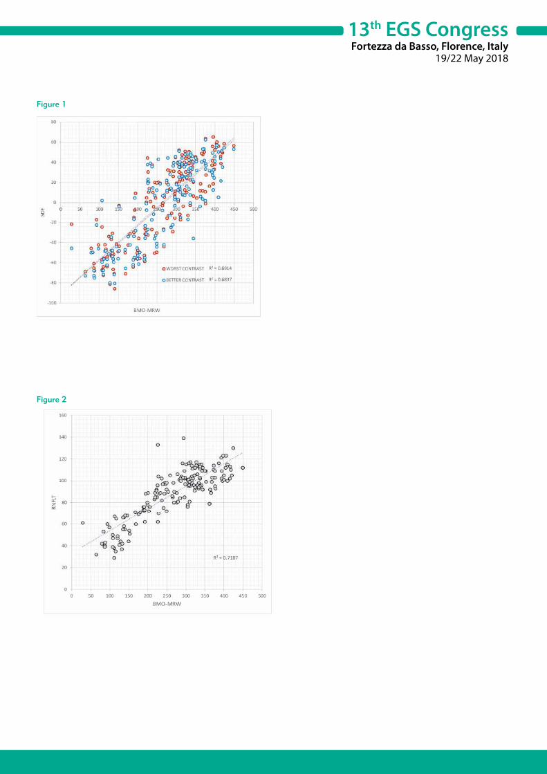
13th EGS CongressFortezza da Basso, Florence, Italy
19/22 May 2018
Figure 1
Figure 2

13th EGS CongressFortezza da Basso, Florence, Italy
19/22 May 2018
P2.025WILL 24-HOUR IOP MEASUREMENTS IMPROVE PATIENT CARE AND REDUCE THE COSTS OF GLAUCOMA MANAGEMENT?Frances Meier-Gibbons1, Marc Töteberg-Harms2
1Eye Center Rapperswil, Rapperswil - Switzerland2UniversityEyeClinic, Zurich - Switzerland
Purpose: Intraocular pressure measurements limited to office hours do not reflect the true circadian intraocular pressure patterns. Often intraocular pressure spikes do not occur during office hours and will be missed. Hence, a continuous measurement or a more frequent measurement method conducted the patient himself could improve patient care. By involving the patient in his or her glaucoma management, adherence to a therapy could be enhanced and in the longterm, direct and indirect costs of glaucoma management could be reduced.
Methods: A literature research was conducted using PubMed in August 2017. Search strings were “continuous intraocular pressure“, “self-“ or “home-tonometry”, “triggerfish“, “intraocular pressure contact lens sensor”, “intraocular pressure contact lens“, and “glaucoma and costs”. The goal of this review is to provide an overview of the recent literature on continuous intraocular pressure measurements and costs on glaucoma care.
Results: Many studies have shown that the intraocular pressure is fluctuating in a circadian rhythm and that intraocular pressure spikes often do not occur during office hours. However, until now no device measures continuous intraocular pressure reliable via an intra- or extraocular technique. Very few studies investigated the connection between continuous intraocular pressure measurements and disease progression, which could eventually influence the costs of glaucoma treatment.
Conclusions: The new devices for more accurate diurnal intraocular pressure measurements are promising but no device is available at the moment, which can be compared to Goldmann Applanation Tonometry. The uncertainty of the measurement values in comparison to the “real” intraocular pressure is the main problem. To influence the costs in glaucoma care, the patient should be involved in his/her disease management. Disease costs are related to adherence. Hence, a major goal is to increase the patient’s knowledge about their disease. More studies are needed which focus on connection between direct and indirect costs, intraocular pressure, and progression of glaucoma.

13th EGS CongressFortezza da Basso, Florence, Italy
19/22 May 2018
P2.026USE OF IBOPAMINE EYE DROPS IN OPHTHALMOLOGYItalo Giuffrè1
1Department of Ophthalmology, Catholic University of Rome, Rome - Italy
Purpose: To evaluate the impact of the use of ibopamine eye drops in the International Literature. It is a D1 and α-adrenergic drug. It is a mydriatic, not-cycloplegic drug.
Methods: To evaluate all the scientific papers cited on Pubmed (update: December 27th 2017), searching ibopamine and eye key-words.
Results: We found 43 scientific papers published on this topic on Pubmed (update: December 27th 2017). They stressed all the fields of use of ibopamine eye drops in Ophthalmology. In particular, we examined: pharmacology, provocative test, diagnostic of outflow impairement also in relatives of glaucomatous patients, safety mydriasis in the preoperative setting, therapeutic use in hypotony secondary to vitreo-retinal surgery and long-lasting uveitis.
Conclusions: This is a smart drug without any retinotoxic side-effects, tested by pattern-electroretinogram examinations. As it is a D1 and α-adrenergic drug, it has a mydriatic, not-cycloplegic effect. It is very useful in every setting in Ophthalmology, where cycloplegia is not needed.

13th EGS CongressFortezza da Basso, Florence, Italy
19/22 May 2018
P2.027TREATMENT OF REFRACTORY GLAUCOMA WITH TWO TRABECULAR MICRO-BYPASS STENTS, ONE SUPRACHOROIDAL STENT, AND POSTOPERATIVE TOPICAL PROSTAGLANDIN: FIVE YEAR OUTCOMESInder Singh1
1The Eye Centers of Racine and Kenosha, Racine - USA
Purpose: To evaluate the long-term effects of micro-invasive glaucoma surgery (MIGS) with two trabecular micro-bypass stents (iStent®) and a suprachoroidal stent (iStent SUPRA®) combined with postoperative prostaglandin in patients with refractory glaucoma.
Methods: This prospective, single-arm study enrolled subjects with refractory OAG who have had a previous trabeculectomy and are on 1-3 ocular hypotensive medications. Enrollment in the study required a medicated IOP of 18-45 mmHg and an IOP of 21-45 mmHg post-washout. Qualifying eyes were implanted with 2 iStent devices and one iStent SUPRA as a standalone procedure. Topical prostaglandin QD was postoperatively prescribed the day after surgery. The following study assessments were performed: IOP, BCVA, slit lamp examination, funduscopy, medication use, and AEs. Medication washouts occurred annually to assess unmedicated IOP. Five year outcomes are intended to be presented at the time of the meeting.
Results: Eighty subjects were enrolled into the study with 48-month follow-up completed for 70 subjects. Preoperative mean IOP was 22.0 ± 3.1 mmHg on 1.2 ± 0.4 medications, and 26.4 ± 2.4 mmHg after washout. Through 48 months postoperative, mean medicated IOP remained ≤ 13.7 mmHg with most subjects on travoprost alone. At Month 48, 97% had IOP ≤ 15 mmHg on 1 medication. IOP assessed after the annual washouts show that over 92% of eyes achieved ≥ 20% reduction in IOP compared to preoperative unmedicated IOP. All but one subject underwent uncomplicated implantation with 2 iStent and an iStent SUPRA. In 1 subject, the iStent SUPRA was unable to be placed due to inadequate intraoperative visualization. BCVA loss due to cataract progression was noted in 11 eyes; 10 of whom subsequently had cataract surgery.
Conclusions: A novel treatment approach combining 2 iStents, an iStent SUPRA, and a postoperative prostaglandin, leverages both the trabecular and uveoscleral outflow pathways. The outcomes from this study demonstrate long-term IOP management to ≤ 15 mmHg with a high safety profile out to 4 years in subjects with refractory glaucoma. This combination therapy may be a viable treatment for subjects with refractory glaucoma where there are limited treatment options.

13th EGS CongressFortezza da Basso, Florence, Italy
19/22 May 2018
P2.028DETECTION OF GLAUCOMA WITH TELEMEDICINE OPTIC DISK PHOTOS AND OCT IN A PRIMARY CARE CENTREAlfonso Anton1, Gianluca Fatty2, Alejandra Herranz3, Eleonora Ayala4, Antonio Morilla4, Juan Jose Gomez Valverde5
1Institut Catala de Retina, Univ. Internacional de Catalunya and Parc de Salut Mar, Barcelona - Spain2Hospital Vall d’Hebron, Barcelona - Spain3Parc de Salut Mar - Hospital Esperanza, Barcelona - Spain4Institut Catala de Retina, Barcelona - Spain5Universidad Politécnica de Madrid, Madrd - Spain
Purpose: To describe the application of a telemedicine platform for the detection of glaucoma and to compare the results between instruments and between expert and non-expert evaluators.
Methods: A telemedicine web-based platform (DYSEO) was designed and developed. Four thousand subjects over 55 years were randomly selected from census. All participants signed an informed consent. Measures of visual acuity and ocular pressure were obtained, and macular and optic disks images were acquired with a non-mydriatic retinograph (Topcon) and a portable SD-OCT (iVue, Optóvue). Eight ophthalmologists, 4 experts and 4 none experts, evaluated the cases and the report was automatically generated. A second evaluation was performed for agreement analysis and in case of disagreement two glaucoma experts decided by consensus the final classification. Comparisons between instruments, and between expert and no-expert evaluators, were performed with Chi Square Test. Agreement between instruments was evaluated with Kappa index.
Results: After a telephone call, 1655 subjects initially accepted to participate in the study and, of these, 1006 (60%) were fully examined. The quality of the images was good in 69.8% for the retinographies and 92.5% for optic disk’s OCTs. The screening program identified 195 cases (19.4%) with suspicion of glaucoma. The number of cases detected increased with increasing age (p < 0.005). Optic disc photographs were classified as normal in 826 cases (82.1%), pathological without glaucoma in 32 (3.1%), non-useful in 20 (1.9%) and glaucoma suspect in 128 (12.7%). OCT images were classified as normal in 827 cases (82.2%), pathological without glaucoma in 15 (1.5%), non-useful in 43 (4.2%) and glaucoma suspect in 127 (12.6%). Kappa agreement index was 0.37. The evaluation of photographs and OCT agreed in 64 cases (50%) of those classified as glaucoma suspects. Disagreement between evaluators occurred in 262 cases (26%) and, in these cases, expert evaluators agreed with final consensus classification in a greater proportion (71.7%, p < 0.001) than non-expert evaluators (18.7%).
Conclusions: The screening program identified glaucoma suspects in 19.4% of cases examined. Agreement between photographs and OCT images was moderate. The level of expertise of evaluators may significantly influence screening results.

13th EGS CongressFortezza da Basso, Florence, Italy
19/22 May 2018
P2.030REPRODUCIBILITY OF VESSEL DENSITY CALCULATIONS USING DIFFERENT MACULAR IMAGING METHODS FROM TWO SWEPT-SOURCE OPTICAL COHERENCE TOMOGRAPHY ANGIOGRAPHY SYSTEMSTakuhei Shoji1, Junji Kanno1, Yuji Yoshikawa1, Kimitake Ozaki1, Hirokazu Ishii1, Yoshikazu Ichikawa1, Kei Shinoda1
1Department of Ophthalmology, Saitama Medical University, Iruma - Japan
Purpose: To evaluate the reproducibility of vessel density calculations using different imaging methods obtained using 2 commercially available swept-source optical coherence tomography angiography (SS-OCTA) systems.
Methods: Consecutive healthy volunteers presenting at Saitama Medical University Hospital were imaged by 2 SS-OCTA devices: PLEX Elite and Triton. SS-OCTA examinations were performed using a 3 × 3-mm volume scan pattern centered on the fovea. Six methods were used for binarization in Image J: global thresholding using Otsu and Mean methods and local adaptive thresholding using Sauvola, Phansalkar, Otsu, and Niblack methods. Vessel density values were compared between the instruments and methods of binarization.
Results: Sixty eyes from 30 healthy subjects were assessed by two reviewers who were blinded to the scanning system used. Twenty-two eyes were excluded because of poor image quality (17 eyes from Triton, 4 eyes from PLEX Elite, and 1 eye from both instruments). Thirty-eight eyes of 23 subjects were eligible for analysis. The 6 binarization methods and 2 instruments showed measurements with different median values and a limited number of comparisons that were not significantly different between instruments. The coefficients of variation of the vessel density measurements ranged from 0.3% to 2.3% and 0.6% to 4.7% in PLEX Elite and Triton, respectively. Local adaptive thresholding methods showed higher reproducibility than did global thresholding methods in both instruments.
Conclusions: We found good reproducibility of SS-OOCTA macula vessel density measurements between the two systems. PLEX Elite recorded fewer poor images and had higher reproducibility than Triton with local adaptive thresholding. This suggests that these methods and instruments are most reliable among currently available SS-OCTA systems.

13th EGS CongressFortezza da Basso, Florence, Italy
19/22 May 2018
P2.031LOOKING AT THE OPTICAL DISC IN THE DIABETIC RETINOPATHY TELESCREENING: IS IT WORTH IT?Diogo Lopes1, Sónia Parreira1, Tomás Loureiro1, João Cardoso1, Inês Machado1, Nuno Campos1
1Department of Ophthalmology, Garcia de Orta Hospital, Almada - Portugal
Purpose: To assess the prevalence of glaucoma in diabetic patients and the importance of looking at the optic disc in a diabetic retinopathy (DR) telescreening program.
Methods: We performed a prospective study, in patients with diabetes mellitus who underwent telescreening for DR. Retinal fundus photographs were taken and graded for presence and severity of DR. The patients with retinography showing signs of glaucoma such as abnormal cup-disc ratio and disc asymmetry were identified and referred to a complete ophthalmologic examination at our hospital. The presence of glaucoma was established based on optic disc and visual field examination. An elevated intraocular pressure was not required for the diagnosis of glaucoma.
Results: All images were processed by an automated DR screening software (Retmarker) and only the ungradable fundus photographs or the ones with signs of retinopathy were analyzed by ophthalmologists. Between 5 of June and 22 of September 2017 were analyzed a total of 2530 retinography images from 1265 patients with diabetes mellitus and 45 patients (3.6%) had signs of glaucoma. Among the subjects who were referred to the hospital, 58% were identified to have increased cup-to-disc ratio, 42% were identified to have a disc asymmetry and 53% (24 patients) attended the ophthalmologic appointment. About 58% (14 patients) had confirmed glaucoma and 64% of those patients were already followed and medicated for glaucoma.
Conclusions: The prevalence of patients with signs suggestive of glaucoma in this diabetic population was 3.6%. The positive predictive value of 58% supports the value of optic disc appearance in a RD telescreening program. Disc asymmetry presents a slightly higher predictive positive value than bilateral increased cup-to-disc ratio (67% vs 60%). However, only 36% of the patients with confirmed glaucoma were a de novo diagnosis of glaucoma.
13th EGS CongressFortezza da Basso, Florence, Italy
19/22 May 2018
P2.032OPTICAL COHERENCE TOMOGRAPHY ANGIOGRAPHY OF OPTIC DISC PERFUSION IN GLAUCOMA CASESAna Hervás1, Ricardo Díaz2, Empar Sanz2, David Salom1, Patricia Udaondo3, Salvador García-Delpech3
1Hospital of Manises, Valencia - Spain2Hospital of Manises, AIKEN, Valencia - Spain3Hospital la Fe, Valencia - Spain
Purpose: To compare optic disc perfusion between normal subjects and subjects with glaucoma (mild, moderate, severe) using optical coherence tomography (OCT) angiography and to detect optic disc perfusion changes in glaucoma.
Methods: We studied 35 eyes of 20 patients (10 with glaucoma and 10 healthy controls) using Angio-OCT (AngioVue, Imex). The total density of vessels by OCT-A (ONH) and the density of temporal vessels in the optic disc have been observed in both groups. Clinical data and evaluation of the spectral domain OCT (RNFL: thickness of the retinal nerve fiber layer, GCC: ganglion cell complex) were also evaluated.
Results: In normal discs, a dense microvascular network was visible in angio-OCT, whereas this network was attenuated in all subjects with glaucoma. In the glaucoma group, the density of the total and temporal ONH vessels was reduced by 25% and 22% respectively, compared to the control group. Significant correlations were found between the total vascular density ONH and temporal density measured by OCT-A, the thickness of the retinal nerve fiber layer (RNFL), the ganglion cell layer (GCC) and the clinical parameters.
Conclusion: Through angio-OCT, repeated measurements of the optic disc perfusion can be useful in the evaluation of glaucoma and its progression. More clinical investigations are necessary to confirm these promising results.

13th EGS CongressFortezza da Basso, Florence, Italy
19/22 May 2018
P2.033INTRAOCULAR PRESSURES IN PATIENTS WITH THYROID EYE DISEASEAi Kozaki1
1Olympia Eye Hospital, Tokyo - Japan
Purpose: To investigate factors affecting intraocular pressures (IOP) in patients with Thyroid Eye Disease (TED).
Methods: From January to December 2014, 1823 patients (334 males, 1489 females) without treatment history for TED and glaucoma were included. The mean age was 43.2 years. Non-contact tonometry, proptosis with Hertel’s exophthalmometer and MRI were performed. The areas of the four rectus muscles were measured in a coronal section of the orbital MRI. In addition, clinical activity score (CAS) and NOSPECS classification of extraocular muscle involvement were scored. Multivariable analysis was also applied to the test the influence of various independent variables such as proptosis, each area of the four rectus muscles, CAS and NOSPECS.
Results: The mean IOP was 15.5 (7-33) mmHg. Of 141 patients with IOP over 21 mmHg, 32 cases were glaucoma patients. The IOPs were correlated with the area of inferior rectus muscle (IR) (p < 0.0001, r = 0.29) and the medial rectus muscle (MR) (p < 0.0001, r = 0.21). However there were not correlated with proptosis (r = 0.14), the superior rectus muscle (SR) (r = 0.18), and the lateral rectus muscle (LR) (r = 0.17). Between IOPs and CAS, it showed a positive relationship (p < 0.0001), as well as between IOPs and NOSPECS (p < 0.0001). Multivariable analysis revealed that the area of IR (p < 0.0001), proptosis (p = 0.0006), CAS (p < 0.0001), and NOSPECS (p = 0.0002) positively correlated with the IOP in TED.
Conclusions: The most remarkable correlation affecting IOP in patients with TED is with the area of IR. Moreover, the degree of activity, diplopia and proptosis also affect IOP elevation. If patients with TED showed elevated IOP, it is necessary to check their extraocular muscles, especially the IR.
13th EGS CongressFortezza da Basso, Florence, Italy
19/22 May 2018
P2.034DRIVING STATUS IN NEW GLAUCOMA REFERRALSClara Hoi Ka Wu1, David Lunt1, Mark Doherty1
1Sunderland Eye Infirmary, Sunderland - United Kingdom
Purpose: Patients with glaucomatous visual field defects are more likely to be involved in road traffic accidents. In the United Kingdom, The Royal College of Ophthalmologists (RCO) recommends that we consider driving status during every consultation, provide appropriate information to patient and document any advice given. This study aims to compare documentation of driving status in new glaucoma referrals against RCO guidelines.
Methods: All new referrals in July 2017 to the glaucoma service at Sunderland Eye Infirmary were reviewed. This included all patients of driving age seen in nurse led, registrar and consultant clinics. Referrals were analysed against criteria included in the RCO guidelines. The standard was set to 100% compliance.
Results: 71 referrals were identified during the specified time period. 75% of patients were seen in clinics led by glaucoma nurse specialists, 13% in registrar clinics and 11% in consultant clinics. The majority (69%) of these were from community optometrists, 28% were referred from other subspecialties within the hospital eye service, two patients were referred from the UK national diabetic retinopathy screening programme. Of all referrals received, only 5% had relevant information relating to driving status. Once seen in the glaucoma clinic, this increases to 90%. 56% were drivers. 10 patients did not meet the minimum criteria for driving, only 2 of which had documented evidence that any discussion about this took place.
Conclusions: Our results suggest that non-glaucoma specialists may not be aware of or consider driving status as an integral part of glaucoma assessment and referral. This is a part of the legal documentation of patients’ records. More importantly, it is our role as healthcare professionals to identify patients at risk who could post a risk to themselves or society at large. We propose additional training to aid compliance and the use of electronic referral proforma with force choice fields.
13th EGS CongressFortezza da Basso, Florence, Italy
19/22 May 2018
P2.035COMPARISON OF STRUCTURE – FUNCTION RELATIONSHIP WITH CONTRAST MODULATION AND SIZE MODULATION PERIMETRYGiovanni Montesano1, Marco Miranda1, Bledi Petriti1, Haogang Zhu2, David Garway-Heath1
1NIHR Biomedical Research Centre at Moorfields Eye Hospital NHS Foundation Trust and UCL Institute of Ophthalmology, London, UK, London - United Kingdom2Computer Science and Engineering, Beihang University, Beijing - China
Purpose: To compare the structure-function relationship obtained with size modulation (Trail Traced Threshold Test, T4 Strategy) and contrast modulation (SITA-Standard) in glaucoma subjects.
Methods: We analysed data from 29 glaucoma subjects. Each subject underwent one Retinal Nerve Fibre Layer (RNFL) OCT scan (Spectralis SD-OCT, Heidelberg Engineering) two 24-2 tests with both size modulation and contrast modulation perimetry. Size modulation was performed on an EIZO Radiforce GS521 screen (EIZO Limited, Berkshire, UK) with a custom strategy (T4). Contrast modulation was performed with a standard Humphrey Perimeter (HFA, Zeiss Meditec) with SITA-Standard strategy. Threshold values estimated with both strategies were transformed into inverted energy units (1/E). We calculated the log of the average 1/E within each of the 6 visual field clusters as defined by Garway-Heath et al. Correlation between cluster log average and the average thickness of the corresponding RNFL were analysed with the use of mixed effect models to account for repeated measures. P-values were corrected for multiple comparisons with the Bonferroni – Holm method.
Results: There was no significant difference in the cluster-wise correlation with mean sector RNFL thickness between the two strategies (p = 0.09). Cluster correlations were all significant (p < 0.05) except for sector 3 (macular, p = 0.1) with T4. In both strategies, the strongest correlation was found for clusters 1 (superior peripheral) and 6 (temporal), followed by cluster 2 (superior mid-peripheral). The conditional R2 (including random effects) was 0.87 for T4 and 0.83 for HFA, while the marginal R2 (only fixed effects) was 0.32 for T4 and 0.35 for HFA.
Conclusions: The two strategies did not differ significantly in the strength of the structure-function relationship. However, our dataset did not include patients with a wide range of macular damage, where a stronger advantage of size modulation is expected. Further analyses on subjects with a wide range of severity of glaucoma damage are needed to evaluate the effect of size modulation in structure-function assessment.

13th EGS CongressFortezza da Basso, Florence, Italy
19/22 May 2018
P2.036ACCURACY AND AGREEMENT IN DETECTING DISC HAEMORRHAGES BETWEEN HEIDELBERG RETINA TOMOGRAPH AND FUNDUS PHOTOGRAPHY IN THE UNITED KINGDOM GLAUCOMA TREATMENT STUDYJibran Mohamed-Noriega1, Corrado Gizzi1, Isaraporn Treesit1, Tetsuya Togano1, Cedric Schweitzer1, Kentaro Yamamoto1, David F. Garway-Heath1
1NIHR Biomedical Research Centre at Moorfields Eye Hospital NHS Foundation Trust and UCL Institute of Ophthalmology, London - United Kingdom
Purpose: Disc haemorrhages (DH) are a sign strongly related to glaucoma progression that clinicians use to guide the management of patients. However, DH are missed during slit-lamp examination when compared to fundus photography (FP) in as many as 84% of the eyes. We compared FP and Heidelberg retina tomograph (HRT) images to detect DH. Methods: The United Kingdom Glaucoma Treatment Study (UKGTS) is the first randomized placebo-controlled trial in glaucoma and showed preservation of visual fields with latanoprost in newly diagnosed glaucoma patients. During the 24 months follow-up, patients were imaged with monoscopic FP and HRT3 (Heidelberg Engineering, Heidelberg, Germany; version 3.0.60, Heyex 1.6.2.0) over 11 visits. From the 516 enrolled patients, 122 were clasified as DH+ based on the analysis of the full series of both HRT images and photographs using a flickering method between baseline and each follow-up scan. Twenty-five DH+ and 25 DH- patients were randomly selected from the participants with photography and HRT on the same day. Anonymized flicker pairs of HRT and photograph were converted into a GIF file and uploaded into an online survey manager (SurveyMonkey®). Images were presented in a random order and 25 images were presented twice. Eighteen glaucoma specialists participated classifying images as DH+ or DH- and its location in clock hours. Accuracy to detect DH+ patients and within- and between-observer agreement was calculated using ROC and Fleiss’ kappa statistics.
Results: The area under ROC among the 18 observers was 0.80 to 0.96 in HRT and 0.89 to 1.00 in photography (Figure 1). For DH presence, the mean between-observer agreement kappa was 0.69 and 0.86, and the mean within-observer agreement kappa was 0.94 and 0.93 based on HRT and photography, respectively.
Conclusions: Flickered HRT and photographs have very good accuracy when assessed by glaucoma specialist. However, there is lower agreement between them when presented with HRT compared to photographs. It is interesting to notice that the best performing observers obtained very similar results with both techniques; this raises the possibility that an automated version of this analysis would perform equally well with both imaging modalities.
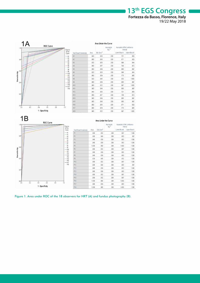
13th EGS CongressFortezza da Basso, Florence, Italy
19/22 May 2018
Figure 1. Area under ROC of the 18 observers for HRT (A) and fundus photography (B).

13th EGS CongressFortezza da Basso, Florence, Italy
19/22 May 2018
P2.037QUANTIFICATION OF RAPD BY AN AUTOMATED PUPILLOMETER AND ITS RELATIONSHIP TO RETINAL NERVE FIBER LAYER THICKNESS AND VISUAL FIELDS IN GLAUCOMAManju Pillai1, Sapna Sinha1, Pradeep Aggarwal1, RD Ravindran1, Claudio Privitera2
1Department of Glaucoma, Aravind Eye Hospital, Madurai, Tamil Nadu - India2School of Optometry, University of California, Berkeley - USA
Purpose: The relative afferent pupillary defect (RAPD) is an important clinical sign of asymmetrical retinal ganglion cell damage. Detection of RAPD by the swinging flashlight test is subjective and confined to only experienced clinicians with advanced training on the test. The purpose of this study was to quantify RAPD by a pupillometer (RAPiDo, Neuroptics) and assess its correlation with asymmetric glaucoma, visual fields and retinal nerve fiber layer thickness and compare with manual pupillary assessment.
Methods: A total of 173 subjects were enrolled in the study and categorized into the glaucoma group (n = 130) and control group (n = 43). RAPD was assessed using an automated pupillometer and compared with a swinging flash light test conducted by an experienced ophthalmologist. Linear regression analysis was conducted to look at the correlation between RAPD and the intereye difference in cup-to-disc ratio (CDR), mean deviation (MD) of visual field testing, and retinal nerve fiber layer thickness (RNFL). For the comparison, we assessed the associative model using sensitivity, specificity and the area under the receiver operator characteristic curve (AUROC).
Results: Glaucoma patients had significant RAPD (0.55 ± 0.05 log units) when compared to the controls (0.25 ± 0.05 log units), p < 0.001. Significant intereye differences in CDR, MD, and RNFL between the glaucoma and the control group (p < 0.001) were seen. There was a good correlation between the magnitude and sign of RAPD and intereye differences in CDR (r = 0.52, p = 0), MD (r = 0.44, p = 0) and RNFL thickness (r = 0.59, p = 0). When compared with assessments by the experienced ophthalmologist, the associative model resulted in an AUROC of 0.94 with 89% sensitivity and 91.7% specificity.
Conclusions: The good correlation between the magnitude of RAPD and intereye differences in MD, CDR and RNFL thickness suggests that pupillometry may be useful for quantifying asymmetric glaucoma. This model could be a probable screening tool to assess asymmetric glaucoma.

13th EGS CongressFortezza da Basso, Florence, Italy
19/22 May 2018
P2.038NEW EXPERIMENTAL APPLANATION TONOMETER FOR MYOPIC PATIENTS AFTER REFRACTIVE LASER SURGERYMaría Iglesias1, Andrea Laiseca1, Bachar Kudsieh1
1Barraquer Ophthalmology Center, Barcelona - Spain
Purpose: Controversy exists regarding Goldmann tonometer (GT) for measuring intraocular pressure (IOP) in myopic patients after laser assisted in situ keratomileusis (LASIK) or photorefractive keratectomy (PRK). In order to reduce this inaccurate measurement, we designed a new experimental GT model with an apex convexly shaped (CT).
Methods: Two different radius (r) for the convex apex of the CT were created: CT1 (r = 12.17 mm) and CT2 (r = 13.42 mm). We present a prospective, double-blind, comparative study between GT with CT1 and CT2. n = 102 myopic patients who underwent surgery with LASIK (L) (n = 73) or PRK (P) (n = 29) were evaluated. IOP, spherical equivalent (EE), simulated keratometry (simk) and central corneal thickness (CCT) (Pentacam), Ocular Response Analizer (ORA), were obtained before and after surgery. Given IOP measured in pre-surgery with GT as gold standard, we used Bland-Altman analysis to compare GT pre-surgery IOP (GTpre) with post-surgery IOP measured with GT, CT1 and CT2 (GTpost, CT1post and CT2post respectively).
Results: Considering together L and P patients, Bland-Altman analysis showed poor agreement between GTpre and GTpost (mean differences: -3.56 mmHg, p < 0.001; limits of agreement: -8.10 – 0.96). However, a much better agreement is observed between GTpre and CT1post (mean differences: 0.32 mmHg, p = 0.187; limits of agreement: -4.24 – 4.89) and CT2post (mean differences: 0.91 mmHg, p > 0.001; limits of agreement: -3.85 – 5.67). Regarding subgroups L and P, mean differences present the lowest values between GTpre and CT1post, -0.19 and 1.62 respectively for L and P. In accordance, limits of agreement for CT1post are narrower than for CT2post and GTpost. Differences between CT1post and GTpre present some bias that appear to be related to simK values (p < 0.001).
Conclusions: Our new experimental tonometer appears to be more precise for measuring IOP comparing to GT in myopic refractive surgery patients. CT1 proved better accuracy than GT in the LASIK subgroup. Thus, we can conclude that we have developed a new version of applanation tonometer that could be used in post-surgery LASIK patients with a much accurate result than the gold standard.
This scientific abstract has been submitted to the 2018 ARVO meeting.

13th EGS CongressFortezza da Basso, Florence, Italy
19/22 May 2018
P2.039PERIPAPILLARY VESSEL DENSITY BY OCT-ANGIOGRAPHY IN NORMAL AND GLAUCOMATOUS EYESSusana Henriques1, Ana Sofia Lopes1, Diana Silva1, Mafalda Mota1, Maria Lisboa1, Fernando Trancoso Vaz1, Isabel Prieto1
1Department of Ophthalmology, Hospital Prof. Doutor Fernando Fonseca, Lisboa - Portugal
Purpose: The aim of this study was to evaluate the peripapillary retinal vessel density (VD) measured by OCTA in glaucoma patients. In addition, it is also intended to verify if there is a correlation between these values of VD and the severity of glaucoma, given by the mean defect (MD) in the functional exams (standard automated perimetry) and with the location of the defect in the peripapillary retinal nerve fiber layer (RNFL), given by the thickness of the RNFL in the structural tests (OCT).
Methods: Prospective and cross-sectional observational study, which included 14 eyes of patients with glaucoma and 10 eyes of healthy individuals (control group). The Cirrus Angioplex (Carl Zeiss Meditec, Inc.) was used for tomographic and angiographic study. Perimetry was performed with Octopus 101 (Haag Streit, USA), G-test. For the OCTA, a 6 x 6 mm cube centered in optic nerve head (ONH) was used and the mean VD of the superficial plexus was quantified in density maps in the temporal, superior, nasal and inferior quadrants in an area with a diameter between 3 and 6 mm from the center of the ONH (outer circle of the ETDRS grid).
Results: The vascular network was visibly attenuated in patients with glaucoma, and there was a statistically significant difference between the glaucoma group and the control group, measured in temporal, superior, nasal and inferior quadrants (p < 0.001; p = 0.001; p = 0.016; p = 0.006; p < 0.001, respectively). No correlation was found between VD and MD measurements (p = 0.213). There was a correlation between the values of VD and the thickness of the RNFL in the inferior quadrant (p = 0.009), but there was no correlation in the remaining quadrants (p > 0.05).
Conclusions: In glaucoma, peripapillary vessel density measured with OCTA is decreased compared to healthy individuals. Although no correlation with functional exams has been found in this study, this technology opens a new path in the diagnosis, monitoring and knowledge of the pathophysiology of glaucoma.
13th EGS CongressFortezza da Basso, Florence, Italy
19/22 May 2018
P2.040MORPHOLOGICAL STRUCTURE OF LAMINA CRIBROSA AND PERIPAPILLARY MICROVASCULATURE FOR GLAUCOMA DIAGNOSIS, USING DEEP-RANGE IMAGINGRita Silva1, Pedro Camacho1, Diogo Cabral1, Pedro Rodrigues1, Manuel Marques1, Catarina Dias1, Joana Valadares1, João Segurado1, José Fernandes1
1Instituto de Oftalmologia Dr Gama Pinto, Lisboa - Portugal
Purpose: To compare the diagnostic power of lamina cribrosa depth (LCD), lamina cribrosa curve depth (LCCD) and lamina cribrosa curvature index (LCCI) with vessel density and fractal analysis in primary open-angle glaucoma (POAG) and pseudoexfoliation glaucoma (PXG).
Methods: In this cross-sectional observational study, one eye of each of 22 controlled glaucomatous patients (11 POAG and 11 PXG) and 33 healthy subjects (15 of them with nonglaucomatous pseudoexfoliation syndrome (PXS)), were enrolled. All patients underwent complete ophthalmic examination. We obtained B-scan images from swept-source optical coherence tomography (OCT) to measure LCD, LCCD and LCCI, at three locations, and OCT angiography images (protocol 4.5x4.5mm) to assess peripapillary microvasculature in superficial vascular plexus and deep-layers, in a 0.7mm circle beyond the optic disc (Topcon DRI OCT; Triton).
Results: Comparing mean values between the four groups (healthy vs PXS vs POAG vs PXG), there were no significant differences in terms of age (68.9 ± 10.5; p = 0.43), axial length (23.2 ± 1.3; p = 0.78), intraocular pressure (14.5 ± 3.2; p = 0.42) and central LCD (483.5 ± 122.5; p = 0.65). Though, we found different statistical significance in visual acuity (p = 0.019), RNFL (p < 0.001), MD (p = 0.001), PSD (p = 0.023) and optic disc excavation (p = 0.028) between the groups in study. We also found differences in central LCCD (125.5 ± 37.3; p = 0.37) and central LCCI (7.9 ± 2.4; p = 0.35) between healthy and PXG patients. OCT angiography criteria were also statistically different, regarding fractal dimension (p = 0.001 between healthy and PXG and p = 0.003 between PXS and PXG - in deep-layers), lacunarity (p = 0.088 between healthy and POAG and p = 0.081 healthy and PXG - in superficial layers; p = 0.045 between PXS and PXG - in deep-layers) and vessel density (p=0.036 between healthy and POAG and p = 0.045 between healthy and PXG - in superficial layers).
Conclusions: Even now, there are plenty of challenges in finding improved high-sensitivity parameters that can assist and contribute to early glaucoma diagnosis. Morphological structural changes of the optic disc and decreased optic disc perfusion can be part of those parameters, but unfortunately still in an initial stage of development.
13th EGS CongressFortezza da Basso, Florence, Italy
19/22 May 2018
P2.041INTEREST OF THE ANALYSIS OF PERI-PAPILLARY RETINAL NERVE FIBERS WITH DIFFERENT DIAMETERS OF MEASUREMENTHussam El Chehab1, Emilie Agard1, Corinne Dot1
1Department of Ophthalmology, Desgenettes Military Hospital, Lyon - France
Purpose: The analysis of peri-papillary retinal nerve fibers layer (pRNFL) is an essential data in structural analysis of the of glaucoma neuropathy. Since OCT development, the diameter of analysis is 3.4mm. The enlargement of this circle may be interesting in patients with peri-papillary abnormalities making segmentation difficult. Larger circles may be able to go beyond these anomalies. This study evaluates the diagnostic value for glaucoma of analysis with different diameter of pRNFL with an OCT-Spectral Domain.
Methods: 65 glaucoma patients and 69 control patients underwent pRNFL analysis by OCT Spectralis (Heidelberg Engineering, Germany) using 3 circles of different diameters: 3.5, 4.1 and 4.7 mm. Each circle was the summation of 100B-Scans. The analysis was performed on a single eye selected at random. The diagnostic capabilities for glaucoma in each of these circles were analyzed for global mean thickness and for mean thicknesses in each of temporal, supero-temporal, superior, nasal, infero-nasal and supero-nasal with areas under the ROC curve. In patients with peri-papillary abnormalities reaching the first circle of analysis, we analyzed the automatic segmentation of the different circles and assessed whether there were segmentation errors in these patients.
Results: Patients were comparable in terms of clinical features (age, gender, side-by-side, refraction). In the glaucoma group, 59.4% of patients had early glaucoma (MD > -6dB) and 21.9% of patients had severe glaucoma (MD < -12dB). Of the 128 patients, the 10 patients (6 controls and 4 glaucomatous patients) had periapillary abnormalities reaching the first FNRp (3.5mm) analysis circle. For the 3.5 mm measurement, the global mean pRNFL thickness has the highest diagnostic capacity (area under curve = 0.880). For the 4.1mm measurement, the global mean pRNFL thickness has the highest diagnostic capacity (area under curve = 0.886). For the measurement at 4.7 mm, the mean thickness of the temporal sector has the highest diagnostic capacity (area under curve = 0.884). There is no statistically significant difference between these 3 areas under curves. Conclusions: Whatever the pRNFL analysis diameter, the diagnostic capacity remains very good without any significant difference. Further study on the reliability of the segmentation on these circles of higher diameters will make it possible to evaluate its reliability.
13th EGS CongressFortezza da Basso, Florence, Italy
19/22 May 2018
P2.042COMPARISON OF CIRRUS OCT AND SPECTRALIS OCT ON THE ABILITY TO MEASURE THE PERIPAPILLARY RETINAL NEVER FIBER LAYER THICKNESS IN CONCURRENT GLAUCOMA AND EPIRETINAL MEMBRANEMi Jeung Kim1, Duck Jin Hwang1, Jung Hyun Lee2, Jung Hwa Na1, Hye Jin Chung1, Yun Suk Chung1, Jin Young Choi1
1Department of Ophthalmology, HanGil Eye Hospital, Department of Ophthalmology, Catholic Kwandong University College of Medicine, Incheon - Republic of Korea, 2Department of Ophthalmology, HanGil Eye Hospital, Incheon - Republic of Korea
Purpose: To compare the frequency of segmentation errors and the ability to detect retinal nerve fiber layer (RNFL) defects between two different spectral domain optical coherence tomography (SD-OCT) devices for the glaucoma patients with epiretinal membrane (ERM).
Methods: Eyes of 36 patients who were diagnosed with glaucoma concurrent with ERM were imaged by Cirrus (Carl Zeiss Meditec) and Spectralis (Heidelberg Engineering) OCT. For peripapillary RNFL scans of each device, only the final print out was examined. We compared the frequency and the type of segmentation errors between the two devices. The segmentation error frequency was defined as the percentage of examinations affected by at least one error in identifying the boundaries of RNFL. The segmentation errors were classified into three types by the location of the misidentification (inner, outer and concurrent inner and outer). Based on the internal normative database from each device, the sensitivity of detecting RNFL defects was evaluated.
Results: The segmentation error was more frequent in Spectralis OCT (69.4%) than in Cirrus OCT (44.4%) (p = 0.035). The inner boundary misidentification was the most common type of segmentation errors in both OCT devices (71.4% of Spectralis, 70.6% of Cirrus). The sensitivity to detect RNFL defect of Spectralis OCT parameters ranged from 72.2% to 88.8% and that of Cirrus OCT parameters ranged from 83.3% to 88.9% with the criterion of abnormality at the 5% level. Based on the normative database, the highest Spectralis sensitivity was observed from the TSNIT thickness graph (88.9%), and the highest Cirrus sensitivity was observed from the deviation-from-normal map (88.9%). There was no significant difference in the sensitivity between the two OCT parameters except the sector map of Spectralis (72.2%) which showed significantly lower sensitivity than the TSNIT thickness graph of Spectralis and the deviation-from-normal map of Cirrus (88.9%, p = 0.031).
Conclusions: With the presence of ERM, the boundaries of peripailly RNFL are frequently misidentified in both Spectralis and Cirrus OCT, and the misidentification was more frequent in Spectralis compared to Cirrus. The possibility of segmentation errors should be taken into consideration in the interpretation of OCT results in patients with concurrent glaucoma and ERM.

13th EGS CongressFortezza da Basso, Florence, Italy
19/22 May 2018
P2.043MACULAR MORPHOLOGICAL PARAMETERS IN GLAUCOMA DIAGNOSISAndrés Blasco Alberto1, Marta González Hernández1, Valentín Tinguaro Díaz Alemán1
1Department of Ophthalmology, Hospital Universitario de Canarias, La Laguna - Spain
Purpose: To determine sensitivity values of macular retinal layers in patients with glaucoma and to compare them with optic nerve morphological parameters.
Methods: The sample was made up by patients with primary open angle, pigmentary and pseudoexfoliation glaucoma. Functional analysis was made with an Octopus® perimeter and TOP strategy and morphological analysis was made with a Spectralis® Optical Coherence Tomograph using the Posterior Pole and Glaucoma Premium strategies. Several thicknesses were analyzed at the macula: retinal thickness (Retina), retinal nerve fibre layer (RNFL), ganglion cell layer (GCL), inner plexiform (IPL), inner nuclear (INL), outer plexiform (OPL), outer nuclear (ONL), pigment epithelium (RPE), inner retinal layers (IRL), photoreceptors (Pho), optic nerve rim area (MRW-BMO), nerve fibre layer thickness with and without anatomical positioning system (RNFL-APS) and (RNFL-E).
Results: 54 normal subjects (92 eyes) and 48 glaucoma patients (80 eyes) were included. Mean defect (MD) of normal subjects was 0.18 dB (SD 1.6) and that of glaucoma patients was 7.91 dB (SD 7.57). MD correlation indices were: -0.50 for Retina (p < 0.05), -0.64 RNFL (p < 0.05), -0.67 GCL (p < 0.05), -0.42 IPL (p < 0.05), -0.03 INL, 0.05 OPL, -0.001 ONL, 0.09 RPE, -0.51 IRL (p < 0.05), and 0.07 Pho. For 95% specificity, global sensitivity at each layer was: Retina 50% (ROC 0.82, IC 0.88-0.75), RNFL 72.15% (ROC 0.88, IC 0.93-0.82), GCL 70% (ROC 0.85, IC 0.91-0.80), IPL 51.25% (ROC 0.80, IC 0.87-0.73), INL 45% (ROC 0.66, IC 0.74-0.58), OPL 1% (ROC 0.31, IC 0.39-0.23), ONL 20% (ROC 0.53, IC 0.61-0.44), RPE 3% (ROC 0.38, IC 0.46-0.30), IRL 54.43% (ROC 0.82, IC 0.88-0.75), Pho 10% (ROC 0.42, IC 0.50-0.33), MRW-BMO 83.75% (ROC 0.94, IC 0.98-0.91), RNFL-APS 77.5% (ROC 0.91, IC 0.96-0.87) and RNFL-E 73.41% (ROC 0.89, IC 0.94-0.84).
Conclusions: The macular layers with best sensitivities were RNFL and GCL (p < 0.05), however, when analyzing ROC areas, their sensitivity was not better than that of MRW-BMO (p < 0.05).

13th EGS CongressFortezza da Basso, Florence, Italy
19/22 May 2018
P2.044EVALUATION OF MACULAR CHOROIDAL THICKNESS WITH SPECTRAL DOMAIN OPTIC COHERENCE TOMOGRAPHY IN PATIENTS WITH OCULAR HYPERTENSIONÖzlem Gürbüz Köz1, Pinar Topcu Yilmaz1, Alper Yarangümeli1, Mehmet Numan Alp1
1Department of Ophthalmology, Ankara Numune Education and Research Hospital, Ankara - Turkey
Purpose: In patients diagnosed with ocular hypertension (OHT), it was aimed to evaluate macular choroidal thickness (MCT) with spectral domain optic coherence tomography (SD-OCT) and compare results with healthy individuals.
Methods: The present study consisted of 25 eyes of 25 patients diagnosed and untreated for OHT at the Ophthalmology Clinic, Ankara Numune Education and Research Hospital, Ankara, Turkey and 24 eyes of 24 healthy individuals. In all cases, following routine ophthalmologic examination, changes in intraocular pressure (IOP) during the day and measurements of central corneal thickness (CCT) were performed at the Glaucoma Department and visual fields were evaluated. In patients diagnosed with OHT, SD-OCT (Cirrus HD-OKT, Carl Zeiss Meditec) was used for MCT measurements at the fovea and at 500 μ, 1000 μ, and 1500 μ nasal and temporal of fovea. Mann-Whitney U test was used in statistical analyses. The correlation of IOP with CCT and MCT was evaluated with Spearman correlation coefficient. Statistical significance was accepted as p < 0.05.
Results: In cases with OHT; MCT at 1000 μ (251.40 ± 74.40 μ, healthy eyes 275.92 ± 47.34 μ; p = 0.02) and 1500 μ (236-84 ± 69.89 μ, healthy eyes 265.46 ± 47.56 μ; p = 0.012) temporal of fovea were significantly lower when compared with healthy individuals. At nasal, fovea, and 500 μ temporal of fovea measurement points, MCT was thinner in patients with OHT, however, the difference was not statistically significant (p > 0.05). IOP showed no significant relationship with CCT and MCT (p > 0.05).
Conclusion: In patients with OHT, MCT gets thinner. Further studies investigating the effects of MCT changes on the prognosis of OHT in terms of glaucoma development are required.
13th EGS CongressFortezza da Basso, Florence, Italy
19/22 May 2018
P2.045OPTICAL COHERENCE TOMOGRAPHY ANGIOGRAPHY AND OPTIC NERVE HEAD DRUSEN. A QUALITATIVE AND QUANTITATIVE EVALUATION IN RELATION TO AUTOMATED PERIMETRY DEFECTSAndreas Diagourtas1, Konstantinos Andreanos2, Stylianos Kandarakis2, Eustathia Asimakopoulou2, Dimitrios Papaconstantinou1 1Glaucoma Department, Athens University Eye Clinic, Athens - Greece2Athens University Eye Clinic, Athens - Greece
Purpose: To document qualitative and quantitative characteristics of the optic nerve head and the peripapillary area in patients with optic nerve head drusen (ONHD), with the use of Optical Coherence Tomography Angiography (OCTA) and to correlate findings with automated perimetry.
Methods: Fourteen eyes of 7 patients with average age of 35.71 (SD ± 10.89) and a previous diagnosis of Optic Nerve Head Drusen, either deep or superficial, were examined with OCTA (Topcon DRI OCT Triton Plus swept source OCT system). Images of the superficial and deep capillary plexuses in the parapapillary area were acquired and sectoral analysis in relation to the drusen localization was realized. Furthermore, eventual visual field defects (Humphrey Perimeter), were correlated with ONHD localization and peripapillary vessel density.
Results: Peripapillary vessel density both in the superficial and in the deep capillary plexuses resulted reduced in the sectors where the ONHD were localized. The difference between the non - Drusen occupied sectors was statistically significant (p < 0.05). Furthermore, a spatial moderate positive correlation (r = 0.35) was documented between ONHD-occupied sectors, vessel density and pattern of visual field defects.
Conclusions: This is the first report of a case series of peripapillary vessel density documentation in ONHD with the use of OCTA. It seems that it could be a valuable tool in the recognition of vascular alterations in this particular pathology. Furthermore, in longitudinal studies possibly it could elucidate the temporal sequence between peripapillary vascular deprivation, retinal nerve fiber thinning and visual field defects.

13th EGS CongressFortezza da Basso, Florence, Italy
19/22 May 2018
P2.046SENSITIVITY AND SPECIFICITY OF RAREBIT VISUAL FIELD TESTING FOR GLAUCOMA IN THE OPTOMETRY SERVICEJoanna Przezdziecka-Dolyk1, Aleksandra Matkowska2, Ewa Fidewicz1, Justyna Szkudlarek-Wodzinska1, Ewa Walek1, Marta Misiuk-Hojlo1
1Department of Ophthalmology, Wroclaw Medical University, Wroclaw - Poland2Department of Optics and Photonics, University of Science and Technology, Wroclaw - Poland
Purpose: To evaluate the sensitivity and specificity of RareBit visual field testing for glaucoma performed in the optometry service as a pre-ophthalmological assessment.
Methods: There were 75 participants (25 patients with different stages of glaucoma and 50 healthy participants) included in this prospective case control study. The RareBit (on LCD laptop) was performed during the first line assessment by an optometrist along with the refraction and slit lamp examination. Every patient underwent also full ophthalmological examination with the standard visual field testing. The mean hit rate [%] (MHR), mean miss rate [%] (MMR), area < 90% (A < 90) and each of twenty-four localisation hit rates [%] were evaluated from RareBit testing. The Receiver Operating Characteristic (ROC) curve analysis (with the Z statistic) was performed with the MedCalc software for sensitivity, specificity and defining of the cut-off criterium of the variables. Additionally, the comparison of the ROC curves was performed.
Results: 75 eyes of 75 patients were included in the study the mean age for glaucoma and control groups were respectively 57 and 52 years, gender were evenly distributed in both groups (p > 0.05 for age and gender). Each of evaluated parameters can be used to differentiate glaucomatous patients (p < 0.0001 for each area under the ROC curve/AUC), however the AUC > 0.97 was obtained only by the MHR, MMR and A < 90. The sensitivity and specificity for MHR, MMR and A < 90 were respectively 100 and 92.5 for all parameters and the cut-off criterium ≤ 55, > 35 and > 78%. Additionally, three locations obtained the AUC > 0.96 - upper and lower temporal paracentral areas and lower nasal paracentral area. Their sensitivity, specificity and cut-off criterium were respectively: 92.9; 95.0 and ≤ 60%, 92.9; 92.5 and ≤ 38%, 100.0; 92.5 and ≤ 12%.
Conclusions: The RareBit visual field testing enables clinician-optometrists to correctly assess patients with glaucoma in the pre-ophthalmological environment. This mobile assessment can be used even during home visits as it can be used at the bedside table.

13th EGS CongressFortezza da Basso, Florence, Italy
19/22 May 2018
P2.047INTER-OBSERVER AND INTER-DEVICE AGREEMENT FOR THE ASSESSMENT OF ANTERIOR-CHAMBER ANGLE PHOTOGRAPHY WITH AN AUTOMATED GONIOSCOPERiccardo Scotto1, Carlo Alberto Cutolo1, Michele Iester1, Carlo Enrico Traverso1
1DiNOGMI, Eye Clinic, Genova - Italy
Purpose: To investigate inter-observer and inter-device agreement in gonioscopic evaluation analizing Gonio-Photography (GP) performed by a 360° automated indirect gonioscope.
Methods: Two masked senior ophthalmologist with glaucoma expertise, evaluated GP taken by an automated indirect gonioscope prototype (NIDEK CO., Japan). Apparent iris insertion, pigmentation and the presence of abnormal findings were compared with slit-lamp gonioscopy. Inter-observer and inter-device agreement for apparent iris insertion and pigmentation were determined by using Cohen’s linearly weighted κ (KW) coefficient of concordance.
Results: 450 photos of 25 eyes were examined; inter-observer agreement in apparent iris insertion and pigmentation was 0.4 (95% CI 0.25 to 0.54) and 0.08 (95% CI -0.04 to 0.21) respectively. For iris-insertion, after grouping the angles as wide and narrow the agreement was 0.51 (95% CI 0.26 to 0.75). Inter-device agreement in apparent iris insertion and pigmentation was 0.56 (95% CI 0.45 to 0.67), 0.10 (95% CI 0.01 to 0,22) for Observer 1; and 0.35 (95% CI 0.23 to 0.49), 0.45 (95% CI 0.31 to 0.59) for Observer 2. Observer 1 had higher agreement with gonioscopy than Observer 2, for iris insertion, after grouping the angles as wide and narrow, 0.92 (95% CI 0.82 to 1.0) and 0.56 (95% CI 0.31 to 0.81). The image quality was indirectly proportional to the agreement. Both observer identified anterior chamber angle (ACA) abnormalities, i.e. peripheral anterior synechiae (PAS), iridectomy, internal ostium of trabeculectomy, Xen® device, Ex-Press® device, tube draining, anterior chamber IOL haptics and iris snake vessels.
Conclusions: Gonioscopy is an essential part of the eye examination, crucial to the diagnosis and classification of glaucoma. Our data suggest that GP is more difficult to interpret for cases of deeper iris insertion. GP using the NIDEK GS-1 Automated Gonioscope prototype (NIDEK CO., Japan) is a well-tolerated, quick and useful method for recording photographically apparent iris insertion, pigmentation, pathological and post-operative ACA findings in glaucoma patients. Intra-image quality over 360° is being addressed.
These data were presented at the ARVO 2018 congress.

13th EGS CongressFortezza da Basso, Florence, Italy
19/22 May 2018
P2.048COMPARISON OF CENTRAL CORNEAL THICKNESS WITH ULTRASOUND PACHYMETRY, NON-CONTACT SPECULAR MICROSCOPY, HAND HELD PACHMETRY AND SPECTRAL DOMAIN OPTICAL COHERENCE TOMOGRAPHYGöktug Demirci1, Gokhan Gulkilik2, Sevil Karaman Erdur2, Cafer Tanriverdio2, Selim Kocabora2, Mustafa Ozsutcu2
1Department of Ophthalmology, Istanbul Medipol University, Istanbul - Turkey2Istanbul Medipol University, Istanbul - Turkey
Purpose: To determine the agreement of central corneal thickness (CCT) measurements taken with ultrasonic pachymetry (USP), noncontact specular microscopy (NSM), handheld pachymetry (HP) and spectral domain optical coherence tomography (SD-OCT).
Methods: A prospective, observational, cross-sectional study was performed in the outpatient ophthalmology clinic. CCT was measured in a total of 50 eyes of 50 consecutive healthy patients with USP, NSM, SD-OCT and HP. All examinations were performed by the same examiner. Bland–Altman plots were used to evaluate the agreement between instruments.
Results: The average CCT values obtained by USP, NSM, SD-OCT and HP were 547 ± 28 μm, 543 ± 25 μm, 536 ± 30 μm and 552 ± 25 μm, respectively. There was no significant difference in CCT measurements between USP and NSM (p > 0.05). There was a strong correlation between instruments: USP with SD-OCT (r = 0.880, p < 0.01), USP with NSM (r = 0.923, p < 0.01), USP with HP (r = 0.915, p < 0.01), SD-OCT with HP (r = 0.894, p < 0.01), SD-OCT with NSM (r = 0.891, p < 0.01) and NSM with HP (r = 0.885, p < 0.01) for CCT.
Conclusions: USP and NSM were found to have comparable CCT measurements and these two methods can be used correspondingly. However, CCT measurements by SD-OCT were lower when compared to other methods whereas CCT measurements by HP were higher compared to those.

13th EGS CongressFortezza da Basso, Florence, Italy
19/22 May 2018
P2.049MENINGIOMA MISDIAGNOSED AS PROGRESSING GLAUCOMA - A CASE REPORTJoanna Wierzbowska1, Mateusz Jacunski1
1Department of Ophthalmology, Military Institute of Medicine, Warsaw - Poland
Purpose: To report a clinical case of a patient with glaucoma and a coexisting brain tumor, falsely diagnosed as progressive glaucoma.
Methods: Interventional case report.
Results: 62 year-old male patient treated topically with antiglaucoma drops for 7 years admitted to our ophthalmology department with a diagnosis of the very advanced glaucoma in the right eye. The patient presented visual field loss in the nasal and superior temporal region of the right eye with decreased central visual acuity (20/70) but 20/20 acuity in the left eye. He had a right relative afferent pupillary defect. The fundus examination revealed an increased cup-to-disc ratio in both eyes (0.8 in the right eye and 0.5 in the left eye, however the right disc also was pallor. Intraocular pressures were 15 mmHg (in the right eye) and 16 mmHg (in the left eye) and central corneal thickness of both eyes were normal. Because of the decreased visual acuity, pallor of the optic disc and rapid asymmetric progression of axonal loss in the right eye and the fact that intraocular pressure had been under control, a compressive optic neuropathy was suspected. Cranial magnetic resonance imaging demonstrated a 5 cm tumor in sella turcica region. The patient underwent craniectomy and subtotal removal of the tumor. Histopathology revealed a meningothelial meningioma. The visual field improved slightly postoperatively, however in long-term observation a slow deterioration of the vision in the right eye was observed.
Conclusions: Compressive lesions of the visual pathways should be considered in patients suspected of or undergoing treatment for glaucoma that present with atypical progression of visual loss. Decreased visual acuity and /or visual field defects that are inadequate to the appearance of the optic nerve disc, rapid progression of lesions, should be an indication for magnetic resonance imaging of central nervous system.
13th EGS CongressFortezza da Basso, Florence, Italy
19/22 May 2018
P2.050RELATIONSHIP BETWEEN OPTICAL COHERENCE TOMOGRAPHY ANGIOGRAPHY VESSEL DENSITY AND SEVERITY OF GLAUCOMATOUS VISUAL FIELD LOSS IN MYOPIC EYES: COMPARISON WITH RETINAL NERVE FIBER LAYER THICKNESSEun Ji Lee1, Seung Hyen Lee2, Tae-Woo Kim1
1Department of Ophthalmology, Seoul National University Bundang Hospital, Seoul National University, Seongnam - Republic of Korea 2Department of Ophthalmology, Bundang Jesaeng General Hospital, Daejin Medical Center, Seongnam - Republic of Korea
Purpose: To determine the relationship between vessel density (VD) measurement using optical coherence tomography (OCT) angiography (OCTA) and severity of visual field (VF) loss in myopic eyes with primary open-angle glaucoma (POAG), in comparison with the association between retinal nerve fiber layer (RNFL) thickness and severity of VF loss.
Methods: Peripapillary VD was evaluated using OCTA, and circumpapillary RNFL thickness was measured using spectral-domain OCT in 113 myopic eyes with POAG (spherical equivalent, -4.75 to -23.25 diopters). The VD was defined as the percentage of area occupied by flowing blood vessels in the selected region. Linear relationship between the VD and VF mean deviation, and that between the RNFL thickness and VF mean deviation were determined globally and in the four sectors.
Results: Both VD and RNFL thickness showed good correlation with VF severity in global (both p < 0.001, R2 = 0.148 and R2 = 0.264, respectively) and in temporal (both p < 0.001, R2 = 0.314 and R2 = 0.295, respectively), superior (both p < 0.001, R2 = 0.236 and R2 = 0.235, respectively) and inferior sectors (both p < 0.001, R2 = 0.168 and R2 = 0.256, respectively) in the myopic POAG eyes. In the eyes showing segmentation error in the circumpapillary RNFL scanning (n = 24, 21.2%), VD was significantly associated with VF in the temporal, superior, and inferior sectors (p ≤ 0.007, R2 = 0.291, R2 = 0.434, R2 = 0.171, respectively), while RNFL thickness showed significant correlation only in superior sector (p = 0.008, R2 = 0.279).
Conclusions: Peripapillary VD measured with OCTA showed good topographic correlation with the severity of glaucomatous VF loss in myopic eyes with POAG. OCTA could be a useful adjunct to evaluate glaucomatous visual field damage in myopic eyes, where the OCT results are frequently confounded.
13th EGS CongressFortezza da Basso, Florence, Italy
19/22 May 2018
P2.0512-YEAR RESULTS FROM THE HORIZON TRIAL: A RANDOMIZED STUDY OF A SCHLEMM’S CANAL MICROSTENT FOR REDUCTION OF IOP IN PRIMARY OPEN ANGLE GLAUCOMANorbert Pfeiffer1 1Department of Ophthalmology, University Medical Center, Mainz - Germany
Purpose: To determine the ability of a new Schlemm’s canal microstent to lower diurnal IOP and medication use in patients with primary open angle glaucoma (POAG) and cataract.
Methods: HORIZON is a prospective, multicenter, randomized clinical trial comparing concurrent phacoemulsification with a Schlemm’s canal microstent (Hydrus Microstent, Ivantis Inc, Irvine, CA) to phacoemulsification alone. Eligible subjects had mild to moderate POAG treated with 1-4 glaucoma medications, age related cataract, and washed out diurnal IOP (DIOP) 22-34 mmHg. Following successful phacoemulsification, subjects were randomized 2:1 into Hydrus Microstent (HMS) or no stent (NS) groups. Follow-up IOP, medication use, visual acuity, ocular health and adverse event assessment was conducted at 1 and 7 days, and after 1, 3, 6, 12, 18 and 24 months. Medication wash out was repeated at 24 months and DIOP measured in order to assess HMS effect on DIOP without the influence of medications.
Results: 556 eyes were randomized to HMS (N = 369) or NS (N = 187) from 38 sites located in 9 countries. Mean glaucoma medication use was 1.7 ± 1.0 in each group at screening, and after wash out DIOP was 25.5 ± 3.0 mmHg in the HMS group and 25.4 ± 2.9 mmHg in the NS group. Prior to 24 month wash out, mean medication count was reduced to 0.3 ± 0.8 in the HMS group vs. 0.7 ± 0.9 in the NS group (difference = 0.4, p < 0.001). After 24 month wash out, mean reduction in DIOP from baseline was 7.5 ± 4.1 mmHg in the HMS group and 5.2 ± 3.9 mmHg in the NS group (difference = 2.3 mmHg, 95% CI 3.0 to 1.6, p < 0.001). Follow up BCVA and VF changes were equivalent in both groups. The HMS group was associated with transient postoperative hyphema and focal adhesions near the device; the NS group had higher rates of IOP elevated > 10 mmHg over baseline and rescue filtration surgery. There were no significant differences in other adverse event rates.
Conclusion: Concurrent Schlemm’s canal stenting in mild to moderate POAG patients undergoing cataract surgery is safe and effective in lowering IOP and medication use compared to cataract surgery alone at 24 months.

13th EGS CongressFortezza da Basso, Florence, Italy
19/22 May 2018
P2.052COMPARISON OF SIGNAL ALTERATION IN OPTIC NERVE HEAD ON 3DT2 MRI BETWEEN GLAUCOMA AND OPTIC ATROPHYJong Yeon Lee1, Hyung Jun Park1
1Department of Ophthalmology Gachon University Gil Medical Center, Incheon - Republic of Korea
Purpose: To compare the signal alteration (SA) in optic nerve head (ONH) on 3DT2 MRI between glaucoma and optic atrophy.
Methods: A total of 19 patients with open-angle glaucoma and 13 patients with optic atrophy underwent 3D high-resolution T2-weighted MRI and spectral domain optical coherence tomography (OCT) were included. The prevalence of signal alteration was compared between the two groups. The OCT measurements were compared between prominent SA and those without SA.
Results: Of the 19 eyes with glaucoma, 15 (78.9%) revealed a SA in the ONH, whereas it was observed in 2 (15.4%) of 13 eyes with optic atrophy (p = 0.024). Glaucoma eyes had thicker RNFL thickness (p = 0.001) and larger cup volume (p < 0.001) and larger vertical cup-to-disc ratios (p = 0.02) than eyes with optic atrophy. The eyes with a prominent SA had a larger cup volume than those without a SA (p = 0.007).
Conclusion: The SA in the ONH on 3D T2 MRI was significantly more frequent in eyes with glaucomatous optic neuropathy than in eyes with optic atrophy. The SA may be related with larger cup volume of optic nerve head. I
13th EGS CongressFortezza da Basso, Florence, Italy
19/22 May 2018
P2.053CORRELATION OF THE DISC DAMAGE LIKELIHOOD SCALE (DDLS) WITH RETINAL NERVE FIBER LAYER (RNFL) USING OPTICAL COHERENCE TOMOGRAPHY (OCT)Paolo Formichella1,2, Roxanne Annoh1,3, Fabrizio Zeri4, Andrew J. Tatham1,3
1University of Edinburgh, Edinburgh - United Kingdom 2IRSOO Vinci - Italy 3Princess Alexandra Eye Pavilion, Edinburgh - United Kingdom4Ophthalmic Research Group, Aston University, Birmingham - United Kingdom
Purpose: The disc damage likelihood scale (DDLS) has shown superiority for identifying glaucomatous optic nerve head changes compared to the cup-to-disc ratio (CDR), due to its incorporation of both disc size and degree of neuroretinal rim loss. The aim of this study was to examine the relationship between DDLS assessment by community optometrists and a more objective measure of glaucomatous structural change, namely RNFL thinning assessed using spectral-domain optical coherence tomography (SD-OCT).
Methods: A retrospective analysis of 1430 new glaucoma referrals by community optometrists was conducted. 129 referrals (9%) included an optometrist’s assessment of DDLS. These 129 patients had OCT imaging of the circumpapillary RNFL performed in the hospital eye clinic. All OCT images were reviewed and included only if they had a sufficient quality score, were well centred and were without artefact. One eye from each patient was included in the analysis (n = 57). The relationship between DDLS scores and average and sectoral RNFL thickness was examined using scatterplots and correlation coefficients were calculated using Spearman’s rank correlation. Differences in RNFL thickness between DDLS classifications were compared using Kruskal–Wallis test one-way ANOVA on ranks.
Results: There was a significant (at the p < 0.05* and p < 0.01** level) relationship between increasing DDLS score and thinner average (rho = -0.378**), superotemporal (rho = -0.359**), inferotemporal (rho = -0.353**), temporal (rho = -0.313*) and inferonasal (rho = -0.284*) RNFL thickness. In contrast, no significant correlation was found between DDLS and superonasal and nasal RNFL thickness. There was a significant (at the p < 0.05* and p < 0.01** level) relationship between increasing DDLS score and thinner average (rho = -0.378**), superotemporal (rho = -0.359**), inferotemporal (rho = -0.353**), temporal (rho = -0.313*), and inferonasal (rho = -0.284*) RNFL thickness. In contrast, no significant correlation was found between DDLS and superonasal and nasal RNFL thickness. Patients diagnosed with glaucoma had significantly higher DDLS scores than those discharged or diagnosed as glaucoma suspects (p = 0.019).
Conclusions: DDLS grading by community optometrists had moderate correlation to RNFL thickness measured using OCT, with strongest association with average, inferotemporal and superotemproal RNFL thickness, regions known to be sensitive to glaucomatous structural change.

13th EGS CongressFortezza da Basso, Florence, Italy
19/22 May 2018
P2.054A COMPARISON OF RELATIVE DIAGNOSTIC PRECISION BETWEEN THE COMPASS FUNDUS PERIMETER AND THE HUMPHREY FIELD ANALYZERLuca Mario Rossetti1, Giovanni Montesano1, Susan Ruth Bryan2, Paolo Fogagnolo1, Francesco Oddone3, Allison M McKendrick4, Andrew Turpin5, Paolo Lanzetta6, Andrea Perdicchi7, Chris A. Johnson8, David Garway-Heath9, David Crabb2
1University of Milan, Milan - Italy2City, University of London, London - United Kingdom3Glaucoma unit, IRCCS GB Bietti Eye Foundation, Rome - Italy4Optometry & Vision Sciences, University of Melbourne, Melbourne - Australia5Computing and Information Systems, University of Melbourne, Melbourne - Australia6Department of Ophthalmology, University of Udine, Udine - Italy7Azienda Ospedaliera Sant’Andrea, Rome - Italy8Ophthal & Visual Sci, University of Iowa, Iowa City - USA9NIHR Biomedical Research Centre at Moorfields Eye Hospital NHS Foundation Trust and UCL Institute of Ophthalmology, London - United Kingdom
Purpose: To evaluate relative diagnostic precision of the Compass (CMP, CenterVue, Italy) fundus perimeter and the Humphrey Field Analyzer (HFA, Zeiss, Dublin) in detecting glaucomatous optic neuropathy (GON).
Methods: One eye of 498 glaucoma patients and 436 age similar normals was tested with the index tests: HFA (SITA-Standard strategy) and CMP (ZEST strategy) with a 24-2 grid. The reference test for GON was specialist evaluation of fundus photographs or OCT, independent of the visual field. For both devices, linear regression was used to calculate normal age-related sensitivity decrease to compute pointwise Total Deviation (TD) values and Mean Deviation (MD). We derived 5% and 1% pointwise age-corrected normative limits. MD and the total number of TD values below the 5% (TD5%) or the 1% (TD1%) limit per field were used as classifiers for ROC curves and partial Area Under the Curve (pAUC) to compare the diagnostic precision of the devices. Additionally, 44 glaucoma and 54 normal subjects were tested twice on both instruments. Pointwise Mean Absolute Deviation (MAD) and Bland-Altman plots for the mean sensitivity (MS) were computed. Results are reported as mean difference ± standard error.
Results: Both devices showed similar discriminative power (Figure 1). Differences between pAUC were negligible (for MD 0.019, p = 0.03; for TD5% 0.012, p = 0.023; for TD1% 0.003, p-value = 0.54). The 95% limits of agreement for the MS were reduced by 14% in CMP compared to HFA in glaucoma subjects, and by 49% in normal subjects (Figure 2). MAD was very similar, being slightly smaller (not signifincant) in CMP compared to HFA for glaucoma (0.03 ± 0.2 dB) and for normals (0.08 ± 0.16 dB). Average MS was lower with CMP than with HFA for glaucoma (-1.45 ± 0.01 dB) and normal subjects (-1.82 ± 0.11 dB).
Conclusions: Relative diagnostic precision of the two devices is equivalent. Test-retest variability of MS for CMP was better than HFA.
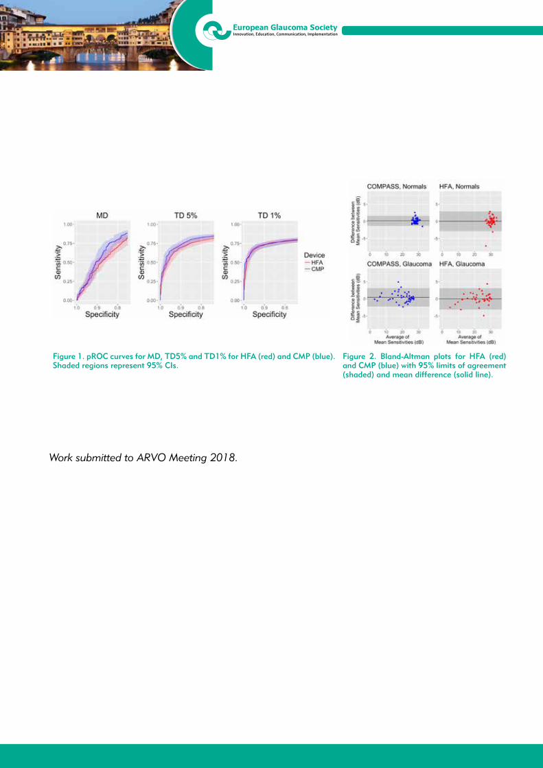
13th EGS CongressFortezza da Basso, Florence, Italy
19/22 May 2018
Figure 1. pROC curves for MD, TD5% and TD1% for HFA (red) and CMP (blue). Shaded regions represent 95% CIs.
Figure 2. Bland-Altman plots for HFA (red) and CMP (blue) with 95% limits of agreement (shaded) and mean difference (solid line).
Work submitted to ARVO Meeting 2018.
13th EGS CongressFortezza da Basso, Florence, Italy
19/22 May 2018
P2.055PERIPAPILLARY RETINAL NERVE FIBER LAYER THICKNESS AND BRUCH’S MEMBRANE OPENING-MINIMUM RIM WIDTH ASSESSMENT IN NONGLAUCOMATOUS EYES WITH LARGE AND SMALL DISCSSerife Bayraktar1, Gulnar Sultanova1, Belgin Izgi1
1Department of Ophthalmology, Istanbul Faculty of Medicine, Istanbul University, Istanbul - Turkey
Purpose: To compare the new spectral-domain optical coherence tomography (SD-OCT) algorithm for measuring circumpapillary retinal nerve fiber layer thickness (RNFLT) centered on Bruch’s membrane opening (BMO), with the conventional circumpapillary RNFLT measurement centered on the optic disc and assess the BMO- minimum rim width (BMO-MRW) in nonglaucomatous eyes with large and small discs.
Methods: This cross-sectional, observational study included a total of 257 eyes of 155 patients. The patients were divided into three groups. We investigated the eyes with small discs (disc size < 1.60 mm2) in Group 1, large discs (disc size > 2.55 mm2) in Group 2 and healthy patients as a control group in Group 3. The patients in Group 1 and Group 2 were diagnosed as glaucoma suspect or ocular hypertension. The optic nerve head (ONH), peripapillary RNFL, BMO area and BMO-MRW were imaged with SD-OCT (Spectralis, Glaucoma Module Premium Edition; Heidelberg Engineering GmbH, Heidelberg, Germany).
Results: Group 1 included 75 eyes of 53 patients (36 female, 17 male), Group 2 included 82 eyes of 52 patients (38 female, 14 male) and Group 3 included 100 eyes of 50 patients (37 female, 13 male). The average conventional circumpapillary RNFLT measurements were 98.8 ± 9.6 μ in Group 1, 98.3 ± 9.6 μ in Group 2 and 103.5 ± 6.9 μ in Group 3. The average RNFLT measurements by centering BMO were 100.0 ± 9.15 μ in Group 1, 100.6 ± 12.8 μ in Group 2 and 106.0 ± 6.7 μ in Group 3. The difference was statistically significant in all quadrants in Group 2. In Group 1 it was significant in all quadrants except inferior nasal and temporal quadrants. In control group it was also significant in all quadrants except superior-temporal quadrant. Average BMO-MRW thickness measurements were 376.5 ± 55.1 μ in Group 1, 266.5 ± 45.4 μ in Group 2 and 332.1 ± 57.5 μ in Group 3.
Conclusion: The new circumpapillary RNFL scanning algorithm centered on BMO may provide a more reliable RNFL profile in eyes with large and small discs.
13th EGS CongressFortezza da Basso, Florence, Italy
19/22 May 2018
P2.056DIAGNOSTIC CAPACITY OF THE RETINAL VASCULAR DENSITY VERSUS THE STUDY OF THE RETINAL NERVE FIBER LAYER THICKNESS AND MACULAR GANGLION CELL COMPLEXLaura Morales Fernandez1, Noemi Guemes Villahoz1, Liseth Salazar Quiñones1, Jose Maria Martinez De La Casa1, Federico Saenz Frances1, Julian Garcia Feijoo1
1Department of Ophthalmology, Hospital Clinico San Carlos, Madrid - Spain
Purpose: To evaluate and compare the vascular density of the optic nerve head and the macular area with the retinal nerve fiber layer (RNFL) thickness and the macular ganglion cell complex (GCC) thickness in patients with and without glaucoma, using Optical Coherence Tomography Angiography (OCT-A). Methods: In a cross-sectional study, 55 patients diagnosed with glaucoma and 45 control subjects underwent vessel density and structural measurements with OCT Angiography. They were evaluated in the ophthalmology department at Hospital Clínico San Carlos (HCSC). One eye was included per patient presenting visual acuity (VA) greater than 0.5 and a mean defect (MD) in the visual field test less than 10dB. The OCT-A of Nidek (NIDEK CO., LTD), AngioScan RS-3000 was used, evaluating an area of 4.5 x 4.5 mm peripapillary and macular respectively. The differences between the two groups were analyzed using the student’s t test. The power of discrimination of each parameter was analyzed with ROC curves (AUROC)
Results: Both groups were comparable in age (p = 0.132). The papillary vessel density in the group with glaucoma was 3.33 ± 0.09 and 4.52 ± 0.10 in the control group (p < 0.001, AUROC 0.881). The macular vessel density in the group with glaucoma was 2.80 ± 0.06 and 3.19 ± 0.08 in the control group (p < 0.001, AUROC 0.705). The average thickness of the RNFL was 74.82 ± 2.09 in the group with glaucoma and 97.24 ± 1.76 in the control group (p < 0.001, AUROC 0.868). The best discrimination parameter of the ganglion cell complex thickness was the inferotemporal external sector (AUROC: 0.822).
Conclusions: The papillary vascular density in patients with glaucoma was significantly lower than the control group, showing a diagnostic yield similar to the RNFL thickness and macular ganglion cell complex analysis.

13th EGS CongressFortezza da Basso, Florence, Italy
19/22 May 2018
P2.057DOES ELECTROMAGNETIC RADIATION OF EVERYDAY LIFE GADGETS INFLUENCE THE SIGNAL OF THE SENOR CONTACT LENS TRIGGERFISH?Christoph Faschinger1, Dieter Rabensteiner1
1Department of Ophthalmology, Medical University Graz, Graz - Austria
Purpose: To evaluate a possible influence of electromagnetic radiation of everyday life gadgets to the measurements of the intraocular pressure (IOP) sensor contact lens Triggerfish. Fluctuations of the signals might be interpreted as false-positive changes of the IOP.
Material and Methods: During the measurement procedure of the sensor contact lens Triggerfish (Sensimed, Switzerland) an activated wireless phone and smartphone, an activated laptop (WLAN), as well as an activated microwave oven were positioned close to the data-transferring antenna of the sensor contact lens for a certain time. For comparison reasons, the noise and a possible signal drift of the sensor contact lens were examined in advance during 24-hours in conditions with no extern electromagnetic radiations.
Results: During 24-hour measurements with no electromagnetic radiation influences there was no signal drift and the noise was 8 μVeq. The influence of an activated wireless phone and smartphone, the activity of the WLAN of a laptop as well as a microwave oven did not alter the measurement profile during that time.
Conclusion: During the 24-hour measurements of the sensor contact lens Triggerfish electronic devices of everyday life gadgets like a wireless phone, smartphone, laptop with WLAN or microwave oven can be used without any concerns to influence the results. The amount of the noise might help to define the minimal magnitude of a fluctuation.

13th EGS CongressFortezza da Basso, Florence, Italy
19/22 May 2018
P2.058EVALUATING THE PALLOR AT THE OPTIC NERVE HEAD BY RETINOGRAPHY COLORIMETRIC ASSESSMENT IN GLAUCOMA PATIENTSLiseth Salazar Quiñones1, Carmen Dora Méndez Hernández1, Surina Wang1, Julián García Feijoó1
1Department Of Ophthalmology, Hospital Clinico San Carlos, Madrid - Spain
Purpose: The computer program Laguna ONhE determines optic nerve head hemoglobin (ONH Hb) on retinal photographs based on detecting color differences. This study quantifies the colorimetric differences at the ONH using this program in glaucoma patients. Methods: In an observational cross-sectional study, the retinographies of 100 eyes of 100 glaucomatous patients (primary open angle, pseudoexfoliative and pigment dispersion glaucoma patients) were examined by Laguna ONhE. The following Laguna ONhE parameters were determined: estimated cup-disc-ratio, ONH Hb across the ONH, in the optic disk cup and temporal, nasal, superior and inferior papillary sectors. Differences between the amount of Hb were evaluated by T-test. Results: The estimated vertical cup-to-disc was (0.5 ± 0.07%). The pallor at the ONH was greater at the temporal than in the nasal ONH rim sector. The amount of ONH Hb was significantly greater than the amount of Hb in the optic disk cup (78.8 vs 65.6%, p < 0.0001) and temporal (63.2%, p < 0.0001), superotemporal (67.7%, p < 0.0001) y inferotemporal sectors (71.8%, p < 0.0001). No significant differences were found between the global amount of ONH Hb and the nasal (78.8 vs 77.1%, p < 0.106), superonasal (76.9%, p < 0.1) and inferonasal (78.9%, p < 0.858) sectors. Conclusion: Laguna ONhE quantifies greater pallor in the cup and in the temporal ONH papillary sector in glaucoma patients.

13th EGS CongressFortezza da Basso, Florence, Italy
19/22 May 2018
P2.059SELF-MEASUREMENT WITH ICARE HOME TONOMETERMakedonka Atanasovska Velkovska1, Špela Štunf Pukl1, Barbara Cvenkel1
1Department of Ophthalmology, University Medical Centre Ljubljana, Ljubljana - Slovenia
Purpose: To evaluate and compare the accuracy of self-measurement of intraocular pressure (IOP) using Icare Home tonometer with Goldmann applanation tonometry (GAT) and to assess acceptability of self-tonometry in patients with glaucoma and ocular hypertension.
Methods: Fifty-two patients were trained to use Icare Home for self-measurement using manufacturer protocol for certification. Icare Home tonometer readings were compared with GAT, including one eye per patient. Agreement between the two methods of measurement were evaluated by Bland and Altmann analysis. Questionnaire was used to evaluate patients’ perception of self-tonometry.
Results: Among the 52 patients, the mean (SD) age was 59.4 (13.8) years. Forty-eight out of 52 patients (92%) were able to measure their own IOP and 46 (88%) fulfilled the requirements for certification. The mean (SD) difference between GAT-Icare Home was 1.0 (2.8) mmHg (95% limits of agreement, -4.5 to 6.5 mmHg). Thirty-six patients (76%) felt that self-tonometry was easy to use and 84% responded that they would use the device at home.
Conclusions: Icare Home tonometry tends to slightly underestimate IOP compared to GAT. Most patients were able to perform self-tonometry and found it acceptable for home use. Measurements using rebound self-tonometry could improve the quality of IOP data and optimize treatment regimen.
13th EGS CongressFortezza da Basso, Florence, Italy
19/22 May 2018
P2.060SELF-MONITORING OF INTRAOCULAR PRESSURE USING ICARE HOME TONOMETRY IN CLINICAL PRACTICEBarbara Cvenkel1, Makedonka Atanasovska Velkovska1, Špela Štunf Pukl1
1Department of Ophthalmology, University Medical Centre Ljubljana, Ljubljana - Slovenia
Purpose: To determine the value of self-monitoring diurnal intraocular pressure (IOP) by Icare Home rebound tonometer in patients with glaucoma.
Methods: Patients with open angle-glaucoma (primary, pigmentary or exfoliative), controlled IOP at office visits, and at least 5 years of follow-up in the glaucoma clinic were included. Progression of glaucoma was based on medical records and defined by documented structural and/or repeatable visual field change. Patients were trained to correctly perform self-tonometry and instructed to measure IOP (from 8 am to 9 pm, every 3 hours) in a home setting for 3 days. IOP characteristics (mean, peak IOP, fluctuation of IOP as range and SD) were documented and compared between the progressive and stable eyes using parametric test for normally distributed variables.
Results: Fourty-six patients (91 eyes) with the mean (SD) age 58.5 (14.8) years were included. Among the 91 eyes, 68 (74.7%) had primary open-angle glaucoma, 12 (13.2%) exfoliative, and 11 (12.1%) pigmentary glaucoma. Thirty-eight eyes showed progression and 53 eyes were stable. Progression group had higher average IOP [mean 15.8 (1.6) mmHg vs 13.5 (3.9) mmHg, p = 0.006], peak IOP [mean 21.8 (5.8) mmHg vs 18.8 (5.0) mmHg, p = 0.01], and greater IOP fluctuation range [mean 11.3 (5.0) vs 9.1 (3.7) mmHg, p = 0.023] compared to non-progression group.
Conclusions: Self-monitoring of IOP using Icare home tonometry provides more complete data on variability of IOP to assist in the management of glaucoma.
13th EGS CongressFortezza da Basso, Florence, Italy
19/22 May 2018
P2.061CENTRAL VISUAL FIELD DEFECT ASSOCIATED WITH THE LOCATION OF RETINAL NERVE FIBRE LAYER DEFECTHo Soong Kim1, Min Kyu Shin1, Young Hee Kim1, Jung Ae Bae1, Ji Won Jung1, Se Kwang Park1
1BGN eye hospital, Busan - Republic of Korea
Purpose: To investigate the relationship between paracentral scotoma and the location of retinal nerve fibre layer (RNFL) defect in early glaucomatous eyes with either superior or inferior hemifield defect.
Methods: One hundred and five eyes from 105 patients with glaucoma (53 with isolated inferior hemifield defect, and 52 with isolated superior hemifield defect) were enrolled. Red-free fundus photography and reliable standard automated perimetry were performed on all patients.
Results: Fifteen of the 53 (28.3%) eyes with isolated inferior hemifield defect, and 33 of the 52 (63.5%) eyes with isolated superior hemifield defect, also had paracentral scotoma (p < 0.001). The angular distances of RNFL defects were significantly closer to the fovea in the superior hemifield defect group than in the inferior hemifield defect group (39.07° ± 12.88° and 54.24° ± 11.74°, respectively; p < 0.001). In the superior hemifield defect group, the angular distance of the RNFL defect from the fovea was significantly associated with paracentral scotoma in both univariate (p = 0.001) and multivariate (OR = 0.875, 95% CI: 0.800-0.958, p = 0.004) logistic regression analyses. In the inferior hemifield defect group, on the other hand, no variables were identified that were associated with paracentral scotoma by multivariate logistic regression analysis.
Conclusions: The angular distance of RNFL defect from the fovea was associated with paracentral scotoma in glaucomatous eyes with inferior hemifield defect, but not in those with superior hemifield defect.

13th EGS CongressFortezza da Basso, Florence, Italy
19/22 May 2018
P2.062OCT ANGIOGRAPHY AND COLOR DOPPLER IMAGING IN GLAUCOMA DIAGNOSTICSNatalia Kurysheva1
1Consultative-Diagnostic Department of Ophthalmology Center of the Federal Medical and Biological Agency of the Russia, Moscow - Russia
Purpose: To evaluate the diagnostic accuracy of OCT-angiography (OCTA) and Color Doppler Images (CDI) for early glaucoma diagnosis and its monitoring.
Methods: The study involved 65 patients with primary open-angle glaucoma (POAG) (65 eyes) and 22 healthy subjects matched with respect to age (22 eyes). The eyes were examined by means of Angiovue SD-OCT-A (RTVue-XR Avanti) and CDI. Vessel density (VD) inside Disc and Peripapillary VD were evaluated in ONH and in 750 μm wide elliptical annulus extending from the optic disc boundary, as Whole En Face VD (disc scan) and Whole En Face VD (macula scan) comprising fovea and parafovea in superficial and deep plexuses of the inner layers of retina. Retrobulbar blood flow parameters in ophthalmic artery (OA) and short posterior ciliary artery (SPCA) were measured by CDI. SPSS Statistics 21 and R were used. Wilcoxon-Mann-Whitney test and AUC were used to determine the diagnostic ability of studied data.
Results: To discriminate the early glaucoma from normal eyes the largest AUC were revealed for: Whole En Face Superficial VD in macula [AUC 0.8 (0.69-0.90)], Peripapillary Vessel Density [AUC 0,.75 (0.63-0.87)], end-diastolic flow velocity in OA [AUC 0.74 (0.61-0.86)] and in the temporal SPCA [AUC 0.72 (0.58-0.86)]. To differentiate the early glaucoma from the moderate/severe stages, the highest accuracy was determined for Inferotemporal Peripapillary VD (AUC 0.94 (0.86-1.0) and mean flow velocity in the CRA (AUC 0,81 (0,69-0,92). In early glaucoma, RNFL inferotemporal correlated with Whole En Face Superficial VD in macula (r = 0.590, p < 0.001) and peripapillary VD (r = 0.340, p = 0.034) and global loss volume of the ganglion cell complex (r = -0.5, p = 0.001). There was no significant correlation between IOP and OCTA parameters in glaucoma subjects.
Conclusion: Both methods - OCTA and CDI - are useful for early glaucoma diagnosis and its monitoring.

13th EGS CongressFortezza da Basso, Florence, Italy
19/22 May 2018
P2.063CORRELATION AMONG FOVEAL AVASCULAR ZONE (FAZ) DETECTED BY OCT ANGIOGRAPHY AND INNER RETINAL THICKNESS, AND VISUAL FILED DEFECTS IN POAG/NTG PATIENTSTakeo Fukuchi1, Ryoko Igarashi1, Shun Ochiai1, Tetsuya Togano1, Yuta Sakaue1, Ryu Iikawa1, Aki Suetake1
1Divison of Ophthalmology and Visual Science, Niigata Univeristy - Japan
Purpose: To observe foveal avascular zone (FAZ) of POAG/NTG patients by OCT Angiography (OCTA) and to evaluate the correlation among the size of FAZ, inner macular thickness, visual field defects and some clinical profiles.
Patients and Methods: Fifty-two eyes from 19 POAG and 33 NTG patients were included in this study (mean age 56.1 ± 10.9 yr-old). Mean deviation (MD) value of Humphrey(HFA) 10-2 SITA program was -12.1 ± 7.65 dB. The size of FAZ was calculated from OCTA (6.0 x 6.0 mm square, XR Avanti AngioVue OCT, Optovue, CA) image. Inner retinal thickness (RNFL, GCL+IPL, GCC: RNFL+GCL+IPL) were measured by SD-OCT (6.0 x 6.0 mm square, 3D-OCT2000, Topcon, Tokyo). As the indexes of visual field defects, mean sensitivity (MS), mean deviation (MD), and foveal threshold (FT) were picked up from the results of HFA10-2. Then we evaluated the correlation among the size of FAZ, inner retinal thickness, visual field defects, and some clinical profiles by Spearman’s rank correlation coefficient. In addition, we separated the macular into 3 areas (upper, lower, and papillo-macular) and investigated the relation between FAZ and inner retinal thickness or visual field defects in each area.
Results: The size of FAZ was 0.26 ± 0.07 mm square, otherwise mean thickness of RNFL 19.1 ± 6.5 μm, GCL+IPL 56.7 ± 7.3 μm, GCC 75.9 ± 13.3 μm, foveal threshold 35.2 ± 3.2 dB, and mean sensitivity 20.8 ± 7.2 dB. The size of FAZ had negative correlations with inner retinal thickness (RNFL: R = -0.477, p < 0.001, GCL+IPL: R = -0.437, p < 0.01, GCC: R = -0.490, p < 0.001), and visual field defects (MD: R = -0.352, p < 0.011, MS: R = -0.368, p < 0.007, FT: R = -0.504, p < 0.001). No correlation was found between FAZ and age, central corneal thickness or axial length. Inner retinal thickness as well as visual field defects in the papillo-macular area was related markedly with FAZ more than those in the upper and lower areas.
Conclusions: FAZ was correlated various morphological and functional indexes in the macula, particularly foveal threshold, in POAG/NTG patients. FAZ observed by OCTA might become an index to evaluate glaucomatous damage in the macula.
13th EGS CongressFortezza da Basso, Florence, Italy
19/22 May 2018
P2.064CORRELATION BETWEEN OPTIC NERVE HEAD TOPGRAPHY AND CENTRAL CORNEAL THICKNESS IN PATIENTS WITH PRIMARY OPEN-ANGLE GLAUCOMANatasa Civcic Kalinic1, Nada Civcic1, Vladimir Civcic1
1Ophthalmology practice, Department of glaucoma, Belgrade - Serbia
Purpose: To correlate the optic nerve head topography (ONH) with central corneal thickness (CCT) measurements in patients with primary open-angle glaucoma (POAG).
Methods: ONH and CCT thickness was measured in 97 patients (97 eyes) with POAG. Patients were divided into a thin CCT < 540 μm (I group), or a thick CCT > 540 μm (II group), using ultrasound pachymetre. ONH topography parameters measured by spectral domaine optical coherence tomography (SD-OCT, Optopol tehnology, ver. 4.3.1), optic disc area, cup area, rim area, cup volume, rim volume, cup/disc ratio (C/D), horizontal and vertical C/D ratio, peripapillary RNFL thickness (average). The results were statistically tested with t-test.
Results: In I group with CCT < 540 μm was 45 patients, average age 59.73 ± 12.59, mean CCT 512.44 ± 20.39 μm and mean RNFL is 102.88 ± 11.04 μm. Disc area is 1.72 ± 0.4 mm², rim area 0.877 ± 0.29 mm², rim volume 0.126 ± 0.06 mm³, C/D ratio is 0.48 ± 0.15, horizontal C/D 0.67 ± 0.13 and vertical C/D 0.71 ± 0.12. II group with CCT > 540 μm was 52 patients, average age 54.80 ± 13.91, mean CCT is 569.65 ± 22.06 μm and mean RNFL 110.32 ± 10.83 μm. Disc area is 1.78 ± 0.36 mm², rim area 0.97 ± 0.31 mm², rim volume 0.158 ± 0.05 mm³. C/D ratio is 0.42 ± 0.11, horizontal C/D 0.63 ± 0.12 and vertical C/D 0.65 ± 0.1. CCT compare I group (512.44 μm) to II group (569.65 μm), p < 0.0001. Mean RNFL in I group (102.88 μm) to compare II group (110.32 μm), p < 0.001. Statistically significantly different were between I and II group in rim volume (p < 0.01), vertical C/D (p < 0.01) and C/D ratio (p < 0.03).
Conclusions: RNFL thickness in patients with POAG is significantly thinner in the eyes with thinner CCT.Patients with thinner CCT have smaller rim volume,higher vertical C/D and C/D ratio. Measurement CCT and ONH parameters with SD-OCT provide significant parameters in early diagnosis and monitoring progression of glaucoma.

13th EGS CongressFortezza da Basso, Florence, Italy
19/22 May 2018
P2.065SEGMENTAL ANALYSIS OF THE INNER MACULAR RETINAL LAYER THICKNESSES AS EARLY DIAGNOSIS OF PREPERIMETRIC OPEN ANGLE GLAUCOMAEmma Beatriz Ausin Gonzalez1, Marta Sanchez Dehesa Saez1, Javier Iturria Soler1, Jose Maria Ruiz Moreno1, Ana Royuela2
1Deparment of Ophthalmology, 2Statistics Department, Puerta de Hierro Hospital, Majadahonda, Madrid - Spain
Purpose: To evaluate circumpapillary retinal nerve fiber layer (RNFL), macular ganglion cell layer (GCL), macular inner plexiform layer (IPL), and macular ganglion cell-inner plexiform layer (GPIL) thicknesses and analyze structure-function associations of 4 retinal layers using swept source optical coherente tomography (SS-OCT).
Methods: Cross-sectional transverse study. A total of 107 eyes with pre-perimetric to early open- angle glaucoma and 29 normal controls were included. The glaucomatous eyes were subdivided into two groups: a preperimetric glaucoma group (53 eyes) and an early glaucoma group (54 eyes). Circumpapillary RNFL was measured. We also measured the GCL, IPL and GCIPL thicknesses at the level of the macula by SS-OCT with automated segmentation sofware. We have measured and compared the thicknesses in six differents sectors of each of the macular layers: Superior; Inferior; Nasal superior; Nasal inferior; Temporal superior and Temporal inferior adjusting by the age. Glaucoma functional diagnosis mean deviation (MD) was measured using 24-2 SITA-Standard perimetry Humphrey® (Zeiss).
Results: Mean age were 54.62 ± 6.37 years in normal controls; 61.44 ± 10.43 in early glaucoma and 62.71 ± 9.13 in pre-perimetric group. Early glaucoma subjects had thinner corneas than normal controls and pre-perimetric glaucoma subjects; 535 ± 33 microns; 544 ± 21 μm and 565 ± 31 μm respectively.Mean MD in visual field was 0.43 ± 0.60 dB in control group; -2.34 ± 1.53 dB in early glaucoma group and 0.51± 0.86 in preperimetric group. We have found statistically significant association (p < 0.05) between the three different functional groups (control group, pre-perimetric and early galucoma) and the thicknesses of the 4 layers, except in temporal RNFL and Temporal superior GCL. There is a statistically significant association (p < 0.017) between control group and pre-perimetric glaucoma in the following layers: inferior RNFL (p = 0.0003), Temporal inferior macular GCL (p = 0.0040); Nasal inferior GCIPL (p = 0.0070) and Temporal inferior GCIPL (p = 0.0023); Nasal superior IPL and Temporal inferior IPL (p = 0.0002 and p = 0.0003 respectively).
Conclusions: Segmental analysis of the IPL, GCL and GCIPL in macular area in different sectors with SS-OCT help us in the diagnosis of preperimetric Glaucoma. These results nevertheless need to be confirmed with longer follow-up studies.
13th EGS CongressFortezza da Basso, Florence, Italy
19/22 May 2018
P2.066USING A SMART CONTACT LENS TO IDENTIFY A NEW BIOMARKER ASSOCIATED WITH PRIMARY OPEN ANGLE GLAUCOMAKeith R. Martin1, Kaweh Mansouri2, Robert N Weinreb3, Robert Wasilewicz4, Christophe Gisler5, Jean Hennebert6, Dominique Genoud6
1Department of Ophthalmology, Cambridge University Hospitals, Cambridge - United Kingdom2Glaucoma Center, Montchoisi Clinic, Lausanne - Switzerland3Shiley Eye Institute, Hamilton Glaucoma Center and Department of Ophthalmology, San Diego - USA 4 W-Eye clinic, Poznan - Poland5University of Applied Sciences Western Switzerland Fribourg, Fribourg - Switzerland6University of Applied Sciences Western Switzerland Valais, Sierre - Switzerland
Purpose: To evaluate if a smart contact lens provides information relevant to the discrimination between healthy (H) and primary open angle glaucoma (POAG) subjects.
Methods: 136 H and 136 age matched POAG subjects underwent a single intraocular pressure (IOP) measurement by applanation tonometry followed by 24-hour recording with SENSIMED Triggerfish® (TF; Sensimed, Lausanne, Switzerland). For all subjects one eye, selected randomly or at the responsible clinician’s discretion, was assigned to receive the TF. POAG subjects were treated or not with IOP-lowering medication during the TF recording. Statistical and physiological parameters were computed for each TF recording. Data driven machine learning methods were applied to build algorithms for the distinction between H and POAG subjects. The discriminative ability of TF parameters alone, pre-recording IOP alone and both sets combined were evaluated using area under the receiver operating characteristic curve (ROC AUC) and its 95% confidence interval (CI).
Results: Mean age (± standard deviation - SD) was 58.5 ± 12.1 years for POAG and 56.1 ± 12.6 years for H (p = 0.12; two-tailed t-test). The mean ROC AUCs for the algorithms built using TF parameters or pre-recording IOP alone were statistically different (p < 0.0001) and just above the informative limit (0.611 CI 0.493-0.722 and 0.681 CI 0.603-0.765, respectively). The discriminative ability was highest for the combined set of parameters (ROC AUC of 0.759, CI 0.654-0.855) yielding statistically significantly better discrimination than any of the sets alone (p < 0.0001).
Conclusions: The ability to predict the risk of an apparently healthy eye developing glaucoma remains relatively limited. The current results show that TF parameters contain information complementary to IOP to discriminate between POAG and H eyes: a combined set of parameters including the pre-recording IOP and the TF parameters yielded the best discriminative power. Thus, TF parameters may be additional and novel markers for predicting the onset and progression of POAG.

13th EGS CongressFortezza da Basso, Florence, Italy
19/22 May 2018
P2.067CLINICAL ASSESSMENT OF OPTIC DISC AUTOMATIC HIGH DEFINITION MULTICOLOR STEREO VS DIGITAL MONOSCOPIC PHOTOGRAPHS BY GLAUCOMA SPECIALISTS AND BY GENERAL OPHTHALMOLOGISTSFrancesco Oddone1, Carmela Carnevale1, Manuela Ferrazza1, Manuele Michelessi1, Lucia Tanga1, Francesca Berardo1, Ivano Riva1, Luca Agnifili2, Gianluca Manni3
1Glaucoma Unit, IRCCS-Fondazione G.B. Bietti, Rome - Italy2Ophthalmology Clinic, Department of Medicine and Aging Science, “G. d’Annunzio” University of Chieti-Pescara, Chieti - Italy3DSCMT, University of Rome Tor Vergata, Rome - Italy
Purpose: To compare the intraobserver and the interobserver agreement and the accuracy of classification of optic disc automatic high definition multicolor stereophotographs vs digital monoscopic photographs by glaucoma specialists and by general ophthalmologists.
Methods: 1 eye from 36 consecutive healthy subjects (mean age 56.7 ± 6 years, 20 females and 16 males) and 31 age-matched glaucomatous patients (mean age 57.3 of 38 ± 5 years, 18 females and 13 males) were included in the study. Glaucoma was defined by the presence of a reproducible visual field defect with corresponding damage at the retinal nerve fiber layer and neuroretinal rim at the OCT. Digital monoscopic optic disc photographs were obtained by a non mydriatic fundus camera (Nidek AFC-230/210, Nidek Technologies, Japan) while automatic optic disc stereophotograhs were obtained by the Compass Perimeter (CenterVue s.p.a., Padova, Italy). Optic disc assessment was performed twice, 1 week apart, by 1 glaucoma specialist and by 1 general ophthalmologist. Stereo and mono photographs were presented in random order and graded as either normal or glaucomatous in a masked fashion. Intra-observer and inter-observer agreement were analyzed by Cohen’s Kappa. Agreement with the diagnosis of glaucoma was assessed by sensitivity and specificity.
Results: Intraobserver agreement with stereophotographs (K value range: 0.68-0.82) was greater than monoscopic photographs (K value range: 0.57-0.67). Interobserver agreement had a Kappa value of 0.65 with stereophotographs and 0.58 with monoscopic photographs. Classification based on stereophotographs showed a greater agreement with the diagnosis of glaucoma for both glaucoma specialist (Stereoscopic photographs, sensitivity = 75%, specificity = 93%; monoscopic photographs, sensitivity = 70%, specificity = 91%) and general ophthalmologist (Stereoscopic photographs, sensitivity = 68%, specificity = 88%; monoscopic photographs, sensitivity = 65%, specificity = 85%).
Conclusion: Optic disc automatic high definition multicolor stereophotographs evaluation showed higher intra- and interobserver agreement than monoscopic photographs. Both glaucoma specialist and general ophthalmologist showed higher ability in discriminating normal from glaucoma optic disc when assessing automatic stereoscopic than monoscopic photographs.
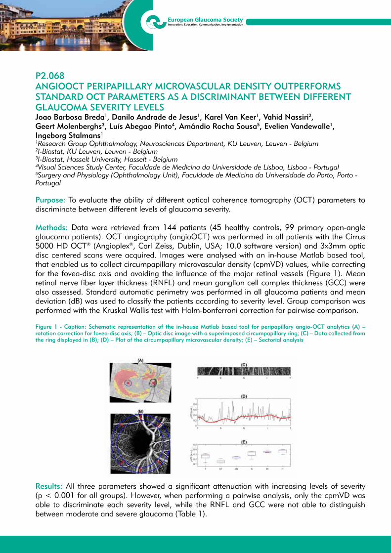
13th EGS CongressFortezza da Basso, Florence, Italy
19/22 May 2018
P2.068ANGIOOCT PERIPAPILLARY MICROVASCULAR DENSITY OUTPERFORMS STANDARD OCT PARAMETERS AS A DISCRIMINANT BETWEEN DIFFERENT GLAUCOMA SEVERITY LEVELSJoao Barbosa Breda1, Danilo Andrade de Jesus1, Karel Van Keer1, Vahid Nassiri2, Geert Molenberghs3, Luís Abegao Pinto4, Amândio Rocha Sousa5, Evelien Vandewalle1, Ingeborg Stalmans1
1Research Group Ophthalmology, Neurosciences Department, KU Leuven, Leuven - Belgium2I-Biostat, KU Leuven, Leuven - Belgium3I-Biostat, Hasselt University, Hasselt - Belgium4Visual Sciences Study Center, Faculdade de Medicina da Universidade de Lisboa, Lisboa - Portugal5Surgery and Physiology (Ophthalmology Unit), Faculdade de Medicina da Universidade do Porto, Porto - Portugal
Purpose: To evaluate the ability of different optical coherence tomography (OCT) parameters to discriminate between different levels of glaucoma severity.
Methods: Data were retrieved from 144 patients (45 healthy controls, 99 primary open-angle glaucoma patients). OCT angiography (angioOCT) was performed in all patients with the Cirrus 5000 HD OCT® (Angioplex®, Carl Zeiss, Dublin, USA; 10.0 software version) and 3x3mm optic disc centered scans were acquired. Images were analysed with an in-house Matlab based tool, that enabled us to collect circumpapillary microvascular density (cpmVD) values, while correcting for the fovea-disc axis and avoiding the influence of the major retinal vessels (Figure 1). Mean retinal nerve fiber layer thickness (RNFL) and mean ganglion cell complex thickness (GCC) were also assessed. Standard automatic perimetry was performed in all glaucoma patients and mean deviation (dB) was used to classify the patients according to severity level. Group comparison was performed with the Kruskal Wallis test with Holm-bonferroni correction for pairwise comparison.
Figure 1 - Caption: Schematic representation of the in-house Matlab based tool for peripapillary angio-OCT analytics (A) – rotation correction for fovea-disc axis; (B) – Optic disc image with a superimposed circumpapillary ring; (C) – Data collected from the ring displayed in (B); (D) – Plot of the circumpapillary microvascular density; (E) – Sectorial analysis
Results: All three parameters showed a significant attenuation with increasing levels of severity (p < 0.001 for all groups). However, when performing a pairwise analysis, only the cpmVD was able to discriminate each severity level, while the RNFL and GCC were not able to distinguish between moderate and severe glaucoma (Table 1).

13th EGS CongressFortezza da Basso, Florence, Italy
19/22 May 2018
Table 1 - Caption: Only cpmVD is able to discriminate between all glaucoma severity levels
OCT parameter Normal Glaucoma p
Median (IQR) N = 45 Mild N = 45 Moderate N = 27 Severe N = 27 Overall N-Mi N-Mo N-S Mi-Mo Mi-Se Mo-Se
RNFL 90 (9) 69 (18) 60 (18) 57 (12) < 0.001 < 0.001 0.007 0.17
GCC 78 (10) 66 (11) 61 (9) 56 (9) 0.003 0.07
cpmVD 0.34 (0.07) 0.27 (0.07) 0.22 (0.09) 0.18 (0.08) 0.003 0.01
RNFL - mean retinal nerve fiber layer thickness; GCC - mean ganglion celle complex thickness: cpmvd - circumpapillary microvascular density; IQR - interquartile range; N - normal; Mi - mild; Mo - Moderate; S - severe; p - p value (Kruskal Wallis)
Conclusions: Circumpapillary microvascular density, measured with angioOCT, was the best discriminant between different glaucoma severity levels, when compared to commonly used structural OCT parameters. This serves to show the potential of angioOCT in everyday glaucoma management, especially in the follow-up of more advanced disease stages, where conventional OCT parameters face challenges due to a floor effect at an earlier disease stage.
13th EGS CongressFortezza da Basso, Florence, Italy
19/22 May 2018
P2.069EFFECT OF MACULAR VASCULAR DENSITY ON CENTRAL VISUAL FUNCTION AND MACULAR STRUCTURE IN GLAUCOMA PATIENTSSooji Jeon1, Hae-Young Lopilly Park1, Chan Kee Park1
1Department of Ophthalmology, Seoul St. Mary’s Hospital, College of Medicine, The Catholic University of Republic of Korea, Seoul - Republic of Korea
Purpose: To evaluate the effect of perifoveal vascular density on central visual function and structure in glaucoma patients.
Methods: We enrolled 35 eyes of 35 normal tension glaucoma (NTG) patients with parafoveal scotoma. All subjects underwent standard automated perimetry (SITA 24-2 and 10-2) and structural evaluation including macular segmentation. Vascular density (VD) of macula was reported using optical coherence tomography angiography (OCTA) in superficial and deep retinal layer. Mean VD were compared according to severity of central visual field defect (CVFD), and coefficient correlations between VD and structural or functional parameters were recorded. Multivariate regression analysis was performed to determine factors influencing the severity of CVFD.
Results: In group with worse MD of SITA 10-2, mean vessel densities of perifovea were lower (29.72% of early CVFD vs. 26.99% of moderate CVFD in superficial layer; 32.13% of early CVFD vs. 30.31% of moderate CVFD in deep layer; p = 0.009 and 0.001, respectively). Correlation coefficients showed distinguishable characteristics between retinal layer – superficial VD showed significant correlation with mGCC and segmented macular thickness except INL, but deep VD did not show meaningful correlation with all sutructural parameters. However, deep VD showed significant correlations with MD, mean sensitivity, center sensitivity of SITA 24-2 and MD, PSD of SITA 10-2 (R = 0.505, 0.556, 0.506, 0.613 and -0.456; p = 0.002, 0.001, 0.002, < 0.001, and 0.003, respectively). In multivariate regression analysis, the significant factors affecting central visual function were cpRNFL thickness and deep VD (p = 0.015 and 0.002 for MD in SITA 10-2; p = 0.008 and 0.016 for center sensitivity in SITA 24-2). Different multivariate regression analysis models were compared and adjusted R2 was best in model including cpRNFL thickness, segmented macular thickness and only deep VD in variables (adjusted R2 = 0.623, p < 0.05).
Conclusions: Through OCT angiography, the effect of vascular incompetence can be visualized in retina when we evaluate NTG patients with early central scotoma. Decreased deep macular VD may have influence on the central visual function of NTG patients separately from retinal structural thinning.
13th EGS CongressFortezza da Basso, Florence, Italy
19/22 May 2018
P2.070LONG-TERM PERSONAL EXPERIENCE WITH SECOND-GENERATION TRABECULAR MICRO-BYPASS STENTS IN PATIENTS WITH GLAUCOMA: 3-YEAR OUTCOMESFritz H. Hengerer1, Gerd Uwe Auffarth1, Ina Conrad-Hengerer1
1Department of Ophthalmology, University of Heidelberg, Heidelberg - Germany
Purpose: This prospective case series evaluates the safety and efficacy of 2 second-generation trabecular micro-bypass stents (iStent inject®) in combination with cataract surgery (inject+CS) and as a standalone surgery (CS) in patients with glaucoma.
Methods: This consecutive case series from University Eye Clinic Heidelberg evaluates implantation of 2 iStent inject devices. The vast majority of patients had OAG; a few cases of PXE, PDS, narrow angle and secondary glaucoma were also included. IOP, medication use, visual acuity, C/D ratio, complications and secondary surgeries were assessed.
Results: One hundred and twenty-five eyes were successfully implanted with 2 iStent inject devices without surgical complications (81 eyes in the stent+CS group and 44 in the CS group). Forty-one eyes in the inject+CS group and 29 eyes in the CS group have completed three years of follow-up. In the inject+CS eyes, preoperative medicated mean IOP was 22.6 ± 6.2 mmHg with subjects on an average of 2.5 medications while mean medicated IOP was 25.3 ± 6.0 mmHg on 3.0 medications in the CS group. At 3 years postop, the inject+CS eyes had mean IOP (14.2 ± 1.6 mmHg). The mean IOP in the CS eyes was 14.7 ± 2.1 mmHg. Mean medication use decreased from 2.5 to 0.8 in the inject+CS eyes and from 3.0 to 0.6 in the CS eyes. At 3-year follow-up, 73% of inject+CS eyes were using 0 or 1 medication as compared to 21% preoperatively, and 86% of CS eyes were using 0 or 1. Three inject+CS eyes and 2 CS eyes underwent additional glaucoma procedures for disease progression not related to stent implantation.
Conclusions: Implantation of the iStent inject in combination with cataract surgery or as a standalone procedure resulted in meaningful sustained IOP reduction and reduced medication burden with a high safety profile. The favorable long-term outcomes of the iStent inject trabecular micro-bypass stents reported in this series of implants is encouraging. The iStent inject is a promising treatment option for patients with glaucoma.

13th EGS CongressFortezza da Basso, Florence, Italy
19/22 May 2018
P2.071IMPLEMENTATION OF A GLAUCOMA RISK FACTOR CALCULATOR MOBILE APPLICATION (APP) FOR AN EARLY LOW-TECH DIAGNOSISDanilo Renato Mazzacane1, Pier Franco Marino2, Ciro Costagliola2
1ASST Pavia, Pavia - Italy2Dipartimento di Medicina e Scienza per la Salute Università del Molise, Campobasso
Glaucoma represents the world’s second cause of blindness. The WHO estimates in about 80 million the number of glaucomatous in 2020: 60 mlns due to POAG and 20 mlns to ACG. POAG is the most common form of glaucoma and is often called “the silent thief of sight”, due to the lack of early detectable signs and symptoms in the early phase of the disease. It has been estimated that about 30% of patients may be unaware of their pathology. Moreover, the only sure way to diagnose glaucoma is with a complete eye exam; in fact, glaucoma screenings strategies are not enough to find glaucoma patients. The aim of this proposal is a case finding strategy based on using a glaucoma risk factor calculator mobile application (APP) for an early low-tech diagnosis.
Porpouse of the study is the capillary diffusion, through the APP, of a diagnostic flow chart based on the value of Integrated Risk Index (IRI) derived from the risk factors of the disease: IOP, CCT, race, age, family history, myopia.
Methods of case finding strategy is: - IRI < 0.3 patient will be newly evaluated at 3 months; - IRI > 0.3 patient will be submitted to: daily tonometric curve and ofthalmoscopy papillary region.
In case of negativity of these examinations, the patient will returns at 3 months after the first evaluation; if 1 or both the exams are positive, would be necessary to execute: SAP with threshold strategy and stage with GSS2 of Brusini and gonioscopy.
Results: To date, 526 patients (288 males, 238 females) have been evaluated for a total of 1052 eyes. Diagnostic predictivity with the integrated risk index for glaucoma was positive for 12 patients, 2.28% of the considered sample.
Conclusions: The APP diffusion will allow a significant reduction of the rate of undiagnosed patients, with a method economically sustainable and easily reproducible. In addition, with this procedure it is possible to storage clinical data from glaucomatous patients, realizing a database useful for further epidemiological and statistical investigations.
13th EGS CongressFortezza da Basso, Florence, Italy
19/22 May 2018
P2.072THE CORRELATION OF AVASCULAR ZONE WITH DIFFERENT OCT PARAMETERS IN PATIENTS WITH GLAUCOMAEwa Kosior-Jarecka1, Urszula Łukasik1, Dominika Wróbel-Dudzinska1, Jolanta Wójcikowska-Karska1, Tomasz Zarnowski1
1Dep. Diagnostics and Microsurgery of Glaucoma, medical University of Lublin, Lublin - Poland
The aim of this study is to look for the correlation between size of avascular zone and OCT glaucoma parameters.
Methods: 112 eye of patients with primary open angle glaucoma and pseudoexfoliative glaucoma were consecutively recruited to the study. All patients had performed visual field (30-2 Humphrey SAP) and Zeiss OCT (RNFL, GCC and Angio - module). Statistical analysis was performed with Statistica 13.0 and p < 0.05 was found significant.
Results: Mean size of avascular zone was 0.23 mm2 ± 0.1 mm2. It was not correlated with mean RNFL (p = 0.563), sectoral RNFL (Inferior p = 0.480, Superior p = 0.382, Nasal p = 0.556, Temporal p = 0.546) nor mean GCC (p = 0.232) and MD in VF (p = 0.842). The age of patients also did not influence the size of avascular zone (p = 0.844).
Conclusion: The size of avascular zone assessed in Angio-OCT is not correlated with the results of RNFL nor GCC measurements.

13th EGS CongressFortezza da Basso, Florence, Italy
19/22 May 2018
P2.073MOLECULAR-GENETIC STUDY OF PRIMARY OPEN-ANGLE GLAUCOMA IN PATIENTS FROM BASHKORTOSTAN REPUBLIC, RUSSIAAigul Zagidullina1, Bulat Aznabaev1, Semen Lobov2, Lilya Dzhemileva2, Ramzilya Khasanova3, Alia Zainitova3, Elsa Khusnutdinova3
1Department of Ophthalmology, Bashkir Medical University, Ufa - Russia2Genetic Laboratory, Institute of Biochemistry and Genetics, Ufa - Russia3Department of Genetics and Fundamental Medicine, Bashkir State University, Ufa - Russia
Purpose: To study a spectrum and frequency of mutations in the MYOC gene (myocilin) and replicate the results of genome-wide association studies (GWAS) of primary open angle glaucoma (POAG) with 6 polymorphic loci in five genes rs3213787 (SRBD1), rs4236601 (CAV1), rs4656461 (TMCO1), rs4977756 (CDKN2B-AS1), rs3825942, rs1048661 (LOXL1) in patients with POAG from the Republic of Bashkortostan, Russia.
Materials and Methods: It was examined 931 persons (496 patients with POAG and 435 individuals from the control group) being followed-up at the Optimed Laser Ophthalmology Clinic, Ufa. Identification of mutations in the MYOC gene was carried out by high resolution melting curve analysis in real time, and subsequent re-sequencing of DNA samples with altered melting curves analysis. Molecular genetic studies included: a genomic DNA extraction, amplification of the investigated genes sections by polymerase chain reaction, single-stranded conformation polymorphism analysis followed by sequencing.
Results: The mutation p.Q368X (c.1102C > T) in the MYOC gene was screened in 496 unrelated patients. In 8 unrelated patients, this mutation was detected in the heterozygous state, which amounted to 1.6% of all the examined families with POAG. 435 DNA samples of healthy donors from the control group (Russians, Tatars, Bashkirs) were analyzed. The mutation p.Q368X (c.1102C > T) in the MYOC gene was identified in the heterozygous state in 1 Russian individual (0.17%). The total p.Q368X (c.1102C > T) mutation in the MYOC gene identified in patients with POAG was 0.8%. The p.Q368X (c.1102C > T) mutation in the MYOC gene was 1.3% among patients with POAG from the Republic of Bashkortostan. Replication of the GWAS of POAG with 6 polymorphic loci in five genes was performed (rs3213787 (SRBD1), rs4236601 (CAV1), rs4656461 (TMCO1), rs4977756 (CDKN2B-AS1), rs3825942, rs1048661 (LOXL1)). It was discovered a rs4977756 polymorphic variant association of the cyclin-dependent kinases (CDKN2A-AS1) with the development of POAG in patients of Tatar ethnicity from Bashkortostan.
Conclusions: The major mutation p.Q368X (c.1102C > T) (1.3%) and the polymorphic variant rs61730974 (c.1041T > C, p.Tyr347 =) were detected in patients with POAG from Bashkortostan in the myocilin gene (MYOC) (0.17%) with ambiguous pathogenicity. The rs4977756 polymorphic variant association was established in the gene (CDKN2A-AS1) with the POAG development.

13th EGS CongressFortezza da Basso, Florence, Italy
19/22 May 2018
P2.074CHANGES IN CORNEAL ENDOTHELIAL CELL DENSITY IN PATIENTS WITH DIFFERENT STAGES OF OPEN-ANGLE GLAUCOMAYosip Saldan1, Nataliia Kapshuk2, Yulia Saldan3, Lesia Khrystych3
1Vinnytsia Regional Medical University named after N.I. Pirogov, Vinnytsia - Ukraine2Department of Ophthalmology, Vinnitsa Regional Clinical Hospital Named After N.I.Pirogov, Vinnitsya - Ukraine3Vinnitsya Regional Medical University named after N.I. Pirogov, Vinnitsya - Ukraine
Purpose: Determine the endothelial cell density in patients with different stages of open-angle glaucoma. In glaucoma, there are many factors that can decrease the number of hexagonal cells, including high intraocular pressure, hereditary changes in cell structure, pseudoexfoliation, previous trauma, and surgical intervention. Evaluation of the endothelial cell density is an important predictor of the development and progression of glaucoma and corneal decompensation.
Methods: The study group included 24 patients (33 eyes), 6 women, 18 men, average age was 64 ± 8.9 years. Previous trauma, surgical intervention, presence of concomitant eye diseases, uncompensated intraocular pressure and closed-angle glaucoma served as exclusion criteria. Standard and additional examination methods were performed for all patients. Non-contact endothelial microscope Topcon SP-1P was used to evaluate the state of endothelial cells. Central corneal thickness, endothelial cell density, maximum, minimum and average size of hexagonal cells, and the coefficient of variation were measured. All patients were divided into groups according to the stages of glaucoma: group 1 - stage I - 7 eyes, group 2 - stage II - 10 eyes, group 3 - stage III - 10 eyes, group 4 – stage IV - 6 eyes.
Results: The density index of hexagonal cells was used to determine the state of the endothelium. As a result, the average values of the endothelial cells density within the group were as follows: 1 group - 2469.3 ± 147.3; Group 2 - 2250 ± 342.1; Group 3 -2173.4 ± 376.3; Group 4 - 1919.7 ± 374.9.
Conclusions: The obtained data indicates decreasing in the endothelial cells density with the glaucoma progression and correlate with the visual fields loss and changes in the optic nerve disc in an optical coherent tomography. Evaluating the status of hexagonal cells helps to predict the possibility of corneal decompensation, more differentiated approach for surgical intervention with additional protection for endothelium.

13th EGS CongressFortezza da Basso, Florence, Italy
19/22 May 2018
P2.075CHARACTERISTIC OF OPTIC NERVE HEAD IN GLAUCOMA PATIENTSAleksandar Miljkovic1, Marija Trenkic Božinovic2, Vladimir Canadanovic1, Nikola Babic1
1Medical Faculty of Novi Sad, University of Novi Sad, Eye Clinic, Novi Sad - Serbia2University of Nis, Medical Faculty, Department of Ophthalmology, Nis - Serbia
Purpose: The aim of this study was to determine the difference in parameters of optic nerve head (ONH) in patients with open angle glaucoma (POAG) in comparison to healthy population, as well as to determine the difference in parameters of ONH according to progression of the disease.
Methods: This clinical study was analytical and observational, “case-control“ type of study. 120 patients were included. On the basis of clinical finding four groups of patients were formed: group without glaucoma, group with mild POAG, group with moderate POAG and group with preperimetric glaucoma. Complete ophthalmological examination, visual field and optic coherent tomography of ONH were performed.
Results: The parameters of ONH such as: volume of excavation, C/D vertical, C/D horizontal and C/D total ratio in patients with mild POAG are higher comparing to healthy population. Parameters of ONH such as: neuroretinal rim area and neuroretinal rim volume in patients with mild POAG are lower than in healthy population. Total ONH area does not change in: healthy subjects, patients with mild and moderate POAG and in patients with preperimetric glaucoma, so this parameter does not determine glaucomatous disease. Parameters of ONH that are the best determinants of progression of glaucoma are: C/D total, C/D vertical and C/D horizontal ratio. ONH parameters such as: volume of excavation, C/D vertical, C/D horizontal and C/D mean ratio in patients with preperimetric glaucoma are statistically significant higher than in healthy population. The best predictors of appearance and progression of glaucomatous disease are: RimArea, RimVol, DiscArea, CupAear, C/DHorRat, C/DVertRat, C/DAreaRat, and the worst is CupVol, shown by ROC curves. Cut-off for: C/DHorRat is 0.867, C/DVertRat is 0.814 and C/DAreaRat is 0.693; and according to this study they represents the higher valus for fisiological excavation.
Conclusion: Determination of parameters of ONH in patients with POAG using optical coherent tomography represents the method that distinguishes the patients with POAG from healthy subjects. Difference in parameters of ONH according to progression of the disease suggests that they are good tool in early diagnosis and following of glaucoma patients.
13th EGS CongressFortezza da Basso, Florence, Italy
19/22 May 2018
P2.076POINT-WISE CORRELATION BETWEEN VISUAL FIELD AND OCT IN OPEN ANGLE GLAUCOMA PATIENTSPaola Cirafici1, Guido Maiello2, Chiara Ancona1, Alessandro Masala1, Carlo Enrico Traverso1, Michele Iester1
1Dipartimento di Neuroscienze, Riabilitazione, Oftalmologia, Genetica e Scienze Materno-Infantili; Clinica Oculistica; Università di Genova, Genova - Italy2Department of Experimental Psychology, University of Giessen, Germany, Giessen - Germany
Purpose: Optical Coherence Tomography (OCT) is a powerful instrument for helping clinicians to detect and monitor glaucoma onset and progression. However, the diagnostic capacity of OCT data to predict glaucomatous visual loss is unclear. The aim of this study was to provide a detailed mapping of the relationships between visual field (VF) sensitivities and several measures of retinal structure provided by a commercial Spectral Domain (SD)-OCT system (RTvue-100 Optovue).
Methods: Sixty-three eyes of open angle glaucoma patients were evaluated at the University Eye Clinic of Genoa and included in this retrospective, observational clinical study. The thickness of various retinal structures was recorded using SD-OCT. Thickness values for superior and inferior retina, as well as average values, were recorded for the full retina, the outer retina, the ganglion cell complex (GCC), and the peripapillary retinal nerve fibre layer (RNFL). The thickness of the RNFL was further evaluated along eight separate sectors (temporal lower, temporal upper, superior temporal, superior nasal, nasal upper, nasal lower, inferior nasal, inferior temporal). Point-wise correlations were then computed between each of these OCT measures and the visual sensitivities at all VF locations assessed via Humphrey 10-2 and 24-2 perimetry.
Results: Point-wise correlations between retinal thickness measurements and visual sensitivities reflect the known topography of the retina. The spatial correlation patterns between visual sensitivities and RNFL thickness along different sectors broadly agree with previously hypothesised structure–function maps, yet suggest that structure-function maps still require more precise characterizations. Superior GCC thickness correlates maximally with sensitivities measured in the inferior visual field for both 24-2 (r = 0.53, p < 0.001) and 10-2 (r = 0.57, p < 0.001) perimetry. Conversely, inferior GCC thickness correlates best with sensitivities measured in the superior visual field for both 24-2 (r = 0.50, p < 0.001) and 10-2 (r = 0.50, p < 0.001) perimetry.
Conclusions: We provide detailed mappings of the relationships between VF sensitivities and OCT data. These spatial correlation patterns could be employed to develop more sensitive OCT protocols for monitoring glaucoma. Our data and analyses highlight GCC and RNFL thickness measurements as the most promising candidate metrics for glaucoma detection and monitoring.

13th EGS CongressFortezza da Basso, Florence, Italy
19/22 May 2018
P2.077QUALITY OF GLAUCOMA REFERRALS FROM PRIMARY CARE TO THE HOSPITAL EYE SERVICE IN A TEACHING HOSPITAL - A REVIEWLiam Rogerson1, Edward Ridyard1, Jan Van der Hoek1
1Department of Ophthalmology, York Teaching Hospitals, York - United Kingdom
Purpose: The majority of glaucoma referrals in the UK are from primary care optometrists. Ageing population and an improvement in quality of screening methods has contributed to the vast increase in referrals to HES. The quality of referrals can be variable and NICE produced a set of updated guidelines for Glaucoma referrals November 2017 in an effort to reduce the number of false positives. The aim of the study was to assess the quality of Glaucoma referrals made to the Hospital Eye Service at Scarborough Hospital from primary care.
Methods: A retrospective manual analysis of referrals from primary care between August 2017 and November 2017 was undertaken (n = 50). Referral reason, examination findings by Ophthalmologist and outcome of the referral were analysed.
Results: The average time from referral to review in HES was 8 months. Opticians optometrists accounted for 68% of total referrals. The primary reason for referral was raised intraocular pressure (42%) followed by suspected disc cupping (22%). Intraocular pressure was > 22 mmHg in either eye 44% of time (p ≤ 0.005). OCT RNFL thinning was observed in 22% of cases (p ≤ 0.0001). Further follow-up and investigation was arranged in 70% of referrals (p ≤ 0.0045).
Conclusion: The majority of glaucoma referrals from primary care have merit and primary care is an invaluable safety net in identifying patients at risk of developing glaucoma. Unnecessary referrals may be prevented by providing education to primary care clinicians locally.
13th EGS CongressFortezza da Basso, Florence, Italy
19/22 May 2018
P2.078COMPARISON OF REPRODUCIBILITY IN GLAUCOMA SEVERITY EVALUATION BETWEEN STRUCTURAL MEASUREMENT AND FUNCTIONAL TESTDaisuke Shiba1, Sayaka Adachi1, Eri Hirano1, Takeshi Ono1, Kenya Yuki1, Kazuo Tsubota1
1Department of Ophthalmology, Keio University School of Medicine, Tokyo - Japan
Purpose: The objective of the present study is to compare reproducibility in quantitative evaluation of glaucomatous optic neuropathy between ganglion cell complex thickness (GCCT) with spectral-domain optical coherence tomography (SD-OCT) and visual field sensitivity (VFS) with standard automated primetry (SAP).
Methods: In this prospective cohort study, we included 60 eyes of glaucoma patients without cataract or any other ocular abnormality. The included patients underwent SAP by Humphry field analyzer with central 30-2 program and GCCT measurement by SD-OCT (Nidek; RS-3000 Advance) with macular scan. The same examinations were repeated at the next visit for less than two months. We used mean GCCTs in superior semicircle (SSC) with the radius of 3mm and inferior semicircle (ISC) with the same radius as structural measures. The center of SSC or ISC was positioned at the fovea. Meanwhile, mean VFSs of superior hemi-field (SHF) and inferior hemi-field (IHF) were adopted as functional measures. Eyes were classified into 10 stages based on the deciles of each measure. We adopted intraclass correlation coefficients (ICC) and Cohen’s weighted kappa statistic as measures of the inter-visit agreement.
Results: The mean first / second GCCTs were 84.1 ± 10.4 μm / 83.9 ± 10.5 μm in SSC and 74.6 ± 1.5 μm / 74.6 ± 1.51 μm in ISC. Mean VFSes at first / second visit was 25.6 ± 5.6 dB / 25.4 ± 5.6 dB in IHF, 22.9 ± 7.5 dB / 22.9 ± 7.1 dB in SHF. ICCs [95%CI] with SD-OCT were 0.989 [0.981, 0.993] in SSC and 0.988 [0.980, 0.993] in ISC, whereas ICCs with SAP were 0.805 [0.695, 0.879] in IHF and 0.960 [0.934, 0.976] in SHF. Weighted kappa values with SD-OCT were 0.982 [0.974, 0.990] and 0.987 [0.981, 0.993] in SSC and ISC, whereas Kappa values with SAP were 0.683 [0.279, 1.09] in IHF and 0.968 [0.959, 0.977] in SHF.
Conclusion: Structural measures had a higher reproducibility than functional measures in this study population with glaucoma.
13th EGS CongressFortezza da Basso, Florence, Italy
19/22 May 2018
P2.079THE COMPRASION OF CENTRAL MACULAR THICKNESS IN CASES WITH HAVING PSEUDO EXFOLIATION SYNDROME AND OCULAR HYPERTENSIONUgur keklikci1
1Department of Ophthalmology,Faculty of Medicine, Dicle University, Diyarbakir - Turkey
Purpose: The comparison of central macular thickness (CMT) obtained by spectral domain optical coherence tomography (SD-OCT) in the cases with having pseudo exfoliation syndrome (PXF) and ocular hypertension (OHT).
Methods: This study performed between January 2016 and December 2016 by Dicle University Medical Faculty, department of ophthalmology glaucoma section, in which 31 eyes of 18 patients diagnosed as having pseudo exfoliation syndrome and 46 eyes of 25 patients diagnosed as having ocular hypertension were included. This is a prospective trial. Central macular thickness measurements obtained by SD-OCT were recorded and values comparised between the groups.
Results: Mean central macular thickness was 237.1 ± 21.2 in pseudo exfoliation syndrome group and 236.3 ± 19.3 in ocular hypertension group. There was not found statistically significant difference between central macular thickness obtained by OCT in pseudo exfoliation syndrome and ocular hypertension cases (p > 0.05).
Conclusion: Macular OCT may be beneficial in the diagnosis and follow-up of glaucoma. No difference was found between the groups for central macular thickness.

13th EGS CongressFortezza da Basso, Florence, Italy
19/22 May 2018
P2.080EXPEDIENCY OF THE AUTOMATED PERIMETRY USING THE GOLDMANN V STIMULUS SIZE IN VISUALLY IMPAIRED GLAUCOMA PATIENTSMauricio Paolera1, Niro Kasahara1
1Department of Ophthlamology, Santa Casa de Sao Paulo, Sao Paulo - Brazil
Purpose: White-on-white standard automated perimetry (AP) uses a white round stimulus with 0.43° diameter and 4 mm2 area (Goldmann size III). Patients with low vision have difficulty seeing such a small stimuli and are often tested with kinetic perimetry using the Goldmann size V stimulus with 1.7° diameter and 64 mm2 area. We undertook an observational case-control study to compare the performance of patients on AP using two different size stimuli.
Methods: Patients with glaucoma and visual acuity worse than 20/100 underwent AP using standard size III stimulus, SITA standard test and size V stimulus full threshold test, fastpac strategy. All patients were familiar with the procedure having done the test at least twice previously. Another group of glaucoma patients with visual acuity better than 20/60 served as controls. The main outcome measures included test duration, mean sensitivity, fixation loss (FL), false positive (FP), false negative (FN), and the patient subjective preference.
Results: Patients demographics are depicted on table 1. FL, FP, and FN rates were lower with the size V stimulus as compared to SITA standard. MS was higher with size V stimulus test.
Conclusion: Size V stimulus full threshold test provides more reliable exams and higher mean sensitivity in glaucoma patients with low visual acuity. Patients seem to prefer size V than SITA standard strategy.

13th EGS CongressFortezza da Basso, Florence, Italy
19/22 May 2018
P2.081A COMPARISON OF RETINAL NERVE FIBER LAYER THICKNESS TO VISUAL FIELD SENSITIVITY IN PATIENTS WITH GLAUCOMAVeljko Andreic1
1Clinical center of Vojvodina, Novi Sad - Serbia
Purpose: To explore the relationship between retinal nerve fiber layer thickness and visual field sensitivity in patients with primary open angle glaucoma.
Methods: Automated static perimetry (HumphreyTM Field Analyzer; Carl Zeiss Meditec) and spectral domain optical coherence tomography scans (CirrusTM HD-OCT; Carl Zeiss Meditec) of optic nerve head and retinal nerve fiber layer, were obtained from 54 age-matched normal control subjects and 26 patients with open angle glaucoma.
Results: The average retinal nerve fiber layer thickness in the control eyes was 93.4 ± 8.0 μm, and 65.0 ± 11.0 μm in glaucomatous eyes. The average MD value in control eyes was -0.91 ± 1.26 dB, and -13.82 ± 8.11 dB in glaucomatous eyes. A significant correlation was found between the retinal nerve fiber layer thickness and visual field loss in patients with glaucoma (r = +0.64). Slightly less correlation was found in control eyes (r = -0.04).
Conclusions: A retinal nerve fiber layer thickness value less than 76 μm points to high probability of glaucoma. Values over 86 μm suggest absence of glaucoma.
13th EGS CongressFortezza da Basso, Florence, Italy
19/22 May 2018
P2.082EXPRESSION PROFILE OF MICRO RNAS IN TEARS. A FINGERPRINT FOR THE PRE-CLINICAL PHASE OF PRIMARY OPEN-ANGLE GLAUCOMA VERSUS OCULAR HYPERTENSIONMaria Dolores Pinazo-Durán1, Vicente Zanón-Moreno1, Jorge Raga-Cervera1, Silvia M. Sanz-González1, Jose J. García-Medina1
1Ophthalmic Research Unit “Santiago Grisolia” / Fisabio, Valencia - Spain
Purpose: To study the expression profile of microRNAs in tears of patients diagnosed of primary open-angle glaucoma (POAG) and patients with ocular hypertension (OHT) with the aim of finding molecular biomarkers for better managing OHT and to achieve the preclinical POAG diagnosis.
Methods: A case-control study was carried out in 24 POAG patient vs 24 OHT patient matched by age. Total RNA was isolated using the miRCURY™ RNA Kit [Biofluids (Exiqon®)] and quantified in a 2100 Bioanalyzer (Agilent ®). The expression profile of microRNA in tear samples was analysed by next generation sequencing using the Illumina NextSeq500 Technology. Bioinformatic analyses were performed by specific programs.
Results: We identified 120 microRNA in tears of the participants. Among them, only 10 microRNAs showed significant differences (p < 0.05) in POAG vs OHT samples (2 microRNAs down-regulated and 8 up-regulated in POAG tears). Therefore, these microRNAs were statistically analysed to create the receiver operating characteristic curve (ROC) obtaining for all of them an area under the curve higher than 0.65.
Conclusions: Tear samples have provided essential information to differentially identify 10 microRNAs that presumptively might be used as biomarkers for early identification of the POAG risk in OHT individuals.
13th EGS CongressFortezza da Basso, Florence, Italy
19/22 May 2018
P2.083POTENTIAL METABOLIC BIOMARKERS IN GLAUCOMA PATIENTS AND THEIR REGULATION IN RESPONSE TO HYPOXIAMiriam Kolko1, Line Marie Dalgaard2, J.V. Jensen2, Linda Hildegaard Bergersen3, Niels V. Olsen4, Bjørnar Hassel3, Farrukh Abbas Chaudhry3, Rupali Vohra2
1Department of Ophthalmology, Copenhagen - Denmark2University of Copenhagen, Copenhagen - Denmark3University of Oslo, Oslo - Norway4Department of Anesthesiology, Copenhagen - Denmark
Purpose: To assess novel differences in serum levels of glucose, lactate and amino acids in patients with low-tension glaucoma (LTG) compared to age-matched controls; at baseline and in response to universal hypoxia.
Method: Twelve patients diagnosed with LTG and eleven control subjects underwent normobaric hypoxia for two hours. Peripheral venous blood samples were taken at baseline, during hypoxia and in the recovery phase. Serum glucose and lactate levels were measured by a blood-gas analyzer. Amino acids were analyzed by high-performance liquid chromatography.
Results: Baseline levels of lactate and total amino acids were significantly lower in patients with LTG compared to healthy controls. No differences were seen in blood glucose levels between the test groups. Lactate levels remained unchanged during hypoxia in the control group, but increased in patients with LTG. In the recovery phase, total amino acid levels were reduced in the control group, whereas no changes were found in patients with LTG.
Conclusion: Reduced serum levels of lactate and total amino acids were identified as potential diagnostic markers for LTG. Moreover, significant different regulatory patterns of certain amino acids were found in patients with LTG compared to control subjects. Over all, our results suggest a link between systemic energy metabolites and LTG and support a novel understanding of glaucoma as an inner retinal manifestation of a systemic condition.

13th EGS CongressFortezza da Basso, Florence, Italy
19/22 May 2018
P2.084STRUCTURE-FUNCTION RELATIONSHIP BETWEEN SD-OCT MULTILAYERED MACULAR ANALYSIS AND VISUAL FIELD 24-2 AND 10-2 DEFECTS, IN PERIMETRIC GLAUCOMADavide Rastelli1, Sara Giammaria1, Manuele Michelessi2, Gianluca Manni1
1DSCMT, Università di Roma “Tor Vergata”, Rome - Italy2IRCCS-Fondazione G.B. Bietti, Rome - Italy
Purpose: To evaluate the structure-function relationship between the thickness of the inner retinal layers of the macular region, assessed by spectral-domain optical coherence tomography (SD-OCT) and the visual field sensivity loss of the corresponding areas, assessed by 24-2 and 10-2 Standard Automated Perimetry (SAP), in glaucoma.
Methods: 50 eyes of 50 glaucomatous patients were enrolled. All patients underwent a full eye examination, SAP with HFA (Carl Zeiss Meditec) including both SITA 24-2 and 10-2, performed in different days and imaging with Spectralis SD-OCT (Heidelberg Engineering, posterior pole analysis), within one month. The reference for glaucoma was the 24-2 SAP. The 64 squares of the SD-OCT posterior pole analysis were grouped in 2x2 squared area to spatially match both the sensitivity of each 24-2 SAP central test point and the sensitivity of the 68 10-2 SAP test points, grouped correspondingly. The visual sensitivity loss was expressed by total deviation (TD) both in logarithmic and linear (1/Lambert) scale. We evaluated the thickness of macular retinal nerve fiber layer (mRNFL), ganglion cell layer (GCL) and inner plexiform layer, separately. The structure-function relationship was measured as coefficient of determination (R2) p < 0.05 was considered statistically significant.
Results: The R2 ranged from 0.002 (p = 0.9) to 0.52 (p < 0.0001). The R2 for the association between SD-OCT 2x2 squares and the corresponding 10-2 TD test areas were higher than the R2 for the association of the SD-OCT 2x2 squares and the corresponding 24-2 TD test points, in 95% of comparisons. In the central 6° of the posterior pole, GCL thickness provided the strongest relationship with visual sensitivity loss, whereas in the peripheral area of the posterior pole the sensitivity loss showed the strongest relationship with mRNFL. The use of linear scale to Ex-Press the TD did not improve the structure-function relationship strength.
Conclusions: The relationship between macular thickness and 10-2 TD was stronger than the relationship between the corresponding macular thickness and the 24-2 TD. The GCL thickness showed the strongest association with functional glaucoma damages in the central 6° of the macular area.
13th EGS CongressFortezza da Basso, Florence, Italy
19/22 May 2018
P2.085COLOR-CODED NORMATIVE CLASSIFICATION VS CONTINUOUS DATA BY SPECTRAL DOMAIN-OPTICAL COHERENCE TOMOGRAPHY FOR DETECTING GLAUCOMA: TRANSLATING THE STATISTICS INTO CLINICAL PRACTICEManuele Michelessi1, Ivano Riva2, Enrico Martini3, Michele Figus4, Paolo Frezzotti5, Luca Agnifili6, Gianluca Manni7, Luciano Quaranta8, Stefano Miglior9, Chiara Posarelli4, Francesco Oddone1
1IRCCS - Fondazione G.B. Bietti, Rome - Italy2IRCCS - Fondazione G.B. Bietti, Brescia - Italy3U.O. Ophthalmology, Sassuolo Hospital, Sassulo - Italy4Ophthalmology, Department of Neurosciences, University of Pisa, Pisa - Italy5Department of Medicine, Surgery and Neuroscience, University of Siena, Siena - Italy6Ophthalmology Clinic, Department of Medicine and Aging Science, University G. d’Annunzio of Chieti-Pescara, Chieti - Italy7DSCMT, University of Rome, Tor Vergata, Rome - Italy8Department of Medical and Surgical Specialties, Radiological Sciences and Public Health, University of Brescia, Brescia - Italy9Department of Ophthalmology, University Bicocca of Milan, Milan - Italy
Purpose: To evaluate and compare the diagnostic accuracy of peripapillary retinal nerve fiber layer (pRNFL) and Minimum rim width (MRW) color-coded classification and continuous data by SD-Optical coherence tomography in discriminating healthy from glaucoma eyes.
Methods: healthy controls and primary open angle glaucoma were enrolled consecutively All patients underwent a full eye examination, standard automated perimetry (Humphrey Field Analyzer) and imaging with Spectralis SD-OCT (Heidelberg Engineering, Optic Nerve Head protocol). For both pRNFL and MRW, color coded classification as well as continuous data were considered. Sensitivity (sn) and specificity (sp) were calculated from 2 x 2 tables for color-coded classifications. For continuous data sensitivity was calculated by matching specificity from the ROC curve to that of the corresponding color-coded classification. Borderline results were considered either as within or outside normal limits. p value < 0.05 was considered statistically significant.
Results: 197 glaucoma and 83 controls were included in this study. For categorical data the sensitivities significantly increase with minimal decrease in specificities when borderline were considered as glaucoma. For pRNFL parameters, continuous data provided similiar sensitivity (range 47.21-84.26) to that of categorical classification (range 42.64-84.77). For MRW, continuous parameters provided significantly higher sensitivity (range 67.01-89.85) compared to that of categorical classifications (range 48.57-78.68).
Conclusions: Categorical and continuous data offered diagnostic performance that were similar for pRNFL parameters but significantly different for MRW analysis. Clinicians must be careful when translating the results from diagnostic accuracy studies in their clinical daily practice.
13th EGS CongressFortezza da Basso, Florence, Italy
19/22 May 2018
P2.08624-HOUR CONTINUOUS MONITORING OF INTRAOCULAR PRESSURE-RELATED FLUCTUATIONS IN PROGRESSING PRIMARY OPEN ANGLE GLAUCOMA USING A CONTACT LENS SENSORCedric Schweitzer1, Clemence Andrebe1, Laury Tellouck1, Stephanie Roseng1, Florence Malet1, Thibaut Gaboriau1
1Department of Ophthalmology, Bordeaux University Hospital, Bordeaux - France
Purpose: To compare intraocular pressure (IOP)-related fluctuations using a contact lens sensor (CLS) between two groups of progressing open angle glaucoma (slow and fast progressor).
Methods: Cross-sectional study performed at Bordeaux University Hospital including primary open angle glaucoma patients with a progression on visual field test. Each patient underwent a monitoring of IOP fluctuations using a strain gauge CLS (Triggerfish, Sensimed, Lausanne, Switzerland). A standard automated perimetry was performed using Octopus (Haag-Streit, Koenis, Berne, Switzerland) and the rate of progression was calculated using a linear regression of the mean deviation (MD) parameter based on at least 5 reliable visual fields over a minimum period of 2 years before CLS measurements. Patients were classified in two groups of similar size of progression rate: group 1 with a slower rate of progression (MD < 0.5 dB/year) and group 2 with a faster rate of progression (MD ≥ 0.5 dB/year). The initial output signal was analyzed using Matlab software. A specific model was used taking into account abnormal noises and blinks.
Results: 54 eyes of 54 patients were included. The mean age was 68.48 ± 6.02 years. The mean rate of progression was -1.09 ± 0.60 dB/year in group 1 (n = 22, MD = -10.57 ± 6.97dB) and was -0.12 ± 0.14 dB/year in group 2 (n = 32, MD = -5.43 ± 4.32 dB). During the 24-hour period, peak amplitude was significantly greater in group 1 than in group 2 (343.1 ± 62.3 mVeq and 274.0 ± 75.0 mVeq respectively, p < 0.05) and the absolute area under the curve was significantly larger in group 1 than in group 2 (28.28 ± 2.10 V2eq and 6.82 ± 2.71 V2eq respectively, p < 0.05). Short-term IOP-related fluctuations (60-220 minutes) analysis showed a significantly greater amplitude in group 1 than in group 2 (110.0 ± 33.3 mVeq et 86.1 ± 26.7 mVeq respectivement, p < 0.05) and an absolute area under the curve significantly larger in group 1 than in group 2 (1.24 ± 0.30 V2eq et 1.01 ± 0.23 V2eq respectivement, p < 0.05).
Conclusion: Our study shows that eyes with a faster rate of progression exhibited longer and larger nocturnal IOP-related fluctuations. Short-term peak amplitude was also longer and larger in this group of eyes. 24-hour continuous monitoring of IOP-related fluctuations could be an additional biomarker of glaucoma progression. However prospective and comparative studies are mandatory to determine the exact role of this biomarker in the onset or progression of glaucoma.

13th EGS CongressFortezza da Basso, Florence, Italy
19/22 May 2018
P2.087THE ROLE OF PHACOEMULSIFICATION IN THE MANAGEMENT OF PRIMARY ANGLE-CLOSURE GLAUCOMAIlgaz Yalvac1, Raciha Beril Kucumen1
1Department of Ophthalmology, Yeditepe University, Istanbul - Turkey
Background: To evaluate ocular biometric parameters in pre and post phacoemulsification in patients with primary angle-closure glaucoma (PACG).
Methods: This is a retrospective chart review study. A total of 96 eyes (96 patients) with PAGC were recruited. All patients underwent ocular examination and biometry. Outcome variables included anterior chamber depth (ACD), lens thickness (LT), axial length (AL), lens position and relative lens position (LP and RLP, respectively) pre and postoperatively.
Results: Anterior chamber depth was shallower in preoperatively than postoperatively (p < 0. 01). The AL was not significantly different pre and postoperatively. The lens position in RPL was greater than preoperatively (p < 0.05).
Conclusions: Good outcomes in terms of AC parametres and long-term IOP control have been found following lens extraction for PACG. Lens extraction should be considered in patients with PACG, especially with thick and anteriorly vaulted lens status.
13th EGS CongressFortezza da Basso, Florence, Italy
19/22 May 2018
P2.088APHAKIC GLAUCOMA AFTER CONGENITAL CATARACT SURGERYHatem Marey1, Abd el Rahman El Sebaey1
1Department of ophthalmology, Menofia University, Shibin el koom - Egypt
Purpose: To determine the incidence rate and the risk factors for development of aphakic glaucoma after pars plana lensectomy for congenital cataract.
Methods: A retrospective study was performed on 68 eyes of 38 patients. Pars plana lensectomy was done for all cases for management of congenital cataract from January 2004 to January 2009. Aphakic glaucoma was defined as repeated intraocular pressure measurement greater than 22 mmHg with vertical Cup\disc ratio greater than 0.3 after pars plana lensectomy that necessitated medical therapy or surgical intervention.
Results: Fourteen out of 68 eyes (20.6%) developed aphakic glaucoma. The mean follow up period was 33.93 months with a range of 6 to 66 months. The mean intraocular pressure (IOP) at diagnosis of aphakic glaucoma was 33 mmHg and the mean duration between pars plana lensectomy and development of aphakic glaucoma was 35 months. Fourteen eyes had micro cornea out of which 6 eyes (42.9%) developed aphakic glaucoma. Thirty five eyes were operated on before 3 months out of which 12 eyes (34.3%) developed aphakic glaucoma.
Conclusions: The incidence rate of aphakic glaucoma after congenital cataract surgery was 20.6%. Many risk factors affect the incidence of development of aphakic glaucoma after pars plana lensectomy, in this study micro cornea and surgery before 3 months age were associated with a statistically significant increase in the incidence rate.

13th EGS CongressFortezza da Basso, Florence, Italy
19/22 May 2018
P2.089A CASE OF CHILDHOOD GLAUCOMA WITH DELETION IN THE 6P25 REGION CONTAINING FOXC1 GENEKazuhide Kawase1, Yusuke Niimi1, Tetsuya Yamamoto1, Takahiro Yamamoto2, Toshiyuki Fukao2, Kentaro Kurata3, Nobutaka Tachibana3, Katsuhiro Hosono3, Yoshihiro Hotta3
1Department of Ophthalmology, Gifu University school of Medicine, Gifu - Japan2Department of Pediatrics, Gifu University School of Medicine, Gifu - Japan3Department of Ophthalmology, Hamamatsu University School of Medicine, Hamamatsu - Japan
Purpose: We report a case of 6p25 deletion syndrome, one of subtelomeric microstructural abnormality, which is Childhood glaucoma with right eye aniridia and left eye Peters anomaly.
Methods: 0-year old girl. Cornea opacity of both eyes was observed, suspected of early-onset developing glaucoma on 6th birth date. Intraocular pressures of right and left eyes were 19-26 and 24-34 mmHg, respectively, at first visit (i-care pro, Icare Finland, Finland). The cornea of the right eye was edematous with aniridia. The cornea of the left eye was opacity and the central thickness was thin. These findings suspected Peters anomaly. Patent ductus arteriosus, patent foramen ovale, atrial septal defects, deafness on both sides, right accessory ear, and saddle nose were identified. Familial history of glaucoma was not recognized and there was no abnormality and infection during pregnancy. Two additional trabeculotomies were done on left eye. No mutations were identified by Sanger sequencing of all 13 exons of PAX6 gene. Then, we performed whole exome the sequencing using NexSeq500TM, after exon capture using SureSelect Human All Exon V6™.
Result: Exome data suggested that a patient had a de novo deletion in the 6p25 region containing the FOXC1 gene.
Conclusion: A phenotype of de novo deletion in the 6p25 region showed the Childhood glaucoma with aniridia and Peters anomaly with multiple malformations.

13th EGS CongressFortezza da Basso, Florence, Italy
19/22 May 2018
P2.090LARGE CUP/DISC RATIO IN PEDIATRIC AGE - SD-OCT AND OCT-A EVALUATIONDiogo Hipólito-Fernandes1, Maria-Elisa Luis1, Nuno Moura-Coelho1, Sara Crisóstomo1, Ana Paixão1, Cristina Brito1, Alcina Toscano1
1Department of Ophthalmology, Lisbon Central Hospitalar Centre, Lisbon - Portugal
Purpose: Analyse peripapillary retinal nerve fiber layer (ppRNFL) using SD-OCT, as well as vascular density (VD) of the peripapillary vascular network (PVN) - OCT-A, between children with an increased cup/disc (C/D) ratio and normal ratio.
Methods: Non-randomized cross-sectional study of a sample of 28 eyes with an increased C/D ratio (group 1) and 28 eyes with a normal ratio (group 2), age and gender matched. Patients were characterized for cycloplegic spherical equivalent (cSE), intraocular pressure (IOP), C/D ratio and were evaluated for ppRNFL, macular thickness and PCN using SD-OCT and OCT-A. VD was obtained by AngioTool software.
Results: There was a significant decrease in the overall thickness of ppRNFL (p = 0.006), as well as on the upper temporal (p = 0.013), upper nasal (p < 0.001) and nasal (p = 0.013) quadrants in group 1, compared to group 2. Macular thickness in the external upper and nasal sector was also decreased in group 1. Children in this group had a significantly lower VD of those in group 2 (p < 0.001). Unlike cSE (p = 0.003), a significant difference in IOP between the two groups was not seen (p = 0.315).
Conclusions: The observed differences make us question if optical discs with an increased C/D ratio are really healthy and show the significance of using OCT and OCT-A as screening methods in these children. Vascular density changes although potentially relevant, requires greater validation in order to guarantee reproducibility.

13th EGS CongressFortezza da Basso, Florence, Italy
19/22 May 2018
P2.091THE IMPORTANCE OF ANTERIOR SEGMENT IMAGING IN ASSESSING PEDIATRIC TRAUMATIC INTRA-OCULAR HYPERTENSION: A CASE REPORTPedro Arede1, Joana Portelinha1, Helena Spohr1, Helena Prior Filipe2, Sara Patrício1
1Serviço de Oftalmologia do Hospital Egas Moniz, Lisboa - Portugal2Hospital das Forças Armadas, Lisboa - Portugal
Purpose: Trauma is a common cause of ocular morbidity and often occurs during childhood. Anterior chamber angle (ACA) examination is, thus, extremely important as glaucoma after closed globe injury is a major concern. Several cases go unnoticed and without close follow-up, being diagnosed many years later, with irreversible glaucomatous optic nerve damage. The classical methods for assessing the ACA and the anterior segment (AS) are slit lamp biomicroscopy and gonioscopy. This case report aims to highlight the role of ultrasound biomicroscopy (UBM) in characterizing the ACA and the AS in a case of blunt trauma in a child.
Methods: The authors describe a case of a 9-year-old boy who presented at the emergency room, with blurry vison and pain, after blunt trauma in the right eye (RE). Ophthalmologic examination revealed best corrected visual acuity of 0.6 in the RE and 1.0 in the left eye (LE) and anisocoric pupils. Biomicroscopic examination revealed microhyphema and posttraumatic uveitis. The intra-ocular pressure (IOP) was 34 mmHg (RE) and 12 mmHg (LE). The patient was started on topic prednisolone, tropicamide and bromfenac along with a combination of ocular and oral hypotensive medical therapy.
Results: The gonioscopy revealed areas of ciliary body band widening and focal clefts displacing posteriorly the iris root and ciliary body. UBM radial cross-sectional images were obtained every clock hour using a Sonomed Vumax II UBM®. The echograms showed angle recession and cyclodialysis confirming the gonioscopic findings. Iridodialysis, zonular disruption, lenticular subluxation or dislocation were not found. One month after, visual acuity was 1.0 in the RE and IOP 14 mmHg, without medication.
Conclusion: Detailed AS evaluation is essential in closed globe injury, namely in the pediatric population. Despite some minor discomfort and the age-related challenge of patient cooperation, UBM enabled a comprehensive high resolution AS visualization, including the retro-iridal structures. It provided a better understanding of the ciliary body position and the iris root insertion. UBM can provide crucial diagnostic information for the assessment of traumatic ocular hypertension and careful follow-up.
13th EGS CongressFortezza da Basso, Florence, Italy
19/22 May 2018
P2.092SECONDARY GLAUCOMA WITH BILATERAL CLEAR LENS SUBLUXATION AND CILIARY STAPHYLOMA IN ACHONDROPLASIA: A CASE STUDYSneha Karkera1, Susheel Deshmukh1
1Glaucoma Department, Lotus Eye Hospital, Mumbai - India
Purpose: Aim of the study is to present a case of Secondary glaucoma with bilateral lens subluxation and ciliary staphyloma in a child with achondroplasia.
Methods: 7 year old female patient with a history of high myopia presented to Glaucoma department with reduced vision and pain. On examination she had short stature, rhizomelic short limbs, normal intelligence. A thorough orthopedic examination was done and the patient was diagnosed to have achondroplasia. She had congenital heart disease, pulmonary stenosis and difficult air entry. Family history was positive.(father is achondroplastic) She is on eye drops bimatoprost, dorzolamide, brimonidine, timolol for both eyes. Slit lamp biomicroscopy showed superior scleral thinning in the right eye, Eccentric sluggish pupil, nasally subluxated clear lens. Goldmann applanation tension of 24 mmhg and 44 mmhg respectively. On Gonioscopy both eyes showed 360 degree of intermittent peripheral synechia and posterior trabecular meshwork. Vertical C:D ratio of 0.9 in both eyes. Lefteye showed ciliary staphyloma superiorly. Pachymetry readings were 666 and 691 respectively.
Results: The achondroplastic child was diagnosed with bilateral secondary glaucoma with uncontrolled high IOP on maximum medical therapy with bilateral lens subluxation and ciliary staphyloma. For the left eye Ahmed glaucoma valve was placed in the sulcus via the pars plana route due to gross peripheral anterior synechiaes in infero temporal quadrant under peribulbar anesthesia in October 2017. Post operative IOP is 20mm of Hg . She is awaiting Ahmed Glaucoma Valve in right eye.
Conclusions: No previous association of glaucoma with achondroplasia was reported. Such concurrence of achondroplasia and glaucoma raises the possibility of a genetic linkage, although a chance association cannot be excluded. Reports implicate gross structural changes in the gene encoding Type II collagen (col2a1) as the basic defect in achondroplasia. Although Type II collagen is not found in the angle and lamina cribrosa, the presence of a defect in a type of collagen may lead us to think of the possibility that other types of collagen are affected as well. This could explain the association between glaucoma and achondroplasia since angle and lamina cribrosa contain collagen.
13th EGS CongressFortezza da Basso, Florence, Italy
19/22 May 2018
P2.093SCHLEMM’S CANAL SIZE AND CORRELATION STUDY IN A HEALTHY CHILDREN CAUCASIAN POPULATION BY OPTICAL COHERENCE TOMOGRAPHYBachar Kudsieh1, Jose Ignacio Fernandez-Vigo2, Cristina Fernandez-Vigo1, Lucia De Pablo3, Ana Macarro1, Ignacio Almorin1, Ines Sanchez1, Jose A Fernandez-Vigo4, Julián García Feijóo2
1Centro Internacional de Oftalmología Avanzada, Madrid - Spain2Department of Ophthalmology, Hospital Clínico San Carlos, Instituto de Investigación Sanitaria (IdISSC), Madrid - Spain3Department of Ophthalmology, Hospital 12 de Octubre, Madrid - Spain4Department of Ophthalmology, Universidad de Extremadura, Badajoz - Spain
Purpose: To measure the diameter and area of the Schlemm’s canal (SC) in vivo using Fourier domain optical coherence tomography (FD-OCT) in a Caucasian population of healthy children.
Methods: Cross-sectional study of the right eyes of 290 healthy children. Age, gender, refractive error, anterior chamber angle and trabecular meshwork (TM) metrics were noted down in all subjects. The SC diameter and area were measured through FD-OCT RTVue® (Optovue Inc, Fremont, CA, USA) in the nasal and temporal quadrants. The correlation between SC size and the remaining parameters was studied. Finally, the reproducibility of the measurements was assessed for a subgroup of 30 patients.
Results: The mean age of patients was 10.7 ± 3.4 years (range 3 to 18) being 50.3% females. We were able to measure the SC diameter in the 70.6% and 70.4% in the temporal and nasal sector respectively. The mean SC diameter was 266.7 ± 84.1 μm (range 131 to 509) and 273.2 ± 77.3 μm (range 124 to 486) in the temporal and nasal quadrants respectively, with no differences (p = 0.125). The mean SC area was 9,975 ± 3,514 μm2 (range 4,000 to 23,000) in the temporal sector being 9,688 ± 3,297 μm2 (range 3,000 to 24,000) in the nasal one, with no differences (p = 0.167). No differences were found in the SC size between gender, nor refractive error or angle and TM measurements (R ≤ 0.116; p ≥ 0.125), except age that correlates with SC size (p ≤ 0.041). The reproducibility was excellent (Intraclass correlation coefficient ≥ 0.936).
Conclusions: FD-OCT allows to identify and measure the SC diameter and area in healthy children, observing that its size increase with aging.

13th EGS CongressFortezza da Basso, Florence, Italy
19/22 May 2018
P2.094CORNEAL RING SEGMENT IN PAEDIATRIC PATIENTS WITH KERATOCONUSPaula Arribas Pardo1, Carmen Mendez Hernandez2, Diego Puertas Bordallo1
1Hospital Infantil Universitario Niño Jesús, Madrid - Spain2Hospital Clínico San Carlos, Madrid - Spain
Purpose: To compare intraocular pressure (IOP) measurements and corneal parameters in paediatric patients with keratoconus with and without intrastromal corneal ring segments versus adult patients.
Methods: Observational case series study of 13 eyes of 13 patients with keratoconus, 8 of which had undergone corneal ring segment placement were prospectively evaluated versus 78 adults with ICRS and 56 withput ICRS. IOP was measured using ocular response analyzer (ORA), and Goldmann applanation tonometer (GAT) Corneal parameters were evaluated using Pentacam. U- Mann Whitney test was used.
Results: Children in both groups presented thinnest central corneal thickness than adults (No-ICRS group 431 ± 40.32; 501 ± 44.20 p = 0.006) (ICRS group, 451 ± 58.88; 504 ± 50.49 p = 0.018). No statistically significant differences were found in corneal curvature (CC) p = 0.159 and p = 0.257, nor in corneal astigmatism (CA), p = 0.260 and p = 0.841, without and with ICRS respectively. In the No-ICRS group no statistically significant differences were found in IOP measurements with any tonometer. In the ICRS group statistically significant differences were found using ORA (IOPcc 10.83 ± 3.19; 12.41 ± 3.52 p = 0.333) (7.42 ± 3.45; 8.53 ± 3.59 p = 0.451).
Conclusions: Children with corneal ectasia present lower central corneal thickness values than adults, being CC and CA similar in both groups. In keratoconic eyes with ICRS implantation IOP measurements could change due to patient age, being ORA the tonometer that appears not to be affected by patient age. In children with ICRS implantation ORA should be considered as a better option for determining IOP.
13th EGS CongressFortezza da Basso, Florence, Italy
19/22 May 2018
P2.095CONFOCAL MICROSCOPY AS A DIFFERENTIAL DIAGNOSTIC TOOL BETWEEN IRIDOCORNEO ENDOTHELIAL SYNDROME AND RIEGER ANOMALYArturo Burchakchi1, Agustina de Gainza2, Debora Bakofski2
1Department of Glaucoma, Hospital Italiano de Buenos Aires, Buenos Aires - Argentina2Glaucoma Division, Hospital Italiano de Buenos Aires, Buenos Aires - Argentina
Purpose: To present confocal microscopy as a valuable aid for differential diagnosis between two clinical entities that can be confused.
Methods: Two patients who arrived at the glaucoma division of Hospital Italiano de Buenos Aires showing uncontrolled IOP levels together with abnormalities of the anterior segment were examined. Visual acuity, biomicroscopy, IOP readings, fundoscopy, gonioscopy and visual fields were initially performed in both patients. Under the suspicion of ICE syndrome in the first patient, and Rieger anomaly on the second patient, confocal microscopy was performed as to aid definitive diagnosis.
Results: Confocal microscopy of the first patient confirmed ICE syndrome by showing epithelial-like endothelial cells with hyperreflective nuclei on the affected eye, whereas endothelial appearance of the fellow eye was normal. The second patient, on the other hand, showed normal endothelial appearance in both eyes. In both patients glaucoma drainage surgery was required in order to control IOP wich was above target despite maximum medication. Ahmed Glaucoma Valve drainage device was the preferred choice in both cases.
Conclusion: Although the distinct clinical features between the ICE syndrome and the Rieger anomaly (such as laterality and acquired vs congenital condition) will usually orientate the diagnosis, a confocal microscopy can be a valuable tool in order to state a definitive diagnosis in case of doubt. Moreover, on cases of monocular or asymmetric Rieger presentations, confocal microscopy may come as a useful way of stating a differential diagnosis.
13th EGS CongressFortezza da Basso, Florence, Italy
19/22 May 2018
P2.096ASSESSMENT OF INTRAOCULAR PRESSURE USING OCULAR RESPONSE ANALYZER AND GOLDMANN APPLANATION TONOMETRY BEFORE AND AFTER PENETRATING KERATOPLASTYAmr Wassef1, Khaled Mansour1, Soheir Essmat1, Heba Magdy1
1Department of Ophthalmology, Cairo University, Cairo - Egypt
Purpose: To compare the obtainability and values of IOP measurements by Goldmann Applanation Tonometry (GAT) and Ocular Response Analyzer (ORA) before and after penetrating keratoplasty (PKP) testing the degree of their agreement and the impact of corneal biomechanical factors on IOP measurement.
Patients and Methods: The study is a comparative prospective study in which patients scheduled for penetrating keratoplasty (PKP) undergo intraocular pressure measurement (IOP) using the Ocular Response Analyzer (ORA) then the Goldmann Applanation Tonometer (GAT) one day before surgery to be repeated one month after their surgery.
Results: Forty patients undergoing PKP were enrolled in the study, 28 males (70%) and 12 females (30%). The mean age of patients involved is 42.8 ± 15.4, ranging between 9 and 75 years. Obtainability of ORA (92.5% of the patients) was significantly higher than GAT (60%) postoperatively (p < 0.001), while no significant difference was elicited preoperatively. The mean cornea corrected IOP (IOPcc) was significantly higher than GAT and Goldmann related IOP (IOPg) both pre and postoperatively. In addition, both mean IOPcc and GAT postoperatively (19.2 ± 8.31, 15.65 ± 6.99 mmHg respectively) were significantly higher than their preoperative values (14.44 ± 7.03, 11.78 ± 4.55 mmHg respectively). Strong correlations existed between GAT and ORA measurements both pre and postoperatively. The level of agreement between GAT and IOPg was higher than IOPcc.
Conclusion: ORA has proven to be superior to GAT in the ability to obtain reliable IOP measurements post PKP. IOPcc measurements also prooved to be relavant, independent on corneal biomechanical factors (CH and CRF) but judging the accuracy of its values needs further large scale studies.
13th EGS CongressFortezza da Basso, Florence, Italy
19/22 May 2018
P2.097COMPARISON OF CHOROIDAL MACULAR AND PERIPAPILAR THICKNESS IN PATIENTS WITH PSEUDOEXFOLIATION GLAUCOMA VS CONTROLS USING OPTICAL COHERENCE TOMOGRAPHYAna Figueiredo1, David Dias1, Diana José1, Rita Reis1, Isabel Sampaio1, Maria João Menéres1,2
1Department of Ophthalmology, Centro Hospitalar do Porto - Hospital Santo António, Porto - Portugal2ICBAS - University of Porto, Porto - Portugal
Purpose: To investigate macular and peripapillary choroid thickness (CT) using OCT (Spectralis OCT, Heidelberg Engineering) in patients with pseudoexfoliation glaucoma and healthy controls. To evaluate the hypothesis of choroidal macular thickness being lower in patients with pseudoexfoliation glaucoma (PXS) compared to healthy controls. Methods: Case-control, observational study, including 32 eyes with PXS glaucoma and 28 randomly recruited voluntary controls. The macular region was studied with enhanced depth imaging (EDI) technology, with choroidal thickness (CT) measured perpendicularly between the outer border of the retinal pigment epithelium (RPE) and the innermost portion of the sclera at 5 points (fovea, 1.5 mm nasal and temporal, 3.0 mm nasal and temporal to it). Demographic data, spherical equivalent, IOP, pupil diameter, axial length and diameter of the largest choroidal vessel were also studied.
Results: Mean age and axial length in the PXS group and control group were respectively 73.65 ± 8.59 vs 64.92 ± 7.96 and 23.54 ± 0.79 vs 23.17 ± 0.99 mm (p = 0.015 and 0.035). The data collected were analyzed with SPSS 24.0. The subfoveal CT was 235.15 μm in the PXS group and 277.42 μm in the control group (p = 0.01). The macular choroidal thickness was significantly lower at all points except the 3 mm nasal of the fovea (p = 0.06). On the other hand, no statistical difference was found between CT at the optical disc and its quadrants in the two groups. A moderate negative significant correlation was established between subfoveal CT and axial length in the PXF group (r = -0.476, p = 0.006). Conclusions: Our findings demonstrate that the macular choroid appears to be significantly thinner in patients with pseudoexfoliation glaucoma. The same could not be demonstrated in the papillary region. The role of choroid in the development of glaucomatous damage remains unknown; it will be useful continuing to explore which CT related changes could be important in the etiopathogenesis of PXS glaucoma.

13th EGS CongressFortezza da Basso, Florence, Italy
19/22 May 2018
P2.098CORNEAL BIOMECHANICS AND COMPARED TONOMETRY AFTER LAMELLAR KERATOPLASTYPatricia Toro Utrera1, Carmen Mendez Hernandez2, Ruben Sanchez Jean2, Cristina Fernandez Perez3, Jose Manuel Benitez del Castillo4, Julian Garcia Feijoo2
1Department of Glaucoma, Vissum Ophthalmic Corporation, Madrid - Spain2Glaucoma Department, Madrid Clinic Hospital, Madrid - Spain3Methodology and Clinical Epidemiology Department, Madrid Clinic Hospital, Madrid - Spain4Cornea Department, Madrid Clinic Hospital, Madrid - Spain
Purpose: To determine the influence of corneal biomechanics on intraocular pressure (IOP) in patients undergoing deep lamellar keratoplasty (DALK).
Methods: 23 eyes of patients submitted to DALK were included in this cross-sectional study determining hysteresis (CH) and corneal resistance factor (CRF) with Ocular Response Analyzer (ORA) and IOP values successively and randomly with Goldmann applanation tonometer (GAT), iCare Pro, Dynamic Contour (DCT), Tonopen XL and ORA tonometers. The agreement between tonometers and the influence of CH, CRF and pachymetry on IOP was evaluated.
Results: Reliable IOP measurements were obtained in all subjects with GAT, iCare and Tonopen XL, not being possible in 3 individuals with ORA and in 7 with DCT. All tonometers overestimate the IOP with respect to GAT: 1 ± 7.1 mmHg (p = 0.56), 2.3 ± 2.8 mmHg (p = 0.002), 3.5 ± 2.1 mmHg (p < 0.01), 4 ± 4 mmHg (p < 0.01) and 6.1 ± 2.7 mmHg (p < 0.01) for DCT, IOPg from ORA, iCare Pro, IOPcc from ORA and Tonopen XL respectively. IOPg from ORA showed the highest concordance with GAT (ICC 0.6, IC95% 0.2, 0.8, p = 0.003) followed by iCare Pro (ICC 0.4, IC95% 0.04, 0.7, p = 0.02). CRF and CH influenced only ORA IOPcc (r = -0.6 p = 0.03).
Conclusions: IOPg from ORA presents good agreement with GAT in patients with DALK. The low GAT-DCT concordance and the difficulties in taking the IOP lead to discard it as a method to monitor the IOP in patients with DALK.
13th EGS CongressFortezza da Basso, Florence, Italy
19/22 May 2018
P2.099SECONDARY GLAUCOMA IN NEUROFIBROMATOSIS TYPE 2: A CASE REPORTMartin Wasik1, Srilakshmi Sharma1, Dorothy Halliday2, Allyson Parry2
1Oxford Eye Hospital, 2Oxford NF2 Service, John Radcliffe Hospital, Oxford - United Kingdom
Purpose: Neurofibromatosis type 2 (NF2) is a genetic disorder characterised by neoplasms of the central nervous system. Bilateral schwannomas of the vestibular nerve are the hallmark of the disease, but this particular type of tumour can affect other cranial nerves, the most common being the trigeminal and oculomotor nerve. The cavernous sinus is a common location for such tumours, and they can extend through into the orbit. Sporadic orbital tumours have been described as causing venous outflow obstruction with subsequent elevation of episcleral venous pressure and secondary glaucoma.
Methods: Case report.
Results: We present a 56 year old man with clinically and genetically confirmed NF2 who presented to our NF2 multidisciplinary service with longstanding unilateral secondary glaucoma. He had undergone a right trabeculectomy 10 years previously in a different ophthalmic unit. Although having a constricted field of vision, there has been no evidence of deterioration and he retains excellent visual acuity. There was no intraocular pathology to explain other causes of secondary glaucoma. The fellow eye was completely normal. Radiological imaging demonstrated a right orbital schwannoma extending from the cavernous sinus through the superior orbital fissure into the right orbital apex, with no direct compression of the right optic nerve; the right superior ophthalmic vein also appeared dilated.
Conclusions: Unlike neurofibromatosis type 1, glaucoma has never been reported in NF2. We present a novel case of secondary glaucoma in a patient with NF2 likely arising from venous outflow obstruction from a orbital schwannoma.

13th EGS CongressFortezza da Basso, Florence, Italy
19/22 May 2018
POSTER SESSION 3
FOLLOW-UP
13th EGS CongressFortezza da Basso, Florence, Italy
19/22 May 2018
P3.001 MRI evaluation of Ahmed glaucoma valve bleb and correlation to intraocular pressure in neovascular glaucoma C. Kalamkar, A. Mukherjee (India)
P3.002 Retinal nerve fiber layer, minimal rim width and macular thickness changes after femtosecond laser-assisted lens surgery J. Reñones, B. Estévez, J.M. González Martín, H. Carreras, V. Rodríguez, A. Antón (Spain)
P3.003 Visual field test based on virtual reality: design and implementation of the APP MyVisualField© (MVF) for smartphones D. Paoli, P. Basso, L. Chittaro, C.E. Traverso (Italy)
P3.004 Long term effect of prostaglandin analogues on central corneal thickness B.J. Cho, K.E. Kang (Republic of Korea)
P3.005 The effect of phacoemulsification on intraocular pressure in normal and glaucoma patients W. Suh (Republic of Korea)
P3.006 The influence of antiangiogenic therapy on internal retina layers in patients with age-related macular degeneration and glaucoma A. Fursova, M. Tarasov, I. Niculich, N. Litvinova, M. Vasilyeva (Russia)
P3.007 Differences in the response of the intraocular pressure after ozurdex injection according to the use of previous ocular antihypertensive treatment or the type of glaucoma C. Pujadas-Garcia, E. Cantal, D. Mora, C.L. Moser (Spain)
P3.008 Trail Traced Threshold Test (T4) algorithm shows reduced variability compared to the Swedish Interactive Thresholding Algorithm (SITA) B. Petriti, M. Miranda, H. Zhu, P. Mulholland, D. Crabb, C. Bronze, R. Anderson, D. Garway-Heath (United Kingdom)
P3.009 Study of intraocular pressure after implantation of a phakic collamer intraocular lens with a central hole P.P. Rodriguez-Calvo, I. Rodriguez-Una, L. Fernandez-Vega Cueto, C. Lisa, A. Fernandez-Vega Cueto, J.F. Alfonso (Spain)
P3.010 Syndrome of pigment dispersion, the results of 10 years of follow-up T. Iureva (Russia)
P3.011 Progression of visual field loss before and after trabeculectomy S. Kanayama (Japan)
P3.012 Analyzing test-retest variability of cluster sensitivity incorporating the structure-function relationship T. Togano, G. Montesano, D. Garway-Heath (United Kingdom)
P3.013 Clinical findings of glaucomatous patients with newly diagnosed brain tumours: a tertiary referral centre-based retrospective study G. Triolo, A.G. Nita (United Kingdom)
P3.014 A glaucoma progression risk assessment hand-held chart. Feasibility and preliminary validation S.A. Gandolfi, R. Altafini, P. Brusini, M. Ciancaglini, P. Frezzotti, M. Iester, G. Marchini, G. Manni, E. Martini, G. Milano, A. Perdicchi, A. Rapisarda, T. Rolle, G. Rossi, T. Salgarello, M. Uva, L. Rossetti, L. Agnifili, M. Figus, L. Quaranta, P. Bettin (Italy)
P3.015 Infrared pupillometry assessing pupil dilation and glaucoma severity in the pseudoexfoliation syndrome A. George, A. Bank, I. Francis, A. Agar (Australia)
P3.016 Comparison of visual field results within 10 degree between 24plus and 24-2 visual field test locations in patients with moderate and severe stage glaucoma H. Nomoto, C. Matsumoto, T. Kimura, S. Okuyama, Y. Shimomura (Japan)
P3.017 Comparison of trabeculectomy and gel stent XEN® in chronic open-angle glaucoma: a retrospective study J.A. Salinas López, M.T. Marcos Parra (Spain)

13th EGS CongressFortezza da Basso, Florence, Italy
19/22 May 2018
P3.018 Intraocular pressure elevation following pars plana vitrectomy for diabetic retinopathy Z. Kayaarasi Ozturker, E. Yaman Pinarci, A. Akman (Turkey)
P3.019 Comparison of anterior chamber angle and iris measurements by anterior segment optical coherence tomography in primary juvenile glaucoma and normal subjects B. Basarir, I. Pasaoglu, C. Altan, B. Solmaz Satana, A. Inal, F.E. Onmez, T. Yasar, M. Taskapili (Turkey)
P3.020 Evaluation of functional filtering bleb using optical coherence tomography angiography J.H. Seo, K.H. Park, Y.A. Kim, Y. Lee (Republic of Korea)
P3.021 Changes in corneal pulse characteristic after canaloplasty: a 12-month follow-up study M. Danielewska, M. Placek, A. Kicinska, K. Lewczuk, M. Rekas (Poland)
P3.022 Comparison of IOP change after cataract surgery in normal and patients with open angle glaucoma H.Y. Jae, J.H. In, D.K. Kim, S.Y. Lee (Republic of Korea)
P3.023 Regional differences in intraocular pressure seasonal variation in patients with primary open-angle glaucoma M. Shimizu, Y. Ikeda, K. Mori, K. Yoshii, M. Ueno, C. Sotozono, S. Kinoshita (Japan)
P3.024 Follow up of correlation between square root of loss variance and mean defect in primary open angle glaucoma D. Janjic, J. Janjic (Serbia)
P3.025 Choroidal thickness changes after discontinuing topical treatment with prostaglandin analogues in preperimetric open angle glaucoma J. Iturria Soler, E.B. Ausin Gonzalez, J.M. Ruiz Moreno, M. Sanchez Dehesa Saez, V.A. Gerena Arévalo, A. Royuela, E. Casado López De La Franca (Spain)
P3.026 Giant bleb Post-XEN implant: a follow-up with OCT of the anterior segment of three cases S. Quijada Angeli, I. Pana, M. Sánchez-Dehesa Saez, J. Iturria Soler, M. Hijos Gastón (Spain)
P3.028 Agreement between ophthalmologists when assessing visual field tests A. Ferreras, G. Olavarri, J.M. Martinez de la Casa, J.L. Urcelay, B. Monsalve (Spain)
P3.029 Hematoma in the scleral lake: a non-penetrating deep sclerectomy complication J.C. Herranz Heras, C. Navarro Perea, B. Alonso Martin, J.L. Torres Peña, E. Gutiérrez Díaz (Spain)
P3.030 Cataract surgery in patients with end-stage glaucoma D. Raonic1, B. Dacic-Krnjaja2, B. Scepovic1 (1Montenegro, 2Serbia)
P3.031 Comparison of intraocular pressure measurements between Icare PRO tonometer, Goldmann applanation tonometer and non-contact tonometer in healthy and glaucomatous eyes T. Queirós, M. Rodrigues, C. Vale, J. Serino, T. Paínhas, G. Morgado, N. Lopes (Portugal)
P3.032 Peculiarity of intraocular pressure and ocular perfusion pressure in primary normal-tension glaucoma patients Z. Veselovskaya, N. Veselovskaya, I. Zerebko (Ukraine)
P3.033 Evaluation of patient experience in “virtual” glaucoma stable monitoring clinics M. Ting, A. Martins, A. Kotecha, H. Jayaram (United Kingdom)
P3.034 Efficacy of 5-fluorouracil subconjunctival injections in the post-XEN gel stent implantation follow-up period E. Walek, J. Przezdziecka-Dolyk, I. Helemejko, M. Helemejko, M. Misiuk-Hojlo (Poland)
P3.035 Comparison of rate of progression between treated primary open angle glaucoma and treated exfoliative open angle glaucoma S. Kostianeva-Zhelinska, M. Konareva-Kostianeva, M. Atanassov (Bulgaria)
P3.036 Glaucoma progression: an individual approach to the assessment of rates and causes Z. Muravey (Ukraine)
13th EGS CongressFortezza da Basso, Florence, Italy
19/22 May 2018
P3.037 Involuntary injection of viscoelastic through corneal stroma in a case of post-surgical atalamia after filtering surgery J. Paz Moreno-Arrones, M.A. Teus, A. Rodero Serrano, G. Laucirika Saez (Spain)
P3.038 Impact of Inter-eye visual field asymmetry on subsequent disease progression in patients with primary open-angle glaucoma E. Bak, A. Ha, D.W. Kim, J.W. Jeoung, K.H. Park, Y.K. Kim (Republic of Korea)
P3.039 Changes of functional and biomechanical parameters in normal-tension glaucoma patients receiving a magnesium-containing drug L. Arutyunyan, E. Iomdina, S. Anisimov, S. Anisimova (Russia)
P3.040 Changes in lamina cribrosa depth versus the rate of retinal nerve fiber layer thinning following glaucoma surgery P. Krzyzanowska-Berkowska, K. Czajor, I. Helemejko, D.R. Iskander (Poland)
P3.041 Hereditary juvenile open angle glaucoma - Management and long term follow up C. Feraru, D. Chiselita, A. Pantalon (Romania)
P3.042 Therapeutic compliance and primary open angle glaucoma W. Ammari, S. Mbarek, A. Mahmoud, M. Khairallah, A. Bouabana, R. Messaoud (Tunisia)
P3.043 Iris-trabecular contact index change after cataract surgery in acute angle closure glaucoma J. Park (Republic of Korea)
P3.044 Mid term and long term outcomes following acute primary angle closure in a tertiary care hospital in South India R. David, S. Balekudaru, S. Shah (India)
P3.046 Phacoemulsification as a preventing open-angle glaucoma in pseudoexfoliative patients. When it is enough V. Melnyk (Ukraine)
P3.047 Pigmentary glaucoma,clinical and therapeutic, our experience B. Hakim, D. Meriem, K. Souad, S. Amina, A. Mohamed, A.Y. Ilhem, T. Malika (Algeria)
P3.048 Correlation of ganglion cell complex and ophthalmic artery resistivity index in pseudoexfoliation syndrome: preliminary results T. Kocaturk, Ö. Key, Y. Durum, H. Çakmak, I. Kurt Omürlü (Turkey)
P3.049 Management of a unilateral glaucoma in a patient with Sturge-Weber syndrome and ipsilateral ocular haemangiomas L. Flores de los Reyes, N. Mendieta Rasós, J. Suárez Jáuregui, L. Sararols Ramsay, M. Guarro Miralles (Spain)
P3.050 Incidence and treatment of elevated intraocular pressure after pars plana vitrectomy with silicon oil implantation due to primary retinal detachment M. Yaneva (Bulgaria)
P3.051 Intravitreal fluocinolone acetonide and intraocular pressure: 1 year of follow-up in a clinical settin J. Coelho, A.C. Abreu, B. Pessoa, A. Meireles, M.J. Menéres (Portugal)
P3.052 Changes in peripapillary retinal nerve fiber layer thickness in patients with secondary glaucoma due to ocular amyloidosis after phacoemulsification cataract surgery V. Lages, J. Coelho, C. Abreu, A. Figueiredo, I. Sampaio, R. Reis, M.J. Menéres (Portugal)
13th EGS CongressFortezza da Basso, Florence, Italy
19/22 May 2018
P3.001MRI EVALUATION OF AHMED GLAUCOMA VALVE BLEB AND CORRELATION TO INTRAOCULAR PRESSURE IN NEOVASCULAR GLAUCOMACharudutt Kalamkar1, Amrita Mukherjee1
1Shri Ganesh Vinayak Eye Hospital, Opposite Colors Mall, Pachpedi Naka, Raipur - India
Purpose: To report results of morphological evaluation of blebs in patients with Ahmed Glaucoma Valve (AGV) implant using Magnetic Resonance Imaging (MRI) and to correlated these parameters with pre-operative and post-operative intraocular pressures (IOP).
Method: Retrospective, consecutive, hospital based case series of 12 adult refractory neovascular glaucoma (NVG) patients who underwent AGV implantation from September 2014 onwards. Minimum follow-up of 6 months. MRI was done to evaluate bleb morphology (bleb height, measurements in 2 axis and bleb volume). Pre-operative and post-operative IOP were correlated with bleb parameters.
Results: There was a positive correlation between post operative IOP and Bleb volume [r = +0.557, p < 0.0001] and bleb height [r = +0.406, p < 0.0001]. There was no correlation between preoperative IOP or percentage reduction in IOP and bleb parameters. IOP reduced significantly from preoperative levels (preoperative: 41.5 ± 9.77 mmHg vs. Postoperative: 15.5 ± 2.28 mmHg, p < 0.001). MRI demonstrated presence of fluid not only above the AGV plate but also between plate and sclera in all patients. Small sample size and shorter duration of follow-up are the main limitations of this study.
Conclusion: In patients with AGV implant, evaluation of bleb morphology using MRI may provide useful insight into the mechanisms of functioning and failure of AGV.
13th EGS CongressFortezza da Basso, Florence, Italy
19/22 May 2018
P3.002RETINAL NERVE FIBER LAYER, MINIMAL RIM WIDTH AND MACULAR THICKNESS CHANGES AFTER FEMTOSECOND LASER-ASSISTED LENS SURGERYJosefina Reñones1, Beatriz Estévez1, Jesús María González Martín2, Humberto Carreras1, Vicente Rodríguez1, Alfonso Antón3
1Eurocanarias Oftalmológica, Las Palmas de Gran Canaria - Spain2Hospital Universitario de Gran Canaria Doctor Negrín, Las Palmas de Gran Canaria - Spain3Institut Catala de Retina, Barcelona - Spain
Purpose: To evaluate the effect of femtosecond laser-assisted lens surgery (cataract surgery or clear lens extraction) on the structure of the optic nerve head and the macula.
Methods: This prospective longitudinal study included patients undergoing femtosecond laser-assisted cataract surgery or femtosecond laser-assisted clear lens extraction using a LenSx platform (Alcon LenSx Inc). Retinal nerve fiber layer (RNFL), Bruch´s membrane opening - minimal rim width (BMO-MRW) and macular thickness (MT) were measured preoperatively, one month and six months after surgery using spectral domain optical coherence tomography (SD-OCT). Changes were evaluated and statistical analysis was performed using R Core Team (2016).
Results: A total of 87 eyes of 46 patients were included in this study. Mean (±SD) preoperative RNFL, BMO-MRW and MT were 100.77 (±10.39), 330.31 (±49.99) and 276.30 (±33.39), respectively. Mean (±SD) postoperative RNFL, BMO-MRW and MT were 104.74 (±11.55), 348.32 (±54.05) and 279.83 (±22.65) one month after surgery and 102.93 (±11.17), 343.11 (±53.4) and 278.90 (±22.19) six months after surgery, respectively; which equals an increase of 3.93%, 5.45% and 1.27%, respectively, one month after surgery, and 2.14%, 3.87% and 0.94% six months after surgery. The differences between the preoperative and the postoperative RNFL and BMO-MRW values were statistically significant (p < 0.001). Regarding MT values, these differences were not statistically significant (p = 0.26).
Conclusion: Our study suggests that femtosecond laser-assisted lens surgery does not have a negative impact on the structural status of the optic nerve head, assessed by SD-OCT, and therefore this procedure may not be contraindicated in patients with mild glaucoma or ocular hypertension. There was a slight but statistically significant increase in the values of RNFL and BMO-MRW one month and six months after surgery. There was no statistically significant change in MT values.

13th EGS CongressFortezza da Basso, Florence, Italy
19/22 May 2018
P3.003VISUAL FIELD TEST BASED ON VIRTUAL REALITY: DESIGN AND IMPLEMENTATION OF THE APP MYVISUALFIELD© (MVF) FOR SMARTPHONESDaniela Paoli1, Pietro Basso2, Luca Chittaro2, Carlo Enrico Traverso3
1Glaucoma Centre, Monfalcone-Gorizia - Italy2Department of Mathematics and Computer Science, Udine University, Udine - Italy3Eye Clinic, Hospital San Martino Genova University, Genova - Italy
Purpose: Evaluate the possibility of using virtual reality in an innovative way to create an eye test usable with a mobile APP. Methods: 42 participants were selected (84eyes), aged from 23-88. 22 patients affected by POAG. 20 healthy subjects, selected for not having peripheral vision problems. Patients being treated with miotics, concomitant conditions and ametropia above 4 diopters were excluded. We employed: MVF App installed on Smartphone, Android 5.0, display touchscreen 5.7”, resolution 1080x1920 pixels. Bluetooth remote compatible with Android and iOS smartphones. iHarbort VR-G virtual reality helmet, resin lenses, pupillary distance adjustable 55-75mm, compatible with Android smartphone and iOS display 4.0-6.0”, focal distance ±5 mm, for myopia less than 6,00sf. All 42 participants underwent a visual field test with 30/2 Sita programmes and 120 Humphrey points before doing the MyVisualField(MVF) test. MVF simulates a test done with kinetic perimetry: central fixation point, light point sources with centripetal movement where perception is registered by patients using a clicker. The problem of brilliance in MVF is resolved as with automated perimetry machines, using a logarithmic scale. The representation of the luminous stimuli is obtained using colour modulation with the RGB scale, where values higher than the RGB model correspond to more intense stimuli during the test represented in 3 concentric graphs simulating the isopters.
Results: MyVisualFieldAPP, generates a graph in real time on the cellphone, and takes on average 3.29 minutes. The glaucoma sufferers were found to have a lower number of light source perceptions than healthy patients from which we derived a graph which evidences a positive pathological test result from a normal one, with a calculable index.
Conclusions: The set objectives were achieved, of using virtual reality to carry out a test which provides a graph of the peripheral sensitivity and the difference between a pathology and a normal condition. A future version will be able to precisely identify scotomas, make the graph usable via the Cloud and create a standard on a large scale usable for screening for driving licenses and hemianopia defects in neurology.
13th EGS CongressFortezza da Basso, Florence, Italy
19/22 May 2018
P3.004LONG TERM EFFECT OF PROSTAGLANDIN ANALOGUES ON CENTRAL CORNEAL THICKNESSByung Joo Cho1, Kyung Eun Kang1
1Department of Ophthalmology, Konkuk University Medical Center, Seoul - Republic of Korea
Purpose: To evaluate the effects of each subgroup of prostaglandin analogues on central corneal thickness(CCT) in patients with normal tension glaucoma(NTG).
Methods: We retrospectively reviewed 124 eyes of 63 patients with NTG who were receiving prostaglandin analogues therapy. Patients were included who are treated with 0.005% latanoprost (34 eyes of 18 patients), 0.0015% tafluprost (32 eyes of 16 patients), 0.01% bimatoprost (12 eyes of 6 patients), or 0.004% travoprost (46 eyes of 23 patients) monotherapy. As a control group, 112 patients who diagnosed with glaucoma suspect were included. IOP and CCT assessments were performed at baseline and 1, 2, and 3 years after the initiation of the treatment. CCT was performed with pachymeter.
Results: The mean age of the patients treated with prostaglandin analogues was 57.2 ± 14.0 years old. There was no statistically significant change in CCT in the control group except at 1 year. In prostaglandin analogues group, the mean CCT trend to be decreased and there was a significant difference at 1, 2 and 3 years [baseline, 538.2 ± 32.9; 1year, 530.4 ± 37.8 (p = 0.00); 2 year, 529.6 ± 38.4 (p = 0.00); 3 year, 528.7 ± 38.5 μm (p = 0.00)]. The reduction of CCT was confirmed by each subgroup. In 0.005% latanoprost group, the mean CCT was significantly decreased except at 3 years. [baseline, 550.2 ± 40.0; 1year, 545.1 ± 40.6 (p=0.01); 2 year, 540.4 ± 43.3 (p = 0.00); 3 year, 544.8 ± 44.7 μm (p = 0.08)]. In 0.0015% tafluprost group, the mean CCT was significantly decreased only at 3 years [baseline, 530.0 ± 26.5; 1year, 527.9 ± 29.4 (p = 0.67); 2 year, 524.6 ± 30.7 (p = 0.27); 3 year, 519.4 ± 26.0 μm (p = 0.00)]. In 0.01% bimatoprost, there was a tendency to decrease, but statistically insignificant. [baseline, 547.1 ± 33.9; 1year, 549.6 ± 38.3 (p = 0.51); 2 year, 543.3 ± 34.8 (p = 0.30); 3 year, 550.5 ± 38.3 μm (p = 0.28)]. In 0.004% travoprost group, the mean CCT was significantly decreased at all years. [baseline, 532.7 ± 28.8; 1 year, 521.4 ± 35.7 (p = 0.00); 2 year, 517.7 ± 38.5 (p = 0.00); 3 year, 516.4 ± 35.4 μm (p = 0.28)].
Conclusion: Topical therapy with prostaglandin analaogues appears to cause a significant decrease in CCT, but there is a difference in each subgroup. The greatest decrease occurred in 0.004% travoprost group and the least was 0.005% latanoprost group. Only a significantly and steady decrease were confirmed in 0.005% latanoprost and 0.004% travoprost group. However, long-term follow up studies including more numbers are more needed.

13th EGS CongressFortezza da Basso, Florence, Italy
19/22 May 2018
P3.005THE EFFECT OF PHACOEMULSIFICATION ON INTRAOCULAR PRESSURE IN NORMAL AND GLAUCOMA PATIENTSWool Suh1
1Department of Ophthalmology, Dongtan Hallym University Sacred Heart Hospital, Hallym University College of Medicine, Hwaseong - Republic of Korea
Purpose: To investigate the effect of cataract surgery on long-term intraocular pressure (IOP) change in both healthy individuals and glaucoma patients
Methods: A retrospective analysis of patients who had clear corneal phacoemulsification with a minimum of 12 months follow-up was performed. The serial change of IOP after phacoemulsification and its degree of IOP changes were analyzed. The association of IOP changes and independent parameters including gender, age, systemic disease, preoperative IOP, spherical equivalent, cataract density, axial length, the presence of glaucoma, preoperative visual field index was assessed with mixed model univariate and multivariate analysis.
Results: In total 754 eyes of 754 patients, 106 patients with glaucoma and 648 patients without glaucoma, were enrolled. In total patients, preoperative mean IOP was 14.48 ± 3.37 mmHg. Mean IOP decreased to 12.59 ± 3.84 mmHg at postoperative 1 week and gradually increased to 13.49 ± 2.97 mmHg at 1 year, 14.59 ± 3.35 mmHg at 2 year, and 14.03 ± 2.83 at 3 years (all p > 0.05). Comparing with preoperative IOP, the degree of IOP change showed negative coefficient value from postoperative 1 week to 1 year (all p < 0.001). In the univariate analysis, age, preoperative IOP, preoperative spherical equivalent and axial length showed significant association with postoperative IOP change at fixed time of follow-up. In the multivariable analysis, age and preoperative IOP were statistically correlated with IOP change (regression coefficient: - 0.034 and 0.419 respectively, all p < 0.001).
Conclusions: The phacoemulsification results in IOP reduction and that effect tend to be lessened for long term follow-up. Age and preoperative IOP were statistically correlated with IOP changes after phacoemulsification
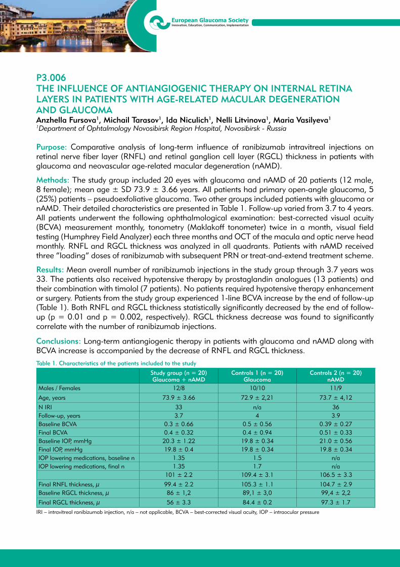
13th EGS CongressFortezza da Basso, Florence, Italy
19/22 May 2018
P3.006THE INFLUENCE OF ANTIANGIOGENIC THERAPY ON INTERNAL RETINA LAYERS IN PATIENTS WITH AGE-RELATED MACULAR DEGENERATION AND GLAUCOMA Anzhella Fursova1, Michail Tarasov1, Ida Niculich1, Nelli Litvinova1, Maria Vasilyeva1
1Department of Ophtalmology Novosibirsk Region Hospital, Novosibirsk - Russia
Purpose: Comparative analysis of long-term influence of ranibizumab intravitreal injections on retinal nerve fiber layer (RNFL) and retinal ganglion cell layer (RGCL) thickness in patients with glaucoma and neovascular age-related macular degeneration (nAMD).
Methods: The study group included 20 eyes with glaucoma and nAMD of 20 patients (12 male, 8 female); mean age ± SD 73.9 ± 3.66 years. All patients had primary open-angle glaucoma, 5 (25%) patients – pseudoexfoliative glaucoma. Two other groups included patients with glaucoma or nAMD. Their detailed characteristics are presented in Table 1. Follow-up varied from 3.7 to 4 years. All patients underwent the following ophthalmological examination: best-corrected visual acuity (BCVA) measurement monthly, tonometry (Maklakoff tonometer) twice in a month, visual field testing (Humphrey Field Analyzer) each three months and OCT of the macula and optic nerve head monthly. RNFL and RGCL thickness was analyzed in all quadrants. Patients with nAMD received three “loading” doses of ranibizumab with subsequent PRN or treat-and-extend treatment scheme.
Results: Mean overall number of ranibizumab injections in the study group through 3.7 years was 33. The patients also received hypotensive therapy by prostaglandin analogues (13 patients) and their combination with timolol (7 patients). No patients required hypotensive therapy enhancement or surgery. Patients from the study group experienced 1-line BCVA increase by the end of follow-up (Table 1). Both RNFL and RGCL thickness statistically significantly decreased by the end of follow-up (p = 0.01 and p = 0.002, respectively). RGCL thickness decrease was found to significantly correlate with the number of ranibizumab injections.
Conclusions: Long-term antiangiogenic therapy in patients with glaucoma and nAMD along with BCVA increase is accompanied by the decrease of RNFL and RGCL thickness.Table 1. Characteristics of the patients included to the study
Study group (n = 20)Glaucoma + nAMD
Controls 1 (n = 20) Glaucoma
Controls 2 (n = 20)nAMD
Males / Females 12/8 10/10 11/9
Age, years 73.9 ± 3.66 72.9 ± 2,21 73.7 ± 4,12
N IRI 33 n/a 36Follow-up, years 3.7 4 3.9Baseline BCVA 0.3 ± 0.66 0.5 ± 0.56 0.39 ± 0.27Final BCVA 0.4 ± 0.32 0.4 ± 0.94 0.51 ± 0.33Baseline IOP, mmHg 20.3 ± 1.22 19.8 ± 0.34 21.0 ± 0.56Final IOP, mmHg 19.8 ± 0.4 19.8 ± 0.34 19.8 ± 0.34IOP lowering medications, baseline n 1.35 1.5 n/aIOP lowering medications, final n 1.35 1.7 n/a
101 ± 2.2 109.4 ± 3.1 106.5 ± 3.3
Final RNFL thickness, μ 99.4 ± 2.2 105.3 ± 1.1 104.7 ± 2.9Baseline RGCL thickness, μ 86 ± 1,2 89,1 ± 3,0 99,4 ± 2,2
Final RGCL thickness, μ 56 ± 3.3 84.4 ± 0.2 97.3 ± 1.7 IRI – intravitreal ranibizumab injection, n/a – not applicable, BCVA – best-corrected visual acuity, IOP – intraocular pressure

13th EGS CongressFortezza da Basso, Florence, Italy
19/22 May 2018
P3.007DIFFERENCES IN THE RESPONSE OF THE INTRAOCULAR PRESSURE AFTER OZURDEX INJECTION ACCORDING TO THE USE OF PREVIOUS OCULAR ANTIHYPERTENSIVE TREATMENT OR THE TYPE OF GLAUCOMACristina Pujadas-Garcia1, Enriqueta Cantal2, Diana Mora1, Carlos Luis Moser3
1Department of Glaucoma, 2Department of Retina, 3Department of Cornea, CSI Hospitalet del LLobregat, Hospitalet del Llobregat - Spain
Purpose: Analyze if there is any variation in the intraocular pressure after Ozurdex injection in patients who are receiving treatment for ocular hypertension-related conditions (TOH) with special interest in those suffering primary open angle glaucoma (POAG) .
Methods: A cohort retrospective study was carried out including patients treated with Ozurdex during the period between May to December 2015. If bilateral treatment was performed, only the first eye treated was included. Eyes with vitrectomy or cataract extraction in the early six months of follow up were excluded. It was defined as corticosteroid responders (CR) if IOP > 5 mmHg above baseline and high corticosteroid responders (HCR) if IOP ≥ 30 mmHg.
Results: Of the 175 treated eyes, 125 eyes were included in the study. The mean IOP was 16.07 ± 2.0 mmHg and 30.4% used TOH. Considering the eyes with TOH (N 38), there were identified POAG in 16 eyes. It was observed CR in 44% (N 55) and HCR in 16% (N 20) of total eyes treated with Ozurdex, with statistic differences between the group with TOH and the one not previously treated (CR p 0.014 and HCR p 0.002). Of the POAG eyes, CR was observed in 87.5% (N 14) and HCR in 37.5% (N 6). There were statistic differences in CR prevalence between POAG versus TOH non POAG (p 0.006) but not in HCR (p 0.503).
Conclusion: The use of sustained release corticosteroids is becoming more frequent in the treatment of recurrent macular edema of multiple etiologies. The mild corticoid response can be observed in almost half of the patients treated, although hypertensive peaks above 30 mmHg are less frequent. However, a mild corticoid response in glaucoma patients may involve the progression of their underlying disease, so we should have special attention in this type of patients.

13th EGS CongressFortezza da Basso, Florence, Italy
19/22 May 2018
P3.008TRAIL TRACED THRESHOLD TEST (T4) ALGORITHM SHOWS REDUCED VARIABILITY COMPARED TO THE SWEDISH INTERACTIVE THRESHOLDING ALGORITHM (SITA)Bledi Petriti1, Marco Miranda1, Haogang Zhu1,2, Padraig J. Mulholland1,2, David P. Crabb3, Carol Bronze1, Roger S. Anderson1,2, David F. Garway-Heath1
1National Institute for Health Research (NIHR) Biomedical Research Centre at Moorfields Eye Hospital NHS Foundation Trust and UCL Institute of Ophthalmology, London - United Kingdom2Optometry and Vision Science Research Group, University of Ulster, Coleraine - United Kingdom3Division of Optometry & Visual Sciences, City University London, London - United Kingdom
Purpose: To compare the test-retest variability of a novel threshold algorithm (T4sm) for automated perimetry compared to SITA-Standard.
Method: We have developed a VF algorithm (T4sm) with two novel components: stimuli are modulated primarily in size (sm) rather than intensity and thresholds are determined by an algorithm (T4), which derives a probability function obtained from patient’s responses to all stimuli presented at the location of interest and anatomically related locations. Stimulus dynamic range and step units were matched between algorithms. Study eye inclusion criteria: glaucomatous VFs, BCVA ≥ 20/40, no ocular morbidity other than glaucoma or prior cataract surgery, reliable VF (false positive <15%), IOP < 30mmHg. Subjects performed T4sm and SITA VFs twice in the same order on two runs. Test sequence and test eye were block randomized. Analysis focused on the standard deviation (SD) of the pointwise test-retest difference of each strategy. Bland-Altman repeatability plots were created for each.
Results: Results of the first 40 out of 100 subjects. SITA MD -6.18 dB (SD 7.26; 5% -21.50 dB, 95% 0.96 dB). Mean age 67 (SD 11) years. Mean test time: SITA 5.40 (SD 1.07) mins, T4sm 5.16 (SD 0.53) mins. Mean absolute test retest differences for sensitivity values ≥ 26 dB for SITA and T4sm were 1.41 and 1.63 dB respectively, and for values < 26 dB, 5.63 and 2.95 dB respectively. Fig. 1 plots the SD of test retest difference against the mean sensitivity for every location. Results from SITA are in good agreement with previous estimates (Russell et al. 2012). Bland-Altman plots (Fig. 2) showed the 95% test retest limits of agreement to be ±13.4dB for SITA (mean difference 0.2 dB) and ± 11.5 dB for T4sm (mean difference 0.5 dB). The heteroscedasticity of test-retest variability is markedly reduced with T4 compared to SITA.
Figure 1. The SD of absolute differences between test and retest sensitivity at various sensitivity levels
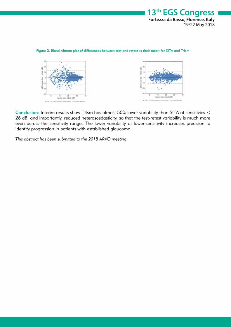
13th EGS CongressFortezza da Basso, Florence, Italy
19/22 May 2018
Figure 2. Bland-Altman plot of differences between test and retest vs their mean for SITA and T4sm
Conclusion: Interim results show T4sm has almost 50% lower variability than SITA at sensitivies < 26 dB, and importantly, reduced heteroscedasticity, so that the test-retest variability is much more even across the sensitivity range. The lower variability at lower-sensitivity increases precision to identify progression in patients with established glaucoma.
This abstract has been submitted to the 2018 ARVO meeting.

13th EGS CongressFortezza da Basso, Florence, Italy
19/22 May 2018
P3.009STUDY OF INTRAOCULAR PRESSURE AFTER IMPLANTATION OF A PHAKIC COLLAMER INTRAOCULAR LENS WITH A CENTRAL HOLEPedro Pablo Rodriguez-Calvo1, Ignacio Rodriguez-Una1, Luis Fernandez-Vega Cueto1, Carlos Lisa1, Andres Fernandez-Vega Cueto1, Jose F. Alfonso1
1Instituo Oftalmológico Fernandez-Vega, Oviedo - Spain
Purpose: To investigate the middle-term intraocular pressure (IOP) results after implantation of a posterior chamber collagen copolymer phakic intraocular lens (pIOL) (V4c Visian) with a central hole in patients with myopia.
Methods: This study enrolled patients who had implantation of a pIOL with a central hole for more natural aqueous flow, eliminating the need for neodymium:YAG iridotomy or peripheral iridectomy. IOP, central vault and adverse events were evaluated 1, 3, 6, 12 and 24 months postoperatively.
Results: The study enrolled 763 eyes (384 patients, 128 males and 256 females). Mean follow-up was 7.0 ± 7.2 months (range: 1 to 24 months). The mean IOP was 13.2 ± 2.1 mmHg preoperatively. Postoperatively, the mean IOP was 12.4 ± 1.7 mmHg at 1 month, 12.5 ± 1.8 mmHg at 3 months, 12.6 ± 1.3 mmHg at 6 months, 12.6 ± 1.4 mmHg at 12 months, and 12.7 ± 1.1 mmHg at 24 months. Only one case (0.13%) presented an increased IOP (> 21 mmHg) during the observation period. No pupillary block, or acute angle closure was recorded. IOP at the final follow-up visit was 12.8 ± 1.3 mmHg.
Conclusions: Implantation of central hole pIOL in myopic patients provided good and safe IOP outcomes throughout the 24-month observation period.

13th EGS CongressFortezza da Basso, Florence, Italy
19/22 May 2018
P3.010SYNDROME OF PIGMENT DISPERSION, THE RESULTS OF 10 YEARS OF FOLLOW-UPTatiana Iureva1
1Irkutsk Branch of S. Fyodorov Eye Microsurgery Federal State Autonomous Institution, Irkutsk State Medical Academy of Continuing Education, Irkutsk - Russia
Purpose: To assess the change in IOP level, degree of pigmentation of trabeculae and thickness of nerve fiber layer in patients with pigment dispersion syndrome 10 and more years after laser iridectomy.
Materials and Methods: 126 patients were examined, average age was 34.2 ± 6.8 years, clinical diagnosis of syndrome of pigment dispersion was established between 1996 and 2006, according to Iureva’s and Shchuko’s classification (2003), all patients are divided into 3 groups: 1st - preclinical stage (pathological irido-zonular contact according to UBM data, no exogenous pigmentation) -34 patients, 24.6 ± 4.7 years; 2nd initial clinical stage (pathological iridozonular contact, pigment dispersion of 1-2 degrees, single transillumination zones) - 46 patients, 34.2 ± 3.5 years; 3rd manifestation stage (pathological iridozonular contact according to UBM data, 3-4 pigment dispersion degree, multiple zones of transillumination, positive loading test with mydriatics) - 42 patients, 37.8 ± 6.4 years. 88 patients of 2nd and 3rd groups underwent laser iridectomy. UBM results, IOP, the degree of trabecular pigmentation in points (0-3), thickness of nerve fiber layer according to OCT data were compared with initial data.
Results: 36% patients in the first group had spontaneous elimination of inverse pupillary block, in 64% an exopigment appeared within 3-5 years, laser iridectomy was performed. In 100% of cases in second group after laser iridectomy iridozonular contact is absent according to UBM data. IOP did not change (14.89 ± 1.24 - 15.19 ± 1.76, p ≥ 0.05), degree of pigmentation decreased from 1.56 ± 0.62 to 1.19 ± 0.46, p < 0.05, thickness of nerve fiber layer decreased from 139.17 ± 7.54 to 128.87 ± 9.00 (p ≥ 0.05). That is, all patients had clinical recovery. Patients of third group had an IOP increase from 15.25 ± 2.55 to 17.90 ± 3.24 (p < 0.05), degree of pigmentation decreased from 3.5 ± 0.50 to 2.19 ± 0.32 (p < 0.05), thickness of nerve fiber layer decreased from 139.18 ± 7.54 to 120.10 ± 14.37 (p < 0.05).
Conclusion: After laser iridectomy complete elimination of inverse pupillary block was observed in all cases, degree of trabecular pigmentation decreased more than 2-fold during 10 years, iris pigmentary sheet was restored. Despite this, in 44% cases in patients with SPD in stage of manifestation the formation of pigment glaucoma occurred.

13th EGS CongressFortezza da Basso, Florence, Italy
19/22 May 2018
P3.011PROGRESSION OF VISUAL FIELD LOSS BEFORE AND AFTER TRABECULECTOMYShunsuke Kanayama1
1Kitasato University Department of Ophthalmology, Sagamihara-shi - Japan
Objective: To study the progression of visual field loss before and after trabeculectomy.
Methods: Thirty-one eyes in 29 glaucoma patients (mean age: 60.1 ± 14.8) were analysed pre- and post-trabeculectomy. Patients were analysed at least three times using Humphrey visual field tests (24-2 or 30-2 program), and classified according to the level of pre-operative intraocular pressure (IOP): ≥ 20 mmHg (H group) or < 20 mmHg (L group). Post-operative changes in the mean deviation slope (MDS) were used to determine whether the patients’ IOP had reached standard levels following surgery. Standard IOPs were defined as 12 mmHg (A) and 10 mmHg (B). Patients were then divided into those who reached the standard IOP levels post--operatively (D) and those who did not (I).
Results: In total, 21 eyes (68%) were classified as AD, and 14 eyes (45%) were classified as BD. In the H group, 10 eyes (50%) were classified as AD and 6 (30%), as BD. In the L group, 11 eyes (100%) were classified as AD, and 8 (73%), as BD. The MDS (dB/Y) showed a significant improvement in all groups post-trabeculectomy, changing from -3.1 (pre-operatively) to .03 (AD group), -0.3 (AI group), 0.1 (BD group), and -0.3 (BI group). The MDS did not differ significantly between the AD and AI, or BD and BI groups. In the H group, the MDS changed from -4.2 (pre-operatively), to 0.1 (AD group), -0.3 (AI group), 0.1 (BD group), and -0.2 (BI group), post-operatively. There was no significant difference between the AD and AI groups, or BD and BI groups. In the L group, the pre-operative MDS was -1.1 and changed to -0.2 (AD group) and 0.1 (BD group), and -1.0 (BI group) post-operatively.
Conclusions: We were able to slow the progression of visual field loss after trabeculectomy by decreasing intraocular pressure to < 12 mmHg. The results of the study also suggest that patients with a pre-operative IOP < 20 mmHg should be maintained at an IOP ≤ 10 mmHg.
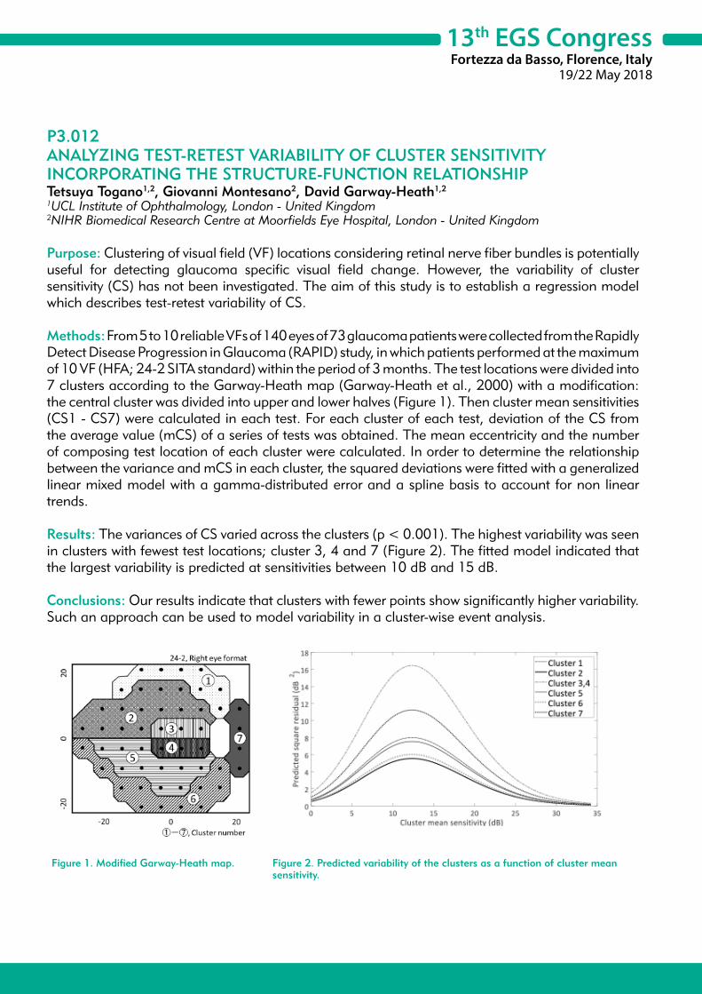
13th EGS CongressFortezza da Basso, Florence, Italy
19/22 May 2018
P3.012ANALYZING TEST-RETEST VARIABILITY OF CLUSTER SENSITIVITY INCORPORATING THE STRUCTURE-FUNCTION RELATIONSHIPTetsuya Togano1,2, Giovanni Montesano2, David Garway-Heath1,2
1UCL Institute of Ophthalmology, London - United Kingdom2NIHR Biomedical Research Centre at Moorfields Eye Hospital, London - United Kingdom
Purpose: Clustering of visual field (VF) locations considering retinal nerve fiber bundles is potentially useful for detecting glaucoma specific visual field change. However, the variability of cluster sensitivity (CS) has not been investigated. The aim of this study is to establish a regression model which describes test-retest variability of CS.
Methods: From 5 to 10 reliable VFs of 140 eyes of 73 glaucoma patients were collected from the Rapidly Detect Disease Progression in Glaucoma (RAPID) study, in which patients performed at the maximum of 10 VF (HFA; 24-2 SITA standard) within the period of 3 months. The test locations were divided into 7 clusters according to the Garway-Heath map (Garway-Heath et al., 2000) with a modification: the central cluster was divided into upper and lower halves (Figure 1). Then cluster mean sensitivities (CS1 - CS7) were calculated in each test. For each cluster of each test, deviation of the CS from the average value (mCS) of a series of tests was obtained. The mean eccentricity and the number of composing test location of each cluster were calculated. In order to determine the relationship between the variance and mCS in each cluster, the squared deviations were fitted with a generalized linear mixed model with a gamma-distributed error and a spline basis to account for non linear trends.
Results: The variances of CS varied across the clusters (p < 0.001). The highest variability was seen in clusters with fewest test locations; cluster 3, 4 and 7 (Figure 2). The fitted model indicated that the largest variability is predicted at sensitivities between 10 dB and 15 dB.
Conclusions: Our results indicate that clusters with fewer points show significantly higher variability. Such an approach can be used to model variability in a cluster-wise event analysis.
Figure 1. Modified Garway-Heath map. Figure 2. Predicted variability of the clusters as a function of cluster mean sensitivity.
13th EGS CongressFortezza da Basso, Florence, Italy
19/22 May 2018
P3.013CLINICAL FINDINGS OF GLAUCOMATOUS PATIENTS WITH NEWLY DIAGNOSED BRAIN TUMOURS: A TERTIARY REFERRAL CENTRE-BASED RETROSPECTIVE STUDYGiacinto Triolo1, Anca G. Nita1
1Royal Victoria Infirmary, Newcastle upon Tyne - United Kingdom
Purpose: To report the clinical findings of glaucomatous patients with newly diagnosed brain tumours in a tertiary referral hospital.
Methods: This study was carried out at the Royal Victoria Infirmary (RVI) in Newcastle upon Tyne, United Kingdom. The diagnostic codes corresponding to generic diagnosis of “glaucoma” (Glc) and “brain tumour” (BT) were input in the hospital electronic medical records (EMR) system to search patients diagnosed with Glc and BT. A list of patients with both diagnoses was obtained and their notes were retrospectively analysed.
Results: Sixteen patients with diagnosis of both Glc and BT were found since 2002. Nine out of 16 patients were excluded because the diagnosis of BT preceded that of Glc, or because Glc was in the patients’ medical history with no documented ophthalmological notes. Out of 7 patients included, 5 were male. The mean (M±SD) age at diagnosis of Glc and BT was 63.14 ± 7.41 and 72.43 ± 6.30 months, respectively. The MSD follow up length was 107.14 ± 77.43 months. Two patients had ocular hypertension, 4 primary open angle glaucoma (POAG), and 1 pigment dispersion glaucoma. Four patients had biopsy-based diagnosis of glioblastoma multiforme (GBM), 1 glioma, 1 anaplastic astrocytoma. One patient did not receive biopsy because of extremely poor prognosis. Symptoms that led to the diagnosis of BT were not visually significant and were as follows: 5 patients had seizures and behaviour/emotional changes, 1 had speech problems, 1 had droopy mouth with slurred speech. At baseline, 4 patients had typical glaucomatous visual field (VF) defects, 3 had full VFs. At the last follow up visit, only 1 patient developed a VF defect (homonimous hemianopia) related to the BT, whereas 3 patients showed progressive glaucomatous VF defects because of poor IOP control.
Conclusions: Among thousands of patients who attended a tertiary referral glaucoma centre since 2002, only 7 patients were diagnosed with BT afterwards. In our series, more frequently patients were male, had POAG, developed GBM, had no visual symptoms, and the presence of a BT did not significantly affect their visual outcomes.

13th EGS CongressFortezza da Basso, Florence, Italy
19/22 May 2018
P3.014A GLAUCOMA PROGRESSION RISK ASSESSMENT HAND-HELD CHART. FEASIBILITY AND PRELIMINARY VALIDATIONStefano A. Gandolfi1, Romeo Altafini2, Paolo Brusini3, Marco Ciancaglini4, Paolo Frezzotti5, Michele Iester6, Giorgio Marchini7, Gianluca Manni8, Enrico Martini9, Giovanni Milano10, Andrea Perdicchi11, Antonio Rapisarda12, Teresa Rolle13, Gemma Rossi10, Tommaso Salgarello14, Maurizio Uva15, Luca Rossetti16, Luca Agnifili17, Michele Figus18, Luciano Quaranta19, Paolo Bettin20
1Eye, Dpt, University of Parma, Parma - Italy2Eye Dpt, Community Hospital, Dolo - Italy3Eye Clinic, Città di Udine, Udine - Italy4Eye Dpt., Unuiversity of L’Aquila, L’Aquila - Italy Eye Dpt, University of Siena, Siena - Italy6Eye Dpt, University of Genova, Genova - Italy7Eye Dpt, University of Verona, Verona - Italy8Eye Dpt., University Roma Tor Vergata, Rome - Italy9Eye Clinic, Community Hospital, Sassuolo - Italy10Eye Dpt., University of Pavia, Pavia - Italy11Eye Dpt., University of Rome La Sapienza (Sant’Andrea Hospital), Rome - Italy,12Eye Clinic, Policlinico Morgagni, Catania - Italy13Eye Dpt., University of Turin, Turin - Italy14Eye Dpt., Catholic University of Rome, Rome - Italy15 Eye Dpt.,,University of Catania, Catania - Italy16 Eye Dpt., University of Milan, San Paolo Hospital, Milan - Italy17 Eye Dpt., University of Chieti, Chieti - Italy18 Eye Dpt., University of Pisa, Pisa - Italy19 Eye Dpt., University of Brescia, Brescia - Italy20 Eye Dpt., San Raffaele University Hospital, Milan - Italy
Purpose: To test feasibility and validity of a novel hand-held glaucoma progression risk assessment chart.
Methods: A hand-held glaucoma progression risk assessment chart was developed on a consensus-based approach among several glaucoma specialists. Similarly to what described in other specialties, a table reporting (a) the main risk-factor for progression (i.e. IOP) on the horizontal axis and (b) concurrent risk factors for progression, in a relevance-increasing rank, on the vertical axis was designed. A 4-color-code (green yellow, orange, red) was assigned to each cell of the table, according to the increasing risk for progression (see table 1) . On the back side of this chart, the complimentary table, suggesting a color-driven follow up timing and procedures, is also displayed (table 2) .
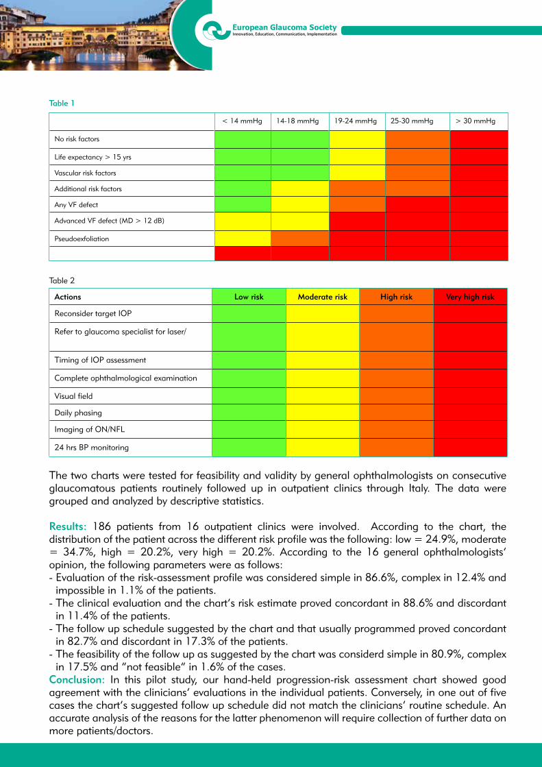
13th EGS CongressFortezza da Basso, Florence, Italy
19/22 May 2018
Table 1
< 14 mmHg 14-18 mmHg 19-24 mmHg 25-30 mmHg > 30 mmHg
No risk factors
Life expectancy > 15 yrs
Vascular risk factors
Additional risk factors
Any VF defect
Advanced VF defect (MD > 12 dB)
Pseudoexfoliation
Table 2
Actions Low risk Moderate risk High risk Very high risk
Reconsider target IOP
Refer to glaucoma specialist for laser/
Timing of IOP assessment
Complete ophthalmological examination
Visual field
Daily phasing
Imaging of ON/NFL
24 hrs BP monitoring
The two charts were tested for feasibility and validity by general ophthalmologists on consecutive glaucomatous patients routinely followed up in outpatient clinics through Italy. The data were grouped and analyzed by descriptive statistics.
Results: 186 patients from 16 outpatient clinics were involved. According to the chart, the distribution of the patient across the different risk profile was the following: low = 24.9%, moderate = 34.7%, high = 20.2%, very high = 20.2%. According to the 16 general ophthalmologists’ opinion, the following parameters were as follows:- Evaluation of the risk-assessment profile was considered simple in 86.6%, complex in 12.4% and
impossible in 1.1% of the patients. - The clinical evaluation and the chart’s risk estimate proved concordant in 88.6% and discordant
in 11.4% of the patients.- The follow up schedule suggested by the chart and that usually programmed proved concordant
in 82.7% and discordant in 17.3% of the patients. - The feasibility of the follow up as suggested by the chart was considerd simple in 80.9%, complex
in 17.5% and “not feasible” in 1.6% of the cases.Conclusion: In this pilot study, our hand-held progression-risk assessment chart showed good agreement with the clinicians’ evaluations in the individual patients. Conversely, in one out of five cases the chart’s suggested follow up schedule did not match the clinicians’ routine schedule. An accurate analysis of the reasons for the latter phenomenon will require collection of further data on more patients/doctors.

13th EGS CongressFortezza da Basso, Florence, Italy
19/22 May 2018
P3.015INFRARED PUPILLOMETRY ASSESSING PUPIL DILATION AND GLAUCOMA SEVERITY IN THE PSEUDOEXFOLIATION SYNDROMEAdarsh George1, Allan Bank2, Ian Francis2, Ashish Agar2
1Faculty of Medicine, University of New South Wales, Sydney - Australia2Department of Ophthalmology, Prince of Wales Hospital, Sydney - Australia
Purpose: Poor pupillary dilation has long been known to be a hallmark clinical feature in the Pseudoexfoliation (PXF) syndrome. Previous studies have demonstrated that very poor dilation is a risk factor for progression to PXF glaucoma. However, to date, no studies have correlated the degree of pupil dilation with the severity of glaucoma in PXF patients. This study aimed to assess the association between pharmacological pupil dilation and glaucoma parameters in PXF patients, and thus to evaluate its sensitivity as a clinical marker of disease progression in this group.
Methods: This study evaluated eyes of patients with and without glaucoma presenting to Ophthalmology clinics with clinically evident PXF. The patients’ pupils were pharmacologically dilated and their vertical pupil diameter was measured with an infrared handheld pupilometer (Neuroptics NPi-200, Irvine California). Pupil dilation was graded as either ‘Poor’ (< 5 mm), ‘Moderate’ (5-6 mm) or ‘Good’ (> 6 mm), based on the final pupil size. The association of dilation with glaucomatous changes or severity was assessed using a range of quantitative parameters, including visual field Mean Deviation (MD), Visual Field Index (VFI), Corrected Distance Visual Acuity (CDVA), cup-to-disc (C/D) ratio, Retinal Nerve Fibre Layer Thickness (RNFL), Intraocular Pressure (IOP) and glaucoma medications.
Results: 72 participants were included. Of the 144 eyes, 113 had PXF and 67 had glaucoma. Eyes with PXF dilated less than non-PXF eyes and this was statistically significant (p < 0.05). PXF eyes were also significantly more likely to have glaucoma than non-PXF eyes (p < 0.005). Eyes that had already been diagnosed with glaucoma, regardless of the presence of PXF, dilated significantly less than non-glaucomatous eyes (p < 0.005). Significant associations were found between the grade of pupil dilation and MD, VFI and CDVA (p < 0.05 in all cases). Eyes with pupils that dilated less had worse outcomes in these measures.
Conclusions: Pseudoexfoliation patients with poor pharmacological pupil dilation are at an increased risk of both developing glaucoma and increased glaucomatous severity.
13th EGS CongressFortezza da Basso, Florence, Italy
19/22 May 2018
P3.016COMPARISON OF VISUAL FIELD RESULTS WITHIN 10 DEGREE BETWEEN 24PLUS AND 24-2 VISUAL FIELD TEST LOCATIONS IN PATIENTS WITH MODERATE AND SEVERE STAGE GLAUCOMAHiroki Nomoto1, Chota Matsumoto1, Tairo Kimura2, Sachiko Okuyama1, Yoshikazu Shimomura1
1Department of Ophthalmology, Kindai University, Faculty of Medicine, Osaka-sayama - Japan2Ueno Eye Clinic, Tokyo - Japan
Purpose: Assessing precise central visual field (VF) is crucial especially in progressed glaucoma patients. “24plus” is a new VF test pattern characterized by added 24 test points to central 10 degree of ordinal 24-2 test locations. The purpose of this study was to compare VF test results within 10 degree between 24 plus and 24-2 test patterns in patients with moderate and severe stage glaucoma.
Method: This study included 208 eyes of 158 progressed glaucoma (mean deviation (MD) < -9 dB) patients who underwent 24plus test using head mounted perimeter imo (Matsumoto C, et al. PLos One. 2016). Number of test points within 10 degree were 30 points in 24plus and 12 points in 24-2. Abnormal and normal test points were defined by pattern plot probability level < 1% and > 5%, respectively. Residual VF was estimated by the number of normal test points. The number of normal, abnormal test points, MD10 (MD within 10 degree area) were calculated. Linear regression analysis was used to assess a relationship between abnormal test points and MD10, number of normal test points of 24plus and 24-2 pattern.
Results: 24plus MD10 was significant better than 24-2 MD10 (-15.2 ± 6.6dB vs -16.6 ± 5.8dB, p = 0.02) and number of normal test points with 24plus (11.7 ± 5.8) were larger than 24-2 (4.8 ± 2.3) (p < 0.001). (Linear regression analysis, slope: 1.95, intercept: 2.38, p < 0.001). Slope of a line with 24plus MD10 and number of 24plus abnormal points were significantly steeper than 24-2 slope (-0.86 vs -0.39, p < 0.001).
Conclusion: 24plus test pattern detected more detailed deterioration and residual of central area of VF than 24-2 test pattern in progressed stage glaucoma patients. Our results suggest that 24plus test could be a useful examination for detecting glaucomatous VF changes with severe glaucoma patients.
13th EGS CongressFortezza da Basso, Florence, Italy
19/22 May 2018
P3.017COMPARISON OF TRABECULECTOMY AND GEL STENT XEN® IN CHRONIC OPEN-ANGLE GLAUCOMA: A RETROSPECTIVE STUDYJavier Alejandro Salinas López1, Maria Teresa Marcos Parra2
1Department of Ophthalmology, Hospital General Universitario de Alicante, Alicante - Spain2Glaucoma Unit, Department of Ophthalmology, Hospital General de Alicante, Alicante - Spain
Purpose: To report our experience using Trabeculectomy and XENÒ gel stent in chronic open-angle glaucoma (COAG) surgical management.
Methods: A retrospective six-month follow-up study was conducted. 89 patients (129 eyes) underwent to glaucoma surgery (trabeculectomy or XENÒ gel stent implantation; in combination with cataract/phacoemulsification surgery or glaucoma surgery alone) between 2013 and 2017 were selected. Surgery was performed by one surgeon. They were divided in 6 groups: Group I underwent XENÒ gel stent implantation alone (15 eyes of 89 patients), Group II underwent XENÒ gel stent implantation combined with cataract/phacoemulsification surgery (42 eyes of 89 patients), Group III underwent all XENÒ gel stent implantation in phacoemulsification combination or alone procedure (57 eyes of 89 patients), Group 4 underwent trabeculectomy alone (41 eyes of 89 patients), Group 5 underwent trabeculectomy combined with cataract/phacoemulsification surgery (31 eyes of 89 patients) and Group 6 underwent all trabeculectomy in phacoemulsification combination or alone procedure (72 eyes of 89 patients). Factors including gender, age, number of glaucoma medication classes used before and six-month after surgery, intraocular pressure (IOP) before and after surgery, anterior chamber (AC) flattening, hyphema, bleb leaks, bleb-related infection, subconjunctival fibrosis in the bleb, Tenon’s capsule cysts (TCCs), bleb needling, surgical revision (bleb surgical revision, surgical revision for implant extrusion, new filtering surgery) were compared between groups.
Results: All groups showed a successful IOP reduction, defined as ≥ 20%: Group I showed an IOP reduction of 25.5 (SD 20.5)%; Group II 22,8 (SD 24.7)%; Group III 23.6 (SD 23.6)%; Group IV 43.2 (SD 25.0)%; Group V 37.5 (SD 22.4)% and Group VI 40.9 (23.9)%. XEN groups showed lower AC flattening (1.8%) and hyphema (8.8%) rates than trabeculectomy groups (27.8% and 37.5% respectively), but higher subconjunctival fibrosis in the bleb (12.3% versus 2.8%) and bleb needling (21.1% versus 4.2%) rates.
Conclusions: XEN® gel stent is a new implant that produces an important IOP reduction, in general terms, is an effective and safe technique that requires a learning curve to minimize complications.

13th EGS CongressFortezza da Basso, Florence, Italy
19/22 May 2018
P3.018INTRAOCULAR PRESSURE ELEVATION FOLLOWING PARS PLANA VITRECTOMY FOR DIABETIC RETINOPATHYZeynep Kayaarasi Ozturker1, Eylem Yaman Pinarci1, Ahmet Akman2 1Baskent University, Faculty of Medicine, Ophthalmology Department, Istanbul - Turkey2Baskent University, Faculty of Medicine, Ophthalmology Department, Ankara - Turkey
Purpose: To evaluate the risk factors for postvitrectomy intraocular pressure (IOP) elevation in diabetes.
Methods: Medical records of 104 eyes of 92 diabetic patients undergoing vitrectomy were reviewed. Preoperative and postoperative IOPs at 1st week, 1st month and 3rd month were recorded. Baseline risk characteristics including demographics of the patients, lens status, diabetes, and IOP elevation in relation to surgical procedures and tamponades were evaluated.
Results: Mean baseline IOP was 14.7 mmHg, and mean final IOP was 17.4 mmHg (p < 0.05). Seventeen eyes (16.3%) of 104 study eyes had an IOP ≥ 21 mmHg at last visit. No study eye developed open-angle glaucoma or required laser, or surgical treatment for glaucoma. Patients’ blood glucose level and active retinal neovascularization resulted as risks for IOP increase (p = 0.07, p = 0.08, respectively). External buckling procedures and oil tamponade for proliferative diabetic retinopathy tractions carried no risks for IOP increase. Pseudophakia was not associated with significantly increased IOP.
Conclusion: In this series, vitrectomy appears to increase IOP in diabetic patients with uncontrolled blood glucose level and proliferative retinal disease.

13th EGS CongressFortezza da Basso, Florence, Italy
19/22 May 2018
P3.019COMPARISON OF ANTERIOR CHAMBER ANGLE AND IRIS MEASUREMENTS BY ANTERIOR SEGMENT OPTICAL COHERENCE TOMOGRAPHY IN PRIMARY JUVENILE GLAUCOMA AND NORMAL SUBJECTSBerna Basarir1, Isil Pasaoglu1, Cigdem Altan1, Banu Solmaz Satana1, Asli Inal1, Funda Ebru Onmez1, Tekin Yasar1, Muhittin Taskapili1
1UHS, Beyoglu Eye Research and Training Hospital, Istanbul - Turkey
Purpose: To evaluate quantitatively the iridocorneal angle and iris parameters measured by anterior segment optical coherence tomography (AS-OCT) in primary juvenile glaucoma and to compare them with healthy eyes.
Methods: 13 eyes of seven patients with primary juvenile glaucoma were enrolled to the study (six female and one male). 15 eyes of 15 normal age and gender matched subjects were also included. Exclusion criteria consisted of, history of intraocular surgery, laser or trauma, any other anterior segment abnormalities such as corneal abnormalities, and intumescent cataract which may effect measurements. The iridocorneal angle and iris parameters were measured by AS-OCT (Visante OCT 3.0 Model 1000 Carl Zeiss Meditec, Inc.).
Results: The mean age of the patients was 31.15 ± 12.46. Best corrected visual acuity was 0.74 ± 0.4. Intraocular pressure was 17.33 ± 4.5 mmHg. In temporal quadrant, the mean angle opening distance at 500 μm anterior to the scleral spur (AOD 500) was 1.19 ± 0.2 mm, the mean angle opening distance at 750 μm anterior to the scleral spur (AOD 750) was 1.57 ± 0.64 mm, trabecular-iris space area at 500 μm (TISA 500) 0.38 ± 0.17 mm2, at 750 μm (TISA 750) was 0.74 ± 0.32 mm2, scleral spur angle was 63.62 ± 14.08°. In nasal quadrant AOD 500 was 1.07 ± 0.49 mm, AOD 750 was 1.35 ± 0.52 mm, TISA 500 0.35 ± 0.19 mm2, at 750 μm was 1.35 ± 0.52 mm2, scleral spur angle was 59.86 ± 0.14°. Iris shape was concave in nine eyes and convex in four eyes. The mean iris bowing was in temporal quadrant -0.25 ± 0.28 mm and in nasal quadrant -0.27 ± 0.23 mm. The iris thickness in the middle of iris in temporal quadrant was 0.27 ± 0.1 mm and 0.30 ± 0.8 mm in nasal quadrant. All anterior chamber biometric parameters except anterior chamber depth were significantly larger in eyes with juvenile glaucoma than in healthy subjects’ eyes (p < 0.05). There were statistically significant differences in iris thickness at its middle part, iris bowing and iris shape between the eyes with juvenile glaucoma and healthy subjects (p < 0.05).
Conclusions: High intraocular pressure can be concluded by iris concavity, iris thinning, iris bowing in eyes with primary juvenile glaucoma. This atrophy and configuration changes in the iris may be responsible for the widening of the iridocorneal angle parameters in AS-OCT.
13th EGS CongressFortezza da Basso, Florence, Italy
19/22 May 2018
P3.020EVALUATION OF FUNCTIONAL FILTERING BLEB USING OPTICAL COHERENCE TOMOGRAPHY ANGIOGRAPHYJe Hyun Seo1, Keun Heung Park2, Ye An Kim3, Young Lee4
1Department of Ophthalmology, Pusan National University Yangsan Hospital, Yangsan - Republic of Korea2Department of Ophthalmology, Pusan National University School of Medicine, Busan - Republic of Korea3Division of Endocrinology, Department of Internal Medicine, Veterans Health Service Medical Center, Seoul - Republic of Korea4Veterans Medical Research Institute, Veterans Health Service Medical Center, Seoul - Republic of Korea
Purpose: To investigate the bleb evaluation using optical coherence tomography angiography (OCT-A) and to compare vascularity parameters with conventional bleb grading system.
Methods: Ninety-six eyes of 96 glaucoma patients who underwent mitomycin C augmented trabeculectomy were enrolled in this study. The subjects were conducted OCT-A (DRI, Topcon) external mode and anterior segment photography for bleb evaluation. For bleb vascularity evaluation, masked observer carefully draw bleb area of original OCT-A image using semi-automated program which calculates color density, bright density, and threshold density of selected area. Also, a masked observer classified bleb vessel grades using Indiana Bleb Appearance Grading Scale (IBAGS) and Moorfields Bleb Grading System (MBGS). The vascularity parameters using OCT-A were compared the results by IBAGS and MBGS. In addition, correlation analysis was performed intraocular pressure (IOP) and vascularity parameters of bleb.
Results: Vessel density measured by OCT-A showed excellent inter-, intra-observer reproducibility. Color density, bright density, and threshold density showed positive correlation with vascularity gradings of IBAGS and MBGS. Color density, bright density, and threshold density using OCT-A were correlated with IOP, whereas vessel gradings of IBAGS and MBGS did not show relation with IOP.
Conclusions: Bleb evaluation using OCT-A can evaluate vessel vascularity not inferior to slit lamp based grading system. Potentially, it could provide objective and quantitative vessel parameters of bleb evaluation after trabeculectomy.
13th EGS CongressFortezza da Basso, Florence, Italy
19/22 May 2018
P3.021CHANGES IN CORNEAL PULSE CHARACTERISTIC AFTER CANALOPLASTY: A 12-MONTH FOLLOW-UP STUDYMonika Danielewska1, Michal Placek1, Aleksandra Kicinska2, Katarzyna Lewczuk2, Marek Rekas2
1Department of Biomedical Engineering, Wroclaw University of Science and Technology, Wroclaw - Poland 2Department of Ophthalmology, Military Institute of Medicine, Warsaw - Poland
Purpose: To assess the effect of canaloplasty on changes in spectral parameters of the corneal pulse (CP) signal for primary open-angle glaucoma patients during 12 months of follow-up.
Methods: Twelve eyes of 12 patients who underwent canaloplasty were included. Preoperative wash-out from antiglaucoma medications was done. Standard ophthalmological examination was conducted at pre-washout, on the day of surgery (post-washout), and at 3, 6, and 12 months after surgery. Additionally, the CP signal was continuously recorded using non-contact ultrasonic transducer. The CP and CP velocity (CPV) signals were preprocessed and parameters related to the amplitudes of their first three harmonics were estimated in spectral domain. Next, changes of the considered parameters in time were tested using a repeated-measure analysis of variance based on the general linear model.
Results: At post-washout, intraocular pressure (IOP) reached the highest value of 23.5 ± 1.0 mmHg (mean ± SEM). At 3, 6, and 12 months after canaloplasty, IOP significantly decreased to 14.0 ± 1.2 (p = 0.008), 13.2 ± 0.8 (p < 0.001), and 14.6 ± 1.0 mmHg (p = 0.007), respectively, in relation to pre-washout 17.4 ± 1.0 mmHg. Statistically significant changes in the amplitude of the 3rd harmonic normalized with respect to that of the 1st harmonic of both the CP (ACP3) and CPV (ACPV3) signals were observed between pre-washout and the 3-month post-op (p = 0.020 and p = 0.017, respectively). Significant differences in those parameters were also seen between the 3- and 6-month post-ops (ACP3, p = 0.024 and ACPV3, p = 0.019) and between the 3- and 12-month post-ops (ACP3, p = 0.030 and ACPV3, p = 0.035). In addition, ACPV3 and amplitudes of the 2nd harmonic (ACP2 and ACPV2) were found to significantly change between pre- and post-washout (p = 0.048, p = 0.034, and p = 0.033, respectively). At 12-month follow-up, one patient required glaucoma medication.
Conclusions: The higher amplitudes of the 2nd and 3rd harmonic in the CP and CPV spectra reflects higher corneal stiffness corresponding to an increase in IOP values and even more to the tensioning of the suture inserted into the Schlemm’s canal lasting up to 3 months after surgery. Spectral analysis of corneal pulsation complements the existing knowledge about the effect of canaloplasty on the stiffness of cornea.
13th EGS CongressFortezza da Basso, Florence, Italy
19/22 May 2018
P3.022COMPARISON OF IOP CHANGE AFTER CATARACT SURGERY IN NORMAL AND PATIENTS WITH OPEN ANGLE GLAUCOMAHong Young Jae1, Jung Hee In1, Don Kyung Kim1, So Yeon Lee1
1Glaucoma Center, Nune Eye Hospital, Seoul - Republic of Korea
Purpose: To evaluate intraocular pressure(IOP) after phacoemulsification in patients with primary open-angle glaucoma(OAG), and examine the association of variables to IOP changes.
Methods: 157 eyes of Normal controls and 197 eyes of open-angle glaucoma patients undergoing phacoemulsification by a single surgeon between January 1, 2014 and August 31, 2015 were evaluated.IOP was measured preoperative, 1 month and 6 months postoperatively in normal, OAG patients. Patient charts were reviewed to obtain demographic information, preoperative glaucoma medications, severity and treatment measures, and preoperative and postoperative IOP.
Results: In OAG patients, the average preoperative IOP of 14.5 ± 4.0 mmHg elevated to 16.7 ± 7.1 mmHg at 1 day postoperatively, substantial IOP elevation is 20 eyes (10%), High IOP elevation was 12 eyes (6%). In normal controls, the average preoperative IOP of 14.6 ± 2.9 mmHg elevated to 16.1 ± 4.8 mmHg at 1 day postoperatively, substantial IOP elevation is 12 eyes (8%), High IOP elevation was 1 eye (1%). There was statistically significant difference in IOP change at 6 months postoperatively between normal control and OAG (OAG = -1.06 ± 4.0 mmHg, normal control = -2.61± 2.7 mmHg) (p = 0.018).
Conclusions: OAG patients may experience the IOP elevation after cataract surgery compared with normal eyes undergoing cataract surgery. A sizeable proportion of glaucoma patients with open angles undergoing phacoemulsification experienced an increase in IOP or required more aggressive treatment to control IOP postoperatively.
13th EGS CongressFortezza da Basso, Florence, Italy
19/22 May 2018
P3.023REGIONAL DIFFERENCES IN INTRAOCULAR PRESSURE SEASONAL VARIATION IN PATIENTS WITH PRIMARY OPEN-ANGLE GLAUCOMAMiho Shimizu1, Yoko Ikeda2, Kazuhiko Mori3, Kengo Yoshii4, Morio Ueno4, Chie Sotozono3, Shigeru Kinoshita5
1Sapporo City General Hospital, Sapporo - Japan2Oike-Ikeda Eye Clinic, Kyoto - Japan3Department of Ophthalmology, Kyoto Prefectural University of Medicine, Kyoto - Japan4Department of Medical Statistics, Kyoto Prefectural University of Medicine, Kyoto - Japan5Department of Frontier Medical Science and Technology for Ophthalmology, Kyoto - Japan
Purpose: In Japan, there are four distinct seasons,which produce a seasonal variation (SV) of intraocular pressure (IOP) (SV-IOP). SV-IOP is caused by numerous factors, including climate temperature. The city of Sapporo is located in Hokkaido (latitude 43 degree North), the most northern region of Japan, while the city of Kyoto is located in more central region of Japan(latitude 35 degree N). The seasonal climates of these two cities are distinctly different. In this study, we investigated the SV-IOP differences between the two cities in Japanese primary open-angle glaucoma (POAG) patients.
Methods: This study involved 515 Japanese POAG patients who continued with the same medication throughout the 12 months of 2016; 109 patients (46 males and 63 females, mean age: 72.2 ± 9.3 years) seen at Sapporo City General Hospital, Sapporo, Japan, and 406 patients (173 males and 233 females, mean age: 68.4 ± 11.8 years) seen at Oike-Ikeda Eye Clinic, Kyoto, Japan in whom IOP could measure 4 times per season; i.e., spring (March through May, Season 1), summer (June through August, Season 2), fall (September through November, Season 3), and winter (December through February, Season 4). IOP was measured by Goldmann applanation tonometer. In all 4 seasons, the seasonal IOP fluctuation was diagnosed as highest IOP-lowest IOP. The IOP in each season was evaluated using repeated ANOVA, with Bonferroni correction and the Witch t test.
Results: The mean IOP in Seasons 1-4 were 13.9 ± 3.0, 13.7 ± 2.8, 14.1 ± 3.0, and 13.6 ± 2.7 mmHg, respectively, in Sapporo, and 12.9 ± 3.2, 12.3 ± 2.9, 12.4 ± 3.0, and 12.9 ± 3.0 mmHg, respectively, in Kyoto. There was no significant difference in each season’s IOP in Sapporo, however, there was significant difference in the 4 seasons in Kyoto. There was no significant difference in the IOP seasonal fluctuation between Sapporo (fall-winter) and Kyoto (winter-summer).
Conclusions: There was significant SV-IOP in Kyoto, however, no significant differences of SV-IOP in Sapporo.
13th EGS CongressFortezza da Basso, Florence, Italy
19/22 May 2018
P3.024FOLLOW UP OF CORRELATION BETWEEN SQUARE ROOT OF LOSS VARIANCE AND MEAN DEFECT IN PRIMARY OPEN ANGLE GLAUCOMADragan Janjic1, J. Janjic1
1Department of Ophthalmology, General Hospital, Pozarevac - Serbia
Purpose: To follow-up the correlation between perimetric indices square root of loss variance (sLV) and mean defect (MD) in different stages of primary open angle glaucoma (POAG).
Methods: A group of 82 visual fields suffering from POAG,obtained by Octopus 123 g1x,TOP, have been divided in three subgroups owing to MD values according to Hodapp classification 1. mild group +2.00dB < MD <+6.00dB, 2. moderate group +6.0 < MD < 12.00dB and 3. advanced group +12.00dB < MD. For each visual field group the average values of MD and sLV with standard deviation (SD) have been established. The relationship between mentioned indices has been established with statistical method of correlation and expressed as numerical Pearson’r. Results: In the mentioned POAG groups(mild,moderate and advanced), the average values of MD = +3.15 (SD = 1.25); +8.74dB (SD = 2.0); +23.4 (SD = 1.9) and average values of sLV = 4.22 (SD = 0.71); 6.47 (SD = 40.94); 5.33 (SD = 1.14). Correlative coefficient between perimetric indices sLV and MD in mild group has been determined as positive low/little connection (r = +0.24). In moderate group,the correlation between mentioned variables has been determined as negative low,little connection (r = -0.23). In advanced group, the correlation between mentioned variables has been determined as negative moderate,bit connectivity (r = -0.64).
Conclusion: In the course of POAG dissease, the correlation between perimetric indices MD and sLV is variable. It’s conditional by different preponderance of localized over diffuse type of visual field damage during glaucoma dissease.

13th EGS CongressFortezza da Basso, Florence, Italy
19/22 May 2018
P3.025CHOROIDAL THICKNESS CHANGES AFTER DISCONTINUING TOPICAL TREATMENT WITH PROSTAGLANDIN ANALOGUES IN PREPERIMETRIC OPEN ANGLE GLAUCOMAJavier Iturria Soler1, Emma Beatriz Ausin Gonzalez1, Jose Maria Ruiz Moreno1, Marta Sanchez Dehesa Saez1, Vanesa Antonia Gerena Arévalo1, Ana Royuela2, Esther Casado López De La Franca1
1Deparment of Ophthalmology, Puerta de Hierro hospital, Majadahonda - Spain2Statistics department, Puerta de Hierro Hospital, Majadahonda - Spain
Purpose: To compare the macular choroidal thickness (CT) profile in preperimetric open angle glaucoma (POAG) subjects before and after treatment with topical prostaglandin analogues (PGs) using swept source optical coherence tomography (SS-OCT).
Methods: A total of 23 eyes diagnosed with POAG were treated with topical PGs. For functional evaluation, mean deviation (MD) was measured using 24-2 SITA-Standard perimetry Humphrey® (Zeiss). Horizontal and vertical CT profile was created measuring subfoveal choroidal thickness (SFCT) from the posterior edge of the retinal pigmentary epithelium to the choroidal-scleral junction. Three determinations were performed at sucesive points 1000 μm nasal, five more 1000 μm temporal to the fovea; five more points superior to the fovea and five more inferior to the fovea. Two observers determined CT independently. All these eyes were re-evaluated three months after discontinuing the topical treatment with PGs, and changes in CT profile were measured.
Results: Mean age was 63 ± 10 years. Mean pachymetry was 551.04 ± 24.04 μm. Mean MD was -0.911 ± 1.18 dB and -0.087 ± 1.11 dB before and after discontinuing topical treatment with PGs respectively. We have found a decrease in CT in all the measured points with statistical significance, except in the three nasal ones where we find an increase of CT, without statistical significance (nasal 1: +40 ± 14.02 μm; p = 0.36; nasal 2: +24 ± 4.03 μm; P=0.15 and nasal 3: +8 ± 1.05 μm; p = 0.80). The agreement between two observers was excellent (ICC > 0.90), except in inferior vertical sectors (ICC: 0.80).
Conclusions: Discontinuing the topical treatment with PGs induce a statistically significant decrease in CT profile except in the nasal sector. Longer studies with more number of patients are necessary to confirm our findings and to determine if clinical correspondence exits.

13th EGS CongressFortezza da Basso, Florence, Italy
19/22 May 2018
P3.026GIANT BLEB POST-XEN IMPLANT: A FOLLOW-UP WITH OCT OF THE ANTERIOR SEGMENT OF THREE CASESSimón Quijada Angeli1, Iulia Pana1, Marta Sánchez-Dehesa Saez1, Javier Iturria Soler1, Monica Hijos Gastón1
1Department of Ophthalmology, Hospital Universitario Puerta de Hierro, Madrid - Spain
Purpose: To describe and illustrate the morphology of giants blebs after the placement of 3 XEN glaucoma gel implant combined with phacoemulsification.
Methods: Study of a case series of three patients with primary open angle glaucoma (POAG) that underwent combined surgery phacoemulsification plus XEN implant and presented a giant bleb. We performed a follow-up of the blebs with images of anterior segment optical coherence tomography (ASOCT).
Results: Three patients, two females of 70 and 84 years old and one male of 74 years old with POAG, that underwent XEN implant and phacoemulsification, presented giant blebs. ASOCT images was performed in all cases in every post-operative visit. The mean follow-up period was 6 months. We obtained a mean in height of 1566 μm and 5.66 hours in extension. We achieved a good distance between the endothelium and the implant, as well as a good intraescleral path. The average intraocular pressure (IOP) was 12 mmHg without hypotensive agents. Long term steroid therapy was used to manage the symptoms with good results.
Conclusions: XEN gel implant is a novel device that usually achives a good IOP control and is related with diffuse and flat blebs, however in some cases, like our series, we see the formation of giant multi-cystic blebs of great height (Moorefields and Indiana grading scales) and many clock hours of extension and is possible and easy to visualize these structures with ASOCT, as well as control their evolution. This blebs, despite being adequate for IOP control, can produce discomfort, tearing and redness, that to our understanding is controlled with topical steroids for periods longer than 7 weeks, achieving big and functional blebs, well tolerated by the patient. It is still necessary to perform longer follow-ups and also, studies with bigger samples to prove the complete functionality of the blebs, rule out problems in the position of the implants and describe with the ASOCT the internal architecture of the conjunctiva.
13th EGS CongressFortezza da Basso, Florence, Italy
19/22 May 2018
P3.028AGREEMENT BETWEEN OPHTHALMOLOGISTS WHEN ASSESSING VISUAL FIELD TESTSAntonio Ferreras1, Gloria Olavarri2, Jose María Martinez de la Casa3, Jose Luis Urcelay4,Blanca Monsalve4
1Hospital Universitario Miguel Servet, Zaragoza - Spain 2Clinica Baviera, Barcelona - Spain3Hospital Clínico, Madrid - Spain4Hospital Universitario Gregorio Marañón, Madrid - Spain
Purpose: To assess agreement between ophthalmologists with different clinical profiles when evaluating the extension and deep of visual field (VF) defects of the same patients performed with standard automated perimetry (SAP).
Methods: One hundred twenty-four open-angle glaucoma patients were recruited. All participants underwent two reliable SAPs with the Humphrey Field Analizer (24-2 SITA Standard strategy). Three evaluators with different clinical profiles examined the VFs: a glaucoma specialist (E1), a refractive surgery specialist with methodical personality (E2) and a general ophthalmologist (E3). Each evaluator provided the extension (location of the VF defects) and level of damage (compared to Hodapp-Parrish-Anderson classification) for the second SAP. No analysis criteria were given to the evaluators prior to assessing the VF tests. The level of agreement between them was calculated using the kappa statistic (K).
Results: K statistic for the extension of the VF defect ranged from 0.261 ± 0.087 (superior nasal step) to 0.510 ± 0.082 (inferior arcuate defect) between E1 and E2, from 0.201 ± 0.061 (central VF defect) to 0.698 ± 0.065 (inferior nasal step) between E1 and E3 and from 0.098 ± 0.090 (superior nasal step) to 0.422 ± 0.081 (superior arcuate defect) between E2 and E3 (p < 0.05). E1 correctly classified 77.5%, 62.9%, and 36.4% of mild, moderate and severe VF defects, respectively; E2 correctly classified 70.0%, 66.1%, and 22.7% of mild, moderate and severe VF defects, respectively; while E3 correctly classified 35.0%, 58.1%, and 86.4% of mild, moderate and severe VF defects, respectively.
Conclusions: Overall, agreement between evaluators was moderate. The glaucoma specialist had a better ability to identify mild glaucomatous VF defects.
13th EGS CongressFortezza da Basso, Florence, Italy
19/22 May 2018
P3.029HEMATOMA IN THE SCLERAL LAKE: A NON-PENETRATING DEEP SCLERECTOMY COMPLICATIONJuan Carlos Herranz Heras1, Carmen Navarro Perea1, Beatriz Alonso Martin1, José Luis Torres Peña1, Esperanza Gutiérrez Díaz1
1Department of Ophtalmology, Madrid - Spain
Purpose: To describe five cases of hematoma in scleral lake after a phacoemulsification and nonpenetrating deep sclerectomy (NPDS) with 5-fluoracil surgery.
Methods: We describe five patients suffering from open-angle glaucoma and one patient from secondary glaucoma after Fuchs’ heterochromic uveitis. All of them underwent phacoemulsification and NPDS with 5-fluoracil. Two patients presented hematoma in scleral lake 24 hours after the surgery, two of them presented it one week after the surgery and the last patient presented it two weeks later. This adverse event was found by gonioscopy in all cases. More over, all of them had elevated intraocular pressure (IOP).
Results: Hematoma usually appears within two weeks after surgery. Goniopuncture was performed in all cases; it was effective without further treatment in two cases. A bleb needling was made in one case without success so goniopuncture was made. A second goniopuncture was necessary in one patient. Three patients needed topic treatment with beta blockers to achieve a proper IOP control.
Conclusions: NPDS associated with intrasurgical antimetabolites is an effective surgery to lower IOP in patients with glaucoma. Adverse events due to hyperfiltration are less frequent than found in trabeculectomy; however, choroid detachment, extended hypotonia or hyphema are been described. Our cases series show another adverse event: hematoma of scleral lake. It is described as a rare complication and it has not been related to a failure of IOP control. However, in this series all patients had elevated IOP. In our opinion, hematoma could interfere with a proper flow of aqueous humor causing an elevated IOP. In all of our cases a total or partial reabsortion of the hematoma was found; nevertheless, a goniopuncture was performed in all of them. The objective was to facilitate a blood flow to the anterior chamber and aqueous humor exit so the IOP could lower. In conclusion, scleral lake hematoma may be a cause of a failure of the DS so an appropriate treatment should be made. We purpose goniopuncture as a therapeutic alternative to treat this adverse event.

13th EGS CongressFortezza da Basso, Florence, Italy
19/22 May 2018
P3.030CATARACT SURGERY IN PATIENTS WITH END-STAGE GLAUCOMADanijela Raonic1, Bojana Dacic-Krnjaja2, Biljana Scepovic1
1Clinic for Eye Diseases of the Clinical Centre of Montenegro, Podgorica - Montenegro2Clinic for Eye Diseases of the Clinical Centre of Serbia, Beograd - Serbia
Introduction: Visual function improvement in everyday life of a patient is the essence of a cataract surgery. In the past years, there have been controversies about the benefits of cataract surgery in patients with coexisting cataract and end-stage glaucoma. Taking into account the gravity of the visual field (VF) defects in end stage glaucoma, improvement of the visual function after cataract surgery can be questioned.
Purpose: Assessment of benefits of phacoemulsification with intraocular lens implantation (PHACO IOL) in patients with end-stage glaucoma.
Methods: In a prospective study, we investigated 15 consecutive patients (15 eyes) of either gender, older than 65 years. Inclusion criteria were the diagnosis of cataract and end-stage glaucoma with a cup-disc ratio (CD) of 0.9-1.0 and marked visual field defects with partially preserved central function. The indications for PHACO IOL included a best corrected visual acuity (BCVA) of 0.5 or worse, with visual disturbance caused by the cataract. In all patients cataract surgery was performed with topical anaesthesia. Intraocular pressure (IOP) was measured before and one week, 1, 3 and 6 months after PHACO IOL. The BCVA, the number of anti-glaucomatous drugs and the visual fields (VF) test results before PHACO IOL and at the end of follow-up were evaluated.
Results: The follow-up period after PHACO IOL was 6 months in all eyes. The mean BCVA improved significantly from 0.2 to 0.7 (p = 0.007). The mean IOP before surgery was 13.4 mmHg and 13.0 mmHg postoperatively, which is not a statistically significant difference (p > 0.05). The mean number of anti-glaucomatous drugs was 1.6 and 1.8 postoperatively (p = 0.104). The mean deviation (MD) improved from 20.31 dB up to 19.41 dB (p = 0.036) after 6 months.
Conclusion: The benefit PHACO IOL in patients with coexisting cataract and end-stage glaucoma resulted in a stable IOP and significantly improved BCVA without worsening of the VF, which improves the quality of life in such patients.
13th EGS CongressFortezza da Basso, Florence, Italy
19/22 May 2018
P3.031COMPARISON OF INTRAOCULAR PRESSURE MEASUREMENTS BETWEEN ICARE PRO TONOMETER, GOLDMANN APPLANATION TONOMETER AND NON-CONTACT TONOMETER IN HEALTHY AND GLAUCOMATOUS EYESTatiana Queirós1, Márcia Rodrigues2, Carolina Vale3, Josefina Serino3, Teresa Paínhas3, Gabriel Morgado3, Nuno Lopes3
1Department of Ophthalmology, Hospital de Braga, Portugal, Braga - Portugal2School of Medicine, University of Minho, Braga, Portugal, Braga - Portugal3Department of Ophthalmology, Hospital de Braga, Braga - Portugal
Purpose: To validate the comparative measurements of intraocular pressure performed with IcarePRO tonometer (IPT) in relation to Goldmann applanation tonometer (GAT) and non-contact tonometer (NCT), as well as to evaluate the influence of central corneal thickness (CCT) on these values.
Methods: This was a prospective and comparative study conducted during 6 months in the Department of Ophthalmology, Hospital de Braga, Portugal. The study population comprised two groups: healthy adults and adults with primary open angle glaucoma (POAG). The IOP values were obtained by the three tonometers and the order of execution was randomly selected. CCT values were obtained by ultrasonic pachymetry.
Results: A total of 168 eyes (74 with diagnosis of POAG and 94 healthy) of 84 patients were included in this study. The mean IOP values obtained by IPT, GAT and NCT were 17.36 ± 3.78 mmHg; 14.59 ± 3.32 mmHg; and 17.04 ± 4.01 mmHg, respectively. The comparison between IPT and NCT did not reveal statistically significant differences in the two groups studied. However, statistically significant differences were obtained between IPT and GAT values (p ≤ 0.001). The IOP values, recorded by all the tonometers, were significantly and positivily correlated with the CCT (p ≤ 0.001). For CCT < 470 μm there were no statistically significant differences between the IPT and the GAT measurements, for healthy and POAG eyes.
Conclusions: IOP readings measured by IPT are comparable with those obtained by NCT, but higher than those obtained by the GAT. CCT values are correlated with IOP measurements with IPT as it does in GAT and NCT, and should be always taken into consideration.

13th EGS CongressFortezza da Basso, Florence, Italy
19/22 May 2018
P3.032PECULIARITY OF INTRAOCULAR PRESSURE AND OCULAR PERFUSION PRESSURE IN PRIMARY NORMAL-TENSION GLAUCOMA PATIENTSZoya Veselovskaya1, Natalia Veselovskaya2, Inna Zerebko2
1Department of Ophthalmology, Kiev Medical University, Kiev - Ukraine2Kiev Eye Center, Kiev - Ukraine
Purpose: To study the fluctuation of intraocular pressure (IOP) and ocular perfusion pressure (OPP) in patients with primary normal-tension glaucoma (NTG).
Methods: Twenty-two primary NTG patients (35-65 aged) with visual field (VF) defects, ganglion cells layer (GCL) loss, optic disc excavation and disc hemorrhage were examined during 24 hours: IOP, blood pressure (BP) and OPP. Patients were allocated to one of two groups according to the systemic hypertension. Patients in group 1 obtained calcium channel blockers (CCB) to treat high BP; patients in group 2 had normal or low levels of BP. IOP and BP were measured 8 times per day: 8 am, 12 noon, 5 pm, 9 pm, midnight, 3 am, 5 am and 8 am. Ocular perfusion pressure (OPP) was calculated.
Results: In groups 1 and 2 average IOP increased in interval 12 noon - 5 am on 2.5 ± 1.3 mmHg (p < 0.001) and on 3.2 ± 1.9 mmHg (p < 0.001), respectively. In both groups BP reduced at each time point, but more significantly in group 1. The level of OPP in group 1 was lower than in group 2 on 2-5% respectively.
Conclusions: The diurnal rhythm of IOP was the same in both groups with more increase at midnight – 5 am. The character of diurnal rhythm of BP and OPP were identical and at the same points. Untreated NTG patients have significant IOP fluctuation at night time. Systemic administration of CCB for hypertonia control results in extra lowering of BP and OPP which create real risk NTG progression.

13th EGS CongressFortezza da Basso, Florence, Italy
19/22 May 2018
P3.033EVALUATION OF PATIENT EXPERIENCE IN “VIRTUAL” GLAUCOMA STABLE MONITORING CLINICSMichelle Ting1, Alessandra Martins1, Aachal Kotecha1, Hari Jayaram1
1Moorfields Eye Hospital, London - United Kingdom
Purpose: A 40% increase in referrals to glaucoma clinics in the last year has led to significant strain upon service provision and clinic capacity in a large UK tertiary referral centre. To meet this challenge, “virtual” clinics were established for glaucoma suspects and patients with ocular hypertension and stable early POAG. Patients have visual acuity, IOP measurement, visual fields and OCT imaging performed at these nurse-led clinics with results reviewed remotely by a consultant ophthalmologist. This novel model of care reduces demands on capacity in consultant-led glaucoma clinics, is more cost-effective and also reduces the total patient journey time in clinic. This study aimed to evaluate patient satisfaction with the glaucoma stable monitoring clinics across six clinical sites within the same organisation, in order to ensure that a uniform high-quality standardised patient experience was being delivered.
Methods: A questionnaire-based patient feedback exercise was performed for one calendar month to include all patients attending clinics at six separate locations. The duration of time spent in clinic was also captured for all patients by noting arrival and departure times.
Results: 371 questionnaires were completed. 68% of patients rated the information received before the appointment as “Excellent”. 86% rated the efficiency of the appointment as “Excellent” with a median time spent in clinic of 62 minutes. 59% rated the speed at which patients received a copy of the doctor’s letter, and 63% rated the information contained in the doctor’s letter as “Excellent”. 92% of patients rated the staff at their appointment as “Excellent” with 97% of patients stating that staff respected their privacy and dignity. 96% of patients were happy with their overall experience in the stable monitoring service. Furthermore, responses were consistent across all sites, demonstrating a standardised patient experience throughout the Trust.
Conclusions: The majority of patients were satisfied with the care they received in the stable monitoring glaucoma service. This new model of care is an effective method of managing stable patients in order to increase capacity for patients requiring clinical decisions in glaucoma clinics.
13th EGS CongressFortezza da Basso, Florence, Italy
19/22 May 2018
P3.034EFFICACY OF 5-FLUOROURACIL SUBCONJUNCTIVAL INJECTIONS IN THE POST-XEN GEL STENT IMPLANTATION FOLLOW-UP PERIODEwa Walek1, Joanna Przezdziecka-Dolyk1, Iwona Helemejko1, Maciej Helemejko1, Marta Misiuk-Hojlo1
1Department of Ophthalmology, Wroclaw Medical University, Wroclaw - Poland
Purpose: To evaluate the efficacy of 5-Fluorouracil (5-FU) subconjunctival injections in the 12-month follow-up period after XEN Gel Stent implantation and to determine factors correlating with the number of interventions. Methods: 22 eyes (of 20 patients aged 67 ± 8.25 years old) with open-angle glaucoma on maximal tolerable pharmacotherapy that underwent XEN implantation alone (11 eyes) or combined phacoemulsification with XEN implantation (with subconjunctival injection of mitomycin C in all cases) were enrolled in a prospective case study. Baseline demographic and ocular characteristics were recorded, as well as the best-corrected visual acuity, IOP measured by Goldmann applanation tonometry and medication score (number of glaucoma medications multiplied by their dosage). In the 12-month follow-up period 5-FU subconjunctival injections were used to maintain the function of the bleb. Indications for the treatment were as follows: relative IOP spike compared to post-operative IOP values, morphology of the bleb – either flat or cystic, fibrosis of the bleb, vascularised bleb. Mean time of observation was 14 ± 6.3 months. The statistical analysis (Kaplan-Meier survival comparison curves) was performed with the MedCalc software.
Results: Averagely three 5-FU injections per patient were performed, predominantly in the first fortnight and the third month after the surgery. No age- and gender-related differences were observed. The number of interventions was significantly higher in patients with pre-surgical IOP exceeding 22 mmHg (p = 0.044) and in those who needed more intensive antiglaucoma treatment prior to XEN implantation (p = 0.018 for medication score > 6). Patients who simultaneously underwent phacoemulsification and XEN implantation also required more 5-FU injections (p = 0.011). However, there was no statistically significant difference in the survival curves of 5-FU efficacy regardless of the type of surgery (p = 0.3572) and the number of previous 5-FU injections (p = 0.1672).
Conclusions: No difference in efficacy or increasing resistance to 5-FU were observed regardless of the number of previous injections. The inflammatory reaction is more intensive in combined surgeries, however careful follow-up and strict indications for the treatment followed by higher number of 5-FU injections, allowed to maintain the function of the bleb at the same level as in XEN Gel Stent implantation alone.
13th EGS CongressFortezza da Basso, Florence, Italy
19/22 May 2018
P3.035COMPARISON OF RATE OF PROGRESSION BETWEEN TREATED PRIMARY OPEN ANGLE GLAUCOMA AND TREATED EXFOLIATIVE OPEN ANGLE GLAUCOMASnezhina Kostianeva-Zhelinska1, Marieta Konareva-Kostianeva1, Marin Atanassov1
1Department of Ophthalmology, Medical University Plovdiv, Plovdiv - Bulgaria
Purpose: To compare the rate of progression (RoP) in treated patients with primary open angle glaucoma (POAG) and in treated patients with exfoliative open angle glaucoma (EOAG).
Methods: Sixty eyes of 32 patients with POAG (mean age 59.2 ± 8.9 years at the beginning of the follow up) and 60 eyes of 39 patients with EOAG (mean age 67.2 ± 7.2 years were enrolled in this study. The mean follow-up period for POAG patients was 7.94 ± 2.35 years and 7.31 ± 2.00 years for the EOAG patients respectively. Using Humphrey field analyzer (SITA standard 30-2) the visual field progression rates were calculated as slopes of mean deviation (MD) over time. All statistical analyses were performed using SPSS v. 22.0.
Results: The EOAG patients were significantly older compared to POAG patients (p < 0.001, Independent Samples Test). We observed the wide range of progression rates in treated patients in both observed groups. RoP of all included POAG eyes was -0.2 ± 0.8 dB/years and RoP of EOAG eyes was -0.51 ± 0.85 dB/years. A difference regarding RoP was significant (p = 0.038, Independent Samples Test). When we considered eyes with negative slope only, there was not a difference between POAG and EOAG eyes. (RoP of POAG eyes was -0.67 ± 0.6 dB/years vs RoP of EOAG eyes -0.78 ± 0.75 dB /years; p = 0.48). Thirty-four POAG eyes (56.7%) showed negative slope of MD values versus 46 EOAG eyes (76.7%). The percentage of progressing eyes with EOAG was significantly higher than those of eyes with POAG (p = 0.017, Kendall’s tau-b; p = 0.016, Pearson Chi-Square). There was a positive tail of no progressing eyes in both groups (43.3% in POAG vs 23.3% in EOAG). There were no correlations between RoP and baseline MD and between RoP and the patients’ age.
Conclusions: The percentage of progressing EOAG eyes is higher compared to POAG eyes. Our results show that the RoP of glaucoma treated patients (POAG and EOAG) are independent of the baseline MD that determines the stage of the disease and independent of the patients’ age.

13th EGS CongressFortezza da Basso, Florence, Italy
19/22 May 2018
P3.036GLAUCOMA PROGRESSION: AN INDIVIDUAL APPROACH TO THE ASSESSMENT OF RATES AND CAUSESZhanna Muravey1
1Department of Ophthalmology, Regional Clinical Hospital, Kharkiv - Ukraine
Purpose: Progression of glaucomatous optic neuropathy: rates, causes, correct evaluation, ways of influence, are important issues in the treatment of patients with primary open-angle glaucoma (POAG). The main factors of progression are known: age, unbalanced level of intraocular pressure (IOP), heredity, sex. Structural changes in the layer of retinal ganglion cells (RGC), the thickness of the retinal nerve fiber layer (RNFL), the size of the neuroretinal rim (NRR) of the optic nerve (ON) and, correspondingly, the functional changes in the visual fields (VF) are considered to be the most informative for the evaluation of progression. Is it always correct to evaluate progression with a combination of POAG and myopia, ischemic optic neuropathy, neurodegenerative changes in Parkinson’s disease and other pathological conditions of the ON? What is the tactic of managing the patients with rapid progression of glaucoma against the background of combined ophthalmopathology? The purpose of the study was to compare the rate of progression of POAG in different patients.
Methods: Seven patients with POAG in combination: 1 – myopia 8.5 D, 2 – nonarteritic anterior ischemic optic neuropathy (NAION), 3.4 – macular degeneration (dry and wet), 5 – left homonymous hemianopsia due to ischemic stroke, 6 – in patients with Parkinson’s disease, 7 – without concomitant pathology. Basic and follow-up examination include: a slit-lamp examination, fundoscopy, IOP measurement, Standard Automated Perimetry (SAP), Optic Coherence Tomography (OCT) – (RNFL and GCC thickness). The follow-up period was 2 years.
Results: The mean IOP was 14.8 ± 3.5 mmHg. Stabilization of visual function (dynamics of MD ≤ 0.3 dB/year) noted in patients with POAG and macular degeneration and without concomitant disease. MD – 0.4 dB/year – in patient with hemianopsia. MD – 0.6 dB/year – in patient with myopia . MD – 0.8 dB/year – in patient with NAION. MD – 1.0 dB/year – in patient with Parkinson’s disease. Changes in the visual fields correlated with structural indicators (decrease thickness of RNFL and GCC).
Conclusions: The course of POAG against the background of the pathological conditions of the ON is more aggressive. Further studies are needed to determine the parameters of the individual approach in such patients.

13th EGS CongressFortezza da Basso, Florence, Italy
19/22 May 2018
P3.037INVOLUNTARY INJECTION OF VISCOELASTIC THROUGH CORNEAL STROMA IN A CASE OF POST-SURGICAL ATALAMIA AFTER FILTERING SURGERYJavier Paz Moreno-Arrones1, Miguel A. Teus1, Alberto Rodero Serrano2, Gorka Laucirika Saez2
1University Hospital Principe de Asturias, Alcala de Henares - Spain2Clinicas Novovision, Madrid - Spain
Purpose: We present the clinical case of a 68-years-old patient operated for trabeculectomy in the left eye the day before.
Methods: Anterior segment examination at slit lamp, anterior chamber showed an atalamia grade III. Intraocular pressure was 4 mmHg and bleb was raised, diffuse and with a negative Seidel sign. In order to solve the atalamia, we injected viscoelastic (1% sodium hyaluronate - Provisc - into the anterior chamber - AC), through the upper temporal paracentesis performed during the surgery. Viscoelastic diffuses involuntarily through corneal stroma, reforming the AC that remained stable throughout the postoperative period, without any other complications; but a diffuse corneal opacity secondary to injection remained over time, lasting for 6 months. The corticosteroid eye drops were kept for 2 months. Results: Viscoelastic devices are solutions that, due to their biocompatible and biodegradable characteristics, simultaneously provide different degrees of tissues protection and maintain a relatively stable wide retrocorneal space in postoperative atalamia.
Conclusion: Viscoelastic devices contain sodium hyaluronate, a component that is found in many ocular tissues, including the cornea, so inadvertent diffusion of them through corneal stroma could be considered to be non-aggressive. However, this case showed the important corneal opacity that occurs, which, although temporary, limits patient’s visual acuity and is slow to recover.
13th EGS CongressFortezza da Basso, Florence, Italy
19/22 May 2018
P3.038IMPACT OF INTER-EYE VISUAL FIELD ASYMMETRY ON SUBSEQUENT DISEASE PROGRESSION IN PATIENTS WITH PRIMARY OPEN-ANGLE GLAUCOMAEunoo Bak1, Ahnul Ha1, Dai Woo Kim2, Jin Wook Jeoung1, Ki Ho Park1, Young Kook Kim1
1Department of Ophthalmology, Seoul National University Hospital, Seoul National College of Medicine, Seoul - Republic of Korea2Department of Ophthalmology, Kyungpook National University Hospital, Kyungpook National College of Medicine, Dae gu - Republic of Korea
Purpose: To compare the rate of visual field (VF) progression in primary open-angle glaucoma (POAG) eyes with (1) inter-eye asymmetric and (2) inter-eye symmetric VF defects.
Methods: ‘Asymmetric VF defect’ was defined as the mean deviation (MD) difference between the two eyes of at least 6 dB. 35 eyes of symmetric VF group and 38 eyes of asymmetric VF group with at least 5-years of follow-up were included. VF progression was evaluated using event-based analysis with Guided Progression Analysis (GPA) software, and MD slope of the Humphrey Field Analyzer (HFA). A linear regression analysis against time was performed to calculate the MD slope in each eye.
Results: The patients’ mean age was 63.41 ± 14.07 years in the symmetric VF group, 63.53 ± 13.87 years in the asymmetric VF group. Over the course of the mean 5.2 ± 4.8-year follow-up period, the cumulative VF-progression probability was significantly greater in worse eye of the asymmetric VF group compared to the symmetric VF group (p = 0.001). The MD slope also showed significant difference between the groups (Asymmetric group: -0.63 ± 0.31 vs. symmetric group: -0.10 ± 0.57 dB/y, p <0.001). In subgroup analysis classified by the baseline MD, asymmetric VF group showed more rapid MD slope progression compared to the symmetric VF group in mild (MD < -6 dB) and severe damage (MD > -12 dB) subgroup.
Conclusions: In POAG patients, the worse eyes of asymmetric VF patients showed significantly faster rate of VF progression compared to the eyes of symmetric VF patients. Our results suggest that (1) inter-eye VF asymmetry may be a risk factor for POAG progression and (2) the visual function of the worse eye in POAG patients might be suppressed by the relatively-activated better eye.

13th EGS CongressFortezza da Basso, Florence, Italy
19/22 May 2018
P3.039CHANGES OF FUNCTIONAL AND BIOMECHANICAL PARAMETERS IN NORMAL-TENSION GLAUCOMA PATIENTS RECEIVING A MAGNESIUM-CONTAINING DRUG
Lusine Arutyunyan1, Elena Iomdina2, Sergey Anisimov3, Svetlana Anisimova3
1Department of Glaucoma at the Helmholtz Research Institute of Eye Diseases, Moscow - Russia2Myopia Laboratory Moscow Helmholtz Research Institute of Eye Diseases, Moscow - Russia3Eye center “East Sight Recovery 2”, Moscow - Russia
Purpose: To assess the effect of a magnesium level on changes of functional and biomechanical parameters in normal-tension glaucoma (NTG) patients
Material and Methods: 41 NTG patients aged 58 to 76 (69.1 ± 2.5 yrs) received hypotensive therapy supplemented by Magnerot (Wörwag Pharma, Germany) daily for 6 weeks: 2 tbls. of Magnerot 3 times a day during the first week, then 1-2 tbls. 2-3 times a day. The control group consisted of 36 NTG patients (66.7 ± 2.8 yrs), who received hypotensive therapy but no Magnerot. We assessed the functional structure of the eye four times: before treatment, 1 month, 2-3 months, and 5 months after treatment. For all patients, we measured corneal-compensated IOPcc, IOP equivalent to Goldmann (IOPG) and corneal hysteresis (CH) using ORA (Reichert), performed computer perimetry and retinal tomography of the optic nerve.
Results: Our previous studies showed a positive effect of magnesium-containing drugs on the functional state of patients with various stages of POAG. NTG patients who received magnerot demonstrated a CH increase from 8.4 ± 0.3 mmHg before magnerot treatment to 9.2 ± 0.4 mmHg one month after treatment (p < 0.05), which can be viewed as an indirect evidence of a positive effect of the drug on the biomechanical parameters of the corneoscleral capsule of these patients. The analysis of MD index showed a tendency to reduction in the total depression of sensitivity from an average of -6.9 dB to -5.8 dB (p > 0.5) in NTG patients. The results of confocal scanning retina tomography taken after therapy showed that the average thickness of the retinal nerve fiber layer tends to grow in patients with the initial (from 0.26 ± 0.07 mm to 0.31 ± 0.01 mm) and moderate (0.18 ± 0.04 mm to 0.3 ± 0.04 mm) stages of glaucoma. The main group showed almost twice as many cases with improved visual field as the control group.
Conclusion: The first results of Mg-containing drug administration showed its positive effect on IOP and functional condition of eyes with normal-tension glaucoma. We have thus reasons to suggest that the drug has a stabilizing effect on the course of the NTG, but future study is needed.
13th EGS CongressFortezza da Basso, Florence, Italy
19/22 May 2018
P3.040CHANGES IN LAMINA CRIBROSA DEPTH VERSUS THE RATE OF RETINAL NERVE FIBER LAYER THINNING FOLLOWING GLAUCOMA SURGERYPatrycja Krzyzanowska-Berkowska1, Karolina Czajor1, Iwona Helemejko1, D. Robert Iskander2
1Department of Ophthalmology, Wroclaw Medical University, Wroclaw - Poland2Department of Biomedical Engineering, Wroclaw University of Science and Technology, Wroclaw - Poland
Purpose: To assess whether lamina cribrosa depth (LCD) reduction and the rate of change in LCD (DLCD/Dt) is associated with retinal nerve fiber layer (RNFL) thickness and the rate of RNFL thinning (DRNFL/Dt) to test the hypothesis that, in a long term, RNFL thinning occurs irrespectively to the displacement of the lamina cribrosa following glaucoma surgery.
Methods: Twenty-nine primary open-angle glaucoma patients underwent glaucoma surgery. Sixteen patients underwent trabeculectomy and 13 patients undertook non-penetrating deep sclerectomy. Images of optic nerve head were obtained using spectral-domain optical coherence tomography preoperatively, at one-, three-, six-month and follow-up postoperative visit from 12 to 29 months after surgery (1 pv, 3 pv, 6 pv, and FUpv, respectively). Correspondingly, measurements of the circumpapillary RNFL thickness were acquired.
Results: Intraocular pressure decreased from 24.0 ± 8.9 to 10.9 ± 3.9 mmHg at 6 pv (p < 0.001) and to 12.7 ± 4.4 mmHg at FUpv (p < 0.001). LCD was reduced from 465.3 ± 136.4 μm to 402.9 ± 126.4 μm at 1 pv (p < 0.001) and maintained similar position at 6 pv (394.3 ± 118.4 μm; p = 0.170 with respect to 1 pv). A significant decrease in the LCD was noted at FUpv (342.8 ± 90.3 μm, p < 0.001) with respect to 6 pv. RNFL thickness increased significantly to 64.9 ± 19.8 μm at 1 pv (p = 0.005) and subsequently decreased to baseline level at 3 pv. Further statistically significant decrease in RNFL thickness with respect to previous visit was found at 6 pv and at FUpv (56.4 ± 15.6 μm and 55.0 ± 14.0 μm, p = 0.023 and p = 0.045, respectively). A thinner RNFL thickness at FUpv was not related to the LCD at FUpv (p = 0.129) but was correlated with DLCD/Dt at FUpv (p = 0.003). The DRNFL/Dt at FUpv was statistically significantly correlated with DLCD/Dt at FUpv (p < 0.001).
Conclusions: This is the first study that considers direct correlation between the rate of change in LCD with the rate of RNFL thinning. A thinner RNFL thickness following glaucoma surgery was associated with the rate of LCD reduction, not with position of the LC at the FUpv.

13th EGS CongressFortezza da Basso, Florence, Italy
19/22 May 2018
P3.041HEREDITARY JUVENILE OPEN ANGLE GLAUCOMA – MANAGEMENT AND LONG TERM FOLLOW UPCrenguta Feraru1, Dorin Chiselita1, Anca Pantalon1
1Gr.T. Popa University of Medicine and Pharmacy, Ophthalmology Clinic - Sf. Spiridon University Hospital, Iasi - Romania
Juvenile open angle glaucoma (JOAG) is a rare subset of primary open angle glaucoma (POAG), affecting in principle those between 5 and 35 years old. Frequency is estimated at 1:50.000 individuals. Aim of the present study is to report the difficulties to manage properly a series of two siblings (male and female) with JOAG diagnosed at 35, respectively 38 years old.
Method: We analyzed retrospectively their family history, which recalled three family members blind by glaucoma (both parents and the paternal grandfather). Bilateral poor IOP control in both cases (IOP > 40 mmHg), under maximal tolerated therapy required multiple surgical interventions to obtain a convenient IOP and prevent progression.
Results: In 2009 the male sibling underwent primary augmented trabeculectomy with MMC (0.3 mg/dl, 5 minutes) in OS. Long term monitoring offered many difficulties in the management, ranging from persistent ocular hypotony, treated with scleral patching to extensive scarring, bleb revisions and the need for a second trabeculectomy, that eventually managed to control the IOP (14 mmHg), at the last visit in may 2017. Second eye (OD) of the same patient underwent two augmented trabeculectomies, a mini-shunt Ex-Press® (50 μm) implantation, which all failed in a 5 year time interval. As such, an artificial drainage system (Ahmed valve) was introduced and offered a good IOP control (10 mmHg), stabile functional/structural parameters, with no major tube related complications until present time. The female sibling had a similar clinical course, starting 2011 with uncontrolled IOP in both eyes, despite laser treatment and maximal topical medications. In 2017 XEN 45® (Allergan) offered good IOP control at 9 months follow up (12 mmHg) in both eyes, but after adjuvant needling procedures and one device repositioning.
Conclusion: JOAG is potentially blinding optic neuropathy, therefore a correct management is required but often very difficult to fulfill. Surgeon must be patient, skilled and experienced to manage such cases. Genetic counseling is mandatory for a complete overview of such family history and for providing support in early detection and treatment of a potentially blinding disease.
13th EGS CongressFortezza da Basso, Florence, Italy
19/22 May 2018
P3.042THERAPEUTIC COMPLIANCE AND PRIMARY OPEN ANGLE GLAUCOMAWafa Ammari1, Sameh Mbarek1, Anis Mahmoud1, Molka Khairallah1, Asma Bouabana1, Riadh Messaoud1
1Department of Ophthalmology, University Medical Center Tahar Sfar, Mahdia - Tunisia
Purpose: To evaluate the knowledge of Primary Open Angle Glaucoma (POAG) and the constraints related to medical treatment, to determine the rate of adherence and the factors that can influence it.
Methods: It’s a cross-sectional and descriptive study conducted at the department of Ophthalmology at the Tahar Sfar University Hospital, in Mahdia, between July and December 2016. She was interested in 81 glaucomatous patients undergoing medical treatment for at least one year, not having received surgical treatment. Compliance was assessed according to the regularity of the controls, the respect of the schedules, the regularity of the treatment, and the respect of the prescriptions.
Results: The mean age of the patients was 65.3 ± 12.3 years, of which 76.5% knew the chronic nature of the POAG, 54.3% knew that in the absence of the treatment, the POAG evolved towards the irreversible blindness, 84% knew that medical treatment was instituted for life and 88.9% knew that the regularity of treatment schedules should be respected. The overall adherence rate was 53.1%. Only the level of education, knowledge of the need for continuity of treatment, knowledge of the need for regular medication compliance and satisfaction with treatment were found to be influential factors (p < 0.05).
Conclusion: Therapeutic compliance is a dynamic variable that ophthalmologists must evaluate throughout the follow-up of patients with POAG, in order to contribute to the reduction of the prevalence of blindness by glaucoma, a real public health problem.
13th EGS CongressFortezza da Basso, Florence, Italy
19/22 May 2018
P3.043IRIS-TRABECULAR CONTACT INDEX CHANGE AFTER CATARACT SURGERY IN ACUTE ANGLE CLOSURE GLAUCOMAJaehong Park1
1Department of Ophthalmology, Maryknoll Medical Center, Busan - Republic of Korea
Purpose: To evaluate the change of iris-trabecular contact index (ITC index) after cataract surgery in acute angle closure glaucoma.
Methods: Twelve patients (17 eyes) who had a history of acute angle closure glaucoma underwent swept source optical coherence tomography (OCT) before and after cataract surgery. Correlations between lens vault (LV), ITC index and intraocular pressure (IOP), anterior chamber depth (ACD), anterior chamber volume (ACV), and angle parameters were analyzed before and after cataract surgery.
Results: IOP (p = 0.007), ACD (p < 0.001), ACV (p < 0.001), angle parameters (p = 0.001), and ITC index (p = 0.012) were improved after cataract surgery. ITC index decreased from 88.42 ± 23.59% to 48.91 ± 35.13% after cataract surgery. There was no correlation between LV and ACD (p = 0.075), ACV (p = 0.864), angle parameter (p = 0.112~0.707), or ITC index (p = 0.288) before cataract surgery. The correlations between ITC index and IOP (p = 0.021), ACD (p = 0.002), ACV (p < 0.001), and angle parameter (p = 0.001~0.030) were statistically significant before surgery but not statistically significant (p = 0.223/0.206/0.761/0.096~0.819) after surgery.
Conclusion: ITC index significantly improved after cataract surgery, but part of angle closure was not resolved in some cases of acute angle closure glaucoma.

13th EGS CongressFortezza da Basso, Florence, Italy
19/22 May 2018
P3.044MID TERM AND LONG TERM OUTCOMES FOLLOWING ACUTE PRIMARY ANGLE CLOSURE IN A TERTIARY CARE HOSPITAL IN SOUTH INDIARathini David1, Shantha Balekudaru1, Shruti Shah1 1Sankara Nethralaya, Department of Glaucoma, Chennai - India
Purpose: To study the course of eyes in the aftermath of suffering an acute angle closure crisis.
Methods: Retrospective case study wherein 63 eyes of 60 patients who presented with acute primary angle closure (APAC) between 2000 and 2017 in a tertiary care hospital with a minimum follow up of 6 months were included.
Results: Baseline characteristics analysed included gender, with females constituting 51.6% and the mean age at presentation being 56.9 ± 28.3 years. Time interval from onset of symptoms to presentation to our hospital ranged from one day to 90 days with the median duration being 7 days. The IOP at presentation was found to be 40.6 ± 216.9 mmHg (mean ± 2SD). The average duration of follow up was 3 5.8 ± 23.8 months. 15 eyes (23.8%) had PACG at presentation (primary angle closure glaucoma being defined as primary angle closure with disc changes compatible with glaucomatous damage). 25 (39.6%) eyes required surgical modalities for IOP control in the form of trabeculectomy (9 eyes), glaucoma + cataract surgery (15 eyes) and diode cyclophotocoagulation (1 one) within a mean duration of 93.6 ± 225.7 days from presentation. 16 eyes (25.3%) which had primary angle closure at baseline went on to develop PACG over the course of follow up (primary angle closure being defined as occludable angles with either raised IOP or PAS or presence of both). However, in these 16 eyes there was no significant association between the identifiable risk factors namely age, gender, presenting IOP and duration of attack and development of PACG. 6 eyes (9.5%) were blind due to advanced glaucoma at the last follow up.
Conclusions: Acute primary angle closure is often associated with a high risk of morbidity with 50% of the eyes either presenting with or progressing to develop primary angle closure glaucoma. APAC necessitates prompt intervention and close follow up as more than one third of these eyes require surgical management in the first 6 months.
13th EGS CongressFortezza da Basso, Florence, Italy
19/22 May 2018
P3.046PHACOEMULSIFICATION AS A PREVENTING OPEN-ANGLE GLAUCOMA IN PSEUDOEXFOLIATIVE PATIENTS. WHEN IT IS ENOUGHVolodymyr Melnyk1
1Association of Ophthalmosurgeons of Ukraine, Kiev - Ukraine
Purpose: Pseudoexfoliative syndrome (PES) is one of the most common reason of open-angle glaucoma development. Usually it begins on the one eye and then damages the next eye. That’s why we can’t explain pseudoexfoliations being only by body changes. We think, that one of the most important reason of glaucoma associated with PES development is intraocular changes such as cataract and crystal lens growing. The another risk factor of glaucoma is increasing viscosity of intraocular liquid. It can lead to the slowing of liquid flow and increasing of intraocular pressure.
Setting: We assessed and compared the parameters of the anterior segment of the eye and intraocular liquid of the patients with PES in Clinic Visiobud and Pisarzhevsky Institute of Physical Chemistry of the National Academy of Sciences of Ukraine.
Materials and Methods: One surgeon operated all patients in term from 04/04/2017 to 05/06/17. There were 3 groups of patients: 1 – patients with cataract and PES (27 eyes); 2 – patient with cataract, PES and glaucoma (18 eyes); 3 – only cataract patients (44 eyes). We measured parameters of the anterior segment of the eye (anterior chamber depth and lens length) and made spectrographic analysis of the intraocular liquid.
Results: In the first and second groups of patients parameters of the anterior segment of the eye were similar – increasing of the lens length and narrowing of the anterior chamber depth – anterior chamber depth – 2.9 ± 0.37 and 3.09 ± 0.24 accordingly; lens length – 4.85 ± 0.39 and 4.74 ± 0.32 accordingly. In the third group anterior chamber was significantly deeper (3.27 ± 0.29) and lens was significantly shorter (4.45 ± 0.32). Optical density of intraocular liquid was similar in the first and second groups – 0.542 ± 0.092 and 0.576 ± 0.14. In the third we had got significantly lower optical density of intraocular liquid – 0.433 ± 0.06. Optical density of liquid depends on the quantity of proteins in it. More proteins – more viscosity of liquid and higher of its pressure.
Conclusion: Increasing of lens length and narrowing of the anterior chamber in patients with cataract and PES can lead to the changes of the chemical structure of intraocular liquid and as a result to the IOP increase. Early cataract extraction in the patients with PES can normalize parameters of the anterior segment of the eye and prevent glaucoma development.
13th EGS CongressFortezza da Basso, Florence, Italy
19/22 May 2018
P3.047PIGMENTARY GLAUCOMA, CLINICAL AND THERAPEUTIC, OUR EXPERIENCEBoudedja Hakim1, Dahmani Meriem1, Khazen Souad1, Seray Amina1, Azzouni Mohamed1, Aba Yahia Ilhem1, Terahi Malika1
1CHU Nafissa Hammoud, Algiers - Algeria
Purpose: Pigmentary glaucoma is the most common cause of secondary glaucoma in young adults in the West where it affects 1 to 1.5% of cases of glaucoma. The aim of our study is to determine the clinical and therapeutic features of this clinical entity among the patients on our consultation.
Methods: This is a retrospective study of 25 eyes of 14 patients followed up for PG in the ophthalmology unit at the Nafissa Hammoud University Hospital, from June 2007 to October 2017.
Results: The average duration of the follow-up of our patients was about 41.6 ± 3 months. The average age of our patients was 44.3 ± 15 years with a peak between 41 and 50 years with a Sex ratio (W / M) of 0.14, 11 patients were Caucasian (78.5%) and all our patients had myopia (100%). PG was bilateral among 11 patients (78.5%) and 3 patients (21.4%) had a family history of glaucoma. Regarding the epidemiological aspect, all the data in our study are consistent with what is reported in the PG literature. Pigment dispersion syndrome was present in all the studied patients (100%) with: Pigment on the posterior area of the cornea in 25 eyes (100%), Krukenberg’s spindle in 15 eyes (60%), Tyndall pigment in 4 eyes (16%), Iris atrophy range in 8 eyes (32%) and Pigment on the anterior area of the lens in 8 eyes (32%). Gonioscopy have found a pigmentation of the irido-corneal angle grade 4 (Scheie classification) present in 100% of cases. The average IOP under treatment at the time of diagnosis was 19.7 ± 12 mmHg with an average C / D ratio of 7.5 ± 3. The average MD (mean defect) was 10.4 ± 9.4 and 40% of the eyes had MD ≥ 12 (advanced / severe glaucoma), while the average pachymetry was 540 ± 24 μm. The average number of antiglaucoma medications (MMAGs) used at the time of diagnosis was 3 ± 1 med / eye, prostaglandins was the most commonly used antiglaucoma drug in our series (100% of patients). As regards the therapeutic methods used in the care of the eyes, all the eyes has received medical treatment during their follow-up, 10 filtering surgery (6 deep sclerectomy and 4 trabeculectomy), 7 have been treated with a peripheral iridotomy and 2 SLT. The average number of antiglaucoma drug at the end of the study was 0.84 ± 1 drug / eye, a decrease of 72%. All these means allowed us to reduce the average IOP at the end of the study to 11.5 ± 3 mmHg, a decrease of 41.8%. Glaucomatous damage of the optic disc and the visual field is not different from that seen in POAG. However, it differs in: its speed of evolution and its prognosis which is generally serious, since in our series, 48% of patients had a C / D = 1 and 40% a MD ≥ 12 (advanced / serious glaucoma), this despite their young age (44.3 ± 15 years). The thin cornea, which is a risk factor for POAG, is not for the GP. The average central corneal pachymetry of our patients was 540 ± 24.2 μm (normal). Filtration surgeries remain an effective remedy to overcome resistance to the flow of aqueous humor. This is confirmed by our results, since filtering surgery has been proposed (in situations of resistance to medical or laser treatment) in 10 eyes (40%). All eyes that have undergone surgery had a controlled IOP without antiglaumatous treatment at the end of our study. It is necessary to consider this therapeutic alternative in order to prevent irreversible important deficits of the visual field.
Conclusions: Pigmentary glaucoma is an open-angle secondary glaucoma. It follows a pigment dispersion syndrome and has particular clinical and evolutionary features. Its management depends on the stage and the evolution of the pathology. Hence, it explains the importance of following-up

13th EGS CongressFortezza da Basso, Florence, Italy
19/22 May 2018
our patients in order to adapt case management on a case-by-case basis, depending on the clinical picture and the eventual progression of the case. It is one of the best indications of filtering surgery, with lasting effectiveness.
13th EGS CongressFortezza da Basso, Florence, Italy
19/22 May 2018
P3.048CORRELATION OF GANGLION CELL COMPLEX AND OPHTHALMIC ARTERY RESISTIVITY INDEX IN PSEUDOEXFOLIATION SYNDROME: PRELIMINARY RESULTSTolga Kocaturk1, Özge Key1, Yasemin Durum2, Harun Çakmak2, Imran Kurt Omürlü3
1Department of Ophthalmology, Adnan Menderes University, Aydin - Turkey2Department of Radiology, Adnan Menderes University, Aydin - Turkey3Department of Biostatistics, Adnan Menderes University, Aydin - Turkey
Purpose: To investigate the association between retinal thickness parameters of optic coherence tomography (OCT) measurement and resistivity indices (RIs) of both ophthalmic artery (OA) and central retinal artery (CRA) in pseudoexfoliation syndrome (XFS).
Methods: This prospective case series included 26 XFS patients. All subjects were given a complete ophthalmological examination including evaluation of retinal nerve fiber layer (RNFL) thickness and ganglion cell complex (GCC) parameters with spectral-domain OCT (SD-OCT). Color Doppler imaging was performed to evaluate orbital flow parameters of OA and CRA. A masked radiologist obtained the peak systolic velocity (PSV) and end diastolic velocity (EDV) of OA. RIs were calculated. Then we searched for a correlation with retinal thicknesses and RIs of OA and CRA.
Results: The mean age was 68.73 ± 8.30. 22 (84.6%) were male (p = 1.000). Mean intraocular pressure was 16.92 ± 10.35 mmHg. The mean GCC thicknesses ( μm) were as follows: minimum 66.65 ± 18.82, average 76.00 ± 10.35, nasal-superior 76.00 ± 11.88, superior 75.04 ± 14.5, temporal-superior 76.23 ± 11.23, nasal-inferior 76.42 ± 9.02, inferior 76.08 ± 8.99, temporal-inferior 77.73 ± 12.35. The mean OARI and CRARI values were 0.73 ± 0.05, 0.67 ± 0.07 cm/second, respectively. OARI was negatively correlated to the minimum, average, and nasal-superior GCC (Pearson correlation coefficient, r of -.435, -.420 and -.417 with p values of 0.026, 0.033 and 0.034, respectively). A negative correlation was found between CRARI and minimum GCC (r of -.449, p = 0.21). RNFL parameters were not found to be correlated with RIs. There was no correlation between the OARI and CRARI measurements (r = 0.290, p = 0.151).
Conclusions: The minimum, average, and nasal-superior GCC thicknesses have been affected by the hemodynamic changes in OA with negative correlation in XFS. Such correlation may serve as bases for developing potentially biomarkers of diagnosis, follow-up and the conversion of XFS to glaucoma. OARI may be useful in evaluation of the patients as an alternative or supporting tool in diagnosis, conversion to glaucoma and follow up. Further studies are justified for examining the correlation of OARI and GCC in XFG and other types of glaucoma for determining their power in diagnosis and follow up.

13th EGS CongressFortezza da Basso, Florence, Italy
19/22 May 2018
P3.049MANAGEMENT OF A UNILATERAL GLAUCOMA IN A PATIENT WITH STURGE-WEBER SYNDROME AND IPSILATERAL OCULAR HAEMANGIOMASLorena Flores de los Reyes1, Núria Mendieta Rasós1, Joel Suárez Jáuregui1, Laura Sararols Ramsay1, Mercè Guarro Miralles1
1Department of Ophthalmology, Hospital General de Granollers, Granollers - Spain
Purpose: To understand the difficulty in the management of glaucoma in a patient with Sturge Weber Syndrome (SWS) and identify the main factors involved in its pathogenesis.
Methods: We present a case of a 49-year-old man with a history of Sturge-Weber Syndrome and a unilateral glaucoma in his right eye (RE). He has a right facial haemangioma, involving his upper eyelid, and an episcleral haemangioma. His best-corrected visual acuity is 0.6 in his RE and 1.0 in his left eye (LE). The funduscopic examination shows a papillary asymmetry with superior and inferior rim thinning of his RE, which is reflected as an inferior defect in the visual field test. He has a complementary magnetic resonance imaging study which obtained an image of choroidal hyperintensity and a hyperintense lesion posterior to the lacrimal gland consistent with and choroidal haemangioma and a haemangioma of the orbit. When he first arrived, he was being treated with Bimatoprost and Timolol combination, presenting intraocular pressures (IOP) of 21mmHg in his RE.
Results: Because of his IOP, Brinzolamide was added. 2 years later, progression of his visual field test was detected and the treatment was changed to Bimatoprost alone and Brinzolamide and Timolol combination in his RE with better IOP control (IOP 17 mmHg). His visual field has remained stable since his medication was changed.
Conclusions: SWS-related glaucoma is diagnosed in the adulthood in a 40% of the cases. The most accepted theory of the pathogenesis is the limited aqueous outflow through the trabecular meshwork due to an elevated episcleral venous pressure. For these reasons, the initial treatment consists on topic prostaglandins, which increase the drainage through the uveoscleral via. Beta-blockers and carbonic anhydrase inhibitors have also demonstrated effectiveness on lowering IOP. Although not proved, some authors have also related a hypothetical hypersecretion of fluid by choroidal haemangiomas and the ciliary body resulting in an elevated IOP. Surgery procedures can be useful in advanced glaucoma or refractive to treatment. However, ocular malformations and complications related to surgical procedures can led to a poor prognostic.
13th EGS CongressFortezza da Basso, Florence, Italy
19/22 May 2018
P3.050INCIDENCE AND TREATMENT OF ELEVATED INTRAOCULAR PRESSURE AFTER PARS PLANA VITRECTOMY WITH SILICON OIL IMPLANTATION DUE TO PRIMARY RETINAL DETACHMENTMaria Yaneva1
1Zora Eye Clinic, Sofia - Bulgaria
Purpose: To follow the incidence of high intraocular pressure after pars plana vitrectomy with silicon oil implantation due to primary retinal detachment.
Methods: Retrospective analysis of the incidence of elevated intraocular pressure in 421 eyes with primary retinal detachment, which had pars plana vitrectomy with silicon oil implantation between January 1st 2015 and June 6th 2017. The high intraocular pressure was treated topically, with one to four hypotensive medications, usually 2. Very few eyes needed surgical treatment because the IOP couldn’t be compensated with topical antiglaucoma medications
Results: In all eyes the IOP was checked on the first postoperative day and on the first. Almost all of the patients didn’t have any symptoms of elevated IOP. 120 eyes (28%) had elevated IOP. In 65 (54%) the IOP was less than 30 mmHg, in 42 eyes (35% ) – the IOP was between 30 - 50 mmHg and in 13eyes (10.83%) – the IOP was higher than 50 mmHg. In 108 eyes ( 90%) the IOP was successfully reduced to a mean of 18 mmHg with hypotensive drops, usually with the combination of beta blockers and carbonic anhydrase inhibitors or alfa agonists and carbonic anhydrase inhibitors. 12 eyes required surgical treatment due to bad topical IOP control.
Conclusion: The incidence of elevated IOP in eyes with retinal detachment, pars plana vitrectomy and silicon oil implantation was 28%. Most of the patients were treated successfully with topical hypotensive mefications. Only 10% required surgical treatment due to bad topical IOP control.

13th EGS CongressFortezza da Basso, Florence, Italy
19/22 May 2018
P3.051INTRAVITREAL FLUOCINOLONE ACETONIDE AND INTRAOCULAR PRESSURE: 1 YEAR OF FOLLOW-UP IN A CLINICAL SETTINGJoao Coelho1, Ana Carolina Abreu1, Bernardete Pessoa1, Angelina Meireles1, Maria João Menéres1
1Department of Ophthalmology, Centro Hospitalar Universitário do Porto, Porto - Portugal
Purpose: To evaluate the tensional response to the intravitreal fluocinolone acetonide implant (Iluvien®) for chronic DME.
Methods: Retrospective, non-randomized, clinical data review of eyes with chronic DME treated with Iluvien®. Only patients with at least 1 year of follow-up were included in this analysis. Previous intravitreal short-acting steroids, best-corrected visual acuity (BCVA; ETDRS letters), and intraocular pressure (IOP) status before, at baseline and during the follow-up were evaluated.
Results: From the 36 eyes that met the inclusion criteria, previously to intravitreal Fluocinolone Acetonide 13 were already taking IOP lowering medication, one of these had undergone one cyclodestructive procedure and one had undergone Ahmed’s Valve implantation. Preceding Iluvien® implant, 32 eyes (88.8%) had received intravitreal injections of short-acting steroids (mean ± standard deviation: 2.22 ± 1.60), 9 (28.1%) of which started IOP lowering eye drops. Mean follow-up time was 20.8 ± 7.1 months. At baseline, mean BCVA was 41.5 ± 17.9 ETDRS letter and at the last visit had increased by +11.4 ± 4.4 ETDRS letters (p = 0.005). At baseline mean IOP was 15.2 ± 2.6 mmHg. At month 1, 3, 6, 9, 12, 18, 24 it was observed an IOP value of 16.8 ± 2.9 mmHg (p = 0.03), 17.1 ± 4.9 mmHg (p = 0.03), 16.8 ± 4.6 mmHg (p = 0.61), 16.5 ± 4.1 mmHg (p = 0.13), 16.9 ± 4.6 mmHg (p = 0.024), 16.6 ± 4.1 mmHg (p = 0.57) and 15.6 ± 4.2 mmHg (p > 0.05) respectively. During follow-up, in 36.1% (n = 13) of the eyes it was observed an IOP value > 21 mmHg, 9 of them with no previous history of ocular hypertension. During the follow-up, 43.5% of the eyes without IOP lowering medication at baseline start IOP lowering eye drops and in 61.5% of the eyes with IOP lowering medication at baseline need additional medication to IOP control – p > 0.05. All cases were managed with IOP lowering eye drops with success, except for one (2.8%) in which surgery was required, namely cyclophotocoagulation.
Conclusions: Although there was an increase in IOP with the Iluvien® implant, ocular hypertension was observed in just 36.1% of the cases and there was a significant visual gain of +11.4 ± 4.4 ETDRS letters. Most of these cases were managed only with IOP lowering eye drops.
13th EGS CongressFortezza da Basso, Florence, Italy
19/22 May 2018
P3.052CHANGES IN PERIPAPILLARY RETINAL NERVE FIBER LAYER THICKNESS IN PATIENTS WITH SECONDARY GLAUCOMA DUE TO OCULAR AMYLOIDOSIS AFTER PHACOEMULSIFICATION CATARACT SURGERYVania Lages1, Joao Coelho1, Carolina Abreu1, Ana Figueiredo1, Isabel Sampaio1, Rita Reis1, Maria João Menéres1
1Centro Hospitalar do Porto, Porto - Portugal
Purpose: To compare changes in peripapillary retinal nerve fiber layer thickness in patients with secondary glaucoma due to ocular amyloidosis before and after cataract surgery.
Methods: This retrospective study included patients with stable secondary glaucoma due to ocular amyloidosis who underwent uneventful phacoemulsification cataract surgery. The changes in average and segmental peripapillary RNFL thickness were evaluated comparing the OCT (Spectralis, Heidelberg®) measures obtained within the previous and the following 6 months of cataract extraction. Comparison between pre- and post-surgery were analyzed with a paired t-test.
Results: 15 eyes, of 12 patients, were analyzed. The mean age was 56.1 ± 4.5 years and 53.3% were male. Average RNFL thickness before phacoemulsification (94.0 ± 18.2 μm) suffered a mild reduction (91.9 ± 20.0 μm) after the surgery, without attaining a statistically significant difference (p = 0.384). There were also no significant changes in segmental RNFL thickness after the surgery.
Conclusion: RNFL thickness in patients with secondary glaucoma due to ocular amyloidosis did not show any significant changes after cataract surgery. However, the subtle reduction might be a sign of a greater susceptibility to RNFL damage in this patients, as several studies comparing RNFL thickness in normal eyes before and after cataract surgery showed there is a RNFL thickness increase after the surgery.
13th EGS CongressFortezza da Basso, Florence, Italy
19/22 May 2018
POSTER SESSION 4
TREATMENT PRIMARY OPEN-ANGLE GLAUCOMA
AND TREATMENT ANGLE-CLOSURE
GLAUCOMA
13th EGS CongressFortezza da Basso, Florence, Italy
19/22 May 2018
P4.001 A new XEN Gel stent internal ostium coagulum blockage solution A. Olgun, E. Duzgun, M. Karapapak (Turkey)
P4.002 Morphological change of corneal endothelial cells post Rho-associated protein kinase inhibitor ripasudil 0.4% eye-drop instillation (case study) Y. Maruyama, Y. Ikeda, K. Mori, M. Ueno, K. Imai, Y. Yamamoto, H. Yoshikawa, C. Sotozono, S. Kinoshita (Japan)
P4.003 Post-treatment inflammation following selective laser trabeculoplasty in Afro-Caribbeans with primary open-angle glaucoma T. Realini, G. Balasubramani (USA)
P4.004 Comparison of the mean decrease in central corneal thickness by topical latanoprost 0.005% and topical travoprost 0.004% drops in patients with primary open angle glaucoma A. Rauf (United Kingdom)
P4.005 Conjunctival Implants to manage chronic Seidel and repair bleb holes G. Barreto-Fong (Peru)
P4.006 Effect of Palmitoylethanolamide on inner retinal function in stable glaucoma patients. A prospective, randomized, single blind, crossover, clinical trial by pattern electroretinogram G.C. Rossi, F. Bettio, P. Piccinini, C. Lumini, E. Picasso, G.M. Pasinetti, L. Scudelle (Italy)
P4.007 Results of 40 Ex-Press implantation in open-angle glaucoma E. Conesa Hernández, C. Montón, E. Mata, C. De Pablo (Spain)
P4.008 Study of the ocular surface in glaucoma patients after Ex-Press implantation E. Mata, E. Conesa Hernández, C. Montón, C. De Pablo (Spain)
P4.009 Results of surgical treatment of advanced glaucoma with collagen antiglaucomatous drainage Xenoplast and subtenon implantation of the Xenoplast devices around the optic nerve S. Anisimova, S. Anisimov, L. Arutyunyan (Russia)
P4.010 Retrospective analysis of outcomes with ab interno gel implant in the treatment open angle glaucoma patients M.S. Kocabora, C. Yilmazli, S. Karaman Erdur, A. Asici (Turkey)
P4.011 QUAlity of LIfe and COntrast Sensitivity based study in patients with glaucoma P.F. Marino, G. Albioni, N. Benedetti, D. Capobianco, V. Carbè, L. de Polo, G. Fanton, R. Giannini, N. Lavermicocca, P. Malfatti, A. Mauro, C. Sannace (Italy)
P4.012 Real-world outcomes of selective laser trabeculoplasty in the English National Health Service A. Khawaja1, J. Campbell2, I. Keyzor1, N. Kirby1, A. McNaught1 (1United Kingdom, 2USA)
P4.013 Kissing choroidal detachment after previously success xen glaucoma implant L.I. Bernal Montesdeoca, J. Reñones De Abajo, L.B. Alfaya Muñoz (Spain)
P4.014 Long-term surgical outcomes of trabeculotomy ab-externo combined with deep sclerectomy in primary open angle glaucoma: 10 year follow-up study N. Toyokawa, H. Kimura, S. Kuroda (Japan)
P4.015 Two year results of modified trabeculectomy. Smart or phaco-smart? C. Terzidou, A. Trivli, G. Dalianis (Greece)
P4.016 Effectiveness of combined phacoemulsification with modified tunnel trabeculopuncture in patients with open-angle glaucoma. Five years of follow up V. Melnyk (Ukraine)
P4.017 Short-term clinical results of trabecular micro-bypass stent implantation combined with cataract surgery in Japanese patients K. Kano, Y. Kuwayama, N. Okazaki, R. Kuwamura, J. Oi (Japan)
13th EGS CongressFortezza da Basso, Florence, Italy
19/22 May 2018
P4.018 Kahook Dual Blade: comparative study of combined cataract plus ab-interno trabeculectomy vs cataract N. Ventura Abreu1, J.M. Martinez de la Casa1, L. Morales Fernandez1, F.J. Moreno Morillo1, G. Lazcano Gomez2, J. Garcia Feijoo1 (1Spain, 2Mexico)
P4.019 Comparison of IOP reduction after iStent inject® implantation in POAG eyes depending on lens condition and previous surgery F. Rüfer, M. Matthäi, J. Förster, T. Herbst, D. Holland (Germany)
P4.020 The efficacy and safety of the low intensity ultrasound treatment in patients with open angle glaucoma H.W. Bae, C.Y. Kim, G.J. Seong (Republic of Korea)
P4.021 Ultrasound ciliary plasty to treat glaucoma. Efficacy and safety results at 2 years on 152 patients J.-F. Rouland, F. Aptel (France)
P4.022 24-hour contact lens sensor monitoring of intraocular pressure-related profiles in medical versus surgical glaucoma treatment M.J. Muniesa Royo, J. Ezpeleta Campaña, I. Benítez, M. Sánchez de la Torre (Spain)
P4.023 The extent of intraocular pressure changes in glaucoma treated patients undergoing anti-VEGF factor injections for age related macular degeneration M. Bafa, M. Lygerou, I. Kastis, S. Papassavas, A. Skoufaras, T. Papadopoulos, G. Makrygiannis (Greece)
P4.024 Pulse-wave analysis of optic nerve head circulations before and after medical or surgical treatment in eyes with glaucoma K. Ishida, T. Masai, A. Anraku, T. Takumi, G. Tomita (Japan)
P4.025 The effectiveness of non-invasive surgery - Trabectomy ab interno in patients with open-angle glaucoma N. Kapshuk, L. Khrystych (Ukraine)
P4.026 Phacoemulsificacion plus ABI trabeculectomy - Kahook dual blade and endocyclophotocoagulation in patients with primary open angle glaucoma and cataract M.C. Ponte, J.C. Izquierdo, F. Quezada, I. Ramirez, B. Rubio, L.A. Cañola-Ramirez (Peru)
P4.027 Prospective randomized study comparing Ex-Press to trabeculectomy: 6 months results V. Soto Biforcos, A. Ibañez Muñoz, A. Arrieta Los Santos, E. Lizuain Abadia, O. Rua Galisteo, M. Chacon Gonzalez, L. Rodriguez Vicente, I. Ortega Renedo (Spain)
P4.028 Rationale of ab interno trabeculotomy as an antiglaucoma component in combined surgery of cataract and glaucoma in patients with far-advanced stage of glauCOMA D. Ivanov (Russia)
P4.029 iStent inject® trabecular micro-bypass implantation vs. micro-invasive Kahook dual-blade goniotomy both combined with phacoemulsification: early 4-month results T.S. Arnljots, M.A. Economou (Sweden)
P4.030 iStent inject® trabecular micro-bypass implantation as a stand-alone procedure or in combination with phacoemulsification cataract surgery: 6-month retrospective case review M.A. Economou, T.S. Arnljots (Sweden)
P4.031 Clinical effectiveness of brinzolamide 1%-brimonidine 0.2% fixed combination for normal tension glaucoma in South Korean I.T. Kim, J.M. Kim, S.H. Shim, K.H. Park (Republic of Korea)
P4.032 Interim 12-month results from two centres of an open-label study of the InnFocus MicroShunt® glaucoma drainage System in patients with primary open-angle glaucoma F. Aptel, E. Bluwol, M. Graber, Y. Lachkar (France)
P4.033 TRPC ion channels regulate extracellular matrix gene transcription and proliferation in human lamina cribrosa cells via activation of NFATc3 signaling pathway M. Irnaten1, G. O’Malley1, A.F. Clark2, C.J. O’Brien1 (1Ireland, 2USA)
13th EGS CongressFortezza da Basso, Florence, Italy
19/22 May 2018
P4.034 Real world prescription patterns of beta-blocker-free combination treatment in the UK Y.S. Kim1, L.R. Torres1, A. Durus1, A. Gondos2, V. Bezlyak1, D. Viriato1, J. Banhazi1, N. Boxall3, T. Wintermantel3, A. Sagkriotis1, D.A. Hubatsch4 (1Switzerland, 2Germany, 3United Kingdom, 4USA)
P4.035 Long term results of micro-invasive glaucoma surgery (MIGS) with two first generation trabecular micro-bypass stents as a standalone procedure compared to prostaglandin in newly diagnosed open-angle glaucoma (OAG) subjects A. Carceller (Spain)
P4.036 Low power selective laser trabeculoplasty (SLT) Repeated yearly as primary treatment in open angle glaucoma(s): long term comparison with conventional SLT and ALT L. Varano, N. Ungaro, S. Saccà, G. Calzetti, S.A. Gandolfi (Italy)
P4.037 Interim single-centre 12-month results from an open-label study of the InnFocus MicroShunt® glaucoma drainage system in patients with primary open-angle glaucoma H. Beckers, S. Kujovic-Aleksov, C. Webers (Netherlands)
P4.038 Long-term results of standalone micro-invasive glaucoma surgery (MIGS) with two first-generation trabecular micro-bypass stents in eyes with open-angle glaucoma (OAG) not controlled with one preoperative medication J.M. Martinez de la Casa (Spain)
P4.039 Outcomes for OAG subjects on two medications who underwent implantation of one, two or three trabecular micro-bypass stents after 54 months A. Jünemann (Germany)
P4.040 Collagen matrix implant (Ologen) combined with mitomycin C versus mitomycin C in filtration glaucoma surgery D. Castanera, M.J. Muniesa Royo, J. Ezpeleta Capaña, I. Caral Vanaclotxa, I. Benítez (Spain)
P4.041 Evaluation of bleb morphology and reduction in IOP and glaucoma medication following implantation of XEN in selected glaucoma patients A. Llovet, M.I. Canut, M. Mármol (Spain)
P4.042 Efficacy of subconjuctival bevacizumab associated to mitomycin-C on glaucoma filtering surgery J.C. Mesa-Gutierrez (Spain)
P4.043 Two-stage continuum of non-penetrating deep sclerectomy N. Volkova (Russia)
P4.044 Efficacy and safety of brinzolamide/brimonidine fixed combination compared to concomitant administration of brinzolamide and brimonidine in open-angle glaucoma or ocular hypertension Y. Astakhov1, N. Wang2, D.-W. Lu3, Y. Pan2, T. Iureva1, A. Adewale4, T.M. Walker4 (1Russia, 2China, 3Taiwan, 4USA)
P4.045 Early clinical outcomes of micropulsed transcleral cyclophotocoagulation in open angle glaucoma C. Altan, B. Satana, B. Basarir, I. Pasaoglu, T. Yasar (Turkey)
P4.046 Long-term IOP and medication reduction with second-generation MIGS trabecular micro-bypass stents for OAG patients on 1 preoperative medication K. Schargel, J. Belda, L. Rial (Spain)
P4.047 Pooled phase 3 efficacy analysis of a once-daily fixed-dose combination (FDC) of netarsudil 0.02% and latanoprost 0.005% in ocular hypertension (OHT) and open-angle glaucoma (OAG) S. Asrani, H. McKee, B. Shultz Scott, R. Lewis, C. Kopczynski, T. Heah (USA)
P4.048 Pooled safety analysis of a once-daily fixed-dose combination (FDC) of netarsudil 0.02% and latanoprost 0.005% in ocular hypertension (OHT) and open-angle glaucoma (OAG) J. Bacharach, E. Holland, H. Sheng, B. Shultz Scott, R. Lewis, C. Kopczynski, T. Heah (USA)
P4.049 The efficacy of deep sclerectomy on posture induced intraocular pressure changes V. Aragno, P. Hamard, C. Baudouin, A. Labbé (France)

13th EGS CongressFortezza da Basso, Florence, Italy
19/22 May 2018
P4.050 Ocular pulse amplitude and the use of polarized water (Junfeng Water®) in open angle glaucoma G. Renieri1, Y. Liu2, X. Zhao2, F. Liu2, H. Thieme1, D. Eisenmann3 (1Germany, 2China, 3Switzerland)
P4.051 Trabeculotomy ab interno combined with phacoemulsification E. Tomilova, M. Pravosudova (Russia)
P4.052 Photobiomodulation in the complex treatment of primary open-angle glaucoma using MACDEL-08 device A. Kleyman, O. Kiseleva, E. Iomdina, A. Bessmertny, P. Luzhnov, D. Shamaev (Russia)
P4.053 Bleb compression sutures in management of overfiltration after antiglaucoma surgery T. Zarnowski, D. Wróbel-Dudzinska, E. Kosior-Jarecka (Poland)
P4.054 Impact of laser pulse duration on the reduction of intraocular pressure during selective laser trabeculoplasty S. Stunf Pukl, B. Drnovšek Olup, M. Atansovska Velkovska, B. Cvenkel (Slovenia)
P4.055 Antiglaucoma treatment methods are predictors for an intraocular pressure decrease in patients with primary open angle glaucoma after endoscopic cyclophotocoagulation M.P. Wiacek, T. Miszczuk, M. Drobek-Slowik, A. Palacz, A. Lipinski, M. Modrzejewska (Poland)
P4.056 Efficacy and safety of iStent trabecular microbypass stent in open angle glaucoma J.Y. Rhew, T.H. Lee, K.H. Lee (Republic of Korea)
P4.057 Pre-descemet hematoma after non-penetrating deep sclerectomy (NDPS) M.D. Lago, B. Alonso, A. De Pablo, J.L. Torres, J.C. Herranz (Spain)
P4.058 Combined laser-surgical treatment of primary open-angle glaucoma and complicated cataracts T. Sokolovskaya, D. Magaramov, V. Teplovodskaya, V. Yashina (Russia)
P4.059 Three year results of i-Stent + phacoemulsification cataract surgery for glaucoma A. Lewis, D. Ramanathan, C. Wong, R. Imonikhe, E. Ansari (United Kingdom)
P4.060 Influence of antiglaucomatous therapy with latanoprost on optic nerve head blood flow in Caucasian patients with normal tension glaucoma A.S. Mursch-Edlmayr1, N. Luft2, M. Ring1, M. Bolz1 (1Austria, 2Germany)
P4.061 A comparison between repeat 180-degree and repeat 360-degree selective laser trabeculoplasty D. Lee, E. Kweon (Republic of Korea)
P4.062 Can modern trabeculectomy control long term visual field progression? H. Bobat, A. Sepetis, S. Begum, A. Lockwood, J. Kirwan (United Kingdom)
P4.063 Surgical outcomes and effect of XEN-45 implant on corneal endothelium in open angle glaucoma S. Guvenyilmaz, M. Palamar, M. Tehmen, H. Ates (Turkey)
P4.064 Filtering bleb characteristics in combined cataract surgery with Ex-Press implant vs. non-penetrating deep sclerectomy. Prospective, randomized and multicentre study M. Muñoz, A. Antón, M. Castany, A. Gil, A. Martinez, J. Moreno, F. Muñoz Negrete, J. Urcelay (Spain)
P4.065 Xen45 gel stent - A clinical and in vivo confocal microscopy study M. Cardoso, J. Cardigos, S. Crisostomo, G. Neri Pires, D. Hipólito, L. Vieira, I. Domingues, T. Gomes (Portugal)
P4.066 Choroidal thickening following non-penetrating deep sclerectomy in open angle glaucoma F.J. Muñoz-Negrete, G. Rebolleda, L. Diez-Alvarez, V. de Juan (Spain)
P4.067 The efficacy of severe surgically-operated primary glaucoma combined laser treatment N. Tumanyan, T. Lubimova, E. Ivashchenko (Russia)
P4.068 Patient-reported outcomes from open angle glaucoma patients treated with bimatoprost sustained release implants extended analysis from the phase I/II study I. Stalmans1, K. Martin2, T. Walters3, J.B. Searle3, M. Chen3, M.L. Goodkin3, M. Bejanian3, E.R. Craven3
(1Belgium, 2United Kingdom, 3USA)
13th EGS CongressFortezza da Basso, Florence, Italy
19/22 May 2018
P4.069 One-year efficacy and safety of preservative-free vs preserved prostaglandins eyedrops. Final results of the FREE survey J. Majszyk-Ionescu1, I. Grabska-Liberek1, M.A. Economou2, H. Laukeland3, J.F. Rouland4
(1Poland, 2Sweden, 3Norway, 4France)
P4.070 Migration of XEN glaucoma implant into the anterior chamber E. Ciloglu, A. Ozcan, F. Unal (Turkey)
P4.071 Supraciliary micro-stent implantation for open-angle glaucoma: multicenter 3-year outcomes J. Garcia-Feijoo1, J. Dickerson2 (1Spain, 2 USA)
P4.072 Ultrasound cyclo plasty for treatment of surgery naïve open angle glaucoma patients. Interim results at 1 year of a prospective multicenter clinical trial M. Figus1, I. Stalmans2, S. Melamed3, M. Nardi1, L. Abegão Pinto4 (1Italy, 2Belgium, 3Israel, 4Portugal)
P4.073 12-month results of a prospective, multicenter, randomized comparison of standalone Hydrus versus two iStents for reduction of IOP in open angle glaucoma (COMPARE Trial) L. Au (United Kingdom)
P4.074 Effect of orally administered nitrates on central retinal venous pressure in primary open angle glaucoma patients K. Pillunat, E. Spoerl, J. Orphal, L. Pillunat (Germany)
P4.075 Viscocanaloplasty with vs without prolene suture. 12 month results J.A. Urcola, S. Zubia, C. Dalmasso, M. Urbano, I. Perez-Landaluce (Spain)
P4.076 Comparison of methods of administration of intraoperative cytostatics: subconjunctival versus subscleral during non-penetrating deep sclerectomy B. Alonso, A. De Pablo, J.L. Torres, M.D. Lago, M. Montero, J.C. Herranz, E. Gutiérrez (Spain)
P4.077 The bleb needling revision with adjunctive use of 5-fluorouracil in encapsulated bleb M. Okka, E. Mirza, R. Oltulu, S. Belviranli, K. Gündüz (Turkey)
P4.078 Short-term results and safety profile of combined Cypass micro-stent and phacoemulsification in patients with POAG undergoing cataract surgery A. Porteous, L. Abeywickrama, P. Bloom, L. Crawley, S. Ameen, F. Ahmed (United Kingdom)
P4.079 Combined phacoemulsification and Xen Gel Stent implantation in patients with glaucoma and cataract M.S. Saricaoglu, S. Akkan, M. Hamurcu (Turkey)
P4.080 Safety and efficacy of three variants of canaloplasty with phacoemulsification to treat open-angle glaucoma and cataract: 6 month follow-up A. Kicinska, M. Danielewska, K. Lewczuk, A. Byszewska, M. Rekas (Poland)
P4.081 Micropulsed transcleral cyclophotocoagulation in glaucoma management: the Tunisian experience W. Zbiba, S. Sayadi, E. Bouayed, I. Elleuch, M. Kharrat (Tunisia)
P4.082 The effectiveness of the new drainage device’s using in the surgery of primary open-angle glaucoma N. Zavgorodnya, D. Martynov (Ukraine)
P4.083 Primary PSLT for newly diagnosed patients seen in the glaucoma clinic - Current practice in a DGH A. Mitchell, A. Raj, A. Yousif, B. Elahi (United Kingdom)
P4.084 A “real life” prospective study on the efficacy of the XEN45: a European Study XEN45 European Group, E. Barenechea1, A. Bron1, M. Economou2, A.M. Fea3, M. Figus3, R. La Coppola3, G. Laffi3, C. Lavia3, E. Martini3, L. Moschetta3, C. Posarelli3, F. Oddone3, G. Roberti3 (1France, 2Sweden, 3Italy)
P4.085 Outcomes of Xen glaucoma implant and phacoemulsification in patient with open angle glaucoma and cataract N. Yuksel, F. Ozer, E. Onder (Turkey)

13th EGS CongressFortezza da Basso, Florence, Italy
19/22 May 2018
P4.086 Surgical outcomes of gonioscopy assisted transluminal trabeculotomy with prolene suture for the treatment of open angle glaucoma Z. Aktas, S.S. Karamert, A.Y. Ücgül, C. Bektas (Turkey)
P4.087 Trabeculectomy outcomes in the elderly D. Burton, P. Brogden (United Kingdom)
P4.088 Efficacy and tolerability of a new thermostable formulation of latanoprost in nanoparticles L. Rodriguez, M.S. Passerini, J. Casiraghi, D. Grigera, A. Peyret (Argentina)
P4.089 Long-term outcomes of MIGS with 2 second-generation trabecular bypass stents combined with topical prostaglandin in eyes with OAG on two preoperative medications C. Erb (Germany)
P4.090 Characteristics of CyPass micro-stent and its clinical effectiveness in COMPASS clinical trial V. Raja1, R. Uzunov (1USA, 2Bulgaria)
P4.092 Medium term outcomes of canaloplasty for glaucoma E. Bertelli, P. Santorum, M. Simonazzi (Italy)
P4.093 Ab-interno canaloplasty combined with phacoemulsification in POAG patients, a 12-month prospective study S. Aguilar Munoa, N. Ventura Abreu, J.M. Martinez de la Casa, R. Sanchez Jean, J. García Feijóo (Spain)
P4.094 Scientific rationale of Double Xen Gel Stent Implant in severe trabecular outflow deficiency M. Montali (Italy)
P4.095 Efficacy of XEN glaucoma Implant in patients with POAG compared to patients with PXFG M. Gottfredsdottir (Iceland)
P4.097 External micropulse diode laser trabeculoplasty for primary open angle glaucoma: a pilot study M.C. Aquino, P. Chew (Singapore)
P4.098 Argon laser iridoplasty as a treatment for plateau iris syndrome: long term outcome of 48 eyes N. Romito, E. Bluwol, M. Graber, C. Le Pape, Y. Lachkar (France)
P4.099 Bleb morphology evaluation in primary angle closure glaucoma (PACG) eyes with functional filtering blebs post phacoemulsification K. Wali, M. Bhattacharyya, K. Singh, A. Mutreja, N. Gotmare (India)
P4.100 Long-term efficacy of Yag-iridotomy in the treatment of angle-closure glaucoma V. Dimovska (Macedonia)
P4.101 Combined operation for treatment of primary angle-closure glaucoma I. Pastukh, N. Goncharova, O. Shkil, Y. Diomin (Ukraine)
P4.102 Argon laser peripheral iridoplasty (ALPI) as initial treatment of acute primary and secondary angle closure glaucoma and its assessment by AS-OCT I.A. Placinta, J. Vila Arteaga, C. Martinez Rubio, R. Molina Pallete, P. Martinez Lopez-Corell (Spain)
P4.103 Phacoemulsification with goniosynechialysis and endoscopic cyclophotocoagulation as a first line treatment in primary angle closure glaucoma F. Quezada, J.C. Izquierdo, I. Ramirez, M.C. Ponte, B. Rubio, L.A. Cañola-Ramirez (Peru)
P4.104 Risk factors of acute attack of primary angle closure glaucoma, their elimination A. Marchenko, E. Sorokin, O. Danilov (Russia)
P4.105 Endoscopic laser cycloplasty in the treatment of patients with angle-closure glaucoma with plateau iris A. Markova, N. Pozdeyeva, N. Gorbunova (Russia)
P4.106 The role of sclerotomies in cataract surgery for glaucoma associated with nanfotalmia A. Rosselló, I. Relimpio, M. Parrilla, M.J. Cano, A. Alcantara, B. Ponte (Spain)

13th EGS CongressFortezza da Basso, Florence, Italy
19/22 May 2018
P4.107 Lensectomy vs deep sclerectomy for the treatment of the angle-closure glaucoma patients M. Pravosudova, E. Tomilova (Russia)
P4.108 Two-year outcomes of phacoemulsification for primary angle-closure C.A. Cutolo, C. Bonzano, L. Negri, S. Olivari, C. Catti, C.E. Traverso (Italy)
P4.109 Predictors of endothelial cell loss after phacoemulsification for primary angle-closure C. Bonzano, C.A. Cutolo, C. Catti, C.E. Traverso (Italy)
P4.110 Anisometropia in the course of treatment of malignant glaucoma U. Łukasik, E. Kosior-Jarecka, D. Wróbel-Dudzinska, A. Wilkos-Kuc, T. Zarnowski (Poland)
P4.111 Supraciliary microstent insertion in the management of primary angle-closure T. Yap, A. Porteous, S. Ameen, L. Crawley, P. Bloom, F. Ahmed (United Kingdom)

13th EGS CongressFortezza da Basso, Florence, Italy
19/22 May 2018
P4.001A NEW XEN GEL STENT INTERNAL OSTIUM COAGULUM BLOCKAGE SOLUTIONAli Olgun1, Eyup Duzgun1, Murat Karapapak1
1Medical Sciences University, Sisli Hamidiye Etfal Training and Research Hospital, Department of Ophthalmology, Istanbul - Turkey
Case Report: The objective of developing micro-invasive techniques or smaller size tube implant in glaucoma filtration surgery, is to effectively control intraocular pressure as well as to minimize or to eliminate complications encountered by conventional surgery. XEN gel stent (Allergan, Dublin, CA) is one of the micro-invasive glaucoma surgery implants that have a long and narrow tube structure, designed accordingly by the principle of Hagen-Poiseuille laminar flow law in order to control the outflow. As with all new techniques, different complications may seem to be related to the characteristics of surgical technique. A 65-year-old female underwent XEN stent implantation surgery, due to uncontrolled intraocular pressure and visual field deterioration in the right eye despite medical treatment. However, during gonioscopy examination, a blood clot occluding the stent orifice was detected. Intracameral injection of tissue plasminogen activator (tPA) was performed to dissolve the blood clot and to remove the obstruction of lumen. Intracameral tPA is useful option in breaking down the blood clot obstructing the XEN stent lumen but it should not be forgotten that anterior chamber hemorrhage may come out. Sharing clinical experience to manage potential complications, will increase the chance of success for this new surgical technique.
13th EGS CongressFortezza da Basso, Florence, Italy
19/22 May 2018
P4.002MORPHOLOGICAL CHANGE OF CORNEAL ENDOTHELIAL CELLS POST RHO-ASSOCIATED PROTEIN KINASE INHIBITOR RIPASUDIL 0.4% EYE-DROP INSTILLATION (CASE STUDY)Yuko Maruyama1, Yoko Ikeda1, Kazuhiko Mori1, Morio Ueno1, Kojiro Imai1, Yuji Yamamoto1, Haruna Yoshikawa2, Chie Sotozono1, Shigeru Kinoshita3
1Department of Ophthalmology, Kyoto Prefectural University of Medicine, Kyoto - Japan2Department of Ophthalmology, Kyoto Second Red Cross Hospital, Kyoto - Japan3Department of Frontier Medical Science and Technology for Ophthalmology, Kyoto Prefectural University of Medicine, Kyoto - Japan
Purpose: It has previously been reported that the instillation of Rho-associated protein kinase inhibitor eye-drop ripasudil 0.4% (Rip) transiently influences the morphology of corneal endothelial cells (CECs), and which recovers within a few hours post instillation in normal subjects. However, and to the best of our knowledge, there are no previous reports regarding the time-dependent changes of CECs post Rip instillation in glaucoma patients. The purpose of this present study was to investigate the time-dependent change of CEC morphology in glaucoma patients post instillation of Rip eye-drops.
Methods: This study involved 8 eyes of 4 glaucoma patients (3 females and 1 male, mean age: 64.8 ± 13.3 years) who consulted with glaucoma specialists at the Oike-Ikeda Eye Clinic, Kyoto, Japan and underwent Rip eye-drop instillation only in one eye. In all patients, CEC shape was evaluated by noncontact specular microscopy (EM-3000; Tomey, Nagoya, Japan) prior to treatment and at 1-, 2-, 3-, 4-, and 6-hours post Rip instillation. In each patient, the specular microscopic examination was performed in both eyes, and the data from the eye that did not undergo Rip instillation was used as a control. CEC morphology were compared between pre and post Rip instillation, and also compared with the control eye.
Results: Noncontact specular microscopy findings revealed distinctive morphological changes of CECs in all patients from 0- to 2-hours post initiation of Rip instillation. The CEC images showed highly irregular and blurred cells from 0- to 2-hours post Rip instillation, yet revealed that the CEC morphology had returned to normal by 6-hours post instillation. In all control-eye, no morphological changes of CECs were found at any time point.
Conclusions: The findings of this study revealed a time-dependent morphological change and recovery post Rip instillation in glaucoma patients identical to that reported in normal subjects, and that in glaucoma patients using Rip eye-drops, CEC shape should be cautiously monitored for 6-hours post final instillation.
13th EGS CongressFortezza da Basso, Florence, Italy
19/22 May 2018
P4.003POST-TREATMENT INFLAMMATION FOLLOWING SELECTIVE LASER TRABECULOPLASTY IN AFRO-CARIBBEANS WITH PRIMARY OPEN-ANGLE GLAUCOMATony Realini1, Goundappa Balasubramani2
1Department of Ophthalmology, West Virginia University, Morgantown - USA2Epidemiology Data Center, Department of Epidemiology, Graduate School of Public Health, University of Pittsburgh, Pittsburgh - USA
Purpose: To characterize the prevalence, severity and duration of anterior chamber inflammation (cells and flare) following selective laser trabeculoplasty (SLT) in Afro-Caribbean eyes with primary open-angle glaucoma (POAG).
Methods: 143 eyes of 72 POAG patients underwent first-time 360-degree SLT treatment following washout of all topical medications in the prospective West Indies Glaucoma Laser Study. No anti-inflammatory therapy was used post-SLT. Anterior chamber cells and flare were characterized pre-SLT after medication washout, and 1 week, 6 weeks, and 3, 6, 9 and 12 months post-SLT using the standardized methodology described by the Society for Uveitis Nomenclature (SUN) in which cells and flare are each graded on a scale of 0-4+ using specific slit-lamp settings.
Results: Four eyes had trace cells or flare pre-treatment. The prevalence of post-SLT anterior chamber inflammation (cells > 0, flare > 0, or both) was 45.5% at Week 1, 2.2% at Week 6, 1.4% at Month 3, and ranged from 0.0-3.7% at Months 6-12. Mean cell grade at baseline was 0.01 (0.09), rose to 0.27 (0.32) at Week 1 (p < 0.0001), and returned to baseline for Week 6 through Month 12 (range 0.0-0.02; p > 0.05 for all time points). Mean flare grade at baseline was 0.03 (0.26), rose to 0.12 (0.35) at Week 1 (p = 0.015), and returned to baseline for Week 6 through Month 12 (range 0.0-0.02; p > 0.05 for all time points). One subject developed bilateral symptomatic anterior iritis one day postoperatively and reported a previously undisclosed history of recurrent iritis; the iritis resolved with topical steroid therapy. No other clinical sequellae of inflammation were observed.
Conclusion: SLT in Afro-Caribbean people with POAG is associated with mild, short-lived and self-limited anterior chamber inflammation. Routine anti-inflammatory therapy to suppress post-treatment inflammation is unnecessary in this population.

13th EGS CongressFortezza da Basso, Florence, Italy
19/22 May 2018
P4.004COMPARISON OF THE MEAN DECREASE IN CENTRAL CORNEAL THICKNESS BY TOPICAL LATANOPROST 0.005% AND TOPICAL TRAVOPROST 0.004% DROPS IN PATIENTS WITH PRIMARY OPEN ANGLE GLAUCOMAAwais Rauf1
1James Cook University Hospital, Middlesbrough - United Kingdom
Purpose: Prostaglandin analogue are known to reduce central corneal thickness (CCT). However, there is a debate on whether this reduction is significant enough to effect measurement of intraocular pressure in primary open angle glaucoma. The purpose of this study is to compare of the mean decrease in central corneal thickness by topical Latanoprost 0.005% and topical travoprost 0.004% drops in patients with primary open angle glaucoma.
Methods: This was a prospective study of newly diagnosed patients with primary open angle glaucoma patients. All patients had central corneal thickness (CCT) done before treatment and at 3 months after treatment. All patients (n = 256) were randomly assigned into either topical latanoprost 0.005% group (n =128) or to topical travoprost 0.004% group (n=128).
Results: Mean difference between baseline CCT and CCT after 3 months in the latanoprost group was 1.81 μm as mean baseline CCT level was 535.07 μm with SD ±18.05 and mean CCT level after 3 months 533.26 μm with SD ±18.28 while mean difference between baseline CCT and CCT after 3 months in the travoprost group was 3.17 μm as mean baseline CCT level was 537.62 μm with SD ±17.22 and mean CCT level after 3 months 534.45 μm with SD ±17.76. The travoprost group had 1.35 μm (p-value 0.002) more reduction in the CCT at 3 months as compared to the latanoprost group.
Conclusion: Our study shows that topical 0.004% Travoprost eye drops reduces the central corneal thickness 1.36 ±0.42 μm more than the latanoprost eye drops. However this reduction is not clinically significant to affect the measurement of intraocular pressure. Longer term studies are needed to quantify the long-term change in central corneal thickness following topical prostaglandin analogue therapy.

13th EGS CongressFortezza da Basso, Florence, Italy
19/22 May 2018
P4.005CONJUNCTIVAL IMPLANTS TO MANAGE CRONIC SEIDEL AND REPAIR BLEB HOLESGuillermo Barreto-Fong1
1Department of Ophthalmology, Clinica Internacional, Lima - Peru
Background: Since the use of anti metabolites the blebs pot Trabeculectomy have suffer different changes of the structure and behavior. Post op Seidel after bleb formation has become one of the common bleb problems, the presence of thin walls and posterior holes are the main causes. This technique has been used to repair bleb holes that can not be repair by displacement of the conjunctiva or by sutures.
Methods: We start to develop this technique in 2007 for patient with more than two surgeries for Pterigium. We found that the elasticity of the conjunctiva and its capability to expand and regenerate was useful to repair bleb holes; we did the first case in November 2007. The technique has two phases. Phase 1: The implantation of the conjunctiva Implant made of silicone next to the hole (s) area. After 3-4 weeks. Phase 2: Extraction of the conjunctiva implant and displacement of the new conjunctiva to the area of the hole(s). By now we have done 21 cases, 10 males 11 females average age is 65 years old, the entire group have at least one bleb hole, and the average time of the hole as a complication was 3 years.
Results: The successful was defined as no Seidel and absence of a new hole in the surrounding area. The last control of the group we didn’t have new holes but we found a 2 cases with conjunctiva retraction. In 1 case we have dehiscence of the sutures in the implant area but we manage with out any inconvenience to finish the healing process.
Conclusion: We found this technique very useful to repair bleb holes and stop leaking. Also is important to manage the periods using amniotic membrane during the first and some times the second phase of the procedure.

13th EGS CongressFortezza da Basso, Florence, Italy
19/22 May 2018
P4.006EFFECT OF PALMITOYLETHANOLAMIDE ON INNER RETINAL FUNCTION IN STABLE GLAUCOMA PATIENTS. A PROSPECTIVE, RANDOMIZED, SINGLE BLIND, CROSSOVER, CLINICAL TRIAL BY PATTERN ELECTRORETINOGRAMGemma Caterina Rossi1, Federica Bettio1, Patrizia Piccinini1, Chiara Lumini1, Erica Picasso2, Gian Maria Pasinetti3, Luigia Scudeller4
1University Eye Clinic, Pavia - Italy2University Of Pavia, Medicine and Surgery, Pavia - Italy3c/o Clinica Beato Palazzolo, Bergamo - Italy4Servizio di Statistica ed Epidemiologia, Direzione scientifica, Fondazione IRCCS Policlinico San Matteo, Pavia - Italy
Purpose: Glaucoma is a neurodegeneration involving retinal ganglion cells (RGC). Palmitoylethanolamide (PEA) acts on dopamine release in cones and bipolar cells. We evaluated the benefit of PEA 600 mg on RGCs function and assessed effects on IOP, VF, and quality of life (QoL).
Methods: Monocentric, randomized, single blind, phase II, crossover study on OAG, age >18 ys; controlled and stable IOP; stable disease. At baseline, M4 and M8 patients underwent ophthalmic examination, PERG, VF, QoL evaluation (NEIVFQ25).Patients were randomized to group A (PEA) or to group B (current topical therapy) for 4 months, then were crossed-over. Statistical analysis.Sample size was set at 20 patients for group to achieve 87% power to infer that mean difference is not 1.0, the actual mean difference is 2.1, the standard deviation of the period differences for each subject within each sequence is 0.7, the significance level is 0.05, with a two-sided t-test.Descriptive statistics, parametric/non-parametric tests, Fisher exact test were used. Linear regression models for repeated measures were used to include both eyes per patients and trends over time.
Results: 40 patients completed the study: 21 (52.5%) male; 22 (66,5%) POAG; median age 66.7 [61.3-73.2], IOP 14 [12-16] mmHg; MD VF defect mild. At baseline all data were similar. There were no carry over effects for any of the outcomes considered: all data were used for all patients. After the treatment with PEA 600 mg a significant increase of PERG p50 amplitude was found (p = .048), no effects were observed regarding latency. About IOP a reduction of 1 mmHg was observed (p < .05). VF indices did not significantly change over the considered time, QoL score significantly improved with PEA assumption (p < .0001).
Conclusion: Treatment with PEA induces an enhancement in the inner retinal function in stable glaucomas, it may also reduce IOP and improve quality of life. Considering the phase II stage and the small number of patients, larger, phase III studies are warranted.
Data have been submitted in part to ARVO 2018.
13th EGS CongressFortezza da Basso, Florence, Italy
19/22 May 2018
P4.007RESULTS OF 40 EX-PRESS IMPLANTATION IN OPEN-ANGLE GLAUCOMAEduardo Conesa Hernández1, Cristina Montón2, Esther Mata2, Carlos De Pablo2
1Hospital Universitario Infanta Sofía, Madrid - Spain2Hospital Central cruz roja, Madrid - Spain
Purpose: The purpose of this study is to evaluate the results, with a one year follow-up, of external filtration surgery by implantation of Ex-Press mini shunt device (Alcon/Novartis laboratories) in 40 eyes with open-angle glaucoma.
Methods: A prospective study of 40 eyes with long-term open-angle glaucoma was performed from 2013 to 2015 in two hospitals of Madrid (Hospital Universitario Infanta Sofía and Hospital Central de la Cruz Roja) for this study. All patients had filtration surgery (Ex-Press Implant model P-50 with the same surgical technique and without phacoemulsification) stopping topical medication. All patients were evaluated during one year.
Results: All patients completed the study and had a significant reduction in intraocular pressure (IOP) after the filtration surgery. Mean IOP before surgery was 24.2 ± 5.18 mmHg decreasing to 17.4 ± 3.3 at the end of the study. There was no statistically significant difference in the mean values of visual acuity after surgery (not associated to phacoemulsification). Flat anterior chamber related to hypotony during the early postoperative period appeared in 32.5% of cases but was solved spontaneously excepting in 3 cases where an AC reformation with viscolastic was necessary. Seven patients had choroidal effusion, without hypotonous maculopathy. Three patients had early bleb leak resolved with contact lenses.
Conclusions: Ex-Press filtration surgery allowed stopping topical treatment by decreasing IOP in all patients. It had no effect on postoperative values of visual acuity and had minimal complications in these patients.

13th EGS CongressFortezza da Basso, Florence, Italy
19/22 May 2018
P4.008STUDY OF THE OCULAR SURFACE IN GLAUCOMA PATIENTS AFTER EX-PRESS IMPLANTATIONEsther Mata1, Eduardo Conesa2, Cristina Montón1, Carlos De Pablo1
1Hospital Central Cruz Roja, Madrid - Spain 2Hospital Universitario Infanta Sofía, Madrid - Spain Purpose: Glaucoma is a chronic disease with long-term instillation of preserved eye-drops that can damage ocular surface after years of topical treatment, including symptomatic eye dryness and several degrees of squamous metaplasia. The purpose of this study is to evaluate how ocular surface condition may change in patients with glaucoma after Ex-Press Implant surgery and topical therapy abandon.
Methods: A prospective study of 40 eyes (20 patients) with long-term open-angle glaucoma was performed. Patients underwent an objective ophthalmic examination including Tear film break-up time (BUT), Schirmer test and fluorescein staining. Subjective evaluation was assessed by the Ocular Surface Disease Index (OSDI) questionnaire. Posteriorly, all patients had filtration surgery (Ex-Press Implant without phacoemulsification) in both eyes stopping topical medication, and after 1, 6 and 12 months, they were newly evaluated with the same procedure.
Results: All patients completed the study and had a significant reduction in intraocular pressure (IOP) after the filtration surgery. There was no statistically significant difference in the mean values of BUT and Schirmer test post surgery. There was a statistically significant decrease in fluorescein staining and ocular surface disease index score at 6 and 12 months, indicating less severity of dry eye symptoms and significant reduction in ocular discomfort.
Conclusions: Filtration surgery allowed stopping topical treatment, decreasing IOP in all patients. It had no effect on postoperative values of BUT and Schirmer tests but improved subjective ocular surface symptoms in these patients.
13th EGS CongressFortezza da Basso, Florence, Italy
19/22 May 2018
P4.009RESULTS OF SURGICAL TREATMENT OF ADVANCED GLAUCOMA WITH COLLAGEN ANTIGLAUCOMATOUS DRAINAGE XENOPLAST AND SUBTENON IMPLANTATION OF THE XENOPLAST DEVICES AROUND THE OPTIC NERVESvetlana Anisimova1, Sergey Anisimov2, Lusine Arutyunyan1
1The Eye Center “East Sight Recovery”, Moscow - Russia2Medico-Dental University, Moscow - Russia
Purpose: To study the results of surgical treatment of advanced glaucoma with collagen antiglaucomatous drainage Xenoplast and subtenon implantation of the Xenoplast Devices around the optic nerve. Collagen antiglaucomatous drainage (DCA Xenoplast) consists of I type bone collagen (xenotissue). The biological material Xenoplast is resistant for bio-destruction, elastic. Its shape and size could be easily modified.
Methods: The clinical analysis of 138 patients (159 eyes) after NPDS or trabeculectomy with drainage was performed. One part of DCA Xenoplast was implanted into the anterior chamber, and the other part was fixed in deep sclera layers or on the surface of ciliary body. In NPDS technique Xenoplast was implanted under the surperficial scleral flap. Follow-up period was 1-4 years. Antiglaucoma procedure was combined with implantation of Xenoplast devices subtenon on the surface of sclera in four quadrantes around the optic nerve.
Results: The IOP was lowered from 25.5-35.5 mmHg preoperatively on 3-4 medications a day to 13.0-20.0 mmHg postoperatively on 1-3 medication a day (on 12.5 ± 2.5 mmHg average). By the end of 4 years normalized IOP (11.0-15.7 mmHg) without hypotensive medications was in 40.9% of all cases. 58.1% patients had to use 1.7 ± 0.8 medications per day. Statistically significant increase of CH - 8.8 ± 1.7 mmHg compared with the same parameter before operation was noticed. Analysis of changes in perimetery MD and PSD indexes shows increased retina photosensitivity from 6.8 dB to 6.1 dB and shows a reduction of numbers of relative defects of various depths (7.1 dB to 5.9 dB). Stabilization of glaucoma process according to perimetry and structural parameters was in 77.8%, p = 0.001. After operations with Xenoplast in all cases filtering blebs were flat. There were no cases of cystic thin filtering blebs. In the long-term postoperative period during bio-microscopy the DCA Xenoplast is visible in the anterior chamber.
Conclusions: Implantation of Xenoplast devices on the surface of sclera changed the biomechanics of sclera around the optic nerve and allowed to reduce the tensile stress on the nerve fibers. We received the improvement of MD during 1-4 years of follow-up in 70% of cases.
13th EGS CongressFortezza da Basso, Florence, Italy
19/22 May 2018
P4.010RETROSPECTIVE ANALYSIS OF OUTCOMES WITH AB INTERNO GEL IMPLANT IN THE TREATMENT OPEN ANGLE GLAUCOMA PATIENTSMehmet Selim Kocabora1, Cemil Yilmazli2, Sevil Karaman Erdur1, Arzu Asici2
1Ophthalmology Department, Medipol University Hospital, Istanbul - Turkey2Istanbul Goz Hospital, Istanbul - Turkey
Purpose: The aim of this retrospective study is to evaluate the IOP lowering and safety profiles of the XEN45 implantation during a 6 months follow-up in open angle glaucomatous eyes.
Methods: Thirteen eyes of 12 consecutive open angle glaucoma patients with high IOP despite topical medication with no prior surgery or laser and with a healthy and mobile conjunctiva were taken in the study. All eyes had a XEN45 implantation to superonasal quadrant combined with phaco-cataract surgery. Each eye received subconjonctival injection of MMC (0.2 ml of 0.1mg/ml) to the superonasal quadrant 10 minutes before the beginning of the operation.
Results: During the follow-up period, mean IOP decreased from 28.4 ± 2.7 mmHg to 13.8 ± 2.7, 18.6 ± 4.2, 18 ± 1.5 mmHg in the first day, first month and sixth month respectively. The number of topical anti-glaucoma molecules decreased from a mean of 1.6 ± 0.6 to 0.2 ± 0.7 and 0.5 ± 0.5 first month and sixth month respectively. No hypotony was observed in the postoperative follow-up period. Needling with MMC was required in one eye to restore the drainage. One eye experienced serious hypertony within the first month, thereafter a trabeculectomy was required to decrease it. No other serious complication was observed.
Conclusions: Our study showed that the XEN45 gel implant combined with phaco-cataract surgery is an effective surgical method for decreasing IOP in case of uncontrolled IOP despite topical medical therapy. Moreover it has a favorable safety profile with a low complication rate.
13th EGS CongressFortezza da Basso, Florence, Italy
19/22 May 2018
P4.011QUALITY OF LIFE AND CONTRAST SENSITIVITY BASED STUDY IN PATIENTS WITH GLAUCOMAPier Franco Marino1, Giancarlo Albioni1, Nicola Benedetti1, Decio Capobianco1, Vincenzo Carbè1, Laura de Polo1, Giordano Fanton1, Roberta Giannini1, Nicola Lavermicocca1, Pio Malfatti1, Annamaria Mauro1, Carmela Sannace1Dipartimento di Scienze per la Salute Università del Molise, Campobasso - Italy
Purpose: Although most patients with early stage glaucoma do not report any specific symptoms or changes in vision it may affect the visual-related quality of life (VrQoL) and vision-related activities since the beginnig. Contrast sensitivity (CS) strongly impact these skills. A new online CS test, SPARCS, which evaluates the ability to detect contrast at peripheral and central levels, has recently been developed for glaucoma. Citicoline is a dietary supplement that promotes cholinergic transmission and cells integrity. Homotaurine is an amino acid active against neurodegenerative amyloid-related pathologies, lately available even for glaucoma. Aims of this pilot study is to evaluate the impact of a supplementation with a fixed-combination of citicoline 500mg and homotaurine 50mg (CIT/HOMO) on CS and VrQoL as neuroprotective and neuroenhancer add-on strategy to IOP-lowering therapy.
Methods: A multicenter, prospective, open-label, cross-over study is ongoing. Subjects with mild/moderate visual field defects (MD ≥ -12) and a stable IOP-lowering therapy, were divided in two groups receiving CIT/HOMO supplementation or continuing just with topical treatment (2:1). After four months all patients were crossed over. At each timepoint T0, T1 (4 months/cross-over) and T2 (8 months) we evaluate visual field, SPARCS, IOP and VrQoL with Glaucoma Quality of Life-15 (GQL-15).
Results: Here we report the data for the first, completed, study period. At T1 CIT/HOMO supplementation (n = 102) improved SPARCS score (85.5 vs 79.0 p < 0 0001) and sligthly GQL-15 (24.0 vs 28.0 p < 0.0001) while the IOP-lowering therapy alone (n = 57) was not associated with a significant improvement neither in CS (82.0 vs 82.0 p = 0.16) nor in GQL-15 (30.0 vs 29.0 p = 0.44). In both groups IOP remained clinically unaltered (CIT/HOMO 15.0 vs 15.5 p < 0.0001; IOP-lowering 15.7 vs 15.8 p = 0.37). Interestingly we revealed a sligthly, althounght highly significant, improvement in visual field for treated group (-1.1MD vs -1.9MD p < 0.0001).
Conclusions: CS plays an important role on daily functioning and activities in glaucoma patients,moreover the fundamental visual performance tests (e.g. visual field/acuity) are based on the ability to discriminate the contrast. CIT/HOMO could be a value nutraceutical option to support this essential function from early-stage glaucoma.

13th EGS CongressFortezza da Basso, Florence, Italy
19/22 May 2018
P4.012REAL-WORLD OUTCOMES OF SELECTIVE LASER TRABECULOPLASTY IN THE ENGLISH NATIONAL HEALTH SERVICEAnthony Khawaja1, Joanna Campbell2, Ian Keyzor3, Nick Kirby4, Andy McNaught5
1Moorfields Eye Hospital, London - United Kingdom2Health Outcomes, Allergan, plc, Irvine - USA3Health Economics & Outcomes Research, Allergan, plc, Marlow - United Kingdom4Medisoft Limited, Leeds - United Kingdom5Gloucestershire Hospitals NHS Foundation Trust, Cheltenham - United Kingdom
Purpose: The use of selective laser trabeculoplasty (SLT) in the management of glaucoma has been increasing, both as first-line treatment in newly diagnosed patients, and in patients who require additional intraocular pressure (IOP) lowering beyond that achieved with topical medications. This study assesses the effectiveness of SLT in clinical practice in the UK.
Methods: We used de-identified electronic medical record (Medisoft®) data from five centres in England. Anonymized data from patients who underwent SLT between 1st July 2010 and 30th June 2014 in hospital eye services were extracted and analysed. We included one eye per patient, and for patients undergoing SLT to both eyes during the study period, we selected one eye at random. The pre-SLT timepoint was defined as the latest recorded examination in the six months prior to SLT. The post-SLT timepoint was defined as the latest recorded examination between 12-18 months after SLT. Mean changes in IOP and number of glaucoma medications used were assessed using the paired t-test.
Results: In total, 399 eyes of 399 patients undergoing SLT had complete IOP and medication data for both timepoints, after exclusion of 35 eyes which received glaucoma surgery after SLT and before the post-SLT timepoint. The majority of eyes had primary open-angle glaucoma or ocular hypertension (82%). For eyes with available data, the mean cup-disc ratio was 0.74 (n = 332) and the mean visual field mean deviation was -7.9 dB (n = 372). The mean pre-SLT IOP was 21.6 mmHg (95% CI 21.0-22.1) and the mean number of pre-SLT glaucoma medication classes prescribed was 1.18 (95% CI 1.06-1.29). Mean post-SLT IOP was 16.8 mmHg, a significant reduction of 4.8 mmHg (95% CI 4.3-5.3, p < 0.00001). The mean number of glaucoma medications used post-SLT was 1.31, a significant increase of 0.14 (95% CI 0.04-0.24, p = 0.008).
Conclusions: While IOP was around 5 mmHg lower one year following SLT, this was in the context of an average increase in the number of glaucoma medications prescribed, and was potentially biased towards a greater effect due to exclusion of eyes undergoing glaucoma surgery following poor response to SLT.

13th EGS CongressFortezza da Basso, Florence, Italy
19/22 May 2018
P4.013KISSING CHOROIDAL DETACHMENT AFTER PREVIOUSLY SUCCESS XEN GLAUCOMA IMPLANTLaura Isabel Bernal Montesdeoca1, Josefina Reñones De Abajo1, Laura Blanca Alfaya Muñoz2 1Hospital Universitario Insular de Gran Canaria, Las Palmas de Gran Canaria - Spain2Hospital de Cabueñes, Gijon - Spain Purpose: We report a clinical case of a Kissing choroidal detachment following a XEN device insertion and how it was managed.
Methods: We present a case of a 77-year-old White male patient with advanced POAG in both eyes, with IOP above target in the right eye despite máximum medical treatment (bimatoprost, timolol, dorozolamide). A XEN implant was inserted without incidences under peribulbar anesthesia and subconjunctival injection of mitomycin C ( 1ml 0.02%) on superiornasal sector. On the 5 postoperative day, the patient presented a subit deterioration in visual acuity to hands movement, IOP was unmeasurable with a flat anterior chamber, anterior subluxation of IOL and kissing choroidal detechment (CD). BMU and gonioscopy excluded cyclodyalisis as first hypothesis. Prior reconformation of Anterior chamber, sclerectomy and extraction of XEN device resolved completely CD after 3 weeks and visual acuity was recovered as initial.
Results: Minimally invasive glaucoma surgery aims to provide a safer and less-invasive means of reducing IOP compared with traditional surgery. As with any new device there is lack of experience and knowledge about XEN´s long-term results in terms of efficacy, technique and complications The exact explanation for Kissing choroidal detachment in this case is difficult. Cyclodyalisis cleft was excluded. We have found as hyphotesis the combination of several facts: no discontinuation of topical Prostaglandinas presurgery and an exceptional individual inflammatory response to the surgical trauma that may increase the permeability of the choroidal vessels and secondary increases the protein leakage into the suprachoroidal space. There may have been and inadverted penetration of intraocular mitomycinC leading to aqueous hyposecretion.
Conclusions: To the best of our knowledge, the complications of combined serous retinal detatchment and kissing choroidal detachment after a XEN device insertion have not been previously reported. This case highlights the importance of be aware to resolve the complications brought by these new devices. If total Choroidal Detachment is managed properly, the IOP and vision can be restored. Sclerectomy and extraction of XEN device is an effective mode of managing uveal síndrome effusion.

13th EGS CongressFortezza da Basso, Florence, Italy
19/22 May 2018
P4.014LONG-TERM SURGICAL OUTCOMES OF TRABECULOTOMY AB-EXTERNO COMBINED WITH DEEP SCLERECTOMY IN PRIMARY OPEN ANGLE GLAUCOMA: 10 YEAR FOLLOW-UP STUDYNoriko Toyokawa1, Hideya Kimura1, Shinichiro Kuroda1
1Nagata Eye Clinic, Nara - Japan
Purpose: To compare the long-term surgical outcomes between trabeculotomy ab-externo (LOT) with metal probes and trabeculotomy ab-externo combined with deep sclerectomy (LOT/DS) in phakic eyes with primary open angle glaucoma.
Patients and method: This was a retrospective study, including 25 eyes in the LOT group and 44 eyes in the LOT/DS group. The postoperative intraocular pressure (IOP), the number of glaucoma medications, and the number of eyes which needed additional glaucoma surgery were evaluated. The success probabilities were compared between the 2 groups by Kaplan-Meier life table analysis with log rank test.
Results: The mean follow-up period was 7.6 ± 2.4 years. The mean preoperative IOPs were 20.6 ± 3.7 mmHg in the LOT group vs 20.0 ± 4.2 mmHg in the LOT/DS group. The mean number of preoperative glaucoma medications was 2.5 ± 0.7 in the LOT group vs 2.2 ± 0.9 in the LOT/DS group. The mean postoperative IOP in both groups significantly decreased up to 10 years. The mean postoperative IOP (LOT group vs LOT/DS group) was 16.8 ± 2.1 vs 14.7 ± 2.7 mmHg at 1year, 15.7 ± 3.0 vs 15.1 ± 3.4 mmHg at 5 years, and 14.9 ± 2.4 vs 15.0 ± 2.7 mmHg at 10 years, respectively. The mean number of postoperative glaucoma medications was 1.3 ± 0.7 vs 0.7 ± 0.7 at 1 year, 1.8 ± 0.8 vs 1.4 ± 0.9 at 5 years and 2.1 ± 0.9 vs 1.9 ± 1.0 at 10 years. The success probabilities at 10 years (LOT group vs LOT/DS group) for IOP control under 16 mmHg were 0.5 vs 0.65 (p = 0.2), and those under 14 mmHg were 0.19 vs 0.35 (p = 0.04). The success rate of IOP control under 14 mmHg at 10 years was significantly higher in the LOT/DS group than the LOT group. There was no significant difference in the incidence of additional glaucoma surgery between the 2 groups.
Conclusions: IOP was well-maintained up to 10 years after the surgery in both groups, although the number of glaucoma medications increased gradually. Additional deep sclerectomy seems to be effective to lower IOP in LOT procedure.

13th EGS CongressFortezza da Basso, Florence, Italy
19/22 May 2018
P4.015TWO YEAR RESULTS OF MODIFIED TRABECULECTOMY. SMART OR PHACO-SMART?Chryssa Terzidou1, Alexandra Trivli1, George Dalianis1
1Department of Ophthalmology, Konstantopouleio General Hospital, N.Ionia, Ahens - Greece
Purpose: To evaluate the possible difference of efficacy of our modified trabeculectomy (SMART-trab: Stab Incision MMC-Assisted Rapid Technique of Trabeculectomy) alone and combined with phaco, two years postoperatively.
Methods: 22 eyes of 15 patients (60-82 years, mean age 73.22 years) with uncontrolled POAG, 15 of them with end stage glaucoma, underwent glaucoma (14 cases-Group 1) or combined cataract- glaucoma surgery by the same surgeon, (8 cases-Group 2) with the SMART trab technique: superiorly subconjunctival injection of MMC 0.05 μg mixed with lidocaine in 0.1 ml, followed by small fornix base conjunctival opening. Stab incision to enter AC 1,5 mm post limbus using a 2.4 cataract knife, double 1mm punch and peripheral iridectomy. 1 releasable suture and matrix sutures to close conjunctiva. During follow-up, modulation of bled with 5-FU injections was performed as needed.
Results: For group 1 mean pre-operative IOP was 25.4 mmHg (range 18-40 mmHg) with an average 3.8 meds. Early postoperative complications appeared in 2 cases (hypotony with shallow anterior chamber) and no late postoperative complications appeared. Two years postoperatively, mean IOP was 11.58 mmHg (range 6-15 mmHg) with an average of 0.58 meds. Additional 5-FU injections were needed in 9 cases with a mean of 2.4 injections. For group 2 mean pre-operative IOP was 23 mmHg (range 14-34 mmHg) with an average 3 meds. Early postoperative complications appeared in 2 cases (hypotony with shallow anterior chamber) and no late postoperative complications appeared. Two years postoperatively mean IOP was 13.50 mmHg (range 12-15 mmHg) with an average of 0.75 meds. Additional 5-FU injections were needed in all cases with a mean of 1.5 injections.
Conclusions: Although patients who undergo trabeculectomy alone tend to have lower IOPs, there appears to be no statistically significant difference in final IOP, number of medications, 5-FU injections needed or complications between the two groups. SMART- trab with wound modulation appears to be a safe alternative to classic trabeculectomy with good IOP control and no major complications. More patients and longer follow-up is needed to confirm our results.

13th EGS CongressFortezza da Basso, Florence, Italy
19/22 May 2018
P4.016EFFECTIVENESS OF COMBINED PHACOEMULSIFICATION WITH MODIFIED TUNNEL TRABECULOPUNCTURE IN PATIENTS WITH OPEN-ANGLE GLAUCOMA. FIVE YEARS OF FOLLOW UPVolodymyr Volodymyr Melnyk1
1Association of Ophthalmosurgeons of Ukraine, Kiev - Ukraine
Purpose: To assess the hypotensive effect and long-term efficacy of combined phacoemulsification with modified tunnel trabeculopuncture (MTTP) in patients with open-angle glaucoma during five years of follow up.
Methods: The 1342 patients (1993 eyes) with primary open-angle glaucoma were operated in Clinic Visiobud in Kiev, Ukraine in period from 2012 to 2017 years. At all patients were performed combined phacoemulsification with modified tunnel trabeculopuncture. The main specialty of this operation is a performing of trabecolopunctures of internal wall of shlemm’s canal laterally of filtrating zone and implanting into the space of shlemm’s canal of rests of capsular bag of the lens as a tube. All patients were operated by one surgeon in general conditions. During five years after operation every three months we examined level of intraocular pressure and optical nerve condition by tonometry, computer perimetry and OCT of optic nerve. We assessed results of operation in that patients, which were controlled every three months according to our protocol of examination. In the first year after operation we examined 1577 eyes, in the second year – 1168 eyes, in the third year – 665 eyes, in the forth year – 414 eyes and in the fifth year – 153 eyes.
Results: Average IOP in patients before operation was 27.5 ± 0.99 mmHg. During 1st month after operation IOP was 24.2 ± 0.34 mmHg, then IOP decreased to 14.1 ± 0.51 mmHg during three months. From 6 months till 5 years after operation IOP keeps stable in average 14.9-18.0 mmHg during all term of examination. In 87% cases: OCT data were stable; light sensitivity became better during three months after operation, then after six months this data returned to it’s preoperive means and stayed stable during all term of examination. In 87% we didn’t use any hypotensive drops, except pilocarpinum 1%. We assess these 87% cases, as full glaucoma stabilization. In 13% cases we needed to use hypotensive drops, to do laser operations or trabeculectomy.
Conclusion: Combined phacoemulsification with modified trabeculopuncture is effective operation technique, which allows to decrease IOP more then 30% and to achieve long-term (five years) stabilising of visual functions in 87% patients with open-angle glaucoma.
13th EGS CongressFortezza da Basso, Florence, Italy
19/22 May 2018
P4.017SHORT-TERM CLINICAL RESULTS OF TRABECULAR MICRO-BYPASS STENT IMPLANTATION COMBINED WITH CATARACT SURGERY IN JAPANESE PATIENTSKiyoshi Kano1, Yasuaki Kuwayama1, Noriko Okazaki1, Rika Kuwamura1, Junko Oi1
1Fukushima Eye Clinic, Osaka - Japan
Purpose: To evaluate the short-term efficacy and safety of trabecular micro-bypass stent implantation combined with cataract surgery in Japanese patients.
Methods: We retrospectively investigated 15 eyes of 11 consecutive patients with glaucoma who had undergone trabecular micro-bypass stent implantation (iStent) between May 2017 and November 2017, and followed up for at least 1 month after surgery. Those subjects included 11 eyes with primary open-angle glaucoma and 4 eyes with exfoliation glaucoma. The mean age of the 11 patients was 70.5 ± 9.7 (49 - 84) years old. We evaluated their intraocular pressure (IOP), medication score, and intraoperative and postoperative complications.
Results: Preoperative IOP was 16.0 ± 2.6 mmHg and IOP’s at 1 day, 1 week, 2 weeks and 1 month after surgery were 17.1 ± 4.7 mmHg (n.s.), 20.3 ± 7.2 mmHg (n.s.), 17.8 ± 4.2 mmHg (n.s.), and 15.7 ± 3.0 mmHg (n.s.), respectively. The preoperative medication score was 3.0 ± 0.9 and the medication scores at the same times as the IOP examinations were 0.0 ± 0.0 (p < 0.01), 0.2 ± 0.6 (p < 0.01), 0.8 ± 0.9 (p < 0.01), and 0.9 ± 0.9 (p < 0.01), respectively. Five eyes required re-insertion of the stent during the operation because of its instability but there were no severe intraoperative complications. Two eyes developed a transient increase in IOP > 30 mmHg within 1 week after surgery and required additional medication. There were no cases of hyphema, fibrin formation, or dislocation of the stent after surgery.
Conclusions: Our results indicate that trabecular micro-bypass stent implantation has limited efficacy in reducing IOP but it does reduce the medication score by about 2 at 1 month after surgery, and it is a safe procedure without any severe complications.

13th EGS CongressFortezza da Basso, Florence, Italy
19/22 May 2018
P4.018KAHOOK DUAL BLADE: COMPARATIVE STUDY OF COMBINED CATARACT PLUS AB-INTERNO TRABECULECTOMY VS CATARACTNestor Ventura Abreu1, Jose Maria Martinez de la Casa1, Laura Morales Fernandez1, Francisco Javier Moreno Morillo1, Gabriel Lazcano Gomez2, Julian Garcia Feijoo1
1Department of Ophthalmology, Hospital Clinico San Carlos, Madrid - Spain2Glaucoma Department, Asociacion Para Evitar la Ceguera en Mexico, Mexico DF - Mexico
Purpose: The aim of this study is to compare intraocular pressure (IOP)-lowering efficacy of combined cataract plus ab-interno trabeculectomy with a new device (Kahook Dual Blade®) vs cataract alone.
Methods: This is a controlled, randomized clinical trial. 40 patient 18 years or older were enrolled. Eligibility criteria included open angle glaucoma and early-to-moderate defect according to Hodapp-Parrish-Anderson classification. After signing written informed consent, patients were randomly assigned to treatment group (cataract plus ab-interno trabeculectomy, A) or control group (cataract alone, B), including 20 patients in each group. All surgeries were performed by the same experienced surgeon. Clinical course was assessed 1 day, 1 weak, and 1, 3, 6 and 12 months after surgery. This report includes data of patients in the treatment and control who has undergone surgery so far. Primary outcome of this study was IOP-lowering effect of each surgery. Secondary outcome included postoperative number of glaucoma medications.
Results: In this report, data obtained from 14 patients in the treatment group and 12 patients in the control group in a follow-up period of three months have been included. IOP prior to surgery was 17.57 ± 3.34 mmHg in the treatment group and 17.42 ± 2.23 mmHg in the control group. After 3 months, IOP was 14.6 ± 1.67 mmHg in group A and 14.67 ± 3.35 in group B, finding no statistically significantly differences (p = 0.96). The mean number of glaucoma medications was 1.64 ± 0.84 in A and 1.45 ± 0.69 in B preoperatively, and 0.17 ± 0.41 in A y de 0.13 ± 0.35 in B at 3 months postoperative, with no statistically significantly differences between groups (p = 0.846).
Conclusions: Combined cataract and ab-interno trabeculectomy surgery decreased IOP and number of glaucoma medications; however, with the available data, no statistically significantly differences between groups have been found. A larger series of patients and more extended follow-up are needed in order to determine whether clinically relevant differences between these surgeries can be found.

13th EGS CongressFortezza da Basso, Florence, Italy
19/22 May 2018
P4.019COMPARISON OF IOP REDUCTION AFTER ISTENT INJECT® IMPLANTATION IN POAG EYES DEPENDING ON LENS CONDITION AND PREVIOUS SURGERYFlorian Rüfer1, Martina Matthäi1, Jörg Förster1, Tim Herbst1, Detlef Holland1
1nordBLICK Augenklinik Bellevue, Kiel - Germany
Purpose: To compare the IOP-lowering effect after trabecular microstent (iStent inject®) implantation in POAG eyes depending on different lens conditions and history of glaucoma surgery.
Methods: 83 eyes of 56 patients with POAG aged 41-95 years (mean 67 ± 12 years) were included. The IOP was measured preoperatively and postoperatively after 1 day, 1 week, and 1, 3, and 6 months after iStent inject® implantation. IOP development and the number of antiglaucomatous drugs were recorded for phakic eyes which underwent stand alone procedure (PhSTA; n = 8), phakic eyes with combinated cataract surgery (PhCOM; n = 51), pseudophakic eyes without further previous surgery (PsPh; n = 16), and pseudophakic eyes with previous surgery with conjunctiva involvement (PsPh+; n = 8). The last group included 6 eyes after trabeculectomy and 2 eyes after pars plana vitrectomy.
Results: The mean IOP (mmHg) of the collective group was 6 months postoperatively (preoperative values in brackets) 15.2 ± 2.9 (19.1 ± 4.3) and in subgroups PhSTA 18.5 ± 2.8 (20.8 ± 3.1), PhCOM 14.6 ± 1.9 (19.4 ± 4.3), PsPh 15.8 ± 2.0 (17.1 ± 3.5), and PsPh+ 14.9 ± 5.6 (20.0 ± 5.2). In the collective group, the mean number of the given topical antiglaucomatous agents was 6 months after stent implantation (preoperative in brackets) 1.2 (2.0) and in subgroups PhSTA 1.1 (1.4), PhKOM 0.8 (1.8) PsPh 1.7 (2.4), and PsPh+ 1.8 (2.4).
Conclusions: All groups showed significant IOP reduction. Even in eyes after trabeculectomy or PPV a significant IOP reduction was found. In all groups, the need for topical antiglaucomatous agents could be reduced. These results imply a wide range of indications for the procedure.

13th EGS CongressFortezza da Basso, Florence, Italy
19/22 May 2018
P4.020THE EFFICACY AND SAFETY OF THE LOW INTENSITY ULTRASOUND TREATMENT IN PATIENTS WITH OPEN ANGLE GLAUCOMAHyoung Won Bae1, Chan Yun Kim1, Gong Je Seong1
1Department of Ophthalmology, Severance Hospital, Institute of Vision Research, Yonsei University College of Medicine, Seoul - Republic of Korea
Purpose: To evaluate the intra-ocular pressure (IOP)-lowering effects and safety of the low intensity ultrasound treatment (LIUS) in patients with primary open-angle glaucoma (POAG).
Methods: This was a sham controlled, double blind, prospective, randomized, crossover clinical pilot study of 13 blind eyes of 13 patients with POAG. The patients were instructed to discontinue their glaucoma medications for 4 to 6 weeks depending on the type of antiglaucoma medication they were using. After washout period, they were randomized into one of the two crossover sequences of treatment for 2 weeks with LIUS or sham and subsequently crossed over to the alternative treatment for 2 weeks. Two 2-week phases of treatment were separated by a washout period of 4weeks. LIUS was performed using an ultrasound machine that we developed. LIUS was done two times at an interval of 3 to 4 days at a strength of 240 mW, for 10 minutes. Primary outcomes were changes in IOP level and adverse events occurrence rate after 2 weeks of each treatment sequence. The effect of treatment on IOP was assessed using a linear mixed model.
Results: Total 13 eyes of 13 patients with mean age 64.07 (15.40) years were randomized to the LIUS - sham (n = 7) or the sham – LIUS (n = 6) sequence. Two groups did not differ at baseline in age, sex, visual field mean deviation, IOP, systolic and diastolic blood pressure, and body temperature. IOP [95% CI] was decreased by -4.22 (-8.144, -0.313) mmHg after ultrasound treatment, whereas IOP [95% CI] was increased by 2.39 (-1.530, 6.301) mmHg after the sham treatment. After correcting for the sequence and period effect, IOP was significantly decreased after ultrasound treatment (p < 0.001). No adverse events were reported.
Conclusion: The LIUS seems to be an effective and safe way to reduce IOP in patients with open-angle glaucoma.

13th EGS CongressFortezza da Basso, Florence, Italy
19/22 May 2018
P4.021ULTRASOUND CILIARY PLASTY TO TREAT GLAUCOMA. EFFICACY AND SAFETY RESULTS AT 2 YEARS ON 152 PATIENTSJean-François Rouland1, Florent Aptel2
1Department of Ophthalmology, University hospital of Lille, Lille - France2Department of Ophthalmology, University Hospital of Grenoble, Grenoble - France
Purpose: To evaluate the efficacy and safety of the Ultrasound Ciliary Plasty (UCP) procedure using Focused ultrasound with second generation probe on a consecutive series of 152 glaucoma patients.
Methods: Prospective clinical series of 169 eyes of 152 patients with primary and secondary open angle or angle closure glaucoma treated between April 2015 and September 2016 in 2 University Hospitals. Procedure were conducted with second generation therapy probe comprising 6 piezoelectric transducers, with 8-seconds exposure time per transducer (standard protocol). Complete ophthalmic examinations were performed before the procedure, and at 1 day, 1 week, 1, 3, 6, 12, 18 and 24 months after. Primary outcome was IOP reduction compared to baseline. Secondary outcomes were success rate (defined as IOP reduction from baseline ≥ 20% and IOP > 5 mmHg without adding medication compared to baseline), vision-threatening complications, hypotensive medication use, complications, and re-interventions.
Results: No major intra- or post-operative complications occurred. Ocular exam did not reveal lesions of ocular structures other than ciliary body and no or few signs of anterior chamber inflammation. Semi-mydriasis was observed on fifteen eyes, reduced or resolved in a few weeks/months, and transient hypotony occurred in 2 patients. Intraocular pressure was significantly reduced from a mean preoperative value of 27.1 ± 9.2 mmHg (3.1 hypotensive medications) to a mean value of 19.1 ± 7.6 mmHg (3.2 hypotensive medications) at last follow-up, corresponding of a mean IOP reduction of 27%. Nineteen patients were re-treated using focused ultrasound, 7 patients were considered as failure and treated by filtering surgery, and 8 patients with Diode laser cyclo-destructive procedure.
Conclusions: Ultrasound Ciliary Plasty using High Intensity Focused Ultrasound is an effective method to reduce intraocular pressure in patients with glaucoma.
13th EGS CongressFortezza da Basso, Florence, Italy
19/22 May 2018
P4.02224-HOUR CONTACT LENS SENSOR MONITORING OF INTRAOCULAR PRESSURE-RELATED PROFILES IN MEDICAL VERSUS SURGICAL GLAUCOMA TREATMENTMaria Jesús Muniesa Royo1, Juan Ezpeleta Campaña1, Ivan Benítez2, Manuel Sánchez de la Torre2
1Department of Ophthalmology, University Arnau de Vilanova Hospital, Lleida - Spain2Translational Research in Respiratory Medicine, Biomedical Research Institute IRBLLeida, lleida - Spain Purpose: The purpose of this study was to compare the fluctuation of intraocular pressure (IOP) in medical versus surgically treated glaucomatous patients. We obtained continuous IOP values using a Sensimed Triggerfish contact lens sensor (CLS).
Methods: Ninety-one patients were included in the present study; 59 of these patients had open-angle glaucoma (OAG) using ocular hypotensive medications and with no history of previous glaucoma surgery (medical group), and 32 OAG patients with previous glaucoma surgery, trabeculectomy or deep non-penetrating sclerectomy (surgical group). In all 91 patients, the IOP fluctuation was measured continuously for 24 hours with a contact lense sensor. We used the modified cosinor rhythmometry for the analysis of 24-hour IOP patterns obtained with th CLS. The main parameters of the circadian IOP rhythm that we analysed were IOP amplitude as an indicator of fluctuation of IOP, the presence of nocturnal acrophase and identified each patient’s maximun and minimum value.
Results: The mean IOP in the surgically treated glaucomatous patients was 16.5 ± 3.06 mmHg and that in the medical treated glaucoma patients was 18.8 ± 3.90 mmHg (p = 0.002). The IOP amplitud in the surgically treated glaucomatous patients was 100 ± 41.3 and in the medical treated glaucomatous patients was 131 ± 69 (p = 0.010). After adjusting for IOP, these differences in amplitud were statiscally significant (p = 0.0107). We found that 42.9% of surgically glaucoma had absence of nocturnal acrophase while only 13.8% of the medically glaucoma group had absence of nocturnal acrophase (p = 0.011). The maximum value for medical group was 303 ± 176 mEq and for the surgical group was 160 ± 187 mEq (p = 0.001). The minimum value for medical group was 52.8 ± 128 meEq and for the surgical group was -39.25 ± 155 (p = 0.006).
Conclusions: The IOP fluctuation was larger in the eyes with medical treated glaucoma than in the surgically treated glaucomatous patients. A significant number of surgically patients had absence of nocturnal acrophase. This effect could represent an additional benefit of surgery in controlling the intraocular pressure. Measurements of 24-hour continuous IOP might be one of the useful methods to evaluated the fluctuation of IOP and to evaluated the effect of the different glaucoma treatments on IOP.

13th EGS CongressFortezza da Basso, Florence, Italy
19/22 May 2018
P4.023THE EXTENT OF INTRAOCULAR PRESSURE CHANGES IN GLAUCOMA TREATED PATIENTS UNDERGOING ANTI-VEGF FACTOR INJECTIONS FOR AGE RELATED MACULAR DEGENERATIONMaria Bafa1, Meropi Lygerou1, Ioannis Kastis1, Stamatios Papassavas1, Alexandros Skoufaras1, Theodoros Papadopoulos1, Georgios Makrygiannis1
1Department of Ophthalmology, Thriassio General Hospital, Athens - Greece
Purpose: To evaluate the frequency of intraocular pressure elevation in 87 patients undergoing anti-VEGF injections for neovascular age related macular degeneration associated with glaucoma.
Methods: Data recorded included total anti- VEGF injections received and IOP measurement at each visit. The last IOP measurement before the initiation of intravitreal injections was taken as baseline. Increases above baseline IOP of ≥ 2 mmHg were taken under consideration. Twenty five patients without glaucoma, treated with intravitreal injections for the same reason were used as control group.
Results: 10% of treated eyes, on 2 consecutive visits showed IOP rise > 5 mmHg while in the control group the IOP rise was 4%. The mean number of injections was 6.1 in the glaucoma treated group while in the untreated group this was 5.2. There was a statistical significant association between the number of injections and the IOP rise ≥ 2 on 2 consecutive visits (p = 0.05).
Conclusions: The greater number of injections is associated with an increased risk for IOP rise in glaucoma patients undergoing intravitreal anti VEGF factors for coexisting neovascular age related macular degeneration. Extra care must be taken for those patients in order to minimize the possibility of glaucoma deterioration.

13th EGS CongressFortezza da Basso, Florence, Italy
19/22 May 2018
P4.024PULSE-WAVE ANALYSIS OF OPTIC NERVE HEAD CIRCULATIONS BEFORE AND AFTER MEDICAL OR SURGICAL TREATMENT IN EYES WITH GLAUCOMAKyoko Ishida1, Tomoko Masai1, Ayako Anraku1, Tetsuro Takumi1, Goji Tomita1
1Department of Ophthalmology, Toho University Ohashi Medical Center, Tokyo - Japan
Purpose: To determine whether the result of pulse-wave analysis in the optic nerve head circulation determined by laser speckle flowgraphy (LSFG, LSFG-NAVI, software version 3.139.2, Softcare Ltd., Fukuoka, Japan) is changed before and after trabeculectomy or instillation of prostaglandin analogue (PG).
Methods: Thirty-one eyes of 31 patients with primary open angle glaucoma (POAG) planned to have trabeculectomy and twenty-eight eyes of 28 patients to start PG treatment were prospectively included. Pulse wave analysis in optic nerve head circulation using LSFG was conducted on subjects before and 3 months after trabeculectomy or PG treatment. Parameters calculated by LSFG were fluctuation (FL), blowout score (BOS), and acceleration time index (ATI) for the pulse-wave analysis and mean blur rate in tissue area (MBR) for relative measure of blood flow. The following information was also collected: age, sex, refractive error, mean deviation (MD) and pattern standard deviation (PSD) calculated by Humphrey visual field, intraocular pressure (IOP), mean ocular perfusion pressure (OPP), heart rate, mean blood pressure, and IOP reduction rate. Wilcoxon signed-rank test was used to compare clinical parameters before and after treatments. Multiple regression analysis was conducted to detect clinical factors related to the result of pulse-wave analysis.
Results: Average age was 62.4 years for surgery group and 50.6 years for PG group. Average of MD was -14.39 dB for surgery group and -4.84 dB for PG group, respectively. In surgery group, IOP significantly decreased from 19.68 to 10.98 mmHg (p < 0.001) and OPP significantly increased (p = 0.001) after surgery. FL (p = 0.004) and BOS (p = 0.006) changed significantly after surgery. In PG group, IOP significantly decreased from 15.95 to 11.82 mmHg (p = 0.001) and OPP significantly increased (p = 0.004) after treatment. ATI (p = 0.034) changed significantly after PG treatment. In multiple regression analysis for all subjects, changes of FL (r = 0.372, slope = -0.302, p = 0.049) and BOS (r = 0.280, slope = 0.123, p = 0.032) before and after treatment were significantly related to IOP reduction rates, respectively. Change of ATI was related to MBR before treatment (r = 0.352, slope = -1.769, p = 0.012).
Conclusions: Hemodynamics in the optic nerve head may be changed before and after medical or surgical treatment.

13th EGS CongressFortezza da Basso, Florence, Italy
19/22 May 2018
P4.025THE EFFECTIVENESS OF NON-INVASIVE SURGERY – TRABECTOMY AB INTERNO IN PATIENTS WITH OPEN-ANGLE GLAUCOMANataliia Kapshuk1, Lesia Khrystych1
1Department of Ophthalmology, Vinnitsa Regional Clinical Hospital Named After N.I.Pirogov, Vinnitsa - Ukraine
Purpose: Determine the efficiency of ab interno trebectomy lowering effect in patients with different stages of open-angle glaucoma.
Methods: The study group included 24 patients (25 eyes) with open-angle glaucoma. According to their stages of glaucoma, the patients were divided as follows: I st - 4 eyes, II nd - 12 eyes, III rd - 9 eyes. The baseline IOP level with hypotensive tharapy was 25.4 ± 4.8 mmHg. For the surgical treatment of patients with glaucoma, we used the procedure of the electrosurgical ablation of the trabecular meshwork - ab interno trabectomy, performed in the Trabectom machine (Neomedix, USA). In 8 cases, this operation was performed simultaneously with the cataract extraction and IOL implantation; in 3 cases, the operated eye was pseudophakic. In the postoperative period, the patients received antibacterial, nonsteroidal anti-inflammatory drops and pilocarpine.
Results: As a result, on the 1st day after the surgery, the IOP was 14.1 ± 4.55 mmHg, so it had decreased by 41.5% from the baseline. After 1 week the decrease amounted to 38.75% and was equal to 15.4 ± 3.44 mmHg. After 1 month, the average IOP value of the studied group of patients was 16.6 ± 4.95 mmHg, which is 33.32% less than the baseline. After 6 months, the IOP was 17.2 ± 1.3 mmHg which is 23.24% lower than the initial IOP level. Thus, throughout the observation period, a statistically significant difference (p < 0.0001) was noted between the IOP level in patients with open-angle glaucoma before and in different periods after the ab interno trabectomy.
Conclusions: So, trabectomy ab interno provides a stable intraocular pressure level reduction within 6 months after the surgery and has several advantages over the filtering operations due to its selective ability of the ablation of the trabecular zone and the inner wall of the Schlemm’s canal, which ensures a better physiological effect of the procedure, the absence of cosmetic defects, a lower traumatic effect, prevents a future fibrosis of the postoperative region, ensures a fast recovery and reduces the duration of the rehabilitation period, reduces a number of postoperative complications, and effectively helps stabilize the glaucomatous process.

13th EGS CongressFortezza da Basso, Florence, Italy
19/22 May 2018
P4.026PHACOEMULSIFICACION PLUS ABI TRABECULECTOMY - KAHOOK DUAL BLADE AND ENDOCYCLOPHOTOCOAGULATION IN PATIENTS WITH PRIMARY OPEN ANGLE GLAUCOMA AND CATARACT Maria Corina Ponte1, Juan Carlos Izquierdo1, Fabiola Quezada1, Imelda Ramirez1, Barbara Rubio1, Laura A. Cañola-Ramirez1
1Instituto de Ojos Oftalmosalud (Research Department), Lima - Peru
Objective: Describe the results of the use of the combined surgical technique Phacoemulsificacion plus ABI Trabeculectomy -Kahook Dual Blade and Endocyclophotocoagulation (ECP) for patients with primary open angle glaucoma and cataract. Methods: Prospective, descriptive study, case series. We included 27 eyes of 21 patients without previous ocular surgery under maximally tolerated antiglaucoma medication with cataract and open angle glaucoma in the Institute of eyes Oftalmosalud, Lima, Peru, between April 2017 and May 2017. Phacoemulsification plus internal trabeculectomy AB-Kahook Dual Blade and ECP were performed in all patients. The trabeculectomy with the double leaf of Kahook was performed at 90-120 degrees and the ciliary body was treated 360 degrees with ECP. The variables Pre and post (1, 3, and 6 months) studied were BCVA logMAR, number of medications and IOP. The intraoperative and postoperative complications were documented systematically. The success rate was an IOP < 16 mmHg with or without medication. Results: The preoperative results were mean 69.1 ± 8.1 years old for age, mean IOP was 17.0 ± 3.7 mmHg and the percentage of patients with 1 medication was 55.6% and with 2-3 medications was 44.4%. At 6 months, mean IOP was 11.9 ± 2.4 (30% decrease from preoperative IOP). The percentage of patients without medications was 70.4%, with 1 drop was 18.5% and with 2 drops was 11.1%. None of the patients needed 3 drugs. Mean BCVA was 0.4 logMAR preoperative and at 6 months mean was 0.2 logMAR. We found hyphema in 50% of patients which was reabsorbed in 1 week. The Success rate was 100% at 6 months.
Conclusions: Phacoemulsification plus ABI Trabeculectomy - Kahook Dual Blade and ECP decreases the IOP in 30% at 6 months with a reduction of the use of medications, preserving visual acuity with a low percentage of complications.

13th EGS CongressFortezza da Basso, Florence, Italy
19/22 May 2018
P4.027PROSPECTIVE RANDOMIZED STUDY COMPARING EX-PRESS TO TRABECULECTOMY: 6 MONTHS RESULTSVictor Soto Biforcos1, Ana Ibañez Muñoz1, Alexandra Arrieta Los Santos1, Ester Lizuain Abadia1, Oscar Rua Galisteo1, Maria Chacon Gonzalez1, Leticia Rodriguez Vicente1, Irune Ortega Renedo1
1Department of Ophthalmology, Logroño - Spain
Purpose: To compare the efficacy and safety of the Ex-Press shunt versus standard trabeculectomy in patients with primary open-angle glaucoma.
Methods: Prospective, randomized, interventional study in which 40 eyes of 40 patients with medically uncontrolled primary open-angle glaucoma underwent to trabeculectomy or Ex-Press shunt both with mitomycin-C. Standardized data collection sheets were completed at baseline and postoperative day 1, weeks 1, 2, 3, 4 and 6, and months 2, 3, 4, 5, and 6. In each revision, the best corrected visual acuity (BCVA), intraocular pressure (IOP), complications and post-surgical maneuvers were recorded. Complete success was defined as a postoperative IOP ≤ 21 or postoperative IOP drop of ≥ 20% from preoperative baseline at 6 months without any glaucoma medications; Qualified success was defined as a postoperative IOP ≤ 21 or reduction of ≥ 20% at 6 months with medications.
Results: An average IOP reduction of 33% was achieved in the Ex-Press group and 39% in the trabeculectomy group, with no significant differences. From the patients included in this study, 80% of Ex-Press shunt and 66.7% of trabeculectomy achieved complete success. Qualified success was 20% in Ex-Press versus 33.3% in trabeculectomy. A reduction in the number of drugs of 90% in Ex-Press and 82% in trabeculectomy was achieved, without statistically significant differences. Visual acuity at 6-month follow-up achieved an improvement of 7.25% (+0.05) in the Ex-Press group, and 20% (+0.13) in the trabeculectomy one. No differences were found in the number of complications: hyphema, hypertension, hypotension, flat anterior chamber, choroidal detachment or bleb leaks. We did not record severe complications as endophthalmitis, choroidal hemorrhage, persistent hypotony or MMC-related complications. The incidence of postsurgical maneuvers (anterior chamber filling, suturolysis, needling and bleb suture) was also comparable on both samples.
Conclusions: Both types of surgery were an effective surgical treatment for POAG, with significant reduction in IOP and glaucoma medications, and preservation of visual acuity at six-month follow-up.

13th EGS CongressFortezza da Basso, Florence, Italy
19/22 May 2018
P4.028RATIONALE OF AB INTERNO TRABECULOTOMY AS AN ANTIGLAUCOMA COMPONENT IN COMBINED SURGERY OF CATARACT AND GLAUCOMA IN PATIENTS WITH FAR-ADVANCED STAGE OF GLAUCOMADmitry Ivanov1
1IRTC Eye Microsurgery Ekaterinburg Center, Ekaterinburg - Russia
Purpose: To justify the use of ab interno trabeculotomy as antiglaucoma component in combined surgery of cataract and far-advanced stage of glaucoma.
Methods: The results of combined phaco with ab interno trabeculotomy in 32 patients (38 eyes) with cataract and far-advanced stage of glaucoma with follow-up not less than 3 years (mean, 42 months). Intraoperative study of natural outflow pathways condition was performed in 50 eyes. Intracameral contrast injection was performed before and after trabeculotomy. Before trabeculotomy contrast did not flow outside the anterior chamber. After trabeculotomy several variants of contrast spreading (that is, natural outflow pathways condition) were found. 1) Spreading only along collecting channels in trabeculotomy zone (up to 180 degrees) - 3 eyes; 2) Contrast is found throughout 180 - 270 degrees - 33 eyes; 3) over 270 degrees -14 eyes.
Results: Mean IOP before surgery was 28 mmHg (range, 19 to 42 mmHg) and decreased after surgery to 16 mmHg (range, 10 to 24 mmHg). Hypotensive drops were used in 25 eyes (66%). Visual acuity before surgery ranged from light perception to 0.7 (mean, 0.2); after surgery it ranged from 0.01 to 1.0 (mean, 0.5).
Conclusion: Ab interno trabeculotomy combined with phaco and IOL implantation may be used as an effective method in far-advanced stage of glaucoma combined with cataract.
13th EGS CongressFortezza da Basso, Florence, Italy
19/22 May 2018
P4.029ISTENT INJECT® TRABECULAR MICRO-BYPASS IMPLANTATION VS. MICRO-INVASIVE KAHOOK DUAL-BLADE GONIOTOMY BOTH COMBINED WITH PHACOEMULSIFICATION: EARLY 4-MONTH RESULTSThorsteinn S. Arnljots1, Mario A. Economou2
1St. Erik Eye Hospital, Stockholm - Sweden, 2Anterior Segment Department, St. Erik Eye Hospital, Stockholm - Sweden
Purpose: To report early clinical results in an efficacy and safety comparison of combined iStent inject implantation vs. Kahook dual-blade goniotomy (KDB) in a primary open-angle glaucoma and pseudoexfoliative glaucoma setting.
Methods: Retrospective, non-randomized, consecutive case review of all patients that underwent iStent inject or KDB combined with cataract surgery from October 2016 to October 2017. All procedures were performed by the same surgeon.
Results: A total of 19 eyes (19 patients) that underwent phaco-iStent inject implantation and 10 eyes (10 patients) that had phaco-KDB performed were included. Mean preoperative IOP in the iStent group was 21.7 ± 5.6 and postoperative IOP 15.2 ± 3.4 mmHg resulting in mean reduction of 6.6 ± 4.3 mmHg (p < 0.0001), pre- and postoperative IOPs in the KDB group at 21.6 ± 2.5 and 15.3 ± 3.1 mmHg, respectively; a reduction of 6.3 ± 3.5 mmHg (p = 0.0003). No significant difference in IOP reduction was demonstrated between the groups (p = 0.863). Mean number of antiglaucoma medications was significantly reduced in the iStent group by 0.8 ± 0.9 from 3.0 ± 1.1 to 2.2 ± 1.1 (p = 0.001), non-significantly so in the KDB group (2.9 ± 1.9 to 1.9 ± 1.7, p = 0.0625). Visual acuity increased significantly in the iStent group from 0.7 ± 0.3 to 0.9 ± 0.2 (p = 0.0171), unchanged in the KDB group (p > 0.05). One case in the KDB group experienced hypotony of < 5 mmHg at first postoperative day following a slightly complicated removal of TM which resolved without further sequelae the following day. No further vision-threatening major complications were seen. Mean follow-up was 4 months in both groups (p > 0.05).
Conclusions: Early clinical data demonstrate a similar and promising one-digit IOP reduction in each of the combined iStent inject and KDB procedure groups also resulting in reduced antiglaucomatous medication. Although one case of hypotony was encountered in the KDB group no long-term effects affecting vision were seen, rendering a satisfying safety profile for both techniques.

13th EGS CongressFortezza da Basso, Florence, Italy
19/22 May 2018
P4.030ISTENT INJECT® TRABECULAR MICRO-BYPASS IMPLANTATION AS A STAND-ALONE PROCEDURE OR IN COMBINATION WITH PHACOEMULSIFICATION CATARACT SURGERY: 6-MONTH RETROSPECTIVE CASE REVIEWMario A. Economou1, Thorsteinn S. Arnljots2
1Anterior Segment Department, St. Erik Eye Hospital, Stockholm - Sweden2St. Erik Eye Hospital, Stockholm - Sweden
Purpose: To investigate efficacy and safety of iStent inject as a standalone procedure vs. combined iStent inject and phacoemulsification in eyes with predominantly primary open-angle glaucoma and pseudoexfoliative glaucoma.
Methods: Retrospective, consecutive case series of all patients that underwent iStent inject alone or in combination with cataract surgery from October 2016 to October 2017 with a minimum follow-up of one month. Main study outcomes were IOP reduction, change in visual acuity (VA), number of antiglaucoma medications and complications related to the procedures. All surgeries were carried out by a single experienced surgeon.
Results: A total of 16 eyes (15 patients) in the iStent-alone group and 19 eyes (19 patients) patients in the phaco-iStent group were included, consisting predominantly of primary open-angle glaucoma (50%) and pseudoexfoliative glaucoma (74%) eyes, respectively. Mean preoperative IOP in the iStent-alone group was 20.3 ± 5.1 and postoperative IOP 15.9 ± 4.5 mmHg resulting in a significant mean reduction of 4.4 ± 3.7 mmHg (p = 0.0004). Mean IOP reduction in the phaco-iStent group was 5.8 ± 4.8 mmHg (21.7 ± 5.6 and 15.9 ± 3.1 mmHg pre- and postoperatively, respectively), non-significantly higher in the phaco-iStent group (p = 0.346). Mean VA (p = 0.164) and number of antiglaucoma medications (p = 0.313) remained unchanged in the iStent-alone group. However, a significant reduction in antiglaucomatous medication as well as increase in VA was seen in the combined group of 3.0 ± 1.1 to 2.3 ± 1.0 (p = 0.0010) and 0.7 ± 0.3 to 0.9 ± 0.3 (p = 0.0171), respectively. Only minor complications occurred and no major vision-threatening complications such as postoperative hypotony < 5 mmHg were observed. Mean follow-up was 142.3 ± 82.7 in the iStent-alone and 155.4 ± 80.6 days in and combined groups (p > 0.05).
Conclusions: In this short-term study implantation of iStent inject resulted in a promising and meaningful IOP reduction at the same time demonstrating a highly favorable safety profile. Furthermore, a combined procedure may provide an increasingly larger IOP reduction while simultaneously reducing the antiglaucomatous medication burden of the patients.

13th EGS CongressFortezza da Basso, Florence, Italy
19/22 May 2018
P4.031CLINICAL EFFECTIVENESS OF BRINZOLAMIDE 1%-BRIMONIDINE 0.2% FIXED COMBINATION FOR NORMAL TENSION GLAUCOMA IN SOUTH KOREANIn Tae Kim1, Joon Mo Kim1, Seong Hee Shim1, Ki Ho Park2
1Department of Ophthalmology, Kangbuk Samsung Hospital, Sungkyunkwan University School of Medicine, Seoul - Republic of Korea2Department of Ophthalmology, Seoul National University College of Medicine, Seoul - Republic of Korea
Purpose: To evaluate the efficacy and safety of brinzolamide 1%-brimonidine 0.2% fixed combination (BBFC) for normal tension glaucoma (NTG) in South Korean.
Methods: This study included 45 patients who were newly diagnosed NTG treated with BBFC as the first therapy between January 2016 and December 2016. Glaucomatous eye of all patients were enrolled. If both eyes were eligible, the eye with severe glaucomatous change was selected, and when similar glaucomatous change showed in both eyes, the right eye was selected. The patients instilled BBFC 2 times daily. We measured diurnal intraocular pressure (IOP) every 2 and 1/2 hours between 09:00 am and 04:30 pm and IOP change dependent on body position (positional IOP) at baseline and 6 months after eyedrop instillation. Throughout the study, all side effects were recorded and monitored by the investigators.
Results: Ten patients were excluded due to allergic reaction or follow-up loss. Total 35 patients were enrolled in this study. The mean IOP was 15.32 ± 4.00 mmHg at baseline and 13.38 ± 3.30 mmHg at 6 months after BBFC instillation. IOP fluctuation was also decreased from 3.33 ± 3.10 mmHg to 2.35 ± 1.40 mmHg after BBFC instillation. The mean change of positional IOP showed statistically significant reduction from 16.94 ± 3.18 mmHg to 14.80 ± 3.27 mmHg. There was no serious adverse drug reaction except allergic reaction.
Conclusions: Brinzolamide 1%-brimonidine 0.2% fixed combination (BBFC) was effective for reduction of mean IOP, diurnal IOP variation and IOP change dependent on body position in normal tension glaucoma patients.
13th EGS CongressFortezza da Basso, Florence, Italy
19/22 May 2018
P4.032INTERIM 12-MONTH RESULTS FROM TWO CENTRES OF AN OPEN-LABEL STUDY OF THE INNFOCUS MICROSHUNT® GLAUCOMA DRAINAGE SYSTEM IN PATIENTS WITH PRIMARY OPEN-ANGLE GLAUCOMAFlorent Aptel1, Elisa Bluwol2, Martin Graber2, Yves Lachkar2
1CHU de Grenoble, Grenoble - France2Hôpital Paris Saint Joseph, Paris - France
Purpose: The InnFocus MicroShunt® Glaucoma Drainage System (MicroShunt) is an 8.5 mm long, 70 μm lumen drainage device made from SIBS (poly[styrene-block-isobutylene-block-styrene]). We present the results of two centres from the interim 12-month analysis of a prospective, open-label, 24-month study (NCT02177123), which evaluated the efficacy and safety of the MicroShunt in patients with primary open-angle glaucoma.
Methods: The MicroShunt was implanted ab externo with topical Mitomycin C (0.2–0.4 mg/mL) for 2-3 minutes, then rinsed with saline, in patients not controlled on maximum tolerated medical therapy and/or where glaucoma progression warrants surgery with intraocular pressure (IOP) ≥ 18 and ≤ 35 mmHg. Month 12 outcomes included reduction in IOP from baseline, glaucoma medication use, and adverse events (AEs). Reoperations (surgical bleb revisions, implantation of another drainage implant or reoperation to improve aqueous drainage, for example trabeculectomy) were considered failures.
Results: Of the 40 enrolled patients (40 eyes), 34 completed the Month 12 visit. Overall, seven patients (18%) underwent bleb revisions and one (3%) converted to trabeculectomy. Based on all observed patients, mean IOP ± standard deviation decreased from 21.0 ± 4.25 mmHg at baseline to 14.4 ± 3.4 mmHg at Month 12. When excluding patients who were considered failures, mean IOP decreased to 13.7 ± 3.0 mmHg at Month 12. The mean number of medications/patient decreased from 2.0 ±1.26 at baseline to 0.9 ± 1.2 at Month 12. There were five cases of transient hypotony (12.5%) in the studied eye that resolved within a month. There were no bleb needlings and no sight-threatening AEs.
Conclusions: These results are in line with the overall study results showing that the MicroShunt decreased IOP and glaucoma medications with an acceptable safety and tolerability profile.
13th EGS CongressFortezza da Basso, Florence, Italy
19/22 May 2018
P4.033TRPC ION CHANNELS REGULATE EXTRACELLULAR MATRIX GENE TRANSCRIPTION AND PROLIFERATION IN HUMAN LAMINA CRIBROSA CELLS VIA ACTIVATION OF NFATC3 SIGNALING PATHWAYMustapha Irnaten1, Grace O’Malley2, Abe. F Clark3, Colm J. O’Brien1
1Department of Ophthalmology, Mater Misericordiae University Hospital, Dublin - Ireland2School of Medicine and Medical Science, University College Dublin, Dublin - Ireland3Dept. Cell Biology & Immunology and the North Texas Eye Research Institute, U. North Texas Health Science Center, Ft. Worth, Texas - USA
Purpose: The lamina cribrosa (LC) region of the optic nerve head (ONH) in glaucoma is associated with increased synthesis of extracellular matrix (ECM) proteins and connected tissue fibrosis. Oxidative stress is a significant mechanism associated with the pathogenesis of glaucoma. We have previously shown that intracellular calcium ([Ca2+]i) and profibrotic ECM gene production are raised above physiological levels in glaucoma LC cells. Elevated [Ca2+]i drives ECM production, cell proliferation and contractility via a mechanism involving the calcineurin-NFAT (nuclear factor of activated T-cells) signalling pathway and through the up-regulation of Ca2+ ion channels such as transient receptor potential canonical (TRPC) channels. In the present study, we explored the possible implications of TRPC channels in oxidative stress induced pro-fibrotic ECM gene expression and LC cell proliferation.
Methods: Human LC cells were cultured under physiological conditions or subjected to oxidative stress (H2O2, 100 mM), for 6 hours with or without SKF96365 (10 mM) (a pan TRPC inhibitor) or siRNA knockdown of TRPC1 and TRPC6. Following treatment, ECM gene transcription, LC cell proliferation and NFATc3 phosphorylation, were assessed by real time RT-PCR, cell counting methyl thiazolyl tetrazolium salt (MTS) assay and Western blots analyses, respectively.
Results: TRPC1/6 transcript and protein expression levels are significantly elevated in glaucoma LC cells (8.31 ± 0.47 to 35.73 fold change). SKF96365 and siRNA-TRPC1/6 treatment significantly reduced the H2O2-induced ECM gene production (transforming growth factor-b1 (TGFb1) (from 36.2 ± 8 to 25.2 ± 2 fold change) and collagen type 1A1 (Col1A1) From (37.2 ± 7 to 25.3), and LC cell proliferation (from 137.7% ± 11.7 to 79.4 ± 5.1%) in normal and from 119.3 ± 9.9% to 58.26 ± 6.43%) in glaucoma LC cells. The effect of SKF96365 on TRPC channels occurred through of the NFATc3 signalling pathway, as it’s pre-treatment blocked the H2O2-induced NFATc3 protein dephosphorylation.
Conclusions: The expression of TRPC-1 and -6 is elevated in glaucoma LC cells and contribute to the H2O2-induced profibrotic ECM gene transcription and LC cells proliferation through a mechanism involving the Ca2+-NFATc3 signaling pathway. TRPC1/6 channels might constitute important therapeutic targets for preventing ECM remodelling and fibrosis progression in glaucoma.
13th EGS CongressFortezza da Basso, Florence, Italy
19/22 May 2018
P4.034REAL WORLD PRESCRIPTION PATTERNS OF BETA-BLOCKER-FREE COMBINATION TREATMENT IN THE UKYong Soo Kim1, Laura Rodriguez Torres1, Alexandra Durus1, Adam Gondos2, Vladimir Bezlyak1, Daniel Viriato1, Judit Banhazi1, Naomi Boxall3, Tim Wintermantel3, Alexandros Sagkriotis1, Douglas A. Hubatsch4
1Novartis Pharma, Basel - Switzerland2IQVIA Real World Insights, Frankfurt - Germany3IQVIA Real World Insights, London - United Kingdom4Novartis Pharma, Fort Worth - USA
Purpose: Fixed-dose combination (FDC) therapy may be preferable to two separate agents due to fewer drops and decreased exposure to preservatives. The first beta-blocker (BB) free FDC, containing a carbonic anhydrase inhibitor (CAI) and an alpha-2 adrenergic receptor agonist (AA), became available in the UK in 2014.The objective of this study was to describe the prescription patterns of BB-free FDC in the real-world.
Methods: Data from six UK ophthalmology centers using Medisoft electronic medical records system were retrospectively examined. Included patient-eyes were identified from 2006 to 2017, had a diagnosis of primary open angle glaucoma, a minimum follow-up of 6 months, and at least one BB-free FDC prescription. Use of BB-free FDC as first-line, as add-on and as switch from existing pharmacologic treatment was examined.
Results: The study included 413 eyes; whereof 93 (22.5%) were prescribed BB-free FDC as first-line, 70 (16.9%) as add-on and 250 (60.5%) were switched to treatment containing BB-free FDC from other pharmacologic therapies. BB-free FDC was added to monotherapy in 47 (67.1%) eyes, whereof the most common was add-on to PGA (45, 95.7%). BB-free FDC was added to PGA/BB FDC in 20 (28.6%) eyes. Switch to BB-free FDC from monotherapy was observed in 34 (13.6%) eyes, whereof the most common switch was from CAI (20, 58.8 %). Of all switched eyes, 48 (19.2%) switched from unfixed to fixed CAI with AA combination and 28 (11.2%) were switched from BB-containing to BB-free FDC-containing treatment.
Conclusion: The UK data show that BB-free FDC prescription patterns are heterogeneous in the real world. Switching to BB-free FDC (alone or in combination with other agents) was more common than adding BB-free FDC to existing pharmacologic treatment. In some cases, BB-free FDC was prescribed as first-line therapy. Exploration of real-world data from other countries is needed to validate these findings.

13th EGS CongressFortezza da Basso, Florence, Italy
19/22 May 2018
P4.035LONG TERM RESULTS OF MICRO-INVASIVE GLAUCOMA SURGERY (MIGS) WITH TWO FIRST GENERATION TRABECULAR MICRO-BYPASS STENTS AS A STANDALONE PROCEDURE COMPARED TO PROSTAGLANDIN IN NEWLY DIAGNOSED OPEN-ANGLE GLAUCOMA (OAG) SUBJECTSCarceller Amadeu1
1Ophthalmology Department, Hospital QuirónSalud Barcelona, Barcelona - Spain
Purpose: This is a prospective study of subjects with newly diagnosed OAG intended to evaluate long-term safety and performance of MIGS with two iStent® devices (Glaukos, San Clemente CA) compared to initial medication therapy (travoprost).
Methods: This prospective, randomized, unmasked study evaluated 101 subjects with OAG naïve to both medical and surgical treatments. Enrolled eyes were required to have normal angle anatomy, a preoperative IOP of ≥ 21 and ≤ 40 mmHg, and a C/D ratio ≤ 0.9. Qualified eyes were randomized to implantation of two iStent devices (N = 54) or to travoprost once-daily (N = 47). Safety measures included complications and AEs, BCVA, optic nerve, VF, gonioscopy and corneal thickness assessments. Following initial treatment, efficacy outcomes included IOP reduction and use of additional medications.
Results: Mean pretreatment IOP was 25.5 ± 2.5 mmHg in the stent group and 25.1 ± 4.6 mmHg in the travoprost group. Mean IOP was ≤ 16.5 mmHg in both study groups through Year 5. In the stent group, 12 eyes required additional medication, while 18 eyes in the travoprost group required medication by Year-5. IOP ≤ 18 mmHg without additional medical therapy was reported in 77% of eyes in the stent group compared to 53% of eyes in the travoprost group at Year-5. Safety was favorable in both groups through Year-5. Approximately 30% of eyes in both groups reported progression of pre-existing cataracts.
Conclusion: Both study groups experienced notable long-term reduction in IOP and favorable safety results out to 5 years. In this study, more subjects in the 2 iStent treatment group achieved IOP ≤ 18 mmHg without additional medical therapy and remained medication-free through 5 years as compared to initial topical prostaglandin therapy. The results support effectiveness of using multiple iStent implanted as a standalone procedure as an initial therapy for patients newly diagnosed with OAG. This is as a possible alternative to starting topical ocular hypertensive medications as a first line therapy, potentially mitigating issues related to patient compliance with topical therapy.
13th EGS CongressFortezza da Basso, Florence, Italy
19/22 May 2018
P4.036LOW POWER SELECTIVE LASER TRABECULOPLASTY (SLT) REPEATED YEARLY AS PRIMARY TREATMENT IN OPEN ANGLE GLAUCOMA(S): LONG TERM COMPARISON WITH CONVENTIONAL SLT AND ALTLuigi Varano1, Nicola Ungaro1, Sergio Saccà2, Giacomo Calzetti1, Stefano A. Gandolfi1
1Department of Ophthalmology, University of Parma, Parma2Department of Ophthalmology, University of Genova, Genova
Purpose: To compare the efficacy of 360° low-power selective laser trabeculoplasty (SLT) timed yearly with (a) 360° SLT repeated as needed, and (b) 360°argon laser trabeculoplasty (ALT) performed once, as primary treatment in open angle glaucoma(s).
Methods: The data base of the Glaucoma Clinic of our dpt. was searched for patients, affected by open angle glaucoma in at least one eye (IOP repeatedly > 22 mmHg and abnormal visual field (24/2 or 30/2 SITA Standard) scheduled for a laser trabeculoplasty, as a primary treatment, from the year 2001 onwards. Patients started on topical anti-glaucoma medications, in the fellow eye, during follow up, and eyes undergoing a bulbar surgery during follow up. were excluded. The records of 216 consecutive patients, with a minimum follow up of three years, were considered. In case both eyes were treated, OD only was arbitrarily chosen for the analysis.The following data were collected: IOP, best corrected visual acuity, timing of initiation of medical treatment, optic disc data, visual field. Treatment settings: 360° low power SLT (0.4 mJ, 50-60 spots), to be repeated every year, independently of the measured IOP (Group A), 360° SLT, (70-80 spots, power increased from 0.5 mJ stepwise until an “air-bubble” was obtained; then, the power was lowered by one energy step) to be repeated PRN in case the clinician considered IOP out of control (Group B), 360° ALT, (50 m spot, 0.5 - 0.8 W, 70-90 spots) performed once, with no re-treatments allowed (Group C). Main efficacy outcome: percentage of subjects with no anti-glaucoma medications; secondary efficacy outcome(s): (a) mean time to initiation of medical therapy, (b) number and type of medications.
Results: Mean IOP (mmHg) and MD (dB) on presentations were as follows: 25.4 ± 3.3 and 5.3 ± 3.1 (Group A), 24.7 ± 4.2 and 4.8 ± 4.1 (Group B), 24.3 ± 3.9 and 5.1 ± 3.6 (Group C). 10 years after treatment, 21/32 (Group A) 9/36 (Group B) and 7/31 (Group C) eyes were still untreated (unpaired Student t test: p < 0.001 Group A vs B/C, p > 0.4 Group B vs C). Mean time to medication (yrs) was 6.2 (A), 3.2 (B) and 2.8 (C).
Conclusions: An SLT low-power treatment / re-treatment schedule, timed yearly, performed better than both a conventional SLT PRN schedule and an ALT in (a) delaying the need for medications and (b) medication requirement to control IOP in OAG eyes.
13th EGS CongressFortezza da Basso, Florence, Italy
19/22 May 2018
P4.037INTERIM SINGLE-CENTRE 12-MONTH RESULTS FROM AN OPEN-LABEL STUDY OF THE INNFOCUS MICROSHUNT® GLAUCOMA DRAINAGE SYSTEM IN PATIENTS WITH PRIMARY OPEN-ANGLE GLAUCOMAHenny Beckers1, Stefani Kujovic-Aleksov1, Carroll Webers1
1University Eye Clinic, Maastricht University Medical Center, Maastricht - Netherlands
Purpose: The InnFocus MicroShunt® Glaucoma Drainage System (MicroShunt) (8.5 mm long, 70 μm lumen) is made from SIBS (poly[styrene-block-isobutylene-block-styrene]). We present single-centre (Maastricht, The Netherlands) results from the interim 12-month analysis of a prospective, open-label, 24-month study (NCT02177123), which evaluated the efficacy and safety of the MicroShunt in patients with primary open-angle glaucoma.
Methods: The study was conducted in patients not controlled on maximum tolerated medical therapy and/or where glaucoma progression warrants surgery with intraocular pressure (IOP) ≥ 18 and ≤ 35 mmHg. The MicroShunt was implanted ab externo with intraoperative topical Mitomycin C (0.2 mg/mL for 2 minutes) transferred from LASIK shields, followed by a saline rinse. Month 12 outcomes included decrease in IOP from baseline, overall success (IOP < 21 mmHg and ≥ 20% reduction with or without medications, no reoperation for glaucoma or loss of light perception), utilisation of supplemental glaucoma medications and adverse events (AEs). Patients who required a reoperation (implantation of another drainage implant, surgical bleb revision or trabeculectomy) were considered failures and were excluded from further analysis.
Results: Of the 12 enrolled patients (12 eyes), nine completed the Month 12 visit. Based on observed cases, median IOP decreased from 20.5 mmHg at baseline to 12.0 mmHg at Month 12; median percentage reduction in IOP from baseline was 40%. The median number of medications/patient decreased from 3.5 (n = 12) at baseline to 0.0 (n = 9) at Month 12. No patients underwent reoperation before Month 12. Two patients underwent bleb revisions and were considered failures; seven patients achieved overall success without treatment failure. All AEs were mild or moderate in severity. There were no reports of bleb needlings and no sight-threatening AEs.
Conclusions: The results from this single centre are in line with the overall study, which demonstrated that the MicroShunt decreased IOP and glaucoma medications with an acceptable safety and tolerability profile.
13th EGS CongressFortezza da Basso, Florence, Italy
19/22 May 2018
P4.038LONG-TERM RESULTS OF STANDALONE MICRO-INVASIVE GLAUCOMA SURGERY (MIGS) WITH TWO FIRST-GENERATION TRABECULAR MICRO-BYPASS STENTS IN EYES WITH OPEN-ANGLE GLAUCOMA (OAG) NOT CONTROLLED WITH ONE PREOPERATIVE MEDICATIONJose Maria Martinez-de-la-Casa1
1Department of Ophthlamology., Hospital Clinico San Carlos, Universidad Complutense, Madrid - Spain
Purpose: To evaluate long term results after the implantation of two trabecular micro-bypass stents (iStent®) as a standalone procedure in patients with OAG. This study followed subjects for five years to analyze the safety and efficacy of this procedure in the OAG population.
Methods: This prospective, single cohort study enrolled 35 phakic and 4 pseudophakic subjects with OAG using one preoperative medication. Subjects were required to have C/D ratio of ≤ 0.9 with medicated preoperative IOP of 18-30 mmHg and 22-38 mmHg after medication washout. Qualified subjects were implanted with two iStent® devices as a standalone procedure. Efficacy and safety evaluations throughout the five year study included IOP, medication burden, slit-lamp, gonioscopy, and fundus/optic nerve assessment, BCVA, and adverse events. Annual washouts were performed to assess for unmedicated IOP. Postoperatively, glaucoma medication was prescribed for elevated IOP exceeding 21 mmHg and/or glaucomatous changes in optic nerve appearance.
Results: Thirty-nine subjects underwent uncomplicated implantation of 2 iStent devices; 30 have been followed to M48 postoperatively. Preoperative medicated IOP was 20.6 ± 2.0 mmHg and post-washout IOP was 24.1 ± 1.4 mmHg. A decrease in IOP of 30% from screening (medicated) and 40% baseline (unmedicated) has been achieved by 90% of subjects at M48. Additionally, an
IOP reductions of at least 20% from baseline and unmedicated IOP ≤ 18 mmHg were achieved by 90% of subjects at M48. Postoperatively, mean IOP remained ≤ 15.2 mmHg at all visits through M48. Post-surgery, 87% of subjects did not require additional glaucoma medication following implantation. No device-related AEs were observed. BCVA loss due to progression of pre-existing cataract was reported in four eyes. Outcomes through 5-years postoperative are intended to be presented.
Conclusion: This pilot study demonstrated that phakic and pseudophakic subjects with OAG can be safely implanted with 2 iStent devices as a standalone procedure. Long-lasting IOP reduction to ≤ 15 mmHg with lower medication burden can be achieved out to M48 after implantation of 2 iSte

13th EGS CongressFortezza da Basso, Florence, Italy
19/22 May 2018
P4.039OUTCOMES FOR OAG SUBJECTS ON TWO MEDICATIONS WHO UNDERWENT IMPLANTATION OF ONE, TWO OR THREE TRABECULAR MICRO-BYPASS STENTS AFTER 54 MONTHSAnselm Jünemann1
1Department of Ophthalmology, University Medicine, Rostock - Germany
Purpose: Compare the safety and efficacy results of eyes implanted with 1, 2, or 3 trabecular micro-bypass stents (iStent®) in subjects with OAG on ocular hypotensive medication.
Methods: This prospective study enrolled subjects diagnosed with OAG on two preoperative medications with a preoperative medicated IOP ranging between 18-30 mmHg. Subjects were also required to have a preoperative unmedicated IOP ranging between 22-38 mmHg. Subjects were randomized (1:1:1) to undergo MIGS surgery to 1-Stent (n = 38), 2-Stents (n = 41) or 3-Stents (n = 40) as a standalone procedure. For subjects who required postoperative medication, an annual medication washout was performed with a 1 month follow-up visit for unmedicated clinical evaluation and to determine whether medication needed to be reinstated. The study assessed IOP, medication use, BCVA, slit-lamp examination, gonioscopy findings, fundus/optic nerve evaluation, and adverse events.
Results: Safety was favorable with no intraocular complications for any subjects. Preoperative mean medicated IOPs were 19.8 mmHg in the 1-Stent group, 20.1 mmHg with 2-Stents, and 20.4 mmHg in 3Stents. Post-washout, preoperative IOPs were similar (25.0, 25.0, and 24.9 mmHg) amongst the 1-, 2-, and 3-stent groups. Mean medicated IOP was 17.1 mmHg or lower across all three groups through 54 months postoperative. At M54, 45% (1-Stent), 90% (2-Stent) and 89% (3-Stent) of subjects had a ≥ 20% IOP reduction without medications vs. baseline unmedicated IOP. Medication was needed to maintain IOP control in 21 eyes in the 1-Stent, 4 eyes in the 2-Stent and 3 eyes in the 3-Stent cohorts. At month 54, 2 subjects in the 1-Stent group underwent trabeculectomy due to glaucoma progression not related iStent surgery.
Conclusion: This study has shown a favorable safety and effectiveness profile in subjects with OAG not controlled on medications following single or multiple iStent device implantation as a standalone procedure. Outcomes through 54 months for all three groups that underwent this standalone procedure demonstrated substantial IOP reduction and reduced medication burden indicating the safety and effectiveness of iStent as a treatment option for patients with OAG.
13th EGS CongressFortezza da Basso, Florence, Italy
19/22 May 2018
P4.040COLLAGEN MATRIX IMPLANT (OLOGEN) COMBINED WITH MITOMYCIN C VERSUS MITOMYCIN C IN FILTRATION GLAUCOMA SURGERYCastanera Diego1, Maria Jesús Muniesa Royo2, Juan Ezpeleta Capaña3, Isabel Caral Vanaclotxa1, Iván Benítez4
1Department of Ophthalmology, University Arnau de Vilanova Hospital, Lleida - Spain2Department of Ophthalmology, University Arnau de Vilanova Hospital; Biomedical Research Institute IRBLleida; Translational Research in Respiratory Medicine, Lleida - Spain3Department of Ophthalmology, University Arnau de Vilanova Hospital; Biomedical Research Institute IRBLleida, Lleida - Spain4Biomedical Research Institute IRBLleida; Translational Research in Respiratory Medicine, Lleida - Spain
Purpose: To assess the efficacy of filtration glaucoma surgery with collagen matrix implant (Ologen) and mitomycin C (MMC) versus MMC.
Methods: One hundred and sixty one patients with glaucoma surgery were included in the study (37.5% trabeculectomy; 36.3% deep non-penetrating sclerectomy; 26.1% Ex-Press) and of these patients, 65.8% were operated of combined cataract and glaucoma surgery. In 104 patients we used MMC (0.2 mg/ml for 2 minuts) and in 57 patients we used MMC (0.2 mg/ml for 2 minuts) and matrix collagen implant.
Results: The mean age in MMC group was 73.9 ± 2.9 years and in MMC + Ologen group was 66.3 ± 216.1 (p = 0.003); the mean preoperative intraocular pressure (IOP) in MMC group was 23.6 ± 27.1 mmHg and in MMC + Ologen group was 20.8 ± 26.35 (p = 0.01); the mean preoperative antiglaucoma medication was 2.78 ± 20.66 in MMC group and 3.3 ± 20.69 in MMC + Ologen group (p < 0.001). The mean follow-up was 10 months. Complete success rates at ≤ 21 mmHg without antiglaucoma medication was 55% in MMC group and 82% in MMC + Ologen group (p = 0.002). Mean antiglaucoma medication were significantly reduced (p = 0.001) in MMC+ Ologen group versus MMC group with mean postoperative antiglaucoma medication of 0.31 ± 20.77 and 0.78 ± 20.88, respectively. After adjusting for age, preoperative IOP and preoperative antiglaucoma medication, the differences in complete succes rates and postoperative antiglaucoma medication were significant. The use of Ologen with MMC increases the complete success rate with OR = 4.07 (1.66-10.82) p value < 0.01. The use of Ologen is a protective factor of using postoperative antiglaucoma medication with OR = 0.15 (0.05-0.37) p value < 0.01. Regarding the IOP reduction, at the end of the follow-up there were no significant differences between the two groups with a final IOP in the MMC group of 14.9 ± 20.5 and in the MMC + Ologen group of 14.1 ± 2 0.7.
Conclusions: The use of the collagen matrix implant (Ologen) adjuvant to MMC in filtering glaucoma surgery implies a significant increase in the complete success rates and in the reduction of postoperative antiglaucoma medication.
13th EGS CongressFortezza da Basso, Florence, Italy
19/22 May 2018
P4.041EVALUATION OF BLEB MORPHOLOGY AND REDUCTION IN IOP AND GLAUCOMA MEDICATION FOLLOWING IMPLANTATION OF XEN IN SELECTED GLAUCOMA PATIENTSAndrea Llovet1, Maria Isabel Canut1, Marta Mármol1
1Department of Glaucoma, Barraquer Ophthalmology Center, Barcelona - Spain
Purpose: To evaluate the intraocular pressure (IOP)-lowering performance and safety of an ab interno gelatin stent (XEN 45 Gel Stent, Allergan), in open-angle glaucoma, focusing on special cases of pseudoexfoliation syndrome and high myopia and hyperopia.
Methods: Prospective clinical study on patients with open-angle glaucoma. Fifty eyes underwent implantation of the XEN either alone or combined with cataract surgery. Preoperative evaluation included ocular biometry, Goldmann tonometry, pachymetry, best-corrected visual acuity, visual field and medication used. Treatment outcomes analyzed included: IOP, medication use, intra- and postoperative complications. The Moorfields Bleb Grading System (MBGS) and anterior segment-optical coherence tomography (AS-OCT) were used to assess bleb morphology.
Results: Sixty-eight percent of the patients underwent cataract surgery combined with XEN, while 32% received only XEN implantation. The mean preoperative IOP was 20.0 mmHg (range: 12-37 mmHg). The mean postoperative IOP was 11 mmHg at 24 hours; 13 mmHg at one week, 17 mmHg at one month; 17 mmHg at three months and 14 mmHg at 6 months of follow-up, and subsequent analysis of 3 subgroups: patients with pseudoexfoliation syndrome, patients with biometries from ≤ 22 mm and those who have biometries ≥ 26 mm. There were no major intra- and postoperative complications during the first year of follow-up.
Conclusions: Implantation of XEN Gel Stent appears to be a safe and effective procedure. AS-OCT and MBGS may be helpful in bleb assessment. Although the most common indication for these devices is mild to moderate glaucoma, in our opinion, and always following the right selection criteria, XEN could be considered for severe glaucoma with high IOP where there are potential complications inherent in decompression.
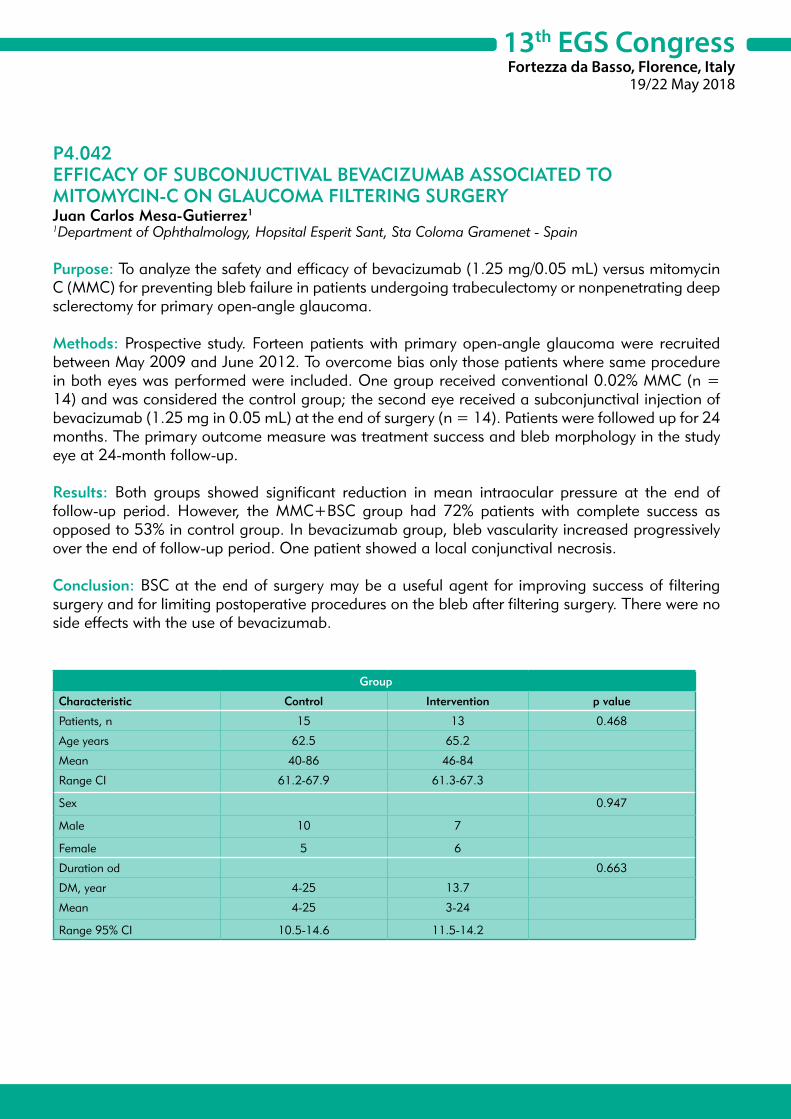
13th EGS CongressFortezza da Basso, Florence, Italy
19/22 May 2018
P4.042EFFICACY OF SUBCONJUCTIVAL BEVACIZUMAB ASSOCIATED TO MITOMYCIN-C ON GLAUCOMA FILTERING SURGERYJuan Carlos Mesa-Gutierrez1
1Department of Ophthalmology, Hopsital Esperit Sant, Sta Coloma Gramenet - Spain
Purpose: To analyze the safety and efficacy of bevacizumab (1.25 mg/0.05 mL) versus mitomycin C (MMC) for preventing bleb failure in patients undergoing trabeculectomy or nonpenetrating deep sclerectomy for primary open-angle glaucoma.
Methods: Prospective study. Forteen patients with primary open-angle glaucoma were recruited between May 2009 and June 2012. To overcome bias only those patients where same procedure in both eyes was performed were included. One group received conventional 0.02% MMC (n = 14) and was considered the control group; the second eye received a subconjunctival injection of bevacizumab (1.25 mg in 0.05 mL) at the end of surgery (n = 14). Patients were followed up for 24 months. The primary outcome measure was treatment success and bleb morphology in the study eye at 24-month follow-up.
Results: Both groups showed significant reduction in mean intraocular pressure at the end of follow-up period. However, the MMC+BSC group had 72% patients with complete success as opposed to 53% in control group. In bevacizumab group, bleb vascularity increased progressively over the end of follow-up period. One patient showed a local conjunctival necrosis.
Conclusion: BSC at the end of surgery may be a useful agent for improving success of filtering surgery and for limiting postoperative procedures on the bleb after filtering surgery. There were no side effects with the use of bevacizumab.
Group
Characteristic Control Intervention p value
Patients, n 15 13 0.468
Age years 62.5 65.2
Mean 40-86 46-84
Range CI 61.2-67.9 61.3-67.3
Sex 0.947
Male 10 7
Female 5 6
Duration od 0.663
DM, year 4-25 13.7
Mean 4-25 3-24
Range 95% CI 10.5-14.6 11.5-14.2

13th EGS CongressFortezza da Basso, Florence, Italy
19/22 May 2018
P4.043TWO-STAGE CONTINUUM OF NON-PENETRATING DEEP SCLERECTOMYNatalia Volkova1
1Irkutsk Branch of S. Fyodorov Eye Microsurgery Federal State Autonomous Institution, Irkutsk State Medical University, Irlutsk - Russia
Purpose: To evaluate ultrabiomicroscopic semiotics of trabeculo-Descemet’s membrane and assess complete hypotensive success of non-penetrating deep sclerectomy, depending on the timing of YAG laser descemetopuncture.
Materials and Methods: Comparative study in groups after non-penetrating deep sclerectomy. 1st group 500 eyes (retrospectively 2006-2008). 2nd group 500 eyes (prospectively 2011-2013). In 1st group period of laser descemetopuncture performance was 11.7 ± 8.11 months; in 2nd group – 1.5 ± 0.2 months. Indication for laser descemetopuncture in 1st group was decompensation of IOP after operation. Indication for laser descemetopuncture in 2nd group was signs of trabeculo-descemet’s membrane failure, found by ultrabiomicroscopic monitoring. IOP decrease by 30% or more from baseline without additional postoperative medications was considered as complete success. Groups were relevant. Follow-up period was 3 years.
Results: Original ultrabiomicroscopic monitoring - Image determined acoustic criteria of the norm (thickness 0.093 ± 0.001 mm, acoustic density ≤ 55 ± 10%) and trabeculo-Descemet’s membrane failure (thickness more than 0.11 ± 0.004 mm, acoustic density > 55 ± 10% ) with its height 0.8 ± 0.09 mm, already found by 1-1.12 months after postoperative period (p = 0.05). In 2nd group by this time number of patients with dysfunction of trabeculo-Descemet’s membrane was 86.6%, in 49.6% it was accompanied by decompensation of IOP, in 37.2%-no. In 13.2% of pathological ultrabiomicroscopic monitoring signs of trabeculo-Descemet’s membrane were not observed. In 2nd group, laser descemetopuncture was performed at a period of 1.5 ± 0.2 months, due to ultrabiomicroscopic semiotics of trabeculo-Descemet’s membrane, both in decompensation of IOP and without it (in 13.2% laser descemetopuncture had a preventive nature).Total hypotensive success was achieved in 59.6% [95% confidence interval (CI) 56.8%-65.7%] and 84.8% [95% CI 80.1%-87.2%] in groups in one year, and in 24.8 [95% CI 80.1%-87.2%] and in 60.4 [95% CI 56.7%-64.2%] in groups in 3 years (p = 0.01) relatively.
Conclusion: Effectiveness of non-penetrating deep sclerectomy is associated with implementation of laser descemetopuncture in 100% of cases at a period of 1.5 ± 0.2 months after the operation. Two-stage technology (1st - operation, 2nd - descemetopuncture) in an absolute number of cases within the defined time limits allows creating an penetrating internal microfistula and excluding its influence on further morphogenesis of outflow pathways. This allows increasing the overall hypotensive effect of non-penetrating interventions.

13th EGS CongressFortezza da Basso, Florence, Italy
19/22 May 2018
P4.044EFFICACY AND SAFETY OF BRINZOLAMIDE/BRIMONIDINE FIXED COMBINATION COMPARED TO CONCOMITANT ADMINISTRATION OF BRINZOLAMIDE AND BRIMONIDINE IN OPEN-ANGLE GLAUCOMA OR OCULAR HYPERTENSIONYury Astakhov1, Ningli Wang2, Da-Wen Lu3, Yingzi Pan4, Tatyana Iureva5, Adeniyi Adewale6, Thomas M. Walker6
1Affiliation Department of Ophthalmology of the First Pavlov Medical University, Saint Petersburg - Russia2Beijing Tongren Eye Center, Beijing - China3Tri-service General Hospital, Taipei City - Taiwan, Province of China4Peking University First Hospital, Beijing - China5Irkutsk branch of the academician S.N. Fyodorov Eye Microsurgery Federal State Institution, Irkutsk - Russia6Novartis Pharmaceuticals Corporation, Fort Worth, Texas - USA
Purpose: To compare the intra-ocular pressure (IOP)-lowering efficacy and safety of twice-daily brinzolamide 10 mg/ml/brimonidine 2 mg/ml fixed combination (BBFC) versus the unfixed combination of brinzolamide 10 mg/ml and brimonidine 2 mg/ml (BRINZ+BRIM), in patients with open-angle glaucoma or ocular hypertension inadequately controlled on monotherapy or those already on multiple IOP-lowering medications.
Methods: This was a 3-month, prospective, phase III, randomized (1:1), multicenter, observer-masked, active-controlled, parallel group study (NCT02339584). Primary endpoint: mean change in diurnal IOP (averaged over 9:00, 11:00, 16:00 hours) from baseline to Month 3; non-inferiority of BBFC to BRINZ+BRIM was established if the upper limit of the 95% CI of the between-group difference in least-square (LS) mean change was <1.5 mmHg. The primary analysis used an analysis of covariance model. Supportive endpoints: percentage of patients with diurnal IOP <18 mmHg; diurnal IOP reduction > 30% at Month 3.
Results: The per-protocol set included 349 patients (BBFC, n = 172; BRINZ+BRIM, n = 177). The mean ± SD diurnal IOP at baseline was 24.6 ± 2.66 mmHg in both groups. At Month 3, the LS mean ± SE change in diurnal IOP from baseline was -7.2 ± 0.34 mmHg and -7.3 ± 0.34 mmHg with BBFC and BRINZ+BRIM, respectively (between-group difference: 0.1 mmHg; 95% CI: -0.5, 0.7). In the BBFC and BRINZ+BRIM groups, 53.3% and 55.0% of patients achieved a diurnal IOP < 18 mmHg and 43.2% and 37.4% of patients achieved a mean diurnal IOP reduction > 30% from baseline, respectively, at Month 3. The most common ocular adverse event (AE) was conjunctival hyperemia (BBFC, 6.4%; BRINZ+BRIM, 6.8%); no serious ocular AEs were reported.
Conclusions: The IOP-lowering efficacy of twice-daily BBFC was non-inferior to BRINZ+BRIM. The safety profile of BBFC was similar to that of BRINZ+BRIM.
13th EGS CongressFortezza da Basso, Florence, Italy
19/22 May 2018
P4.045EARLY CLINICAL OUTCOMES OF MICROPULSED TRANSCLERAL CYCLOPHOTOCOAGULATION IN OPEN ANGLE GLAUCOMACigdem Altan1, Banu Satana1, Berna Basarir1, Isil Pasaoglu1, Tekin Yasar1
1Beyoglu Eye Training and Research Hospital, Istanbul - Turkey
Purpose: The aim of this study is to describe our experience with the novel micropulse transscleral cyclophotocoagulation (MP-TSCPC) in patients with the diagnosis of moderate to severe open angle glaucoma.
Methods: Patients with moderate to severe open angle glaucoma who underwent MP-TSCPC with 810 nm infrared diod laser (IRIDEX IQ810 Laser Systems, CA) were included in this retrospective study. The patients received sub-tenon anestesia prior to the procedure. Laser power was set at 2000 mW on micropulse delivery mode. MicroPulse® P3 probe was applied in a “painting” motion along the upper and lower hemisphere avoiding the 3 and 9 o’clock positions. The laser was delivered for 80 seconds to each hemisphere for a total of 160 seconds.
Results: Five eyes of five patients were included in the study. The mean age of patients (1 female, 4 male) was 60.4 ± 10.3 years. Three eyes had severe glaucoma. One patient had previously undergone cataract surgery. Average best corrected visual acuity was 0.56 ± 0.45 (Snellen). The mean baseline intraocular pressure (IOP) was 22.2 ± 4.2 mmHg and patients were on 2.4 ± 1.5 topical glaucoma medications. Mean IOP was reduced by 39.6%, 36% and 30.6% from baseline at postoperative day 1, day 7 and day 42, respectively. Mean IOP dropped to 13.4 ± 2.1 mmHg at first day (p = 0.009); 14.2 ± 3.6 mmHg at first week (p = 0.04), 15.4 ± 1.1 mmHg at sixth week (p = 0.01) postoperatively. Average medication use did not changed significantly at final postop visit. There were no serious adverse events.
Conclusion: Micropulsed transcleral diode laser seems to be a safe and effective treatment alternative to help lower IOP noninvasively in patients with open angle glaucoma. Long-term evaluation with a larger group of patients is needed to establish its value.

13th EGS CongressFortezza da Basso, Florence, Italy
19/22 May 2018
P4.046LONG-TERM IOP AND MEDICATION REDUCTION WITH SECOND-GENERATION MIGS TRABECULAR MICRO-BYPASS STENTS FOR OAG PATIENTS ON 1 PREOPERATIVE MEDICATIONKonrad Schargel1, Jose Belda2, Lucia Rial1
1Glaucoma Section, Hospital Universitario de Torrevieja, Alicante - Spain2Department of Ophthalmology, Hospital Universitario de Torrevieja, Alicante - Spain
Purpose: To evaluate safety and efficacy of 2 second-generation trabecular micro-bypass stents (iStent inject®) as a standalone procedure in patients with uncontrolled open-angle glaucoma (OAG) treated with 1 ocular hypotensive medication.
Methods: In this prospective single-arm study, subjects with OAG uncontrolled on one ocular hypotensive medication were required to have a preoperative IOP of 18-30 mmHg (medicated) and 22-28 mmHg (after medication washout). Eligible subjects received 2 iStent inject devices as a standalone procedure. Medication burden, IOP, adverse events (AE), best- corrected visual acuity, slit-lamp, gonioscopy, and fundus/optic nerve assessments/examinations were performed throughout the study.
Results: All 57 enrolled subjects underwent uncomplicated implantation of 2 iStent inject devices and have completed 30 months of follow-up. A favorable safety profile was observed with no intraoperative or device-related AEs for all subjects. Preoperative medicated mean IOP was 19.5 ± 1.5 mmHg and post-washout IOP was 24.4 ± 1.3 mmHg. At Month 30, the mean IOP reduction was 32% from medicated preoperative IOP and 46% from unmedicated preoperative IOP (post-washout) with a postoperative mean IOP ≤ 14.6 mmHg maintained through Month 30. Coming into the Month 30 visit, all subjects were medication-free. All subjects achieved both an IOP of ≤ 18 mmHg and ≥ 20% IOP reduction on no medication compared to preoperative unmedicated IOP at Month 30. One subject underwent trabeculectomy after the Month 30 exam for elevated IOP; the event was deemed ‘definitely unrelated’ to stent implantation.
Conclusion: The minimally invasive trabecular micro-bypass implantation of 2 iStent inject devices performed as a standalone procedure results in safe and effective long-term reduction of IOP. Out to 30 months postoperative, IOP is ≤ 15 mmHg with elimination of medication in OAG eyes with IOP not managed to under 18 mmHg on 1 preoperative medication. The iStent inject adds to the physicians’ armamentarium when managing patients with glaucoma.
13th EGS CongressFortezza da Basso, Florence, Italy
19/22 May 2018
P4.047POOLED PHASE 3 EFFICACY ANALYSIS OF A ONCE-DAILY FIXED-DOSE COMBINATION (FDC) OF NETARSUDIL 0.02% AND LATANOPROST 0.005% IN OCULAR HYPERTENSION (OHT) AND OPEN-ANGLE GLAUCOMA (OAG)Sanjay Asrani1, Hayley McKee2, Beth Shultz Scott2, Richard Lewis2, Casey Kopczynski2, Theresa Heah2
1Duke University, Durham - USA2Aerie Pharmaceuticals, Bedminster - USA
Purpose: Netarsudil, a Rho-kinase inhibitor, lowers IOP primarily by increasing trabecular outflow. This mechanism is complementary to that of prostaglandin analogues. The efficacy of a once-daily FDC product containing netarsudil 0.02% and latanoprost 0.005% was compared to its individual components using data pooled from two phase 3 studies: MERCURY-1 (NCT02558400) and MERCURY-2 (NCT02674854).
Methods: Both studies enrolled OAG/OHT patients who had unmedicated IOP in both eyes > 20 to < 36 mmHg at 8:00 AM and > 17 to < 36 mmHg at 10:00 am and 4:00 pm. Data from each study were pooled by treatment arm (netarsudil/latanoprost FDC, netarsudil, and latanoprost); the statistical superiority of the FDC to its individual components was assessed. The primary efficacy endpoint was mean IOP at 8:00 am, 10:00 am, and 4:00 pm at week 2, week 6, and month 3.
Results: The pooled intent-to-treat population included 483 FDC-treated, 499 netarsudil-treated, and 486 latanoprost-treated patients, with 87.4%, 86.0%, and 94.4%, respectively, completing 3 months. Mean baseline IOP was similar for patients randomized to the FDC (22.5-24.8 mmHg), netarsudil (22.7-24.7 mmHg), and latanoprost (22.5-24.7 mmHg). From week 2 to month 3, mean IOP ranged from 15.0-16.4 mmHg for the FDC, 17.4-19.4 mmHg for netarsudil, and 16.9-18.0 mmHg for latanoprost. The FDC met the criteria for superiority to netarsudil and latanoprost at all nine time points (all p < 0.0001), lowering IOP by an additional 2.0-3.2 mmHg vs. netarsudil and an additional 1.4-2.5 mmHg vs. latanoprost.
Conclusions: In this pooled analysis of two phase III trials, netarsudil/latanoprost FDC produced IOP reductions that were statistically and clinically superior to netarsudil and latanoprost across all nine time points through month 3. A once-daily dosed FDC that provides significantly greater IOP-lowering than its individual components has the potential to contribute to better glaucoma management.

13th EGS CongressFortezza da Basso, Florence, Italy
19/22 May 2018
P4.048POOLED SAFETY ANALYSIS OF A ONCE-DAILY FIXED-DOSE COMBINATION (FDC) OF NETARSUDIL 0.02% AND LATANOPROST 0.005% IN OCULAR HYPERTENSION (OHT) AND OPEN-ANGLE GLAUCOMA (OAG)Jason Bacharach1, Edward Holland2, Huan Sheng3, Beth Shultz Scott3, Richard Lewis3, Casey Kopczynski3, Theresa Heah3
1North Bay Eye Associates, Petaluma - USA2Cincinnati Eye Institute, Cincinnati - USA3Aerie Pharmaceuticals, Bedminster - USA
Purpose: FDC products are available for treating OHT/OAG, but most contain timolol, which is contraindicated in patients with certain respiratory and cardiac conditions. Netarsudil, a Rho kinase (ROCK) inhibitor that relaxes the trabecular meshwork, has a mechanism of intraocular pressure (IOP)-lowering complementary to that of latanoprost. To further investigate the safety of netarsudil/latanoprost FDC, we analyzed pooled data from the completed MERCURY-1 and MERCURY-2 phase III studies.
Methods: In MERCURY-1 (NCT02558400) and MERCURY-2 (NCT02674854), once-daily (PM) netarsudil 0.02%/latanoprost 0.005% FDC was compared with its individual components in patients with OHT/OAG. Patients had unmedicated IOP > 20 to < 36 mmHg in both eyes at 8:00 AM and > 17 to < 36 mmHg at 10:00 am and 4:00 pm and met other standard clinical trial enrollment criteria for OHT/OAG. Safety data up to 12 months in MERCURY-1 and 3 months in MERCURY-2 were pooled by treatment arm.
Results: The pooled safety population included 482 FDC-treated, 498 netarsudil-treated, and 488 latanoprost-treated patients, with 78%, 76%, and 90%, respectively completing treatment. No treatment-related serious adverse event (AE) was reported in any treatment arm, and treatment-related systemic AEs were minimal. The most frequent ocular AE was conjunctival hyperemia (FDC, 59%; netarsudil, 47%; latanoprost, 22%); among affected FDC-treated patients, conjunctival hyperemia was graded as mild in 87% and occurred sporadically in most (77%). Other common ocular AEs were cornea verticillata (FDC, 15%; netarsudil, 12%; latanoprost, 0%) and conjunctival hemorrhage (FDC, 11%; netarsudil, 14%; latanoprost, 1%), which were typically graded as mild.
Conclusions: In this pooled analysis of two phase III trials, netarsudil/latanoprost FDC was associated with tolerable ocular safety. The most frequent ocular AE was mild conjunctival hyperemia, which generally occurred sporadically. This once-daily FDC should offer a reduced treatment burden that may improve adherence and eye health outcomes.
13th EGS CongressFortezza da Basso, Florence, Italy
19/22 May 2018
P4.049THE EFFICACY OF DEEP SCLERECTOMY ON POSTURE INDUCED INTRAOCULAR PRESSURE CHANGESVittoria Aragno1, Pascale Hamard1, Christophe Baudouin1, Antoine Labbé1
1CHNO des Quinze Vingts, Hôpital Ambroise Paré, Paris - France
Purpose: To evaluate the efficacy of deep sclerectomy (DS) on posture induced intraocular pressure (IOP) changes in open angle glaucoma (OAG).
Methods: Twenty-five eyes of 25 patients with OAG that underwent DS surgery were included in this prospective study. At baseline, 1 and 3 months after surgery the IOP was measured with the IcarePro (ICP) tonometer in sitting, supine, dependent lateral decubitus position (DLDP) and non-dependent lateral decubitus position (NDLDP).
Results: At baseline, mean IOP was 19.57 ± 6.46 mmHg in sitting position, 22.43 ± 8.72 mmHg in supine, 24.26 ± 9.20 mmHg in DLDP, 23.12 ± 8.38 mmHg in NDLDP. At 1 and 3 months after DS, mean IOP decreased significantly in each position (p < 0.001). At each time point, mean IOP was higher in all lying positions than in sitting position (p < 0.001) and higher in DLDP than in supine and NDLDP (p < 0.001 and p = 0.001). Posture induced IOP changes between sitting and respectively supine, DLDP and NDLDP were significantly reduced by 77% (p = 0.009), 60% (p = 0.001) and 82% (p = 0.01) at 1 month and by 79% (p = 0.004), 70% (p < 0. 001) and 79% (p < 0.001) at 3 months. The IOP fluctuation reduction was significantly inferior when considering sitting-DLDP than other postural changes.
Conclusion: DS is effective in lowering not only the mean IOP in all body positions but also the IOP postural fluctuations. Nevertheless, the mean IOP in DLDP remains higher than in the other positions. This might be a factor involved in the glaucoma progression despite an apparently well-controlled IOP in sitting or in others lying positions. This posture should be avoided in patients with asymmetric glaucoma.

13th EGS CongressFortezza da Basso, Florence, Italy
19/22 May 2018
P4.050OCULAR PULSE AMPLITUDE AND THE USE OF POLARIZED WATER (JUNFENG WATER®) IN OPEN ANGLE GLAUCOMAGiulia Renieri1, Yuying Liu2, Xiumei Zhao2, Fengying Liu2, Hagen Thieme1, Dieter Eisenmann3
1Department of Ophthalmology, Otto-von-Guericke University Magdeburg, Magdeburg - Germany2Department of Pathophysiology, General Hospital of People Liberation Army, Beijing - China3Department of Ophthalmology, Kantonspital Graubünden Chur, Chur - Switzerland
Purpose: Growing interest in polarized water has shown promising results in animal models. Polarized water improves microcirculation, cell hydration and metabolism. This prospective, randomized, controlled, double blind study investigate the effect of polarized water (Junfeng®) on ocular pulse amplitude in open angle glaucoma patients.
Methods: Water was polarized with the Junfeng Water Machine, which expose water to a 40 mV energy for 2 minutes. The Junfeng Water Machine® can polarize 1 l water in 2 minutes. 60 patients (120 eyes) were randomized to use the Junfeng Water Machine® or an identical looking-like water bottle (control group). Patients were advised to drink a minimum of 1 l water a day. All study subjects underwent a complete ophthalmological examination. Ocular Pulse Amplitude (OPA, Ziemer Pascal Dynamic Contour Tonometer®, primary end point), OCT optic nerve parameters (RNFL, ONHR Area and Volume, C/D ratio), IOP and blood analysis were analysed at baseline, after 1 and 2 months of treatment. The ophthalmologist was unaware of patients’ assignment, as well the personal collecting data and performing statistical analysis.
Results: 60 patients (61.17 ± 11.47 years) were randomized, 2 patients were lost to follow up. Demographic characteristics were similar for study- and control- group. OPA, OCT optic nerve parameters, IOP and blood analysis showed no differences between the two groups at baseline. Patients using the Junfeng Water Machine® showed an improved OPA after 1 and 2 months in both eyes (p = 0.007 and 0.000 for RE; 0.001 and 0.00 for LE). OCT, IOD and blood data remained stable in the study period in both groups.
Conclusions: Domestic use of polarized water improves OPA measured with the Ziemer Pascal Dynamic Contour Tonometer® independently from IOD, blood laboratory and plasma viscosity. This might implicate a better microcirculation at the optic nerve head with beneficial effect in glaucomatous disease.
13th EGS CongressFortezza da Basso, Florence, Italy
19/22 May 2018
P4.051TRABECULOTOMY AB INTERNO COMBINED WITH PHACOEMULSIFICATIONElena Tomilova1, Marina Pravosudova1
1S. Fyodorov Eye Microsurgery Federal State Institution, Saint-Petersburg - Russia
Purpose: To evaluate the safety and efficacy of trabeculotomy ab interno combined with phacoemulsification and IOL implantation. To estimate hypotensive results of this combined surgery and conventional phacoemulsification in comparable groups of patients with cataract and glaucoma.
Methods: In all cases cataract was complicated by glaucoma with subcompensated intraocular pressure (IOP) with one or more antihypertensive medications. In the main group trabeculotomy ab interno was performed after completion of phacoemulsification and IOL implantation. Trabeculotomy was made by trabeculotome using surgical gonioscopic lens. In the control group routine phacoemulsification with IOL implantation was performed in all eyes. Main group: 27 eyes (27 patients), before surgery the mean IOP was 27 ± 4 mmHg. The mean glaucoma drops number was 2.1 ± 0.6. Control group: 30 eyes (30 patients) before surgery the mean IOP was 26 ± 4 mmHg. Groups were comparable in cataract types distribution as well as in glaucoma types, iridocorneal angle formation, glaucoma stages, and mean number of drops used.
Results: Schlemm’s canal bleeding during the trabeculotomy was observed in 7 cases. Hyphema was observed in two cases. Mean IOP was 18.2 ± 2 mmHg in the main group and 24 ± 2 mmHg in the control one. The mean glaucoma drops number was 0.3 ± 0.3 in the main group and 2.0 ± 0.7 in the control one. BCVA was approximately the same in the both groups. In the follow-up period the mean IOP was 20.0 ± 5 mmHg with mean glaucoma drops number 0.4 ± 0.3 in the main group. In two cases of the control group IOP decompensation required to perform additional surgery. With such additional surgery the mean IOP in the control group was 22.3 ± 2 and the mean drops number 1.7 ± 0.8. Thus, hypotensive results in the main group, viz. mean IOP and mean hypotensive drops number were reliably better than in the control group (p < 0.05).
Conclusions: Trabeculotomy ab interno combined with phacoemulsification and IOL implantation is technically easy and safe surgery in patients with cataract and glaucoma. Antihypertensive effect is statistically proven and significantly better than in the control group.

13th EGS CongressFortezza da Basso, Florence, Italy
19/22 May 2018
P4.052PHOTOBIOMODULATION IN THE COMPLEX TREATMENT OF PRIMARY OPEN-ANGLE GLAUCOMA USING MACDEL-08 DEVICEAlina Kleyman1, Olga Kiseleva1, Elena Iomdina2, Alexander Bessmertny1, Peter Luzhnov3, Dmitry Shamaev3
1Department of Glaucoma, Moscow Helmholtz Research Institute of Eye Diseases, Moscow - Russia2Department of Refraction Pathology, Binocular Vision Anomalies and Ophthalmoergonomics, Moscow Helmholtz Research Institute of Eye Diseases, Moscow - Russia3Department of Biomedical Engineering, N. Bauman Technical University, Moscow - Russia
Purpose: To evaluate the clinical efficacy of low-level laser therapy (LLLT) using MACDEL-08 based on the He-Ne laser (HNL) in the complex treatment of primary open-angle glaucoma (POAG).
Methods: The study involved two groups of subjects. Group 1 consisted of 29 eyes of 23 patients aged 56-77 (ave. age M ± σ = 67.95 ± 6.14) with advanced POAG. Group 2 consisted of 26 eyes of 21 patients with far advanced POAG aged 58-79 (ave. age M ± σ = 68.89 ± 6.58). The intraocular pressure (IOP) of all subjects was normal without anti-hypertensive drops due to glaucoma surgery (trabeculectomy) and a course of conservative therapy for 6-18 months before LLLT. LLLT was performed with MACDEL-08 device. Patients observed a moving speckle pattern formed by the irradiation of HNL (630 nm). The course of treatment consisted of 10 daily sessions of 10 minutes each. All subjects underwent complete ocular examination and examination of ocular hemodynamics by the transpalpebral rheoophthalmography (TR) before starting and after the end of LLLT. TR signal processing included three basic hemodynamic parameters: rheographic index (RI), period of maximum filling (PMF), indicator of the elastic modulus (IEM).
Results: Upon completion of LLLT improvement in various parameters of the studied eyes was showed. Visual acuity (M ± σ) has changed from 0.64 ± 0.15 to 0.76 ± 0.15 for group 1 and from 0.42 ± 0.22 to 0.52 ± 0.22 for group 2, p < 0.05. The measured IOP (mmHg, M ± σ) decreased from 14.8 ± 1.8 to 14.0 ± 1.6 for group 1, and from 16.7 ± 1.9 to 16.1 ± 1.8 for group 2. Analysis of the rheographic parameters has shown the increase of RI (mΩ, M ± σ) from 12.81 ± 1.12 to 15.58 ± 1.05 for group 1, and from 9.57 ± 0.89 to 13.31 ± 1.29 for group 2, p < 0.05. A slight reduction in the other parameter values (PMF, IEM) was observed in both groups.
Conclusions: We have found the positive influence of photobiomodulation on the visual acuity and parameters of ocular hemodynamics in patients with advanced and far advanced POAG. The application of LLLT using MACDEL-08 device reduces the deficit of blood supply of the glaucomatous eye. This method is a harmless, clinically effective, physiologically sound and technically simple component of the complex treatment of POAG patients.

13th EGS CongressFortezza da Basso, Florence, Italy
19/22 May 2018
P4.053BLEB COMPRESSION SUTURES IN MANAGEMENT OF OVERFILTRATION AFTER ANTIGLAUCOMA SURGERYTomasz Zarnowski1, Dominika Wróbel-Dudzinska1, Ewa Kosior-Jarecka1
1Department of Diagnostics and Microsurgery of Glaucoma, Medical University of Lublin, Lublin - Poland
Purpose: The aim of the study was the assessment of efficacy and safety of compression sutures in patients with overfiltrative hypotony after glaucoma surgery.
Settings: This study analyses the clinical outcomes of conjunctival compression sutures for 17 patients with ocular hypotony in Department of Diagnostics and Microsurgery of Glaucoma, Medical University, Lublin, Poland.
Methods: Only patients with hyperfiltration hypotony defined as intraocular pressure (IOP) ≥ 6mmHg associated with a reduced vision aquity were included. Mean age of patients was 60.5 ± 20.5. The main diagnosis was primary open angle glaucoma in 8 cases; pigmentary glaucoma in 3; pseudoexfoliative glaucoma in 4; primary angle closure glaucoma in 1; traumatic glaucoma in 1. Primary procedure was trabeculectomy in 9; phacotrabeculectomy in 4; revision in 3 cases and deep sclerectomy in 1 case. In 13 patients maculopathy decreasing visual acuity was observed and in 4 choroidal detachments were present. The compressive Nylon 10-0 single sutures were performed in all patients; in 2 patients the procedure was repeated.
Results: Mean intraocular pressure before suturing was 2.3 ± 1.7 mmHg and increased to 14.2 ± 7.03 mmHg (p = 0.00065) in 7 days after and to 11.5 ± 5.4 mmHg (p = 0.0022) in 3 months. After 6 months mean IOP was 10.2 ± 4.3 mmHg (p = 0.005), after one year 9 ± 4.7 mmHg (p = 0.0117). To obtain the target pressure 1 patient needed the sutures to be released and in 3 patients medical therapy was entered. Mean best corrected visual acuity before the sutures was 0.18 ± 0.13 and increased to 0.53 ± 0.25 (p = 0.0004) in 3 months; after 6 months 0.46 ± 0.31 (p = 0.005), after one year 0.31 ± 0.22 (p = 0.025). In 1 case after the procedure the leakage from the bleb was observed which needed resuturing.
Conclusions: The transconjuctival compression sutures placement seems to be efficient and safe technique for managing with ocular hypotony after glaucoma surgery.

13th EGS CongressFortezza da Basso, Florence, Italy
19/22 May 2018
P4.054IMPACT OF LASER PULSE DURATION ON THE REDUCTION OF INTRAOCULAR PRESSURE DURING SELECTIVE LASER TRABECULOPLASTYSpela Stunf Pukl1, Brigita Drnovšek Olup1, Makedonka Atansovska Velkovska1, Barbara Cvenkel1
1University Eye Clinic, University Clinical Center, Ljubljana - Slovenia
Purpose: To evaluate the efficacy and safety of selective laser trabeculoplasty (SLT, 532 nm Nd:YAG laser) in lowering the intraocular pressure (IOP) of patients with open-angle glaucoma (POAG) or ocular hypertension (OHT) when performed with a laser pulse duration of 1 ns compared with the standard 3-5 ns SLT.
Methods: Thirty patients (60 eyes) with bilateral nontreated primary open-angle glaucoma (POAG; n = 7) or ocular hypertension (n = 23) underwent SLT with a 532 nm Q-switched neodymium doped yttrium aluminium garnet (Nd:YAG) laser in a prospective case-control study. A laser pulse duration of 1 ns was used in the right eye (30 eyes; cases) and the standard laser pulse duration of 3-5 ns was used in the left eye (30 eyes; controls). The primary outcome was IOP at 1 hour, 1 day, 8 weeks, 6 months after the procedure and follow up at 1 year. Secondary outcomes were the rate of adverse ocular tissue reactions, and difference in the histological and ultrastructural changes in the drainage angle treated with both lasers of one due to other unrelated pathology enucleated eye.
Results: Mean 1 ns and 3-5 ns SLT IOPs were 24.1 and 24.3 mmHg, respectively, before the laser treatment. No statistically significant difference in mean 1 ns and 3-5 ns SLT IOPs was observed at 1 hour (p = 0.761), 1 day (p = 0.758), 8 weeks (p = 0.352) and 6 months (mean right eye IOP = 18.7 mmHg and mean left eye IOP = 18.7 mmHg; p = 1.000). After 1 year in 6 patients of 25 (12 eyes) additional intervention was performed to remain satisfactory IOP control. No significant difference in anterior chamber inflammation following SLT, was observed between eyes treated with 1ns and 3-5 ns SLT (p = 0.529). Treatment with both laser pulse durations SLT resulted in only minor ultrastructural changes in the drainage angle.
Conclusion: SLT performed with a shorter Nd:YAG laser pulse duration of 1 ns was non-inferior to SLT performed with the standard laser pulse duration of 3-5 ns at lowering IOP of patients with open-angle glaucoma or ocular hypertension early after the procedure as well as after 1 year and caused similar rate of postoperative side effects.
13th EGS CongressFortezza da Basso, Florence, Italy
19/22 May 2018
P4.055ANTIGLAUCOMA TREATMENT METHODS ARE PREDICTORS FOR AN INTRAOCULAR PRESSURE DECREASE IN PATIENTS WITH PRIMARY OPEN ANGLE GLAUCOMA AFTER ENDOSCOPIC CYCLOPHOTOCOAGULATIONMarta P. Wiacek1, Tomasz Miszczuk2, Monika Drobek-Slowik2, Andrzej Palacz2, Andrzej Lipinski2, Monika Modrzejewska3
1I Department of Ophthalmology, Pomeranian Medical University in Szczecin, Szczecin - Poland2Medical Center Dom Lekarski, Szczecin - Poland3II Department of Ophthalmology, Pomeranian Medical University in Szczecin, Szczecin - Poland
Purpose: We aimed to establish predictors for obtaining therapeutic success; this was defined as a decrease of intraocular pressure (IOP) below 15 mmHg for different control points in a one year follow-up period after endoscopic cyclophotocoagulation (ECP) for primary open angle glaucoma (POAG) eyes.
Methods: Prospective analysis of IOP measured by Goldmann tonometer beforehand, on the 1st day, 8 weeks later, and one year after ECP in 66 eyes with POAG was performed. Data about disease duration, antiglaucoma drugs used, previous procedures to decrease IOP, and frequency of control visits were also obtained. A univariate analysis and multivariate stepwise logistic regression method analysis were used to create models for testing exactly which parameters corresponded with therapeutic success after the ECP during examination periods. Model sensitivity and specificity were evaluated by the receiver-operating characteristic curve.
Results: Mean age of individuals was 75 ± 8.06 years, mean IOP before ECP - 24.16 ± 10.45 mmHg. It was found that dorzolamide usage, being over 80-years-old, and high preoperational IOP decrease the chance of therapeutic success in 1st day. The area under the curve (AUC) for this model was 0.85 and SE (standard error) 0.05. Usage of at least two antiglaucoma drugs, combining of ECP with phacoemulsification and high preoperational IOP decrease the chance of therapeutic success in the 8th week after ECP. AUC for this model was found to be 0.85 SE 0.05. During a one year follow-up for the previously performed trabeculectomy, the operated eye had a negative influence on IOP reduction below 15 mmHg. However, AUC was established to be 0.63 and SE 0.08. The sensitivity and specificity for predictors designed for 1st day IOP after ECP were respectively 91.3% and 65.2%, for 8th week - 85.7% and 77.4%, for one year follow up - 96.4% and 30%.
Conclusions: Predictors of IOP that decrease to below 15 mmHg in POAG eyes differ over time from ECP. Antiglaucoma drugs used and previously performed procedures that decreased IOP might influence the IOP reduction after ECP. Results of this study might be useful in those who are qualified for ECP, including prognostic assessment of POAG patients after this procedure.
13th EGS CongressFortezza da Basso, Florence, Italy
19/22 May 2018
P4.056EFFICACY AND SAFETY OF ISTENT TRABECULAR MICROBYPASS STENT IN OPEN ANGLE GLAUCOMAJin Young Rhew1, Tae Heon Lee1, Kyung Heon Lee1
1Busan Sungmo Eye Hospital, Busan - Republic of Korea
Purpose: To evaluate 1 year efficacy and safety of the iStent Trabecular Micro-Bypass stent used as the sole procedure in patients with open-angle glaucoma (OAG).
Methods: Retrospective, consecutive case series involving 12 Korean patients with OAG [normal tension glaucoma (NTG), primary open angle glaucoma (POAG)] that were implanted with 1 iStent trabecular micro-bypass stent. Data were collected preoperatively, and at 1, 3, and 6 months, 12 months, postoperatively. Data included intraocular pressure (IOP), number of glaucoma and complications.
Results: Mean preoperative IOP was 20.9 ± 1.5 (range 12-28) mmHg and significantly decreased in 16.5 ± 2.63 mmHg at 3 months (p < 0.05), in 17.0 ± 2.9 mmHg at 12months postoperatively (p < 0.05). The mean number of glaucoma medications was 2.2 ± 0.25 preoperatively and 1.30 ± 0.30 (p < 0.05) at 12 months postoperatively. No significant changes in visual acuity were noted. The most common complications comprised mild hyphema in 3 eyes, mild uveitis in 4 eyes and transient IOP spike in 2 eyes on postoperative day 1 and all instances were medically well controlled.
Conclusions: The iStent trabecular micro-bypass stent is a safe and effective treatment option in patients with OAG including NTG, and reduces the topical treatment burden in one hypotensive medication.
13th EGS CongressFortezza da Basso, Florence, Italy
19/22 May 2018
P4.057PRE-DESCEMET HEMATOMA AFTER NON-PENETRATING DEEP SCLERECTOMY (NDPS)Maria Dolores Lago1, Beatriz Alonso1, Almudena De Pablo1, José Luis Torres1, Juan Carlos Herranz1
1Department of Ophthalmology, Hospital Universitario 12 de Octubre, Madrid - Spain
Purpose: To report the case of a pre-Descemet hematoma after NPDS and its surgical management.
Methods: Case report. 76 year-old patient with a history of atrial fibrillation treated with acenocoumarol, hypertension and central retinal vein occlusion of the left eye treated with antiangiogenics. He is referred with the diagnosis of primary open-angle glaucoma in medical treatment with 3 hypotensive drugs. Combined surgery of glaucoma and cataract was decided, despite the visual prognosis.
Results: A NPDS with intraoperative subconjunctival 5-fluorouracil was performed. Intraocular pressure within 24 hours was 2 mmHg. After 48 hours, the acenocoumarol was reintroduced, and a week later a hematoma in the scleral lake and upper intracorneal predescemetic space of 1mm was observed. A goniopuncture was performed, with blood flow to the anterior chamber (AC). The following week, the pre-Descemet hematoma had increased to 2.5 mm approaching the pupillary border, so surgical drainage was decided. The procedure consisted of descematorhexis with cystitome and blood drainage using a DSAEK spatula, with hyperpressure controlled with dispersive viscoelastic, achieving the evacuation of the blood clot to the AC. Afterwards the AC was washed and an air bubble was left. In the 5 months follow-up there has been no rebleeding, although some filiform intracorneal blood remains.
Conclusions: Hemorrhagic detachment of the Descemet membrane is a rare complication after NPDS, being described more frequently after canalicular surgery. The mechanism is not clear; it is suggested that it might be due to a reflux of blood of the scleral lake from the Schlemm canal, secondary to surgical hypotension. This blood would dissect the space between the corneal stroma and the Descemet membrane producing a detachment of this layer. In our patient we observed the bleeding after the reintroduction of acenocoumarol, which might have contributed to its etiology. For its management, one option is observation, as long as the hematoma is small and shows signs of resorption. However some authors propose drainage to avoid the residual leukoma. In our case the surgical treatment was effective, achieving almost complete transparency of the area.

13th EGS CongressFortezza da Basso, Florence, Italy
19/22 May 2018
P4.058COMBINED LASER-SURGICAL TREATMENT OF PRIMARY OPEN-ANGLE GLAUCOMA AND COMPLICATED CATARACTSTatiana Sokolovskaya1, Dzhavid Magaramov1, Victoriya Teplovodskaya1, Valeriya Yashina1
1The S. Fyodorov Eye Microsurgery Federal State Institution, Moscow - Russia
At present, combined surgical interventions for complicated cataract and primary open-angle glaucoma become widespread. The application of YAG laser activation of trabecula (YAG-LAT) in combination with cataract phacoemulsification in such patients was developed by Magaramov D.A, Doga A.V., 2005.
Purpose: To study the efficacy of combined treatment of patients with primary open-angle glaucoma and complicated cataracts using a laser activation of trabecula as an anti-glaucoma component.
Methods: The study included 23 patients (23 eyes). Pseudoexfoliation syndrome of the degree I-II was observed in all cases, the pigmentation degree of the structures in the drainage zone was weak (0-1). In the first stage, laser treatment was performed with following parameters: YAG-LAT (Nd-YAG laser, 1064 nm, 0.9-1.5 MJ energy, 30 NS pulse duration, a spot diameter of 10-15 μm, in the quantity of 40-50 pulses in the lower semicircle). The second stage - cataract phacoemulsification with IOL implantation was carried out according to a standard technique 30-60 minutes later. The follow-up period: up to 6 months. Preoperatively the IOP level was 23.5 ± 2.56 mmHg. The uncorrected visual acuity (UCVA) was 0.12 ± 0.08, the best corrected visual acuity (BCVA) was 0.3 ± 0.14. The cataract density by Buratto had the 3rd degree in 73.9% (17 patients), the 4th degree in 26.1% (6 patients). Intra- and post-operative complications were not noted. The IOP decreased on average by 7.17 ± 1.25 mmHg (by 29.4% of the initial IOP level) after 6 months. In the postoperative period the UCVA was 0.72 ± 0.2 and the BCVA revealed 0.82 ± 0.2. Preoperatively the number of hypotensive drugs was 1.6 ± 0.71 and 1.0 ± 0.36 after 6 months. After the combined surgical intervention, the IOP normalization was achieved in 95.7% of patients.
Conclusion: Thus, the proposed technology allows to obtain a visual acuity improvement, a resistant IOP normalization and a stabilization of the glaucoma process.

13th EGS CongressFortezza da Basso, Florence, Italy
19/22 May 2018
P4.059THREE YEAR RESULTS OF I-STENT + PHACOEMULSIFICATION CATARACT SURGERY FOR GLAUCOMAAdam Lewis1, Dharshana Ramanathan1, Caroline Wong1, Richard Imonikhe1, Ejaz Ansari1
1Department of Ophthalmology, Maidstone and Tunbridge Wells NHS Trust, Maidstone - United Kingdom
Purpose: To evaluate long-term safety and efficacy of i-Stent trabecular micro-bypass stent implantation + cataract surgery (phaco) in glaucoma: a retrospective, interventional, open label study.
Methods: Cases of glaucoma who had phacoemulsification surgery planned, were included. Preoperative and postoperative evaluations included intra-ocular pressures (IOP), topical ocular hypotensive agent use, best corrected visual acuity, perioperative complications and adverse events.
Results: A single trabecular micro-bypass i-Stent was implanted at the time of phacoemulsification cataract surgery. A temporal incision approach was used in all cases. The results from 35 eyes in 25 patients were analysed for 3 years postoperatively. 49% (n = 17) were male. Mean age was 80 ± 7 (SD). 3 eyes had angle closure with previous peripheral iridotomies. Mean preoperative IOP was 18.5 ± 3.2 mmHg; this was significantly reduced at 12 months (p = 0.008), 24 months (p ≤ 0.001) and 36 months (p ≤ 0.001). Mean IOP was 15.9 ± 4.5, 15.0 ± 4.5 and 15.6 ± 3.6 respectively. The mean number of preoperative IOP lowering agents was 2.3 ± 1.0 and 2.5 ± 1.0 at 36 months. This was not significantly reduced at any follow up time period. Secondary interventions were required in three eyes. Cyclodiode laser was required in 2 patients. ALT was carried out in 2 eyes. There were no significant intraoperative complications and no post-operative hypotony. At 36 months, visual acuity was ≥ 6/12 in 29 eyes (83%). Conclusions: Trabecular micro-bypass stent implantation during cataract surgery is safe and effective for patients with glaucoma. We measured a sustained reduction in IOP over 36 months in the real world clinic setting.
13th EGS CongressFortezza da Basso, Florence, Italy
19/22 May 2018
P4.060INFLUENCE OF ANTIGLAUCOMATOUS THERAPY WITH LATANOPROST ON OPTIC NERVE HEAD BLOOD FLOW IN CAUCASIAN PATIENTS WITH NORMAL TENSION GLAUCOMAAnna Sophie Mursch-Edlmayr1, Nikolaus Luft2, Michael Ring1, Matthias Bolz1
1Department of Ophthalmology, Kepler University Hospital, Linz - Austria2Department of Ophthalmology, Ludwig-Maximilians-University, Munich - Germany
Purpose: To date the only evidence based therapy in normal tension glaucoma (NTG) relies on lowering the IOP. Topical applied prostaglandin analogues potentially could improve the optic nerve head (ONH) blood flow. Laser speckle flowgraphy (LSFG) enables two-dimensional measurements of perfusion at the ONH, the retina and the choroid using the laser speckle phenomenon. To our knowledge, to date LSFG has not been used to analyse the effects of latanoprost in Caucasians before. Aim of the current study was to assess the changes in ONH blood flow in Caucasian NTG patients with LSFG after stopping their local antiglaucomatous therapy with latanoprost.
Methods: The study included 12 eyes from 12 Caucasian patients with diagnosis of normal tension glaucoma who were treated with a latanoprost monotherapy. Measurements of optic nerve head blood flow were performed with Laser Speckle Flowgraphy (LSFG) at baseline and after pausing latanoprost for 3 weeks. The mean blur rate was obtained for different vascular parts of the optic nerve head (MA, MV, MT). Also, the intraocular pressure (IOP), mean arterial pressure (MAP) as well as ocular perfusion pressure (OPP) were analyzed.
Results: IOP increased significantly with a mean delta of 13.5 ± 12.9% and consecutively OPP decreased significantly by 8.3 ± 10.81%. However, all LSFG parameters remained stable and were not significantly influenced by stopping latanoprost.
Conclusions: The present study shows that although the IOP and therefore also the OPP were significantly altered after stopping latanoprost MA, MV and MT remained stable. This indicates that ONH perfusion is not sustainably influenced by a local therapy with latanoprost, neither directly nor by alternation of OPP. Besides the effect of this finding on therapeutic approaches it is relevant for further research projects on ONH perfusion in NTG patients because it indicates that a long-term therapy with latanoprost does not bias the LSFG outcome parameters.

13th EGS CongressFortezza da Basso, Florence, Italy
19/22 May 2018
P4.061A COMPARISON BETWEEN REPEAT 180-DEGREE AND REPEAT 360-DEGREE SELECTIVE LASER TRABECULOPLASTYDongwook Lee1, Euiyoung Kweon1
1Top Eye Clinic, Jeonju - Republic of Korea
Purpose: To report the efficacy of repeat selective laser trabeculoplasty (SLT) in patients with intraocular pressure (IOP) elevation. Repeat SLT is applying laser to same area that has been treated previously with 360-degree SLT. And to compare two regimens of repeat SLT, 180- degree and 360-degree applications as a second initial treatment.
Methods: A retrospective chart review was undertaken of 30 eyes of 28 patients with open-angle glaucoma (primary or pseudoexfoliation glaucoma) uncontrolled on medical therapy who had undergone repeat SLT treatments. We compared pressure lowering effect of repeat SLT in two groups of patients; group 1 (16 eyes) received SLT on 180-degree of trabecular meshwork, group 2 (14 eyes) on 360-degree of trabecular meshwork at one session. We measured the IOP at baseline before the first SLT (SLT 1) and repeat SLT(SLT 2) at 1 month, 3 months and 6 months. Results: Mean baseline IOP before repeat SLT were 21.92 ± 4.25 mmHg (group 1) and 23.91 ± 5.03 mmHg (group 2). Group 1 was performed with a mean of 85.78 ± 8.57 laser spots and group 2 had 146.43 ± 9.95 laser spots. The group 2 had significantly greater mean reduction of IOP from baseline at 1 months (13.9 % vs. 20.3%, p = 0.023) and 3 months time points (13.1% vs. 18.2%, p = 0.046). But after 6 months, there is no significant difference (10.6 % vs. 13.7 %, p = 0.245). No serious adverse events were recorded.
Conclusions: Repeat SLT produces a significant IOP-lowering effect and treatment on 360-degree may be more effective than 180-degree as a initial treatment without adverse events.

13th EGS CongressFortezza da Basso, Florence, Italy
19/22 May 2018
P4.062CAN MODERN TRABECULECTOMY CONTROL LONG TERM VISUAL FIELD PROGRESSION?Hannaa Bobat1, Anastasios Sepetis1, Shahiba Begum1, Alastair Lockwood1, James Kirwan1
1Ophthalmology Department, Queen Alexandra Hospital, Portsmouth - United Kingdom
Purpose: To evaluate the ability of the trabeculectomy of the modern era to control long-term visual field progression in open angle glaucoma (OAG).
Methods: Serial visual fields (VFs) of eyes undergoing trabeculectomy for OAG at a regional hospital in the UK, were reviewed for evidence of glaucomatous progression. Progression rate was determined by the difference between the better of the last two Mean Deviations (MDs) and the better of the first two MDs (or first and last MD if < 4 VFs were available), divided by time and classified as being stable (< 0.3 dB/year), or showing borderline progression (≥ 0.3dB/year) or definite progression (≥ 0.5dB/year). Eyes with < 3 reliable post-operative VFs over < 2 years following surgery were excluded. The pre-operative progression rate was determined for eyes with sufficient pre-operative VFs, as a comparison to give a better indication of the effect of trabeculectomy.
Results: 77 eyes (59 patients) met the inclusion criteria. Median follow-up was 87 months (range 62-163). IOP (mean ±SD) at final review was 12.4 ± 3.9mmHg, a reduction of 41.4% compared to pre-operative IOP and 48.9% compared to presentation IOP. Use of ocular anti-hypertensives was reduced from 2.5 to 0.4 per eye. Mean MD progression (±SD) post-operatively was -0.13dB ± 0.57 per year. 67.5% (n = 52) eyes were stable (mean -0.1dB per year); 16.9% (n = 13) showed borderline progression (mean -0.37dB per year); 15.6% (n = 12) showed definite progression (mean -0.87 dB per year). Of the 32.5% showing borderline or definite progression, 11 failed to achieve “good” IOP control (≤ 13 mmHg), 2 developed “late” (> 3 months) hypotony, 2 underwent late revision and in 2 cases progression was attributed to dry ARMD. Eight eyes (10.4%) continued to progress despite a technically successful trabeculectomy. 57 eyes had sufficient VFs to assess pre-operative progression; the mean progression rate (± SD) was -0.55dB ± 0.78 per year before trabeculectomy and -0.18 dB after, a reduction of 67.3%.
Conclusions: A trabeculectomy in today’s era can achieve long-term VF stability or markedly reduced progression in the majority of cases. With good IOP control, there is a 90% chance that the VF will remain stable for an average of 7.8 years following surgery.

13th EGS CongressFortezza da Basso, Florence, Italy
19/22 May 2018
P4.063SURGICAL OUTCOMES AND EFFECT OF XEN-45 IMPLANT ON CORNEAL ENDOTHELIUM IN OPEN ANGLE GLAUCOMASuzan Guvenyilmaz1, Melis Palamar2, Meryem Tehmen2, Halil Ates2
1Ege University, Izmir - Turkey2Department of Ophthalmology, Ege University Medical Center, Izmir - Turkey
Purpose: To evaluate the surgical outcome and effect of XEN-45 implant on corneal endothelial cell in patients with open angle glaucoma (OAG).
Materials and Methods: We retrospectively reviewed the records of 12 (7 male and 5 female) cases who had undergone XEN-45 implant with subconjunctival mitomycin-C for medically uncontrolled OAG. A postoperative IOP ≤ 21 mmHg without or with medications were respectively defined as “complete” and “qualified” success while an intraocular pressure (IOP) ≥ 21 mmHg was defined as failure. IOP, number of medications, best-corrected visual acuity (BCVA), and complications were recorded. Endothelial cell measurements were evaluated before and after surgery by CEM-530.
Results: The mean age was 63.5 ± 17.6 (28-87) years and mean follow-up was 9.5 ± 1.1 (9-12) months after surgery. The diagnosis was primary OAG in 9 (75%) eyes and secondary OAG in 3 (25%) (2 postkeratoplasty, 1 angle recession) eyes. The mean IOP decreased from 27.4 ± 6.8 mmHg to 19.8 ± 10.9 mmHg, 17.7 ± 8.6 mmHg, and 15.3 ± 3.2 mmHg at 1, 3, 6, and 9 months (p < 0.05; Wilcoxon rank test), respectively. The average number of antiglaucoma medications decreased from 3.91 ± 0.90 preoperatively to 2.16 ± 1.26 postoperatively (p < 0.05; Wilcoxon rank test). Four (33.3%) eyes achieved complete success and 7 (58.3 %) qualified success, only 1 (8.4%) eye required additional glaucoma surgery. BCVA (LogMAR) was 1.00 ± 1.13 and remained stable with 1.00 ± 1.41 during follow-up (p > 0.05; Wilcoxon rank test). The mean endothelial cell count significantly decreased from 2415.08 ± 559.23 cells/mm2 to 2141.08 ± 482.15 cells/mm2, 2094.33 ± 483.11 cells/mm2, 2055.08 ± 499.06 cells/mm2, 2048.67 ± 498.61 cells/mm2 and 2028.58 ± 547.97 cells/mm2 at 1, 3, 6, 9 months and last visit (p < 0.05;Wilcoxon rank test), respectively. However, there was no sign of clinical corneal edema or haze affecting visual acuity in any of the patients. Complications included transient hypotonia in 1 (8.4%) eye, and bleb encapsulation that required needling in 4 (33.3 %) eyes.
Conclusion: XEN implant is an effective procedure to lower IOP and antiglaucoma medications with a 33% rate of needling for management of bleb encapsulation in OAG. Although XEN procedure induced significant decrement of endothelial cell count, it does not seem to be of clinical importance in the short term. However, studies to evaluate the endothelial cell health in the long run are required.

13th EGS CongressFortezza da Basso, Florence, Italy
19/22 May 2018
P4.064FILTERING BLEB CHARACTERISTICS IN COMBINED CATARACT SURGERY WITH EX-PRESS IMPLANT VS. NON-PENETRATING DEEP SCLERECTOMY. PROSPECTIVE, RANDOMIZED AND MULTICENTRE STUDYMarcos Muñoz1, Alfonso Antón1, Marta Castany2, Alfonso Gil3, Alberto Martinez3, Javier Moreno4, Francisco Muñoz Negrete5, Jose Urcelay6
1Departament of Glaucoma Insitut Català de Retina, Barcelona - Spain2Department of Ophthalmology, University Hospital Vall d’Hebron, Barcelon - Spain3Department of Ophthalmology, San Eloy Hospital, Bilbao - Spain4Department of Ophthalmology. University of Navarra Clinic, Pamplona - Spain6Department of ophthalmology Ramon y Cajal Hospital, Madrid - Spain5Department of Ophthalmology, Gregorio Marañon Hospital, Madrid - Spain
Purpose: To evaluate and compare filtering bleb characteristics after combined cataract surgery with Ex-Press implant vs. non-penetrating deep sclerectomy (NPDS.
Methods: Prospective, multicentre, single-blinded, and randomized trial. Eyes with open angle glaucoma and cataract were randomly assigned to either phacoemulsification and filtration surgery with Ex-Press implant (Alcon) or NPDS with ESNOPER implant (AJL). Main outcomes measures were bleb size (1-5), vascularity (1-5) and height (1-4) as evaluated with Moorfields bleb grading system. The values obtained where compared between the groups, and between month 1 and month 12, using Mann-Whitney and Wilcoxon Tests. The relation with intraocular pressure (IOP) was analysed with Pearson-Spearman’s test.
Results: No significant differences between Ex-Press group and NPDS group were found any of the bleb parameters at 1 month or 12 months. There were significant difference in bleb appearance throughout the study. Vascularity of the central area in the NPDS group significantly decreased (p = 0.02) from 1.8 (0.7) at month 1 to 1.3 (0.5) at month 12. Central area in the Ex-Press group significantly decreased (p = 0.014) from 2.9 (1.1) at month 1 to 2.4 (0.9) at month 12. Bleb height in the Ex-Press group significantly decreased (p = 0.034) from 2.3 (0.7) at month 1 to 1.8 (0.9) at month 12. Bleb vascularity in the Ex-Press group significantly decreased in the central and maximal areas (p = 0.005) from month 1 to month 12. Maximal bleb area was inversely related (r = -0.39; p = 0.03) to intraocular pressure (IOP) in the NPDS group at month 1. Vascularity in the central area was directly related (r = 0.39; p = 0.01) with higher IOP in the Ex-Press group at 12 months. Vascularity in the central area was directly related (r = 0.39; p = 0.01) with higher IOP in the Ex-Press group at 1 month. Vascularity in the central area (r = 0.56; p < 0.001) and maximal areas (r = 0.37; p = 0.012) at month 1 was directly related with higher IOP in the Ex-Press group at month 12.
Conclusions: Bleb appearance was not significantly different at 12 months between the groups but bleb appearance changed with time. Bleb vascularity at month 1 was related to final IOP at month 12 in the Ex-Press group.
13th EGS CongressFortezza da Basso, Florence, Italy
19/22 May 2018
P4.065XEN45 GEL STENT – A CLINICAL AND IN VIVO CONFOCAL MICROSCOPY STUDYMariana Cardoso1, Joana Cardigos1, Sara Crisostomo1, Guilherme Neri Pires1, Diogo Hipólito1, Luisa Vieira1, Isabel Domingues1, Teresa Gomes2
1Centro Hospitalar de Lisboa Central, Lisbon - Portugal2Hospital das Forças Armadas, Lisbon - Portugal
Purpose: To evaluate morphological features of Xen45 gel stent (XEN) filtering blebs according to function.
Methods: Cross-sectional study of 25 eyes of 22 patients submitted to XEN implantation after subconjunctival injection of mitomycin C at 0.02%. A complete ophthalmologic examination was performed, including visual acuity assessment, tonometry, biomicroscopy, anterior segment photography, for bleb evaluation following Moorfields bleb Grading System (MBGS), and in vivo confocal microscopy of the filtering bleb. Blebs were classified as functioning if intraocular pressure (IOP) was < 21 mmHg in the absence of medical treatment and non-functioning if IOP was ≥ 21 mmHg or < 21 mmHg with medical treatment.
Results: Mean postoperative follow-up period was 14.56 ± 5.75 months and mean IOP reduction was 32.66%. 16 blebs were considered functioning, whereas 9 blebs were considered non-functioning blebs. Bleb height was significantly different between the groups (p = 0.017). There was a significant decrease in stromal density (p < 0.0005) and a significant increase of subepithelial microcysts density (p = 0.001) in functioning blebs comparing to non-functioning blebs.
Conclusion: Predictive value of biomicroscopic morphologic systems and their utility for XEN implantation should be reviewed. MCIV may be a promising technique to evaluate XEN filtering blebs.
13th EGS CongressFortezza da Basso, Florence, Italy
19/22 May 2018
P4.066CHOROIDAL THICKENING FOLLOWING NON-PENETRATING DEEP SCLERECTOMY IN OPEN ANGLE GLAUCOMAFrancisco J Muñoz-Negrete1, Gema Rebolleda1, Laura Diez-Alvarez1, Victoria de Juan1
1Hospital Universitario Ramón Cajal, IRYCIS Opthahlmology, Madrid - Spain
Purpose: To assess longitudinal changes in choroidal thickness (CT) and optic nerve head (ONH) morphology, in open angle glaucoma eyes, after deep sclerectomy (DS), and to investigate the factors related to those changes.
Methods: Prospective observational study involving 39 eyes of 39 patients with open angle glaucoma (OAG) underwent DS. Choroidal thickness (CT) was automatically measured at four peripapillary locations (superior, temporal, inferior, and nasal), and at the macular area in nine fields plotted with ETDRS grid, preoperatively and postoperatively (at 1 week, and 2 months) using swept-source optical coherence tomography (SS-OCT). ONH was evaluated by Spectralis OCT with Enhanced Depth Imaging (EDI) technology.
Results: A significant peripapillary and macular choroid thickening was found in all sites (p < 0.002) at one postoperative week. A significant optic disc cupping reversal, anterior lamina cribrosa (LC) movement, and prelaminar thickening was observed at every postoperative stage (p < 0.001). Stepwise multivariate analysis showed significant correlations between the magnitude of IOP reduction, and both the percentage of CT thickening and optic disc cupping reversal.
Conclusions: A significant choroidal thickening and forward LC displacement occurred after DS. Although both CT and ONH morphological changes correlated with the postoperative decreased IOP, there was no association between them.

13th EGS CongressFortezza da Basso, Florence, Italy
19/22 May 2018
P4.067THE EFFICACY OF SEVERE SURGICALLY-OPERATED PRIMARY GLAUCOMA COMBINED LASER TREATMENTNora Tumanyan1, Tatyana Lubimova1, Ekaterina Ivashchenko1
1S. Fyodorov Eye Microsurgery Federal State Institution, Moscow - Russia
One of the most effective method of severe primary open-angle glaucoma (POAG) treatment is a two-step procedure, combining non-penetrating surgery followed by the laser Descemet’s goniopuncture (DGP). However, that only 34% of aqueous humour (AH) surgically-operated eye flow out via trabeculo-Descemet’s membrane (TDM), and 66% - via trabecular meshwork (TM). The ophthalmotonus increase recurrence after non-penetrating POAG surgery is often due to the outflow retention through TM, that can be explaind by the pigment granules and pseudoexfoliation material (PEM) deposits in the intertrabecular spaces and in the surgically formed intrascleral cavity (ISC). The selective laser activation of the trabecular (SLAT) was developed to clear the TM from the pigment.
Purpose: To evaluate the efficacy of the one-step DGP and SLAT in severe surgically-operated POAG patients with a significant pigmentation and PEM in the TM.
Material and Methods: The 1st group included 45 severe surgically-operated POAG patients with a significant pigmentation and PEM deposits were carried out the one-step DGP and SLAT in the surgically-operated zone. The 2ndgroup included 43 patients, in whom a classical DGP was carried out.
Results: The IOP and the AH outflow coefficient in the 1st group (P0 = 15.3 ± 4.1 mmHg, C = 0.24 ± 0.07 mm3/min/mmHg) were lower than in the 2nd (18.5 ± 3.1 mmHg, 0.16 ± 0.07 mm3/min/mmHg) along the 2 year follow-up period with a statistically significant difference (p < 0.05). Gonioscopy in both groups revealed a perforative foramen in TDM and in the 1st group - clear TM structures without pigment granules or PEM. The anterior segment OCT showed a more pronounced filtering bleb (FB) and ISC (with much less incorporations) in the 1st group (0.4 ± 0.06, 0.33 ± 0.1 mm) in comparison with the 2nd group (0.37 ± 0.4, 0.25 ± 0.07 mm) (p < 0.05).
Conclusion: The combined laser surgery severe surgically-operated POAG, including one-step DGP and SLAT, is more effective, then classical DGP. It is confirmed by the stable hypotensive effect not only due to the improved AH outflow via surgically-formed paths with the saved FB and ISC parameters, but also due to its activation through the TM.
13th EGS CongressFortezza da Basso, Florence, Italy
19/22 May 2018
P4.068PATIENT-REPORTED OUTCOMES FROM OPEN ANGLE GLAUCOMA PATIENTS TREATED WITH BIMATOPROST SUSTAINED RELEASE IMPLANTS EXTENDED ANALYSIS FROM THE PHASE I/II STUDYIngeborg Stalmans1, Keith Martin2, Thomas Walters3, Janet B. Searle4, Michelle Chen5, Margot L. Goodkin5, Marina Bejanian5, E. Randy Craven6
1Department of Ophthalmology, University Hospitals Uz Leuven, Leuven - Belgium2University of Cambridge, Cambridge - United Kingdom3Keystone Research, Austin, Texas - USA4Mount Sinai School of Medicine, New York - USA5Allergan, Irvine - USA6Johns Hopkins Univ. School of Medicine, Baltimore - USA
Purpose: Bimatoprost sustained release implant (BIM SR) was developed to address poor patient adherence to topical intraocular pressure - (IOP) lowering medications. The purpose of this report is to evaluate patient-reported outcomes after 24-month BIM SR treatment.
Methods: Phase 1/2, 24-month, multicenter, paired eye study. After washout, 109 enrolled patients with open-angle glaucoma received BIM SR in the study eye and topical bimatoprost 0.03% QD (bim) in the fellow eye. A second BIM SR treatment was allowed in the study eye if patients met retreatment criteria. Three questionnaires using 5-point scales were administered to patients to assess prior experience with topical IOP-lowering treatment (at Screening: 1-not bothered, 2-slightly bothered, 3-moderately bothered, 4-very bothered, 5-extremely bothered), their experience on the study procedure (Day 8 and Week 4 post-treatment: 1-much less burdensome than expected, 2-somewhat less burdensome, 3-as burdensome, 4-somewhat more burdensome, 5-much more burdensome) and their future treatment preference (Week 12 and Month 24: 1-extremely likely, 2-very likely, 3-somewhat unlikely, 4-very unlikely, 5-extreme unlikely).
Results: Over 30% of patients reported that they were bothered by some aspect of their eye drop treatment and 56% reported missing their eye drops at least once/month. After the first BIM SR treatment, > 81% reported the BIM SR procedure as less burdensome than expected. Patients who received a second BIM SR treatment also reported a favorable experience (> 83%). For overall preference, > 80% reported they were very or extremely likely to have the procedure again if allowed to choose BIM SR for their treatment, and equally likely to recommend the treatment to others. For all patients, most adverse events (AE) in the study eye were reported within 48 hours of the procedure, were transient, and of mild-to-moderate severity. AE profile after the 48-hour post-administration period was similar to that of bim. The incidence of conjunctival hyperemia was 15.6% in Bim SR-treated and 24.8% in bim-treated eyes.
Conclusion: Most patients reported a favorable experience with BIM SR treatment and were likely to choose to use BIM SR again. Most were also likely to recommend the treatment to others with glaucoma. The postadministration AE profile was comparable between treatments.

13th EGS CongressFortezza da Basso, Florence, Italy
19/22 May 2018
P4.069ONE-YEAR EFFICACY AND SAFETY OF PRESERVATIVE-FREE VS PRESERVED PROSTAGLANDINS EYEDROPS. FINAL RESULTS OF THE FREE SURVEYJulita Majszyk-Ionescu1, Iwona Grabska-Liberek1, Mario Alexander Economou2, Helene Laukeland3, Jean François Rouland4
1Ophthalmology Clinic Medical Centre of Postgraduate Education, Warszawa - Poland2St Eriks Eye Hospital, Karolinska Institute, Stockholm - Sweden3Klinikk Øyeblikk, Trondheim - Norway4Ophtalmologie, CHRU Lille Hôpital Huriez, Lille - France
Purpose: Local intolerability induced by topical eye drops containing preservatives and lack of efficacy are known to be the two main reasons for glaucoma treatment change. Following the availability of new preservative-free (PF) prostaglandins (PG), the objective of the FREE survey (Follow-up of glaucoma patients treated with prostaglandins EyEdrops) was to evaluate the long-term efficacy of the different PGs and improvement of ocular signs and symptoms after switching from preserved (P) to PF glaucoma treatment.
Methods: FREE is a prospective and observational survey implemented in private practices in 5 European countries. Three visits (inclusion visit and two follow-up visits at 6 and 12 months) were planned during normal follow-up of the glaucoma patients. Hyperaemia and patient satisfaction with regard to tolerance were the main evaluation criteria. Secondary parameters were: mean intraocular pressure (IOP); patient global opinion about current glaucoma treatment; ocular surface disease (OSD); use of tear substitutes; ocular signs; fluorescein staining; and tear break-up time.
Results: From the final analysis of 721 patients (France: 462, Poland: 98, Sweden: 94, Netherlands: 43 and Norway: 24), the mean IOP under treatment with PF latanoprost (Monoprost®) is still stable after 6 months (Mean = 16.7 ± 1.64 mmHg) and after 12 months (Mean = 16.8 ± 1.83 mmHg). The IOP means between preserved treatments and preservative-free latanoprost (Monoprost®) are not different at each of the three visits. The patients satisfaction regarding tolerance under PF latanoprost was significantly improved after switch from preserved treatment (88.9% and 42.5% respectively; p < 0.0001). At visit 2, the use of tear substitutes decreased or stopped for 49.5% of patients after switching from preserved treatment to PF latanoprost. Ocular symptoms and signs were improved after switching to PF latanoprost. Among them, conjunctival hyperaemia was significantly less prevalent at visit 1 and at visit 2 in patients treated with PF latanoprost than with preserved treatment (adjusted p-value = 0.0015).
Conclusion: The final results of the FREE study confirm the clinical advantages of switching from a preserved treatment to PF latanoprost (Monoprost®) for a better tolerability and higher patient satisfaction. The efficacy after one year of PF latanoprost (Monoprost®) is confirmed and the quality of life of the patients is improved.
13th EGS CongressFortezza da Basso, Florence, Italy
19/22 May 2018
P4.070MIGRATION OF XEN GLAUCOMA IMPLANT INTO THE ANTERIOR CHAMBEREmine Ciloglu1, Altan Ozcan2, Fikret Unal1
1Department of Ophthalmology, Adana Numune Training and Research Hospital, Adana - Turkey2Department of Ophthalmology, University of Cukurova School of Medicine, Adana - Turkey
Purpose: To report dislocation of a previously successful XEN glaucoma implant into the anterior chamber.
Methods: A 39 year old male was being followed and treated for primary open angle glaucoma with brimonidine 0.15% and bimatoprost 0.01%. Due to intolerance to the medical treatment in controlling the glaucoma, surgery was offered to the patient. The XEN glaucoma implant was successfully placed superonasally in the right eye and adequate intraocular pressure control was achieved for 5 months. After then, the pressure of the right eye was increased and the XEN implant was found in the anterior chamber.
Results: The anterior chamber was quiet and there was no bleb at the area of previous XEN implantation. The patient had to start topical treatment again in the right eye for adequate pressure control. After controlling the intraocular pressure, the dislocated XEN implant was removed, and another implant was placed again on the superior quadrant without complication. At the 3-month follow-up visit, the patient had well-controlled intraocular pressure.
Conclusion: Complications associated with XEN implant may occur despite a less invasive approach. Dislocation of the XEN implant into the anterior chamber can happen after successful placement. Therefore it is suggested that patients are advised to avoid rubbing the eye in case of XEN implantation. When it was dislocated into anterior chamber it can be easily removed and another implant can be placed to the same quadrant simultaneously.
13th EGS CongressFortezza da Basso, Florence, Italy
19/22 May 2018
P4.071SUPRACILIARY MICRO-STENT IMPLANTATION FOR OPEN-ANGLE GLAUCOMA: MULTICENTER 3-YEAR OUTCOMESJulian Garcia-Feijoo1, Jaime Dickerson2
1Instituto de Investigacion Biomedica, Hospital Clinico San Carlos, Madrid - Spain2Alcon, Fort Worth - USA
Purpose: The CyPass® Micro-Stent (Alcon, Fort Worth, TX) creates a passage for aqueous flow from the anterior chamber into the supraciliary/suprachoroidal space. Favorable safety and efficacy results at 2-years for standalone implantation were reported in the CYCLE trial. The present objective was to describe 3-year outcomes.
Methods: This open-label registry trial included patients with open-angle glaucoma (OAG; Shaffer grade ≥ 3) and excluded angle closure, neovascular, and uveitic glaucomas. Patients enrolled prospectively at 12 sites in Europe. Phakic, aphakic, or pseudophakic eyes underwent implantation of the micro-stent as a standalone procedure. Evaluations were pre-op,1 day, 1 week, and 1, 3, 6, 12, 18, 24, and 36 months postoperatively. The primary outcomes were complications and AE related to the micro-stent. Efficacy outcomes included IOP and required glaucoma medications. Patients were divided into Cohort 1 (uncontrolled on medications, IOP ≥ 21 mmHg) and Cohort 2, controlled on medications, IOP < 21 mmHg). Treatment goals were reduction of IOP for Cohort 1, and reduction in medications for Cohort 2.
Results: The study enrolled 225 eyes of 178 patients. Most patients were male (53%), had primary OAG (88%), and Caucasian (99%), mean age 72.1 (SD: 12.8) years. Most were on 2 (28.9%) or more (43.1%) medications. 44% had prior glaucoma surgery (N: 99). At 36 months, secondary glaucoma surgery was required by 31.6% of eyes. Only stent obstruction occurred > 5% (10.2%, N: 23). 2.7% (N: 6) reported a loss of ≥ 2 lines of BCVA. Medicated baseline IOP was 26.7 and 17.8 mmHg for Cohorts 1 and 2 and 17.9 and 16.0 mmHg on an average of 1.9 and 2.1 medications (SD 1.0, 1.1), a decrease of 0.3 and 0.1 from baseline at 36 months.
Conclusions: This study confirms the favorable 2-year safety profile for the CYCLE study. For Cohort 1, the IOP-lowering demonstrated at 12 and 24 months was sustained through 36 months. For Cohort 2, 36 month IOP control was maintained at baseline level. In both cohorts the incremental addition of adjunctive ocular hypotensive medication seen from peak reduction at 6 months was continued.
13th EGS CongressFortezza da Basso, Florence, Italy
19/22 May 2018
P4.072ULTRASOUND CYCLO PLASTY FOR TREATMENT OF SURGERY NAÏVE OPEN ANGLE GLAUCOMA PATIENTS. INTERIM RESULTS AT 1 YEAR OF A PROSPECTIVE MULTICENTER CLINICAL TRIALMichele Figus1, Ingeborg Stalmans2, Shlomo Melamed3, Marco Nardi1, Luís Abegão Pinto4
1Department of Surgical, Medical, Molecular Pathology and of Critical Area, University of Pisa, Pisa - Italy2Department of Ophthalmology, University Hospital of Leuven, Leuven - Belgium3Department of Ophthalmology, University of Tel-Aviv, Tel-Hashomer - Israel4Department of Ophthalmology, Santa Maria Hospital, University of Lisboa, Lisboa - Italy
Purpose: The aim of this prospective study is to evaluate the efficacy and safety of the Ultrasound Cyclo Plasty (UCP) procedure using High Intensity Focused Ultrasound (HIFU) in surgically naïve open-angle glaucoma patients.
Methods: Prospective non-comparative clinical study conducted in four university hospitals. Sixty-seven eyes with primary open-angle glaucoma, intraocular pressure (IOP) > 21 mmHg and with no history of filtering surgery were treated with the 2nd generation therapy probe comprising 6 piezoelectric transducers, consecutively activated during 8 seconds each. Complete ophthalmic examinations were performed before the procedure, and at 1 day, 1, 3, 6, 12, 18 and 24 months after. Primary outcomes were surgical success (defined as IOP reduction from baseline ≥ 20% and IOP > 5 mmHg without adding medications compared to baseline) at the last follow-up visit, and vision-threatening complications. Secondary outcomes were the presence of complications, and the reduction of the number of medication used.
Results: IOP was significantly reduced after one procedure (p < 0.05), from a mean preoperative value of 24.6 ± 3.8 mmHg (n = 2.3 hypotensive medications) to a mean value of 16.0 ± 4.8 mmHg (n = 2.2 hypotensive medications) at last follow-up [6-18months] (mean IOP reduction of 34%). Surgical success was achieved in 74% of eyes. Notwithstanding the transient side effects such as anterior chamber inflammation, refractive error changes, hypotony (in 3 patients, one of which had choroidal detachment) and transient macular edema (1 case), no major intra or post-operative complications (phthisis or induced cataract) were observed.
Conclusions: UCP procedure with the 2nd generation probe is an effective and well-tolerated method to reduce intraocular pressure in patients with open-angle glaucoma without previous filtering surgery.
All Trial registration: clinicaltrial.gov registration number NCT02789293.
13th EGS CongressFortezza da Basso, Florence, Italy
19/22 May 2018
P4.07312-MONTH RESULTS OF A PROSPECTIVE, MULTICENTER, RANDOMIZED COMPARISON OF STANDALONE HYDRUS VERSUS TWO ISTENTS FOR REDUCTION OF IOP IN OPEN ANGLE GLAUCOMA (COMPARE TRIAL)Leon Au1 1Manchester Royal Eye Hospital, Manchester - United Kingdom
Purpose: To compare the effectiveness of the Hydrus Microstent (Ivantis, Irvine, CA) to two GTS100 iStent Trabecular Micro-Bypass Stents (Glaukos Inc., San Clemente, CA) for intraocular pressure (IOP) and hypotensive medication reduction in open angle glaucoma (OAG) at 12 months postoperative without the concomitant effect of concurrent cataract surgery.
Methods: COMPARE is a prospective, multicenter, randomized clinical trial. Eligible subjects had moderate OAG on ≥ 2 glaucoma medications, phakic or pseudophakic lens, and washed out diurnal IOP 23-39 mmHg. Cataract requiring surgery and prior incisional glaucoma surgery were excluded. Prior SLT was allowed. On the day of surgery, eyes were randomized 1:1 to single Hydrus Microstent (HMS) or two iStent (2IS) groups. All devices were placed ab interno via clear corneal incision to the nasal quadrant. Follow up was conducted at 1 and 7 days, and at 1, 3, 6, and 12 months postoperative.
Results: 152 eyes were randomized, 75 to HMS and 77 to 2IS by highly experienced surgeons at 12 centers located in 9 countries. Study groups were well matched for baseline age, ethnicity, lens status, visual acuity and glaucoma severity. Prior to surgery, mean IOP was 19.0 ± 3.9 mmHg and mean medication count was 2.5 ± 0.7 in the HMS group and 19.1 ± 3.6 mmHg and 2.7 ± 0.8 in the 2IS group. Mean IOP at 12 months was 17.3 ± 0.6 mmHg in the HMS group and 17.9 ± 0.3 mmHg in the 2IS group, both significant reductions vs. baseline (p=0.0048 and 0.0275, respectively). The mean number of medications at 12 months was also significantly reduced in both groups compared to baseline, but the change was greater in the HMS group (0.9 ± 1.1 vs. 1.6 ± 1.3 medications/patient, difference = 0.7 medications, p = 0.0040). The proportion of eyes with a reduction of at least 1 medication and a 20% reduction in IOP at 12 months was 74.7% in the HMS group and 46.8% in the 2IS group (p = 0.0005).
Conclusion: MIGS devices implanted into Schlemm’s canal lower IOP and medication use independent of cataract surgery. A single Hydrus device significantly reduces IOP and medication use compared to two iStents.
13th EGS CongressFortezza da Basso, Florence, Italy
19/22 May 2018
P4.074EFFECT OF ORALLY ADMINISTERED NITRATES ON CENTRAL RETINAL VENOUS PRESSURE IN PRIMARY OPEN ANGLE GLAUCOMA PATIENTSKarin Pillunat1, Eberhard Spoerl1, Johanna Orphal1, Lutz Pillunat1
1Department of Ophthalmology, Medical Faculty, Technical University Dresden, Dresden - Germany
Purpose: POAG patients often show an increased central retinal venous pressure (CRVP) which leads to a significantly reduced ocular perfusion pressure (OPP). Aim of the present study was, to evaluate, whether nitrates could lower an increased CRVP.
Methods: 27 eyes of 17 POAG patients (12 female, 5 male, mean age 63.9 ± 10.1 years) with an increased CRVP were included in this prospective interventional study. All patients received 50-100mg Pentaerithrityltetranitrat orally per day. Topical IOP-lowering therapy was not changed during follow up. Intraocular pressure (IOP), mean blood pressure (MAP) and CRVP (contact lens dynamometer calibrated in mmHg, Imedos, Jena, Germany) were measured before and 4 months after initiation of therapy. For statistical analysis, the mixed-model method was used, which adjusts for the correlation between two eyes in a single person.
Results: IOP did not change (12.1 ± 0.6 vs 12.3 ± 0.5 mmHg; p = 0.677) nor did MAP (92.4 ± 6.3 vs 102.1 ± 3.3 mmHg; p = 0.207). CRVP, however, showed a statistically significant decrease after 4 months of treatment (43.7 ± 3.9 vs 27.7 ± 1.8 mmHg; p < 0.001). No effect on OPP was found, using the wide spread formula for calculating OPP: OPP1= 2/3 MAP – IOP (52.2 ± 3.7 vs 54.8 ± 1.9 mmHg, p = 0.451). Using the measured CRVP in the formula (OPP2 = 2/3 MAP – CRVP), however, lead to a statistically significant increase in OPP2 (18.47 ± 5.59 vs 38.85 ± 2.54 mmHg, p = 0.002).
Conclusions: Orally administered nitrates (Pentaerithrityltetranitrat) reduce an initially increased CRVP in POAG patients and lead, therefore, to an increase in OPP. Reduced OPP, however, represents a risk factor for the prevalence and progression of POAG. Therefore, a treatment with nitrates might be beneficial in the course of the disease in patients who show an initially increased CRVP.

13th EGS CongressFortezza da Basso, Florence, Italy
19/22 May 2018
P4.075VISCOCANALOPLASTY WITH VS WITHOUT PROLENE SUTURE. 12 MONTH RESULTSJ. Aritz Urcola1, Sara Zubia1, Cristian Dalmasso1, Marta Urbano1, Irene Perez-Landaluce1
1Department of Ophthalmology, Hospital Universitario Araba, Vitoria-Gasteiz - Spain
Purpose: To compare viscocanaloplasty with and without implanting prolene tensioning suture into the Schlemm´s canal. Analyzed variables include postoperative IOP, adjunctive hipotensive medications and postsurgical complications. Methods: Prospective, comparative study based on 113 eyes who underwent consecutive viscocanaloplasty with and without intraschlemm canal tensioning suture (Prolene 10/0) (N = 65; 57.5% vs N = 48; 42.4%) were included into this stydy. IOP reduction rate, number of postoperative hipotensive medication and incidence of complications were compared in both arms.
Results: Overall in 12-month follow-up, baseline IOP significantly decreased from 24.4 ± 8.3 mmHg to 16.1 ± 3.4 mmHg and the number of preoperative antiglaucoma medications significantly decreased from 2.9 ± 1.2 to 0.2 ± 0.4 medications in the last follow-up. There were no statistically significant differences between both groups (with prolene suture Vs without) in mean postoperative IOP for each follow-up visit: 14.4 ± 7.4 mmHg Vs 15.5 ± 7.1 mmHg in 1 week, 16.1 ± 5.6 mmHg Vs 16.8 ± 5.1 mmHg in 1 month, 15.4 ± 3.9 mmHg Vs 16.6 ± 5.0 mmHg in 3 months and 16.4 ± 4.1 mmHg Vs 17.6 ± 3.5 mmHg in 6 months respectivelly. Postsurgical complications were seen in 12 patients (8.8%) which included 6 patients with hyphema, 4 patients hemorragic Descemet detachment, one patient presented hypotony with localized choroidal detachment and one patient with iris incarceration. In terms of postoperative complications rate, there were no statistically significant differences between viscocanaloplasty performed with or without prolene tensioning suture implantation.
Conclusion: Viscocanaloplasty was safe and effective in achieving long-term IOP reductions and reduced dependence on antiglaucoma medications. At diferent postoperative times no significant differences were found between viscocanaloplasty with or without suture into the Schlemm´s canal regarding postoperative IOP, antiglaucoma medication needed or postoperative complications.

13th EGS CongressFortezza da Basso, Florence, Italy
19/22 May 2018
P4.076COMPARISON OF METHODS OF ADMINISTRATION OF INTRAOPERATIVE CYTOSTATICS: SUBCONJUNCTIVAL VERSUS SUBSCLERAL DURING NON-PENETRATING DEEP SCLERECTOMYBeatriz Alonso1, Almudena De Pablo1, José Luis Torres1, Maria Dolores Lago1, Marta Montero1, Juan Carlos Herranz1, Esperanza Gutiérrez1
1Department of Ophthalmology, Hospital Universitario 12 de Octubre, Madrid - Spain
Purpose: To compare the efficacy and safety of two intraoperative cytostatic administration methods during glaucoma filtering surgery: technique 1, placing the sponge impregnated in the cytostatic in the subconjunctival space, prior to performing the scleral flaps; and technique 2, placing the sponge in the space below the superficial scleral flap.
Methods: A retrospective, descriptive study was conducted in 76 eyes which underwent a non-penetrating deep sclerectomy (NPDS), associated or not with cataract surgery. 5-Fluorouracil or Mitomycin C was applied intraoperatively by technique 1 (group 1) or technique 2 (group 2). We studied the postoperative intraocular pressure (IOP), the morphology of the filtering bleb, the complications, the need to reintroduce drugs and goniopuncture and / or cystotomy. Follow-up was 6 months.
Results: Cytostatic administration with technique 1 was used in 46.1% and with technique 2 in 53.9%. The mean decrease in IOP at 6 months in group 1 was 7.28 ± 1.3 versus 5.68 ± 0.92 in group 2 (p > 0.05). The bleb morphology was diffuse and broad with both techniques (82.9%), with no differences between the two groups (p > 0.05). One month after surgery, we found 1 complication in group 1 versus 6 cases in group 2 (p > 0.05). No differences were found regarding the need for reintroduction of treatment, goniopuncture and / or cystotomy between both groups (p > 0.05).
Conclusions: We can state that in our sample, both cytostatic administration techniques are comparable in terms of IOP descent, bleb morphology, postoperative complications, need for drug reintroduction, or need for goniopuncture and / or cystotomy at 6 months of follow-up. There seems to be a higher rate of complications in group 2; however the differences found were not statistically significant.
13th EGS CongressFortezza da Basso, Florence, Italy
19/22 May 2018
P4.077THE BLEB NEEDLING REVISION WITH ADJUNCTIVE USE OF 5-FLUOROURACIL IN ENCAPSULATED BLEBMehmet Okka1, Enver Mirza2, Refik Oltulu1, Selman Belviranli1, Kemal Gündüz1
1Department of Ophthalmology, Meram Medical Faculty, Necmettin Erbakan University, Konya - Turkey2Konya Egitim ve Arastirma Hastanesi, Konya - Turkey
Purpose: To investigate the efficacy of the bleb needling revision (BNR) procedure with adjunctive use of 5-Fluorouracil (5-FU) in encapsulated bleb (EB).
Methods: We reviewed 16 eyes of 15 subjects who underwent BNR procedure due to EB after unsuccessful trabeculectomy. Demographic data, type of glaucoma, intraocular pressure (IOP) values of pre-BNR, IOP values of post-BNR at first day, first week and first month, follow up time and complications were recorded from patients’ files, retrospectively.
Results: The mean follow up time was 53.1 ± 26.4 weeks. The average time between previous unsuccessful trabeculectomy and BNR was 11.12 ± 8.79 weeks. The mean IOP of pre-BNR was 26.0 ± 4.4 mmHg and decreased to 12.4 ± 5.0 mmHg post-BNR at first day. The mean IOP value of post-BNR at first week was 13.3 ± 4.9 mmHg and at first month was 14.8 ± 4.8 mmHg. Seven eyes were achieved success (44%) and eight eyes (50%) were achieved qualified success. One eye (6%) was classified as failure.
Conclusions: The bleb needling revision procedure with adjunctive use of 5-FU after unsuccessful trabeculectomy is a simple, useful and repeatable method to restore the dysfunctional bleb.
13th EGS CongressFortezza da Basso, Florence, Italy
19/22 May 2018
P4.078SHORT-TERM RESULTS AND SAFETY PROFILE OF COMBINED CYPASS MICRO-STENT AND PHACOEMULSIFICATION IN PATIENTS WITH POAG UNDERGOING CATARACT SURGERYAlastair Porteous1, Lilani Abeywickrama1, Philip Bloom1, Laura Crawley1, Sally Ameen1, Faisal Ahmed1
1Imperial College Healthcare NHS Trust, London - United Kingdom
Purpose: Supraciliary microstenting using the Cypass Micro-Stent has shown promising initial results in lowering intraocular pressure and glaucoma medication use in patients with POAG undergoing cataract surgery. There is a greater risk of hypotony than trabecular bypass stents and more evidence is required to ascertain their safety profile. We present our initial real-world results and safety analysis for all patients undergoing combined Cypass Micro-Stent and phacoemulsification at a tertiary referral centre serving a multi-ethnic population in London.
Methods: All patients were operated at the Western Eye Hospital, London between June 2017 and November 2017. Analysis was performed on the following outcome measures; visual acuity, intraocular pressure, glaucoma medication use, re-intervention, macular OCT changes and evidence of clinically significant hypotony. Clear corneal cataract surgery was performed initially followed by insertion of the Cypass. All glaucoma medications were stopped immediately after surgery.
Results: 35 eyes of 31 patients were included. The mean visual acuity improved from 0.67 to 0.45. The median IOP reduced from 18 mmHg (range 10-37) pre-operatively to 8 mmHg (range 3-28) at week one and 11 mmHg (range 4-38) at 1 month. The median drop usage reduced from 3 pre-operatively to 0 at 1 month. 6 eyes (17%) had haemorrhage on insertion, one having to be abandoned. 2 patients (6%) required surgical re-positioning and 1 (3%) required laser to clear a blocked stent. 6 eyes (17%) had an IOP ≤ 5 mmHg at week 1, 2 had reduced vision with 1 showing evidence of maculopathy. 5 eyes (14%) had choroidal folds on OCT, 4 (11%) of which resolved and 1 (3%) had choroidal effusions that resolved with no intervention. 2 eyes (6%) had reduced visual acuity compared to pre-operatively; neither had low IOP, both had advanced glaucoma and both exhibited myopic astigmatism post-operatively.
Conclusion: Our results showed a significant short-term reduction in both IOP and drop usage. There is a minimal risk of peri-operative complications but there is a potentially significant risk of post-operative low IOP that could have an impact on vision. Further large-scale studies with longer-term follow-up are required to investigate these potential risks further.
13th EGS CongressFortezza da Basso, Florence, Italy
19/22 May 2018
P4.079COMBINED PHACOEMULSIFICATION AND XEN GEL STENT IMPLANTATION IN PATIENTS WITH GLAUCOMA AND CATARACTM. Sinan Saricaoglu1, Sema Akkan1, Mualla Hamurcu1
1Ankara Numune Training and Research Hospital, Eye Clinic, Ankara - Turkey
Purpose: To investigate the efficacy and safety of combined phacoemulsification and Xen gel stent implantation in patients with glaucoma and cataract.
Methods: The surgical outcomes of 16 eyes of 10 patients with glaucoma and cataract were evaluated prospectively. Three cases were pseudoexfoliation glaucoma and seven cases were primary open angle glaucoma. Six patients underwent consecutive bilateral surgery. Phacoemulsification was performed temporal approach while the Xen gel stent implants were placed in the superonasal region. No complications were encountered during the operations. The position of Xen gel stent implants and subconjunctivally bleb status were visualized by anterior segment optical coherence tomography (AS-OCT).
Results: The mean age of the patients was 75.2 ± 4.4 years, who were followed by 5-10 months postoperatively. Preoperative mean intraocular pressure (IOP) with antiglaucoma treatment was 22.9 ± 2.2 mmHg, and 13.7 ± 1.9 mmHg at last postoperative examination. IOP decrease was statistically significant (p < 0.05). The preoperative mean number of medications were 1.3 ± 0.6 and most of the cases were using the fix combination. There was no need for antiglaucoma medication after surgery. Functional bleb formation was observed in all cases postoperatively.
Conclusion: Combined phacoemulsification and Xen gel stent implantation in patients with glaucoma and cataract was found effective and reliable in visual rehabilitation and IOP control.
13th EGS CongressFortezza da Basso, Florence, Italy
19/22 May 2018
P4.080SAFETY AND EFFICACY OF THREE VARIANTS OF CANALOPLASTY WITH PHACOEMULSIFICATION TO TREAT OPEN-ANGLE GLAUCOMA AND CATARACT: 6 MONTH FOLLOW-UPAleksandra Kicinska Kicinska1, Monika Danielewska2, Katarzyna Lewczuk1, Anna Byszewska1, Marek Rekas1
1Department of Ophthalmology, Military Institute of Medicine, Warsaw - Poland2Department of Biomedical Engineering, Wroclaw University of Science and Technology, Wroclaw - Poland
Purpose: To compare outcomes of three modifications of canaloplasty: ab-externo (C), ab-interno (ABiC) and minicanaloplasty (MC), combined with cataract surgery, in primary open-angle glaucoma (POAG) patients.
Methods: 25 POAG patients undergoing one of three types of canaloplasty: C (8 eyes), ABiC (8 eyes) or MC (9 eyes) and phacoemulsification were enrolled in this prospective study. MC is a novel, less invasive method performed in the Military Institute of Medicine, in which scleral flaps are smaller in size and the deep flap is not excised. Complete success was defined as an IOP ≤ 18 mmHg. Before surgery antiglaucoma medications were washed out. Intraocular pressure (IOP), anterior and posterior segments of the eye and a number of medications were examined before and at the day of surgery, day 1, 7 and at 3, 6, 12 months postoperatively. Statistical analysis included standard descriptive statistics and paired t-test.
Results: After 6 months of follow up patients of all three groups achieved significant reduction in IOP compared to preoperative state. Mean pre-washout IOP was 16.4 ± 0.9 (C), 18.3 ± 0.6 mmHg (MC) and 18.7 ± 0.5 mmHg (ABiC) (mean ± SEM) and decreased to 13.5 ± 0.7 (C) mmHg 13.6 ± 0.8 mmHg (MC) and 16.0 ± 1.0 mmHg (ABiC) (p = 0.029, p = 0.001, p = 0.025 respectively). Success rates were reached in approximately 100% in all groups at 6-month post-op. Statistically significant difference between washout and postoperative IOP level was present in all groups. The mean number of medications before surgery was 2.13 ± 0.35 (C), 2.25 ± 0.53 (ABiC), 1.78 ± 0.22 (MC) and decreased significantly after 6 months in all groups (p = 0.001, p = 0.003, p < 0.001, respectively). The most frequent complication was hyphema observed in 20% of patients and required anterior chamber lavage in one case - following C. Other complications including IOP rise over 30 mmHg and macular oedema were less frequent - 8% each.
Conclusion: All three types of canaloplasty combined with phacoemulsification lead to an effective decrease in IOP in the 6-month follow-up and are of similar safety profile. MC is a less invasive procedure, which does not require scleral lake but is as effective as C and may be considered as an alternative approach.
13th EGS CongressFortezza da Basso, Florence, Italy
19/22 May 2018
P4.081MICROPULSED TRANSCLERAL CYCLOPHOTOCOAGULATION IN GLAUCOMA MANAGEMENT: THE TUNISIAN EXPERIENCEWalid Zbiba1, Sana Sayadi1, Emna Bouayed1, Issam Elleuch1, Malek Kharrat1
1Department of Ophtalmology, Hospital of Nabeul, Nabeul - Tunisia
Purpose: The aim of the study was to describe our experience with the novel micropulse transcleral cyclophotocoagulation (MP-TSCPC IRIDEX) in patients with advanced open angle glaucoma (OAG).
Methods: This prospective cohort included patients > 18 years old with OAG refractory to well conducted medical treatment. Cyclophotocoagulation was realized at one eye using the CycloG6TM
(IRIDEX) glaucoma laser system and a micropulse P3 probe delivering 810 nm wave length laser. Patients examination (on presentation, pre MP-TSCPC, day 1, week 1, week 2, 1 month, 3 months and 6 months after MS-TSCPC) was based on the measurement of IOP, the anterior segment examination, the structural evolution and the number of medications. The success was defined as an IOP between 6 and 21 mmHg or a reduction of IOP by 20%.
Results: The study concerned 33 patients (33 eyes). Preliminary results showed a success in 90 % of patients. The average of IOP before treatment, 1st, 7th, 15th day and 1st month was respectively 28.66 mmHg, 15 mmHg (47%),11.5 mmHg (59%),12 mmHg (57%) and 14 mmHg (50%). No patient reported severe pain. Most patients presented with mild conjunctival hyperemia. 2 patients presented a mild reaction of the anterior chamber. Only one patient has a moderate anterior chamber reaction. A severe complication was seen in one patient which was an intravitreous hemorrhage.
Conclusion: In our experience Micropulsed diode laser cyclophotocoagulation permits to diminish IOP in patients with OAG for a better control of the disease with minimal secondary effects. It seems that this is an efficient and predictable technique to help patients control glaucoma, diminish the charge of medical treatments and avoid invasive surgical interventions.
13th EGS CongressFortezza da Basso, Florence, Italy
19/22 May 2018
P4.082THE EFFECTIVENESS OF THE NEW DRAINAGE DEVICE’S USING IN THE SURGERY OF PRIMARY OPEN-ANGLE GLAUCOMANatalia Zavgorodnya1, Dmytro Martynov1
1Department of Ophthalmology, Zaporizhzhia State Medical University, Zaporizhzhia - Ukraine
Purpose: To increase the effectiveness of surgical treatment of primary open-angle glaucoma by using a new drainage device for anti-glaucomatous operations.
Methods: The results of surgical treatment of 50 patients (50 eyes) with primary uncompensated open-angle glaucoma were presented. Of the examined, 38 were men (76%) and 12 women (24%) aged 48 to 83 years (average age 64.0 ± 2.5 years). In 26 cases, non-penetrating deep sclerectomy (NPDS) was performed, in 24 cases - penetrative-type surgery (with or without basal iridotomy). For all patients during the operation, after the formation of an artificial outflow pathway, a titanium drainage was implanted under the surface scleral flap, which was fixed by the nodal sutures to the sclera, followed by the suture fixation of the scleral surface flap. Studies were conducted pre- and postoperatively.
Results: The compensations of the glaucomatous process have been achieved in all patients within one month of the operation. The level of intraocular pressure (IOP) before surgical treatment ranged from 27 to 37 mmHg (average level of IOP – 32.0 ± 2.5 mmHg). The IOP after the operation was 16.8 ± 1.2 mmHg. The coefficient of outflow (C) - from 0.03 to 0.09 mm3 / min (the average level of C – 0.059 ± 0.007 mm3 / min). After the operation it made 0.220 ± 0.015 mm3 / min.
Conclusions: The use of a new drainage device in the surgical treatment of patients with primary open-angle glaucoma contributes to the normalization of IOP and hydrodynamic parameters, opening up new perspectives in glaucoma microsurgery. Necessary follow up of remote postoperative results of compensation of the glaucomatous process in the patients under study.

13th EGS CongressFortezza da Basso, Florence, Italy
19/22 May 2018
P4.083PRIMARY PSLT FOR NEWLY DIAGNOSED PATIENTS SEEN IN THE GLAUCOMA CLINIC – CURRENT PRACTICE IN A DGHAlexander Mitchell1, Akash Raj1, Ateeque Yousif1, Babar Elahi1
1Department of Ophthalmology, Russells Hall Hospital, Dudley - United Kingdom
Purpose: To identify the proportion of patients being started on drops or Pattern Scanning Laser Trabeculoplasty (PSLT) as primary therapy in open angle glaucoma and ocular hypertension, and identify factors influencing patient choice.
Methods: New patients seen from May to July 2017 at Russells Hall Hospital, Dudley, were reviewed retrospectively by clinical letters. Patients who had previously started glaucoma treatments were excluded. Patients starting treatment for Primary Open Angle Glaucoma (POAG), Normal Tension Glaucoma (NTG) and Ocular Hypertension (OHT) were identified. Patient comments about factors influencing their decision for drops vs PSLT were collected prospectively. Data was analysed using Excel.
Results: 174 new patients were assessed for glaucoma, of whom 35 (20%) required primary treatment (of those, 31 (89%) had open angles, and 4 (11%) had narrow angles and others). Of those with open angles starting primary pressure lowering therapies, 65% were given a diagnosis of POAG, 29% of NTG, 6% of OHT. 25 of 31 patients (81%) started eyedrops vs 6 (19%) for PSLT. Reasons for choosing drops included logistical difficulties, waiting list concerns, apprehension about lasers, and familiarity with drops. Reasons for choosing PSLT included reducing the need for long term regular drops, compliance concerns, and difficulty instilling drops. Mean age for choosing drops was 74 (StD 12.7) and for PSLT was 63 (StD 18.2).
Conclusions: Drops are used more commonly as a primary treatment than PSLT at present. Further investigation is required to examine patient preference to better inform discussions about primary therapies.
13th EGS CongressFortezza da Basso, Florence, Italy
19/22 May 2018
P4.084A “REAL LIFE” PROSPECTIVE STUDY ON THE EFFICACY OF THE XEN45: A EUROPEAN STUDYXEN45 European Group1, Elsa Barenechea2, Alain Bron2, Mario Economou3, Antonio Maria Fea4, Michele Figus5, Raffaella LaCoppola6, Gianluca Laffi7, Carlo Lavia4, Enrico Martini6, Laura Moschetta7, Chiara Posarelli5, Francesco Oddone8, Gloria Roberti8
1Study Group, Torino - Italy2University Hospital, Dijon - France3St Erick’s Hospital, Karolinska Institute, Stockholm - Sweden4Dipartimento di Scienze Chirurgiche, Torino - Italy5Dipartimento di Scienze Chirurgiche, Pisa - Italy6Ospedale di Sassuolo, Sassuolo - Italy7Ospedale S. Orsola Malpighi, Bologna - Italy8IRCCS Fondazione G.B. Bietti, Roma - Italy
Purpose: Several MIGS procedures have been presented during the last years. The XEN45 aims to decrease intraocular pressure shunting the aqueous humor from the anterior chamber to the subconjunctival space. The aim of the present paper is to present the real life results of the XEN45 in several glaucoma centres in Europe.
Methods: Prospective data were collected in 7 European Centres. Data analysis included: age, ethnicity, previous surgery, previous laser trabecular surgery and previous medical therapy. The IOP and the numbers of medications were analysed at 1 day, 1 week, 1, 3, 6, 9 and 12 months post-op. The number of needling procedures and complications were collected.
Results: A total of 285 eyes were included in the analysis. Of the available baseline data 47% of patients were female, 95% Caucasians, 58% phakic at the time of surgery, 6% had previous glaucoma surgery and 24% underwent previous trabecular laser surgery. The mean age at surgery was 70.0 ± 10.1 yrs. The mean MD was -12.7 ± -8.6. Data for 265 eyes were available at 1 day, 1 week and 1 month, 199 at 3 months, 187 at 6 months,104 at 9 and 12 months. Before surgery the mean IOP was 23.9 ± -7.7 and decreased at 12 months to 15.4 ± -3.6 mmHg. Before surgery the patients were on 3 ± -1 drugs and 25% were on systemic acetazolamide. At 12 months, the average number of drugs was 0.7 ± 1 and none of the patients was on systemic acetazolamide. Intra-operative complications mainly included bleeding. A serious post-operative complication (malignant glaucoma) occurred in one eye.
Conclusions: To our knowledge this is the largest “real world” data series on XEN45; it suggests that this device is effective in reducing the IOP and the number of drugs in glaucoma patients.
13th EGS CongressFortezza da Basso, Florence, Italy
19/22 May 2018
P4.085OUTCOMES OF XEN GLAUCOMA IMPLANT AND PHACOEMULSIFICATION IN PATIENT WITH OPEN ANGLE GLAUCOMA AND CATARACTNursen Yuksel1, Feyzi Ozer1, Ecem Onder1
1Department Ophthalmology, Kocaeli University, Kocaeli - Turkey
Purpose: To evaluate postoperative outcomes of ab interno gelatin microstent (Xen) and phacoemulsification surgery with intraocular lens implantation in patients with open angle glaucoma and cataract at least 6 months follow-up.
Methods: This observational case series included 30 consecutive patients with open angle glaucoma and cataract underwent implantation of the Xen microstent combined with mitomycin and phacoemulsification surgery. Eligibility into the study was determined by the following criteria: diagnosis open angle glaucoma or pseudoexfoliation glaucoma with cataract, IOP reading >18 mmHg < 35 mmHg despite medications. Other types of glaucoma, previous ocular surgery were excluded. Before the surgical procedure, 1mL 0.001% mitomycin C was injected the superior nasal sector. All patients underwent standard phacoemulsification, IOL implantation and Xen implantation. The main outcomes variables studied were change in visual acuity (VA), intraocular pressure (IOP), and number of medication compared with preoperative levels. Procedure related complications were assessed.
Results: None of the patients exhibited intraocular surgery. Average follow-up was 10.0 ± 4.0 months. VA improved all the patients. The mean IOP was 21.7 ± 3.4 mmHg with preoperatively and 14.7 3.0 mmHg at mean 10 mounths. Mean number of medications decreased from 2.8 ± 0.8 preoperatively to with mean 0.8 ± 0.9 at mean 10 mounths (p < 0.05). Postoperative complications were hyphema and transient hypotony. Needling was performed in 40% patients for encapsulated and fibrotic bleb formation with subconjunctival mitomycin C. No significant differences between patients with primary open-angle glaucoma and pseudoexfoliation glaucoma were found.
Conclusions: In open angle glaucoma with cataract, significant IOP reduction can be achieved with Xen microimplant and phacoemulsification with a low incidence of complications. Needling was performed to create a filtrating bleb. Bleb needling recommended procedure to maximize long-term outcomes of glaucoma surgery.

13th EGS CongressFortezza da Basso, Florence, Italy
19/22 May 2018
P4.086SURGICAL OUTCOMES OF GONIOSCOPY ASISTED TRANSLUMINAL TRABECULOTOMY WITH PROLENE SUTURE FOR THE TREATMENT OF OPEN ANGLE GLAUCOMAZeynep Aktas1, Selin Sahin Karamert2, Ahmet Yücel Ücgül2, Caglar Bektas2
1Besevler, Ankara - Turkey2Gazi University Medical Faculty, Dept of Ophthalmology, Ankara - Turkey
Purpose: To evaluate the outcomes of gonioscopy assisted transluminal trabeculotomy (GATT) in patients with open angle glaucoma. Methods: This was a retrospective chart review of 73 eyes of 67 patients who underwent prolene GATT due to inability to achieve target intraocular pressure (IOP) despite multiple antiglaucoma medications or intolerance to medications. Success rate, IOP, and number of glaucoma medications were the main outcome measures. Success was defined as IOP of ≤ 21 mmHg and no need for further glaucoma surgery.
Results: Mean age of patients was 56.7 ± 19.2; mean follow-up time was 13.8 ± 8.2 months. Preoperative and postoperative mean IOPs were 24.9 ± 6.5 mmHg and 14.2 ± 4.7 mmHg, respectively (p < 0.05). Mean number of medications significantly decreased from 3.2 to 0.2 (p < 0.05). Surgical success (with and without antiglaucoma medications) was achieved in 90.5% and 75.7% of the patients, respectively. Further glaucoma surgery was needed in 4 patients. Macro/micro hifemas were observed on first postoperative day in 28.3% of cases. Conclusıons: Prolene GATT seems to be a safe and promising minimal invasive glaucoma surgery for the treatment of adult open angle glaucoma.
13th EGS CongressFortezza da Basso, Florence, Italy
19/22 May 2018
P4.087TRABECULECTOMY OUTCOMES IN THE ELDERLYDavid Burton1, Paul Brogden1
1Department of Ophthalmology,Bradford Teaching Hospitals NHS Foundation Trust, Bradford - United Kingdom
Purpose: Glaucoma prevalence increases with age and requires treatments that preserve quality of life. The population is ageing and the outcomes of surgery in the elderly need to be known in order to justify treatment. It is therefore important to know the effectiveness and safety of trabeculectomy with increasing age. The purpose of this retrospective study was to compare the surgical outcomes of trabeculectomy performed in elderly patients (above 80 years) with those of younger controls (less than 80 year).
Methods: We retrospectively reviewed the electronic medical records using Medisoft (Leeds, United Kingdom) for patients who underwent trabeculectomy procedures performed from July 2006 to December 2017 at Bradford Teaching Hospital Foundation Trust by a single ophthalmic consultant surgeon. The outcomes of surgery for patients over the age of 80 years were compared to those under the age of 80. Comparison was made of visual acuity, anaesthetic employed, complications and Intraocular Pressure (IOP).
Results: One hundred and ninety two eyes for patients under 80 were compared to forty four eyes of patients over 80 years of age. Mean follow up time was 515 days and 442 days respectively. 54% of patients below age 80 underwent trabeculectomy surgery under local anaesthetic compared to 95% of those over 80 years. Pre-op IOP fell from 25.5 and 23.5 to 12.9 and 13.9 in the respective group, with a statistical significant difference shown between pre and post op IOP in both groups (p < 0.05). Surgical complications included bleb leak (4% vs 14%) and hypotony (4% vs 2.9%) in the under and over 80s groups respectively.
Conclusions: The surgical outcomes of trabeculectomy in patients older and less than than 80 years were found to show similar rate of ocular pressure lowering. There was a trend towards a higher rate of bleb leak in the over 80s group. This retrospective review is limited by incomplete data entry. Further study could include prospective analysis including visual field parameters and quality of life data to be able to offer an increasingly ageing population the best treatments for glaucoma in their advanced years.

13th EGS CongressFortezza da Basso, Florence, Italy
19/22 May 2018
P4.088EFFICACY AND TOLERABILITY OF A NEW THERMOSTABLE FORMULATION OF LATANOPROST IN NANOPARTICLESLourdes Rodriguez1, María Silvia Passerini1, Javier Casiraghi2, Daniel Grigera3, Alejo Peyret4 1Medical Affairs, Laboratorios Poen, Buenos Aires - Argentina2Department of Glaucoma, Hospital de Clínicas Jose de San Martín, Buenos Aires - Argentina3Department of Glaucoma, Hospital Santa Lucía, Buenos Aires - Argentina4Department of Glaucoma, Hospital Durand, Buenos Aires - Argentina
Purpose: Benzalkonium chloride (BAK) has known toxic effects on the ocular surface on long-term use. A new latanoprost 0.005% BAK-free nanoemulsion (LNe) was developed to improve patient tolerability. It is stable at 30°C and 35% relative humidity for 24 months. We hypothesized that this formulation has the same intraocular pressure (IOP)-lowering efficacy and is better tolerated than the BAK-containing latanoprost solution (LSc).
Methods: An open-label, single-arm, 12-week study was carried on. Patients over 18 years old with primary open-angle glaucoma under treatment with LSc for ≥ 6 months (baseline), switched to LNe once daily. As primary outcome, IOP-lowering efficacy was evaluated after 4, 8 and 12 weeks of treatment with LNe. Non inferiority was defined as a mean difference (95% CI) from baseline of < 1.5 mmHg at every timepoint after switching. As secondary outcome, ocular surface damage was determined using Ocular Surface Disease Index (OSDI) score, Break-up time (BUT), conjunctival hyperemia and corneal staining at baseline and after 4, 8 and 12 weeks of treatment with LNe. Two-tailed repeated measures ANOVA was used, and significance was set at p < 0.05.
Results: 103 patients (198 eyes) concluded the study. Six patients discontinued because of ocular itching, increased tearing, blurred vision, strange body sensation, dry eye or allergic eye reactions. No serious treatment-related adverse effects were reported. No patient had IOP > 20 mmHg. LNe was non-inferior in lowering IOP than LSc, as 95% CI of mean IOP at every timepoint after switch to LNe were within the 1.5 mmHg non-inferiority margin from baseline IOP (13.13-16.13 mmHg): 13.65-14.35, 13.38-14.07, 13.33-13.98 mmHg (4, 8 and 12 weeks). After 12 weeks of treatment with LNe, OSDI score decreased from 25.39 to 13.88, p < 0.05; BUT increased from 7.47 to 9.22 seconds, p < 0.05; and eyes with conjunctival hyperemia and corneal staining decreased by 27.7% and 19.2%.
Conclusions: The new formulation of latanoprost in nanoemulsion showed the same IOP-lowering efficacy as the conventional formulation with significant improvements of ocular surface parameters, adding the advantage of being stored at room temperature.
Abstract sent to ARVO

13th EGS CongressFortezza da Basso, Florence, Italy
19/22 May 2018
P4.089LONG-TERM OUTCOMES OF MIGS WITH 2 SECOND-GENERATION TRABECULAR BYPASS STENTS COMBINED WITH TOPICAL PROSTAGLANDIN IN EYES WITH OAG ON TWO PREOPERATIVE MEDICATIONSCarl Erb1
1Eye Clinic Wittenbergplatz Berlin, Berlin - Germany
Purpose: To assess safety and efficacy of 2 second-generation trabecular micro-bypass stents (iStent inject®) implanted as a standalone procedure combined with postoperative travoprost in eyes with OAG using two preoperative medications
Methods: This prospective, single-arm study enrolled 53 subjects. Enrolled eyes were required to have a medicated IOP ranging between 18-30 mmHg and an unmedicated IOP ranging between 22-38 mmHg post-medication wash-out. Qualified eyes were implanted with 2 iStent inject devices as a standalone procedure. Once-daily topical travoprost was started postoperatively on Day 1. The following efficacy and safety evaluations were assessed over the course of the study including IOP, medication usage, visual acuity, slit-lamp, gonioscopy, adverse events, and funduscopy including optic nerve exams. Subjects underwent medication washouts annually to assess for unmedicated IOP. Ongoing safety and efficacy evaluations are continuing through five years.
Results: All eyes underwent uncomplicated implantation of 2 iStent inject devices. All 53 eyes enrolled in the study completed follow-up through 30 months. Mean medicated IOP at M30 was 11.7 mmHg compared to 19.7 mmHg preoperative. Mean unmedicated IOP at M30 was 16.5 mmHg compared to 24.9 mmHg preoperative. Postoperative mean IOP on travoprost remained at or below 13.0 mmHg at all visits through M30 (excluding assessments after annual washouts). All subjects achieved IOP of ≤ 18 mmHg on 1 medication at M24. Best-corrected visual acuity, mean vertical C/D ratio, and visual field mean deviation remained stable throughout the study. Other than one report of cataract progression, no ocular adverse events have been reported.
Conclusion: Glaucoma surgery with 2 iStent inject devices implanted as a standalone procedure combined with postoperative travoprost in this study demonstrated clinically meaningful IOP and medication reduction in eyes with OAG on 2 preoperative glaucoma medications. These results support the existing evidence showing safe and long-term IOP reduction in patients with OAG treated with iStent trabecular micro-bypass devices.

13th EGS CongressFortezza da Basso, Florence, Italy
19/22 May 2018
P4.090CHARACTERISTICS OF CYPASS MICRO-STENT AND ITS CLINICAL EFFECTIVENESS IN COMPASS CLINICAL TRIALVishnu Raja1, Rossen Uzunov2
1Alcon, Lake Forest - USA, 2Alcon, Sofia - Bulgaria
Purpose: The limitations of medical therapy, laser treatment and conventional incisional surgery have led to the advent of Minimally Invasive Glaucoma Surgery (MIGS), which includes innovative approaches to safely and predictably reduce IOP.
Methods: The CyPass system consists of the CyPass Micro-Stent, which is contained in a loading device (loader), and the CyPass applier. The COMPASS trial was a 505 patient, 2-year randomized clinical trial comparing supraciliary microstenting with phacoemulsification to hacoemulsification alone for the surgical treatment of primary open-angle glaucoma (POAG).
Results: The Micro-Stent has a single piece design and is 6.35 mm long with inner and outer diameter of 0.30 mm, and 0.43 mm. The proximal end of the device has 3 retention rings and a collar with outer diameter 0.53 mm. It is made of polyimide, a biocompatible material. There are 64 fenestrations along the length, spaced circumferentially. When it is released from the guidewire it achieves a tenting effect within the supraciliary space by straightening out and supporting a pocket where fluid can be drained. The applier is designed to provide for ergonomic delivery of the Micro-Stent in the angle of the eye. On placement, the proximal end extends into the anterior chamber, with the distal end residing in the supraciliary space. The supraciliary space was chosen due to its potential for significant outflow through the uveoscleral pathway. In the COMPASS trial 73% of Micro-Stent patients (compared to 58% of patients in the Control group) had a ≥ 20% decrease in unmedicated diurnal intraocular pressure from Baseline at Month 24 (p = .002). The mean number of ocular hypotensive medications used in the Micro-stent group was 1.4 (SD 0.9) at screening and 0.2 (SD 0.6) at the 24-month visit (p < 0.001).
Conclusions: The Micro-Stent is biocompatible and is designed to create a conduit for aqueous fluid outflow from the anterior chamber into the supraciliary space. Supraciliary microstent implantation demonstrates safe and sustained IOP reduction in POAG patients undergoing cataract surgery.
13th EGS CongressFortezza da Basso, Florence, Italy
19/22 May 2018
P4.091EARLY AND LATE RESULTS OF MINIMAL INVASIVE GLAUCOMA SURGERY WITH XEN IMPLANTSBanu Solmaz Satana1, Beril Tulu2, Cigdem Altan1, Berna Basarir1, Isil Basgil Pasaoglu1, Can Ozturker2, Tekin Yasar1
1Glaucoma Unit, University of Health Science, Beyoglu Eye Research and Training Hospital, Istanbul - Turkey2University of Health Science, Beyoglu Eye Research and Training Hospital, Istanbul - Turkey
Purpose: To evaluate early results of Xen Glaucoma Implantations’ efficiency, reliability and complications as a minimal invasive glaucoma surgery.
Method: Thirty-six eyes of 36 Primary Open Angle Glaucoma (POAG) patients who have poor glaucoma control despite full glaucoma medical treatment or unable to use glaucoma medication, underwent XEN glaucoma implant surgery, with at least 3 months follow-up, included to the study. Independent sample-t test was applied to compare the mean IOPs.
Results: Mean age was 68.50 ± (51-84 years). Female/male ratio was 8/ 28 (22.2 / 77.8%), preoperative mean intra ocular pressure (IOP) was 24.6 ± 3.4 (18-37 mmHg) and mean number of glaucoma medication was 3.7 ± 0.4 (3-4 medication). Postoperative mean IOP at 1, 3 and 6 months were respectively, 14.29 ± 7.08 (2-21 mmHg), 16.21 ± 5.8 (10-18 mmHg), 14.0 ± 3.9 (8-17 mmHg) (p < 0.01). Postoperative mean number of glaucoma medications were 0.68 ± 1.1 (0-3 med.), 0.75 ± 1.3 (0-3 med.), 2.1 ± 1.3 (0-3 med.) respectively at 1, 3 and 6 months (p < 0.01). Mean follow-up period was 10.06 ± 6.7 (3-24 months), mean IOP at last visit was 15.25 ± 4.3 mmHg. Viscoelastic material injection was needed in 3 patients postoperative next day due to hypotony and flat anterior chamber. Two of them had need to be taken the Xen implant out due to hypotony.
Conclusion: The early results of XEN gel glaucoma implant are efficient and reliable in terms of IOP control and postoperative complication but should be followed with close follows for complications.
13th EGS CongressFortezza da Basso, Florence, Italy
19/22 May 2018
P4.092MEDIUM TERM OUTCOMES OF CANALOPLASTY FOR GLAUCOMAEnrico Bertelli1, Paolo Santorum1, Michael Simonazzi1
1San Maurizio Regional Hospital, Bolzano - Italy
Purpose: To evaluate the probability of a favourable outcome years after surgery in patients undergoing suture-based ab externo canaloplasty to treat elevated intraocular pressure (IOP) refractory to medications in eyes with primary open angle glaucoma (POAG) or pseudoexfoliation glaucoma (PEX).
Methods: Retrospective case series. The charts of all patients that underwent canaloplasty for POAG or PEX between 2010 and 2016 were reviewed. Kaplan-Meier survival analysis, which takes varying length of follow-up into consideration, was performed to determine the probability of a successful outcome 1, 2, 3 and 4 years after surgery. Complete success was defined as not needing reoperation or drops to control the IOP, and qualified success as not needing reoperation to control the IOP.
Results: Forty-nine eyes were included. Probability of complete success was 60%, 48%, 39% and 39% respectively 1, 2, 3, and 4 years after surgery. Probability of qualified success was 98%, 96%, 96% and 96% respectively 1, 2, 3, and 4 years after surgery. Canaloplasty was completed in 45 (92%) eyes, with successful suture placement in 41 (84%) and only viscodistension in 4 (8%). Four (8%) eyes were converted to deep sclerectomy because of failure to cannulate Schlemm’s canal.
Conclusions: Canaloplasty is both safe and effective in lowering IOP in POAG and PEX. A significant amount of patients remains drop-free even years after surgery. Reoperation for glaucoma is rare.

13th EGS CongressFortezza da Basso, Florence, Italy
19/22 May 2018
P4.093AB-INTERNO CANALOPLASTY COMBINED WITH PHACOEMULSIFICATION IN POAG PATIENTS, A 12-MONTH PROSPECTIVE STUDYSoledad Aguilar Munoa1, Nestor Ventura Abreu1, José María Martinez De La Casa2, Ruben Sanchez Jean1, Julián García Feijóo2
1Department of Ophthalmology, Hospital Clínico San Carlos, Madrid - Spain 2Department of Glaucoma, Hospital Clínico San Carlos, Madrid - Spain
Purpose: The aim of this study is to evaluate the intraocular pressure (IOP) reduction after combined cataract and ab-interno canaloplasty after a follow-up period of 12 months.
Methods: This is a prospective, non-controlled clinical trial, with a follow-up period of 12 months. Eligibility criteria included primary open angle glaucoma patients and early-to-moderate defect according to Hodapp-Parrish-Anderson classification. Absolute success was defined as intraocular pressure (IOP) ≤ 18 mm Hg reduction without topical medication. Relative success was defined with the same criteria but with the addition of any antihypertensive medication. Primary outcome of this study was IOP-lowering effect and comparison prior and after surgery. Secondary outcome included number of glaucoma medications. Complications after surgery were also reported.
Results: In this study, 9 patients -4 males and 5 females- were included. Mean age was 68.3 ± 6.7 years. Significant IOP decrase was observed, from 21.3 ± 1.4, to 12 months 15.17 ± 3.5 (p < 0.001) after 12 months. Number of glaucoma medications also decreased from 2.3 ± 0.5 to 0.55 ± 0.72 (p < 0.001) after 12 months. 5 out of 9 patients achieved absolute success, and 4 of 9 patients obtained relative success. We found one patient with transient hyphema, and one patient with hypotonic maculopathy due to 3 months hypotony, with good resolution.
Conclusions: Combined cataract and ab interno canaloplasty is a good surgical method in order to obtain a good and maintained IOP decrease in patients with early-to-moderate glaucoma and concomitant cataract.

13th EGS CongressFortezza da Basso, Florence, Italy
19/22 May 2018
P4.094SCIENTIFIC RATIONALE OF DOUBLE XEN GEL STENT IMPLANT IN SEVERE TRABECULAR OUTFLOW DEFICIENCYMargherita Montali1
1Department of Ophthalmology, San Bassiano Hospital, Bassano del Grappa - Italy
Purpose: To assess the hydrodinamyc effects of a double Xen Gel stent implants in glaucomatous eyes with a very compromised trabecular Meshwork permeability.
Methods: A mathematical model based on the Poiseuille law was designed to assess the trabecular Meshwork permeability in function of the preoperative intraocular pressure (IOP) to obtain a customized indication on how many stent should be implanted in each patient to reach the target IOP. The analysis was performed on 70 eyes who underwent a single Xen Gel implant using the preoperative washout IOP and the postoperative medications off values at 1 month.
Results: The mean preperative IOP after 15 days of medical therapy wash-out was 28.9 ± 5.4 mmHg and at 1 month postoperative the mean IOP was 15.3 ± 4.4 mmHg without medical therapy. There was a positive correlation between the two group (Pearson correlation = 0.5). On the basis of these values we obtained a constant through the Poiseuille law allowing to etablish the number of stents to implant for a known pressure IOP range.
Conclusions: We conclude that a double Xen Gel Stent Implant should be considered a further tool to reach the target IOP in patients with severe trabecular outflow deficiency .
13th EGS CongressFortezza da Basso, Florence, Italy
19/22 May 2018
P4.095EFFICACY OF XEN GLAUCOMA IMPLANT IN PATIENTS WITH POAG COMPARED TO PATIENTS WITH PXFGMaria Gottfredsdottir1
1Haahlid 10, Reykjavik - Iceland
Purpose: To compare the efficacy, safety and failure rate of an ab interno gelatin microstent implantation (XEN,Aquesys) in patients with primary open angle glaucoma (POAG) compared to patients with pseudoexfoliation glaucoma (PXFG).
Methods: Prospective, interventional study. One-hundred ninety-two eyes (162 patients) with POAG or PXFG and uncontrolled intraocular pressure (IOP) despite medical treatment were enrolled in one academic ophthalmology center and followed up for a minimum of one year. 50% of the patients underwent simultaneous phacoemulsification and XEN and the other half XEN surgery alone. Complete success was defined as IOP reduction of 20% or more from preoperative baseline at 1 year without any glaucoma medications while partial success as IOP reduction of 20% or more with medications. Mean IOP, mean number of medications at last follow-up, and incidence of adverse effects and failure were analyzed.
Results: Of 192 enrolled eyes, data of 92 eyes were available at 1 year. 52 eyes had POAG and 40 eyes PXFG. IOP dropped from 20.3 mmHg to 12.4 mmHg in the POAG group undergoing XEN+phaco and from 20.6 mmHg to 12.8 in the XEN surgery alone group. In the PXFG group having XEN+phaco, IOP dropped from 22.2 mmHg to 12.6 and from 23.0 mmHg to 13.3 respectively. Mean number of medications dropped from 2.5 ± 2 preoperatively to 0.2 ± 0.5 (p = 0.002) in the POAG group and from 2.8 ± 1 to 0.4 ± 0.5 (p = 0.003) in the PXFG group. Complete success was achieved in 56.7% of eyes and partial success in 75.1% of eyes. Complications included hyphema in 6 eyes, implant exposure in 2 eyes and 2 (4%) eyes in the POAG group underwent reoperation (trabeculectomy) and 3 (7.5%) eyes in the PXFG group. Bleb needling with MMC was done in 28% of the POAG patients and 32% of the PXFG patients. There was no significant difference in the pressure drop at 1 year between the POAG and PXFG groups (p = 0.2 and p = 0.25).
Conclusion: The XEN gel implant as a standalone procedure or combined with phacoemulsification demonstrated sustained IOP reduction after one year. There was no statistically significant difference in efficacy or failure rate between patients with POAG versus PXFG.
13th EGS CongressFortezza da Basso, Florence, Italy
19/22 May 2018
P4.097EXTERNAL MICROPULSE DIODE LASER TRABECULOPLASTY FOR PRIMARY OPEN ANGLE GLAUCOMA: A PILOT STUDYMaria Cecilia Aquino1, Paul Chew1
1National University Hospital, National University Health System, Singapore - Singapore
Aim: To determine the short term effectivelness of transscleral micropulse diode laser trabeculoplasty in lowering intraocular pressure (IOP) for patients with primary open angle glaucoma (POAG).
Methods: This was a prospective case series comprising of patients with primary open angle glaucoma with IOP ≥ 22 mmHg. Demographics and baseline clinical characteristics were recorded. Micropulse diode laser delivered via a customized probe was applied to the trabecular meshwork through the corneosscleral junction under topical anaesthesia with the following laser setting: 1.5Watt power, duration of 0.5 milliseconds, interval of 1.1 milliseconds. Only the superior 180 degrees of the trabecular meshwork over a total time of 30 seconds were treated. Follow-up examinations were performed at intervals of 1 day, 1 week, 1 month and 3 months. Visual acuity, IOP, slit-lamp biomicroscopy and ocular fundus examination findings were recorded.
Results: Eight patients with POAG were treated. Mean IOP pre-treatment was 24.75 mmHg. Post laser, IOP decreased to an average of 19.12 mmHg on day 1, 22.75 mmHg on week 1, 22.25 mmHg at 1 month and 21.88 at 3 months of follow-up. IOP decreased to an average of 2.87 mmHg after 3 months. Results of treatment showed a mean IOP reduction of 14% at 3 months after the procedure with no significant complications. There was no change in the number of glaucoma eye drops pre (mean = 2) & 3 months post laser (mean = 2 eye drops).
Conclusion: External micropulse diode laser treatment of the trabecular meshwork provided IOP reduction in short term follow-up without significant complications.
13th EGS CongressFortezza da Basso, Florence, Italy
19/22 May 2018
P4.098ARGON LASER IRIDOPLASTY AS A TREATMENT FOR PLATEAU IRIS SYNDROME: LONG TERM OUTCOME OF 48 EYESNorman Romito1, Elisa Bluwol1, Martin Graber1, Clemence Le Pape2, Yves Lachkar1
1Institut du Glaucome, Groupe Hospitaliser Paris Saint joseph, Paris - France2Service ophtalmologie, Rennes - France
Purpose: To report long term outcomes of plateau iris syndrome (PIS) patients treated by argon laser peripheral iridoplasty (ALPI)
Patients and Methods: A retrospective chart review was performed on all patients with PIS treated by ALPI from 2001 to 2012. The study included 48 eyes from 28 patients with PIS after peripheral iridotomy, confirmed by ultrasound biomicroscopy with a follow-up of at least 5 years. Patients with advanced glaucoma (intraocular pressure > 21mmHg or vertical cup-to-disc ratio > 0.7).were excluded. Primary outcome was incidence on IOP medications number, need for complementary selective laser trabeculoplasty (SLT) or surgery (trabeculectomy and/or phacoemulsification). Secondary outcomes were optic nerve head changes and adverse events.
Results: The mean IOP and mean number IOP lowering medications statistically decreased after ALPI:15.91 ± 2.62 mmHg versus 14.35 ± 2.1 8mmHg (p > 0.001) and 0.81 ± 0.94 versus 0.41 ± 0,2 (p > 0.001) respectively. Mean follow-up was 92,4 +/-26,5 months. At the end of the follow-up, 12 (25%) eyes had no medications, 20 (42%) had one medication, 11 (23%) had two medications, 3 (6%) had three medications and 2 (4%) had four. Ten (21%) eyes underwent SLT, six (10%) underwent phacoemulsification and no trabeculectomy was necessary during follow-up. The change in cup to disc ratio pre-ALPI to the last follow-up was no statistically significant and no adverse events have been reported.
Conclusions: ALPI is effective and safe to prevent angle closure glaucoma and trabeculectomy in patients with PIS. This procedure often helps to reduce IOP medications although SLT is frequently necessary to maintain IOP target.

13th EGS CongressFortezza da Basso, Florence, Italy
19/22 May 2018
P4.099BLEB MORPHOLOGY EVALUATION IN PRIMARY ANGLE CLOSURE GLAUCOMA (PACG) EYES WITH FUNCTIONAL FILTERING BLEBS POST PHACOEMULSIFICATIONKeerti Wali1, Mainak Bhattacharyya1, Kirti Singh1, Ankush Mutreja1, Nikhil Gotmare1
1Guru Nanak Eye Centre, Delhi - India
Purpose: To evaluate bleb morphology in primary angle closure glaucoma (PACG) with functional filtering blebs post phacoemulsification with or without sodium hyaluronate cushion.
Methods: 35 PACG eyes with functioning filters and visually significant cataract were subjected to phacoemulsification using intraoperative visco-cushion for sclerostomy ostium blockage in group A (study group) and without visco-cushion in the group B (control group). Bleb function in terms of IOP, number of antiglaucoma medications required, bleb morphology (IBAGS) with quantification (AS-OCT) along with visual outcome, change in ACD and gonioscopic angle grading was evaluated upto 6 months postoperatively.
Results: Blebs documented statistically significant reduction in height (H), increased bleb vascularity (V) and bleb wall but no change in bleb extent (E) for no visco- cushion group (p < 0.05) while no significant change in bleb extent, height and vascularity was noted in visco-cushion group (group A). Bleb functionality was maintained/improved in most of our cases, with enhanced bleb function at 4 months in 7 cases. Reduction in IOP was highly significant [p=0.000] in both the groups. Need for antiglaucoma medications could be reduced to zero. Mean visual acuity increase was 5 Snellen lines, with 13 eyes achieving acuity of 6/12 or better. Highly significant (p=0.000) gonioscopic angle widening and AC deepening was noted.
Conclusions: Phacoemulsification in filtered PACG results in significantly improved IOP control. Detrimental changes in bleb morphology do not translate into loss of IOP control. Visco – cushion significantly prevents bleb attrition.
13th EGS CongressFortezza da Basso, Florence, Italy
19/22 May 2018
P4.100LONG-TERM EFFICACY OF YAG-IRIDOTOMY IN THE TREATMENT OF ANGLE-CLOSURE GLAUCOMAVesna Dimovska1
1University Eye Clinic, School of Medicine, Skopje - Macedonia
Purpose: To evaluate the efficacy and safety of YAG-iridotomy in different forms of angle closure glaucoma.
Methods: 22 patients (27 eyes) with different types of angle closure glaucoma were enrolled in the study in order to evaluate the long-term efficacy of YAG-iridotomy. The patients were examined for best corrected visual acuity (BCVA), tonometry, gonioscopy, C/D-ratio, standard automated perimetry and RNFL assessment by SD-OCT Topcon 3D 2000. Primary endpoint referred to intraocular pressure reduction (IOP), and secondary endpoints were determined regarding BCVA and the necessity of surgical treatment following iridotomy. Tonometry was obtained prior to iridotomy, one hour after and at the regular checkups.
Results: Nd-YAG iridotomy was performed in 27 eyes, mostly with primary chronic angle closure glaucoma (19 eyes-70.37%), 5 eyes (18.5%) with secondary glaucoma, 2 eyes (7.4%) with acute angle closure glaucoma and one eye (3.7%) with pseudoexfoliative angle closure glaucoma. Mean IOP value prior to iridotomy was 20.6 mmHg, one hour following treatment 19.4 mmHg and at the end of follow up 16.1 mmHg. Visual acuity was improved in 16 eyes (59.25%), unchanged in 6 eyes (22.25%) and worsened in 5 eyes (18.5% - two with diagnosed cataract). Regarding long-term efficacy, 26 eyes remained stable regarding IOP during two years follow up, and in one patient was performed trabeculectomy. Referring to side effects and complications, only slight hyphema was registered in one patient.
Conclusions: YAG-iridotomy has proven as efficient, well tolerated, beneficial and safe method for IOP reduction in patients with angle closure glaucoma. It is widely used as routinely method in daily glaucoma practice and should be performed in chronic angle closure glaucoma in order to prevent acute angle closure attack.

13th EGS CongressFortezza da Basso, Florence, Italy
19/22 May 2018
P4.101COMBINED OPERATION FOR TREATMENT OF PRIMARY ANGLE-CLOSURE GLAUCOMAIryna Pastukh1, Nataliya Goncharova1, Olena Shkil1, Yuri Diomin1
1Ophthalmology Department, Kharkov Medical Academy of Postgraduate Education, Ukraine, Kharkiv - Ukraine
Purpose: To present and analyze results of combined operation: clear-lens extraction (CLE) and microinvasive non-perforating deep sclerectomy (NDSE) for patients with primary angle-closure glaucoma (PACG).
Methods: Ultrasonic biomicroscopy (UBM) allows to visualize in real time different structures of anterior chamber angle (ACA). We measured size of lens and analyzed anatomo-topographical changes of anterior chamber in patients. Surgical treatment (CLE and NDSE) was used in 24 patients (29 eyes) with PACG. After NDSE we put peace of patient’s lentis capsule between superficial scleral flap and deep sclera to prevent scarring (our modification). We used optical coherence tomography (OCT) before and after operation for control of optic disc (OD). We performed UBM after operation for control of ACA. In cases of trabecular pigmentation (7 eyes of 7 patients) laser trabeculotomy was performed.
Results: Two years data of our patients showed normalization of intraocular pressure (IOP), stabilization of visual acuity and visual field in 21 patients (26 eyes). In 3 patients (3 eyes) we added Brimonidine 0.15% eye drops for normalization of IOP.
Conclusions: For surgical treatment of patients with PACG we performed CLE and NDSE in our modification. We received stabilization of visual functions in 89.6%. Such kind of operation can prevent development of irreversible structural and functional damages in patients with PACG.

13th EGS CongressFortezza da Basso, Florence, Italy
19/22 May 2018
P4.102ARGON LASER PERIPHERAL IRIDOPLASTY (ALPI) AS INITIAL TREATMENT OF ACUTE PRIMARY AND SECONDARY ANGLE CLOSURE GLAUCOMA AND ITS ASSESSMENT BY AS-OCTIoan Alexandru Placinta1, Jorge Vila Arteaga1, Clara Martinez Rubio1, Rodrigo Molina Pallete1, Paula Martinez Lopez-Corell1
1University and Polytechnic Hospital la Fe, Valencia - Spain
Purpose: To determine the efficacy of Argon laser peripheral iridoplasty (ALPI) as initial treatment of acute primary and phacomorphic secondary angle closure glaucoma and the utility of anterior segment optical coherence tomography (AS-OCT) in assessing angle morphology changes due to ALPI.
Methods: 6 consecutive patients with acute primary and phacomorphic secondary angle closure glaucoma with intraocular pressure (IOP) greater than 50 mmHg were recruited. AS-OCT was performed before and after ALPI. Irodocorneal parameters: angle opening distance (AOD), trabecular iris angle (TIA), trabecular iris space area (TISA), and angle recess area (ARA) at 500 and 750 μm from scleral spur were measured in the temporal quadrant. Anterior chamber depth (ACD) and lens vault (LV) were also assessed. ALPI was performed 360º using a 400 μm spot, 400 mW power and 0.5 - 0.7 s of exposition, immediately after diagnosis. Goldmann aplanation IOP was documented at diagnosis, 15 and 60 minutes after ALPI.
Results: After ALPI the mean IOP was reduced from 72.3 mmHg to 33.8 mmHg at 15 min and 22.3 mmHg at 60 min. Angular parameters experienced the next variation after ALPI: average AOD 500 increased from 0 mm to 0.19 mm and average AOD 750 from 0.03 mm to 0.27 mm; mean TIA 500 increased from 0º to 23.41º and mean TIA 750 from 2.6º to 21.23º; average TISA 500 increased from 0 mm2 to 0.06 mm2 and average TISA 750 from 0.01 mm2 to 0.11 mm2; mean ARA 500 increased from 0 mm2 to 0.6 mm2 and mean ARA 750 from 0.01 mm2 to 0.12 mm2. Average ACD decreased from 1.52 mm to 1.51 mm while mean LV increased from 1.36 mm to 1.55 mm after ALPI. Conclusions: ALPI appeared to be safe and effective in rapidly decreasing IOP in patients with acute primary and phacomorphic secondary angle closure glaucoma, as a first line treatment. AS-OCT permits quantification of morphologic changes in the anatomy of iridocorneal angle, and anterior chamber in patients treated with ALPI.
13th EGS CongressFortezza da Basso, Florence, Italy
19/22 May 2018
P4.103PHACOEMULSIFICATION WITH GONIOSYNECHIALYSIS AND ENDOSCOPIC CYCLOPHOTOCOAGULATION AS A FIRST LINE TREATMENT IN PRIMARY ANGLE CLOSURE GLAUCOMAFabiola Quezada1, Juan Carlos Izquierdo1, Imelda Ramirez1, Maria Corina Ponte1, Barbara Rubio1, Laura A. Cañola-Ramirez1
1Instituto de Ojos Oftalmosalud (Research Department), Lima - Peru
Purpose: Describe the results of the use of combined surgical technique of Phacoemulsification with goniosynechialysis plus Endociclophotocoagulation (ECP) as treatment of primary angle closure glaucoma.
Methods: Prospective, descriptive study, case series. We included 29 eyes of 22 patients without previous ocular surgery with diagnosis of Primary Angle Closure glaucoma with ≥ 90° Peripheral anterior synequias and significant lens opacity in the Institute of eyes Oftalmosalud, Lima, Peru, between April 2017 and May 2017 Phacoemulsification with goniosynechialysis and ECP were performed. The goniosynechialysis was performed 360 degrees with direct angle visualization with a Goniolens (Volk Alcon AVG Surgical Gonio Lensâ) and with Healon GV and a micro utrata and the ciliary body was treated 360 degrees with ECP. The variables Pre and post (1, 3, and 6 months) studied were BCVA logMAR, number of medications and IOP. The intraoperative and postoperative complications were documented systematically. The success rate was an IOP <16 mmHg with or without medication.
Results: The preoperative results were mean age 66.6 ± 10.16 years old, mean IOP was 18.2 ± 6.6 mmHg and the percentage of patients with 1 medication was 55.2% and with 2-3 medications was 44.8%. At 6 months, mean IOP was 12.6 ± 3.3 (31% decrease from preoperative IOP). The percentage of patients without medications was 58.6%, with 1 drop was 27.6%, with 2 and 3 drops was 13.7%. Mean BCVA was 0.4 logMAR preoperative and at 6 months mean was 0.3 logMAR. We found pupilary membrane in 10% of patients which was reabsorbed in 1 week. The Success rate was 96.6% at 6 months.
Conclusions: The combination of cataract extraction with goniosynechialysis and endocyclophotocoagulation might give a good IOP control in patients with diagnosis of Primary Angle Closure glaucoma with ≥ 90° Peripheral Anterior Synequias and significant lens opacity . Our outcomes showed that the combined surgery decreases the IOP in 31% at 6 months with reduction in medications, preserving visual acuity with a low percentage of complications.

13th EGS CongressFortezza da Basso, Florence, Italy
19/22 May 2018
P4.104RISK FACTORS OF ACUTE ATTACK OF PRIMARY ANGLE CLOSURE GLAUCOMA, THEIR ELIMINATIONAlexei Marchenko1, Evgenii Sorokin1, Oleg Danilov1
1State Institution Eye Microsurgery Complex named after S.N. Fyodorov, Khabarovsk - Russia
Purpose: Estimate share of high risk of acute attack of primary angle closure glaucoma (PACG), the effectiveness of diagnosis and pathogenetic treatment.
Methods: 125 eyes of 69 patients with PACG at age of 31-56 years with short axial lengths (from 21.2 to 23.1 mm). Intraocular pressure (IOP) was from 20 to 34 mmHg. Coefficient of risk for development of acute attack of PACG was calculated by formula: K = 35 / ultrasonic cross-sectional area of lens (SL - square of lens). Volumes of lens and anterior chamber were calculated from SL and cross-sectional area of eye’s anterior segment (SASE – square of eye’s anterior segment), respectively. High risk of acute attack of PACG corresponded to indices of coefficient K ≤ 1. Distance “trabecular-iris”, extent of opening of eye anterior chamber angle in degrees were specified (B-scan, UD-6000 “Tomey”, sensor 20.0 MHz; UBM, sensor 40.0 MHz).
Results: SL indicator statistically significantly increased: from 33.1 ± 0.2 (20 eyes with IOP permanent compensation, 1st subgroup) to 35.6 ± 0.2 mm² (30 eyes with IOP subcompensation, 2nd subgroup); reaching maximum 39.9 ± 0.2 mm² (25 eyes with IOP decompensation, 3rd subgroup), p < 0.01. SASE indicator significantly decreased from 18.3 ± 0.2 to 11.2 ± 0.2 mm2 in 1st and 3rd subgroups, respectively. It was accompanied statistically significant depression of distance “trabecular-iris”: from 0.11 ± 0.01 to 0.02 ± 0.01 mm with full closing of anterior chamber angle from 1st to 3rd subgroups (p < 0.01). There was strong linear direct link between SL indicator and IOP level (r = 0.81 Pearson’s coefficient), moderate linear direct link between SL indicator and distance “trabecular-iris” (r = -0.59 Pearson’s coefficient) and extent of full closing of the anterior chamber angle (r = 0.65 Pearson’s coefficient).
Conclusions: High risk of acute attack of PACG was noted in 55 eyes (44%). Main cause of high risk was age-related augmentation of lens volume with block of eye anterior chamber angle by iris root. Phacoemulsification were performed to all these patients, despite transparent lens in some eyes. Opening of eye anterior chamber angle, deepening of anterior chamber to 2.9-3.2 mm took place in all eyes. Phacoemulsification completely eliminated risk of acute attack of PACG.
13th EGS CongressFortezza da Basso, Florence, Italy
19/22 May 2018
P4.105ENDOSCOPIC LASER CYCLOPLASTY IN THE TREATMENT OF PATIENTS WITH ANGLE-CLOSURE GLAUCOMA WITH PLATEAU IRISAnna Markova1, Nadezhda Pozdeyeva1, Nadezhda Gorbunova1
1S. Fyodorov Eye Microsurgery Federal State Institution, Cheboksary - Russia
Purpose: To study efficacy and safety of endoscopic laser cycloplasty in treatment of patients with angle-closure glaucoma with plateau iris.
Materials and Methods: The study involved 15 patients (44 eyes) with angle-closure glaucoma with plateau iris. The first stage of glaucoma was observed in 34.7% of cases; the second – 34.8 %; the third – 30.4 %. Mean age was 69.1 ± 8.7 years, 13 (85%) women, 2 (15%) men. Level of intraocular pressure (IOP) before surgery on the background of medical therapy ranged – 17 - 30 mmHg (mean of 22.1 ± 3.4 mmHg). Mean quantity of used hypotensive medicine – 3.1 ± 1.8. At first all patients underwent ultrasound phacoemulsification with IOL implantation, then endoscopic laser cycloplasty (hjwer – 250-400 mW, continuous mode of exposure for 7-8 hour meridians). In postoperative period, all patients underwent tonometry, tonography, OCT of anterior segment and ultrasonic biomicroscopy (UBM). Follow-up was 6 months.
Results: All patients had improvement of visual acuity in the postoperative period. In all cases we observed a decrease in IOP from 22.1 ± 3.4 mmHg (before surgery) to 19.3 ± 2.2 mmHg (in 6 months) and increase the ratio of outflow lightness from 0.18 ± 0.06 mm3/min (before surgery) to 0.34 ± 0.05 mm3/min (in 6 months). According OCT Visante in 6 months, anterior chamber angle remains open, mean value of its width is 29.6 ± 5.2 degrees. In according to results of UBM, an increase in morphometric values of anterior chamber angle with a reduction in length of ciliary processes from 512.1 ± 118.6 μm before surgery to 403,3 ± 112.4 in 6 months.
Conclusion: Endoscopic laser cycloplasty in the absolute number of cases allows to achieve reduction in IOP without use of hypotensive therapy and stabilize glaucoma due to opening of angle of anterior chamber and eliminate risk of IOP increase attacks development.

13th EGS CongressFortezza da Basso, Florence, Italy
19/22 May 2018
P4.106THE ROLE OF SCLEROTOMIES IN CATARACT SURGERY FOR GLAUCOMA ASSOCIATED WITH NANFOTALMIAAinhoa Rosselló1, Isabel Relimpio1, Maria Parrilla1, María José Cano1, Ana Alcantara1, Beatriz Ponte1
1Department of Ophthalmology, Hospital Virgen Macarena, Sevilla - Spain
Purpose: To review the surgical treatment of glaucoma in the patient with nanophthalmia.
Methods: We present a 53-year-old woman with bilateral nanophthalmia, sent for progression of glaucomatous damage despite complete medical treatment and iridotomies performed. Both eyes had a high hyperopia, with an axial length of 16 mm, a narrow angle and a thickness of the retina-choroid-sclera complex of 2.7 mm. The preoperative IOP was 31 and 18 mmHg. We planned the surgery of the right eye in two separated interventions, under general anesthesia. We performed two lower sclerotomies, starting 3.5 mm from the limbus and creating an inverted double scleral flap, 3.5 x 3.5 mm, with a single stitch on each flap. In the immediate postoperative period, IOP with acetazolamide was 24mmHg, the retina was attached and the retina-choroid-sclera thickness unchanged. A week later, we performed a central vitrectomy, introducing the trocars 2.5 mm from the limbus. We noticed subtly engorged vessels and a very slight neurosensory retina elevation. We stabilized the iris with a subincisional retractor and performed cataract surgery through microincision.
Results: One month later, the IOP was 12 mmHg with timolol, brimonidine and acetazolamide. The retina was attached and the retina-choroid-sclera thickness stable.
Conclusion: These patients are considered to have a thick and rigid sclera, impermeable to certain proteins, causing choroidal congestion due to venous compression. In this context, the sudden decompression generated by cataract surgery can induce a massive choroidal effusion, producing a serous retinal detachment, a choroidal detachment, and even an expulsive hemorrhage or malignant glaucoma. The goal of previous sclerotomies is to reduce this risk. Vitrectomy deepens the anterior chamber in order to facilitate the cataract surgery, and makes the use of mannitol unnecessary. It also reduces the risk of angular blockage. So, cataract surgery preceded by sclerotomies at a surgical time prior, has been effective and safe in this case.
13th EGS CongressFortezza da Basso, Florence, Italy
19/22 May 2018
P4.107LENSECTOMY VS DEEP SCLERECTOMY FOR THE TREATMENT OF THE ANGLE-CLOSURE GLAUCOMA PATIENTSMarina Pravosudova1, Elena Tomilova1
1St.Petersburg Branch, S. Fyodorov Eye Microsurgery Federal State Institution, St. Petersburg - Russia
Purpose: To evaluate the clinical outcomes of lens extraction by phacoemulsification with intraocular lens (IOL) and deep sclerectomy in primary angle-closure glaucoma (PACG) eyes.
Methods: In our prospective study we included 87 patients (105 eyes) with PACG with uncontrolled intraocular pressure (IOP). IOP remained uncontrolled in all cases despite of using laser iridotomy and iridoplasty. Phacoemulsification with intraocular lens (IOL) implantation was performed in 57 eyes (main group), deep sclerectomy - in 48 eyes (control group). Detailed ophthalmologic examination included standard methods and also high-speed optical coherence tomography (OCT). All patients were followed up for 5-6 years.
Results: During long-term follow-up good IOP control was occurred in all eyes. After phacoemulsification IOP decreased from the mean preoperative level of 32.2 mmHg to 19.2 mmHg (p ≤ 0.001) at the final follow-up. In 52.6% eyes IOP control was controlled without medications. The number of medications decreased from 2.4 to 0.54 (p ≤ 0.001). In this group the mean visual acuity was 0.76 (I-II stages - 0.82, III stage - 0.4) in long-term follow-up and all patients had stable visual fields. OCT examination showed significant deepening of the anterior chamber from 1.91 to 3.18 mm (p ≤ 0.001), increasing of the trabecular-iris angle from 17.2 to 36.9 degrees (p ≤ 0.001) at average. After deep sclerectomy we recorded 2 serious complications, which were accompanied by flat anterior chamber, IOP increasing, decreasing of visual acuity, that needed subsequent surgical procedures. During long-term follow-up swelling of the lens was in 2 eyes, progression of opacification of the lens - in 24, which required fulfilling 26 phacoemulsifications.
Conclusions: The data of our study revealed significant changes of the anterior segment in PACG eyes after phacoemulsification. It allows us to create the favorable conditions for aqueous humor outflow and achieve good IOP control in these eyes. After phacoemulsification perfect visual results were produced and visual functions remained stable. We recommend primary phacoemulsification for the treatment of PACG patients instead of standard penetrating glaucoma operations.
13th EGS CongressFortezza da Basso, Florence, Italy
19/22 May 2018
P4.108TWO-YEAR OUTCOMES OF PHACOEMULSIFICATION FOR PRIMARY ANGLE-CLOSURECarlo A. Cutolo1, Chiara Bonzano1, Letizia Negri1, Sara Olivari1, Carlo Catti2, Carlo E. Traverso1
1Clinica Oculistica, DiNOGMI, University of Genoa, Genoa - Italy2Medical School, University of Genoa, Genoa - Italy
Purpose: To investigate intraocular pressure (IOP) change after phacoemulsification (PE) with intraocular lens implantation in eyes with primary angle closure (PAC) and primary angle-closure glaucoma (PACG) and its relation to clinical and anatomical anterior segment parameters.
Methods: In this prospective study, visual acuity, IOP, number of IOP-lowering drugs were recorded preoperatively and 24 months after surgery. Postoperative biometric parameters modifications measured by non-contact optical biometry and anterior segment optical coherence tomography preoperatively and 6 months after surgery. Percentage of long-term IOP change and its relation to biometric parameters, including aqueous depth (AQD), lens thickness (LT), axial length (AL) and angle parameters including Schwalbe line-angle opening distance (SL-AOD) and Schwalbe line-trabecular-iris space area (SL-TISA) were evaluated.
Results: 37 eyes with PACG and 17 eyes with PAC were included in the study. The mean (SD) patient age was 69.3 (10.7) years; thirty-three (61.1%) were women. The mean IOP [mmHg] changed from 20.0 (8.3) preoperatively to 15.3 (2.8) at 24 months with a mean reduction of -4.7 (p < 0.001). The mean number of IOP-lowering drugs decreased from 1.4 (1.4) to 1.0 (1.1) (p < 0.001). Uncorrected visual acuity (UCVA) increased significantly. The means of SL-AOD [mm] and SL-TISA [mm2] increased significantly from 0.174 (0.143) to 0.524 (0.251) and, from 0.08 (0.13) to 0.19 (0.09) respectively. The average AQD [mm] increased from 2.05 (0.3) preoperatively to 3.5 (0.4) after surgery (p < 0.001). In the multivariate regression model, preoperative IOP was associated with the IOP-lowering efficacy of PE (r = 0.7, p < 0.001).
Conclusions: In our sample of patients, PE resulted in long-term IOP reduction with improvements in UCVA, AQD and, anterior chamber angle parameters. After two years the preoperative IOP level was the strongest predictor of IOP change.
13th EGS CongressFortezza da Basso, Florence, Italy
19/22 May 2018
P4.109PREDICTORS OF ENDOTHELIAL CELL LOSS AFTER PHACOEMULSIFICATION FOR PRIMARY ANGLE-CLOSUREChiara Bonzano1, Carlo Alberto Cutolo1, Carlo Catti2, Carlo Enrico Traverso1
1Clinica Oculistica, Di.N.O.G.M.I. University of Genoa and Ospedale Policlinico San Martino, Genoa - Italy 2Medical School, University of Genoa, Genoa - Italy
Purpose: To investigate clinical and anatomical factors associated with a reduction in endothelial cell count (ECC) after phacoemulsification (PE) in eyes with primary angle-closure (PAC) and primary angle-closure glaucoma (PACG).
Methods: In this prospective study, endothelial cells were evaluated by non-contact specular microscopy, biometric parameters both by non-contact optical biometry and anterior segment optical coherence tomography, preoperatively and six months after surgery. Anterior segment biomicroscopy and gonioscopy were always performed. Percentage of ECC change and its relation to biometric parameters, including aqueous depth (AD) and angle parameters including Schwalbe line-angle opening distance (SL-AOD) and Schwalbe line-trabecular-iris space area (SL-TISA) were also evaluated.
Results: 37 eyes with PACG and 17 eyes with PAC were included in the study. The mean (SD) patient age was 69.3 (10.7) years; thirty-three (61.1%) were women. Pseudoexfoliation (PEX) was observed in 5 cases (9.3%). The mean ECC [cells/mm2] changed from 2274 (463) preoperatively to 2011 (639) postoperatively with a mean reduction of -263 (95% CI - 410 to -115; p < 0.001). In the multivariate regression model after correction for age, no biometric factors were associated with ECC change (AD: p = 0.9; SL-AOD: p = 0.43; SL-TISA: p = 0.38). The presence of PEX was associated with an increased ECC loss (r = -0.37; p = 0.03).
Conclusions: PE in angle closure causes a significant ECC reduction. In our population of angle-closure patients, PEX was the only factor that significantly affects the ECC change. Preoperative endothelial cells evaluation should be performed in all patients submitted to PE for the treatment angle closure. The presence of PEX should be considered not only as a risk factor for intraoperative complications but also as a risk factor for postoperative corneal issues.

13th EGS CongressFortezza da Basso, Florence, Italy
19/22 May 2018
P4.110ANISOMETROPIA IN THE COURSE OF TREATMENT OF MALIGNANT GLAUCOMAUrszula Łukasik1, Ewa Kosior-Jarecka1, Dominika Wróbel-Dudzinska1, Agnieszka Wilkos-Kuc1, Tomasz Zarnowski1
1Department of the Diagnostics and Microsurgery of Glaucoma, Lublin,Medical University - Poland
Purpose: The aim of the study is to present the cases with difficulties in intraocular lens calculation and anisometropia in the course of malignant glaucoma after cataract surgery in patients with primary close angle glaucoma.
Methods: The clinical history of patients will be reviewed.
Results: 1st case, female, aged 53, treated for primary close angle glaucoma. Right eye: BCVA RE = 0.25 cc +1.75DSph, IOP = 34 mmHg; Left eye: BCVA LE = 1.0 cc + 2.0 Dsph, IOP = 19 mmHg. Surgery: Facotrabeculectomy RE. After surgery: malignant glaucoma RE. Treatment: anterior chamber capsulo-hyaloidectomy and anterior vitrectomy RE. BCVA RE = 0.4 cc – 2.75 Dsph; IOP RE = 15 mmHg. 2nd case: female aged 67, treated for primary close angle glaucoma. Right eye: BCVA RE = 1.0 cc + 2.5 Dsph, IOP = 38 mmHg. Left eye: BCVA LE = 1.0 cc + 2.5 Dsph, IOP = 18 mmHg. Surgery: Faco +ILCP RE. After surgery: malignant glaucoma RE. Treatment: anterior chamber capsulo-hyaloidectomy and anterior vitrectomy RE. BCVA RE = 0.4 cc – 3.0 Dsph; IOP = 15 mmHg. 3rd case: female aged 67, treated for primary close angle glaucoma. Right eye: BCVA RE = 1.0 cc+3,5 Dsph, IOP = 14 mmHg, Left eye: BCVA LE = 0.2 cc + 3.5 Dsph, IOP = 25 mmHg. Surgery: Faco+ILCP LE. After surgery: malignant glaucoma LE. Treatment: anterior chamber capsulo-hyaloidectomy and anterior vitrectomy LE. BCVA LE = 0.2 cc – 1.0 Dsph; IOP = 10 mmHg.
Conclusions: Malignant glaucoma is one of the most serious complications after cataract surgery in patients with narrow-angle or angle-closure glaucoma. Its quality-of-life affecting complication can be a postoperative anisometropia the risk of which needs to be considered during calculation of intraocular lenses.

13th EGS CongressFortezza da Basso, Florence, Italy
19/22 May 2018
P4.111SUPRACILIARY MICROSTENT INSERTION IN THE MANAGEMENT OF PRIMARY ANGLE-CLOSURETimothy Yap1, Alastair Porteous1, Sally Ameen1, Laura Crawley1, Philip Bloom1, Faisal Ahmed1
1Western Eye Hospital, London - United Kingdom
Purpose: Supraciliary microstenting has been shown to be a useful adjunct to cataract surgery in patients with primary open-angle glaucoma. We present our initial safety and efficacy results on the use of Cypass Micro-Stent insertion as an adjunct to lens extraction in the management of patients with primary chronic angle-closure.
Methods: A retrospective case-series of consecutive primary angle-closure patients undergoing cataract surgery augmented by ab-interno insertion of the Cypass supraciliary microstent. Primary outcome measures include intraocular pressure (IOP), topical medication use, and surgical complications. Secondary outcome measures include visual acuity and manifest refraction.
Results: We present the results of five patients. Baseline characteristics: median age 77 (range 31-85), mean baseline IOP 22.4 mmHg (range 14-35), mean baseline MD -12.9 dB (range -5.82 to -26), mean baseline number of agents 2.2 drugs (range 1-4). 1 week post-operatively, mean IOP reduction 15.8 mmHg (range 6-25). 1 month post-operatively, mean IOP reduction 9.8 mmHg (range 7-12), a 46% reduction (range 31-61) with only one patient using topical drops in the form of a single agent. No technical difficulties were reported in performing the surgery. No serious adverse events occurred. Adverse events included one small anterior chamber haemorrhage.
Conclusions: This case-series demonstrates the safe operative use of supraciliary microstenting in patients with primary chronic angle-closure and the corresponding favourable and consistent reductions in intraocular pressure in the short term. Further follow up is required to ascertain long term efficacy and safety.
13th EGS CongressFortezza da Basso, Florence, Italy
19/22 May 2018
POSTER SESSION 5
TREATMENT GLAUCOMA ALL TYPES, TREATMENT PEDIATRIC GLAUCOMA,
TREATMENT SECONDARY GLAUCOMAS
13th EGS CongressFortezza da Basso, Florence, Italy
19/22 May 2018
P5.001 The effect of prostaglandin analogues on central corneal thickness G. Kaya (Turkey)
P5.002 Aurolab aqueous drainage implant (AADI) in refractory glaucoma - A short term (6 month) outcome study G. Venkataraman (India)
P5.003 Non-penetrating deep-sclerotomy vs Ex-press shunt in open angle glaucoma B. Puerto Hernandez, C. López Caballero, C. Sanchez Sanchez, I. Contreras, V. Blazquez (Spain)
P5.004 Combined phacoemulsification with non-penetrating deep-sclerotomy and combined phacoemulsification with Ex-press shunt device implantation in open angle glaucoma C. López Caballero, B. Puerto, C. Sanchez Sanchez, I. Contreras, N. Oblanca (Spain)
P5.005 Efficacy, safety and predictability of Ex-press implant in 5 years follow-up F. March de Ribot, A. Verdugo, J. Tarrús, T. Torrent, F. Escalada (Spain)
P5.006 Changes in factors that affect visual acuity after trabeculectomy M. Radenkovic, J. Djordjevic Jocic, G. Stankovic Babic, P. Jovanovic, M. Zivkovic, G. Zlatanovic, S. Sreckovic, M. Trenkic Bozinovic, M. Petrovic (Serbia)
P5.007 Posterior subcapsular cataract formation after Laser iridotomy performed as a preparation for phakic IOL implantantion in a high myopia patient K.T. Ma, H.W. Bae, G.J. Seong, C.Y. Kim (Republic of Korea)
P5.008 Change in visual acuity after minimally invasive transscleral glaucoma gel microstent implanation with adjunctive mitomycin C M. Lenzhofer, C. Strohmaier, M. Hohensinn, W. Hitzl, M. Gerner, V. Steiner, K. Motloch, H.A. Reitsamer (Austria)
P5.009 New surgical technique of combined Ahmed glaucoma valve implantation with trabeculectomy to reduce risk of hypertensive phase: ‘double flap method’ A. Mukherjee, C. Kalamkar (India)
P5.010 Pseudoexfoliation glaucoma and cataract surgery complications M. Janicijevic-Petrovic, T. Sarenac Vulovic, N. Petrovic, K. Janicijevic, S. Radevic, S. Radovanovic, S. Kocic (Serbia)
P5.011 A multi-centre interventional case series of 215 ab-interno Xen gel implants for glaucoma, with and without combined cataract surgery A. Karimi, A. Turnbull, C. Dimitriou, B. Bhatia, P. Gouws, A. Hanifudin, N. Amerasinghe, A. Jacob, D. Lindfield (United Kingdom)
P5.012 Early outcomes of non-penetrating deep sclerectomy after trabeculectomy failure S. Pachon, I. Rodriguez-Una, F. Perez-Bartolome, P. Rodriguez-Calvo (Spain)
P5.013 Prevalence of ocular surface disease in patients with healthy eyes and patients using topical intraocular pressure lowering therapy R. Perez Grossman, G. Barreto-Fong (Peru)
P5.014 Xen-augmented Baerveldt tube vs. Ahmed valve implantation in glaucoma - A comparative study from a tertiary center L. Abegão Pinto, D. Cordeiro Sousa, N. Martins Machado, A. Barata, C. Marques Neves (Portugal)
P5.015 Morphological change of corneal endothelial cells post instillation of rho-associated kinase inhibitor eye drops (cross sectional study) Y. Ikeda, Y. Maruyama, K. Mori, K. Yoshii, M. Uneno, Y. Yamamoto, K. Imai, H. Yoshikawa, C. Sotozono, S. Kinoshita (Japan)
P5.016 Early outcomes of micropulse diode transscleral cyclophototherapy for treatment of mild to moderate glaucoma C.J. Goenadi, M.C. D’Aquino, M. Suwandono L, P.T.K. Chew (Singapore)
13th EGS CongressFortezza da Basso, Florence, Italy
19/22 May 2018
P5.017 Assessing the effect of using Neukron Ofta on visual field rates of progression in patients with progressing glaucoma P. Impagliatelli, D. Kazakova (Bulgaria)
P5.018 Selective laser trabeculoplasty in primary angle closure glaucoma and primary open angle glaucoma after laser peripheral iridotomy - Long term results C. Civriz, D. Kazakova (Bulgaria)
P5.019 Palmberg suture for treatment of a trabeculectomy overfiltering bleb M.M. Suriano, J. Vila-Arteaga, I. Gregori Gisbert (Spain)
P5.020 Treatment of the Ahmed valve tube extrusion with a patch from its own capsule J. Vila-Arteaga, M.M. Suriano, E. Vila-Mascarell (Spain)
P5.021 Image study of the filtering blebs after implantation of the Ex-Press shunt with the use of the Triton optical coherence tomography R. de la Cruz Aguiló, P. Rochina Pérez, V. Pérez Torregrosa, Á. Olate Pérez, L. Manfreda Domíguez,R. Clemente Tomás, P. Neira Ibáñez, M.d.M. Schilt Catafal, I. Gil Hernández, A. Barreiro Rego,A. Duch Samper (Spain)
P5.023 Comparative study of filtering surgeries with Ologen and mitomycin-C versus mitomycin-C alone N. Mendieta Rasós, J. Suárez Jáuregui, R. Herrero Cubero, B. Barrios Moreno, L. Flores de los Reyes,M. Guarro Miralles (Spain)
P5.024 Transient ciliochoroidal detachment after 360-degree suture trabeculotomy ab interno for open-angle glaucoma: 1-year follow-up T. Sato, T. Kawaji (Japan)
P5.025 Bleb morphology resulting from novel “conjunctival frill incision” in trabeculectomy K. Singh, M. Bhattacharyya, S. Kumar, N. Gotmare (India)
P5.026 Merits of conjunctival frill incision in trabeculectomy-induced astigmatism & patient discomfort M. Bhattacharyya, K. Singh, S. Kumar, K. Wali, H. Aggarwal (India)
P5.027 Results of Xen45 implantation in refractory glaucoma E. Baser (Turkey)
P5.028 Effects of ripasudil (K-115), a Rho kinase inhibitor, on the chemotaxis of human monocytes A. Futakuchi, T. Inoue, U. Tsutsumi-Kuroda, H. Tanihara (Japan)
P5.029 eyeWatch, an innovative adjustable GDD for the treatment of complicated glaucoma A. Villamarin, S. Roy, N. Stergiopulos, A. Mermoud (Switzerland)
P5.030 The (USA)ge of CsA 0.05% in management of drug-induced allergic blepharoconjunctivitis in glaucoma patients A. Loshkareva, D. Maychuk (Russia)
P5.031 AS-OCT and aquous humour drainage assessment in non penetrating deep sclerectomy with spurectomy C. Martinez Rubio, J. Vila Arteaga, P. Martinez-Lopez-Corell, R. Molina-Pallete, I.A. Placinta,A. Barreiro-Gonzalez (Spain)
P5.032 Evaluation of ab-interno XenGel implants in the control of intraocular pressures H. Ali, T. Kersey (United Kingdom)
P5.034 Effectiveness of trabecular microbypass stent implantation (iStent) on intraocular pressure in moderate and severe glaucoma: one year results M. Ittarat, A.C. Fisher, K. Singh, R.T. Chang (USA)
P5.035 Second Ahmed glaucoma valve implantation T. Imshenetskaya, G. Vashkevich, Y. Milasheuski, V. Yarmak (Belarus)

13th EGS CongressFortezza da Basso, Florence, Italy
19/22 May 2018
P5.036 A prospective, randomized, multi-center phase 2 study evaluating travoprost intraocular implants compared to timolol: three-month interim results K. Singh, J. Berdahl (USA)
P5.037 The effect of photobiomodulation on a rat acute ocular hypertension retinal ischemia/reperfusion model J.W.H. Shum, J. Lai (Hong Kong)
P5.038 Establishing a glaucoma drainage device service in a district general hospital L.Y. Goh, A. Hustler (United Kingdom)
P5.039 The impact of long term glaucoma medications on structure and function of meibomian glands A. Rousseau, B. Scemla, S. Andrieu, C. Lambotin, B. Rousseau, E. Barreau, M. Labetoulle (France)
P5.040 Ocular complications of cosmetic iridian implants in 8 eyes I. Ramirez, J.C. Izquierdo, F. Quezada, M.C. Ponte, B. Rubio, L.A. Cañola-Ramirez (Peru)
P5.041 Transscleral cyclophotocoagulation with diode laser for refractory glaucoma. Our experience B. Rodriguez Aguado, L. Almudi Cortes, I. Luengo Arroyo, M. Asaad (Spain)
P5.042 Short-term safety and efficacy of carteolol/latanoprost fixed-combination ophthalmic solution in actual clinical setting K. Tada, Y. Ikeda, M. Ueno, K. Mori, S. Kinoshita, C. Sotozono (Japan)
P5.043 Implantation of two first-generation trabecular micro-bypass stents as a standalone procedure combined with topical prostaglandin in subjects with OAG on 2 preoperative medications: five year outcomes J.I. Belda, L. Rial (Spain)
P5.044 Method of surgical treatment of patients with resistant forms of glaucoma G. Malinovsky, N. Sidenko, A. Dvalishvili (Belarus)
P5.045 Use of tissue adhesive in complex glaucoma surgery C. Romero Royo, A. Fernandez Garcia, S. Perucho Martinez, E. Martin Giral, C. Fernandez Escamez,F. Poyales Galan, R. Pérez Izquierdo, B. Poyales Villamor (Spain)
P5.046 Additional tube shunt with Baerveldt implant after failed Ahmed glaucoma valve surgery and revisions R. Inoue (Japan)
P5.047 Lifespan of endocyclophotocoagulation probes M. Syrimi, S. Lim (United Kingdom)
P5.048 Early refractive effect of cataract surgery in medically controlled glaucoma E. Sen, D. Ozkoyuncu, U. Elgin (Turkey)
P5.049 “Baerveldt valve flap-less implant”: the Florentine experience T. Caporossi, F. Franco, L. Finocchio, R. Tartaro, S. Rizzo (Italy)
P5.050 The efficacy and safety of the Xen gel implant in POAG and PXG patients R. Kasina, M.A. Economou (Sweden)
P5.051 Intraoperative rock-hard eye syndrome in combined glaucoma surgery J. Bueno Palacin, A. Fernandez Garcia, F. Poyales Galan, N. Garzon Jimenez, R. Perez Izquierdo,B. Poyales Villamor (Spain)
P5.052 Femtosecond-laser usage in glaucoma patients A. Fernandez Garcia, N. Garzon Jimenez, F. Poyales Galan, B. Poyales Villamor, R. Perez Izquierdo,C. Poyales Galan (Spain)
P5.053 Ahmed glaucoma valve (AGV) tube insertion technique (graft Sparing). Subhan’s scleral tunnel technique I.A. Subhan (Saudi Arabia)
P5.054 The effect of nocturnal continuous positive airway pressure (CPAP) therapy on the 24-hours intraocular pressure rhythm in patients with sleep apnea syndrome using a contact lens sensor J. Ezpeleta Campaña, M.J. Muniesa Royo, I. Benítez, M. Sánchez de la Torre, F. Barbé (Spain)
13th EGS CongressFortezza da Basso, Florence, Italy
19/22 May 2018
P5.055 Influence of incision arc amount of trabecular meshwork on the short-term outcome of modified gonioscopy-assisted transluminal trabeculotomy/suture-trabeculotomy ab interno K. Mori, Y. Yamamoto, Y. Ikeda, M. Ueno, K. Imai, C. Sotozono (Japan)
P5.056 Histological change after biodegradable collagen matrix application on Ahmed glaucoma valve in rabbits S. Lee, N. Han, S. Rho, J.W. Park (Republic of Korea)
P5.057 Real world clinical experience for the CyPass micro-stent at a University Affiliated Medical Center in the United Kingdom I.E. Gabbay, S. Ruben (United Kingdom)
P5.058 The study of knowledge and ocular tension in glaucoma patients of Chumphon Khet Udomsakdi Hospital A. Bhornmata (Thailand)
P5.059 Evaluation effects of antiglaucomatous drugs on conjunctival thickness using anterior segment optical coherens tomography T. Yasar, S. Tekin, E. Seven (Turkey)
P5.060 Evaluation of transscleral cyclophotocoagulation in Northern Sweden glaucoma patients E. Rasmuson, G. Jòhannesson, C. Lindén (Sweden)
P5.061 XEN glaucoma implant with mitomycin C 6-months follow-up: results and complications J. Guzmán, C. Pizzamiglio, A. Bracamonte, P. Ortega, C. Fernández, C. Lázaro, T. Pérez (Spain)
P5.062 Modified 360-degree suture trabeculotomy using two sutures Y. Shinmei, A. Shinkai, M. Kusajima, R. Kijima, T. Ohguchi, S. Chin, S. Ishida (Japan)
P5.063 Surgical treatment in inflammatory glaucoma: non-penetrating deep sclerectomy A. Ichaso Ortueta Olartecoechea, J.C. Herranz Heras, J.L. Torres Peña, B. Alonso Martin, A. de Pablo Cabrera, E. Gutiérrez Díaz (Spain)
P5.064 New design of surgical treatment of glaucoma A. Zhuravleva, E. Suleiman, O. Kiseleva, A. Bessmertny, K. Lugovkina (Russia)
P5.065 Value of short-protocol office hour phasing in the management of glaucomas H. Ho, C. Ford, P. Campbell, I. Rodrigues, K.S. Lim (United Kingdom)
P5.066 A prospective, randomized, multi-center pivotal study of second-generation trabecular micro-bypass stents implanted in combination with cataract surgery: two year data R. Lewis (USA)
P5.067 Study of the hypertensive phase after glaucoma surgery with implantation of the Ahmed glaucoma valve A.S. Lopes, F. Trancoso Vaz, S. Henriques, M. Lisboa, C. Vendrell (Portugal)
P5.069 An in vivo confocal, prospective, masked, 36 months study on glaucoma patients medically treated with preservative-free or preserved monotherapy A.V. Mirabile, F. Bettio, E. Picasso, C. Lumini, G.M. Pasinetti, G. Milano, L. Scudeller, G.C. Rossi (Italy)
P5.070 Alternatives in glaucoma surgery with damaged ocular surface I. Gesé Bordils, L. Guerrero Altares, E. Guzmán Almagro, R. Castro Florez, N. Alejandre Alba (Spain)
P5.071 Longitudinal changes in choroidal thickness and IOP reduction after glaucoma surgery S. Cha, S.-H. Lim, I. Kim, A. Jeong (Republic of Korea)
P5.072 SubCyclo laser: clinical and anatomical results in refractory glaucoma N. Benhatchi, L. Sauvan, D. Bensmail, M. Graber, O. Abitbol, E. Bluwol, M.N. Jeanteur, G. Kretz, M. Voigt, Y. Lachkar (France)
P5.074 The “real world” management of the neuro-implementation in glaucoma treatment in Italian territorial clinics E. Mengoni, D. Mazzacane, P.F. Marino (Italy)

13th EGS CongressFortezza da Basso, Florence, Italy
19/22 May 2018
P5.075 Surgical effects of 360-degree suture trabeculotomy ab externo for adult glaucoma patients T. Ono, D. Shiba, K. Yuki, S. Adachi, R. Ui, S. Hosoda, N. Ozeki, K. Tsubota (Japan)
P5.076 Comparison of trabeculectomy and Ahmed valve implantation on axial length values D. Belov, V. Nikolaenko (Russia)
P5.078 Effects of Y-27632 on scarring formation after glaucoma filtration surgery H. Okumichi1, J.-A. Ko1, W. Iwata1, D.G. Ibrahim2, Y. Kiuchi1 (1Japan, 2Indonesia)
P5.079 XEN fracture after needling procedure S. Olivari, L. Negri, C.A. Cutolo, M. Iester, C.E. Traverso (Italy)
P5.080 The role of the assessment of intraocular inflammation after ultrasound cyclo plasty: possible implication for a timely retreatment S. Sebastiani, G. Giannaccare, M. Pellegrini, E. Campos (Italy)
P5.081 Ultrasound cyclo plasty for glaucoma: relationship between treatment efficacy and anatomical parameters M. Pellegrini, G. Giannaccare, S. Sebastiani, L. Urbini, L. Carmassi, E. Bulone, F. Bergamini, E. Campos(Italy)
P5.082 Single surgeon evaluation of second-generation trabecular micro-bypass stents in patients with mild to severe glaucoma J. Lapointe, P. Harasymowycz (Canada)
P5.083 Ocular surface in patients with chronic glaucoma treatment and different artificial tear substitutes M. Iester1, P. Fogagnolo1, F. Oddone1, M. Figus1, P. Frazzotti1, A. Ferreras2 (1Italy, 2Spain)
P5.084 Choroidal detachment after XEN gel stent filtering surgery L. Negri (Italy)
P5.085 Evaluation of the effectiveness of trabectome surgery and combined trabectome-phaco surgery Y. Un1, C. Buyukavsar1, Y. Yildirim2, M. Sonmez1 (1Turkey, 2Azerbaijan)
P5.086 Enrichment of a biodegradable glaucoma drainage with cyclosporine a in prevention of postoperative scarring E. Karlova, V. Germanova, A. Korigodskiy, A. Zolotarev (Russia)
P5.087 Effect of blue light-filtered intraocular lens on contrast sensitivity of glaucoma patients N. Kitnarong, L. Udomwech, N. Chongthurakit (Thailand)
P5.089 Management of thin-walled blebs. Our experience L. Almudi Cortes, B. Rodriguez Aguado, M. Asaad (Spain)
P5.090 It is never too late for surgery in end stage glaucoma: a prospective two-center case series G. Marchini, D. Berzaghi, M. Giacomelli, F. Chemello, M. Marcigaglia, N. Ungaro, L. Varano, S.A.Gandolfi (Italy)
P5.091 May MIGS and filtering surgery influence circadian aqueous humor fluctuation range? C. Posarelli1, P. Ortenzio1, A. Ferreras2, P. Loiudice1, A. Passani1, M.P. Bartolomei1, M. Nardi1, M. Figus1
(1Italy, 2Spain)
P5.092 Safety and efficacy of CO2 LASER-assisted sclerectomy versus trabeculectomy - One year results J. Cardoso, M. Marques, J. Providência, M. Oliveira, P. Faria, J.F. Silva, J. Moura Pereira, J. Murta(Portugal)
P5.093 Late trabeculectomy-bleb needling with 5-FU: safety and efficacy C. Gizzi, J. Mohamed-Noriega, W. Nolan, D.F. Garway-Heath (United Kingdom)
P5.094 Clinical characteristics of Posner-Schlossman syndrome in Korea S.H. Lee (Republic of Korea)

13th EGS CongressFortezza da Basso, Florence, Italy
19/22 May 2018
P5.095 Amniotic membrane graft in the management of late bleb leakage after trabeculectomy with mitomycin C M. Moussalli, A. de Gainza (Argentina)
P5.096 Recent trends for glaucoma operations in German hospitals C. Wolfram (Germany)
P5.097 Management of an intraoperative suprachoroidal haemorrhage during a tube insertion in an aphakic patient D. Sinapis (Greece)
P5.098 Nonpenetrating deep sclerectomy and the development of epiretinal membranes T. Gomes, S. Crisóstomo, M.E. Luis, L. Costa, J. Cardigos, G.N. Pires, N. Moura-Coelho, L. Vieira(Portugal)
P5.099 XEN45 gel stent implantation results in a secondary center R. Figueiredo, T. Morais Sarmento, A.L. Rebelo, J. Carvalho, A. Candeias (Portugal)
P5.100 A 12 months outcomes of XEN 45 gel implant with and without combined cataract surgery M. Radwan, C. Dimitriou (United Kingdom)
P5.101 Polyester vascular grafts in different types of glaucoma surgery - 12 year results N. Palarie, T. Pasenco (Moldova)
P5.102 Initial experience of per-operative intracameral bevacizumab for high risk trabeculectomy and glaucoma drainage device surgery A. Sandhu, G. Gazzard (United Kingdom)
P5.103 The iStent trabecular micro-bypass stent reduces post cataract surgery IOP spikes in advanced glaucoma G. Moussa, P. Pandey, S. Begum, I. Masood (United Kingdom)
P5.104 Phacoemulsification with Trabecular micro-bypass stents in moderate to advanced glaucoma: long term outcomes G. Moussa, P. Pandey, J. Panthagani, M. Kutubi, I. Masood (United Kingdom)
P5.105 Incidence, risk factors and outcomes of suprachoroidal hemorrhage following Ahmed Glaucoma Valve implantation in children S. Balekudaru, R. George, V. Lingam, M. Khurana, T. Basu, M. Pandey (India)
P5.106 The Glaucoma Italian Pediatric Study (GIPSy): three-year results L. Quaranta1, E. Biagioli1, F. Galli1, D. Poli1, R. Weinreb2, L. Napoli1, I. Riva1 (1Italy, 2USA)
P5.107 Congenital glaucoma: rationale for surgical technique in a non-specialized clinic A. Lazareva, O. Kuleshova (Russia)
P5.108 Pediatric eversional angle closure with headache: effective treatment with laser iridotomy W.E. Sponsel, Francesc March (USA)
P5.109 Newborn primary congenital glaucoma: is early surgical intervention important? M.E. Luís, D. Hipólito Fernandes, R. Figueiredo, M. Cardoso, C. Ferreira, C. Brito, A. Toscano (Portugal)
P5.110 Review of non surgical management of paediatric glaucoma: a 10-year local experience W.L. Ho, J.S.M. Lai (Hong Kong)
P5.111 Rare congenital iris membrane with secondary glaucoma: a case report P. Tein (Estonia)
P5.112 Tube revision: a different approach G. Tomais (Greece)
P5.113 New possibilities of compensation of intraocular pressure after drainage surgery neovascular glaucoma in patients with diabetes mellitus D. Lipatov (Russia)
P5.114 Outcomes of needling with 5-fluorouracil in encapsulated blebs after Ahmed glaucoma valve implantation B. Erdem, S. Imamoglu, N.Y. Erçalik (Turkey)
13th EGS CongressFortezza da Basso, Florence, Italy
19/22 May 2018
P5.115 A novel surgical technique for Ahmed valves in refractory glaucoma with silicone oil endotamponade A. Lanzagorta-Aresti, J. Davo-Cabrera, P. Alcocer-Yuste, S. Montolio-Marzo (Spain)
P5.116 Comparison of mitomycin C-augmented trabeculectomy and Ex-Press miniature glaucoma device in the management of neovascular glaucoma U. Elgin, E. Sen, S. Colak, P. Yilmazbas (Turkey)
P5.117 An influence of the primary ALTP on the life quality in patients with pseudoexfoliative glaucoma T. Sarenac Vulovic, D. Todorovic, N. Petrovic, M. Janicijevic Petrovic, S. Jovanovic, S. Sreckovic (Serbia)
P5.118 1-year results of XEN45 collagen implant for the treatment of pseudoexfoliation glaucomaA. Ibáñez Muñoz, O. Rua Galisteo, V.S. Soto Biforcos, M. Chacon González (Spain)
P5.119 Efficacy and management of deep sclerectomy in exfoliation glaucoma: differences with primary open-angle glaucoma I. Paños Palacios, A. Moreno Valladares, N. Puerto Amorós, F. González López, M. Martínez Díaz (Spain)
P5.120 Two-year results of two cases with scleral fixation of intraocular lens after trabeculectomy T. Inoue , H. Tanihara (Japan)
P5.121 Twenty-four-hour variation of intraocular pressure after treatment with micropulse laser trabeculoplasty in patients with pseudoexfoliation glaucoma O. Makri, F. Tsapardoni, D. Pagoulatos, P. Plotas, A. Pallikari, C. Georgakopoulos (Greece)
P5.122 Ocular hypertension in patients with macular edema treated with intravitreal dexamethasone implant Ozurdex® K.G. Sotelo Monge, M.E. Ayala Fuentes, J.M. Navero Rodríguez, A. Morilla Grasa, V. García del Puerto, M. Muñoz Escudero, A. Antón López (Spain)
P5.123 Neovascular glaucoma treatment in patient with chronic lymphocytic leukemia G. Vashkevich, T. Imshenetskaya (Belarus)
P5.124 Surgical treatment of secondary glaucoma after silicone oil tamponade for retinal detachment V. Yarmak, T. Imshenetskaya, G. Vashkevich (Belarus)
P5.126 Long term outcomes of glaucoma surgery in Axenfeld-Rieger Spectrum - A retrospective study P. Annadurai, R. Jacob George, B. Shantha, L. Vijaya (India)
P5.127 Oral propranolol in the prevention of sight threatening choroidal effusion after glaucoma surgery in Sturge Weber syndrome S. Kaushik, P. Kataria, G. Joshi, S. Pandav, J. Ram, A. Gupta (India)
P5.129 Intraocular pressure reduction after phacoemulsification with capsular tension ring suture fixation in pseudoexfoliation glaucoma N. Lutsenko, O. Isakova, O. Rudycheva, N. Unguryan (Ukraine)
P5.130 XF glaucoma - A risk factor for glaucoma surgery failure? F. Trancoso-Vaz, A.S. Lopes, S. Pinto-Henriques, D. Silveira e Silva, J. Roque, M. Lisboa, C. Vendrell(Portugal)
P5.131 XEN45 gel stent implant: comparison of 1-year results in primary open-angle glaucoma vs secondary open-angle glaucoma N. Moura-Coelho, E. Vares Luís, S. Crisóstomo, A.L. Basílio, J.P. Ferreira, M. Dutra-Medeiros,M. Sá Cardoso, T. Gomes (Portugal)
P5.132 Orbital abscess and secondary glaucoma: a case report S. Parreira, D. Lopes, N. Marques, N. Donaire, N. Barbosa, N. Campos (Portugal)
P5.133 Ex-Press implant as the first surgical option in the iridocorneal endothelial syndrome E. Arranz-Marquez, S. Rodrigo-Rey, G. Bolívar, J. Paz-Moreno-Arrones, M. Teus (Spain)
P5.134 Efficacy of intracameral and intravitreal ranibizumab, panretinal photocoagulation, and “low doses” statins in the management of neovascular glaucoma M. Karliychuk, S. Pinchuk, I. Konovalets, V. Zamyatin (Ukraine)

13th EGS CongressFortezza da Basso, Florence, Italy
19/22 May 2018
P5.135 Evaluation of Ahmed glaucoma valve implantation with preoperative intracameral aflibercept injection in refractory neovascular glaucoma M.L. Gezer Ormen, S. Imamoglu, N.M. Yenerel, N.B. Celik (Turkey)
P5.136 Trabeculectomy with mitomycin (MMC) in neo-vascular glaucoma: importance of doing as a primary surgery J. Shakrawal, R. Sihota, V. Gupta, S. Gupta (India)
P5.137 Surgical success of Aurolab Aqueous drainage implant (AADI) in neovascular glaucoma S. Shroff, G.V. Puthuran, M. Sithiq (India)
P5.138 Secondary glaucoma in anterior uveitis: challenges in IOP control A. Suryono (Indonesia)

13th EGS CongressFortezza da Basso, Florence, Italy
19/22 May 2018
P5.001THE EFFECT OF PROSTAGLANDIN ANALOGUES ON CENTRAL CORNEAL THICKNESSGokhan Kaya1
1Department of Ophthalmology, Kagithane State Hospital, Istanbul - Turkey
Purpose: To investigate the effects of prostaglandin analogues (PG) on central corneal thickness (CCT).
Method: 90 eyes of 45 patients with newly diagnosed glaucoma, were studied and latanoprost (group I), bimatoprost (group II) and travoprost (group III) monotherapy were begun as initial treatment, for the cases who were distributed into three groups randomly. Patients with non-glaucomatous eye disease, history of trauma, contact lens use, topical drop/oinment use, lacrimal problems, or any pathology that could affect CCT were discluded from the study.
Results: There were not statistically significant differences among the groups for age, gender, race, and baseline values of intraocular pressure (IOP) and CCT. There were statistically significant decreases in IOPs at the post-treatment 1th, 3th, 6th, 12th months compared with the mean baseline values in all groups. The 3 groups were similar regarding age (p = 0.508) and baseline CCT (p = 0.100). Statistically significant decreases in CCT were detected at the post-treatment 3th month in all of the groups, compared with the baseline values. The highest decrease of CCT was seen in group III at the 3th month of the treatment. The CCT values of all three groups at the following visits were not statistically significant different from baseline.
Conclusion: PG analogues were seen to provide a potent effect on IOP and to cause a temporary decrease in CCT. Latanoprost, bimatoprost ve travoprost similarly caused significant CCT thinning at 4th and 12th weeks of treatment. It may be important to obtain pachymetry measurements at each visit in the early period of primary open angle glaucoma treatment.

13th EGS CongressFortezza da Basso, Florence, Italy
19/22 May 2018
P5.002AUROLAB AQUEOUS DRAINAGE IMPLANT (AADI) IN REFRACTORY GLAUCOMA - A SHORT TERM (6 MONTH) OUTCOME STUDYGanesh Venkataraman1
1Glaucoma Clinic, Aravind Eye Hospital, Coimbatore - India
Purpose: To evaluate the short term outcomes of Aurolab Aqueous Drainage Implant (AADI) in patients with refractory and sever secondary glaucoma.
Methods: Retrospective study of patients who underwent AADI surgery from June 2013 to May 2016 with a minimum follow up of 6 months. The primary outcomes measured were intraocular pressure (IOP) and number of anti-glaucoma medications (AGM). Permission was obtained from the Institutional Ethics Committee and the study adhered to the tenets of Declaration of Helsinki. Complete success was defined as IOP > 5 and < 21 mmHg without AGM, and qualified success as IOP > 5 and < 21 mmHg with or without AGM. Failure was defined as IOP < 5 or > 21 mmHg with AGM, glaucoma surgery for IOP control and explantation of implant.
Results: Of the total 87 patients who underwent the AADI surgery for refractory glaucoma 50 patients were included (who met the inclusion and exclusion criteria) with a mean (±SD) age of 42.4 ± 23.2 years and mean follow up of 14.9 ± 9.2 months. Mean IOP reduced from 32.4 ± 11.4 mmHg to 13.6 ± 4.6 mmHg and 13.4 ± 4.7 mmHg at 6 months and 1 year follow up, respectively. The number of AGM reduced from 3.18 ± 0.9 preoperatively to 0.8 ± 1.1 and 0.7 ± 0.8 at 6 months and 1 year postoperatively. Mean best corrected visual acuity was 6/36 preoperatively and 6/60 at 1 year. Percentage of complete success was 47% and qualified success was 90% at 1 year. Three patients failed during the follow up period. One patient underwent filtering surgery and two patients had the implants removed because of tube erosion, infection and endophthalmitis.
Conclusion: AADI is a safe and effective treatment option for IOP control in patients with refractory glaucoma. All the patients in this series had refractory glaucoma and who would otherwise have been managed with diode laser cyclophotocoagulation. In regions of the world where implant costs are a barrier to adequate intraocular pressure control, the aurolab aqueous drainage implant provides an affordable alternative.

13th EGS CongressFortezza da Basso, Florence, Italy
19/22 May 2018
P5.003NON-PENETRATING DEEP-SCLEROTOMY VS EX-PRESS SHUNT IN OPEN ANGLE GLAUCOMABeatriz Puerto Hernandez1, Cristina López Caballero1, Carmen Sanchez Sanchez1, Ines Contreras1, Vanesa Blazquez1
1Clinica Rementeria, Glaucoma Department, Madrid - Spain
Purpose: To evaluate and compare the IOP lowering effect of non-penetrating deep-sclerotomy (NPDS) and Ex-Press shunt device 6 months after surgery.
Methods: Patients who had undergone isolated NPDS or isolated Ex-Press shunt device implantation were evaluated for inclusion. Patients were excluded if they had less than 6 months follow-up or previous glaucoma surgeries . Demographic data as well as pre and postoperative visual acuity, intraocular pressure (IOP), measured with Goldmann applanation tonometry, number of hypotensive drugs and visual field mean deviation and mean retinal nerve fiber layer thickness were recorded.
Results: Twenty-eight eyes were included in the NPDS group and 15 in the Ex-Press group. Mean age at the time of surgery was 74.3 years (standard deviation 10.34 years) and 76.4 years (SD 7.78) respectively. Mean IOP before surgery was 18.4 mmHg (SD 5.53) with a mean of 2.8 hypotensive drugs (SD 0.90) in the NPDS group; surgery lead to a mean 24% reduction in IOP 6 months after the procedure, with a mean IOP of 13.4 mmHg with a mean of 0.6 hypotensive drugs (SD 0.96). For the Ex-Press group, mean IOP decreased from 18.1 mmHg (SD 4.91) with a mean of 3.1 hypotensive drugs (SD 0.80) to 13.0 mmHg (SD 4.09) with a mean of 0.5 hypotensive drugs (SD 0.99), for an IOP reduction of 27%. There were no significant differences between both groups in pre- or postoperative IOP or number of hypotensive drugs, or in IOP percentage reduction.
Conclusions: NPDS and Ex-Press shunt device implantation lead to a similar reduction in IOP 6 months after surgery.

13th EGS CongressFortezza da Basso, Florence, Italy
19/22 May 2018
P5.004COMBINED PHACOEMULSIFICATION WITH NON-PENETRATING DEEP-SCLEROTOMY AND COMBINED PHACOEMULSIFICATION WITH EX-PRESS SHUNT DEVICE IMPLANTATION IN OPEN ANGLE GLAUCOMACristina López Caballero1, Beatriz Puerto1, Carmen Sanchez Sanchez1, Inés Contreras1, Noelia Oblanca1
1Rementeria Clinica, Glaucoma Department, Madrid - Spain
Purpose: To evaluate and compare the IOP lowering effect of combined phacoemulsification and non-penetrating deep-sclerotomy (Phaco-NPDS) and combined phacoemulsification with Ex-Press shunt device implantation (Phaco-Ex-Press) 6 months after surgery.
Methods: Patients who had undergone Phaco-NPDS or Phaco-Ex-Press were evaluated for inclusion. Patients were excluded if they had less than 6 months follow-up. Demographic data as well as pre and postoperative visual acuity, intraocular pressure (IOP), measured with Goldmann applanation tonometry, number of hypotensive drugs and visual field mean deviation and mean retinal nerve fiber layer thickness were recorded.
Results: Twenty-four eyes were included in the Phaco-NPDS group and 16 in the Phaco-Ex-Press group. Mean age at the time of surgery was 70.5 years (standard deviation 6.49 years) and 73.4 years (SD 10.44) respectively. Mean IOP before surgery was 18.3 mmHg (SD 4,57) with a mean of 2.9 hypotensive drugs (SD 0.65) in the Phaco-NPDS group; surgery lead to a mean 29% reduction in IOP 6 months after the procedure, with a mean IOP of 12.4 mmHg (SD 2.98) with a mean of 0.4 hypotensive drugs (SD 0.65). For the Phaco-Ex-Press group, mean IOP decreased from 18.7 mmHg (SD 6.21) with a mean of 3.1 hypotensive drugs (SD 0.77) to 13.1 mmHg (SD 3.32) with a mean of 0.7 hypotensive drugs (SD 0.93), for an IOP reduction of 27%. There were no significant differences between both groups in pre- or postoperative IOP or number of hypotensive drugs, or in IOP percentage reduction.
Conclusions: Phaco-NPDS and Phaco-Ex-Press lead to a similar reduction in IOP 6 months after surgery.
13th EGS CongressFortezza da Basso, Florence, Italy
19/22 May 2018
P5.005EFFICACY, SAFETY AND PREDICTABILITY OF EX-PRESS IMPLANT IN 5 YEARS FOLLOW-UPF. March de Ribot1, A. Verdugo1, J. Tarrús1, T. Torrent1, F. Escalada1
1Department of Ophthalmology, University Hospital, Girona - Spain
Purpose: To report the efficacy, safety and predictability of filtering surgery with an Ex-Press implant in patients with glaucoma. We want to share our follow-up experience in a third level hospital.
Method: A retrospective study, series of cases was conducted, including patients between July 2011 to July 2016. Patients with glaucoma surgery with an Ex-Press implant with follow-up longer than 3 months were included. Efficacy (intraocular pressure (IOP), number of drugs, total and partial success rate) and safety (visual acuity, frequency and type of complications) were evaluated.
Results: 96 eyes of 71 patients were included. The mean intraocular pressure decreased from 21.92 ± 7.07 preoperatively to 12.10 ± 3.15 at one year and 11.80 ± 2.16 at 5 years of follow-up, with a 45% reduction in IOP. The total success rate (IOP 5-18 mmHg without medication) was 78% at one year and 40% at 5 years. The partial success rate (IOP 5-18 mmHg with medication) was 100% at one year and 96% at 5 years. The number of instilled drugs fell from 3.48 ± 0.85 preoperatively to 0.58 ± 0.97 at one year and 1.6 ± 1.14 at 5 years, showing significant differences (p < 0.001). Visual acuity reached its preoperative value 7 days after surgery, both in the group that underwent surgery combined with phacoemulsification and in which only Ex-Press was performed. Among the complications described: 3 cases of hypotonia with hypothalamia, 3 cases of delayed fibrosis of the blister, 1 case with blebitis and 3 cases with iris retraction.
Conclusions: Ex-Press implantation is an effective technique to reduce IOP, with a rapid recovery of visual acuity, a low number of complications and predictable results. The results are comparable with other published series. Multicenter, prospective, long-term studies are necessary.
13th EGS CongressFortezza da Basso, Florence, Italy
19/22 May 2018
P5.006CHANGES IN FACTORS THAT AFFECT VISUAL ACUITY AFTER TRABECULECTOMYMarija Radenkovic1, Jasmina Djordjevic Jocic1, Gordana Stankovic Babic1, Predrag Jovanovic1, Maja Zivkovic1, Gordana Zlatanovic1, Suncica Sreckovic2, Marija Trenkic Bozinovic1, Maja Petrovic1
1Eye Clinic, Clinical Center, Nis - Serbia2Eye Clinic, Clinical Center, Kragujevac - Serbia
Purpose: Since 1968 trabeculectomy has become operation of choice for glaucoma. Surgery affects corneal curvature thus patients report visual acuity changes afterwards. Earliest studies (1991) recognized surgically induced postoperative astigmatism and biometry changes that are variable and show spontaneous resolution in different time interval. Anterior chamber depth(ACD) change of 1mm results in 2 diopters change in refractive sphere. Other changes with refractive implications affect posterior eye segment: axial length(Lax) decrease and increased choroidal thickness due to ocular blood flow improvement as dynamic parameter, influenced by IOP fluctuations. Methods: Retrospective-prospective clinical study among 60 phakic patients with open angle glaucoma (42POAG, 18XFG) was conducted to determine postoperative changes of cornea, visual acuity and biometry at day 7/30/60 after trabeculectomy without antimetabolities. Each patient underwent pre/postoperative measurements of same parameters: visual acuity (Snellen chart), slit lamp examination, tonometry, keratometry, A-scan ultrasonographic biometry(ACD, Lax) (Tomey UD6000). Results: Average IOP was 32.50 ± 8.64 mmHg at baseline and 15.93 ± 3.46 mmHg after trabeculectomy (p < 0.0001). Visual acuity worsened after trabeculectomy from 0.4 to 0.2 and returned to preoperative 2 months later (p < 0.0001). Keratomety showed significant changes and steepening only in flat meridian after trabeculectomy, from 42.62 ± 1.53D to 42.95 ± 1.32D (p < 0.05). Corneal astigmatism changed significantly during time from 1.20 ± 0.99D to 1.27 ± 1.06D (at day 7) and 0.82 ± 0.79D (day 60) (p < 0.05). ACD differs significantly through time: from 2.79 ± 0 65 mm preoperatively became deeper at 7th day (3.89 ± 1 42 mm) and almost returned to preoperative after 2 months (3.08 ± 1.02 mm) (p < 0.0001). Lax changed significantly in both glaucoma types (p < 0.05), from 23.41 ± 1.26 mm at baseline, reduced to 23.02 ± 1.27 mm (7th day), than 23.09 ± 1.16 mm (30th day) and after 2 months returned to approximately preoperative values 23.36 ± 1.89 mm.
Conclusions: These results suggest that large IOP decrease following trabeculectomy causes significant changing in biometry. Improved perfusion, associated with Lax reduction and ACD change due to irido-lental diaphragm moving, can affect visual acuity. Corneal astigmatism appears to be altered, sometimes significantly due to created scleral flap that reshape cornea or tight sutures. Visual acuity was decreased with spontaneous recovery so patients should be warned of possible changes, after variable time they will have preoperative acuity.

13th EGS CongressFortezza da Basso, Florence, Italy
19/22 May 2018
P5.007POSTERIOR SUBCAPSULAR CATARACT FORMATION AFTER LASER IRIDOTOMY PERFORMED AS A PREPARATION FOR PHAKIC IOL IMPLANTANTION IN A HIGH MYOPIA PATIENTKyoung Tak Ma1, Hyoung Won Bae2, Gong Je Seong2, Chan Yun Kim2
1Jeil Eye Clinic, Suwon - Republic of Korea2Department of Ophthalmology, Severance Hospital, Yonsei University, Seoul - Republic of Korea
Purpose: Laser iridotomy is a common treatment performed in angle-closure glaucoma patients. It is also performed in high myopia patients as a preparation for phakic IOL implantation. Although it is a rather common procedure, unexpected complications; such as endothelial cell damage, cataract formation, intraocular pressure spike and iris tissue-related complications, may occur. We would like to introduce a case report of posterior subcapsular cataract formation after laser iridotomy performed as a preparation for phakic IOL implantantion in a high myopia patient.
Methods: 23 year old male patient with high myopia received laser iridotomy for preparation of phakic IOL surgery. Laser setting was 500 μm spot size, 0.5 sec duration, 100 mW power for Argon laser contraction and 50 μm spot size, 0.02 sec duration, 1000 mW power for Argon laser punch. Yag laser setting was 3.4 mJ. In this particular patient, posterior subcapsular cataract occured in both eyes after laser iridotomy, and patient’s best corrected vision was 0.5 in the right eye and 1.0 in the left eye. 14 days after laser iridotomy, cataract surgery was performed on the right eye. Regarding the patient’s young age, tri-focal lens (AT Lisa tri 839MP) was inserted for improvement of far and near vision.
Results: Patient’s vision recovered to 1.0 at near & far on POD #1, and during 12 months of follow up, visual acuity was stable and intraocular lens location was perfectly intact. In spite of the posterior subcapsular cataract on the left eye, corrected vision was 1.0 and the patient preferred close follow up for the time being.
Conclusion: Although laser iridotomy is a common procedure in glaucoma clinic, extra cautions should be taken when performing it to patients preparing for phakic IOL surgery. Also, sufficient explanation of possible complications beforehand is mandatory.
13th EGS CongressFortezza da Basso, Florence, Italy
19/22 May 2018
P5.008CHANGE IN VISUAL ACUITY AFTER MINIMALLY INVASIVE TRANSSCLERAL GLAUCOMA GEL MICROSTENT IMPLANATION WITH ADJUNCTIVE MITOMYCIN CMarkus Lenzhofer1, Clemens Strohmaier1, Melchior Hohensinn1, Wolfgang Hitzl1, Matthias Gerner1, Veit Steiner1, Karolina Motloch1, Herbert A. Reitsamer1
1Department of Ophthalmology, Paracelsus Medical University, Salzburg - Austria
Purpose: New minimally invasive surgical methods have been implemented in glaucoma therapy. Safety data is rare on these new methods. We know from literature, that the risk for a significant and permanent decrease in vision after classic filtering glaucoma surgery is high. 1 year after trabeculectomy, 32% of the patients experience a decrease of best-corrected visual acuity (BCVA, ≥ 2 lines). The present study therefore aims to assess changes in BCVA after the XEN Glaucoma Gel Microstent (XEN-GGM, Allergan Plc., USA).
Methods: In this prospective non-randomized monocentric study, 172 eyes with open angle glaucoma received one XEN-GGM. 85 eyes were XEN-solo-procedures, 87 eyes combined procedures with cataract operation. BCVA was collected at baseline, day 1, after 1 and 2 weeks, 1, 3, 6, 9, 12, 18, and 24 months postoperatively (Snellen chart, logMAR scale). Risk factors for a decrease of BCVA postoperatively were calculated.
Results: In the XEN-solo group the BCVA was 0.20 ± 0.30 preoperatively, decreased the first postoperative day significantly to 0.33 ± 0.28 (p = 0.029) and stabilized after 1 week at preoperative levels (0.24 ± 0.33, p = 0.092). In the combined group no significant change in BCVA was detected at day 1 or week 1 compared to baseline (preoperative: 0.32 ± 0.30; day 1: 0.40 ± 0.55, p = 0.085; week 1: 0.29 ± 0.26, p = 0.123). After 1 week the BCVA was stable at baseline levels in the XEN-solo group at all postoperative visits, while in the combined group the BCVA significantly increased at 3 (0.21 ± 0.27, p=0.003), 6 (0.19 ± 0.24, p = 0.002), 12 (0.18 ± 0.28, p < 0.001), and 24 (0.17 ± 0.28, p < 0.001) months compared to baseline. In the XEN-solo group the proportion of decrease in BCVA (≥ 2 lines) was 19% after 1 year (11% after 2 years), in the combined group 9% after 1 year (6% after 2 years). A higher risk for decrease in BCVA was better baseline visual acuity in XEN-solo-procedures (18% vs. 4%, p = 0.037), and presence of PEX in XEN-solo-procedures (23 vs. 8%, p = 0.045).
Conclusions: The XEN-GGM procedure shows no deterioration of mean BCVA postoperatively in long term. BCVA recovery is short. Visual acuity decrease seems to be less prevalent after XEN-GGM compared to classic trabeculectomy literature.
13th EGS CongressFortezza da Basso, Florence, Italy
19/22 May 2018
P5.009NEW SURGICAL TECHNIQUE OF COMBINED AHMED GLAUCOMA VALVE IMPLANTATION WITH TRABECULECTOMY TO REDUCE RISK OF HYPERTENSIVE PHASE: “DOUBLE FLAP METHOD”Amrita Mukherjee1, Charudutt Kalamkar1
1Shri Ganesh Vinayak Eye Hospital, Opposite Colors Mall, Pachpedi Naka, Raipur - India
Purpose: To present a new surgical technique “Double Flap Method” which would reduce the risk of Ahmed Glaucoma Valve (AGV) implantation related hypertensive phase (HP) in patients undergoing AGV FP7 model for refractory neovascular glaucoma (NVG).
Method: 14 eyes of adult patients with refractory NVG underwent combined AGV implantation with trabeculectomy with releasable suture. Instead of using graft to cover the tube, partial thickness scleral flap was made. Trabeculectomy with releasable suture ensured intraocular pressure control in initial 6 months. Use of releasable suture reduced the risk of shallow anterior chamber or hypotony due to overfiltration.
Results: None of the cases developed hypertensive phase in our series. 2 cases (14.3%) developed hypotony which was successfully managed conservatively. IOP reduced significantly from preoperative levels (p < 0.001).
Conclusion: Use of Double flap method will reduce the risk of hypertensive phase without increasing complications. Larger studies and longer follow-up periods are required to confirm our findings.

13th EGS CongressFortezza da Basso, Florence, Italy
19/22 May 2018
P5.010PSEUDOEXFOLIATION GLAUCOMA AND CATARACT SURGERY COMPLICATIONSMirjana Janicijevic Petrovic1, Tatjana Sarenac Vulovic1, Nenad Petrovic1, Katarina Janicijevic2, Svetlana Radevic2, Snezana Radovanovic2, Sanja Kocic2
1Clinic of ophthalmology, Clinical Centre Kragujevac, Department of ophthalmology, Faculty of Medical Sciences, University of Kragujevac, Kragujevac - Serbia2Department of Social Medicine, Faculty of Medical Sciences, Univeristy of Kragujevac, Kragujevac - Serbia
Purpose: Pseudoexfoliative glaucoma/syndrome is systemic anomally with primarily affects of eyes. Authors present the best possible approach on pseudoexfoliative syndrome and surgical methods for patients with cataract with special accent of surgical complications. Our purpose was to show that patients with pseudoexfoliaton glaucoma, who were operated cataract in the early stages of the disease had much better results.
Methods: Retrospective 5-year study (from 2013 to 2017) based of general and ophthalmic history records, and included of 270 eyes (175 patients), aged 55 years and more. Ophthalmological examination involved visual acuity, measuring of intraocular pressure, slit lamp examination and indirect ophthalmoscope. Type of surgical treatment was adjusted for patient (phacoemulsiphication and extracapsular cataract extraction).
Results: Preoperative slit lamp examination showed phacodonesis in 15.91% (46), iridodonesis in 2.38% (7), pigment dispersion in 7.72% (20), lens subluxation in 3.85% (12) of all patients. Extra capsular cataract extraction was performed in 35.94% (96) and the phacoemulsiphication in rest. Distribution of intra operative complications showed: posterior capsular rupture 16.91% (46), vitreous loss 13.88% (38), zonular dialysis or break 6.97% (18), intraocular bleeding 3.98% (9), lens subluxation 1.66% (4). Postoperative complications include: anterior chamber reaction 46.90% (125), pigment dispersion 38.68% (102), secondary cataract 22.46% (59), endothelial decompensate 22.64% (59), intraocular lens tilt 14.67% (40), residual lens matter 14.80% (38), posterior synechiae 7.72% (19), increased IOP 12.80% (36), hyphema 2.73% (9), subluxation/luxation IOL 2.73% (9) and iris prolapsed 1.73% (7).
Conclusions: Cataract surgery in pseudoexfoliative glaucoma will frequently encounter small pupils, shallow anterior chambers, weak zonular support, posterior adhesions, partial subluxation or complete dislocation of lens. Authors presented the best possible approach on pseudoexfoliative glaucoma and surgical methods (phacoemulsiphication) for patients with cataract with special accent of surgical complications.
13th EGS CongressFortezza da Basso, Florence, Italy
19/22 May 2018
P5.011A MULTI-CENTRE INTERVENTIONAL CASE SERIES OF 215 AB-INTERNO XEN GEL IMPLANTS FOR GLAUCOMA, WITH AND WITHOUT COMBINED CATARACT SURGERYAyesha Karimi1, Andrew Turnbull2, Chrys Dimitriou3, Bhairavi Bhatia3, Pieter Gouws4, Abdul Hanifudin2, Nishani Amerasinghe2, Aby Jacob2, Dan Lindfield1
1Department of Ophthalmology, Royal Surrey County Hospital, Guildford - United Kingdom2Department of Ophthalmology, University Hospital Southampton, Southampton - United Kingdom3Department of Ophthalmology, Essex County Hospital, Colchester - United Kingdom4Department of Ophthalmology, Conquest Hospital, Saint Leonards on Sea - United Kingdom
Purpose: To assess efficacy of the ab-interno Xen gel stent in reducing intraocular pressure (IOP) in a variety of real-life glaucoma subtypes, translatable to most surgeons’ patient cohorts. To assess effect of combined phacoemulsification. To determine frequency of postoperative complications and further bleb management needed, compared to trabeculectomy outcomes.
Methods: Retrospective case note review of all patients undergoing Xen implantation across four centres from August 2015 to May 2017.
Results: 215 surgeries of 191 patients were reviewed. IOP reduced from 20.1 (±0.9) mmHg preoperatively to 8.4 (±0.6) mmHg at Day 1 (p < 0.0001), 15.0 (±1.0) mmHg at Month 6 (p < 0.0001), 14.0 (±1.8) mmHg at Month 12 (p < 0.05) and 14.9 (±2.1) mmHg at Month 18 (p < 0.05). Medication usage reduced from 2.7 (±0.2) preoperatively to 0.7 (±0.2) at Month 6 (p < 0.0001), 0.9 (±0.4) at Month 12 (p < 0.0001) and 1.1 (±1.1) medications at Month 18 (p < 0.05). Simultaneous phacoemulsification appeared to lead to less favourable outcomes. Xen appears to be effective and safe in primary angle closure glaucoma and in previous failed filtration surgery, with less predictable outcomes for neovascular glaucoma. Adverse events included: IOP spikes of ≥ 30mmHg in 20 (9.3%) cases, secondary filtration surgery required in 16 (7.4%) cases; implant exposure in 4 (1.9%) cases; persistent hypotony in 3 (1.4%) cases; persistent choroidal effusions in 2 (0.9%) cases; a cyclodialysis cleft secondary to implant insertion in 1 (0.5%) case. 58% of cases followed up to 6 months required postoperative bleb management.
Conclusions: Xen reduces IOP and medications at 18 months. Adverse events are rare. Xen has a more favourable safety profile, faster visual recovery and requires less surgical time than trabeculectomy and can be considered if target IOP is 14 to 16 mmHg, therefore can be considered in mild to moderate glaucoma. Careful postoperative surveillance and low threshold for bleb management is needed, with similar postoperative intervention rates to trabeculectomy.

13th EGS CongressFortezza da Basso, Florence, Italy
19/22 May 2018
P5.012EARLY OUTCOMES OF NON-PENETRATING DEEP SCLERECTOMY AFTER TRABECULECTOMY FAILURESusy Pachon1, Ignacio Rodriguez-Una1, Francisco Perez-Bartolome2, Pedro Rodriguez-Calvo1
1Instituto Oftalmologico Fernandez-Vega, Oviedo - Spain2Department of Ophthalmology, Hospital Universitario Ramón y Cajal, Madrid - Spain
Purpose: To assess the safety and efficacy of separate site non-penetrating deep sclerectomy (NPDS) in eyes having late trabeculectomy failure.
Methods: A consecutive observational cohort of patients with previously failed trabeculectomy undergoing NPDS with MMC was studied. All postoperative bleb interventions, complications, glaucoma medications and subsequent glaucoma and non-glaucoma interventions were recorded and intraocular pressure (IOP), visual acuity values and visual field parameters were recorded at baseline, 1-year, 2-year, 3-year and final post-operative follow-up visits. Failure was defined as: IOP > 21 mmHg or not reduced by 20% below baseline on two consecutive follow-up visits after 3 months; or IOP ≤ 5 mmHg on two consecutive follow-up visits after 3 months; or additional glaucoma surgery (including bleb revision, but not laser goniopuncture and bleb needling); or loss of light perception vision; or acetazolamide required to control IOP. Eyes that had not failed were classified as complete success (if supplemental medical therapy was not needed) or as qualified success (when medication was required). Complete and qualified success rates were presented on the basis of three levels of IOP control: ≤ 21 mmHg, ≤ 17 mmHg and ≤ 14 mmHg.
Results: Fifteen eyes of 15 patients, mean age: 74.3 ± 8.8 years (range: 58-85), were included. Mean follow-up was 2.7 ± 2.0 years (range: 0.1 - 7.5). Mean preoperative IOP was 24.7 ± 8.6 mmHg. At the final follow-up visit recorded the mean IOP was 15.1 ± 10.2 mmHg. At 2 years mean IOP was 15.9 ± 7.7 mmHg (n = 10). Mean reduction from preoperative IOP was 8.8 mmHg (35.6%). Requirement for topical medications dropped from 3.4 to 1.3 per patient. Eight patients (53.3%) required drops, and 5 (30.0%) were on three or more medications. At the latest follow-up appointment, 6 patients (40.0%) achieved complete success (mean survival time 26.1 months) and 11 patients (73.3%) achieved qualified success (mean survival 32.4 months). Two choroidal detachments and one early hypotony were reported.
Conclusions: NPDS may represent a possible surgical alternative to manage late trabeculectomy failure at early follow-up.

13th EGS CongressFortezza da Basso, Florence, Italy
19/22 May 2018
P5.013PREVALENCE OF OCULAR SURFACE DISEASE IN PATIENTS WITH HEALTHY EYES AND PATIENTS USING TOPICAL INTRAOCULAR PRESSURE LOWERING THERAPYRodolfo Perez Grossman1, Guillermo Barreto-Fong1
1Department of Ophthalmology, Clinica Internacional, Lima - Peru
Purpose: To determine the prevalence of (OSD) ocular surface disease in patients with healthy eyes and patients with Glaucoma or OHT using topical Intraocular Pressure Lowering medications.
Methods: This prospective observational study enrolled patients with healthy eyes and ocular hypertension or Primary Open Angle Glaucoma who where on anti glaucoma medications. Patients who were enrolled have completed the (OSDI) ocular surface index and OSDI scores (0-100, this cero represents with out symptoms) were calculated for each patient.
Results: Overall 313 patients from one center (Peruvian Millitary Hospital) were interviewed, the 202 (64.54%) patients were found to have scored in the OSD test. The distribution was mild (n =100, 31.95%), moderate (n = 54, 17.25%) or severe (n = 48, 15.34%) OSD symptoms. The group OAPG and OHT have this distribution mild, 40 patients (corresponding to 42.55%), moderated, 24 patients (corresponding to 25.53%) and severe 16 patients (corresponding to 17.02%). The amount of glaucoma and ocular hypertension patients that used topical Intraocular Pressure-lowering medication it was measured, finding 103 patients. From these group, those that used 1 medication, were 18 (46.15%) and had a mild score, 11 (28.21%) was moderated, and 5 (12.82%) was severe. On the other hand, those that used 2 medications, were 23 (53.49%) with mild score, 8 (18.60%) was moderated, and 5 (11.63%) was severe. And those that used 3 topical Intraocular Pressure-lowering medications, were 4 (19.05%) had a mild score, 6 (28.57%) was moderated and 7 (33.33%) was severe. Patients that used artificial tears, had compatible results with OSD, mild, 39 patients (40.20%), moderated 24 patients (24.75%), and severe 21 (21.65%), meanwhile, those interviewed that did not used artificial tears had a lower incidence, resulting in a mild score 61 patients (28.24%), moderated score 30 (13.89%), and severe 27 (12.50%).
Conclusions: OSD is prevalent disease over the glaucoma patients and is proportional directly to the number of medications. We found a correlation between the severity of the OSD symptoms with the number of drugs and the presence of preservatives.
13th EGS CongressFortezza da Basso, Florence, Italy
19/22 May 2018
P5.014XEN-AUGMENTED BAERVELDT TUBE VS. AHMED VALVE IMPLANTATION IN GLAUCOMA - A COMPARATIVE STUDY FROM A TERTIARY CENTERLuis Abegão Pinto1, David Cordeiro Sousa1, Nuno Martins Machado2, André Barata1, Carlos Marques Neves1
1Department of Ophthalmology, Centro Hospitalar Lisboa Norte, Lisbon - Portugal2Faculty of Medicine of Lisbon University, Lisbon - Portugal
Purpose: The Ahmed Glaucoma Valve (AGV) is a flow-restrictive glaucoma drainage device that aims to reduce postoperative hypotony that may occur with other glaucoma draining devices and related complications. Xen-augmented Baerveldt (XAB) is a new technique developed to avoid hypotony after placement of the Baerveldt tube that can occur until encapsulation takes place around the plate. This study compared early safety and efficacy outcomes of AGV with XAB implantation.
Methods: Comparative case-series. Eleven consecutive eyes of glaucoma patients with indication for a draining device were scheduled to XAB implantation and compared with eleven age- and sex-matched individuals with previous AGV surgery. In all patients, number of ocular hypotensive drugs, intra-ocular pressure (IOP), best-corrected visual acuities (BCVA) and adverse events were gathered. The hypertensive phase was defined as an intraocular pressure (IOP) > 21 mmHg during the first month after surgery. Statistical analyses were performed using STATA 14.1.
Results: Age, gender, ethnicity and pre-op BCVA, IOP and number ocular hypotensive drugs did not differ between groups (p > 0.05). Mean preoperative IOP and number of ocular hypotensive drugs decreased significantly in both groups (AGV: 28.6 ± 7.1 to 15.0 ± 17 mmHg, 3.4 ± 0.7 to 1.4 ± 0.8 meds; XAB 34.5 ± 8.7 to 15.6 ± 8.4 mmHg, 2.9 ± 0.7 to 0.7 ± 0.9 meds) (all changes p < 0.001). IOP at 1 month was not different between groups (p = 0.84). In 4 (36%) AGV patients and 2 (18%) XAB cases a hypertensive phase with need for topical medication was noted, with 6 (55%) AGV patients and 3 (27%) XAB patients under 2 or more drugs at 1-month, respectively. A trend for an increased number of hypotensive drugs in the AGV group at 1-month was observed (1.4 ± 0.8 vs. 0.7 ± 0.9 in XAB) (p = 0.08). In the 10 patients under oral acetazolamide, this drug was withdrawn after surgery in all cases. Regarding safety, mean IOP at day 1 was 12.4 ± 6.8 mmHg in the AGV group and 15.4 ± 9.5 mmHg in the XAB group (p = 0.49). Only one case of hypotony (4 mmHg) occurred, in the AGV group. Mean BCVA did not change significantly with the procedure in both groups (p > 0.05).
Conclusions: Our results suggest that XAB was comparable to AGV in terms of early safety and efficacy outcomes, along with an apparently less pronounced hypertensive phase after the first month. Since Baerveldt tube may have advantages in the long-term IOP control comparing to fluid-restrictive devices, XAB may be a promising modified technique with a favorable safety profile.

13th EGS CongressFortezza da Basso, Florence, Italy
19/22 May 2018
Wilcoxon signed-rank test
sign obs sum ranks expected
positive 5 44 25.5
negative 1 7 25.5
zero 5 15 15
all 11 66 66
unadjusted variance 126.50adjustment for ties -1.00adjustment for zeros -13.75 _______
adjusted variance 111.75
Ho: n_meds_pos_01 = n_meds_pos_02
z = 1.750
Prob > |z| = 0.0801
13th EGS CongressFortezza da Basso, Florence, Italy
19/22 May 2018
P5.015MORPHOLOGICAL CHANGE OF CORNEAL ENDOTHELIAL CELLS POST INSTILLATION OF RHO-ASSOCIATED KINASE INHIBITOR EYE DROPS (CROSS SECTIONAL STUDY)Yoko Ikeda1, Yuko Maruyama1, Kazuhiko Mori1, Kengo Yoshii2, Morio Uneno1, Yuji Yamamoto1, Kojiro Imai1, Haruna Yoshikawa1, Chie Sotozono1, Shigeru Kinoshita3
1Department of Ophthalmology, Kyoto Prefectural University of Medicine, Kyoto - Japan 2Department of Medical Statistics, Kyoto Prefectural University of Medicine, Kyoto - Japan3Department of Frontier Medical Science and Technology for Ophthalmology, Kyoto Prefectural University of Medicine, Kyoto - Japan
Purpose: Rho-associated kinase (ROCK) inhibitor ripasudil (Rip) eye-drop instillation reportedly temporally changes the morphology of corneal endothelial cells (CEC) in normal subjects. However, there are no previous reports regarding Rip-related morphological change of CECs in glaucoma patients. In this study, we investigate the time-dependent change of CEC density (CECD) in glaucoma patients post Rip eye-drop instillation.
Methods: This retrospective cross-sectional study involved 163 eyes of 163 glaucoma patients (75 males and 88 females; mean age: 67.2 ± 13.1 years) with a CECD > 1000 cells/mm2 and no history of laser or eye surgery within 1-year pre initiation of Rip eye-drop treatment at Oike-Ikeda Eye Clinic, Kyoto, Japan from June 2016 to February 2017. CECD was calculated by use of a noncontact specular microscope with automated calculation software (EM-3000; Tomey Corporation, Nagoya, Japan) at pre and 1- or 3-months post Rip instillation. The change of CECD, i.e., the calculated rate of CECD change [(post-pre)/pre x 100], was calculated and plotted along with elapsed time from last instillation of Rip. The patients were divided into the following 3 groups based on elapsed time post Rip instillation: Group A [< 2 hours, n = 41], Group B [≥ 2 hours, yet < 6 hours, n = 92], and Group C [≥ 6 hours, n = 30]. The rate of CECD change was then analyzed and compared between Groups A, B, and C using the Kruskal-Wallis test. Bonferroni’s correction (a p-value of < 0.002 considered statistically significant) and the Steel-Dwass’ test (a p-value of < 0.05 considered statistically significant) were performed.
Results: Pseudo-decrease of CECD was significant in Group A (mean rate of CECD change: -5.68%) and Group B (mean rate of CECD change: -4.95%) compared with Group C (mean rate of CECD change: +0.51%) (p < 0.05). Transient morphological changes in corneal endothelium induced by Rip instillation, and recovery to normal morphology within 6-hours post instillation were observed.
Conclusions: Within 6-hours post Rip instillation, CECD calculated by the specular microscope’s automated software possibly included artifacts and failed to show the true CECD number.
This abstract is same as ARVO 2018.

13th EGS CongressFortezza da Basso, Florence, Italy
19/22 May 2018
P5.016EARLY OUTCOMES OF MICROPULSE DIODE TRANSSCLERAL CYCLOPHOTOTHERAPY FOR TREATMENT OF MILD TO MODERATE GLAUCOMACatherina Josephine Goenadi1, Maria Cecilia D’Aquino1, Maya Suwandono L1, Paul T.K. Chew1
1Department of Ophthalmology, National University Hospital, Singapore - Singapore
Purpose: Micropulse diode transscleral cyclophototherapy (MPTCP) is an established method of treatment for refractory glaucoma. However, previous studies on this procedure only included advanced, or end-stage glaucoma. Our study evaluates the efficacy and safety of MPTCP in patients with mild to moderate glaucoma.
Methods: Retrospective review of all patients with mild to moderate glaucoma treated with MPTCP (MP3 Iris Medical Instruments, Mountain View, CA, USA) in National University Hospital, Singapore between January 2016 to June 2017. Severity of glaucoma was assessed using the Hodapp-Parrish-Anderson (HPA) criteria. Surgical success was defined as a reduction of intraocular pressure (IOP) by 20% at the last follow-up visit or if number of medications was reduced by 2 or more despite less than 20% IOP reduction. Patients were seen at post-operative week 1, and post-operative month 1, 3, and 6. Follow up time was at least 3 months.
Results: Twelve patients with mild to moderate glaucoma underwent MPTCP. The mean age was 63.5 ± 15.7 years. The mean follow-up period was 5.8 ± 4.2 months. Visual acuity ranged between 6/6 to 6/24. Average Humphrey visual field mean deviation (MD) was -7.18 ± 2.64 dB. The mean intraocular pressure (IOP) before MPTCP was 22.3 ± 4.5 mmHg. Mean IOP decreased to 14.9 ± 3.6 mmHg at 1 week (n = 11), 14.5 ± 4.5 mmHg at 1 month (n = 9), 16.4 ± 3.7 mmHg at 3 months (n = 8), and 17.0 ± 3.4 mmHg at 6 months (n = 6) (p value < 0.01 at 1 week, 1 month; p value = 0.07 at 3 months; p value < 0.05 at 6 months). Mean IOP was reduced by 26.5% and 23.8% at 3 months and 6 months follow up respectively. No patient had hypotony or loss of best-corrected visual acuity. None of the patients received repeat MPTCP. Overall success rate was 62.5% at 3 months and 50% at 6 months.
Conclusion: Micropulse diode transscleral cyclophototherapy is a safe and effective method of lowering IOP even in cases of mild to moderate glaucoma.

13th EGS CongressFortezza da Basso, Florence, Italy
19/22 May 2018
P5.017ASSESSING THE EFFECT OF USING NEUKRON OFTA ON VISUAL FIELD RATES OF PROGRESSION IN PATIENTS WITH PROGRESSING GLAUCOMAPio Impagliatelli1, Dida Kazakova1
1Department of Ophthalmology, University Hospital Lozenets, Sofia - Bulgaria
Purpose: To assess the effect of Neukron Ofta on visual field rates of progression in patients with progressing glaucoma
Methods: 15 /fifteen/ patients with diagnosis of progressing glaucoma received Neukron Ofta solution for 1 year. Patients with a disease progression of at least -1dB/year (mean deviation) for at least 2 years before entering the study despite controlling the pressure (IOP) were included. Patients were followed by 3 visual field examination per year.
Results: At baseline the mean rate of progression was -1.1 (±0.7) dB year despite the fact that the IOP was bellow 18 mmHg for at least 1 year. During the study the mean IOP was 16 mmHg ±2 mmHg and the mean MD was ±6.5 dB in the worst eye. Starting from the first cycle of treatment with Neukron Ofta, the mean rate of progression significantly changed to 0.15 dB/year at the end of the study.
Conclusion: By using the Neukron Ofta solution for 1 year in patients with progressing glaucoma satisfying effects were achieved.
13th EGS CongressFortezza da Basso, Florence, Italy
19/22 May 2018
P5.018SELECTIVE LASER TRABECULOPLASTY IN PRIMARY ANGLE CLOSURE GLAUCOMA AND PRIMARY OPEN ANGLE GLAUCOMA AFTER LASER PERIPHERAL IRIDOTOMY - LONG TERM RESULTSCansu Civriz1, Dida Kazakova1
1University Hospital Lozenets, Sofia - Bulgaria
Purpose: To evaluate the outcomes of selective laser trabeculoplasty (SLT) in patients with primary angle closure (PAC/PACG) following a YAG peripheral iridotomy (PLI) compared to primary open angle glaucoma (POAG).
Methods: A case study compared the effectiveness of SLT in PAC/PACG to POAG. Data from patients who underwent SLT after a successful PLI for PAC/ PACG (PAC/PACG group) with an opening of the angle for at least 180 degrees were compared to a POAG group that was randomly matched to the PAC/PACG group for age, baseline intraocular pressure (IOP), and severity of glaucoma. Data were collected on the change in IOP from baseline and reduction in number of medications following SLT in both groups. SLT was considered successful when IOP decreased by ≥ 20% of the baseline IOP without further medical or surgical intervention or a reduction in glaucoma medications by ≥ 1 from the baseline number.
Results: In the PAC/PACG group, 10 eyes with persistent IOP elevation following successful PLI underwent SLT in areas where the angle was open for at least 180 degrees. In the POAG group, 10 eyes underwent SLT. Both groups had 360° treatment at 0.53 and 0.62 mj per laser application respectively. In the PAC/PACG group, IOP was19.3 ± 6.5 mmHg at baseline and 15 ± 3.5 mmHg 10 months following SLT and the number of medications decreased from 2.3 at baseline to 1.4 In the POAG group, IOP 19.6 ± 5.6 mmHg at baseline, and 16.1 ± 3.7 mmHg, 11 months following SLT and the glaucoma medications decreased from 2.3 to 1.1. The success rate of achieving clinically significant IOP reduction of 20% or more from baseline, or discontinuation of one or more of glaucoma medications was observed in 8 eyes in the PAC/PACG group and 7 eyes in the POAG group. An IOP spike occurred in 1 eye with PACG/PAC and 2 eyes with POAG and was controlled with topical medications.
Conclusions: The safety and efficacy of SLT was equivalent in PAC/PACG and POAG.
13th EGS CongressFortezza da Basso, Florence, Italy
19/22 May 2018
P5.019PALMBERG SUTURE FOR TREATMENT OF A TRABECULECTOMY OVERFILTERING BLEBMayerling Mercedes Suriano1, Jorge Vila-Arteaga2, Irene Gregori Gisbert3
1Hospital General Universitario de Castellón / Clínica Jaime I, Castellón - Spain2Hospital La Fé / Innova Ocular Clínica Vila, Valencia - Spain3Hospital General Universitario de Castellón, Castellón - Spain
Purpose: To present a case of a trabeculectomy operated patient with a dysesthetic hyperfiltrating bleb, which did not respond to medical treatment and was treated with Palmberg suture.
Methods: This technique consists in placing cross-stitched points with silk or Nylon 10-0 suture anchored in the episclera or cornea over the hyperfiltration area. Results: The evolution was satisfactory with reduction of the hyperfiltrating bleb and disappearance of the symptoms.
Conclusions: Palmberg’s suture, described by this author in 1996, is an effective option in those cases in which hyperfiltrating blebs do not respond to medical treatment.

13th EGS CongressFortezza da Basso, Florence, Italy
19/22 May 2018
P5.020TREATMENT OF THE AHMED VALVE TUBE EXTRUSION WITH A PATCH FROM ITS OWN CAPSULEJorge Vila-Arteaga2, Mayerling Mercedes Suriano1, Emilio Vila-Mascarell31Hospital General Universitario de Castellón / Clínica Jaime I, Castellón - Spain2Hospital La Fé / Innova Ocular Clínica Vila, Valencia - Spain3Innova Ocular Clínica Vila, Valencia - Spain
Purpose: To demonstrate that the use of the Ahmed valve capsule is effective to cover the extruded tubes.
Methods: 7 patients previously operated with Ahmed valve implantation with scleral flap between 2000 and 2007 whose drainage tubes were extruded during the postoperative period and whose complication was resolved by coating the tube with the capsule of the cyst of the valve itself, were included.
Results: The time from valve implantation to erosion varied between 6 and 27 months, the follow-up period of the extrusion repair was between 1 and 7 years, the intraocular pressure remained within normal limits in 5 patients without medication and two patients required a hypotensive drug, no complications occurred during the follow-up period.
Conclusions: The use of the Ahmed valve capsule to cover the extruded tube represents a safe, effective and economical option that reduces the risk of rejection of the graft and is within the reach of any surgeon, in addition it could allow us to quickly solve the extrusion for avoid the risk of complications such as endophthalmitis.
13th EGS CongressFortezza da Basso, Florence, Italy
19/22 May 2018
P5.021IMAGE STUDY OF THE FILTERING BLEBS AFTER IMPLANTATION OF THE EX-PRESS SHUNT WITH THE USE OF THE TRITON OPTICAL COHERENCE TOMOGRAPHYRosario de la Cruz Aguiló1, Prudencia Rochina Pérez1, Vicente-T Pérez Torregrosa1, Álvaro Olate Pérez1, Laura Manfreda Domíguez1, Rodrigo Clemente Tomás1, Paulina Neira Ibáñez1, María del Mar Schilt Catafal1, Irene Gil Hernández1, Antonio Barreiro Rego1, Antonio Duch Samper1
1Department of Ophthalmology, University Medical Center, Valencia - Spain
Purpose: To analyze the features of the filtering blebs after implantation of the Ex-Press glaucoma device with the aid of the Triton Anterior Segment Optical Coherence Tomography (AS-OCT) at one month after the surgery, correlating it with the Intra Ocular Pressure (IOP).
Methods: Prospective study of 23 filtering blebs of 18 patients with medically uncontrolled open angle glaucoma treated with the insertion of the Ex-Press device with or without cataract surgery. AS-OCT scans were taken at 1 month postoperatively; the morphologic features were analyzed correlating it with the IOP. Surgical success was defined by an IOP of ≤ 21 mmHg without medication.
Results: Filtering blebs were analyze one month after the surgery. 19 eyes (82.6%) had a combined procedure (FACO-Ex-Press surgery) and 4 (16.39%) ExPESS shunt implantation. There were 4 bleb failures, two were encapsulated and two were flat (17.39%) requiring topical medication for an optimal IOP. In the functioning blebs (82.6%) we observed a wide episcleral lake at the site of the plate of the implant and abundant subconjuntival microcysts in the bleb wall; on the other hand, the epiescleral lake was narrow in the non-functioning blebs with less microcysts. The median IOP of the functioning blebs was 12 ± 2.47 mmHg.
Conclusions: AS-OCT is a helpful tool to describe the morphology of the blebs and to predict the functionality of the glaucoma surgery with the Ex-Press shunt implantation at one month postoperative, since blebs with wide epiescleral lake at the site of the plate of the implant had better hypotensive results.

13th EGS CongressFortezza da Basso, Florence, Italy
19/22 May 2018
P5.023COMPARATIVE STUDY OF FILTERING SURGERIES WITH OLOGEN AND MITOMYCIN-C VERSUS MITOMYCIN-C ALONENúria Mendieta Rasós1, Joel Suárez Jáuregui1, Roger Herrero Cubero2, Begoña Barrios Moreno2, Lorena Flores de los Reyes2, Mercè Guarro Miralles1
1Hospital General de Granollers, Institut d’Oftalmologia Mèdica i Quirúrgica, Granollers - Spain2Hospital General de Granollers, Granollers - Spain
Purpose: To compare the efficacy and safety of filtering surgeries using an Ologen implant combined with mitomycin C versus filtering surgeries with mitomycin C alone.
Methods: Retrospective comparative study of 38 filtering surgeries with Ologen and mitomycin C against 40 filtering surgeries with mitomycin C. Patients with primary open angle glaucoma or primary angle closure glaucoma, pigmentary glaucoma and pseudoexfoliative glaucoma were accepted. Patients with other forms of glaucoma or previous vitreo-retinal surgery were excluded. Trabeculectomies and deep sclerectomies were both included, even the ones with combined phacoemulsification. Intraocular pressure values, the number of antiglaucoma medications used and complications were analysed for a 12-month follow-up period. Complete (intraocular pressure less than 21 mmHg without medications) and qualified success (intraocular pressure less than 21 mmHg with or without medications) were also evaluated.
Results: During the first year of postoperative follow-up, intraocular pressure was 18% lower in the Ologen group (p < 0.015). Regarding the number of antiglaucoma medications needed, there was a major decrease in the Ologen group, with a reduction of 2,89 medications versus 1,95 in the mitomycin C group (p < 0.001). Complications were higher in the mitomycin C group, but without reaching levels of significance.
Conclusions: The association of Ologen and mitomycin C in filtering surgeries seems to offer better intraocular pressure results and less need of antiglaucoma medications without increasing the number of complications.

13th EGS CongressFortezza da Basso, Florence, Italy
19/22 May 2018
P5.024TRANSIENT CILIOCHOROIDAL DETACHMENT AFTER 360-DEGREE SUTURE TRABECULOTOMY AB INTERNO FOR OPEN-ANGLE GLAUCOMA: 1-YEAR FOLLOW-UPTomoki Sato1, Takahiro Kawaji1
1Sato Eye and Interal Medicine Clinic, Arao City - Japan
Purpose: To investigate ciliochoroidal detachment (CCD) after 360° suture trabeculotomy ab interno (SLOT ab interno) for open-angle glaucoma (OAG) and evaluate the effects on intraocular pressure (IOP) or postoperative complications until 12-months postoperatively.
Methods: We prospectively examined 44 eyes of 44 patients with OAG, including primary open-angle glaucoma in 20 eyes, normal tension glaucoma in 13 eyes, and exfoliation glaucoma in 11 eyes. Of these 44 eyes, 35 eyes (79.5%) were combined with cataract surgery. CCD was detected by anterior-segment optical coherence tomography (CASIA 2; Tomey, Nagoya, Japan). The main outcome measures were the course of mean postoperative IOP, number of medications, and postoperative complications between the CCD group and non-CCD group. Postoperative complications included hyphema, hypotony, and IOP spike (> 30 mmHg). All patients were followed up for 12 months postoperatively.
Results: Mean IOP in all patients was significantly reduced from 17.5 ± 6.2 mmHg to 13.3 ± 3.5 mmHg (24.0% reduction; p < 0.0001, paired t-test), and the number of medication was also significantly reduced from 2.3 ± 1.7 to 0.9 ± 1.2 (p < 0.0001, Mann-Whitney nonparametric test). CCD appeared in 21 eyes (47.7%) within postoperative day 7 and disappeared within postoperative month 1 in 19 (90.5%) of 21 eyes. Postoperative day 1 IOP was significantly lower in the CCD group than in the non-CCD group (p < 0.0284). However, the subsequent postoperative IOP was similar in both the CCD group and non-CCD groups through the postoperative 12 months. There was no significant difference in the number of medications and the frequency of postoperative complications.
Conclusion: CCD occurred in about half of patients after 360° SLOT ab interno, and might have transient effects of lowering IOP immediately after surgery. Postoperative CCD had no effect on the subsequent IOP, the number of medications, and the postoperative complications until 12 months after surgery.

13th EGS CongressFortezza da Basso, Florence, Italy
19/22 May 2018
P5.025BLEB MORPHOLOGY RESULTING FROM NOVEL “CONJUNCTIVAL FRILL INCISION” IN TRABECULECTOMYKirti Singh1, Mainak Bhattacharyya2, Sumit Kumar3, Nikhil Gotmare4
1Director Professor, Guru Nanak Eye Centre, Delhi - India2Senior Resident, Guru Nanak Eye Centre, Delhi - India3Resident, Guru Nanak Eye Centre, Delhi - India4Guru Nanak Eye Centre, Delhi - India
Purpose: To evaluate bleb morphology after novel “Conjunctival frill/Smile” incision trabeculectomy vs conventional flap surgery.
Methods: Sixty patients scheduled for trabeculectomy were randomised to either conjunctival frill incision (Study group), made 1.5 -2.0 mm from limbus sutured with running, horizontal mattress suturing vs fornix based flap (Control group). Bleb evolution was studied over 6-months period by IBAGS & AS-OCT.
Results: Bleb morphology was similar regarding extent (E), vascularity (V) in two groups with moderate bleb height (H2) being more frequent in study group. Bleb maturation in control group resulted in a slight flattening, documented by statistically significant decrease in bleb height (p < 0.04, friedman test), whereas for conjunctival frill incision (study group) moderate bleb height was maintained at 6 months. Quantitative bleb assessment by AS-OCT documented bleb wall thickness as 115-127 μ and 114-121 μ in study vs control, bleb height reduced from 242 to 230 μ after attaining bleb maturity in control group, with more number of microcysts (15 vs 13 out of 30) in study group. Our study documented a positive correlation between bleb wall thickness, internal reflectivity, presence of microcysts and IOP control and a negative correlation between bleb height and IOP.
Conclusion: Conjunctival frill incision generates a functional healthy bleb with better wall thickness, increased internal reflectivity at an intermediate follow up of 6 months.

13th EGS CongressFortezza da Basso, Florence, Italy
19/22 May 2018
P5.026MERITS OF CONJUNCTIVAL FRILL INCISION IN TRABECULECTOMY-INDUCED ASTIGMATISM & PATIENT DISCOMFORTMainak Bhattacharyya1, Kirti Singh1, Sumit Kumar1, Keerti Wali1, Himshikha Aggarwal1
1Guru Nanak Eye Centre, Delhi - India
Purpose: To compare results of a novel “Conjunctival frill/ smile incision” on surgically induced astigmatism (SIA) and patient discomfort vs conventional trabeculectomy in initial postoperative period.
Methods: Sixty trabeculectomy cases (30 in each group) were subjected to either conjunctival frill incision 1.5- 2.0 mm from limbus (Study group) or fornix based conjunctival flap (Control group). Corneal astigmatism & suture induced discomfort was assessed by keratometry & self-devised patient questionnaire respectively. Surgical Technique. In this technique conjunctival incision is made 1.5-2.0 mm from limbus leaving a conjunctival frill between 10 and 2 o’clock, without any requirement of relaxing incision. At the end of surgery the conjunctival incision is sutured with single continuous 8-0 nylon suture, with interlocking done at end.
Results: Both groups generated a “with the rule” SIA, which was 1.77 vs 2.42 at 1 week & reduced to 1.27 vs 1.8 in study vs control group; after removal of sutures - both releasable & conjunctival at 1 month. Further, SIA stabilizes somewhere between 1-3 months post trabeculectomy, thus any refractive correction by spectacles should be deferred by that time. Patient discomfort score, revealed enhanced comfort in 37% patients (study group) vs 17% (control group) while by 1 month good comfort was regained in all cases.
Conclusion: This novel suturing technique results in reduced SIA, patient discomfort during first month after trabeculectomy.
13th EGS CongressFortezza da Basso, Florence, Italy
19/22 May 2018
P5.027RESULTS OF XEN45 IMPLANTATION IN REFRACTORY GLAUCOMAEsin Baser1
1Department of Ophthalmology, Karsiyaka Eye Hospital, Izmir - Turkey
Purpose: XEN45 is a minimally invasive subconjunctival gel stent that is implanted through an ab interno approach to provide intraocular pressure (IOP) lowering. The aim of this study is to present the experiences of a glaucoma surgeon after implantation of XEN45 gel in refractory glaucoma.
Methods: A total of 20 eyes of 16 patients were implanted with XEN45. Patients had either primary open angle glaucoma or pseudoexfoliation glaucoma. None of the patients had prior glaucoma surgery. XEN45 implantation was performed under local anesthesia, with either 5-fluorouracil or MMC as an antifibrotic agent. Four eyes were operated in combination with cataract surgery. IOP lowering medications use were recorded preoperatively and postoperatively. All implantations except one were deemed technically succesfull with the examination of bleb formation during the procedure. Patients were followed at postoperative 1 day, 1 week, 1, 3, 6, 12 and 18 months postoperatively. IOP and slit lamp findings were recorded.
Results: Mean age of patients were 63.9 (39-81) years. Mean preoperative IOP was 26.5 mmHg (12-47). Patients were using 2.8 (1-4) glaucoma medications preoperatively. Patiens were followed 10.6 (1-19) months postoperatively. Mean postoperative IOP was 17.2 (10-27) mmHg. Two eyes had hyphema postoperatively, and one required anterior chamber lavage. Three eyes developed bleb fibrosis and underwent needling procedure. One eye was implanted with two XEN45 implants, after nonresolving fibrosis around the first implant. One eye developed localised choroidal detachment with persistent hypotony and resolved after 3 weeks. Tenon cyst around the implant occured in one eye and was managed conservatively. Postoperatively 6 eyes required glaucoma medications, and one eye underwent trabeculectomy in order to obtain target IOP values. The rest of the eyes (65%) were medication free after XEN45 implation.
Conclusions: XEN45 gel stent reduced IOP and medication use without raising safety concerns in a group of refractory glaucoma patients. This ab interno gel stent offers a minimally invasive surgical alternative for lowering IOP in open-angle glaucoma patients who are unresponsive to or who cannot tolerate glaucoma medications.
13th EGS CongressFortezza da Basso, Florence, Italy
19/22 May 2018
P5.028EFFECTS OF RIPASUDIL (K-115), A RHO KINASE INHIBITOR, ON THE CHEMOTAXIS OF HUMAN MONOCYTESAkiko Futakuchi1, Toshihiro Inoue1, Utako Tsutsumi-Kuroda1, Hidenobu Tanihara1
1Kumamoto University, Kumamoto City - Japan
Purpose: Ripasudil, a selective Rho kinase inhibitor, is an ophthalmic solution which was approved in Japan for the twice-daily treatment of glaucoma and ocular hypertension in 2014. The purpose of this study is to assess the effects of ripasudil on the chemotaxis of human monocytes.
Methods: Chemotaxis of THP-1 monocytic cells was determined using 24-well Boyden chambers, using 5 um pore-sized membranes. RPMI-1640 supplemented with 100 ng/ml Monocyte Chemotactic Protein-1 (MCP-1), or 5 % fetal bovine serum (FBS) was applied to the lower wells as chemoattractant medium, and different concentrations of ripasudil (0, 1, or 10 uM) was added to the lower wells. THP-1 cells (4 x 10⁵ cells per well) were loaded onto the upper chambers and incubated for 2 hours. Images of migrated cells towards the lower well were taken.
Results: MCP-1 and FBS enhanced monocyte migration. Ripasudil clearly inhibited monocyte migration towards MCP-1 and FBS. Concretely, 1 uM ripasudil reduced monocyte migration towards 100 ng/ml MCP-1 by 98.1%, and 10 uM ripasudil almost completely inhibited the migration towards 100 ng/ml MCP-1. Similarly, 10 uM ripasudil reduced monocyte migration towards 10% FBS by 98.2%.
Conclusions: Ripasudil attenuated the MCP-1 or FBS-mediated chemotaxis of human monocytic cells. Our results suggest that ripasudil might have a therapeutic potential in the prevention of inflammation.
13th EGS CongressFortezza da Basso, Florence, Italy
19/22 May 2018
P5.029EYEWATCH, AN INNOVATIVE ADJUSTABLE GDD FOR THE TREATMENT OF COMPLICATED GLAUCOMAAdan Villamarin1, Sylvain Roy2, Nikos Stergiopulos1, André Mermoud2
1Laboratory of Hemodynamics and Cardiovascular Technology, EPFL, Lausanne - Switzerland2Montchoisi Clinic, Lausanne - Switzerland
Purpose: To report results after glaucoma surgery using seton tubes connected to a continuously adjustable glaucoma drainage device (GDD).
Methods: Prospective, multi-centric, clinical study. After placement of a Baerveldt tube in the orbital space, the adjustable GDD was inserted into the anterior chamber through a 25G channel. The eyeWatch was connected to the seton tube, about 5 mm posterior to the limbus. The implant was covered by a scleral patch. A magnetic system enables the opening or closing of the system to provide a precise adjustment of the intraocular pressure (IOP). During the postoperative follow-up, the IOP was controlled by finely adjusting the outflow resistance using the eyeWatch system. This prevented early postoperative hypotony, a complication usually encountered after using conventional seton tubes.
Results: 30 patients were operated, with a mean follow-up of 15 ± 6 months. The mean baseline IOP was 26.5 ± 9.5 mmHg and the number of anti-glaucoma medications (AGM) was 2.9 ± 0.6. The mean postoperative pressure at 1 month was 12.6 ± 4.6 mmHg and AGM was 0.4 ± 0.7, at 3 months IOP was 14.1 ± 4.9 mmHg AGM was 1.0±0.9, and at 12 months IOP was 13.8 ± 3.8 mmHg and AGM was 0.9 ± 0.8. The adjustment of the system was performed during the first 2 months, then the system was left open. No serious adverse device events were observed. Complications were mainly related to the state of the conjunctiva. The IOP was well controlled throughout the entire follow-up period and none of the conventional seton tubes complications were recorded.
Conclusion: The new adjustable GDD eyeWatch better addresses the hypotony phase observed after insertion of seton tubes. The system allows fine modulation of the aqueous humor flow through the tube to enable a proper IOP control. Finally, the portion of the implant placed in the anterior chamber has a fixed angle to prevent corneal touch and is of a much smaller diameter compared to the classic seton tubes. We hypothesize this should prevent endothelial cells alteration leading to late corneal decompensation.

13th EGS CongressFortezza da Basso, Florence, Italy
19/22 May 2018
P5.030THE USAGE OF CSA 0.05% IN MANAGEMENT OF DRUG-INDUCED ALLERGIC BLEPHAROCONJUNCTIVITIS IN GLAUCOMA PATIENTSAnastasia Loshkareva1, Dmitry Maychuk1
1Therapeutic Ophthalmology Department Svyatoslav Fyodorov State Institution Eye Microsurgery Complex, Moscow - Russia
Purpose: The usage of antiglaucomal eye drops rather often leades to an allergy blepharoconjunctivites. At the same time, long-term local antihypertensive therapy leads to instability of tear film. The aim of this study was to estimate effectiveness of CsA 0.05% in patients with allergic blepharoconjunctivites and dry eye syndrome caused by antiglaucomal eye drops.
Methods: The study involved 20 patients with different stages of primary open-angle glaucoma. All patients, included in this study, were using more than one drug for reducing intraocular pressure. The main complaints of patients were: redness, burning, itching of the eyes and eyelids, everyday feeling of foreign body sensation. At biomicroscopy all patients had clinical picture of allergic blepharoconjunctivites: hyperemia and edema of eyelids and conjunctiva, follicular reaction of tarsal and bulbar conjunctiva. Depression in the height of the lacrimal meniscus was also visualized. Suggested therapy included: the changing of antiglaucomatic drops to another group (if the patient had in anamnesis using of two or several monocomponent drugs we tried to choose in favor of fixed combinations), instillations of Levocabastin 0.05% 2 times per day for 1 month, sodium hyaluronate 0.2% 4 times per day. CsA 0.05% 2 times per day for 6 months was added to the therapy after 3 weeks of treatment with positive effect. Patients with positive dynamic, but having more than 2 types of drops per day, used for stabilization of intraocular pressure, were advanced to surgery.
Results: All patients, included in the study, had positive dynamic, reduction of complaints, increasing of lacrimal meniscus at biomicroscopy. In 2 patients we had allergic reaction to CsA 0.05%, and the therapy was changed to Olopatadin 0.2% 1 time per day for next 3 months. After elimination of inflammatory symptoms antiglaucomal surgery was performed in 8 patients.
Conclusions: The usage of CsA 0.05% in cases of combined ocular allergy and dry eye in glaucoma patients provides a positive results in reducing ocular surface inflammation.
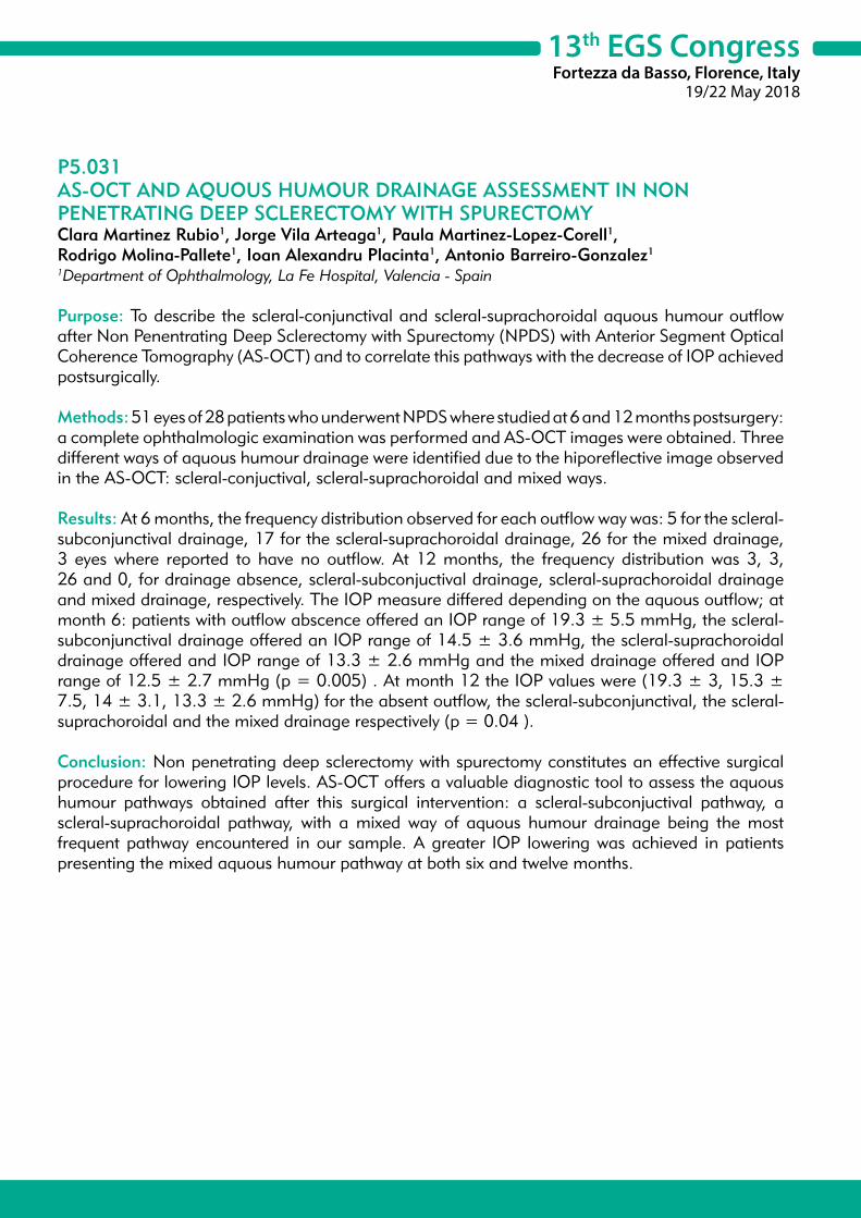
13th EGS CongressFortezza da Basso, Florence, Italy
19/22 May 2018
P5.031AS-OCT AND AQUOUS HUMOUR DRAINAGE ASSESSMENT IN NON PENETRATING DEEP SCLERECTOMY WITH SPURECTOMYClara Martinez Rubio1, Jorge Vila Arteaga1, Paula Martinez-Lopez-Corell1, Rodrigo Molina-Pallete1, Ioan Alexandru Placinta1, Antonio Barreiro-Gonzalez1
1Department of Ophthalmology, La Fe Hospital, Valencia - Spain
Purpose: To describe the scleral-conjunctival and scleral-suprachoroidal aquous humour outflow after Non Penentrating Deep Sclerectomy with Spurectomy (NPDS) with Anterior Segment Optical Coherence Tomography (AS-OCT) and to correlate this pathways with the decrease of IOP achieved postsurgically.
Methods: 51 eyes of 28 patients who underwent NPDS where studied at 6 and 12 months postsurgery: a complete ophthalmologic examination was performed and AS-OCT images were obtained. Three different ways of aquous humour drainage were identified due to the hiporeflective image observed in the AS-OCT: scleral-conjuctival, scleral-suprachoroidal and mixed ways.
Results: At 6 months, the frequency distribution observed for each outflow way was: 5 for the scleral-subconjunctival drainage, 17 for the scleral-suprachoroidal drainage, 26 for the mixed drainage, 3 eyes where reported to have no outflow. At 12 months, the frequency distribution was 3, 3, 26 and 0, for drainage absence, scleral-subconjuctival drainage, scleral-suprachoroidal drainage and mixed drainage, respectively. The IOP measure differed depending on the aquous outflow; at month 6: patients with outflow abscence offered an IOP range of 19.3 ± 5.5 mmHg, the scleral-subconjunctival drainage offered an IOP range of 14.5 ± 3.6 mmHg, the scleral-suprachoroidal drainage offered and IOP range of 13.3 ± 2.6 mmHg and the mixed drainage offered and IOP range of 12.5 ± 2.7 mmHg (p = 0.005) . At month 12 the IOP values were (19.3 ± 3, 15.3 ± 7.5, 14 ± 3.1, 13.3 ± 2.6 mmHg) for the absent outflow, the scleral-subconjunctival, the scleral-suprachoroidal and the mixed drainage respectively (p = 0.04 ).
Conclusion: Non penetrating deep sclerectomy with spurectomy constitutes an effective surgical procedure for lowering IOP levels. AS-OCT offers a valuable diagnostic tool to assess the aquous humour pathways obtained after this surgical intervention: a scleral-subconjuctival pathway, a scleral-suprachoroidal pathway, with a mixed way of aquous humour drainage being the most frequent pathway encountered in our sample. A greater IOP lowering was achieved in patients presenting the mixed aquous humour pathway at both six and twelve months.
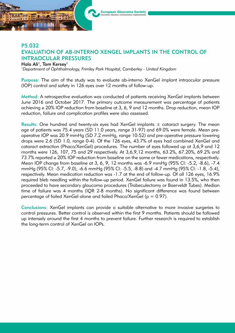
13th EGS CongressFortezza da Basso, Florence, Italy
19/22 May 2018
P5.032EVALUATION OF AB-INTERNO XENGEL IMPLANTS IN THE CONTROL OF INTRAOCULAR PRESSURESHala Ali1, Tom Kersey1
1Department of Ophthalmology, Frimley Park Hospital, Camberley - United Kingdom
Purpose: The aim of the study was to evaluate ab-interno XenGel implant intraocular pressure (IOP) control and safety in 126 eyes over 12 months of follow-up.
Method: A retrospective evaluation was conducted of patients receiving XenGel implants between June 2016 and October 2017. The primary outcome measurement was percentage of patients achieving ≥ 20% IOP reduction from baseline at 3, 6, 9 and 12 months. Drop reduction, mean IOP reduction, failure and complication profiles were also assessed.
Results: One hundred and twenty-six eyes had XenGel implants ± cataract surgery. The mean age of patients was 75.4 years (SD 11.0 years, range 31-97) and 69.0% were female. Mean pre-operative IOP was 20.9 mmHg (SD 7.2 mmHg, range 10-52) and pre-operative pressure lowering drops were 2.6 (SD 1.0, range 0-4). Of the 126 eyes, 43.7% of eyes had combined XenGel and cataract extraction (Phaco/XenGel) procedures. The number of eyes followed up at 3,6,9 and 12 months were 126, 107, 75 and 29 respectively. At 3,6,9,12 months, 63.2%, 67.20%, 69.2% and 73.7% reported ≥ 20% IOP reduction from baseline on the same or fewer medications, respectively. Mean IOP change from baseline at 3, 6, 9, 12 months was -6.9 mmHg (95% CI: -5.2, -8.6), -7.4 mmHg (95% CI: -5.7, -9.0), -6.6 mmHg (95% CI: -5.5, -8.8) and -4.7 mmHg (95% CI: -1.8, -5.4), respectively. Mean medication reduction was -1.7 at the end of follow-up. Of all 126 eyes, 16.9% required bleb needling within the follow-up period. XenGel failure was found in 13.5%, who then proceeded to have secondary glaucoma procedures (Trabeculectomy or Baerveldt Tubes). Median time of failure was 4 months (IQR 2-8 months). No significant difference was found between percentage of failed XenGel alone and failed Phaco/XenGel (p = 0.97).
Conclusions: XenGel implants can provide a suitable alternative to more invasive surgeries to control pressures. Better control is observed within the first 9 months. Patients should be followed up intensely around the first 4 months to prevent failure. Further research is required to establish the long-term control of XenGel on IOPs.

13th EGS CongressFortezza da Basso, Florence, Italy
19/22 May 2018
P5.034EFFECTIVENESS OF TRABECULAR MICROBYPASS STENT IMPLANTATION (ISTENT) ON INTRAOCULAR PRESSURE IN MODERATE AND SEVERE GLAUCOMA: ONE YEAR RESULTSMantapond Ittarat1, Ann Caroline Fisher1, Kuldev Singh1, Robert T. Chang1
1Department of Ophthalmology, Stanford University School of Medicine, Palo Alto - USA
Purpose: To evaluate the effectiveness of single trabecular microbypass stent implantation (iStent) in combination with cataract surgery on intraocular pressure (IOP) and medication reduction in moderate to severe glaucoma.
Methods: Retrospective chart review of all subjects (N = 156) undergoing iStent surgery from January 2013 to October 2017 at a single site among three surgeons. As defined by standard automated perimetry (SAP), 53 eyes had moderate glaucoma with mean deviation (MD) greater than -6 to -12 dB and severe glaucoma with MD greater than -12. Outcome measures include MD and pattern standard deviation (PSD) on SAP, IOP and number of ocular hypotensive medications at time 0, 1, 3 and 12 months follow up, evaluated using Shapiro-Wilk test and histogram. The skewed distribution of data was estimated by the geometric mean and standard deviation.
Results: Among 53 eyes, MD and PSD was 10.72 dB (Min 6.12, Max 31.99) and 8.06 dB (Min 2.01, Max 15.29), respectively. The logarithmic mean of the preoperative IOP 2.80 ± 0.27 mmHg (Geometric mean of preoperative IOP =16.40 mmHg). At 1 year postoperatively, the mean of the postoperative IOP logarithm was 2.61 ± 0.21 mmHg (Geometric mean of preoperative IOP =13.56 mmHg). The change in IOP was -0.19 ± 0.30 mmHg (Geometric mean IOP reduction = 1.21 mmHg, p < 0.001). The number of glaucoma medications was 2.58 ± 1.25 preoperatively and 2.26 ± 1.23 (p = 0.014) at 1 year postoperatively. Eighty-three percent of patients achieved an IOP below 18 mmHg with medications and 4 percent without medication. A filtering procedure was required in 3 eyes at 3 months of follow up after iStent implantation.
Conclusions: Combined cataract surgery with iStent was found to be an effective procedure in patients with moderate and severe glaucoma with modest IOP lowering at 1 year follow up. Several eyes required additional glaucoma procedures in the early postop period.

13th EGS CongressFortezza da Basso, Florence, Italy
19/22 May 2018
P5.035SECOND AHMED GLAUCOMA VALVE IMPLANTATIONTatsiana Imshenetskaya1, Galina Vashkevich1, Yauheni Milasheuski1, Volha Yarmak1
1Department of Ophthalmology, Belarusian Medical Academy of Postgraduate Education, Minsk - Belarus
Purpose: To evaluate the outcome of the second glaucoma valve drainage device (GDD) in patients with failed previous GDD surgery.
Methods: Our study included 37 patients (37 eyes) undergoing a second Ahmed glaucoma valve implantation (AGV) for uncontrolled refractory glaucoma between 2014 and 2017 requiring further glaucoma procedures for intraocular pressure (IOP) control. Data were obtained from the medical records for the preoperative period and after the 1st, 7th, and 15th, 30th day, 3, 6, and 12 months, and then yearly until the last postoperative visit. Outcome measures included visual acuity, IOP, number of glaucoma medications (NGM) and any intraoperative or postoperative complications.
Results: Thirty - seven eyes of 37 patients were studied as having undergone implantation of a second AGV in the same eye with a mean follow-up time of 24 ± 21 months. Most second devices were implanted in the nasal superior quadrant (76,5%) due to better surgical approach and better psychological comfort to the patient . In 21,5% of the eye the second GDD were implanted in the temporal inferior quadrant, in 1% - the second GDV were implanted in the nasal inferior quandrant and in 1% of the second tube were implanted in the vitreous cavity . The mean preoperative IOP was 29.7 ± 1.2 mmHg, and decreased to 15.9 ± 4.0 mmHg after 1 year, 15.5 ± 3.9 mmHg after 2 years. The mean interval between the first and second GDV was 28.8 ± 3.7 months. The qualified success was 89% at the first and second years and 84% at the third year. The most frequent early and late postoperative complications was hypertensive phase (12.1%) and persistent corneal edema (24.5%). In our investigation, 4 patients required additional surgery to eliminate the contact between the tube and the posterior corneal surface.
Conclusions: After failure of an initial drainage implant to control IOP, a repeated AGV implantation is a safe and effective procedure not only to reduce IOP but to preserve the residual visual functions.
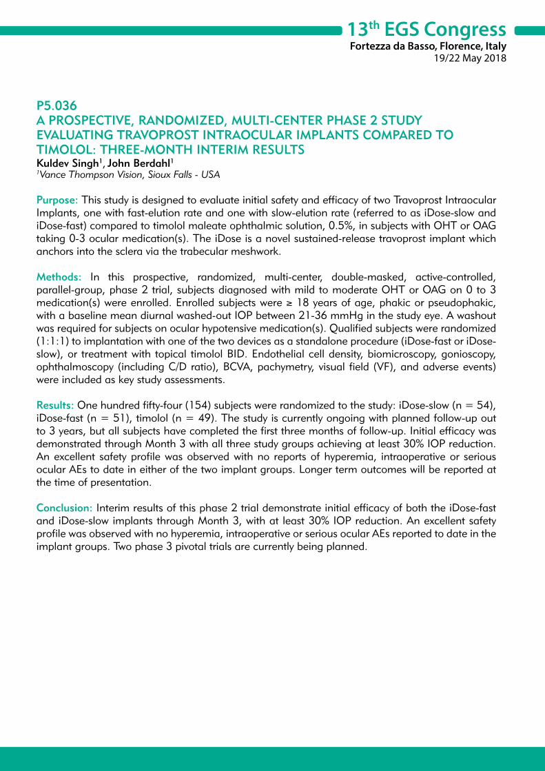
13th EGS CongressFortezza da Basso, Florence, Italy
19/22 May 2018
P5.036A PROSPECTIVE, RANDOMIZED, MULTI-CENTER PHASE 2 STUDY EVALUATING TRAVOPROST INTRAOCULAR IMPLANTS COMPARED TO TIMOLOL: THREE-MONTH INTERIM RESULTSKuldev Singh1, John Berdahl1 1Vance Thompson Vision, Sioux Falls - USA
Purpose: This study is designed to evaluate initial safety and efficacy of two Travoprost Intraocular Implants, one with fast-elution rate and one with slow-elution rate (referred to as iDose-slow and iDose-fast) compared to timolol maleate ophthalmic solution, 0.5%, in subjects with OHT or OAG taking 0-3 ocular medication(s). The iDose is a novel sustained-release travoprost implant which anchors into the sclera via the trabecular meshwork.
Methods: In this prospective, randomized, multi-center, double-masked, active-controlled, parallel-group, phase 2 trial, subjects diagnosed with mild to moderate OHT or OAG on 0 to 3 medication(s) were enrolled. Enrolled subjects were ≥ 18 years of age, phakic or pseudophakic, with a baseline mean diurnal washed-out IOP between 21-36 mmHg in the study eye. A washout was required for subjects on ocular hypotensive medication(s). Qualified subjects were randomized (1:1:1) to implantation with one of the two devices as a standalone procedure (iDose-fast or iDose-slow), or treatment with topical timolol BID. Endothelial cell density, biomicroscopy, gonioscopy, ophthalmoscopy (including C/D ratio), BCVA, pachymetry, visual field (VF), and adverse events) were included as key study assessments.
Results: One hundred fifty-four (154) subjects were randomized to the study: iDose-slow (n = 54), iDose-fast (n = 51), timolol (n = 49). The study is currently ongoing with planned follow-up out to 3 years, but all subjects have completed the first three months of follow-up. Initial efficacy was demonstrated through Month 3 with all three study groups achieving at least 30% IOP reduction. An excellent safety profile was observed with no reports of hyperemia, intraoperative or serious ocular AEs to date in either of the two implant groups. Longer term outcomes will be reported at the time of presentation.
Conclusion: Interim results of this phase 2 trial demonstrate initial efficacy of both the iDose-fast and iDose-slow implants through Month 3, with at least 30% IOP reduction. An excellent safety profile was observed with no hyperemia, intraoperative or serious ocular AEs reported to date in the implant groups. Two phase 3 pivotal trials are currently being planned.

13th EGS CongressFortezza da Basso, Florence, Italy
19/22 May 2018
P5.037THE EFFECT OF PHOTOBIOMODULATION ON A RAT ACUTE OCULAR HYPERTENSION RETINAL ISCHEMIA/REPERFUSION MODELJennifer Wei Huen Shum1,2, Jimmy Lai2
1Department of Ophthalmology, Caritas Medical Centre, Hong Kong - Hong Kong2Department of Ophthalmology, The University of Hong Kong, Hong Kong - Hong Kong
Purpose: Photobiomodulation (PBM) is a non-invasive treatment that shows potential for treating various neuronal and vascular diseases. Its biological effects include enhancing mitochondrial metabolism, improving microcirculation, decreasing oxidative stress, anti-inflammation and neuroprotection. Glaucoma pathogenesis is multifactorial involving both mechanical damage through elevated intraocular pressure and vascular dysregulation. This leads to secondary insult involving disrupted microcirculation, excitotoxic neuronal injury and oxidative damage via various reactive oxygen species. We hypothesize that PBM may have a neuroprotective effect in glaucoma, because its biological effects coincide with the mechanisms of the secondary insult in glaucoma.
Methods: A rat retinal ischemia/reperfusion model induced by acute ocular hypertension (AOH) was used as an animal glaucoma model. Rats were randomized into receiving treatment (PBM) or no treatment (no PBM). The PBM regimen was adopted with reference to major PBM studies. At 1 day after AOH, we analyzed apoptotic count. At 1 week after AOH, we conducted RGC count and electroretinography (ERG) to assess retinal function.
Results: Among eyes that underwent AOH, apoptotic TUNEL analysis showed a reduced mean apoptotic count in eyes that received PBM when compared with no PBM, which was statistically significant. For RGC count, it was significantly reduced in eyes undergoing AOH compared to those not receiving AOH. Eyes receiving PBM and AOH showed a higher RGC count than eyes undergoing AOH only, but statistical significance was not reached. For ERG analysis, AOH was shown to result in pan-retinal dysfunction. PBM did not show significant improvement in ERG measurement.
Conclusion: In conclusion, our work suggests that PBM may have a beneficial role in glaucoma, as suggested by the reduced apoptotic count and higher RGC count observed among eyes that received PBM. General ERG may be too non-specific to assess RGC function. Suggestions for future study directions include adding different study time points and behavioral tests for retinal function.
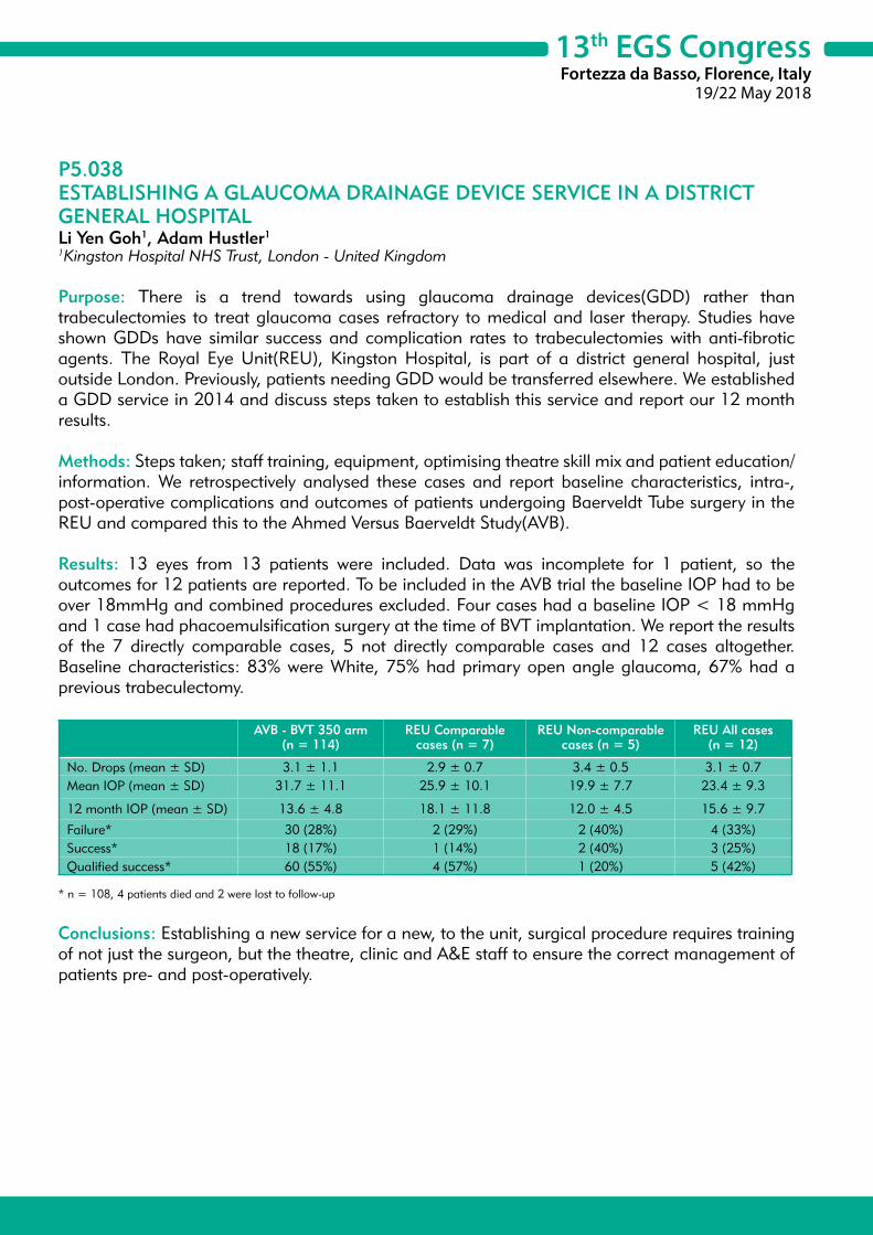
13th EGS CongressFortezza da Basso, Florence, Italy
19/22 May 2018
P5.038ESTABLISHING A GLAUCOMA DRAINAGE DEVICE SERVICE IN A DISTRICT GENERAL HOSPITALLi Yen Goh1, Adam Hustler1
1Kingston Hospital NHS Trust, London - United Kingdom
Purpose: There is a trend towards using glaucoma drainage devices(GDD) rather than trabeculectomies to treat glaucoma cases refractory to medical and laser therapy. Studies have shown GDDs have similar success and complication rates to trabeculectomies with anti-fibrotic agents. The Royal Eye Unit(REU), Kingston Hospital, is part of a district general hospital, just outside London. Previously, patients needing GDD would be transferred elsewhere. We established a GDD service in 2014 and discuss steps taken to establish this service and report our 12 month results.
Methods: Steps taken; staff training, equipment, optimising theatre skill mix and patient education/information. We retrospectively analysed these cases and report baseline characteristics, intra-, post-operative complications and outcomes of patients undergoing Baerveldt Tube surgery in the REU and compared this to the Ahmed Versus Baerveldt Study(AVB).
Results: 13 eyes from 13 patients were included. Data was incomplete for 1 patient, so the outcomes for 12 patients are reported. To be included in the AVB trial the baseline IOP had to be over 18mmHg and combined procedures excluded. Four cases had a baseline IOP < 18 mmHg and 1 case had phacoemulsification surgery at the time of BVT implantation. We report the results of the 7 directly comparable cases, 5 not directly comparable cases and 12 cases altogether. Baseline characteristics: 83% were White, 75% had primary open angle glaucoma, 67% had a previous trabeculectomy.
AVB - BVT 350 arm (n = 114)
REU Comparable cases (n = 7)
REU Non-comparable cases (n = 5)
REU All cases(n = 12)
No. Drops (mean ± SD) 3.1 ± 1.1 2.9 ± 0.7 3.4 ± 0.5 3.1 ± 0.7Mean IOP (mean ± SD) 31.7 ± 11.1 25.9 ± 10.1 19.9 ± 7.7 23.4 ± 9.3
12 month IOP (mean ± SD) 13.6 ± 4.8 18.1 ± 11.8 12.0 ± 4.5 15.6 ± 9.7
Failure* 30 (28%) 2 (29%) 2 (40%) 4 (33%)Success* 18 (17%) 1 (14%) 2 (40%) 3 (25%)Qualified success* 60 (55%) 4 (57%) 1 (20%) 5 (42%)
* n = 108, 4 patients died and 2 were lost to follow-up
Conclusions: Establishing a new service for a new, to the unit, surgical procedure requires training of not just the surgeon, but the theatre, clinic and A&E staff to ensure the correct management of patients pre- and post-operatively.

13th EGS CongressFortezza da Basso, Florence, Italy
19/22 May 2018
P5.039THE IMPACT OF LONG TERM GLAUCOMA MEDICATIONS ON STRUCTURE AND FUNCTION OF MEIBOMIAN GLANDSAntoine Rousseau1, Benjamin Scemla1, Solène Andrieu1, Céline Lambotin1, Brigitte Rousseau1, Emmanuel Barreau1, Marc Labetoulle1
1Department of ophthalmology, Le Kremlin-Bicêtre - France
Purpose: Glaucoma treatments may be associated with meibomian gland disease (MGD). We used multiple clinical and imaging parameters to assess the impact of glaucoma treatment on meibomian gland structure and function.
Methods: We included 18 eyes of 18 long term treated glaucoma patients (> 12 months) and 14 eyes of 14 age-matched controls in this prospective cross-sectional study. Ocular surface evaluation included ocular surface fluorescein staining (Oxford scheme), tear break-up time (TBUT), Schirmer I test, clinical MGD score, lipid layer thickness (LLT) measurement using interferometry. Meibomian glands (MG) structure was studied using non contact infrared meibography and scored for atrophy (meiboscore) shortening and distortion. Ocular surface related quality of life was evaluated using Ocular Surface Disease Index (OSDI) and OSD QoL questionnaires. Topical glaucoma treatments number, type and duration were noted. Kruskal-Wallis non parametric test was used for statistical comparison and Spearman test for correlations.
Results: Patients received a mean of 1.8 ± 0.7 eyedrops with a mean treatment duration of 35 months. Ocular surface staining and clinical MGD score were significantly higher in patients than in controls. Structural study of MG using meibography revealed that shortening and atrophy of meibomian glands (but not distortion) was significantly higher in patients as compared to controls (p = 0.05 and p < 0.01, respectively). TBUT tended to be significantly shorter in patients (p = 0.07). However, there was no significant difference in LLT, OSDI and OSD QoL scores between patients and controls. Besides, we did not find correlation between number and duration of treatments with MG abnormalities.
Conclusions: These preliminary results suggest that glaucoma treatments induce morphological and potentially functional changes of MG. Further studies are warranted to elucidate the role of preservatives and further characterize the risk factors of MG alterations in topically treated glaucoma patients.
13th EGS CongressFortezza da Basso, Florence, Italy
19/22 May 2018
P5.040OCULAR COMPLICATIONS OF COSMETIC IRIDIAN IMPLANTS IN 8 EYESImelda Ramirez1, Juan Carlos Izquierdo1, Fabiola Quezada1, Maria Corina Ponte1, Barbara Rubio1, Laura A. Cañola-Ramirez1
1Instituto de Ojos Oftalmosalud (Research Department), Lima - Peru
Purpose: To report the presentation, management, and complications of 8 eyes of 4 phakic patients presenting with iridian cosmetic implants.
Methods: Descriptive case report. In this evaluation of patients with cosmetic iridian implants, data collected included patient demographics, visual acuity, intraocular pressure, endothelial cell count and slitlamp examination findings at presentation. Medical and surgical interventions and the postoperative course were recorded.
Results: Eight eyes of four patients (ages 24 to 48; 50% men) were identified. Three eyes (37.5%) presented with severe decreased in visual acuity, 6 (75%) had elevated IOP despite the use of maximal tolerated antiglaucoma therapy, 6 (75%) had corneal edema. 6 of the 8 eyes had explantation of the iris prosthesis in a range of 9 to 72 months. One patient refused to explant. Postoperative complications included hiphema in the ab interno trabeculectomy, cataract, and corneal edema. Secondary surgeries included cataract extraction with intraocular lens placement (2 eyes), conventional trabeculectomy (1 eye) ab interno trabeculectomy (1 eye), Descemet’s Membrane Endothelial Keratoplasty (2 eyes), Ex-Press shunt placement (1 eye), Baerveldt drainage device (1 eye), cyclophotocoagulation (2 eyes, 1 transcleral, 1 endoscopic)
Conclusions and importance: Anterior iridian cosmetic implants is often used in young and healthy patients. It can lead to severe ocular morbidity and complications even after explantation, such as difficulty in managing secondary glaucoma, corneal failure, cataract and irreversible visual loss.

13th EGS CongressFortezza da Basso, Florence, Italy
19/22 May 2018
P5.041TRANSSCLERAL CYCLOPHOTOCOAGULATION WITH DIODE LASER FOR REFRACTORY GLAUCOMA. OUR EXPERIENCEBeatriz Rodriguez Aguado1, Lorena Almudi Cortes1, Iris Luengo Arroyo1, Mouafk Asaad1
1Department of Ophtalmology, Terrassa- Barcelona - Spain
Purpose: Ciclodestructive procedures have been successfully used in the treatment of refractory glaucoma. The goal of this study is to describe our experience with micropulse transscleral cyclophotogoaculation (MP3- TSCPC) in our patients with uncontrolled glaucoma.
Methods: This is a retrospective view of the cases treated with TSCPC in our ophthalmology service. We treated 10 patients with uncontrolled glaucoma, 6 of them with neovascular glaucoma, 2 refractary glaucoma, one had previous retina surgery and one refractory glaucoma. Laser settings were 2000mW of diode laser on micropulse delivery mode during 240 seconds placed over 270º of the limbus. The mean follow-up was 6 months heretofore. Surgical success was defined as a reduction of intraocular pressure (IOP) by 20 %. We visited our patients at 24 hours, 1st and 4th
weeks and 3rd-6th month.
Results: The mean preoperative intraocular pressure was 39.2 mmHg. Significant IOP reduction was observed during the first week after treatment. 2 patients withpainful eyes had pain relief. Patients with higher preoperative IOP suffered a new rise of IOP at first month; two of them underwent a second treatment. At the 6th month we have observed a significant IOP reduction in 8 of the 10 patients.
Conclusions: Traditionally, TSCPC is reserved for end-stage. We have observed that patients tolerate the procedure very well, and relieve ocular pain due to high IOP. This study suggest that micropulse diode transscleral cyclophotocoagulation can be used successfully to reduce intraocular pressure in the treatment of refractory glaucoma.
13th EGS CongressFortezza da Basso, Florence, Italy
19/22 May 2018
P5.042SHORT-TERM SAFETY AND EFFICACY OF CARTEOLOL/LATANOPROST FIXED-COMBINATION OPHTHALMIC SOLUTION IN ACTUAL CLINICAL SETTINGKaori Tada1, Yoko Ikeda1, Morio Ueno1, Kazuhiko Mori1, Shigeru Kinoshita2, Chie Sotozono1
1Department of Ophthalmology, Kyoto Chubu Medical Center, Kyoto - Japan2Department of Frontier Medical Science and Technology for Ophthalmology, Kyoto Prefectural University of Medicine, Kyoto - Japan
Purpose: In September 2016, long-acting 2% carteolol hydrochloride and latanoprost fixed-combination eye drops (Car/Lat; Mikeluna® Combination Ophthalmic Solution, Otsuka Pharmaceutical Co., Ltd.) was approved for use in Japan. In this study, we investigated the safety and efficacy of Car/Lat in the actual clinical setting.
Methods: This study involved 62 eyes of 62 patients (18 male /44 female; mean age 66.8 ± 14.0 years) who had been prescribed Car/Lat to strengthen the existing medication (add group) or to switch from similar the medications (switch group) in the actual clinical setting of Glaucoma Clinic in Kyoto Prefectural University of Medicine, and Oike-Ikeda Eye Clinic, Kyoto, Japan. Intraocular pressure (IOP) measured by Goldmann applanation tonometer before the initiation (baseline) and after 1-month administration, and associated adverse events were investigated. Paired t-test was used for statistical analysis. If data from both eyes were available, right-eye data were selected.
Results: Of the 62 patients, 10 were primary open angle glaucoma (POAG), 36 were normal tension glaucoma (NTG), and 16 were other types of glaucoma. Distribution of eyes between add group and switch group were 38 (11 male / 27 female, mean age: 67.26 ± 13.9 years) and 24 (8 male / 16 female, 65.9 ± 13.8 years) respectively. In switch group, mean IOP at baseline vs. 1 month were 13.4 ± 4.1 vs. 13.0 ± 3.4 mmHg (no significant difference), while in add group, 14.2 ± 3.8 vs. 12.1 ± 3.8 mmHg [significant IOP reduction (p < 0.001)], respectively. Adverse events observed during the follow-up periods included eye pain (3 eyes, 4.8%), conjunctival hyperemia (1 eye, 1.6%), itching (1 eye, 1.6%), and corneal erosion (1 eye, 1.6%). Three patients (1 from add group and 2 from switch group) discontinued because of these adverse events, while the others continued the administration of Car/Lat.
Conclusion: Car/Lat was found effective for maintaining IOP (switch group) or strengthening IOP lowering effect (add group) with safe and well-tolerated profile in the actual clinical setting.

13th EGS CongressFortezza da Basso, Florence, Italy
19/22 May 2018
P5.043IMPLANTATION OF TWO FIRST-GENERATION TRABECULAR MICRO-BYPASS STENTS AS A STANDALONE PROCEDURE COMBINED WITH TOPICAL PROSTAGLANDIN IN SUBJECTS WITH OAG ON 2 PREOPERATIVE MEDICATIONS: FIVE YEAR OUTCOMESJose I. Belda1, Lucia Rial1
1Department of Ophthalmology, Hospital Universitario de Torrevieja, Torrevieja - Spain
Purpose: Prospective evaluation of safety and IOP following implantation of two trabecular micro-bypass stents (iStent®) and treated with topical travoprost started postoperatively on Day 1 in subjects with open-angle glaucoma (OAG) who were using two ocular hypotensive medications preoperatively.
Methods: Subjects with OAG on 2 preoperative ocular hypotensive medications with mean medicated IOP of 18-30 mmHg and unmedicated (post-washout) IOP of 22-38 mmHg were enrolled into this prospective single arm study. Qualified subjects were successfully implanted with 2 iStent devices as a standalone procedure. Topical travoprost was started on postoperative Day 1. Additional ocular hypotensive medication was prescribed if a subject’s postoperatively IOP was > 21 mm Hg. Subjects were evaluated at Day 1, Week 1, 3, 6, 12 and every six months through 60 months with annual washouts. As part of routine follow-up, the following efficacy and safety assessments were conducted: IOP, medication usage, slit-lamp examination, gonioscopy, fundus/optic nerve evaluation, BCVA, and AEs.
Results: All subjects underwent uncomplicated implantation with 2 iStent devices and no intraoperative or device-related AEs noted. Post-operative, there were consistent reductions in IOP. Mean medicated IOP was ≤ 14.2 mmHg at all visits through M48. When comparing medicated IOP, at M48 the mean IOP was 12.8 ± 1.7 mmHg compared to 22.4 ± 2.3 mmHg preoperatively. Postoperative mean unmedicated IOP was ≤ 17.7 mmHg at all visits, and at M49 the mean unmedicated IOP was 16.9 ± 1.2 mmHg vs 25.3 ± 1.9 mmHg preoperatively. At M48, IOP ≤ 18 mmHg on travoprost was observed in 91% of subjects. Outcomes to 5 years is intended to be presented. AEs reported include progression of pre-existing cataract (4 of 5 eyes underwent cataract surgery) and trabeculectomy at month 24 for glaucoma progression (one eye); all deemed unrelated to stent implantation.
Conclusion: Treatment with 2 iStent devices combined with topical travoprost can safely achieve significant and sustained reduction of IOP to ≤ 14 mmHg and reduced medication burden through 48 months postop.

13th EGS CongressFortezza da Basso, Florence, Italy
19/22 May 2018
P5.044METHOD OF SURGICAL TREATMENT OF PATIENTS WITH RESISTANT FORMS OF GLAUCOMAGrigoriy Malinovsky1, Nataliya Sidenko2, Ana Dvalishvili1
1Belarusian Medical Academy of Post Graduate Education, Minsk - Belarus210th State Clinical Hospital, Minsk - Belarus
Purpose: To improve the effectiveness of surgical treatment of resistant glaucoma.
Methods: We developed an operation - deep meridional scleral trabeculectomy with autoscreening (PATENT No. 13653, July 21, 2010, Republic of Belarus). The subject of the study were 61 patients (61 eyes) with surgery-resistant forms of glaucoma with the presence of risk factors for excessive scarring. The average age is 66.5 years, of which: men - 34, (55.7%), women - 27 (44.3%). According to the degree of “refractoriness”, according to the algorithm proposed by A.M. Immortal and V.P. Erichev, the studied patients were divided into three groups: Group I - 5 patients with low risk of scarring; Group II - 38 patients with moderate risk of scarring; Group III - 18 patients with neovascular glaucoma and a high risk of scarring, of which 13 patients with diabetes mellitus and 5 patients after central retinal vein thrombosis. All patients were operated according to the method developed by us.
Results: At the follow-up of more than 1 year, all patients treated received a positive result. Compensation of ophthalmotonus and preservation of visual functions in patients of the first and second groups was noted in 91.8% of operated patients, in 19 of them with the use of local antihypertensive agents. In the group of patients with high risk of scarring, IOP compensation was observed in 55% of cases. And in five patients subcompensation of IOP without pain syndrome was observed, and three patients underwent additional transcleral laser traetment.
Conclusions: 1. Deep meridional sclerosal-sinutrabecullectomy with auto sclera dranaige is an effective and predictable operation in the treatment of patients with resistant forms of glaucoma. 2. A differentiated approach to the choice of the operation method, allows to increase the results of treatment of this category of patients.

13th EGS CongressFortezza da Basso, Florence, Italy
19/22 May 2018
P5.045USE OF TISSUE ADHESIVE IN COMPLEX GLAUCOMA SURGERYRomero Royo Concepción1, Fernandez Garcia Aitor1, Perucho Martinez Susana2, Martin Giral Elena2, Fernandez Escamez Carlos2, Poyales Galan Francisco1, Pérez Izquierdo Ricardo1, Poyales Villamor Blanca1
1Department of Ophtalmology IOA Madrid Innova Ocular, Madrid - Spain2Department of Ophtalmology Fuenlabrada Hospital, Madrid - Spain
Purpose: To analyse tissue adhesive usefulness for conventional glaucoma surgery and in particularly complex patient cases.
Methods: This case study comprises three surgical procedures carried out by the Glaucoma Unit at IOA Madrid Innova Ocular. Each case is presented - together with surgical photographs, anterior-pole OCT-Visante images and serial ultrasound-scan images - and thoroughly discussed.
Results: First case: Male patient who had undergone combined cataract-glaucoma surgery (PHACO-XEN) with no intraoperative complications. One week later a dysesthetic bleb is formed, which doesn’t improve neither with conventional topical therapy nor with compression measures. It is decided to drain this conjunctival bleb and to fill the bottom half with tissue adhesive, by means of the so-called “Dry-Lake” technique, which results in a significant improvement of the bleb and of the patient’s discomfort. The case is presented accompanied by serial photographs and OCT images of the anterior chamber. Second case: Female patient who had undergone glaucoma surgery with Ahmed glaucoma valve implantation and scleral patch graft. However, a conjunctival suture dehiscence occurs, which has to undergo resuturing on three consecutive occasions. It is then decided to perform a scleral de-epithelialization (i.e., epithelium removal) with absolute alcohol injection and the use of tissue adhesive, and a subsequent resuturing with 10.0 nylon mattress stitches. This procedure resulted in the condition being resolved. The case is presented accompanied by serial photographs. Third case: Male patient who had undergone glaucoma surgery; more specifically, trabeculectomy with EX-PRESS valve implantation. Multiple conjunctival ruptures occurred during surgery, which renders impossible a primary conjunctival closure. It is then decided to perform an autologous conjunctival graft (i.e., autograft) using tissue from the same eye, which is then adhered by means of tissue adhesive and a conjunctival suture. The case is presented accompanied by serial photographs.
Conclusions: The use of tissue adhesive can be of great help in complex glaucoma surgery cases to strengthen the sclero-conjunctival junction, thus acting as coadjuvant to traditional suture.

13th EGS CongressFortezza da Basso, Florence, Italy
19/22 May 2018
P5.046ADDITIONAL TUBE SHUNT WITH BAERVELDT IMPLANT AFTER FAILED AHMED GLAUCOMA VALVE SURGERY AND REVISIONSRishu Inoue1
1Olympia Eye Hospital, Tokyo - Japan
Purpose: To describe postoperative surgical success of additional Baerveldt implantation after failed Ahmed tube shunt surgery and revisions for refractory glaucoma.
Case: A woman who is identified with congenital glaucoma underwent gonio surgery in both eyes at 6 months old. She went through enucleation in her left eye at the age of 12 due to absolute glaucoma. She had trabeculectomy twice in her right eye in her mid-twenties. However, her intraocular pressure (IOP) was high in spite of medications. At 56 years old, we performed an Ahmed glaucoma valve (AGV) implantation in the superior temporal quadrant and required a tube revision 3 months after. Intraocular pressure elevated a month later, a second tube shunt in the inferior temporal quadrant was performed. Intraocular pressure was often spiked at 30mmHg with medications. Forty months after the second tube shunt implantation, the patient received four revisions in six years at 1-4 year interval. In 2013, a third tube shunt surgery was deemed necessary. With Baervedlt implant (BI) surgery, her IOP was significantly reduced and stable without medications. We evaluated tube function through magnetic resonance imaging, and found that the bleb area was larger around BI than AGV.
Conclusion: This case showed that implantation of Baervedlt tube shunt provided successful postoperative reduction in IOP.
13th EGS CongressFortezza da Basso, Florence, Italy
19/22 May 2018
P5.047LIFESPAN OF ENDOCYCLOPHOTOCOAGULATION PROBESMarina Syrimi1, Sheng Lim1
1Department of Ophthalmology, Guys and St Thoma’s Foundation Trust, London - United Kingdom
Introduction: Endocyclophotocoagulation (ECP) is controlled distraction of the ciliary body using 810 microns diode laser and achieving reduction in aqueous humour production. ECP is increasingly being used for the management of glaucoma in non uveitis patients. There is no published literature on the lifespan of ECP probes.
Objective: Our study aims to identify how many ECP procedures were performed in our tertiaty referral hospital and how many probes were needed to achieve this.
Methods: The total number of phaco-ECP/ECP alone procedures that were performed at our hospital from the start up to the 09/11/2017 were identified from our coding system. The following search combinations were used: Laser to ciliary body aloneLaser to ciliary body + corneal suturePhacoemulsification (phaco) + laser to ciliary body Phacoemulsification (phaco) + laser to ciliary body + corneal sutureThe corneal suture code was used by some nursing staff to acknowledge the higher cost of ECP versus trans-scleral diode laser. The number of ECP probes used was identified from our order system.
Results: A total of 482 cases were identified from the search. Trans-scleral cyclodiode laser and High frequency ultrasound ablation of the ciliary body were excluded from the study. This resulted in a total of 231 cases of phaco-ECP/ECP alone performed in our unit until the 9th of November 2017. The total of ECP probes ordered until then was 24. This equals to ECP 9.6 procedures per probe.
Conclusion: Our study supports that ECP is a good economical Investment for lowering the intraocular pressure compared to other minimally invasive glaucoma surgery procedures.

13th EGS CongressFortezza da Basso, Florence, Italy
19/22 May 2018
P5.048EARLY REFRACTIVE EFFECT OF CATARACT SURGERY IN MEDICALLY CONTROLLED GLAUCOMAEmine Sen1, Dilara Ozkoyuncu1, Ufuk Elgin1
1SBÜ Ulucanlar Eye Education and Research Hospital, Ankara - Turkey
Purpose: To evaluate the refractive outcomes of phacoemulsification in patients with glaucoma under medical treatment.
Methods: The study group with glaucoma under medical treatment was prospectively compared with a nonglaucomatous control group. Lenstar laser interferometry was used for ocular biometry measurements. The difference between the predicted and current postoperative refractive changes was measured by intraocular lens (IOL) prediction formulae (Haigis, Holladay 2, Hoffer Q, and SRK-II) preoperatively and at the 1st, 7th and 30th days. The difference between the predicted and current postoperative changes was considered a ‘Mean Error’ (ME). The accuracy of several IOL calculation formulas (SRK-II, Holladay, Hoffer Q and Haigis) was also compared by examining ME of the groups.
Results: In totally, 30 eyes of 30 cases were included to the study (17 eyes in the study group and 13 eyes in the control group). The eyes in the study group had significantly more myopic shift than the control group according to Holladay (1st day p = 0.001, 7th day p = 0.200, 30th day p = 0.74), Hoffer Q (1st day p = 0.006, 7th day p = 0.148, 30th day p = 0.124) and SRK-II (1st day p = 0.100, 7th day p = 0.449, 30th day p = 0.207) IOL formulas postoperatively. In the study group the ME calculated by the SRK-II formula was more accurate than the other formulas (Mean ± SD ME 0.150 ± 0.511, p = 0.207). However, for each measurement in both groups, Haigis formula had significantly more hyperopic shift than the other formulas (1st day p = 0.006, 7th day p = 0.005, 30th day p = 0.001).
Conclusions: When phacoemulsification and intraocular lens implantation was performed in patients with glaucoma, the probable the myopic shift should be kept in mind. The least the myopic shift was seen in the SRK II. Formulas for the intraocular lens may differ between glaucoma subgroups.
13th EGS CongressFortezza da Basso, Florence, Italy
19/22 May 2018
P5.049“BAERVELDT VALVE FLAP-LESS IMPLANT”: THE FLORENTINE EXPERIENCETomaso Caporossi1, Fabrizio Franco1, Lucia Finocchio1, Ruggero Tartaro1, Stanislao Rizzo1
1Eye Clinic AOU Careggi, Florence
Purpose: To evaluate the efficacy and safety of a Baerveldt valve implant technique without creating a scleral flap and without using a scleral patch, in case of refractory glaucoma in particular in case of secondary glaucoma after vitreoretinal surgery.
Methods: Surgery is reserved for cases of refractory glaucoma and is performed under locoregional anesthesia. Before the implant, the valve is prepared using a 5/00 prolene suture inside the tube lumen and a 7/00 vycril suture to throttle the tube. The valve is positioned in the temporal quadrant. Then a pocket is created in the scleral thickness, until it reaches the blue line. Finally, with a needle you enter the anterior chamber and position the tube.
Results: 26 eyes of 26 patients underwent Baerveldt valve implant for refractory glaucoma. 10 left eyes and 16 right eyes. The surgery was performed by three expert surgeon in the Eye Clinic of AOU Careggi in Florence in the last two years. In 21 cases patients had previous vitreoretinal surgery, in the other 5 cases they had previous glaucoma surgery (trabeculectomy or Ex-Press). In all cases the intraocular pressure is under control after valve implant. In two cases patients still use IOP therapy and in one case the patient underwent cyclophotocoagulation and still use IOP therapy. In one case we removed the prolene suture from the tube. In 2 cases we had choroidal detachment during follow-up managed with medical therapy. In 3 cases we registered opacity and corneal decompensation, probably because they already had a compromised corneal situation. In no case we recorded complications related to the tube exposure or to the scleral pocket. In no case did the tube touch the corneal endothelium.
Conclusion: “Baerveldt valve flap-less implant” is a safe and effective technique, the learning curve is very easy and the incidence of tube complications was lower.

13th EGS CongressFortezza da Basso, Florence, Italy
19/22 May 2018
P5.050THE EFFICACY AND SAFETY OF THE XEN GEL IMPLANT IN POAG AND PXG PATIENTSRafael Kasina1, Mario Alexander Economou1
1St Erik Eye Hospital, Stockholm - Sweden
Purpose: To investigate the safety and efficacy of the Xen implant (Allergan, Dublin, Ireland) alone or in combination with phacoemulsification in patients with medically uncontrolled glaucoma.
Setting/Venue: The study was conducted at St. Erik Eye Hospital at the Karolinska Institute in Stockholm.
Methods: Retrospective chart review of 43 eyes of 39 patients with POAG (Primary Open Angle Glaucoma) or PXG (Pseudoexfoliation Glaucoma) that underwent either implantation of Xen alone or in combination with cataract surgery. Primary outcome measures with one year follow-up included intra-operative and post-operative complications, visual acuity, intraocular pressure (IOP), and number of glaucoma medications.
Results: 12 eyes underwent combined phaco-Xen procedure and 31 eyes only Xen. The average age of the patients was: 74.5. 24 had POAG and the remaining 19 had PXG with an open angle. 36 (83.7%) eyes had undergone at least one prior laser trabeculoplasty treatment. 62.8% of patients had IOP > 21 mmHg on decision for surgery. IOP and Medication. Mean pre-op IOP in the combined group was 22.8 ± 4.7 mmHg on 2.8 ± 1.0 medications and in the Xen alone group 23.0 ± 5.7 mmHg on 3.3 ± 1.4 medications. Mean IOP was reduced to 16.5 ± 3.1 mmHg (p < 0.05) and 0.3 ± 0.7 medications (p < 0.05) in the combined group and 15.5 ± 4.3 mmHg (p < 0.05) in the Xen alone group at 1- year follow up on 0.5 ± 1.1 (p < 0.05) glaucoma medications. IOP lowering effect was 27.7% for the combined group and 33.0% for the Xen alone group. 12 eyes required needling (30%). 7 patients in the Xen alone group developed early hypotony.
Conclusions: The ab interno gelatin microstent with MMC is an alternative to trabeculectomy. It shows similar IOP-lowering effects. Our study indicates a lower rate of major complications, but with more postsurgical interventions, mainly in the form of needlings in comparison to suture manipulation postoperatively in trabeculectomy. Further study with longer-term follow up and a larger number of patients are needed to fully assess the utility of this device.

13th EGS CongressFortezza da Basso, Florence, Italy
19/22 May 2018
P5.051INTRAOPERATIVE ROCK-HARD EYE SYNDROME IN COMBINED GLAUCOMA SURGERYBueno Palacin Jose1, Fernandez Garcia Aitor1, Poyales Galan Francisco1, Garzon Jimenez Nuria1, Perez Izquierdo Ricardo1, Poyales Villamor Blanca1
1Department of Ophtalmology IOA Madrid Innova Ocular, Madrid - Spain
Purpose: To analyse the signs, symptoms and prognosis of the different factors that might trigger a sudden rise in intraoperative intraocular pressure during a combined cataract-glaucoma surgery.
Methods: This case study comprises three surgical procedures carried out by the Retina and Glaucoma Unit at IOA Madrid Innova Ocular. Each case is presented and analysed, together with surgical photographs, anterior-pole OCT-Visante images and serial ultrasound-scan images.
Results: First case: Patient diagnosed with both narrow-angle glaucoma and cataract. During combined surgery there was an increase in intraocular pressure with a significant shallowing of the anterior chamber together with iris prolapse. Following eye fundus examination - and once choroidal haemorrhage had been ruled out - the patient underwent 21-gauge dry pars plana vitrectomy. The case was thus solved, and the surgery was successfully completed with no further complications. Diagnosis: fluid misdirection syndrome.Second case: Patient diagnosed with both narrow-angle glaucoma and cataract. Intraocular pressure rises during femtosecond-laser cataract surgery, and the eye-fundus reflection diminishes. A 23-gauge central vitrectomy is then performed, but IOP did not decrease significantly. The subsequent eye-fundus examination revealed a peripheral choroidal haemorrhage. Surgery was therefore delayed, but two weeks later - once the haemorrhage had been solved - it was completed uneventfully. Diagnosis: Intraoperative choroidal haemorrhage. Third case: Patient diagnosed both with narrow-angle glaucoma and cataract. Sudden IOP rise during intracameral anaesthesia delivery. Surgery is therefore delayed but, following a 23-gauge pars plana vitrectomy, it is then completed with no further complications. Diagnosis: fluid misdirection syndrome.
Conclusions: The rise in intraocular pressure is an infrequent complication in combined cataract-glaucoma surgery. A rapid and accurate diagnosis is required so as to prevent more severe complications and to be able to successfully complete the surgical procedure. Most cases require central vitrectomy in order to reduce vitreous prevision, but a choroidal haemorrhage should always be ruled out first.
13th EGS CongressFortezza da Basso, Florence, Italy
19/22 May 2018
P5.052FEMTOSECOND-LASER USAGE IN GLAUCOMA PATIENTSFernandez Garcia Aitor1, Garzon Jimenez Nuria1, Poyales Galan Francisco1, Poyales Villamor Blanca1, Perez Izquierdo Ricardo1, Poyales Galan Concepcion1
1Department of Ophtalmology, IOA Madrid Innova Ocular, Madrid - Spain
Purpose: Femtosecond laser is being increasingly employed in cataract surgery. A high proportion of glaucoma patients have very narrow chambers and could therefore benefit from this technology, in the context of cataract surgery alone (prior to the subsequent glaucoma procedure) but also in combined surgeries.
Methods: Eight patient cases were analysed and subsequently split into two separate groups: the first one comprised 4 patients who had previously undergone NPDS with functioning blebs. They then underwent cataract surgery at our clinic using femtosecond-laser-assisted phacoemulsification. The second group comprised 4 patients who underwent combined cataract-glaucoma surgery; i.e., femtosecond-laser-assisted phacoemulsification plus XEN™ gel stent implantation in the same surgical procedure. As outcome measures we chose epidemiological data, IOP, anterior-chamber depth (ACD), number of drugs that were employed, the laser’s vacuum time and the amount and severity of perioperative complications. The study includes anterior chamber’s serial photographs and OCT images.
Results: Mean vacuum time was: 84 seconds (77-98). Within the first group, Mean IOP was: 15.2 (12.3-22). Mean ACD was: 2.45 (2.11-2.90). The mean number of treatments ranged from (pre /1 week/1 month /3 months): 1.4 (1-3) / 0 / 0 / 0. As for the second group, mean IOP was: 24.2 (22.3-27.4). Mean ACD was: 2.23 (1.98-2.72).The mean number of treatments ranged from: 2.25 (2-3) / 0 / 0 / 0.25. Neither intra- nor post-operative complications occurred in either group. Subconjunctival petechiae were observed in 3 eyes, but they had no impact upon the surgical procedure. As for the implanted IOLs, 6 of them were monofocal, 1 was toric and 1 was an extended-range IOL.
Conclusions: Femtosecond laser may well be used in patients with prior glaucoma in the context of both cataract surgery alone as well as in combined surgeries. For narrow-chamber eyes this technique offers a solid safety profile, thus making the procedure somewhat easier facilitating the procedure.
13th EGS CongressFortezza da Basso, Florence, Italy
19/22 May 2018
P5.053AHMED GLAUCOMA VALVE (AGV) TUBE INSERTION TECHNIQUE (GRAFT SPARING). SUBHAN’S SCLERAL TUNNEL TECHNIQUEIrshad Ahamed Subhan1
1King Abdulla Medical City, Makkah - Saudi Arabia
Purpose: The purpose of this presentation is to demonstrate a NEW technique of Tube insertion in AGV implantations.
Method: The presentation has 2 videos showing AGV insertion techniques involving only needles. It gives an easy route for the tube as the scleral tunnel is designed using only needles. Hence the difficulty in pushing the tube from the large tunnel created by crescent blade (Gdih technique) into the 23G tunnel is totally avoided. There is no need to have a crescent blade and graft to cover the tube.
Results: Graft sparing technique.
Conclusion: The tube insertion techniques provide a safe, simple and secure method, saving the need for crescent blade and graft.
13th EGS CongressFortezza da Basso, Florence, Italy
19/22 May 2018
P5.054THE EFFECT OF NOCTURNAL CONTINUOUS POSITIVE AIRWAY PRESSURE (CPAP) THERAPY ON THE 24-HOURS INTRAOCULAR PRESSURE RHYTHM IN PATIENTS WITH SLEEP APNEA SYNDROME USING A CONTACT LENS SENSORJuan Ezpeleta Campaña1, Maria Jesús Muniesa Royo2, Ivan Benítez3, Manuel Sánchez de la Torre3, Ferràn Barbé4
1Department of Ophthalmology, University Arnau de Vilanova Hospital; Biomedical Research Institute IRBLleida, Lleida - Spain2Department of Ophthalmology, University Arnau de Vilanova Hospital; Biomedical Research Institute IRBLleida; Translational Research in Respiratory Medicine, Lleida - Spain, 3 Biomedical Research Institute IRBLleida; Translational Research in Respiratory Medicine, Lleida - Spain, 4 Biomedical Research Institute IRBLleida; Translational Research in Respiratory Medicine; CIBER, Lleida - Spain
Purpose: To evaluate the effect of nocturnal continuous positive airway pressure (CPAP) therapy on the 24-hour intraocular pressure (IOP) rhythm in patients with obstructive sleep apnea syndrome(OSAS) using a Sensimed Triggerfish contact lens sensor (CLS).
Methods: This was a prospective study of 10 patients newly diagnosed OSAS. First, the IOP fluctuation was measured continuously for 24 hours with a CLS without CPAP therapy and in a second exam, each patient underwent CLS-based continuous IOP monitoring simultaneously with nocturnal CPAP therapy. We used the modified cosinor rhythmometry for the analysis of 24-hour IOP patterns obtained with the CLS. The main parameters of the circadian IOP rhythm that we analysed were IOP amplitude as an indicator of fluctuation of IOP, the presence of nocturnal acrophase and IOP measurement time of 30 seconds every 5 minuts. The unit of measurement used in monitoring the IOP fluctuation with the CLS is m Veq (millivolt equivalent).
Results: Mean age of participants was 63.7 ± 11 years (males, 90%) and all of them had severe OSAS. The mean baseline IOP in patients was 14.7 ± 3.5 mmHg. Baseline measurements in all patients showed a significant nycththemeral fluctuation in the average IOP, with the highest IOPs at night (nocturnal acrophase). The IOP amplitud in patients without CPAP was 123 ± 42.2 and in patients with CPAP was 123 ± 36.5 (p value = 1). Mean IOP without CPAP was 51.8 ± 84.1 mVeq during the day and 209 ± 115 mvEq during night period. Mean IOP with nocturnal CPAP was 73.7 ± 12 mVeq during the day and 202 ± 107 mVeq during the night period. There was not significant differences between IOP monitoring with or without CPAP therapy. In the individualized comparative analysis, an increase in IOP was detected in half of the patients during the time when patients used CPAP compared to monitoring without CPAP.
Conclusions: There is a circadian rhythm of IOP in patients with OSAS with higher IOP at night that does not change significantly after starting treatment with CPAP. An individualized comparative assessment would be indicated that treatment with CPAP could increase IOP in certain patients.

13th EGS CongressFortezza da Basso, Florence, Italy
19/22 May 2018
P5.055INFLUENCE OF INCISION ARC AMOUNT OF TRABECULAR MESHWORK ON THE SHORT-TERM OUTCOME OF MODIFIED GONIOSCOPY-ASSISTED TRANSLUMINAL TRABECULOTOMY/SUTURE-TRABECULOTOMY AB INTERNOKazuhiko Mori1, Yuji Yamamoto1, Yoko Ikeda1, Morio Ueno1, Kojiro Imai1, Chie Sotozono1
1Department of Ophthalmology, Kyoto Prefectural University of Medicine, Kyoto - Japan
Purpose: Gonioscopy-assisted transluminal trabeculotomy (GATT), or suture trabeculotomy ab interno (SLOT), are micro-invasive glaucoma surgery (MIGS) procedures which incise the trabecular meshwork (TM) using an illuminated catheter or suture. Both procedures were designed to cut the whole TM (360 degrees), however, it is still controversial as to whether or not the intraocular pressure (IOP) reduction correlates to the incision amount. In this retrospective study, we evaluated the influence of incision arc amount of TM, and other factors, on the short-term outcome of modified GATT/SLOT procedures.
Methods: This study involved 79 eyes of Japanese glaucoma patients (age range: 28-92 years). Inclusion criteria were those who underwent modified GATT/SLOT at the University Hospital of Kyoto Prefectural University of Medicine, Kyoto, Japan between March 2016 and September 2017, and who were followed up for more than 2-months postoperative. Briefly, the surgical procedures of modified GATT/SLOT were as follows. Under the double-mirror goniolens guide, an initial small goniotomy was performed and a 5-0 nylon suture with a rounded edge was continuously inserted into the Schlemm’s canal until the end stacked. Then, another goniotomy was performed at the tip, and the suture was pulled into the anterior chamber, cutting the TM from the insertion point to the end. The incision arc amount was then calculated from the recorded operation video. The clinical features of age, sex, glaucoma type, coexistent phaco surgery, pre and postoperative IOP, anti-glaucoma drug use were also recorded. Among each variable, stepwise multiple regression analysis with the forward selection method and the Cox proportion hazard test were performed.
Results: Patient demographics are as follows; pre-IOP 21.7 mmHg, incision arc amount 186.8 degree, follow-up period 5.6 months, phaco ± 261/18, and POAG/PE/PACG/SG 42/17/14/6. Stepwise multiple regression analysis showed that pre-IOP was the only statistically significant determinant of IOP decrease at 3-months post surgery (p = 0.001). The Cox proportion hazard test showed that pre-IOP was the only significant explanatory variable for the success rate of modified GATT/SLOT (hazard ratio 1.098, p = 0.027, [1.011-1.193]).
Conclusions: The short-term outcomes of the modified GATT/SLOT procedures were statistically influenced by pre-IOP and not by incision arc amount.
This study is also submitted to ARVO 2018.
13th EGS CongressFortezza da Basso, Florence, Italy
19/22 May 2018
P5.056HISTOLOGICAL CHANGE AFTER BIODEGRADABLE COLLAGEN MATRIX APPLICATION ON AHMED GLAUCOMA VALVE IN RABBITSSeunghyun Lee1, Nara Han1, Seungsoo Rho1, Jong Woon Park2
1Department of Ophthalmology, CHA Bundang Medical Center, Seongnam - Republic of Korea2NHIS Ilsan Hospital, Goyang - Republic of Korea
Purpose: To assess the influence of a biodegradable collagen matrix implantation with Ahmed glaucoma valve (AGV) in rabbits.
Methods: AGV was implanted with or without Ologen (Aeon Astron, Taiwan) in 8 eyes of 8 New Zealand white rabbits. The plate outlet of the AGV was covered with Ologen in the study group (n = 4). Intraocular pressure (IOP)s were measured with 1-week interval using a rebound tonometer (TonoVet, Tiolat, Finland). The histologic slides were obtained after 2 months and assessed using Masson Trichrome stain.
Results: The mean IOP and the percentage of IOP change of each group was not statistically different during the 2 months. Preoperative IOPs were 11.00 ± 4.54 mmHg and 12.75 ± 3.10 mmHg in AGV only group and AGV + Ologen group, respectively. After 2 months, the percentage of IOP changes were -4.60 ± 101.75 % and 41.48 ± 24.72%, respectively. The thickness of the fibrous capsule (roof) around the implant was thinner in the study group (251.89 ± 128.47 μm) than in the control group (625.66 ± 30.06 μm).
Conclusions: The application of Ologen on AGV can modify the bleb fibrosis which can lead to bleb morphology change after AGV surgery.

13th EGS CongressFortezza da Basso, Florence, Italy
19/22 May 2018
P5.057REAL WORLD CLINICAL EXPERIENCE FOR THE CYPASS MICRO-STENT AT A UNIVERSITY AFFILIATED MEDICAL CENTER IN THE UNITED KINGDOMItay E. Gabbay1, Simon Ruben1
1Department of Ophthalmology, Southend University Hospital NHS Trust, Southend-on-Sea - United Kingdom
Purpose: To evaluate real life efficacy and clinical outcomes of the CyPass Micro-Stent, for surgical treatment of glaucoma patients.
Methods: A retrospective interventional case series of 20 consecutive patients surgically treated for glaucoma at a university-affiliated medical center between July 2017 and December 2017. Intra-ocular pressure (IOP) was recorded before treatment and for each follow-up visit. Glaucoma medications were discontinued at surgery and restarted as was needed during follow-up. The main outcome measures were IOP changes, adverse events and number of IOP-lowering medications.
Results: Records for twenty eyes of 18 patients were reviewed. Nine underwent combined phacoemulsification with intraocular lens insertion, and microstent implantation into the supraciliary space. Mean follow-up time is 90 days following surgery. Treatment success was defined as any reduction in IOP. 16 out of 20 eyes (80%) were defined as treatment success. Mean reduction in IOP was -13.4 ± 6 mmHg, mean reduction in IOP drops -2.1 ± 1.1 drops. For those who underwent combined surgery the mean reduction in IOP was -11.1 ± 6.7 mmHg, while eyes who underwent single procedure showed a reduction of -15.7 ± 4.2 mmHg. Early postoperative IOP elevation occurred in 3 patients (15%), one subject developed transient hyphema that did not require further treatment and none exhibited persistent inflammation or hypotony.
Conclusions: CyPass surgery is a viable, well tolerated option for surgical intervention of glaucoma with minimal complications while preserving the conjunctiva for future bleb surgery. Further long-term evaluation of efficacy and safety are needed.
13th EGS CongressFortezza da Basso, Florence, Italy
19/22 May 2018
P5.058THE STUDY OF KNOWLEDGE AND OCULAR TENSION IN GLAUCOMA PATIENTS OF CHUMPHON KHET UDOMSAKDI HOSPITALAnant Bhornmata1
1Chumphon Khet Udomsakdi Hospital Medical Education Center, Chumphon - Thailand
Purpose: To compare both intraocular pressure (IOP) and glaucoma basic knowledge before and after using medication record sheet and giving the information of glaucoma respectively, at day 1 (d1) and day 30 (d30).
Methods: This quasi experiment study comprised 99 eyes from 50 glaucoma patients. All of patients using only anti-glaucoma eye drop medication with exclude post trabeculectomy, laser peridotomy and other intervention to control IOP. The visual acuity, IOP, underlying disease and type of eye drops were noted. Intraocular pressure was measured by using Goldmann applanation at d1 (baseline) and d30 after using the medication record sheet. Pre-test (d1) and Post-test (d30) with ten questions were done to compare the glaucoma knowledge after giving the information of glaucoma disease. Data were analysed by using Pair T-test and Wilcoxon signed rank test.
Results: A total of 50 patients, male and female were 56% and 44%, respectively. The mean ± standard deviation age was 69.82 ± 0.05 (range 41-88) years. Underlying diseases were found in 74% of patients with mostly are hypertension (56%). Intraocular pressure was significantly decreased from 17.49 ± 5.07 mmHg to 15.00 ± 3.71 mmHg (p = 0.0005). The glaucoma basic knowledge was significantly increased from 8.30 ± 1.27 to 9.72 ± 0.83 (p = 0.0005). Only 4% (2 patients) had missed the eye drop for one time and all patients (100%) had used only one bottle of anti-glaucoma eye drop per month after knowing the technique of how to use eye drop.
Conclusion: Using the medication record sheet and giving the information of glaucoma can improve the treatment in glaucoma patients to control their IOP (good compliance). This finding leads to develop the treatment plan in the rural area of glaucoma patients in Chumphon, Thailand.

13th EGS CongressFortezza da Basso, Florence, Italy
19/22 May 2018
P5.059EVALUATION EFFECTS OF ANTIGLAUCOMATOUS DRUGS ON CONJUNCTIVAL THICKNESS USING ANTERIOR SEGMENT OPTICAL COHERENS TOMOGRAPHYTekin Yasar1, Serek Tekin2, Erbil Seven2
1Department of Ophthalmology, University of Health Sciences, Istanbul - Turkey2Department of Ophthalmology, University of Yuzuncu Yil, Van - Turkey
Purpose: To evaluate the effects of antiglaucomatous drops on the conjunctival thickness.
Methods: Patients diagnosed with glaucoma and received topical antiglaucomatous treatment for at least 6 months between September 2017 and December 2017 were included in the study. Conjunctival thicknesses (epithelium + stroma) were measured by anterior segment optical coherens tomography (AS-OCT). Horizontal multisections were taken on conjunctiva 3 mm from the limbus at 12 o’clock. Multiple sections were taken from limbus up to 10 mm away. The average of conjunctival thicknesses was calculated. The mean conjunctival thickness between the two groups was compared.
Results: The mean age of the patient group was 64.5 ± 10.15 (30-82) years; of which 37 (72.5%) were male and 14 (27.5%) were female. The mean age of the control group was 63.2 ± 10.4 (48-82) years; 38 of them (58.47%) were male, and 27 (41.53%) were female. The mean conjunctival thickness of the patient group was 182.48 ± 26.83 microns (130-240) and 236.4 ± 27.2 microns (185-331) in control group. There was a statistically significant difference between the mean of the conjunctival thickness of the patient group and the control group (p < 0.01).
Conclusion: Antiglaucomatous drops may affect conjunctival thickness. Changes in conjunctival thickness may disrupt the function and morphology of the blebs that can not be controlled by the drops and require surgical intervention, resulting in surgical failure.
13th EGS CongressFortezza da Basso, Florence, Italy
19/22 May 2018
P5.060EVALUATION OF TRANSSCLERAL CYCLOPHOTOCOAGULATION IN NORTHERN SWEDEN GLAUCOMA PATIENTSErika Rasmuson1, Gauti Jòhannesson1, Christina Lindén1
1Department of Ophthalmology, Umeå University Hospital, Umeå - Sweden
Purpose: Transscleral cyclophotocoagulation (TCP) is an effective cyclodestructive glaucoma treatment. TCP is most often used in refractory glaucoma with limited visual prognosis due to fear of severe complications such as significant vision loss, prolonged hypotony and phtisis. There is, however, gathering evidence that TCP can be considered suitable in glaucomatous eyes with better vision and lesser degree of optic nerve damage. We aimed to retrospectively evaluate the efficacy and safety of TCP treatments performed during a 5-year period at our clinic.
Methods: Medical records of all patients that had undergone TCP treatment between 2010 and 2014 at the Dept of Ophthalmology, Umeå University Hospital, Sweden, were evaluated. Clinical data including intraocular pressure (IOP), visual acuity (VA), number of topical glaucoma medications, use of oral acetazolamide, retreatments and complications were recorded at baseline and during a two-year follow-up. Global success was defined as IOP 5-21 mmHg with or without glaucoma medication.
Results: Three hundred patients underwent TCP during the time period. Mean IOP at baseline was 29.3 (SD ± 11.0) mmHg (n = 297) with a mean reduction of 10.7 (±11.3) mmHg at one year (n = 214) and 11.8 (±11.5) mmHg at two years (n = 195). Global success at two years was 75 %, achieved by a mean of 1.3 treatments (n = 195). The mean number of topical glaucoma medications was 3.1 (±1.1) at baseline (n = 296), reduced by 0.9 (±1.3) medications at two-year follow-up (n = 193). Use of oral acetazolamide decreased from 28% (n = 300) at baseline to 4% (n = 193) at two-year follow-up. In eyes with decimal VA ≥ 0.1, the mean VA was 0.29 (0.54 logMAR) (n = 132) at baseline and 0.1 (1.0 logMAR) (n = 53) at two years. No further serious complications were reported.
Conclusions: This retrospective study shows promising signs of a substantial and long-term reduction of IOP following TCP with decrease in topical and oral glaucoma medications. The treatment appears to be safe but the decrease in VA during follow-up is a concern that needs to be further evaluated.

13th EGS CongressFortezza da Basso, Florence, Italy
19/22 May 2018
P5.061XEN GLAUCOMA IMPLANT WITH MITOMYCIN C 6-MONTHS FOLLOW-UP: RESULTS AND COMPLICATIONSJavier Guzmán1, Carmen Pizzamiglio1, Azucena Bracamonte2, Pilar Ortega1, Carmen Fernández1, Carlos Lázaro2, Teresa Pérez1
1Department of Ophthalmology, Complejo Hospitalario Universitario de Toledo, Toledo - Spain2Department of Ophthalmology, Hospital Nuestra Señora del Prado, Talavera de la Reina - Spain
Purpose: To assess the safety and effectiveness of XEN45 implant surgery (combined or not with phacoemulsification) in patients with cataract and open-angle glaucoma, with 6-months follow-up.
Methods: A prospective study on 25 eyes (24 patients) with open-angle glaucoma, with at least one medication to control intraocular pressure (IOP). 0.1 ml MMC 0.1 mg/ml was injected in the superonasal quadrant 5 mm from limbus. A record was made of IOP before surgery and 1 month, 3 months and 6 months after surgery, the number of antiglaucomatous medications, and complications. Data were statistically described as mean ± standard deviation and percentages when appropriate. Comparisons of numerical variables were done using Student t test, and chi2
test. All statistical calculations were done using the computer program IBM® SPSS® Statistics 20 ; p values less than 0.05 were considered significant.
Results: Average age was 74.28 ± 7.34 years. Four patients had previous glaucoma surgeries in the studied eyes. The preoperative IOP was 18.04 ± 6,34 mmHg, with 1.84 ± 0.85 drugs, decreasing significantly by 21.73% (p= 0.015) at 1 month (14.12 ± 7.02), 25.28% (p = 0.002) at 3 months (1.48 ± 3.5), and 27.22% (p = 0.001) at 6 months. The number of medications decreased significantly by 91.30% (0.16 ± 0.47). Three eyes (12%) required needling during postoperative follow-up. One patient required removal of the XEN because of hypertrophic bleb and one patient required a new XEN implant. Three patients (12%) needed medical treatment at 6 months to control IOP.
Conclusions: XEN45 implant surgery can effectively reduce IOP and the number of drugs in mild-moderate open-angle glaucoma and even in severe cases with previous glaucoma surgeries.

13th EGS CongressFortezza da Basso, Florence, Italy
19/22 May 2018
P5.062MODIFIED 360-DEGREE SUTURE TRABECULOTOMY USING TWO SUTURESYasuhiro Shinmei1, Akihiro Shinkai1, Moemi Kusajima1, Riki Kijima1, Takeshi Ohguchi1, Shinki Chin1, Susumu Ishida1
1Department of Ophthalmology, Hokkaido University Graduate School of Medicine, Sapporo - Japan
Purpose: To introduce a new improvement of 360-degree suture trabeculotomy and to report the results. Conventional trabeculotomy with metal trabeculotomes applies to a 120-degree incision of Schlemm canal, whereas a 360-degree suture trabeculotomy technique opens the entire circumference of Schlemm canal in a single surgical procedure with a suture instead of metal trabeculotomes. When a suture occasionally stopped in the middle of Schlemm canal during this procedure, we could not open the entire circumference. We developed a new technique using two sutures and approaching from both ends of a trabeculo-desmetic window.
Methods: Medical records of patients with glaucoma having a modified 360-degree trabeculotomy using only a single suture (67 eyes of 51 patients, from May 2012 to January 2015) and that using a single or two sutures with both ends approaching (67 eyes of 58 patients, from February 2015 to May 2017) were reviewed. The technique of two sutures with both ends approaching is the following way: when a suture stops in the middle of Schlemm canal, another suture is inserted from the opposite end of trabeculo-desmetic window. After the first suture partially opens Schlemm canal by the stuck point, the second suture goes ahead. It comes through the entire circumference and opens the residual Schlemm canal, if it can overcome the stuck point from the opposite side. All surgical procedures were performed by a single surgeon (Y.S.) at Hokkaido University Hospital, Sapporo, Japan.
Results: The rate of opening the entire circumference was 83.6% when using only a single suture and the rate of opening the entire circumference was 94.0% when using a single or two sutures with both sides approaching. There was a statically significant difference between two groups (p = 0.049, Fisher’s exact test).
Conclusions: When a suture stopped in the middle of Schlemm canal during a modified 360-degree suture trabeculotomy, two sutures technique was a useful option to open the entire circumference of Schlemm canal.

13th EGS CongressFortezza da Basso, Florence, Italy
19/22 May 2018
P5.063SURGICAL TREATMENT IN INFLAMMATORY GLAUCOMA: NON-PENETRATING DEEP SCLERECTOMYAna Ichaso Ortueta Olartecoechea1, Juan Carlos Herranz Heras1, José Luis Torres Peña1, Beatriz Alonso Martin1, Almudena de Pablo Cabrera1, Esperanza Gutiérrez Díaz1
1Department of Ophtalmology, Madrid - Spain
Purpose: To determine the efficacy and safety of non-penetrating deep sclerectomy in the surgical management of inflammatory glaucoma.
Methods: Retrospective observational study of 24 eyes diagnosed with inflammatory glaucoma which underwent a non-penetrating Deep sclerectomy using cytostatic (mitomycin C or 5-fluorouracil) and Esnoper® implant (AJL Ophthalmic S.A., Álava, Spain). Postoperative follow-up was made for at least 24 months.
Results: 43.5% of the patients were women and the mean age was 52.1 years. 65.2% of the eyes were right eyes. The average preoperative intraocular pressure with medication, using in average 2.74 drugs, was 34.48 mmHg ± 7.6 mmHg and mean best corrected visual acuity in decimal scale was 0.46. Chronic anterior uveitis was the main cause of glaucoma followed by herpetic and postsurgical uveitis. In 73.9% of the cases, 5-fluorouracil (2+1 minutes) was the cytostatic used. After 2 years of follow-up the intraocular pressure dropped in average to 13.09 mmHg ± 4.2 mmHg, with an average reduction of 21.31 mmHg ± 7.37 mmHg (p < 0.01). Postsurgical visual acuity after 24 months follow-up was 0.67 ± 0.8 (p > 0.05). Only 5 of the 24 eyes needed postsurgical anti-glaucoma drops to control the intraocular pressure; surgical bleb revision was needed in 6 eyes (26.1%) and goniopuncture in 5 eyes (21.7%). 10 of the eyes presented persistent inflammation (43.5%) which was the main postsurgical complication.
Conclusions: Our patients, which were treated with non-penetrating deep sclerectomy, had a great control of intraocular pressure preserving their visual acuity and with no severe complications. In our opinion that makes the nonpenetrating deep sclerectomy a high efficient surgical technique with low postoperative complications for uveitic glaucoma surgical management.

13th EGS CongressFortezza da Basso, Florence, Italy
19/22 May 2018
P5.064NEW DESIGN OF SURGICAL TREATMENT OF GLAUCOMAAnastasia Zhuravleva1, Elena Suleiman1, Olga Kiseleva1, Alexander Bessmertny1, Ksenia Lugovkina2
1Department of Glaucoma, Moscow Helmholtz Research Institute of Eye Diseases, Moscow - Russia2Department of Ultrasound Diagnostics, Moscow Helmholtz Research Institute of Eye Diseases, Moscow - Russia
Purpose: To demonstrate the efficacy and safety of a new microinvasive surgical procedure – modification of trabeculectomy in primary open-angle glaucoma (POAG) treatment.
Methods: The operation was performed in 30 patients (30 eyes) with POAG. All patients had the maximum hypotensive therapy. The following technique of the operation was used. After limbus-based conjunctival dissection in superior part of a limbus prepared scleral thickness outer and deep flap. Performed of trabeculectomy and iridectomy. Further the edges deep scleral flap twisting outwards and stitched together, forming a “cushion”. Thus, the sides of the cushion formed “grooves” in the deep layers of sclera which were established ways for aqueous humor outflow. Had covered the formed “cushion” with a superficial scleral flap and fixed it by seams. This area had a low risk of scarring because of constant for aqueous humor outflow. Reposition of conjunctival flap was performed in the usual way.
Results: Follow-up time was at least 12 months (range 12-24 months, mean 15.3 months). At the end of 1 week after operation mean IOP was reduced to 11.8 ± 5.5 mmHg without hypotensive drops in all cases. After 1 and 3 months two patients was an increase of IOP to 26-27 mmHg, which had returned to normal range after instillation of hypotensive medicines. All patients had unchanged visual acuity and visual field examination before and after the surgery. The quality of the operation in postoperative period was assessed by method of ultrasound biomicroscopy in 3, 6, 9 and 12 months after surgery. The functionally active cavity without elements of excessive proliferation was determined in the surgery area in all periods after surgery.
Conclusions: Design of a new method allows preserve a sustained hypotensive effect in long-term period after surgery and can be used in glaucoma surgery.

13th EGS CongressFortezza da Basso, Florence, Italy
19/22 May 2018
P5.065VALUE OF SHORT-PROTOCOL OFFICE HOUR PHASING IN THE MANAGEMENT OF GLAUCOMASHenrietta Ho1, Carolyn Ford1, Peter Campbell1, Ian Rodrigues1, Kin Sheng Lim1
1Department of Ophthalmology, St Thomas Hospital, Londo - United Kingdom
Purpose: A single measurement of intraocular pressure (IOP) in a glaucoma clinic is suboptimal due to diurnal variations in IOP. The aim of our study is to evaluate the value of short-protocol office hour phasing in the management of patients in a glaucoma clinic.
Methods: In this retrospective cohort study, the clinical records of 48 consecutive patients who attended the glaucoma specialist outpatient clinic of St Thomas’ Hospital for phasing between June and October 2017 were reviewed. IOP was measured using Goldmann applanation tonometry, at hourly intervals from 0900 to 1600 hours, or until a peak in IOP was detected, whereby phasing was terminated early. Changes in diagnosis and management based on phasing results were evaluated.
Results: The main indication for phasing was the presence of a suspicious optic disc (72.9%). In majority of patients (72%), a peak IOP was identified between 9-11 am, and phasing was required for mean duration of 4.4 ± 1.5 hr. The mean peak IOP obtained before phasing was 18.9 ± 4.8 mmHg, compared to a mean peak IOP during phasing was 20.4 ± 4.4 mmHg (p = 0.0145). 33.3% of patients had a change in diagnosis after phasing. Out of 31 patients who were glaucoma suspects, 8 (25.8%) were diagnosed as primary open angle glaucoma (POAG), 2 (6.5%) as normal tension glaucoma (NTG), and 4 (13%) as ocular hypertension (OHT). The diagnosis of NTG was changed to POAG in 2 patients. Ocular hypotensives were commenced in 21 (43.8%) patients after phasing. In this group of patients, a larger IOP fluctuation during phasing (3.1 ± 1.4 mmHg vs 4.6 ± 2.3 mmHg, p = 0.01) was obtained compared to patients who remained without treatment.
Conclusion: Phasing IOP for just 4.4 hr refined the diagnosis in 33.3% of our patients, and led to an initiation of treatment in 43.8% of patients. Short protocol office hour phasing is effective in diagnosis and management of patients in a glaucoma clinic.
13th EGS CongressFortezza da Basso, Florence, Italy
19/22 May 2018
P5.066A PROSPECTIVE, RANDOMIZED, MULTI-CENTER PIVOTAL STUDY OF SECOND-GENERATION TRABECULAR MICRO-BYPASS STENTS IMPLANTED IN COMBINATION WITH CATARACT SURGERY: TWO YEAR DATARick Lewis1
1Capitol City Surgery Center, Sacramento - USA
Purpose: This US IDE pivotal trial assesses the IOP-lowering efficacy and safety of the second-generation trabecular micro-bypass stents (iStent inject®) implanted in combination with cataract surgery compared to cataract surgery alone in subjects with mild to moderate open-angle glaucoma.
Methods: The study enrolled subjects ≥ 45 years diagnosed with mild to moderate OAG on 1 to 3 ocular hypotensive medications with a cataract eligible for surgery. A baseline post-washout mean diurnal IOP ranging from 21-36 mmHg was required in the study eye. Qualified subjects were randomized to implantation with 2 iStent inject devices in combination with cataract surgery or cataract surgery alone. Annual medication washouts were performed to assess unmedicated IOP. Pachymetry, VF, specular microscopy, biomicroscopy, gonioscopy, funduscopy (including ON and C/D ratio), IOP, BCVA, and AEs were among the key study assessments.
Results: A total of 505 subjects were randomized to this two year prospective, randomized, concurrently-controlled, parallel-group, US IDE multi-center trial. The primary efficacy endpoint is the proportion of subjects with ≥ 20% reduction in IOP at Month 24 compared to baseline. The secondary efficacy endpoint is IOP reduction from baseline at Month 24. Both efficacy and safety results through Month 24 will be reported on this novel second-generation trabecular micro-bypass stent (iStent inject) at the time of presentation.
Conclusion: This will be one of the initial reports of the US IDE pivotal trial on iStent inject. Accordingly, safety and efficacy results will be revealed during the presentation but cannot be disclosed within the deadline date for abstract submission.
13th EGS CongressFortezza da Basso, Florence, Italy
19/22 May 2018
P5.067STUDY OF THE HYPERTENSIVE PHASE AFTER GLAUCOMA SURGERY WITH IMPLANTATION OF THE AHMED GLAUCOMA VALVEAna Sofia Lopes1, Fernando Trancoso Vaz1, Susana Henriques1, Maria Lisboa1, Cristina Vendrell1
1Department of Ophthalmology, Hospital Prof. Doutor Fernando Fonseca, Lisbon - Portugal
Purpose: Analyze the hypertensive phase (HTP) in patients undergoing Ahmed valve implantation in our hospital between January 2015 and July 2017.
Methods: Retrospective study of 19 patients (19 valves) undergoing Ahmed glaucoma valve implant with 3 months ≤ follow-up ≥ 1 year. The HTP was defined as intraocular pressure (IOP) ≥ 22 mmHg within 6 months after surgery, with no obstruction, retraction or tube dysfunction. The sample was divided into a group with HTP and a group without HTP. The analyzed parameters were: demographic variables, the IOP evolution, the number of hypotensive medication, other therapeutic, complications and surgical success. Surgical success was: IOP ≥ 6 and ≤ 21 mmHg or IOP greater than 20% reduction compared to preoperative values, without additional surgery, with medication (relative success) or no medication (complete success).
Results: The HTP was observed in 58% of the cases (11 eyes, 11 patients). The most prevalent type of glaucoma for the HTP group was uveitis (46%) and for the non-HTP group were primary open-angle glaucoma and neovascular glaucoma (both 34%). The HTP occurred mainly at the first month (68%), with an average IOP peak of 28.05 ± 5.4 mmHg and the maximum number of hypotensive agents during the follow-up of 3.15 ± 0.71. The mean pre and postoperative values at the last IOP and medication number were higher for the HTP group. Other therapy during follow-up in the HTP group included injection of 5FU, needling and surgical revision (2 cases of capsule trepanation). At the last visit 34% of the eyes were medicated with 1, 2 and 3 hypotensive agents. The prevalence of postoperative complications was higher (45% vs 18%). There was an overall surgical success of 89% in the HTP group. Statistically significant differences were: age (p = 0.024), number of hypotensive agents at the end of follow-up (p = 0.0172), prevalence of complications (p = 0.012) and complete surgical success (p = 0.042).
Conclusions: Our results are comparable to the previous studies and the literature. The HTP is associated with a higher final IOP and a higher number of drugs, so regular follow-up is essential.
13th EGS CongressFortezza da Basso, Florence, Italy
19/22 May 2018
P5.069AN IN VIVO CONFOCAL, PROSPECTIVE, MASKED, 36 MONTHS STUDY ON GLAUCOMA PATIENTS MEDICALLY TREATED WITH PRESERVATIVE-FREE OR PRESERVED MONOTHERAPYAlessandra Venera Mirabile1, Federica Bettio1, Erica Picasso1, Chiara Lumini1, Gian Maria Pasinetti2, Giovanni Milano1, Luigia Scudeller3, Gemma Caterina Rossi41University Eye Clinic, Irccs Policlinico San Matteo Foundation, Pavia - Italy2Uo Oculistica, Istituto Beato Palazzolo, Bergamo - Italy3Direzione Scientifica, Irccs Policlinico San Matteo Foundation, Pavia - Italy4Irccs Fondazione Policlinico San Matteo, Pavia - Italy
Purpose: To evaluate the in vivo effects of preserved and preservative-free (PF) prostaglandine analogues on corneal health status.
Material and Methods: Prospective, masked study on consecutive patients with a new prescription of PF-tafluprost (naïve-N or switched-S, 44 and 14 patients, respectively), and preserved bimatoprost 0.003% or travoprost 0.004% (P-group, 35 patients). All patients were submitted to a complete ophthalmic examination and in vivo corneal confocal microscopy evaluation (Confoscan) at baseline and every 6 months for a total of 3 years of follow-up.
Results: Clinical parameters were similar in the three groups at baseline, apart from IOP, lower in the S-group (p = .012). Both at baseline and over time, confocal microscopy parameters had different trends in the three groups. At baseline, keratocyte activation was similar in the three groups (p = 0.43) but over the next 36 months naïve patients treated with PF-tafluprost presented a significant (p = 0.004) reduction in keratocite activation. Sub-basal nerves tended to increase in patients switched to PF-tafluprost (p = 0.07) while they were stable in the other two groups (p = 0.11 in PF and 0.40 in P group). Grade of tortuosity was stable over the time in the three groups. Beading-like formations were stable over the time for P- and PF-group, while significantly increased in S-group (p = 0.027).
Conclusions: Our data show that a preservative-free tafluprost formulation does not significantly alter the corneal structures as examined by confocal microscopy after 36 months of topical daily therapy, while improving corneal alterations due to chronic preserved therapies.

13th EGS CongressFortezza da Basso, Florence, Italy
19/22 May 2018
P5.070ALTERNATIVES IN GLAUCOMA SURGERY WITH DAMAGED OCULAR SURFACEIsabel Gesé Bordils1, Laura Guerrero Altares1, Elena Guzmán Almagro1, Raquel Castro Florez1, Nicolás Alejandre Alba1
1Department of Ophtalmology, Hospital Universitario Fundación Jiménez Díaz, Madrid - Spain
Purpose: To show the usefulness of new tools in order to reduce and improve the process of repairment and tissue healing, such as PRGF®-Endoret® a concentrate of growth factors and other proteins obtained from the patient’s own plasma. This advance can be of special relevance in glaucoma surgery where it is known that scarring is one of the main causes of surgical failure. To present the first case of the use of this material in the implant of an Ahmed valve.
Methods: A 31-year-old female diagnosed of familial bilateral pseudoexfoliative glaucoma with several surgeries such as 3 trabeculectomies and phacoemulsification with intraocular lens implant in the left eye (LE) in 2012 and a trabeculectomy in right eye (RE) in 2013, with visual acuity in RE 1.0 and LE 0.6, and intraocular pressure (IOP) in both eyes of 18 mmHg with double treatment. During her follow-up, she underwent corneal decompensation in LE after cataract surgery, so a corneal transplant (DSAEK) was performed. After surgery, good IOP control was not achieved despite maximum medical treatment, so Ahmed valve placement was proposed in the anterior chamber. However, after two months, the valvular tube had extruded. This led to the repositioning of valvular tube into the vitreous cavity, with scleral graft and conjunctival closure with PRFG.
Results: The conjunctival closure with placement of PRFG in the form of clot and fibrin mesh, obtained a good tissue coating of the valvular zone and IOP control in the postoperative period.
Conclusions: The fibrin mesh of PRGF is a novel material with scarce publications of its use in glaucoma surgery. The use of PRGF may be another alternative to consider in the surgical treatment of glaucoma, especially in those cases with damaged ocular surface.
13th EGS CongressFortezza da Basso, Florence, Italy
19/22 May 2018
P5.071LONGITUDINAL CHANGES IN CHOROIDAL THICKNESS AND IOP REDUCTION AFTER GLAUCOMA SURGERYSooncheol Cha1, Su-Ho Lim2, Inhye Kim1, Areum Jeong1
1Department of Ophthalmology, Yeungnam University Medical Center, Daegu - Republic of Korea2Department of Ophthalmology, Daegu Veterans Health Service Medical Center, Daegu - Republic of Korea
Purpose: The choroid is considered important in glaucoma pathophysiology because of its significant role in ocular blood flow. However, little is known about the long-term choroidal thickness (CT) changes following glaucoma surgery. In present study, we investigated longitudinal changes in CT, mean ocular perfusion pressure (MOPP), axial length (AL), and IOP after glaucoma surgery and evaluate potential associations between these parameters.
Methods: This prospective longitudinal study included 100 eyes of 85 patients who had trabeculectomy or Ahmed valve implantation. Subfoveal CT (SFCT) and peripapillary CT (PPCT) were measured by enhanced depth imaging-optical coherence tomography (EDI-OCT) at preoperatively, 1 week, 2, 6, and 12 months postoperatively. PPCT were measured at nasal, temporal, superior and inferior 1500 μm apart from the optic disc center and then average value was calculated. The IOP, MOPP, and AL were also analyzed as independent variables. Univariate and multivariate linear regression analyses were used to evaluate possible association between these parameters and changes of CT. For subgroup analysis, the patients were divided into 2 groups with IOP reduction less than or equal to 40% of the baseline IOP (A) and more than 40% (B).
Results: The increase in SFCT and PPCT was observed through the postoperative 1 year (p < 0.001, p < 0.001). MOPP and AL showed statistical significant increment (p < 0.001, p = 0.008). Pearson correlation analysis showed that increase in SFCT and PPCT correlated with magnitude of IOP reduction (r = 0.514, p < 0.001, r = 0.586, p < 0.001), changes of MOPP (r = 0.214, p = 0.002, r = 0.238, p = 0.001) at postoperative 1 year. Univariate analysis demonstrated that baseline IOP, IOP changes, baseline OPP and OPP changes had significant association with SFCT and PPCT changes (p < 0.001, p < 0.001, p < 0.001, p = 0.002 for SFCT; p < 0.001, p < 0.001, p < 0.001, p = 0.001 for PPCT). However, multivariate analysis showed only IOP changes had significant association with CT changes (p = 0.007 for SFCT, p = 0.001 for PPCT). Group B showed greater increment of SFCT and PPCT than group A.
Conclusions: Changes in IOP, but not MOPP was the only factor to be associated with thickening of PPCT and SFCT. Our results suggest that IOP reduction following glaucoma surgery may cause the increase in CT correlating with the amount of IOP reduction.

13th EGS CongressFortezza da Basso, Florence, Italy
19/22 May 2018
P5.072SUBCYCLO LASER: CLINICAL AND ANATOMICAL RESULTS IN REFRACTORY GLAUCOMANassima Benhatchi1, Lauren Sauvan1, Djawed Bensmail1, Martin Graber1, Olivia Abitbol1, Elisa Bluwol1, Marie Nathalie Jeanteur1, Gilles Kretz1, Monika Voigt1, Yves Lachkar1
1Institut du Glaucome, Saint Joseph Hospital Paris, Paris - France
Purpose: To evaluate the efficacy and safety of subliminal cyclophotocoagulation using a 25% duty cycle laser diode (Supra 810, SubCyclo, Quantel Medical®).
Methods: This is a pilot study of patients who underwent a subliminal cyclophotocoagulation procedure with a 25% duty cycle (Supra 810, SubCyclo, Quantel Medical®), between April 2016 and April 2017 at the Glaucoma Institute at Hospital Saint-Joseph in Paris. All patients presented an evolving moderate to severe glaucoma, resistant to a previous surgical treatment or with a contraindication for filtration surgery. Primary outcome measure was: intraocular pressure (IOP) and secondary outcomes measures were: visual acuity, glaucoma medications, side effects and early ciliary anatomical changes as found via ultrasound biomicroscopy. Laser settings were programmed as follows: power 2000 mW, “on” time 0.63 ms, “off” time 1.9 ms, and 25% duty cycle (Supra 810, SubCyclo, Quantel Medical®).
Results: A total of 43 eyes of 34 patients were treated with subliminal cyclophotocoagulation, with a mean follow-up time of 9 months. The mean age of patients was 63.83 ± 16.21 years. Diagnosis included: 22 (51%) primary open angle glaucoma, 3 (6%) chronic angle closure glaucoma, 4 (9%) neovascular glaucoma, 2 (4%) congenital glaucoma, 2 (4%) post-keratoplasty glaucoma, 3 (6%) post-uveitic glaucoma and 7 (16%) other types (aphakic, high myopia, traumatic, etc.). The mean preoperative IOP was 32.8 ± 11.8 mmHg and than decreased postoperatively: 24.02 ± 7.03 mmHg at 1 day, 18.34 ± 8.31 mmHg at 1 week, 18.26 ± 8.53 mmHg at 1 month, 20.5 ± 8.53 mmHg at 3 months, 19 ± 10.64 mmHg at 6 months and 18 ± 4.6 mmHg at 9 months, representing a 35% decrease of IOP (p < 0.05). Mean number of ocular antihypertensive medications used was 3.4 ± 1.7 before treatment and decreased to 2.9 ± 1.6 after treatment. No significant complications or anatomical modifications were found after subliminal cyclophotocoagulation.
Conclusions: Subliminal cyclophotocoagulation 25% duty cycle (Supra 810, SubCyclo, Quantel Medical®) is a safe and effective procedure forlowering IOP in cases of refractory glaucoma and seems to be safer than conventional transcleral cyclophotocoagulation (TSCPC).

13th EGS CongressFortezza da Basso, Florence, Italy
19/22 May 2018
P5.074THE “REAL WORLD” MANAGEMENT OF THE NEURO-IMPLEMENTATION IN GLAUCOMA TREATMENT IN ITALIAN TERRITORIAL CLINICSElisabetta Mengoni1, Danilo Mazzacane2, Pier Franco Marino3 1ASST OVESTMI, Magenta - Italy2ASST Pavia, Pavia - Italy 3Dipartimento Medicina e Scienze per la Salute Università degli Studi Molise, Campobasso - Italy
Introduction: Glaucoma is a disease of the optic nerve characterized by apoptosis of RGCs and functionally by typical changes in the visual field. This led to a revision of the conventional treatment from baric to an integrated approach based both on IOP lowering medications and neuroprotective supplements.
Purpose: The primary end point was to evaluate the ophthalmologist’s inclination to associate complementary neuro-implementation therapy to IOP lowering medications to slow down the progression and / or conversion of the disease. The secondary end point was to understand, in light of the therapeutic alliance, how the patient’s adherence and persistence are influenced by the cost of the medication, the route of administration and the information provided by the doctor on the importance of the neuro-implementation. The tertiary end point was to identify how ophthalmologists evaluate the effectiveness of the neuro-implementation.
Methods: 879 patients affected by POAG, OH or NTG in mono- or poly- IOP lowering treatment. afferent to the territorial outpatients clinics, for the first or follow-up visit, were enrolled by 26 ophthalmologists. The ophthalmologists filled a questionnaire with 10 multiple-choice questions and 1 that accepted multiple answers for their enrolled patients.
Results: 94% of ophthalmologists believe that neuro-implementation is useful to support IOP lowering therapy. 94% of doctors chose to inform their patients about the benefits of the integrated approach. 84% of ophthalmologists believe that the patient can be adherent to the neuro-implementation. For the patients the topical was the most acceptable route of administration, followed by the mixed (oral and topical) and finally the oral. Based on the possibility of giving multiple answers to this question, the most frequent methods to evaluate the effectiveness of the neuro-implementation result: S.A.P. (710); ophthalmoscopic nerve fiber layer evaluation (314); papillary and peripapillary area ophthalmoscopic aspect evaluation (222); contrast sensitivity test results (155); patient’ symptoms evaluation (114); other (66).
Conclusions: Neuro-implementation seems to be considered a useful and well accepted support to traditional glaucoma treatment by both ophthalmologists and patients.

13th EGS CongressFortezza da Basso, Florence, Italy
19/22 May 2018
P5.075SURGICAL EFFECTS OF 360-DEGREE SUTURE TRABECULOTOMY AB EXTERNO FOR ADULT GLAUCOMA PATIENTSTakeshi Ono1, Daisuke Shiba1, Kenya Yuki1, Sayaka Adachi1, Rihito Ui1, Shingo Hosoda1, Naoki Ozeki1, Kazuo Tsubota1
1Department of Ophthalmology, Keio University School of Medicine, Tokyo - Japan
Purpose: To investigate the efficacy and the safety of circumferential 360-degree suture trabeculotomy (S-LOT) ab externo for adult glaucoma patients.
Methods: This is a retrospective, noncomparative study. The patients who underwent primary S-LOT ab externo between February 2010 and August 2014 and were followed up for more thanone months at Keio University Hospital (Tokyo, Japan) were included in this survey. Main outcomemeasures were intraocular pressure (IOP), number of the antiglaucoma medications, complicationsand success rates. Transient intraocular pressure elevation was defined as IOP above 30mmHgwithin one month after surgery. The definitions of failure were following; Criteria A: 22mmHgor more and IOP decrease less than 20% from the preoperative IOP and Criteria B: 18mmHg ormore and IOP reduction less than 20% from the preoperative IOP at any two consecutive follow-upexaminations after three months from surgery.
Results: A total of 155 eyes of 129 consecutive glaucoma patients who underwent S-LOT ab externo were analyzed. The mean preoperative IOP value (the mean number of antiglaucoma medications) was 30.2 ± 8.7 mmHg (4.1 ± 1.5). The mean postoperative IOP value were 14.3 ± 4.9 mmHg, 14.5 ± 4.2 mmHg, 14.2 ± 4.2 mmHg and 14.0 ± 4.4 mmHg at 6, 12, 24 and 36 months after S-LOT ab externo, respectively. The mean number of antiglaucoma medications were 1.1 ± 1.5, 1.0 ± 1.4, 1.2 ± 1.6 and 1.1 ± 1.5 at 6, 12, 24 and 36 months after S-LOT ab externo, respectively. Complications included hyphema in 146 eyes (94.2%), transient IOP elevation above 30 mmHg in 41 eyes (26.5%) and Descemet’s membrane detachment in 10 eyes (6.5%). The success rate of Criteria A was 73.5%, 63.9% and 55.5% in 12, 24 and 36 months, respectively. The success rate of Criteria B was 65.2%, 53.5 and 47.1% in 12, 24 and 36 months, respectively. 37 eyes (23.2%) underwent additional filtration surgeries for 3 years after S-LOT.
Conclusions: S-LOT ab externo considerably reduced IOP with a favorable safety profile in adult glaucoma patients for three years.
13th EGS CongressFortezza da Basso, Florence, Italy
19/22 May 2018
P5.076COMPARISON OF TRABECULECTOMY AND AHMED VALVE IMPLANTATION ON AXIAL LENGTH VALUESDmitry Belov1, Vadim Nikolaenko1
1Multifield Hospital N. 2, Saint-Petersburg - Russia
Purpose: To compare effect of trabeculectomy (TE) and Ahmed valve (AV) implantation on Axial length (AL) values.
Methods: Retrospective comparative analysis. A total 91 glaucoma patients were enrolled in this study, and 62 (28 male, 34 female) patients underwent TE without antimetabolites (1st group), and 29 (16 male, 13 female) received AV implantation (2nd group). AL measurement (IOL-Master 500 Carl Zeiss), IOP (iCare) was performed for each patient before surgery, in the 1st postsurgical day, in 1 month and in 6 months after surgery.
Results: We found out mild decrease of AL -0.14 ± 0.15 mm (from 23.98 ± 1.15 mm to 23.83 ± 1.32 mm) in the 1st group and -0.06 ± 0.15 mm (from 23.61 ± 1.05 to 23.56 ± 1.10 mm) in the 2nd group. In both cases AL shortening strongly correlated with IOP lowering (p < 0.00001).
Conclusions: TE and AV implantation leads to AL shortening and these changes can resulting in hyperopic shift and be causes of IOL power calculation errors. There was no statistically significant difference between two groups in our study but TE has stronger impact on AL values.

13th EGS CongressFortezza da Basso, Florence, Italy
19/22 May 2018
P5.078EFFECTS OF Y-27632 ON SCARRING FORMATION AFTER GLAUCOMA FILTRATION SURGERYHideaki Okumichi1, Ji-Ae Ko1, Wakana Iwata1, Diah Gemala Ibrahim2, Yoshiaki Kiuchi1
1Department of Ophthalmology and Visual Science, Hiroshima University, Hiroshima - Japan2Department of Ophthalmology, Hasanuddin University, Makassar - Indonesia
Purpose: Scar formation is the most common cause of glaucoma filtration surgery fails, in which fibroblast play a pivotal role. This study is to elucidate the effect of Y-27632, rho-associated protein kinase (ROCK) inhibitor, on post-surgical scarring formation in human tenon fibroblasts.
Methods: Collagen gel contraction assay was used to compare contractility activity of Y-27632 with various anti-glaucoma drugs. The effects of Y-27632 in combination with latanoprost, timolol or TGF-β were also tested in this study. Immunofluorescence and western blot analysis were used to study the expression of factors relating scarring formation.
Results: Y-27632 inhibited contraction in collagen gel assay and reduced α-SMA and vimentin expressions, which were inversely promoted by latanoprost, timolol and TGF-β.
Conclusions: These results suggest that the ROCK inhibitor Y-27632 may inhibit excessive fibrosis after glaucoma filtering surgery via blocking the transdifferentiation of tenon fibroblast into myofibroblast. ROCK inhibitors may have a potential effect as anti-scarring drugs after glaucoma filtration surgery.
This abstract was submitted to the ARVO 2018 Annual Meeting.

13th EGS CongressFortezza da Basso, Florence, Italy
19/22 May 2018
P5.079XEN FRACTURE AFTER NEEDLING PROCEDURESara Olivari1, Letizia Negri1, Carlo Alberto Cutolo1, Michele Iester1, Carlo Enrico Traverso1
1Clinica Oculistica, DINOGMI, University of Genoa, Genoa - Italy
Purpose: XEN gel implant is a new device approved for the treatment of glaucoma. It has been developed with the aim to reduce the intraocular pressure (IOP) similarly to bleb filtering techniques while reducing the risks of trabeculectomy. For the success of trabeculectomy, a careful postoperative bleb management is mandatory. Bleb needling and antimetabolites subconjunctival injections are widely used. No studies have defined the role of bleb management after XEN implant and its complications.
Methods: To describe the clinical and anatomical outcomes in three cases of XEN fracture caused by needling procedure.
Results: In our case series (n = 105) bleb needling has been performed in 66 cases (63%). In three cases (2.9%) we observed an unintentional damage to the implant after the procedure. The needling-associated XEN’s fracture happened between 5 to 6 months after the implantation. The mean distal fractured segment length of the fractured XEN measured 0.8 (±0.3) mm. In those eyes, the mean (SD) IOP changed from to 34.1 (±13.5) mmHg pre-needling to 15.3 (±1.2) post-needling with a mean follow-up of 8.3 (±3.2) months. No vision-threatening complications were recorded during the entire follow-up.
Conclusions: XEN fracture related to needling procedure should be considered as a possible adverse event of the bleb management. Because XEN is composed of a soft and flexible gelatine material, it could be easily damaged by the needle. The fracture doesn’t seem to impair the efficacy of the draining device. As a matter of fact, according to Poiseuille’s laws, shortening of the implant’s length decreases the resistance while increasing the flow rate. Despite our results, we recommend to preserve the integrity of the implant.
13th EGS CongressFortezza da Basso, Florence, Italy
19/22 May 2018
P5.080THE ROLE OF THE ASSESSMENT OF INTRAOCULAR INFLAMMATION AFTER ULTRASOUND CYCLO PLASTY: POSSIBLE IMPLICATION FOR A TIMELY RETREATMENTStefano Sebastiani1, Giuseppe Giannaccare1, Marco Pellegrini1, Emilio Campos1
1Ophthalmology Unit, S.Orsola-Malpighi University Hospital, Bologna - Italy
Purpose: To assess the recovery of postoperative intraocular inflammation rise occurring after Ultrasound Cyclo Plasty, in order to identify the most reasonable time window for repeated treatments, when required.
Methods: Prospective interventional study including 18 eyes of 18 patients (8 males, 10 females; mean age 66.4 years) affected by various types of glaucoma (10 primary open-angle, 5 primary angle-closure, 3 pseudoexfoliative), presenting uncontrolled intraocular pressure (IOP) or intolerance to hypotensive medications. Patients were treated with EyeOP1 (EyeTechCare, France), which performs a selective coagulation of the ciliary body by means of high-intensity focused ultrasound delivered for 8 seconds by 6 piezoelectric transducers. Intraocular pressure (Goldmann applanation tonometer) and intraocular inflammation (laser flare cell photometry, FM-500, Kowa, Tokyo, Japan) were performed before and at day 1, week 1 and 2, and month 1 and 3.
Results: Three months after treatment, the mean IOP decrease from 27.1 ± 7.2 to 19.1 ± 6.6 mmHg, as well as the mean number of daily hypotensive eye drops and acetazolamide tablets from 3.9 ± 1.9 and 0.4 ± 0.9 to respectively 2.2 ± 1.7 and 0.0 ± 0.0 (always p < 0.05). The mean laser flare cell photometry value significantly increased at day 1 (60.6 ± 53.3 vs 12.1 ± 7.5 ph/ms. ph/ms; p = 0.001); it remained approximately stable until week 1 (60.6 ± 49.7 ph/ms), and then progressively decreased to 43.5 ± 38.5 ph/ms at week 2 and to 28.2 ± 18.3 ph/ms at month 1, returning to values similar to pre-operative ones at month 3 (16.7 ± 6.2 ph/ms; p > 0.05). Significant correlation was found between anterior chamber depth and intraocular inflammation rise after treatment (R = -0.568; p = 0.034). No correlations were found between intraocular inflammation and IOP values.
Conclusions: Intraocular inflammation rise occurs after ultrasound coagulation of the ciliary body as a result of the correct targeting of ultrasound beams on the ciliary body. This increase is higher in the first postoperative week, and then progressively decrease returning to baseline values within 3 months from surgery. We speculate that this time window may be considered the proper temporal interval for eventual repeated treatments.

13th EGS CongressFortezza da Basso, Florence, Italy
19/22 May 2018
P5.081ULTRASOUND CYCLO PLASTY FOR GLAUCOMA: RELATIONSHIP BETWEEN TREATMENT EFFICACY AND ANATOMICAL PARAMETERSMarco Pellegrini1, Giuseppe Giannaccare1, Stefano Sebastiani1, Lara Urbini2, Luciana Carmassi2, Ennio Bulone2, Fulvio Bergamini2, Emilio Campos1
1Ophthalmology Unit S.Orsola-Malpighi University Hospital, Bologna - Italy2Department of Ophthalmology, Scientific Insitute Capitanio Hospital - Istituto Auxologico Italiano, Milano - Italy
Purpose: To evaluate the effect of ultrasound cyclo plasty (UCP) on intraocular pressure (IOP) reduction in patients with glaucoma, and to further correlate it with anatomical parameters.
Methods: Eyes with various type of glaucoma were treated with EyeOP1 (EyeTechCare, France), which performs a selective coagulation of the ciliary body by means of high-intensity focused ultrasound delivered for 8 seconds by 6 piezoelectric transducers. The axial length (AL) and anterior chamber depth (ACD) were measured using the LenStar LS-900, (Haag-Streit AG, Switzerland). The IOP was measured by Goldmann applanation tonometry before and 1 day, 1 and 2 weeks, 1, 3 and 6 months after the procedure. Failures of the procedure were identified in the presence of increased IOP, or increased postoperative hypotensive medications (regardless of the IOP value), or if additional surgery other than UCP was required.
Results: Forty-eight eyes of 48 patients (22 males, 26 female; mean age 70.6 ± 12.6 years), with a mean AL of 25.55 ± 3.62 and a mean ACD of 3.81 ± 0.93 mm were treated. Six months after treatment, the mean IOP was significantly reduced (from 26.6 ± 6.7 mmHg to 15.8 ± 4.4 mmHg, -34.2%, p < 0.001). A significant negative correlation was found between the AL and the IOP reduction at 1 day (R = -0.438, p = 0.002), 1 week (R = -0.377, p = 0.012) and 1 month (R = -0.324, p = 0.032). At the same time intervals, the IOP reduction was significantly higher in patients with AL ≤ 24 mm compared to those with AL > 24 mm (respectively 10.6 ± 6.5 vs 4.4 ± 4.4 mmHg, p = 0.004; 12.0 ± 7.3 vs 8.0 ± 5.9, p = 0.047; 12.6 ± 9.5 vs 6.8 ± 5.5, p = 0.018). A higher percentage of failures were observed in highly myopic eyes (AL > 27 mm) compared to the others (3 of 9 vs 6 of 39). The ACD was not correlated with the IOP reduction.
Conclusions: Ultrasound Cyclo Plasty reduces significantly intraocular pressure in patients with glaucoma, particularly in patients with normal/short axial lenght. The efficacy of the procedure may be even better if highly myopic patients (conventionally poor responders to UCP) are excluded.

13th EGS CongressFortezza da Basso, Florence, Italy
19/22 May 2018
P5.082SINGLE SURGEON EVALUATION OF SECOND-GENERATION TRABECULAR MICRO-BYPASS STENTS IN PATIENTS WITH MILD TO SEVERE GLAUCOMAJulie Lapointe1, Paul Harasymowycz1 1Department of Ophthalmology, Hôpital Maisonneuve-Rosemont, University of Montreal, Montréal - Canada
Purpose: To assess outcomes following implantation of two second-generation trabecular micro-bypass stents (iStent inject®) with concomitant cataract surgery in glaucoma patients with varying glaucoma severity from mild to severe.
Methods: This is a retrospective review of patients from a single ophthalmology clinic in Montréal. All patients underwent implantation of 2 iStent inject devices with concomitant cataract surgery. Patients were diagnosed with POAG (53.8%), NTG (14%), PACG (20.5%), pseudoexfoliative (7%) and pigmentary glaucoma (2.9%). Treatment outcomes analyzed included intraocular pressure (IOP) and glaucoma medication usage prior to surgery compared to postoperative measures. The per-protocol analysis reports outcomes at 6 months to 1 year with longer term follow-up ongoing.
Results: A total of 179 eyes of 105 patients were included in the analysis. The mean preoperative IOP was 16.2 mmHg on an average of 2.3 glaucoma medications. Follow-up from 6 months to 1 year showed a significant reduction of mean IOP to 13.8 mmHg (~16% reduction from preoperative) and the medication burden was reduced to 1.3 drops. At 6 months to 1 year postoperative, the required amount of topical ocular hypotensive medications was reduced by ≥ 1 in 84% of patients and by ≥ 2 in 40% of patients. All eyes were successfully implanted with no intraoperative complications. Six eyes underwent secondary surgery for management of elevated IOP (4 eyes underwent SLT and 2 eyes underwent non-penetrating glaucoma surgery); all of these eyes were deemed to have moderate to severe glaucoma.
Conclusions: This retrospective case series from a single ophthalmology clinic in Canada demonstrates that combined cataract surgery and implantation of second-generation trabecular micro-bypass stents (iStent inject) is an effective treatment modality for significantly reducing the medication burden and IOP in mild to severe glaucoma patients.

13th EGS CongressFortezza da Basso, Florence, Italy
19/22 May 2018
P5.083OCULAR SURFACE IN PATIENTS WITH CHRONIC GLAUCOMA TREATMENT AND DIFFERENT ARTIFICIAL TEAR SUBSTITUTESMichele Iester1, Paolo Fogagnolo2, Francesco Oddone3, Michele Figus4, Paolo Frazzotti5, Antonio Ferreras6
1Eye Clinic, DiNOGMI, University of Genoa, Genoa - Italy2Eye Clinic, S Paolo Hospital, University of MiIlan, Milan - Italy3Glaucoma Unit, IRCCS Fondazione G.B.Bietti, Britannic Hospital, Rome - Italy4Eye Clinic, University of Pisa, Pisa - Italy5Eye Clinic, University of Siena, Siena - Italy6Eye Clinc, University of Zaragoza, Zaragoza - Spain
Purpose: To assess the citoprotective effect of different artificial tear substitutes on ocular surface disease (OSD) induced by preserved prostaglandins eye drops.
Patients and methods: Prospective, observational, multicentre, open label study. We enrolled 60 POAG patients treated with a preserved prostaglandins eye drops with OSD clinical signs detected by an Oxford score > 8 after fluorescein/lissamine green staining (Oxford score grading 0-15). OSDI questionnaire, BUT, Schirmer test, fluorescein and lissamine green staining by oxford score were performed at baseline (T0) and then repeated 7 days (T7) and 30 days (T30) after preservative free tear substitutes administration. At T0 patient were randomly assigned to a TID four week treatment with a cytoprotective tear substitute: Threalose/Hyaluronic acid solution [Group A] or Coenzyme Q10/ vitamine E / Hypromellose solution [Group B]. A third group received 0,9% saline solution TID for four week [Group C]. Oxford Score reduction was considered the primary study endpoint.
Results: Oxford score and OSDI decreased in all groups vs baseline (p < 0.001). In Oxford score a statistically significant difference (p < 0.05) was observed only between group A and C (delta vs T0 - 4.8 vs -3.0 respectively). OSDI score decrease: group A (-19.3) and B (-16.0) > group C (-8.0) (p < 0.05). No statistically significant difference was found between group A and B.
Conclusion: Long-term use of preserved anti-glaucoma eye drops may affect ocular surface health. In OSD due to preserved anti-glaucoma treatment, cytoprotective tear substitutes (Threalose/Hyaluronic acid and Q10/vitE/Hypromellose) provide a better efficacy than 0.9% saline solution for sign and symptoms relief.

13th EGS CongressFortezza da Basso, Florence, Italy
19/22 May 2018
P5.084CHOROIDAL DETACHMENT AFTER XEN GEL STENT FILTERING SURGERYLetizia Negri1
1Department of Ophthalmology, University Eye Clinic of Genoa, Genoa - Italy
Purpose: Choroidal detachment (CD) describes an abnormal accumulation of fluid in the suprachoroidal space and is a possible complication after glaucoma surgery. The purpose of this study is to estimate the recurrence of CD after Xen Gel Stent filtering surgery.
Methods: In this study we evaluated CD after Xen Gel filtering surgery with adjunctive application of mitomycin C, performed without cataract extraction. Age, gender, systemic and ocular history, refractive status, intra-operative and post-operative complications, onset time of serous CD, visual acuity (VA) and intraocular pressure (IOP) before and after surgery were analysed. Ultrasonography was always performed to assess the occurrence of CD.
Results: In our case series (n = 85), ten patients (11.76%) developed CD after Xen gel Stent surgery, six men and four woman with a mean age 74.30 ± 9.33 (range 62 - 89). CD was diagnosed after 8.10 ± 4.20 (range 2-15) days from surgery. In the CD cases, the mean value of IOP 1 day after surgery was 5.20 ± 4.91 (mmHg range 1-18), after one week was 7.10 ± 3.69 (mmHg range 1-11). The mean preoperative IOP was 25.50 ± 10.31 (17-52 mmHg range). All the patients received medical treatment with topical steroids, cycloplegic eye drops (1% atropine) and oral corticosteroids (prednisone 25-75 mg/day tapering off the dose until resolution of CD). CD resolved successfully in 14.10 ± 6.40 (range 2-23) days with medical treatment and VA significantly improved in all cases treated.
Conclusions: CD is a possible complication of Xen filtering surgery. Fundus examination and ultrasonography are useful to diagnosis and follow-up. In our case series, medical therapy was effective for a prompt resolution.
13th EGS CongressFortezza da Basso, Florence, Italy
19/22 May 2018
P5.085EVALUATION OF THE EFFECTIVENESS OF TRABECTOME SURGERY AND COMBINED TRABECTOME-PHACO SURGERYYasemin Un1, Cihan Buyukavsar1, Yildiray Yildirim2, Murat Sonmez1
1Sultan Abdulhamit Han Education and Research Hospital, Istanbul - Turkey2Baku Dunyagoz Hospital, Baku - Azerbaijan
Purpose: Trabectome surgery is a minimally invasive glaucoma surgery to help glaucoma control. Our aim is to investigate whether trabectome surgery is an effective way to reduce intraocular pressure (IOP)and the number of antiglaucomatous drugs used and to compare effectiveness of trabectome alone and combined with phaco.
Methods and Patients: This is a retrospective study. All the records of glaucoma patients underwent trabectome surgery and phaco plus trabectome surgery reviewed and preoperative and postoperative (at least 6 month) IOP measurements and number of antiglaucomatous agents used were noted. 20 eyes of 18 patients included in this study. Among these, 12 eyes (%60) were in the trabectome alone group and the 8 eyes (%40) were in the phaco+trabectome surgery group.
Results: Mean age was 71.35 (±12.75). 11 patients were male (%55). 75% of eyes were diagnosed as primary open angle glaucoma and the rest were pseudoexfoliative glaucoma. Preoperative mean IOP was 26.9 mmHg (±7.608) and mean number of antiglaucomatous drugs used was 3.45 (±0.915). Postoperative mean IOP was 18.65 mmHg (± 4.66) and the number of drugs was 2.25 (±1.552). Both the decrease in IOP and number of drops were statistically significant (p: 0.001 and 0.007 respectively). Mean preoperative IOP was 24.58 mmHg (±6.868) in the trabectome group and was 30.38 mmHg (±7.715) in the phaco+trabectome group. Preoperative number of drugs used was 3.67 (±0.651) in the tabectome group and was 3.13 (±1.216) in the phaco+ trabectome group. There is no statistically important difference between the groups with the regard of preoperative IOP and number of drugs used. Postoperative mean IOP was 18.67 mmHg (±5.78) in the trabectome group and was 18.63 mmHg (±2.501) in the other group (p: 0.312). Postoperative number of drugs used was 2.5 (±1.382) in the trabectome group and was 1.88 (±1.808) in the phaco+trabectome group (p: 0.317). So there is a tendency to be more effective in the phaco plus trabectome surgery but it is not statistically significant.
Conclusion: Trabectome surgery is an effective way to lower IOP and the number of antiglaucomatous drugs used.

13th EGS CongressFortezza da Basso, Florence, Italy
19/22 May 2018
P5.086ENRICHMENT OF A BIODEGRADABLE GLAUCOMA DRAINAGE WITH CYCLOSPORINE A IN PREVENTION OF POSTOPERATIVE SCARRINGElena Karlova1, Viktoriia Germanova1, Alexander Korigodskiy2, Andrey Zolotarev1
1Department of Ophthalmology, State Medical University, Samara - Russia2Deputy CEO for academic affairs “HiBiTech”, Moscow - Russia
Purpose: Mechanism of action of Cyclosporine A (CsA), an immunomodulatory antiinflammatory drug, supposes that it can safely and efficiently weaken inflammatory cascade leading to postoperative scarring – an unsolved challenge in glaucoma surgery. We tested the hypothesis that enrichment of a biodegradable glaucoma drainage (GD) with CsA will maintain therapeutic concentration of the drug for sufficient period of time in vitro.
Methods: We analyzed the ability of poly (lactic-co-glycolic) acid GD weighing 0.70 ± 0.04 mg to cumulate CsA from solutions with decreasing drug concentrations from 50.0 to 1.0 mg/ml. In order to study in vitro the dynamics of CsA desorption in close to real conditions drainage samples enriched with CsA were placed in containers with 9 ml balanced salt solution and kept at constant temperature 37 C° in a shaker (50-100 rpm). At specific times from 12 hours to 10 days drainage samples were removed from the solutions and residual CsA content was evaluated. Quantitative analysis of CsA content during the experiment was carried out by means of chromatography-mass spectrometry. 10 drainage samples were used for each concentration and exposure time.
Results: Drainage samples enriched in solutions with CsA concentrations exceeding 2 mg/ml released potentially toxic concentrations of CsA (more than 5.0 mg/ml) during first hours or even led to drainage deformation in concentrations exceeding 10 mg/ml. Constant rate release of safe CsA concentrations (less than 5.0 μg/ml) was achieved after drainage samples exposure in solution with CsA concentration 1.6 mg/ml for 15 minutes. The initial amount of CsA cumulated in a drainage was 3.87 ± 0.29 μg. Therapeutic concentration of CsA (0.05-0.1 μg/ml) was maintained for 8 ± 0.5 days in vitro.
Conclusions: We developed a safe and simple method to maintain therapeutic concentration of CsAin vitro for a period of time essential in terms of fibroblast proliferation and collagenogenesis triggering. This method is easily displayable in operation room and can potentially reduce scarring intensity in glaucoma surgery.
13th EGS CongressFortezza da Basso, Florence, Italy
19/22 May 2018
P5.087EFFECT OF BLUE LIGHT-FILTERED INTRAOCULAR LENS ON CONTRAST SENSITIVITY OF GLAUCOMA PATIENTSNaris Kitnarong1, Lunla Udomwech2, Nisarate Chongthurakit2
1Faculty of Medicine Siriraj hospital, Mahidol University, Bangkok - Thailand2Department of ophthalmology, Faculty of Medicine, Siriraj hospital, Mahidol University, Bangkok - Thailand
Purpose: To evaluate whether Blue Light-Filtered Intraocular Lens (BF-IOLs) would affect post-operative contrast sensitivity in glaucomatous patients.
Methods: This was a prospective randomized patient-masked trial. Seventeen glaucomatous patients (28 eyes) received implantation of BF-IOLs (AcrySof SN60WF) and 11 glaucomatous patients (14 eyes) received implantation of Ultraviolet-Blocking IOLs (Tecnis ZCB00) following phacoemulsification surgery for cataracts. Pre-operative and post-operative ophthalmic examination including contrast sensitivity test were performed. Visual acuity, Intraocular pressure and contrast sensitivity between each group were evaluated.
Results: Mean age of patients were 66.53 years (SD 6.58) and 70.73 years (SD 7.10) in BF-IOLs and UV-Blocking IOLs, respectively. Of the total of 28 patients, 24 (85.7%) were female and 4 (14.3%) were male. Of the total of 34 eyes, 20 (58.8%) had PACG, 14 (41.2%) had POAG, 24 (70.6%) had mild glaucoma and 10 (29.4%) had moderate glaucoma, with no statistical difference in distribution of type or severity of disease between 2 groups. The mean IOP were 17.4 mmHg and 15.14 mmHg in BF-IOLs and UV-Blocking IOLs, respectively. There were no significant difference between the group receiving BF-IOLs and UV-Blocking IOLs in visual acuity and contrast sensitivity (p > 0.05) at 2 months post-operative.
Conclusion: Blue Light-Filtered Intraocular Lens (BF-IOLs) does not affect post-operative contrast sensitivity in glaucomatous patients.
13th EGS CongressFortezza da Basso, Florence, Italy
19/22 May 2018
P5.089MANAGEMENT OF THIN-WALLED BLEBS. OUR EXPERIENCELorena Almudi Cortes1, Beatriz Rodriguez Aguado1, Mouafk Asaad1
1Department of Ophtalmology, Terrassa, Barcelona - Spain
Purpose: Thin-walled blebs are a late complication of trabeculectomy, especially when antimetabolites are used. They can cause hypotony, due to external leakage or overfiltration. It is a serious sightthreatening complication of glaucoma surgery which may lead to hypotony, a flat anterior chamber, choroidal detachment,failure of the filtration bleb and endophthalmitis. We present two patients of our service of Ophtalmology who showed thin-walled blebs and underwent surgical treatment with synthetic patch graft. Methods: We present two patients who came to our service with avascular thin-walled blebs. The patients did not show visual loss or pain. Intraocular pressure in both patients stayed within the normal limits. Due to risk of complications, we decided to repair the blebs surgically. During this procedure, the avascular area of the bleb is excised taking care to include the underlying Tenon´s capsule. We use a synthetic patch graft to repair areas of avascular conjunctiva. Two corners are sutured using 10-0 nylon sutures. We secured conjunctiva back with two 8-0 nylon sutures at the limbus. Antibiotics, steroid and cycloplegic drops are administered postoperatively. Results: After procedure, one of the patients showed high postoperative IOP and cyst bleb, so a needeling with adjunctive 5-fluorouracil was performed and IOP was controlled with medical therapy. Filtering bleb integrity was preserved in the other patient without complications and without medical treatment. Conclusions: Surgical revision and repair has a hight success rate of closing late bleb leaks, maintaining glaucoma control, and preserving vision, with few postoperative complications. There are several procedures, such as cyanoacrylate glue,fibrin tissue glue,injection of autologous blood, and surgical revision. The appropriate surgical procedure for revision must be based on the individual clinical situation.

13th EGS CongressFortezza da Basso, Florence, Italy
19/22 May 2018
P5.090IT IS NEVER TOO LATE FOR SURGERY IN END STAGE GLAUCOMA: A PROSPECTIVE TWO-CENTER CASE SERIESGiorgio Marchini1, Davide Berzaghi1, Michelangelo Giacomelli2, Francesca Chemello1, Mattia Marcigaglia1, Nicola Ungaro2, Luigi Varano2, Stefano A. Gandolfi2
1Eye Dpt, Dpt of Neuroscience Biomedicine and Movement, University and AOUI of Verona, Verona - Italy 2Eye Dpt, University of Parma, Parma - Italy
Purpose: To test safety and efficacy of filtering surgery in end-stage. Methods: Prospective, two-center case series observational study. Patients with advanced-end stage glaucoma (visual field (VF) stage 4 and 5 at Brusini GSS2 with MD > -15 dB and/or threat-to-fixation) and scheduled for filtering surgery were followed. Any type of glaucoma was considered and previous glaucoma surgery was not an exclusion criterion. The filtering procedure was arbitrarily chosen by each surgeon (MG and GS). VF and follow-up visits were routinely performed at 4-6 months interval time. Main outcome measures were: IOP, MD (dB), BCVA, number of medications and surgical complications.
Results: 39 end-stage consecutive glaucoma patients (26 POAG, 7 Pseudoexfoliative, 2 chronic PACG, 3 Secondary [ICE, post-uveitic, childhood] and 1 NTG) were followed. The n = 39 procedures were as follows: 10 Trabeculectomy with MM-C, 11 Deep Sclerectomy [4 with MM-C], 7 long-tubes [5 Baerveldt and 2 Ahmed], 7 Phaco-Trabeculectomy combined surgery with MM-C, 2 Canaloplasty with external filtration and 2 Deep Sclerectomy converted in trabeculectomy. The data collected two years after surgery are the followings: (a) IOP: from preop. 22.8 ± 8.1 to 11.1 ± 3.1 mmHg (-51.3%, p < 0.001); (b) MD: from preop. -24.2 ± 5.3 to -24.1 ± 5.2 dB (NS); (c) BCVA: from preop. 0.6 ± 0.3 to 0.6 ± 0.3 (NS); (d) Number of drugs: from preop. 4.1 ± 1.3 to 0.8 ± 1.3 (p < 0.01). 5/39 patients (13%) developed serous choroidal effusion. All surgical complications were conservatively managed. No patient had postoperative wipe out syndrome.
Conclusion: In our cohort of end-stage glaucomas, filtering surgery proved safe and effective.
13th EGS CongressFortezza da Basso, Florence, Italy
19/22 May 2018
P5.091MAY MIGS AND FILTERING SURGERY INFLUENCE CIRCADIAN AQUEOUS HUMOR FLUCTUATION RANGE?Chiara Posarelli1, Pierluigi Ortenzio1, Antonio Ferreras2, Pasquale Loiudice1, Andrea Passani1, Maria Pia Bartolomei1, Marco Nardi1, Michele Figus1
1Department of Surgical, Medical, Molecular Pathology and of Critical Area, University of Pisa, Pisa - Italy 2Department of Ophthalmology, Miguel Servet University, Zaragoza - Spain
Purpose: This study assessed the 24-h circadian rhythm of aqueous humor using a contact lens sensor in three groups of patients with open-angle glaucoma.
Methods: This study was a monocentric, cross-sectional, non-randomized, prospective, pilot study. Eighty-nine patients were enrolled: 29 patients previously underwent an Ex-Press mini glaucoma device procedure (Group 1), 28 patients previously underwent Hydrus Microstent implantation (Group 2), and 32 patients were currently being treated medically for primary open-angle glaucoma (Group 3). Circadian aqueous humor rhythm patterns were considered with five circadian indicators: fluctuation ranges, maximum, minimum, acrophase (time of peak value), and bathyphase (time of trough value). A two-tailed Mann-Whitney U-test was used to evaluate differences between groups.
Results: All subjects exhibited a circadian rhythm and a nocturnal pattern. The aqueous humor fluctuation range was significantly smaller in the surgical groups than in the medically treated group (Group 1 vs Group 3, p = 0.003; Group 2 vs Group 3, p = 0.010). Subjects who underwent the Ex-Press procedure (Group 1) exhibited significant differences compared with the drug therapy group (Group 3) with regard to the minimum value (p = 0.015), acrophase (p = 0.009), and bathyphase (p = 0.002). The other circadian indicators were not significantly different among groups.
Conclusions: Patients who underwent intraocular pressure lowering surgery had an intrinsic nyctohemeral rhythm. Both surgical procedures, Ex-Press and Hydrus, were associated with smaller aqueous humor fluctuations compared with medical treatment.
13th EGS CongressFortezza da Basso, Florence, Italy
19/22 May 2018
P5.092SAFETY AND EFFICACY OF CO2 LASER-ASSISTED SCLERECTOMY VERSUS TRABECULECTOMY – ONE YEAR RESULTSJoao Cardoso1, Marco Marques1, Joana Providência1, Mariana Oliveira1, Pedro Faria1, Joao Filipe Silva1, José Moura Pereira1, Joaquim Murta1
1Department of Ophthalmology, Centro Hospitalar e Universitário de Coimbra, Coimbra - Portugal
Purpose: This study aimed to compare the safety and efficacy of glaucoma surgery using either CO2 LASER-assisted sclerectomy surgery (CLASS) or trabeculectomy (TRAB), in patients with open angle glaucoma.
Methods: Retrospective case series including consecutive patients with primary and pseudoexfoliative open angle glaucoma, submitted to filtration surgery between January and December 2016. The best corrected visual acuity (BCVA), intraocular pressure (IOP) and number of instilled antiglaucomatos medications were documented at baseline and at 1 week, 3 weeks and 1, 3, 6 and 12 months. All intra and postoperative complications were registered. Complete success was defined as an IOP ranging from 5-19 mmHg, with a minimal reduction of 30% with no medications. Qualified success was defined by the same criteria with and without medications.
Results: Thirty seven eyes from 37 patients were submitted to CLASS (n = 17) or TRAB (n = 20). In the immediate postoperative period, were registered in the CLASS group: 3 cases of hypotony, no other complications; in the TRAB group: 8 cases of hypotony (PIO ≤ 5), 3 cases of hyphema, 2 shallow anterior chambers and one choroidal detachment. On the follow-up, two goniopunctures and two needlings were required on the CLASS group vs. six needlings on the TRAB group. At the 12th month visit, a mean IOP reduction of 40.1% was registered in CLASS group (vs. 44.6% in TRAB), p = 0.003, with a mean reduction of 2.5 medications (vs. 2.8 in the TRAB group). The complete success rate was 55% in CLASS group vs. 50% in TRAB group and the qualified success rate was, respectively, 88% and 90%,
Conclusions: The results of our study provide evidence of a similar efficacy profile between CLASS an TRAB, but with less postoperative complications in the CLASS group. CLASS yields an easier and safer deep sclerectomy surgery mostly due to a controlled ablation of the Schlemm canal.

13th EGS CongressFortezza da Basso, Florence, Italy
19/22 May 2018
P5.093LATE TRABECULECTOMY-BLEB NEEDLING WITH 5-FU: SAFETY AND EFFICACYCorrado Gizzi1, Jibran Mohamed-Noriega1, Winifred Nolan1, David F. Garway-Heath1
1NIHR Biomedical Research Centre, Moorfields Eye Hospital NHS Foundation Trust and UCL Institute of Ophthalmology, London - United Kingdom
Purpose: Investigate the safety and efficacy of a single 5-FU augmented needling, performed as a late procedure (at least 12 months after trabeculectomy).
Methods: We included 133 eyes of 116 patients who underwent needling in theatre with a consistent technique between 2004 and 2014 in a single centre (Moorfields Eye Hospital, NHS Trust, London, UK).Briefly, the procedure consisted in the following: under topical anaesthesia, a 25 or 30 gauge needle was used to pierce the conjunctiva and rupture the fibrotic tissue underneath. When necessary, the needle was advanced further under the scleral flap and through the sclerostomy to restore the fistula. The endpoint was the formation or extension of a bleb with a softening of the eye. A subconjunctival injection of 0.1-0.2 ml of 5-FU (50 mg/ml) and 0.1 ml of steroid were given at the end of the procedure. Topical steroids and antibiotics were prescribed postoperatively as necessary. Additional 5-FU or steroid subconjunctival injections were administered at the discretion of the clinician. Success was defined according to three criteria: A) IOP ≤ 21 mmHg and at least 20% reduction from baseline; B) IOP ≤ 18 mmHg and at least 25% reduction from baseline; C) IOP ≤ 15 mmHg and at least 30% reduction from baseline. Persistent hypotony (IOP < 5 mmHg), loss of perception of light, devastating complications (such as endophthalmitis) and the need for further glaucoma surgery were also included as failure criteria.
Results: Baseline characteristics of the patients are shown in table 1. Overall success according to criterion A was 61%, 47% and 36% at 1, 3 and 5 years respectively (figure 1). IOP was 21.8 ± 5.7 mmHg at baseline and 17.4 ± 7.5 at last follow-up (p < 0.001); glaucoma medications were 1.7 ± 1.4 at baseline and 1.3 ± 1.4 at last follow-up (p = 0.017). Table 2 provides data on IOP, glaucoma medications and visual acuity at baseline and at last follow-up. Complications are reported in table 3. No loss of perception of light was observed.Table 1
Age, mean ± SD 66.6 ± 12.8
Ethnicity
- White 59 (44.3%)
- African 51 (38.3%)
- Asian 18 (13.6)
- Other 5 (3.8%)
Glaucoma diagnosis
POAG 89 (66.9%)
Uveitic 10 (7.5%)
XFG 9 (6.7%)
NTG 7 (5.3%)
PG 7 (5.3%)
PACG 5 (3.8%)
Other 6 (4.5%)
IOP (mmHg), mean ± SD 21.8 ± 5.7
Glaucoma medications, mean ± SD 1.7 ± 1.4

13th EGS CongressFortezza da Basso, Florence, Italy
19/22 May 2018
Table 2
Baseline Last follow-up p value
IOP (mmHg), mean ± SD 21.8 ± 5.7 17.4 ± 7.5 < 0.001
Glaucoma medications, mean ± SD 1.7 ± 1.4 1.3 ± 1.4 0.017
Visual activity, mean ± SD logMAR 0.3 ± 0.4 0.4 ± 0.7 0.003
Table3
Complications Number (%)
Hypotony (transient) 16 (12%)
Choroidal effusion 12 (9%)
Loss of more than 2 snellen lines 8 (6%)
Further glaucoma surgery 55 (41.2%)
- redo needling 43 (32.3%)
- aqueous shunt 10 (7.5%)
- redo trabeculectomy 1 (0.7%)
- diode 1 (0.7%)
Conclusions: Needling can be considered a reasonably safe and effective procedure to enhance the function of long standing, previously functioning trabeculectomies.
13th EGS CongressFortezza da Basso, Florence, Italy
19/22 May 2018
P5.094CLINICAL CHARACTERISTICS OF POSNER-SCHLOSSMAN SYNDROME IN REPUBLIC OF KOREASeung Hyuck Lee1
1Yonsei Plus Eye Center, Sungnam - Republic of Korea
Purpose: To analyze the clinical features and determine the factors that affect glaucomatous change of patients with Posner-Schlossman syndrome (PSS). Methods: A retrospective analysis of 51 eyes of 51 patients diagnosed with PSS was performed. We analyzed the factors including age of first attack, highest intraocular pressure (IOP), duration of the disease, number of the attacks and interval between attacks among the patients who developed glaucoma and those who did not and compared the 2 groups.
Results: The age of first attack was 34.73 ± 10.77 years, and highest IOP was 47.75 ± 9.43 mmHg. Duration of the disease was 62.06 ± 69.84 months, number of the attacks was 6.20 ± 7.73 times, and interval between attacks was 12.65 ± 8.95 months. Of 51 eyes of 51 patients, 12 eyes (23.5%) of 12 patients showed significant glaucomatous change. In the glaucoma group, highest IOP was 52.81 ± 7.87 mmHg, number of attacks was 11.91 ± 10.63 times, and interval between attacks was 8.07 ± 3.97 months. In the non-glaucomatous group highest IOP was 46.19 ± 9.14 mmHg, number of attacks was 4.59 ± 5.94 times, and interval between attacks was 14.59 ± 9.79 months, respectively. Highest IOP was significantly greater, number of attacks was higher, and interval was shorter with statistical significance in the glaucoma group (p = 0.025, p = 0.001, p = 0.028).
Conclusions: A significant number of patients with PSS tend to show glaucomatous change over time. Patients with high IOP during attacks and those having frequent attacks with short intervals should be closely monitored and evaluation for glaucomatous damage is recommended.
13th EGS CongressFortezza da Basso, Florence, Italy
19/22 May 2018
P5.095AMNIOTIC MEMBRANE GRAFT IN THE MANAGEMENT OF LATE BLEB LEAKAGE AFTER TRABECULECTOMY WITH MITOMYCIN CMaria Moussalli1, Agustina de Gainza1
1Department of ophthalmology Hospital Italiano de Buenos Aires, CABA - Argentina
Purpose: To analyze mid term results of the surgical management of a late-onset leaking bleb using amniotic membrane graft.
Methods: An 82 year old female who had a trabeculectomy with mitomycin C performed on her right eye at another institution 14 years ago, consulted at the glaucoma division of Hospital Italiano de Buenos Aires claiming blurred vision and increased tearing. Slit lamp examination showed a shallow anterior chamber due to a leaking bleb which was evidenced by fluorescein staining. IOP reading was of 3 mmHg. The conjunctiva surrounding the cystic bleb appeared thin and fibrotic. The patient was taken to the OR. Under local anesthesia, the edges of the bleb were carefully dissected, the surface of the bleb was denuded and the epithelium removed so that the graft would adhere. Next, the amniotic membrane was placed over the leaking bleb, and sutured to the sclera. Finally the conjunctiva was closed using Vycril 7.0, prior performing a relaxing incision close to the fornix. Results: After a 8-month follow up period, the bleb remained functioning, IOP readings were of 12 mmHg without medication, anterior chamber was well formed, and there was no evidence of seidel. The patient expressed no sign of discomfort. Conclusion: Bleb repair using an amniotic membrane graft proved to be a safe and effective method to recover function on the mid-term on this specific case.
13th EGS CongressFortezza da Basso, Florence, Italy
19/22 May 2018
P5.096RECENT TRENDS FOR GLAUCOMA OPERATIONS IN GERMAN HOSPITALSChristian Wolfram1
1University Medical Center Mainz, Mainz - Germany
Purpose: The variety of surgical options in the therapy of glaucoma has grown in the past years. There is also a rising demand due to demographic aging of the population. The purpose of this study was to analyse the frequencies for the different surgical therapies and to describe recent trends for glaucoma surgeries in Germany.
Methods: Glaucoma operations are classified according to the German procedure classification (Operationen- und Prozedurenschlüssel OPS), which is published by the German Institute of Medical Documentation and Information (DIMDI). The Federal Statistical Office of Germany (Statistisches Bundesamt) regularly publishes data for in-patient treatments since the year 2005 as well as official population statistics, which were both used to describe and analyse patterns and trends for the performance of glaucoma operations in Germany between 2005 and 2015.
Results: In the year 2015 glaucoma operations account for 6.8 percent of all in-patient surgeries in ophthalmology in Germany and increased since 2005 by 51.2 percent, the largest growths for in-patient operations within ophthalmology. Most surgical procedures were performed in the 8th and 9th decade of age. The most common procedures were trabeculectomies and cyclophotocoagulations with approx. 8,000 surgeries each per year in Germany, yet showing a slight decrease since 2012. The adjuvant use of mitomycin C has become a gold standard in trabeculectomies since 2005. Bleb revisions increased more than did trabeculectomies, now reaching a ratio of 1 in 4 (revisions vs trabeculectomies). Newer surgical procedures became more and more established since 2005 as alternative treatment options for glaucoma.
Conclusions: Glaucoma operations in hospitals have been a growth engine in ophthalmology in Germany in the past years. Surgical procedures have strengthened as an alternative or complement to the conservative glaucoma therapy.
13th EGS CongressFortezza da Basso, Florence, Italy
19/22 May 2018
P5.097MANAGEMENT OF AN INTRAOPERATIVE SUPRACHOROIDAL HAEMORRHAGE DURING A TUBE INSERTION IN AN APHAKIC PATIENTDimitrios Sinapis1
1Sinapis Eye Institute, Athens - Greece
Purpose: To demonstrate the effects of a conservative, non surgical management of a serious complication in a only eye aphakic patient.
Methods: A 55 years old male aphakic patient underwent tube surgery for glaucoma. BCVA was 0.55 logMAR in his only eye. After the removal of the anterior chamber maintener at the end of the procedure the patient developed a suprachoroidal heamorrhage. Corneal wounds were tightly sutured and a 3 day course of 1000mg of intravenous methylprednisolone was started. Suprachoroidal heamorrhage was confirmed with a B scan after the end of the procedure. B scan was repeated weekly to demonstrate the effectiveness of the treatment. After the 3 day course of methylprednisolone, this was changed to 60 mg of oral prednisolone for a week and then reducing 10 mg each week.
Results: 8 weeks post operation BCVA was 0.6 logMAR and 6 months post operation was 0.55 logMAR, returning to preoperative visual acuity levels. Weekly B scans confirmed the improvement and the complete resolution of the suprachoroidal heamorrhage 8 weeks after the operation.
Conclusions: This case demonstrates the effectiveness of a non surgical management of an intraoperative suprachoroidal heamorrhage. Systemic methylprednisolone and prednisolone are also believed to play a role in the final outcome.

13th EGS CongressFortezza da Basso, Florence, Italy
19/22 May 2018
P5.098NONPENETRATING DEEP SCLERECTOMY AND THE DEVELOPMENT OF EPIRETINAL MEMBRANESTeresa Gomes1, Sara Crisóstomo1, Maria Elisa Luis1, Livio Costa1, Joana Cardigos1, Guilherme Neri Pires1, Nuno Moura-Coelho1, Luisa Vieira1
1Centro Hospitalar de Lisboa Central, Lisbon - Portugal
Purpose: An association has been postulated between trabeculectomy procedures and the development of epiretinal membranes (ERM) in scientific literature. The purpose of this study is to evaluate a possible association between nonpenetrating deep sclerectomy (NPDS) and the development of ERM.
Methods: A retrospective study on patients subjected to NPDS with at least 6 months of follow-up was conducted. Demographic data, complete ophthalmologic examination, type of surgery, best corrected visual acuity (BCVA), and pre- and postoperative optical coherence tomography (SD-OCT) were analyzed. Eyes with other ophthalmologic conditions or surgical/laser procedures, trauma or refractive error >± 4D were excluded from this study
Results: A total of 26 eyes of 23 patients were included, with a mean age of 64.73 ± 8.91 years. The mean follow-up time was 8.91± 3.41 months. BCVA (in decimal scale) did not decrease in the NPDS group and increased by 0.54 ± 0.20 in the combined surgery group (NPDS plus cataract surgery). The prevalence of ERM was 23% in the postoperative group (of which all had cellophane macular reflex and 7.6% had preretinal macular fibrosis). There was no statistically significant association between the NPDS procedure and the development of ERM, with only two patients developing de novo ERM during the follow-up period. There was no difference between eyes submitted to NPDS or combined surgery. Mean central macular thickness did not change significantly in either group (p > 0.05).
Conclusion: NPDS and combined surgery did not show an association with the development of ERM, in contrast to previous studies on eyes submitted to trabeculectomy. These results point toward an increased retinal safety profile of NDPS.
13th EGS CongressFortezza da Basso, Florence, Italy
19/22 May 2018
P5.099XEN45 GEL STENT IMPLANTATION RESULTS IN A SECONDARY CENTERRicardo Figueiredo1, Tiago Morais Sarmento1, Ana Luísa Rebelo1, João Carvalho1, Augusto Candeias1
1Department of Ophthalmology, Évora Espírito Santo Hospital, Évora - Portugal
Purpose: To report the efficacy, safety and postoperative management of XEN45 gel stent implantation in the treatment of open angle glaucoma.
Methods: Retrospective, nonrandomised interventional case series involving patients who underwent XEN45 implantation in a secondary center. 12 eyes of 8 patients (5 female) were included. Ten eyes had primary open angle glaucoma (83.3%) and 2 (16.7%) had pseudoexfoliative glaucoma. All patients were examined preoperatively and on the 1st day, 1st week, 1st, 3rd and 6th months postoperative. We analyzed intraocular pressure (IOP), bleb dimensions on a subjective scale of 1 to 4 (1 being flat and 4 being high), complications and postoperative management. All procedures were sole stent implantations and performed by a single surgeon. Mean follow-up was 7.4 ± 1.2 months (range 6-9 months).
Results: Mean patient age was 61.2 ± 13.6 years (range 34-74). Mean preoperative IOP was 21.0 ± 4.5 mmHg with maximum medical therapy. Mean postoperative IOP were 6.4 ± 5.2 mmHg; 9.7 ± 7.5 mmHg; 13.0 ± 6.9 mmHg; 17.2 ± 6.6 mmHg and 17.7 ± 5.4 mmHg on the 1st day, 1st week, 1st, 3rd and 6th months, respectively. Mean bleb dimensions were 3.7 ± 0.6; 3.0 ± 0.8; 2.7 ± 1.0; 2.8 ± 0.8 and 2.6 ± 1.3 on the 1st day, 1st week, 1st, 3rd and 6th months, respectively. Intraocular pressure was significantly reduced at all timepoints (p < 0.05). Medical therapy was significantly reduced from 3.9 ± 0.3 to 1.3 ± 1.3 drugs at 6 months postoperative (p < 0.05). Two eyes were submitted to needling for filtration bleb rehabilitation and 1 eye underwent direct debridement and removal of peritube fibrosis. Hypotony (IOP < 6 mmHg) was noted in 6 eyes (50%) on the 1st day postoperative, all of which resolved spontaneously by the 1st month.
Conclusions: XEN45 gel stent significantly reduced IOP and the need of medical therapy without significant complications. Despite the safety and the minimally invasive character of this procedure, the significant incidence of day one hypotony and need for bleb management demonstrates the need to closely follow these patients.
13th EGS CongressFortezza da Basso, Florence, Italy
19/22 May 2018
P5.100A 12 MONTHS OUTCOMES OF XEN 45 GEL IMPLANT WITH AND WITHOUT COMBINED CATARACT SURGERYMahmoud Radwan1, Chrys Dimitriou1
1Essex County Hospital, Colchester Hospital University Foundation Trust, Colchester - United Kingdom
Purpose: To evaluate the efficacy of Xen 45 gel implant in reducing the intra-ocular pressure in different types of glaucoma combined with or without cataract extraction.
Method: A retrospective case notes review of patient underwent XEN implantation with subconjunctival Mitomycin-C. in the period between April 2016 and March 2017. 60 eyes underwent either Solo XEN or a combined with Phacoemulsification procedures were identified and studied.
Outcomes: Pre- and postoperative IOP, medications, best corrected visual acuity (BCVA), complications and further management. Significant reduction of the intraocular pressure and the number of medication was noted at 1, 3 and 12 months. Simultaneous phacoemulsification appeared to lead to less favourable outcomes.
Conclusion: XEN implant is an effective surgical treatment for POAG, with significant reduction in IOP and glaucoma medications at 1-year follow-up
13th EGS CongressFortezza da Basso, Florence, Italy
19/22 May 2018
P5.101POLYESTER VASCULAR GRAFTS IN DIFFERENT TYPES OF GLAUCOMA SURGERY - 12 YEAR RESULTSNatalia Palarie1, Tatiana Pasenco2
1International Clinic, Ophthalmology Department, Orhei - Moldova2Department of Ophthalmology, State University of Medicine and Pharmacy “Nicolae Testemitanu”, Chisinau - Moldova
Purpose: Drainage devices that prevent scarring and adhesion in the filtration area are gaining more and more attention from glaucoma specialists. Purpose of this study was to evaluate the efficacy of penetrating or non-penetrating glaucoma surgery in combination with the drainage device made from Hemashield Gold™ (Maquet Gmbh, USA), which is polyester collagen impregnated graft used in vascular surgery. Methods: This study involved 124 eyes of 97 patients with therapy-resistant glaucoma in advanced stages operated in 2005-2006. The major types of glaucoma were primary open-angle glaucoma (POAG) - 92 eyes, phakolytic glaucoma - 20 eyes, diabetes-associated neovascular glaucoma - 12 eyes. All patients were divided into 2 groups by the type of operation: group A – sinustrabeculectomy with basal iridectomy (103 eyes), group B – deep sclerectomy (21 eyes). Group B included patients with POAG only, Group A included the rest of the patients. In all patients “Hemashield Gold” was implanted under the scleral flap. All patients were operated at the Republican Clinical Hospital, Chisinau between June 2005 - July 2007.
Results: The follow-up period varied from 9 to 11 years. Patients were followed at day 1, 3, 6, week 2, months 1, 3, 6 and 12, years 5 and 11-12 following surgery. Surgery failed in 8 cases in patients with diabetes-associated neovascular glaucoma and in 5 cases of advanced glaucoma. The complete success rate, defined as an IOP lower than 21 mmHg without medications, was 90 % (111 eyes) at 6 months. At 1 year this rate was 85 % (105 eyes), because 6 patients after deep sclerectomy underwent Nd:YAG laser goniopuncture. The other patients had no further changes in peripheral visual field, visual acuity remained as before the operation. Optical coherence tomography of the anterior segment made in 12 years after operation showed a maintained space under the scleral flap. Implant rejection was not observed in either patient.
Conclusions: Drainage devices made from Hemashield Gold™ polyester vascular grafts demonstrate a pronounced and sustained effect in preventing adhesion of scleral flap in different types of therapy-resistant glaucoma in penetrating and non-penetrating glaucoma surgery.
13th EGS CongressFortezza da Basso, Florence, Italy
19/22 May 2018
P5.102INITIAL EXPERIENCE OF PER-OPERATIVE INTRACAMERAL BEVACIZUMAB FOR HIGH RISK TRABECULECTOMY AND GLAUCOMA DRAINAGE DEVICE SURGERYAmanjeet Sandhu1, Gus Gazzard1
1Glaucoma Service, Moorfields Eye Hospital, London - United Kingdom
Purpose: To evaluate the use of intracameral bevacizumab as an adjunct to high risk trabeculectomy or glaucoma drainage device surgery (GDD) with 0.4 mg/ml mitomycin C (MMC): 12 month outcomes.
Methods: A retrospective cohort study of patients at high risk of failure requiring trabeculectomy or glaucoma drainage device surgery (Baerveldt 350) at a single centre. All patients received intracameral bevacizumab (1.25 mg / 0.05 ml) per-operatively in addition to standard 0.4 mg/ml MMC, and were followed up for 12 months. Stringent outcome measures included achievement of unqualified or qualified success in reducing intraocular pressure (IOP) by at least 30% from preoperative baseline and to ≤ 18 mmHg, need for further surgery and loss of visual acuity.
Results: 22 eyes (of 18 patients) were identified as having completed a 12 month follow-up with complete data. 10 eyes underwent MMC trabeculectomy, 12 eyes GDD (Baerveldt 350). 9 of the 10 trabeculectomy cases were Black or Asian in ethnicity; 2 were pseudophakic. Preoperative baseline mean IOP was 22.7 ± 7.4 mmHg in the trabeculectomy group; 27.8 ± 7.9 mmHg in the GDD group. Mean number of preoperative medications was 3.30 ± 0.82 and 3.25 ± 1.22, and median preoperative visual field mean deviation was -14.33 dB and -16.37 dB for the trabeculectomy and GDD groups respectively. In the trabeculectomy group at 12 months postoperatively: mean IOP was 11.4 ± 2.2 mmHg; 60% achieved unqualified success; 4 needle revisions were required, 2 revisions for hypotony. 2 trabeculectomy patients required IOP lowering medication at 12 months. In the GDD group at 12 months postoperatively: mean IOP was 13.8 ± 3.1 mmHg; 33% achieved unqualified success; all cases underwent removal of supramid stent suture within 7 months of initial surgery; the mean number of postoperative IOP medications was 1.5 ± 1.51. At 12 months, only 1 trabeculectomy and no GDD had failed the stringent outcome criteria used. No cases of severe vision loss occurred.
Conclusions: In this cohort of eyes at very high risk of failure undergoing trabeculectomy and GDD, intracameral bevacizumab helped achieve comparable levels of success to populations at lower risk.

13th EGS CongressFortezza da Basso, Florence, Italy
19/22 May 2018
P5.103THE ISTENT TRABECULAR MICRO-BYPASS STENT REDUCES POST CATARACT SURGERY IOP SPIKES IN ADVANCED GLAUCOMAGeorge Moussa1, Pravin Pandey1, Shahiba Begum1, Imran Masood1
1Birmingham Midland Eye Centre, Birmingham - United Kingdom
Purpose: Cataract surgery with advanced glaucoma is fraught with risk. The trabecular meshwork has far less reserve and therefore this group of patients can suffer with higher post-operative IOP spikes leading to pain, corneal oedema, glaucomatous nerve damage or anterior ischemic optic neuropathy, further visual field loss and loss of fixation. We present data, to demonstrate the impact that the iStent has on blunting the post-operative IOP spike following cataract surgery in moderate to advanced glaucoma.
Methods: A prospective non-comparative case series on patients with moderate to advanced glaucomatous optic neuropathy. Following routine clear corneal temporal section phacoemulsification was followed by implantation of 1-2 iStents depending on the target IOP in the nasal quadrant of Schlemm’s canal. IOP readings were checked 2 hours post-operatively and then 24 hours post-operatively. All patients were prescribed a stat dose of Diamox 250 mg immediately after surgery. Statistical analysis was performed with SPSS Version 23.0 statistic software package. Data were collected in a tertiary referral centre at the Birmingham Midland Eye Centre, West Midlands. Results: Sixty-four eyes from 56 patients (25:39-F:M, mean age 76.0 years, SD ± 9.9). Twenty with advanced glaucoma. Mean IOP; pre-operatively 23.0 mmHg (SD ± 7.1), immediately post-operatively 19.1 mmHg (SD ± 9.5), 1-day post-operatively 14.5 mmHg (SD ± 6.9) significant using Wilks’ Lambda Multivariate Analysis of Variance was significant (MANOVA), p = 4*10^-9). Difference in mean IOP pre-operatively and immediately post-operatively was significant (paired t-test, p = 0.004). No complications reported. Percentage of patients with 2-hour postop IOP < 1 5 mmHg, 42.2%. IOP < 21 mmHg, 65.6%. IOP < 25 mmHg, 71.9%. IOP < 30 mmHg, 85.9%. IOP < 35 mmHg 93.8%. One IOP spike reported at 55 mmHg. No statistical significance was found between immediately post-operative IOP reduction or on Day one between one iStent (n = 17, mean = -1.3 mmHg, SD ± 7.9) and two iStents (n = 46, mean = -4.7mmHg, SD ± 10.7) (independent samples t-test p = 0.387); or on day one between one iStent (n = 17, mean = -8.3, SD ± 8.6) and two iStents (n = 46, mean = -8.9, SD ± 9.7). Conclusions: Most patients did not suffer a significant IOP spike despite borderline IOP in a number of patients with vulnerable optic discs. Two-hours postoperative IOP was statistically significantly lower than preoperatively. Previously, many of these patients required combined cataract and filtering surgery but now they can be managed successfully with iStent implantation at the time of cataract surgery, a procedure with an extremely favourable risk profile. The iStent may have great value in managing such complex cases.
13th EGS CongressFortezza da Basso, Florence, Italy
19/22 May 2018
P5.104PHACOEMULSIFICATION WITH TRABECULAR MICRO-BYPASS STENTS IN MODERATE TO ADVANCED GLAUCOMA: LONG TERM OUTCOMESGeorge Moussa1, Pravin Pandey1, Jesse Panthagani1, Muhammad Kutubi1, Imran Masood1
1Birmingham Midland Eye Centre, Birmingham - United Kingdom
Purpose: iStent® is an innovative device designed to be implanted into Schlemm’s canal via an ab interno approach. We aim to evaluate long-term safety and efficacy of iStent trabecular micro-bypass stent implantation during cataract surgery in patients with We report 36-month outcomes of iStent implantation with in a series of complex glaucoma patients.
Methods: Ongoing, prospective, interventional case series involving 69 patients with complex disease. Following routine clear corneal temporal section phacoemulsification followed by implantation of 1-2 iStents depending on the target IOP in the nasal quadrant Schlemm canal. Data collection took place at a tertiary referral centre; Birmingham Midland Eye Centre, West Midlands, UK.
Results: Total 69 patients. M:F 36:34, mean age 71.2 years. Diagnoses - 34 primary open angle glaucoma (POAG) and two patients with Juvenile open angle glaucoma, 9 pseudoexfoliations, 5 uveitics, 4 ocular hypertension, 2 pigmentary glaucoma, 1 post trauma and 1 NTG patient. Eleven had previous trabeculectomies and 26 had advanced disease. One patient had an iStent implanted in addition to surgical iridectomy in for uveitis and angle closure glaucoma due to posterior synechiae. Mean IOP: pre-operatively 22.8 mmHg (SD ± 7.1), M36 post-operative IOP 16.3 mmHg (SD ± 4.4) significant using paired t-test, p = 0.00000001). Patients with preoperative IOP ≥ 23 had mean reduction of IOP of 11.7 mmHg by M36 compared to PreOperative IOP < 23 had mean reduction of IOP of 2.0 mmHg by M36 (p < 0.00001). Mean medication used from 3.1 preoperatively to 2.0 at M36 (p < 0.0001, paired t-test). Using one iStent led to a mean reduction of IOP by 2.5 mmHg. Using two iStents led to an average reduction of 7.1 mmHg (p = 0.011, t-test). Overall mean deviation (MD) dropped from -9.4 to -10.3. MD data for preoperative and postoperative patients at 36 months was available for 41 patients (conversion to filtration surgery excluded, p = 0.08). 8 (11.6%) required conversion to filtration surgery. No intraoperative complications noted and blood reflux seen in all cases, which was a reliable sign of proper placement of the device. Of the patients not requiring further surgery, 9 (15.0%) were on no medication at the 36-month stage.
Conclusion: The iStent® is effective and safe option for selected patients with complex glaucoma at the 36 months stage.

13th EGS CongressFortezza da Basso, Florence, Italy
19/22 May 2018
P5.105INCIDENCE, RISK FACTORS AND OUTCOMES OF SUPRACHOROIDAL HEMORRHAGE FOLLOWING AHMED GLAUCOMA VALVE IMPLANTATION IN CHILDRENShantha Balekudaru1, Ronnie George1, Vijaya Lingam1, Mona Khurana1, Tamonash Basu1, Manish Pandey1
1Department of glaucoma, Medical Research Foundation, Sankara Nethralaya, Chennai - India
Purpose: To assess the incidence of, risk factors for and outcomes of the development of perioperative suprachoroidal hemorrhage in children following Ahmed Glaucoma Valve implantation.
Methods: A retrospective case-control study was performed on 191 eyes of 191 children (less than 18 years of age) who underwent Ahmed Glaucoma Valve (AGV) implantation between January 2009 and February 2017. Nine cases of suprachoroidal hemorrhage were compared to twenty seven age, gender and surgeon matched controls. Only children with a minimum follow-up period of 2 months following surgery were included.
Results: Baseline demographics were compared between cases and controls .The only significant difference was the number of IOP lowering medications (p = 0.01) and frequency of non-glaucoma surgeries performed (p = 0.001) which were both higher amongst controls. The incidence of suprachoroidal hemorrhage was 4.7 %( 95% C.L 1.7%, 7.7%). The onset of choroidal hemorrhage following surgery was one day in 7 eyes, 4 days in one eye and 7 days in one eye. The mean duration of choroidal hemorrhage in the entire group was 61.8 ± 26.9 days. Three children underwent surgical drainage of the choroidal hemorrhage; the indications included kissing choroidal detachments in 2 eyes and retinal detachment in one eye. Risk factors assessed for choroidal hemorrhage by logistic regression analysis included all of the baseline demographic characteristics as well as post operative hypotony (IOP < 6 mmHg or low digital IOP). The risk factors which were significant on univariate analysis included preoperative diagnosis of primary congenital glaucoma [O.R: 11.2 (95%CL; 1,125.6)] (p = 0.05) and axial length [O.R 1.4 (95%CL; 1, 1.9)] (p = 0.03). On multivariate analysis, both factors became insignificant; Diagnosis of primary congenital glaucoma; O.R 4.3 (95% CL; 0.3, 63.4) (p = 0.3) and axial length; O.R 1.3 (95%CL; 0.8, 1.9) (p = 0.2). Successful outcome in terms of maintaining preoperative visual acuity and achieving IOP control (≤ 21 mmHg with or without medications) was achieved in 3 children amongst cases (33.3%) and 23 children amongst controls (85.1%) (p = 0.002).
Conclusions: The incidence of choroidal hemorrhage in children undergoing AGV implantation in our series was 4.7%. Prognosis was poor in children who developed this complication.
13th EGS CongressFortezza da Basso, Florence, Italy
19/22 May 2018
P5.106THE GLAUCOMA ITALIAN PEDIATRIC STUDY (GIPSY): THREE-YEAR RESULTSLuciano Quaranta1, Elena Biagioli2, Francesca Galli2, Davide Poli2, Robert Weinreb3, Luca Napoli1, Ivano Riva1
1Department of Medical and Surgical Specialties, Radiological Sciences and Public Health, Glaucoma Unit, University of Brescia, Brescia - Italy2IRCCS Istituto di Ricerche Farmacologiche Mario Negri, Milan - Italy3Ophthalmology, University of California, San Diego - USA
Purpose: To investigate the efficacy of a treatment strategy with latanoprost and dorzolamide in primary pediatric glaucoma (PPG).
Methods: Single-arm, prospective, interventional clinical trial. Children affected by PPG with post-surgical intraocular pressure (IOP) between 22 and 26 mmHg were eligible. At baseline, patients were administered latanoprost once-daily. Depending on IOP reduction, patients were allocated to one of three groups: continuation of latanoprost monotherapy, addition of dorzolamide twice daily, or switch to dorzolamide monotherapy three-times a day. Patients in the dorzolamide monotherapy group with IOP reduction < 20% from baseline were considered non-responders. The primary endpoint was the percentage of responders. Secondary endpoints were the time to treatment failure and the frequency of adverse events. Study treatment continued for three years or until treatment failure.
Results: A total of 37 patients (69 eyes) were enrolled. Mean age at baseline was 4.1 years (SD 3.8). Forty-three eyes were included in the efficacy analysis an 61 eyes in the safety analysis. At the end of the three-year follow-up, 33 of 43 eyes were considered responders (76.7%; 95%CI: 64.1 - 89.4): 19 eyes were treated with latanoprost, 11 eyes with the combination of latanoprost and dorzolamide, and 3 eyes with dorzolamide monotherapy. Patients who had surgery before 6 months of age were less responsive to medical treatment than older patients (55.6% and 91.3% of responder eyes in patients younger and older than 6 months, respectively, p = 0.01). As regards safety analysis, moderate degree of eyelash hypertrichosis was observed in 22 eyes (36.1%), 15 during the treatment with latanoprost and 7 during the treatment with latanoprost/dorzolamide combination. Moderate degree of corneal epitheliopathy was observed in 28 eyes (45.9%): 19 with latanoprost, 7 with latanoprost/dorzolamide, and 2 with dorzolamide monotherapy. No study withdrawal due to adverse events was recorded.
Conclusions: Latanoprost alone or in combination with dorzolamide is highly effective in lowering IOP in children after surgical intervention. Non-responders are mainly patients with early presentation of the disease.
13th EGS CongressFortezza da Basso, Florence, Italy
19/22 May 2018
P5.107CONGENITAL GLAUCOMA: RATIONALE FOR SURGICAL TECHNIQUE IN A NON-SPECIALIZED CLINICAnastasiya Lazareva1, Olga Kuleshova1
1S. Fyodorov Eye Microsurgery Federal State Institution, Novosibirsk - Russia
Purpose: to evaluate results of modified deep sclerectomy in patients with primary congenital glaucoma (PCG).
Methods: Deep sclerectomy with implantation of two Xenoplast collagen drainage devices located at the edges of the deep sclerectomy dissection area providing a stable window in the trabeculo-Descemet membrane (author’s technique) in 22 children (41 eyes) with PCG was carried out (no anti-scarring agents). All children aged < 12 months, < 2 months - 10 children, 2 to 6 months - 10 children, > 6 months - 2 children. Follow-up period varied from 1 to 16 years. There were 15 male patients and 9 female with a ratio of 2.1:1; 19 children (86.4%) were bilaterally affected. At the time of admission children were aged 3 days to 11 months. Intraocular pressure (IOP) level exceeded 26 mmHg. in 51% of cases (21 eyes), > 32 mmHg. in 24% (10 eyes). The degree of biomicroscopic changes in the cornea corresponded to the level of IOP increase, with visualization of the central corneal edema at moderately elevated IOP and descemet membrane rupture at high IOP. Axial length (AL) on the affected eyes exceeded 18.5 mm in all children, while in 78% (32 eyes) AL exceeded 20.5 mm. In 86% (35 eyes) the diameter of the cornea exceeded or was equal to 12.0 mm.
Results: Normalized level of IOP with no progression of the glaucomatous process (no transition to the next stage by morphometric parameters, visual field and optical coherence tomography) was achieved in 94.9% of cases after first surgery. Two children (2 eyes) underwent repeated surgery in the near postoperative period (5 months and 6 months). We observed an areactive process, absence of goniosinechia in the surgical area and no encapsulation of Xenoplast drainage during repeated surgery. In the distant periods of observation, myopic refraction was recorded in 41% cases (9 children).
Conclusions: In a non-specialized clinic, in the absence of tools for goniotomy and no constant influx of patients (1-2 children per year), a modified deep sclerectomy with implantation of collagen drainage Xenoplast seems to be optimal.

13th EGS CongressFortezza da Basso, Florence, Italy
19/22 May 2018
P5.108PEDIATRIC EVERSIONAL ANGLE CLOSURE WITH HEADACHE: EFFECTIVE TREATMENT WITH LASER IRIDOTOMYWilliam Eric Sponsel1, Francesc March1
1Vision Sciences, UIW; Glaucoma Service WESMDPA, San Antonio - USA
Purpose: We have encountered a large set of juvenile patients who present with three consistent findings: 1) recurrent accommodation- and light-induced frontal headache, 2) distinctive gonioscopic findings (profound distensibility of the iris on goniocompression and concentric linear iris pigment tidemark anterior to the trabecular meshwork), and 3) generalized depression of light sensitivity on Frequency Doubling Technology (FDT) perimetry. We call this syndrome “pediatric eversional angle closure with headache” (PEACH). This study objectively assesses the alteration in symptoms and visual field performance in such pediatric patients after 2-step argon/YAG laser peripheral iridotomy (LPI) for this newly identified form of intermittent angle closure glaucoma.
Methods: Case control clinical review of subjects <18 years with PEACH undergoing LPI from 2010-2015 with pre- and post-laser FDTs. Resolution of frontal headache, change in mean deviation on FDT were assessed at baseline and the closest assessment to 1 year post-laser (9-15 mo). An age-associated control group of non- PEACH pediatric glaucoma patients on other therapy was assessed to adjust for any potential learning effect in FDT findings. Student t-test analyzed the comparative results. Ultrabiomicroscopic (UBM) imaging was performed before and after inducing characteristic frontal headache in one subject who had previously undergone LPI in one eye only.
Results: 38 eyes (20 patients; range 7-15 years) qualified for laser treatment with all 3 clinical syndrome features confirmed. A ll 2 0 p atients h ad t otal r esolution o f t heir l ongstanding recurrent frontal headaches. Their pre-laser FDT mean MD was -7.5 ± 0.8 dB, and post-laser -3.4 ± 0.5 (p < 0.0001). Control group eyes (22; range 7-14 years) yielded mean baseline MD -6.7 ± 0.9 dB and follow-up -5.5 ± 0.8 (p < 0.05). The difference between groups was substantial and highly significant (+3.0 dB; p = 0.003; Figure 1). Figure 2 shows UBM images of the untreated eye of a teenage male before and after 10 min reading his iPhone to induce IOP elevation and frontal headache over that eye. His previously worse-afflicted fellow eye pupil constricted, but its previously treated iris remained planar with open angle, normal IOP, and no ipsilateral headache.
Figure 1
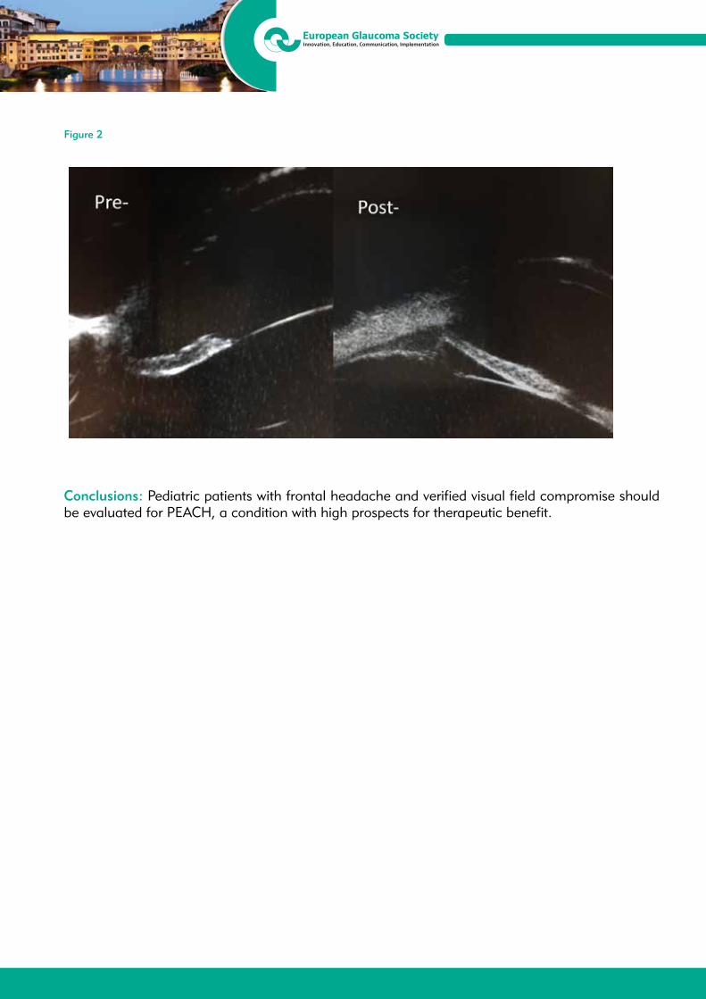
13th EGS CongressFortezza da Basso, Florence, Italy
19/22 May 2018
Figure 2
Conclusions: Pediatric patients with frontal headache and verified visual field compromise should be evaluated for PEACH, a condition with high prospects for therapeutic benefit.
13th EGS CongressFortezza da Basso, Florence, Italy
19/22 May 2018
P5.109NEWBORN PRIMARY CONGENITAL GLAUCOMA: IS EARLY SURGICAL INTERVENTION IMPORTANT?Maria Elisa Luís1, Diogo Hipólito Fernandes1, Ricardo Figueiredo2, Mariana Cardoso1, Cristina Ferreira1, Cristina Brito1, Alcina Toscano1
1Department of Ophthalmology, Centro Hospitalar Lisboa Central, Lisboa - Portugal2Department of Ophthalmology, Hospital do Espírito Santo de Évora, Évora - Portugal
Purpose: To evaluate the surgical and clinical outcomes of primary trabeculotomy in children diagnosed with newborn primary congenital glaucoma (PCG), and compare them when surgery is performed during the first month of life versus after 1 month of life.
Methods: Retrospective study of trabeculotomies performed as primary procedure between January 2006 and June 2017 in children diagnosed with newborn PCG. Surgical success was defined meeting the following criteria: post-surgical intraocular pressure (IOP) ≤16 mmHg with or without additional medication; absence of glaucoma progression; no severe intraoperative or postoperative complications; no more than 1 additional trabeculotomy or any other glaucoma surgery during the first 12 months after primary procedure. Two groups were defined: 1-surgery performed within 1st month of life; 2-surgery performed after 1 month of life.
Results: A total of 31 eyes of 16 children were included during a mean follow-up period of 51.3 months with standard deviation (SD = 41.9) . After trabeculotomy, mean IOP was 11.8 mmHg (SD = 5.2) in group 1 and 13.9 mmHg (SD = 6.8) in group 2 at 1 month follow-up. Additional antiglaucomatous medication was required in 71.4% (group1) and 58.9% (group 2). Corneal opacification reduced in both groups with statistical significance (p = 0.001; p = 0.016). The most frequent complications were hyphema (intraoperative) and goniosynechiae (postoperative). A mean of 0.4 (SD = 0.7) and 0.5 (SD = 0.9) reinterventions were recorded in group 1 and 2 respectively. Seven eyes (50%) of group 1 and 3 (23%) of group 2 recorded an acceptable or good best corrected visual acuity, at last visit. Twelve (85.7%) eyes of group 1 and 11 (64.7%) of group 2 met the surgical success criteria.
Conclusion: Earlier trabeculotomy seems to be associated with better results for visual acuity, corneal transparency and surgical success. Even when diagnosed late, primary trabeculotomy may be attempted with good outcomes.
13th EGS CongressFortezza da Basso, Florence, Italy
19/22 May 2018
P5.110REVIEW OF NON SURGICAL MANAGEMENT OF PAEDIATRIC GLAUCOMA: A 10-YEAR LOCAL EXPERIENCEWing Lau Ho1, Jimmy Shiu Ming Lai2
1Department of Ophthalmology, Grantham Hospital, Wong Chuk Hang, Hong Kong - Hong Kong2Department of Ophthalmology, University of Hong Kong, Wong Chuk Hang, Hong Kong - Hong Kong
Purpose: To review the outcomes of the paediatric glaucoma in a local centre and identify the difference in outcome of various procedures on paediatric glaucoma patients as compared to adult patients.
Methods: All patients under 18 receiving non-surgical treatment of glaucoma in a local centre during 1/2008-6-2017 were searched using the clinical management system. The records of all patients found were then reviewed. Age of presentation, diagnosis, intraocular pressure on presentation were noted. Outcome in terms of complete/qualified success defined as pressure equal to or below 21 mmHg without/with medication were reviewed at 1 month, 3 months and 1 year post-operatively.
Results: 20 eyes from 13 patients were identified. Age of presentation ranges from 1 day to 13 years. Average IOP on presentation was 34.0 mmHg. A total of 69 procedures were performed, including 2 selective laser trabeculoplasties, 29 cyclophotocoagulations, 7 goniotomies, 6 trabeculotomies, 7 Ex-Press shunt implantations, 1 trabeculectomy, 5 Ahmed valve implantations, 11 needling procedures and 1 cryotherapy. Complete and qualified success account for 36.2% and 13.0% at 1 month, 15.9% and 24.6% at 3 months and 11.6% and 13.0% at 1 year for all procedures. At the end of the review period, the average pressure was 18.1 mmHg. Only 45% of the eyes retain useful vision of 0.05 or above.
Conclusions: Paediatric glaucoma remains an important health burden as the outcome is very different from adult patients. Different methods may need to be used.

13th EGS CongressFortezza da Basso, Florence, Italy
19/22 May 2018
P5.111RARE CONGENITAL IRIS MEMBRANE WITH SECONDARY GLAUCOMA: A CASE REPORTPille Tein1
1East- Central Tallinn Eye Clinic, Tallinn - Estonia
Purpose: To present evaluation and treatment of a child with rare congenital iris membrane.
Subject: 4-month old girl with left eye corneal clouding and buphthalmos, epiphora and IOP 40 mmHg. One week after birth, the mother noticed that the pupil of the left eye was different and enlarged, but the GP decided to wait.
Methods: Operation (deep sclerectomy with trabeculotomy and pupillotomy) was performed immediately and the most accurate measurements were obtained during examination under anesthesia. During operation, congenital iris membrane was discovered and there was no pupil and anterior chamber in the left eye. Pre- and postoperative IOP, mean corneal diameter and corneal status, cycloplegic refraction and imaging of optic disc with Ret-Cam were performed.
Results: Postoperative examination IOP with ICare was normal. Visual acuity- fixed and follows objects. Under mydriasis the pupil enlarges and is without posterior synechiae. Patient has good control of IOP.
Conclusion: It is important to recognize congenital iris membrane as soon as possible as it has impact on vision. Timely pupillotomy has been necessary to restore vision and to treat angle closure glaucoma.

13th EGS CongressFortezza da Basso, Florence, Italy
19/22 May 2018
P5.112TUBE REVISION: A DIFFERENT APPROACHGeorge Tomais1
1Laser Vision, Athens - Greece
Purpose: To investigate the efficacy of a different approach of treating an encysted Ahmed valve, creating a new bleb and cutting the tube of the valve under the overlying conjunctiva.
Methods: After stabilizing the globe with an 8-0 vicryl suture, we approach the tube beginning from at least 6 mm away from it. We separate the conjunctiva together with the underlying Tenon’s capsule, reaching the tube and expanding the separation as much as we can. We then cut a small part of the tube using a glaucoma punch instrument. We check the flow of the aqueous humor through the opening we created and then we close the conjunctiva water tightly. We can also use mitomycin-c before creating the hole on the tube.
Results: On the first postoperative day the bleb was well shaped and the IOP was below 8 mmHg. One month after the operation the bleb was flatter and the IOP about 13mmHg. Unfortunately the bleb was non functioning six months after the operation and the IOP raised again at preoperative levels.
Conclusions: The above procedure is a safe and easy way to revise a non functioning valve. The key point for the success of this method is probably the use of Mitomycin-C.

13th EGS CongressFortezza da Basso, Florence, Italy
19/22 May 2018
P5.113NEW POSSIBILITIES OF COMPENSATION OF INTRAOCULAR PRESSURE AFTER DRAINAGE SURGERY NEOVASCULAR GLAUCOMA IN PATIENTS WITH DIABETES MELLITUSDmitry Lipatov1
1Department of Ophthalmology, National Endocrinology Research Centre, Moscow - Russia
Relevance: Drainage surgery of neovascular glaucoma in patients with sugar-NYM diabetes has long been the gold standard treatment of this complex pathology. Unfortunately, their effectiveness according to the Russian and foreign authors does not exceed 75%. Therefore, in the postoperative period almost every fourth patient raises the question of the search of means for the normalization of elevated intraocular pressure (IOP).
Purpose: To evaluate the effectiveness of contact transscleral diode laser cyclom-coagulation (CTDLC) after drainage surgery neovascular glaucoma, which did not result in normalization of IOP.
Material and Methods: The study included 12 patients observed in OFTEL-mological Department FSBI “Endocrinological research center” of healthcare Ministry. All of them were previously carried out drainage operation (implantation of the Ahmed valve) about uncompensated secondary neovascular glaucoma on the background of diabetic retinopathy (8 patients were operated on the of the endocrine research centre, the rest in other institutions). In the postoperative period the IOP to stabilize failed and was performed contact transscleral diode laser cyclocoagulation according to the original technique (patent of the Russia for the invention N. 2625595 from 22.04.2016).
Results and Discussion: All patients after implementation of a contact TRANS-scleral diode-laser cyclocoagulation intraocular pressure was compensate. but in the range of 12-15 mmHg. article Any complications prior to the year of observation were noted. Slight hyperemia of the conjunctiva within 3-4 weeks was observed in 8 cases (67%). Corrected and uncorrected visual acuity and perimetry data after 3, 6 and 12 months after the procedure CTDLC was not statistically different from coope-ration indices. The implementation of the intervention did not lead to a deterioration of carbohydrate metabolism - change in glycated hemoglobin was also statistically significant.
Conclusion: Contact transscleral diode laser cyclocoagulation can be effectively used for normalization of the IOP after decompencirovannogo Dre-changes require surgery of neovascular glaucoma in patients with diabetes.

13th EGS CongressFortezza da Basso, Florence, Italy
19/22 May 2018
P5.114OUTCOMES OF NEEDLING WITH 5-FLUOROURACIL IN ENCAPSULATED BLEBS AFTER AHMED GLAUCOMA VALVE IMPLANTATIONBurak Erdem2, Serhat Imamoglu1, Nimet Yesim Erçalik1
1Department of Ophthalmology, Haydarpasa Numune Training and Research Hospital, University of Health Sciences, istanbul - Turkey 2Haydarpasa Numune Training and Research Hospital, University of Health Sciences, Istanbul - Turkey
Purpose: In our study, we investigate the effectiveness and long-term results of 5-fluorouracil (FU) needling of encapsulated blebs after Ahmed Glaucoma Valve (AGV) implantation in patients with medically uncontrolled secondary glaucoma diagnosis.
Materials and Methods: Records of 147 patients with a history of long-scleral tunnel technique AGV implantation by the same surgeon secondary to medically uncontrolled glaucoma between January 2011- January 2017 in University of Health Sciences, Haydarpasa Numune Training and Research Hospital Eye Clinic were retrospectively screened. Among these records, 38 eyes of 37 patients (27 male, 10 female) with a history of encapsulated bleb formation and 5-FU needling were included to our study.
Results: The rates of patients fulfill the partial and complete success criteria (Intra Ocular Pressure ≤ 21 mmHg and no light perception loss or phytisis bulbi [complete: without medication, partial: with medication]) after needling in 2 nd hour, 1 st month, 3 rd month, 6 th month and final visit were 84.2%, 94.7%, 86.8%, 83.3% and 71.1%. Cumulative predictive success rates of patients with partial or complete success were 96.4%, 77.4%, 60.3% in 6th, 12th, 24th months respectively.
Conclusions: Bleb needling with 5-FU seems safe, simple and effective method in patients with encapsulated bleb formation after AGV implantation. Thus, in the event of AGV failure target IOP could be provided with an easy and cost-effective method without an additional need to operations and medications. In patients with neovascular glaucoma 5-FU needling seems less favourable. Accordingly, different modalities and treatments should be developed for this group of patients.
13th EGS CongressFortezza da Basso, Florence, Italy
19/22 May 2018
P5.115A NOVEL SURGICAL TECHNIQUE FOR AHMED VALVES IN REFRACTORY GLAUCOMA WITH SILICONE OIL ENDOTAMPONADEAitor Lanzagorta-Aresti1, Juan Davo-Cabrera1, Pablo Alcocer-Yuste1, Santiago Montolio-Marzo1
1Fisabio Oftalmologia, Valencia - Spain
Purpose: The purpose of the study is to describe a novel technique to implant Ahmed valves in patients with refractory glaucoma because of silicone oil (SO) endotamponade. Methods: Three patients with glaucoma without SO removal were used as an example for this technique. Silicone oil floatation in aqueous humor causes a superior displacement to iridocorneal angle with mechanical obstruction of trabecular meshwork and migration through the drainage device. If a drainage device is necessary, many surgeons place the tube as far as possible from the superior iridocorneal angle with a long tube or with a nasal inferior location.
Results: We performed corneal suture traction to expose superotemporal quadrant. The plate was placed gently in a conjunctival pocket between the rectus muscles and was anchored to sclera with 8/0 nylon at 8 mm posterior to the limbus. In our convectional technique for Ahmed valve, the plate is oriented completely perpendicular to the limbus. In these cases, we positioned it a bit more superior and tangential to limbus curvature. The tube can be longer and run parallel to pupil, without invading it. Finally, we used a sutureless fascia lata graft and the conjunctival flap was sutured with 8/0 nylon.
Conclusion: This technique allows use superotemporal quadrant (fewer complications), avoid corneal touch and decrease SO loss through the tube to subconjunctival space.

13th EGS CongressFortezza da Basso, Florence, Italy
19/22 May 2018
P5.116COMPARISON OF MITOMYCIN C-AUGMENTED TRABECULECTOMY AND EX-PRESS MINIATURE GLAUCOMA DEVICE IN THE MANAGEMENT OF NEOVASCULAR GLAUCOMAUfuk Elgin1, Emine Sen1, Salih Colak1, Pelin Yilmazbas1
1University of Health Sciences, Ulucanlar Eye Education and Research Hospital, Ankara - Turkey
Purpose: To compare the outcomes of initial trabeculectomy with mitomycin C (mit C) and preoperative intravitreal bevacizumab injection and Ex-Press miniature glaucoma device with mit C and preoperative intravitreal bevacizumab injection, in the surgical management of neovascular glaucoma (NVG).
Methods: This retrospective study consisted of 11 eyes of 11 cases with NVG who underwent initial trabeculectomy with mit C and 9 eyes of 9 cases with NVG who underwent Ex-Press miniature glaucoma device with mit C. Preoperatively full panretinal photocoagulation had been performed for all the eyes. Also intravitreal bevacizumab (1.25 mg / 0.05 mL) had been injected 10 days before the surgery for all eyes. The intraocular pressure (IOP) and the number of anti-glaucomatous medications were compared at the first week, first month, third month, sixth month and first year after the surgery. Independent t, Kolmogorov-Smirnov and chi square tests were used for statistical analysis.
Results: There were no significant differences in age and sex of trabeculectomy group (6 male, 5 female, mean age 66.9 ± 5.5) and Ex-Press group (5 male, 4 female, mean age 67.8 ± 6.5) (p = 0.32, p = 0.66 respectively). In trabeculectomy group preoperative IOP was 39.4 ± 3.6 mmHg and postoperative values were 10.4 ± 2.3 mmHg at the first week, 11.4 ± 1.9 mmHg at the first month, 15.2 ± 2.04 mmHg at the third month, 16.7 ± 2.6 mmHg at the sixth month and 18.9 ± 3.6 mmHg at the first year. In Ex-Press group preoperative IOP was 38.4 ± 5.8 mmHg and postoperative values were 10.3 ± 2.1 mmHg at the first week, 11.5 ± 2.3 mmHg at the first month, 15.4 ± 1.8 mmHg at the third month, 18.3 ± 2.9 mmHg at the sixth month and 18.2 ± 3.9 mmHg at the first year. In both groups IOP reduced significantly postoperatively. There were no significant differences between the preoperative and the postoperative IOP values of the two groups.
Conclusion: Both mit C-augmented trabeculectomy and mit C-augmented Ex-Press implant provided similar reduction of IOP in cases with NVG in 1 year follow-up period.

13th EGS CongressFortezza da Basso, Florence, Italy
19/22 May 2018
P5.117AN INFLUENCE OF THE PRIMARY ALTP ON THE LIFE QUALITY IN PATIENTS WITH PSEUDOEXFOLIATIVE GLAUCOMATatjana Sarenac Vulovic1,2, Dusan Todorovic2, Nenad Petrovic1,2, Mirjana Janicijevic Petrovic1,2, Svetlana Jovanovic1,2, Suncica Sreckovic1,2
1Clinic of ophthalmology, Clinical Centre Kragujevac, Kragujevac - Serbia 2Department of Ophthalmology, Faculty of Medical Sciences, University of Kragujevac, Kragujevac - Serbia, Purpose: The aim of this studyis to evaluate the impact of the primary ALTP on improvement of the life quality in patients with pseudoexfoliative (XFG) glaucoma.
Methods: The study was conducted from January 2016 until Jun 2017 in Department of Ophthalmology at the Clinical Centre Kragujevac, Serbia. It included 58 patients with XFG divided into two groups. The first group included 26 patients treated with ATLT, while other 32 patients were treated by using antiglaucomatous drugs. We observed the effect that ALTP had on: the IOP values, the number of used antiglaucomatous drugs and artificial tears, dry eye development (OSDI, Schirmer and TBUT test). Patients were examined on the first day, 6, 12 and 18 months after the ALTP.
Results: The statistically significant difference (p < 0.05) among the IOP values was noticed during the whole follow up period, with the highest significance (p < 0.001) six months after the ALTP. A huge effect ALTP had on the dry eye parameters. The statistically significantly lower values of OSDI and TBUT testwere recorded in the first group during the first twelve months (p < 0.001), while the results of the Schirmer test were quite similar during the study. The number of used antiglaucomatous drugs and artificial tears were also statistically significantly lower in the ALTP group.
Conclusion: ALTP seemed to be a better choice in a temporary regulation of the IOP in patients with XFG. By avoiding the effects of the benzalkonium chloride, ALTP had the consequent positive impact on the tear film stability, previously impaired due to pseudoexfoliations.

13th EGS CongressFortezza da Basso, Florence, Italy
19/22 May 2018
P5.1181-YEAR RESULTS OF XEN45 COLLAGEN IMPLANT FOR THE TREATMENT OF PSEUDOEXFOLIATION GLAUCOMAAna Ibáñez Muñoz1, Oscar Rua Galisteo1, Víctor Santiago Soto Biforcos1, Maria Chacon González1
1Department of Ophthalmology, San Pedro Hospital, Logroño - Spain
Purpose: To verify the efficacy in intraocular pressure (IOP) reduction and safety of XEN45 implant surgery in patients with pseudoexfoliation glaucoma, with 12-month follow-up.
Methods: Nonrandomized prospective clinical study. 21 eyes of 20 patients with medically uncontrolled pseudoexfoliation glaucoma underwent a XEN45 Gel Stent implantation with subconjunctival mitomycin-C. The average age was 80.9 ± 8.1 years old (35% women, 65% men). Of those eyes, 38% underwent solo XEN implant and 63% underwent simultaneous phacoemulsification and XEN. Treatment outcomes analyzed included: IOP, vision, number of medications, intra- and postoperative complications. At the end of the follow-up, we evaluated the complete success, defined as a postoperative IOP ≤ 18 mmHg with ≥ 20% reduction in IOP at 12 months without any glaucoma medications and the qualified success defined as a postoperative IOP ≤ 18 mmHg with ≥ 20% reduction in IOP at 1 year with medications. Failure was defined as vision loss of light perceptions vision or worse, need for additional glaucoma surgery, or < 20% reduction of IOP from baseline.
Results: The mean preoperative IOP was 21 ± 4 mmHg on 2.95 ± 0.97 medication classes. Twelve months postoperatively, the mean IOP was reduced to 15 ± 4 mmHg on 1.23 ± 1.22 medication classes. This resulted in a qualified success of 57.1% and a complete success rate off medications of 28.6%. The best corrected visual acuity was 0.55 ± 0.31 and improved significantly to 0.68 ± 0.32 at 1 year follow-up. Postoperative complications were 2 eyes with uveitis, 1 eye with choroidal detachment, 1 eye with hyphema and 1 eye with cystoid macular oedema, which resolved without visual impairment.
Conclusions: This study demonstrated that the smaller diameter XEN45 gel implant is statistically effective in reducing IOP and medications in pseudoexfoliation glaucoma patients with a low rate of complications.
13th EGS CongressFortezza da Basso, Florence, Italy
19/22 May 2018
P5.119EFFICACY AND MANAGEMENT OF DEEP SCLERECTOMY IN EXFOLIATION GLAUCOMA: DIFFERENCES WITH PRIMARY OPEN-ANGLE GLAUCOMAInmaculada Paños Palacios1, Antonio Moreno Valladares1, Nieves Puerto Amorós1, Francisca González López1, Mónica Martínez Díaz1
1Glaucoma Unit, University Hospital of Albacete, Albacete - Spain
Purpose: Know the differences in management and evolution between exfoliation glaucoma (EG) and primary open-angle glaucoma (POAG) after deep sclerectomy (DS) or phaco-deep sclerectomy (PDS).
Material and Methods: Retrospective case control study. 21 consecutive EG patients were enroled 2013-2015. As control 28 POAG patients operated in the same period were randomly selected. Intraocular pressure (IOP), visual acuity, anti-glaucoma drugs (AD), visual field, complications, and viability of the success rate were assessed a 48-month follow-up period.
Results: No significant differences were observed bettwen groups in terms of age, sex, pre-operative IOP (25.67 ± 6.7 vs 23.75 ± 5.7) or pre-operative AD (2.67 ± 0.7 vs 2.57 ± 0.8) (p > 0.05). 71% surgeries were PDS vs 29% of DS, same in both groups. No significant difference in the IOP was observed between the two groups at any follow-up including 48 months (16 ± 5.8 vs 14.5 ± 3.9). AD reduction was slighty higher in OAG up to 18 months (0.89 vs 0.56) but with no significant difference at any follow-up including 48 months (1.00 vs 0.92). These differences were greater when DE was performed in isolation at 6 (1.57 ± 2.14 vs. 0.38 ± 0.74), 12 (1.33 ±1.21 vs 0.57 ± 0.78) and 24 months (1.60 ± 1.14 vs 1.17 ± 1.6). The need of postoperative maneuvers was similar in both groups (29.2% vs 28.5%) while reoperations at 4 years was higher in EG (20.8% vs 10.7%). The total success rate was lower compared with POAG, although it was progressively equipped up to 48 months (20% vs 30%).
Conclusions: Although DE is an equally effective technique in the management of EG, it presents a higher probability of reoperation and a lower success rate comparing with POAG patients. It has been shown as PDE allows a greater reduction in the number of anti-glaucoma drugs in the medium term vs DE in isolation.
13th EGS CongressFortezza da Basso, Florence, Italy
19/22 May 2018
P5.120TWO-YEAR RESULTS OF TWO CASES WITH SCLERAL FIXATION OF INTRAOCULAR LENS AFTER TRABECULECTOMYToshihiro Inoue1, Hidenobu Tanihara1
1Department of Ophthalmology, Faculty of Life Sciences, Kumamoto University, Kumamoto - Japan
Purpose: To report two-year results of two cases who needed the scleral fixation of intraocular lens (IOL) after trabeculectomy implicating the fragmentation of ciliary zonule with the escape of vitreous during trabeculectomy in exfoliation glaucoma.
Methods: Case 1 is a 79-year-old man and intraocular pressure (IOP) was 40 mmHg in the right eye before trabeculectomy. The eye was undergone the cataract surgery and implanted an intraocular lens (IOL) ten years ago. During carrying out trabeculectomy in the right eye, the vitreous prolapsed after the peripheral iridotomy. Inferior disorientation of IOL was observed at the next day. Extraction and scleral fixation of IOL were performed 96 days later. IOP 2 years later was 14 mmHg and the IOL was located in center. Case 2 is a 77-year-old woman. IOP was 34 mmHg in the right eye before trabeculectomy. During suturing scleral flap in trabeculectomy, superior anterior chamber became shallow and vitreous-like material was prolapsed. The upper anterior chamber was shallow because of the forward lens dislocation at the next day. Phacoemulsification and scleral fixation of IOL were performed 28 days later because of the superior lens opacification. One year later, the patient received bleb revision because of bleb leak. IOP 2 years later was 14 mmHg without bleb leak and the IOL was fixed well.
Conclusions: The results indicate that the scleral fixation of IOL may be useful in cases with the fragmentation of ciliary zonule after trabeculectomy.
13th EGS CongressFortezza da Basso, Florence, Italy
19/22 May 2018
P5.121TWENTY-FOUR-HOUR VARIATION OF INTRAOCULAR PRESSURE AFTER TREATMENT WITH MICROPULSE LASER TRABECULOPLASTY IN PATIENTS WITH PSEUDOEXFOLIATION GLAUCOMAOlga Makri1, Foteini Tsapardoni1, Dionisios Pagoulatos2, Panagiotis Plotas1, Athina Pallikari1, Constantine Georgakopoulos1
1Department of Ophthalmology, University Hospital of Patras, Patras - Greece2Department of Ophthalmology, “Ippokrateio” General Hospital of Athens, Athens - Greece
Purpose: To evaluate the immediate 24-hour effect on intraocular pressure (IOP) of a single session of micropulse laser trabeculoplasty (MLT) in patients with pseudoexfoliation glaucoma (PEXG).
Methods: Patients with PEXG under prostaglandine analogue monotherapy with inadequate IOP control were treated with 360-degrees 532 nm MLT. Patients were evaluated at 1, 4, 8, 12 and 24 hours post-MLT. After trabeculoplasty patients were maintained in treatment with the same drug regimen as pre-MLT. Neither anti-inflammatory nor additional glaucoma medications were administered as prophylactic treatment before or after MLT. Any IOP spike ≥ 5 mmHg was recorded and treated accordingly.
Results: Twenty-three eyes of 18 patients were included in the study. The mean age of the patients was 71.83 ± 6.51 years. Five patients underwent bilateral treatment. The IOP before MLT was 20.61 ± 1.8 mmHg. The mean IOP was reduced by 2.17 ± 3.31 mmHg 1 hour after MLT, by 2.7 ± 3.85 mmHg 4 hours post-trabeculoplasty, by 0.87 ± 3.02 mmHg 8 hours and by 2.13 ± 2.8 mmHg 12 hours post-MLT. The IOP measured at 4 and 12 hours after MLT was statistically significantly lower compared to baseline (p=0.043 and p = 0.021 respectively). Twenty-four hours after treatment the measured IOP was 19.74 ± 3.12 mmHg and did not differ significantly from the baseline IOP. Regarding IOP spikes, the maximal IOP increase that was measured throughout the 24 hours after MLT was 4 mmHg and occurred in 3 eyes at 1 hour after MLT, in 2 eyes at 4 hours after MLT, and in 1 eye at 8 and 12 hours after MLT.
Conclusions: Treatment with MLT in PEXG eyes did not result in any significant, potentially dangerous IOP spikes in the early postoperative period.
13th EGS CongressFortezza da Basso, Florence, Italy
19/22 May 2018
P5.122OCULAR HYPERTENSION IN PATIENTS WITH MACULAR EDEMA TREATED WITH INTRAVITREAL DEXAMETHASONE IMPLANT OZURDEX®
Katia Guadalupe Sotelo Monge1, Miriam Eleonora Ayala Fuentes1, José Manuel Navero Rodríguez1, Antonio Morilla Grasa1, Virginia García del Puerto1, Marcos Muñoz Escudero1, Alfonso Antón López1
1Institut Català de Retina, Barcelona - Spain
Purpose: Evaluate the incidence and treatment required in cases of ocular hypertension (OHT) after intravitreal dexamethasone 0.7 mg implant (Ozurdex®) injection in patients with macular edema.
Methods: Retrospective consecutive case series of patients receiving one or more intravitreal Ozurdex® implant between February 2011 and November 2017. The data was collected originally with a standardized electronic medical record. Ocular hypertension was defined as an intraocular pressure (IOP) ≥ 25 mmHg and or an increase of ≥ 10 mmHg compared with baseline IOP. These values were used based on the criteria defined in previous large multi-center studies involving Ozurdex®. Patients who had a minimum follow-up of 6 months were included in the study.
Results: A total of 237 eyes of 212 patients were analyzed. A total of 428 intravitreal injections of Ozurdex® were administered over the course of the study. One hundred twenty (56.6%) patients were male. The mean age was 69.2 (±11.6) years. The main indications for treatment were macular edema secondary to retinal vein occlusion (34%), diabetic macular edema (27%), postsurgical macular edema (15%), uveitis (8.4%), and other etiologies (16%). Before Ozurdex® injection, 13 (5.48%) had pre-existing glaucoma, 43 (18.14%) had preexisting OHT, 3 were glaucoma suspects based on optic nerve appearance and 178 had a healthy optic nerve. Patients received a mean of 1.83 (±1.55) Ozurdex® implants ( range 1-11). One hundred fifty three eyes received a single injection, thirty-five received two injections, twenty four received three injections, and twenty five received more than three injections. Ocular hypertension developed in 89 (37.5%) of injected eyes over a mean follow-up period of 52.5 (±32.8) months. The greatest IOP elevation was seen at 41.11 (±33.3) days follow-up interval. In most cases (97%), IOP elevation was successfully managed with topical hypotensive treatment. Further treatment with oral acetazolamide was required in 9 patients, 7 of them used it transiently during 4.57 (±3.2) months, and two of them used it continuously and required surgery.
Conclusions: Eighty-nine patients (37.5%) who received Ozurdex® developed OHT. Episodes of ocular hypertension were generally successfully managed with topical treatment. Three patients required invasive glaucoma surgery, two of whom had history of prior glaucoma or OHT.

13th EGS CongressFortezza da Basso, Florence, Italy
19/22 May 2018
P5.123NEOVASCULAR GLAUCOMA TREATMENT IN PATIENT WITH CHRONIC LYMPHOCYTIC LEUKEMIAGalina Vashkevich1, Tatsiana Imshenetskaya1
1Department of Ophthalmology, Belarusian Medical Academy of Post-Graduate Education, Minsk - Belarus
Purpose: To present a case of neovascular glaucoma treatment in patient with chronic lymphocytic leukemia.
Methods: Male, 68 y.o., suffering with chronic lymphocytic leukemia for several years, developed visual loss on his right eye within 9 months. Patient had open angle glaucoma controlled by medications (beta-blockers, prostaglandin analogues) in both eyes. Right eye is pseudophakic. Right eye examination found iris and angle rubeosis, IOP rising up to 40 mmHg, corneal edema and retinal hemmorrages alike in central retinal vein occlusion. Right eye visual acuity was only light perception. Patient’s left eye revealed no signs of retinal hemorrhages, so retinal changes on right eye were considered as a central retinal vein occlusion consequences. Patient was assessed by hematologist - there no signs of leuklemia activity. Then panretinal photocoagulation was performed on the right eye.
Results: Iris rubeosis in right eye was diminished during the week after panretinal photocoagulation, IOP became lower. During the first month after the procedure iris rubeosis mostly disappeared, but IOP remains high. Valved glaucoma drainage device implantation was performed in the right eye. Several days later patient developed choroidal effusion in right eye, that disappeared after topical corticosteroid therapy. One month later right eye had almost no signs of iris rubeosis, IOP was 17 mmHg without topical medications.
Conclusions: Panretinal photocoagulation may be a method of iris rubeosis treatment and can be used as a first step of surgery in cases of neovascular glaucoma without significant optic media opacification.

13th EGS CongressFortezza da Basso, Florence, Italy
19/22 May 2018
P5.124SURGICAL TREATMENT OF SECONDARY GLAUCOMA AFTER SILICONE OIL TAMPONADE FOR RETINAL DETACHMENTVolha Yarmak1, Tatsiana Imshenetskaya1, Galina Vashkevich1
1Department of Ophthalmology, Belarusian Medical Academy of Post-Graduate Education, Minsk - Belarus
Purpose: To evaluate the surgical treatment with Ahmed valve implantation of secondary glaucoma after pars plana vitrectomy and silicone oil tamponade for rhegmatogenous retinal detachment.
Methods: We studied 8 eyes of 8 patients with Ahmed valve implantation for secondary glaucoma after pars plana vitrectomy and silicone oil tamponade for rhegmatogenous retinal detachment. The device was inserted under scleral flap with a tube in anterior chamber by the same surgeon. Success was defined as IOP 17-23 mmHg with or without medication with no surgical reoperation for glaucoma.
Results: The reasons associated with elevated IOP included emulsified oil in the anterior chamber. The pre-operative IOP was 36-51 mmHg with maximal anti-glaucoma medications postoperative IOP 19-22 mmHg in all cases. Visual function was saved in postoperative period. The control of IOP was achieved in all but 2 patients under anti-glaucoma medication (number of medication 2). The postoperative complications included: postoperative hypertensive phase in 2 patients which was controlled by additional anti-glaucoma medications.
Conclusions: Ahmed valve implantation for secondary glaucoma after silicone oil tamponade for rhegmatogenous retinal detachment achieved good IOP control, didn’t worsen visual function and demonstrated a low rate of complications. The authors declare no financial interests.
13th EGS CongressFortezza da Basso, Florence, Italy
19/22 May 2018
P5.126LONG TERM OUTCOMES OF GLAUCOMA SURGERY IN AXENFELD-RIEGER SPECTRUM - A RETROSPECTIVE STUDYParivadhini Annadurai1, Ronnie Jacob George2, B. Shantha3, Lingam Vijaya4
1Consultant, Smt Jadhavbai Nathmal Singhvee Glaucoma Service, Sankara Nethralaya, Chennai - India 2Senior Consultant, Smt Jadhavbai Nathmal Singhvee Glaucoma Service, Sankara Nethralaya, Chennai - India3Director, Smt Jadhavbai Nathmal Singhvee Glaucoma Service, Sankara Nethralaya, Chennai - India, 4Distinguished Senior Consultant, Smt Jadhavbai Nathmal Glaucoma Service, Sankara Nethralaya, Chennai - India
Purpose: To study the long term outcomes of Trabeculectomy with MMC in patients with Axenfeld Rieger spectrum (ARS) in a tertiary care centre
Methods: This retrospective study evaluated data from records of patients with ARS who underwent Trabeculectomy with MMC with at least 6 months of post operative follow up. The main outcome studied was surgical success rate
Success: Complete success: intraocular pressure (IOP) between 6 to ≤ 18 mmHg without IOP lowering medications. Qualified success: IOP between 6 to ≤ 18 mmHg with IOP lowering medications. Failure: IOP < 6 or >18 mmHg or when additional glaucoma surgery was required for IOP control
Results: Of the 41 eyes of 28 patients, 14 were males (50%) aged 0.1-49 years (mean 16.4 years). 34 eyes (82.6%) had not undergone any previous glaucoma surgery. 30 eyes (73.17%) underwent trabeculectomy + MMC, 7 eyes (17.07%) external trabeculotomy + trabeculectomy (aged < 5 years), 4 eyes (9.75%) phacoemulsification + IOL + trabeculectomy with MMC for IOP control. With a mean follow up of 7.46 ± 6.92 years (6months to 29 years) there was a statistically significant reduction of IOP at final visit (32 ± 7.33 vs.13.22 ± 6.19) with a mean reduction of 45.8%. Combined success (complete + qualified) probability by Kaplan-Meier survival analysis was 95% for the 1st year (n = 35), 89% till the 3rd year (n = 29), 78% for the 5th year (n = 23) and then 60% at 10 years (n = 10). 12 eyes underwent resurgery for IOP control. Previous glaucoma surgery (odds ratio 1.56, CI 0.29-8.25, p-0.5), totally closed angles (odds ratio 1.33, CI 0.09-18.19, p-0.8), when both eyes underwent glaucoma surgery (odds ratio 1.71, CI 0.42-6.9, p-0.4) and glaucoma surgery at 5-10 years of age (odds ratio 10, CI 0.6-128.7, p-0.07) were found to be risk factors for glaucoma resurgery on univariate regression analysis. Complications noted were corneal decompensation (4 eyes), hypotony (2 eyes) and graft failure (1 eye). There were no cases of suprachoroidal haemorrhage, endophthalmitis or blindness.
Conclusion: Trabeculectomy with MMC combined with or without external trabeculotomy in ARS provides satisfactory IOP control

13th EGS CongressFortezza da Basso, Florence, Italy
19/22 May 2018
P5.127ORAL PROPRANOLOL IN THE PREVENTION OF SIGHT THREATENING CHOROIDAL EFFUSION AFTER GLAUCOMA SURGERY IN STURGE WEBER SYNDROMESushmita Kaushik1, Pankaj Kataria1, Gunjan Joshi1, Surinder Pandav1, Jagat Ram1, Amod Gupta1
1Advanced Eye Centre, Postgraduate Institute of Medical Sciences and Research, Chandigarh - India
Purpose: Choroidal effusion (CE) is a common complication after intraocular pressure (IOP) reduction in Sturge Weber Syndrome (SWS). Oral propranolol is the standard of care for cutaneous infantile haemangioma, but its role in choroidal haemangioma is largely unexplored. We administered oral propranolol pre-operatively in patients with SWS, to see the occurrence of choroidal effusion following glaucoma surgery.
Methods: In this cohort study, 14 eyes of 12 consecutive patients with SWS requiring glaucoma surgery for IOP control, received oral propranolol 2 mg/kg body weight daily, in two divided doses, at least one week prior to undergoing glaucoma surgery. No additional manoeuvres such as sclerostomy slits or anterior chamber maintainers were used intra-operatively. Patients were followed up for a minimum of 3 months. The Main Outcome Measures were the incidence and extent of intra-operative and/or post-operative choroidal effusion, additional procedures required, and any adverse effects of the drug.
Results: The average follow-up was 25.7 ± 12.1 months (95% C.I. 25.7 ± 6.4 months). The IOP reduced from 25.2 ± 9.7 mmHg at presentation to 16.25 ± 6.2, 14.6 ± 4.5, 16.5 ± 8.9, and 13.1 ± 5.1 mmHg at one week, one month, one year and two-years post-operatively respectively. No patient developed choroidal effusion within the vascular arcades. 4 of the 13 eyes operated upon had peripheral choroidal effusion, which settled with topical and systemic steroids and atropine drops. All 4 of them had bilateral disease, and it was a repeat surgery in 3 of the 4 eyes. There were no adverse effects of propranolol in any patient.
Conclusions: Oral propranolol appears to be an effective and safe modality to reduce the development of sight threatening choroidal effusions following glaucoma surgery in SWS.
13th EGS CongressFortezza da Basso, Florence, Italy
19/22 May 2018
P5.129INTRAOCULAR PRESSURE REDUCTION AFTER PHACOEMULSIFICATION WITH CAPSULAR TENSION RING SUTURE FIXATION IN PSEUDOEXFOLIATION GLAUCOMANina Lutsenko1, Oxana Isakova1, Olga Rudycheva1, Natalya Unguryan2
1Ophthalmology Department of State Institution Zaporizhzhya Medical Academy of Postgraduate Education, Ministry of Public Health of the Ukraine, Zaporizhzhya - Ukraine2Ophthalmology Department of Municipal institution, Zaporizhzhya Regional Hospital, Zaporizhzhya Regional Council, Zaporizhzhia - Ukraine
Purpose: To study intraocular pressure (IOP) change after phacoemulsification with capsular tension ring (CTR) suture fixation or CTR implantation in pseudoexfoliation glaucoma (PEG).
Methods: Retrospective analyses of the IOP change after phacoemulsification with CTR suture fixation (13 eyes) and CTR implantation (13 eyes) was performed. Inclusion criteria were the presence of phacoiridodenesis and PEG. Single eyelets CTR were sutured to the sclera using 10-0 polypropylene in the first group, CTR implantation in the second group. Analysis includes IOP level, intraoperative complications, number of glaucoma drops preoperative and six month postoperative examination.
Results: Mean age of patients was 71.2 ± 2.1 years in the first and 68.4 ± 5.2 years in the second group. Preoperative IOP level was 27.1 ± 1.2 mmHg in the first and 26.4 ± 1.8 mmHg in the second group. After phacoemulsification with CTR suture fixation in all eyes IOP decreased to 20.4 ± 0.8 mmHg (24.7%), the number of glaucoma medication reduced from 2.6 ± 0.4 to 1.4 ± 0.8 in 10 eyes (76.9%), remained unchanged in 3 eyes (23.1%). In the group where phacoemulsification with CTR implantation was performed IOP decreased to 22.4 ± 1.3 mmHg (15.1%), the number of medications was the same in 8 eyes, increased in 2 cases and glaucoma surgery was performed in 3 eyes.
Conclusion: Scleral suture fixation of the capsular tension ring during phacoemulsification in PEG provides not only absence of intraoperative complications, stability and good centration of IOL, but reduce pseudophacoiridodenesis, intraocular pressure and number of glaucoma medications.
13th EGS CongressFortezza da Basso, Florence, Italy
19/22 May 2018
P5.130XF GLAUCOMA - A RISK FACTOR FOR GLAUCOMA SURGERY FAILURE?Fernando Trancoso-Vaz1, Ana Sofia Lopes1, Susana Pinto-Henriques1, Diana Silveira e Silva1, Joana Roque1, Maria Lisboa1, Cristina Vendrell1
1Ophthalmology Service of Fernando Fonseca Hospital, Lisbon - Portugal
Purpose: Pseudoexfoliation glaucoma (XFG) has a higher inflammatory profile associated with some factors as the variations of the lysyl oxidase-like protein 1 (LOXL1) gene, that has a role in the oxidative stress. Long-term failure rates of trabeculectomy are associated with bleb scarring that can be reduced with MMC. We intend to evaluate the outcomes of trabeculectomy with MMC in PXG and compare the results with the outcomes of trabeculectomy with MMC in POAG.
Methods: A total of 90 eyes of 76 patients who performed trabeculectomy MSS were included in a retrospective study, over a year of follow-up. 58 eyes with XFG were divided into group 1 or group 2 (with or without application of MMC), with 28 and 30 eyes respectively. Then, the group 1 results were compared with 32 eyes with POAG that performed trabeculectomy with MMC (group 3). Main outcome measures were the intraocular pressure (IOP), the number of medications, bleb failure (encapsulation, flattening and vascularization) and 5FU, needling with 5FU or secondary trabeculectomy.
Results: In PXG, trabeculectomy with MMC tends to have a lower: IOP (the preoperative mean was 28.6 ± 5.4 in group 1 and 32.2 ± 8.2 in group 2 and at the 1st year was 13.9 ± 3.9 in group 1 and 16.1 ± 5.9 in group 2); number of medications (the preoperative mean was 3.1 ± 0.60 in group 1 and 2.8 ± 0.81 in group 2 and at the 1st year was 1.1 ± 1.1 in group 1 and 1.1 ± 1.0 in group 2); bleb failure (47% in group 1 and 53% in group 2); and necessity of other surgical procedures (47% in group 1 and 57% in group 2). In the comparation with the POAG surgery with MMC (group 3), all the factors showed superior values in the group 1, but the differences were only statistically significative in: IOP at the 6th month and number of medication at the 6th month and at the 1st year (p = 0.045, p = 0.048 and p = 0.036, respectively), bleb failure (p = 0.02) and other procedures performed after the surgery (p = 0.013).
Conclusions: PXG seems to be a potential risk factor to filtration bleb failure and probably should be included in surgery protocols of MMC application.
13th EGS CongressFortezza da Basso, Florence, Italy
19/22 May 2018
P5.131XEN45 GEL STENT IMPLANT: COMPARISON OF 1-YEAR RESULTS IN PRIMARY OPEN-ANGLE GLAUCOMA VS SECONDARY OPEN-ANGLE GLAUCOMANuno Moura-Coelho1, Elisa Vares Luís1, Sara Crisóstomo1, Ana Luísa Basílio1, Joana Patrícia Ferreira1, Marco Dutra-Medeiros1, Mariana Sá Cardoso1, Teresa Gomes2
1Central Lisbon Hospital Center, Lisbon - Portugal2Hospital das Forças Armadas - EMGFA, Lisbon - Portugal
Purpose: To compare the efficacy and safety of the XEN45 Gel Stent Glaucoma Treatment System (Allergan, Irvine, CA) between patients with primary open-angle glaucoma (POAG) and patients with secondary open-angle glaucoma (SOAG). Methods: Retrospective comparative interventional case series of 19 eyes (18 patients) with medically refractory OAG who underwent XEN implantation at our institution. Clinical outcomes measured at 12-month follow-up included intra-ocular pressure (IOP) reduction, IOP reduction rate (IOP-RR), reduction in number IOP-lowering drugs, and complications. Failure was defined as IOP-RR ≤ 20% from baseline and/or IOP ≥ 18 mmHg, persistent hypotony, and/or need for further procedures for glaucoma, including needling. The study was conducted at a tertiary center for Ophthalmology in Portugal, and reports the initial experience with XEN implantation at the national level. Results: At 12 months post-Xen implantation, there was a significant reduction in median IOP in the POAG group (10 mmHg; IQR 13; p-value = 0.006) with a significant reduction in median number of IOP-lowering drugs (3.0; p-value = 0.003). No significant IOP reduction was observed in the SOAG group (-2 mmHg; IQR 11; p-value = 0.069). Median IOP reduction and IOP-RR were statistically significantly higher in the POAG group compared with the SOAG group (p-values = 0.039 and 0.039, respectively). Although the total number of complications requiring additional surgical procedures was higher in the SOAG group compared with the POAG group, the difference between both patient groups was not statistically significant (p-value = 0.209). There was no statistically significant difference in number of stent failures between both patient groups (p-value = 0.164).
Conclusions: In our study, XEN stent showed higher efficacy in IOP reduction in primary than in secondary OAG at 12 months, and statistically non-different safety profiles. However, we found no statistical association between etiology of glaucoma and failure of the XEN stent. Larger, prospective studies are needed to confirm our results that etiology of glaucoma may play a role in XEN surgery outcomes and to ascertain whether secondary glaucoma might be associated with surgical complications.

13th EGS CongressFortezza da Basso, Florence, Italy
19/22 May 2018
P5.132ORBITAL ABSCESS AND SECONDARY GLAUCOMA: A CASE REPORTSonia Parreira1, Diogo Lopes1, Nadine Marques1, Nelvia Donaire1, Nuno Barbosa2, Nuno Campos1
1Department of Ophthalmology, Hospital Garcia de Orta, Almada - Portugal2Department of Otorhinolaryngology, Hospital Garcia de Orta, Almada - Portugal
Purpose: Orbital cellulitis is an inflammation of the orbital tissues posterior to the orbital septum that can progress to orbital abscess. This condition may lead to ocular hypertension since it causes a change in the anatomy of the orbit and the vascular function. Here we report a case of secondary glaucoma due to an intraorbital abscess.
Methods: A 74-year-old female with a history of upper respiratory tract infection presented to our department with symptoms of pain, erythema and swelling of the left eyelids. On ophthalmology examination her visual acuity was 20/25 OD and 20/50 OS and intraocular pressure (IOP) was 14 mmHg OD and 35 mmHg OS. The patient still had slight proptosis and a partial supraduction and abduction deficit of the left eye. Fundus examination was unremarkable. For clinical suspicion of orbital cellulitis, orbit and cranial CT scan was done. The exam revealed an anterior, inferior and medial orbital abscess on the left side with deformation of the eye globe. There was no involvement of the paranasal sinuses neither intracranial extension.
Results: The patient was admitted and started intravenous medication with vancomycin (1 g/bid) and ceftriaxone (2 g/day). Antihypertensive eye drops (timolol maleate 0.5%, one drop/bid) and oral acetazolamide (250 mg/bid) was initiated. Due to size, location and discomfort, the abscess was drained 24h after admission and fluid collection was sent for microbiological analysis that revealed flucloxacillin sensitive Staphylococcus aureus. There was a clinical and imaging improvement and the patient was discharged ten days after admission continued antibiotic medication with oral flucloxacillin (500 mg, bid) and timolol maleate (one drop/bid) during more ten days. At one month follow-up her visual acuity was 20/30 and IOP was 16 mmHg. The patient underwent perimetry and optic nerve optical coherence tomography without alterations.
Conclusion: Orbital cellulitis can be potentially sight and life threatening. Currently, imaging studies for detection of orbital abscess, the use of antibiotics and early drainage have mitigated visual morbidity significantly. Prompt and correct diagnosis of orbital abscess is important for minimizing severe future complications such as irreversible visual loss by untreated secondary glaucoma.
13th EGS CongressFortezza da Basso, Florence, Italy
19/22 May 2018
P5.133EX-PRESS IMPLANT AS THE FIRST SURGICAL OPTION IN THE IRIDOCORNEAL ENDOTHELIAL SYNDROMEEsther Arranz-Marquez1, Sara Rodrigo-Rey2, Gema Bolívar2, Javier Paz-Moreno-Arrones2, Miguel Teus2
1Rey Juan Carlos Universitary Hospita, Mostoles - Spain2Principe de Asturias Universitary Hospital, Alcalá de Henares - Spain
Purpose: To evaluate the Ex-Press implant as the first surgical option in patients with Iridocorneal Endothelial Syndrome (ICE).
Methods: We describe the clinical history of two patients that had been diagnosed of ICE syndrome. They both had medically uncontrolled glaucoma and the Ex-Press implant was implanted in these patients. They have been evaluated with a follow-up period of more than three years.
Results: Baseline mean intraocular pressure has disminished in both patients after the implant of the Ex-Press device, and their IOP remains stable during the follow-up period without any medical antiglaucoma treatment. Both implants were placed sucessfully, with no major sight-threatening adverse events.
Conclusions: ICE syndrome is an infrequent disease with a dificult management, so medical treatment, trabeculectomy and the drainage devices have a high failure rate. The Ex-Press implant can be considered as the first surgical option in this patients, due to the fact that it can achieve a good control of IOP, and that the implant allows the ostium of the trabeculectomy to remain open in spite of the progressive synechial angle closure typical of the ICE syndrome. Furthermore, with the Ex-Press implant we avoided the complications related with the tube of the glaucoma drainage devices. In addition there is less postsurgical inflamation, a faster visual recuperation and a reduction of additional medical treatments and reinterventions.

13th EGS CongressFortezza da Basso, Florence, Italy
19/22 May 2018
P5.134EFFICACY OF INTRACAMERAL AND INTRAVITREAL RANIBIZUMAB, PANRETINAL PHOTOCOAGULATION, AND “LOW DOSES” STATINS IN THE MANAGEMENT OF NEOVASCULAR GLAUCOMAMaryna Karliychuk1, Serhii Pinchuk2, Ivanna Konovalets1, Victor Zamyatin2
1Department of Ophthalmology, Bucovinian State Medical University, Chernivtsi - Ukraine2Center of Modern Ophthalmology “Vash Zir”, Chernivtsi - Ukraine
The purpose was to evaluate the clinical efficacy of intracameral and intravitreal ranibizumab, panretinal photocoagulation, and application of “low doses” statins in the management of neovascular glaucoma.
Methods: The retrospective study included 27 eyes of 25 neovascular glaucoma patients with type 2 diabetes who were treated with topical intraocular pressure (IOP)-lowering medications, intracameral and intravitreal ranibizumab and panretinal photocoagulation, and were followed-up for 6 months. 14 patients (15 eyes) additionally received statins in low doses (rosuvastatin 10 mg orally once a day during 4 months). The main outcome measures were: control of IOP, regression and recurrence rate of neovascularization of the iris (NVI) and the anterior chamber angle (NVA).
Results: The mean pre-surgical IOP was 38.2 mmHg (±9.4). One week after intracameral and intravitreal ranibizumab the IOP had decreased to 31.2 ± 4.8 mmHg. In 23 eyes of 24 patients intracameral and intravitreal ranibizumab resulted in a marked regression of NVI and NVA within 7 days. In 21 eyes we observed a complete regression of neovascularization within 14 days. One month after intracameral and intravitreal ranibizumab the average IOP had decreased to 14.2 ± 4.2 mmHg (with topical hypotensive medications) in 25 eyes. In 3 months the recurrence of NVI and NVA was observed in 66.67% of eyes of patients who did not receive statins, and in 33.33% of eyes of patients who received statins in low doses (p < 0.05). There was no recurrence of NVI and NVA in 33.33% of eyes of patients who received statins in low doses during the follow-up period of 6 months. These results suggest that application of statins may prolong the duration of the period of neovascularization regression.
Conclusions: It was established that combined treatment with intracameral and intravitreal ranibizumab, panretinal photocoagulation, and application of “low doses” statins is effective in the management of neovascular glaucoma.
13th EGS CongressFortezza da Basso, Florence, Italy
19/22 May 2018
P5.135EVALUATION OF AHMED GLAUCOMA VALVE IMPLANTATION WITH PREOPERATIVE INTRACAMERAL AFLIBERCEPT INJECTION IN REFRACTORY NEOVASCULAR GLAUCOMAMelike Lider Gezer Ormen1, Serhat Imamoglu1, Nursal Melda Yenerel1, Nimet Burcu Celik1
1Department of Ophthalmology, Haydarpasa Training and Research Hospital, Istanbul - Turkey
Purpose: To evaluate the effect and safety of preoperative intracameral aflibercept injection before Ahmed glaucoma valve(AGV) implantation in the treatment of refractory neovascular glaucoma.
Methods: This retrospective study included 12 eyes of 12 neovascular glaucoma patients who underwent preoperative(average, 3 days) intracameral aflibercept injection (2 mg/50 μl) and AGV implantation. All patients were treated with topical and/or systemic intraocular pressure(IOP)-lowering medications, intracameral aflibercept and AGV implantation. The main outcome measures were changes of visual acuity, IOP, rubeosis iridis, number of antiglaucoma medications.
Results: Twelve eyes of 12 patients (5 female, 7 male) were enrolled in the study. The mean age of the study population was 64.3 years (range 47-84). The mean IOP decreased from 35.5 mmHg to 15 mmHg over a median follow-up of 8 months (range 2-19) (p < 0.05; Wilcoxon signed-rank test). Median preoperative visual acuity was 3.0 (1.0-3.0) logMAR. Median postoperative last visual acuity was 1.5 (1.0-3.0) logMAR. There was no significant difference between preoperative and postoperative last visual acuity (p = 0.14). Two of 12 patients had still rubeosis iridis after treatment (%16.6). The median number of antiglaucoma medications decreased from 3 (range 1-5) preoperatively to 1 (range 0-4) postoperatively (p < 0.05; Wilcoxon signed rank test).
Conclusions: Preoperative intracameral aflibercept injection may enhance success of AGV implantation in neovascular glaucoma and can effectively control IOP and preserve visual function.
13th EGS CongressFortezza da Basso, Florence, Italy
19/22 May 2018
P5.136TRABECULECTOMY WITH MITOMYCIN (MMC) IN NEO-VASCULAR GLAUCOMA: IMPORTANCE OF DOING AS A PRIMARY SURGERYJyoti Shakrawal1, Ramanjit Sihota1, Viney Gupta1, Shikha Gupta1
1Dept of Ophthalmology, RPC, AIIMS, New Delhi - India
Purpose: To evaluate the long-term outcomes of trabeculectomy with 0.04% mitomycin-C (MMC), as a initial surgery followed by retinal intervention (Pan-retinal photocoagulation/ Pars plana vitrectomy) in neo-vascular glaucoma (NVG) patients.
Design: Retrospective study.
Method: Thirty eyes of 30 patients with neo-vascular glaucoma, all presented in synechial angle closure stage were evaluated, out of which 26 patients underwent trabeculectomy (intracameral bevacizumab 4 days prior to surgery) as a primary surgery followed by retinal pathology treatment. Two patients underwent retinal surgery (Pars plana vitrectomy) initially, followed by trabeculectomy. Anterior retinal coagulation (ARC) was done in 2 eyes. Primary outcome measure was intra-ocular pressure (IOP) reduction. The success criterion was defined as IOP£ 18 mmHg without or with anti-glaucoma medications (absolute success and qualified success, respectively).
Results: The mean age of our study population was 58.56 years, with a mean follow-up duration of 3.9 years (median = 3 years). The mean pre-operative IOP and log-mar visual acuity was 43.73 ± 8.01 mmHg and 1.287 ± 0.588 respectively. The absolute and qualified success in patients whom trabeculectomy with MMC was done as primary surgery was 53.84% (14/26) and 100% (26/26) respectively. Whereas, in patients with retinal surgery as a primary therapy, absolute and qualified success was 0% and 50%. The overall qualified success rate of neo-vascular glaucoma was 93.33%, with a significant IOP reduction of 76.15% respectively (from 43.73 ± 8.01 mmHg to 10.26 ± 6.67 mmHg, p < .001). There was no significant drop in visual acuity with a mean log-mar visual acuity of 1.158 ± 0.631 at final follow-up.
Conclusion: In neo-vascular glaucoma, trabeculectomy with mitomycin C (MMC) as a primary surgery followed by retinal intervention performed better in terms of IOP control and visual acuity preservation than if trabeculectomy is done later.
13th EGS CongressFortezza da Basso, Florence, Italy
19/22 May 2018
P5.137SURGICAL SUCCESS OF AUROLAB AQUEOUS DRAINAGE IMPLANT (AADI) IN NEOVASCULAR GLAUCOMASujani Shroff1, George V. Puthuran1, Mohammed Sithiq2
1Department of Glaucoma, 2Department of Biostatistics, Aravind Eye Hospital, Madurai - India
Purpose: Neovascular glaucoma is a secondary glaucoma which is challenging to manage and has a poor visual prognosis. Artificial drainage implants have been advocated as a primary surgical option for management of these patients. This study aims to evaluate the surgical success of non valved Aurolab aqueous drainage implant (AADI) in cases with neovascular glaucoma (NVG).
Method: Retrospective study which included patients who underwent implantation of AADI between January 2015 and January 2017. 34 eyes of 34 patients with NVG were included in the study with a follow up period of 6 months. All statistical analysis was done using statistical software STATA 11.1 (Texas, USA).
Results: Mean age of the study population was 56.29 ±11.55 years. Mean preoperative intraocular pressure (IOP) was 40.26 ± 9.03 which reduced to 15.14 ± 8.65 at 6 months. Surgical success was defined as IOP of < 22 mmHg and > 5 mmHg without additional glaucoma surgery and without loss of light perception. The cumulative success at 6 months was 86.2%. Need for antiglaucoma medications (AGM) reduced from 3.44 ± 0.66 to 1.48 ± 1.02. The most common complication was hyphema. Two patients had a repeat glaucoma surgery, where one underwent a diode CPC and another had the tube repositioned into vitreous cavity. The mean BCVA dropped from 0.78 to 1.08, with no patient loosing light perception.
Conclusion: AADI provides good early IOP control for the treatment of neovascular glaucoma. Post operative vision loss despite good IOP control is a common occurrence in these patients. It is cost effective and could be considered as a primary surgical intervention.
13th EGS CongressFortezza da Basso, Florence, Italy
19/22 May 2018
P5.138SECONDARY GLAUCOMA IN ANTERIOR UVEITIS: CHALLENGES IN IOP CONTROLAstrianda Suryono1
1Department of Ophthalmology, Cipto Mangunkusumo Hospital (RSCM Kirana)- Universitas Indonesia, Jakarta - Indonesia
Purpose: To demonstrate the challenges in management of uveitic glaucoma.
Methods: A case report.
Results: A 27 year-old man came with blurness on RE since 2 months before admission with no redness, painless, and slowly progressing. There were history of cataract surgery 10 years prior. Other medical history included inguinale lump with discharge from external urethral orifice. Multiple unprotected sexual contact was identified. He had been treated with methylprednisolone and Pred Forte® 1 month before presentation but discontinued. On ophthalmic examination, LE was within normal limits. RE’s BCVA was 20/20 with IOP 47 mmHg. Anterior segment showed corneal edema with KPs, deep AC, traces of cells, pseudophakic with mild posterior capsule opacity. Posterior segment showed clear vitreous, enlarged CDR 0.7-0.8 with thinned inferior-temporal rim, no retinal abnormality. A workup for focal infection were negative (chest-ray, blood test, serologic tests for Chlamydia and CMV). Oral steroids continued and 3 glaucoma medications was given while preparing for surgery. At 2 weeks, IOP increased to 62 mmHg. An AGV insertion was done and IOP was well controlled in the first 4 days. Both IOP was increased at 1 week (RE 36 and LE 27). A prominent high bleb with thick wall was noted, and a hypertensive phase was suspected. Glaucoma medications were restarted, steroids were tapered off. At 2 months post-op, IOP was slowly decreasing to 29 mmHg on RE and normalized on LE during which steroid treatment had been tapered to 1 x 8 mg, and Azathioprin 3 x 50 mg was added. IOP continued to stabilize, and at 5 months reached 20 mmHg. At 7 months, still on Azathioprin, RE IOP was stable at 16 mmHg with 3 medications and no signs of active uveitis.
Conclusions: Uveitic glaucoma needs complex management and often became refractory. Steroid treatment gives inflammation control but can elevate IOP in responders. GDD offers good IOP control but with a careful post-operative watch. Hypertensive phase may occur and would resolve with time. Diligent care and close observation are necessary to keep inflammation and IOP control balanced and adequate.