

Efficient purification and reconstitution of ABCB6 for ... · Efficient purification and...
26
1 Efficient purification and reconstitution of ABCB6 for functional and structural studies Hemantkumar Chavan 1 , Mohiuddin Md. Taimur Khan 2, 3 , George Tegos 4, 5, 6, 7 and Partha Krishnamurthy 1 1 Department of Pharmacology, Toxicology and Therapeutics, University of Kansas Medical Center, Kansas City, KS 66160 2 Department of Chemical Engineering and Bioengineering, Washington State University, Pullman, WA, 99146, USA 3 Division of Bioenergy and Biotechnology, Pacific Northwest National Laboratory (PNNL), Richland, WA 99352, USA 4 Center for Molecular Discovery, University of New Mexico, Albuquerque, NM 87131, USA 5 Department of Pathology, University of New Mexico School of Medicine, Albuquerque, NM 6 Wellman Center for Photomedicine, Massachusetts General Hospital, 7 Department of Dermatology, Harvard Medical School *Running title: Purification and reconstitution of ABCB6 1 Address correspondence to : Partha Krishnamurthy, Department of Pharmacology, Toxicology and Therapeutics, University of Kansas Medical Center, 3901 Rainbow Boulevard, Kansas City, KS 66160. E-mail: [email protected] Keywords; ABCB6; transporters; purification, liposomes. Background: The ABCB6 protein is proposed to transport Coproporphyrinogen from the cytoplasm into the mitochondria. Results: Purified ABCB6 reconstituted into liposomes demonstrates Coproporphyrinogen stimulated ATP hydrolysis and Coproporphyrinogen transport. Conclusion: ABCB6 does not require additional components for substrate stimulated ATPase activity and substrate transport Significance: Development of an in vitro system with pure and active ABCB6 for structure and functional studies. http://www.jbc.org/cgi/doi/10.1074/jbc.M113.485284 The latest version is at JBC Papers in Press. Published on June 21, 2013 as Manuscript M113.485284 Copyright 2013 by The American Society for Biochemistry and Molecular Biology, Inc. by guest on August 14, 2019 http://www.jbc.org/ Downloaded from
Transcript of Efficient purification and reconstitution of ABCB6 for ... · Efficient purification and...

1
Efficient purification and reconstitution of ABCB6 for functional and structural studies
Hemantkumar Chavan1, Mohiuddin Md. Taimur Khan2, 3, George Tegos4, 5, 6, 7 and Partha Krishnamurthy1
1Department of Pharmacology, Toxicology and Therapeutics, University of Kansas Medical Center, Kansas City, KS 66160
2Department of Chemical Engineering and Bioengineering, Washington State University, Pullman, WA, 99146, USA
3Division of Bioenergy and Biotechnology, Pacific Northwest National Laboratory (PNNL), Richland, WA 99352, USA
4Center for Molecular Discovery, University of New Mexico, Albuquerque, NM 87131, USA
5Department of Pathology, University of New Mexico School of Medicine, Albuquerque, NM
6Wellman Center for Photomedicine, Massachusetts General Hospital,
7Department of Dermatology, Harvard Medical School
*Running title: Purification and reconstitution of ABCB6
1Address correspondence to : Partha Krishnamurthy, Department of Pharmacology, Toxicology and Therapeutics, University of Kansas Medical Center, 3901 Rainbow Boulevard, Kansas City, KS 66160. E-mail: [email protected]
Keywords; ABCB6; transporters; purification, liposomes.
Background: The ABCB6 protein is proposed to transport Coproporphyrinogen from the cytoplasm into the mitochondria.
Results: Purified ABCB6 reconstituted into liposomes demonstrates Coproporphyrinogen stimulated ATP hydrolysis and Coproporphyrinogen transport.
Conclusion: ABCB6 does not require additional components for substrate stimulated ATPase activity and substrate transport
Significance: Development of an in vitro system with pure and active ABCB6 for structure and functional studies.
http://www.jbc.org/cgi/doi/10.1074/jbc.M113.485284The latest version is at JBC Papers in Press. Published on June 21, 2013 as Manuscript M113.485284
Copyright 2013 by The American Society for Biochemistry and Molecular Biology, Inc.
by guest on August 14, 2019
http://ww
w.jbc.org/
Dow
nloaded from

2
ABSTRACT
The mitochondrial ATP binding cassette transporter ABCB6 has been associated with a broad range of physiological functions, including growth and development, therapy related drug resistance and the new blood group system Langereis. ABCB6 has been proposed to regulate heme synthesis by shuttling coproporphyrinogen III from the cytoplasm into the mitochondria. However, direct functional information of the transport complex in not known. In order to understand ABCB6s’ role in mitochondrial transport we developed an in vitro system with pure and active protein. ABCB6 overexpressed in HEK293 cells was solubilized from mitochondrial membranes and purified to homogeneity. Purified ABCB6 showed a high binding affinity for MgATP (Kd = 0.18 µM) and an ATPase activity with a Km of 0.99 mM. Reconstitution of ABCB6 into liposomes allowed biochemical characterization of the ATPase including (i) substrate stimulated ATPase activity (ii) transport kinetics of its proposed endogenous substrate coproporphyrinogen III and (iii) transport kinetics of substrates identified using a High-throughput screening (HTS) assay. Mutagenesis of the conserved lysine to alanine (K629A) in the Walker A motif abolished ATP hydrolysis and substrate transport. These results suggest a direct interaction between mitochondrial ABCB6 and its transport substrates that is critical for the activity of the transporter. Further, the simple immunoaffinity purification of ABCB6 to near homogeneity and efficient reconstitution of ABCB6 into liposomes might provide the basis for future studies on the structure, function of ABCB6.
INTRODUCTION
The ATP-binding cassette (ABC) superfamily constitutes the largest and most broadly expressed class of proteins found in all kingdoms of life (1). They couple the hydrolysis of ATP to active transport of an
array of biological compounds, including drugs, bile acids, peptides, steroids, ions, and phospholipids across membranes (2-5). Two highly conserved hydrophilic cytosolic nucleotide-binding domains (NBDs), and at least two hydrophobic membrane-spanning domains (MSDs) characterize the eukaryotic ABC transporters (6,7). The MSDs serve as the substrate-binding sites and the transmembrane domains (located within the MSD) form the translocation pore. The two NBDs are responsible for the binding and hydrolysis of ATP, which provides energy for uphill movement of substrates across membranes. The TMDs and NBDs are found as homo- or heterodimers and can be arranged in any combination, e.g. as separate polypeptides, as single polypeptides or as half-size transporters with fused TMD and NBD. Each NBD contains the highly conserved Walker A and B motifs as well as the C-, H- and D-loops (8,9). The Walker A motif contains a lysine that coordinates with the gamma-phosphate of ATP, whereas the aspartate in Walker B interacts with Mg2+. The ABC signature or Walker C motif distinguishes ABC transporter proteins from other ATP-binding proteins. These domains are required to execute ABC transport activities.
The ATP binding cassette transporter subfamily member B6 (ABCB6) gene encodes a membrane protein of 842 amino acids with a TMD followed by a NBD (10). Hydrophobicity and sequence homology analysis suggests that the TMD contains six transmembrane helices with the N and C termini located in the cytoplasm. The minimal functional unit has been suggested to be a homodimer residing in the outer mitochondrial membrane (11,12). ABCB6 has been characterized as a mitochondrial transporter involved in the translocation of coproporphyrinogen III (CPIII) from the cytoplasm into the mitochondria (11,13). CPIII a byproduct of heme synthesis in the cytoplasm requires active transport into the mitochondria to complete heme synthesis (14,15). Thus,
by guest on August 14, 2019
http://ww
w.jbc.org/
Dow
nloaded from

3
ABCB6 has been characterized with a physiological role in heme synthesis. However, several other functions and localization for ABCB6 have been suggested, e.g. ABCB6 has been localized to the plasma membrane and specifies the new blood group system Langereis (Lan) (16). Loss of ABCB6 function has been associated with developmental defects including ocular coloboma (17). Further, exogenous expression of ABCB6 has been correlated with increased cell growth and proliferation while loss of ABCB6 expression results in delayed progression through the mitotic phase of the cell cycle (17,18). These observations along with the fact that ABCB6 unlike other mitochondria targeted ABC transporters lacks a classical mitochondrial targeting sequence has favored reports of the existence of differentially localized ABCB6 with potentially unique functional properties.
Here, we describe the development of a cell free system to understand the mechanistic relationship between ATP binding and hydrolysis and the coupling of these events to substrate transport mediated by the mitochondrial transporter ABCB6. Overexpression, purification and functional reconstitution of ABCB6 into liposomes permitted us to study the biochemical properties of ABCB6 for the first time in the absence of contaminating ATPases and carriers/ transporters. In addition, immunoaffinity purification of ABCB6 to homogeneity, allowed identification of two isoforms of ABCB6.
EXPERIMENTAL PROCEDURES
Materials 8-Azido-[α-32P]ATP was purchased
from MP Biomedicals (Solon, OH). Coproporphyrinogen III, Protoporphyrin IX, Hemin and Porphobilinogen were purchased from Frontier Scientific (Logan, UT). N-octyl-β-D-glucopyranoside, Phosphatidylcholine,
Phosphatidylethanolamine, Phosphatidylserine and Ergosterol were from Avanti Polar Lipids (Alabaster, AL). Handee spin cups were from Pierce (Rockford, IL). All other chemicals and reagents were obtained from either Sigma-Aldrich or Fluka unless otherwise indicated.
Overexpression in HEK293 cells
To construct ABCB6-FLAG expressing lentiviral plasmid, the human ABCB6/pcDNA3.1 Topo vector carrying FLAG-tagged ABCB6 (18,19) was cut with restriction enzymes XbaI and EcoRI, and the ABCB6-FLAG containing fragment was ligated into corresponding restriction sites of Lentiviral vector; pLenti PGK Puro (20). HEK 293 cells overexpressing the ABCB6-FLAG lentiviral plasmid was generated by infecting cells with filtered viral supernatants followed by selection with 0.5 µg/mL puromycin. 293 cells were cultured in Dulbecco’s modified Eagle’s medium supplemented with 10% fetal bovine serum, penicillin and streptomycin. The non-functional mutant of ABCB6 was generated as previously described (18). Briefly, the critical lysine residue in the Walker A domain of ABCB6, which is the nucleotide binding domain in ATP binding cassette transporters that is essential for transport function was changed to alanine by site directed mutagenesis as described (18).
Cellular PPIX measurements Intracellular protoporphyrin IX
concentration was measured as previously described (21). Briefly, cells were harvested and washed once with PBS. Protoporphyrin IX concentration was measured by using a Vantage flow cytometer (BD Biosciences, San Jose, CA) with the excitation wavelength set at 405 nm, and the emission filter set at 695 nm/40 nm.
Purification of ABCB6 from total lysate HEK 293 cells (70% confluent) were
harvested with IX PBS and lysed (10 min at
by guest on August 14, 2019
http://ww
w.jbc.org/
Dow
nloaded from

4
4° C with gentle shaking ) in buffer A (1X PBS; 0.2% Triton X100) containing EDTA-free complete protease inhibitor mixture (Roche Applied Science, Indianapolis, IN). Soluble and insoluble fractions were separated by centrifugation (10 min at 10,000 rpm, 4°C) and the supernatant was incubated with sepharose conjugated anti-M2 beads (F2426 EZviewTM Red Anti-FLAG M2 affinity gel) overnight at 4°C with gentle shaking. The supernatant with the sepharose conjugated M2-beads were transferred to handee spin cups (paper filter) and separated by centrifugation (2,000 RPM for 5 min at 4°C). The beads were washed twice with buffer A, twice with buffer B (10 mM Tris; 0.2% Triton-X-100) and five times with Hepes buffer [Hepes-EGTA-1% n-Octyl-β-D-glucopyranoside (OGP)]. Proteins bound to the affinity column were eluted using elution buffer (Hepes-EGTA-1% OGP-0.5 mg FLAG peptide). The eluted protein was identified by immunoblotting using ABCB6 specific and FLAG-epitope specific antibody (Sigma, StLouis, MO) followed by matrix-assisted laser desorption/ionization time-of-flight (MALDI-TOF) peptide mass fingerprint analysis after trypsin digestion, using Mascot (data base NCBInr 20040521)
Purification of ABCB6 from mitochondria
Mitochondria were isolated as previously described (11). Isolated mitochondria were solubilized in buffer A (1X PBS; 0.2% Triton X100) containing EDTA-free complete protease inhibitor mixture (Roche Applied Science, Indianapolis, IN) for 30 min at 4°C with gentle shaking. Soluble and insoluble fractions were separated by ultracentrifugation (100,000 x g, 30 min, 4°C) and the supernatant was incubated with anti-M2 sepharose beads as described above for whole cell lysate. Proteins bound to the affinity column was washed and eluted as described above for total cell fraction using FLAG-peptide in elution buffer. The bound protein was identified by immunoblotting and matrix-assisted laser
desorption/ionization time-of-flight peptide mass fingerprint analysis after trypsin digestion, using Mascot (data base NCBInr 20040521)
Nucleotide binding assay
Nucleotide binding was measured by 8-azido-[α-32P]ATP photo cross-linking experiments. Cross-linking reactions were performed in a 96-well microtiter plate in a final volume of 25 uL/reaction. Purified wild-type ABCB6 (2 µM final concentration) was incubated for 5 min on ice with 8-azido-[α-32P]ATP (0.05 – 16 µM) in reaction buffer (100 mM KCl, 2.5 mM MgCl2, 50 mM Tris-HCl, pH 7.4). For competition experiments, 0.1 µM to 10 mM unlabeled ATP was included in the buffer. Subsequently, samples were irradiated with UV light (254 nm, 8 watts) for 5 min at 4°C, separated by SDS-PAGE, Coomassie blue-stained, dried, and exposed to Kodak SO230 phosphor screen. Photo cross-linked protein was quantified by phosphorimaging (Phosphor-Imager 445Si, Molecular dynamics) and bands were quantified using ImageJ software (version 1.43u). Intensities were plotted against the 8-azido-ATP concentration and apparent Kd values for 8-azido-[α-32P]ATP were obtained from the best fit of the data to a hyperbolic curve using GraphPad Prism software (version 5, San Diego, CA). The half-maximal inhibitory concentration (IC50) for ATP was derived by plotting labeling intensities corresponding to ABCB6 as a function of unlabeled ATP concentrations. The Kd(azidoATP) values and the IC50 values were used to calculate Kd(ATP) by applying the Cheng-Pursoff equation (22).
ATP hydrolysis assay
ATPase activity was determined essentially as described by measuring the release of inorganic phosphate (Pi) (11). Briefly, purified ABCB6 (5 µM final concentration) was incubated at 37°C in 100 µL of an ATP buffer containing 50 mM Tris-MOPS; 70 mM KCl; and 2 mM EGTA, pH
by guest on August 14, 2019
http://ww
w.jbc.org/
Dow
nloaded from

5
7.5, and the ATPase reaction was started by the addition of 12 mM MgATP. The reaction was stopped by the addition of 100 µL of 10% SDS solution, and the amount of inorganic phosphate was determined immediately. ATPase activity was estimated by the difference obtained in Pi levels between ATP buffer alone and ATP buffer containing the purified protein. Inorganic phosphate was measured by a sensitive colorimetric reaction. The SDS-containing samples were supplemented with 200 µL of freshly prepared reagent A containing a final concentration of 5.5% ascorbic acid; 5 mM ammonium molybdate and 2 mM zinc acetate and the reduced complex was measured at an optical density of 850 nM. A similar protocol was used to determine the ATPase activity of liposome reconstituted ABCB6. For substrate stimulated ATPase activity, indicated drugs were added in dimethyl sulfoxide (DMSO). The final concentration of DMSO in the assay medium was 1%. Control experiments indicated that DMSO at this concentration had no appreciable effect on the ATPase activity.
Reconstitution of ABCB6 For reconstitution of functional (WT)
and non-functional (mutant; MT) ABCB6 into lipids, protein bound to the FLAG-affinity column as described above was washed with Hepes-EGTA-1% n-Octyl-β-D-glucopyranoside (OGP) 5 times, and eluted using Hepes-EGTA-1% OGP-0.5 mg FLAG peptide. Lipid solutions (5 mg/mL Phosphatidylcholine (PC), 5 mg/mL Phosphatidylserine (PS), 5 mg/mL Phosphatidylethanolamine (PE), and 10 mg/mL Ergosterol (ES) were dissolved in chloroform, and the solvent was evaporated under a gentle stream of argon and evacuated overnight in a desiccator under vacuum to remove any residual chloroform. The lipid film was rehydrated in 1 mL of reconstitution buffer (15 mM HEPES, 0.5 mM EGTA, pH 7.4) to produce a 1 mM lipid solution. FLAG-tagged WT and MT ABCB6 protein at a concentration of 3.3 µg/µL was
added to the rehydrated liposomes and incubated for 1 h at 4°C with gentle agitation. The lipid-protein suspension mixture was sonicated using an ultrasonic cleaner (Avanti polar lipids Inc) for 20 sec; 3 times at 4°C. This procedure resulted in a nearly uniform suspension of relatively unilamellar liposomes. Protein-free liposomes were prepared similarly by replacing purified protein with elution buffer. Unincorporated materials and detergent were removed by dialysis using 1L of reconstitution buffer changed every 24 h for a total duration of 48 h. The resultant proteoliposomes were collected, flushed quickly with argon, and stored at -80°C until used.
Liposome flotation assay Proteoliposomes were floated in a
discontinuous Nycodenz density gradient in a buffer containing 100 mM KCl and 10 mM MOPS/Tris, pH 7.0. Nycodenz and proteoliposomes were solubilized in the same buffer and mixed to a final nycodenz concentration of 40%. Subsequent layers of decreasing Nycodenz concentration (20, 10, 5, and 2%) were layered onto the 40% nycodenz-proteoliposomes mixture. Percentages indicated in the figures are the final concentrations of Nycodenz. The gradient was centrifuged at 4°C for 60 min at 100,000 x g. Subsequently, the gradient was fractionated by taking equal volumes from the top. Fractions were analyzed by SDS-PAGE and western blotting.
Hemin agarose affinity chromatography
The assay was performed as described previously (23). Briefly, ABCB6-FLAG proteoliposomes (4 µg), was incubated at room temperature for 15 min in the presence of 167.5 nM hemin-agarose (Sigma, StLouis, MO). The reaction mixture was centrifuged at 4°C. The hemin-agarose-ABCB6-FLAG proteoliposome complex was washed thrice with 1 mL lysis buffer, resuspended in 50 µL of 2 X SDS sample-loading buffer, and then centrifuged. The
by guest on August 14, 2019
http://ww
w.jbc.org/
Dow
nloaded from

6
supernatant from the final spin was analyzed on a 4-15% gradient gel, transferred to a nylon membrane, and probed with a monoclonal antibody to the FLAG epitope to identify ABCB6-FLAG (Sigma, StLouis, MO).
Coproporphyrinogen (CPIII) transport assays
CPIII transport assays were performed as previously described (11,13) with slight modification to accommodate the use of liposomes. Control, ABCB6-WT and ABCB6-MT liposomes (25 to 50 µg for ABCB6 liposomes and equal volume of control liposomes) were resuspended in reconstitution buffer (final volume of 50 µL). Transport reaction was started by adding 50 µL of a reaction mixture (50 mM MOPS-Tris, 70 mM KCl, 10mM MgCl2, and either 4 mM AMP, 4 mM AMP:PNP or 4 mM ATP) and incubated at 37°C for the duration of the experiment as described in the respective figures. Reactions were stopped by diluting the reaction with 1 mL of ice-cold reconstitution buffer. In early experiments using nitrocellulose filters (pore size 0.45 µM) to measure CPIII uptake, we observed that a significant proportion of the proteoliposomes passed through the filters. To avoid this problem, an affinity purification method employing FLAG-affinity columns using anti-M2 magnetic beads was used to separate proteoliposomes from free CPIII. The affinity columns were washed three times with cold reconstitution buffer to remove any free CPIII not transported into the proteoliposomes. The affinity-purified proteoliposomes were lysed by incubation for 10 min in 10% SDS with vortexing. CPIII fluorescence was measured at excitation wavelength 405 nm and emission wavelength 630 nm (13). Concentration of CPIII was determined from a standard curve of CPIII in 10% SDS. The rate of active transport was calculated as the difference between ATP-dependent and independent uptake.
Verteporfin and tomatine transport assays The assay was performed as previously described (18). Briefly, following uptake of tomatine and verteporfin into proteoliposomes, the reaction was stopped at the indicated time and liposomes were separated by affinity purification employing FLAG-affinity columns as described above (CPIII transport assay). The affinity column was washed three times with cold reconstitution buffer to remove any free tomatine or verteporfin not transported into proteoliposomes. The affinity-purified proteoliposomes were resuspended in 100 µL of acetonitrile and the amount of verteporfin or tomatine taken up into proteoliposomes was analyzed using Ultra Performance Liquid Chromatography-time-of-flight mass spectrometry (UPLC-TOFMS) as previously described (18). Concentration of verteporfin and tomatine was determined from a standard curve of verteporfin and tomatine in acetonitrile. The rate of active transport was calculated as the difference between ATP-dependent and independent uptake.
Determination of Transport kinetics
Transport kinetics was estimated by non-linear curve-fitting using GraphPad Prism 5.0 software (San Diego, CA). The Michaelis-Menten equation was used to calculate apparent Km and Vmax values.
RESULTS
Overexpression and purification of ABCB6
To facilitate purification of ABCB6 for biochemical analysis, the protein was tagged at the C-terminus with the FLAG epitope and expressed from a lentiviral vector under the control of a strong constitutively active promoter. As shown in Fig 1A, immunoblot analysis of total cellular fraction, using antibody specific for the
by guest on August 14, 2019
http://ww
w.jbc.org/
Dow
nloaded from

7
FLAG-epitope demonstrates induced expression of FLAG-tagged ABCB6 in HEK293 cells transduced with the ABCB6-FLAG lentiviral construct but not in vector only transduced cells. Ponceau staining of the western blot before immunoblot analysis demonstrates that the induced expression of ABCB6 in the overexpressing cell is not due to differences in loading (Fig 1B). To confirm that the addition of the C- terminal FLAG- tag to ABCB6 does not affect its function we measured heme synthesis in ABCB6-FLAG overexpressing cells. We have previously demonstrated that ABCB6 expression and function is directly related to cellular heme synthesis (11). As seen in Figure 1C cells overexpressing the FLAG tagged ABCB6 protein demonstrate increased heme synthesis relative to empty vector control cells, suggesting that the addition of the FLAG-tag to the C-terminus of ABCB6 does not alter its function. Interestingly, both the endogenous protein, and overexpressed protein resolve as three bands of about 180, 90 and 50 kDa, as evidenced by immunoblot analysis using antibodies specific for either ABCB6 or the FLAG-tag (Fig 1A).
For ABCB6 purification, in initial studies, total cellular fractions were employed as starting material. To solubilize the protein from the membranes, the non-ionic detergent Triton was effective in either the presence or absence of n-Octyl-β-D-glucopyranoside. ABCB6-FLAG in the detergent solubilized fractions was separated from other cellular constituents by anti-FLAG affinity chromatography. As seen in Figure 1D and supplementary Figure 2, under the conditions of loading and washing of FLAG-sepharose beads, the ABCB6-FLAG protein was quantitatively bound, and upon elution with a buffer containing the FLAG peptide, was highly enriched and readily detectable by Coomassie blue staining. Bovine serum albumin (BSA) shown on the same gel was used as a loading control for the estimation of protein concentration (Fig 1D). The entire purification procedure produces
approximately 40 µg of purified ABCB6 from one 150-cm2 plate of confluent cells. Aside from the major ABCB6 monomer band of approximately 90 kDa, two additional staining bands, one with an apparent molecular weight approaching that of a homodimer (~180 kDa) and one with a lower molecular weight of approximately 50 kDa were also observed in the elution fractions (Fig 1D and 1E). All these three bands clearly contain ABCB6 as indicated by its detection with both ABCB6 specific monoclonal antibody (Fig 1E) as well as by MALDI-TOF peptide mass fingerprint analysis.
Purified mitochondrial ABCB6 binds ATP and displays ATPase activity
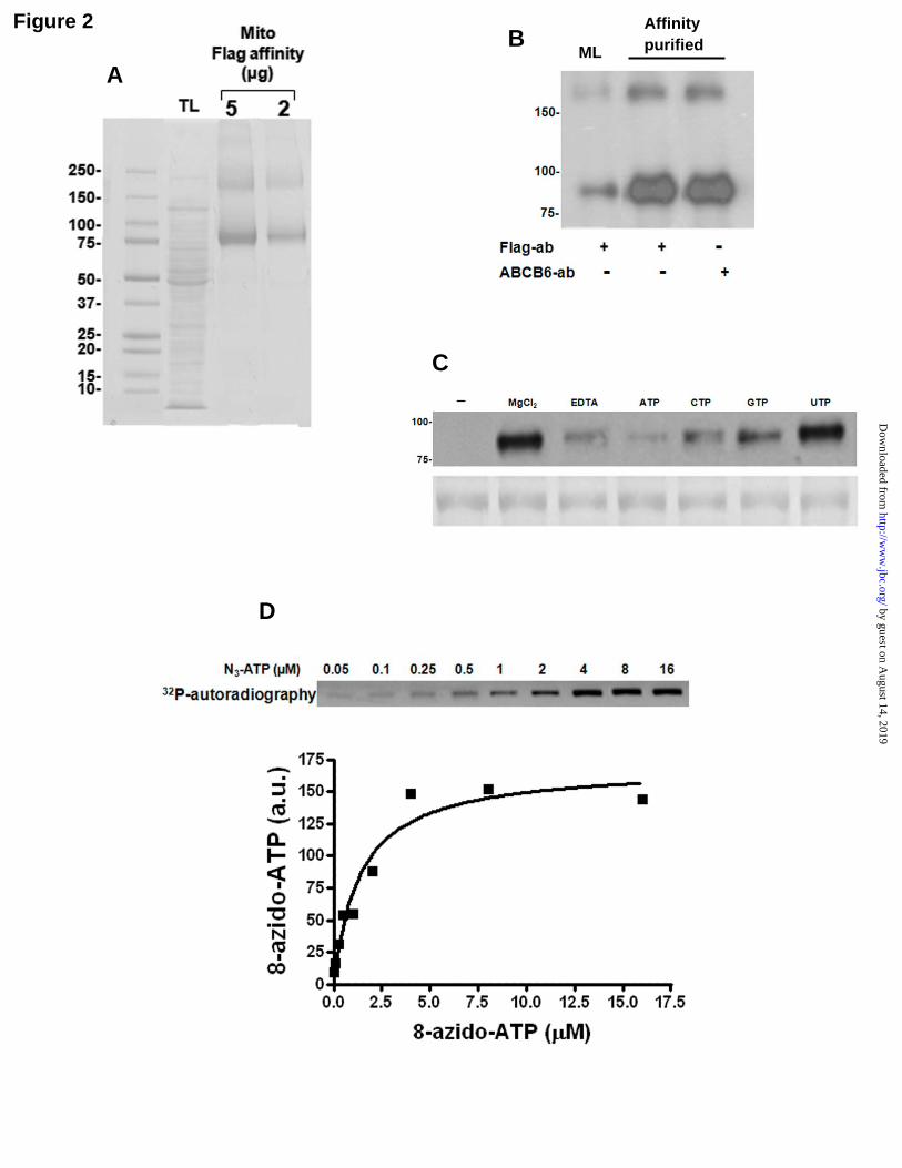
To identify the form of ABCB6 expressed in the mitochondria, differential density gradient centrifugation was used to isolate relatively pure mitochondrial fractions from ABCB6-FLAG overexpressing cells. Proteins in the mitochondrial membrane were solubilized with Triton in the presence or absence of n-Octyl-β-D-glucopyranoside. Detergent solubilized fractions were subjected to anti-FLAG affinity chromatography to separate ABCB6-FLAG from other mitochondrial membrane proteins. Proteins bound to the affinity column were subsequently eluted by treatment with a buffer containing the FLAG peptide. As seen in Figure 2A and supplementary Figure 3, ABCB6-FLAG protein was highly enriched in the eluted fraction and readily detectable by Coomassie blue staining. In contrast to ABCB6 purified from total cellular fraction, ABCB6 eluted from the mitochondrial preparations revealed only two bands; the ABCB6 monomer band of approximately 90 kDa, and the potential homodimer of approximately 180 kDa (Fig 2A). These results are consistent with our previous observations demonstrating a ~90 kDa ABCB6 monomer protein band and a ~180 kDa potential ABCB6 homodimer band seen in mitochondria isolated from ABCB6 overexpressing cells (11,24). Both the 90
by guest on August 14, 2019
http://ww
w.jbc.org/
Dow
nloaded from

8
and 180 kDa bands contain ABCB6 as indicated by its detection with both ABCB6 specific monoclonal antibody (Fig 2B) as well as by MALDI-TOF peptide mass fingerprint analysis.
Transport functions mediated by members of the ABC transporter family require ATP binding and hydrolysis. To examine the ATP binding property of ABCB6, the detergent-solubilized, purified mitochondrial ABCB6 protein was incubated with 8-azido-[α-32P]ATP and the azido photoprobe interacting with ABCB6 was irreversibly cross-linked by irradiation with ultraviolet light. The ABCB6-ATP interacting complex was separated from free probe by Sodium dodecyl sulphate - polyacrylamide gel electrophoresis (SDS-PAGE) and were subjected to autoradiography for detection of 8-azido-[α-32P]ATP labeling and to Coomassie blue staining for determination of protein content (Fig 2C). A major band of about 90 kDa consistent with the molecular mass of mitochondrial ABCB6 was identified by Coomassie blue staining (Fig 2C: lower panel) and found to be photoaffinity labeled by 8-azido-[α-32P]ATP (Fig 2C: upper panel). Labeling of ABCB6 by 8-azido-[α-32P]ATP required magnesium and was strongly inhibited by the addition of EDTA (1 mM) or excess cold ATP (10 mM) (Fig 2C). Nucleotide specificity of ABCB6 appeared to be in the following order; ATP > CTP > GTP > UTP (inhibition from 85% to 10%; Fig 2C). The apparent affinity constant for 8-azido-ATP was determined to be 1.39 ± 0.37 µM (Fig 2D). Photoaffinity labeling of ABCB6 by 8-azido-[α-32P]ATP (5 µM) was inhibited by MgATP with an IC50 value of 1.03 µM and an estimated Hill coefficient approaching a value of 1 (0.87) (Fig 2E). Based on the apparent affinity of 8-azido ATP, a dissociation constant for ATP of 0.18 µM was estimated. Taken together these data indicate that ABCB6 had retained its ability to bind ATP through the purification process and like other ABC transporter proteins has relatively broad nucleotide specificity.
Purified recombinant ABC transporters have been reported to possess
intrinsic ATPase activity. To determine the intrinsic ATPase activity of immune affinity purified ABCB6 we used a spectrophotometric assay to measure the amount of Pi released from ATP. The dependence of the rate of hydrolysis on ATP concentration shown in Fig 2E exhibited Michaelis-Menten behavior and, when expressed as a Lineweaver-Burk plot (Fig 2G), showed a linear relationship, yielding a Km for ATP of approximately 0.99 ± 0.11 mM and Vmax of 492.3 ± 17.25 nmol/mg/min. In parallel experiments the ATPase activity of the ATPase-inactive mutant of ABCB6 (ABCB6-MT) was also analyzed; it was solubilized and purified identically to the wild type protein in similar yields (Fig. 3) The purified mutant protein showed very little ATPase activity suggesting that the observed ABCB6-WT-ATPase activity is ABCB6 specific (Fig. 2E). In addition, we found that the purified ABCB6 protein was able to interact with and its ATPase activity was stimulated by its proposed substrate CPIII (supplementary Fig. 1).
Reconstitution of ABCB6 into lipid membranes
To analyze ABCB6 in its membrane-embedded state, purified mitochondrial ABCB6 (both WT and MT forms) was reconstituted into preformed detergent-destabilized liposomes composed of 0.13 µmol PC; 0.25 µmol PS; 0.67 µmol PE and 0.25 µmol ES. Following reconstitution detergent was dialyzed from the proteoliposomes as described in methods. Efficiency of ABCB6 reconstitution into liposomes was analyzed by discontinuous Nycodenz density gradient centrifugation. As seen in Figure 3A reconstitution of ABCB6 into liposomes is evident from the co-migration of ABCB6-FLAG containing liposomes to the top of the Nycodenz gradient. A majority of reconstituted proteoliposomes float into the 5% fraction of the gradient. In contrast, a very small fraction of the purified non-reconstituted protein floats into the 40% fraction, which
by guest on August 14, 2019
http://ww
w.jbc.org/
Dow
nloaded from

9
represents the bottom of the gradient (Fig 3A). These data suggest that both the ABCB6-WT-FLAG and ABCB6-MT-FLAG proteins were successfully reconstituted into liposomes with a reconstitution efficiency of ~80%. Integrity of liposome-reconstituted ABCB6 was analyzed by resolving the proteoliposomes obtained from the 5% fraction of the gradient by SDS-PAGE. As seen in Figure 3B and 3C analysis of ABCB6 reconstituted liposomes by Coomassie staining (Fig 3B) and immunoblot using ABCB6 specific antibody (Fig 3C) demonstrate that ABCB6 was not degraded during the reconstitution process.
The orientation of ABCB6 following insertion into liposomes was determined by the protease protection assay of the C-terminal FLAG-tag in the presence and absence of detergent. The C-terminal FLAG-tag in ABCB6 is accessible to protease-mediated degradation on the outside of the intact vesicles with an ‘NBD-out’ orientation but not with an ‘NBD-in’ orientation. In contrast, in the presence of detergent, the C-terminal FLAG-tag in ABCB6 is accessible to protease degradation irrespective of NBD orientation. Comparison of the protease assay data of intact and detergent-permeabilized vesicles revealed that about 70-80% of ABCB6-FLAG was inserted into liposomes in an “NBD-out” orientation (Fig 3D).
Liposome reconstituted ABCB6 interacts with its proposed substrate Coproporphyrinogen III.
ABC transporters associate with their transport substrates to facilitate its movement across the membrane. Using hemin-agarose affinity chromatography, we have previously demonstrated that ABCB6 associates with CPIII, a potential endogenous transport substrate of ABCB6 (11). To determine if liposome reconstituted ABCB6 retained its ability to associate with CPIII we performed hemin-agarose affinity chromatography as described (11). We found that CPIII was able to displace
liposome reconstituted ABCB6 from hemin-agarose in a dose dependent manner (Fig 4A). In contrast, porphobilinogen, a molecule that has been previously demonstrated not to interact with ABCB6, does not compete with hemin-agarose for ABCB6 (Fig 4A). These results suggest that liposome reconstituted ABCB6 retains its ability to interact with its transport substrates.
ATPase activity of reconstituted ABCB6 is stimulated by Coproporphyrinogen III (CPIII)
To determine the ATPase activity of liposome reconstituted ABCB6 the release of inorganic phosphate from ATP was assayed as described for the purified protein. As shown in Figure 4B lipid reconstituted ABCB6 demonstrated basal ATPase activity that was comparable to the ATPase activity of the purified detergent solubilized protein (Fig 2F). The basal ATPase activity was specific to ABCB6 because no such activity was observed either in the presence of control liposomes (data not shown) or in the presence of liposomes carrying an ATPase inactive mutant of ABCB6 (ABCB6-MT) (Fig 4B). Further, the basal activity was reduced to 98% by ortho-vanadate that inhibits ABC transporters and P-type ATPases, indicating that no additional ATPases are present in the ABCB6 proteoliposome preparations (data not shown). The dependence of the rate of hydrolysis on ATP concentration shown in Figure 4B exhibited Michaelis-Menten behavior and, when expressed as a Lineweaver-Burk plot (Fig 4C), showed a linear relationship, yielding a Km for ATP of approximately 0.97 ± 0.07 mM and Vmax of 614 ± 11.53 nmol/mg/min.
Stimulation of ATPase activity by transport substrates is a feature that is common to most ABC transporters. Consequently, it was of interest to determine whether known ABCB6 substrates affected its ATPase activity in a similar manner. As shown in Figure 4D
by guest on August 14, 2019
http://ww
w.jbc.org/
Dow
nloaded from

10
ATPase activity of liposome reconstituted ABCB6 was strongly stimulated in the presence of CPIII, hemin and protoporphyrin IX (CPIII > Hemin > PPIX) but not in the presence of porphobilinogen (PBG). This multifold increase in ABCB6 ATPase activity by CPIII, hemin and protoporphyrin IX (PPIX) suggests that these compounds are potential transport substrates of ABCB6. These results are in agreement with our earlier observations demonstrating that porphyrins such as CPIII and hemin are potential substrates of ABCB6 (11).
Coproporphyrinogen III transport kinetics of reconstituted ABCB6
ABCB6 is proposed to be involved in the transport of CPIII from the cytoplasm into the mitochondria. However, a direct role for ABCB6 in CPIII transport has not been demonstrated in the absence of other potential interacting components (mitochondrial preparations). Thus, kinetic parameters of CPIII transport mediated by ABCB6 were carried out with the ABCB6 proteoliposomes. According to the Lineweaver-Burk analysis, the apparent Km for CPIII was 11.97 ± 1.10 µM with a Vmax of 29.6 ± 0.81 pmole/mg/min (Fig 4E and 4F). To confirm these observations further CPIII uptake studies were performed in control liposomes and in liposomes reconstituted with either a transport competent ABCB6 (ABCB6-WT) or the transport incompetent ABCB6 (ABCB6-MT) protein. As seen in Figure 4E CPIII uptake in ABCB6-MT proteoliposomes were very low and reflected transport rates similar to what was seen in control liposomes (Fig 4E).
We recently developed a HTS assay to identify potential substrates and inhibitors of ABCB6 (24). Two of the compounds identified in the HTS assay, tomatine and verteporfin were transported into mitochondria in an ABCB6 dependent manner but a third compound benzethonium was not transported by ABCB6 (24). We
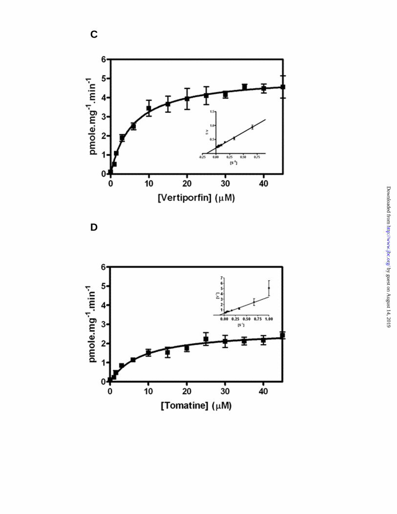
used the transport features of these three molecules to test and validate the robustness of liposome reconstituted ABCB6 as a potential model to identify ABCB6 transport substrates. As with CPIII, both tomatine and verteporfin competed with hemin binding to ABCB6 (Fig 5A), stimulated ABCB6 ATPase activity (Fig 5B) and were transported into liposomes by ABCB6 (Fig 5C and 5D) with varying transport kinetics (verteporfin Vmax 5.13 ± 0.23 pmol/mg/min; tomatine Vmax 2.7 ± 0.19 pmol/mg/min). However, unlike verteporfin and tomatine, benzethonium did not stimulate ABCB6 ATPase activity (Fig 5B) nor was it transported by ABCB6 proteoliposomes (data not shown). These results are consistent with the transport properties of ABCB6 observed using mitochondrial fractions isolated from ABCB6 overexpressing cells (24).
DISCUSSION
Mitochondrial ABC transporters are difficult to study because of the two-membrane architecture of mitochondria, problems associated with analyzing transport process, and the high abundance of other ATPases and carriers/transporters. Thus the development of an in vitro system with pure and active protein, is a necessary prerequisite toward understanding the mechanistic relationships between ATP binding and hydrolysis and coupling of these events to translocation of substrates across the lipid membranes. In this study, we describe for the first time a successful purification protocol and biochemical characterization of the putative mitochondrial porphyrin transporter ABCB6. The purification procedure comprised of lentiviral-mediated overexpression of ABCB6 carrying a C-terminal FLAG tag followed by a single step solubilization of ABCB6 from mitochondrial membranes and immunoaffinity purification using the FLAG antibody. Purified ABCB6 was found to be mostly monodisperse by PAGE electrophoresis and was efficiently
by guest on August 14, 2019
http://ww
w.jbc.org/
Dow
nloaded from

11
reconstituted into liposomes allowing its biochemical characterization with respect to ATP binding, ATP hydrolysis and transport kinetics.
A prerequisite for the purification of integral membrane proteins is the dissolution of the biological membrane in which the protein resides so that the membrane protein exists in solution in a monodisperse state while maintaining its structure in a physiological relevant condition. The success of a purification strategy is thus largely dependent on the type of detergent used, for both initial solubilization and the subsequent steps of column chromatography. The choice of detergent (Triton and n-Octyl-β-D-glucopyranoside) used for solubilization and purification of ABCB6 appeared acceptable as reasonable amounts of ABCB6 were extracted from the membrane, with preserved oligomeric state and activity of the proteins. After purification by immunoaffinity chromatography, ABCB6 bound MgATP with a relatively high affinity (Kd = 0.18 µM), and showed an ATPase activity of 0.493 µmol/mg/min. The observed activity is similar to that reported for ABC transporters such as MRP1 (0.46 µmol/min/mg; (25) but is relatively high compared to ABC transporters e.g. MRP3 (0.075 µmol/mg/min: (26) and MsbA (0.15 µmol/mg/min; (27). Interestingly ABCB6 ATPase activity, is lower than activities seen for other mitochondrial ABC proteins such as Mdl1 [2.3 µmol/mg/min; (28)] and Atm1 [1.9 µmol/min/mg; (29)]. The relatively high ATPase activity indicates that immunoaffinity purified ABCB6 is in an active state. This conclusion is further supported by the observation that solubilized and reconstituted ABCB6 have comparable ATPase activities. Further, given that the apparent affinity for MgATP is 700 times higher than the Michaelis-Menten constant for ATP hydrolysis (Km = 0.99 mM) this implies that ATP binding and ATP hydrolysis are potentially distinct steps.
There is increasing evidence that ABCB6 localizes to multiple cellular compartments including the plasma membrane (30-33). However, the precise identity, substrate specificity (if any), and functional significance of the differentially localized ABCB6 is not well defined. In our studies, although the principal form of ABCB6 purified from total cell fractions was a ~90 kDa protein, a second band of ~50 kDa was also purified following affinity chromatography. Both the purified bands contain ABCB6 as confirmed by western blot and MALDI-TOF analysis. The ~90 kDa band represents the mature form of the protein, which has been previously described as localizing to the mitochondria (11,12). Interestingly however, the 50 kDa band was not seen in the ABCB6 purified from mitochondrial fractions suggesting that the ~50 kDa form of ABCB6 might localize to cellular compartments other than the mitochondria. The ability of our experimental protocol to purify sufficient quantities of these two forms of ABCB6 should in the future enable us to characterize their localization, identity and potential functional significance.
It is well established that the lipid environment in which a membrane protein is reconstituted can affect its reconstitution and interaction with its substrates thus modulating its activity (34). For example, it has been shown that MRP3, which is expressed in both the liver and the brain, exhibits efficient reconstitution and highest ATPase activity when reconstituted in liver lipids but not in brain lipids (26). Similarly, certain lipids significantly influence the characteristics of purified Pgp ATPase activity and the apparent coupling between its drug-binding and catalytic sites (35,36). In contrast, the ATPase activity of human ABCA4 could not be stimulated by its substrate, all-trans-retinal, when the protein was reconstituted in vesicles composed of single synthetic lipids (37). Thus, overall, there appears to be considerable variability among the ABC transporters with respect to their response to their lipid environment.
by guest on August 14, 2019
http://ww
w.jbc.org/
Dow
nloaded from

12
Results from the present study suggest that for ABCB6 a combination of various phospholipids constitutes an optimal environment for substrate binding and stimulation of basal ATPase activity. However, whether these values are true representations of the ATPase activity of native ABCB6 requires further investigation. Infact, given that the basal ATPase activity of lipid reconstituted ABCB6 was relatively higher than that observed for the purified protein suggests that the lipid environment may play a role in influencing the ATPase activity of native ABCB6. In this context, and given that ABCB6 is ubiquitously expressed, native ABCB6 in different organ systems or in different cellular compartments might exhibit varied ATPase activity and potentially different substrate specificities. We speculate that these suppositions could provide the basis to explain some of the ABCB6 loss of function phenotypes described in the literature (17,19,38-41).
Many ABC transporters have been shown to possess intrinsic ATPase activity that is stimulated in the presence of transported substrates. The best-investigated example is the mammalian Mdr1, which possesses an ATPase activity stimulated by various drugs that are known to be transported (42,43). The ATPase activity of lipid reconstituted ABCB6 was stimulated by porphyrins known from previous studies to be potential substrates for this transport protein (11,13). CPIII has the highest affinity for ABCB6 and this compound stimulated ATPase activity by approximately five fold above basal activity. The lack of substrate stimulated ATPase activity in control liposomes and in liposomes reconstituted with a non-functional mutant of ABCB6 indicates that the observed ATPase activity is specific to ABCB6. In addition to CPIII significant induction in ATPase activity was also observed with verteporfin a compound which we recently identified as a potential substrate of ABCB6, by a HTS assay that was developed to identify functional modulators of ABCB6 (24). In contrast,
tomatine, which was also identified as a potential transport substrate of ABCB6, was not as efficient as verteporfin in stimulating ABCB6 ATPase activity. The observation that porphobilinogen, which lacks a completed ring structure is unable to stimulate ATPase activity, leads to the speculation that transport substrates of mitochondrial ABCB6 might require a ring structure.
The results presented here demonstrate for the first time transport of CPIII by purified ABCB6 in an artificial lipid bilayer system. The relatively high hydrophilicity and the inherent endogenous fluorescence capacity of CPIII facilitated analysis of its transport kinetics in this system. The lack of transport in control liposomes and in liposomes reconstituted with a non-functional mutant of ABCB6 confirmed that the ATP-dependent uptake of this substrate by ABCB6 proteoliposomes involved transport into the vesicle lumen. This conclusion is further supported by our findings that the Km values for CPIII were comparable with those reported previously in mitochondrial transport studies (13). The specific stimulation of ABCB6 ATPase activity by CPIII and the efficient transport of this molecule into proteoliposomes suggest that this molecule might be the primary substrate for mitochondrial ABCB6, and transporting across the mitochondrial membrane could be the physiological function of ABCB6. Collectively these data provide strong support for the conclusion that mitochondrial ABCB6 acts as an energy-driven pump that appears not to require additional components for its basal and substrate stimulated ATPase activity and substrate transport.
In summary, we have purified ABCB6 to homogeneity and reconstituted it into proteoliposomes. The reconstituted ABCB6 demonstrates both substrate stimulated ATPase activity and ATP dependent translocation of candidate transport substrates across the lipid membrane. These studies represent the first
by guest on August 14, 2019
http://ww
w.jbc.org/
Dow
nloaded from

13
characterization of the ATPase activity and ATP dependent transport kinetics of purified ABCB6 in the absence of other potential interacting components. ABCB6 is increasingly being recognized as a relevant physiological and therapeutic target (16,17,38-41,44). ABCB6 expression is upregulated in many tumor cells with acquired chemotherapeutic resistance (19,45-50). However, the mechanism by which ABCB6 provides therapy resistance is not known. In this regard, reconstitution of purified ABCB6 into proteoliposomes in the
absence of contaminating factors might facilitate mechanistic studies of ATP dependent ABCB6 mediated transport of therapy drugs. In addition, the simple immunoaffinity purification of ABCB6 to near homogeneity might provide the basis for future studies on the structure, function of ABCB6, and potentially help characterize the identity, localization and functional significance of the smaller isoform of ABCB6.
by guest on August 14, 2019
http://ww
w.jbc.org/
Dow
nloaded from

14
Abbreviations The abbreviations used are: ABCB6; ATP binding cassette transporter B6: CPIII; Coproporphyrinogen III: PBG; Porphobilinogen: OGP; n-octyl-β-D-glucopyranoside: PC; Phospotidylcholine: PS; Phosphotidylserine: PE; Phosphotidylethanolamine: ES; Ergosterol: PhA; Pheophorbide A: PPIX; Protoporphyrin IX
Acknowledgements We thank Dr Xiaochao Ma for help with UPLC analysis of verteporfin, tomatine and benzethonium. We thank the University of Kansas Medical Center proteomics facility for technical help with the MALDI-TOF analysis of immunoaffinity purified ABCB6. We acknowledge funding support from the National Institutes of Health grants P20RR021940 and R03MH093193.
by guest on August 14, 2019
http://ww
w.jbc.org/
Dow
nloaded from

15
References
1. Dean, M., Rzhetsky, A., and Allikmets, R. (2001) Genome Res 11, 1156-1166 2. Tarling, E. J., Vallim, T. Q., and Edwards, P. A. (2013) Trends Endocrinol Metab 3. Abele, R., and Tampe, R. (2009) Curr Opin Cell Biol 21, 508-515 4. Wink, M., Ashour, M. L., and El-Readi, M. Z. (2012) Front Microbiol 3, 130 5. Coleman, J. A., Quazi, F., and Molday, R. S. (2013) Biochim Biophys Acta 1831, 555-
574 6. Jones, P. M., and George, A. M. (2013) Crit Rev Biochem Mol Biol 48, 39-50 7. Hollenstein, K., Dawson, R. J., and Locher, K. P. (2007) Curr Opin Struct Biol 17, 412-
418 8. George, A. M., and Jones, P. M. (2012) Prog Biophys Mol Biol 109, 95-107 9. Biemans-Oldehinkel, E., Doeven, M. K., and Poolman, B. (2006) FEBS Lett 580, 1023-
1035 10. Emadi-Konjin, H. P., Zhang, H., Anandan, V., Sun, D., Schuetz, J., and Furuya, K. N.
(2002) Biochim Biophys Acta 1574, 117-130 11. Krishnamurthy, P. C., Du, G., Fukuda, Y., Sun, D., Sampath, J., Mercer, K. E., Wang, J.,
Sosa-Pineda, B., Murti, K. G., and Schuetz, J. D. (2006) Nature 443, 586-589 12. Fukuda, Y., Aguilar-Bryan, L., Vaxillaire, M., Dechaume, A., Wang, Y., Dean, M., Moitra,
K., Bryan, J., and Schuetz, J. D. (2011) J Biol Chem 286, 8481-8492 13. Ulrich, D. L., Lynch, J., Wang, Y., Fukuda, Y., Nachagari, D., Du, G., Sun, D., Fan, Y.,
Tsurkan, L., Potter, P. M., Rehg, J. E., and Schuetz, J. D. (2012) J Biol Chem 287, 12679-12690
14. Rebeiz, N., Arkins, S., Kelley, K. W., and Rebeiz, C. A. (1996) Arch Biochem Biophys 333, 475-481
15. Krishnamurthy, P., Xie, T., and Schuetz, J. D. (2007) Pharmacol Ther 114, 345-358 16. Helias, V., Saison, C., Ballif, B. A., Peyrard, T., Takahashi, J., Takahashi, H., Tanaka,
M., Deybach, J. C., Puy, H., Le Gall, M., Sureau, C., Pham, B. N., Le Pennec, P. Y., Tani, Y., Cartron, J. P., and Arnaud, L. (2012) Nat Genet 44, 170-173
17. Wang, L., He, F., Bu, J., Zhen, Y., Liu, X., Du, W., Dong, J., Cooney, J. D., Dubey, S. K., Shi, Y., Gong, B., Li, J., McBride, P. F., Jia, Y., Lu, F., Soltis, K. A., Lin, Y., Namburi, P., Liang, C., Sundaresan, P., Paw, B. H., Li, W., Li, D. Y., Phillips, J. D., and Yang, Z. (2012) Am J Hum Genet 90, 40-48
18. Polireddy, K., Chavan, H., Abdulkarim, B. A., and Krishnamurthy, P. (2011) Mol Oncol 5, 410-425
19. Chavan, H., Oruganti, M., and Krishnamurthy, P. (2011) Toxicol Sci 120, 519-528 20. Mitta, B., Rimann, M., Ehrengruber, M. U., Ehrbar, M., Djonov, V., Kelm, J., and
Fussenegger, M. (2002) Nucleic Acids Res 30, e113 21. Chavan, H., and Krishnamurthy, P. (2012) J Biol Chem 287, 32054-32068 22. Cheng, Y., and Prusoff, W. H. (1973) Biochem Pharmacol 22, 3099-3108 23. Krishnamurthy, P., Ross, D. D., Nakanishi, T., Bailey-Dell, K., Zhou, S., Mercer, K. E.,
Sarkadi, B., Sorrentino, B. P., and Schuetz, J. D. (2004) J Biol Chem 279, 24218-24225 24. Polireddy, K., Khan, M. M., Chavan, H., Young, S., Ma, X., Waller, A., Garcia, M., Perez,
D., Chavez, S., Strouse, J. J., Haynes, M. K., Bologa, C. G., Oprea, T. I., Tegos, G. P., Sklar, L. A., and Krishnamurthy, P. (2012) PLoS One 7, e40005
25. Chang, X. B., Hou, Y. X., and Riordan, J. R. (1997) J Biol Chem 272, 30962-30968 26. Zehnpfennig, B., Urbatsch, I. L., and Galla, H. J. (2009) Biochemistry 48, 4423-4430 27. Doerrler, W. T., and Raetz, C. R. (2002) J Biol Chem 277, 36697-36705 28. Hofacker, M., Gompf, S., Zutz, A., Presenti, C., Haase, W., van der Does, C., Model, K.,
and Tampe, R. (2007) J Biol Chem 282, 3951-3961
by guest on August 14, 2019
http://ww
w.jbc.org/
Dow
nloaded from

16
29. Kuhnke, G., Neumann, K., Muhlenhoff, U., and Lill, R. (2006) Mol Membr Biol 23, 173-184
30. Kiss, K., Brozik, A., Kucsma, N., Toth, A., Gera, M., Berry, L., Vallentin, A., Vial, H., Vidal, M., and Szakacs, G. (2012) PLoS One 7, e37378
31. Tsuchida, M., Emi, Y., Kida, Y., and Sakaguchi, M. (2008) Biochem Biophys Res Commun 369, 369-375
32. Jalil, Y. A., Ritz, V., Jakimenko, A., Schmitz-Salue, C., Siebert, H., Awuah, D., Kotthaus, A., Kietzmann, T., Ziemann, C., and Hirsch-Ernst, K. I. (2008) Am J Physiol Cell Physiol 294, C579-590
33. Paterson, J. K., Shukla, S., Black, C. M., Tachiwada, T., Garfield, S., Wincovitch, S., Ernst, D. N., Agadir, A., Li, X., Ambudkar, S. V., Szakacs, G., Akiyama, S., and Gottesman, M. M. (2007) Biochemistry 46, 9443-9452
34. Klappe, K., Hummel, I., Hoekstra, D., and Kok, J. W. (2009) Chem Phys Lipids 161, 57-64
35. Sharom, F. J., Yu, X., and Doige, C. A. (1993) J Biol Chem 268, 24197-24202 36. Sharom, F. J., Lugo, M. R., and Eckford, P. D. (2005) J Bioenerg Biomembr 37, 481-487 37. Beharry, S., Zhong, M., and Molday, R. S. (2004) J Biol Chem 279, 53972-53979 38. Zhang, C., Li, D., Zhang, J., Chen, X., Huang, M., Archacki, S., Tian, Y., Ren, W., Mei,
A., Zhang, Q., Fang, M., Su, Z., Yin, Y., Liu, D., Chen, Y., Cui, X., Li, C., Yang, H., Wang, Q., Wang, J., Liu, M., and Deng, Y. (2013) J Invest Dermatol
39. Andolfo, I., Alper, S. L., Delaunay, J., Auriemma, C., Russo, R., Asci, R., Esposito, M. R., Sharma, A. K., Shmukler, B. E., Brugnara, C., De Franceschi, L., and Iolascon, A. (2013) Am J Hematol 88, 66-72
40. Saison, C., Helias, V., Peyrard, T., Merad, L., Cartron, J. P., and Arnaud, L. (2013) Vox Sang 104, 159-165
41. Zhao, S. G., Chen, X. F., Wang, L. G., Yang, G., Han, D. Y., Teng, L., Yang, M. C., Wang, D. Y., Shi, C., Liu, Y. H., Zheng, B. J., Shi, C. B., Gao, X., and Rainov, N. G. (2012) Ann Surg Oncol
42. Palmeira, A., Sousa, E., Vasconcelos, M. H., and Pinto, M. M. (2012) Curr Med Chem 19, 1946-2025
43. Sharom, F. J. (2011) Essays Biochem 50, 161-178 44. Hlavata, I., Mohelnikova-Duchonova, B., Vaclavikova, R., Liska, V., Pitule, P., Novak, P.,
Bruha, J., Vycital, O., Holubec, L., Treska, V., Vodicka, P., and Soucek, P. (2012) Mutagenesis 27, 187-196
45. Borel, F., Han, R., Visser, A., Petry, H., van Deventer, S. J., Jansen, P. L., and Konstantinova, P. (2011) Hepatology
46. Warren, M. S., Zerangue, N., Woodford, K., Roberts, L. M., Tate, E. H., Feng, B., Li, C., Feuerstein, T. J., Gibbs, J., Smith, B., de Morais, S. M., Dower, W. J., and Koller, K. J. (2009) Pharmacol Res 59, 404-413
47. Heimerl, S., Bosserhoff, A. K., Langmann, T., Ecker, J., and Schmitz, G. (2007) Melanoma Res 17, 265-273
48. Chloupkova, M., Pickert, A., Lee, J. Y., Souza, S., Trinh, Y. T., Connelly, S. M., Dumont, M. E., Dean, M., and Urbatsch, I. L. (2007) Biochemistry 46, 7992-8003
49. Park, S., Shimizu, C., Shimoyama, T., Takeda, M., Ando, M., Kohno, T., Katsumata, N., Kang, Y. K., Nishio, K., and Fujiwara, Y. (2006) Breast Cancer Res Treat 99, 9-17
50. Yasui, K., Mihara, S., Zhao, C., Okamoto, H., Saito-Ohara, F., Tomida, A., Funato, T., Yokomizo, A., Naito, S., Imoto, I., Tsuruo, T., and Inazawa, J. (2004) Cancer Res 64, 1403-1410
by guest on August 14, 2019
http://ww
w.jbc.org/
Dow
nloaded from

17
FIGURE LEGENDS
Figure 1. Expression and purification of ABCB6 from total cell fraction. Total cell fractions were analyzed by SDS-PAGE (4-15%) followed by (A) immunoblotting and (B) Ponceau staining. 50 µg of protein were applied per lane. Ponceau staining was used to confirm uniform loading of samples. Actin and Porin are cellular and mitochondrial proteins serving as controls. (C) ABCB6-FLAG overexpressing cells show increased heme synthesis compared to vector control cells. Values represent mean ± SD. ‘*’ significantly different from vector control cells; P < 0.01. (D and E) Solubilization and purification of FLAG-tagged ABCB6 via FLAG-affinity chromatography. (D) 2 and 5 µg of protein eluted from the affinity column was analyzed by SDS-PAGAE (4-15%) followed by Coomassie staining. Bovine Serum Albumin (BSA; 2 µg) used as a loading control for the estimation of protein concentration is shown on the same gel. Affinity purified ABCB6-FLAG protein bands (at 180, 90 and 50 kDa) were identified as ABCB6 by (E) immunoblotting (500 ng-purified protein) using ABCB6-specific antibody and peptide mass fingerprinting. Results representative of three independent experiments.
Figure 2. Purified mitochondrial ABCB6 binds ATP and shows intrinsic ATPase activity. (A and B) Solubilization and FLAG affinity purification of ABCB6-FLAG from mitochondria. 2 and 5 µg of protein eluted from the affinity column was analyzed by SDS-PAGAE (4-15%) followed by (A) Coomassie staining and (B) immunoblott (500 ng-purified protein) using ABCB6 specific and FLAG-specific antibody. (C – G) ATP binding and ATPase activity of detergent-solubilized FLAG-tagged ABCB6. (C-top panel) Phosphor image analysis of purified ABCB6 (2 µM) labeled with 8-azido-[α-32P]ATP followed by (C-bottom panel) Coomassie blue staining to verify equal protein loading. The effect of EDTA (1mM), ATP (10 mM) and various nucleotides (0.3 mM each) on ABCB6 labeling with 8-azido-[α-32P]ATP is also shown. (D) Binding assay with increasing concentrations of 8-azido-[α-32P]ATP. The photo cross-linking efficiencies were estimated from (D-top panel) phosphor imaging analysis and by (D-bottom panel) quantifying the spots and plotting the intensities against the concentration of 8-azido-[α-32P]ATP. The apparent Kd(azidoATP) value for ABCB6 (1.39 µM) was obtained from the best fit of the binding data to a hyperbolic curve. (E) Competition of 8-azido-[α-32P]ATP binding to ABCB6 (2 µM) by MgATP. (E-top panel) Photo cross-linking was performed with 5 µM of 8-azido-[α-32P]ATP with increasing concentrations of MgATP and (E-bottom panel) the IC50 for MgATP was derived by plotting labeling intensities corresponding to ABCB6 as a function of unlabeled MgATP concentrations. The sample without competitor was set to 100%. From the curve, an IC50 value of 0.82 µM for ABCB6 was estimated. a.u., arbitrary units. (F and G) ATPase activity of wild type and mutant ABCB6. The ATPase activity of ABCB6 (5 µM) was measured as a function of the ATP concentration at 37°C. Purified ABCB6 is active in ATP hydrolysis. The data were fitted to (F) Michaelis-Menten and (G) Lineweaver-Burk plot resulting in a Km of 0.99 mM and a Vmax of 492.3 nmol/mg/min. The ABCB6-nonfunctional mutant (ABCB6-MT) showed only background ATPase activity. Results representative of three independent experiments. TL; total lysate: ML; mitochondrial lysate: Flag-ab; Flag specific antibody: ABCB6-ab; ABCB6 specific antibody
Figure 3. Purified ABCB6 was efficiently reconstituted into liposomes. Efficiency of ABCB6 reconstitution in liposomes was analyzed by (A) Western blot analysis of a flotation assay of ABCB6-FLAG proteoliposomes (2 µg of each fraction) in a Nycodenz gradient, (B) Coomassie blue staining of equal amounts of proteoliposomes (25 µg of protein) obtained from the 5% fraction of the gradient and (C) Immunoblotting of equal amounts of proteoliposomes obtained from the 5% fraction with ABCB6 specific antibody. Flotation assay shows co-migration of majority of ABCB6 proteoliposomes into the 5% fraction of the gradient while unincorporated
by guest on August 14, 2019
http://ww
w.jbc.org/
Dow
nloaded from

18
protein is found at the bottom of the gradient (40%). Coomassie staining and immunoblotting show comparable efficiency of reconstitution of both ABCB6-wildtype and ABCB6-mutant protein. (D) Membrane orientation of reconstituted ABCB6-FLAG (4 µg protein) was determined by protease protection assay utilizing the FLAG-tag at the C-terminus and orientation of ABCB6 was followed by a FLAG-antibody. SDS was used to permeabilize the proteoliposomes (almost 100% cleavage). Data in all panels are derived from three independent measurements. WT; ABCB6 wildtype (functional) protein: MT; ABCB6 mutant (non-functional) protein: Con-Lip; control liposomes: WT-Lip; ABCB6 wildtype liposomes: MT-Lip; ABCB6 mutant liposomes
Figure 4. Liposome reconstituted ABCB6 shows substrate stimulated ATPase activity and ATP dependent substrate transport. (A) Liposome reconstituted ABCB6 interacts with its substrates. (A – top panel) ABCB6 liposomes (4 µg protein) were subjected to hemin affinity chromatography in the presence or absence of either coproporphyrinogen III or porphobilinogen and bound protein was subjected to 4-15% SDS-PAGE followed by western blot using FLAG M2 antibody. (A – bottom panel) Percent binding was estimated by plotting the band intensities corresponding to untreated ABCB6, which was set to 100%. (B - D) Liposome reconstituted ABCB6 shows both basal and substrate stimulated ATPase activity. ATPase activity of ABCB6-WT and ABCB6-MT proteoliposomes (25 -50 µg protein) was measured as a function of the ATP concentration at 37°C. The data were fitted to (B) Michaelis-Menten and (C) Lineweaver-Burk plot resulting in a Km of 0.97 ± 0.070 mM and a Vmax of 614.4 ± 11.53 nmol/mg/min. The ABCB6-nonfunctional mutant (ABCB6-MT) showed only background ATPase activity. (D) Liposome reconstituted ABCB6 shows substrate stimulated ATPase activity. Fold change was calculated relative to basal ATPase activity. Values represent mean ± SD. ‘*’ significantly different from basal ATPase activity; P < 0.01. ‘&’ significantly different from CPIII ATPase activity; P < 0.01. (E and F) Effect of substrate concentration on ATP dependent CPIII uptake by ABCB6 liposomes (25 – 50 µg protein). Kinetic parameters were determined by (E) fitting the data to Michaelis-Menten and (F) linear regression analysis of the Lineweaver-Burk transformation of the data points. Transport kinetics of liposome reconstituted ABCB6 showed a Km of 11.97 µM and Vmax of 29.6 pmol/mg/min. Copro III; Coproporphyrinogen: PBG; Porphobilinogen: PPIX; Protoporphyrin IX: PhA; Pheophorbide A
Figure 5. Liposome reconstituted ABCB6 shows ATP dependent transport of molecules identified in a HTS assay. (A) Liposome reconstituted ABCB6 interacts with both tomatine and verteporfin. (A – top panel) ABCB6 liposomes (4 µg protein) were subjected to hemin affinity chromatography in the presence or absence of molecules identified in the HTS assay and bound protein was subjected to 4-15% SDS-PAGE followed by western blot using FLAG M2 antibody. (A- bottom panel) percent binding was estimated by plotting the band intensities corresponding to untreated ABCB6 which was set to 100%. (B) Liposome reconstituted ABCB6 shows increased ATPase activity in the presence of molecules identified in the HTS assay. Values represent mean ± SD. ‘*’ significantly different from basal ATPase activity; P < 0.01. ‘&’ significantly different from Copro III ATPase activity; P < 0.05. ‘#’ significantly different from vertiporfin ATPase activity; P < 0.01. (C and D) Liposome reconstituted ABCB6 shows ATP dependent transport of (C) verteporfin and (D) tomatine. Kinetic parameters were determined by linear regression analysis of the Lineweaver-Burk transformation of the data points.
by guest on August 14, 2019
http://ww
w.jbc.org/
Dow
nloaded from

A B C
D E
Figure 1 by guest on A
ugust 14, 2019http://w
ww
.jbc.org/D
ownloaded from
A
C
D
BML
Affinity purified
Figure 2 by guest on A
ugust 14, 2019http://w
ww
.jbc.org/D
ownloaded from

B
CD
A
Figure 3 by guest on A
ugust 14, 2019http://w
ww
.jbc.org/D
ownloaded from

Figure 4
A B
E F
C
D
by guest on August 14, 2019
http://ww
w.jbc.org/
Dow
nloaded from

KrishnamurthyHemantkumar Chavan, Mohiuddin Md Taimur Khan, George Tegos and Partha
studiesEfficient purification and reconstitution of ABCB6 for functional and structural
published online June 21, 2013J. Biol. Chem.
10.1074/jbc.M113.485284Access the most updated version of this article at doi:
Alerts:
When a correction for this article is posted•
When this article is cited•
to choose from all of JBC's e-mail alertsClick here
Supplemental material:
http://www.jbc.org/content/suppl/2013/06/21/M113.485284.DC1
by guest on August 14, 2019
http://ww
w.jbc.org/
Dow
nloaded from


