Efficient Editing of Malaria Parasite Genome Using …¬cient Editing of Malaria Parasite Genome...
9
Efficient Editing of Malaria Parasite Genome Using the CRISPR/Cas9 System Cui Zhang, a Bo Xiao, a Yuanyuan Jiang, a Yihua Zhao, a Zhenkui Li, a Han Gao, a Yuan Ling, a Jun Wei, a Shaoneng Li, a Mingke Lu, a Xin-zhuan Su, b Huiting Cui, a Jing Yuan a State Key Laboratory of Cellular Stress Biology, Innovation Center for Cell Biology, School of Life Sciences, Xiamen University, Xiamen, Fujian, China a ; Laboratory of Malaria and Vector Research, National Institute of Allergy and Infectious Diseases, National Institutes of Health, Bethesda, Maryland, USA b C.Z., B.X., Y.J., and Y.Z. contributed equally to this work. ABSTRACT Malaria parasites are unicellular organisms residing inside the red blood cells, and current methods for editing the parasite genes have been inefficient. The CRISPR/Cas9 (clustered regularly interspaced short palindromic repeats and Cas9 endonuclease-mediated genome editing) system is a new powerful technique for genome editing and has been widely employed to study gene function in various organisms. However, whether this technique can be applied to modify the genomes of malaria parasites has not been determined. In this paper, we demonstrated that Cas9 is able to introduce site-specific DNA double- strand breaks in the Plasmodium yoelii genome that can be repaired through homologous recombination. By supplying engi- neered homologous repair templates, we generated targeted deletion, reporter knock-in, and nucleotide replacement in multiple parasite genes, achieving up to 100% efficiency in gene deletion and 22 to 45% efficiencies in knock-in and allelic replacement. Our results establish methodologies for introducing desired modifications in the P. yoelii genome with high efficiency and accu- racy, which will greatly improve our ability to study gene function of malaria parasites. IMPORTANCE Malaria, caused by infection of Plasmodium parasites, remains a world-wide public health burden. Although the genomes of many malaria parasites have been sequenced, we still do not know the functions of approximately half of the genes in the genomes. Studying gene function has become the focus of many studies; however, editing genes in malaria parasite genomes is still inefficient. Here we designed several efficient approaches, based on the CRISPR/Cas9 system, to introduce site-specific DNA double-strand breaks in the Plasmodium yoelii genome that can be repaired through homologous recombination. Using this system, we achieved high efficiencies in gene deletion, reporter tagging, and allelic replacement in multiple parasite genes. This technique for editing the malaria parasite genome will greatly facilitate our ability to elucidate gene function. Received 31 May 2014 Accepted 4 June 2014 Published 1 July 2014 Citation Zhang C, Xiao B, Jiang Y, Zhao Y, Li Z, Gao H, Ling Y, Wei J, Li S, Lu M, Su Z, Cui H, Yuan J. 2014. Efficient editing of malaria parasite genome using the CRISPR/Cas9 system. mBio 5(4):e01414-14. doi:10.1128/mBio.01414-14. Editor L. David Sibley, Washington University School of Medicine Copyright © 2014 Zhang et al. This is an open-access article distributed under the terms of the Creative Commons Attribution-Noncommercial-ShareAlike 3.0 Unported license, which permits unrestricted noncommercial use, distribution, and reproduction in any medium, provided the original author and source are credited. Address correspondence to Huiting Cui, [email protected], or Jing Yuan, [email protected]. This article is a direct contribution from a member of the American Academy of Microbiology. D eletion or modification of a gene is a powerful approach to study gene function in many organisms. Various gene editing methods such as gene knockout (KO), gene tagging, or allelic re- placement (AR) have been developed and widely used to study gene function in Plasmodium parasites (1–3). These methods are generally based on homologous recombination to insert a piece of DNA containing a drug selectable marker into a target gene or to replace a gene with a modified gene sequence. However, these approaches are time-consuming and inefficient, requiring a long period of drug selection and parasite cloning. Additionally, the site of DNA insertion or replacement is often not very specific, occurring at a random site of the homologous regions. Recently, more efficient and site-specific genome editing tech- nologies such as zinc finger nuclease (ZFN)-mediated modifica- tions have been developed for studying genes in Plasmodium fal- ciparum parasites (4, 5); however, limited choices of targeting sites and high cost have restricted the wide application of this technol- ogy. Transcription activator-like effector nuclease (TALEN) (6, 7), another potent method that has been developed for genomic editing in many organisms, has not been successfully adapted for editing genes in Plasmodium parasites. More recently, another simple but powerful genome editing technology, CRISPR/Cas9 (clustered regularly interspaced short palindromic repeats and Cas9 endonuclease-mediated genome editing), has been devel- oped and successfully applied to modify the genomes of many organisms (8–12), including another apicomplexan parasite, Tox- oplasma gondii (13). The CRISPR/Cas9 system was originated from a prokaryotic RNA programmable nuclease that can intro- duce a double-strand break (DSB) at a specific site on a chromo- some through heterologous expression of two components: Cas9 nuclease and a targeting single guide RNA (sgRNA) (14). Target- specific DSBs introduced by the CRISPR/Cas9 system can be re- paired either by activating the error-prone nonhomologous end- joining (NHEJ) pathway (15) or by homologous recombination if RESEARCH ARTICLE July/August 2014 Volume 5 Issue 4 e01414-14 ® mbio.asm.org 1 on June 15, 2019 by guest http://mbio.asm.org/ Downloaded from
-
Upload
dangkhuong -
Category
Documents
-
view
223 -
download
0
Transcript of Efficient Editing of Malaria Parasite Genome Using …¬cient Editing of Malaria Parasite Genome...
Efficient Editing of Malaria Parasite Genome Using the CRISPR/Cas9System
Cui Zhang,a Bo Xiao,a Yuanyuan Jiang,a Yihua Zhao,a Zhenkui Li,a Han Gao,a Yuan Ling,a Jun Wei,a Shaoneng Li,a Mingke Lu,a
Xin-zhuan Su,b Huiting Cui,a Jing Yuana
State Key Laboratory of Cellular Stress Biology, Innovation Center for Cell Biology, School of Life Sciences, Xiamen University, Xiamen, Fujian, Chinaa; Laboratory of Malariaand Vector Research, National Institute of Allergy and Infectious Diseases, National Institutes of Health, Bethesda, Maryland, USAb
C.Z., B.X., Y.J., and Y.Z. contributed equally to this work.
ABSTRACT Malaria parasites are unicellular organisms residing inside the red blood cells, and current methods for editing theparasite genes have been inefficient. The CRISPR/Cas9 (clustered regularly interspaced short palindromic repeats and Cas9endonuclease-mediated genome editing) system is a new powerful technique for genome editing and has been widely employedto study gene function in various organisms. However, whether this technique can be applied to modify the genomes of malariaparasites has not been determined. In this paper, we demonstrated that Cas9 is able to introduce site-specific DNA double-strand breaks in the Plasmodium yoelii genome that can be repaired through homologous recombination. By supplying engi-neered homologous repair templates, we generated targeted deletion, reporter knock-in, and nucleotide replacement in multipleparasite genes, achieving up to 100% efficiency in gene deletion and 22 to 45% efficiencies in knock-in and allelic replacement.Our results establish methodologies for introducing desired modifications in the P. yoelii genome with high efficiency and accu-racy, which will greatly improve our ability to study gene function of malaria parasites.
IMPORTANCE Malaria, caused by infection of Plasmodium parasites, remains a world-wide public health burden. Although thegenomes of many malaria parasites have been sequenced, we still do not know the functions of approximately half of the genes inthe genomes. Studying gene function has become the focus of many studies; however, editing genes in malaria parasite genomesis still inefficient. Here we designed several efficient approaches, based on the CRISPR/Cas9 system, to introduce site-specificDNA double-strand breaks in the Plasmodium yoelii genome that can be repaired through homologous recombination. Usingthis system, we achieved high efficiencies in gene deletion, reporter tagging, and allelic replacement in multiple parasite genes.This technique for editing the malaria parasite genome will greatly facilitate our ability to elucidate gene function.
Received 31 May 2014 Accepted 4 June 2014 Published 1 July 2014
Citation Zhang C, Xiao B, Jiang Y, Zhao Y, Li Z, Gao H, Ling Y, Wei J, Li S, Lu M, Su Z, Cui H, Yuan J. 2014. Efficient editing of malaria parasite genome using the CRISPR/Cas9system. mBio 5(4):e01414-14. doi:10.1128/mBio.01414-14.
Editor L. David Sibley, Washington University School of Medicine
Copyright © 2014 Zhang et al. This is an open-access article distributed under the terms of the Creative Commons Attribution-Noncommercial-ShareAlike 3.0 Unportedlicense, which permits unrestricted noncommercial use, distribution, and reproduction in any medium, provided the original author and source are credited.
Address correspondence to Huiting Cui, [email protected], or Jing Yuan, [email protected].
This article is a direct contribution from a member of the American Academy of Microbiology.
Deletion or modification of a gene is a powerful approach tostudy gene function in many organisms. Various gene editing
methods such as gene knockout (KO), gene tagging, or allelic re-placement (AR) have been developed and widely used to studygene function in Plasmodium parasites (1–3). These methods aregenerally based on homologous recombination to insert a piece ofDNA containing a drug selectable marker into a target gene or toreplace a gene with a modified gene sequence. However, theseapproaches are time-consuming and inefficient, requiring a longperiod of drug selection and parasite cloning. Additionally, thesite of DNA insertion or replacement is often not very specific,occurring at a random site of the homologous regions.
Recently, more efficient and site-specific genome editing tech-nologies such as zinc finger nuclease (ZFN)-mediated modifica-tions have been developed for studying genes in Plasmodium fal-ciparum parasites (4, 5); however, limited choices of targeting sitesand high cost have restricted the wide application of this technol-
ogy. Transcription activator-like effector nuclease (TALEN) (6,7), another potent method that has been developed for genomicediting in many organisms, has not been successfully adapted forediting genes in Plasmodium parasites. More recently, anothersimple but powerful genome editing technology, CRISPR/Cas9(clustered regularly interspaced short palindromic repeats andCas9 endonuclease-mediated genome editing), has been devel-oped and successfully applied to modify the genomes of manyorganisms (8–12), including another apicomplexan parasite, Tox-oplasma gondii (13). The CRISPR/Cas9 system was originatedfrom a prokaryotic RNA programmable nuclease that can intro-duce a double-strand break (DSB) at a specific site on a chromo-some through heterologous expression of two components: Cas9nuclease and a targeting single guide RNA (sgRNA) (14). Target-specific DSBs introduced by the CRISPR/Cas9 system can be re-paired either by activating the error-prone nonhomologous end-joining (NHEJ) pathway (15) or by homologous recombination if
RESEARCH ARTICLE
July/August 2014 Volume 5 Issue 4 e01414-14 ® mbio.asm.org 1
on June 15, 2019 by guesthttp://m
bio.asm.org/
Dow
nloaded from

a donor template is provided (16). The CRISPR/Cas9 system hasbeen shown to be more efficient than the other techniques ingenerating gene knock-in (KI), KO, or AR in many organisms andhas revolutionized many fields of biomedical research (17). It istherefore desirable to develop a CRISPR/Cas9-based genome ed-iting method for studying gene function of malaria parasites.However, malaria parasites reside within red blood cells (RBCs),and a piece of foreign DNA has to pass four layers of membranes(RBC membrane, parasitophorous membrane, parasite cyto-plasm membrane, and the parasite nuclear membrane) to reachthe parasite nuclei. The efficiency of transfection of parasites hasbeen low compared with the efficiency of transfection of mamma-lian cells. Additionally, because of its parasitic lifestyle, a malariaparasite has a “reduced” genome encoding ~6,500 predictedgenes. Many enzymes in eukaryotic cells are often absent in theparasites. For example, the enzymes required for RNA interfer-ence (RNAi) have not been found, and the use of RNAi to silentgene expression in malaria parasites has not been possible so far(18). Whether the CRISPR/Cas9 system can be employed to editthe genomes of malaria parasites remains an open question, andintense efforts are ongoing in the malaria community to answerthis question.
In this study, we report the development of a method based onthe CRISPR/Cas9 system to efficiently edit the genome of Plasmo-dium yoelii, including KO, KI, or AR multiple P. yoelii genes withhigh efficiency and accuracy. Using this powerful technique to editthe malaria parasite genome will greatly facilitate our ability to
elucidate gene function, and hopefully to control this deadly dis-ease.
RESULTSConstruction of a CRISPR/Cas9 system for modification of theP. yoelii genome. CRISPR/Cas9-mediated genome editing re-quires expression of two components: Cas9 nuclease and a target-ing single guide RNA (sgRNA), which form a complex to induce adouble-strand break (DSB) at the targeted site. To reduce the sizeof the plasmid construct and to overcome the problem of limitedselectable markers available for P. yoelii, we constructed an expres-sion plasmid containing the human dihydrofolate reductase(hdhfr)-2A peptide-gfp genes under the Plasmodium bergheieef1�a (Pbeef1�a) promoter and showed bicistronic expression ofboth genes after introduction into the P. yoelii 17XNL strain (seeFig. S1 in the supplemental material). The viral “ribosome skip”2A peptide has been shown to coordinate coexpression of twoindividual genes under a single promoter in P. falciparum (4). Wenext generated transgenic parasites transiently expressing hdhfr-2A-Flag-S. pyogenes Cas9 (SpCas9) fusion driven by the Pbeef1�apromoter (Fig. 1A). Immunoblot detection of the Flag tag fused tothe Cas9 gene downstream of the 2A peptide revealed an expected163-kDa product, suggesting expression of Cas9 (Fig. 1B). Be-cause Cas9 is a nuclease functioning within the nucleus, we at-tached two nuclear localization signals (NLSs) to the 5= and 3= ofthe Cas9 gene to direct the protein to the nucleus. As expected,expression of Cas9 was detected within the parasite nucleus using
FIG 1 Coexpression of Cas9 and sgRNA in Plasmodium yoelii. (A) Schematic of a construct for coexpression of 2A-linked hdhfr and SpCas9 genes driven by thePbeef1�a promoter. (B) SpCas9 expression detected using immunoblotting with anti-Flag antibody. P. yoelii 17XNL with no transfection was used as a control.Histone H3 was used as the loading control (anti-H3, anti-histone H3 antibody). The positions of molecular mass markers (in kilodaltons) are indicated to theleft of the blots. (C) Nuclear localization of SpCas9 detected using immunofluorescence with anti-Flag antibody. Nuclei were stained with Hoechst 33342. 17XNLis the untransfected parasite control. DIC, differential interference contrast. Bar � 5 �m. (D) Schematic of sgRNA construct driven by the P. yoelii U6 snRNApromoter. The protospacer sequences (20 nucleotides [nt]) of sgRNA1 and sgRNA2 are indicated. p1/p3 and p2/p3 are the forward and reverse primer pairs fordetecting sgRNA1 and sgRNA2 transcript, respectively. (E) sgRNA transcripts detected using RT-PCR. PCR amplification of the cDNA after reverse transcription(�) and PCR amplification of the RNA without reverse transcription (�) are indicated. Endogenous U6 snRNA serves as an internal control. The positions ofmolecular size markers (in base pairs) are indicated to the left of the blots.
Zhang et al.
2 ® mbio.asm.org July/August 2014 Volume 5 Issue 4 e01414-14
on June 15, 2019 by guesthttp://m
bio.asm.org/
Dow
nloaded from

anti-Flag antibody in an immunofluorescence assay (IFA)(Fig. 1C). In mammalian systems, sgRNA is synthesized by RNApolymerase III, and transcription is driven by a U6 small nuclearRNA (snRNA) promoter (8, 9). By searching the P. yoelii genomedatabase, we identified a U6 snRNA homolog and cloned a 350-base-pair (bp) segment upstream of the transcriptional start site ofU6 snRNA to function as a promoter (Fig. S2). To test the capa-bility of the P. yoelii U6 (PyU6) promoter to drive the transcrip-tion of sgRNA, we transfected parasites with plasmids containingeither sgRNA1 or sgRNA2, which differ by 20-bp protospacer se-quences, and detected the transcript of the 98-bp sgRNA driven bythe PyU6 promoter (Fig. 1D and E), confirming the activity of thePyU6 promoter in driving sgRNA expression. Because DNA DSBsare repaired by homologous recombination (HR) pathways inPlasmodium (4, 19), donor templates were provided for HR repairfor all the modifications in this study. We next constructed a Cas9-sgRNA plasmid containing both the hdhfr-2A-SpCas9 and PyU6-sgRNA cassettes with cloning sites for the insertion of donor tem-plate sequences (Fig. 2A, 3A, and 4A). This vector was named pYC(for plasmid for P. yoelii CRISPR/Cas9).
Cas9-mediated gene deletion after homologous repair. Wenext attempted to delete the parasite gene encoding a serine pro-tease (P. yoelii sera1 [Pysera1]) that has been shown to be nones-
sential for the growth of blood stages (20). We constructed a plas-mid pYC-Pysera1 containing a 46-bp tag DNA (for PCR primers)flanked by two homologous regions of Pysera1 (0.7 kb of the 5=-flanking region and 0.8 kb of the 3=-flanking region) (Fig. 2A).Considering potential variation in target site accessibility by theCas9/sgRNA complex, we designed two sgRNAs to target the 3=end of the Pysera1 exon 2, generating plasmids pYC-sera1-sgRNA1 and pYC-sera1-sgRNA2. One day after electroporation ofthe plasmids into the P. yoelii 17XNL strain, parasites were se-lected with pyrimethamine (Pyr) supplied in drinking water. Pyr-resistant parasites were observed microscopically 5 to 7 days afterelectroporation. PCR analysis of genomic DNA from parentalstrain 17XNL and plasmid-transfected parasites indicated suc-cessful integration of left and right homologous arms at specificsites directed by sgRNA1 and sgRNA2, respectively (Fig. 2B). Twoindependent transfections with pYC-sera1 plasmids withoutsgRNA or with random sgRNA failed to yield parasites withPysera1 deletion even after 10 days (Fig. 2B), indicating eventsspecifically mediated by the Cas9/sgRNA complex. After limitingdilution cloning and injection of 21 mice, we obtained 5 clones, allof which (100%) had targeted integration of donor template andexpected genomic deletion by PCR analysis (Fig. 2C). DNA sam-ples from three clones (�sera1-c1/c2/c3) were sequenced to con-
FIG 2 CRISPR/Cas9-mediated deletion of Plasmodium yoelii sera1 gene. (A) Schematic construct for disrupting the Pysera1 gene. The plasmid contains Cas9and sgRNA expression cassettes and donor template for HR repair after a double-strand break (DSB) at the 3= end of the Pysera1 exon 2 (red thunderbolt). TheDNA inserted (In) between the left and right arms was added to detect donor integration in the design of the PCR primers. Exons 1 to 4 are indicated by the yellowboxes. TS (blue box) indicates the sgRNA target sequence. The positions and directions of primers p10 to p21 are indicated by the small black arrows. (B) PCRanalysis of 5= and 3= integrations in P. yoelii 17XNL parasite and plasmid-transfected uncloned cultures. The positions of primers (e.g., p10/p12) are shown inpanel A. Ctrl, control. (C) PCR screening of clonal parasites for targeted Pysera1 deletion. DNAs from five individual clones (c1 to c5) and from 17XNL parasitewere screened. (D) DNA sequencing confirms a 5.0-kb deletion in the Pysera1 gene from clonal parasite c1. The top panel shows the partial nucleotide sequenceof the left and right arms from parental strain 17XNL. The bottom panel shows the 46-bp DNA insert between the left and right arm sequences in clone c1. (E)RT-PCR of Pysera1 mRNA from parental strain 17XNL and clones c1, c2, and c3, showing the lack of Pysera1 transcription in the three cloned lines. PCRamplification of the cDNA after reverse transcription (�) and PCR amplification of the RNA without reverse transcription (�) are indicated. P. yoelii gapdhmRNA serves as the endogenous control.
CRISPR/Cas9 Editing Plasmodium yoelii Genome
July/August 2014 Volume 5 Issue 4 e01414-14 ® mbio.asm.org 3
on June 15, 2019 by guesthttp://m
bio.asm.org/
Dow
nloaded from

firm the targeted deletion of a 5.0-kb coding region of Pysera1(Fig. 2D). Pysera1 expression in the modified parasite clones wasundetectable at the mRNA level using reverse transcriptase PCR(RT-PCR) analysis (Fig. 2E).
To further confirm the general usage of the CRISPR/Cas9 sys-tem for gene deletion, we applied this method to delete anothertwo genes: Pysera2, which encodes another Plasmodium serine
protease expressed in the asexual blood stages, and PyPDH/E1�,which is required for liver stage development (20, 21). Deletionmutations of both Pysera2 and PyPDH/E1� genes were detected inthe CRISPR/sgRNA-transfected parasites (see Fig. S3 in the sup-plemental material). Targeted deletions of Pysera2 coding region(3.9 kb) and PyPDH/E1� coding region (1.6 kb) were both con-firmed using DNA sequencing from transfected parasite mixtures
FIG 3 Tagging of the endogenous Py03652 gene with the gfp gene in Plasmodium yoelii. (A) Schematic construct for tagging Py03652 with gfp. The plasmidcontains the Cas9 and sgRNA expression cassettes and donor template for HR repair after double-strand break (DSB) targeting the Py03652 C-terminal part ofthe coding sequence (CDS) (red thunderbolt). The directions and positions of primers p53 to p60 are indicated by the small black arrows. (B) The homologousleft arm in the donor template sequence is identical to the genomic sequence but contains five silent nucleotide substitutions (red) at the sgRNA-binding site(blue) to prevent binding and cleavage of the integrated DNA by the Cas9/sgRNA complex. The sequence of the protospacer adjacent motif (PAM) is in bold type.The pink TAG codon is the stop codon of the Py03652 gene. The green ATG codon is the start codon of the gfp gene. (C) PCR detection of 5= and 3= integrationof gfp into the P. yoelii 17XNL genome from uncloned parasites. (D) FACS detection of parasites expressing GFP-tagged PY03652 protein. No GFP expressionin the control groups (group with no sgRNA or with random sgRNA). (E) Immunofluorescence of parasites with or without PY03652-GFP integration, asdetected using anti-GFP antibody. Nuclei were stained with Hoechst 33342 (blue). Bar � 5 �m. (F) PCR detection of clonal parasites with targeted gfp tagging.wt, wild type. (G) DNA sequencing confirmation of gfp-tagged Py03652 gene in clonal parasite c4. The top panel shows the nucleotide sequence between the leftand right arms from parental strain 17XNL, and the bottom panel shows the gfp tag (shown in red) insertion at the C-terminal region of Py03652 in clone c4. Thestop codon is shown in bold type. (H) Immunofluorescence detection of PY03652-GFP in clones c2 and c4 using anti-GFP antibody. 17XNL is the negativecontrol. Bar � 5 �m. (I) FACS analysis confirming PY03652-GFP expression in parasite clones c2 and c4.
Zhang et al.
4 ® mbio.asm.org July/August 2014 Volume 5 Issue 4 e01414-14
on June 15, 2019 by guesthttp://m
bio.asm.org/
Dow
nloaded from

and seven Pysera2-disrupted clones after limiting dilution cloning(Fig. S3D and S3H). These results demonstrated that desired de-letion of P. yoelii genes at specific sites could be successfullyachieved via homologous repair of DSBs introduced by CRISPR/sgRNA.
Cas9-mediated reporter “knock-in.” Tagging endogenousgenes with fluorescent proteins or reporters is often used for stud-ies of protein subcellular localization and protein interaction. Totest whether Cas9-mediated HR could be used to insert tags intoendogenous genes, we built a construct (pYC-Py03652-gfp) con-taining a 710-bp C-terminal region of the Py03652 gene followedby the gfp gene and a 779-bp 3= untranslated region (3= UTR) ofthe Py03652 gene (Fig. 3A). The Py03652 gene encodes an earlytranscribed membrane protein with unknown function(ETRAMP) (22). To prevent binding and cleavage of the inte-grated DNA by the Cas9/sgRNA complex, we introduced five si-lent nucleotide substitutions at the sgRNA target site in the donortemplate (Fig. 3B). We obtained parasites with integration of do-nor template into the 3= end of Py03652 5 days after transfection(Fig. 3C). In contrast, transfections with pYC control plasmid orpYC-Py03652-gfp without sgRNA or with random sgRNA failedto yield parasites with targeted recombination 10 days after elec-troporation (Fig. 3C). The expression of recombinant PY03652-green fluorescent protein (GFP) protein was detected in ~38% ofthe infected RBCs using fluorescence-activated cell sorting(FACS) analysis and IFA (Fig. 3D and E). After limited dilutioncloning, we obtained nine parasite clones from 22 parasite-
injected mice. Two clones (Py03652-gfp-c2 and -c4) out of thenine clones (22%) had the expected replacement of the endoge-nous locus by the donor Py03652-gfp, as detected using site-specific PCR and DNA sequencing (Fig. 3F and G), and the ex-pression of GFP was further confirmed using FACS analysis(Fig. 3I). In asexual blood stages, PY03652-GFP is localized to theparasite periphery as punctate dots (Fig. 3E and H), which is con-sistent with the protein localization reported previously (22).
For HR-based genome modification, long donor template isoften used to increase the targeting efficiency (23); however, forsome genes, long donor sequences may not be obtained easilybecause of difficulty in cloning AT-rich DNA from many Plasmo-dium genomes. We next investigated whether the length of tem-plate DNA could influence KI efficiency using two pairs of tem-plates with different lengths (5= 345 bp and 3= 412 bp in Fig. S4 inthe supplemental material and 5= 710 bp and 3= 779 bp in Fig. 3).We obtained integration of 28.2% and 45.4% for the short andlong templates (P � 0.001 by t test), respectively (Fig. S4D). Com-parable results were obtained in another set of experiments intro-ducing myc-tagged PY03652 using both the long (710- and 779-bp) or short (345- and 412-bp) donor templates (Fig. S5). Theseresults demonstrate that the CRISPR/Cas9 system can be used toefficiently tag genes of interest in P. yoelii, and longer templatesshould be used if possible.
Cas9-mediated nucleotide replacement. Targeted nucleotidesubstitution is widely used to characterize the functional roles ofspecific amino acids in a gene. To test whether a precise nucleotide
FIG 4 Targeted nucleotide replacement in Plasmodium yoelii heat shock protein 70 gene (Pyhsp70). (A) Schematic construct for Pyhsp70 nucleotide replace-ment. The homologous donor template comprises a fragment of Pyhsp70 spanning 340 bp upstream and 620 bp downstream of the Cas9 target site (redthunderbolt). (B) The donor template sequence is identical to the genomic sequence but contains three nucleotide substitutions (green lowercase letters) thatcreate an AatII restriction site for detecting modification by restriction enzyme digestion. In addition, three nucleotides at the sgRNA-binding site in the donorsequence are mutated (blue lowercase letters). The sequence of the protospacer adjacent motif (PAM) is shown in bold type. (C) Gene replacement was detectedin uncloned parasites 14 days after electroporation. No recombinant was detected with the parasite transfected with plasmid without sgRNA or with randomsgRNA. The small black arrows indicate the DNA bands (620 bp and 310 bp) digested with AatII from the PCR product of the recombinant genome. (D)Increased intensities of digested bands (arrows) from parasites 18 (#1 and #2 were parasites from two infected mice) and 22 days postinfection. (E) RFLPscreening of clonal parasites with targeted gene replacement (two bands). (F) DNA sequencing confirmation of allelic replacement. The top panel shows thenucleotide sequence from strain 17XNL; the middle panel shows the double peak (red arrow) of targeted nucleotide from transfected parasite mixtures; thebottom panel shows targeted mutations at both AatII and sgRNA-binding site in clone c5.
CRISPR/Cas9 Editing Plasmodium yoelii Genome
July/August 2014 Volume 5 Issue 4 e01414-14 ® mbio.asm.org 5
on June 15, 2019 by guesthttp://m
bio.asm.org/
Dow
nloaded from
substitution could be introduced at a desired site of the genome,we designed a construct to introduce silent mutation of nucleo-tides into the coding region of the Pyhsp70 gene. We first gener-ated a donor template containing three silent nucleotide substitu-tions, creating an AatII site into the Pyhsp70 coding region for easyrestriction fragment length polymorphism (RFLP) analysis(Fig. 4A). One sgRNA was designed to target the site 100 bp up-stream of the AatII site (Fig. 4B). To avoid donor DNA digestionby Cas9/sgRNA effector, three nucleotide substitutions were in-troduced at the binding site of sgRNA in the donor template(Fig. 4B). Pyr-resistant parasites emerged 5 to 7 days after electro-poration, and by RFLP gel analysis, we clearly detected replace-ment events specifically in the transfected parasites 14 days afterelectroporation (Fig. 4C). Independent transfections with pYCplasmids without sgRNA or with random sgRNA failed to yielddetectable replacement at day 14 (Fig. 4C). The parasite popula-tions with introduced restriction site increased 18 and 22 daysposttransfection (Fig. 4D). In a clonal analysis, we detected re-placement events in parasites from 3 of 12 (25%) positive miceafter injecting 32 mice (Fig. 4E). All three clones were confirmedto have correct replacement of targeting sequence after DNA se-quencing (Fig. 4F). These results show that site-specific nucleotidesubstitutions can be generated efficiently using this method.
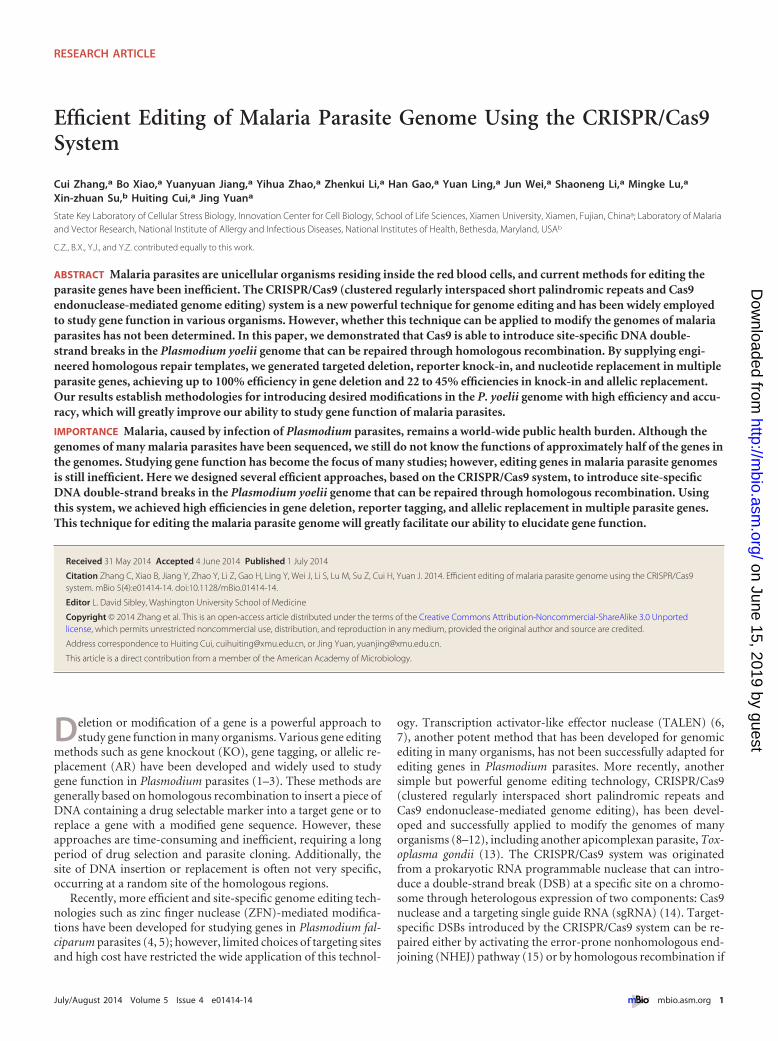
Assessment of Cas9 specificity in P. yoelii. One concern usingCRISPR/Cas9-mediated genome editing is the induction of DSBsat off-target sites. Previous studies of other organisms indicatedthat Cas9 could introduce a DSB at sequences with as many as fivemismatches of the target sequence (24, 25). To evaluate the sitespecificity, we searched the 17XNL genome for potential se-quences homologous to the target site and include a protospaceradjacent motif (PAM) sequence in experiments of Pysera1 dele-tion and Py03652 tagging with gfp. We did not identify anygenomic off-target sequences sharing fewer than six nucleotidemismatches. When search criteria were relaxed (6 to 10 mis-matches), we found three and five potential off-targets in Pysera1and Py03652 sgRNA targeting sites, respectively (Table 1). WePCR amplified the potential off-target regions from genomicDNA from two Pysera1 deletion clones (c1 and c2) and twoPy0365-gfp tagging clones (c2 and c4). DNA sequencing of thePCR products detected no mutations at any of these sites (Table 1;see Fig. S6 in the supplemental material), suggesting no off-targetmutation by CRISPR/Cas9-mediated modification. The relative
smaller genome size and lack of NHEJ DNA repair pathway inmalaria parasites may reduce the chance of off-target events (19).
DISCUSSION
In this study, we developed a CRISPR/Cas9-based genome editingsystem and successfully applied the system to modify the rodentmalaria P. yoelii genome, which is an important model commonlyused for studying malaria biology and malaria parasite and hostinteraction. We showed that the system could be used to KO, KI,or replace nucleotides in the parasite genes with high efficiencyand specificity (100% for each Pysera1 and Pysera2 gene deletion,22 to 45% in tagging PY03652 with gfp and 25% in nucleotidereplacement of the hsp70 locus) (see Table S1 in the supplementalmaterial). The development of this system will greatly improveour ability to modify the parasite genome and impact the func-tional studies of malaria parasites. Our results definitely provedthat the CRISPR/Cas9 system is functional in P. yoelii. It would beinteresting to apply this technique to other malaria parasites suchas the human malaria parasite P. falciparum.
The pYC plasmid we developed contains Cas9/sgRNA cassetteswith targeting sgRNA protospacer and restriction sites for cloningDNA templates, allowing construction of a single vector for vari-ous gene modification strategies. In addition to high efficiencyand site specificity, the Cas9/sgRNA method has other advantagescompared with the traditional gene editing methods. First, theconventional method of gene modification based on HR generallyrequires insertion of selection markers and other plasmid ele-ments into the targeting locus of the parasite genome, which mayinfluence the expression of other genes near the targeted locus.Gene editing by homologous repair of DSBs introduced byCRISPR/Cas9 requires only transient expression of Cas9/sgRNAfrom a plasmid; no integration of selection marker or other plas-mid elements into the target site occurs, reducing the unexpectedimpacts of foreign DNA on the parasite genome. Second, targetedmodification of multiple genes is increasingly preferred in variousfunctional studies such as deletion of multiple copies of genes in agene family. The insertion of drug selection markers in the targetsite will restrict the number of genes that can be modified, becauselimited drug markers are available for malaria parasites. TheCRISPR/Cas9 system we developed will allow modification ofmultiple genes without worrying about running out of drug selec-tion markers. Third, we also demonstrated that short (300- to
TABLE 1 Numbers of mutations detected at the potential off-target cleavage sites in the Plasmodium yoelii genome
sgRNA Sequencea No. of mismatches Chromosome locationb Mutation
Pysera 1 targeting sgRNA1 GTACCGGGAAACTCTGATCAAGG NoneACAAAAGCACTTTCTGATCAAGG 9 13: 2055967�2056749(�) None detectedGATATTGAAAAATCTGATCATGG 7 10: 899397�901846(�) None detectedATTTTAAAAAATTCTGATCATGG 8 10: 1255700�1258432(�) None detected
PY03652 targeting sgRNA GTCTTTATGTGGGTAACTTAAGG NoneTTAAATAATATTGTAACTTATGG 9 04: 309670�311468(�) None detectedTTTTCTATTTCTGTAACTTTAGG 7 11: 790345�792444(�) None detectedTTAAATTTTTTCGTAACTTTTGG 9 04: 529947�530873(�) None detectedGTTTTTTCACTAGTAACTTGGGG 8 04: 995696�1000093(�) None detectedATGTTTTTTTCAGTAACTCCGGG 8 11: 1701710�1706203(�) None detected
a Nucleotides in bold type are mismatches between the sgRNA sequences and the potential off-target sites. Underlined nucleotides are nucleotides in the protospacer adjacent motif(PAM) following the 20-nt sgRNA targeting sequence.b The chromosome number is shown before the colon. The numbers after the colon are the positions on the chromosome. Minus and plus symbols in parentheses indicate theforward or reverse strand of genome DNA, respectively.
Zhang et al.
6 ® mbio.asm.org July/August 2014 Volume 5 Issue 4 e01414-14
on June 15, 2019 by guesthttp://m
bio.asm.org/
Dow
nloaded from

400-bp) homologous templates were efficient in mediating re-combination repair. Using the traditional method, longer homol-ogous sequences are often preferred because of low recombina-tion and transformation frequencies (23). This high efficiency ofgene modification using short template sequences will benefitstudies of AT-rich genomes such as P. falciparum or P. yoelii ge-nome, because long AT-rich DNAs are often unstable in Esche-richia coli and are difficult to clone into plasmid vectors.
In this study, we achieved high efficiency (100%) in deletingthree gene segments with sizes ranging from 1.6 to 5.0 kb. How-ever, lower efficiencies were observed in gene tagging (22 to 45%)and nucleotide replacement (~25%). Although we do not knowthe exact reasons for the lower efficiencies in gene tagging andnucleotide replacement, the introduction of nonsense nucleotidesubstitutions to alter the sgRNA targeting sites and/or to createrestriction sites in the homologous sequences in these two types ofexperiments could contribute to the lower efficiencies observed.In the three gene deletion experiments, the DNA sequences ofboth left and right homologous arms were identical to the corre-sponding genomic DNA sequences. In the Py03652 tagging exper-iment, we introduced five nonsense nucleotide substitutions at thesgRNA-binding site in the left arm of the donor template. In thePyhsp70 nucleotide replacement, we replaced three nucleotides atthe sgRNA-binding site and another three nucleotides to create anAatII site in the homologous template. These substitutions mightaffect the stability of DNA pairing and/or the efficiency of homol-ogous recombination, leading to lower efficiency in obtainingclones with the targeted modifications. Another possible explana-tion for lower efficiency in gene modification could be due tofunctional constraint to some specific genes, i.e., some genes con-tribute more than others to parasite survival and are more difficultto modify. Additional experiments are necessary to reveal the ex-act reasons for the lower efficiencies in our gene tagging and nu-cleotide replacement experiments. Nonetheless, the efficiencies ofour gene tagging and nucleotide replacement are still higher thanthose obtained using the “traditional” homologous recombina-tion and drug selection.
MATERIALS AND METHODSPlasmid construction. To test whether Thosea asigna virus 2A peptidecan drive expression of two genes at the same time in Plasmodium yoelii,we generated a Pbeef1�a promoter-hdhfr-2A-gfp-Pbdhfr/ts 3= UTR plas-mid (see Fig. S1A in the supplemental material) derived from plasmidpL0019 obtained from the Malaria Research and Reference Reagent Re-source Center (MR4) (http://www.mr4.org) (8). SpCas9 nuclease codingfragment was amplified from plasmid 42230 obtained from Addgene,containing N-terminal triple Flag tag and simian virus 40 (SV40) nuclearlocalization signals (NLS) on both sides. Replacement of gfp in thePbeef1�a promoter-hdhfr-2A-gfp-Pbdhfr/ts 3= UTR plasmid with Cas9generated plasmid Pbeef1�a promoter-hdhfr-2A-Cas9-Pbdhfr/ts 3= UTRcassette using ClaI and AgeI restriction sites. Potential U6 snRNA pro-moter was PCR amplified from genomic DNA from P. yoelii 17XNL strainusing the primers PyU6proF/PyU6proR (see Table S2 in the supplementalmaterial). DNA fragment encoding sgRNA scaffold was also amplifiedfrom plasmid 42230 (8) and ligated with U6 snRNA promoter to generatethe U6 promoter-sgRNA cassette (Fig. 1D). An annealed oligonucleotidepair encoding sgRNA targeting sequence was inserted in the dual BsmBIsites upstream of the sgRNA scaffold, leading to a plasmid containing boththe hdhfr-2A-SpCas9 and PyU6-sgRNA cassettes, as well as the sites for HRdonor template insertion. This plasmid was named pYC (for plasmid forP. yoelii CRISPR/Cas9).
To generate the Cas9-sgRNA vector for gene deletion, including dele-
tion of Pysera1 (Gene ID in PlasmoDB database PY17X_0305700),Pysera2 (PY17X_0305600), and PDH/E1� (PY17X_0925800), we firstamplified the 5=- and 3=-flanking genomic regions (500 to 800 bp) as leftand right homologous arms by PCR using the primers listed in Table S2 inthe supplemental material. The left and right arms were inserted into thelinker site in the pYC plasmid. To detect HR, a 46-bp DNA fragment wasinserted between the left and right arms as a template for designing prim-ers p12 and p13 (Table S2).
To generate the Cas9-sgRNA vector for tagging gene Py03652(PY17X_0946500) with gfp or myc tags, we first amplified the C-terminalpart (300 to 800 bp) of the coding region as the left arm and 400 to 800 bpfrom the 3= UTR region following the translation stop codon as the rightarm using the primers listed in Table S2 in the supplemental material. ADNA fragment encoding the gfp or myc tag was inserted between the leftand right arms in frame with the gene of interest. For each gene, onesgRNA was designed to target the site close to the C-terminal part of thecoding region. To avoid binding of sgRNA to the target site in the left armof the donor template after integration, three nucleotides in the sgRNA-binding site were replaced.
To generate the Cas9/sgRNA vector for targeted nucleotide replace-ment in the Pyhsp70 (PY17X_0712100), we first PCR amplified approxi-mately 1 kb of the coding region (with the Cas9/sgRNA cleavage site in thecenter of the region) as the donor template using primers listed in Table S2in the supplemental material. Two or three nucleotide substitutions tocreate restriction sites without change of amino acids were introducedusing synthetic oligonucleotides and PCR amplification. For each gene,one sgRNA was designed to target the site close to the restriction enzymecutting site (Table S3). To prevent binding of sgRNA to the target site inthe donor template, 3 nucleotide mutations were also introduced into thesgRNA-binding site in the template. All plasmids used in this study areavailable from the authors upon request.
Parasites used and parasite transfection. All transgenic P. yoelii par-asites were generated from P. yoelii 17XNL strain. The parasites werepropagated in Kunming (KM) outbred mice purchased from the AnimalCare Center of Xiamen University. All mouse experiments were per-formed in accordance with approved protocols (XMULAC20140004) bythe Committee for Care and Use of Laboratory Animals at the School ofLife Sciences of Xiamen University. The procedures for parasite transfec-tion, selection, and cloning were described previously (26) and illustratedin Fig. S7 in the supplemental material. Briefly, parasites were electropo-rated with purified circular plasmid DNA. Transfected parasites were in-travenously injected into new mice, and parasites were selected with py-rimethamine (Pyr) in drinking water 1 day after injection. Parasites withtransfected plasmids usually appear 5 to 7 days after injection. In allelicreplacement experiments, parasite-infected RBCs were transferred to newmice when the parasitemia declined to ~0.5%.
DNA analysis. Blood samples from infected mice were collected fromthe orbital sinus, and RBCs were lysed using 1% saponin in phosphate-buffered saline (PBS). Parasite genomic DNAs were isolated using DNeasyblood kits (Qiagen) after washing off the hemoglobin, and these genomicDNAs were used in PCR amplifications. For gene deletion or gene tagging,targeted modification was confirmed by PCRs using two or three pairs ofprimer to detect 5= and 3= integrations. For targeted nucleotide substitu-tions, PCR-RFLP was used to detect introduced restriction sites in thetransfected parasite mixtures. PCR products were purified and digestedwith restriction enzymes (AatII used for Pyhsp70 replacement). SomePCR products were also sequenced directly without cloning into a plasmidvector.
RNA preparation and RT-PCR. Parasite pellets after lysis of RBCswere mixed with TRIzol (Invitrogen), and RNA was extracted according tothe manufacturer’s instructions (Qiagen). RNA quantitation was done usingNanodrop (Thermo). Purified RNA was treated with DNase using TurboDNA-free kit (Life Technologies). cDNA was synthesized using RevertAidreverse transcriptase (Fermentas). Unique primers were designed for re-verse transcription or for amplifying short regions of target genes.
CRISPR/Cas9 Editing Plasmodium yoelii Genome
July/August 2014 Volume 5 Issue 4 e01414-14 ® mbio.asm.org 7
on June 15, 2019 by guesthttp://m
bio.asm.org/
Dow
nloaded from

Western blotting. Total proteins extracted from parasite pellets wereseparated on 4 to 15% SDS-polyacrylamide gels and transferred to poly-vinylidene difluoride (PVDF) membranes (Millipore). The blot was incu-bated with blocking buffer (PBS with 3% bovine serum albumin [BSA]) atroom temperature for 1 h and then incubated at 4°C overnight with anti-Flag (mouse; 1:5,000; Abmart), anti-GFP (rabbit; 1:1,000; Cell Signaling),anti-histone H3 (rabbit; 1:1,000; Cell Signaling), or anti-Myc (rabbit;1:1,000; Cell Signaling). Antibody to histone H3 was used as a control.Horseradish peroxidase-conjugated goat anti-rabbit or anti-mouse anti-bodies (Sigma) were incubated with PVDF membranes for 2 h at roomtemperature before three washes with blocking and enhanced chemilumi-nescence (ECL) detection.
Fluorescence analysis of recombinant parasites. For live-cell imag-ing, parasite-infected mouse blood was washed twice with PBS beforestaining the cells with 2 �g/ml Hoechst 33342 (Sigma) (in PBS). The cellswere applied to poly-L-lysine-coated glass-bottom culture dishes (NEST,China).
For fixed-cell imaging, the parasites were washed twice with PBS andwere fixed with 4% paraformaldehyde�0.0075% glutaraldehyde on poly-L-lysine-coated glass slides for 30 min. After 3 washes in PBS, the parasiteswere treated with 0.1% Triton X-100 in PBS, blocked with 3% BSA in PBS,and stained with the primary antibody (mouse anti-Flag [1:1,000;Abmart], rabbit anti-GFP [1:500; Cell Signal], rabbit anti-Myc [1:500;Cell Signal]) at 4°C overnight. After 3 washes with blocking buffer, thesamples were incubated with goat anti-mouse or anti-rabbit antibodylabeled with Alexa Fluor 488 (1:2,000; Invitrogen) at room temperaturefor 1 h. Cells were stained with Hoechst 33342 to visualize nuclei. Allimages were captured and processed using identical settings in the ZeissLSM 780 laser scanning confocal microscope with a 100�/1.49-numerical-aperture (NA) oil objective. Duplicate cultures were exam-ined, and similar results were obtained in at least three independent ex-periments. To quantify the proportion of GFP-positive parasites afterPy03652-gfp tagging, parasites were washed with PBS and analyzed with aflow cytometer. Cell nuclei were first stained with 2 �g/ml Hoechst 33342(Sigma) in PBS. After a single wash, 20,000 cells were counted on a BD LSRFortessa flow cytometer. Data were analyzed with FlowJo 7.6.3 with gatingfor nuclear stain Hoechst 33342 (FL1) and for green fluorescence (FL4).
SUPPLEMENTAL MATERIALSupplemental material for this article may be found at http://mbio.asm.org/lookup/suppl/doi:10.1128/mBio.01414-14/-/DCSupplemental.
Figure S1, PDF file, 1 MB.Figure S2, PDF file, 0.4 MB.Figure S3, PDF file, 0.6 MB.Figure S4, PDF file, 1.5 MB.Figure S5, PDF file, 1.1 MB.Figure S6, PDF file, 0.3 MB.Figure S7, PDF file, 0.1 MB.Table S1, PDF file, 0.1 MB.Table S2, PDF file, 0.1 MB.Table S3, PDF file, 0.2 MB.
ACKNOWLEDGMENTS
This work was supported by grants from China Thousand Youth TalentsPlan, the 863 National High Technology Research and Development Pro-gram of China (2014AA020530), the 973 National Key Basic ResearchProgram of China (2014CB744501), the Fundamental Research Funds forthe Central Universities of China (2013121033), the Special ResearchFund for the Doctoral Program of Higher Education of China(20130121120023 and 20130121120024), and the 111 Project of the Min-istration of Education of China (B06016). This work was also partly sup-ported by the Intramural Research Program of the Division of IntramuralResearch, National Institute of Allergy and Infectious Diseases, NationalInstitutes of Health.
We thank the NIH library staff for editing the manuscript.
REFERENCES1. Wu Y, Kirkman LA, Wellems TE. 1996. Transformation of Plasmodium
falciparum malaria parasites by homologous integration of plasmids thatconfer resistance to pyrimethamine. Proc. Natl. Acad. Sci. U. S. A. 93:1130 –1134. http://dx.doi.org/10.1073/pnas.93.3.1130.
2. van Dijk MR, Waters AP, Janse CJ. 1995. Stable transfection of malariaparasite blood stages. Science 268:1358 –1362. http://dx.doi.org/10.1126/science.7761856.
3. van Dijk MR, Janse CJ, Waters AP. 1996. Expression of a Plasmodium geneintroduced into subtelomeric regions of Plasmodium berghei chromosomes.Science 271:662–665. http://dx.doi.org/10.1126/science.271.5249.662.
4. Straimer J, Lee MC, Lee AH, Zeitler B, Williams AE, Pearl JR, Zhang L,Rebar EJ, Gregory PD, Llinás M, Urnov FD, Fidock DA. 2012. Site-specific genome editing in Plasmodium falciparum using engineered zinc-finger nucleases. Nat. Methods 9:993–998. http://dx.doi.org/10.1038/nmeth.2143.
5. McNamara CW, Lee MC, Lim CS, Lim SH, Roland J, Nagle A, SimonO, Yeung BK, Chatterjee AK, McCormack SL, Manary MJ, ZeemanAM, Dechering KJ, Kumar TR, Henrich PP, Gagaring K, Ibanez M,Kato N, Kuhen KL, Fischli C, Rottmann M, Plouffe DM, Bursulaya B,Meister S, Rameh L, Trappe J, Haasen D, Timmerman M, SauerweinRW, Suwanarusk R, Russell B, Renia L, Nosten F, Tully DC, KockenCH, Glynne RJ, Bodenreider C, Fidock DA, Diagana TT, Winzeler EA.2013. Targeting Plasmodium PI(4)K to eliminate malaria. Nature 504:248 –253. http://dx.doi.org/10.1038/nature12782.
6. Moscou MJ, Bogdanove AJ. 2009. A simple cipher governs DNA recog-nition by TAL effectors. Science 326:1501. http://dx.doi.org/10.1126/science.1178817.
7. Boch J, Scholze H, Schornack S, Landgraf A, Hahn S, Kay S, Lahaye T,Nickstadt A, Bonas U. 2009. Breaking the code of DNA binding specific-ity of TAL-type III effectors. Science 326:1509 –1512. http://dx.doi.org/10.1126/science.1178811.
8. Cong L, Ran FA, Cox D, Lin S, Barretto R, Habib N, Hsu PD, Wu X,Jiang W, Marraffini LA, Zhang F. 2013. Multiplex genome engineeringusing CRISPR/Cas systems. Science 339:819 – 823. http://dx.doi.org/10.1126/science.1231143.
9. Mali P, Yang L, Esvelt KM, Aach J, Guell M, DiCarlo JE, Norville JE,Church GM. 2013. RNA-guided human genome engineering via Cas9.Science 339:823– 826. http://dx.doi.org/10.1126/science.1232033.
10. Hwang WY, Fu Y, Reyon D, Maeder ML, Tsai SQ, Sander JD, PetersonRT, Yeh JR, Joung JK. 2013. Efficient genome editing in zebrafish usinga CRISPR-Cas system. Nat. Biotechnol. 31:227–229. http://dx.doi.org/10.1038/nbt.2501.
11. Shan Q, Wang Y, Li J, Zhang Y, Chen K, Liang Z, Zhang K, Liu J, XiJJ, Qiu JL, Gao C. 2013. Targeted genome modification of crop plantsusing a CRISPR-Cas system. Nat. Biotechnol. 31:686 – 688. http://dx.doi.org/10.1038/nbt.2650.
12. Ren X, Sun J, Housden BE, Hu Y, Roesel C, Lin S, Liu LP, Yang Z, MaoD, Sun L, Wu Q, Ji JY, Xi J, Mohr SE, Xu J, Perrimon N, Ni JQ. 2013.Optimized gene editing technology for Drosophila melanogaster usinggerm line-specific Cas9. Proc. Natl. Acad. Sci. U. S. A. 110:19012–19017.http://dx.doi.org/10.1073/pnas.1318481110.
13. Shen B, Brown KM, Lee TD, Sibley LD. 2014. Efficient gene disruptionin diverse strains of Toxoplasma gondii using CRISPR/CAS9. mBio 5(3):e01114-14.
14. Jinek M, Chylinski K, Fonfara I, Hauer M, Doudna JA, Charpentier E.2012. A programmable dual-RNA-guided DNA endonuclease in adaptivebacterial immunity. Science 337:816 – 821. http://dx.doi.org/10.1126/science.1225829.
15. Bibikova M, Golic M, Golic KG, Carroll D. 2002. Targeted chromosomalcleavage and mutagenesis in Drosophila using zinc-finger nucleases. Ge-netics 161:1169 –1175.
16. Urnov FD, Miller JC, Lee YL, Beausejour CM, Rock JM, Augustus S,Jamieson AC, Porteus MH, Gregory PD, Holmes MC. 2005. Highlyefficient endogenous human gene correction using designed zinc-fingernucleases. Nature 435:646 – 651. http://dx.doi.org/10.1038/nature03556.
17. Mali P, Esvelt KM, Church GM. 2013. Cas9 as a versatile tool for engi-neering biology. Nat. Methods 10:957–963. http://dx.doi.org/10.1038/nmeth.2649.
18. Mueller AK, Hammerschmidt-Kamper C, Kaiser A. 2014. RNAi in
Zhang et al.
8 ® mbio.asm.org July/August 2014 Volume 5 Issue 4 e01414-14
on June 15, 2019 by guesthttp://m
bio.asm.org/
Dow
nloaded from

Plasmodium. Curr. Pharm. Des. 20:278 –283. http://dx.doi.org/10.2174/13816128113199990027.
19. Kirkman LA, Lawrence EA, Deitsch KW. 2014. Malaria parasites utilizeboth homologous recombination and alternative end joining pathways tomaintain genome integrity. Nucleic Acids Res. 42:370 –379. http://dx.doi.org/10.1093/nar/gkt881.
20. Huang X, Liew K, Natalang O, Siau A, Zhang N, Preiser PR. 2013. Therole of serine-type serine repeat antigen in Plasmodium yoelii blood stagedevelopment. PLoS One 8:e60723. http://dx.doi.org/10.1371/journal.pone.0060723.
21. Pei Y, Tarun AS, Vaughan AM, Herman RW, Soliman JM, Erickson-Wayman A, Kappe SH. 2010. Plasmodium pyruvate dehydrogenase ac-tivity is only essential for the parasite’s progression from liver infection toblood infection. Mol. Microbiol. 75:957–971. http://dx.doi.org/10.1111/j.1365-2958.2009.07034.x.
22. MacKellar DC, Vaughan AM, Aly AS, DeLeon S, Kappe SH. 2011. Asystematic analysis of the early transcribed membrane protein family
throughout the life cycle of Plasmodium yoelii. Cell. Microbiol. 13:1755–1767. http://dx.doi.org/10.1111/j.1462-5822.2011.01656.x.
23. Philip N, Orr R, Waters AP. 2013. Transfection of rodent malaria para-sites. Methods Mol. Biol. 923:99 –125.
24. Fu Y, Foden JA, Khayter C, Maeder ML, Reyon D, Joung JK, Sander JD.2013. High-frequency off-target mutagenesis induced by CRISPR-Cas nu-cleases in human cells. Nat. Biotechnol. 31:822– 826. http://dx.doi.org/10.1038/nbt.2623.
25. Hsu PD, Scott DA, Weinstein JA, Ran FA, Konermann S, Agarwala V,Li Y, Fine EJ, Wu X, Shalem O, Cradick TJ, Marraffini LA, Bao G,Zhang F. 2013. DNA targeting specificity of RNA-guided Cas9 nucleases.Nat. Biotechnol. 31:827– 832. http://dx.doi.org/10.1038/nbt.2647.
26. Janse CJ, Franke-Fayard B, Mair GR, Ramesar J, Thiel C, Engelmann S,Matuschewski K, van Gemert GJ, Sauerwein RW, Waters AP. 2006.High efficiency transfection of Plasmodium berghei facilitates novel selec-tion procedures. Mol. Biochem. Parasitol. 145:60 –70. http://dx.doi.org/10.1016/j.molbiopara.2005.09.007.
CRISPR/Cas9 Editing Plasmodium yoelii Genome
July/August 2014 Volume 5 Issue 4 e01414-14 ® mbio.asm.org 9
on June 15, 2019 by guesthttp://m
bio.asm.org/
Dow
nloaded from