Efficient differentiation and polarization of primary cultured … · polarization of cultured...
31
Title Efficient differentiation and polarization of primary cultured neurons on poly(lactic acid) scaffolds with microgrooved structures Asako Otomo 1,2† *, Mahoko Takahashi Ueda 1† , Toshinori Fujie 3,4 , Arihiro Hasebe 5 , Yosuke Okamura 1,6 , Shinji Takeoka 5 , Shinji Hadano 2 , So Nakagawa 1,2 * 1 Micro/Nano Technology Center, Tokai University, Hiratsuka, Kanagawa 259-1292, Japan 2 Department of Molecular Life Sciences, Tokai University School of Medicine, Isehara, Kanagawa 259-1193, Japan 3 School of Life Science and Technology, Tokyo Institute of Technology, B-50, 4259 Nagatsuta-cho, Midori-ku, Yokohama, Kanagawa 226-850, Japan 4 PRESTO, Japan Science and Technology Agency, 4-1-8 Honcho, Kawaguchi-shi, Saitama 332-0012, Japan 5 Graduate School of Advanced Science and Engineering, Waseda University, TWIns, 2-2, Sinjuku-ku, Tokyo 162-8480, Japan 6 Department of Applied Chemistry, School of Engineering, Tokai University, Hiratsuka, Kanagawa 259-1292, Japan These authors equally contributed to this study. *Authors to whom correspondence should be addressed: E-mail: [email protected] and [email protected] KEYWORDS: nanosheet, primary cultured cortical neurons, RNA sequencing, synthetic biodegradable polymer, scaffold . CC-BY 4.0 International license certified by peer review) is the author/funder. It is made available under a The copyright holder for this preprint (which was not this version posted May 21, 2019. . https://doi.org/10.1101/644781 doi: bioRxiv preprint
Transcript of Efficient differentiation and polarization of primary cultured … · polarization of cultured...
1
Title
Efficient differentiation and polarization of primary cultured neurons on
poly(lactic acid) scaffolds with microgrooved structures
Asako Otomo1,2†*, Mahoko Takahashi Ueda1†, Toshinori Fujie3,4, Arihiro Hasebe5,
Yosuke Okamura1,6, Shinji Takeoka5, Shinji Hadano2, So Nakagawa1,2*
1Micro/Nano Technology Center, Tokai University, Hiratsuka, Kanagawa 259-1292, Japan 2Department of Molecular Life Sciences, Tokai University School of Medicine, Isehara,
Kanagawa 259-1193, Japan 3School of Life Science and Technology, Tokyo Institute of Technology, B-50, 4259
Nagatsuta-cho, Midori-ku, Yokohama, Kanagawa 226-850, Japan 4PRESTO, Japan Science and Technology Agency, 4-1-8 Honcho, Kawaguchi-shi, Saitama
332-0012, Japan 5Graduate School of Advanced Science and Engineering, Waseda University, TWIns,
2-2, Sinjuku-ku, Tokyo 162-8480, Japan 6Department of Applied Chemistry, School of Engineering, Tokai University, Hiratsuka,
Kanagawa 259-1292, Japan
†These authors equally contributed to this study.
*Authors to whom correspondence should be addressed:
E-mail: [email protected] and [email protected]
KEYWORDS: nanosheet, primary cultured cortical neurons, RNA sequencing, synthetic
biodegradable polymer, scaffold
.CC-BY 4.0 International licensecertified by peer review) is the author/funder. It is made available under aThe copyright holder for this preprint (which was notthis version posted May 21, 2019. . https://doi.org/10.1101/644781doi: bioRxiv preprint
2
Abbreviations
differently expressed gene, DEG; poly(lactic acid), PLA; poly(glycolic acid), PVA;
polydimethylsiloxane, PDMS; poly-D-lysine, PDL; RNA sequencing, RNA-Seq;
vitronectin, VTN; fetal bovine serum, FBS; phosphate-buffered saline without Mg2+ or
Ca2+, PBS(−)
.CC-BY 4.0 International licensecertified by peer review) is the author/funder. It is made available under aThe copyright holder for this preprint (which was notthis version posted May 21, 2019. . https://doi.org/10.1101/644781doi: bioRxiv preprint

3
Abstract
Synthetic biodegradable polymers including poly(lactic acid) (PLA) are attractive cell
culture substrates because their surfaces can be micropatterned to support cell
adhesion. The cell adhesion properties of a scaffold mainly depend on its surface
chemical and structural features; however, it remains unclear how these characteristics
affect the growth and differentiation of cultured cells or their gene expression. In this
study, we fabricated two differently structured PLA nanosheets: flat and microgrooved.
We assessed the growth and differentiation of mouse primary cultured cortical neurons
on these two types of nanosheets after pre-coating with poly-D-lysine and vitronectin.
Interestingly, prominent neurite bundles were formed along the grooves on the
microgrooved nanosheets, whereas thin and randomly extended neurites were only
observed on the flat nanosheets. Comparative RNA sequencing analyses revealed that
the expression of genes related to postsynaptic density, dendritic shafts, and asymmetric
synapses was significantly and consistently up-regulated in cells cultured on the
microgrooved nanosheets when compared with those cultured on the flat nanosheets.
These results indicate that microgrooved PLA nanosheets can provide a powerful means
of establishing a culture system for the efficient and reproducible differentiation of
neurons, which will facilitate future investigations of the molecular mechanisms
underlying the pathogenesis of neurological disorders.
.CC-BY 4.0 International licensecertified by peer review) is the author/funder. It is made available under aThe copyright holder for this preprint (which was notthis version posted May 21, 2019. . https://doi.org/10.1101/644781doi: bioRxiv preprint
4
Introduction
Dissociated primary neuronal cultures are widely used not only for basic
neuroscience research but also for drug discovery for neurological disorders1,2,3. In such
culture systems, scaffolds are one of the key factors providing the cells with structural
support for attachment and subsequent growth and differentiation. Thus far, numerous
synthetic polymers including polystyrene, poly(lactic acid) (PLA), poly(glycolic acid), and
poly(lactic-co-glycolic acid)4,5,6 have been developed to serve as scaffolds. Among
these, PLA, a biodegradable and resorbable polyester, has recently come into the
limelight for its utility in medical applications such as tissue regeneration7.
Polymeric ultrathin film consisting of PLA, hereinafter called “PLA nanosheet,”
is a thin, soft, and flexible material, with properties that allow it to adhere anywhere
without any adhesive materials.8 Many studies have demonstrated that nanosheets can
be used to dress wounds to avoid suture, prevent infection, and bone regeneration etc.
for biomedical applications8,9,10,11,12,13. Nanosheets are also suitable for use as a sheet
substrate in cell culture for several reasons. First, nanosheets can easily adhere to the
surface of standard culture plates, culture dishes, and cover glass without any adhesive
materials. Second, cells and/or tissues cultured on nanosheets can be easily recovered,
allowing researchers to easily analyze biological molecules such as proteins, DNA, and
RNA. Last, a variety of structural patterns of the nanosheet surface, such as grooves
and pores, is possible14,15.
Despite these advantages, there are a number of issues that can hinder the
application of nanosheets to cell culture experiments. The surface of the nanosheet is
.CC-BY 4.0 International licensecertified by peer review) is the author/funder. It is made available under aThe copyright holder for this preprint (which was notthis version posted May 21, 2019. . https://doi.org/10.1101/644781doi: bioRxiv preprint
5
hydrophobic, which prevents cell adhesion. Therefore, surface pre-treatment of the
nanosheets is required16,17,18. For cells, particularly dissociated neurons, cell adhesion
molecules such as poly-D-lysine (PDL) peptides, which confer a positive charge on the
nanosheet surface and assist cell adhesion19, are required. Further, dissociated cultured
neurons under standard culture conditions20 extend neurites in random directions, which
prevents neurons from forming organized neuronal networks. This may mainly be due
to a lack of appropriate attractive and repulsive biological cues from the surrounding
cells as well as an absence of scaffold-linked mechanical cues to guide the direction of
axon pathfinding. Moreover, it has been shown that morphogenesis in cultured neurons
can be affected by topographical differences on the PLA substrate, and grooved
structures, in particular, may improve the guidance of neurite extension21, although the
molecular mechanisms underlying such phenomena remain to be investigated.
In this study, we fabricated microgrooved nanosheets with different
microgroove widths and used flat nanosheets as the control. After coating with cell
adhesion molecules, we then investigated the effects of the topographical features of
these nanosheets on the morphology of mouse primary cultured cortical neurons.
Further, to elucidate the molecular basis of the observed differences, we compared the
gene expression of cell cultures on the two types of nanosheets. Our findings indicated
that microgrooved nanosheets serve as an effective scaffold for the controlled neurite
polarization of cultured neurons, thereby promoting the efficient and reproducible
differentiation of neurons. Thus, microgrooved nanosheets are expected to be applied
to a large number of investigations in neuroscience research as well as regenerative
.CC-BY 4.0 International licensecertified by peer review) is the author/funder. It is made available under aThe copyright holder for this preprint (which was notthis version posted May 21, 2019. . https://doi.org/10.1101/644781doi: bioRxiv preprint

6
medicine.
Results and Discussions
Assessment of materials used to pre-coat the nanosheet surface
The surface of a PLA nanosheet is smooth and hydrophobic, thereby
preventing cell adhesion16,18. To establish the appropriate conditions for pre-coating the
surface of the nanosheets, we assessed PDL and PDL coated over truncated
recombinant human vitronectin (PDL + VTN-N). VTN-N is a recombinant human protein
that can provide a defined surface for feeder-free culture of human pluripotent stem
cells22. VTN-N is an extracellular matrix molecule that supports neurite outgrowth in vitro
both under normal conditions and after trauma23. We then cultured PC12 cells, a cell
line derived from pheochromocytoma in the rat adrenal medulla, on a glass coverslip or
nanosheet pre-coated with PDL or PDL + VTN-N. PC12 cells attached and grew on the
glass as well as the nanosheets that were pre-coated with either PDL or PDL + VTN-N
(Figure 1A). In contrast, without any PDL coating, the PC12 cells hardly adhered to the
nanosheet (Figure 1A). We further confirmed that mouse primary cultured cortical
neurons grew and differentiated on the nanosheets coated with PDL or PDL + VTN-N as
was observed for the PC12 cells (Figure 1B). At 2 days in vitro (DIV2), cell adhesion
and neurite protrusion were observed for mouse primary cortical neurons on the
nanosheet coated with either PDL or PDL + VTN-N (Figure 1B). Notably, PDL + VTN-N
coating particularly enhanced cell adhesion of the primary cultured cortical neurons on
the nanosheet (Figure 1B). At 6 days in vitro (DIV6), this observation became even
.CC-BY 4.0 International licensecertified by peer review) is the author/funder. It is made available under aThe copyright holder for this preprint (which was notthis version posted May 21, 2019. . https://doi.org/10.1101/644781doi: bioRxiv preprint
7
more evident: the degree of neurite extension on the PDL + VTN-N-coated nanosheet
was much higher than that on the PDL-coated one (Figure 1B). At this time point,
neuron-extended neurites were beginning to connect to each other (Figure 1B),
suggesting that normal differentiation of the neurons was achieved on the nanosheet.
Moreover, the cell viability of the cultured cortical neurons on the nanosheet at DIV6
reached approximately 92%, which was comparable with that on a conventional glass
substrate (Figure 1C). Taken together, the nanosheet coated with PDL + VTN-N is likely
suitable for neuronal cultures.
Development of the microgrooved nanosheet
To develop the nanosheet with a microgrooved surface that was applicable to
neuronal cultures, we first sought to fabricate nanosheets with three different surface
structures, of which the parallel microgrooves were 20, 30, or 50 μm wide with a height
of 6 μm, according to previously reported procedures24,25,26 (Figure S1). In brief, PLA
nanosheets were prepared by spin coating of the PLA-dichloromethane solution on a
polydimethylsiloxane (PDMS) negative replica with microgrooved motifs. The
micropatterned PLA nanosheets were overlaid with a poly(vinyl alcohol) (PVA)
supporting layer and then released from the PDMS mold. The nanosheets with the PVA
supporting layer were immersed into phosphate-buffered saline without Mg2+ or Ca2+
[PBS(−)] and then captured by a glass substrate.
Next, to assess the quality of the processed nanosheets, we measured the
fine structure of each surface with a Dektak stylus profiler (Bruker, Billerica, MA, USA;
.CC-BY 4.0 International licensecertified by peer review) is the author/funder. It is made available under aThe copyright holder for this preprint (which was notthis version posted May 21, 2019. . https://doi.org/10.1101/644781doi: bioRxiv preprint

8
Figure S2). It was revealed that nanosheets with 50 μm wide parallel microgrooves
were most stably and reproducibly fabricated, whereas those with 20 or 30 μm in width
were not (Figure S2). Therefore, we decided to use nanosheets with 50 μm wide
parallel microgrooves for subsequent experiments.
Morphological analysis of neuronal cells cultured on microgrooved nanosheets
To assess the effects of the surface microstructure of the nanosheet on the
cell adhesion, neurite outgrowth, and morphology of the cultured neurons, we cultured
mouse primary cortical neurons on nanosheets with either a flat or
parallel-microgrooved surface that were pre-coated with PDL + VTN-N. At 9 days in
vitro, we fixed and stained the cells with anti-MAP2 and anti-Tuj-1 antibodies and
investigated their neurite orientations and morphologies on the nanosheet. Tuj-1 and
MAP2 are an axon and a dendrite marker, respectively. Cultured neurons on both the
flat and microgrooved nanosheets were positive for MAP2 and Tuj-1, suggesting that
the cells firmly attached to and fully differentiated on the nanosheets irrespective of their
surface structure (Figure 2A). Notably, cultured neurons on the microgrooved
nanosheet extended neurites along the direction of the parallel microgrooves and
formed thick neurite bundles (Figure 2A). In contrast, neurons on the flat nanosheet
extended thin and separated neurites in random directions (Figure 2A).
To examine how the grooved structure serves as a structural scaffold for
neurons, we investigated the locations where neurons adhered to the microgroove
structures by using scanning electron microscopy (SEM) (Figure 2B). For this purpose,
.CC-BY 4.0 International licensecertified by peer review) is the author/funder. It is made available under aThe copyright holder for this preprint (which was notthis version posted May 21, 2019. . https://doi.org/10.1101/644781doi: bioRxiv preprint

9
we cultured a limited number of cells on the microgrooved nanosheet because a
high-density culture (such as in Figure 2A) did not allow us to observe the location of
cell adhesion. Neurons on the flat nanosheet adhered to each other and elongated
neurites from the cell body in random directions (Figure 2B-a). In contrast, neurons on
the microgrooved nanosheet densely colonized the inside of the microgrooves between
the vertical ridges (Figure 2B). Interestingly, enlarged images revealed that some
neurons appeared to adhere to the sidewall of the ridges rather than to the bottom of the
microgrooves (Figure 2B-b, indicated by arrowheads) and then extended neurites
(Figure 2B-c, indicated by arrows), suggesting that neurons may also use the vertical
surface of the ridges as scaffolds. These results indicate that the microgrooves on a
nanosheet affect the architecture of cell-to-substrate adhesion, thereby changing the
neurite morphology and, presumably, also its biological function.
Gene expression patterns of neuronal cells cultured on the nanosheets
To understand the molecular basis of the observed morphological differences
in neuronal cells between the flat and microgrooved nanosheets, we performed whole
exome sequencing (RNA-Seq) analyses on the primary cultured mouse cortical
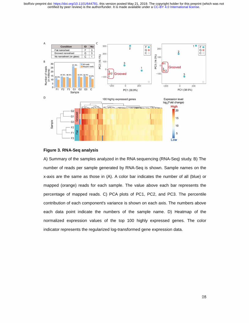
neurons. Seven different samples were subjected to RNA-Seq analysis: flat nanosheet,
n = 3; microgrooved nanosheet, n = 3; and glass coverslip, n = 1 (Figure 3A). For each
sample, 75-bp paired-end sequencing reads were mapped to the mouse reference
genome (mm10), and the results are summarized in Figure 3B. To examine gene
expression levels among different samples, the number of mapped reads were
.CC-BY 4.0 International licensecertified by peer review) is the author/funder. It is made available under aThe copyright holder for this preprint (which was notthis version posted May 21, 2019. . https://doi.org/10.1101/644781doi: bioRxiv preprint
10
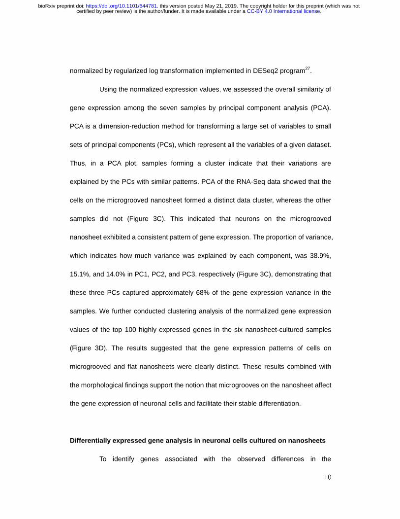
normalized by regularized log transformation implemented in DESeq2 program27.
Using the normalized expression values, we assessed the overall similarity of
gene expression among the seven samples by principal component analysis (PCA).
PCA is a dimension-reduction method for transforming a large set of variables to small
sets of principal components (PCs), which represent all the variables of a given dataset.
Thus, in a PCA plot, samples forming a cluster indicate that their variations are
explained by the PCs with similar patterns. PCA of the RNA-Seq data showed that the
cells on the microgrooved nanosheet formed a distinct data cluster, whereas the other
samples did not (Figure 3C). This indicated that neurons on the microgrooved
nanosheet exhibited a consistent pattern of gene expression. The proportion of variance,
which indicates how much variance was explained by each component, was 38.9%,
15.1%, and 14.0% in PC1, PC2, and PC3, respectively (Figure 3C), demonstrating that
these three PCs captured approximately 68% of the gene expression variance in the
samples. We further conducted clustering analysis of the normalized gene expression
values of the top 100 highly expressed genes in the six nanosheet-cultured samples
(Figure 3D). The results suggested that the gene expression patterns of cells on
microgrooved and flat nanosheets were clearly distinct. These results combined with
the morphological findings support the notion that microgrooves on the nanosheet affect
the gene expression of neuronal cells and facilitate their stable differentiation.
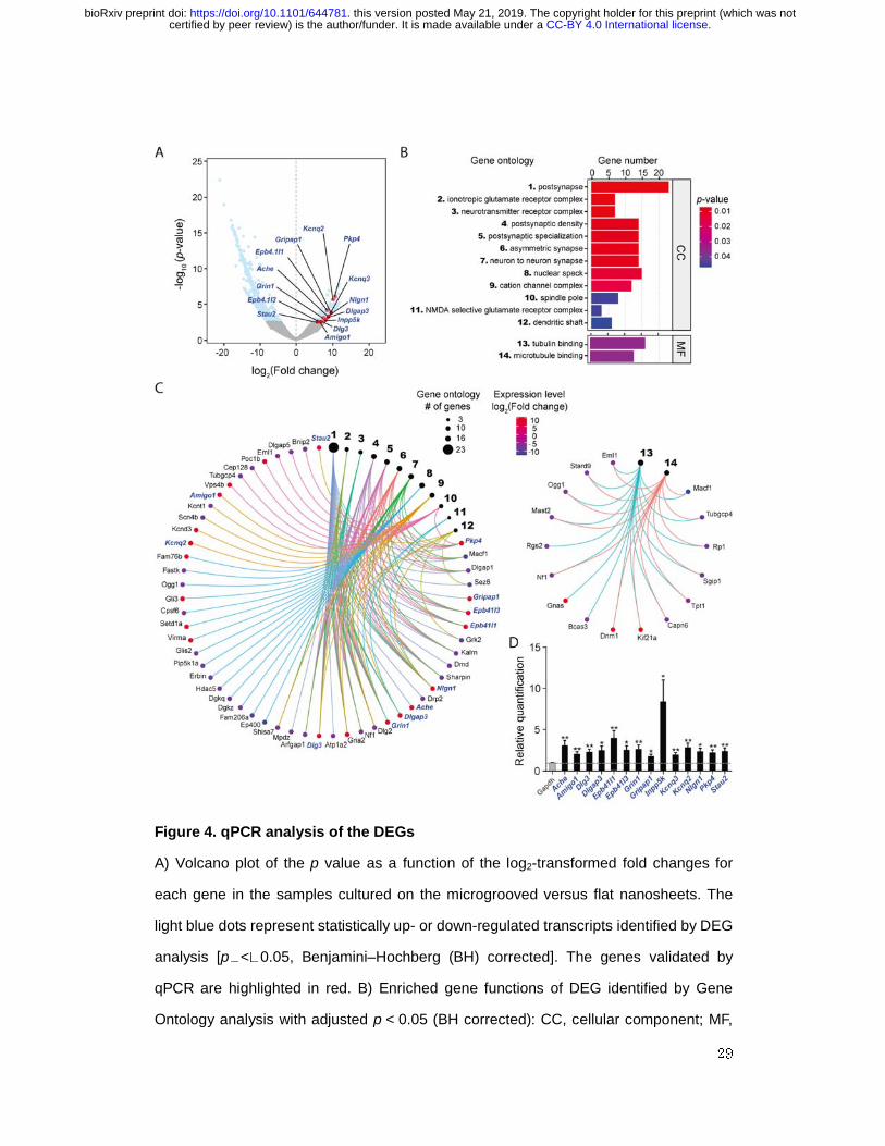
Differentially expressed gene analysis in neuronal cells cultured on nanosheets
To identify genes associated with the observed differences in the
.CC-BY 4.0 International licensecertified by peer review) is the author/funder. It is made available under aThe copyright holder for this preprint (which was notthis version posted May 21, 2019. . https://doi.org/10.1101/644781doi: bioRxiv preprint
11
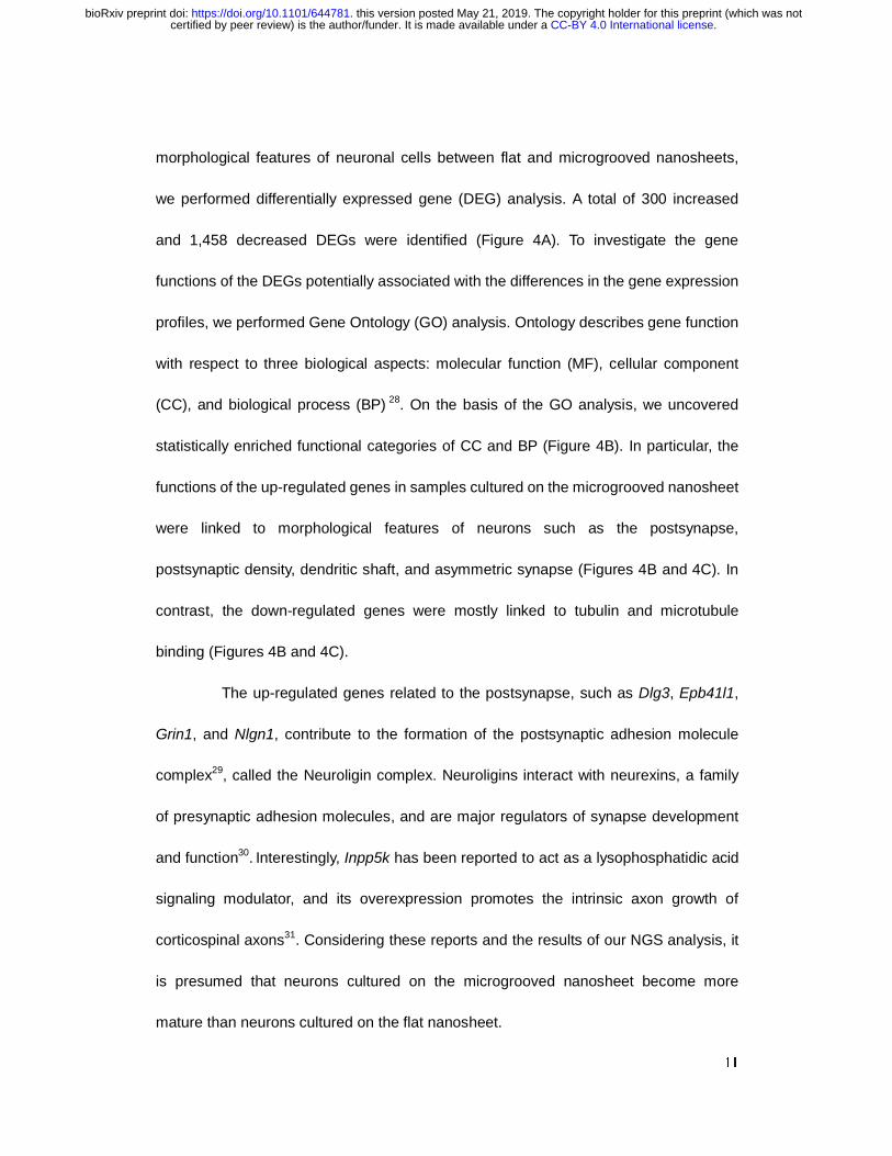
morphological features of neuronal cells between flat and microgrooved nanosheets,
we performed differentially expressed gene (DEG) analysis. A total of 300 increased
and 1,458 decreased DEGs were identified (Figure 4A). To investigate the gene
functions of the DEGs potentially associated with the differences in the gene expression
profiles, we performed Gene Ontology (GO) analysis. Ontology describes gene function
with respect to three biological aspects: molecular function (MF), cellular component
(CC), and biological process (BP) 28. On the basis of the GO analysis, we uncovered
statistically enriched functional categories of CC and BP (Figure 4B). In particular, the
functions of the up-regulated genes in samples cultured on the microgrooved nanosheet
were linked to morphological features of neurons such as the postsynapse,
postsynaptic density, dendritic shaft, and asymmetric synapse (Figures 4B and 4C). In
contrast, the down-regulated genes were mostly linked to tubulin and microtubule
binding (Figures 4B and 4C).
The up-regulated genes related to the postsynapse, such as Dlg3, Epb41l1,
Grin1, and Nlgn1, contribute to the formation of the postsynaptic adhesion molecule
complex29, called the Neuroligin complex. Neuroligins interact with neurexins, a family
of presynaptic adhesion molecules, and are major regulators of synapse development
and function30. Interestingly, Inpp5k has been reported to act as a lysophosphatidic acid
signaling modulator, and its overexpression promotes the intrinsic axon growth of
corticospinal axons31. Considering these reports and the results of our NGS analysis, it
is presumed that neurons cultured on the microgrooved nanosheet become more
mature than neurons cultured on the flat nanosheet.
.CC-BY 4.0 International licensecertified by peer review) is the author/funder. It is made available under aThe copyright holder for this preprint (which was notthis version posted May 21, 2019. . https://doi.org/10.1101/644781doi: bioRxiv preprint

12
To validate the up-regulated DEGs identified by RNA-Seq, we performed
quantitative RT-PCR (qRT-PCR) on independently prepared mRNA samples from both
flat and microgrooved nanosheets. Since NGS analysis revealed that synapse
formation and maturation of the neurons were promoted on microgrooved nanosheets,
we selected 14 DEGs known to play crucial roles in either postsynapse or presynapse
maturation in neurons: Ache, Amigo1, Dlg3, Dlgap3, Epb41l1, Epb41l3, Grin, Gripap1,
Inpp5k, Kcnd3, Kcnq2, Nlgn1, Pkp4, and Stau2. All the selected DEGs were
significantly up-regulated in mRNA samples from neuronal cultures on the
microgrooved nanosheets compared with those from the flat nanosheets [Ache*,
Amigo1**, Dlg3**, Dlgap3*, Epb41l1*, Epb41l3**, Grin**, Gripap1*, Inpp5k*, Kcnd3**,
Kcnq2**, Nlgn1*, Pkp4**, and Stau2** (**p < 0.01, *p < 0.05)] (Figure 4D). These results
strongly indicate that microgrooves on a nanosheet can more efficiently facilitate
neuronal differentiation, which is consistent with the morphological findings obtained in
this study (Figure 2). Importantly, the microgroove structure controls the position of cell
adhesion on the nanosheet by limiting it to the bottom and sides of the parallel grooves,
which may facilitate neurite bundle formation and lead to the efficient formation of
synapses.
In this study, we newly developed a neural cell culture system using a
microgrooved PLA nanosheet, which provided a more reproducible and efficient culture
environment for the neurons. Thus far, the precise molecular mechanisms by which
neuronal maturation is accelerated in the presence of a microgrooved scaffold are still
unclear. Nonetheless, this microgrooved nanosheet could provide a powerful means to
.CC-BY 4.0 International licensecertified by peer review) is the author/funder. It is made available under aThe copyright holder for this preprint (which was notthis version posted May 21, 2019. . https://doi.org/10.1101/644781doi: bioRxiv preprint

13
establish a novel experimental system for neuroscience research and regenerative
medicine and may facilitate future investigations of the molecular mechanisms
underlying the pathogenesis of many neurological disorders.
Methods
Reagents and preparation of nanosheets
All reagents used in this study including the PLA were of analytical grade.
Silicon wafers (SiO2 substrate; KST World, Fukui, Japan) cut to an appropriate size
(typically 3 × 3 cm) were treated with piranha solution, followed by washing with distilled
water. PVA (Mw: 22 kDa; Kanto Chemical, Tokyo, Japan) was dissolved in distilled water
at a concentration of 10 mg/mL, and this solution was dropped onto the SiO2 substrates
and spin-coated at 4,000 rpm for 20 s (Spin Coater MS-A100; Mikasa, Tokyo, Japan),
followed by drying at 50°C for 2 min. A solution of PLA (Mw: 80–100 kDa; Polysciences,
Warrington, PA, USA) at 10 mg/mL was then dropped onto the PVA-coated substrates
and spin-coated at 4,000 rpm for 20 s, followed by drying at 50°C for 2 min. The
obtained substrates were immersed in distilled water to collect free-standing
nanosheets. The nanosheets were scooped up with coverslips and fully dried in a
desiccator overnight. PDL or PDL with VTN-N was coated onto the PLA nanosheets
immediately prior to use for culture experiments.
Preparation of microgrooved nanosheets
.CC-BY 4.0 International licensecertified by peer review) is the author/funder. It is made available under aThe copyright holder for this preprint (which was notthis version posted May 21, 2019. . https://doi.org/10.1101/644781doi: bioRxiv preprint
14
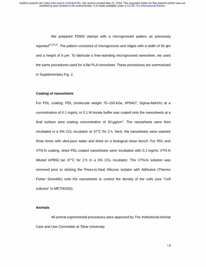
We prepared PDMS stamps with a microgrooved pattern as previously
reported24,25,26. The pattern consisted of microgrooves and ridges with a width of 50 μm
and a height of 6 μm. To fabricate a free-standing microgrooved nanosheet, we used
the same procedures used for a flat PLA nanosheet. These procedures are summarized
in Supplementary Fig. 2.
Coating of nanosheets
For PDL coating, PDL (molecular weight 70–150 kDa, #P6407; Sigma-Aldrich) at a
concentration of 0.1 mg/mL in 0.1 M borate buffer was coated onto the nanosheets at a
final surface area coating concentration of 30 μg/cm2. The nanosheets were then
incubated in a 5% CO2 incubator at 37°C for 2 h. Next, the nanosheets were washed
three times with ultra-pure water and dried on a biological clean bench. For PDL and
VTN-N coating, dried PDL-coated nanosheets were incubated with 0.1 mg/mL VTN-N
diluted inPBS(−)at 37°C for 2 h in a 5% CO2 incubator. The VTN-N solution was
removed prior to sticking the Press-to-Seal Silicone Isolator with Adhesive (Thermo
Fisher Scientific) onto the nanosheets to control the density of the cells (see “Cell
cultures” in METHODS).
Animals
All animal experimental procedures were approved by The Institutional Animal
Care and Use Committee at Tokai University.
.CC-BY 4.0 International licensecertified by peer review) is the author/funder. It is made available under aThe copyright holder for this preprint (which was notthis version posted May 21, 2019. . https://doi.org/10.1101/644781doi: bioRxiv preprint
15
Cell cultures
PC12 cells were cultured in Dulbecco's Modified Eagle's medium with High
Glucose (Wako) supplemented with 7.5% (w/v) heat-inactivated fetal bovine serum
(FBS; PAA Laboratories), 7.5% (w/v) heat-inactivated horse serum (Gibco), 100 U/mL
penicillin G, 100 µg/mL streptomycin, and 100 µg/mL sodium pyruvate. Mouse primary
cortical neurons were cultured as previously reported.29,30 In brief, tissues from each
embryo were dissected out and immediately placed into 1 mL of ice-cold HBSS(−). After
removing the HBSS(−) by aspiration, 0.5 mL of 0.25% trypsin-EDTA was added and the
embryo was incubated for 15 min at 37°C. The trypsin-EDTA was removed, and the
embryo was washed several times with 20% FBS/neurobasal medium (Invitrogen).
Tissue samples were treated with 50 µg/mL DNase I in 20% FBS/neurobasal medium
for 10 min at room temperature (RT). After centrifugation at 150 ×g for 15 s, the
resulting tissue pellets were dissociated in 0.6 mL of 20% FBS/neurobasal medium by
pipetting with a flame-sterilized Pasteur pipette. After counting the number of living cells
with the trypan blue assay, 9 × 10⁵ cells were placed onto the PDL- and VTN-N-coated
nanosheets using the Press-to-Seal Silicone Isolator with Adhesive to control the cell
numbers on the nanosheets. The nanosheets were then immersed in neuronal cell
culture media [neurobasal medium containing 1× B-27 supplement (Invitrogen),
25 µg/mL insulin (Sigma-Aldrich), 0.5 mM L-glutamine, 50 µg/mL streptomycin, and
50 U/mL penicillin G] and cultured at 37°C. The medium was then exchanged for fresh
medium containing 5% FBS, and the cells were cultured on the nanosheets for another
36 h.
.CC-BY 4.0 International licensecertified by peer review) is the author/funder. It is made available under aThe copyright holder for this preprint (which was notthis version posted May 21, 2019. . https://doi.org/10.1101/644781doi: bioRxiv preprint

16
Cell viability assay
alamarBlue Cell Viability Reagent (Thermo Fisher Scientific) was added to
10% (v/v) of the medium at DIV6 of the primary cultured neurons. After 6 h incubation,
we detected the fluorescence intensity of resorufin with the Spectra Max i3 (Perkin
Elmer). Resazurin, a PC of the alamarBlue reagent, is reduced to the highly red
fluorescent resorufin in viable cells only. Experiments were repeated four times.
Immunocytochemistry
The cells were fixed with 4% (w/v) paraformaldehyde in PBS(−) pH 7.5 for
30 min at RT and permeabilized with 0.1% (w/v) TritonX-100 in PBS(−) for 30 min. The
primary antibodies used in previous reports32,33 were diluted in PBS(−) containing 1.5%
(v/v) normal goat serum and incubated with the samples. After washing the cells with
PBS(−), Alexa 594-conjugated goat anti-mouse IgG (1:500; Molecular Probes) or Alexa
594-conjugated goat anti-rabbit IgG (1:500; Molecular Probes) was used for the
detection of proteins of interest. Fluorescent signals were captured with the BZ-X
fluorescence microscope (Keyence) and processed with Adobe Photoshop (Adobe).
Library preparation
Total RNA was extracted from the cultured cells with the RNeasy Plus Micro
Kit (Qiagen) according to the manufacturer's protocol. The quality of the total RNA
samples was validated with the RNA 6000 Pico Kit (Agilent) on the Bioanalyzer (Agilent).
.CC-BY 4.0 International licensecertified by peer review) is the author/funder. It is made available under aThe copyright holder for this preprint (which was notthis version posted May 21, 2019. . https://doi.org/10.1101/644781doi: bioRxiv preprint

17
High-quality RNA samples with an RNA integrity number >9 were used for library
preparation. RNA-Seq libraries were prepared with the Encore Complete RNA-Seq DR
Multiplex system (NuGEN) in accordance with the manufacturer's instructions.
RNA-Seq analysis
Indexed paired-end cDNA sequencing libraries were sequenced by MiSeq
(Illumina, San Diego, CA, USA). A total number of 75-bp paired-end reads were
sequenced. After trimming the reads with the fastq_quality_trimmer tool in the
FASTX-Toolkit (version 0.0.14) using the option (−Q 33, −t 20, −l 30), the reads were
then mapped onto the mouse reference genome (mm10) using HISAT2 (version 2.1.0)34
with the default options. StringTie (version 1.3.4b)35 was used to quantify gene
expression. The R package of DESeq2 (version 1.18.1)27 was used for RNA-Seq
differential expression analysis. We first normalized the gene expression values for
each sample using regularized log transformation implemented in the DESeq2 program.
For each gene, the gene expression data of cells cultured on both the flat and
microgrooved nanosheets were statistically examined. We assumed that genes
differentially expressed on these two types of nanosheets with a statistical significance
of p�<�0.05 (Benjamini–Hochberg corrected) was indeed a DEG. Enrichment analysis
was carried out with the enrichGO function in the R package of clusterProfiler (version
3.7.1) 36.
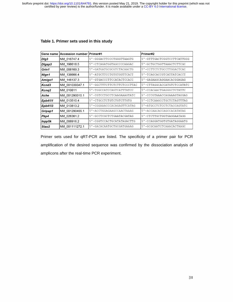
qRT-PCR
.CC-BY 4.0 International licensecertified by peer review) is the author/funder. It is made available under aThe copyright holder for this preprint (which was notthis version posted May 21, 2019. . https://doi.org/10.1101/644781doi: bioRxiv preprint
18
qRT-PCR was performed on 10 ng of total RNA using the Thunderbird SYBR
qPCR/RT Set (Toyobo) with the specific primers (0.2 µM each) listed in Table 1. The
transcript levels were normalized by the amount of Gapdh mRNA in each sample.
Statistical analyses were conducted with Prism 7 (GraphPad). Statistical significance
was evaluated by ANOVA followed by appropriate post hoc tests for multiple
comparisons between groups.
Data Availability
The RNA-Seq data obtained in this study have been deposited in the DDBJ DRA
database (https://www.ddbj.nig.ac.jp/dra/index-e.html) under the accession numbers
DRR166653–DRR166659.
References
1. Sunol, C. et al. Studies with neuronal cells: From basic studies of mechanisms of
neurotoxicity to the prediction of chemical toxicity. Toxicol. In. Vitro. 22, 1350-1355
(2008).
2. Ji, C., Tang, M. & Johnson, G. V. W. Assessing the degradation of tau in primary
neurons: The role of autophagy. Methods Cell Biol. 141, 229-244 (2017).
3. Wellbourne-Wood, J. & Chatton, J. Y. From Cultured Rodent Neurons to Human
Brain Tissue: Model Systems for Pharmacological and Translational Neuroscience.
ACS Chem. Neurosci. 9, 1975-1985 (2018).
.CC-BY 4.0 International licensecertified by peer review) is the author/funder. It is made available under aThe copyright holder for this preprint (which was notthis version posted May 21, 2019. . https://doi.org/10.1101/644781doi: bioRxiv preprint
19
4. Huang, W., Sunami, Y., Kimura, H. & Zhang, S. Applications of Nanosheets in
Frontier Cellular Research. Nanomaterials (Basel) 8, 10.3390/nano8070519 (2018).
5. Giordano, G. G. et al. Retinal pigment epithelium cells cultured on synthetic
biodegradable polymers. J. Biomed. Mater. Res. 34, 87-93 (1997).
6. Huang, H. D., Xu, J. Z., Fan, Y., Xu, L. & Li, Z. M. Poly(L-lactic acid) crystallization in
a confined space containing graphene oxide nanosheets. J Phys Chem B 117,
10641-10651 (2013).
7. Annunziata, M., Nastri, L., Cecoro, G. & Guida, L. The Use of Poly-d,l-lactic Acid
(PDLLA) Devices for Bone Augmentation Techniques: A Systematic Review.
Molecules 22, 10.3390/molecules22122214 (2017).
8. Okamura, Y., Kabata, K., Kinoshita, M., Saitoh, D. & Takeoka, S. Free-Standing
Biodegradable Poly(lactic acid) Nanosheet for Sealing Operations in Surgery. Adv
Mater 21, 4388-4392 (2009).
9. Komachi, T. et al. Adhesive and robust multilayered poly(lactic acid) nanosheets for
hemostatic dressing in liver injury model. J. Biomed. Mater. Res. B. Appl. Biomater.
105, 1747-1757 (2017).
10. Miyazaki, H. et al. An ultrathin poly(L-lactic acid) nanosheet as a burn wound
dressing for protection against bacterial infection. Wound Repair Regen. 20,
573-579 (2012).
11. Okamura, Y. et al. Fragmentation of poly(lactic acid) nanosheets and patchwork
treatment for burn wounds. Adv Mater 25, 545-551 (2013).
.CC-BY 4.0 International licensecertified by peer review) is the author/funder. It is made available under aThe copyright holder for this preprint (which was notthis version posted May 21, 2019. . https://doi.org/10.1101/644781doi: bioRxiv preprint

20
12. Huang, K.C. et al. Sandwich-type PLLA-nanosheets loaded with BMP-2 induce
bone regeneration in critical-sized mouse calvarial defects. Acta Biomater 59, 12-20
(2017).
13. Murahashi, Y. et al. Multi-layered PLLA-nanosheets loaded with FGF-2 induce
robust bone regeneration with controlled release in critical-sized mouse femoral
defects. Acta Biomater 85, 172-179 (2019).
14. Altomare, L., Gadegaard, N., Visai, L., Tanzi, M. C. & Fare, S. Biodegradable
microgrooved polymeric surfaces obtained by photolithography for skeletal muscle
cell orientation and myotube development. Acta Biomater. 6, 1948-1957 (2010).
15. Barbanti, S. H., Santos, A. R.,Jr, Zavaglia, C. A. & Duek, E. A. Porous and dense
poly(L-lactic acid) and poly(D,L-lactic acid-co-glycolic acid) scaffolds: in vitro
degradation in culture medium and osteoblasts culture. J. Mater. Sci. Mater. Med.
15, 1315-1321 (2004).
16. Fujie, T. et al. Evaluation of substrata effect on cell adhesion properties using
freestanding poly(L-lactic acid) nanosheets. Langmuir 27, 13173-13182 (2011).
17. Lin, Y. et al. Surface modification of poly(L-lactic acid) to improve its
cytocompatibility via assembly of polyelectrolytes and gelatin. Acta Biomater. 2,
155-164 (2006).
18. Niwa, D., Fujie, T., Lang, T., Goda, N. & Takeoka, S. Heterofunctional nanosheet
controlling cell adhesion properties by collagen coating. J. Biomater. Appl. 27,
131-141 (2012).
.CC-BY 4.0 International licensecertified by peer review) is the author/funder. It is made available under aThe copyright holder for this preprint (which was notthis version posted May 21, 2019. . https://doi.org/10.1101/644781doi: bioRxiv preprint
21
19. Kim, Y. H., Baek, N. S., Han, Y. H., Chung, M. A. & Jung, S. D. Enhancement of
neuronal cell adhesion by covalent binding of poly-D-lysine. J. Neurosci. Methods
202, 38-44 (2011).
20. Kaech, S. & Banker, G. Culturing hippocampal neurons. Nat. Protoc. 1, 2406-2415
(2006).
21. Morelli, S. et al. Influence of micro-patterned PLLA membranes on outgrowth and
orientation of hippocampal neurites. Biomaterials 31, 7000-7011 (2010).
22. Sugii, S. et al. Human and mouse adipose-derived cells support feeder-independent
induction of pluripotent stem cells. Proc. Natl. Acad. Sci. U. S. A. 107, 3558-3563
(2010).
23. Bergen, K., Frodin, M., von Gertten, C., Sandberg-Nordqvist, A. -. & Skold, M. K.
Neurite Growth and Polarization on Vitronectin Substrate after in Vitro Trauma is not
Enhanced after IGF Treatment. Brain Sci. 8, 10.3390/brainsci8080151 (2018).
24. Shi, X. et al. Periosteum-mimetic structures made from freestanding microgrooved
nanosheets. Adv Mater 26, 3290-3296 (2014).
25. Fujie, T. et al. Spatial coordination of cell orientation directed by nanoribbon sheets.
Biomaterials 53, 86-94 (2015).
26. Hasebe, A et al., Biohybrid actuators based on skeletal muscle-powered
microgrooved ultra-thin films consisting of
poly(styreneblock-butadiene-block-styrene). ACS Biomater. Sci. Eng. in press
.CC-BY 4.0 International licensecertified by peer review) is the author/funder. It is made available under aThe copyright holder for this preprint (which was notthis version posted May 21, 2019. . https://doi.org/10.1101/644781doi: bioRxiv preprint

22
27. Love, M. I., Huber, W. & Anders, S. Moderated estimation of fold change and
dispersion for RNA-seq data with DESeq2. Genome Biol. 15, 550-014-0550-8
(2014).
28. The Gene Ontology Consortium. Expansion of the Gene Ontology knowledgebase
and resources. Nucleic Acids Res. 45, D331-D338 (2017).
29. Fabregat, A. et al. The Reactome Pathway Knowledgebase. Nucleic Acids Res. 46,
D649-D655 (2018).
30. Südhof T. C. Synaptic Neurexin Complexes: A Molecular Code for the Logic of
Neural Circuits. Cell 171, 745-769 (2017).
31. Fink, K. L., Lopez-Giraldez, F., Kim, I. J., Strittmatter, S. M. & Cafferty, W. B. J.
Identification of Intrinsic Axon Growth Modulators for Intact CNS Neurons after
Injury. Cell. Rep. 18, 2687-2701 (2017).
32. Otomo, A. et al. ALS2/alsin deficiency in neurons leads to mild defects in
macropinocytosis and axonal growth. Biochem. Biophys. Res. Commun. 370, 87-92
(2008).
33. Hadano, S. et al. Loss of ALS2/Alsin exacerbates motor dysfunction in a
SOD1-expressing mouse ALS model by disturbing endolysosomal trafficking. PLoS
One 5, e9805 (2010).
34. Kim, D., Langmead, B. & Salzberg, S. L. HISAT: a fast spliced aligner with low
memory requirements. Nat. Methods 12, 357-360 (2015).
35. Pertea, M. et al. StringTie enables improved reconstruction of a transcriptome from
RNA-seq reads. Nat. Biotechnol. 33, 290-295 (2015).
.CC-BY 4.0 International licensecertified by peer review) is the author/funder. It is made available under aThe copyright holder for this preprint (which was notthis version posted May 21, 2019. . https://doi.org/10.1101/644781doi: bioRxiv preprint

23
36. Yu, G., Wang, L. G., Han, Y. & He, Q. Y. clusterProfiler: an R package for comparing
biological themes among gene clusters. OMICS 16, 284-287 (2012).
Acknowledgments
We thank Hiromi Takahashi and all the members of the Support Center for Medical
Research and Education at Tokai University for their technical support in this study. This
research was funded by the MEXT (Japanese Ministry of Education, Culture, Sports,
Science and Technology)-Supported Program for the Strategic Research Foundation at
Private Universities, Grant #S1411010. A.O. was supported by 2015–2016 Tokai
University School of Medicine Research Aid. T.F. was supported by the Precursory
Research for Embryonic Science and Technology (PRESTO) program from the Japan
Sc ience and Technology Agency (JST; g rant number JPMJPR152A).
Author Contributions
A.O., Y.O., and S.N. conceived the study idea. A.O. conducted the biological
experiments. M.T.U. and S.N. conducted the data analysis. Y.O., A.H., T.F., and S.T.
fabricated the nanosheets. S.H. provided mice for the culture experiments and
interpreted the data. A.O., M.T.U., S.H. and S.N. wrote the manuscript. All authors read
and approved the final manuscript.
.CC-BY 4.0 International licensecertified by peer review) is the author/funder. It is made available under aThe copyright holder for this preprint (which was notthis version posted May 21, 2019. . https://doi.org/10.1101/644781doi: bioRxiv preprint
24
Additional Information
Supplementary information: A detailed description of the protocol used to fabricate the
microgrooved nanosheet, the results of the surface analysis of the microgrooved
nanosheet, and the sequences of the primer sets used in this study are available online.
Competing interests: The authors declare no conflict of interests.
Author Information
Corresponding Authors
Correspondence to Asako Otomo and So Nakagawa
*(Asako Otomo) E-mail: [email protected] Tel.: +81 463 1121.
*(So Nakagawa) E-mail: [email protected] Tel.: +81 463 1121.
ORCID
Asako Otomo: 0000-0002-7849-4309
Mahoko T Ueda: 0000-0002-3960-0922
Toshinori Fujie: 0000-0003-1417-8670
Shinji Takeoka: 0000-0002-6230-1517
Shinji Hadano: 0000-0002-4997-3968
So Nakagawa: 0000-0003-1760-3839
.CC-BY 4.0 International licensecertified by peer review) is the author/funder. It is made available under aThe copyright holder for this preprint (which was notthis version posted May 21, 2019. . https://doi.org/10.1101/644781doi: bioRxiv preprint

25
Figure 1. Culturing neurons on the PLA nanosheet (Figure legend up to 350
Words)
A) PC12 cells cultured on a glass substrate or PLA nanosheet. The glass substrate and
PLA nanosheet were coated with PDL or PDL + VTN-N. In the absence of any coating,
the PC12 cells hardly adhered to the PLA nanosheet. Scale bars, 50 µm. B) Mouse
primary cultured cortical neurons on a PDL + VTN-N-coated PLA nanosheet. Neural
morphology at DIV2 and DIV6 are shown. Mouse primary cultured cortical neurons
adhered to and displayed more prominently elongated neurites on the
PDL + VTN-N-coated PLA nanosheet. Scale bars, 50 µm. C) Cell viability of mouse
primary cultured neurons on the glass substrate and PLA nanosheet were compared
with neurons cultured on conventional plastic culture dishes coated with PDL + VTN-N.
The cell viability of neurons on the PLA nanosheet was comparable with that on the
glass substrate [glass: 93.75 ± 1.65, nanosheet: 92.00 ± 2.16 (mean ± SE), expressed
.CC-BY 4.0 International licensecertified by peer review) is the author/funder. It is made available under aThe copyright holder for this preprint (which was notthis version posted May 21, 2019. . https://doi.org/10.1101/644781doi: bioRxiv preprint

26
as a percentage of the cell viability on plastic culture dishes].
.CC-BY 4.0 International licensecertified by peer review) is the author/funder. It is made available under aThe copyright holder for this preprint (which was notthis version posted May 21, 2019. . https://doi.org/10.1101/644781doi: bioRxiv preprint

27
Figure 2. Culturing neurons on the flat and microgrooved PLA nanosheets
A) Morphological analysis of neural cell culture on nanosheets. Tuj-1 and MAP2 were
used as a neurite and dendrite marker, respectively. Nuclei were visualized by DAPI
staining. The white arrows indicate the direction of microgroove processing. Cultured
neurons on the microgrooved nanosheet extended neurites along the microgrooves. In
contrast, cultured neurons on the flat nanosheet extended neurites in random directions.
Scale bars, 50 μm. B) SEM analysis of mouse primary cultured cortical neurons on flat
and microgrooved PLA nanosheets. Neurons on the flat nanosheet adhered to each
other and extended neurites from the cell body (a). Neurons on the microgrooved
nanosheet densely colonized the inside of the microgrooves between the vertical ridges.
Interestingly, neurons also adhered to the sidewall of the ridges rather than to the
bottom of the microgrooves (b, indicated by blue arrowheads) and then extended
neurites (c, indicated by blue arrows). Scale bars, 20 μm.
.CC-BY 4.0 International licensecertified by peer review) is the author/funder. It is made available under aThe copyright holder for this preprint (which was notthis version posted May 21, 2019. . https://doi.org/10.1101/644781doi: bioRxiv preprint
28
Figure 3. RNA-Seq analysis
A) Summary of the samples analyzed in the RNA sequencing (RNA-Seq) study. B) The
number of reads per sample generated by RNA-Seq is shown. Sample names on the
x-axis are the same as those in (A). A color bar indicates the number of all (blue) or
mapped (orange) reads for each sample. The value above each bar represents the
percentage of mapped reads. C) PCA plots of PC1, PC2, and PC3. The percentile
contribution of each component's variance is shown on each axis. The numbers above
each data point indicate the numbers of the sample name. D) Heatmap of the
normalized expression values of the top 100 highly expressed genes. The color
indicator represents the regularized log-transformed gene expression data.
.CC-BY 4.0 International licensecertified by peer review) is the author/funder. It is made available under aThe copyright holder for this preprint (which was notthis version posted May 21, 2019. . https://doi.org/10.1101/644781doi: bioRxiv preprint
29
Figure 4. qPCR analysis of the DEGs
A) Volcano plot of the p value as a function of the log2-transformed fold changes for
each gene in the samples cultured on the microgrooved versus flat nanosheets. The
light blue dots represent statistically up- or down-regulated transcripts identified by DEG
analysis [p�<�0.05, Benjamini–Hochberg (BH) corrected]. The genes validated by
qPCR are highlighted in red. B) Enriched gene functions of DEG identified by Gene
Ontology analysis with adjusted p < 0.05 (BH corrected): CC, cellular component; MF,
.CC-BY 4.0 International licensecertified by peer review) is the author/funder. It is made available under aThe copyright holder for this preprint (which was notthis version posted May 21, 2019. . https://doi.org/10.1101/644781doi: bioRxiv preprint

30
molecular function. C) Gene-Concept Network of the CC (left) and MF (right) DEG,
showing the relationship between the GO categories and the genes. The black circles
represent the GO categories, whose functions are indicated by the same numbers used
in (B). The size of the circles for each GO category corresponds to the number of genes
in the category. The value of the log2-transformed fold change of each gene is indicated
by the color bar. Blue italics indicate genes validated by qPCR. D) Relative gene
expression levels of selected DEGs. Genes Ache, Amigo1, Dlg3, Dlgap3, Epb41l3,
Epb41l1, Grin, Gripap1, Inpp5k, Kcnd3, Kcnq2, Nlgn1, Pkp4, and Stau2 in the samples
from the flat and microgrooved nanosheets were quantified by qPCR. The dotted line
represents the gene expression of samples on the flat nanosheet. Statistically
significant differences between the flat and microgrooved nanosheets are indicated by
asterisks (**p < 0.01, *p < 0.05) (Fig. 4D). Expression of Gapdh was used as an internal
control.
.CC-BY 4.0 International licensecertified by peer review) is the author/funder. It is made available under aThe copyright holder for this preprint (which was notthis version posted May 21, 2019. . https://doi.org/10.1101/644781doi: bioRxiv preprint
31
Table 1. Primer sets used in this study
Primer sets used for qRT-PCR are listed. The specificity of a primer pair for PCR
amplification of the desired sequence was confirmed by the dissociation analysis of
amplicons after the real-time PCR experiment.
.CC-BY 4.0 International licensecertified by peer review) is the author/funder. It is made available under aThe copyright holder for this preprint (which was notthis version posted May 21, 2019. . https://doi.org/10.1101/644781doi: bioRxiv preprint