EFFICACY OF DENTAL PULP STEM CELLS FROM...
189
EFFICACY OF DENTAL PULP STEM CELLS FROM DECIDUOUS TEETH IN TREATING DIABETIC WOUNDS PUKANA JAYARAMAN FACULTY OF DENTISTRY UNIVERSITY OF MALAYA KUALA LUMPUR 2016
Transcript of EFFICACY OF DENTAL PULP STEM CELLS FROM...

EFFICACY OF DENTAL PULP STEM CELLS FROM DECIDUOUS TEETH IN TREATING DIABETIC WOUNDS
PUKANA JAYARAMAN
FACULTY OF DENTISTRY UNIVERSITY OF MALAYA
KUALA LUMPUR
2016

EFFICACY OF DENTAL PULP STEM CELLS FROM
DECIDUOUS TEETH IN TREATING DIABETIC
WOUNDS
PUKANA JAYARAMAN
THESIS SUBMITTED IN FULFILMENT OF THE
REQUIREMENTS FOR THE DEGREE OF DOCTOR
OF PHILOSOPHY
FACULTY OF DENTISTRY
UNIVERSITY OF MALAYA
KUALA LUMPUR
2016

ii
UNIVERSITY OF MALAYA
ORIGINAL LITERARY WORK DECLARATION
Name of Candidate: Pukana Jayaraman (I.C/Passport No: )
Registration/Matric No: DHA120010
Name of Degree: Doctor of Philosophy
Title of Project Paper/Research Report/Dissertation/Thesis (“this Work”):
“Efficacy of Dental Pulp Stem Cells from Deciduous Teeth in Treating Diabetic
Wounds”
Field of Study: PhD in Dental Studies (Regenerative Medicine)
I do solemnly and sincerely declare that:
(1) I am the sole author/writer of this Work;
(2) This Work is original;
(3) Any use of any work in which copyright exists was done by way of fair
dealing and for permitted purposes and any excerpt or extract from, or
reference to or reproduction of any copyright work has been disclosed
expressly and sufficiently and the title of the Work and its authorship have
been acknowledged in this Work;
(4) I do not have any actual knowledge nor do I ought reasonably to know that
the making of this work constitutes an infringement of any copyright work;
(5) I hereby assign all and every rights in the copyright to this Work to the
University of Malaya (“UM”), who henceforth shall be owner of the
copyright in this Work and that any reproduction or use in any form or by any
means whatsoever is prohibited without the written consent of UM having
been first had and obtained;
(6) I am fully aware that if in the course of making this Work I have infringed
any copyright whether intentionally or otherwise, I may be subject to legal
action or any other action as may be determined by UM.
Candidate’s Signature Date:
Subscribed and solemnly declared before,
Witness’s Signature Date:
Name:
Designation:

iii
ABSTRACT
Diabetic foot ulcers (DFU) are one of the common complications in diabetes mellitus
(DM). The underlying problems of DFU are delayed wound healing due to
hyperglycemia, poor blood circulation, and nerve damage. Mesenchymal stem cells
(MSCs) from various sources including dental-derived stem cells have been used in
studies in wound healing. The aim of this study is to assess the wound healing ability of
human extracted deciduous teeth in both in vitro and in vivo model in different culture
conditions and dosages. The immune gene expression of dental pulp stem cells from
three different dental sources, namely human extracted deciduous teeth (SHED),
periodontal ligament (PDLSCs), and dental pulp stem cells (DPSCs) were assessed in
early and late passage. All three sources exhibited indistinguishable mesenchymal stem
cell characteristics. Although SHED has increased immune gene expressions compared
to DPSCs and PDLSCs, it is more suitable for clinical usage in its early passages than
prolonged passages. This has led to investigations on biological molecules secreted by
SHED cultured in low serum culture conditions in two different passages which are
passage 2 (P2) and passage 4 (P4). Our findings show that at low serum concentrations,
the expression of growth factors and cytokines were higher and had significant
differences. The overall expression of genes, namely IL1B, IL2, IL4, IFNG, CXCL5,
CD40LG, and CCL7, show increased expression in P2 low serum conditions. The
percentage of wound closure in in vitro study of wound healing showed enhanced
wound closure rate for SHED P2 2% FBS. The further study on enzymatic and non-
enymatic antioxidants reported higher Superoxide Dismutase (SOD) and Total
Glutathione (GSH) and decreased Malondialdehyde (MDA) and Advanced Oxidation
Protein Products (AOPP) level in SHED treated group. The safe dosage for SHED was
studied and it showed that there is no damage to liver and kidney in Sprague Dawley

iv
rats after the transplantation of SHED in two different dosages. After that, the wound
healing rates on diabetic Sprague Dawley rats’ skin were studied on day 5 and 10. The
histology of granulation tissue indicates that SHED has high collagen content and a
thinner epidermal layer. Immunohistostaining presented an over-expression of Heat-
shock protein (Hsp70) marker which indicates higher wound healing rate in SHED
treated group. The SOD activity and GSH level was amplified in the SHED-treated
group with declining levels of MDA and AOPP. The hydroxyproline accumulation was
augmented in the SHED-treated diabetic wound. The diabetic animal wound model was
a pre-clinical study and is important in conveying SHED for therapeutic usage. The
parameters induced by SHED in treating diabetic wounds are an increase in antioxidants
level, reduction of lipid peroxidation and protein oxidation, accumulation of
hydroxyproline, augmented expressions of the Hsp70 marker, promotion of the
angiogenesis process through expression of wound repair genes, greater collagen
deposition, and fewer inflammatory cells.

v
ABSTRAK
Ulser kaki diabetik (DFU) adalah salah satu daripada komplikasi biasa diabetes melitus
(DM). Masalah asas berkaitan DFU adalah kelewatan dalam penyembuhan luka
disebabkan oleh hiperglisemia, kekurangan peredaran darah dan kemusnahan saraf. Sel
stem mesenchymal (MSCs) dari pelbagai sumber termasuk sel stem daripada kegigian
telah digunakan dalam banyak kajian berkaitan penyembuhan luka. Tujuan kajian ini
adalah untuk menilai keupayaan gigi susu manusia yang dicabut dalam merawat
penyembuhan luka dalam model luka in vitro dengan pelbagai kondisi kultur dan
seterusnya dalam model in vivo. Sel stem dari tiga sumber kegigian yang berbeza iaitu
pulpa gigi susu (SHED), ligamen periodontal (PDLSCs), dan gigi kekal (DPSCs)
didapati mengekspresi ciri-ciri immunologi pada ‘passage’ awal dan lewat. Ketiga-tiga
sumber mempamerkan karakteristik sel stem mesenchymal. Walaupun SHED telah
menunjukkan ekspresi gen imun yang tinggi berbanding DPSCs dan PDLSCs, ia adalah
lebih sesuai untuk penggunaan klinikal di passage awal (P2) berbanding dengan passage
lewat (P9). Ini telah menjuruskan kepada penyiasatan ke atas molekul biologi yang
dirembeskan oleh SHED yang dikultur dalam keadaan serum rendah di passage 2 (P2)
dan passage 4 (P4). Kedapatan kami menunjukkan pada konsentrasi serum rendah,
ekspresi faktor-faktor pertumbuhan dan sitokin adalah tinggi dan mempunyai perbezaan
yang ketara. Ekspresi gen secara keseluruhannya iaitu gen IL1B, IL2, IL4, IFNG,
CXCL5, CD40LG dan CCL7 telah menunjukkan peningkatan ekspresi dalam P2 yang
dikultur dalam serum rendah. Imej digital in vitro penyembuhan luka diabetik melalui
cabaran glukosa calaran assay in vitro dengan SHED P2 2% FBS telah menunjukkan
peningkatan kadar penutupan luka. Tambahan pula, peningkatan aktiviti SOD dan tahap
GSH dengan pengurangan pengoksidaan lipid (MDA) dan pengoksidaan protein
(AOPP) telah diperhatikan dalam kumpulan dirawat dengan SHED. Kajian ketoksikan

vi
menunjukkan tiada kesan hepatotoksik atau nefrotoksik oleh SHED. Kesan luka
diabetik makroskopik menunjukkan penyembuhan luka yang lebih baik dalam
kumpulan dirawat dengan SHED pada hari 5 dan 10. Histologi tisu granulasi
menunjukkan bahawa SHED mempunyai kandungan kolagen yang tinggi dan lapisan
epidermis yang lebih nipis. Pewarna pkh-26 ditandakan pada SHED diperhatikan pada
hari 10 selepas pembedahan dilakukan. Kajian ‘immunostaining’ telah menunjukkan
ekspresi Hsp70 protein dalam SHED. Aktiviti SOD dan tahap GSH adalah tinggi dalam
kumpulan dirawat dengan SHED dengan penurunan tahap MDA dan AOPP.
Pengumpulan ‘hydroxyproline’ telah menunjukkan peningkatan dalam luka diabetik
yang dirawat dengan SHED. Model luka haiwan diabetik adalah satu kajian pra-klinikal
dan ia penting dalam menyarankan SHED untuk kegunaan terapeutik. Parameter yang
diketengahkan oleh SHED dalam merawat luka diabetik adalah peningkatan tahap
antioksidan, pengurangan pengoksidaan lipid dan oksidasi protein, pengumpulan
‘hydroxyproline’, peningkatan ekspresi penanda Hsp70, menggalakkan proses
angiogenesis melalui ekspresi gen pembaikan luka, peningkatan pengumpulan kolagen
dan pengurangan sel-sel inflamasi.

vii
ACKNOWLEDGEMENTS
With the deepest gratitude I wish to thank every person who has helped me in
enduring the path of completion of my thesis. For generously sharing their wisdom,
support and guidance, I wish to thank my Supervisors: Associate Professor Dr. Sabri
Musa, Professor Dr. Noor Hayaty Abu Kasim and Professor Dr. Mahmood Ameen
Abdulla Hassan. For their inspirational teachings: Dr. Gokula Mohan Duchiyanda
Mohan and Dr. Vijayendran Govindasamy. And also very much thankful to Prof. Dr.
Ian Charles Paterson for fruitful comments and suggestions. My precious teammates:
Punitha Vasanthan, Loo Zhang Xin and Wijenthiran Kunasekaran. I would also
acknowledge and express gratitude to the University Malaya, Faculty of Dentistry,
Ministry of Higher Education and Postgraduate Research Grant (PPP) in providing
the funding for the research work done: UM0000031.HIR.CI and PG169-2015B.
And finally, to my parents, family members and friends for their endless moral
support and prayers.

viii
TABLE OF CONTENTS
Abstract................................................................................................................... iii
Abstrak..................................................................................................................... v
Acknowledgements................................................................................................ vii
Table of Contents............................................................................................. viii
List of Figures..................................................................................................... xiv
List of Tables..................................................................................................... xvi
List of Symbols and Abbreviations........................................................................ xvii
List of Appendices............................................................................................... xx
CHAPTER 1: INTRODUCTION..................................................................... 1
1.1 Aims of the study.................................................................................... 4
CHAPTER 2: LITERATURE REVIEW
2.1 Diabetes mellitus...................................................................................... 7
2.2 Skin morphology................................................................................... 8
2.3 Wound healing process........................................................................... 9
2.4 The current treatments for chronic wounds................................................... 13
2.4.1 Types of topical applications........................................................ 14
2.4.2 Current technology...................................................................... 16
2.4.3 Stem cells as an alternative for wound repair modality..................... 17
2.5 What is stem cell?.................................................................................. 18
2.6 Types of stem cell.................................................................................. 19
2.6.1 Embryonic stem cells (ESCs)....................................................... 19
2.6.2 Induced pluripotent stem cells (iPSCs)......................................... 20
2.6.3 Mesenchymal stem cells (MSCs)................................................. 20
2.6.4 Types of dental stem cells............................................................ 22

ix
2.7 Anatomy of dental pulp......................................................................... 23
2.8 Animal model and pre-clinical studies..................................................... 24
2.9 Stem cell-conditioned medium (SC-CM)................................................. 29
2.9.1 Wound healing mechanism initiated by secreted cytokines and
paracrine signaling in stem cell therapy........................................... 31
CHAPTER 3: ISOLATION AND CHARACTERIZATION OF STEM
CELLS FROM PRIMARY SOURCE
3.1 Introduction........................................................................................... 36
3.2 Methodology
3.2.1 Sample collection....................................................................... 38
3.2.2 Isolation of dental pulp stem cells from extracted deciduous teeth
(SHED)..................................................................................... 38
3.2.3 Cell culture expansion and cryopreservation.................................. 41
3.2.4 Cell surface analysis.................................................................... 41
3.2.5 Growth kinetics........................................................................... 42
3.2.6 Multilineages differentiation........................................................ 43
3.2.6.1 Adipogenic differentiation............................................... 43
3.2.6.2 Osteogenic differentiation................................................ 44
3.2.6.3 Chondrogenic differentiation............................................ 44
3.2.6.4 Reverse transcription polymerase chain reaction (RT-PCR)
for trilineage markers........................................................ 45
3.2.7 RNA extraction and quantification............................................... 45
3.2.8 Complementary deoxyribonucleic acid (cDNA)............................ 46
3.2.9 TaqMan® Array Human Immune Gene Array................................ 46
3.2.10 Validation of Human Immune Gene using Cytokine Protein
Array......................................................................................... 47

x
3.2.11 Statistical analysis........................................................................ 47
3.3 Results
3.3.1 Characterization of MSCs derived from SHED, DPSCs, and
PDLSCs..................................................................................... 48
3.3.2 Growth kinetics.......................................................................... 50
3.3.3 Gene expression variability between SHED, DPSCs, and PDLSCs
at early and late passages................................................................ 51
3.3.4 SHED cultured at P9 displayed many genes representing pathogen
recognition as well as immune signaling and transduction.............. 54
3.3.5 Pro and anti-inflammatory gene profiling of SHED, DPSCs, and
PDLSCs at P2 and P9.................................................................. 54
3.3.6 SHED cultured at P9 displayed many genes representing many
growth and stimulation factors as well as chemokines..................... 57
3.3.7 DPSCs and PDLSCs cultured at P9 expressed lesser cellular
toxicity and apoptosis related genes as compared to SHED
cultured at P9............................................................................. 57
3.3.8 Validation of immune genes using cytokine array.......................... 58
3.4 Discussion............................................................................................. 60
3.5 Conclusion............................................................................................ 62
CHAPTER 4: WOUND HEALING POTENTIAL OF SHED WHEN
CULTURED IN LOW SERUM CULTURE CONDITIONS
4.1 Introduction.......................................................................................... 63
4.2 Methodology
4.2.1 Low serum culture conditions sample isolation and expansion........ 65
4.2.2 Morphology, growth kinetics and senescence β-Galactosidase cell
staining assay............................................................................... 65

xi
4.2.3 qPCR Human Wound Healing Array Gene expression analysis....... 66
4.2.4 Transformation assay.................................................................. 66
4.2.5 Validation of gene expression by reverse transcriptase and real
time PCR................................................................................... 67
4.2.6 Statistical analysis....................................................................... 67
4.3 Results
4.3.1 SHED cultured in low serum cultured conditions maintained its
characteristics and safe for clinical usage in late passages................ 67
4.3.2 Wound repair gene expressions pattern in altered cultured
conditions of P2 and P4............................................................. 71
4.3.3 Pro- and anti-inflammatory gene profiling of SHED at P2 2% and
P4 2% shown up regulation......................................................... 71
4.3.4 Proliferation genes which improves the impaired wound has seen
up regulated in SHED P2 2%...................................................... 72
4.3.5 SHED P2 2% has possibilities to improve the healing condition by
increasing the number maturation genes....................................... 72
4.3.6 Validation of wound healing genes using cytokine array................. 76
4.4 Discussion............................................................................................ 76
4.5 Conclusion............................................................................................. 78
CHAPTER 5: SHED IN LOW SERUM CULTURE CONDITIONS
REDUCES OXIDATIVE STRESS IN AN IN VITRO WOUND MODEL
5.1 Introduction......................................................................................... 79
5.2 Methodology
5.2.1 Preparation for the in vitro scratch assay........................................ 80
5.2.2 Transwell assay........................................................................... 81
5.2.3 Superoxide Dismutase (SOD) activity assay.................................. 81

xii
5.2.4 Malondialdehyde (MDA) quantitation activity assay....................... 82
5.2.5 Total Glutathione (GSH) assay..................................................... 82
5.2.6 Advanced Oxidation Protein Products (AOPP) assay..................... 83
5.2.7 Statistical analysis 84
5.3 Results
5.3.1 The measurement of wound closure in in vitro co-cultured scratch
assay........................................................................................ 84
5.3.2 The SOD enzyme level................................................................ 85
5.3.3 The GSH level........................................................................... 85
5.3.4 The MDA level........................................................................... 86
5.3.5 Advanced Oxidation Protein Products (AOPP)............................... 86
5.4 Discussion............................................................................................ 89
5.5 Conclusion............................................................................................. 92
CHAPTER 6: THE TOXICITY EFFECT OF SHED ON THE ANIMAL
STUDY AND THE POTENTIAL OF WOUND HEALING ON
EXPERIMENTAL STZ-INDUCED DIABETIC RATS TREATED WITH
SHED
6.1 Introduction.......................................................................................... 93
6.2 Methodology
6.2.1 Animals for toxicity test.............................................................. 95
6.2.2 Preparation of SHED for transplantation for toxicity test................. 95
6.2.3 SHED intravenous transplantation for toxicity test......................... 97
6.2.4 Blood profile analysis for toxicity test........................................... 96
6.2.5 Histological analysis for liver and kidney...................................... 96
6.2.6 Animals for diabetic wound model................................................ 97
6.2.7 SHED culture preparation............................................................ 98

xiii
6.2.8 Wound healing model.................................................................. 98
6.2.9 SHED transplantation to the wound model.................................... 99
6.2.10 Wound healing analysis............................................................... 99
6.2.11 Histological analysis.................................................................... 99
6.2.12 IHC analysis................................................................................ 100
6.2.13 Preparation for rat skin homogenate for biochemical analysis.......... 102
6.2.14 Hydroxyproline determination in rat skin tissue............................... 104
6.2.15 Statistical analysis....................................................................... 104
6.3 Results
6.3.1 Blood profile evaluation for toxicity test........................................ 105
6.3.2 Histological observation for toxicity test........................................ 106
6.3.3 SHED accelerated the wound healing............................................ 107
6.3.4 Histological analysis for diabetic wound healing model................... 110
6.3.5 IHC analysis for diabetic wound healing model.............................. 110
6.3.6 Biochemical analysis for oxidative stress markers........................... 113
6.3.7 Hydroproxyline accumulation in rats’ skin tissue........................... 116
6.4 Discussion............................................................................................ 117
6.5 Conclusion.............................................................................................. 122
CHAPTER 7: CONCLUSION......................................................................... 123
References....................................................................................................... 126
List of Publications and Papers Presented............................................................ 152
Appendix......................................................................................................... 157

xiv
LIST OF FIGURES
Figure 2.1 : Statistics of diabetes mellitus (DM) cases in Malaysia and the
incidence of diabetic foot ulcer (DFU)...................................... 8
Figure 2.2 : Human skin layer has two major parts consist of epidermis and
dermis..................................................................................... 9
Figure 2.3 : The major phases that involved in wound healing........................ 12
Figure 2.4 : The current treatments available for chronic wound healing......... 14
Figure 2.5 : Mechanism of wound healing using stem cell therapy.................. 33
Figure 3.1 : Pictorial diagram showing the procedure of dental pulp tissue
extirpation from the tooth......................................................... 40
Figure 3.2 : The morphology and immunophenotyping of SHED, DPSCs and
PDLSCs.................................................................................... 49
Figure 3.3 : Growth curve and population doubling time of SHED, DPSCs
and PDLSCs........................................................................... 51
Figure 3.4 : Comparison of gene profiling between SHED, DPSCs and
PDLSCs................................................................................. 53
Figure 3.5 : Validation of immune genes using cytokine protein array........... 59
Figure 4.1 : SHED morphology, senescence assay and population doubling
time......................................................................................... 69
Figure 4.2 : Trilineage Differentiation.......................................................... 70
Figure 4.3 : Cell transformation assay......................................................... 70
Figure 4.4 : Comparison of gene profiling for SHED..................................... 71
Figure 4.5 : Wound healing genes in pro-inflammatory stage......................... 73
Figure 4.6 : Wound healing genes in proliferation stage.................................. 74
Figure 4.7 : Wound healing genes in maturation stage................................... 75

xv
Figure 4.8 : Validation of wound healing genes using cytokine array.............. 76
Figure 5.1 : Effect of SHED in rate of wound closure in in vitro scratch
assay....................................................................................
85
Figure 5.2 : Expression of SOD enzyme level.............................................. 87
Figure 5.3 : Expression of GSH enzyme level.............................................. 87
Figure 5.4 : Effect of SHED in MDA level.................................................... 88
Figure 5.5 : Effect of SHED in AOPP level................................................. 88
Figure 6.1 : Flow chart of SHED ameliorate experimental STZ- induced
diabetic wound model in SD rats............................................... 101
Figure 6.2 : SHED’s effect on liver and kidney histology for toxicity study.... 106
Figure 6.3 : Effect of SHED in wound closure rate at day 5 and 10................ 107
Figure 6.4 : Effect of SHED on wound measurement at day 0, 5 and 10 post
surgeries................................................................................. 108
Figure 6.5 : Effect of SHED on skin histology of diabetic rats....................... 111
Figure 6.6 : Effect of SHED on Hsp70 expression......................................... 112
Figure 6.7 : Effect of SHED treatment on SOD level in wound tissue............. 114
Figure 6.8 : Effect of SHED treatment on GSH level in wound tissue............. 114
Figure 6.9 : Effect of SHED treatment on MDA level in wound tissue............ 115
Figure 6.10 : Effect of SHED treatment on AOPP level in wound tissue........... 115
Figure 6.11 : Effect of SHED treatment on hydroxyproline level in wound
tissue...................................................................................... 116

xvi
LIST OF TABLES
Table 2.1 : Approved clinical trials for skin wound healing.............................. 26
Table 2.2 : List of cytokines secreted in stem cell conditioned medium........... 35
Table 3.1 : Composition of culture media for SHED, DPSCs and PDLSCs..... 41
Table 3.2 : Antibodies used for cell surface analysis of SHED, DPSCs and
PDLSCs........................................................................................... 42
Table 3.3 : Fold changes of genes related to pathway recognition & signaling
and transcription.............................................................................. 54
Table 3.4 : Fold changes of genes related to cytokines..................................... 56
Table 3.5 : Fold changes of genes related to cellular cytotoxicity, surface
molecules and apoptosis.................................................................. 57
Table 6.1 : Effect of SHED in liver function test on acute toxicity study......... 105
Table 6.2 : Effect of SHED in kidney function test on acute toxicity study...... 105

xvii
LIST OF SYMBOLS AND ABBREVIATIONS
AOPP : Advanced oxidation protein products
ASC : Adipose tissue-derived mesenchymal stem cells
bFGF : Basic fibroblast growth factor
BM-MSCs : Bone marrow mesenchymal stem cells
BMP : Bone morphogenetic protein
CCL3 : C-C motif chemokine ligand 3
CCR2 : Chemokine (C-C motif) receptor 2
COL1A1 : Collagen type I alpha 1 chain
CTGF : Connective tissue growth factor
CXCL11 : C-X-C motif chemokine ligand 11
DPSCs : Dental pulp stem cells
EGF : Epidermal growth factor
ESCs : Embryonic stem cells
F3 : Coagulation factor III, tissue factor
G-CSF : Granulocyte colony-stimulating factor
Gli : Glibenclamide
GM-SCF : Granulocyte-macrophage colony-stimulating factor
GNLY : Granulysin
GSH : Total Glutathione
GZMB : Granzyme B
HGF : Hepatocyte growth factor
HLA-DR : Major histocompatibility complex, class II, DR alpha
Hsp70 : Heat shock protein 70
IGF-1 : Insulin like growth factor 1

xviii
IGFBP-7 : Insulin-like growth factor binding protein 7
IL-1 : Interleukin-1
IL1A : Interleukin 1 alpha
IL-6 : Interleukin-6
IL-8 : Interleukin-8
IF : Immunofluorescence
IHC : Immunohistochemistry
iPSCs : Induced pluripotent stem cells
KGF : Keratinocyte growth factor
MDA : Malondialdehyde
MCP-1 : Methyl-accepting chemotaxis protein
MMP : Matrix metalloproteinase
MSCs : Mesenchymal stem cells
PDGF : Platelet-derived growth factor
PDLSCs : Periodontal ligament stem cells
PLAU : Plasminogen activator, urokinase
PMSCs : Placental-derived mesenchymal stem cells
PTGS2 : Prostaglandin-endoperoxide synthase 2
PTPRC : Protein tyrosine phosphatase, receptor type C
P2 : Passage 2
P4 : Passage 4
RAC1 : Ras-related C3 botulinum toxin substrate 1
RANTES : Interactions of the chemokine CCL5
SELE : Selectin E
SELP : Selectin P
SD : Sprague Dawley

xix
SHED : Exfoliated or extracted deciduous teeth
SOD : Superoxide dismutase
SPARC : Secreted protein acidic and cysteine rich
STAT3 : Signal transducer and activator of transcription 3
STZ : Streptozotocin
TGF-α : Transforming growth factor-α
TGF-β1 : Transforming growth factor beta 1
TLDA : TaqMan Low Density Arrays
TNF : Tumor necrosis factor
TNFRSF18 : Tumor necrosis factor receptor superfamily, member 18
UCMSCs : Umbilical cord mesenchymal stem cells
VEGF : Vascular endothelial growth factor
WJ-MSCs : Wharton’s Jelly stem cells

xx
LIST OF APPENDICES
Appendix 1.1 : Medical ethics clearance number.......................................... 157
Appendix 1.2 : Up-regulated wound healing genes in pro-inflammatory
phase................................................................................. 158
Appendix 1.3 : Up-regulated wound healing genes in proliferation phase....... 159
Appendix 1.4 : Up-regulated wound healing genes in maturation phase......... 160
Appendix 1.5 : Animal ethics clearance number.......................................... 161
Appendix 1.6 : Protocol for pkh-26 dye tagging onto SHED......................... 162
Appendix 1.7 : Histological slide preparation for H&E staining...................... 163
Appendix 1.8 : Heatmap of gene profiling for P2 & P9 of dental stem cells... 166
Appendix 1.9 : The measurement of wound closure in in vitro co-cultured
scratch assay........................................................................ 167

1
CHAPTER 1: INTRODUCTION
Diabetes mellitus (DM) is a non-communicable disease which affects all age groups. In
2000, approximately 171 million people worldwide were diagnosed with DM and this
figure is projected to increase to 366 million by 2030 (Wild et al., 2004). In Malaysia,
DM cases stood at 2.5 million in 2013 and are expected to increase to 3.2 million by
2020 (Feisul & Azmi, 2013). Uncontrolled DM could lead to microvascular damage
such as retinopathy, nephropathy, and neuropathy and result in increased treatment
costs. Among all the complications of DM, diabetic foot ulcer (DFU) is the most
common forming around 25% of the total diabetes population. In the United States
alone the cost of treating this complication was US$132 billion in 2002 and is expected
to rise to US$192 billion by 2020 (Hogan et al., 2003). A retrospective study conducted
in a Malaysian government hospital for 2012 to 2013 revealed that the total cost to treat
DFU was US$11,000 (Lam et al., 2014).
The underlying problem of DFU is delayed wound healing due to hyperglycemia, poor
blood circulation, and nerve damage. Diabetics not only have to deal with the pain,
infection, repeated hospital admissions, and possible amputations but also the poor
quality of life that they and their families experience. In addition, the cost of treatment
for DFU has been escalating. Therefore, an alternative wound-healing treatment regime
for this chronic condition is timely (Naves et al., 2016).
The current treatment modalities practiced in hospitals for DFU are wound debridement,
care and dressings with topical antibiotics (American Diabetes Association, 2014),
topical negative pressure (Xie et al., 2010), hyperbaric oxygen therapy [HBOT]
(Londahl et al., 2013), and platelet derived-growth factors (Villela & Santos, 2010).
Despite undergoing such treatment, much patience is required by both patients and

2
doctors as the healing process is slow. This has led researchers to seek more advanced
treatments for wound healing including cell-based therapy.
Stem cells have attracted great interest amongst researchers due to their tissue
regeneration and wound healing capabilities. Wound healing is enhanced by cell
adhesion, migration, and proliferation through various signaling pathways within the
stem cells. The candidate stem cell populations for therapeutic use include embryonic
stem cells (ESCs), adult mesenchymal stem cells (MSCs) and induced pluripotent stem
cells (iPSCs).
ESCs could be a good source for the treatment of wounds with Kim et al. (2015)
reporting that mouse embryonic stem cells (mESs) can trigger wound healing signaling-
pathways. Despite this promising finding, the immunogenicity and tumorigenicity of
ESCs make it unsuitable for clinical use (Wu et al., 2007). Further, the ethical and legal
restrictions of using ESCs need to be addressed before clinical applications become
feasible. This has led to the discovery of iPSCs that allow for the generation of
autologous pluripotent stem cell populations derived from differentiated adult tissues.
This avoids ethical issues associated with human ESCs and immunogenic
complications. In a recent in vitro study, Itoh et al. (2013) reported that 3-D skin
equivalents composed of human iPSC-derived keratinocytes and fibroblasts were
successfully generated. Even though iPSCs have several advantages over ESCs, there
are still risks for tumorigenicity in an undifferentiated state. These risks need to be
resolved and improvements made on their safety profile, efficiency, and cost-
effectiveness before widespread clinical adoption becomes possible.
MSCs from various sources are also commonly used in wound healing studies. Bone
marrow stem cells (BM-MSCs) have been shown to promote wound healing in mice
(Wu et al., 2007) while adipose tissue-derived mesenchymal stem cells (ASCs) provide

3
good skin regeneration for wound healing in rats (Ozpur et al., 2016). In another study,
a large scale expansion of Wharton’s Jelly stem cells (WJ-MSCs) on gelatin microbeads
showed their capacity for wound healing (Zhao et al., 2015). In multiple preclinical
trials, placenta-derived mesenchymal stem cells (PMSCs) and human umbilical cord
mesenchymal stem cells (hUCMSCs) have been found to significantly enhance
cutaneous wound healing. This is achieved by an increase in blood vessel formation and
elevated vascular endothelial growth factor (VEGF) expression which promotes healing
through paracrine signaling (Wang et al., 2016).
Dental-derived stem cells are another alternative source of MSCs in wound healing
treatments. The root apices of the third molar are often still open at the age of eighteen
and contain a noticeable pool of undifferentiated cells which reside within the “cell rich
zone” of the dental germ pulp (Iohara et al., 2004; d'Aquino et al., 2009). Stem cells
from human exfoliated or extracted deciduous teeth (SHED) could also be a unique
resource for wound healing treatment. It has been reported that SHED expresses VEGF
which is an essential growth factor in stimulating the angiogenesis process in wound
healing (Yang et al., 2013). SHED also exhibits good potential in wound healing by
providing a better rate of wound closure with increased Type 1 collagen in SHED co-
cultured with fibroblast treated group (Nishino et al., 2011). Other advantages of using
SHED are that it involves a simple and painless procedure and the ethical concerns
differ from the use of ESCs (Miura et al., 2003; Rai et al., 2013).
Several studies in the use of MSCs suggest that the healing was attributed to the
secretion of growth factors, cytokines, and chemokines (Chen et al., 2009; Lee et al.,
2012; Mehanni et al., 2013). The MSCs employed were of ASCs and BM-MSCs. Thus
to fill the gap in the current literature, it is important to study the secretions of biological

4
molecules in SHED if it is to be used for wound healing treatment. Further, the culture
conditions and passages of SHED have to be understood in order to cultivate it as a
potential source for such healing.
The overarching goal of this research was to study if dental derived stem cells were a
good candidate in the treatment modality for diabetic wound healing. The following
research questions were addressed:
i. Are dental-derived stem cells a good cell source for cell-based therapy in
wound healing?
ii. Do low serum culture conditions affect the therapeutic potential of dental
derived stem cells in relation to wound healing?
iii. Are dental derived stem cells effective in the healing of diabetic wounds?
1.1 Aims of the study
This study had four interrelated aims as outlined below.
Aim I: To obtain MSCs from human extracted deciduous teeth (SHED), periodontal
ligament stem cells (PDLSCs) and dental pulp stem cells (DPSCs) from primary
sources suitable for wound healing. The objectives were to:
1. isolate stem cells from SHED, PDLSCs, and DPSCs from their primary sources,
2. characterise and confirm the status of MSCs,
3. evaluate the differences of SHED, PDLSCs, and DPSCs in terms of immune genes
in long-term cultured stem cells using the human immune qPCR array, and
4. assess the validation of randomly chosen immune genes from SHED, PDLSCs, and
DPSCs using the cytokine protein array.

5
Aim II: To study the wound healing potential of SHED in low serum culture conditions
in different early passages. The objectives were to:
1. assess SHED isolation and compare the morphology, growth kinetics, and
senescence level of SHED cultured in passage 2 (P2) and passage 4 (P4) in low
serum culture conditions,
2. evaluate human wound healing genes in SHED in P2 and P4 when cultured in low
serum culture conditions using qPCR,
3. assess malignant transformation of SHED in P2 and P4 when cultured in low
serum culture conditions using transformation assay, and
4. assess the validation of randomly chosen wound healing genes from P2 and P4 using
cytokine protein array.
Aim III: To study the ability of SHED in reducing oxidative stress in an in vitro
diabetic wound model in low serum culture conditions. The objectives were to:
1. assess the digital image of in vitro diabetic wound healing through in vitro glucose-
challenged scratch assay with SHED treated in low serum conditions, and
2. evaluate the oxidative stress enzyme activity level in in vitro diabetic wounds
treated with SHED in low serum culture conditions using transwell assay.
Aim IV: To evaluate the toxicity effect of SHED and to assess the potential of SHED
in treating diabetic wounds in an animal model. The objectives were to:
1. evaluate the blood profile of Sprague Dawley (SD) rats in low and high dosages of
SHED transplantation,
2. evaluate the histology of liver and kidney in low and high dosages of SHED in SD
rats using Hematoxylin and Eosin (H&E) staining,
3. assess the streptozotocin (STZ) induced-diabetic animal model,

6
4. access the macroscopic wound contraction of diabetic induced SD rats,
5. evaluate the histology of granulation tissue [(H&E) and pkh-26 tagged cells
immunofluorescence (IF) and immunohistochemistry (IHC) analysis for Hsp70
upregulation].
6. evaluate the oxidative stress enzyme activity level of the rat skin tissue homogenate;
and
7. assess the level of hydroxyproline accumulations in rat skins using hydroxyproline
assay.
.

7
CHAPTER 2: LITERATURE REVIEW
2.1 Diabetes mellitus
Diabetes mellitus (DM) is a major health concern due to its epidemic nature and related
health complications. Global trends indicate a sharp increase in type 2 DM especially in
developing countries such as Malaysia. Currently, it is estimated that over 200 million
people worldwide suffer from DM and the figure is expected to reach 366 million by
2030 (Wild et al., 2004). This prediction was based on population growth, aging,
urbanization, and the increasing prevalence of obesity and physical inactivity (Frykberg,
2006). The National Health and Morbidity Survey (NHMS) conducted by Ministry of
Health reveals that DM prevalence among Malaysian adults of 30 years and above
increased from 6.3% in 1986 to 8.3% in 1996 and further increased to 14.9% in 2006
(National Health Morbidity Survery, 1996; Letchuman et al., 2010). More recent
statistics show a further increment of approximately 31% in the 5-year period from
2006 and 2011 (Feisul & Azmi, 2013). These numbers are projected to increase steadily
(see Figure 2.1) over the next few years (National Health Morbidity Survey, 2011).
Diabetic foot ulcers (DFU) are a common complication in uncontrolled diabetes and
need to be identified early to avoid amputation. Chronic hyperglycemia can contribute
to foot ulceration and delay wound healing, and is associated with foot amputations
(Frykberg, 1999; American Diabetes Association, 1999; Mayfield et al., 2003). Proper
treatment of DFU is crucial to ensure early wound closure to prevent recurrence and
reduce the incidence of lower limb amputations (Apelqvist et al., 1993; Caputo et al.,
1994; Levin, 1995; Frykberg, 1998; Boulton et al., 1999).
8
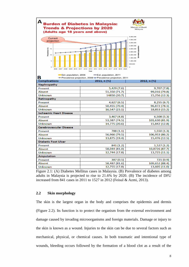
Figure 2.1: (A) Diabetes Mellitus cases in Malaysia. (B) Prevalence of diabetes among
adults in Malaysia is projected to rise to 21.6% by 2020. (B) The incidence of DFU
increased from 841 cases in 2011 to 1527 in 2012 (Feisul & Azmi, 2013).
2.2 Skin morphology
The skin is the largest organ in the body and comprises the epidermis and dermis
(Figure 2.2). Its function is to protect the organism from the external environment and
damage caused by invading microorganisms and foreign materials. Damage or injury to
the skin is known as a wound. Injuries to the skin can be due to several factors such as
mechanical, physical, or chemical causes. In both traumatic and intentional type of
wounds, bleeding occurs followed by the formation of a blood clot as a result of the

9
rupture of the blood vessels. Skin injuries can also be caused by pressure ulcers and
ischemia, for example in cases of arterial leg ulcers. In such wounds, there will be an
occlusion of blood within the blood vessels which leads to necrosis and ulcer formation
(Dealey, 2008).
Figure 2.2: The human skin layer has two major parts consisting of the epidermis and
dermis.
2.3 Wound healing process
Wounds can be categorised as either acute or chronic. Chronic wounds can be due to
tissue deficit caused by wounds that are unable to heal over a prolonged period of time
or that are frequent in occurrence. Pressure ulcers, leg ulcers, and DFUs are examples of
chronic wounds (Fowler, 2005). Acute wounds on the other hand are known as sudden
onset or short duration wounds which heal easily. These wounds include surgical,
traumatic, and burn wounds.
Wound healing is the process of tissue repair and proceeds through three phases,
namely inflammatory, proliferative and maturation (Figure 2.3). When a tissue is

10
damaged, neutrophils initiate the inflammatory process by removing any contaminating
bacteria (Clark, 1996). Further differentiation of monocytes into macrophages induces
the pro-inflammatory pathway leading to the secretion of cytokines and growth factors
(Mahdavian et al., 2011). Granulation tissue formation begins with the formation of
immature fibrin matrix which will be replaced by collagen and scar tissue (Singer &
Clark, 1999).
The inflammatory process occurs when damaged blood vessels begin to bleed resulting
in their vasoconstriction. This is followed by platelet aggregation and neutrophil
migration to the wound to neutralise bacteria on the injury site (Dealey, 2008). Blood
circulation reaches the injured tissues once the blood vessel becomes dilated and more
permeable. Platelets then release fibronectin and growth factors, or platelet derived
growth factors (PDGF), made up of transforming growth factor alpha and transforming
growth factor beta (TGFα & TGFβ), which play an important role in cell migration and
growth. The growth factors are a subclass of cytokines that function in cellular
communication (Greenhalgh, 1996) and are involved in forming new fibroblasts,
stimulating angiogenesis, and promoting the proliferation and migration of epithelial
cells (Witte & Barbul, 1997).
Cell migration is crucial for the re-epithelialization of damaged tissue (Tsirogianni et
al., 2006). TGFβ attracts monocytes to the wound which differentiate into macrophages
while fibronectin binds to the surface receptors on the cells promoting phagocytosis.
This process can only occur when there is a sufficient amount of oxygen (Cherry et al.,
2000). Both macrophages and lymphocytes are present in wounds from day one, with
the former populations peaking between day three and six and the latter between day
eight and fourteen (Martin & Muir, 1990). Throughout the inflammatory process,

11
macrophages not only phagocytize bacteria but also aid wound debridement as well as
secrete collagenases, growth factors, and other enzymes. In addition, macrophages also
play an important role in recruiting fibroblasts and keratinocytes, which are important
for tissue formation (Tsirogianni et al., 2006).
Fibroblasts are involved in the production of the extracellular matrix, secretion of
growth factors, promotion of angiogenesis, and protease synthesis (Broughton et al.,
2006) while keratinocytes help in resurfacing the wound and maintain the barrier
function. This inflammation stage lasts about four to five days and is followed by the
formation of granulation tissue. Macrophages promote wound healing by producing
platelet-derived growth factors (PDGF) and fibroblast growth factors (FGF) which later
divide into producing collagen fibers. Fibronectin also plays a role in wound healing by
enhancing fibroblast activity (Orgill & Demling, 1988).
Collagen is a tertiary protein that can be seen from the second day of wound healing
with Collagen type III being the most common. Contraction is the process of wound
closure by approximately 40-80% that occurs between the fifth and sixth day. Wound
healing by first intention will be completed in approximately 24 days depending on the
type and size of the wound. The number of macrophages and fibroblasts will gradually
reduce throughout this process.
During the maturation process, the wound becomes less vascularised with the old
collagen replaced by newly synthesised collagen and this occurs at its highest peak from
day 14 to 21 (Cherry et al., 2000) followed by the formation of scar tissue. A
hypertrophic scar is the result of impaired wound healing caused by excessive fibrous

12
tissue deposition during the healing process and leads to extreme accumulation of
collagen with a thick wound scar (Munro, 1995).
Figure 2.3: The major phases that involve in wound healing. Adapted from Jayaraman
et al., 2013.

13
2.4 The current treatments for chronic wounds
Current treatment methods for chronic wounds include surgical (Hampton, 2015),
enzymatic (Konig et al., 2005), and biological debridement (Jones & Wall, 2008; Klaus
& Steinwedel, 2015), hyperbaric oxygen therapy (HBOT) (Bakker, 2000), negative
pressure wound device (NPWD) (Suess et al., 2006), and plasma mediated bipolar
radiofrequency ablation (PBRA) (Nusbaum et al., 2012). These treatments have been
developed with the basic aim of promoting angiogenesis. However, major pitfalls due to
cost and healing inconsistencies have not improved the process of wound healing and
this has led to advanced treatment methods such as stem cell therapy (Figure 2.4).

14
Figure 2.4: The current treatments available for chronic wound healing. Adapted from
Jayaraman et al., 2013.
2.4.1 Types of topical applications
The wound healing process can be enhanced by applying topical agents followed by
dressing administered externally onto the body. A topical agent is a medicine applied

15
externally onto the wound while dressing is done to cover the wound to promote healing
and to avoid further injury (Dealey, 2008). Topical medicines are available in the form
of lotions, creams, ointments, powders (talc), and solutions (liquids) which can also
function as a transport medium when specific doses of medication are added to them.
Topical lotions are water-based and thin, readily absorbed into the skin, and usually
invisible after application. Topical creams are thicker and visible on the skin after
application and require more time for the medication to be absorbed into the skin.
Ointments or unguents are the thickest form of topical medication applications in which
the medicine is suspended in a greasy substance and adheres to the skin until the
medicine is absorbed (Zaghi & Maibach, 2007).
Topical wound healing management products include antiseptics which need to be in
contact with the bacteria for about 20 minutes to destroy them (Russel et al., 1982). For
example, cetrimide is useful for the cleansing of traumatic wounds. Lammers et al.
(1990) noted that providone iodine showed increased debridement and a decline in
bacterial count. Other than that, silver has also been used as an antiseptic and found to
have significant results in controlling wound infections from burns (Lansdown &
Williams, 2004). Silver-coated dressings on burn wounds cause less pain on removal
and reduced infection and this could also reduce dressing and nursing costs (Tredget et
al., 1998). Carneiro and Nyawawa, (2003) suggest that phenytoin significantly reduces
pain, exudate levels, and wound sizes in diabetic ulcers and burn wound patients.
Biafine, another topical emulsion, is known to increase the number of macrophages
migrating to the wound site to treat skin damage caused by ulcers, burns, and dermatitis
(Cohen et al., 2007).

16
2.4.2 Current Technology
Advanced technologies have been developed in medical and healthcare institutions to
improve wound care treatments. Recombinant growth factors is one of the early
technologies that have been performed and show high rates of healing among foot ulcer
patients (Knighton et al., 1990) while skin gene therapy has been seen to generate a
number of different growth factors (Guarini, 2003). Galeano (2003) noted the
possibility of transferring the virus vector-mediated VEGF gene to the burn wound of
an animal model. Hyperbaric Oxygen treatment involves placing patients in a 100%-
oxygen chamber, and Kalani et al. (2002) suggest that it is of benefit to most diabetic
ulcer patients. Topical negative pressure (TNP) or vacuum assisted pressure (VAC)
devices involve applying universal pressure to the wound which improves blood flow
and tissue granulation (Luckraz et al., 2003; Loree et al., 2004). The system is cost-
effective but appropriate skills are required in handling the devices. Tissue culture has
been well-established since the 1980s and has become a preferable method of topical
application. Skin from the patient or the donor is cultured in the laboratory to form a
large sheet of cells and grafted onto a granulating wound. Kumagai and Uchikoshi,
(1997) suggest that autologous cultured epithelial grafting procedure is a promising
treatment for patients with hypomelanosis. In addition, tissue engineering is an
advanced step of tissue culturing where human dermal fibroblasts are cultured on a
biosynthetic scaffold. The fibroblasts then proliferate, secrete protein and growth
factors, and eventually generate into three dimensional human grafts that can be applied
over wounds. They can also be described as cultured human skin equivalents (HSE) and
have significantly high healing rates for diabetic ulcers (Marston et al., 2003).

17
2.4.3 Stem cells as an alternative to wound repair modality
Stem cells are becoming an alternative source for topical applications in many medical
care sectors. This method seeks to heal wounds with minimal scar formation. There is a
medical need for methods and compositions to promote wound healing by cellular
regeneration therapy and work has been done on exploring the capability of stem cells
in cell regeneration and enhancing the wound healing process. Clinicians understand the
use of growth factors and their capability in growing cells in vitro and this method has
resulted in stem cell advances as an alternative source for wound healing (Bell et al.,
1981; Boyce, 2001).
Wound healing factors include recombinant growth factors that heal chronic wounds,
epidermal growth factor (Falanga et al., 1992), keratinocyte growth factor-2 for venous
ulcers (Robson et al., 2001), fibroblast growth factor, and platelet derived growth factor
(PDGF) for pressure ulcers (Robson et al., 1992; Pierce et al., 1994). Keratinocyte
sheets are an example of a bioengineered product which has been developed and tested
on human wounds.
The bioengineered skin is a type of material that delivers living cells which release
growth factors and cytokines onto the wound sites. Bioengineered skin may work by
delivering living cells which are known as a “smart material” because they are capable
of adapting to their environment. There is evidence that some of these living constructs
are able to release growth factors and cytokines (Mansbridge et al., 1998; Falanga et al.,
2002). However, this cannot be fully implemented as some of these allogeneic
constructs are unable to survive more than a few weeks when placed in a chronic wound
(Philips et al., 2002).

18
2.5 What is a stem cell?
Stem cells are known as undifferentiated cells that are able to differentiate into
specialized cells and categorized mainly into embryonic and adult stem cells. Despite
the pluripotentiality of embryonic cells they are not preferred in the research field due to
ethical issues and adult stem cells play a major role in regenerative medicine instead.
Nevertheless, both embryogenic and adult stem cells are known to have the potential to
differentiate into different cell types such as liver, skin, muscle, bone, and others. Adult
or somatic stem cells have the ability to divide and generate a range of cells types. They
are found in many parts of human body tissues such as bone marrow, blood, muscle,
brain, skin, and the liver. Embryonic stem cells (ESCs) are derived from a fertilized
embryo four or five days of post fertilization which is known as blastocyst. Blastocysts
consist of an inner (embryoblast) and an outer (trophoblast) cell mass. The outer part of
the cell mass becomes part of the placenta while the inner mass will differentiate into a
complete organism (Thomson et al., 1998).
Researchers have recently discovered induced pluripotent stem cells (iPSCs) which are
known to have the characteristics of embryonic stem cells. These cells are created by
inducing the specialized cells to express genes present in the embryonic stem cells
which control cellular functions. Adult stem cells derived from human adult skin tissue
are reprogrammed to give pluripotent capabilities. The generated pluripotent stem cells
are useful in regenerative medicine but the technique used to induce them has to be
carefully refined (Itoh et al., 2013).

19
2.6 Types of stem cells
2.6.1 Embryonic stem cells (ESCs)
The first human ESCs were isolated in 1998 by Dr. Thomson and colleagues from in
vitro fertilization (IVF) clinic embryos (Thomson et al., 1998). ESCs with their
pluripotent capabilities exhibit indefinite self-renewal in vitro and can be differentiated
into almost every cell type of the body. However, their use has been hampered due to
ethical issues, and there are risks of teratoma formation and immune rejection upon
transplantation. A study conducted by Lee et al. (2011) maximizes the therapeutic
benefit of ESCs for wound healing by using single cell suspension with application of
ESC on an excisional wound in the mouse model. The application of ESCs to growth
factors helps to stimulate the differentiation of the cells that controls teratoma and
improve the wound healing process. In another in vivo study, differentiated
keratinocyte-like cells derived from mouse embryonic stem cells provided positive
effects on a surgically wounded mouse (Vatansever et al., 2013). In addition, Shamis et
al. (2011) report that fibroblasts derived from human embryonic stem cells (hESC) have
the ability to enable the re-epithelialization of wounds and generated using a 3D tissue
model of cutaneous wound healing.
Stem cells derived from human embryos known as hESC are able to accelerate wound
healing. The hESC-derived endothelial precursor cells (EPC) have enhanced the tensile
strength of wounds after topical treatment and subcutaneous injection in animal models.
Further, granulation tissue regeneration and re-epithelialization of wounds were noticed
in ischemic tissue. The in vitro results of hESC-EPC conditioned medium also suggest
the proliferation and migration of dermal fibroblasts and epidermal keratinocytes which
increased the synthesis of the extracellular matrix by fibroblasts (Lee et al., 2011).

20
2.6.2 Induced Pluripotent Stem Cells (iPSCs)
Induced pluripotent stem cells (iPSCs) were first produced in 2006 from mouse cells
(Takahashi & Yamanaka, 2006) and in 2007 from human cells (Takahashi et al., 2007).
ESCs’ immune rejection after transplantation and ethical concerns led researchers to the
discovery of iPSCs, which are known to have the characteristics of embryonic stem
cells. These cells are created by inducing specialized cells to express genes that control
cellular functions. Yamanaka first explained the technique where the introduction of
four genes (Oct-3/4, Sox2, c-Myc, and KLF4) into an adult human skin cell could
reprogram the cell back to an embryonic state. In another study, Kattman et al. (2011)
reported that iPSCs promote vascular regeneration by producing vascular cells which
express TGF-β, BMP-2, 4, 6 and 7, Nodal and activins.
2.6.3 Mesenchymal stem cells (MSCs)
The use of mesenchymal stem cells (MSCs) is not as controversial as the use of
embryonic stem cells. They are abundant in bone marrow, adipose tissue, Wharton’s
jelly, umbilical cord blood, amniotic fluids, and foreskin. Caplan (1991) was among the
first to propose the use of MSC as a therapeutic concept. The ease of isolation, in vitro
expansion, and hypoimmunogenecity has brought MSCs into the limelight. Despite
significant advances in medical and surgical wound care, the current treatment for
cutaneous wounds with bone marrow-derived mesenchymal stem cells (BM-MSC)
accelerates wound healing kinetics and increases epithelialization and angiogenesis
(McFarlin et al., 2006). Apart from bone-marrow, other sources of MSCs that promote
wound healing are adipose tissue (Kim et al., 2007, 2009), cartilage tissue (Bos et al.,
2008), dental pulp (Nishino et al., 2011), umbilical cord (Zebardast et al., 2010), cord
blood (Luo et al., 2010), synovial fluid (Kim et al., 2015), Wharton’s jelly (Zhao et al.,
2015), and periodontal ligaments (Chen et al., 2012). Therapeutic studies on skin wound

21
healing have established several tissue-specific stem or progenitor cell types, such as
bone marrow-derived mesenchymal stem cells (BM-MSCs), adipose tissue-derived
stem cells (ASCs), dental pulp stem cells (DPSCs), and umbilical cord mesenchymal
stem cells (UCMSCs) which have been proven to improve neovascularisation.
BM-MSC contributes to enhanced growth of epidermal cells, angiogenesis, wound
contraction, and collagen deposition via engineered collagen-based scaffold
implantation in excisional wounds on the murine model (Huang et al., 2012). Green
fluorescent protein (EGFP) expressing bone marrow cells were isolated from C57BL/6-
Tg (ACTB-EGFP) 1Osb/J donor mice and transplanted into lethally irradiated wild-type
C57BL/6 mice to investigate the role of bone-marrow derived cells in a burn injury. The
study showed that the large number of fibroblasts, inflammatory cells, epithelial cells,
endothelial cell, and hair follicles enhanced the rate of wound healing (Rea et al., 2009).
A study by Inokuma et al. (2006) indicated that chemokine/chemokine receptor
interactions mediate the migration of the bone marrow keratinocyte precursor cells via a
cutaneous T-cell attracting chemokine, CTACK/CCL27. There is a possibility that the
bone marrow itself is activated by circulating cytokines or some other signal that
initiates cell migration to the wound (Wu et al., 2007).
ASCs represent an alternative source of multipotent cells with similar characteristics to
BM-MSC (Kim et al., 2009) and are easier to isolate. ASCs improve the healing rate in
diabetic mice by secreting growth factors and cytokines, enhancing granulation tissue
and capillary formation, and epithelialization (Mizuno & Nambu, 2011). An in vitro
study by Lee et al. (2012) revealed that adipose stem-cell conditioned medium (ASC-
CM) promotes HaCaT keratinocytes and foreskin-derived dermal fibroblast growth by
upregulating type I procollagen alpha 1 chain gene in fibroblasts derived from ASC-

22
CM. In another study, ASCs enhanced the proliferation of human dermal fibroblast
(HDF) through co-culturing and culturing with ASC-CM. The secretion of type I
collagen stimulated the migration of HDF (Kim et al., 2007). One of the secreted
growth factors identified in ASC-CM is vascular endothelial growth factor (VEGF)
which promotes vasculogenesis and angiogenesis. The genetically modified ASC
reported longer maintainable stem cells, improved tissue regeneration potential, and
increased secretion of VEGF (Lee et al., 2012).
UCMSCs has shown pluripotent characteristics, and harvested UCMSC differentiated
into dermal fibroblasts in a conditioned induction media upon being induced at third
passage (Han et al., 2011). When UCMSC is incubated in appropriate conditions, the
umbilical cord epithelium cells can differentiate and organize into an epidermis-like
structure (Mizoguchi et al., 2004), and differentiation of keratinocytes were observed at
a ratio of 1 to 10 when co-cultured with keratinocytes (Akino et al., 2005).
2.6.4 Types of dental stem cells
Dental stem cells are isolated from different parts of the teeth, such as from human
extracted or exfoliated deciduous teeth (SHED), adult dental pulp stem cells (DPSCs),
the apical part of the papilla (SCAP), the dental follicle (DFSC), and periodontal
ligament stem cells (PDLSCs). SHED are isolated from the pulp of human deciduous
teeth and shown to have higher proliferation rates compared to DPSCs (Miura et al.,
2003). DPSCs have been isolated and grown from pulp tissue of permanent human
dental pulp. SCAP is another type of dental stem cells isolated from human teeth and
found at the apex of the tooth root (Bluteau, 2008) while DFSC is a dental follicle
surrounding the developing tooth germ that has long been considered a multipotent
tissue based on its ability to generate cementum, bone, and periodontal ligaments (Yao,

23
2008). PDLSCs are isolated from the root surface of extracted teeth and differentiated
into cells or tissues very similar to periodontium (Estrela, 2011).
DPSCs have emerged as an alternative source of MSC compared to BMMSC in pre-
clinical trials and have created a significant impact in regenerative medicine. Growth
factors, such as TGFBs, BMPs, TGFB1, IGFs, and FGFs show up-regulation at the
injury site and play an important role in the healing process (D’Souza et al., 1998). An
in vitro study by Aranha et al. (2010) showed that DPSCs exposed to hypoxic
conditions had VEGF expressions in participation of revascularization. Though
differentiation of MSCs into tissue-specific cells has been reported, differentiation-
independent mechanisms seem to play a more significant role in tissue repair, and need
to be addressed further.
2.7 Anatomy of dental pulp
Teeth are made up of hard tissues which are the enamel, dentin, and cementum, and
pulp which is the soft tissue. The enamel forms the outer surface of a crown of a tooth
which is the hardest tissue in the human body and has the capability to withstand
chewing pressures and temperature changes. Dentin includes the main portion of the
tooth which is softer than enamel but harder than human bone. It consists of
microtubules that have dental fibres to transmit pain stimuli and nutrition throughout the
tissues. Cementum is the tissue that covers the root of the tooth in a very thin layer. It is
not as hard as enamel or dentin but is harder than bone. It contains periodontal ligament
fibers that help to anchor the tooth within the bone (Metivier & Bland, 2013).

24
2.8 Animal model and pre-clinical studies
Studies on the role of stem cells on wound healing require an experimental design to
evaluate its significance in the healing process, and animal models and human subjects
play an important part in this. The laboratory mouse is the common animal model
chosen and numerous transgenic strains and models have been developed to help
researchers study the molecular pathways involved in wound repair and regeneration
(Wong et al., 2011). Animal studies have established preliminary evidence on the
safety, feasibility, and efficacy of several important endpoints using BM-MSCs and
progenitor cells as a potential therapeutic option to induce angiogenesis (Amann et al.,
2010). The excisional wound model of diabetic rats has been reported to accelerate
epithelialization, granulation, and angiogenesis upon topical antimicrobial therapy.
Stem cell application is found to be promising for the treatment of difficult-to-heal
wounds in diabetic rats (Falanga et al., 2007). In another study, a topical treatment of
soluble factors from fibroblast stimulated regeneration in an in vivo porcine model
(Peura et al., 2012). Ayatollahi et al. (2014) isolated and cultured rat bone marrow
MSCs and studied the comparison of the differentiation potential of human and rat
MSCs in selective culture media.
Despite successful research outcomes in animal studies, pre-clinical studies offer more
future prospects in wound care management. Using stem cells has been an alternative
and better treatment strategy for various skin wounds. There are many published studies
(www.ClinicalTrials.gov) which explore autologous cells as an alternative for wound
healing (Table 2.1).
Peripheral vascular disease, diabetic foot ulcer (DFU), burn wounds, and other skin
deformities have been treated with autologous stem cells. The findings of clinical trials
performed on patients from 2005-12 show only 19 pre-clinical studies conducted on

25
wound healing of which eight were successfully completed. Recently, a randomized
design clinical trial on critical limb ischemia diabetics treated with BMMSC
successfully induced revascularization in affected limbs and has proven to be safe and
feasible (Kirana et al., 2012). Other completed pre-clinical studies have used topical
applications of a combination of alginate dressing and mouse epidermal growth factor
(Bi et al., 2012), and autologous skin fibroblasts (Zorin et al., 2014). However, as the
inflammatory cells in bone marrow cells may cause adverse effects by increasing the
inflammatory response and inhibit the regeneration process (Gonzalez et al., 2003), long
term clinical studies are required to determine the precise therapeutic effects.

26
Table 2.1: Approved clinical trials using stem cells for skin wound healing
Clinical trial
identification
Status Disease
indications
Investigational
drug/study
phase
Patients
enrolled
Route
of injection
Sponsor
PMID:22340556
Completed
(2012)
Patients with
refractory
wound/randomized
control trial
Combination therapy
with alginate dressing
and mouse epidermal
growth factor
18 Topical Dept. of orthopedics
Hangzhou, Zhejiang
China
PMID:21649569
Completed
(2011)
Critical limb ischemia
(CLI)
Autologous bone
marrow mononuclear
cell injection
20 Intra-
muscular
Faculty of medicine,
Cairo University
PMID:17518741
Completed
(2007)
Acute wound from
skin cancer surgery
and non-healing lower
extremity wound
Autologous bone
marrow
13 Topical Roger Williams Medical
Center, USA
PMID:17159798
Completed
(2006)
Diabetic ulcer Autologous skin
fibroblast and
autologous
mesenchymal stem cells
Not given Topical and
injection
Comenius University,
Slovakia
PMID:19224912
Completed
(2006)
Refractory lateral
epicondylitis
Skin dermal fibroblasts 12 Injection Royal National
Orthopaedic Hospital, UK

27
Table 2.1, continued.
NCT01065337 Completed
(2012)
Diabetic Foot Ulcer Autologous bone
marrow/Phase II
30 Intra-
muscular
Ruhr University of
Bochum, Germany
NCT00535548 Unknown
(2007)
Pressure Sore Hematopoietic stem
cell/Phase I
5 Injection University Hospital,
Basel, Switzerland
NCT00796627 Recruiting
(2010)
Burn wound HIF-1 Regulated
Endothelial Progenitor
Cell
100 Not found Johns Hopkins
University, USA
NCT01353937 Not
recruiting
(2011)
Diabetic foot ulcer Autologous bone
marrow/Phase I
27 Topical New York University,
USA
NCT00434616 Active, not
recruiting
(2011)
Critical Limb
Ischemia
Autologous bone
marrow/Phase II & III
90 Injection Hospital Berlin Vascular
Center Berlin, Germany
N NCT01232673 Active, not
recruiting
(2010)
Critical Limb
Ischemia
Autologous bone
marrow/Phase II
90 Injection University Hospital
Ostrava, Czech Republic
NCT01216865 Not
recruiting
(2010)
Diabetic Foot Ulcer Umbilical Cord
mesenchymal stem cells
Phase II / III
50
Injection Qingdao University,
China

28
Table 2.1, Continued.
NCT00616980 Completed
(2011)
Critical Limb
Ischemia
Autologous plasma/
Phase I &II
28 Injection Northwestern University,
USA
NCT00442143 Unknown
(2007)
Critical Limb
Ischemia
Autologous Bone
marrow Derived
Mononuclear
Cells/Phase 1
10 Injection Odense University
Hospital, Denmark
NCT00113243 Unknown
(2005)
Peripheral Vascular
Disease
Autologous Bone
marrow / Phase 1
20 Injection Indiana University School
of Medicine, USA
NCT00488020 Unknown
(2007)
Critical Limb
Ischemia
Autologous Bone
marrow / Phase 1
10 Injection Instituto de Molestias
Cardiovasculares, Brazil
NCT00871221 Unknown
(2009)
Scleroderma,
Systemic Sclerosis
Autologous Bone
marrow / Phase II & III
30 Injection National Institute of
Allergy and Infectious
Diseases (NIAID), USA
NCT01115634 Completed
(2010)
Facial deformities Autologous fibroblast
transplantation/ Phase II
/ III
40 Injection Royan Institute, Iran
NCT01305863 Recruiting
(2011)
Limb ischemia Autologous adipose
tissue/ Phase I & II
60
Vascular graft Tissue Genesis, Inc., USA

29
2.9 Stem cell-conditioned medium (SC-CM)
Recently, stem cell derived-soluble factors have demonstrated biological activity such
as enhanced wound healing (Heo et al., 2011) and other functions (Barreto & Salgado,
2010). Examples of soluble factors which secrete growth factors from the conditioned
medium of stem cells have been identified (Kilroy et al., 2007; Skalnikova et al., 2011).
The stem cell-conditioned medium has generated interest on cell based therapy.
However, the paracrine factors secreted by MSC that are responsible for wound healing
processes have not been fully elucidated. MSCs are known for their immunomodulatory
and anti-inflammatory properties as well as the secretion of pro-angiogenic elements
like vascular endothelial growth factor (VEGF), hepatocyte growth factor (HGF), and
insulin-like growth factor (IGF) (Imberti et al., 2007; Togel et al., 2007; Zarjou et al.,
2011). The production and secretion of growth factors by ASCs into the media is known
to provide regenerative effects in the skin. Kim et al. (2009) discovered that ASC-CM
stimulated both collagen synthesis and the migration of dermal fibroblasts which
promote wound healing and improves wrinkling via paracrine routes in animal models.
ASC-CM also inhibited melanogenesis and protected dermal fibroblasts from oxidative
damage caused by irradiation (Kim et al., 2009). The effect of the conditioned ASC
medium was also reported to increase the proliferation of HaCaT cells and dermal
fibroblasts in an in vitro study by upregulating the transcription of type I procollagen-
alpha-1 chain gene of fibroblasts (Lee et al., 2012). The stimulatory effect of ASC-CM
also accelerates the proliferation of endothelial cells (Moon et al., 2012; Lee et al.,
2009).
Keratinocytes that are activated during wound healing release growth factors and
various cytokines that stimulate fibroblasts and endothelial cells, initiate the influx of

30
immune cells, and produce systemic effects (Shephard et al., 2004; Werner et al., 2007).
The RhoA-ROCK signaling pathway in ASC-CM triggers the migration of
keratinocytes which enhances in vitro wound healing (Moon et al., 2012). The presence
of the hepatocyte growth factor (HGF) in in vitro scratch assay has been shown to
stimulate the proliferation of HaCaT keratinocyte cells (Delehedde et al., 2002). HGF
functions to enhance the migration of neutrophils, monocytes and mast cells into wound
areas and promotes the secretion of pro-angiogenesis factors (Bevan et al., 2004). Apart
from HGF, other growth factors that promote keratinocyte migration include IL-6 (Sato
et al., 1999), CTGF (Igarashi et al., 1993), GM-CSF (Mann et al., 2001), and TGF-β3
(Bandyopadhyay et al., 2006). However, the secretion of these growth factors may be
different when pooled in ASC prepared from different donors (Moon et al., 2012). In
another study by Rehman et al. (2004), ASC-CM showed various growth factor
secretions by ASC such as PDGF, IGF, KGF, bFGF, TGF-B, HGF, and VEGF, while a
study by Kim et al. (2007) reported the secretion of PDGF, FGF, TGF-β1, KGF, HGF,
and VEGF.
BM-MSC-CM injected and topically applied onto diabetic and non-diabetic mice
significantly increased the rate of wound closure compared to fibroblast-CM treated
mice (Chen et al., 2008). BM-MSC-CM initiated chemo attractive and mitogenic effects
on endothelial cells which resulted in the recruitment of endothelial and endothelial
progenitor cells into the wound. Several analyses including Real-Time PCR, antibody-
based protein array, and ELISA showed expression of cytokines and growth factors in
BM-MSC-CM. Compared to fibroblast-CM, BM-MSC-CM secreted cytokines
including VEGF-alpha, IGF, EGF, keratinocyte growth factor, angioprotein-1, stromal
derived factor-1, macrophage inflammatory protein-1 alpha and beta, and
erythropoietin. VEGF-alpha, IGF-1, PDGF-BB, and Ang-1 are known as angiogenic

31
cytokines found in BM-MSC-CM which lead to the proliferation of endothelial cells
and neovascularization in wound healing. Fibroblast-CM on the other hand, was found
to secrete IGF-1, KGF, PDGF-BB, EPO, G-CSF, and TPO (Chen et al., 2008).
The growth factors or extracellular matrix proteins secreted by stem cells derived from
human extracted or exfoliated deciduous teeth (SHED) and SHED-derived conditioned
medium (SHED-CM) have the ability to enhance the wound-healing potential of human
dermal fibroblasts (HDF). Wound contraction occurs by increasing collagen synthesis
and by activating the proliferation and migration activity of HDFs (Ueda & Nishino,
2010). Inoue et al. (2013) reported that the enhancement of vasculogenesis, migration,
and differentiation of endogenous neuronal progenitor cells can be initiated by SHED-
CM in ischemic brain injuries in the rat model.
Lee et al. (2011) successfully used the conditioned medium of human embryonic stem
cell (hESC)-derived endothelial precursor cells (EPC) in the treatment of excisional
wound healing in rats. The secretory factors that were detected using multiplex cytokine
array were epidermal growth factor (EGF), fibroblast growth factor (bFGF), fractalkine,
granulocyte-macrophage colony-stimulating factor (GM-CSF), interleukin (IL)-6, IL-8,
platelet-derived growth factor-AA (PDGF-AA), and vascular endothelial growth factor
(VEGF), which stimulate angiogenesis.
2.9.1 Wound healing mechanism initiated by secreted cytokines and paracrine
signaling in stem cell therapy
Tissue injury causes blood vessel damage and leakage of blood constituents into the
wound site. Hemostasis begins immediately after wounding, with vascular constriction
and fibrin clot formation. Hemostasis caused by coagulation provides the basic

32
architecture for initiating the inflammatory phase and tissue formation (Szpaderska et
al., 2003).
The clot and surrounding wound tissue release pro-inflammatory cytokines and growth
factors such as PDGF, FGF, EGF, TGF-β, transforming growth factor-α (TGF-
α), interleukin-1β (IL-1β), and interferon-γ (IFN-γ) (Szpaderska et al., 2003). The
migration of inflammatory cells into the wound, or chemotaxis, starts with the
infiltration of neutrophils, macrophages, and lymphocytes. Macrophage infiltration is
highly regulated by gradients of different chemotactic factors, including growth factors,
pro-inflammatory cytokines, and chemokines [macrophage inflammatory protein 1α
(MIP-1α, MCP-1, RANTES)] (Gosain & DiPietro, 2004; Broughton et al., 2006).
Mesenchymal stem cells play an important therapeutic role in wound healing.
Endogenous MSCs or exogenously delivered MSCs can migrate to the wound site to
heal the damaged tissue. Not only can the MSCs participate in the process but the
concentrated conditioned medium from MSCs can also modulate the wound healing
process without the presence of MSCs in the wound (Li & Fu, 2012). As such, this
study on the MSC-conditioned medium will focus on determining its significance in
skin wound healing. Figure 2.5 shows the pathway of a stem cell conditioned medium
in wound healing. The mechanisms responsible for enhanced wound healing in skin
include the possible trans-differentiation of MSC to form cells of epidermal and dermal
lineages (Sasaki et al., 2008). Growth factors secreted by MSCs stimulate paracrine
effects and this signaling between the cytokines and pro-inflammatory factors enhances
wound closure (Kilroy et al., 2007).

33
Figure 2.5: Mechanism of wound healing using stem cell therapy. Adaptation from
Jayaraman et al., 2013.
A number of wound healing mediators were identified in MSC-CM, including TGF-β1,
the chemokines IL-6, IL-8, MCP-1 and RANTES, and collagen type I, fibronectin,
SPARC, and IGFBP-7. Table 2.1 shows the types of cytokines secreted during wound
healing and their functions. Cytokines play an important role in accelerating the healing
process. The enhancement of wound healing is due to accelerated cell migration rather
than increased cell proliferation (Walter et al., 2010).
Neovascularization is an important step in wound healing which promotes the growth of
new blood vessels to sustain the newly formed granulation tissue and keratinocytes. Wu
et al. (2007) discovered that BM-MSCs enhanced angiogenesis in wound healing.

34
Vascular endothelial growth factor (VEGF) is one of the key factors that promote
vasculogenesis and angiogenesis in wound healing (Bueno et al., 2016).
Throughout the process of wound healing, keratinocytes migrate from the basal
population around the wound’s edge to cover the lesion and later differentiate into a
basal phenotype of stratified squamous keratinizing epidermal cells (Metcalfe &
Ferguson, 2007). The dermal fibroblasts attracted to the wound site then differentiate
into myofibroblasts induced by macrophage-derived cytokines such as TGF-β1 (Serini
et al., 1998). These myofibroblasts synthesize ECM that is required for wound
contraction (Moulin et al., 1998). Subsequently, the myofibroblasts will be replaced by
a second wave of dermal fibroblasts from the surrounding tissue and produce a new
collagenous ECM (Sorrell et al., 2008). Therefore, the MSC-CM which contains
cytokines and other mediators are essential for stimulating biological activity in order to
accelerate the healing process (Walter et al., 2010).

35
Table 2.2: List of cytokines secreted in stem cell conditioned-medium
Paracrine
factors
Function Phase of Wound
healing
TGF-β1 Stimulates migration of macrophages, dermal
fibroblasts. Increases angiogenesis and
granulation tissue for re-epithelialisation process.
Inflammatory,
proliferation
TGF-α Stimulates epithelial cells and granulation tissue
for re-epithelialization process.
Inflammatory,
proliferation
bFGF Increases fibroblast proliferation, angiogenesis
and matrix deposition.
Proliferation,
maturation
IL-6 Influencing inflammatory cells influx and
promotes re-epithelialisation.
Inflammatory,
proliferation
IL-8 Promotes skin re-epithelialisation by increasing
keratinocyte migration and proliferation.
Proliferation
IL-1 Increases pro-inflammatory cell and fibroblast
proliferation.
Inflammatory,
proliferation
EGF Enhance migration of keratinocyte and fibroblast.
Increased granulation tissue.
Proliferation
VEGF Endothelial survival and migration and
proliferation. Regulates angiogenesis and
granulation tissue formation.
Inflammatory,
proliferation
PDGF Increase macrophage activation, fibroblast
proliferation, angiogenesis and collagen
metabolism.
Inflammatory,
proliferation,
maturation
KGF Stimulation of keratinocytes' proliferation and
migration.
Proliferation
G-CSF Initiate inflammatory cells and increases
keratinocytes.
Inflammatory,
proliferation
GM-SCF Proliferation of epidermal cell. Proliferation
TNF Increases fibroblast. Proliferation
IGF-1 Fibroblast and collagen synthesis. Proliferation,
maturation.
HGF Promotes re-epithelialisation, vasculogenesis and
granulation tissue formation.
Proliferation
(MCP-1) and
RANTES
Promote dermal wound healing as a
chemoattractant to cells of the immunesystem
particularly macrophages
Proliferation
Collagen type 1
and fibronectin
Stimulates fibroblast and keratinocycte cell
adhesion and migration.
Maturation
SPARC Cell-matrix interaction. Maturation
IGFBP-7 Regulate proliferation and migration of
keratinocytes.
Inflammatory,
proliferation
CTGF Chemo attractant for fibroblast Proliferation

36
CHAPTER 3: ISOLATION AND CHARACTERIZATION OF DENTAL STEM
CELLS FROM PRIMARY SOURCES
3.1 Introduction
Stem cell (SC) therapy has entered a new dimension following the conduct of many
clinical trials. Among the commonly used cells in clinical trials are mesenchymal stem
cells derived from bone marrow (BM-MSCs). Recently, stem cells of dental origins
such as permanent teeth (DPSCs), exfoliated deciduous teeth (SHED), periodontal
ligaments (PDLSCs), dental follicles (DF), and dental apical papilla (SCAP) have
emerged as other attractive sources of cells and have been investigated in clinical trials
especially in treating neuro-ectoderm related diseases (Ponnaiyan, 2014; Rodriguez-
Lozano et al., 2012; Sedgley & Botero, 2012; Tan et al., 2015). Despite this, a
continuous uninterrupted supply of stem cells is needed for successful completion of
any clinical trials. Such trials are carried out by culturing the cells in multiple
subcultures to attain the required cell number for transplantation purposes. While the
process is easier by using BM-MSCs or umbilical cord stem cells, the main caveat
surrounding the usage of dental stem cells in a clinical set up is the low cell yield.
This is because, compared to other tissues, the starting material is extremely low and
requires additional subcultures to achieve an adequate cell number. For example, a
single umbilical cord is able to generate more than 1 x 10⁸ cells within a subculture
(Nekanti et al., 2010), while the use of dental tissues to generate a similar cell number
requires an additional four to five subcultures, and that too in a pooled sample situation
(Govindasamy et al., 2011). Despite this, using stem cells for transplantation has proven
to be safe in long term expansion (Huang et al., 2015; Zhuang et al., 2015). It was
recently reported that DPSCs cultured at long term expansion are able to maintain cell
function, proliferation, and viability thus making them suitable for therapeutic usage

37
(Martin-Piedra et al., 2014). While these results look promising, most of them focus in
the plasticity, stemness, and immunophenotyping. However, another vital aspect worth
considering in long term cell culture systems is the property of immunity. In general,
stem cells are regarded as immuno-privileged cells and there is convincing evidence
that they affect innate and adaptive immune cells in two possible ways; they decrease T
and B-cell proliferation by cell-cell contact, and release a wide range of paracrine
factors (Meisel et al., 2004). Similar mechanisms were also reported in stem cells of
dental origins (Wada et al., 2009; Machado et al., 2013; De Miguel et al., 2012).
There are several methods to evaluate the immune properties of stem cells and one of
them is by examining their gene levels. In this context, gene expression profiling is a
reliable method and has been commonly used in understanding the biology and
ontogeny of the cells (Vasandan et al., 2014; Winkler et al., 2015). Here, we
investigated for the first time the immune properties of dental-derived stem cells in
long-term expansion. It is believed that understanding the pattern of immune properties
of the dental stem cells especially at late culture, will eventually add value to the
existing knowledge on the safety of cell transplantation.
To meet the objectives of the study, experimental works were carried out to:
1. Isolate stem cells from SHED, PDL, and DPSC from the primary source;
2. Characterise and confirm the status of MSCs;
3. Evaluate the differences of SHED, PDL, and DPSC in terms of immune genes in
long-term cultured stem cells using human immune qPCR array; and
4. Assess the validation of randomly chosen immune genes from SHED, PDLSCs, and
DPSCs using cytokine protein array.

38
3.2 Methodology
3.2.1 Sample collection
This study was approved by the Medical Ethics Committee, Faculty of Dentistry,
University of Malaya with Medical Ethics Clearance Number DF CD1411/0087(P), as
attached in Appendix 1.1, and with all donors providing written consent. Dental pulp
stem cells from extracted deciduous teeth (SHED) [n=3; ages 7-11 years] and
permanent teeth (DPSCs) [n=3; ages 24-35] were established as previously described by
us (Govindasamy et al., 2010) whereas periodontal ligament stem cells (PDLSCs) [n=
3; ages 7-11 years] were isolated and cultured up to passage 10 (P10) according to
previously reported protocols with slight modification (Seo et al., 2004). Cells at P2 and
P9 were regarded as early and late passage respectively.
3.2.2 Isolation of dental pulp stem cell from extracted deciduous teeth
The isolation method described by Govindasamy et al. (2010) was used in this study.
Root surfaces of the extracted teeth were cleaned with povidone-iodine (Sigma Aldrich,
St. Louis, MO, USA) and the pulps extirpated within 2 hours post-extraction and
processed. The deciduous teeth were mounted on a specially designed jig and sectioned
horizontally, layer by layer using a diamond disc on a straight hand piece until the pulp
chamber was exposed. Pulp tissue was removed using barbed broach and rinsed with
washing media consisting of Dulbecco’s Phosphate Buffered Saline (DPBS)
[Invitrogen, Carlsbad, CA, USA] and 0.5% of penicillin/streptomycin (Invitrogen) in a
1:1 ratio. The tissues were transferred into several 1.5 ml tubes (Axygen, Union City,
CA, USA) containing Dulbecco’s modified Eagle’s medium-knock-out (DMEM-KO)
(Invitrogen). Mechanical destruction was carried out by mincing the tissues into small
fragments using sterile scissors followed by immersion into 3 mg/ml Collagenase Type
I solution (Gibco, Grand Island, NY, USA) in a 1.5 ml microcentrifuge tube (Axygen)

39
which was then incubated for 40 minutes in humidified atmosphere of 95% air and 5%
CO2 at 37°C. The mixture was then neutralized by adding 6 ml of culture media (Table
3.1) and the entire mixture then transferred into 15 ml tube (BD Bioscience, Franklin
Lakes, NJ, USA) and subsequently centrifuged at 1250 rpm for 6 minutes at 25°C.
Supernatant was discarded and the minced pulp was resuspended in 7 ml of culture
media, seeded into a new T25 cm2 culture flask (BD Pharmingen, San Diego, CA,
USA) and incubated in humidified atmosphere of 95% air and 5% CO2 at 37°C. Non-
adherent cells were removed 48 hours after the initial plating. The medium was replaced
every 3 days until the cells reached an 80–90% confluence. Cell morphology was
captured using an inverted microscope (Olympus model XC-50, Tokyo, Japan). The
procedure of the dental pulp tissue extirpation from the extracted tooth is shown in
Figure 3.1.

40
Figure 3.1: Extirpation of pulp and scraping of periodontal tissue (PDL) (arrows). (A)
Tooth surface was cleaned with PVP. (B) Periodontal ligament tissue. (C)Tooth was
tightly placed onto a jig. (D-G) Crown of tooth was cut at the cementum-enamel
junction using sterilized diamond disc to expose the pulp chamber, double
distilled water (ddH2O) was apply during cutting process. (H) The pulp tissue was
gently extirpated from the root canals using a sterile tweezer (K-L). The dental pulp and
PDL tissues were transported in media (Ronald, 2013).

41
Table 3.1: Composition of culture media for SHED, DPSCs and PDLSCs
Component Manufacturer Volume (ml)
DMEM KO Invitrogen 442.5
Foetal Bovine Serum
(FBS)
Hyclone; Thermo Fisher
Scientific Inc, Waltham, MA
50.0
Penicillin/Streptomycin Invitrogen 2.5
Glutamax Invitrogen 5.0
Total Volume - 500.0
3.2.3 Cell culture expansion and cryopreservation
Upon SHED, DPSCs and PDLSCs attaining 80-90% confluence, the cells were rinsed
twice with 5 ml of DPBS (-/-) (Invitrogen), detached using 2 ml of 0.05% Trypsin-
EDTA (Invitrogen), and incubated at 37°C for 2 minutes before being neutralized with
8 ml of culture media. The solution was centrifuged at 500 g for 6 minutes, and
supernatant discarded and cells resuspended in culture media. Aliquot of the solution
was used to determine the total cell count. A total of 10μl from the solution was mixed
with 90μl of Trypan Blue stain (Sigma Aldrich) and 10μl of the mixture was then
transferred to a haemocytometer (Sigma Aldrich) for viewing under a microscope
(Olympus). Cells not stained with Trypan Blue stain were counted based on the
following formula: [Number of cells x dilution factor x 104 x volume of media] used for
resuspending the cells. The cryopreservation is a standard method for dental stem cells
in which one cyovial contains 1 million cells diluted with freezing media (90% FBS
mixed with 10% DMSO).
3.2.4 Cell surface analysis
SHED, DPSCs, and PDLSCs were checked for their cell surface markers expression
(CD34, CD44, CD45, CD73, CD90, CD166, and HLA-DR) with a flow cytometer.

42
Upon reaching 90% confluence, the cells were harvested with 0.05% Trypsin-EDTA
(Invitrogen) and resuspended in 10 ml of DPBS (-/-)(Invitrogen).The cell suspension
was centrifuged at 500g for 5 minutes at 25°C. The supernatant was discarded and the
cell pellet resuspended in DPBS (-/-) (Invitrogen) at a cell density of 1x10⁶ cells/ml.
Approximately 1x10⁵ of cells in 200μl DPBS (-/-(Invitrogen) was incubated with the
labeled antibodies in the dark for 1 hour at 37°C. The following antibodies were used to
mark the cell surface epitopes: CD90-phycoerythrin (PE), CD44-PE, CD73-PE, CD166-
PE, and CD34-PE, CD45-fluoroisothiocyanate (FITC), and HLA-DR-FITC. All
analyses were standardized against negative control cells incubated with isotype-
specific immunoglobulin (Ig) IgG1-PE and IgG1-FITC. At least10,000 events were
acquired on a Guava Technologies flow cytometer, and the results analyzed using
Cytosoft, Version 5.2, Guava Technologies.
Table 3.2: Antibodies used for cell surface analysis of SHED, DPSCs and PDLSCs
Cell surface antigen Volume used Manufacturer
CD 34-PE 20 μl BD Pharmigen
CD 44-PE 20 μl BD Pharmigen
CD 45- FITC 20 μl BD Pharmigen
CD 73-PE 20 μl BD Pharmigen
CD 90-PE 20 μl BD Pharmigen
CD 166-PE 20 μl BD Pharmigen
HLA-DR- FITC 20 μl BD Pharmigen
3.2.5 Growth kinetics
The proliferation rate was determined by plating cells of SHED, DPSCs, and PDLSCs
per T25 cm2 culture flask (BD Pharmingen) with a cell density of 1,000/cm2. Three
replicates were performed for each passage and time pointed for a total of nine
passages. The SHED, DPSCs, and PDLSCs that were expanded under the culture

43
conditions were detached by trypsinisation upon reaching a confluence of 90%. Cells
were counted by means of a Trypan Blue stain (Sigma Aldrich) before the next passage,
and 25,000 cells were re-plated. Growth kinetics was analyzed by calculating
population doubling time (PDT) which was obtained using the formula:
PDT = log2 (time)
log (initial number of cell – final number of cell)
3.2.6 Multilineages differentiation
SHED, DPSCs, and PDLSCs (Sections 3.2.2 and 3.2.3) at passage 3 were plated at a
density of 1,000 cells/cm2 in a 6-welled plate (BD Bioscience, Franklin Lakes, NJ,
USA) and grown to confluence in a humidified atmosphere of 95% air and 5% CO2 at
37°C. The cells were differentiated into adipogenic, chondrogenic, and osteogenic
lineages.
3.2.6.1 Adipogenic differentiation
Adipogenic differentiation was stimulated by inducing the cells in an adipogenic
induction medium consisting of DMEM-KO (Invitrogen), 10% (v/v) FBS (Hyclone),
1% Glutamax (Invitrogen), 200 μM indomethacin, 0.5 μM 3-isobutyl-1-methyxanthine,
10μg/ml insulin, and 1 μM dexamethasone (all from Sigma Aldrich). Lipid droplets in
the produced adipocytes were visualized by staining with red oil stain (Sigma Aldrich).
The differentiated cells were fixed using 4% paraformaldehyde (PFA) (Sigma Aldrich)
for 30 minutes at 25°C and rinsed twice with 1 ml of DPBS (-/-) (Invitrogen). The cells
were then washed twice using sterilized distilled water. Oil Red O working solution was
gently added and incubated for 50 minutes at 25°C. The stained solution was carefully
removed and rinsed thrice with sterilized distilled water. The formation of red lipid
droplets was observed under the microscope (Olympus).
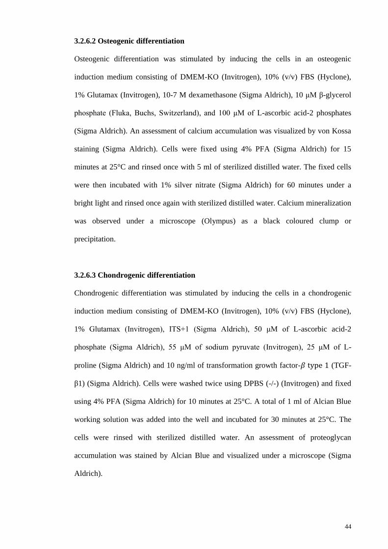
44
3.2.6.2 Osteogenic differentiation
Osteogenic differentiation was stimulated by inducing the cells in an osteogenic
induction medium consisting of DMEM-KO (Invitrogen), 10% (v/v) FBS (Hyclone),
1% Glutamax (Invitrogen), 10-7 M dexamethasone (Sigma Aldrich), 10 μM β-glycerol
phosphate (Fluka, Buchs, Switzerland), and 100 μM of L-ascorbic acid-2 phosphates
(Sigma Aldrich). An assessment of calcium accumulation was visualized by von Kossa
staining (Sigma Aldrich). Cells were fixed using 4% PFA (Sigma Aldrich) for 15
minutes at 25°C and rinsed once with 5 ml of sterilized distilled water. The fixed cells
were then incubated with 1% silver nitrate (Sigma Aldrich) for 60 minutes under a
bright light and rinsed once again with sterilized distilled water. Calcium mineralization
was observed under a microscope (Olympus) as a black coloured clump or
precipitation.
3.2.6.3 Chondrogenic differentiation
Chondrogenic differentiation was stimulated by inducing the cells in a chondrogenic
induction medium consisting of DMEM-KO (Invitrogen), 10% (v/v) FBS (Hyclone),
1% Glutamax (Invitrogen), ITS+1 (Sigma Aldrich), 50 μM of L-ascorbic acid-2
phosphate (Sigma Aldrich), 55 μM of sodium pyruvate (Invitrogen), 25 μM of L-
proline (Sigma Aldrich) and 10 ng/ml of transformation growth factor-𝛽 type 1 (TGF-
β1) (Sigma Aldrich). Cells were washed twice using DPBS (-/-) (Invitrogen) and fixed
using 4% PFA (Sigma Aldrich) for 10 minutes at 25°C. A total of 1 ml of Alcian Blue
working solution was added into the well and incubated for 30 minutes at 25°C. The
cells were rinsed with sterilized distilled water. An assessment of proteoglycan
accumulation was stained by Alcian Blue and visualized under a microscope (Sigma
Aldrich).

45
3.2.6.4 Reverse transcription polymerase chain reaction (RT-PCR) for
trilineage markers
The RT-PCR was performed using complementary DNA (cDNA) prepared as
described in section 3.2.8. A total of 25μl of PCR reaction was prepared by adding the
following components: 2.5μl of 10x PCR (–Mg2+
), 1.25μl of 50 mM MgCl2, 0.5μL of
10 mM dNTP Mix, 1μl of forward primer and 1μl of reverse primer, 0.2μl of Taq DNA
polymerase,1μl of template cDNA and 17.55μl of autoclaved ddH2O. The mixture was
centrifuged at 500 rpm for 3 seconds. The cDNA amplification was performed at 94°C
for 5 minutes, 94°C for 45 seconds for 30 cycles, 58.5°C for 30 seconds for 30 cycles,
72°C for 45 seconds for 30 cycles, and a final elongation at 72°C for 10 minutes and
4°C using a thermocycler. Polymerase chain reaction (PCR) products were resolved
on1.5% agarose gel which was immersed in 1 x Trisborate-ethyl-enediamine tetra
borate acid (TBE) buffer which was visualized via ethidium bromide staining (Sigma
Aldrich).
3.2.7 RNA extraction and quantification
Total RNA was extracted using either Trizol technique or RNeasy Mini Kit (Qiagen). In
the Trizol method, the cells were washed twice with DPBS (-/-) (Invitrogen) and lysed
by adding 1 ml Trizol reagent (Invitrogen). The lysate was transferred to a 1.5 ml
microcentrifuge tube (Axygen) and incubated for 5 minutes at 25°C. Approximately
200μl chloroform (Sigma Aldrich) was added to the tube and mixed by inverting it 20-
25 times followed by incubation for 2-3 minutes at 25°C. The tube was then centrifuged
at 12000 rpm for 15 minutes at 4°C following which the aqueous phase was transferred
into a fresh microcentrifuge tube (Axygen). To precipitate the RNA, 0.5ml of
isopropanol (Sigma Aldrich) was added, mixed, and incubated at 25°C for 10 minutes.
Then microcentrifuge tube was centrifuged again at 12000 rpm for 10 minutes at 4°C.
Supernatant was discarded and the pellet washed twice with 75% ethanol (Sigma

46
Aldrich). The pellet was air-dried, dissolved in 30μl of RNase and DNase-free water,
and incubated at 37°C in a water bath for 10 minutes. The extracted RNA was treated
with DNase (Qiagen) to remove genomic DNA. 1μl RNA was used for quantification
and lμl of distilled water was used as a blank. The absorbance was recorded at 260 nm
and 280 nm using Nano drop (Thermo Fisher Scientific Inc.) before being stored at -
80°C. RNA concentration and purity was calculated using the following equations
respectively:
RNA concentration (μg/μl) = OD260 x (40μg RNA/ml), and ratio of reading of OD260
/OD280
3.2.8 Complimentary deoxyribonucleic acid (cDNA)
Synthesis of cDNA was carried out according to the Superscript II Reverse
Transcriptase (Invitrogen) or RT2 first strand kit (Qiagen) manufacture protocol. In the
Invitrogen protocol, 1μl Oligo dT (50 μM) and 1μl dNTP mix (10 mM) were added
to1μg of RNA (Section 3.2.8), and sterile distilled water added to make up the final
volume of 13μl in a 0.2ml tube (Axygen). The tube was then incubated at 65°C for 5
minutes and then rapidly cooled on ice followed by a brief spin. A total of 4μl of 5
XFirst strand buffer and 2μl of 0.1 M DTT were added into the same tube and incubated
at 42°C for 2 minutes. Then, 1μl of Super Script II (200 units) was added into the tube,
mixed by pipetting gently up and down, then finally incubated at 42°C for 50 minutes.
The reaction was heat inactivated at 70°C for 15 minutes and the cDNA used for
subsequent experiments or stored at -20°C.
3.2.9 TaqMan® Array Human Immune Gene Array
Total cellular RNA from all cell lines at P2 and P9 were isolated with TRIzol® RNA
isolation reagent (Invitrogen). The complementary DNAs (cDNAs) were synthesized
using 1 µg RNA sample with a reverse transcriptase enzyme kit (Invitrogen). The

47
immune related genes were profiled by using TaqMan® Array Human Immune Gene
Array. Briefly, the cDNAs were loaded on the microfluidic cards for thermal cycling on
an ABI PRISM7900HT Sequence Detection System (Fisher Scientific), and expression
values for target genes were normalized to the expression of 18srRNA. For estimation
of the fold change by TLDA when the initial transcript levels were undetectable, the
initial cycle threshold (CT) value was assigned to be 35, which would lead to a possible
underestimation of the actual fold change.
3.2.10 Validation of Human Immune Genes Using Cytokine Protein Array
The secretions of randomly selected immune genes were validated by using a
customized human cytokine protein array (Panomics, Redwood City, CA). Briefly,
conditioned medium from all cell lines at P2 and P9 were collected and the assay
performed according to the manufacturer`s instructions. The analysis was performed
using Luminex platform.
3.2.11 Statistical analysis
Data were presented as mean + standard deviation (SD). Statistical comparisons were
made and data analyzed by using analysis of variance (ANOVA) with the significance
level set at P = 0.05. Tukey post hoc multiple comparison tests were carried out to
determine the differences between the groups (Nishino et al., 2011; Nakamura et al.,
2013).

48
3.3 Results
3.3.1 Characterization of MSCs derived from SHED, DPSCs and PDLSCs
To ascertain that the cell lines that were established were bone fide MSCs, we
performed some basic MSC characterization studies. All the three types of cells
displayed fibroblastic morphology (Figure 3.2 A). We investigated the mesoderm
differential potential of SHED, DPSCs, and PDLSCs into adipogenic, chondrogenic,
and osteogenic lineages under appropriate media induction and they were able to
undergo adiopogenesis, chondrogenesis and osteogenesis respectively (Figure 3.3 B i-
ix). To further characterize these cells, immunophenotyping was done by flow
cytometry. Both samples were negative for hematopoietic markers CD34 and CD45,
whereas more than 85% of the results were positive for MSC markers CD44, CD73,
CD90, and CD166 (Figure 3.3 C). The PCR trilineage markers for osteogenesis (Osterix
and Osteocalcin), adiopogenesis (Leptin and LPL) and chondrogenesis (Collagen II and
Aggrecan) showed the gene expression (Figure 3.3 D).

49
Figure 3.2: A. The morphology of SHED, DPSCs and PDLSCs for passage 2 and
passage 9 observed under phase contrast microscope at 10X magnification. B.
Immunophenotyping of SHED, DPSCs and PDLSCs which were tested against human
antigens CD34, CD44, CD45, CD73, CD90, CD166, and HLA-DR.

50
Figure 3.2 C. In vitro mesoderm differential potential of SHED (i-iii), DPSCs (iv-vii)
and PDLSCs (vii-ix). All the staining was done 21 days after induction. Osteogenesis
was confirmed by mineralized matrix deposition stained with von Kossa staining.
Adipogenesis was detected by neutral oil droplet formation stained with Oil Red O.
Chondrogenesis was detected by the presence of proteoglycans stained with Alcian
blue. D. The trilineage RT-PCR markers shown gene expression for all the three dental
sources.
3.3.2 Growth Kinetics
The growth curve line graph indicates that the cumulative cell count in per ten
thousand, at the end of passage nine, had overall cell yield that was significantly higher
in PDLSCs (370,000 cells) followed by SHED (340,000 cells), and DPSCs (300, 000

51
cells) in T25 cm2 flask (Figure 3.3). The average population doubling times in hours at
P9 for SHED, DPSCs and PDLSCs were (26.8 +6.41), (26.1 +9.01), and (25.3 +6.67),
respectively.
Figure 3.3 A. Long-term growth curves of SHED, DPSCs, and PDLSCs up to 9
passages. B. Comparison of population-doubling time in hours for the dental sources.
The results represent the average of six culture replicates (n=6) with standard deviation.
3.3.3 Gene expression variability between SHED, DPSCs, and PDLSCs at early
and late passages
To understand the immunological behaviour between these cell lines during their
transition period from P2 to P9 (Martin-Piedra et al., 2014), we looked at the expression
patterns of immuno-regulated genes. The genes were defined as being 2-fold up or
down-regulated in each sample at P2 and P9 when compared to SHED at P2, which
acted as a control with an accompanying p value of 0.05. Since huge fold variations
were noticed among all the samples, they were tentatively classified into 5 distinct
categories based on their fold expression value as follows: up-regulated genes
consisting of 3 categories, namely, highly up-regulated (≥ 50 fold), medium-up-
regulated (≥ 10 to 49.99 fold), and low up-regulated (≥ 2 to 9.99 fold) followed by
unchanged genes (≥ 0.5-1.99 fold) and down regulated (≤ 0.499 fold) categories. In this

52
regard, in P2, it was noticed that the SHED has indicated mostly unchanged genes
compared to DPSCs and PDLSCs. On the other hand, in the medium up-regulated
category, no significant differences of gene expression percentage were found among
all the cell lines. PDLSCs showed a significant lower percentage (P<.05) of genes in the
low up-regulated category as compared to SHED and DPSCs. Surprisingly, in P9, a
tremendous augmentation of genes percentage were seen in all up-regulated categories
within all cell lines (P<.05). In terms of the unchanged category, a significant swift
(P<.05) was observed in SHED, DPSCs, and PDLSCs from P2 to P9 where the genes
decreased from 78% to 6%, 57% to 9%, and 60% to 9% respectively. In contrast, a
significant increment (P<.05) of gene percentages ranging from 8% -17% was observed
in all the cell lines between the same passages (Figure 3.4). To better understand the
role played by each immune-related gene within the cell lines and passages, we further
categorized the genes according to their similar biological functions (Table 3.3-3.5).
Meanwhile, SHED expressed many genes at P9 that are related to pro-inflammatory and
anti-inflammatory elements such as CCL3, CCL5, IL1A, IL4 and IL8.

53
Figure 3.4: Comparison of gene profile between SHED, DPSCs and PDLSCs using a
Human Immune gene array with percentages of up-regulated genes in P2 and P9.

54
3.3.4 SHED cultured at P9 displayed many genes representing pathogen
recognition as well as immune signaling and transduction
Among the 15 genes grouped under the category of pathogen recognition as well as
signaling and transcription, 7 (HLA-DRB1, CD80, CD34, TNFRSF18, PTPRC, SELE,
and SELP; P<.001) were highly significant in SHED cultured at P9 compared to those
cultured at P2. A similar observation with lesser significant values (P<.01; P<.05) was
observed for DPSCs and PDLSCs cultured in P9 as compared to P2 (Table 3.3).
Table 3.3: Fold changes of genes related to pathogen recognition & signaling and
transcription.
3.3.5 Pro and anti-inflammatory gene profiling of SHED, DPSCs, and PDLSCs
at P2 and P9
The transition of SHED from P2 to P9 resulted in an increment of 7 pro-inflammatory
genes namely TNF, ILIB, IFN-ɣ, LTA, IL-12B, IL-1A, and IL-15 (P<.001). However,
except for IFN-ɣ, not much significance was noticed for the same group of genes in
DPSCs and PDLSCs during the transition period. On the other hand, tremendous
increments (P<.001) of anti-inflammatory genes such as IL-4, IL-5, IL-7, IL-9, IL-10,

55
and IL-13 were observed in SHED cultured at P9 as compared to P2. Surprisingly, we
did not observe a similar expression in DPSCs and PDLSCs, indicating SHED
experience immune related genes turbulence when cultured for a prolonged period of
time (Table 3.4).

56
Table 3.4: Fold changes of genes related to cytokines

57
3.3.6 SHED cultured at P9 displayed many genes representing several growth
and stimulation factors as well as chemokines
It is worth noting that growth and other stimulation factors also play an integral part in
immune systems. Accordingly, we noticed that TGF-β1 and other stimulation factors
such as CSF3 and IL2RA were highly expressed in SHED cultured at P9 compared to
P2 (P<.001) and, again, these genes were stagnant in DPSCs and PDLSCs in similar
culture conditions. In terms of chemokine gene expression, only those genes related to
the CXC group such as CCL5, CXCL11, CCR2, CCR4, CCR5, and CCR7 were
noticeable in all cell lines at P9 (Table 3.5).
Table 3.5: Fold change of genes related to cellular cytotoxicity, surface molecules and
apoptosis
3.3.7 DPSCs and PDLSCs cultured at P9 expressed less cellular toxicity and
apoptosis-related genes compared to SHED cultured at P9
We postulated that the expressions of most of the immune genes in SHED cultured at
P9 were most likely contributed by the elevated expression (P<.001) of cellular toxicity

58
and apoptosis such as GNLY, GZMB, PRF1, CD3E, CD8A, CD19, CD28, CD38 and
CD40LG.
3.3.8 Validation of immune genes using cytokine protein array.
We randomly selected a few genes namely IL2, IL4, IL5, IL6, IL7, IL8, IL10, VEGF,
and TNF that showed expression patterns in good agreement with the PCR array data
(Figure 3.5).

59
Figure 3.5: Detection of cytokines released by SHED, DPSCs, and PDLSCs at passage
2 (P2) and passage 9 (P9) via Luminex platform. The cytokines selected include
interleukin 6 (IL6), vascular endothelial growth factor (VEGF), interleukin 10 (IL10),
interleukin 4 (IL4), tumour necrosis factor (TNF), interleukin 5 (IL5), interleukin 7
(IL7), interleukin 2 (IL2) and interleukin 8 (IL8). (*P value <0.05, **P value <0.005
and ***P value<0.001).

60
3.4 Discussion
The findings of this study prompted a revisit of some of the key aspects of stem cells for
clinical usage one being the suitability of using pro-long cultured cells. Generally, cells
cultured for long-term are considered to be safe (Bogdanova et al., 2014) although a few
studies report that they undergo genomic instabilities (Wang et al., 2013; Roselli et al.,
2013). More recent studies have shown that DPSCs cultured up to P14 too have
maintained high mitogenic and functionality thus making them a safe source for
therapeutic usage (Martin-Piedra et al., 2014). Nevertheless, later studies as well as
others in the past (Scheers et al., 2013; Frazier et al., 2013) mainly focused on
proliferation and functionability, and may not truly represent the state of the cells. This
is true in our situation that despite maintaining proliferation and functionality, a
significant shift was observed in the immunomodulatory aspect especially for long-term
cultured cells. Interestingly, this shift occurred across all tested cell lines with SHED
being the highest.
There was a sharp increase of immune gene expression related to T-lymphocyte
activation such as HLA-DR and CD80 across all cell lines at P9. We postulate that the
presence of these molecules indicates that the cells are losing their stemness and
transforming into terminally differentiated cells (Laurin et al., 2004) and have the
potential for creating a graft-versus-host disease scenario if transplanted in allogeneic
settings. Further, we noticed a significant increase in the number of pro-inflammatory
genes during the transition of the cell lines from P2 to P9. In vivo condition, these genes
are expressed at large during the early inflammation phase to eradicate infection, and
the over-expression of these genes especially TNF, IL1B, IFN-ɣ, LTA, IL12B, IL1A,
and IL2 could potentially harm host tissues (Morales-Garcia et al., 2012; Katz et al.,
2014; Xu et al., 2015). Likewise, we also noticed that an increment of several anti-
inflammatory genes at P9. In an ideal situation, the expressions of pro-inflammatory

61
and anti-inflammatory genes complement each other; for example, the pattern of TNF is
inversely proportional to IL-10 (Strassmann et al., 1994). In contrast, we found both
groups of genes were highly expressed in P9 indicating a disturbance in the genetic
makeup though this needs further investigation. It was not surprising to see the
elevation of TGF-β expression at P9. The pivotal function of TGF-β in the immune
system is to maintain tolerance via the regulation of lymphocyte proliferation,
differentiation, and survival (Yoshimura & Muto, 2011) by suppressing pro-
inflammatory cytokines. CCL5, CXCL11, CCR2, CCR4, CCR5, and CCR7 were also
up-regulated in all cell lines at P9. All these molecules are members of the CXC
chemokine family and, apart from playing a role in immunogenicity, they are potent
promoters of angiogenesis and mediate their angiogenic properties (Balasubramanian et
al., 2012). Perhaps this might contribute to the continuous growth of the cells. It was
surprising to notice that despite the cells being cultured under similar conditions, SHED
surpassed other cell lines in expressing unwanted immune genes at late passage. This
perhaps is attributed to the high expression of cellular toxicity markers such as GNLY
and GZMB in SHED as compared to DPSCs and PDLSCs. These genes are known to
be involved as intracellular effectors of target cell death, with recent data suggesting
that GNLY may have a role in the propagation of immune signals (Cullen et al., 2010).
Collectively, the occurrence of immune genes turbulence from our findings suggest that
long-term culture conditions of stem cells may not be feasible for use in transplantation.
Having said that, what is the remedy for using cells having a low starting material such
as dental pulp or periodontal ligament tissue? To overcome this issue, a large expansion
of cells using 3D-bioreactor (Vecchiatini et al., 2015) can be possibly made to dental
stem cells at the early passage without triggering any side effects (Elseberg et al., 2015).
Apart from that, pooled allogeneic samples are another choice, but further
characterization is needed to maintain their functionality. In conclusion, we propose a

62
comprehensive biological characterization which includes the immune properties before
the stem cells cultured at later passage are brought into clinical application.
3.5 Conclusion
1. The growth curve indicates that PDLSCs have the highest accumulated cells at
P9, followed by SHED and DPSCs. When observed under the microscope the
morphology had no difference either in P2 or P9.
2. The fundamental characteristics of mesenchymal stem cells were observed in all
three dental sources.
3. The human immune gene expression in SHED, DPSCs, and PDLSCs in long
term cultured cells in P9 has highly expressed genes compared to P2. However,
there was increased gene expression of cellular toxicity markers in SHED when
compared to DPSCs and PDLSCs. The viability and cell proliferation was also
decreased therefore P9 sources are not feasible for clinical usage.
From this study, we can conclude that SHED cultured at P2 has expressed genes namely
IL12A and VEGF which plays an important role in angiogenesis process in wound
healing which are comparable to other dental stem cells. Therefore SHED at P2 was
chosen to further study on its functionality in different culture conditions in our next
chapter.

63
CHAPTER 4: WOUND HEALING POTENTIAL OF SHED WHEN CULTURED
IN LOW SERUM CULTURE CONDITIONS
4.1 Introduction
Wound healing is a complex process that undergoes the three major phases of pro-
inflammation, proliferation, and maturation. As the wound closes, paracrine signaling
occurs within the wound area which activates genes involved in the healing. In
addition, upregulation of signaling pathways such as TGFβ1, phosph-ERK, and NF-
κBA have shown to enhance the wound healing process by increasing α-smooth muscle
actin expression (Wang et al., 2016).
Wound debridement starts with a coagulation cascade that stops the bleeding and an
infusion of neutrophils if infection is present. During the late stages of the inflammatory
process, macrophages initiate angiogenesis and re-epithelialisation. The proliferation
stage follows with the formation of granulation tissues which comprise a collagen and
extracellular matrix. Angiogenesis is then initiated leading to the formation of a
network of new blood capillaries. The maturation phase is the final phase where wound
closure is completed. Here, further collagen deposition and cross linking of extracellular
matrix takes place, which allows scar tissue formation (Hutchinson, 1992).
There are a number of genes which contribute to the epithelialisation rate, namely
CCL2, IL10, and IL6 at the pro-inflammatory phase; CSF, EGF, and TGF at the
proliferation phase; and MMP, PLAU, and COL1A1 which are involved in the
maturation phase. The cross-talk between these genes increases the rate of wound
healing and reduces fibrosis, which is a type of scar that occurs in delayed wound
healing although this does not occur during normal wound healing (Yang et al., 2014).

64
MSCs in the wound healing process generate good results through the secretion of
growth factors, cytokines, and chemokines which lead to angiogenesis (Chen et al.,
2015). Stem cells from human extracted deciduous teeth (SHED) can be a unique
resource for wound healing treatment. Nishino et al. (2011) have reported that SHED
works better together with other compounds or different therapeutic strategies. Their
study shows that SHED co-cultured with fibroblast enhances skin wound healing in
mice. Hence, more studies need to be conducted on SHED to optimize its function on
wound treatment.
Previously, Kanafi et al. (2013) reported SHED’s cell migration in 5% of FBS is faster
compared to DPSCs. Therefore we have decided to study the efficacy of SHED in
treating wound healing in low serum culture conditions (2% of FBS) which reduces
animal derived substances in our study and also looking at the expression of wound
repair genes.
Experimental works were carried out to meet the following objectives:
1. Assessing SHED isolation and compare the morphology, growth kinetics, and
senescence level of SHED cultured in early passages: passage 2 (P2) and passage 4
(P4) in low serum culture conditions;
2. Evaluating human wound healing genes in SHED in P2 and P4 when cultured in
low serum culture conditions using qPCR;
3. Assessing malignant transformation of SHED in P2 and P4 when cultured in low
serum culture conditions using transformation assay; and
4. Assessing the validation of gene expression by reverse transcriptase and real time
PCR

65
4.2 Methodology
4.2.1 Low serum culture conditions sample isolation and expansion
Dental pulp stem cells from extracted deciduous teeth (SHED) [n=3; ages 7-11 years]
were isolated and cultured up to passage 4 (P4) according to reported protocols with
slight modification (Jung et al., 2013). In this study, cells at P2 and P4 were both
classified as early passages. Cells were cultured in Dulbecco’s modified Eagle’s
medium (DMEM), containing 10% fetal bovine serum (FBS) and glutaMAX 1% (Life
Technologies, CA, USA). For 2% FBS, SHED were isolated and seeded into T75
flasks. Once attached, they were expanded into DMEM-KO without FBS and later
cultured into 2% FBS, which is a low serum culture media.
4.2.2 Morphology, growth kinetics, and senescence of β-galactosidase cell
staining assay
Cells cultured in different culture medium conditions were compared in terms of
morphology, growth kinetics, and senescence using β-galactosidase cell staining assay
data obtained at P2 and P4. The morphology of cell growth and its population-doubling
time were analyzed (Govindasamy et al., 2011). When SHED had reached 60%
confluence, growth media was removed from the cells and rinsed with DPBS
(Invitrogen) and stained according to the manufacturer’s protocol (Cell Signaling
Technology, Danvers, Mass., USA). Briefly, cells were maintained in fixative solution
for 10–15 min. Then the plates were rinsed 2 times with DPBS (Invitrogen) and
subsequently they were stained with β-galactosidase staining solution overnight and
visualized under a microscope. The presence of blue-stained cells indicates senescence
and the number of senescent cells in 35mm tissue culture dish were counted under a
light microscope.

66
4.2.3 qPCR human wound healing array gene expression analysis
Total RNA was extracted from tissue samples using TRIZOL reagent (Life
Technologies, Grand Island, NY) according to the manufacturer’s instructions. The
resulting RNA pellet was then resuspended in 30µl of diethyl pyrocarbonate (DEPC)–
treated water. The extracted RNA was quantified by analyzing 1µl of total RNA using
the Nanodrop ND1000 Spectrophotometer (Nanodrop Technologies Inc.). Following
RNA extraction, complementary DNA (cDNA) was combined with an appropriate
amount of RNA through a reverse transcription reaction. Subsequently, messenger RNA
(mRNA) expression of 84 wound healing genes found in the human wound-healing
PCR array (PAHS-121Z; SABiosciences, Frederick, MD) was investigated for pathway
profiling by real-time reverse transcription polymerase chain reaction (RT-PCR).
Healthy SHED cultured under 10% FBS were used as controls. Finally, the real-time
PCR array data were analyzed using the RT2 profiler PCR Array Data Analysis online
software (SABiosciences).
4.2.4 Transformation assay
The quantitation of anchorage-independent growth was performed using Cyto Select
96-well Cell Transformation Assay. Anchorage–independent growth is one of the
hallmarks of transformation and which is an indication for in vitro detection of
malignant transformation of cells. A culture medium of 100 µL containing cell growth
activator was added into each well. The cells were incubated for 6-8 days at 37ºC and
5% CO2. Then the cell colony formation was observed under a light microscope and
colorimetric detection was performed by measuring the absorbance at 570nm for both
the 10% and 2% FBS culture mediums, while immortalized HepG2 cells were used as
positive control.

67
4.2.5 Validation of gene expression by Reverse Transcriptase and Real Time
PCR
cDNA amplification was performed in a thermocycler using Taq polymerase supplied
with KCl buffer and 1.5 mM/L MgCl2 (Invitrogen) at 94°C/1 min, 58°C/30 sec, 72°C/1
min. Polymerase chain reaction (PCR) products were resolved in 1.5% agarose
(Invitrogen) gel in 1 X Tris borate-ethylene-diaminetetraacetic acid buffer. The
expression of some primers in the reverse transcriptase-PCR analysis was quantified in
duplicate with SYBR green master mix (Applied Biosystems, Foster City, CA, USA).
PCR reactions were run on an ABI 7500 Fast Sequence Detection System (Applied
Biosystems) and all measurements were normalized by 18s rRNA. For data analysis, the
comparative CT method (ΔΔCT) was used. Randomly selected genes were then
validated. The validated genes are CXCL2, IL6, TNF, TGFB1, and COL4A1.
4.2.6 Statistical analysis
Data were presented as mean + standard deviation (SD). Statistical comparisons were
made and the data analyzed using analysis of variance (ANOVA) with the significance
level set at P = 0.05. Tukey post hoc multiple comparison tests were carried out to
determine the differences between the groups (Nishino et al., 2011; Nakamura et al.,
2013).
4.3 Results
4.3.1 SHED cultured in low serum cultured conditions
In order to understand further on wound repair gene expression by SHED, the areas that
we have looked into included its characteristics of morphology, accumulated cells,
population doubling time (PDT), cumulative population doubling (PD) and percentage

68
of senescent cell. The understanding of these areas has indicated that it does not
contradict with our findings (Figure 4.1).
As described in the literature, the characteristics of multipotent stem cells (MSCs) were
determined by trilineage study on dental pulp stem cells. The results showed adipocyte,
chondrocyte, and osteocyte differentiations (Figure 4.2). The safety profile of the SHED
cultured in P2 and P4 showed no growth anchorage in different culture conditions
against positive control HepG2 (Figure 4.3).

69
Figure 4.1: A: Morphology of SHED when cultured in different culture conditions and
long term cell expansion at 4X magnification. B. The accumulated number of cells (1 X
106 cells) has no significance difference in both P2 and P4 SHED culture conditions. C.
Cumulative population doubling indicates the number of times cell doubled between
passages in different culture conditions. D. Population doubling time (PDT) is time
taken in hours by the cells to increase in between passage and different culture
conditions. E. The senescence β-galactosidase Cell Staining assay was performed and
the presence of blue colour under microscope at 10 x magnification is considered as
senescence cell. F. The percentage of senescence cells has no significance difference in
between passage and different culture conditions. All experiments were conducted at
respective passages with 3 biological replicates for each established cell line.

70
Figure 4.2: Trilineage differentiation of adult multipotent stem cell. Adipogenesis was
detected by neutral oil droplet formation stained with oil red O. Chondrogenesis was
detected by presence of proteoglycans stained by Alcian Blue. Osteogenesis was
confirmed by mineralized matrix deposition stained with von Kossa staining. All the
staining were done 21 days after induction for P2 10% FBS (i-iii), P4 10% FBS (iv-vi),
P2 2% FBS (vii-ix) and P4 2% FBS (x-xii).
Figure 4.3: The observation of anchorage dependent growth on transformation assay
agar matrix layer. HepG2 acts as positive control shown anchorage growth on the layer
and none for all other culture conditions. The arrows indicate the anchorage growth.

71
4.3.2 Wound repair gene expressions pattern in altered cultured conditions of P2
and P4
To better understand gene expression between cell lines, the expression patterns of the
functional categories in wound healing genes were observed. The fold changes of
expressed markers were determined with ct values. The four categories of ct values
were: ct<29 (high expression), ct=30-34 (medium expression), ct=35-39 (low
expression) and ct>40 (non-expressed markers). In this regard, wound healing genes
were highly expressed in P2 cells cultured in 2% FBS. To better understand the role
played by cytokines and growth factors, genes were categorised according to three
major phases in wound healing: pro-inflammatory phase (13 genes), proliferation phase
(28 genes), and maturation phase (43 genes). The genes were defined as up-regulated or
down-regulated based on fold changes compared to SHED cultured in 10% FBS which
acted as a control with an accompanying P value of 0.05 (Appendix 1.2). Since huge
fold variations were noticed among all the samples, only a number of genes (12 genes)
were randomly chosen to represent each phase of wound healing.
4.3.3 Pro- and anti-inflammatory gene profiling of SHED at P2 2%and P4 2%
FBS shown up regulation
All pro-inflammatory cytokines and chemokines were up-regulated, namely CCL2,
CXCL1, CXCL2, IL10, and 1L6. Pro-inflammatory markers such as CCL2 and IL6 and
anti-inflammatory cytokines, namely 1L10 were highly expressed with significant
difference (P<0.05) in comparison to the control group (10% FBS). SHED cultured in
P2 shows higher gene expression compared to P4 (Figure 4.5).

72
4.3.4 Proliferation genes which improves the impaired wound has seen up
regulated in SHED P2 2% FBS
Proliferation is a crucial phase that leads to the healing of a wound. Most of the markers
observed indicated significant difference (P< 0.05). Some highly expressed markers
include EGF, TGFA, PTGS2, STAT3, and TNF (Figure 4.6). There were no up
regulated genes in P4 except for PTEN and WISP 1 gene. Conversely, there were more
down regulated genes in P4 2% FBS compared to P2 2% FBS (Appendix 1.3). Most
markers in SHED P2 2% FBS were up-regulated, hence increasing the healing rate.
4.3.5 SHED P2 2% FBS has possibilities to improve the healing condition by
increasing the number maturation genes
We postulated that the increase in proliferation markers by SHED P2 2% has
contributed to the rise of maturation genes, which are involved in the final phase of
wound healing. Most of the maturation genes, noticeably COL4A1, COL4A3, MMP2,
MMP7, MMP9, PLAU, and PLG were more highly expressed in SHED P2 2% with
significance difference (P < 0.05). However, RAC1 and F3 genes were highly
expressed in SHED P4 2% (Appendix 1.4). Highly expressed markers in SHED P2 2%
such as COL543 was up-regulated more than 10-fold (Figure 4.7)

73
Figure 4.5: Wound healing genes categorized under pro-inflammatory phase. All the
experiments were conducted at respective passage with 3 biological replicates for each
established cell line. * P < 0.05 indicates significance difference.

74
Figure 4.6: Wound healing genes categorized under proliferation phase. All the
experiments were conducted at respective passage with 3 biological replicates for each
established cell line. * P < 0.05 indicates significance difference.

75
Figure 4.7: Wound healing genes categorized under maturation phase. All the
experiments were conducted at respective passage with 3 biological replicates for each
established cell line. * P < 0.05 indicates significance difference.

76
4.3.6 Validation of gene expression by Reverse Transcriptase and Real Time
PCR
Randomly selected genes showed expression patterns in good agreement with the PCR
array data (Figure 4.8).
Figure 4.8: Quantitative of gene expression by Reverse Transcriptase and Real Time
PCR significance difference for CXCL2, IL6 and TNF with P<0.05. All the
experiments were conducted at respective passage with 3 biological replicates for each
established cell line.
4.4 Discussion
The findings of this study have encouraged further analysis of reduced serum in
cultivating dental pulp stem cells for clinical usage. The reason for using FBS in
culturing stem cells is to increase the cell growth and supplement for cells. However,
there were drawbacks such as safety issues where animal derived proteins can seep into
the media. On the other hand, our research also prompted us to know more about wound
healing genes expression when cultured in different passages and culture conditions.
Dental pulp tissue is mesenchymal cells that need to be cultivated in a large scale

77
expansion to harvest large numbers of cells for the needs in clinical usage. It was
observed that despite maintaining its proliferation and safety profile, a significant
difference was seen in expression of wound healing genes in FBS 2% in P2. There was
a high increment of pro-inflammatory genes seen in FBS 2% in P2 for all the genes in
this category. A sharp increase in these genes can cause a delay in wound healing.
However, the presence of anti-inflammatory genes, namely IL10 and IL4 actually
complement the process of wound healing. Generally, pro-inflammatory genes such as
CCL2, CXCL2, and CXCL5 initiate the healing process by reducing infection by
initiating macrophage response. A research conducted by Wood et al. (2014) reported
that CCL2 promotes healing by increasing the macrophages response.
For the proliferation category, the highest number of genes were expressed in FBS 2%
in P2, namely CSF, EGF, and VEGF. These genes are also crucial in the wound healing
process. A full skin thickness wound in mouse model has shown better healing with the
expression of EGF and VEGF markers (Kim et al. 2015). The genes found in this
category help the vascularization process in wound healing. Ram et al. (2015) has
reported that VEGF is one of the key factors in enhancing wound healing in diabetic
rats. HGF and VEGF play an important role as angiogenic and pro-survival factors in
treating critical limb ischemia (Prochazka et al., 2015).
The maturation phase is the final stage of the complex wound healing process. The
noticeable markers at this stage are MMP1, MMP2, MMP7, COL141, COL14A1 and
COL14A2. A recent study on wound healing has discovered that COL13A1 and PLAU
promote the extracellular matrix formation (ECM) in maturation stage (van den Broek
et al., 2015). CSF2 and MMP9 markers were seen expressed in the wound three days
after the skin incision was made in mouse (Kameyama et al., 2015).

78
Most therapeutic research on mesenchymal stem cells (MSC) studied cytokines, growth
factors, and paracrine signaling. Nonetheless, these studies were done based in standard
in vitro culture conditions. This study has established the potential for a large number of
wound repair genes to be up-regulated in FBS 2% P2. These results strongly support the
notion of SHED cultured in low serum conditions has enhanced the angiogenesis and
wound healing process with the expression of namely CCL2, IL10, EGF and MMP7. In
low serum conditions, the cells were in a circumstance to produce and release paracrine
signals with chemotactic and pro-angiogenic activity (Fierro et al., 2015).
4.5 Conclusions
1. The morphology, growth kinetics, and senescence level showed no significant
difference in the lower serum culture conditions in P2 and P4 SHED.
2. There is a marked increase of wound healing genes expressed in SHED P2 2%
FBS culture conditions compared to SHED P4 2% FBS.
3. There is no growth anchorage for malignant transformation of cells in P2 and P4
SHED.
4. The wound repair genes upregulated in pro-inflammatory, proliferation and
maturation phases were from SHED P2 2% FBS.
From this study, we can draw a conclusion that SHED cultured at P2 low serum
condition which is 2% of FBS had shown an increase in wound healing genes namely
IL10, IL6, MMP7 and COL1A1 when compared with standard condition of 10% FBS.
Hence we proceed with SHED P2 2% FBS in our in vitro diabetic wound model in our
next chapter.

79
CHAPTER 5: SHED IN LOW SERUM CULTURE CONDITIONS REDUCES
OXIDATIVE STRESS IN AN IN VITRO WOUND MODEL
5.1 Introduction
Oxidative stress occurs due to the overproduction of reactive oxygen species (ROS) that
are generated in aerobic organisms by the electron transport chain (ETC) of
mitochondria. The ROS readily attacks the major organelles within the cells including
nucleic acids. Under physiological conditions, the complex I of the ETC is the major
location of ROS production (Leverve & Fontaine, 2001; Magder, 2006). Hruda et al.
(2010) reported that high glucose increases susceptibility to oxidative-stress-induced
oxidative damage in K-562 commercial cell lines. The metabolic state of the cells was
affected by the toxic effect of the oxidizing agent. In clinical practice, similar conditions
occur in intensive care when a patient with varying blood glucose control and energy
substrate availability is exposed to oxidative agents.
The adverse effect of hyperglycemic condition in wound healing is that it alters the
angiogenesis process (Lerman et al., 2003). The migrations of cells in diabetic mice are
lower at 75% compared to the non-diabetic group; however the underlying mechanism
needs to be identified (Wagner & Wehrmann, 2007). Wound healing is a complex
process which involves cells namely keratinocytes, fibroblasts, endothelial cells,
macrophages, and platelet. Fibroblasts are the most important cells involved in
producing and remodeling the extracellular matrix, and fibroblast cell proliferation and
migration play key roles in the formation of granulation tissues and further wound
repair (Wagner & Wehrmann, 2007; Kanazawa et al., 2010).
Wound debridement is a phase regulated by number of growth factors. For example,
basic fibroblast growth factor (bFGF) modulates the growth, differentiation, migration,

80
and survival of different type of cells (Bikfalvi et al., 1997). The bFGF also regulates
the P13K-Rac1-JNK pathway to promote fibroblast cell migration (Kanazawa et al.,
2010). In diabetic conditions this process is halted due to oxidative stress. In our
previous chapter, Chapter 4, we found that SHED P2 in low serum condition (FBS 2%)
showed increased human wound healing genes. Therefore, it will be chosen to be
further studied in the in vitro wound healing scratch assay.
Experimental works were carried out to meet the objectives of
1. Assessing digital images captured in in vitro diabetic wound healing through an in
vitro glucose-challenged scratch assay with SHED treated in low serum conditions;
and
2. Evaluating the oxidative stress enzyme activity level in in vitro diabetic wounds
treated with SHED in low serum culture conditions using transwell assay.
5.2 Methodology
5.2.1 Preparation for the in vitro scratch assay
A SHED sample was cultured in a 6-well plate with seeding density of 1500cell/cm2.
The concentration of glucose in the culture medium was 5mM for normal glucose and
45mM for high glucose concentrations (Foresti et al., 2015). There were three 6-well
plates (CORNING) representing each group.
Group A: co-culture of human foreskin fibroblast from normal glucose together
with fibroblast from high glucose.
Group B: only SHED P2 2% FBS cultured in high glucose were seeded.
Group C: co-culture of SHED P2 2% FBS together with fibroblast from high
glucose.

81
Upon reaching 90% of confluency, each of the wells was scratched using 10μl pipette
tips. Following the wound (scratch assay) the migration of the cells was observed
through a phase contrast microscope at 0, 10, 23, 47, and 90 hours (Walter et al., 2010).
5.2.2 Transwell assay
The analysis of oxidative stress level was analyzed by using a transwell assay. The
fibroblast with high glucose condition was seeded into a transwell plate (CORNING) at
1500cell/cm2. Upon confluency, the fibroblast at the bottom layer was scratched with 10
μl pipette tips. The 24 mm polycarbonate membrane transwell insert with a pore size of
0.4 um was placed on top. Then the top layer was seeded with different cell conditions
and categorized into 3 groups, namely SHED P2 2% FBS treated in normal glucose,
fibroblast in high glucose, and fibroblast in normal glucose. The permeable insert is
very useful in studying tissue remodeling because it permits small molecules like
growth factors to be transported to the lower compartment. After the wound closure was
complete for the entire group, the fibroblast cells at the bottom layer of the transwell
were harvested for SOD, GSH, MDA, and AOPP assays. These tests would determine if
the SHED treated in the in vitro wound model experienced reduced oxidative damage
(Rodriguez-Menocal et al., 2012).
5.2.3 Superoxide Dismutase (SOD) activity assay
The adherent cells, upon the in vitro closure, were harvested with a cell scraper in 1 ml
of cold 1X Lysis Buffer (10 mM Tris, pH 7.5, 150 mM NaCl, 0.1 mM EDTA). The
cells were then lysed with homogenation by centrifugation at 12000 x g for 10 minutes.
The cell lysate supernatant was collected in a 1.5 ml centrifuge tube and quantified
using OxiSelect™ Superoxide Dismutase Activity Assay (Cell Biolabs Inc). Here, the
samples were prepared including blanking in a 96-well microtiter plate and 10 µl of pre-

82
diluted 1X Xanthine Oxidase Solutions were added into each well. The solutions were
mixed thoroughly and incubated for 1 hour at 37ºC. Based on OD490 measured, the
SOD content in each sample was determined on a microplate reader. The SOD activity
level was calculated using the formula below.
SOD Activity (inhibition %) = (ODblank-ODsample)/( ODblank) x 100
5.2.4 Malondialdehyde (MDA) quantitation activity assay
The cells were detached from the flask using a cell scrapper and resuspended in PBS
and then homogenized by centrifugation at 12000 x g for 10 minutes. The whole
homogenate was used in the OxiSelect™ TBARS Assay Kit (MDA Quantitation)
Activity Assay (Cell Biolabs Inc).
The MDA assay provided by the standard protein solution which was a series of
standard solutions ranging from 0µM-125µM was prepared to construct a standard
curve. 100 µL of SDS Lysis Solution was added to both 100µL unknown samples and
the MDA standards. The well-mixed samples were incubated for 5 minutes at room
temperature in microcentrifuge tubes. A 250 µL of TBA Reagent was added to each
sample and standard to be tested. The tubes were closed and incubated at 95°C for 45-
60 minutes using a heat block, cooled at room temperature for 5 minutes, and
centrifuged at 3000 rpm for 15 minutes. The supernatant were used for analysis of
MDA content. Based on OD532 measured, the MDA content in each sample was
determined on a microplate reader.
5.2.5 Total Glutathione (GSH) assay
The cells were detached from the flask via a serum-free trypsinisation process using
Trypsin EDTA Express (Invitrogen). The cells were centrifuged at 500 rpm for 5
minutes at 4ºC and washed with cold 1X PBS. Supernatant was removed and cells

83
washed with cold 1X PBS. Centrifugation was repeated and the solution was removed.
Immediately the pellet was resuspended with 200-500 µL ice-cold 5% MPA for a cell
concentration of 1-5 x 10⁶ cells. The cells were thoroughly mixed and homogenized and
the suspension transferred to a microfuge tube and centrifuged at 12,000 rpm for 5
minutes at 4ºC. The supernatant was collected and analysed for total gluthatione using
GSH quantitation activity assay kit (Cell Biolabs Inc).
The GSH assay provided by the standard protein solution, which was a series of
standard solutions ranging from 0µM-0.5µM, was prepared to construct a standard
curve. Into each well of a 96-well plate, 25µL of the 1X Glutathione Reductase solution
was added followed by 25µL of the 1X NADPH solution. Next, 100µL of the prepared
glutathione standards and samples were added into each well and the solutions mixed
thoroughly. 50µL of the 1X Chromogen was added and mixed briefly. The plate reader
was prepared for a kinetic assay and set to read at OD405. GSH readings were taken
immediately at 1-minute intervals for 10 minutes.
5.2.6 Advanced Oxidation Protein Products (AOPP) assay
The cells were detached from the flask by the serum free trypsinisation process using
Trypsin EDTA Express (Invitrogen), centrifuged at 500 rpm for 5 minutes at 4ºC, and
washed with cold 1X PBS. The supernatant was collected and analyzed using the
OxiSelect™ Advanced Oxidation Protein Products (AOPP) Activity Assay (Cell
Biolabs Inc).
The AOPP assay was provided by the standard protein solution, a dilution series of
Chloramine Standard in the concentration range of 100 µM – 0 µM and a positive
control of AOPP. A 200µL sample was added to microtiter wells followed by 10µL
chloramine reaction initiator to the well, mixed, and incubated on a shaker for 5

84
minutes. Later, 20µL of stop solution was added to the wells and mixed. The
absorbance of AOPP was read at OD340 using 0µM chloramine as blank.
5.2.7 Statistical analysis
Data were presented as mean + standard deviation (SD). Statistical comparisons were
made and the data analyzed using analysis of variance (ANOVA) with the significance
level set at P = 0.05. Tukey post hoc multiple comparison tests were carried out to
determine the differences between the groups (Nishino et al., 2011; Nakamura et al.,
2013).
5.3 Results
5.3.1 The measurement of wound closure in in vitro co-cultured scratch assay
The wound closure rate was fastest in the B group (only SHED P2 2% FBS cultured in
high glucose), followed by the C (co-culture of SHED P2 2% FBS together with
fibroblast from high glucose) and A (co-culture of human foreskin fibroblast from
normal glucose together with fibroblast from high glucose) at 10 hours with P<0.05. At
23, 47, and 90 hours, the rate of wound closure was higher in groups B and C compared
to A which had no significant difference (Figure 5.1). The in vitro wound healing rate
was interpreted in percentage by deduction of the initial width of the cells scratched and
multiplied by 100 (Walter et al., 2010).

85
Figure 5.1: SHED only and co-culture of SHED with fibroblast has indicated a faster
and even closure of the cells at 10, 23 and 47 hours. All the experiments were
conducted at respective passage with 6 biological replicates for each established cell
line. *P < 0.05.
5.3.2 The SOD enzyme level
The quantitative enzyme was observed to have the highest SOD level in fibroblast
treated in normal glucose with scratched monolayer of fibroblast in normal glucose. The
SHED 2% which was treated in normal glucose with scratch monolayer of fibroblast in
high glucose showed a significantly lower SOD level with (p<0.05) (Figure 5.2).
5.3.3 GSH level
The quantification of the quantitative enzyme was observed having the highest GSH
level in scratched monolayer of fibroblast in normal glucose when treated with
fibroblast in normal glucose, followed by the scratched monolayer of fibroblast in high
0
20
40
60
80
100
120
10 HRS 23HRS 47 HRS 90 HRS
Pe
rce
nta
ge o
f in
vit
ro w
ou
nd
clo
sure
(%
)
wound closure rate in in vitro scratch assay
A
B
C
*
Hours

86
glucose when treated with SHED, followed by fibroblast in high glucose with no
significant difference (Figure 5.3).
5.3.4 The MDA level
The lipid peroxidation in the in vitro scratch assay was analyzed and found to be highest
in transwell which was scratched and treated with high glucose. However, when
compared to transwell treated with SHED 2% normal glucose and fibroblast normal
glucose, the lipid was less with no significant difference (Figure 5.4).
5.3.5 Advanced Oxidation Protein Products (AOPP)
The protein oxidation in the in vitro scratch assay was analyzed and found to be highest
in the transwell scratched and treated with high glucose. However, when compared to
the transwell treated with SHED 2% FBS in normal glucose and fibroblast normal
glucose, protein oxidation was reduced with no significant difference (Figure 5.5).

87
Figure 5.2: Expression of SOD enzyme for the hyperglycemic fibroblast monolayer
scratch at the bottom chamber. The treatment were on the upper compartment which are
SHED P2 2% FBS, fibroblast in high glucose and fibroblast in normal glucose. *P <
0.05.
Figure 5.3: Expression of GSH for the hyperglycemic fibroblast monolayer scratch at
the bottom chamber. The treatment were on the upper compartment which are SHED P2
2% FBS, fibroblast in high glucose and fibroblast in normal glucose.
0
2
4
6
8
10
12
14
SHED treated in normalglucose
Fibroblast in high glucose Fibroblast in normal glucose
SOD
act
ivit
y (%
)
Transwell scratch Assay
Expression of SOD enzyme for in vitro scratch assay
*
0.00
0.05
0.10
0.15
0.20
0.25
0.30
SHED treated in normalglucose
Fibroblast in high glucose Fibroblast in normal glucose
GSH
leve
l (μ
M)
Transwell scratch assay
Expression of GSH level for in vitro scratch assay

88
Figure 5.4: Expression of MDA for the hyperglycemic fibroblast monolayer scratch at
the bottom chamber. The treatment were on the upper compartment which are SHED P2
2% FBS, fibroblast in high glucose and fibroblast in normal glucose.
Figure 5.5: Expression of AOPP for the hyperglycemic fibroblast monolayer scratch at
the bottom chamber. The treatment were on the upper compartment which are SHED P2
2% FBS, fibroblast in high glucose and fibroblast in normal glucose.
0
0.2
0.4
0.6
0.8
1
1.2
1.4
SHED treated in normalglucose
Fibroblast in high glucose Fibroblast in normal glucose
MD
A le
vel (
μM
)
Transwell scratch assay
Expression of MDA level for in vitro scratch assay
0.00
0.05
0.10
0.15
0.20
0.25
0.30
0.35
0.40
0.45
0.50
SHED treated in normalglucose
Fibroblast in high glucose Fibroblast in normal glucose
AO
PP
leve
l (μ
M)
Expression of AOPP level for in vitro scratch assay

89
5.4 Discussion
Walter et al. (2010) conducted a similar in vitro study on fibroblast and keratinocyte
scratch assays. The study indicated that the co-culture of L929 fibroblast and HaCaT
keratinocyte in MSC conditioned medium (MSC-CM) enhanced cell migration with the
fibroblasts leading the way in closing the scratches. The paracrine factors enhancing
wound healing were TGFB1, IL6, IL8, MCP-1, and collagen Type I. In our study, the
in vitro model suggests that the similar co-culture system had good wound healing
potential. The SHED-only scratched assay performed better with significant difference
when compared to the fibroblast scratch assay.
The co-culture of SHED and fibroblast also showed increased healing rates compared to
fibroblast scratch assay. This indicates that the paracrine factors in SHED enhance the
migration of cells and assist in faster wound closure. This in vitro scratch assay method
mimics the migration of cells in vivo and also allows for a study on the interaction
between different cell types. The unique advantage of scratch assay is that we can
choose and label the cell population with a specific dye (Rodriguez-Menocal et al.,
2012). Transwell migration assay performs a different system than co-culture system. In
this assay, the interaction that occurs is between soluble factors secreted by the cells at
the upper and bottom chambers. The two-chamber dishes prevent direct contact but
permit the exchange of soluble diffusible factors (Kim et al., 2007). In our study,
fibroblast migration at the bottom chamber was induced by the SHED as a chemo
attractant.
This in vitro study also evaluates wound closure under high glucose conditions. A high
glucose concentration of more than 30mM causes oxidative stress which hinders wound
healing (Hruda et al., 2010). This study confirms the previous findings on the high

90
glucose mediate production of reactive oxygen species (ROS) and oxidative stress
(James & Murphy, 2002; Hruda et al. 2010). In the study by Xuan et al. (2014), high
glucose concentrations inhibited human fibroblast cell migration in wound healing via
the repression of bFGF-regulating JNK phosphorylation. In a recent study, the wound
healing capacity of human retinal pigment epithelial cells (ARPE-19) cultured in a
medium containing normal (NG, 5mM) or high (HG, 45mM) glucose levels modulates
oxidative stress responses regulated by Nrf2. The cell culture condition of HG also
reduced ATP production and mitochondrial function (Foresti et al., 2015).
Our study not only sought to measure wound closure but also investigated the ability of
SHED in improving oxidative stress conditions among the high glucose-induced
fibroblasts monolayer of scratch assay.
The overproduction of ROS diminishes the expression of antioxidant enzymes such as
manganese superoxide dismutase, glutathione peroxidase, and catalase (Miranda-Diaz
et al., 2016). Therefore, it is important to investigate the antioxidant enzymes in order to
know more about damage occurrence in cells. Our study made estimates on the
oxidative stress of in vitro wound healing by determining the quantitative value of the
antioxidant enzymes. The SOD enzyme, for example antioxidative enzyme, that
protects cells from further oxidation is found to be highly expressed in the SHED-
treated group with p< 0.05. This is one the indications in our in vitro wound model
where fibroblasts at the bottom layer of transwell representing the skin layer scratched
in hyperglycemic condition, had increased the wound gap closure following treatment
with SHED P2 in low serum at the upper layer of the transwell assay. The upper
compartment would only allow small molecules like growth factors and paracrine
factors of the SHED to be transported to the lower compartment to induce the closure of
the gap. Thus, the evaluation of SOD showed that it was higher in transwell treated with
SHED compared to fibroblast in normal and high glucose. This shows that SHED

91
induces the production of antioxidative enzymes which help to reduce further cell
injury. Similar results were noted for the GSH antioxidant enzyme level which shows
that both the compartment treated with SHED and fibroblast in normal glucose were
high in GSH levels, followed by fibroblast in high glucose. Other than antioxidant
enzymes, we also investigated the lipid peroxidation and protein oxidation levels in the
in vitro wound model.
Lipid peroxidation is a mechanism in cellular damage in animals and lipid peroxides are
unstable in forming reactive compounds such as MDA which is a marker for oxidative
stress. The MDA expression for the SHED P2 low serum treated group and fibroblast in
normal glucose is lower compared to fibroblast in high glucose. This shows that SHED
has the ability to further reduce injury and damage to cells. Similar results were
obtained from protein oxidation. For example, advanced oxidation protein products
(AOPP) are a type of uremic toxin created during oxidative stress. The AOPP is seen
elevated in patients with diabetes mellitus, inflammation, and others. Our in vitro study
shows that the expression of AOPP decreased in fibroblast cells treated with SHED and
in normal glucose compared to fibroblasts in high glucose. Therefore, the results for the
in vitro wound model suggest that protein oxidation is higher in high glucose fibroblasts
compared to SHED and fibroblasts in normal glucose.
The studies on in vitro wound healing were helpful despite some which showed positive
treatment results with the induction of paracrine factors. For example, the conditioned
media of adipose derived stem cells (ASC-CM) have reported improvements in in vitro
wound healing assay (Lee et al., 2012). Another study suggests that the level of SOD
and GSH are elevated in human umbilical cord mesenchymal stem cells (UCMSCs)
after their treatment in vitro and in vivo wound models. Wound healing is stimulated by
reducing the accumulation of oxidative stress (Zhou et al., 2013).

92
This study showed the SHED-expressed paracrine factors that would be vital for the
healing process. SHED which was cultured in P2 in low serum culture condition (FBS
2%) showed the ability to enhance wound closure in high glucose conditions. In
addition, the treatment with SHED also reduced the accumulation of oxidative stress in
high glucose conditions. Therefore, SHED improves healing ability in diabetics.
5.5 Conclusion
1. The digital image of in vitro diabetic wound healing through in vitro glucose
challenged scratch assay with SHED P2 2% FBS showed enhanced wound
healing rates at the 10, 23, 47, and 90 hour marks.
2. The antioxidant enzymes SOD and GSH increased while lipid peroxidation and
protein oxidation decreased in SHED P2 2% FBS thus improving in vitro
diabetic wound healing.
From this study we conclude that, SHED cultured at P2 of 2% FBS has reduced the
oxidative stress condition and enhanced the in vitro wound healing rate. Therefore we
proceed to the pre-clinical study of wound healing using SHED P2 in 2% FBS in our
next chapter.

93
CHAPTER 6: THE TOXICITY EFFECT OF SHED ON THE ANIMAL STUDY
AND THE POTENTIAL OF WOUND HEALING ON EXPERIMENTAL STZ-
INDUCED DIABETIC RATS TREATED WITH SHED
6.1 Introduction
Diabetes mellitus is a metabolic disease caused by insulin insufficiency (World Health
Organization, 2016). Insulin is necessary to break down glucose into glycogen and any
impairment of this function leads to high glucose levels in the blood or hyperglycemia
(National Institute of Diabetes and Digestives and Kidney Diseases, 2014). One of the
common consequences of diabetes is reduced blood flow in the feet due to neuropathy
which increases the risk of foot ulcers and infections which could necessitate limb
amputation (World Health Organization, 2016).
Current treatments available are wound debridement, care and dressing with topical
antibiotics (American Diabetes Association, 2014), topical negative pressure (Xie et al.,
2010), hyperbaric oxygen therapy [HBOT] (Londahl, 2013), and introducing platelet
derived-growth factors (Villela & Santos, 2010). Since the duration of the wound
healing process depends on its severity and is likely to be long, researchers are
investigating more advanced treatments such as cell-based therapy.
Stem cells play an important role in regenerative medicine. Several studies have been
conducted in using MSCs for the treatment of skin wounds. The studies include BM-
MSCs, ASCs, WJ-MSCs, UCMSCs and PMSCs [(Yang et al., 2013; Ozpur et al., 2016;
Zhao et al., 2015; Wang et al., 2016; Du et al., 2016)]. Dental derived stem cells are a
unique stem cell resource and have shown promise as an alternative wound healing
treatment. An in vivo wound healing study by Nishino et al. (2011) reported that SHED
exhibits good potential in wound healing where it has proven to provide better rates of

94
wound closure with increased Type 1 collagen co-cultured with a fibroblast treated
group. In comparison, it is hypothesized that SHED can be a potential treatment
modality for diabetic wound healing by increasing the antioxidant enzymes such as
SOD and GSH and reducing lipid peroxidation (MDA) and protein oxidation conditions
(AOPP).
Experimental works were carried out to meet the following objectives:
A. General objective
To evaluate the toxicity condition of SHED and its ability to treat diabetic wounds in
rats.
B. Specific objectives
To evaluate the blood profile of Sprague Dawley (SD) rats in two different dosages of
SHED transplantation. This involved:
1. Evaluating the histology of livers and kidneys in two different dosages of SHED
in SD rats using Hematoxylin and Eosin (H&E) staining;
2. Assessing the streptozotocin (STZ) induced-diabetic animal model;
3. Assessing macroscopic wound contractions of diabetic-induced SD rats;
4. Evaluating the histology of granulation tissue [(H&E); pkh-26 tagged cells
immunofluorescence (IF) and immunohistochemistry (IHC) analysis for Hsp70
upregulation];
5. Evaluating the oxidative stress enzyme activity level of rats’ skin tissue
homogenate; and
6. Assessing the level hydroxyproline accumulation in rats’ skin using
hydroxyproline assay.

95
6.2 Methodology
6.2.1 Animals for toxicity test
A total of 36 seven-week old male and female SD rats weighing 250-300 grams were
obtained from the animal experimental unit (AEU). To determine the safe dosage for
the SHED cultured in low serum of 2% FBS, the rats were placed in 3 groups each
comprising 6 males and 6 females. A single dose of the SHED which is 0.5x10⁶ cells/ml
and 1x10⁶ cells/ml was injected intravenously for each rats. After administration of the
doses the animals were observed for 30 minutes, and 24 and 48 hours for toxicological
signs. The mortality was also checked for two weeks, and at day 15 the animals were
euthanized (Shwter et al., 2016). The experiment was conducted with the approval of
and guidelines from FOM Institutional Animal Care and Use Committee, University of
Malaya (FOM IACUC, ethics No.2015-180505/DENT/R/SM) (Appendix 1.5).
6.2.2 Preparation of SHED for transplantation for toxicity test
The cell culture protocol is summarized in a previous chapter (3.2.3). The SHED was
harvested upon 90% confluency at passage 2 from a T125 flask. The conditioned
medium was aspirated from the flask and washed twice with DPBS. The cells were
detached using Trypsin TrypLE Express (Gibco, USA), collected in a 15 ml Falcon
centrifuge tube, and later calculated for total cells. The cell pellet was then centrifuged
again and the supernatant discarded. The pellet of SHED which was needed for
transplantation was immediately resuspended with 1ml of PBS and ready for
transplantation for the rats.
6.2.3 SHED intravenous transplantation for toxicity test
The prepared SHED in 1ml of PBS based on the number of cells was transplanted into
the experimental rats in groups via intravenous injection. The SD rats used for this

96
experiment were categorized into 6 male and female rats for each of the groups as
listed:
Group 1: vehicle control (PBS only injected);
Group 2: lower dosage of SHED 500,000 cells/ml in PBS (SHED-L); and
Group 3: higher dosage of SHED 1,000,000 cells/ml in PBS (SHED-H).
Only one rat was injected at a time. The rat was gently grasped around the thorax with
thumb and forefingers to secure and control the animal and its head placed in the
opening of the restraint plastic. A long thick thread was tied around the base of the tail
to prevent the rat from moving backwards. Next, the rat’s tail was cleaned with alcohol
swab and the injection made at least half a tail length from the body. With the tail under
tension, the 21 G needle was inserted into the skin parallel with the vein. The proper
placement of needle was at least 3 mm into the lumen of vein and the administration
must be steady to avoid rupture of the vessel. Upon completion, the rats were ensured
good homeostasis before being returned to the cage (Li et al., 2016).
6.2.4 Blood profile analysis for toxicity test
At day 15 the animals were euthanized at one time with an overdose of ketamine (50
mg/kg via intramuscular) and xylazine (5 mg/kg via intramuscular). Immediately after
administration of the doses, a cardiac puncture was made on the rats’ abdomens to
aspirate 2ml blood in serum separator tubes (SST®) for serum liver and renal profile
determination. The blood samples were immediately sent to the laboratory for analysis.
6.2.5 Histological analysis for liver and kidney
After the cardiac puncture was performed on the rats on day 15, the abdomen of the rats
was cut open and the livers and kidneys removed and stored in a container containing
10% phosphate buffered formalin solution. The organs were kept in formalin until they

97
were used for the histological examination (Ding et al., 2004). The histological process
includes tissue processing, embedding, sectioning, staining, and mounting (Appendix
1.7). The slides were then ready for observation at 20x resolution microscope
(OLYMPUS).
6.2.6 Animals for diabetic wound model
The animal ethics were obtained from FOM Institutional Animal Care and Use
Committee, University of Malaya (FOM IACUC, ethic No.2015-180505/DENT/R/SM)
for this experimental study. The SD rats were housed under controlled conditions of
light (12 h of light and 12 h of darkness) at 50% relative humidity and 24ºC
temperatures and maintained on normal chow and water. After an adaptation period of
one week, the rats were put into overnight fasting with only access to water and the
glucose level was monitored using a glucometer. For this procedure, the rats were
restrained with a restrainer, the tail cleaned using 70% alcohol swab, and 10µl of blood
was taken from each tail vein using a 25G needle to check glucose levels.
Thirty-six rats weighing 250 grams were included in this experiment with six rats in
each group including SHAM group (non-diabetic rats fed with chow and water). For the
diabetic rats, ten per group were initially chosen and induced with STZ. These rats were
given a single IP injection of STZ (55 mg/kg, Sigma–Aldrich, St. Louis, MO), dissolved
in sterile citrate buffer (0.05 mol/L sodium citrate, pH 4.5) (Skovso, 2014). Six days
after STZ was induced, the rats were put into overnight fasting, 10 µl of blood was
taken from their tail veins, and the glucose levels measured using a glucometer (Accu-
Check, Roche Ltd, Swiss). Rats with blood glucose levels exceeding 7 mmol/l after
overnight fasting were considered diabetic. The rats which did not turn into diabetics
were removed from the experiment. Therefore, of the ten rats in each group, only six

98
diabetic rats for each group were used in this experiment. All the thirty-six rats in this
experiment were separated in six rats per group including STZ-induced diabetic rats
from groups 2 to 6.
Group 1 (G1): Negative control (Sham group: normal chow and water consumption);
Group 2 (G2): Vehicle control (PBS only injected);
Group 3 (G3): Treatment 1: low dosage SHED (SHED-L) of 500,000 cells/ml in PBS;
Group 4 (G4): Treatment 2: high dosage SHED (SHED-H) of 1,000,000 cells/ml in
PBS;
Group 5 (G5): Topical application of intrasite gel (0.1 ml); and
Group 6 (G6): Topical application of intrasite gel (0.1ml) with Glibenclamide (Gli)
given orally.
6.2.7 SHED culture preparation
The SHED used for this study was cultured at passage 2 with a low serum condition.
The protocol is summarized in a previous chapter (4.2.1). SHED was harvested upon
90% confluency at passage 2 from T125 flask and detached using a serum free trypsin,
EDTA Express (Invitrogen). The total number of cells needed for SHED-L (500,000
cells/ml in PBS) and SHED-H (1,000,000 cells/ml in PBS) was calculated and separated
in a 15 ml Falcon centrifuge tube.
6.2.8 Wound healing model
Seven days after the rats were confirmed diabetic they underwent general anesthesia
with ketamine (30 mg/kg via intramuscular) and xylazine (3 mg/kg via intramuscular).
Dorsal hair was removed with electric clippers and the skin disinfected with a
Povidone-iodine solution. A skin biopsy was done with a circular blade ranging 6 mm
diameter in size on the dorsum of each rats. The blade, which is attached to a pencil-like

99
handle, is rotated down through the epidermis and dermis and into the subcutaneous fat,
producing a cylindrical core of tissue (Zuber, 2002; Nishino et al., 2011).
6.2.9 SHED transplantation to the wound model
The cells which were separated according to SHED-L and SHED-H were centrifuged
again for cell pellet and tagged with pkh-26 dye of 80 µM (Appendix 1.6). Using a
21G-sized needle, 1ml of PBS containing stem cells tagged with pkh-26 dye was
injected intra-dermally into the adjacent skin of a rat after wounding (Maharlooei et al.,
2011). For the vehicle control group, only PBS was injected while for the positive
control group, G5, the treatment was a topical application of intrasite gel of 0.1ml
around the wound area. For G6, a topical application of intrasite (0.1ml) on the wound
area and Gli treatment of 10mg/kg were given orally once a day (Li et al., 2012). The
Gli is a type of sulfonyl urea used to treat DM (Kirchheiner et al., 2002) and aids in
increasing insulin production in diabetic rats (Mutalik & Udupa, 2004). Therefore, G6
can be considered as a controlled diabetic group (Figure 6.1).
6.2.10 Wound healing analysis
The wound area was measured and digital images taken at days 0, 5, and 10 post-
surgery. The wound was measured using permanent markers and transparent papers
under light anesthesia with ketamine and xylazine. The transparent paper was then
transferred to a graph paper to measure the wound closure area (mm2) of each rat. The
wound closure percentage was assessed for each group by calculating the percentage of
wound reduction from the original wound (Moghadamtousi et al., 2015).
6.2.11 Histological analysis
Upon post-surgery at day 10, the skin sample was harvested. The wounded skin area
was cut carefully and stored in separately in a container containing DPBS solution for

100
tissue homogenate preparation and Bouins’ fixative solution for skin histology until
tissue processing. The slide preparation process is summarized in Appendix 1.7.
Histological examination included H&E staining and IF observation for pkh-26 tagged
cells.
6.2.12 IHC analysis
The immunostaining was performed using the EGFR pharmDx™ kit (DakoCytomation,
Carpinteria, CA, USA). The endogenous peroxidase activity was quenched using a
peroxidase block. Tissue sections were then incubated with Hsp70 (1:500, Cat: ab2787,
Abcam) (Hajrezaie et al., 2014) biotinylated primary antibody for 15 minutes followed
by another 15 minutes of incubation with streptavidin–horseradish peroxidase. The
sections were incubated with diaminobenzidine tetrahydrochloride for 5 minutes and
then counterstained with hematoxylin and 0.5% ammonia in water. The brown
illustrations of samples under a light microscope demonstrated the positive findings.

101
Figure 6.1: Flow chart of SHED ameliorate experimental STZ- induced diabetic wound
model in SD rats.

102
6.2.13 Preparation for rat skin homogenate for biochemical analysis
The skin sample for tissue homogenate (6.2.12) was kept in -80oC until use. For the
homogenization step, 100mg of the tissue was defrosted and grinded by adding 4ml of
cold 1X cell lysis buffer (10 mM Tris, pH 7.5, 150 mM NaCl, 0.1 mM EDTA) to pre-
chilled Dounce homogenizer (SIGMA). The minced tissue was then transferred into a
microcentrifuge tube and centrifuged at 40C for 10 minutes. The supernatant was
collected in a pre-chilled fresh microcentrifuge tube. The tissue lysate supernatant was
kept in -80oC until it was used for biochemical tests (Simpson, 2010).
I. Superoxide Dismutase (SOD) Activity Assay
The tissue lysate (step 6.2.6) were collected into a 1.5 ml centrifuge tube, quantified
using OxiSelect™ Superoxide Dismutase Activity Assay (Cell Biolabs Inc). In the
assay, the samples were prepared including a blank in a 96-well microtiter plate, and 10
µl of the pre-diluted 1X Xanthine Oxidase Solution were added into each well. The
solutions were thoroughly mixed and incubated for 1 hour at 37ºC. Based on OD490
measured, the SOD content in each sample was determined on a microplate reader. The
SOD activity level was calculated using the following formula:
SOD Activity (inhibition %) = (ODblank-ODsample)/(ODblank) x 100
II. Total Gluthathione (GSH) Assay
The tissue lysate (step 6.2.6) was used for OxiSelect™ Total Gluthatione Assay Kit
(GSH Quantitation) Activity Assay (Cell BiolabsInc). The GSH assay provided by the
standard protein solution, a series of standard solutions ranging from 0µM-0.5µM was
prepared to construct a standard curve. For testing purposes, 25 µl of the 1X
Glutathione Reductase solution followed by 25 µl of the 1X NADPH solution were

103
added into each of the 96-wells of the plate. Next, 100 µl of the prepared glutathione
standards and samples were added into each well and the solutions mixed thoroughly.
50 µl of the 1X Chromogen was added and mixed briefly. The plate reader was
prepared for a kinetic assay and set to read at OD405. Immediately the GSH readings
were taken at 1-minute intervals for 10 minutes
III. Malonaldehyde (MDA) Quantitation Activity Assay
The tissue lysate (step 6.2.6) was used in the OxiSelect™ TBARS Assay Kit (MDA
Quantitation) Activity Assay (Cell BiolabsInc). The MDA assay provided by the
standard protein solution, a series of standard solutions ranging from 0µM-125µM, was
prepared to construct a standard curve. 100 µl of the SDS Lysis Solution was added to
both 100 µl unknown samples and the MDA standards. The well-mixed samples were
incubated for 5 minutes at room temperature in microcentrifuge tubes. 250 µl of TBA
Reagent was added to each sample and the standard to be tested. The tubes were closed
and incubated at 95°C for 45-60 minutes using a heat block. Next the tubes were cooled
at room temperature for 5 minutes and centrifuged at 3000 rpm for 15 minutes. The
supernatant were used for analysis of MDA content. Based on the OD532 measured, the
MDA content in each sample was determined on a microplate reader.
IV. Advanced Oxidation Protein Products (AOPP) Assay
The tissue lysate (6.2.6) was collected and analyzed using the OxiSelect™ Advanced
Oxidation Protein Products (AOPP) Activity Assay (Cell BiolabsInc). The AOPP assay
was provided by the standard protein solution, a dilution series of Chloramine Standard
in the concentration range of 100 µM – 0 µM, and a positive control of AOPP. 200 µl
of samples were added to the microtiter wells, followed by 10 µl chloramine reaction
initiator, then mixed and incubated on a shaker for 5 minutes. Later, 20µl of stop

104
solution was added to the wells and mixed. The absorbance of AOPP was read at OD340
using 0µM chloramine as a blank.
6.2.14 Hydroxyproline determination in rat skin tissue
The level of hydroxylproline can be used as an indicator of collagen content.
Approximately 10 mg of tissue was added into 100 μl of water and transferred into a
microcentrifuge tube. Then, 100 μl of concentrated hydrochloric acid (HCl ~12 M) was
added into the microcentrifuge tube containing the tissue sample and capped tightly. It
was then hydrolyzed at 120°C for 3 hours using a heating block (CORNING). 50μl of
supernatant from each sample group was transferred to a 96-well plate which was
placed in an oven at 60°C to dry. 100μl of the Chloramine T/Oxidation buffer mixture
was added to each sample and standard well and incubated for 5 minutes at room
temperature. Then, 10 μl of Diluted DMAB Reagent was added to each sample and
standard well, and incubated for 90 minutes at 60°C. The measurement of absorbance
560 nm (A560) was done using a spectrophotometer (Xue et al., 2015).
6.2.15 Statistical Analysis
Data were presented as mean + standard deviation (SD). Statistical comparisons were
made and data analyzed using analysis of variance (ANOVA) with the significance
level set at P = 0.05. Tukey post hoc multiple comparison tests were carried out to
determine the differences between the groups (Nishino et al., 2011; Nakamura et al.,
2013).

105
6.3 Results
6.3.1 Blood profile evaluation for toxicity test
The blood profiling for the toxicity test included the liver function tests (ALT and AST)
and the renal function test (creatinine). The results for the experimental group are
presented in mean+ standard deviation (SD) value (Table 6.1).
Table 6.1: Effect of SHED in liver function test on acute toxicity study in U/L and g/L
in mean ± standard deviation of G1: Vehicle, G2: SHED-L and G3: SHED-H
______________________________________________________________________
Group AST ALP TP Albumin
(U/L) (U/L) (g/L)_ (g/L)_____
1 male 128 + 0.73 135 + 0.02 60 + 2.11 36 + 0.28
female 129 + 0.62 134 + 0.04 60 + 1.86 37 + 0.30
2 male 128 + 1.34 138 + 0.03 58 + 1.98 36 + 0.11
female 126 + 2.23 140 + 0.02 60 + 2.44 36 + 0.27
3 male 130 + 1.56 137 + 0.12 58 + 2.36 36 + 0.22
female 131 + 1.54 134 + 0.02 58 + 1.09 37 + 0.23
______________________________________________________________________
The data was presented as mean+ SD value. There is no significant difference between
the groups when compared to the vehicle control group (AST: Aspartate
Aminotransferase; ALP: Alkaline Phosphatase; TP: Total Protein).
Table 6.2 Effect of SHED in kidney function test on acute toxicity study in mmol/L and
μmol/L in mean ± standard deviation of G1: Vehicle, G2: SHED-L and G3: SHED-H
______________________________________________________________________
Group Sodium Potassium BUN Creatinine
(mmol/L) (mmol/L) (mmol/L)_ (μmol/L)_________
1 male 140 + 0.03 4.4 + 0.49 5.5 + 0.01 23 + 0.28
female 140 + 0.19 4.4 + 0.43 5.5 + 0.36 24 + 0.30
2 male 140 + 0.34 4.3 + 0.01 5.0 + 2.27 21 + 0.11
female 143 + 0.23 4.3 + 0.02 5.0 + 1.76 20 + 0.27
3 male 140 + 0.02 4.3 + 0.13 5.5 + 1.03 23 + 0.22
female 141 + 0.04 4.3 + 0.16 5.5 + 1.48 23 + 0.19
______________________________________________________________________
The data was presented as mean+ SD value. There is no significant difference between
the groups when compared to the vehicle control group (BUN: Blood Urea Nitrogen).

106
6.3.2 Histological observation for toxicity test
At day 10 after SHED transplantation of the liver, the histology indicated a normal liver
consisting of a network of hepatocytes. For the kidney histology, the presence of
glomeruli indicated no damage to the kidney in the treatment group (Figure 6.2).
Figure 6.2: SHED’s effect on liver and kidney histology for toxicity study. A: liver
vehicle control, B: liver (SHED-L), C: liver SHED-H, D: kidney vehicle control, E:
kidney SHED-L, F: kidney SHED-H. The H&E stained tissues showed no damage to
the liver and kidney. Microscopic resolution: 100μm.

107
6.3.3 SHED accelerated the wound healing
The rats’ percentage of wound closure was higher in the SHED-treated group compared
to the vehicle control and intrasite gel treatment groups (Figure 6.3).The digital images
captured on days 0, 5, and 10 demonstrate that wound healing was enhanced by SHED
(Figure 6.4).
Figure 6.3: Effect of SHED in wound closure rate at day 5 and 10. The G1: normal non-
diabetic rats, G2: vehicle group; G3: SHED-L treatment with 0.5x10⁶ cell/ml, G4:
SHED-H treatment with 1.0x10⁶ cells/ml, G5: intrasite gel treated group, G6: intrasite
gel treated with Gli. Highest rate of wound closure is noticed at day 10 in SHED-H
treated group. (* p < 0.05 between the groups).

108
Figure 6.4: Effect of SHED on wound measurement at day 0, 5, and 10 post surgeries.
The G1: normal non-diabetic rats, G2: vehicle group; G3: SHED-L treatment with
0.5x10⁶ cell/ml, G4: SHED-H treatment with 1.0x10⁶ cells/ml, G5: intrasite gel treated
group, G6: intrasite gel treated with Gli.

109
Figure 6.4, continued.

110
6.3.4 Histological analysis for diabetic wound healing model
Post-surgery at day 10, a thinner keratin layer of rat skin was demonstrated in the
SHED-L and SHED-H categories compared to the vehicle control and topical
application of intrasite gel groups. The SHED which was tagged with pkh-26 for
SHED-L and SHED-H in diabetic rats was observed at this stage (Figure 6.5).
6.3.5 IHC analysis for diabetic wound healing model
Heat-shock proteins (HSPs) are highly conserved family of proteins and among the
most copious intracellular proteins (Benjamin & McMillan, 1998). Hsp70 accelerates
wound healing by upregulating macrophage-mediated phagocytosis (Kovalchin et al.,
2006; Atalay et al., 2009). Higher levels of HSP70 were observed in the SHED treated
group compared to the vehicle control group. The brown staining indicated the
elevation of Hsp70 protein expression (Figure 6.6).

111
Figure 6.5: Effect of SHED on skin histology of diabetic rats. A: G1: normal non-
diabetic rats; B: G2: vehicle group; C: G3: SHED-L treatment with 0.5x10⁶ cell/ml; D:
G4: SHED-H treatment with 1.0x10⁶ cells/ml; E: G5: topical application of intrasite gel
treated group; and F: G6: topical application of intrasite treated with Gli. The white
arrows illustrate the epidermal layer for each group. G: SHED-L pkh-26 tagged and H:
SHED-H pkh-26 tagged. Scale bar: 100 μm. The H&E stained skin tissues. Microscopic
resolution: 100μm.

112
Figure 6.6: Effect of SHED on Hsp70 expression. A: G1: normal non-diabetic rats, B:
G2: vehicle group; C: G3: SHED-L treatment with 0.5x10⁶ cell/ml, D: G4: SHED-H
treatment with 1.0x10⁶ cells/ml, E: G5: topical application of intrasite gel treated group,
F: G6: topical application of intrasite treated with Gli. The immunostaining of skin
tissue. Microscopic resolution: 10μm.

113
6.3.6 Biochemical analysis for oxidative stress markers
The oxidative markers are used for testing oxidative damage and indicate that SHED
treatment reduced such damage. The results of the enzyme tested are shown below:
I. SOD level
The changes in SOD activity of the wound tissue homogenate of the rats after the
administration of SHED are shown in Figure 6.7. The SOD level shows a significance
difference of P< 0.05 for G3 (SHED-L) and G4 (SHED-H). The treatment appears to be
a protective mechanism to promote the wound healing process. The SHED showed a
more protective effect against oxidative damage than the vehicle and the topically
applied intrasite gel groups.
II. GSH level
The GSH level in rats was much lower in the SHED treated group compared to the
normal rats. The rats in the G5 intrasite treated group had the lowest GSH activity with
P< 0.05 indicating that the oxidative stress is higher in this group compared to the
negative control group (Figure 6.8).
III. MDA level
The lipid peroxidation was measured using MDA level in wound tissue homogenate,
and the treatment of SHED showed lower lipid peroxidation compared to the intrasite-
treated rats and the vehicle groups with no significance difference (Figure 6.9).
IV. AOPP level
The AOPP level indicates the toxic level of wound tissue and the AOPP content showed
that the high-dose SHED treatment group contained lower AOPP amounts compared to
the vehicle group with a significance difference of P < 0.05. The other groups including
the intrasite and SHED low dose group reported lower AOPP content indicating lesser
oxidative damage compared to the vehicle group (Figure 6.10).

114
Figure 6.7: Effect of SHED treatment on SOD level in wound tissue. The groups
include (G1) negative control, (G2) vehicle control, (G3) SHED-L, (G4) SHED-H, (G5)
Intrasite application, and (G6) Intrasite topical application with orally treated Gli. A
value of * P < 0.05 was considered significant.
Figure 6.8: Effect of SHED treatment on GSH level in wound tissue. The groups
include (G1) negative control, (G2) vehicle control, (G3) SHED-L, (G4) SHED-H, (G5)
Intrasite application, and (G6) Intrasite application with orally treated Gli. A value of
* P < 0.05 was considered significant.

115
Figure 6.9: Effect of SHED treatment on MDA level in wound tissue. The groups
include (G1) negative control, (G2) vehicle control, (G3) SHED-L, (G4) SHED-H, (G5)
Intrasite application, and (G6) Intrasite application with orally treated Gli.
Figure 6.10: Effect of SHED treatment on AOPP level in wound tissue homogenate
from six animal groups. The groups include (G1) negative control, (G2) vehicle control,
(G3) SHED-L, (G4) SHED-H, (G5) Intrasite application, and (G6) Intrasite application
with orally treated Gli. A value of P< 0.05 was considered significant. A value of * P<
0.05 was considered significant.

116
6.3.7 Hydroxyproline accumulation in rats’ skin
The hydroxyproline accumulation in rats’ skin is important for determining the wound
recovery of the rats. The amount of hydroxyproline deposition in skin wound
homogenate showed the highest deposition in group G3 (SHED-L) and G4 (SHED-H)
when compared to vehicle and intrasite group with significance difference P< 0.05
(Figure 6.11).
Figure 6.11: Effect of SHED treatment on hydroxyproline level in wound tissue
homogenate from six animal groups. The groups include (G1) negative control, (G2)
vehicle control, (G3) low dose SHED, (G4) high dose of SHED, (G5) Intrasite
application, and (G6) Intrasite application with orally treated Gli. A value of * P< 0.05
was considered significant.

117
6.4 Discussion
Dental pulp stem cells have been attracting attention as a novel stem cell source in
recent years (Nishino et al., 2011). The toxicity test for SHED injected intravenously in
rats suggests that it is safe to use for transplantation. Nishino et al. (2011) used the 5 x
106 million of cells transplanted as a topical application around the wound. Nonetheless,
our previous study noted that such cells are well known as having the ability to express
a variety of wound healing markers namely IL6, IL10, VEGF, and TGFβ (Jayaraman et
al., 2016). Previous studies show that SHED has promising wound healing capabilities.
A study revealed that cell transplantation of SHED combined with human fibroblast
significantly promoted wound healing in nude mice after 14 days (Nishino et al., 2011).
In another study, dental pulp stem cells and their conditioned medium demonstrated
capable wound healing effects on seven-week-old nude mice through the angiogenesis
mechanism (Yang et al., 2013). When compared with our results, the SHED in low
serum culture conditions enhanced wound healing in STZ-induced diabetic rats.
Wound healing studies generally evaluate skin defects created through excisional
wound models (Rouhollahi et al., 2015; Moghadamtousi et al., 2015). Seeing the
promising wound healing activity of dental pulp stem cells, we created a similar wound
healing model. However, a punch biopsy of 6mm was used to achieve a standardized
wound defect and closure in this study.
A macroscopical examination indicated that wound closure was enhanced in the SHED-
L and SHED-H groups and show a marked wound healing closure rate at day 10
compared to the vehicle control group as well as the two positive control groups of
topical application intrasite gelonly and the intrasitegel with Gli. A study by Nishino et
al. (2011) also reported enhanced wound closure after treatment by SHED (5x10⁶
cell/ml) co-cultured with 100ug/ml of bFGF in mice.

118
Histological analysis showed that the SHED-L (0.5 x 10⁶ cells per ml) and SHED-H
(1 x 10⁶ cells per ml) treated groups had markedly accelerated original tissue
regeneration in establishing a thinner epidermal layer thus providing protection against
further injury. The granulation tissue contained more collagen fiber accumulation in
SHED-L and SHED-H groups compared to the non-treated groups. This supports a
study of excisional wounds reported that tissue sections of mice had enhanced wound
closure and accumulated Type1 collagen in the SHED treated group (Nishino et al.
2011). Intradermal dental pulp transplantation promoted wound healing in the
proliferative phase by stimulating the proliferation and angiogenesis process (Yang et
al., 2013).
The HSP 70 has a pivotal role in the wound-healing process through attenuation of the
inflammatory responses. And also provides a tissues defense mechanism against
injuries by preserving synthesis and conformation of proteins, repairing damaged
proteins and enhancing the healing process (Lamore et al., 2010). It was previously
established that Hsp70 is the most abundant inducible HSP in the wound bed, therefore
its protein expression was determined in this study using immunohistochemistry
analysis (Wagstaff et al., 2007). Mosser et al. (1997) suggested that Hsp70 is able to
prevent the effector steps of apoptotic cell death. It is known to have resistance to
stress-induced apoptosis in which the expression of hsp70 was constitutively elevated.
Wound debridement in diabetics is known to undergo an ischemic condition which
promotes generation of reactive oxygen species triggered by inflammatory cells around
the injured tissue. The increase in oxygen-derived free radicals in the wound area causes
more oxidative damage to the cells, which will lead to delayed wound healing (Bickers
& Athar, 2006). In a normal person, the free radicals will be balanced by the body’s
homeostasis system through the person’s endogenous antioxidant capacity. But in

119
uncontrolled diabetics, the high glucose mediates the production of ROS and oxidative
stress that counterparts changes in cell function. The increased ROS disrupts the
regulation of oxidative stressors that lead to ROS-induced injuries in diabetic wounds
(James & Murphy, 2002).
Cells have evolved an elaborate system of enzymatic and non-enzymatic antioxidants
which help to scavenge these indigenously generated ROS. Several enzymes involved
in ROS-scavenging have been investigated to understand the role of antioxidant
systems. These enzymatic and non-enzymatic antioxidants are useful in promoting
environmental stress tolerance (Ahmad et al., 2010). We also examined enzymatic
antioxidants namely superoxide dismutase (SOD), total glutathione (GSH), and non-
enzymatic antioxidants such as malondialdehyde (MDA) which is a major lipid
peroxidation products and advanced oxidation protein products (AOPP).
The superoxide (O2-) and hydrogen peroxide (H2O2) are types of ROS which are
continuously produced during the metabolic process in all living organisms.
Nevertheless these disproportionate ROS will lead to cellular injury and, therefore, need
to be removed from the cells immediately by antioxidant defense mechanisms. SOD is
one of the most important antioxidative enzymes which catalyze the dismutation of O2-
into H2O2and O2 (Lepock et al., 1990; Zelko et al., 2002; Valentine & Hart, 2003). In a
previous chapter, chapter 5, the in vitro transwell scratch assay reported that SHED
demonstrated antioxidant activity in the in vitro wound model. When compared to
normal glucose fibroblast, the SHED treated group demonstrated the highest SOD
antioxidant activity with significance difference of P< 0.05. This supports our in vivo
findings that verified that SOD activity is significantly high in SHED-treated group with
P< 0.05. Recently, stem cell treatment in wound healing has also revealed that the SOD

120
implicates significant antioxidant activity condition by protecting the cell from further
oxidative damage (Geesala et al., 2016)
Reduced glutathione (GSH) is mostly discovered in living cells. It is an essential
antioxidant that helps protect the cells from ROS attacks, maintain exogenous
antioxidants such as vitamins C and E, helps peroxide breakdown, and regulates nitric
acid cycle. The enzyme glutathione reductase assists in the conversion of GSH from its
oxidized form during oxidative stress (Julius et al., 1994; Anderson, 1996; Halliwell,
1999; Mytilineou et al., 2002). In the previous chapter, the GSH level in normal
fibroblast was seen to be higher than in the SHED treated group; however the results for
GSH in this in vivo study indicates that the GSH is higher in the SHED high-dose group
compared to the vehicle control and intrasite gel groups with significant difference P <
0.05. A different study using bone marrow stem cells for wound healing treatment
reported significant GSH antioxidant activity indicating a protective effect in a murine
model (Geesala et al. 2016).
Lipid peroxidation is a mechanism that is involved in cellular impairment in living
organisms. It produces lipid peroxides which are unstable during oxidative stress in
cells and elevates biproducts, for example malondialdehyde (MDA). Therefore an
increase in MDA levels is considered high oxidative damage occurrence (Ugochukwu
& Cobourne, 2003). When compared to the results presented in the previous chapter, it
was suggested that lipid peroxidation increased in high glucose fibroblast than the
SHED treated group. In addition our animal study also implicates the same results by
revealing high levels of MDA in the vehicle control and the intrasite gel treated groups.
This shows that SHED plays an important part in protecting cell injury from lipid
peroxidation. Zhang et al. (2014) reported that MDA content decreased in the skin

121
homogenate after treatment with adipose tissue stem cells in a study on skin damage
due to glycation in mice.
The oxidative stress that occurs through the reaction of chlorinated oxidants, such as
chloramines and hypochlorous acid, with plasma proteins created uremic toxins is
known as advanced oxidation protein products (AOPP). Excessive production of AOPP
induces oxdative damage in cells caused by ROS (Witko-Sarsat et al., 1996;
Deschamps-Latscha et al., 2004; Servettaz et al., 2007). In our previous chapter, the in
vitro transwell scratch assay results indicated that high AOPP was produced in high
glucose fibroblast compared to the normal glucose fibroblast and SHED. Nevertheless
in our in vivo study, the AOPP was proven to decrease in the SHED high-dose treated
group compared to the vehicle control and intrasite gel groups with significance
difference of P< 0.05. In another study, the AOPP level was measured to study the
ability of novel dairy products in improving tissue repair. The AOPP level was found to
decrease in reducing oxidative stress (Kocic et al., 2014).
Hydroxyproline is a major component of collagen which is used to quantitate its content
in tissue homogenates. The mechanism is determined by post-translational
hydroxylation of proline that produces hydroxyproline. In the in vivo hydroxyproline
test, the results indicate that the collagen deposition was significantly higher in the
SHED group compared to the vehicle control and intrasite gel groups with significance
difference of P< 0.05. This result suggests that both low and high doses of SHED
played an important role in collagen accumulated for the wound repair model. In a
study by Xue et al. (2015), type I collagen accumulation with osteocalcin were
demonstrated in transfected MSCs as indicated by the high production of
hydroxyproline. Another study also showed that increased hydroxyproline levels in

122
human bone marrow stem cells helps in development of a mature collagen network
(Murdoch et al., 2016).
In conclusion, these findings show that SHED is proven to be not hepatotoxic or
nephrotoxic upon transplantation on rats. In the in vivo diabetic wound model, SHED
accelerates wound healing in the maturation phase of healing as indicated by wound
contraction, collagen formation (histology), and collagen synthesis (biochemical
analyses). Further, it also reduced oxidative damage in the wound area. In addition,
lipid peroxidation and AOPP decreased while enzymatic antioxidants, SOD and GSH
increased.
6.5 Conclusion
1. The liver and kidney blood profiles indicated no liver and kidney damage in the
vehicle control, SHED-L, and SHED-H groups.
2. The histological analysis indicated no damage to the liver and kidney tissues.
4. The macroscopic measurement of wound contraction and the rate of wound closure
improved in the SHED-treated group.
5. The skin histology of the rats indicated a thin epidermal layer in the SHED group
with collagen fiber accumulation in the maturation stage and no inflammatory
cells, and that the wound completely healed at this stage.
6. The immunohistochemical analysis indicated more Hsp70 protein expression in
skin wounds treated with SHED compared to the vehicle control group.
7. The oxidative stress activity was assayed and showed increased SOD and GSH
activity and lowered MDA and AOPP in the rat skin homogenate.
8. The level of hydroxyproline content in the rats’ skin homogenate indicated higher
collagen synthesis in the SHED-treated group.

123
CHAPTER 7: CONCLUSION
Within the limitations of this study the following conclusions can be drawn:
1. The dental derived stem cells, SHED, DPSCs, and PDLSCs showed
indistinguishable MSC characteristics. Further, no significant difference was
observed in the morphology of all three groups at P2 and P9. Even though a
larger amount of immune genes was expressed in P9 as compared to P2, more
cellular toxicity markers were detected in P9. This led to the selection of SHED
P2 as stem cell source of choice for our wound healing study and SHED also
expressed IL12A and VEGF markers which plays important role for wound
healing when compared to gene expression by DPSCs and PDLSCs.
2. SHED’s morphology, growth kinetics, and senescence level showed no
significant difference in the low serum culture conditions in P2 and P4. There
was a marked increase of wound healing genes expressed in SHED P2 when
cultured in 2% FBS culture conditions compared to SHED P4. Most of the
wound repair markers representing the 3 stages of healing were from the SHED
P2 2% FBS group.
3. The in vitro glucose-challenged scratch assay showed an improved wound
healing rate in the SHED-treated group. SHED could also induce the healing
rate by increasing the level of antioxidant enzymes (SOD and GSH) and
reducing lipid peroxidation (MDA) and protein oxidation (AOPP).
4. The acute toxicity test on an animal model showed no damage to the liver and
kidney upon intravenous transplantation. The pre-clinical usage of SHED in the
diabetic wound model reported an enhanced wound healing rate with thinner

124
epidermal layer. The results signify that SHED is a good candidate for diabetic
wound healing by enhancing the over-expression of Hsp70 markers in the
SHED-treated group. Moreover, SHED reduced the oxidative stress condition
by improving SOD and GSH activity as they are scavengers of ROS and
decreases the MDA and AOPP level for enhanced healing. The level of
hydroxyproline also increased in the SHED-treated group. SHED also induced
collagen deposition and less inflammatory cells in the granulation tissue at the
wound site.
Study limitations
The limitations of the study are as follows:
1. Developing a precise in vitro wound healing model was a challenge as the wound
gaps had to be equalised for all test groups. However, we managed to secure the
average for more accurate results.
2. Creating the animal model was challenging in terms of detecting the wound
proliferation stage because of the tiny wound size. We only managed to study the
wound at the maturation stage.
Future studies
Suggestions for future studies are as follows:
1. A better in vitro wound healing study using a digital video capturing format would
be useful for observation of the co-culture cells on wound closure.

125
2. A larger wound area would provide a better understanding of the pro-
inflammatory, proliferation, and maturation stages of healing on the animal wound
healing model.
3. Other immunohistochemistry (IHC) markers such as PCNA and TGFβ markers can
be used to study the skin wound healing.
4. Further studies on the paracrine signaling pathways involved in wound healing
mechanism is important.

126
REFERENCES
Ahmad, P., Jaleel, C. A., Salem, M. A., Nabi, G., & Sharma, S. (2010). Roles of
enzymatic and nonenzymatic antioxidants in plants during abiotic stress. Critical
Reviews in Biotechnology, 30(3), 161-175.
Akino, K., Mineda, T., & Akita, S. (2005). Early cellular changes of human
mesenchymal stem cells and their interaction with other cells. Wound Repair
and Regeneration, 13(4), 434-440.
Amann, B., Ludemann, C., Ruckert, R., Lawall, H., Liesenfeld, B., Schneider, M., &
Schmidt-Lucke, J. (2008). Design and rationale of a randomized, double-blind,
placebo-controlled phase III study for autologous bone marrow cell
transplantation in critical limb ischemia: the BONe Marrow Outcomes Trial in
Critical Limb Ischemia (BONMOT-CLI). Vasa, 37(4), 319-325.
American Diabetes, A. (1999). Consensus Development Conference on Diabetic Foot
Wound Care: 7-8 April 1999, Boston, Massachusetts. American Diabetes
Association. Diabetes Care, 22(8), 1354-1360.
American Diabetes, A. (2014). Standards of medical care in diabetes--2014. Diabetes
Care, 37 Suppl 1, S14-80.
Anderson, M. S., Eveland, S. S., Onishi, H. R., & Pompliano, D. L. (1996). Kinetic
mechanism of the Escherichia coli UDPMurNAc-tripeptide D-alanyl-D-alanine-
adding enzyme: use of a glutathione S-transferase fusion. Biochemistry, 35(50),
16264-16269.
Apelqvist, J., Larsson, J., & Agardh, C. D. (1993). Long-term prognosis for diabetic
patients with foot ulcers. Journal of International Medicine, 233(6), 485-491.
Aranha, A. M., Zhang, Z., Neiva, K. G., Costa, C. A., Hebling, J., & Nor, J. E. (2010).
Hypoxia enhances the angiogenic potential of human dental pulp cells. Journal
of Endodontics, 36(10), 1633-1637.
Atalay, M., Oksala, N., Lappalainen, J., Laaksonen, D. E., Sen, C. K., & Roy, S. (2009).
Heat Shock Proteins In Diabetes And Wound Healing. Current protein &
peptide science, 10(1), 85-95.
Ayatollahi, M., Hesami, Z., Jamshidzadeh, A., & Gramizadeh, B. (2014). Antioxidant
Effects of Bone Marrow Mesenchymal Stem Cell against Carbon Tetrachloride-

127
Induced Oxidative Damage in Rat Livers. International Journal of Organ
Transplant Medicine, 5(4), 166-173.
Bakker, D. J. (2000). Hyperbaric oxygen therapy and the diabetic foot. Diabetes
Metabolism Research and Reviews, 16 Suppl 1, S55-58.
Balasubramanian, S., Venugopal, P., Sundarraj, S., Zakaria, Z., Majumdar, A. S., & Ta,
M. (2012). Comparison of chemokine and receptor gene expression between
Wharton's jelly and bone marrow-derived mesenchymal stromal cells.
Cytotherapy, 14(1), 26-33.
Bandyopadhyay, B., Fan, J., Guan, S., Li, Y., Chen, M., Woodley, D. T., & Li, W.
(2006). A "traffic control" role for TGFbeta3: orchestrating dermal and
epidermal cell motility during wound healing. Journal of Cell Biology, 172(7),
1093-1105.
Barreto, J. G., & Salgado, C. G. (2010). Clinic-epidemiological evaluation of ulcers in
patients with leprosy sequelae and the effect of low level laser therapy on wound
healing: a randomized clinical trial. BMC Infectious Disease, 10, 237.
Bell, E., Ehrlich, H. P., Buttle, D. J., & Nakatsuji, T. (1981). Living tissue formed in
vitro and accepted as skin-equivalent tissue of full thickness. Science,
211(4486), 1052-1054.
Benjamin, I. J., & McMillan, D. R. (1998). Stress (heat shock) proteins: molecular
chaperones in cardiovascular biology and disease. Circulation Research, 83(2),
117-132.
Bevan, D., Gherardi, E., Fan, T. P., Edwards, D., & Warn, R. (2004). Diverse and
potent activities of HGF/SF in skin wound repair. Journal of Pathology, 203(3),
831-838.
Bi, Q., Zhang, Q., Ma, J., Xu, M., Zhang, S. J., Qiu, B. S., . . . Zhu, D. J. (2012). Effect
of combination therapy with alginate dressing and mouse epidermal growth
factor on epidermal stem cells in patients with refractory wounds. Chinese
Medical Journal (Engl), 125(2), 257-261.
Bickers, D. R., & Athar, M. (2006). Oxidative stress in the pathogenesis of skin disease.
Journal of Investigative Dermatology, 126(12), 2565-2575.
Bikfalvi, A., Klein, S., Pintucci, G., & Rifkin, D. B. (1997). Biological roles of
fibroblast growth factor-2. Endocrine Reviews, 18(1), 26-45.

128
Bluteau, G., Luder, H. U., De Bari, C., & Mitsiadis, T. A. (2008). Stem cells for tooth
engineering. Journal of European Cells and Materials, 16, 1-9.
Bogdanova, A., Berzins, U., Nikulshin, S., Skrastina, D., Ezerta, A., Legzdina, D., &
Kozlovska, T. (2014). Characterization of human adipose-derived stem cells
cultured in autologous serum after subsequent passaging and long term
cryopreservation. Journal of Stem Cells, 9(3), 135-148.
Bos, P. K., Kops, N., Verhaar, J. A., & van Osch, G. J. (2008). Cellular origin of
neocartilage formed at wound edges of articular cartilage in a tissue culture
experiment. Osteoarthritis Cartilage, 16(2), 204-211.
Boulton, A. J., Meneses, P., & Ennis, W. J. (1999). Diabetic foot ulcers: A framework
for prevention and care. Wound Repair and Regeneration, 7(1), 7-16.
Boyce, S. T. (2001). Design principles for composition and performance of cultured
skin substitutes. Burns, 27(5), 523-533.
Broughton, G., 2nd, Janis, J. E., & Attinger, C. E. (2006). The basic science of wound
healing. Plastic and Reconstructive Surgery, 117(7 Suppl), 12S-34S.
Bueno, F. G., Moreira, E. A., Morais, G. R., Pacheco, I. A., Baesso, M. L., Leite-Mello,
E. V., & Mello, J. C. (2016). Enhanced Cutaneous Wound Healing In Vivo by
Standardized Crude Extract of Poincianella pluviosa. PLoS One, 11(3),
e0149223.
Caplan, A. I. (1991). Mesenchymal stem cells. Journal of Orthopaedic Research, 9(5),
641-650.
Caputo, G. M., Cavanagh, P. R., Ulbrecht, J. S., Gibbons, G. W., & Karchmer, A. W.
(1994). Assessment and management of foot disease in patients with diabetes.
The New England Journal of Medicine, 331(13), 854-860.
Carneiro, P. M., & Nyawawa, E. T. (2003). Topical phenytoin versus EUSOL in the
treatment of non-malignant chronic leg ulcers. East Afr Med J, 80(3), 124-129.
Chen, F. M., Sun, H. H., Lu, H., & Yu, Q. (2012). Stem cell-delivery therapeutics for
periodontal tissue regeneration. Biomaterials, 33(27), 6320-6344.

129
Chen, L., Tredget, E. E., Wu, P. Y., & Wu, Y. (2008). Paracrine factors of
mesenchymal stem cells recruit macrophages and endothelial lineage cells and
enhance wound healing. PLoS One, 3(4), e1886.
Chen, M., Przyborowski, M., & Berthiaume, F. (2009). Stem cells for skin tissue
engineering and wound healing. Journal of Critical Reviews in Biomedical
Engineering, 37(4-5), 399-421.
Chen, S., Shi, J., Zhang, M., Chen, Y., Wang, X., Zhang, L., . . . Zhang, L. (2015).
Mesenchymal stem cell-laden anti-inflammatory hydrogel enhances diabetic
wound healing. Scientific Reports, 5, 18104.
Cherry, G.W., Hughes, M.A., Ferguson, M.W.J., Leaper, D.J. (2000). Wound healing,
in (eds) Morris, P.J., Wood, W.C., Oxford Textbook of Surgery, 2nd edn. Oxford
University Press, Oxford.
Clark, R. A. (1996). The molecular and cellular biology wound repair. 2nd ed. New
York: Plenum Press.
Cohen, J. L., Jorizzo, J. L., & Kircik, L. H. (2007). Use of a topical emulsion for wound
healing. Journal of Supportive Oncology, 5(10 Suppl 5), 1-9.
Cullen, S. P., Brunet, M., & Martin, S. J. (2010). Granzymes in cancer and immunity.
Cell Death & Differentiation, 17(4), 616-623.
d'Aquino, R., De Rosa, A., Laino, G., Caruso, F., Guida, L., Rullo, R., . . . Papaccio, G.
(2009). Human dental pulp stem cells: from biology to clinical applications.
Journal of experimental zoology. Part B, Molecular and developmental
evolution, 312b(5), 408-415.
D'Souza, R. N., Cavender, A., Dickinson, D., Roberts, A., & Letterio, J. (1998). TGF-
β1 is essential for the homeostasis of the dentin-pulp complex. European
Journal of Oral Sciences, 106(S1), 185-191.
De Miguel, M. P., Fuentes-Julian, S., Blazquez-Martinez, A., Pascual, C. Y., Aller, M.
A., Arias, J., & Arnalich-Montiel, F. (2012). Immunosuppressive properties of
mesenchymal stem cells: advances and applications. Current Molecular
Medicine, 12(5), 574-591.
Dealey, C. (2008). Review: evidence of the effectiveness of hydrocolloids for healing
pressure ulcers is limited. Evidance-Based Nursing, 11(4), 115.

130
Delehedde, M., Lyon, M., Vidyasagar, R., McDonnell, T. J., & Fernig, D. G. (2002).
Hepatocyte growth factor/scatter factor binds to small heparin-derived
oligosaccharides and stimulates the proliferation of human HaCaT
keratinocytes. Journal of Biological Chemistry, 277(14), 12456-12462.
Descamps-Latscha, B., Witko-Sarsat, V., Nguyen-Khoa, T., Nguyen, A. T., Gausson,
V., Mothu, N., . . . Jungers, P. (2004). Early prediction of IgA nephropathy
progression: proteinuria and AOPP are strong prognostic markers. Kidney
International, 66(4), 1606-1612.
Ding, J., Ichikawa, Y., Ishikawa, T., & Shimada, H. (2004). Effect of formalin on
extraction of mRNA from a formalin-fixed sample: a basic investigation.
Scandinavian Journal of Clinical and Laboratory Investigation, 64(3), 229-235.
Dipietro, L. A., Reintjes, M. G., Low, Q. E., Levi, B., & Gamelli, R. L. (2001).
Modulation of macrophage recruitment into wounds by monocyte
chemoattractant protein-1. Wound Repair and Regeneration, 9(1), 28-33.
Du, L., Lv, R., Yang, X., Cheng, S., Ma, T., & Xu, J. (2016). Hypoxic conditioned
medium of placenta-derived mesenchymal stem cells protects against scar
formation. Life Science Journal, 149, 51-57.
Elseberg, C. L., Salzig, D., & Czermak, P. (2015). Bioreactor expansion of human
mesenchymal stem cells according to GMP requirements. Methods in Molecular
Biology, 1283, 199-218.
Estrela, C., Alencar, A. H., Kitten, G. T., Vencio, E. F., & Gava, E. (2011).
Mesenchymal stem cells in the dental tissues: perspectives for tissue
regeneration. Brazilian Dental Journal, 22(2), 91-98.
Falanga, V., Eaglstein, W. H., Bucalo, B., Katz, M. H., Harris, B., & Carson, P. (1992).
Topical use of human recombinant epidermal growth factor (h-EGF) in venous
ulcers. Journal of Dermatology and Surgical Oncology, 18(7), 604-606.
Falanga, V., Isaacs, C., Paquette, D., Downing, G., Kouttab, N., Butmarc, J., . . .
Hardin-Young, J. (2002). Wounding of bioengineered skin: cellular and
molecular aspects after injury. Journal of Investigative Dermatology, 119(3),
653-660.
Falanga, V., Iwamoto, S., Chartier, M., Yufit, T., Butmarc, J., Kouttab, N., . . . Carson,
P. (2007). Autologous bone marrow-derived cultured mesenchymal stem cells

131
delivered in a fibrin spray accelerate healing in murine and human cutaneous
wounds. Tissue Engineering, 13(6), 1299-1312.
Feisul, M., I., & Azmi, S. (2013). National Diabetes Registry Report, Vol. 1., 2009-
2012. Kuala Lumpur; Ministry of Health Malaysia
Fierro, F. A., O'Neal, A. J., Beegle, J. R., Chavez, M. N., Peavy, T. R., Isseroff, R. R.,
& Egana, J. T. (2015). Hypoxic pre-conditioning increases the infiltration of
endothelial cells into scaffolds for dermal regeneration pre-seeded with
mesenchymal stem cells. Frontiers in Cell and Developmental Biology, 3, 68.
Foresti, R., Bucolo, C., Platania, C. M., Drago, F., Dubois-Rande, J. L., & Motterlini, R.
(2015). Nrf2 activators modulate oxidative stress responses and bioenergetic
profiles of human retinal epithelial cells cultured in normal or high glucose
conditions. Pharmacological Research, 99, 296-307.
Fowler, E. (2005). Plain talk about wound pain. Ostomy Wound Management, 51(11A
Suppl), 4-6.
Frazier, T. P., Gimble, J. M., Devay, J. W., Tucker, H. A., Chiu, E. S., & Rowan, B. G.
(2013). Body mass index affects proliferation and osteogenic differentiation of
human subcutaneous adipose tissue-derived stem cells. BMC Cell Biology, 14,
34.
Frykberg, R. G. (1998). Diabetic foot ulcers: current concepts. Journal of Foot and
Ankle Surgery, 37(5), 440-446.
Frykberg, R. G. (1999). Epidemiology of the diabetic foot: ulcerations and amputations.
Adv Wound Care, 12(3), 139-141.
Frykberg, R. G., Armstrong, D. G., Giurini, J., Edwards, A., Kravette, M., Kravitz, S., .
. . Vanore, J. (2000). Diabetic foot disorders. A clinical practice guideline. For
the American College of Foot and Ankle Surgeons and the American College of
Foot and Ankle Orthopedics and Medicine. Journal of Foot and Ankle Surgery,
Suppl, 1-60.
Frykberg, R. G., Zgonis, T., Armstrong, D. G., Driver, V. R., Giurini, J. M., Kravitz, S.
R., . . . Vanore, J. V. (2006). Diabetic foot disorders. A clinical practice
guideline (2006 revision). Journal of Foot and Ankle Surgery, 45(5 Suppl), S1-
66.

132
Galeano, M., Deodato, B., Altavilla, D., Squadrito, G., Seminara, P., Marini, H., . . .
Squadrito, F. (2003). Effect of recombinant adeno-associated virus vector-
mediated vascular endothelial growth factor gene transfer on wound healing
after burn injury. Critical Care Medicine, 31(4), 1017-1025.
Geesala, R., Bar, N., Dhoke, N. R., Basak, P., & Das, A. (2016). Porous polymer
scaffold for on-site delivery of stem cells--Protects from oxidative stress and
potentiates wound tissue repair. Biomaterials, 77, 1-13.
Gonzalez, R., Glaser, J., Liu, M. T., Lane, T. E., & Keirstead, H. S. (2003). Reducing
inflammation decreases secondary degeneration and functional deficit after
spinal cord injury. Experimental Neurology, 184(1), 456-463.
Gosain, A., & DiPietro, L. A. (2004). Aging and wound healing. World Journal of
Surgery, 28(3), 321-326.
Govindasamy, V., Abdullah, A. N., Ronald, V. S., Musa, S., Ab Aziz, Z. A., Zain, R.
B., . . . Abu Kasim, N. H. (2010). Inherent differential propensity of dental pulp
stem cells derived from human deciduous and permanent teeth. Journal of
Endodontics, 36(9), 1504-1515.
Govindasamy, V., Ronald, V. S., Abdullah, A. N., Ganesan Nathan, K. R., Aziz, Z. A.,
Abdullah, M., . . . Bhonde, R. R. (2011). Human platelet lysate permits scale-up
of dental pulp stromal cells for clinical applications. Cytotherapy, 13(10), 1221-
1233.
Greenhalgh, D. G. (1996). The role of growth factors in wound healing. Journal of
Trauma, 41(1), 159-167.
Guarini, S. (2003). New gene therapy for the treatment of burn wounds. Critical Care
Medicine, 31(4), 1280-1281.
Hajrezaie, M., Hassandarvish, P., Moghadamtousi, S. Z., Gwaram, N. S., Golbabapour,
S., Najihussien, A., . . . Abdulla, M. A. (2014). Chemopreventive evaluation of a
Schiff base derived copper (II) complex against azoxymethane-induced
colorectal cancer in rats. PLoS One, 9(3), e91246.
Halliwell, B. (1999). Antioxidant defence mechanisms: from the beginning to the end
(of the beginning). Free Radical Research, 31(4), 261-272.
Hampton, S. (2015). Selecting wound dressings for optimum healing. Nursing Times,
111(49-50), 20-23.

133
Han, Y., Chai, J., Sun, T., Li, D., & Tao, R. (2011). Differentiation of human umbilical
cord mesenchymal stem cells into dermal fibroblasts in vitro. Biochemical and
Biophysical Research Communications, 413(4), 561-565.
Hata, M., Omi, M., Kobayashi, Y., Nakamura, N., Tosaki, T., Miyabe, M., . . . Naruse,
K. (2015). Transplantation of cultured dental pulp stem cells into the skeletal
muscles ameliorated diabetic polyneuropathy: therapeutic plausibility of freshly
isolated and cryopreserved dental pulp stem cells. Stem Cell Research &
Therapy, 6, 162.
Heo, S. C., Jeon, E. S., Lee, I. H., Kim, H. S., Kim, M. B., & Kim, J. H. (2011). Tumor
necrosis factor-alpha-activated human adipose tissue-derived mesenchymal stem
cells accelerate cutaneous wound healing through paracrine mechanisms.
Journal of Investigative Dermatology, 131(7), 1559-1567.
Hogan, P., Dall, T., Nikolov, P., & American Diabetes, A. (2003). Economic costs of
diabetes in the US in 2002. Diabetes Care, 26(3), 917-932.
Hruda, J., Sramek, V., & Leverve, X. (2010). High glucose increases susceptibility to
oxidative-stress-induced apoptosis and DNA damage in K-562 cells. Biomedical
Papers of Medical Faculty of University Palacky Olomouc Czech Republic,
154(4), 315-320.
Huang, J., Sha, H. F., Wang, G., Bao, G. L., Lu, S., Luo, Q. Q., & Tan, Q. (2015).
Isolation and characterization of ex vivo expanded mesenchymal stem cells
obtained from a surgical patient. Molecular Medicine Reports, 11(3), 1777-
1783.
Huang, S., Lu, G., Wu, Y., Jirigala, E., Xu, Y., Ma, K., & Fu, X. (2012). Mesenchymal
stem cells delivered in a microsphere-based engineered skin contribute to
cutaneous wound healing and sweat gland repair. Journal of Dermatological
Science, 66(1), 29-36.
Hutchinson J. (1992). The Wound Programme. Centre for Medical Education Dundee.
Igarashi, A., Okochi, H., Bradham, D. M., & Grotendorst, G. R. (1993). Regulation of
connective tissue growth factor gene expression in human skin fibroblasts and
during wound repair. Molecular Biology of the Cell, 4(6), 637-645.

134
Imberti, B., Morigi, M., Tomasoni, S., Rota, C., Corna, D., Longaretti, L., . . . Remuzzi,
G. (2007). Insulin-like growth factor-1 sustains stem cell mediated renal repair.
Journal of the American Society of Nephrology, 18(11), 2921-2928.
Inokuma, D., Abe, R., Fujita, Y., Sasaki, M., Shibaki, A., Nakamura, H., . . . Shimizu,
H. (2006). CTACK/CCL27 accelerates skin regeneration via accumulation of
bone marrow-derived keratinocytes. Stem Cells, 24(12), 2810-2816.
Inoue, T., Sugiyama, M., Hattori, H., Wakita, H., Wakabayashi, T., & Ueda, M. (2013).
Stem cells from human exfoliated deciduous tooth-derived conditioned medium
enhance recovery of focal cerebral ischemia in rats. Tissue Engineering Part A,
19(1-2), 24-29.
Iohara, K., Nakashima, M., Ito, M., Ishikawa, M., Nakasima, A., & Akamine, A.
(2004). Dentin regeneration by dental pulp stem cell therapy with recombinant
human bone morphogenetic protein 2. Journal of Dental Research, 83(8), 590-
595.
Itoh, M., Umegaki-Arao, N., Guo, Z., Liu, L., Higgins, C. A., & Christiano, A. M.
(2013). Generation of 3D skin equivalents fully reconstituted from human
induced pluripotent stem cells (iPSCs). PLoS One, 8(10), e77673.
James, A. M., & Murphy, M. P. (2002). How mitochondrial damage affects cell
function. Journal of Biomedical Science, 9(6 Pt 1), 475-487.
Jayaraman, P., Govindasamy, V., Gnanasegaran, N., Kunasekaran, W., Vasanthan, P.,
Musa, S., & Kasim, N. H. (2016). Expression patterns of immune genes in long-
term cultured dental stem cells. Clinical Oral Investigations, 20(1), 109-116.
Jayaraman, P., Nathan, P., Vasanthan, P., Musa, S., & Govindasamy, V. (2013). Stem
cells conditioned medium: a new approach to skin wound healing management.
Cell Biology International, 37(10), 1122-1128.
Jones, G., & Wall, R. (2008). Maggot-therapy in veterinary medicine. Research in
Veterinary Science, 85(2), 394-398.
Julius, M., Lang, C. A., Gleiberman, L., Harburg, E., DiFranceisco, W., & Schork, A.
(1994). Glutathione and morbidity in a community-based sample of elderly.
Journal of Clinical Epidemiology, 47(9), 1021-1026.
Jung, I. H., Kwon, B. S., Kim, S. H., Shim, H. E., Jun, C. M., & Yun, J. H. (2013).
Optimal medium formulation for the long-term expansion and maintenance of

135
human periodontal ligament stem cells. Journal of Periodontology, 84(10),
1434-1444.
Kalani, M., Jorneskog, G., Naderi, N., Lind, F., & Brismar, K. (2002). Hyperbaric
oxygen (HBO) therapy in treatment of diabetic foot ulcers. Long-term follow-
up. Journal of Diabetes Complications, 16(2), 153-158.
Kameyama, H., Udagawa, O., Hoshi, T., Toukairin, Y., Arai, T., & Nogami, M. (2015).
The mRNA expressions and immunohistochemistry of factors involved in
angiogenesis and lymphangiogenesis in the early stage of rat skin incision
wounds. Legal Medicine (Tokyo), 17(4), 255-260.
Kanafi, M. M., Ramesh, A., Gupta, P. K., & Bhonde, R. R. (2013). Influence of
hypoxia, high glucose, and low serum on the growth kinetics of mesenchymal
stem cells from deciduous and permanent teeth. Cells Tissues Organs, 198(3),
198-208.
Kanazawa, S., Fujiwara, T., Matsuzaki, S., Shingaki, K., Taniguchi, M., Miyata, S., . . .
Kubo, T. (2010). bFGF regulates PI3-kinase-Rac1-JNK pathway and promotes
fibroblast migration in wound healing. PLoS One, 5(8), e12228.
Kattman, S. J., Witty, A. D., Gagliardi, M., Dubois, N. C., Niapour, M., Hotta, A., . . .
Keller, G. (2011). Stage-Specific Optimization of Activin/Nodal and BMP
Signaling Promotes Cardiac Differentiation of Mouse and Human Pluripotent
Stem Cell Lines. Cell Stem Cell, 8(2), 228-240.
Katz, L. H., Kopylov, U., Fudim, E., Yavzori, M., Picard, O., Ungar, B., . . . Chowers,
Y. (2014). Expression of IL-2, IL-17 and TNF-alpha in patients with Crohn's
disease treated with anti-TNF antibodies. Clinical Research of Hepatology and
Gastroenterology, 38(4), 491-498.
Kilroy, G. E., Foster, S. J., Wu, X., Ruiz, J., Sherwood, S., Heifetz, A., . . . Gimble, J.
M. (2007). Cytokine profile of human adipose-derived stem cells: expression of
angiogenic, hematopoietic, and pro-inflammatory factors. Journal of Cell
Physiology, 212(3), 702-709.
Kim, M. O., Ryu, J. M., Suh, H. N., Park, S. H., Oh, Y. M., Lee, S. H., & Han, H. J.
(2015). cAMP Promotes Cell Migration Through Cell Junctional Complex
Dynamics and Actin Cytoskeleton Remodeling: Implications in Skin Wound
Healing. Stem Cells and Development, 24(21), 2513-2524.

136
Kim, W. S., Park, B. S., & Sung, J. H. (2009a). Protective role of adipose-derived stem
cells and their soluble factors in photoaging. Archives of Dermatological
Research, 301(5), 329-336.
Kim, W. S., Park, B. S., & Sung, J. H. (2009b). The wound-healing and antioxidant
effects of adipose-derived stem cells. Expert Opinion on Biological Therapy,
9(7), 879-887.
Kim, W. S., Park, B. S., Sung, J. H., Yang, J. M., Park, S. B., Kwak, S. J., & Park, J. S.
(2007). Wound healing effect of adipose-derived stem cells: a critical role of
secretory factors on human dermal fibroblasts. Journal of Dermatological
Science, 48(1), 15-24.
Kim, Y. S., Lee, H. J., Yeo, J. E., Kim, Y. I., Choi, Y. J., & Koh, Y. G. (2015). Isolation
and characterization of human mesenchymal stem cells derived from synovial
fluid in patients with osteochondral lesion of the talus. The American Journal of
Sports Medicine, 43(2), 399-406.
Kim, Y. W., Baek, S. R., Lee, E. S., Lee, S. H., Moh, S. H., Kim, S. Y., . . . Cheon, Y.
W. (2015). Wound Healing Effects of Rose Placenta in a Mouse Model of Full-
Thickness Wounds. Arch Plast Surg, 42(6), 686-694.
Kirana, S., Stratmann, B., Prante, C., Prohaska, W., Koerperich, H., Lammers, D., . . .
Tschoepe, D. (2012). Autologous stem cell therapy in the treatment of limb
ischaemia induced chronic tissue ulcers of diabetic foot patients. International
Journal of Clinical Practice, 66(4), 384-393.
Kirchheiner, J., Brockmoller, J., Meineke, I., Bauer, S., Rohde, W., Meisel, C., &
Roots, I. (2002). Impact of CYP2C9 amino acid polymorphisms on glyburide
kinetics and on the insulin and glucose response in healthy volunteers. Clinical
Pharmacology and Therapeutics, 71(4), 286-296.
Klaus, K., & Steinwedel, C. (2015). Maggot Debridement Therapy: Advancing to the
Past in Wound Care. Medsurg Nursing, 24(6), 407-411.
Knighton, D. R., Phillips, G. D., & Fiegel, V. D. (1990). Wound healing angiogenesis:
indirect stimulation by basic fibroblast growth factor. Journal of Trauma, 30(12
Suppl), S134-144.
Kocic, G., Sokolovic, D., Jevtovic, T., Cvetkovic, T., Veljkovic, A., Kocic, H., . . .
Zivkovic, P. (2014). Short communication: Effect of commercial or depurinized
milk diet on plasma advanced oxidation protein products, cardiovascular

137
markers, and bone marrow CD34+ stem cell potential in rat experimental
hyperuricemia. Journal of Dairy Science, 97(11), 6823-6827.
Konig, M., Vanscheidt, W., Augustin, M., & Kapp, H. (2005). Enzymatic versus
autolytic debridement of chronic leg ulcers: a prospective randomised trial.
Journal of Wound Care, 14(7), 320-323.
Kovalchin, J. T., Wang, R., Wagh, M. S., Azoulay, J., Sanders, M., & Chandawarkar,
R. Y. (2006). In vivo delivery of heat shock protein 70 accelerates wound
healing by up-regulating macrophage-mediated phagocytosis. Wound Repair
And Regeneration, 14(2), 129-137.
Kumagai, N., & Uchikoshi, T. (1997). Treatment of extensive hypomelanosis with
autologous cultured epithelium. Annals of Plastic Surgery, 39(1), 68-73.
Lam, A. W. C., Zaim, M. R., Helmy, H. H., & Ramdhan, I. M. A. (2014). Economic
Impact of Managing Acute Diabetic Foot Infection in a Tertiary Hospital in
Malaysia. Malaysian Orthopaedic Journal, 8(1), 46-49.
Lammers, R. L., Fourre, M., Callaham, M. L., & Boone, T. (1990). Effect of povidone-
iodine and saline soaking on bacterial counts in acute, traumatic, contaminated
wounds. Annals of Emergency Medicine, 19(6), 709-714.
Lamore, S. D., Cabello, C. M., & Wondrak, G. T. (2010). The topical antimicrobial zinc
pyrithione is a heat shock response inducer that causes DNA damage and PARP-
dependent energy crisis in human skin cells. Cell Stress & Chaperones, 15(3),
309-322.
Lansdown, A. B., & Williams, A. (2004). How safe is silver in wound care? Journal of
Wound Care, 13(4), 131-136.
Laurin, D., Kanitakis, J., Bienvenu, J., Bardin, C., Bernaud, J., Lebecque, S., . . .
Eljaafari, A. (2004). Allogeneic reaction induces dendritic cell maturation
through proinflammatory cytokine secretion. Transplantation, 77(2), 267-275.
Lee, E. Y., Xia, Y., Kim, W. S., Kim, M. H., Kim, T. H., Kim, K. J., . . . Sung, J. H.
(2009). Hypoxia-enhanced wound-healing function of adipose-derived stem
cells: increase in stem cell proliferation and up-regulation of VEGF and bFGF.
Wound Repair and Regenerative, 17(4), 540-547.

138
Lee, M. J., Kim, J., Lee, K. I., Shin, J. M., Chae, J. I., & Chung, H. M. (2011).
Enhancement of wound healing by secretory factors of endothelial precursor
cells derived from human embryonic stem cells. Cytotherapy, 13(2), 165-178.
Lee, S. H., Jin, S. Y., Song, J. S., Seo, K. K., & Cho, K. H. (2012). Paracrine effects of
adipose-derived stem cells on keratinocytes and dermal fibroblasts. Annals of
Dermatology, 24(2), 136-143.
Lepock, J. R., Frey, H. E., & Hallewell, R. A. (1990). Contribution of conformational
stability and reversibility of unfolding to the increased thermostability of human
and bovine superoxide dismutase mutated at free cysteines. Journal of
Biological Chemistry, 265(35), 21612-21618.
Lerman, O. Z., Galiano, R. D., Armour, M., Levine, J. P., & Gurtner, G. C. (2003).
Cellular dysfunction in the diabetic fibroblast: impairment in migration, vascular
endothelial growth factor production, and response to hypoxia. American
Journal of Pathology, 162(1), 303-312.
Letchuman, G. R., Wan Nazaimoon, W. M., Wan Mohamad, W. B., Chandran, L. R.,
Tee, G. H., Jamaiyah, H., . . . Ahmad Faudzi, Y. (2010). Prevalence of diabetes
in the Malaysian National Health Morbidity Survey III 2006. Medical Journal of
Malaysia, 65(3), 180-186.
Leverve, X. M., & Fontaine, E. (2001). Role of substrates in the regulation of
mitochondrial function in situ. International Union of Biochemistry and
Molecular Biology Life, 52(3-5), 221-229.
Levin, M. E. (1995). Preventing amputation in the patient with diabetes. Diabetes Care,
18(10), 1383-1394.
Li, H., & Fu, X. (2012). Mechanisms of action of mesenchymal stem cells in cutaneous
wound repair and regeneration. Cell and Tissue Research, 348(3), 371-377.
Li, Y., Chen, C. H., Yin, Y., Mao, W. W., Hua, X. M., & Cheng, J. (2016).
Neuroprotection by intravenous transplantation of bone marrow mononuclear
cells from 5-fluorouracil pre-treated rats in a model of ischemic stroke.
Neurological Research, 1-8.
Li, Y., Wei, Y., Zhang, F., Wang, D., & Wu, X. (2012). Changes in the
pharmacokinetics of glibenclamide in rats with streptozotocin-induced diabetes
mellitus. Acta Pharmaceutica Sinica B, 2(2), 198-204.

139
Londahl, M. (2013). Hyperbaric oxygen therapy as adjunctive treatment of diabetic foot
ulcers. Medical Clinics of North America, 97(5), 957-980.
Loree, S., Dompmartin, A., Penven, K., Harel, D., & Leroy, D. (2004). Is Vacuum
Assisted Closure a valid technique for debriding chronic leg ulcers? Journal of
Wound Care, 13(6), 249-252.
Luckraz, H., Murphy, F., Bryant, S., Charman, S. C., & Ritchie, A. J. (2003). Vacuum-
assisted closure as a treatment modality for infections after cardiac surgery.
Journal of Thorac Cardiovascular Surgery, 125(2), 301-305.
Luo, G., Cheng, W., He, W., Wang, X., Tan, J., Fitzgerald, M., . . . Wu, J. (2010).
Promotion of cutaneous wound healing by local application of mesenchymal
stem cells derived from human umbilical cord blood. Wound Repair and
Regeneration, 18(5), 506-513.
Machado Cde, V., Telles, P. D., & Nascimento, I. L. (2013). Immunological
characteristics of mesenchymal stem cells. Revista Brasileira Hematologia e
Hemoterapia, 35(1), 62-67.
Magder, S. (2006). Reactive oxygen species: toxic molecules or spark of life? Critical
Care, 10(1), 208.
Maharlooei, M. K., Bagheri, M., Solhjou, Z., Jahromi, B. M., Akrami, M., Rohani, L., .
. . Omrani, G. R. (2011). Adipose tissue derived mesenchymal stem cell (AD-
MSC) promotes skin wound healing in diabetic rats. Diabetes Research and
Clinical Practice, 93(2), 228-234.
Mahdavian Delavary, B., van der Veer, W. M., van Egmond, M., Niessen, F. B., &
Beelen, R. H. (2011). Macrophages in skin injury and repair. Immunobiology,
216(7), 753-762.
Mann, A., Breuhahn, K., Schirmacher, P., & Blessing, M. (2001). Keratinocyte-derived
granulocyte-macrophage colony stimulating factor accelerates wound healing:
Stimulation of keratinocyte proliferation, granulation tissue formation, and
vascularization. Journal of Investagive Dermatology, 117(6), 1382-1390.
Mansbridge, J., Liu, K., Patch, R., Symons, K., & Pinney, E. (1998). Three-dimensional
fibroblast culture implant for the treatment of diabetic foot ulcers: metabolic
activity and therapeutic range. Tissue Engineering, 4(4), 403-414.

140
Marston, W. A., Hanft, J., Norwood, P., Pollak, R., & Dermagraft Diabetic Foot Ulcer
Study, G. (2003). The efficacy and safety of Dermagraft in improving the
healing of chronic diabetic foot ulcers: results of a prospective randomized trial.
Diabetes Care, 26(6), 1701-1705.
Martin-Piedra, M. A., Garzon, I., Oliveira, A. C., Alfonso-Rodriguez, C. A., Carriel, V.,
Scionti, G., & Alaminos, M. (2014). Cell viability and proliferation capability of
long-term human dental pulp stem cell cultures. Cytotherapy, 16(2), 266-277.
Martin, C. W., & Muir, I. F. (1990). The role of lymphocytes in wound healing. British
Journal of Plastic Surgery, 43(6), 655-662.
Mayfield, J. A., Reiber, G. E., Sanders, L. J., Janisse, D., Pogach, L. M., & American
Diabetes, A. (2003). Preventive foot care in people with diabetes. Diabetes
Care, 26 Suppl 1, S78-79.
McFarlin, K., Gao, X., Liu, Y. B., Dulchavsky, D. S., Kwon, D., Arbab, A. S., . . .
Gautam, S. C. (2006). Bone marrow-derived mesenchymal stromal cells
accelerate wound healing in the rat. Wound Repair and Regeneration, 14(4),
471-478.
Mehanni, S. S., Ibrahim, N. F., Hassan, A. R., & Rashed, L. A. (2013). New approach
of bone marrow-derived mesenchymal stem cells and human amniotic epithelial
cells applications in accelerating wound healing of irradiated albino rats.
International Journal of Stem Cells, 6(1), 45-54.
Meisel, R., Zibert, A., Laryea, M., Gobel, U., Daubener, W., & Dilloo, D. (2004).
Human bone marrow stromal cells inhibit allogeneic T-cell responses by
indoleamine 2,3-dioxygenase-mediated tryptophan degradation. Blood, 103(12),
4619-4621.
Metcalfe, A. D., & Ferguson, M. W. (2007). Tissue engineering of replacement skin:
the crossroads of biomaterials, wound healing, embryonic development, stem
cells and regeneration. Journal of the Royal Society Interface, 4(14), 413-437.
Metivier A., & Bland K.(2013). Continuing Education Units. Dental Anatomy: A
Review. ADA CERP, (1)-23.
Miranda-Diaz, A. G., Pazarin-Villasenor, L., Yanowsky-Escatell, F. G., & Andrade-
Sierra, J. (2016). Oxidative Stress in Diabetic Nephropathy with Early Chronic
Kidney Disease. Journal of Diabetes Research, 7047238.

141
Miura, M., Gronthos, S., Zhao, M., Lu, B., Fisher, L. W., Robey, P. G., & Shi, S.
(2003). SHED: stem cells from human exfoliated deciduous teeth. Proceedings
of National Academy of Sciences U S A, 100(10), 5807-5812.
Mizoguchi, M., Suga, Y., Sanmano, B., Ikeda, S., & Ogawa, H. (2004). Organotypic
culture and surface plantation using umbilical cord epithelial cells:
morphogenesis and expression of differentiation markers mimicking cutaneous
epidermis. Journal of Dermatological Science, 35(3), 199-206.
Mizuno, H., & Nambu, M. (2011). Adipose-derived stem cells for skin regeneration.
Methods of Molecular Biology, 702, 453-459.
Moghadamtousi, S. Z., Rouhollahi, E., Hajrezaie, M., Karimian, H., Abdulla, M. A., &
Kadir, H. A. (2015). Annona muricata leaves accelerate wound healing in rats
via involvement of Hsp70 and antioxidant defence. International Journal of
Surgery, 18, 110-117.
Moon, K. M., Park, Y. H., Lee, J. S., Chae, Y. B., Kim, M. M., Kim, D. S., . . . Lee, J.
H. (2012). The effect of secretory factors of adipose-derived stem cells on
human keratinocytes. International Journal of Molecular Science, 13(1), 1239-
1257.
Morales-Garcia, G., Falfan-Valencia, R., Garcia-Ramirez, R. A., Camarena, A.,
Ramirez-Venegas, A., Castillejos-Lopez, M., . . . Mejia-Arangure, J. M. (2012).
Pandemic influenza A/H1N1 virus infection and TNF, LTA, IL1B, IL6, IL8, and
CCL polymorphisms in Mexican population: a case-control study. BMC
Infectious Disease, 12, 299.
Mosser, D. D., Caron, A. W., Bourget, L., Denis-Larose, C., & Massie, B. (1997). Role
of the human heat shock protein hsp70 in protection against stress-induced
apoptosis. Molecular and Cellular Biology, 17(9), 5317-5327.
Moulin, V., Castilloux, G., Auger, F. A., Garrel, D., O'Connor-McCourt, M. D., &
Germain, L. (1998). Modulated response to cytokines of human wound healing
myofibroblasts compared to dermal fibroblasts. Experimental Cell Research,
238(1), 283-293.
Munro, K. J. (1995). Hypertrophic and keloid scars. Journal of Wound Care, 4(3), 143-
148.
Murdoch, A. D., Hardingham, T. E., Eyre, D. R., & Fernandes, R. J. (2016). The
development of a mature collagen network in cartilage from human bone
marrow stem cells in Transwell culture. Matrix Biology, 50, 16-26.

142
Mutalik, S., & Udupa, N. (2004). Glibenclamide transdermal patches: physicochemical,
pharmacodynamic, and pharmacokinetic evaluations. Journal of
Pharmacological Science, 93(6), 1577-1594.
Mytilineou, C., Kramer, B. C., & Yabut, J. A. (2002). Glutathione depletion and
oxidative stress. Parkinsonism Related Disorders, 8(6), 385-387.
Nakamura, Y., Ishikawa, H., Kawai, K., Tabata, Y., & Suzuki, S. (2013). Enhanced
wound healing by topical administration of mesenchymal stem cells transfected
with stromal cell-derived factor-1. Biomaterials, 34(37), 9393-9400.
National Institute for Public Health. (1996). National Health Morbidity Survey
Diabetes. Vol. 9. Kuala Lumpur, Malaysia. Ministry of Health Office.
National Institute for Public Health. (2011). National Health Morbidity Survey Diabetes
mellitus. Vol.4. 4th edn. Kuala Lumpur, Malaysia. Ministry of Health Office.
National Institute of Diabetes and Digestive and Kidney Diseases (NIDDK).
(2014).Causes of Diabetes. National Institutes of Health, U.S. Department of
Health and Human Services Office.
Naves, C. C. (2016). The Diabetic Foot: A Historical Overview and Gaps in Current
Treatment. Advance Wound Care (New Rochelle), 5(5), 191-197.
Nekanti, U., Rao, V. B., Bahirvani, A. G., Jan, M., Totey, S., & Ta, M. (2010). Long-
term expansion and pluripotent marker array analysis of Wharton's jelly-derived
mesenchymal stem cells. Stem Cells Developmental, 19(1), 117-130.
Nishino, Y., Ebisawa, K., Yamada, Y., Okabe, K., Kamei, Y., & Ueda, M. (2011).
Human deciduous teeth dental pulp cells with basic fibroblast growth factor
enhance wound healing of skin defect. Journal of Craniofacial Surgery, 22(2),
438-442.
Nusbaum, A. G., Gil, J., Rippy, M. K., Warne, B., Valdes, J., Claro, A., & Davis, S. C.
(2012). Effective method to remove wound bacteria: comparison of various
debridement modalities in an in vivo porcine model. Journal of Surgical
Research, 176(2), 701-707.
Orgill, D., & Demling, R. H. (1988). Current concepts and approaches to wound
healing. Critical Care Medicine, 16(9), 899-908.

143
Ozpur, M. A., Guneren, E., Canter, H. I., Karaaltin, M. V., Ovali, E., Yogun, F. N., . . .
Kaplan, S. (2016). Generation of Skin Tissue Using Adipose Tissue-Derived
Stem Cells. Plastic and Reconstructive Surgery, 137(1), 134-143.
Peura, M., Kaartinen, I., Suomela, S., Hukkanen, M., Bizik, J., Harjula, A., . . . Vuola, J.
(2012). Improved skin wound epithelialization by topical delivery of soluble
factors from fibroblast aggregates. Burns, 38(4), 541-550.
Phillips, T. J., Manzoor, J., Rojas, A., Isaacs, C., Carson, P., Sabolinski, M., . . .
Falanga, V. (2002). The longevity of a bilayered skin substitute after application
to venous ulcers. Archives of Dermatology, 138(8), 1079-1081.
Pierce, G. F., Tarpley, J. E., Allman, R. M., Goode, P. S., Serdar, C. M., Morris, B., . . .
Vande Berg, J. (1994). Tissue repair processes in healing chronic pressure ulcers
treated with recombinant platelet-derived growth factor BB. American Journal
of Pathology, 145(6), 1399-1410.
Ponnaiyan, D. (2014). Do dental stem cells depict distinct characteristics? - Establishing
their "phenotypic fingerprint". Dental Research Journal (Isfahan), 11(2), 163-
172.
Prochazka, V., Jurcikova, J., Lassak, O., Vitkova, K., Pavliska, L., Porubova, L., . . .
Johnstone, B. H. (2015). Therapeutic Potential of Adipose-Derived Therapeutic
Factor Concentrate for Treating Critical Limb Ischemia. Cell Transplantation. 176(1), 283-293.
Rai, S., Kaur, M., & Kaur, S. (2013). Applications of stem cells in interdisciplinary
dentistry and beyond: an overview. Annual Medical Health Science Research,
3(2), 245-254.
Ram, M., Singh, V., Kumawat, S., Kumar, D., Lingaraju, M. C., Uttam Singh, T., . . .
Kumar, D. (2015). Deferoxamine modulates cytokines and growth factors to
accelerate cutaneous wound healing in diabetic rats. European Journal of
Pharmacology, 764, 9-21.
Rea, S., Giles, N. L., Webb, S., Adcroft, K. F., Evill, L. M., Strickland, D. H., . . . Fear,
M. W. (2009). Bone marrow-derived cells in the healing burn wound--more than
just inflammation. Burns, 35(3), 356-364.
Rehman, J., Traktuev, D., Li, J., Merfeld-Clauss, S., Temm-Grove, C. J., Bovenkerk, J.
E., . . . March, K. L. (2004). Secretion of angiogenic and antiapoptotic factors by
human adipose stromal cells. Circulation, 109(10), 1292-1298.

144
Robson, M. C., Phillips, L. G., Thomason, A., Robson, L. E., & Pierce, G. F. (1992).
Platelet-derived growth factor BB for the treatment of chronic pressure ulcers.
Lancet, 339(8784), 23-25.
Robson, M. C., Phillips, T. J., Falanga, V., Odenheimer, D. J., Parish, L. C., Jensen, J.
L., & Steed, D. L. (2001). Randomized trial of topically applied repifermin
(recombinant human keratinocyte growth factor-2) to accelerate wound healing
in venous ulcers. Wound Repair and Regeneration, 9(5), 347-352.
Rodriguez-Lozano, F. J., Insausti, C. L., Iniesta, F., Blanquer, M., Ramirez, M. D.,
Meseguer, L., . . . Moraleda, J. M. (2012). Mesenchymal dental stem cells in
regenerative dentistry. Medicina Oral Patologia Oral Y Cirugia Bucal, 17(6),
E1062-E1067.
Rodriguez-Menocal, L., Salgado, M., Ford, D., & Van Badiavas, E. (2012). Stimulation
of skin and wound fibroblast migration by mesenchymal stem cells derived from
normal donors and chronic wound patients. Stem Cells Translational Medicine,
1(3), 221-229.
Ronald, V.S. (2013). Isolation and expansion of mesenchymal stem cells of dental
origin in vitro and their characterization (Master's thesis). Retrived from
http://www.umlib.um.edu.my.
Roselli, E. A., Lazzati, S., Iseppon, F., Manganini, M., Marcato, L., Gariboldi, M. B., . .
. Simoni, G. (2013). Fetal mesenchymal stromal cells from cryopreserved
human chorionic villi: cytogenetic and molecular analysis of genome stability in
long-term cultures. Cytotherapy, 15(11), 1340-1351.
Rouhollahi, E., Moghadamtousi, S. Z., Hajiaghaalipour, F., Zahedifard, M., Tayeby, F.,
Awang, K., . . . Mohamed, Z. (2015). Curcuma purpurascens BI. rhizome
accelerates rat excisional wound healing: involvement of Hsp70/Bax proteins,
antioxidant defense, and angiogenesis activity. Journal of Drug Design
Development and Therapy, 9, 5805-5813.
Russell, N. A., Atack, D. M., & Benoit, B. G. (1982). Chronic subdural hematoma, a
cause of apparent transient ischemic attacks. Canadian Medical Association
Journal, 127(11), 1105-1106.
Sasaki, M., Abe, R., Fujita, Y., Ando, S., Inokuma, D., & Shimizu, H. (2008).
Mesenchymal stem cells are recruited into wounded skin and contribute to
wound repair by transdifferentiation into multiple skin cell type. Journal of
Immunology, 180(4), 2581-2587.

145
Sato, M., Sawamura, D., Ina, S., Yaguchi, T., Hanada, K., & Hashimoto, I. (1999). In
vivo introduction of the interleukin 6 gene into human keratinocytes: induction
of epidermal proliferation by the fully spliced form of interleukin 6, but not by
the alternatively spliced form. Archives of Dermatological Research, 291(7-8),
400-404.
Scheers, I., Lombard, C., Paganelli, M., Campard, D., Najimi, M., Gala, J. L., . . . Sokal,
E. (2013). Human umbilical cord matrix stem cells maintain multilineage
differentiation abilities and do not transform during long-term culture. PLoS
One, 8(8), e71374.
Sedgley, C. M., & Botero, T. M. (2012). Dental stem cells and their sources. Dental
Clinics of North America, 56(3), 549-561.
Seo, B. M., Miura, M., Gronthos, S., Bartold, P. M., Batouli, S., Brahim, J., . . . Shi, S.
(2004). Investigation of multipotent postnatal stem cells from human periodontal
ligament. Lancet, 364(9429), 149-155.
Serini, G., Bochaton-Piallat, M. L., Ropraz, P., Geinoz, A., Borsi, L., Zardi, L., &
Gabbiani, G. (1998). The fibronectin domain ED-A is crucial for
myofibroblastic phenotype induction by transforming growth factor-beta1.
Journal of Cell Biology, 142(3), 873-881.
Servettaz, A., Guilpain, P., Goulvestre, C., Chereau, C., Hercend, C., Nicco, C., . . .
Batteux, F. (2007). Radical oxygen species production induced by advanced
oxidation protein products predicts clinical evolution and response to treatment
in systemic sclerosis. Annals of Rheumatic Disease, 66(9), 1202-1209.
Shamis, Y., Hewitt, K. J., Carlson, M. W., Margvelashvilli, M., Dong, S., Kuo, C. K., . .
. Garlick, J. A. (2011). Fibroblasts derived from human embryonic stem cells
direct development and repair of 3D human skin equivalents. Stem Cell
Research & Therapy, 2(1), 10.
Shephard, P., Martin, G., Smola-Hess, S., Brunner, G., Krieg, T., & Smola, H. (2004).
Myofibroblast differentiation is induced in keratinocyte-fibroblast co-cultures
and is antagonistically regulated by endogenous transforming growth factor-beta
and interleukin-1. American Journal of Pathology, 164(6), 2055-2066.
Shwter, A. N., Abdullah, N. A., Alshawsh, M. A., El-Seedi, H. R., Al-Henhena, N. A.,
Khalifa, S. A., & Abdulla, M. A. (2016). Chemopreventive Effect of Phaleria
macrocarpa on Colorectal Cancer Aberrant Crypt Foci In vivo. Journal of
Ethnopharmacology, 10-1016.

146
Simpson, R. J. (2010). Homogenization of mammalian tissue. Cold Spring Harbour
Protocols, 2010(7), pdb prot5455.
Singer, A. J., & Clark, R. A. (1999). Cutaneous wound healing. The New England
Journal of Medicine, 341(10), 738-746.
Skalnikova, H., Motlik, J., Gadher, S. J., & Kovarova, H. (2011). Mapping of the
secretome of primary isolates of mammalian cells, stem cells and derived cell
lines. Proteomics, 11(4), 691-708.
Skovso, S. (2014). Modeling type 2 diabetes in rats using high fat diet and
streptozotocin. Journal of Diabetes Investigation, 5(4), 349-358.
Sorrell, J. M., Baber, M. A., & Caplan, A. I. (2008). Human dermal fibroblast
subpopulations; differential interactions with vascular endothelial cells in
coculture: nonsoluble factors in the extracellular matrix influence interactions.
Wound Repair and Regeneration, 16(2), 300-309.
Strassmann, G., Patil-Koota, V., Finkelman, F., Fong, M., & Kambayashi, T. (1994).
Evidence for the involvement of interleukin 10 in the differential deactivation of
murine peritoneal macrophages by prostaglandin E2. Journal of Experimental
Medicine, 180(6), 2365-2370.
Suess, J. J., Kim, P. J., & Steinberg, J. S. (2006). Negative pressure wound therapy:
evidence-based treatment for complex diabetic foot wounds. Current Diabetes
Reports, 6(6), 446-450.
Szpaderska, A. M., Egozi, E. I., Gamelli, R. L., & DiPietro, L. A. (2003). The effect of
thrombocytopenia on dermal wound healing. Journal of Investigative
Dermatology, 120(6), 1130-1137.
Takahashi, K., Tanabe, K., Ohnuki, M., Narita, M., Ichisaka, T., Tomoda, K., &
Yamanaka, S. (2007). Induction of pluripotent stem cells from adult human
fibroblasts by defined factors. Cell, 131(5), 861-872.
Takahashi, K., & Yamanaka, S. (2006). Induction of pluripotent stem cells from mouse
embryonic and adult fibroblast cultures by defined factors. Cell, 126(4), 663-
676.
Tan, J., Xu, X., Lin, J., Fan, L., Zheng, Y., & Kuang, W. (2015). Dental Stem Cell in
Tooth Development and Advances of Adult Dental Stem Cell in Regenerative
Therapies. Current Stem Cell Research & Therapy, 10(5), 375-383.

147
Thomson, J. A., Itskovitz-Eldor, J., Shapiro, S. S., Waknitz, M. A., Swiergiel, J. J.,
Marshall, V. S., & Jones, J. M. (1998). Embryonic stem cell lines derived from
human blastocysts. Science, 282(5391), 1145-1147.
Togel, F., Weiss, K., Yang, Y., Hu, Z., Zhang, P., & Westenfelder, C. (2007).
Vasculotropic, paracrine actions of infused mesenchymal stem cells are
important to the recovery from acute kidney injury. American Journal of
Physiology-Renal Physiology, 292(5), F1626-1635.
Tredget, E. E., Shankowsky, H. A., Groeneveld, A., & Burrell, R. (1998). A matched-
pair, randomized study evaluating the efficacy and safety of Acticoat silver-
coated dressing for the treatment of burn wounds. Journal of Burn Care
Rehabilitation, 19(6), 531-537.
Tsirogianni, A. K., Moutsopoulos, N. M., & Moutsopoulos, H. M. (2006). Wound
healing: immunological aspects. Injury, 37 Suppl 1, S5-12.
Ueda, M., & Nishino, Y. (2010). Cell-based cytokine therapy for skin rejuvenation. J
Journal of Craniofacial Surgery, 21(6), 1861-1866.
Ugochukwu, N. H., & Cobourne, M. K. (2003). Modification of renal oxidative stress
and lipid peroxidation in streptozotocin-induced diabetic rats treated with
extracts from Gongronema latifolium leaves. Clina Chima Acta, 336(1-2), 73-
81.
Valentine, J. S., & Hart, P. J. (2003). Misfolded CuZnSOD and amyotrophic lateral
sclerosis. Proceedings of the National Academy of Sciences U S A, 100(7),
3617-3622.
van den Broek, L. J., van der Veer, W. M., de Jong, E. H., Gibbs, S., & Niessen, F. B.
(2015). Suppressed inflammatory gene expression during human hypertrophic
scar compared to normotrophic scar formation. Experimental Dermatology,
24(8), 623-629.
Vasandan, A. B., Shankar, S. R., Prasad, P., Sowmya Jahnavi, V., Bhonde, R. R., &
Jyothi Prasanna, S. (2014). Functional differences in mesenchymal stromal cells
from human dental pulp and periodontal ligament. Journal of Cell Molecular
Medicine, 18(2), 344-354.
Vatansever, H. S., Uluer, E. T., Aydede, H., & Ozbilgin, M. K. (2013). Analysis of
transferred keratinocyte-like cells derived from mouse embryonic stem cells on
experimental surgical skin wounds of mouse. Acta Histochemistry, 115(1), 32-
41.

148
Vecchiatini, R., Penolazzi, L., Lambertini, E., Angelozzi, M., Morganti, C., Mazzitelli,
S., . . . Piva, R. (2015). Effect of dynamic three-dimensional culture on
osteogenic potential of human periodontal ligament-derived mesenchymal stem
cells entrapped in alginate microbeads. Journal of Periodontal Research, 50(4),
544-553.
Villela, D. L., & Santos, V. L. (2010). Evidence on the use of platelet-rich plasma for
diabetic ulcer: a systematic review. Growth Factors, 28(2), 111-116.
Wada, N., Menicanin, D., Shi, S., Bartold, P. M., & Gronthos, S. (2009).
Immunomodulatory properties of human periodontal ligament stem cells.
Journal of Cell Physiology, 219(3), 667-676.
Wagner, W., & Wehrmann, M. (2007). Differential cytokine activity and morphology
during wound healing in the neonatal and adult rat skin. Journal of Cell
Molecular Medicine, 11(6), 1342-1351.
Wagstaff, M. J. D., Shah, M., McGrouther, D. A., & Latchman, D. S. The heat shock
proteins and plastic surgery. Journal of Plastic, Reconstructive & Aesthetic
Surgery, 60(9), 974-982.
Walter, M. N., Wright, K. T., Fuller, H. R., MacNeil, S., & Johnson, W. E. (2010).
Mesenchymal stem cell-conditioned medium accelerates skin wound healing: an
in vitro study of fibroblast and keratinocyte scratch assays. Experimental Cell
Research, 316(7), 1271-1281.
Wang, S., Yang, H., Tang, Z., Long, G., & Huang, W. (2016). Wound Dressing Model
of Human Umbilical Cord Mesenchymal Stem Cells-Alginates Complex
Promotes Skin Wound Healing by Paracrine Signaling. Stem Cells International,
2016, 3269267.
Wang, Y., Rouabhia, M., & Zhang, Z. (2016). Pulsed electrical stimulation benefits
wound healing by activating skin fibroblasts through the TGFbeta1/ERK/NF-
kappaB axis. Biochima et Biophyocas Acta, 1860(7), 1551-1559.
Wang, Y., Zhang, Z., Chi, Y., Zhang, Q., Xu, F., Yang, Z., . . . Han, Z. C. (2013). Long-
term cultured mesenchymal stem cells frequently develop genomic mutations
but do not undergo malignant transformation. Cell Death Dis, 4, e950.
Werner, S., Krieg, T., & Smola, H. (2007). Keratinocyte-fibroblast interactions in
wound healing. Journal of Investigative Dermatology, 127(5), 998-1008.

149
Wild, S., Roglic, G., Green, A., Sicree, R., & King, H. (2004). Global prevalence of
diabetes: estimates for the year 2000 and projections for 2030. Diabetes Care,
27(5), 1047-1053.
Winkler, B., Taschik, J., Haubitz, I., Eyrich, M., Schlegel, P. G., & Wiegering, V.
(2015). TGFbeta and IL10 have an impact on risk group and prognosis in
childhood ALL. Pediatric Blood Cancer, 62(1), 72-79.
Witko-Sarsat, V., Friedlander, M., Capeillere-Blandin, C., Nguyen-Khoa, T., Nguyen,
A. T., Zingraff, J., . . . Descamps-Latscha, B. (1996). Advanced oxidation
protein products as a novel marker of oxidative stress in uremia. Kidney
International, 49(5), 1304-1313.
Witte, M. B., & Barbul, A. (1997). General principles of wound healing. Surgical
Clinics of North America, 77(3), 509-528.
Wong, V. W., Sorkin, M., Glotzbach, J. P., Longaker, M. T., & Gurtner, G. C. (2011).
Surgical approaches to create murine models of human wound healing. Journal
of Biomedical Biotechnology, 2011, 969618.
Wood, S., Jayaraman, V., Huelsmann, E. J., Bonish, B., Burgad, D., Sivaramakrishnan,
G., . . . Shafikhani, S. H. (2014). Pro-inflammatory chemokine CCL2 (MCP-1)
promotes healing in diabetic wounds by restoring the macrophage response.
PLoS One, 9(3), e91574.
World Health Organization. (2016). Global Report on Diabetes. Geneva, Switzerland:
Chan M.
Wu, D. C., Boyd, A. S., & Wood, K. J. (2007). Embryonic stem cell transplantation:
potential applicability in cell replacement therapy and regenerative medicine.
Frontiers in Bioscience, 12, 4525-4535.
Wu, Y., Chen, L., Scott, P. G., & Tredget, E. E. (2007). Mesenchymal stem cells
enhance wound healing through differentiation and angiogenesis. Stem Cells,
25(10), 2648-2659.
Xie, X., McGregor, M., & Dendukuri, N. (2010). The clinical effectiveness of negative
pressure wound therapy: a systematic review. Journal of Wound Care, 19(11),
490-495.
Xu, G., Liu, G., Xiong, S., Liu, H., Chen, X., & Zheng, B. (2015). The histone
methyltransferase Smyd2 is a negative regulator of macrophage activation by

150
suppressing interleukin 6 (IL-6) and tumor necrosis factor alpha (TNF-alpha)
production. Journal of Biological Chemistry, 290(9), 5414-5423.
Xuan, Y. H., Huang, B. B., Tian, H. S., Chi, L. S., Duan, Y. M., Wang, X., . . . Jin, L. T.
(2014). High-glucose inhibits human fibroblast cell migration in wound healing
via repression of bFGF-regulating JNK phosphorylation. PLoS One, 9(9),
e108182.
Xue, Z., Niu, L. Y., An, G., Guo, Y. S., Lv, S. C., & Ren, X. P. (2015). Expression of
recombinant BMP-7 gene increased ossification activity in the rabbit bone
mesenchymal stem cells. European Review for Medical and Pharmacological
Sciemces, 19(16), 3056-3062.
Yang, C. T., Zhao, Y., Xian, M., Li, J. H., Dong, Q., Bai, H. B., . . . Zhang, M. F.
(2014). A novel controllable hydrogen sulfide-releasing molecule protects
human skin keratinocytes against methylglyoxal-induced injury and dysfunction.
Cell Physiology and Biochemistry, 34(4), 1304-1317.
Yang, H., Shin, S., Ahn, J., Choi, Y., Kim, K. H., & Chung, C. J. (2013). Local
injection of pulp cells enhances wound healing during the initial proliferative
phase through the stimulation of host angiogenesis. Journal of Endodontics,
39(6), 788-794.
Yao, S., Pan, F., Prpic, V., & Wise, G. E. (2008). Differentiation of stem cells in the
dental follicle. Journal of Dental Research, 87(8), 767-771.
Yoshimura, A., & Muto, G. (2011). TGF-beta function in immune suppression. Current
Topics in Microbiology and Immunology, 350, 127-147.
Zaghi, D., & Maibach, H. I. (2007). Survey of safety and efficacy information in drug
inserts for topical prescription medications. American Journal of Clinical
Dermatology, 8(1), 43-46.
Zarjou, A., Kim, J., Traylor, A. M., Sanders, P. W., Balla, J., Agarwal, A., & Curtis, L.
M. (2011). Paracrine effects of mesenchymal stem cells in cisplatin-induced
renal injury require heme oxygenase-1. American Journal of Physiology Renal
Physiology, 300(1), F254-262.
Zebardast, N., Lickorish, D., & Davies, J. E. (2010). Human umbilical cord perivascular
cells (HUCPVC): A mesenchymal cell source for dermal wound healing.
Organogenesis, 6(4), 197-203.

151
Zelko, I. N., Mariani, T. J., & Folz, R. J. (2002). Superoxide dismutase multigene
family: a comparison of the CuZn-SOD (SOD1), Mn-SOD (SOD2), and EC-
SOD (SOD3) gene structures, evolution, and expression. Free Radicical
Biological Medicine, 33(3), 337-349.
Zhang, S., Dong, Z., Peng, Z., & Lu, F. (2014). Anti-Aging Effect of Adipose-Derived
Stem Cells in a Mouse Model of Skin Aging Induced by D-Galactose. PLoS
One, 9(5), e97573.
Zhao, G., Liu, F., Lan, S., Li, P., Wang, L., Kou, J., . . . Liu, J. Y. (2015). Large-scale
expansion of Wharton's jelly-derived mesenchymal stem cells on gelatin
microbeads, with retention of self-renewal and multipotency characteristics and
the capacity for enhancing skin wound healing. Stem Cell Research & Therapy,
6, 38.
Zhou, Y., Xu, H., Xu, W., Wang, B., Wu, H., Tao, Y., . . . Qian, H. (2013). Exosomes
released by human umbilical cord mesenchymal stem cells protect against
cisplatin-induced renal oxidative stress and apoptosis in vivo and in vitro. Stem
Cell Research & Therapy, 4(2), 34.
Zhuang, Y., Li, D., Fu, J. Q., Shi, Q., Lu, Y. Y., & Ju, X. L. (2015). Comparison of
biological properties of umbilical cord-derived mesenchymal stem cells from
early and late passages: Immunomodulatory ability is enhanced in aged cells.
Molecular Medicine Reports, 11(1), 166-174.
Zorin, V., Zorina, A., Cherkasov, V., Deev, R., Kopnin, P., & Isaev, A. (2014).
Clinical-instrumental and morphological evaluation of the effect of autologous
dermal fibroblasts administration. Journal of Tissue Engineering Regenerative
Medicine. 10.1002.
Zuber, T. J. (2002). The mattress sutures: vertical, horizontal, and corner stitch.
American Family Physician, 66(12), 2231-2236.

152
LIST OF PUBLICATIONS AND PAPERS PRESENTED
1. Jayaraman, P., Nathan, P., Vasanthan, P., Musa, S., & Govindasamy, V. (2013).
Stem cells conditioned medium: a new approach to skin wound healing
management. Cell Biology International, 37(10), 1122-1128.
2. Jayaraman, P., Govindasamy, V., Gnanasegaran, N., Kunasekaran, W.,
Vasanthan, P., Musa, S., & Kasim, N. H. (2016). Expression patterns of immune
genes in long-term cultured dental stem cells. Clinical Oral Investigations,
20(1), 109-116.
3. Jayaraman, P., Govindasamy, V., Musa, S., Kasim, N. H. (2015). The role of
nutrient supply on dental pulp stem cells growth. International Society of Stem
Cell Research (ISSCR) in Stockholm, Sweden. (Conference Presentation).
4. Jayaraman, P., Abdulla M. A., Musa, S., Kasim, N. H. (2016). The potential of
wound healing on experimental STZ-induced diabetic rats treated with extracted
human dental pulp stem cells (SHED). Stem Cell Society Singapore (SCSS) in
Biopolis, Singapore. (Conference Presentation).
Articles in progress for submission:
i. Title: Low serum culture conditions enhances wound repair genes in
human exfoliated dental pulp stem cells
ii. Title: Exfoliated dental pulp stem cells (SHED) ameliorate
experimental STZ- induced diabetic wound model in Sprague Dawley rats

153

154

155

156

157
APPENDIX
Appendix 1.1: Medical ethics clearance number

158
Appendix 1.2: Up-regulated wound healing genes in pro-inflammatory phase

159
Appendix 1.3: Up-regulated wound healing genes in proliferation phase
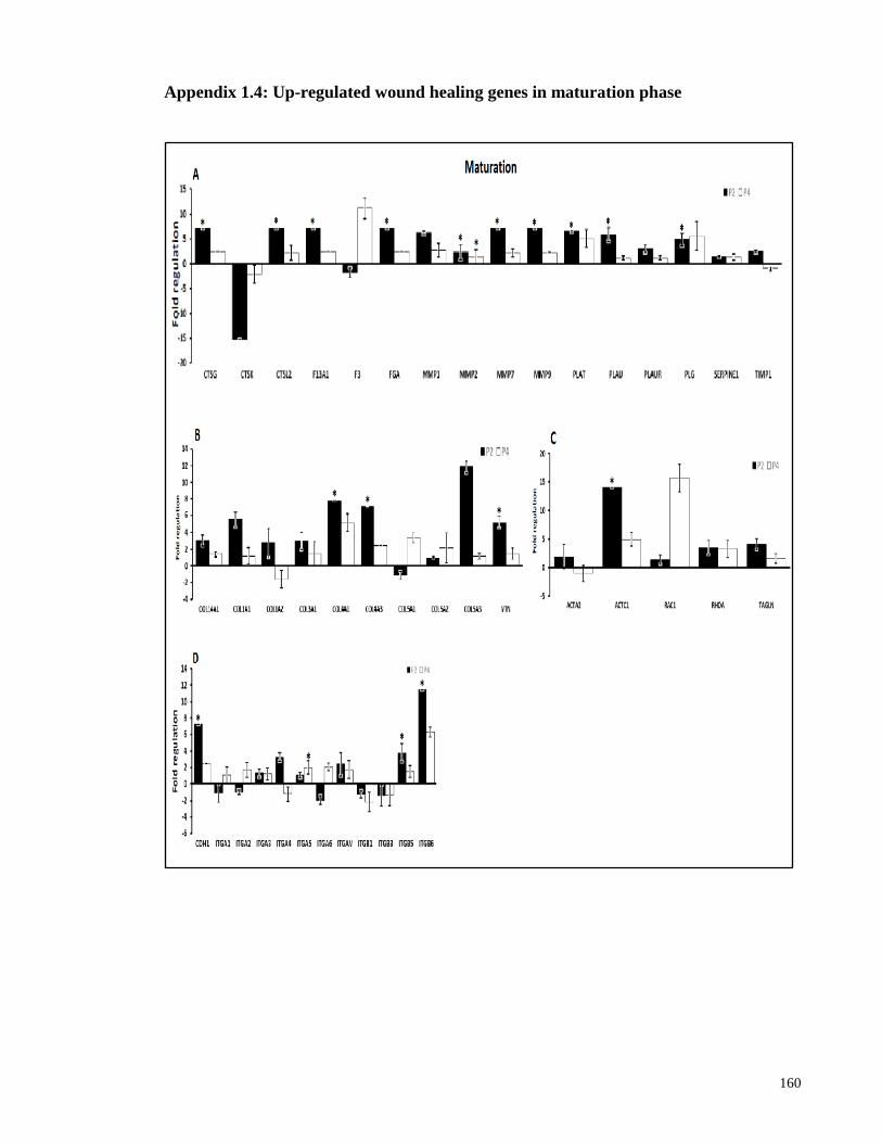
160
Appendix 1.4: Up-regulated wound healing genes in maturation phase

161
Appendix 1.5: Animal ethics clearance number

162
Appendix 1.6: Preparation of pkh-26 dye tagging for SHED
1. Preparation of dye comes first, 0.5 ml of diluent C added to 2 μl of pkh-26 dye.
2. For preparation of cell suspension (for 10 million cells), the diluent C was added to
pellet of loose centrifuge with 25 μl of supernatant
3. Then 0.5 ml of cell suspension added to 0.5 dye solution, it was immediately mixed
by pipetting.
4. The suspension was transferred to 15 ml falcon tube.
5. The suspension was incubated for 5 minutes at room temperature
6. 1 ml of FBS was added to stop the reaction and incubated again for 1 minute.
7. The cells were centrifuged at 400 g for 10 minutes at room temperature.
8. The supernatant was discarded carefully.
9. The pellet was resuspended with 10 ml of culture media.
10. The suspension was centrifuged at 400 g for 5 minutes (repeat 2x)
11. Finally, resuspend the pellet with phosphate buffer saline (PBS) and the cell is
ready for transplantation.

163
Appendix 1.7: Histological tissue slide preparation for H&E staining

164

165

166
Appendix 1.8: Heatmap of Gene Profiling for P2 and P9 of Dental Stem Cells

167
Appendix 1.9: The measurement of wound closure in in vitro co-cultured scratch
assay
The observations of cell migration under phase contrast microscope 4X. The duration of
captured pictures was at 0, 10, 23, 47 and 90 hours. Fastest wound closure was observed in
B, followed by C and A.

168
Appendix 1.8, continued