EFFECTS OF FLUORIDE EXPOSURE ON THE GROWTH, METAMORPHOSIS ... · PDF fileResearch report...
15
Research report Fluoride 49(2)128-142 April-June 2016 Effects of fluoride exposure on the growth, metamorphosis, and skeletal development of Rana chensinesis and Rana nigromaculata larvae Chen, Chai, Zhao, Wu, Wang 128 128 EFFECTS OF FLUORIDE EXPOSURE ON THE GROWTH, METAMORPHOSIS, AND SKELETAL DEVELOPMENT OF RANA CHENSINESIS AND RANA NIGROMACULATA LARVAE Jiayi Chen, a Lihong Chai b,c Hongfeng Zhao, a Minyao Wu, a Hongyuan Wang a, * Xi’an, People’s Republic of China ABSTRACT: Chronic exposure to high fluoride ion levels (F) may lead to local tissue disturbances, known as fluorosis. The present study was conducted to determine the effects of sodium fluoride (NaF) on the growth, metamorphosis, and skeletal development in tadpoles of Rana chensinesis and Rana nigromaculata. The mortality, percentage of tadpoles completing metamorphosis, total length, body weight, and hindlimb length were determined. In addition, skeletal systems were investigated by using double staining methodology at Gosner stages 36, 42, and 46. The results showed that chronic exposure to 50 mg NaF/L significantly increased the mortality, inhibited metamorphosis, and delayed development in R. chensinesis and R. nigromaculata tadpoles. The NaF treatment also produced flexural tail malformations in R. chensinesis tadpoles but not in R. nigromaculata tadpoles, stimulated bone mineralization in R. chensinesis tadpoles, and retarded the deposition of calcium in R. nigromaculata tadpoles. In conclusion, our study suggests that fluoride may affect skeletal ossification differently in different frog species due to differences in sensitivity or the duration of the exposure. Keywords Fluoride; Metamorphosis; Rana chensinesis; Rana nigromaculata; Skeletal development. INTRODUCTION Fluorine is one of common elements in the earth’s crust but is not found naturally in the environment in its free state, due to its great chemical reactivity, and exists in inorganic fluorides, as the fluoride ion (F), or in organic fluoride compounds. 1,2 The primary natural sources of inorganic fluorides are volcanic eruptions, hot springs, soil during droughts, the weathering of rock, and the burning of fossil fuels, etc. 3 Fluoride is not an essential microelement for life and in excess can produce toxic effects in plants, animals, and humans. 4 Fluoride pollution, with increased fluoride concentrations in both water and the atmosphere, may be produced by human activities, such as metallurgy, the production of bricks, ceramics, and glasses, and the processing of nuclear fuel and manure. 5 Human activities may increase the fluoride concentration of surface water 100-fold up to 45 mg F/ L. 6 Fluoride is known to interact with mineralized tissues such as bone and teeth and, when concomitant calcium deficit is present, may aggravate bone loss, via reductions in the mineralized trabecular and cortical bone mass, and cause fragility fractures. 7 Currently, many investigations have demonstrated that fluoride has effects on bone mineralization in both rats and humans. 8,9 Fluoride behaves as a a College of Life Science, Shaanxi Normal University, Xi’an 710119, PR China; b School of Environmental Science and Engineering, Chang’an University, Xi’an 710054, PR China; c Key Laboratory of Subsurface Hydrology and Ecological Effect in Arid Region of Ministry of Education, Xi’an 710062, PR China; *For correspondence: Hongyuan Wang, PhD, College of Life Science, Shaanxi Normal University, Xi’an 710119, PR China; Tel: +86-029-85310266; Fax: +86-029-85310546; E-mail: [email protected]
Transcript of EFFECTS OF FLUORIDE EXPOSURE ON THE GROWTH, METAMORPHOSIS ... · PDF fileResearch report...

Research reportFluoride 49(2)128-142April-June 2016
Effects of fluoride exposure on the growth, metamorphosis, and skeletaldevelopment of Rana chensinesis and Rana nigromaculata larvae
Chen, Chai, Zhao, Wu, Wang
128128
EFFECTS OF FLUORIDE EXPOSURE ON THE GROWTH, METAMORPHOSIS, AND SKELETAL DEVELOPMENT
OF RANA CHENSINESIS AND RANA NIGROMACULATA LARVAE
Jiayi Chen,a Lihong Chaib,c Hongfeng Zhao,a Minyao Wu,a Hongyuan Wanga,*Xi’an, People’s Republic of China
ABSTRACT: Chronic exposure to high fluoride ion levels (F) may lead to local tissuedisturbances, known as fluorosis. The present study was conducted to determine theeffects of sodium fluoride (NaF) on the growth, metamorphosis, and skeletaldevelopment in tadpoles of Rana chensinesis and Rana nigromaculata. The mortality,percentage of tadpoles completing metamorphosis, total length, body weight, andhindlimb length were determined. In addition, skeletal systems were investigated byusing double staining methodology at Gosner stages 36, 42, and 46. The resultsshowed that chronic exposure to 50 mg NaF/L significantly increased the mortality,inhibited metamorphosis, and delayed development in R. chensinesis and R.nigromaculata tadpoles. The NaF treatment also produced flexural tail malformationsin R. chensinesis tadpoles but not in R. nigromaculata tadpoles, stimulated bonemineralization in R. chensinesis tadpoles, and retarded the deposition of calcium inR. nigromaculata tadpoles. In conclusion, our study suggests that fluoride may affectskeletal ossification differently in different frog species due to differences insensitivity or the duration of the exposure.
Keywords Fluoride; Metamorphosis; Rana chensinesis; Rana nigromaculata; Skeletal development.
INTRODUCTION
Fluorine is one of common elements in the earth’s crust but is not foundnaturally in the environment in its free state, due to its great chemical reactivity,and exists in inorganic fluorides, as the fluoride ion (F), or in organic fluoridecompounds.1,2 The primary natural sources of inorganic fluorides are volcaniceruptions, hot springs, soil during droughts, the weathering of rock, and theburning of fossil fuels, etc.3 Fluoride is not an essential microelement for life andin excess can produce toxic effects in plants, animals, and humans.4 Fluoridepollution, with increased fluoride concentrations in both water and theatmosphere, may be produced by human activities, such as metallurgy, theproduction of bricks, ceramics, and glasses, and the processing of nuclear fuel andmanure.5 Human activities may increase the fluoride concentration of surfacewater 100-fold up to 45 mg F/ L.6
Fluoride is known to interact with mineralized tissues such as bone and teethand, when concomitant calcium deficit is present, may aggravate bone loss, viareductions in the mineralized trabecular and cortical bone mass, and cause fragilityfractures.7 Currently, many investigations have demonstrated that fluoride haseffects on bone mineralization in both rats and humans.8,9 Fluoride behaves as aaCollege of Life Science, Shaanxi Normal University, Xi’an 710119, PR China; bSchool ofEnvironmental Science and Engineering, Chang’an University, Xi’an 710054, PR China; cKeyLaboratory of Subsurface Hydrology and Ecological Effect in Arid Region of Ministry ofEducation, Xi’an 710062, PR China; *For correspondence: Hongyuan Wang, PhD, College ofLife Science, Shaanxi Normal University, Xi’an 710119, PR China; Tel: +86-029-85310266; Fax:+86-029-85310546; E-mail: [email protected]

Research reportFluoride 49(2)128-142April-June 2016
Effects of fluoride exposure on the growth, metamorphosis, and skeletaldevelopment of Rana chensinesis and Rana nigromaculata larvae
Chen, Chai, Zhao, Wu, Wang
129129
cumulative toxic agent that can alter the accretion and resorption of tissues as wellas affecting the homeostasis of bone mineral metabolism.10,11 Fluoride exerts abiphasic action at the level of the osteoblasts, on bone mineralization, bonestructure, and bone function. At low dosages, fluoride stimulates the formation ofbone. However, at high concentrations, fluoride reduces the apposition rates ofcalcium ion and results in a prolongation of the mineralization time, thus delayingthe development of the skeleton.12,13 Moreover, at high dosages, fluoride may leadto the formation of abnormally mineralized bone of impaired quality.14
Bone is a composite tissue with a greater mineral component which consists ofcalcium phosphate and other salts deposited in a matrix, osteoid, primarilyconsisting of Type 1 collagen but also containing proteoglycans such aschondroitin sulfate and keratin sulphate. Calcification is crucial for the normalmineralization of bone.15 Alizarin red can stain the calcium in bone red give anindication of the calcium concentration in the bone16 while aleian blue can stainthe chondroitin sulfate and keratin sulfate, in non-mineralized osteoid or cartilage,blue. In our experience, we have found the double-staining method with alizarinred and aleian blue to be useful for investigating the skeletal development oflarvae exposed to fluoride at different development stages by clearly showingbone and shape and the degree of ossification.
Metamorphosis development is an important stage in the life history ofamphibians and involves the reconstruction of the tissues and organs of thetadpole, including the remolding of the skeletal system and the growth of thelimbs.17,18 The reconstruction of the tadpole skeletal system is thought to play anessential role in amphibian metamorphosis, which enables adaptation to occurfrom the aquatic habitat to the land habitat. Although excessive fluoride can impairskeletal development in Bufo gargarizans tadpoles,19 no studies had been done onthe effects of fluoride on skeletal development in R. chensinesis and R.nigromaculata.
The aim of the present study was to investigate the effects of fluoride on skeletaldevelopment in R. chensinesis and R. nigromaculata tadpoles at metamorphosis.Using a control group with water with 0 mg NaF/L, we examined the effects ofchronic fluoride exposure, with water at 50 mg NaF/L, on mortality, body size, andbody mass in R. chensinesis and R. nigromaculata tadpoles. In addition, theskeletal systems at pre-metamorphosis, at metamorphic climax, and at thecompletion of metamorphosis were investigated by using double stainingmethodology.
MATERIALS AND METHODS
Experimental animals: The tadpoles studied originated from sexually maturemale and female frogs, of the species Rana chensinesis and Rana nigromaculata,which were collected in February, from the Qinling Mountains, Shaanxi Province,PR China, and induced to spawn by each couple being placed in a 40 L glassaquarium.

Research reportFluoride 49(2)128-142April-June 2016
Effects of fluoride exposure on the growth, metamorphosis, and skeletaldevelopment of Rana chensinesis and Rana nigromaculata larvae
Chen, Chai, Zhao, Wu, Wang
130130
Chemicals and reagents: The stock solutions were prepared by dissolvingsodium fluoride (NaF), from Sigma-Aldrich Corporation (Sigma, St Louis, MO,USA), in dechlorinated tap water to obtain a nominal concentration of 50 mg NaF/L.
Exposure test: The control group was kept in dechlorinated tap water with 0 mgNaF/L while the experimental treatment group were kept in water with 50 mgNaF/L. After hatching, the R. chensinesis and R. nigromaculata embryos werecollected and placed in individual 20 L glass aquaria with either 0 (control) or 50(treatment) mg NaF/L (nominal concentrations). Using a complete randomizedblock design, at Gosner stage 26,20 the tadpoles were divided into 16 tanks, 4control tanks and 4 tanks with 50 mg NaF/L for each of the two species, with n=90tadpoles in each tank. The temperature was maintained at 22ºC with photoperiodsof 12 hr of light and 8 hr of darkness. The water conditions were pH 7.9, dissolvedoxygen (DO) 7.2 mg/L, and specific conductivity 1477 µS/cm. During this timethe dead embryos were removed twice daily. The data collected on the deadembryos was used to compute the embryonic survival rate. Any unconsumed foodwas siphoned from the tanks daily. The tadpoles were fed two to three times perday. The rearing water was completely renewed three times per week, starting attwo weeks post-hatch.
Development, mortality, metamorphosis, and deformities: Daily records weremaintained for hatching success, and the embryo and larval mortality. Deformities,mass, and the date of completed metamorphosis (Gosner stage 46) were monitoredfor all the individuals. We examined the effects of chronic fluoride exposure on themetamorphosis of R. chensinesis and R. nigromaculata tadpoles, at the fluorideconcentrations of 0 and 50 mg NaF/L, from Gosner stage 26 to Gosner stage 46.The periods of tadpoles development were defined according to Gosner’s 1960classification20 as pre-metamorphosis (Gosner stages 36–37), prometamorphosis(Gosner stages 39–40), metamorphic climax (Gosner stage 42), and completion ofmetamorphosis (complete tail resorption; Gosner stage 46). The sampling plan forthis study was designed to encompass these periods. Thirty tadpoles from bothcontrol and treatment tanks were collected randomly for each of the two species,weighed, and measured for total length and hindlimb length at Gosner stages 36,42, and 46, respectively.
Determination of the skeletal development: The double-staining method withalizarin red and aleian blue was used to investigate the skeletal development of thelarvae exposed to different concentrations of NaF at different development stages.Three tadpoles, at Gosner stages 36, 42, and 46, were collected randomly fromeach aquarium. Both the control (0.00 mg NaF/L) and fluoride-treated (50 mgNaF/L) larvae were anaesthetized with 95% alcohol, decolorized with 30% H2O2,and defatted with acetone. After being eviscerated and cleared, each specimen wasstained using the alizarin red-aleian blue double staining method.21 Bones(containing calcium) were indicated in red and cartilage (containing chondroitinsulfate and keratin sulfate) in blue. The osteological terminology used followsDuellman and Trueb.22 All the descriptions and illustrations were based on

Research reportFluoride 49(2)128-142April-June 2016
Effects of fluoride exposure on the growth, metamorphosis, and skeletaldevelopment of Rana chensinesis and Rana nigromaculata larvae
Chen, Chai, Zhao, Wu, Wang
131131
photographs taken with a Cannon 7D digital camera attached to a Zeiss DiscoveryV12 stereoscope.
Statistical analysis: Statistical analyses were performed using the SPSS 16.0.Differences between treatments were tested by with one-way ANOVA of the totallength, body weight, and hindlimb length as the dependent list, with the dose ofNaF as the variable factor, at Gosner stages 36, 42, and 46. In addition, differencesin the measured variables (mortality and percent metamorphosed) across eachtreatment were also tested using one-way ANOVA. The data were reported asmean±SE. For all analyses, a p<0.05 was considered to be a significant difference.
RESULTS
Mortality and metamorphosis rates in larvae of R. chensinesis and R.nigromaculata: Exposure to 50 mg NaF/L had a significant effect on themortality of R. chensinesis and R. nigromaculata tadpoles (p<0.05). Themortalities of R. chensinesis tadpoles exposed to the 0 (control) and 50 mg NaF/L were 11.1% and 34.4%, respectively. Likewise, the mortalities of R.nigromaculata tadpoles in water with 0 and 50 mg NaF/1 were 17.8% and42.2%, respectively. For both species, the mortalities of the group exposed to 50mg NaF/L were significantly greater (p<0.05) than the mortalities of the groupsexposed only to dechlorinated water (Figure 1).
Compared to the control groups who were exposed to dechlorinated water only,exposure to 50 mg NaF/L significantly delayed (p<0.05) the rates ofmetamorphosis from tadpole to frog of R. chensinesis and R. nigromaculata. Themetamorphosis rates of R. chensinesis exposed to 0 and 50 mg NaF/L were 54.4%and 28.9%, respectively. Similarly, the metamorphosis rates of R. nigromaculata
*
*50
40
30
20
10
0
Mortality rate (%)
Control
50 mg NaF/L
Rana Rana chensinesis nigromaculata
Figure 1. Mortality rate of R. chensinesis and R. nigromaculata tadpoles exposed to controlwater (dechlorinated water) and water with 50 mg NaF/L. Values are mean±SE. At theinitiation of the exposure there were 16 tanks (4 control tanks and 4 tanks with 50 mg NaF/L for each of the two species) with n=90 tadpoles in each tank. Compared to the controlgroup, *p<0.05.

Research reportFluoride 49(2)128-142April-June 2016
Effects of fluoride exposure on the growth, metamorphosis, and skeletaldevelopment of Rana chensinesis and Rana nigromaculata larvae
Chen, Chai, Zhao, Wu, Wang
132132
tadpoles, exposed to 0 and 50 mg NaF/L, were 57.8% and 32.2%, respectively.(Figure 2).
Effect of sodium fluoride on growth and development in larvae of R. chensinesisand R. nigromaculata: The total length of the R. chensinesis tadpoles wassignificantly reduced in the treated group compared with the control group atGosner stages 36 and 42 (p<0.01 and p<0.05, respectively, one-way ANOVA).However, the total length of the individuals that reached completedmetamorphosis at Gosner stage 46 during exposure to 50 mg NaF/L was notsignificantly reduced compared to the untreated control group (p= 0.524, Figure3).
Rana Rana chensinesis nigromaculata
* *
Figure 2. Metamorphosis rate of R. chensinesis and R. nigromaculata tadpoles exposedto control water (dechlorinated water) and water with 50 mg NaF/L. Values are mean±SE.At the initiation of the exposure there were 16 tanks (4 control tanks and 4 tanks with 50mg NaF/L for each of the two species) with n=90 tadpoles in each tank. Compared to thecontrol group, *p<0.05.
*
* †
Figure 3. Total length of R. chensinesis at Gosner stages 36, 42, and 46. Values aremean±SE. Compared to the control group (one-way ANOVA): *p<0.05, †p<0.01.
36 42 46 Gosner stage
40
30
20
10
0
Total length (mm)
Control
50 mg NaF/L
60
50
40
30
20
10
0
Metamorphosis rate (%)
Control
50 mg NaF/L
Research reportFluoride 49(2)128-142April-June 2016
Effects of fluoride exposure on the growth, metamorphosis, and skeletaldevelopment of Rana chensinesis and Rana nigromaculata larvae
Chen, Chai, Zhao, Wu, Wang
133133
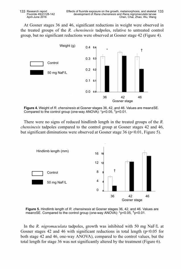
At Gosner stages 36 and 46, significant reductions in weight were observed inthe treated groups of the R. chensinesis tadpoles, relative to untreated controlgroup, but no significant reductions were observed at Gosner stage 42 (Figure 4).
There were no signs of reduced hindlimb length in the treated groups of the R.chensinesis tadpoles compared to the control group at Gosner stages 42 and 46,but significant diminutions were observed at Gosner stage 36 (p<0.01, Figure 5).
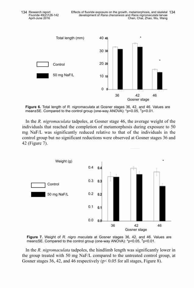
In the R. nigromaculata tadpoles, growth was inhibited with 50 mg NaF/L atGosner stages 42 and 46 with significant reductions in total length (p<0.05 forboth stage 42 and 46, one-way ANOVA), compared to the control values, but thetotal length for stage 36 was not significantly altered by the treatment (Figure 6).
* †
36 42 46 Gosner stage
0.4
0.3
0.2
0.1
0.0
Weight (g)
Control
50 mg NaF/L
Figure 4. Weight of R. chensinesis at Gosner stages 36, 42, and 46. Values are mean±SE.Compared to the control group (one-way ANOVA): *p<0.05, †p<0.01.
†
36 42 46 Gosner stage
16
12
8
4
0
Hindlimb length (mm)
Control
50 mg NaF/L
Figure 5. Hindlimb length of R. chensinesis at Gosner stages 36, 42, and 46. Values aremean±SE. Compared to the control group (one-way ANOVA): *p<0.05, †p<0.01.
Research reportFluoride 49(2)128-142April-June 2016
Effects of fluoride exposure on the growth, metamorphosis, and skeletaldevelopment of Rana chensinesis and Rana nigromaculata larvae
Chen, Chai, Zhao, Wu, Wang
134134
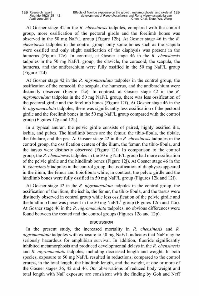
In the R. nigromaculata tadpoles, at Gosner stage 46, the average weight of theindividuals that reached the completion of metamorphosis during exposure to 50mg NaF/L was significantly reduced relative to that of the individuals in thecontrol group but no significant reductions were observed at Gosner stages 36 and42 (Figure 7).
In the R. nigromaculata tadpoles, the hindlimb length was significantly lower inthe group treated with 50 mg NaF/L compared to the untreated control group, atGosner stages 36, 42, and 46 respectively (p< 0.05 for all stages, Figure 8).
*
*
36 42 46 Gosner stage
40
30
20
10
0
Total length (mm)
Control
50 mg NaF/L
Figure 6. Total length of R. nigromaculata at Gosner stages 36, 42, and 46. Values aremean±SE. Compared to the control group (one-way ANOVA): *p<0.05, †p<0.01.
*
0.4
0.3
0.2
0.1
0.0
Weight (g)
Control
50 mg NaF/L
36 42 46 Gosner stage
Figure 7. Weight of R. nigro maculata at Gosner stages 36, 42, and 46. Values aremean±SE. Compared to the control group (one-way ANOVA): *p<0.05, †p<0.01.

Research reportFluoride 49(2)128-142April-June 2016
Effects of fluoride exposure on the growth, metamorphosis, and skeletaldevelopment of Rana chensinesis and Rana nigromaculata larvae
Chen, Chai, Zhao, Wu, Wang
135135
The morphology of R. chensinesis and R. nigromaculata tadpoles: The morphological alterations of R. chensinesis and R. nigromaculata tadpoles
in the control group and the treated group were recorded at the differentdevelopmental stages. Compared to the normal morphology of R. chensinesis fromtadpole to frog in the control group (Figure 9A), exposure to the 50 mg NaF/Linduced significant deformities, involving asymmetric or bent tails in R.chensinesis (Figure 9B). In contrast, no malformations appeared in R.nigromaculata with the NaF treatment (Figure 9C).
Effect of sodium fluoride on bone formation in the larvae of R. chensinesis andR. nigromaculata: The skeletal systems of the tadpoles were demonstrated usingthe double staining method at different developmental stages in the control andtreatment groups. At Gosner stage 36, no ossification was observed in thechondrocranium of the control R. chensinesis group, suggesting that ossificationwas not well developed (Figure 10a). In contrast, ossification of theparasphenoid bones occurred in the 50 mg NaF/L R. chensinesis group (Figure
36 42 46 Gosner stage
* *
*
16
12
8
4
0
Hindlimb length (mm)
Control
50 mg NaF/L
Figure 8. Hindlimb length of R. nigro maculata at Gosner stages 36, 42, and 46. Valuesare mean±SE. Compared to the control group (one-way ANOVA): *p<0.05, †p<0.01.
Figures 9A,B, and C. Photograph illustrating the morphology of R. chensinesis and R.nigromaculata tadpoles exposed to 0 and 50 mg NaF/L at the different developmental stages.A: dorsal view of representative developmental stages from the control group in R.chensinesis; B: dorsal view of representative developmental stages from the treated group inR. chensinesis; and C: dorsal view of representative developmental stages from the treatedgroup in R. nigromaculata.
B CA

Research reportFluoride 49(2)128-142April-June 2016
Effects of fluoride exposure on the growth, metamorphosis, and skeletaldevelopment of Rana chensinesis and Rana nigromaculata larvae
Chen, Chai, Zhao, Wu, Wang
136136
10b). The parasphenoid and exoccipital bones were well ossified indicated byred staining in the control R. chensinesis specimen at stage 42 (Figure 10c). Inthe 50 mg NaF/L R. chensinesis group, greater ossification was observed in theparasphenoid, frontoparietal and exoccipital bones compared to control group(Figure 10d). At stage 46, ossification of the parasphenoid, frontoparietal andexoccipital bones increased significantly in the 50 mg NaF/L R. chensinesisgroup compared with that in the control group (Figures 10e and 10f).
From the ventral view, the chondrocrania of R. nigromaculata, in the control and50 mg NaF/L groups, were completely cartilaginous at stage 36, (Figures 10g and10h). In the crania of larvae at stage 42, the ossified parasphenoid, frontoparietal,exoccipital, and prootic bones were observed clearly in the specimens in thecontrol R. nigromaculata group (Figure 10i). In contrast, only the ossifiedparasphenoid and exoccipital were found in the 50 mg NaF/L R. nigromaculata
Figure 10. Double staining of the cranial skeleton of R. chensinesis and R. nigromaculatalarvae at different development stages, ventral view. a: the cranial skeleton of R. chensinesisat stage G36 in the control group; b: the cranium skeleton of R. chensinesis at stage G36exposed to 50 mg NaF/L; c: the cranial skeleton of R. chensinesis at stage G42 in the controlgroup; d: the cranial skeleton of R. chensinesis at stage G42 exposed to 50 mg NaF/L; e: thecranial skeleton of R. chensinesis at stage G46 in the control group; f: the cranial skeleton ofR. chensinesis at stage G46 exposed to 50 mg NaF/L; g: the cranial skeleton of R.nigromaculata at stage G36 in the control group; h: the cranial skeleton of R. nigromaculata atstage G36 exposed to 50 mg NaF/L; i: the cranial skeleton of R. nigromaculata at stage G42in the control group; j: the cranial skeleton of R. nigromaculata at stage G42 exposed to 50 mgNaF/L; k: the cranial skeleton of R. nigromaculata at stage G46 in the control group; and l: thecranial skeleton of R. nigromaculata at stage G46 exposed to 50 mg NaF/L.

Research reportFluoride 49(2)128-142April-June 2016
Effects of fluoride exposure on the growth, metamorphosis, and skeletaldevelopment of Rana chensinesis and Rana nigromaculata larvae
Chen, Chai, Zhao, Wu, Wang
137137
group (Figure 10j). At Gosner stage 46, more ossification was visible in theparasphenoid, frontoparietal, and exoccipital bones in the control group comparedto the 50 mg NaF/L R. nigromaculata group (Figures 10k and 10l).
In anurans (frogs and toads), the vertebral column is divided into three regions,namely, presacral, sacral, and postsacral regions. The presacral region consists ofeight discrete vertebrae; the sacral region is composed of a single vertebra, and thepostsacral region is formed by the urostyle. At Gosner stage 42 in the R.chensinesis tadpoles, there was no distinct difference in the ossification of thevertebral column between the treated and the control groups (Figures 11a and11b). At Gosner stage 46 in the R. chensinesis tadpoles, in the control group mostof the arches had begun to ossify with the transverse process remainingcartilaginous, as indicated by their staining blue, while in the 50 mg NaF/L groupboth the arches and the transverse process were completely ossified with nocartilage remaining (Figures 11c and 11d).
At Gosner stage 42 in the R. nigromaculata tadpoles, most of the arches andtransverse process were ossified in the control group (Figure 11e). However,compared to the control group, ossification of the vertebral column was obviouslyless in the 50 mg NaF/L group (Figure 11f). At Gosner stage 46 in the R.nigromaculata tadpoles, there was no obvious difference in the ossification of thevertebral column between the treated and control groups (Figures 11g and 11h).
The pectoral girdle is composed of the clavicle, the coracoid, the scapula, thecleithrum, the sternum, and the omosternum. The forelimb bones are the humerus,the antibrachium, the carpus, the ossa metacarpalia, and the phalanges. At Gosner
Figure 11. The axial skeleton in ventral view. a: the axial skeleton of R. chensinesis at stageG42 in the control group; b: the axial skeleton of R. chensinesis at stage G42 exposed to 50mg NaF/L; c: the axial skeleton of R. chensinesis at stage G46 in the control group; d: the axialskeleton of R. chensinesis G46 exposed to 50 mg NaF/L; e: the axial skeleton of R.nigromaculata at stage G42 in the control group;. f: the axial skeleton of R. nigromaculata atstage G42 exposed to 50 mg NaF/L; g: the axial skeleton of R. nigromaculata at stage G46 inthe control group; h: the axial skeleton of R. nigromaculata G46 exposed to 50 mg NaF/L.

Research reportFluoride 49(2)128-142April-June 2016
Effects of fluoride exposure on the growth, metamorphosis, and skeletaldevelopment of Rana chensinesis and Rana nigromaculata larvae
Chen, Chai, Zhao, Wu, Wang
138138
stage 42 in the control group, the ossification centers of the coracoid, the scapula,the humerus, and the antibrachium were distinctly observed in the R. chensinesistadpoles (Figure 12a).
Figure 12. The forelimb and hindlimb skeleton in ventral view. a: the forelimb skeleton of R.chensinesis at stage G42 in the control group; b: the forelimb skeleton of R. chensinesis atstage G42 exposed to 50 mg NaF/L; c: the forelimb skeleton of R. chensinesis at stage G46in the control group; d: the forelimb skeleton of R. chensinesis at stage G46 exposed to 50mg NaF/L; e: the forelimb skeleton of R. nigromaculata at stage G42 in the control group; f:the forelimb skeleton of R. nigromaculata at stage G42 exposed to 50 mg NaF/L; g: theforelimb skeleton of R. nigromaculata at stage G46 in the control group; h: the forelimbskeleton of R. nigromaculata at stage G46 exposed to 50 mg NaF/L; i: the hindlimb skeletonof R. chensinesis at stage G42 in the control group; j: the hindlimb skeleton of R. chensinesisat stage G42 exposed to 50 mg NaF/L; k: the hindlimb skeleton of R. chensinesis at stageG46 in the control group; l: the hindlimb skeleton of R. chensinesis at stage G46 exposed to50 mg NaF/L; m: the hindlimb skeleton of R. nigromaculata at stage G42 in the control group;n: the hindlimb skeleton of R. nigromaculata at stage G42 exposed to 50 mg NaF/L; o: thehindlimb skeleton of R. nigromaculata at stage G46 in the control group; and p: the hindlimbskeleton of R. nigromaculata at stage G46 exposed to 50 mg NaF/L.
Research reportFluoride 49(2)128-142April-June 2016
Effects of fluoride exposure on the growth, metamorphosis, and skeletaldevelopment of Rana chensinesis and Rana nigromaculata larvae
Chen, Chai, Zhao, Wu, Wang
139139
At Gosner stage 42 in the R. chensinesis tadpoles, compared with the controlgroup, more ossification of the pectoral girdle and the forelimb bones wasobserved in the 50 mg NaF/L group (Figure 12b). At Gosner stage 46 in the R.chensinesis tadpoles in the control group, only some bones such as the scapulawere ossified and only slight ossification of the diaphysis was present in thehumerus (Figure 12c). In contrast, at Gosner stage 46 in the R. chensinesistadpoles in the 50 mg NaF/L group, the clavicle, the coracoid, the scapula, thehumerus, and the antibrachium were fully ossified in the 50 mg NaF/L group(Figure 12d)
At Gosner stage 42 in the R. nigromaculata tadpoles in the control group, theossification of the coracoid, the scapula, the humerus, and the antibrachium weredistinctly observed (Figure 12e). In contrast, at Gosner stage 42 in the R.nigromaculata tadpoles in the 50 mg NaF/L group, there was less ossification ofthe pectoral girdle and the forelimb bones (Figure 12f). At Gosner stage 46 in theR. nigromaculata tadpoles, there was significantly less ossification of the pectoralgirdle and the forelimb bones in the 50 mg NaF/L group compared with the controlgroup (Figures 12g and 12h).
In a typical anuran, the pelvic girdle consists of paired, highly ossified ilia,ischia, and pubes. The hindlimb bones are the femur, the tibio-fibula, the tibiale,the fibulare, and the pes. At Gosner stage 42 in the R. chensinesis tadpoles in thecontrol group, the ossification centers of the ilium, the femur, the tibio-fibula, andthe tarsus were distinctly observed (Figure 12i). In comparison to the controlgroup, the R. chensinesis tadpoles in the 50 mg NaF/L group had more ossificationof the pelvic girdle and the hindlimb bones (Figure 12j). At Gosner stage 46 in theR. chensinesis tadpoles in the control group, the ossification of diaphyses appearedin the ilium, the femur and tibiofibula while, in contrast, the pelvic girdle and thehindlimb bones were fully ossified in 50 mg NaF/L group (Figures 12k and 12l).
At Gosner stage 42 in the R. nigromaculata tadpoles in the control group, theossification of the ilium, the ischia, the femur, the tibio-fibula, and the tarsus weredistinctly observed in control group while less ossification of the pelvic girdle andthe hindlimb bone was present in the 50 mg NaF/L1 group (Figures 12m and 12n).At Gosner stage 46 in the R. nigromaculata tadpoles, no obvious differences werefound between the treated and the control groups (Figures 12o and 12p).
DISCUSSION
In the present study, the increased mortality in R. chensinesis and R.nigromaculata tadpoles with exposure to 50 mg NaF/L indicates that NaF may beseriously hazardous for amphibian survival. In addition, fluoride significantlyinhibited metamorphosis and produced developmental delays in the R. chensinesisand R. nigromaculata tadpoles, including decreased length and weight. In bothspecies, exposure to 50 mg NaF/L resulted in reductions, compared to the controlgroups, in the total length, the hindlimb length, and the weight, at one or more ofthe Gosner stages 36, 42 and 46. Our observations of reduced body weight andtotal length with NaF exposure are consistent with the finding by Goh and Neff

Research reportFluoride 49(2)128-142April-June 2016
Effects of fluoride exposure on the growth, metamorphosis, and skeletaldevelopment of Rana chensinesis and Rana nigromaculata larvae
Chen, Chai, Zhao, Wu, Wang
140140
that NaF exposure reduced the head-tail length and the eye diameter of Xenopusembryos.12
We assessed the effects of NaF on tadpole morphology, in order to explorewhether chronic exposure would induce tadpole deformities, and found that NaFproduced flexural tail deformities in R. chensinesis tadpoles. Developmental andmorphological abnormalities (e.g., visceral, mouth, eye, and limb deformities)have been observed in anuran tadpoles exposed to several different classes ofcompounds.23-26 The increased incidences of deformities could result in reducedfitness by affecting foraging or predator avoidance, and cause reduced survivaland reproduction of adults.27 In contrast to the deformities found in the R.chensinesis tadpoles, no malformations appeared in the R. nigromaculata tadpoleswith NaF exposure. The R. nigromaculata embryos metamorphose into frogsapproximately 7–8 weeks postfertilization, whereas the R. chensinesis embryosneed as long as 100 days to complete metamorphosis. As the time for the R.nigromaculata tadpoles to metamorphose to frogs was significantly shorter thanthat required for the R. chensinesis tadpoles, the occurrence of deformities intadpoles may be related to duration of the NaF-exposure time.
Sodium fluoride can also lead to the development of skeletal fluorosis witheffects on bone mineralization, bone cells, and bone architecture.28 The long-termexcessive intake of fluoride can disrupt the balance of bone deposition andremodeling activities leading to skeletal fluorosis.29 Our results showed thatfluoride can stimulate bone mineralization in R. chensinesis tadpoles. Increasedossification was observed in the parasphenoid, frontoparietal, and exoccipitalbones in the tadpoles exposed to 50 mg NaF/L, compared to control group. Inaddition, most of the arches and transverse process in vertebral column ossifiedearlier in the 50 mg NaF/L group compared to control group.
However, in the R. nigromaculata tadpoles, fluoride can retard deposition ofcalcium in bone. In the NaF-treated group, the ossification of the parasphenoid,frontoparietal, and exoccipital bones was significantly less than that in the controlgroup. Furthermore, the ossification of the vertebral column was also obviouslyless in the NaF group compared to the control group. In a similar experiment on B.gargarizans, it was found that fluoride delayed the development of the skeleton byreducing the apposition rates of the calcium ion, leading to a prolongation of themineralization lag time and the bone formation period.19
CONCLUSIONS
The present study is the first to examine the effects of fluoride on R. chensinesisand R. nigromaculata during the entire larval development period. Chronicexposure to NaF increased the mortality, inhibited metamorphosis, and delayeddevelopment in R. chensinesis and R. nigromaculata tadpoles. Exposure to NaFincreased the incidence of deformities in R. chensinesis but no malformationsappeared in the R. nigromaculata tadpoles. In addition, fluoride can stimulate bonemineralization in R. chensinesis tadpoles and retard the deposition of calcium in R.nigromaculata tadpoles. Thus, our study suggests that fluoride has different effects

Research reportFluoride 49(2)128-142April-June 2016
Effects of fluoride exposure on the growth, metamorphosis, and skeletaldevelopment of Rana chensinesis and Rana nigromaculata larvae
Chen, Chai, Zhao, Wu, Wang
141141
in the two species on the ossification of bone. As the postfertilization time tometamorphosis differed in the two species and the increased ossification occurredin the species, R. chensinesis, with the longest time to metamorphosis, wehypothesize that the increased skeletal ossification with NaF in R. chensinesis,may be associated with an increased sensitivity to fluoride or an increased durationof the exposure. Thus our study suggests that fluoride may affect skeletalossification differently in different frog species due to differences in sensitivity orthe duration of the exposure
ACKNOWLEDGEMENTS
The current work was supported by the National Science Foundation of China(No. 41401570, No. 31201726) and the special fund of the Shaanxi NormalUniversity (No. GK20153051).
REFERENCES1 Hardisson A, Rodriguez MI, Burgos A, Flores LD, Gutierrez R, Varela H. Fluoride levels in
publicly supplied and bottled drinking water in the island of Tenerife, Spain. Bull EnvironContam Toxicol 2001;67:163-70.
2 Camargo JA. Fluoride toxicity to aquatic organisms: a review. Chemosphere 2003;50:251-64.
3 Weinstein LH, Davison AW. Fluorides in the environment: effects on plants and animals.Cambridge, MA, USA: CABI Publishing; 2004.
4 Dhar V, Bhatnagar M. Physiology and toxicity of fluoride. Indian J Dent Res 2009;20:350-5.5 Roy RL, Campbell PGC, Premont S. Geochemistry and toxicity of aluminium in the
Saguenay River, Quebec, Canada, in relation to discharges from an aluminium smelter.Environ Toxicol Chem 2000;19:2457-66.
6 Ren FH, Jiao SQ. Distribution and formation of high-fluorine groundwater in China. EnvironGeology 1988;12:3-10.
7 Simon MJ, Beil FT, Rüther W, Busse B, Koehne T, Steiner M, et al. High fluoride and lowcalcium levels in drinking water is associated with low bone mass, reduced bone qualityand fragility fractures in sheep. Osteoporos Int 2014;25:1891-903.
8 Wang YN, Xiao KQ, Liu JL, Dallner G, Guan ZZ. Effect of long term fluoride exposure onlipid composition in rat liver. Toxicology 2000;146:161-9.
9 Shivashankara AR, Shankara YMS, Rao SH, Bhat PG. A clinical and biochemical study ofchronic fluoride toxicity in children of Kheru Thanda of Gulbarga district, Karnataka, India.Fluoride 2000;33:66-73.
10 Miao Q, Xu M, Liu B, You B. In vivo and in vitro study on the effect of excessive fluoride ontype I collagen of rats. Wei Sheng Yan Liu/J Hygiene Res 2002;31:145-7.
11 Everett ET. Fluoride's effects on the formation of teeth and bones, and the influence ofgenetics. J Dent Res 2001;90:552-60.
12 Goh EH, Neff AW. Effects of fluoride on Xenopus embryo development. Food Chem Toxicol2003;41:1501-8.
13 Long YG, Wang YN, Chen J, Jiang SF, Nordberg A, Guan ZZ. Chronic fluoride toxicitydecreases the number of nicotinic acetylcholine receptors in rat brain. Neurotoxicol Teratol2002;24:751-7.
14 Mousny M, Banse X, Wise L, Everett ET, Hancock R, Vieth R, et al. The genetic influenceon bone susceptibility to fluoride. Bone 2006;39:1283-9.
15 Williams AJ, Robson H, Kester MH, van Leeuwen JP, Shalet SM, Visser TJ, Williams GR.Iodothyronine deiodinase enzyme activities in bone. Bone 2008;43:126-34.
16 Kerney R, Wassersug R, Hall BK. Skeletal advance and arrest in giant non-metamorphosing African clawed frog tadpoles (Xenopus laevis: Daudin). J Anat2010;216:132-43.
17 Haas A. Larval and metamorphic skeletal development in the fast-developing frogPyxicephalus adspersus (Anura, Ranidae). Zoomorphology 1999;119:23-35.

Research reportFluoride 49(2)128-142April-June 2016
Effects of fluoride exposure on the growth, metamorphosis, and skeletaldevelopment of Rana chensinesis and Rana nigromaculata larvae
Chen, Chai, Zhao, Wu, Wang
142142
18 Brown DD, Cai L. Amphibian metamorphosis. Dev Biol 2007;306:20-33.19 Zhao HF, Chai LH, Wang HY. Effects of fluoride on metamorphosis, thyroid and skeletal
development in Bufo gargarizans tadpoles. Ecotoxicology 2013;22:1123-32.20 Gosner KL. A simplified table for staging anuran embryos and larvae with notes on
identification. Herpetologica 1960;16:183-90.21 Taylor WR, van Dyke GC. Revised procedures for staining and clearing small fishes and
other vertebrates for bone and cartilage study. Cybium 1985;9:107-11.22 Duellman WE, Trueb L. Biology of amphibians. Baltimore, USA: Johns Hopkins University;
1994.23 Bridges CM. Long-term effects of pesticide exposure at various life stages of the southern
leopard frog (Rana sphenocephala). Arch Environ Contam Toxicol 2000;39:91-6.24 Boone MD, Bridges CM, Rothermel BB. Growth and development of larval green frogs
(Rana clamitans) exposed to multiple doses of an insecticide. Oecologia 2001;129:518-24.25 Fordham CL, Tessari JD, Ramsdell HS, Keefe TJ. Effects of malathion on survival, growth,
development, and equilibrium posture on bullfrog tadpoles (Rana catesbeiana). EnvironToxicol Chem 2001;20:179-84.
26 Greulich K, Pflugmacher S. Differences in susceptibility of various life stages of amphibiansto pesticide exposure. Aquat Toxicol 2003;65:329-36.
27 Rohr JR, Elskus AA, Shepherd BS, Crowley PH, McCarthy TM, Niedzwiecki JH, et al.Lethal and sublethal effects of atrazine, carbaryl, endosulfan, and octylphenol on thestreamside salamander, Ambystoma barbouri. Environ Toxicol Chem 2003;22:2385-92.
28 Matsuda SS, Silva TL, Buzalaf MA, Rodrigues AC, de Oliveira RC. Differential effects offluoride during osteoblasts mineralization in C57BL/6J and C3H/HeJ inbred strains of mice.Biol Trace Elem Res 2014;161:123-9.
29 Yan X, Hao X, Nie Q, Feng C, Wang H, Sun Z, Niu R, Wang J. Effects of fluoride on theultrastructure and expression of Type I collagen in rat hard tissue. Chemosphere2015;128:36-41.
Copyright © 2016 The International Society for Fluoride Research Inc. www.fluorideresearch.org www.fluorideresearch.com www.fluorideresearch.net
Editorial Office: 727 Brighton Road, Ocean View, Dunedin 9035, New Zealand.