Effects of caudate nuclear or frontal cortical ablation in neonatal kittens or adult cats on the...
10
Developmental Brain Research, 4 (1982) 129-138 129 Elsevier Biomedical Press Research Reports Effects of Caudate Nuclear or Frontal Cortical Ablation in Neonatal Kittens or Adult Cats on the Spontaneous Firing of Forebrain Neurons M. S. LEVINE, C. D. HULL, J. R. VILLABLANCA, N. A. BUCHWALD and E. GARCIA-RILL Mental Retardation Research Center, Brain Research Institute, School of Medicine, University of California, Los Angeles, CA 90024 (U.S.A.) (Accepted November 19th, 1981) Key words: caudate nuclear ablation -- frontal cortex ablation -- spontaneous firing -- development -- globus pallidus-entopeduncular nucleus -- ventrolateral nucleus of thalamus In this paper we have determined the long-lasting consequences of caudate and frontal cortical lesions on spontaneous neuronal firing. Lesions were made both in neonatal and adult cats. All recordings were made in adults. Qualitatively, the effects of the cau- date ablations were similar whether they had been carried out in kittens or in adult cats. Caudate lesions produced long-lasting ( ~_ 1 year) decreases in the spontaneous firing of cortical neurons. These changes were more pronounced when made in neonates than in adults. The distributions of mean interspike intervals were also altered by these caudate lesions in the pallidum and in the ventral lateral nucleus of the thalamus. Again these effects were more marked if the animals were lesioned as neonates than as adults. Frontal cortical lesions inflicted upon adult cats produced more widespread changes in spontaneous firing rates than similar lesions made in neonates. In both groups frontal lesions slowed spontaneous firing and changed the distributions of mean interspike intervals of caudate neurons. These effects were long-lasting (I> 1 year in neonatally-ablated animals). Cortical lesions made in adult cats markedly altered thalamic and pallidal spontaneous activity. Similar lesions made in neonates produced relatively small changes in thalamic and pallidal activity. INTRODUCTION This paper is part of a series designed to assess the long-term consequences of early postnatal brain damage. The approach involves subjecting neonatal kittens to surgical lesions of forebrain structures, allowing them to reach maturity and then assessing physiological and/or behavioral consequences of the lesions, The results obtained in these infant operated animals are compared with those secured from ani- mals subjected to similar lesions as adults. This comparison provides information about the ability of the young nervous system to compensate for early postnatal brain damage. In the present paper we have studied an electro- physiological consequence of lesions of the caudate nucleus or frontal cortex. Reports of neurological and behavioral effects of these lesions have been published 21,26,29-31. The electrophysiological event we have examined is the spontaneous activity of forebrain and diencephalic neurons in loci known to be physiologically1 and morphologically 11 related to the areas which were lesioned. We have previously reported the effects on spontaneous firing of lesions deafferenting the caudate nucleus15,19, 20 and on ablation of the frontal cortex 10 in adult eats. The results of these investigations indicated that removal of discrete forebrain areas can have long-term wide- spread effects on the spontaneous firing patterns of neurons remaining in intact areas. In animals with caudate nuclear ablations the spontaneous firing pattern of globus pallidus and entopeduncular (GP-ENTO), ventrolateral and ven- tral anterior thalamus (VA-VL) and precruciate cortical (CX) neurons were determined and com- pared to values obtained in non-lesioned animals. In animals with cortical ablations the spontaneous firing patterns of caudate (CD), GP-ENTO and VA- VL neurons were determined and compared to values obtained from non-lesioned controls. Bilater- al caudate or frontal cortical ablations were per- formed on groups of animals either as adults or as 0165-3806/82/0000-0000/$02.75 © Elsevier Biomedical Press
Transcript of Effects of caudate nuclear or frontal cortical ablation in neonatal kittens or adult cats on the...

Developmental Brain Research, 4 (1982) 129-138 129 Elsevier Biomedical Press
Research Reports
Effects of Caudate Nuclear or Frontal Cortical Ablation in Neonatal
Kittens or Adult Cats on the Spontaneous Firing of Forebrain Neurons
M. S. LEVINE, C. D. HULL, J. R. VILLABLANCA, N. A. BUCHWALD and E. GARCIA-RILL
Mental Retardation Research Center, Brain Research Institute, School of Medicine, University of California, Los Angeles, CA 90024 (U.S.A.)
(Accepted November 19th, 1981)
Key words: caudate nuclear ablation - - frontal cortex ablation - - spontaneous firing - - development - - globus pallidus-entopeduncular nucleus - - ventrolateral nucleus of thalamus
In this paper we have determined the long-lasting consequences of caudate and frontal cortical lesions on spontaneous neuronal firing. Lesions were made both in neonatal and adult cats. All recordings were made in adults. Qualitatively, the effects of the cau- date ablations were similar whether they had been carried out in kittens or in adult cats. Caudate lesions produced long-lasting ( ~_ 1 year) decreases in the spontaneous firing of cortical neurons. These changes were more pronounced when made in neonates than in adults. The distributions of mean interspike intervals were also altered by these caudate lesions in the pallidum and in the ventral lateral nucleus of the thalamus. Again these effects were more marked if the animals were lesioned as neonates than as adults.
Frontal cortical lesions inflicted upon adult cats produced more widespread changes in spontaneous firing rates than similar lesions made in neonates. In both groups frontal lesions slowed spontaneous firing and changed the distributions of mean interspike intervals of caudate neurons. These effects were long-lasting (I> 1 year in neonatally-ablated animals). Cortical lesions made in adult cats markedly altered thalamic and pallidal spontaneous activity. Similar lesions made in neonates produced relatively small changes in thalamic and pallidal activity.
INTRODUCTION
This paper is pa r t o f a series designed to assess the
long- te rm consequences o f ear ly pos tna ta l b ra in
damage. The a p p r o a c h involves subject ing neona ta l
ki t tens to surgical lesions o f fo rebra in structures,
a l lowing them to reach matur i ty and then assessing
physiologica l and /o r behaviora l consequences o f the
lesions, The results ob ta ined in these infant opera ted
an imals are c o m p a r e d with those secured f rom ani-
mals subjected to s imilar lesions as adults . This
com pa r i son provides in fo rma t ion abou t the abi l i ty
o f the young nervous system to compensa te for early
pos tna ta l b ra in damage .
In the present pape r we have s tudied an electro-
physiologica l consequence o f lesions o f the caudate
nucleus or f ronta l cortex. Repor t s o f neurological
and behaviora l effects o f these lesions have been publ i shed 21,26,29-31. The e lec t rophysio logica l event
we have examined is the spon taneous activi ty o f
forebra in and diencephal ic neurons in loci k n o w n to
be physiological ly 1 and morpho log ica l ly 11 re la ted to
the areas which were lesioned. We have previously
repor ted the effects on spontaneous firing o f lesions
deafferenting the caudate nucleus15,19, 20 and on
ab la t ion o f the f ronta l cor tex 10 in adu l t eats. The
results o f these invest igat ions indica ted tha t removal
o f discrete forebra in areas can have long- te rm wide-
spread effects on the spontaneous firing pa t te rns o f
neurons remain ing in intact areas.
In animals with cauda te nuclear ab la t ions the
spontaneous firing pa t te rn o f g lobus pal l idus and
en topeduncu la r (G P-E N T O ) , vent ro la tera l and ven-
t ral an ter ior tha lamus (VA-VL) and precrucia te
cor t ical (CX) neurons were de te rmined and com-
pa red to values ob ta ined in non- les ioned animals. In
an imals with cort ical ab la t ions the spontaneous
firing pa t te rns o f caudate (CD), G P - E N T O and VA-
VL neurons were de te rmined and c o m p a r e d to
values ob ta ined f rom non- les ioned controls . Bilater-
al caudate or f ronta l cor t ical ab la t ions were per-
fo rmed on groups o f an imals ei ther as adul t s or as
0165-3806/82/0000-0000/$02.75 © Elsevier Biomedical Press

130
neonates (11-36 postnatal days). The spontaneous firing patterns were always determined when the ani- mals were adults.
EXPERIMENT I. EFFECTS OF BILATERAL CAUDATE ABLATION IN NEONATAL KITTENS AND ADULT CATS
MATERIALS AND METHODS
Subjects and surgery Twenty-seven intact and caudate ablated cats
were used to determine the spontaneous firing pat- terns of CX, GP-ENTO and VA-VL neurons.
Six kittens, 17-36 days of age obtained from the University of California at Los Angeles Mental Retardation Research Center breeding colony, were subjected to bilateral caudate ablation. The surgical procedures have been described previously z0. Brief- ly, kittens were anesthetized with pentobarbital and chlorpromazine, placed in a modified stereotaxic frame, and appropriate openings made in the skull and dura mater. Using suction, small holes were made in the cingulate cortex and corpus callosum to visualize thehead of the caudate nucleus, which was aspirated bilaterally.
Eight adult cats were also subjected to bilateral caudate ablations by a procedure similar to that used for the kittens 31. Thirteen adult cats served as intact control animals.
Preparation for recording All measurements were made in animals at about
one year of age or older. At various times following the lesion (kittens: 309-619 days, median 416; adults: 30-512 days, median 225) animals were pre- pared for acute electrophysiological recording as described previously 15,19,2°. Anesthesia was induced and maintained for the initial surgery with sodium methohexital. Following surgery animals were im- mobilized with periodic injections of gallamine tri- ethiodide and artificially respirated. All wound mar- gins and pressure points were periodically infiltrated with local anesthetics.
Recording methods and data analysis Spontaneous activity was recorded with 1.6 M
potassium-citrate-filled glass microelectrodes placed
in the CX, GP-ENTO or VA-VL. Recordings were made from one or more areas in each cat. Recor- dings were made simultaneously from two units in bilateral locations (e.g. in right and left caudates) for 5 min. The electrodes were then advanced to secure another pair of units.
The mean of all interspike intervals (ISis) gene-
rated by each unit was calculated. These means were averaged to produce a mean for each area for each side of the brain. Since there were no consistent side- to-side asymmetries in spontaneous firing a mean of both sides for each area was calculated to obtain a mean for each area for each cat. The means for each cat were then averaged to produce an overall mean ISI for each brain area under each experimental condition used. These measures allowed assessment
of central tendency. Distributions of mean ISis for all units in each
area in different animals were also constructed. These distributions provided a measure of the varia- tion in firing rate of a population of units.
The occurrence of very fast firing patterns or
'bursts ' was analyzed for all units. A 'burst ' was arbitrarily defined as two or more intervals of 10 ms
or less occurring sequentially. The mean number of bursts/min was computed for each unit. Distribu- tions of the number of bursts/min were then con-
structed for each location.
Statistical analysis A one-way analysis of variance was performed on
the means for each cat for each nucleus. In cases where significant F ratios were obtained (P < 0.05), a Dunnett 's post-hoc t test 3a was utilized to deter- mine the significance of group differences. Distribu- tions of mean ISis and bursts/min were compared
utilizing X 2 tests 12.
Verification of lesions At the end of the recording session, animals re-
ceived an anesthetic dose of sodium pentobarbital, and were perfused with formalin through the carotid arteries. The brain was removed and placed in formalin for later histological analysis. Sequential frozen sections were taken, slained, and examined for extent of the lesion and for placement of recor-
ding electrodes.
A o
E
- - 2000
Z
I.U
1000
_ b
8 z ~
b
C O R T E X
i z O
z o
I-
G P - E N T O V A - V I
RECORDING SITE
m z
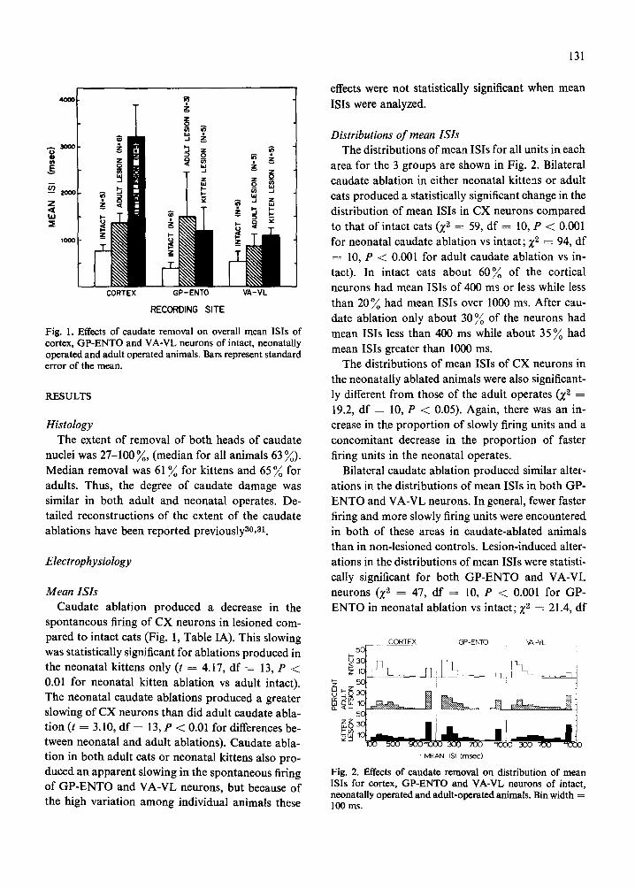
Fig. 1. Effects of caudate removal on overall mean ISis of cortex, GP-ENTO and VA-VL neurons of intact, neonatally operated and adult operated animals. Bars represent standard error of the mean.
RESULTS
Histology The extent of removal of both heads of caudate
nuclei was 27-100 %, (median for all animals 63 %).
Median removal was 61% for kittens and 65 % for adults. Thus, the degree of caudate damage was similar in both adult and neonatal operates. De- tailed reconstructions of the extent of the caudate ablations have been reported previouslya0, 31.
Electrophysiology
Mean ISis Caudate ablation produced a decrease in the
spontaneous firing of CX neurons in lesioned com- pared to intact cats (Fig. 1, Table IA). This slowing was statistically significant for ablations produced in the neonatal kittens only (t = 4.17, df = 13, P < 0.01 for neonatal kitten ablation vs adult intact). The neonatal caudate ablations produced a greater slowing of CX neurons than did adult caudate abla- tion (t = 3.10, df = 13, P < 0.01 for differences be- tween neonatal and adult ablations). Caudate abla- tion in both. adult cats or neonatal kittens also pro- duced an apparent slowing in the spontaneous firing of GP-ENTO and VA-VL neurons, but because of the high variation among individual animals these
131
effects were not statistically significant when mean
ISis were analyzed.
Distributions of mean ISis The distributions of mean ISis for all units in each
area for the 3 groups are shown in Fig. 2. Bilateral caudate ablation in either neonatal kittens or adult cats produced a statistically significant change in the
distribution of mean ISis in CX neurons compared to that of intact cats (Z z ---- 59, df : 10, P < 0.001 for neonatal caudate ablation vs intact; Zz : 94, df = 10, P < 0.00l for adult caudate ablation vs in- tact). In intact cats about 60% of the cortical neurons had mean ISis of 400 ms or less while less
than 20% had mean ISis over 1000 ms. After cau- date ablation only about 30 % of the neurons had mean ISis less than 400 ms while about 35 % had mean ISis greater than 1000 ms.
The distributions of mean ISis of CX neurons in the neonatally ablated animals were also significant- ly different from those of the adult operates (Z 2 ---- 19.2, df : 10, P < 0.05). Again, there was an in- crease in the proportion of slowly firing units and a concomitant decrease in the proportion of faster firing units in the neonatal operates.
Bilateral caudate ablation produced similar alter- ations in the distributions of mean ISis in both GP- ENTO and VA-VL neurons. In general, fewer faster firing and more slowly firing units were encountered in both of these areas in caudate-ablated animals than in non-lesioned controls. Lesion-induced alter-
ations in the distributions of mean ISis were statisti- cally significant for both GP-ENTO and VA-VL neurons (X 2 : 47, d f - - 10, P < 0.001 for GP- ENTO in neonatal ablation vs intact; Z 2 ---- 21.4, df
CORTEX C~PIENTO ~6, -VL
100 5 0 0 900~1C100 3 0 0 7 0 0 "1000 3 0 0 7 0 0 "1030
• M E A N ISI ( m s e c )
Fig. 2. Effects of caudate removal on distribution of mean ISis for cortex, GP-ENTO and VA-VL neurons of intact, neonatally operated and adult-operated animals. Bin width ----- 100 ms.
132
= 10, P < 0.05 for GP-ENTO in adult ablation vs intact; Z ~ = 13l, df = 10, P < 0.001 for VA-VL in
neonatal ablation vs intact; X ~ = 55, df ~- 10, P < 0.001 for VA-VL in adult ablation vs intact).
For both GP-ENTO and VA-VL neurons there
were also significant differences between distribu- tions of mean ISis of the neonatally and adult brain- damaged animals (X s = 47, df = 10, P < 0.001 for
GP-ENTO; Z 2 ---- 21.2, df = 10, P < 0.05 for VA- VL). In both cases distributions of the neonatally
operated animals had fewer faster firing and more slowly firing units than distributions obtained from adult operated animals.
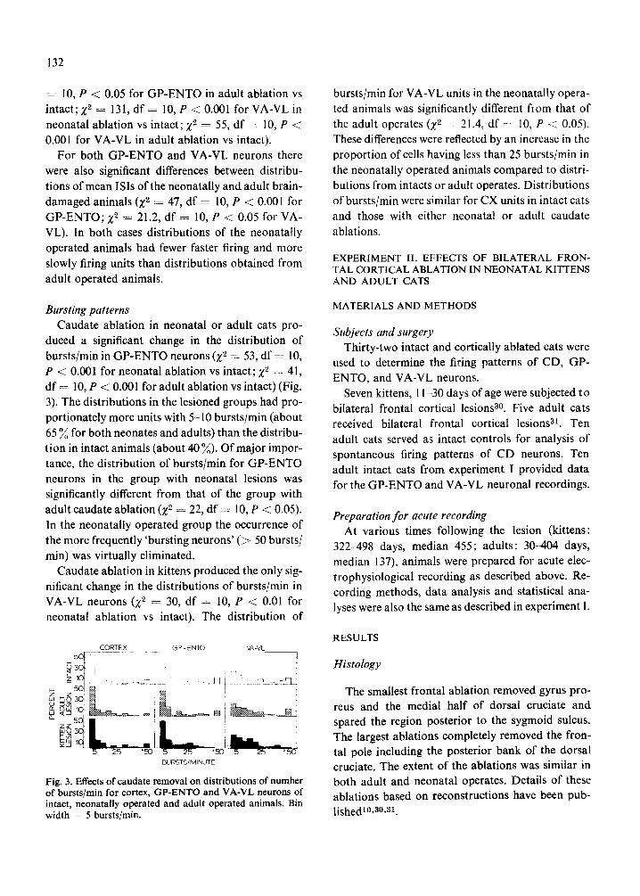
Bursting patterns Caudate ablation in neonatal or adult cats pro-
duced a significant change in the distribution of bursts/rain in GP-ENTO neurons (Z 2 = 53, df = 10, P < 0.001 for neonatal ablation vs intact; X 2 = 41, df -- 10, P <: 0.001 for adult ablation vs intact) (Fig. 3). The distributions in the lesioned groups had pro- portionately more units with 5-10 bursts/min (about 65 ~ for both neonates and adults) than the distribu- tion in intact animals (about 40~) . Of major impor- tance, the distribution of bursts/min for GP-ENTO neurons in the group with neonatal lesions was significantly different from that of the group with adult caudate ablation (Z 2 = 22, df =- 10, P < 0.05). In the neonatally operated group the occurrence of the more frequently 'bursting neurons' ( > 50 bursts/
min) was virtually eliminated. Caudate ablation in kittens produced the only sig-
nificant change in the distributions of bursts/min in VAIVL neurons (Z 2 = 30, df = 10, P < 0.01 for neonatal ablation vs intact). The distribution of
CORTEX G P - E N T O VA-VL
. . . . . i , . . . . LII ~u~ @ I j
a. z 5o~ • i ! |
L i : 5 25 "50 5 25 "50 ~
BURSTS/MINUTE
Fig. 3. Effects of caudate removal on distributions of n u m b e r of bursts/rain for cortex, GP-ENTO and VA-VL neurons of intact, neonatally operated and adult operated animals. Bin width - 5 bursts/rain.
bursts/min for VA-VL units in the neonatally opera- ted animals was significantly different f iom that of the adult operates (Z 2 - 21.4, df = 10, P < 0.05). These differences were reflected by an increase in the proportion of cells having less than 25 bursts/min in the neonatally operated animals compared to distri- butions from intacts or adult operates. Distributions of bursts/min were similar for CX units in intact cats and those with either neonatal or adult caudate
ablations.
EXPERIMENT II. EFFECTS OF BILATERAL FRON- TAL CORTICAL ABLATION IN NEONATAL KITTENS AND ADULT CATS
M A T E R I A L S A N D M E T H O D S
Subjects and surgery Thirty-two intact and cortically ablated cats were
used to determine the firing patterns of CD, GP- ENTO, and VA-VL neurons.
Seven kittens, 11-30 days of age were subjected to bilateral frontal cortical lesions 8°. Five adult cats received bilateral frontal cortical lesions 31. Ten
adult cats served as intact controls for analysis of spontaneous firing patterns of CD neurons. Ten adult intact cats from experiment I provided data for the GP-ENTO and VA-VL neuronal recordings.
Preparation for acute recording At various times following the lesion (kittens:
322-498 days, median 455; adults: 30--404 days, median 137), animals were prepared for acute elec- trophysiological recording as described above. Re- cording methods, data analysis and statistical ana- lyses were also the same as described in experiment I.
R E S U L T S
Histology
The smallest frontal ablation removed gyrus pro- reus and the medial half of dorsal cruciate and spared the region posterior to the sygmoid sulcus. The largest ablations completely removed the fron- tal pole including the posterior bank of the dorsal cruciate. The extent of the ablations was similar in both adult and neonatal operates. Details of these ablations based on reconstructions have been pub- lishedl0,a0,al.

- I - - • l a
z ~ z
CAUDATE GP-ENTO VA- VL
RECORDING SITE
Fig. 4. Effect of frontal cortex removal on overall mean ISis of CD, GP-ENTO and VA-VL neurons of intact, neonatally operated and adult operated animals. Bars represent standard error of the mean.
Electrophysiology
Mean ISis Mean ISis for each area are shown in Fig. 4 and
Table IB. In intact cats CD neurons fired significant- ly slower than neurons in GP-ENTO, VA-VL or CX zo. Frontal cortical lesions in either neonatal or adult cats produced decreases in the spontaneous firing rates of caudate neurons. This difference was statistically significant for comparisons between neonatally lesioned and intact adult animals only (t = 2.49, df = 17, P < 0.05). Although cortical lesions in adult cats tended to result in decreases in
CALIDATE GP-ENTO VA-'VL
133
5o
5 0
_ l d lvEAN I~ (msec)
Fig. 5. Effect of frontal cortex removal on distributions of mean ISis of CD, GP-ENTO and VA-VL neurons of i~act, neonatally operated and adult operated animals. Bin widih = 200 ms for caudate, 100 ms for GP-ENTO and VA-VL.
the overall mean ISI of GP-ENTO and VA-VL neu- rons compared to intact animals, these differences were not statistically significant. Frontal cortical le- sions in kittens produced almost no change in mean ISis for GP-ENTO or VA-VL neurons.
Distribution of mean ISis Distributions of mean ISis for all units recorded
in each area are shown in Fig. 5. Cortical ablation in either neonatal or adult cats produced significant al- terations in the distributions of mean ISis of CD neurons (Z 2 = 70, df = 10, P < 0.001 for neonatal ablation vs intact; Z 2 = 70, df = 10, P < 0.001 for adult ablation vs intact). In intact cats about 40 % of the caudate neurons had mean ISis of 400 ms o f less while about 20 % of the units fired very slowly (mean ISI > 2000 ms). In cortically-lesioned animals only 20-30 % of the units had mean ISis less than 400 ms while 35--40 % fired slowly.
TABLE I
Overall mean ISis o f neurons in intact cats and cats with bilateral caudate or cortical ablations as adults or neonates
Mean 4. S.E. Mean of all cats 5= standard error of the mean/condition. N U represents number of units recorded/site/condition. NC represents number of cats recorded/site/condition.
Recording site Intact Adu~operates Neonatal operates
( A ) Caudate ablation
Mean 4- S.E. (NU) (NC) Mean -t- S.E. (NU) (NC) Mean 4- S.E. (NU) (NC) CX 753 4- 136 (142) (5) 1387 5= 207 (183) (6) 3231 4- 700 (140) (5) GP-ENTO 406 :E 105 (170) (6) 1495 4- 963 (90 ) (3) 1201 4. 519 (150) (5) VA-VL 520 4.4.- 186 (193) (5) 873 5= 241 (149) (5) 1071 4- 332 (150) (5)
( B) Frontal cortical ablation
CD 1694 4- 239 (385) (10) 2463 5= 389 (158) (5) 2758 4. 301 (155) (5) GP-ENTO 406 4- 105 (170) (6) 1089 5= 275 (120) (4) 453 q- 261 (145) (5) VA-VL 520 -4- 186 (193) (5) 746 5= 138 (115) (4) 654 5= 115 (118) (4)

134
Differences between distributions of mean ISis for
neonatal and adult operates were also significantly different (X 2 ---- 25.1, df = 10, P < 0.01). Fewer faster-firing and more slowly-firing neurons were en- countered in the kitten operates.
Cortical ablations in adult cats produced a signifi-
cant change in distributions of mean ISis of GP- ENTO neurons (Z 2 ---- 83, df - - 10, P < 0.001). These changes again were reflected by a decrease in
the proportion of the faster-firing units (mean ISI < 200 ms) coupled with an increase in the proportion of slowly-firing units (mean ISI > 1000 ms). Infant lesioned cats exhibited no changes in the distribu- tion of mean ISis when compared to intact controls.
Distributions of mean ISis of GP-ENTO neurons for neonatal and adult operates were significantly different (g 2 = 59, df = 10, P < 0.001). More slowly firing and fewer rapidly firing neurons were encoun- tered in the adult-lesioned animals.
Cortical ablations in both neonatal and adult cats produced significant alterations in the distributions of mean ISis of VA-VL neurons 0¢ 2 = 24, df = 10,
P < 0.01 for neonatal ablation vs intact; Z 2 = 69, df = 10, P < 0.001 for adult ablations vs intact. Again, fewer faster-firing (mean ISI < 200 ms) and more slowly-firing units (mean ISI > 1000 ms) were en- countered in the brain-damaged animals.
Distributions of mean ISis in neonatally-ablated animals were significantly different from those of the adult-ablated animals 0¢ 2 = 24, df = 10, P < 0.01). Fewer rapidly firing and more slowly-firing units were encountered in the adult brain-damaged animals.
Bursting patterns Distributions of bursts/min are shown in Fig. 6.
Cortical ablation significantly altered the distribu- tions of bursts/min of CD neurons in both neonatal and adult operated animals 0¢ 2 = 42, df = 10, P < 0.001 for neonatal ablation vs intact; X 2 = 34, df : 10, P < 0.001 for adult ablation vs intact). In the adult operated animals caudate units displayed burst activity more frequently. There was an in- crease in the proportion of units having more than 10 bursts/min in the adult operates. In the neonatal- ly ablated animals caudate units tended to burst less frequently than in intact animals. A marked increase in units with less than 3 bursts/min occurred. Differ- ences between distributions of bursts/min for CD
CAUDATE GP - ENTO VA V[ ~_50[ ---
~ 3oi r-1 r ~ I0~ / L ] i S i ' , r [ ,
z 50 ~ ~ , ~, o 3o: ~ ~ : O oO L= Z Z ~ i
Lo 3 , 4 i
BLRSTS/MINUTE
Fig. 6. Effects of frontal cortex removal on distributions of numbers of bursts/rain for CD, GP-ENTO and VA-VL neu- rons of intact, neonatally operated and adult operated ani- mals. Bin width = 3 bursts/min for caudate, 5 bursts/min for GP-ENTO and VA-VL.
neurons for the neonatal and adult lesioned animals were also significant (Z 2 = 25.8, df = 10, P < 0.01).
In adult-lesioned oats, distributions of bursts/min for GP-ENTO neurons were significantly different from those of intact animals (X 2 = 41.3, df = 10, P < 0.001) or animals with neonatal lesions (Z 2 =
23.4, df = 10, P < 0.01). Proportionately more units had decreased bursting activity in the adult-
lesioned animals. Distributions of bursts/min for neonatally-lesioned and intact animals were similar.
Cortical ablation in both neonatal and adult animals produced significant alterations in the dis- tributions of bursts/min of VA-VL neurons (g 2 = 34, df = 10, P < 0.001 for neonatal ablation vs intact; g ~ = 34, df = 10, P < 0.001 for neonatal ablation vs intact; Z 2 = 102, df = 10, P < 0.001 for adult ablation vs intact). In the adult-lesioned cats there was a marked increase in the proportion of units that displayed infrequent bursting. Over 80 % of the bursting units had less than 5 bursts/min. In intact animals and cats lesioned as kittens only about 20 % of the units had less than 5 bursts/min.
Distributions of bursts/min of VA-VL units in neonatally and adult-lesioned animals were also significantly different f rom each other (Z 2 = 289, df = 10, P < 0.001). In this case the effects of the neo- natal lesions were less marked than those of the adult
lesions.
Comparison of effects of caudate ablation and cortical ablation
In addition to assessing the effects of adult or neonatal ablations of caudate or cortex, the effects

of each of these lesions on spontaneous firing of GP- ENTO and VA-VL neurons were compared. Cau- date ablation in either neonatal or adult cats pro- duced greater effects on spontaneous firing of GP- ENTO or VA-VL neurons than did cortical abla- tion.
GP-ENTO neurons
In both adult and neonatal cats caudate removal produced a greater effect on mean ISis and more alterations in distributions of mean ISis and distri- butions of bursts/min in GP-ENTO neurons than did cortical ablation (compare Figs. 1 and 4). Al- though the mean ISI was decreased in both neonatal and adult operates this apparent effect was not sta- tistically significant. However, distributions of mean ISis of both adult and neonatal operates were signi- ficantly different (of. Figs. 2 and 5) (X ~ = 58, df = 10, P < 0.001 for adult caudate ablation vs cortical ablation; X 2 = 33, df = 10, P < 0.001 for kitten caudate ablations vs frontal cortical ablation). In both of these cases the caudate ablation produced a significant decrease in the proportion of faster firing GP-ENTO units and concomitant increase in the proportion of the more slowly firing units.
Patterns of bursting activity of GP-ENTO neu- rons (distributions of bursts/min) were significantly altered by caudate lesions but only in the kitten operates (X 2 = 40, df = 10, P < 0.001 for kitten caudate ablation vs cortical ablation) (of. Figs. 3 and 6).
VA- VL neurons
Overall mean ISis of V,A-VL neurons were similar in animals sustaining eithar caudate or cortical abla- tion as adults (el. Figs. 1 and 4, see Table I). For neonatally-lesioned cats, however, caudate removal produced a somewhat greater decrease in spontane- ous firing of the VA-VL neurons than did the corti- cal ablation (el. Figs. 1 and 4, see Table I) although this effect was not statistically significant when mean ISis were considered. Distribution of mean ISis for VA-VL neurons were only significantly different (of. Figs. 2 and 5) for the kitten operates (X 2 : 44, df ---- 10, P < 0.001 for kitten caudate vs cortical abla- tion). These differences were reflected by a decrease
135
in the proportion of faster-firing VA-VL units and an increase in the proportion of slowly-firing units in the caudate ablated animals compared to those with cortical ablations.
Bursting activity of VA-VL neurons was more intensely affected by the adult cortical ablation than by the adult caudate ablation (of. Figs. 3 and 6) (X 2 ---- 88, df = 10, P < 0.001 for adult caudate vs corti- cal ablation). Removal of the frontal cortex in adult cats produced a marked decrease in the number of units having more than 5 bursts/min. In the neona- tally ablated animals the distributions of bursts/rain were significantly different as well (cf. Figs. 3 and 6) (Z z = 22.2, df ---- 10, P < 0.05 for kitten caudate vs kitten cortical ablation). In this case fewer units with over 50 bursts/min and more units with less than 10 bursts/min were encountered in the caudate ablated than in the cortically ablated animals.
DISCUSSION
Effects o f caudate ablation The major effect of caudate ablation on GP-
ENTO neurons was to produce a decrease in the overall firing rate of these neurons, increase the pro- portion of slowly firing cells, decrease the propor- tion of more rapidly firing cells and decrease 'burst- ing' activity. The net effect of caudate removal is not large. At least two possibilities exist to explain this result. First, the evidence indicates that caudate out- put is both excitatory and inhibitory on GP-ENTO neurons17, tS,a4. Thus, caudate removal would re- move both of these types of inputs and the result might be expected to be a small net change. Second, the caudate ablations in the present experiments left the putamen intact. Thus, putamenal inputs to GP- ENTO would still be functional and capable of regu- lating activity of GP-ENTO neurons. We also can- not exclude the possibility that changes in connecti- vity of the remaining inputs to these nuclei contri- bute to the observed effects.
Spontaneous firing of VA-VL neurons was de- creased by the caudate ablations in both neonatal and adult animals. In addition, 'bursting', a charac- teristic of VA-VL neuronal activity 7,20 was reduced. These effects could have been due to altered activity of the afferents to VA-VL from the caudate via the GP-ENTO since the firing of GP-ENTO neurons

136
was also decreased. It is also possible that the slow- ing of firing observed in the CX after caudate lesions contributed to the VA-VL effects since the pericru- ciate cortex projects to the VA-VL 2s.
Spontaneous firing of neurons in the precruciate area of the CX was also decreased by the caudate removal. CX neurons are affected indirectly from the caudate via pathways consisting of at least 3 additional neuronal projections: caudato-pallidal a, pallido-thalamic 24, and thalamo-cortical 2s. Since
decreases in spontaneous firing of GP-ENTO and VA-VL neurons occur the most likely explanation for the decrease in spontaneous firing in CX neurons is that it reflects the altered activity in its thalamic afferents induced by the caudate ablation.
The important result is that removal of the cau- date nucleus produces a net decrease in the spon- taneous firing of neurons in all brain areas sampled. These effects are long-lasting (for up to 2 years in some cases) and on the whole qualitatively similar whether the ablations are made in neonatal or adult animals.
A great deal of previous research has concentra- ted on the inhibitory effects of caudate outputs 13,25, 34. Stimulation of the caudate nucleus has been shown to inhibit the firing of neurons in many areas of the brain3, s. When the responses of GP-ENTO, VA-VL or CX neurons to caudate activation are examined intracellularly it is apparent that the longer periods of inhibition are often preceded by an initial excitation s,9,14,17,1 s. The results of the present
experiments indicate that removal of the caudate leads to 'tonic' effects indicative of removal of this excitation. The excitation can be relayed over a number of synapses as the spontaneous activity of neurons one, two, or three synapses away are affec- ted by caudate removal. At a simple level the lesion- induced decrease in excitation can be viewed as resulting from a lack of convergence of excitatory inputs. In intact animals the concomitant or sequen- tial arrival of inputs would allow for both spatial and temporal summation. In the brain-damaged animals the missing inputs would not be able to summate with remaining afferents, resulting in sub- threshold excitation and a decrease in action poten- tial generation.
Comparison of effects of neonatal and aduh ablations The main purpose of the present experiments was
to compare the effects of caudate removal in neona- tal and adult animals on the spontaneous activity of neurons in GP-ENTO, VA-VL and CX. For both GP-ENTO and VA-VL marked differences in the effects of caudate ablation in neonates and adults were not apparent. Since the effects of caudate removal in adult animals were similar to those ob- served in animals with neonatal ablations the role of the caudate nucleus in regulating spontaneous firing in other structures was apparently well-developed by the time the lesions were produced. These results are congruent with behavioral findings in animals with neonatal and adult ablations. Similar kinds of be- havioral effects (although developing with different time courses) occur regardless of whether the cau- date nucleus is removed in neonatal or adult animals (refs. 26, 30, 31). The present results are also in agreement with data which suggest that many as- pects of the physiology and morphology of the cau- date nucleus are well developed by the third week of life5,22,23.
The effects of neonatal or adult caudate ablation on the spontaneous firing of CX neurons, while qualitatively similar, were quantitatively quite dif- ferent. Caudate ablation in neonatal kittens pro- duced a more marked slowing of CX neurons than did caudate ablation in adult cats (overall ISis were > 3000 ms). It seems unlikely that the marked de- crease in animals with neonatal ablations is due sole- ly to removal of excitatory caudate output that would normally be relayed to reach the CX.
Effects of frontal cortical ablation We have previously published a report on the
effects of frontal cortical removal on spontaneous firing of CD, GP-ENTO, and VA-VL neurons in adult cats 1°. Therefore, only a comparison of the effects of neonatal and adult ablations will be con-
sidered here. Frontal cortical ablation produced a slowing in
the spontaneous firing of CD neurons which was similar in neonatal and adult cats. These decreases probably reflect the removal of an excitatory corti- co-caudate connection 2. Since the effects of both neonatal and adult ablations are similar, this con- nection must be functional in early postnatal periods
137
(ref. 22). In a series of parallel electrophysiological
and anatomical experiments we have demonstrated,
in fact, that cortical input is present and capable of
activating caudate neurons in newborn kittensS, 22.
Cortical s t imulat ion produces EPSPs and spike po-
tentials in such animals 23.
While cortical abla t ion in adul t cats produces de-
creases in spontaneous firing of G P - E N T O and VA-
VL neurons 10, after similar ablat ions in kittens these
decreases do not occur. In adult cats, the slowing of
G P - E N T O and VA-VL neurons might reflect trans-
mission of altered striatal activity to and through
these structures 10. In neonatal ly-lesioned animals
either compensatory alterations occur in G P - E N T O
and VA-VL dur ing matura t ion or the alterations in
striatal activity induced by the cortical lesions are
no t t ransmit ted to striatal outputs.
ACKNOWLEDGEMENT
Supported by USPHS G r a n t H D 05958.
REFERENCES
1 Buchwald, N. A., Hull, C. D. and Levine, M. S., Neuro- physiological and anatomical interrelationships of the basal ganglia. In N. A. Buchwald and M. A. Brazier (Eds.), Brain Mechanisms in Mental Retardation, Vol. 18, UCLA Forum in Medical Sciences, Academic Press, New York, 1975, pp. 187-203.
2 Buchwald, N. A., Price, D. D., Vernon, L. and Hull, C. D., Caudate intracellular response to thalamic and corti- cal inputs, Exp. Neurol., 38 (1973) 311-323.
3 Buchwald, N. A., Wyers, E. J., Okum, T. and Heuser, G., The 'caudate spindle'. I. Electrophysiological properties, Electroenceph. clin. Neurophysiol., 13 (1961) 509-518.
4 Carpenter, M. B., Anatomical organization of the corpus striatum and related nuclei. In M. D. Yahr (Ed.), The Basal Ganglia, Raven Press, New York, 1976, pp. 1-36.
5 Cospito, J. A., Levine, M. S. and Adinolfi, A. M., The organization of developing precruciate corticostriate projections in kittens, Exp. Neurol., 67 (1980) 447-452.
6 Donaghue, J. P. and Kitai, S. T., Collateral input to the neostriatum from descending axons of the rat somatic sensory-motor cortex, Neurosci. Abstr., 6 (1980) 187.
7 Dormont, J. F., Patterns of spontaneous unit activity in the ventrolateral thalamic nucleus of cats, Brain Res., 37 (1972) 223-239.
8 Frigyesi, T. L. and Machek, J., Basal ganglia diencepha- Ion synaptic relations in the cat. II. Intracellular recor- dings from dorsal thalamic neurons during low-frequency stimulation of the caudatothalamic projection systems and the nigrothalamic pathway, Brain Res., 27 (1971) 59-78.
9 Frigyesi, T. L. and Rabin, A., Basal ganglia--diencepha- Ion synaptic relations. III. An intracellular study of ansa lenticularis, lenticular fasciculus and pallidosubthalamic projection activities, Brain Res., 35 (1971) 67-78.
10 Garcia-Rill, E., Hull, C. D., Levine, M. S. and Buchwald, N. A., The spontaneous firing patterns of forebrain neu- rons. IV. Effects of bilateral and unilateral frontal cortical ablation, Brain Res., 165 (1979) 23-36.
11 Graybiel, A. M. and Ragsdale, C. W., Jr., Fiber connec- tions of the basal ganglia. In E. Cuenod, G. W. Kreutz- berg and F. E. Bloom (Eds.), Development and Chemical Specificity o f Neurons, Progress in Brain Research, VoL 51, Elsevier-North Holland, 1979, pp. 239-283.
12 Hayes, W. J., Statistics for Psychologists, Holt, Rinehart and Winston, New York, 1963.
13 Heuser, G., Buchwald, N. A. and Wyers, E. J., The 'cau- date spindle'. II. Facilitatory and inhibitory caudate- cortical pathways, Electroenceph. clin. NeurophysioL, 13 (1961) 519-524.
14 Hull, C. D., Buchwald, N. A. and Vieth, J. B., Cortical intracellular analyses of responses to inhibitory and dis- inhibitory stimuli, Brain Res., 6 (1967) 22-34.
15 Hull, C. D., Levine, M. S., Buchwald, N. A., Heller, A. and Browning, R. A., The spontaneous firing pattern of forebrain neurons. I. The effects of dopamine and non- dopamine depleting lesions on caudate unit firing patterns, Brain Res., 73 (1974) 241-262.
16 Kitai, S. T., Kocsis, J. D. and Wood, J., Origin and characteristics of the cortico-caudate afferents: an anato- mical and electrophysiological study, Brain Res., 118 (1976) 137-141.
17 Levine, M. S., Cherubini, E., Novack, G. D., Hull, C. D. and Buchwald, N. A., Development of responses of glo- bus pallidus and entopeduncular nucleus neurons to sti- mulation of caudate nucleus and precruciate cortex, Exp. NeuroL, 66 (1979) 479-492.
18 Levine, M. S., Hull, C. D. and Buchwald, N. A., Pallidal and entopedunclear intracellular responses to striatal, cortical thalamic and sensory inputs, Exp. NeuroL, 44 (1974) 448-460.
19 Levine, M. S., Hull, C. D., Buchwald, N. A., Garcia- Rill, E., Heller, A. and Erinoff, L., The spontaneous firing patterns of forebrain neurons. III. Prevention of induced asymm:~ries in caudate neuronal firing rates by unilateral thalamic lesions, Brain Res., 131 (1977) 215- 225.
20 Levine, M. S., Hull, C. D., Buchwald, N. A. and Villa- blanca, J. R., The spontaneous firing patterns of forebrain neurons. II. Effects of unilateral caudate nuclear ablation, Brain Res., 78 (1974) 411-424.
21 Levine, M. S., Hull, C. D., Buchwald, N. A. and Villa- blanca, J. R., Effects of caudate nuclei or frontal cortical ablations in kittens: motor activity and visual discrimin- ation performance in neonatal and juvenile kittens, Exp. NeuroL, 62 (1978) 555-569.
22 Lidsky, T. I., Buchwald, N. A., Hull, C. D. and Levine, M. S., A neurophysiological analysis of the development of cortico-caudate connections in the cat, Exp. Neurol., 50 (1976) 283-292.
23 Morris, R., Levine, M. S., Cherubini, E., Buchwald, N. A. and Hull, C. D., Intracellular analysis of the development

138
of responses of caudate neurons to stimulation of cortex, thalamus and substantia nigra in the kitten, Brain Res., 173 (1979) 471-487.
24 Nauta, W. J. H. and Mehler, W. R., Projections of the lentiform nucleus in the monkey, Brain Res., 1 (1966) 3- 42.
25 Obata, K. and Yoshida, M., Caudate evoked inhibition and actions of GABA and other substances on cat pallidal neurons, Brain Res., 64 (1973) 455-459.
26 Olmstead, C. E., Villablanca, J. R., Marcus, R. J. and Avery, D. L., Effects of caudate nuclei or frontal cortical ablation in cats. IV. Bar pressing and maze learning per- formance, Exp. Neurol., 53 (1976) 670-693.
27 Rinvik, E., The corticothalamic projection from the peri- cruciate and coronal gyri in the cat. An experimental study with silver-impregnation methods, Brain Res., 10 (1968) 79-119.
28 Strick, P. L., Cortical projections of the feline thalamic nucleus ventralis lateralis, Brain Res., 20 (1970) 130--134.
29 Villablanca, J. R., Olmstead, C. E. and deAndres, I.,
Effects of caudate nuclei or frontal cortical ablation in kittens. Responsiveness to auditory stimuli and com- parisons with adult operated littermates, Exp. NeuroL, 61 (1978) 635-649.
30 Villablanca, J. R., Olmstead, C. E., Levine, M. S. and Marcus, R. J., Effects of caudate nuclei or frontal cortical ablations in kittens. I. Neurology and gross behavior, Exp. Neurol., 61 (1978) 615-634.
31 Villablanca, J. R., Olmstead, C. E. and Marcus, R. J., Effects of caudate nuclei or frontal cortical ablations in cats. I. Neurology and gross behavior, Exp. Neurol., 52 (1976) 389-420.
32 Webster, K. E., The cortico-striatal projection in the cat, J. Anat., 99 (1965) 329-337.
33 Winer, B. J., Statistical Principles in Experimental Design, McGraw-Hill, New York, 1962.
34 Yoshida, M., Rabin, A. and Anderson, M., Monosynap- tic inhibition of pallidal neurons by axon collaterals of caudato-nigral fibers, Exp. Brain Res., 15 (1972) 333-347.