
Effects of castration on the seminal vesicles as influenced by age, considered in relation to the...
45
EFFECTS OF CASTRATION ON THE SEMINAL VESICLES AS INFLUENCED BY AGE, CON- SIDERED IN RELATION TO THE DEGREE OF DEVELOPMENT OF THE ADRENAL X ZONE EVELYN HOWARD Department of Physiology, The dohiis Hopkins Uiiiversity School of Yedicine, Baltiniore, Maryland SIX PLATES (TWENTY-NINE FIGURES) INTRODUCTION The recent observations of andromimetic activity in adrenal ext,racts (Reichstein, '36, and others), as well as the associa- tion of certain adrenal tumors with the accentuation of mascu- line characteristics in females, raise the question as to what extent the adrenal exerts such activity in the normal organism. Any andromimetic activity of the normal adult adrenal is presumably quite minor in comparison with the androgenic activity of the testis. It seems possible that the andromimetic activity of adrenal tissue is capable of considerable variation. Accordingly it becomes of interest to see whether physio- logical differences in adrenal andromimetic activity can be detected, and if so, whether such activity can be related to a particular stage of ontogeny. The histological changes in the mouse adrenal associated with castration suggest that the X zone is especially con- cerned in the relationships between the adrenals and the re- productive system (Howard, '27, '29, '30 ; Deanesly, '28). The facts that in mice the X zone in males hypertrophies after castration, and that this can be prevented by testosterone injections (Deanesly and Parkes, '37) suggest that the X zone may tend to compensate for an inadequate supply of testicular 105 THE AYERICAN JOURN.41, OF .4NATO31Y, VOL. 65, NO. 1
-
Upload
evelyn-howard -
Category
Documents
-
view
213 -
download
0
Transcript of Effects of castration on the seminal vesicles as influenced by age, considered in relation to the...
EFFECTS O F CASTRATION ON THE SEMINAL VESICLES AS INFLUENCED BY AGE, CON-
SIDERED I N RELATION TO THE DEGREE O F DEVELOPMENT O F THE ADRENAL
X ZONE
EVELYN HOWARD Department of Physiology, The dohiis Hopkins Uiiiversity School of Y e d i c i n e ,
Baltiniore, Maryland
SIX PLATES (TWENTY-NINE FIGURES)
INTRODUCTION
The recent observations of andromimetic activity in adrenal ext,racts (Reichstein, '36, and others), as well as the associa- tion of certain adrenal tumors with the accentuation of mascu- line characteristics in females, raise the question as to what extent the adrenal exerts such activity in the normal organism. Any andromimetic activity of the normal adult adrenal is presumably quite minor in comparison with the androgenic activity of the testis. I t seems possible that the andromimetic activity of adrenal tissue is capable of considerable variation. Accordingly it becomes of interest to see whether physio- logical differences in adrenal andromimetic activity can be detected, and if so, whether such activity can be related to a particular stage of ontogeny.
The histological changes in the mouse adrenal associated with castration suggest that the X zone is especially con- cerned in the relationships between the adrenals and the re- productive system (Howard, '27, '29, '30 ; Deanesly, '28). The facts that in mice the X zone in males hypertrophies after castration, and that this can be prevented by testosterone injections (Deanesly and Parkes, '37) suggest that the X zone may tend to compensate for an inadequate supply of testicular
105
THE A Y E R I C A N JOURN.41, O F .4NATO31Y, VOL. 65, NO. 1
106 EVELYN HOWARD
hormone, and that any andromimetic activity of the adrenal may be greater in the X zone than in the permanent cortex. Tissue apparently in some respects homologous to the mouse X zone occurs in the human (Howard, '27), and to a lesser degree in a variety of mammals (rat, Howard, '38; rabbit, Roaf, '35; cat, Davies, '37, and probably in other species), and hence conclusions drawn from a study of the mouse X zone, with appropriate modifications, may presumably be transferred to other species.
If the X zone does secrete aiidromimetic material, one should be able to detect a difference in the level of androgenic hormones according to whether or not the X zone is present. Since one of the more sensitive indicators of testicular hor- mone is the epithelium of the seminal vesicles of castrated mammals it seemed possible that variations in the condition of the seminal vesicle epithelium in castrates might be associ- ated with variations in the degree of X zone development. Therefore a study has been made of the reactions of mice seminal vesicles to castration a t various age periods, and, as expected, it has been found that there exist marked varia- tions in the condition of the vesicle epithelium, dependent on the age of the animal a t the time of operation, and related to the condition of the adrenal.
The reactions of the vesicle epithelium indicate that there is in mice an extra-testicular source of andromimetic hor- mone, which produces a small scale imitation of the endo- crine function of the testes. The histological changes in the adrenal cortex strongly suggest that the adrenal X zone is the chief source of the observed andromimetic activity. However the most probable explanation of the observations is that there is not an absolute difference in this respect be- tween the potentialities of the permanent cortex and the X zone, but that the difference between them is one of degree.l
'The studies reported in the preseut paper were begun in 1935. Preliminary reports were made in the Proe. of the Am. Physiol. Soc. (Howard, '37 a and '38 b) . This paper was submitted for publication in July, 1938.
SEMINAL VESICLES AND ADRENAL X ZONE 107
MATERIAL AND METHODS
Alice of various strains were used, but most of the work was done on a strain of white mice derived from a cross be- tween Bagg albino and local stock, which carried a well de- veloped X zone, and will be hereafter referred to as H strain. The white rats used were of unknown lineage. All animals were fed exclusively Purina Chow and water.
Animals were castrated in the usual manner, using ether anesthesia and an abdominal approach. Care was taken to remove all parts of the epididymis, a necessary precaution in view of the findings of Gallagher ('28) that androgenic hor- mone may be extracted from the epididymis.
Tissue was fixed in Bouins, sectioned serially at 6 to 10 p and every sixth section mounted and stained in Ehrlich's haematoxylin. The present paper includes observations on the serially sectioned vesicles and adrenals of 180 mice.
T H E EFFECTS O F CASTRATION I N RATS
By way of introduction to the study of the effects of castra- tion in mice, it is well to refer briefly to those observed in rats, where the situation appears to be less complex. It is known that in the adult rat there is after castration a prompt and relatively uniform regression of the seminal vesicles arid prostate. My observations on rats are in complete agree- ment with the descriptions of Moore, Price and Gallagher ( '30) ; Moore, Hughes and Gallagher ( '30) ; Hansen ( '33) ; and Price ( '36).
Thus, 20 days after castration of postpuberal rats one finds that in the main body of the vesicle, including the most prosi- ma1 portions, with exceptions of less than l%, the epithelium becomes uniformly reduced to low cuboidal cells with very little cytoplasm. This condition is shown in figures 13 and 14. I n addition to the changes in the epithelium, there is a marked relative increase in interacinous connective tissue, and a decrease in the size of the acini. Certain accessory lateral lobules and a certain proportion of the acini closest to the urogenital canal, uniformly show cells with more cyto-
108 EVELYN HOWARD
plasiii than those of the main part of the vesicle. These cells are fa r from normal, but the quantitative evaluation of their state is difficult. Therefore this study will be restricted to the main portion of the vesicles.
I n rats castrated well before puberty it may be observed that the epithelial cells do not become quite so low and shrunken as do those of the postpuberal castrate. This slight difference is of interest in the light of the much more marked differences between prepuberally and postpuberally castrated mice which will be described below.
In rats castrated at birth, it has been shown by Wiesner ('34) and by Price ( ' 3 6 ) , and confirmed in this laboratory (Howard, '38) that there is a marked subsequent differentia- tion of the prostate. This is soon followed by a regression to the condition typical of the adult castrate. The rat seminal vesicle does not share in this reaction in any comparable degree. However these observations are of interest as indi- cating that there does occur in the rat a very brief although well-marked phase of extra-testicular andromimetic hormone production, which is associated with adrenal changes (Howard, '38) , and which appears to be analogous to the much more extensive phase of extra-testicular andromimetic activity occurring in mice.2
T H E EFFECTS O F CASTRATION ON T H E MOUSE ADRENAL, W I T H SOME REhIARKS REGARDING T H E HISTOGENESIS O F
T H E X ZONE
Before proceeding with the description of the reactions of the mouse seminal vesicles to castration, it is desirable to summarize the changes seen in the adrenal, since the reactions in the vesicles can best be interpreted if i t is assumed that they are dependent on the reactions in the adrenal. The adrenal of the normal adult male mouse contains no X zone (fig. 28). In the intact male the X zone is present at 3 weeks of age as a relatively narrow zone which normally disappears
* My suggestion (Howard, '37 a, '38 a) , on histological grounds, tha t the adrenal is responsible for the rat prostate reaction, has been substantiated by other evi- dence, which appeared while this paper was in press. See Hurrill and Greene, '39.
SEMINAL VESICLES AND ADRENAL X ZONE 109
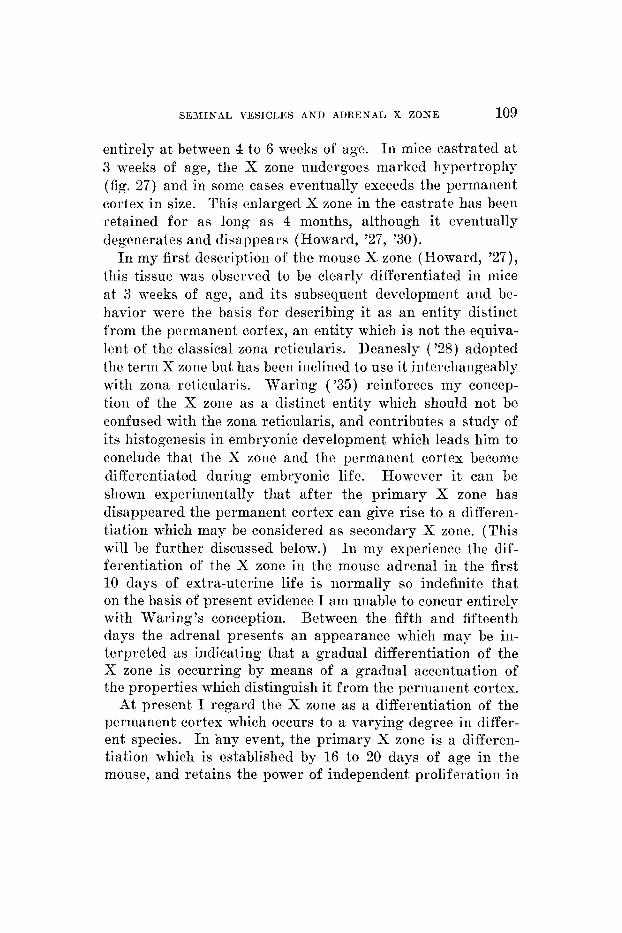
entirely at between 4 to 6 weeks of age. I n mice castrated a t 3 weeks of age, the X zone undergoes marked hypertrophy (fig. 27) and in some cases eventually exceeds the permanent cortex in size. This enlarged X zone in the castrate has been retained for as long as 4 months, although it eventually degenerates and disappears (Howard, '27, '30).
I n my first description of the mouse X zone (Howard, '27), this tissue was observed to be clearly differentiated in mice at 3 weeks of age, and its subsequent development and be- havior were the basis for describing it a s a n entity distinct from the permanent cortex, a n entity which is not the equiva- lent of the classical zona reticularis. Deanesly ('28) adopted the term X zone but has been inclined to use it interchangeably with zona reticularis. Mraring ( ' 3 5 ) reinforces my concep- tion of the X zone as a distinct entity which should not be confused with the zona reticularis, and contributes a study of its histogenesis in embryonic development which leads him to conclude that the X zone and the permanent cortex become differentiated during embryonic life. However it can be shown experimentally that after the primary X zone has disappeared the permanent cortex can give rise to a differen- tiation which may be considered as secondary X zone. (This will be further discussed below.) I n my experience the dif- ferentiation of the X zone in the mouse adrenal in the first 10 days of extra-uterine life is normally so indefinite that on the basis of present evidence I am unable to concur entirely with Waring's conception. Between the fifth and fifteenth days the adrenal presents a n appearance which may be in- terpreted as indicating that a gradual differentiation of the X zone is occurring by means of a gradual accentuation of the properties which distinguish it from the permanent cortex.
At present I regard the X zone as a differentiation of the permaneni cortex which occurs to a varying degree in differ- ent species. In any event, the primary X zone is a differen- tiation which is established by 16 to 20 days of age in the mouse, and retains the power of independent proliferation in
110 EVELYN HOWARD
situ (Whitehead, ’33 b) longer or to a greater degree than does the permanent cortex.
If mice a re castrated a t 5 days of age, a tremendous im- petus is given to the development of the primary X zone. At 16 days of age such mice have developed an X zone which is one-third or more of the total cortical width (see fig. 26) ; this being much larger than the X zone normally is at this age (fig. 2 5 ) and in fact much larger than the X zone ever becomes in the normal male mouse. A further study of intermediate stages in infant castrates might provide more clear-cut evi- dence as to the degree of differentiation between X zone and permanent cortex which exists at this period, and an answer to the question as to whether the enlarged primary X zone of the infant castrate develops entirely by proliferation of existing X cells, or in par t by concomitant differentiation from the permanent cortex.
There has been some question as to the reaction of the gland in males cas- trated after the X zone has disappeared. Previously (’30) I reported observations on a group of sixteen males castrated after puberty, in which “the inner part of the cortex showed irregular changes which tended to make the characteristics of the typical zona reticularis more prominent,” but these changes appeared minor in comparison to the large X zone which appears following prepuberal castration. Deanesly (’28) stated that after castration “an area develops which eventually resembles the X zone in the young female in all respects . . . . but in animals castrated after sexual maturity growth in the adrenal cortex appears to cease before the con- dition characteristic of the young female is reached.” Callow and Deanesly (’35) state that the “zona reticularis . . . . reappears after castration in the mouse.” Whitehead (’33 a) discussed the question whether the X zone reappears after it has once degenerated, and concludes that it does not, but he did not consider specifically the case of the post-puberal castrate. In the course of the present work I have collected a considerable amount of material which clearly demonstrates
The adreizals a f t e r postpuberal castration.
SEMINAL VESICLES AND ADRENAL X ZONE 111
that tissue resembling the X zone tends to be gradually re- differentiated following castration in the adult. This differ- entiation, however, does not resemble the primary X zone in all respects; accordingly it is referred to as secondary X zone (fig. 29). Descriptions of the histological details of the findings after various intervals following castration are given herewith.
Thirty-two mice were castrated after the X zone had pre- sumably disappeared. These ranged in age from 42 to 400 days. The adrenals were examined after intervals of 20, 40, 60, 80 and 100 days. Twenty days after castration the cells of the inner part of the cortex begin to show a reduction in the amount of their cytoplasm, an increase in affinity f o r haematoxylin and an absence of the large clear vacuoles characteristic of the zona fasciculata. At this stage the con- tinuity of the cell cords between the fasciculata and this dif- ferentiated area indicate that we are observing a transforma- tion of the inner part of the radial cell cords of the cortex, rather than a proliferation of any remnants of a specifically different tissue. This arrangement of cell cords is unlike that of the primary X zone. In the early stages the dif- ferentiation appears more like an approach to the type of reticularis which is normal for the rat, than the actual de- velopment of an X zone.
Forty days after castration, some animals showed areas which could be classified as X zone tissue, on the basis of the general appearance of the cells. The differentiated areas when present did not always form a continuous perimedulIar zone. In one animal there did not appear to be any definitive X zone after this interval. After longer intervals (80 and 100 days after castration) the differentiated area in most cases showed evidence of marked degeneration and compression of the X zone tissue. In some cases there remained after 100 days only traces of degenerated X tissue.
I n general the differentiation of secondary X zone tended to be greatest and to last longest in animals castrated a t 13 to 16 gm. body weight ; in older animals much smaller amounts
112 EVELYN HOWARD
of tissue were involved. The size of the regenerated X zone was in no case as large as that usually seen in cases castrated before the primary X zone has disappeared, and in most mice castrated when over 20 gm. body weight, the zone was less than one-quarter of the volume of the permanent cortex, in some cases very much less.
The secondary X zone was in no case observed to inter- digitate with the medulla as does the primary X zone, but was separated from the medulla by connective tissue, as is characteristic of the boundary between medulla and permanent cortex. In two mice castrated a t 306 and 400 days of age, and examined after 80 days, the syncytial arrange- ment characteristic of the primary X zone was present in some places. (This may have been made possible because of an old age breakdown of the normal relationship between the connective tissue and the glandular tissue. Such a break- down was indicated in other areas of the gland where fascicu- lata cells were seen in the arrangement usually characteristic of the glomerulosa. Areas of this kind are often seen in glands from old individuals.) I n other places the origin of the X cells from fasciculata cords was clearly indicated, the regular radial cords of X cells being continuous with the fascicular radial cords for variable distances, with no regular demarcation line between the X area and the fasciculata. In general, in all other mice the continuity of the cell cords be- tween the permanent cortex and the secondary X zone was clearly indicated.
Although the primary and secondary X zones have a some- what different structural organization, the individual cells are similar. Furthermore, the fact that they exhibit a similar response to castration suggests that they are functionally similar. Further evidence of this is that the development of both can be inhibited by the injection of androgens (Deanesly and Parkes, '37).
SEMINAL VESICLES AND ADRENAL X ZONE 113
T H E EFFECTS O F CASTRATION ON THE SEMINAL VESICLES O F MICE
We will now proceed with the description of the reactions of the seminal vesicles of mice to castration, considered in the light of the adrenal changes. The extent of the differences between the reactions of the accessory sex glands to castra- tion in rats and mice appears to have been largely overlooked hitherto. Most of the studies of the male accessories of castrated rodents have actually been made on rats, and it has been frequently assumed that there are no essential differ- ences between mice and rats in this respect. However, Martins and Rocha E. Silve ('29) referred to a variable de- gree of retardation in the atrophy of the seminal vesicles of castrated mice, and stated that the cause of the retardation should be elucidated. Voss ( '30) described variations in the reactions of mice seminal vesicles to castration. He suggested that a slight degree of traction due to adhesions might stimu- late epithelial development, although he adduced evidence that traction causes degeneration of seminal vesicals. Voss and Loewi ('31) also referred to the great variability of the state of the epithelium of the seminal vesicles of castrated mice.
Our consideration of the reactions of the mouse vesicle to castration will begin with a brief description of certain char- acteristics of the normal condition of the seminal vesicles at the three age periods considered.
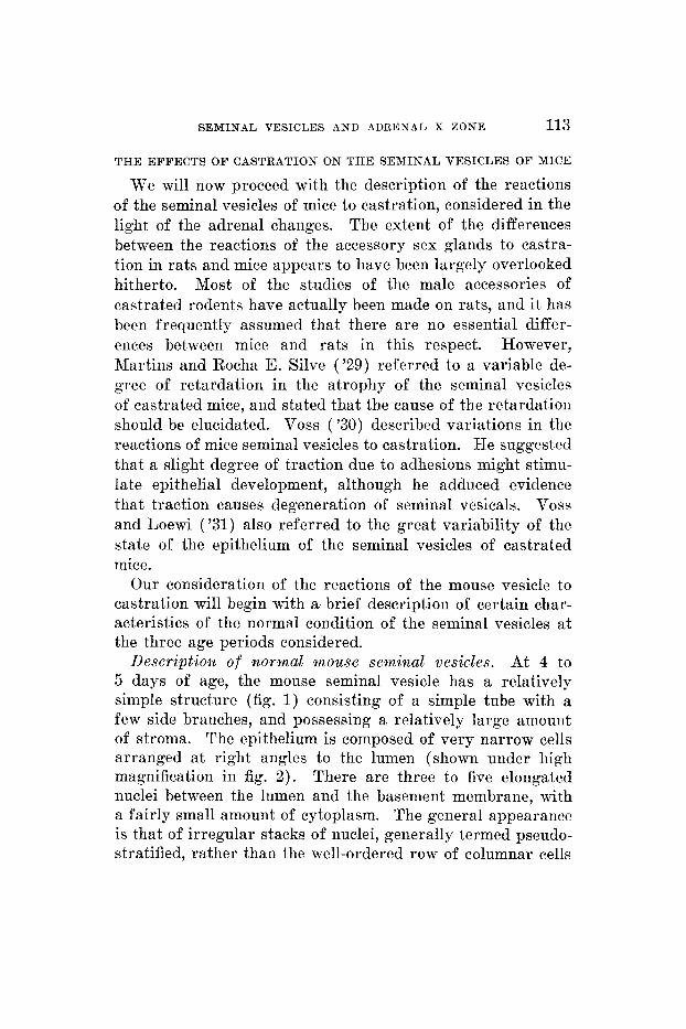
Description of rzormal mouse seminal vesicles. At 4 to 5 days of age, the mouse seminal vesicle has a relatively simple structure (fig. 1) consisting of a simple tube with a few side branches, and possessing a relatively large amount of stroma. The epithelium is composed of very narrow cells arranged at right angles to the lumen (shown under high magnification in fig. 2) . There are three to five elongated nuclei between the lumen and the basement membrane, with a fairly small amount of cytoplasm. The general appearance is that of irregular stacks of nuclei, generally termed pseudo- stratified, rather than the well-ordered row of columnar cells
114 EVELYN HOWARD
characteristic of the mature condition. The general appear- ance at this stage is quite similar to that of the vas deferens.
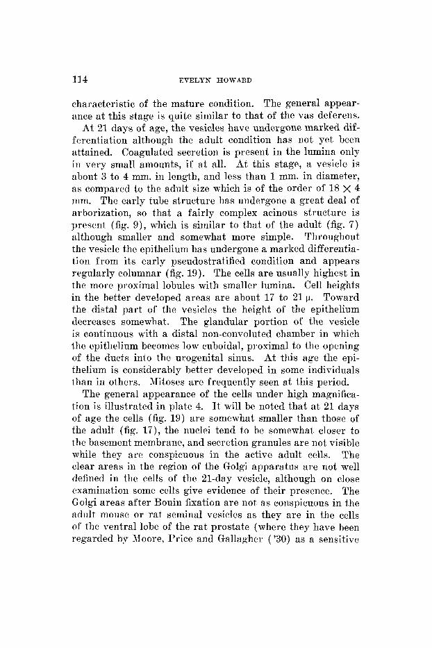
At 21 days of age, the vesicles have undergone marked dif- ferentiation although the adult condition has not yet been attained. Coagulated secretion is present in the lumina only in very small amounts, if at all. At this stage, a vesicle is about 3 to 4 mm. in length, and less than 1 mm. in diameter, as compared to the adult size which is of the order of 18 X 4 mm. The early tube structure has undergone a great deal of arborization, so that a fairly complex acinous structure is present (fig. 9) , which is similar to that of the adult (fig. 7) although smaller and somewhat more simple. Throughout the vesicle the epithelium has undergone a marked differentia- tion from its early pseudostratified condition and appears regularly columnar (fig. 19). The cells are usually highest in the more proximal lobules with smaller lumina. Cell heights in the better developed areas are about 17 to 21 p. Toward the distal part of the vesicles the height of the epithelium decreases somewhat. The glandular portion of the vesicle is continuous with a distal non-convoluted chamber in which the epithelium becomes low cuboidal, proximal to the opening of the ducts into the urogenital sinus. At this age the epi- thelium is considerably better developed in some individuals than in others. Mitoses are frequently seen at this period.
The general appearance of the cells under high magnifica- tion is illustrated in plate 4. It will be noted that at 21 days of age the cells (fig. 19) are somewhat smaller than those of the adult (fig. 17) , the nuclei tend to be somewhat closer to the basement membrane, and secretion granules are not visible while they are conspicuous in the active adult cells. The clear areas in the reg-ion of the Golgi apparatus are not well defined in the cells of the 21-day vesicle, although on close examination some cells give evidence of their presence. The Golgi areas after Bouin fixation are not as conspicuous in the adult mouse or rat seminal vesicles as they are in the cells of the ventral lobe of the rat prostate (where they have been regarded by Rloore, Price and Gallagher ( '30) as a sensitive
S E M I N A L VESICLES AND ADREN.4L X Z O N E 115
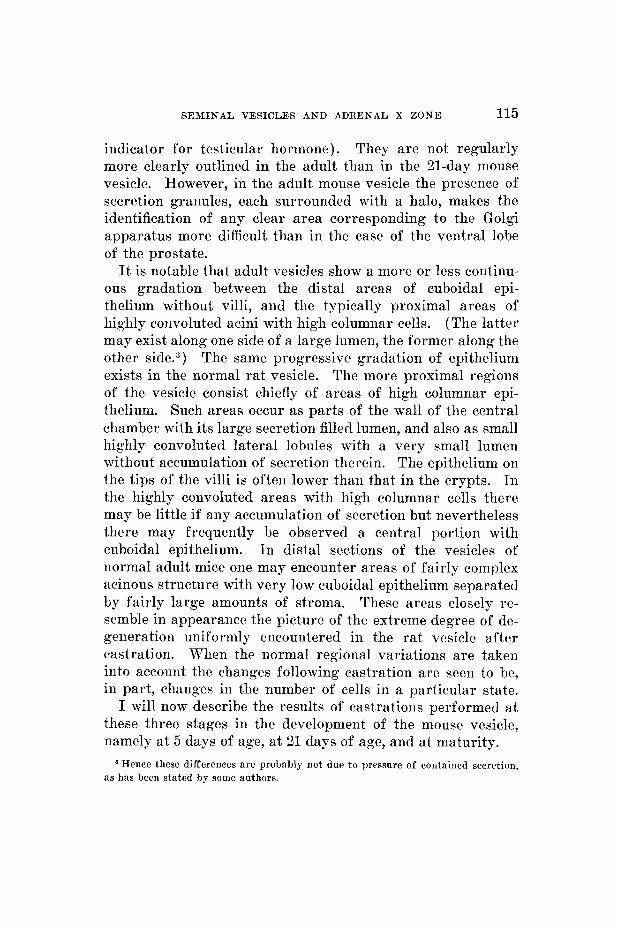
indicator for testicular hormone). They are not regularly more clearly outlined in the adult than in the 21-day mouse vesicle. However, in the adult mouse vesicle the presence of secretion granules, each surrounded with a halo, makes the identification of any clear area corresponding to the Golgi apparatus more difficult than in the case of the ventral lobe of the prostate.
It is notable that adult vesicles show a more or less continu- ous gradation between the distal areas of cuboidal epi- thelium without villi, and the typically proximal areas of highly convoluted acini with high columnar cells. (The latter may exist along one side of a large lumen, the former along the other side.3) The same progressive gradation of epithelium exists in the normal rat vesicle. The more proximal regions of the vesicle consist chiefly of areas of high columnar epi- thelium. Such areas occur as parts of the wall of the central chamber with its large secretion filled lumen, and also as small highly convoluted lateral lobules with a very small lumen without accumulation of secretion therein. The epithelium on the tips of the villi is often lower than that in the crypts. In the highly convoluted areas with high columnar cells there may be little if any accumulation of secretion but nevertheless there may frequently be observed a central portion with cuboidal epithelium. In distal sections of the vesicles of normal adult mice one may encounter areas of fairly complex acinous structure with very low cuboidal epithelium separated by fairly large amounts of stroma. These areas closely re- semble in appearance the picture of the extreme degree of de- generation uniformly encountered in the rat vesicle after castration. When the normal regional variations are taken into account the changes following castration are seen to be, in part, changes in the number of cells in a particular state.
I will now describe the results of castrations performed a t these three stages in the development of the mouse vesicle, namely a t 5 days of age, a t 21 days of age, and at maturity.
Hpnce these differences are probably not due to pressure of containcd secretion, as has been stated by some authors.
116 EVELYN HOWARD
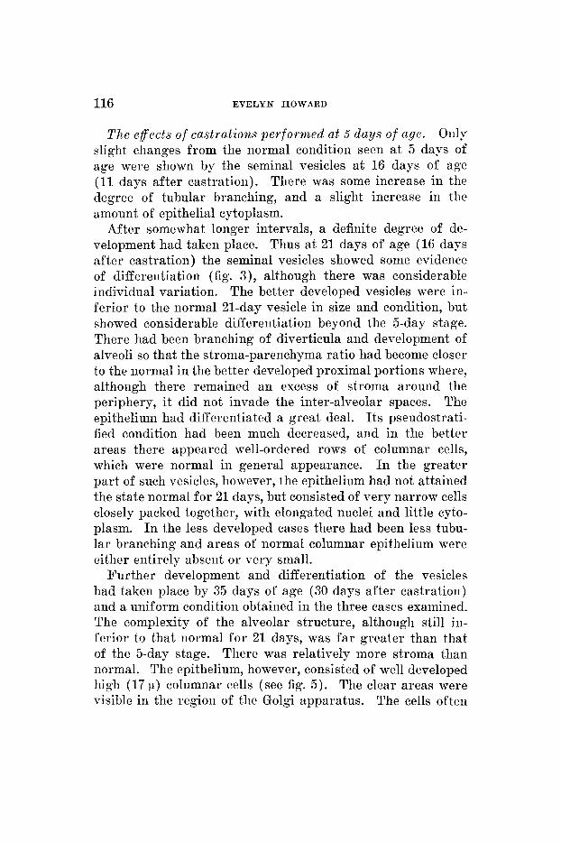
T h e effects of castrat ions per formed a t 5 days of age. Only slight changes from the normal condition seen at 5 days of age were shown by the seminal vesicles at 16 days of age (11 days after castration). There was some increase in the degree of tubular branching, and a slight increase in the amount of epithelial cytoplasm.
After somewhat longer intervals, a definite degree of de- velopment had taken place. Thus at 21 days of age (16 d a p after castration) the seminal vesicles showed some evidence of differentiation (fig. 3), although there was considerable individual variation. The better developed vesicles were in- ferior to the normal 21-day vesicle in size and condition, but showed considerable differentiation beyond the 5-day stage. There had been branching of diverticula and development of alveoli so that the stroma-parenchyma ratio had become closer to the normal in the better developed proximal portions where, although there remained an excess of stroma around the periphery, it did not invade the inter-alveolar spaces. The epithelium had differentiated a great deal. I ts pseudostrati- fied condition had been much decreased, and in the better areas there appeared well-ordered rows of columnar cells, which were normal in general appearance. I n the greater part of such vesicles, however, the epithelium had not attained the state normal f o r 21 days, but consisted of very narrow cells closely packed together, with elongated nuclei and little cyto- plasm. I n the less developed cases there had been less tubu- lar branching and areas of normal columnar epithelium were either entirely absent or very small.
Further development and differentiation of the vesicles had taken place by 35 days of age (30 days after castration) and a uniform condition obtained in the three cases examined. The complexity of the alveolar structure, although still in- ferior to that normal f o r 21 days, was far greater than that of the 5-day stage. There was relatively more stroma than normal. The epithelium, however, consisted of well developed high (17 p ) columnar cells (see fig. 5). The clear areas were visible in the region of the Golgi apparatus. The cells often
SEMINAL VESICLES AND ADRENAL X ZONE 117
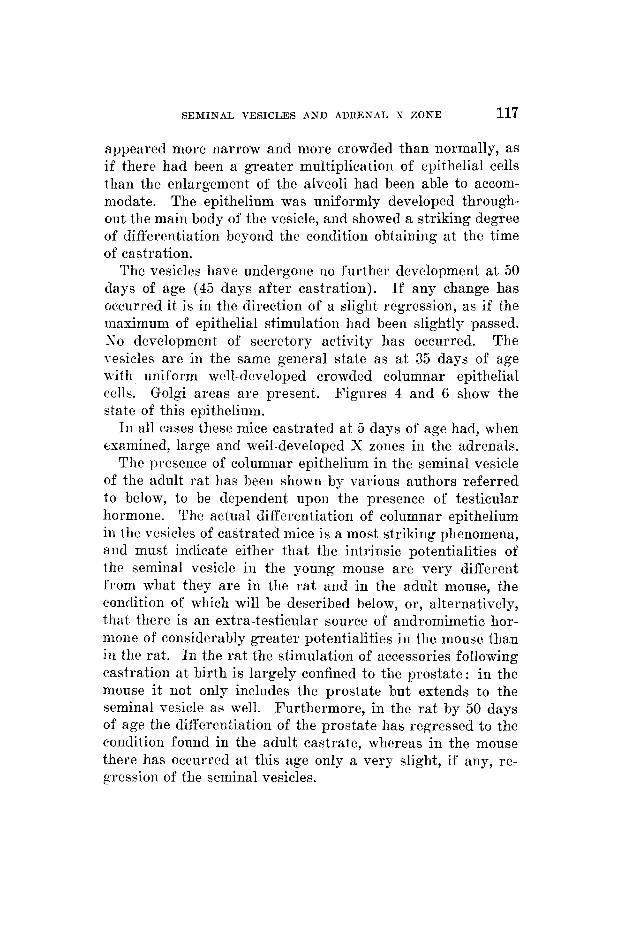
appeared more narrow and more crowded than normally, as if there had been a greater multiplication of epithelial cells than the enlargement of the alveoli had been able to accom- modate. The epithelium was uniformly developed through- out the main body of the vesicle, and showed a striking degree of differentiation beyond the condition obtaining a t the time of castration.
The vesicles have undergone no further development a t 50 days of age (45 days after castration). If any change has occurred it is in the direction of a slight regression, as if the maximum of epithelial stimulation had been slightly passed. S o development of secretory activity has occurred. The vesicles are in the same general state as at 35 days of age with uniform well-developed crowded columnar epithelial cells. Golgi areas are present. Figures 4 and 6 show the state of this epithelium.
In all cases these mice castrated at 5 days of age had, when examined, large and well-developed X zones in the adrenals.
The presence of columnar epithelium in the seminal vesicle of the adult rat has been shown by various authors referred to below, to be dependent upon the presence of testicular hormone. The actual differentiation of columnar epithelium in the vesicles of castrated mice is a most striking phenomena, and must indicate either that the intrinsic potentialities of the seminal vesicle in the young mouse are very different from what they are in the rat and in the adult mouse, the condition of which will be described below, or, alternatively, that tliere is an extra-testicular source of andromimetic hor- mone of considerably greater potentialities in the mouse than iii the rat. I n the rat the stimulation of accessories following castration at birth is largely confined to the prostate: in the mouse it not only includes the prostate but extends to the seminal vesicle as well. Furthermore, in the rat by 50 days of age the differentiation of the prostate has regressed to the condition found in the adult castrate, whereas in the mouse there has occurred at this age only a very slight, if any, re- gression of the seminal vesicles.
118 EVELYN HOWARD
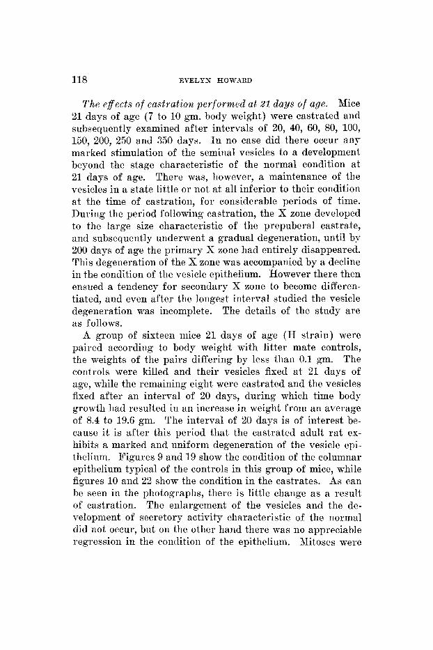
T h e effects of castrat ion per formed a t 21 days of age. Mice 21 days of age (7 to 10 gm. body weight) were castrated and subsequently examined after intervals of 20, 40, 60, 80, 100, 150, 200, 250 and 350 days. I n no case did there occur any marked stimulation of the seminal vesicles to a development beyond the stage characteristic of the normal condition a t 21 days of age. There was, however, a maintenance of the vesicles in a state little or not at all inferior to their condition a t the time of castration, for considerable periods of time. During the period following castration, the X zone developed to the large size characteristic of the prepuberal castrate, and subsequently underwent a gradual degeneration, until by 200 days of age the primary X zone had entirely disappeared. This degeneration of the X zone was accompanied by a decline in the condition of the vesicle epithelium. However there then ensued a tendency for secondary X zone to become differen- tiated, and even after the longest interval studied the vesicle degeneration was incomplete. The details of the study are as follows.
A group of sixteen mice 21 days of age (H strain) were paired according to body weight with litter mate controls, the weights of the pairs differing by less than 0.1 am. The controls were killed and their vesicles fixed a t 2 1 days of age, while the remaining eight were castrated and the vesicles fixed after an interval of 20 days, during which time body growth had resulted in an increase in weight from an average of 8.4 to 19.6 gm. The interval of 20 days is of interest be- cause it is after this period that the castrated adult ra t ex- hibits a marked and uniform degeneration of the vesicle epi- thelium. Figures 9 and 19 show the condition of the columnar epithelium typical of the controls in this group of mice, while figures 10 and 22 show the condition in the castrates. As can be seen in the photographs, there is little change as a result of castration. The enlargement of the vesicles and the de- velopment of secretory activity characteristic of the normal did not occur, but on the other hand there was no appreciable regression in the condition of the epithelium. Mitoses were
SEMINAL VESICLES AND ADRENAL X ZONE 119
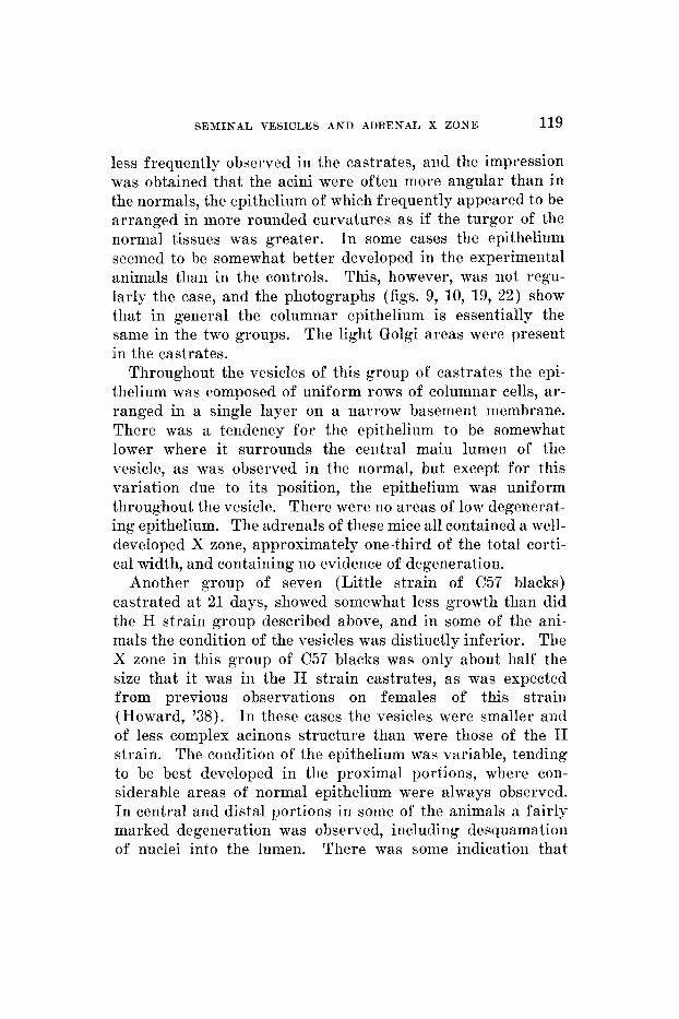
less frequently observed in the castrates, and the impression was obtained that the acini were often more angular than in the normals, the epithelium of which frequently appeared to be arranged in more rounded curvatures as if the turgor of the normal tissues was greater. In some cases the epithelium seemed to be somewhat better developed in the experimental animals than in the controls. This, however, was not regu- larly the case, and the photographs (figs. 9, 10, 19, 22) show that in general the columnar epithelium is essentially the same in the two groups. The light Golgi areas were present in the castrates.
Throughout the vesicles of this group of castrates the epi- thelium was composed of uniform rows of columnar cells, ar- ranged in a single layer on a narrow basement membrane. There was a tendency for the epithelium to be somewhat lower where it surrounds the central main lumen of the vesicle, as was observed in the normal, but except for this variation due to its position, the epithelium was uniform throughout the vesicle. There were no areas of low degenerat- ing epithelium. The adrenals of these mice all contained a well- developed X zone, approximately one-third of the total corti- cal width, and containing no evidence of degeneration.
Another group of seven (Little strain of C57 blacks) castrated a t 21 days, showed somewhat less growth than did the H strain group described above, and in some of the ani- mals the condition of the vesicles was distinctly inferior. The X zone in this group of C57 blacks was only about half the size that it was in the H strain castrates, as was expected from previous observations on females of this strain (Howard, '38). I n these cases the vesicles were smaller and of less complex acinous structure than were those of the H strain. The condition of the epithelium was variable, tending to be best developed in the proximal portions, where con- siderable areas of normal epithelium were always observed. In central and distal portions in some of the animals a fairly marked degeneration was observed, including desquamation of nuclei into the lumen. There was some indication that
120 EVELYN HOWARD
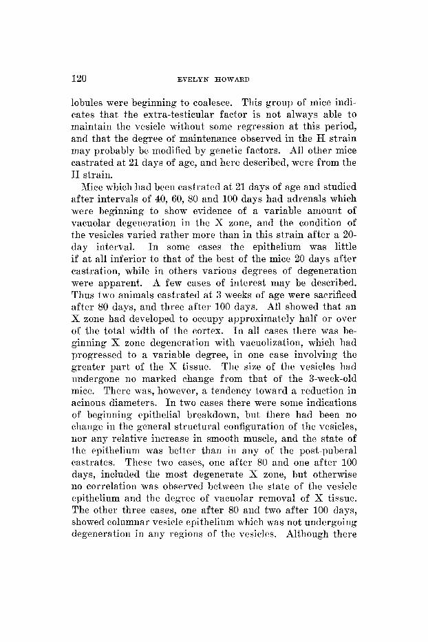
lobules were beginning to coalesce. This group of mice indi- cates that the extra-testicular factor is not always able to maintain the vesicle without some regression at this period, and that the degree of maintenance observed in the H strain may probably be modified by genetic factors. All other mice castrated at 21 days of age, and here described, were from the H strain.
Mice which had been castrated at 21 days of age and studied after intervals of 40, 60, 80 and 100 days had adrenals which were beginning to show evidence of a variable amount of vacuolar degeneration in the X zone, and the condition of the vesicles varied rather more than in this strain after a 20- day interval. I n some cases the epithelium was little if at all inferior to that of the best of the mice 20 days after castration, while in others various degrees of degeneration were apparent. A few cases of interest may be described. Thus two animals castrated at 3 weeks of age were sacrificed after 80 days, and three after 100 days. All showed that an X zone had developed to occupy approximately half or over of the total width of the cortex. I n all cases there was be- ginning X zone degeneration with vacuolization, which had progressed to a variable degree, in one case involving the greater par t of the X tissue. The size of the vesicles had undergone 110 marked change from that of the 3-week-old mice. There was, however, a tendency toward a reduction in acinous diameters. I n two cases there were some indications of beginning epithelial breakdown, but there had been no change in the general structural configuration of the vesicles, nor any relative increase in smooth muscle, and the state of the epithelium was better than in any of the post-puberal castrates. These two cases, one after 80 and one after 100 days, included the most degenerate X zone, but otherwise no correlation was observed between the state of the vesicle epithelium and the degree of vacuolar removal of X tissue. The other three cases, one after 80 and two after 100 days, showed columnar vesicle epithelium which was not undergoing degeneration in any regions of the vesicles. Although there
SEMINAL VESICLES AND ADRENAL X ZONE 121
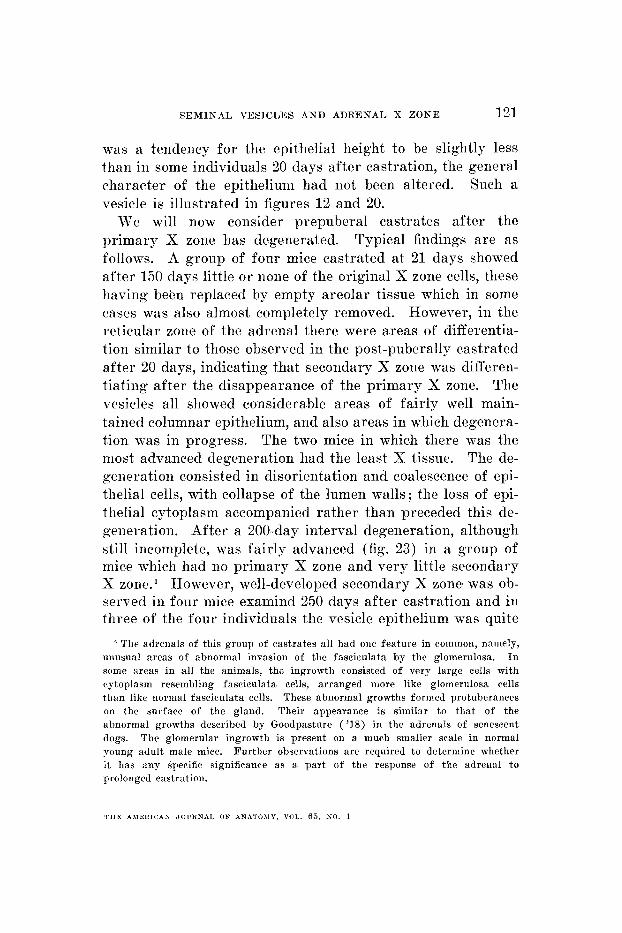
was a tendency for the epithelial height to be slightly less than in some individuals 20 days after castration, the general character of the epithelium had not been altered. Such a vesicle is illustrated in figures 12 and 20.
We will now consider prepuberal castrates after the primary X zone has degenerated. Typical findings are as follows. A group of four mice castrated at 21 days showed after 150 days little or none of the original X zone cells, these having been replaced by empty areolar tissue which in some cases was also almost completely removed. However, in the reticular zone of the adrenal there were areas of differentia- tion similar to those observed in the post-puberally castrated after 20 days, indicating that secondary X zone was differen- tiating after the disappearance of the primary X zone. The vesicles all showed considerable areas of fairly well main- tained columnar epithelium, and also areas in which degenera- tion was in progress. The two mice in which there was the most advanced degeneration had the least X tissue. The de- generation consisted in disorientation arid coalescence of epi- thelial cells, with collapse of the lumen walls; the loss of epi- thelial cytoplasm accompanied rather than preceded this de- generation. After a 200-day interval degeneration, although still incomplete, was fairly advanced (fig. 23) in a group of mice which had 110 primary X zone and very little secondary X zone." However, well-developed secondary X zone was ob- served in four mice examind 250 days after castration and in three of the four individuals the vesicle epithelium was quite
' The adrenals of this group of castrates all had one feature in coinmon, namely, unusual areas of abnormal invasion of the fasciculata by the glomerulosa. I n some areas in all the animals, the ingrowth consisted of very large cells with cytoplasm resembling faseiculata cells, arranged more like gloinerulosa cells than like nornial fascieulata cells. These abnormal growths formed protuberances on the surface of the gland. Their appearaiice is similar to that of the abnormal growths described by Goodpasture ( '18) in the adrenals of senescent dogs. The glomerular ingrowth is present on a much smaller scale in normal young adult male mice. Further observations are required to deterniine whether i t has any specific significance as a part of the response of the adrenal to prolonged castration.
? H h A > I E H I C A S JOI'KNAI. OF A N A T O M Y , VOL. 65, NO. 1
122 EVELYN HOWARD
well developed. An example of such epithelium is shown in figure 24 which shows that epithelium not markedly in- ferior to that of the normal 21-day-old mouse may be ob- served after 250 days of castration in the presence of secon- dary X zone. Such epithelium was typical of the greater part of the vesicle in this individual.
After an even longer interval, 350 days after castration, two individuals had adrenals with little X zone but with marked areas of glomerular invasion of the fasciculata. The seminal vesicles had undergone marked degeneration, but still contained some areas in which the epithelium was colum- nar and the alveoli discrete.
It can be concluded that the vesicles of mice castrated at 21 days of age may be maintained with little change for a considerable period, and then undergo a gradual degeneration concomitant with the degeneration of the primary X zone, but that during or subsequent to the degeneration of primary X zone, there ensues differentiation of secondary X zone, and that during a prolonged period it is not possible to obtain seminal vesicles in a condition of consistent uniform degenera- tion. The condition of partial maintenance, i.e., the co- existence of degenerating areas and areas having fairly good columnar cells resembles the type of reaction seen in the postpuberal castrates 20 days or more following castration. Since the response to postpuberal castration is far from being a complete degeneration, these two groups of cases agree in indicating that the primary X zone is not solely re- sponsible for maintaining some degree of normality in the accessories. Observations will now be presented on mice castrated after the primary X zone has degenerated.
Twenty days after castration the adult mouse seminal vesicle, as is well known, becomes very much reduced in size. This reduction is as- sociated with the loss of a large part of the contained secre- tion, and some actual involution (see figs. 7 and 8). Histo- logical study of twelve such mice, of various strains, includ-
The effects o f postpuberal castration.
SEMINAL VESICLES AND ADRENAL X ZONE 123
iiig H, ranging in age from 6 to 8 weeks to over 1 year, re- vealed in no case the complete and uniform degeneration characteristic of the rat vesicle. I n all cases the proximal and certain lateral portions of the vesicles showed large areas of convoluted acini uniformly bearing high columnar cells and without evidence of connective tissue or smooth muscle en- croachment on the normal acinous structure. Such epithelium is illustrated in figure 21. The cells tend to be not quite so high as in the best normal adult preparations, but their height is not less than that found in the best maintained vesicles of the prepuberal castrates, although the cytoplasm has under- gone alterations. The nuclei are frequently, but not always, closer to the basement membrane than in the normal. 'Secre- tion granules' were not observed. The cells in these areas are in a state not unlike that found in normal 3-week-old mice. The cytoplasm, however, seems to be more attenuated and even the best cells have certainly undergone regressive changes subsequent to castration which have carried them to a condition apparently somewhat inferior to that of the condition typical of the castrates with primary X zone.
I n the distal portions of the vesicle there exists a grada- tion of structure similar to that seen in the normal, with, however, an increase in the proportion of low epithelium, re- sulting from a generalized decrease in cell height following castration. Various states of the epithelium encountered in these adult castrates after a 20-day post-operative interval are shown in figures 8,15,16 and 21. In general there is little if any encroachment of smooth muscle between the epithelial layers in any region. In some cases there appears to be coalescence of epithelium in the more distal portions and a beginning breakdown of the basement membrane, but in other
'Male mice may become fertile' a t 4 to 5 weeks of age. The younger animals described in this group were not full grown and strictly speaking were puberal rather than postpuberal. They are, however, described under the same heading as the strictly postpuberal adults, since they were presumably in the same condition as the adults with regard t o the character with which we are particularly con- cerned, namely that their adrenals did not contain appreciable amounts of primary X zone.
124 EVELYN HOWARD
cases such structural alterations were absent. Coalescence of the lumen edge of two epithelial layers may occur without extreme decrease of cell height.
In most animals the epithelium, when classifiable as cuboidal, is not in any extensive areas as low as it is uniformly in the rat 20 days after castration. Nuclear desquamation is rare, and if it is in evidence i t is confined to the distal regions. The amount of the high discrete columnar epi- thelium varied among different postpuberal castrates, being in some cases not more than approximately one-fifth of the total, and as a maximum, approximately equal to the amoiint of low or coalescing lobules. No regular differences relative to age were observed within the postpuberal group 20 days after castration. The total amount of high epithelium was of the same order of magnitude as in the prepuberal castrate. Thus, although there is an enormous reduction in the secre- tory activity of the cells, as f a r as the visible coagulated secretion is coiicerned, there is nothing comparable to the complete degree of uniform structural degeneration which occurs in the rat. Since there is so much gradation in any normal mouse vesicle, it is necessary to study complete serial sections to evaluate the state of the vesicle as a whole. When the whole vesicle is taken into account the variations from one part to another are fa r greater than the variations from one animal to another.
A point of interest is illustrated by one of the youngest in the group of postpuberal castrates, an individual (H strain) with the smallest vesicles at the time of examination. The vesicles of this individual had apparently not undergone much gross degeneration in structure of the type which is seen to actually reduce the vesicle size after prolonged castration, namely epithelial coalescence with breakdown of the basement membrane and relative increase in smooth muscle and con- nective tissue. The vesicles in this case were in the same state as far as general lobe structure is concerned, as in the others of this group. Hence one may reasonably conclude that the fact of their smaller size is due to a smaller size a t
SEMINAL VESICLES A N D ADRENAL X ZONE 125
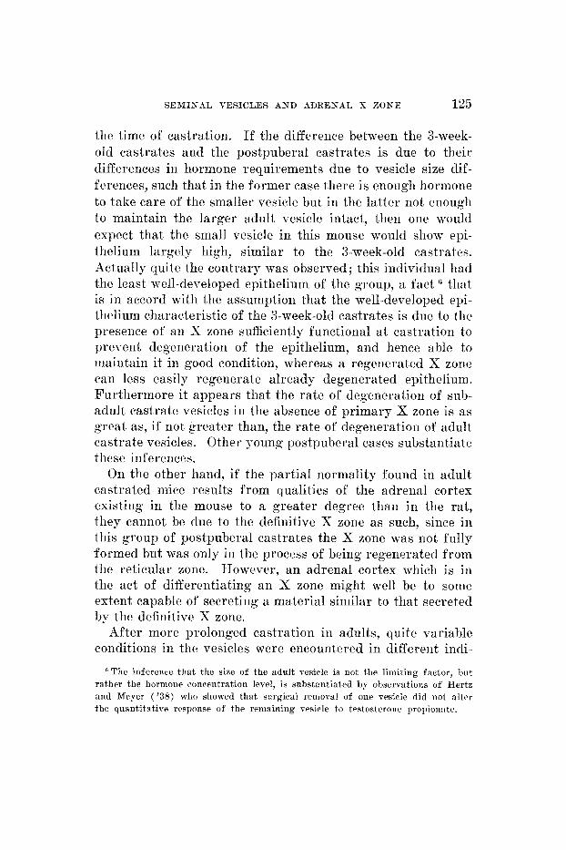
the time of eastration. If the difference between the 3-week- old castrates and the postpuberal castrates is due to their differences in hormone requirements due to vesicle size dif- ferences, such that in the former case there is enough hormone to take care of the smaller vesicle but in the latter not enougli to maintain the larger adult vesicle intact, then oiie would expect that the small vesicle in this mouse would show epi- thelium largely high, similar to the 3-week-old castrates. Actually quite the contrary mas observed ; this individual had the least well-developed epithelium of the group, a fact (i that is in accord with the assumption that the well-developed epi- thelium characteristic of the 3-week-old castrates is due to the presence of an X zone sufficiently functional a t castration to prevent degeneration of the epithelium, and hence able to maintain it in good condition, whereas a regenerated X zone can less easily regenerate already degenerated epithelium. Furthermore it appears that the rate of degeneration of sub- adult castrate vesicles in the absence of primary X zone is a s great as, if not greater than, the rate of degeneration of adult castrate vesicles. Other young postpuberal cases substantiate tliese inferences.
On the other hand, if the partial normality found in adult castrated mice results from qualities of the adrenal cortex existing in the mouse to a greater degree than in the rat, they cannot be due to the definitive X zone as such, since in this group of postpuberal castrates the X zone was not fully formed but was only in the process of being regenerated from the reticular zone. However, an adrenal cortex which is iii the act of diff'erentiating a n X zone might well be to some extent capable of secreting a material similar to that secreted by the definitive X zone.
After more prolonged castration in adults, quite variable conditions in the vesicles were encountered in different indi-
'The inference that the size of the adult vesicle is iiot the limiting factor, but rather the hormone concentration level, is substantiated by observatioiis of Hertz and Mcyer ( '38) wlio showed that surgical removal of one vesicle did not alter the quantitative response of the remaining vesicle to testosteroiie propioiinte.
126 EVELYN HOWARD
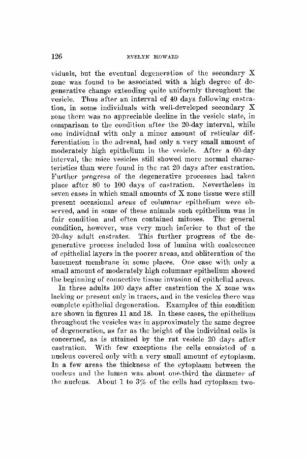
viduals, but the eventual degeneration of the secondary X zone was found to be associated with a high degree of de- generative change extending quite uniformly throughout the vesicle. Thus after an interval of 40 days following castra- tion, in some individuals with well-developed secondary X zone there was no appreciable decline in the vesicle state, in comparison to the condition after the 20-day interval, while one individual with only a minor amount of reticular dif- ferentiation in the adrenal, had only a very small amount of moderately high epithelium in the vesicle. After a 60-day interval, the mice vesicles still showed more normal charac- teristics than were found in the rat 20 days after castration. Further progress of the degenerative processes had taken place after 80 to 100 days of castration. Nevertheless in seven cases in which small amounts of X zone tissue were still present occasional areas of columnar epithelium were ob- served, and in some of these animals such epithelium was in fair condition and often contained mitoses. The general condition, however, was very much inferior to that of the 20-day adult castrates. This further progress of the de- generative process included loss of lumina with coalescence of epithelial layers in the poorer areas, and obliteration of the basement membrane in some places. One case with only a small amount of moderately high columnar epithelium showed the beginning of coiinective tissue invasion of epithelial areas.
In three adults 100 days after castration the X zone was lacking or present only in traces, and in the vesicles there was complete epithelial degeneration. Examples of this condition are shown in figures 11 and 18. In these cases, the epithelium throughout the vesicles was in approximately the same degree of degeneration, as far as the height of the individual cells is concerned, as is attained by the rat vesicle 20 days after castration. With few exceptions the cells consisted of a nucleus covered only with a very small amount of cytoplasm. In a few areas the thickness of the cytoplasm between the nucleus and the lumen was about one-third the diameter of the nucleus. About 1 to 3% of the cells had cytoplasm two-
SEMINAL VESICLES AND ADRENAL X ZONE 127
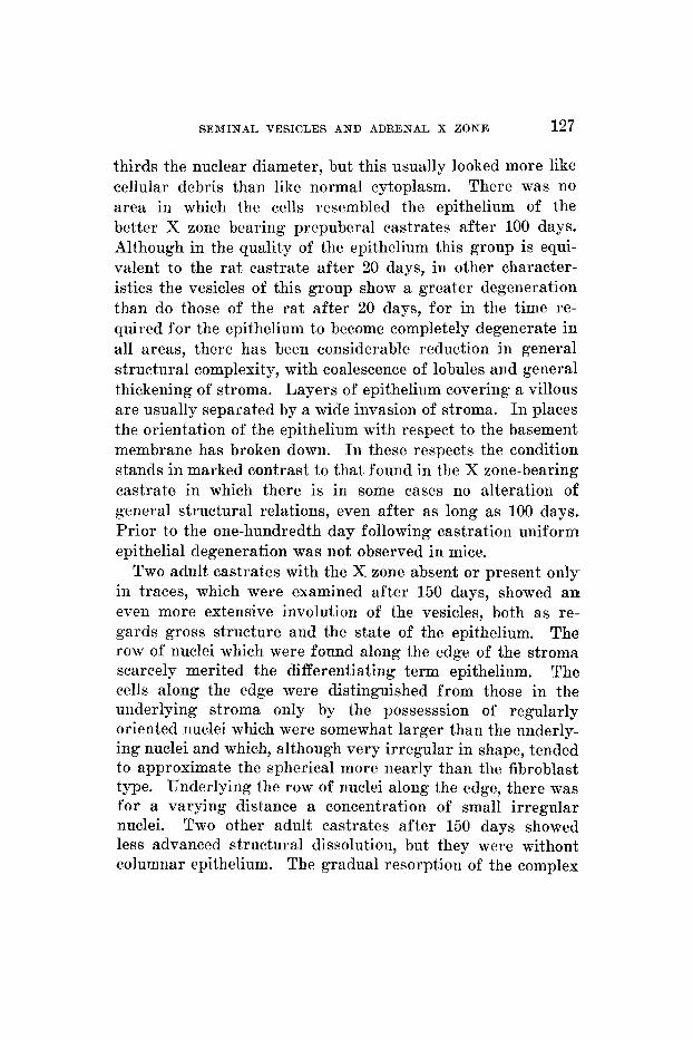
thirds the nuclear diameter, but this usually looked more like cellular debris than like normal cytoplasm. There was no area in which the cells resembled the epithelium of the better X zone bearing prepuberal castrates after 100 days. Although in the quality of the epithelium this group is equi- valent to the ra t castrate after 20 days, in other character- istics the vesicles of this group show a greater degeneration than do those of the rat after 20 days, for in the time re- quired for the epithelium to become completely degenerate in all areas, there has been considerable reduction in general structural complexity, with coalescence of lobules and general thickening of stroma. Layers of epithelium covering a villous are usually separated by a wide invasion of stroma. I n places the orientation of the epithelium with respect to the basement membrane has broken down. I n these respects the condition stands in marked contrast to that found in the X zone-bearing castrate in which there is in some cases no alteration of general structural relations, even after as long as 100 days. Prior to the one-hundredth day following castration uniform epithelial degeneration was not observed in mice.
Two adult castrates with the X zone absent or present only in traces, which were examined after 150 days, showed a n even more extensive involution of the vesicles, both as re- gards gross structure and the state of the epithelium. The row of nuclei which were found along the edge of the stroma scarcely merited the differentiating term epithelium. The cells along the edge were distinguished from those in the underlying stroma only by the possesssion of regularly oriented nuclei which were somewhat larger than the underly- ing nuclei and which, although very irregular in shape, tended to approximate the spherical more nearly than the fibroblast type. Underlying the row of nuclei along the edge, there was for a varying distance a concentration of small irregular nuclei. Two other adult castrates after 150 days showed less advanced structural dissolution, but they were without columnar epithelium. The gradual resorption of the complex
128 EVELYN HOWARD
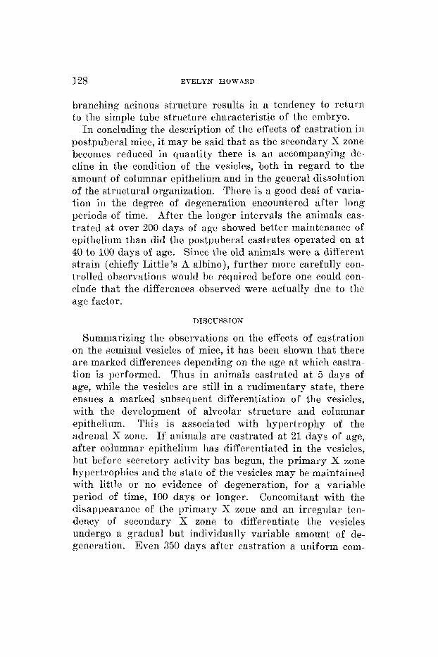
branching acinous structure results in a tendency to return to the simple tube structure characteristic of the embryo.
I n concluding the description of the effects of castration in postpuberal mice, it may he said that as the secondary X zone becomes reduced in quantity there is an accompanying de- cline in the condition of the vesicles, both in regard to the amount of columnar epithelium and in the general dissolution of the structural organization. There is a good deal of varia- tion in the degree of degeneration encountered after long periods of time. After the longer intervals the animals cas- trated a t over 200 days of age showed better maintenance of epithelium than did the postpuberal castrates operated on at 40 to 100 days of age. Since the old animals were a different strain (chiefly Little’s A albino), further more carefully con- trolled observations would be required before one could con- clude that the differences observed were actually due to the age factor.
DISCUSSION
Summarizing the observations on the effects of castration on the seminal vesicles of mice, i t has been shown that there are marked differences depending on the age at which castra- tion is performed. Thus in animals castrated a t 5 days of age, while the vesicles a re still in a rudimentary state, there ensues a marked subsequent differentiation of the vesicles, with the development of alveolar structure aiid columnar epithelium. This is associated with hypertrophy of the adrenal X zone. If animals are castrated at 21 days of age, after columnar epithelium has differentiated in the vesicles, but before secretory activity bas begun, the primary X zone hypertrophies and the state of the vesicles may be maintained with little or no evidence of degeneration, for a variable period of time, 100 days or longer. Concomitant with the disappearance of the primary X zone and an irregular ten- dency of secondary X zone to differentiate the vesicles undergo a gradual but individually variable amount of de- generation. Even 350 days after castration a uniform com-
SEMINAL VESICLES AND ADRENAL X ZONE 129
plete degeneration was not always observed in these pre- puberally castrated mice.
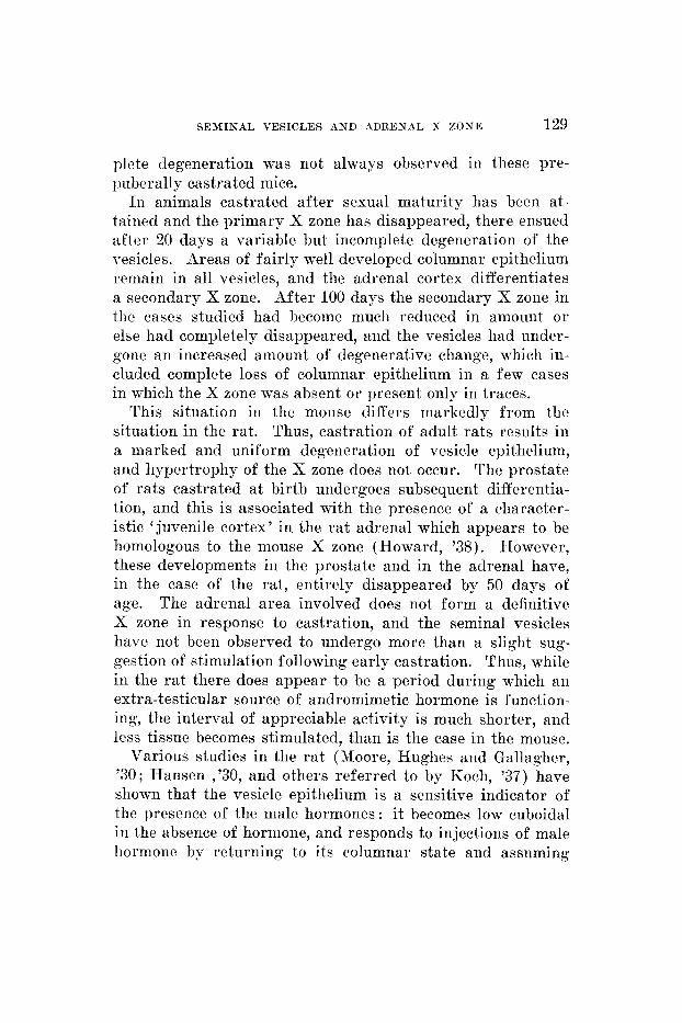
In animals castrated after sexual maturity has been at- tained and the primary X zone has disappeared, there ensued after 20 days a variable but incomplete degeneration of the vesicles. Areas of fairly well developed columnar epithelium remain in all vesicles, and the adrenal cortex differentiates a secondary X zone. After 100 days the secondary X zone in the cases studied had become much reduced in amount o r else had completely disappeared, and the vesicles had under- gone an increased amount of degenerative change, which in- cluded complete loss of columnar epithelium in a few cases in which the X zone was absent or present only in traces.
This situation in the mouse differs markedly from the situation in the rat. Thus, castration of adult rats results in a marked and uniform degeneration of vesicle epithelium, and hypertrophy of the X zone does not occur. The prostate of rats castrated at birth undergoes subsequent differentia- tion, and this is associated with the presence of a character- istic ' juvenile cortex' in the rat adrenal which appears to be homologous to the mouse X zone (Howard, '38). However, these developments in the prostate and in the adrenal hare, in the case of the rat, entirely disappeared by 50 days of age. The adrenal area involved does not form a definitive X zone in response to castration, and the seminal vesicles have not been observed to undergo more than a slight sug- gestion of stimulation following early castration. Thus, while in the rat there does appear to be a period during which an extra-testicular source of andromimetic hormone is function- ing, the interval of appreciable activity is much shorter, and less tissue becomes stimulated, than is the case in the mouse.
Various studies in the rat (Moore, Hughes and Gallagher, '30; Hansen ,'30, and others referred to by Koch, '37) have shown that the vesicle epithelium is a sensitive indicator of the presence of the male hormones: it becomes low cuboidal in the absence of hormone, and responds to injections of male hormone by returning to its columnar state and assuming
130 EVELYN HOWARD
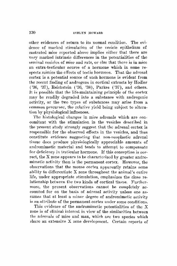
other evidences of return to its normal condition. The evi- dence of marked stimulation of the vesicle epithelium of castrated mice reported above implies either that there are very marked intrinsic differences in the potentialities of the seminal vesicles of mice and rats, or else that there is in mice an extra-testicular source of a hormone which in some re- spects mimics the effects of testis hormone. That the adrenal cortex is a potential source of such hormone is evident from the recent finding of androgens in cortical extracts by Hodler ( '36, '37), Reichstein ( '36, '38), Parkes ( '37), and others. I t is possible that the life-maintaining principle of the cortex may be readily degraded into a substance with androgenic activity, or the two types of substances may arise from a common precursor, the relative yield being subject to altera- tion by physiological influences.
The histological changes in mice adrenals which are con- comitant with the stimulation in the vesicles described in the present study strongly suggest that the adrenal cortex is responsible for the observed effects in the vesicles, and thus constitute evidence suggesting that non-neoplastic adrenal tissue does produce physiologically appreciable amounts of andromimetic material and tends to attempt to compensate for deficiency in testicular hormone. If this conception is cor- rect, the X zone appears to be characterized by greater andro- mimetic activity than is the permanent cortex. However, the observations that the mouse cortex apparently retains some ability to differentiate X zone throughout the animal's entire life, under appropriate stimulation, emphasizes the close re- lationship between the two kinds of cortical tissue. Further- more, the present observations cannot be completely ac- counted for on the basis of adrenal activity unless one as- sumes that at least a minor degree of andromimetic activity is an attribute of the permanent cortex under some conditions.
This evidence of the andromimetic potentialities of the X zone is of clinical interest in view of the similarities between the adrenals of mice and man, which are two species which share an extensive X zone development. Certain reports of
SEMINAL VESICLES AND ADRENAL S ZONE 131
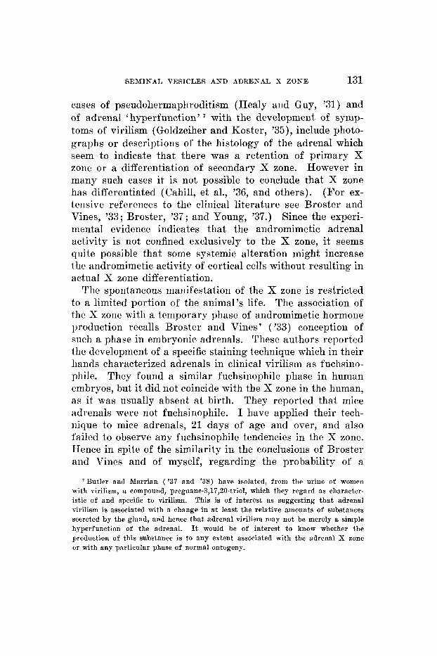
cases of pseudohermaphroditism (Healy and Guy, '31) and of adrenal ' hyperfunction' with the development of symp- toms of virilism (Goldzeiher and Koster, '35), include photo- graphs or descriptions of the histology of the adrenal which seem to indicate that there was a retention of primary X zone or a differentiation of secondary X zone. However in many such cases it is not possible to conclude that X zone has differentiated (Cahill, et al., '36, and others). (For ex- tensive references to the clinical literature see Broster and Vines, '33; Broster, '37; and Young, '37.) Since the experi- mental evidence indicates that the andromimetic adrenal activity is not confined exclusively to the X zone, it seems quite possible that some systemic alteration might increase the andromimetic activity of cortical cells without resulting in actuaI X zone differentiation.
The spontaneous manifestation of the X zone is restricted to a limited portion of the animal's life. The association of the X zone with a temporary phase of andromimetic hormone production recalls Broster and Vines' ('33) conception of such a phase in embryonic adrenals. These authors reported the development of a specific staining technique which in their hands characterized adrenals in clinical virilism as fuchsino- phile. They found a similar fuchsinophile phase in human embryos, but it did not coincide with the X zone in the human, as it was usually absent at birth. They reported that mice adrenals were not fuchsinophile. I have applied their tech- nique to mice adrenals, 21 days of age and over, and also failed to observe any fuchsinophile tendencies in the X zone. Hence in spite of the similarity in the conclusions of Broster and Vines and of myself, regarding the probability of a
'Butler and Marrian ('37 and '38) have isolated, from the urine of women with virilism, a compound, pregnane-3,17,20-triol, which they regard as character- istic of and specific t o virilism. This is of interest as suggesting tha t adrenal virilism is associated with a change in a t least the relative amounts of substances secreted by the gland, and hence tha t adrenal virilism may not be merely a simple hyperfunction of the adrenal. It would be of interest to know whether the production of this substance is to any extent associated with the adrenal X zone or with any particular phase of normal ontogeny.
132 EVELYN HOWARD
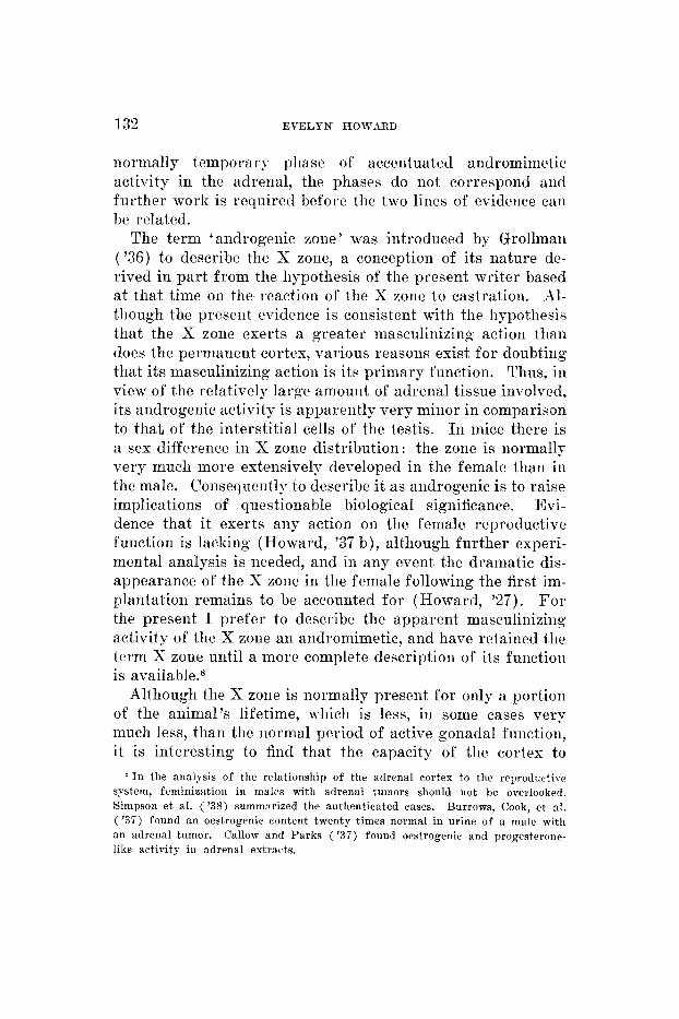
normally temporary phase of accentuated andromimetic activity in the adrenal, tlie phases do not correspond and further work is required before the two lines of evidence can he related.
The term 'androgenic zone' was introduced by Grollmari ('36) to describe the X zone, a conception of its nature cle- rived in part from the hypothesis of the present writer based at that time on the reaction of the X zone to castration. Al- though the present evidence is consistent with the hypothesis that the X zone exerts a greater masculinizing action than does the permanent cortex, various reasons exist for doubting that its masculinizing action is its primary function. Thus, in view of the relatively large amouiit of adrenal tissue involved, its androgenic activity is apparently very minor in comparison to that of the interstitial cells of the testis. In mice there is a sex difference in X zone distribution: the zone is normally very much more extensively developed in the female than in the male. Consequently to describe it as androgenic is to raise implications of questionable biological significance. Evi- dence that i t exerts any action on tlie female reproductive function is lacking (Howard, '37 b), although further experi- mental analysis is needed, and in any event the dramatic dis- appearance of the X zone in the female following the first im- plantation remains to be accounted for (Howard, '27). Fo r the present I prefer to describe the apparent masculinizing activity of the X zone an aiidromimetic, and have retained the term X zone until a more complete description of its function is available.8
Although the X zone is normally present for only a portion of the animal's lifetime, which is less, in some cases very much less, than the normal period of active gonadal function, it is interesting to find that the capacity of the cortex to
' I n the analysis of the relationship of the adrenal cortex t o the reproductive system, feminization in males with adrenal tumors should not be overlooked. Simpson et al. ( '38) summ:irized the authenticated cases. Burrows, Cook, e t nl. ( ' 3 7 ) found an oestrogenic content twenty times normal in urine of a male wltll an adrenal tumor. Callow and Parks ( ' 3 7 ) found orstrogenic and progesterone- like activity in adrenal extracts.
SEMINAL VESICLES AND ADRENAL X ZONE 133
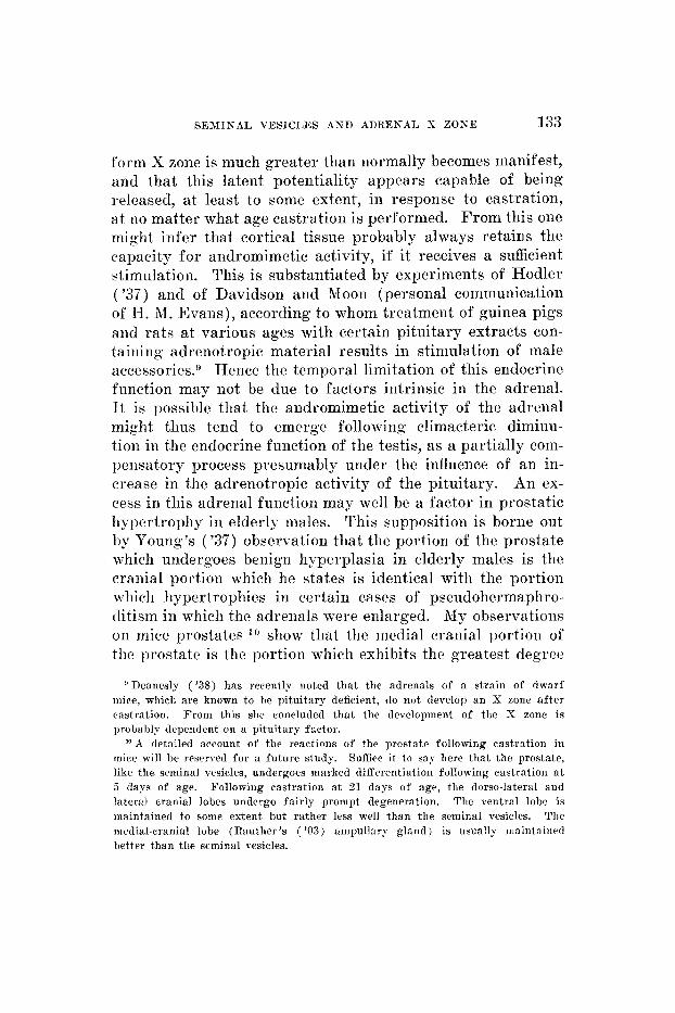
form X zone is much greater than normally becomes manifest, and that this latent potentiality appears capable of being released, a t least to some extent, in response to castration, a t no matter what age castration is performed. From this one might infer that cortical tissue probably always retains the capacity for andromimetic activity, if it receives a sufficient stimulation. This is substantiated by experiments of Hodler ( ' 3 7 ) and of Davidsoii and Moon (personal communication of H. 31. Evans), according to whom treatment of guinea pigs and rats at various ages with certain pituitary extracts con- taining adrenotropic material results in stimulation of male accessories.9 Hence the temporal limitation of this endocrine function may not be due to factors intrinsic in the adrenal. I t is possible that the aiidromimetic activity of the adrenal might thus tend to emerge following climacteric diminu- tion in the endocrine function of the testis, as a partially com- pensatory process presumably under the influence of an in- crease in the adrenotropic activity of the pituitary. An ex- cess in this adrenal function may well be a factor in prostatic hypertrophy in elderly males. This supposition is borne out by Young's ( ' 3 7 ) observation that the portioii of the prostate which undergoes benign hyperplasia in elderly males is the cranial portion which he states is ideiitical with the portion wliicli hypertrophies in certain cases of pseudohermaphro- ditism in which the adrenals were enlarged. Ny observations on mice prostates lo show that the medial cranial portion of the prostate is the portion which exhibits the greatest degree
'Deanesly ('38) has recently noted that the adrenals of a strain of dwarf mice, which are known to be pituitary deficient, do not develop an X zone a f te r castration. From this she concluded tha t the development of the X zone is probably dependent on a pituitary factor.
" A detailed account of the reactions of the prostate following castration in mice will he reserved for a future study. Suffice it t o sn)- here tliat the prostate, like the seminal vesicles, undergoes marked differentiation following castration a t 3 days of age. Following castration a t 21 days of age, the dorso-lateral and lateral cranial lobes undergo fairly prompt degeneration. The ventral lobe is maintained to some extent but rather less well than the seminal vesicles. The medial-cranial lobe (Rauther 's ( '03) ampull:try gland) is usually maintained better than the seminal vesicles.
134 EVELYN HOWARD
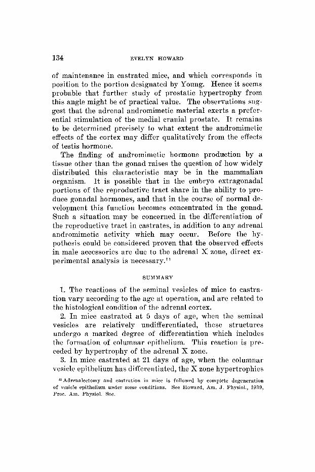
of maintenance in castrated mice, and which corresponds in position to the portion designated by Young. Hence it seems probable that further study of prostatic hypertrophy from this angle might be of practical value. The observations sug- gest that the adrenal andromimetic material exerts a prefer- ential stimulation of the medial cranial prostate. It remains to be determined precisely to what extent the andromimetic effects of the cortex may differ qualitatively from the effects of testis hormone.
The finding of andromimetic hormone prodection by a tissue other than the gonad raises the question of how widely distributed this characteristic may be in the mammalian organism. It is possible that in the embryo extragonadal portions of the reproductive tract share in the ability to pro- duce gonadal hormones, and that in the course of normal de- velopment this function becomes concentrated in the gonad. Such a situation may be concerned in the differentiation of the reproductive tract in castrates, in addition to any adrenal andromimetic activity which may occur. Before the hy- pothesis could be considered proven that the observed effects in male accessories are due to the adrenal X zone, direct ex- perimental analysis is necessary.*'
SUMMARY
1. The reactions of the seminal vesicles of mice to castra- tion vary according to the age at operation, and are related to the histological condition of the adrenal cortex.
2. In mice castrated a t 5 days of age, when the seminal vesicles are relatively undifferentiated, these structures undergo a marked degree of differentiation which includes the formation of columnar epithelium. This reaction is pre- ceded by hypertrophy of the adrenal X zone.
3. I n mice castrated a t 21 days of age, when the columnar vesicle epithelium has differentiated, the X zone hypertrophies
Adrenalectomy and castration in mice is followed by complete degeneration See Howard, Am. J. Physiol., 19.39, of vesicle epithelium under some conditions.
Proc. Am. Physiol. SOC.
SEMINAL VESICLES AND ADRENAL X ZONE 135
and the condition of the vesicles undergoes little alteration for a considerable period which may extend for as long as 100 days. 4. I n mice castrated after sexual maturity, when the
primary X zone has disappeared, the vesicles undergo a de- generation which remains incomplete during the period in which secondary X zone differentiates in the adrenal cortex. One hundred days after castration the secondary X zone has frequently disappeared from the cortex, and in these cases the vesicles are in a state of marked and uniform degeneration.
5. These reactions in mice seminal vesicles are in contrast to the situation in rats, in which castration at corresponding ages is not followed by differentiation or maintenance of columnar epithelium in the vesicles, and in which the X zone does not differentiate in response to castration.
6. In the light of recent findings by others of androgens in adrenal cortex extracts it is believed that the most probable explanation for the present observations is that the adrenal X zone in mice exerts an andromimetic activity which is greater than that of the normal adult permanent cortex.
7. Certain observations on the prostate are referred to which are in accordance with the conception that the adrenal may be a factor in prostatic hypertrophy in elderly males.
LITERATURE CITED
1939 BURRILL, M. W., AND R. R. GREENE Androgenic function of the adrenals in the immature male castrate rat. Proc. Soc. Exp. Biol. and Med., vol. 40, p. 327.
BURROWS, H., J. W. COOK, E. ROE AND F. L. WARREN 1937 Isolation of A 3 : s-androstadiene-17-one from the urine of a man with a malignant tumor of the adrenal cortex.
BUTLER, G. C., AND G. F. MARRIAN 1937 The isolation of pregnane-3,17,40- trio1 from the urine of women showing the adreno-genital syndrome. J. Biol. Chem., vol. 119, p. 565.
1938 Chemical studies on the adreno-genital syndrome. I. J. Biol. Chem., vol. 124, p. 237.
BROSTER, L. R., AND H. W. C. VINES 1933 The adrenal cortex. London (H. K. Lewis).
&ROSTER, L. R. 1937 Eight years experience with the adrenal gland. Arch. Surgery, vol. 34, p. 761.
Biochem. J., vol. 31, p. 950.
136 EVELYN HOWARD
CAHILL, G. F., R. F. LOEB, R. KURZROK, A. P. STOUT A N D F. &I. SIIITII
CALLOW, R. K., AND R. DEANESLY
1936 Adrenal cortical tumors.
The effects of aiidrosterone and male hormone concentrates on the accessory reproductive organs of castrated rats, mice and guinea pigs.
The occurrence of oestrin and progcstin in adrenal, testis and hypophgsis. J. Physiol., vol. 87, p. 28 (Pro- ceedings).
DAVIES, S. 1937 The development of the adrenal gland of the cat. Quart. J. Micr. Sci., vol. 80, p. 81.
DEANESLY, R. 1928 A study of the adrenal cortex in the mouse and its relation to the gonads. Proc. Roy. Soc. B, vol. 103, p- 523. - 1938 Adrenal cortex differences in male and female mice. Nature,
DEANESLY, R., AND A. S. PARKES 1937 Multiple activities of androgenic compounds.
GALLAGHER, T. F. 1928 Distribution of testicular comb growth stimulating principle in tissues.
GOLDZIEHER, M., AND H. KOSTER 1935 Adrenal cortical hyperfunction. Am. J. Surgery X.S., vol. 27, p. 93.
GOODPA4STURE, E. W. 1918 An anatomical study of senescence in dogs, with especial reference to the relation of cellular changes of age to tumors.
Surg. Gynecol. and Obst., vol. 62, p. 287. 1935
Biochem. J., vol. 29, p. 1424. CALLOW, R. K., AND A. S. PARKES 1936
vol. 141, p. 79.
Quart. J. Exp. Physiol., vol. 26, p. 393.
Am. J. Physiol., vol. 87, p. 447.
J. Med. Res., vol. 38, p. 127. GROLLMAN, A. 1936 The Adrenals. Baltimore (Williams and Wilkins). HANSEN, I. B. Rat seminal vesicles and prostate glands as quantitative
HEALY, C. E., A N D C. C. GUY 1931 Pseudohermaphroditismus masculinus ex- Arch.
H m m , R., A N D R. K. MEYER 1938 The effect of surgical reduction of the amount of reacting tissue upon the quantitative effectiveness of testosterone propionate and estrone. Am. J. Physiol., vol. 124, p. 259.
HODLER, D. 1936 Action masculinisante d ’extraits hpdrosolubles dc surrCnales de boeuf sur le cobaye.
____ 1937 Sur rha le s et masculinization. Arch. d. Anat., d. hist., et d’embryol., vol. 24, p. 1.
HOWARD, E. 1927 A transitory zone in the adrenal cortex which shows age and sex relationships.
____ 1929 Development of the mouse adrenal. Science, vol. 69, p. 406. The X zone of the suprarenal cortex in relation to gonadal
maturation in monkeys and mice, and to epiphyseal unions in monkeys. Anat. Rec., vol. 46, p. 93.
__-__ 1937 a Is the adrenal X zone androminietic? Am. J. Physiol., vol. 119, p. 339 (Proc.).
1937 b Adrenalectomy in mice, and the replacement of X zone bearing adrenals by cortical extract with especial reference to adrenal- gonad relationships.
1933 indicators of testicular hormone.
ternus with suprarenal hyperplasia and vascular hypertension.
Endocrinology, rol. 1 7 , p. 163.
Path., vol. 12, p. 543.
Compt. rend. soc. biol., vol. 122, p. 512.
Am. J. Anat., vol. 40, p. 251.
1930
An]. J. Phgsiol., vol. 120, p. 36.
SEMINAL VESICLES A N D ADRENAL X ZOXE 137
IIOTVARD, 3:. 1038 :I The rcpresclntation of the :idrenal X zone in rats, in the light of observations on X zone variability i n mice. Am. J. Anat., vol. 62, 1). 351.
___ 1938 b A tenipor:try phase of extra-testiculiir androiniinetic liornionc~ production in rodents, associated with the adrenal X zone. Am. J. l'hysiol., vol. 123, p. 105 (F'roc.).
KocIi, 3'. C . 1937 The male sex hormones. Physiol. Rev., vol. 17, 1). l.i3. MARTINS, T., AND A. ROCHA E SILVA 1929 ITtilization des vcsicules seininales
de la souris blanche coinme test des hormones testiculaires. Colnpt. rend. soc. biol., vol. 102, p. 480.
1930 Rat seminal-vesicle cy- tology as a testis-hormone indicator and the prevention of castration changes by testis extract injection. Am. J. Anat., 1701. 45, p. 109.
MOORE, C. I%., I). PRICE AND T. F. GALLAGHER 1930 R.at-prost,ate cytology as a testis-hormone indicator and the prevention of castration changes by testis-extract injections.
PARKES, A. S. 1937 Androgenic activity of ovarian extracts. Nature, vol. 139, p. 965.
PRICE, D. 1936 Normal development of the prostate and seminal vesicles of the r a t with a study of experimental postnatal modifications. A m J. Anat., 1701. 60, p. 79.
R A U T I ~ E R , h1. 1903 Ueber den Gcnitalapparat einigcr Nager uiid Insektivoren, insebesondre die akzessorisclien Gcnitxldriiscn derwlben. Jen. Zeit. Nxturw., N.F., H i l . 31, S. 378.
REICHSTEIN, T. 1936 ' Adrenostrroii. ' Cber die Bestandtcilc der Nebennieren- rinde.
1938 Cheinie des Cortins und seiner Beglritstoffe. Ergcb. der Vitamin und IIormon-Forsh., rol. 1, p. 334. 1935 J. Anat., vol. 70, p. 126.
1938 Feminization in a male adul t with carci-
MOORE, C. R., W. HUGHES A N D T. F. GALLAGHER
Am. J. Anat., vol. 45, p. 71.
11. Helvetiea Chim. Acta, vol. 19, p. 223.
ROAF, X.
SInrPsos, S. L., A m C. A. JOLL
A study of the adrenal cortex of the rabbit.
VOSS, noma of tlir adrenal cortex.
H. E. 1930 Zur Frage der extrahormonalen beziehungen zwischen gonaden und sekuiidarcn gese1ileclitsmerkiii:~len. Arch. f . ent\vicklungsmechanik dcs org., 1-01. 122, p. 584.
VOSS, H. I<:., A N D S. LOETVE 1931 Zur Werthestinimung iniinnlichen sexual- hornions an den Vesikulardrilsen dcs Nageriii~~iinsclieiis. Arch. f . Esper . Path. , vol. 159, p. 532.
WARING, H. 1933 The development of the :idreiiaI gland of the mouse. Quart. J. Micr. Sci., vol. 78, p. 329.
WIIITEAEAD, R. 1933 a The involution of the transitory cortex of the mouse suprarenal.
Endoeiinology, vol. 22, p. 593.
J. Anat., vol. 67, p. 387. _ _ ~ - 1933 b Growth and mitosis in the mouse suprarenal. Ibid., vol.
67, p. 399. WIESNER, R. P. 1934 The post-natal developinent of the gcnital organs i n the
all)ino rat . J. Obst. and Gyu. Bri t . Emp. , vol. 41, p. 867 and vol. 42, p. 8.
YOUNG, If. H. 1937 Genital Abnormalities, I-Ierniapliroditisni and Related Adrenal Diseases. Baltimore (Williams ti Wilkins).
I H E A X E R I C A S .JOrHNAI, 03' A S A T O I L Y , YOl , . 65 , K O . 1

I'LSTE 1
ESPLAXATION OF F I G U R E S
The effects of castratioiis perfornied a t 5 days of age 1 Sorum1 mouse scniiiial vesicle a t 5 days of age, showing simple tube strut..
ture. Kote priinitive pseudostratifit,tl epithelium. X 200.
2 Kormal c,pitheliuui of scniiual vesicle a t 5 days c:f age. Froni s:iine spccbiineii as figure 1. x 750.
3 Mouse seminal vesicle a t 2 1 days of age, 16 days after castration, showing general structural differeiiti:rtioii with thc devc~lopnieiit of aciiious structure, in contrast t o the condition a t the time of oprratioa, as sho\vn in figure 1. This specimen shows quite well developed columnar epithelium, typic:il of the better developed areas, which were by no inmiis uiiiforin throughout the specimens :ct this stage, but were uniformly attaiued by 3 5 days of age. The adrenal of this animal had a. well-developed X zone.
4 Mouse seminal vesicle a t 50 days of age, 45 days after castration. The epitheliuui is uniform and columnar. There has bern 110 further differentiation beyond the state typical of 35-day spwiiiieiis and 110 drvelopnient of stwetory activity. X 200.
Typical epithelium f rom seminal vesicle a t 35 days of age, 30 days after castration. Note the differentiation of columnar epitheliuni with cells showing some iiidicaticn of light :ireas in marlied contrast to the 1neudostr:itified condition a t the time of operation (fig. 2). The adrenal of this mouse had a wcll-developed x zone. x 750.
Typical epithclimn froui scniin:il vesicle a t 50 d:iys of age, 45 days after castration from a n acinus shown in figure 4. Kotr general similarity of contli- tion to that a t 35 day-st:igcs, shown in figure 5. x 730.
Braiiching of this tube is just beginning.
X 200.
The adlciial of this iiiouse had a well-developed X zone. 5
6
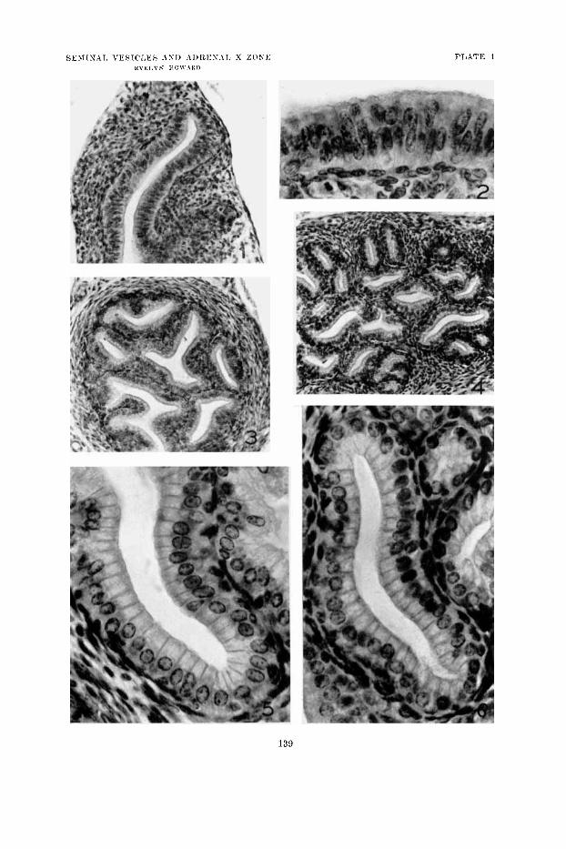
PLATE 1
139

PLATE 2
EXPLANATION O F FIGURES
Differences in the edects of castration performed on adults and on 3-week-old mice, observed after an interval of 20 days. Seminal vesicles. X 200.
Numbers 7, 9 and 10 are litter mates Normal mature secreting vesicle a t 4 1 days of age. Adult type, from a
proximal lobule, showing the epithelial structure characteristic of the peripheral portions of the vesicle. The main body of the vesicle consists of a chamber filled with secretion, tlie lumen of wliicli is often larger than the entire section shown here.
Adult vesicle, 20 days after castration. From m:lin body of vesicle, which has greatly decreased in size. The epithelium shows thc marked regression, characteristic of a la.rgt. part of the veskle. However, the degree of regression 20 days after castration is by no means uniform throughout the vesicle. Compare figure 15 showi~ig typical cuboidal epit,lielium, and figure 2 1 showing coluiiinar epithelium typical of considerable areas, which coexist in such a vesirle.
9 Normal immature wsicle a t 2 1 days of age. Acinous structure is well developed, but vesicle is still relatively small and contains little or no secrrtiou. The epitheiiuni is coluiimir dthough less well devc~lol~vl than is that of tlie adult type.
In Seminal vesiclr from mouse castrated at 21 clays of age and killed after an iuterval of 20 d:iys. The adrenal had a well-dcvelopetl X zone. Notc that the general structure is similar t o that of normal immature mice at 2 1 days of age. The devrloprnent which would normally have occurred lias not taken place sulisequcnt to castration, but neither lias there occurred the degree of regression which follows castration in adults. This degree of uniformity in the epithelial state is characteristie of the entire main body of the vesicles of mice castrated a t 2 1 days of age.
7
8
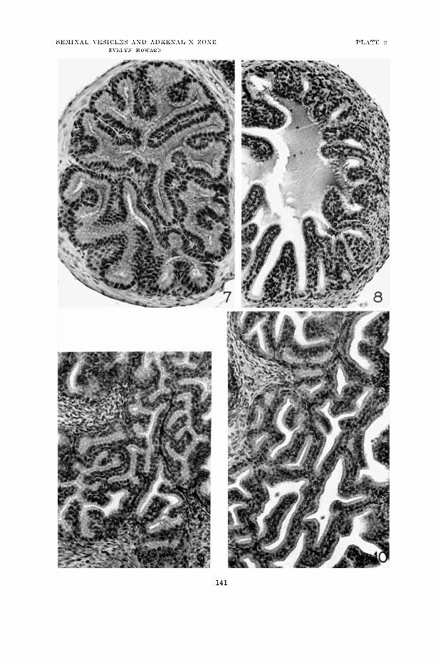
141
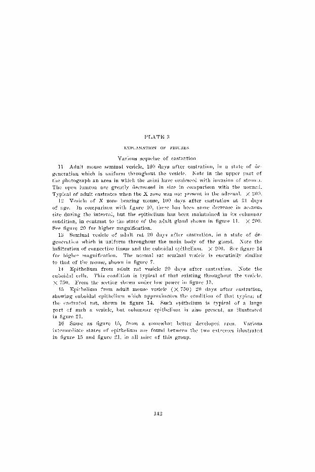
1’IATE 3
EXPLANATION OF FIGURES
Various scquelae of castration
11 Adult mouse seminal vesicle, 100 days after castration, in a st:cte of dc- generation which is uniform througliout the vesicle. Kotc in the upper part of tlic photograph an area i n wliith the aciiii havc coultwed with invasioii of strom:c. The open lumena arc greatly (1ecre;isecl in size in coiiiparison with the norii~:il. Typicaal of adult castrates when the X zone was not present in tlie adrciial. X 200.
12 Vesicle of X zonr I)earing mouse, 100 days after castration a t 2 1 days of age. In coinparison with figure 10, t l rc~e has I ~ c n s o ~ i c t1r:rcIase ill :cc.i:ious size during tlic interv;il, hut the epithelium has been inaintaincd in its colunmar condition, in contrast to tlic, state of tlie adult gland shown in figure 11. X 200. Sce figure 20 for higher maguifieation.
13 Seniinal vesicle of adult rat 20 days after castration, in a state of de- geiicr;ition which is uniforni throughout the main body of the gland. So tc the infiltration of connectivc tissue and tlic cuhoidal epithelium. x 200. Scc figure 14 for liiglier mapiification. The normal rat seuiiiial vcsicle is es;cntiall~ siiiiilar to that of th r mouse, slro1vn in figure 7.
14 Epithcliuni froin adult rat vesicle 20 days after castration. Yote the cuboid:il cc~lls. This contlition is typical of that esisting throughout the yesitle. x 750.
15 12pithelium from adult niousv vesiclc ( X i 5 0 ) 20 days after castratioii, showing cuboidal epitliclinni wliicli ap1)rosini:itrs the condition of that typic:il of the c;istr:rted mt , shown in figure 14. Such epithcliuni is typical of a large p:irt cf such a vesicle, b u t czolunin:w epithcliuiu is also prescnt, as illustratctl in figure 21.
16 Same as figure 15, from a solnewhat better tlevelopcd area. Varinus intcrmrcliatc states o f cpitheliuin a re found lietween tlic two estreinw illustratcd in figure 15 and figure 21, in :dl mice of this group.
From the section sliown nnc1c.r low power in figure 13.
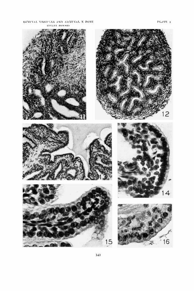
143
T’IA7’1.: 4
’LANATION O F FIGURES
(‘oii1r;ists in tli:. vff‘cyh of castmtion lierforiirecl on adults :iud on 21-dn~-old iiiiw, olisc~rvcd :ifter a n interval of 1 C I O t1:iys. X 750.
1 7 l~~pitlieliuiii of iioriii:il sccretiiig vcsiclc c;f tlie :rdult type. Xote the liigli F ~ O I I I c.oluiiiiinr cells with cyto1il:isiiiic gr:iunlrs, and t l iv sccrction in t lw 1unwn.
vesicle sliowii :iIso in figure 7. Kote the uiiiformlj-
culioic1:il c~ l l s , aiid tlie al)sc~iice of wagulatcd sccretioii. Such epitlicliniii is c1inr:lc- teristic of tlie rcsiele (except, for portions which sliow iiiore :itlv:iucctl structural disiiitcgration) 100 days after castratioii of the adult, when tlie atlrcn:il has no x s011e.
19 S o t e t h a t the cpitlicliuiii is quite well dercloprd coluiiiii:ir, ljut still imiiiuture and less we11 derc~loped tli:iii tliat of tlie adult, iii figure 17. (Fro111 scctioii sliomii in f i g . 9.)
Epitlieliuni of resiclc of X zoiicf bcariiig mouse c:isti.atc~l :it 2 1 days of :ige, a f t e r ail interval of 100 (lays. Kote t1i:it the epitlirliuiii i s still iiiimnturc., 1)ut t hx t i t is coiiiposcd of coluuiiiar cells wliicli 1i:ive ui1drrgoiie little or 110 regrc~ssion froii i the condi t ion iioriiml for the ngc a t \vliicli castration w:is perforiiiecl, slioivii in figiire 19.
18 Epitlirliuiii of adult vesiclc 100 (lays a f tc r castrntion.
Epitlieliuin of norin:il vcsiclc :it 2 1 days of age.
20
From t lw speciiiicu sliowii iii figure 12 .
144
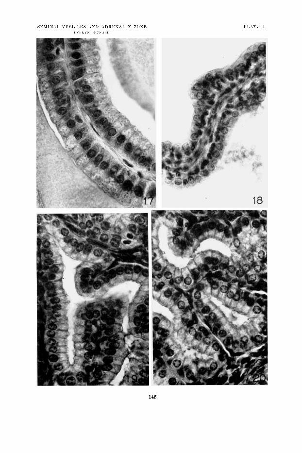
143
PLATE 3
EXPLANATION OF’ FIGURES
Seniiiial vesiclcs X T50 Epithcliuiii f rom :in adult I I I O U S ~ r-c~sic~le, 20 tl:rys a f te r c:rstr:rtion. Suvh
epitheliuni is typicaal of a portion of all such vesicles. Kote t h a t :ilthougli these cells have uudergoiie drgeuer:ition iii conipnrison with the normal condition shown in figure li, nwertlieless they have retained a considerable amount of their cytoplasm, so th:ct they must still be c*l:issificd a s columnar cells. Thus the reaction of the adult iuouse rmicle to caxtratioii is l int a s uniform as is t h a t of the rat. The :idren:ils of specinicns shown in figures 15, 1 G and 2 1 are in the process of differcntiatiug secontlarj- X z o i i ~ .
2 2 Vesicle of mouse at, 41 days of :igc, 20 days a f te r castration, with a well-derelopril primary X zone. Kote the well developed coluiiinar epitheliuin, typical of the condition throughout the resiclcs of such :inim:rls.
Vcsicle of mouse castrated a t 21 d:iys of age, a f te r a n interval of 200 (lays, during which the primary X zone had degcnerntcd and clisnp1~eared, and secondary X zone had n o t becoiur (1ifTerentiatcd. Note the srri:ill acini, aiid rather low cc~lls w i t h attenuated cytol~lnsiii, wliich arc howercr, still not t ruly cuboidal. Typical of g r w t e s t degree of tlc.gciieratioii usually attainctl iu mice of this group.
2 1 Vcsiclc of mouse castr:rtctl :it 2 1 d e ~ s , af ter an interval of 250 d:i>-s, (luring whic#li the ~ i r i inary X ZOIIC had dcgciiernted, ;ind :t well-tlevelol,cd scont la ry X zouc hat1 differenti:rtcd. Kotc the ~~.cll-niaiiitaiii~,(l epithelium which is little if a t all inferior in condition to tliat norui:rl for the 21-daT-old niouse, shown in figure 19. Other iii(lix-idu:ils were less well n~ai i~ t :~ i i icd than this one, ivhirh was choscn to illustrate the fact that ilcgeiier:itiou is not ntccssarily markcd af te r the disappe:iraiicc of priiu:iry X zoue. Siic~h cpitlicliuni was typic.:il of t!lc greater par t (if thr, wsiclc of this :iuini:il.
2 1
2 3
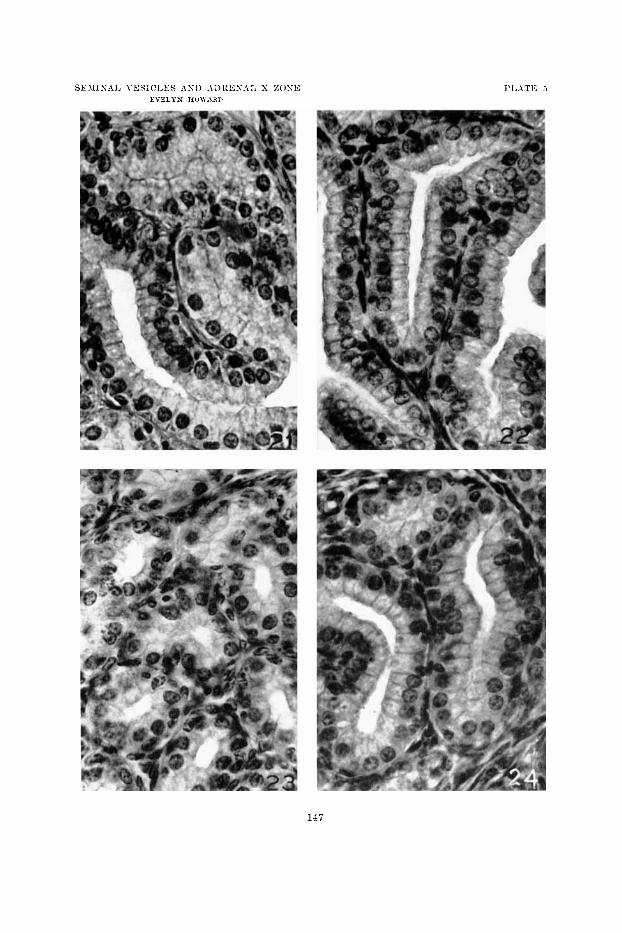
147
P L A T E 6
EXPLIXATION O F FIGURES
Adrcnals of noriiml male niict,, aiid of castratw with priiiiaq- and secondary X zone.
2.i A4tlre1ial of iioriiiul iii:ilc a t l ( i (lays c;f : I ~ c . S o t e the nnrrom X zone :rliove the liglit st:iiiiiiig iiieclullnry cells. Tlie houiid:irios of the I; zone are iiidicnttxtl Iiy ink ni:irks on the riglit edgc of tlicl section.
26 Adreiial of iiialc :it 16 days of age>, w1iic.h had been castrated :it 5 days of age. The X zonc boundaries :ire iiidieatrcl by ink niarks 011 tlie lrft cdgc of the section. x 2 3 0 .
27 Xote the priiiiary X zone, yrc:ttly eiilarged as :I result of c:istr:ition. X 100.
28 Adrenal of normal i n a l c ~ , 41 (lays of age. Gland is of adult type, :mtl without S zone, the prim:iry X zoiie 1i:iring dcgeneratetl. $'roiii B l i t t tr inate of figurc 2 i . X 100.
P!) Adiwial of ni:ile 38ti (lays of :rgc, which has 1)een c:istratrtl ;it 306 diiys of age. Note the typical secondary X zone wliich dereloped froin an adult gland from which tlie priiiiary X zoiie 1i:id previously disalqieared.
X 530.
Note the ciil:irgcinent of the Y zone, by comparison with figure 23.
Adrenal of niale a t 4 1 clays of age, c.:istratcd a t 51 d a y .
x 100.
145
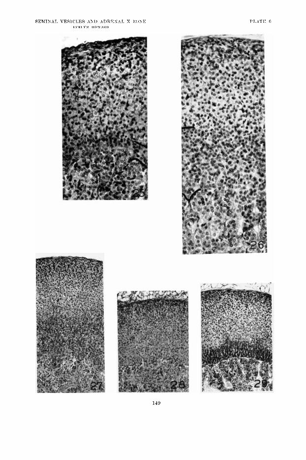
149


![Comparison of penile length at 6–24 months between ... · size of the testes, prostate, seminal vesicles and penis [4,5]. In some reports, it is suggested that congenital cryptorchidism](https://static.fdocuments.in/doc/165x107/5ec848726e987a7df11da875/comparison-of-penile-length-at-6a24-months-between-size-of-the-testes-prostate.jpg)


