Effectiveness of Clavicula Pro Humero Reconstruction for Elderly ...
7
Case Report Effectiveness of Clavicula Pro Humero Reconstruction for Elderly Patients: Report of Two Cases Sho Okimatsu, 1,2 Hiroto Kamoda, 1 Tsukasa Yonemoto, 1 Shintaro Iwata, 1 and Takeshi Ishii 1 1 Division of Orthopedic Surgery, Chiba Cancer Center, Chiba 260-8717, Japan 2 Department of Orthopedic Surgery, Chiba University, Chiba 260-8677, Japan Correspondence should be addressed to Hiroto Kamoda; [email protected] Received 26 May 2016; Accepted 3 October 2016 Academic Editor: Jose I. Mayordomo Copyright © 2016 Sho Okimatsu et al. is is an open access article distributed under the Creative Commons Attribution License, which permits unrestricted use, distribution, and reproduction in any medium, provided the original work is properly cited. Clavicula pro humero (CPH) reconstruction is a method that is used aſter proximal humeral excision. During CPH reconstruction, the ipsilateral clavicle is rotated downward and connected to the preserved distal humerus by using plates and screws. is method is frequently used for reconstruction surgeries involving young patients and has positive outcomes. In this study, we describe two cases of CPH reconstruction that were performed on elderly individuals aſter wide resection of the proximal humerus; postoperative results from these surgeries were satisfactory. e average Musculoskeletal Tumor Society (MSTS) functional score aſter surgery was 68.5%, indicating that CPH reconstruction is suitable for not only younger but also elderly patients, particularly those over the age of 65 years. 1. Introduction e proximal humerus is a common site of both primary and metastatic bone tumors [1, 2]. Several techniques have been established for the reconstruction of the proximal humerus aſter resection of malignant tumors. However, as no single approach has been deemed superior, the approach used is determined on a case-by-case basis by the surgeon. One such reconstructive technique is clavicula pro humero (CPH), which was originally described for reconstruction of the upper extremity in children with limb deficiencies [3]. In this study, we describe the outcomes of CPH reconstruction in two elderly patients. e postoperative results from these surgeries were comparable to those of other reconstructive options. We conclude that CPH is a suitable reconstructive technique not only for the young, but also for some elderly patients aſter tumor resection from the proximal humerus. 2. Case Presentation Case 1. A 67-year-old man presented with pain and swelling in the right shoulder. He was diagnosed to have metastatic thyroid carcinoma at another facility four years ago and was treated with radioactive iodine. At initial presentation, he also underwent open reduction and internal fixation of the right proximal humerus for a pathological fracture with the T2 Humeral Nail (Stryker, Kalamazoo, Milwaukee, USA), followed by radiation therapy to the right humerus. He presented to our institution with plain radiographs of his right shoulder showing an osteolytic lesion of the head of the right humerus along with soſt tissue swelling of the proximal arm. A computed tomography (CT) scan showed the disappearance of the proximal humeral cortex (Figure 1). Magnetic resonance imaging (MRI) showed the lesion to have mild high signal changes on both T1 and T2 weighted imaging, extending to the surrounding muscles (Figure 2). Needle biopsy confirmed recurrent metastatic thyroid carcinoma. Aſter counseling the patient, a surgical resection and reconstruction were planned. Preoperative embolization of tumor feeding vessels was carried out by interventional radiologists. e surgical incision extended from the medial side of the right clavicle, across the coracoid process, extending to the distal end of the right humerus and included the needle biopsy tract. e tumor and surrounding muscles were carefully resected en bloc with a humeral intramedullary rod. e axillary nerve and posterior humeral circumflex vessels were sacrificed. e right clavicle was Hindawi Publishing Corporation Case Reports in Oncological Medicine Volume 2016, Article ID 4140239, 6 pages http://dx.doi.org/10.1155/2016/4140239
Transcript of Effectiveness of Clavicula Pro Humero Reconstruction for Elderly ...
Case ReportEffectiveness of Clavicula Pro Humero Reconstruction forElderly Patients: Report of Two Cases
Sho Okimatsu,1,2 Hiroto Kamoda,1 Tsukasa Yonemoto,1 Shintaro Iwata,1 and Takeshi Ishii1
1Division of Orthopedic Surgery, Chiba Cancer Center, Chiba 260-8717, Japan2Department of Orthopedic Surgery, Chiba University, Chiba 260-8677, Japan
Correspondence should be addressed to Hiroto Kamoda; [email protected]
Received 26 May 2016; Accepted 3 October 2016
Academic Editor: Jose I. Mayordomo
Copyright © 2016 Sho Okimatsu et al. This is an open access article distributed under the Creative Commons Attribution License,which permits unrestricted use, distribution, and reproduction in any medium, provided the original work is properly cited.
Clavicula pro humero (CPH) reconstruction is a method that is used after proximal humeral excision. During CPH reconstruction,the ipsilateral clavicle is rotated downward and connected to the preserved distal humerus by using plates and screws.This methodis frequently used for reconstruction surgeries involving young patients and has positive outcomes. In this study, we describe twocases of CPH reconstruction that were performed on elderly individuals afterwide resection of the proximal humerus; postoperativeresults from these surgeries were satisfactory. The average Musculoskeletal Tumor Society (MSTS) functional score after surgerywas 68.5%, indicating that CPH reconstruction is suitable for not only younger but also elderly patients, particularly those over theage of 65 years.
1. Introduction
The proximal humerus is a common site of both primary andmetastatic bone tumors [1, 2]. Several techniques have beenestablished for the reconstruction of the proximal humerusafter resection of malignant tumors. However, as no singleapproach has been deemed superior, the approach used isdetermined on a case-by-case basis by the surgeon. One suchreconstructive technique is clavicula pro humero (CPH),which was originally described for reconstruction of theupper extremity in children with limb deficiencies [3]. Inthis study, we describe the outcomes of CPH reconstructionin two elderly patients. The postoperative results from thesesurgeries were comparable to those of other reconstructiveoptions. We conclude that CPH is a suitable reconstructivetechnique not only for the young, but also for some elderlypatients after tumor resection from the proximal humerus.
2. Case Presentation
Case 1. A 67-year-old man presented with pain and swellingin the right shoulder. He was diagnosed to have metastaticthyroid carcinoma at another facility four years ago and wastreated with radioactive iodine.
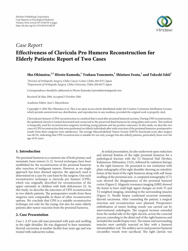
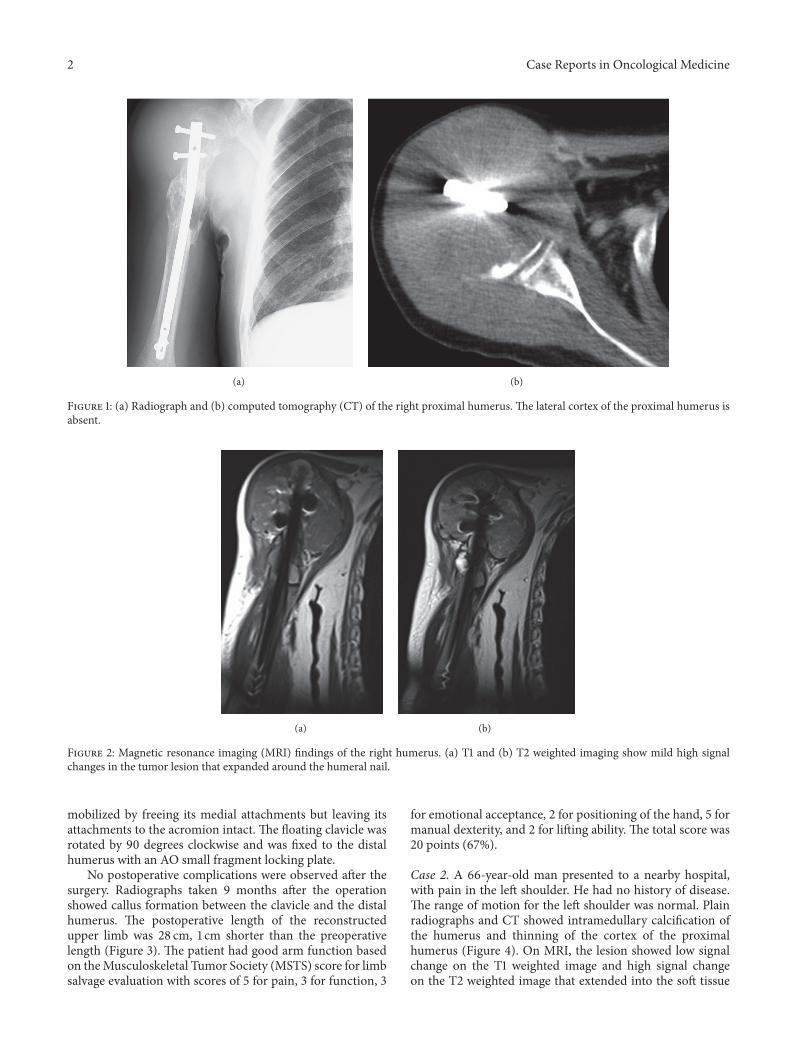
At initial presentation, he also underwent open reductionand internal fixation of the right proximal humerus for apathological fracture with the T2 Humeral Nail (Stryker,Kalamazoo, Milwaukee, USA), followed by radiation therapyto the right humerus. He presented to our institution withplain radiographs of his right shoulder showing an osteolyticlesion of the head of the right humerus along with soft tissueswelling of the proximal arm. A computed tomography (CT)scan showed the disappearance of the proximal humeralcortex (Figure 1).Magnetic resonance imaging (MRI) showedthe lesion to have mild high signal changes on both T1 andT2 weighted imaging, extending to the surrounding muscles(Figure 2). Needle biopsy confirmed recurrent metastaticthyroid carcinoma. After counseling the patient, a surgicalresection and reconstruction were planned. Preoperativeembolization of tumor feeding vessels was carried out byinterventional radiologists. The surgical incision extendedfrom the medial side of the right clavicle, across the coracoidprocess, extending to the distal end of the right humerus andincluded the needle biopsy tract.The tumor and surroundingmuscles were carefully resected en bloc with a humeralintramedullary rod.The axillary nerve and posterior humeralcircumflex vessels were sacrificed. The right clavicle was
Hindawi Publishing CorporationCase Reports in Oncological MedicineVolume 2016, Article ID 4140239, 6 pageshttp://dx.doi.org/10.1155/2016/4140239
2 Case Reports in Oncological Medicine
(a) (b)
Figure 1: (a) Radiograph and (b) computed tomography (CT) of the right proximal humerus. The lateral cortex of the proximal humerus isabsent.
(a) (b)
Figure 2: Magnetic resonance imaging (MRI) findings of the right humerus. (a) T1 and (b) T2 weighted imaging show mild high signalchanges in the tumor lesion that expanded around the humeral nail.
mobilized by freeing its medial attachments but leaving itsattachments to the acromion intact. The floating clavicle wasrotated by 90 degrees clockwise and was fixed to the distalhumerus with an AO small fragment locking plate.
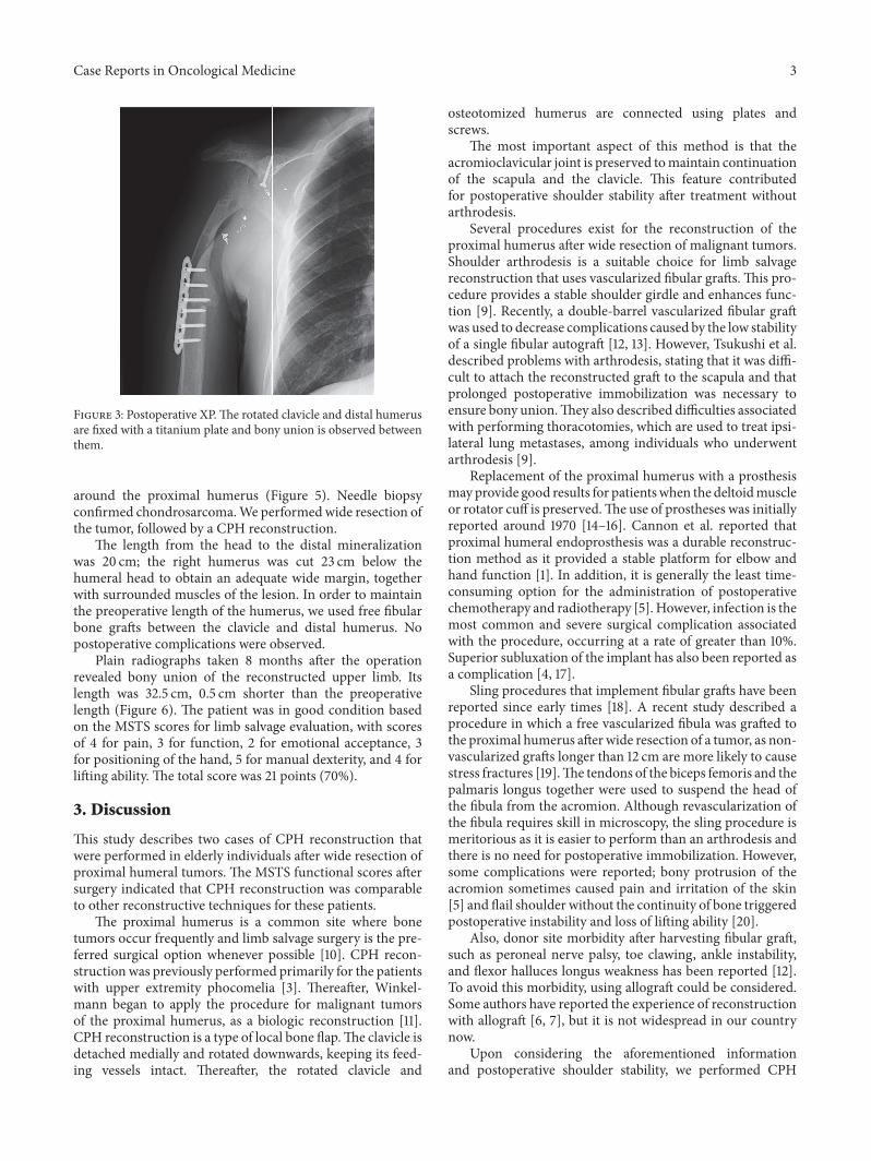
No postoperative complications were observed after thesurgery. Radiographs taken 9 months after the operationshowed callus formation between the clavicle and the distalhumerus. The postoperative length of the reconstructedupper limb was 28 cm, 1 cm shorter than the preoperativelength (Figure 3). The patient had good arm function basedon theMusculoskeletal Tumor Society (MSTS) score for limbsalvage evaluation with scores of 5 for pain, 3 for function, 3
for emotional acceptance, 2 for positioning of the hand, 5 formanual dexterity, and 2 for lifting ability. The total score was20 points (67%).
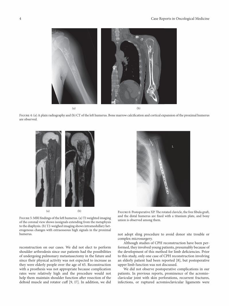
Case 2. A 66-year-old man presented to a nearby hospital,with pain in the left shoulder. He had no history of disease.The range of motion for the left shoulder was normal. Plainradiographs and CT showed intramedullary calcification ofthe humerus and thinning of the cortex of the proximalhumerus (Figure 4). On MRI, the lesion showed low signalchange on the T1 weighted image and high signal changeon the T2 weighted image that extended into the soft tissue
Case Reports in Oncological Medicine 3
Figure 3: Postoperative XP.The rotated clavicle and distal humerusare fixed with a titanium plate and bony union is observed betweenthem.
around the proximal humerus (Figure 5). Needle biopsyconfirmed chondrosarcoma.We performedwide resection ofthe tumor, followed by a CPH reconstruction.
The length from the head to the distal mineralizationwas 20 cm; the right humerus was cut 23 cm below thehumeral head to obtain an adequate wide margin, togetherwith surrounded muscles of the lesion. In order to maintainthe preoperative length of the humerus, we used free fibularbone grafts between the clavicle and distal humerus. Nopostoperative complications were observed.
Plain radiographs taken 8 months after the operationrevealed bony union of the reconstructed upper limb. Itslength was 32.5 cm, 0.5 cm shorter than the preoperativelength (Figure 6). The patient was in good condition basedon the MSTS scores for limb salvage evaluation, with scoresof 4 for pain, 3 for function, 2 for emotional acceptance, 3for positioning of the hand, 5 for manual dexterity, and 4 forlifting ability. The total score was 21 points (70%).
3. Discussion
This study describes two cases of CPH reconstruction thatwere performed in elderly individuals after wide resection ofproximal humeral tumors. The MSTS functional scores aftersurgery indicated that CPH reconstruction was comparableto other reconstructive techniques for these patients.
The proximal humerus is a common site where bonetumors occur frequently and limb salvage surgery is the pre-ferred surgical option whenever possible [10]. CPH recon-structionwas previously performed primarily for the patientswith upper extremity phocomelia [3]. Thereafter, Winkel-mann began to apply the procedure for malignant tumorsof the proximal humerus, as a biologic reconstruction [11].CPH reconstruction is a type of local bone flap.The clavicle isdetached medially and rotated downwards, keeping its feed-ing vessels intact. Thereafter, the rotated clavicle and
osteotomized humerus are connected using plates andscrews.
The most important aspect of this method is that theacromioclavicular joint is preserved tomaintain continuationof the scapula and the clavicle. This feature contributedfor postoperative shoulder stability after treatment withoutarthrodesis.
Several procedures exist for the reconstruction of theproximal humerus after wide resection of malignant tumors.Shoulder arthrodesis is a suitable choice for limb salvagereconstruction that uses vascularized fibular grafts. This pro-cedure provides a stable shoulder girdle and enhances func-tion [9]. Recently, a double-barrel vascularized fibular graftwas used to decrease complications caused by the low stabilityof a single fibular autograft [12, 13]. However, Tsukushi et al.described problems with arthrodesis, stating that it was diffi-cult to attach the reconstructed graft to the scapula and thatprolonged postoperative immobilization was necessary toensure bony union.They also described difficulties associatedwith performing thoracotomies, which are used to treat ipsi-lateral lung metastases, among individuals who underwentarthrodesis [9].
Replacement of the proximal humerus with a prosthesismay provide good results for patientswhen the deltoidmuscleor rotator cuff is preserved.The use of prostheses was initiallyreported around 1970 [14–16]. Cannon et al. reported thatproximal humeral endoprosthesis was a durable reconstruc-tion method as it provided a stable platform for elbow andhand function [1]. In addition, it is generally the least time-consuming option for the administration of postoperativechemotherapy and radiotherapy [5]. However, infection is themost common and severe surgical complication associatedwith the procedure, occurring at a rate of greater than 10%.Superior subluxation of the implant has also been reported asa complication [4, 17].
Sling procedures that implement fibular grafts have beenreported since early times [18]. A recent study described aprocedure in which a free vascularized fibula was grafted tothe proximal humerus afterwide resection of a tumor, as non-vascularized grafts longer than 12 cm are more likely to causestress fractures [19].The tendons of the biceps femoris and thepalmaris longus together were used to suspend the head ofthe fibula from the acromion. Although revascularization ofthe fibula requires skill in microscopy, the sling procedure ismeritorious as it is easier to perform than an arthrodesis andthere is no need for postoperative immobilization. However,some complications were reported; bony protrusion of theacromion sometimes caused pain and irritation of the skin[5] and flail shoulder without the continuity of bone triggeredpostoperative instability and loss of lifting ability [20].
Also, donor site morbidity after harvesting fibular graft,such as peroneal nerve palsy, toe clawing, ankle instability,and flexor halluces longus weakness has been reported [12].To avoid this morbidity, using allograft could be considered.Some authors have reported the experience of reconstructionwith allograft [6, 7], but it is not widespread in our countrynow.
Upon considering the aforementioned informationand postoperative shoulder stability, we performed CPH
4 Case Reports in Oncological Medicine
(a) (b)
Figure 4: (a) A plain radiography and (b) CT of the left humerus. Bonemarrow calcification and cortical expansion of the proximal humerusare observed.
(a) (b)
Figure 5:MRI findings of the left humerus. (a) T1 weighted imagingof the coronal view shows isosignals extending from the metaphysisto the diaphysis. (b) T2-weighed imaging shows intramedullary het-erogenous changes with extraosseous high signals in the proximalhumerus.
reconstruction on our cases. We did not elect to performshoulder arthrodesis since our patients had the possibilitiesof undergoing pulmonary metastasectomy in the future andsince their physical activity was not expected to increase asthey were elderly people over the age of 65. Reconstructionwith a prosthesis was not appropriate because complicationrates were relatively high and the procedure would nothelp them maintain shoulder function after resection of thedeltoid muscle and rotator cuff [9, 17]. In addition, we did
L
Figure 6: Postoperative XP.The rotated clavicle, the free fibula graft,and the distal humerus are fixed with a titanium plate, and bonyunion is observed among them.
not adopt sling procedure to avoid donor site trouble orcomplex microsurgery.
Although studies of CPH reconstruction have been per-formed, they involved young patients, presumably because ofthe development of this method for limb deficiencies. Priorto this study, only one case of CPH reconstruction involvingan elderly patient had been reported [8], but postoperativeupper limb function was not discussed.
We did not observe postoperative complications in ourpatients. In previous reports, prominence of the acromio-clavicular joint with skin perforations, recurrent fractures,infections, or ruptured acromioclavicular ligaments were
Case Reports in Oncological Medicine 5
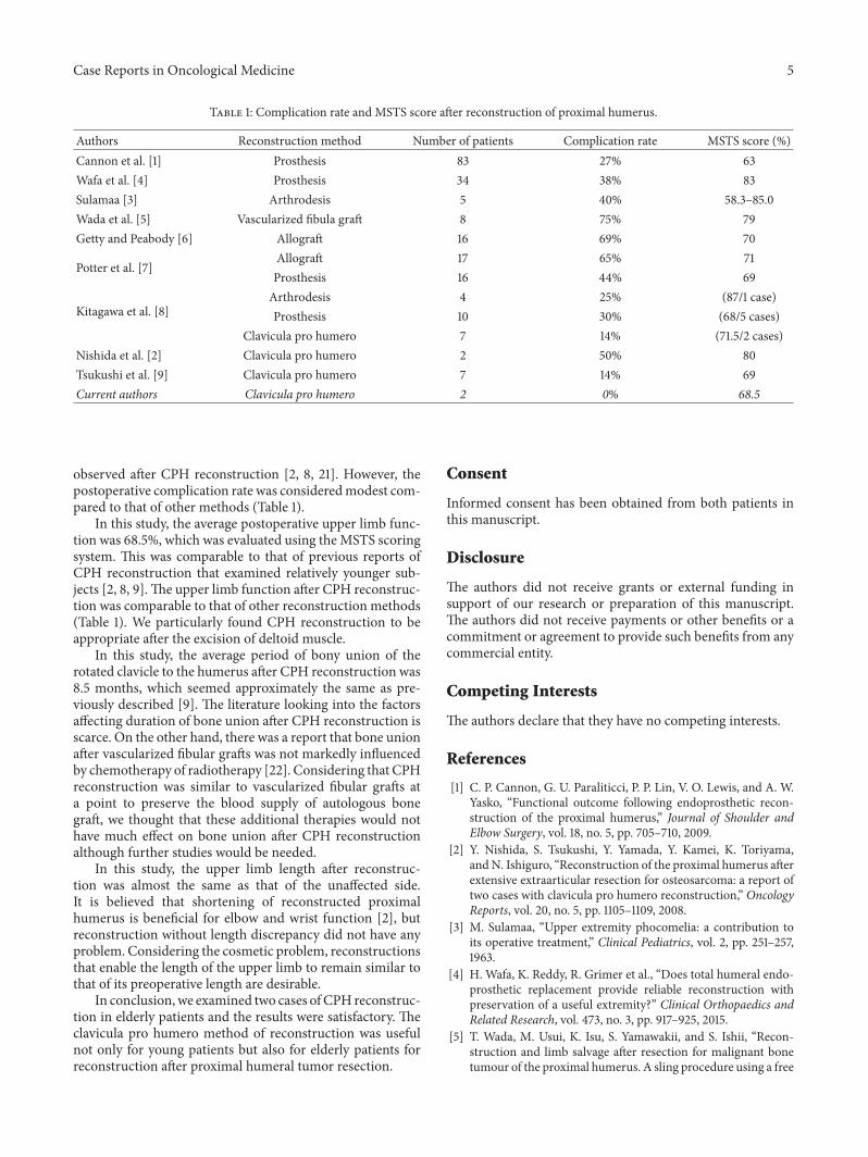
Table 1: Complication rate and MSTS score after reconstruction of proximal humerus.
Authors Reconstruction method Number of patients Complication rate MSTS score (%)Cannon et al. [1] Prosthesis 83 27% 63Wafa et al. [4] Prosthesis 34 38% 83Sulamaa [3] Arthrodesis 5 40% 58.3–85.0Wada et al. [5] Vascularized fibula graft 8 75% 79Getty and Peabody [6] Allograft 16 69% 70
Potter et al. [7]Allograft 17 65% 71Prosthesis 16 44% 69
Kitagawa et al. [8]Arthrodesis 4 25% (87/1 case)Prosthesis 10 30% (68/5 cases)
Clavicula pro humero 7 14% (71.5/2 cases)Nishida et al. [2] Clavicula pro humero 2 50% 80Tsukushi et al. [9] Clavicula pro humero 7 14% 69Current authors Clavicula pro humero 2 0% 68.5
observed after CPH reconstruction [2, 8, 21]. However, thepostoperative complication rate was consideredmodest com-pared to that of other methods (Table 1).
In this study, the average postoperative upper limb func-tion was 68.5%, which was evaluated using theMSTS scoringsystem. This was comparable to that of previous reports ofCPH reconstruction that examined relatively younger sub-jects [2, 8, 9].The upper limb function after CPH reconstruc-tion was comparable to that of other reconstruction methods(Table 1). We particularly found CPH reconstruction to beappropriate after the excision of deltoid muscle.
In this study, the average period of bony union of therotated clavicle to the humerus after CPH reconstruction was8.5 months, which seemed approximately the same as pre-viously described [9]. The literature looking into the factorsaffecting duration of bone union after CPH reconstruction isscarce. On the other hand, there was a report that bone unionafter vascularized fibular grafts was not markedly influencedby chemotherapy of radiotherapy [22]. Considering that CPHreconstruction was similar to vascularized fibular grafts ata point to preserve the blood supply of autologous bonegraft, we thought that these additional therapies would nothave much effect on bone union after CPH reconstructionalthough further studies would be needed.
In this study, the upper limb length after reconstruc-tion was almost the same as that of the unaffected side.It is believed that shortening of reconstructed proximalhumerus is beneficial for elbow and wrist function [2], butreconstruction without length discrepancy did not have anyproblem. Considering the cosmetic problem, reconstructionsthat enable the length of the upper limb to remain similar tothat of its preoperative length are desirable.
In conclusion,we examined two cases ofCPH reconstruc-tion in elderly patients and the results were satisfactory. Theclavicula pro humero method of reconstruction was usefulnot only for young patients but also for elderly patients forreconstruction after proximal humeral tumor resection.
Consent
Informed consent has been obtained from both patients inthis manuscript.
Disclosure
The authors did not receive grants or external funding insupport of our research or preparation of this manuscript.The authors did not receive payments or other benefits or acommitment or agreement to provide such benefits from anycommercial entity.
Competing Interests
The authors declare that they have no competing interests.
References
[1] C. P. Cannon, G. U. Paraliticci, P. P. Lin, V. O. Lewis, and A. W.Yasko, “Functional outcome following endoprosthetic recon-struction of the proximal humerus,” Journal of Shoulder andElbow Surgery, vol. 18, no. 5, pp. 705–710, 2009.
[2] Y. Nishida, S. Tsukushi, Y. Yamada, Y. Kamei, K. Toriyama,andN. Ishiguro, “Reconstruction of the proximal humerus afterextensive extraarticular resection for osteosarcoma: a report oftwo cases with clavicula pro humero reconstruction,” OncologyReports, vol. 20, no. 5, pp. 1105–1109, 2008.
[3] M. Sulamaa, “Upper extremity phocomelia: a contribution toits operative treatment,” Clinical Pediatrics, vol. 2, pp. 251–257,1963.
[4] H. Wafa, K. Reddy, R. Grimer et al., “Does total humeral endo-prosthetic replacement provide reliable reconstruction withpreservation of a useful extremity?” Clinical Orthopaedics andRelated Research, vol. 473, no. 3, pp. 917–925, 2015.
[5] T. Wada, M. Usui, K. Isu, S. Yamawakii, and S. Ishii, “Recon-struction and limb salvage after resection for malignant bonetumour of the proximal humerus. A sling procedure using a free

6 Case Reports in Oncological Medicine
vascularised fibular graft,”The Journal of Bone& Joint Surgery—British Volume, vol. 81, no. 5, pp. 808–813, 1999.
[6] P. J. Getty and T. D. Peabody, “Complications and functionaloutcomes of reconstruction with an osteoarticular allograftafter intra-articular resection of the proximal aspect of thehumerus,” Journal of Bone and Joint Surgery—Series A, vol. 81,no. 8, pp. 1138–1146, 1999.
[7] B. K. Potter, S. C. Adams, J. D. Pitcher Jr., T. I. Malinin, andH. T. Temple, “Proximal humerus reconstructions for tumors,”Clinical Orthopaedics and Related Research, vol. 467, no. 4, pp.1035–1041, 2009.
[8] Y. Kitagawa, D. M. Thai, and P. F. Choong, “Reconstructionsof the shoulder following tumour resection,” Journal of Ortho-paedic Surgery, vol. 15, no. 2, pp. 201–206, 2007.
[9] S. Tsukushi, Y. Nishida, M. Takahashi, and N. Ishiguro, “Clav-icula pro humero reconstruction after wide resection of theproximal humerus,”Clinical Orthopaedics and Related Research,no. 447, pp. 132–137, 2006.
[10] Y. Mimata, J. Nishida, K. Sato, Y. Suzuki, and M. Doita,“Glenohumeral arthrodesis formalignant tumor of the shouldergirdle,” Journal of Shoulder and Elbow Surgery, vol. 24, no. 2, pp.174–178, 2015.
[11] W. Winkelmann, “Clavicula pro Humero—eine neue Opera-tionsmethode fur maligne Tumoren des proximalen Humerus,”Zeitschrift fur Orthopadie undUnfallchirurgie, vol. 130, no. 3, pp.197–201, 1992.
[12] J. Nishida and T. Shimamura, “Vascularized bone reconstruc-tive approaches after tumor resection,” Current OrthopaedicPractice, vol. 22, no. 4, pp. 309–314, 2011.
[13] K. Wieser, K. Modaressi, F. Seeli, and B. Fuchs, “Autologousdouble-barrel vascularized fibula bone graft for arthrodesis ofthe shoulder after tumor resection,”Archives of Orthopaedic andTrauma Surgery, vol. 133, no. 9, pp. 1219–1224, 2013.
[14] H. J. Burrows, J. N.Wilson, and J. T. Scales, “Excision of tumoursof humerus and femur, with restoration by internal prostheses,”Journal of Bone and Joint Surgery—Series B, vol. 57, no. 2, pp.148–159, 1975.
[15] F. F. Parrish, “Allograft replacement of all or part of the end of along bone following excision of a tumor,”The Journal of Bone &Joint Surgery—American Volume, vol. 55, no. 1, pp. 1–22, 1973.
[16] G. Mollowitz, “Vitalium endprosthesis in sarcoma of the proxi-mal humerus,” Chirurg, vol. 37, no. 3, pp. 130–132, 1966.
[17] M. I. O’Connor, F. H. Sim, and E. Y. S. Chao, “Limb salvage forneoplasms of the shoulder girdle: intermediate reconstructiveand functional results,” The Journal of Bone & Joint Surgery—American Volume, vol. 78, no. 12, pp. 1872–1888, 1996.
[18] K. Clark, “A case of replacement of the upper end of thehumerus by a fibular graft reviewed after twenty-nine years,”TheJournal of Bone & Joint Surgery—British Volume, vol. 41, no. 2,pp. 365–368, 1959.
[19] W. F. Enneking, J. L. Eady, and H. Burchardt, “Autogenouscortical bone grafts in the reconstruction of segmental skeletaldefects,” Journal of Bone and Joint Surgery—Series A, vol. 62, no.7, pp. 1039–1058, 1980.
[20] Y. Nishida and S. Tsukushi, “Reconstruction modalities afterresection for malignantbone tumor of the proximal humerus,”Kotsu Kansetsu Jintai, vol. 19-12, pp. 1143–1150, 2006.
[21] R.W.Rodl, G.Gosheger, C.Gebert, N. Lindner, T.Ozaki, andW.Winkelmann, “Reconstruction of the proximal humerus afterwide resection of tumours,” The Journal of Bone & Joint Sur-gery—British Volume, vol. 84, no. 7, pp. 1004–1008, 2002.
[22] P.H.Hilven, L. Bayliss, T. Cosker et al., “The vascularised fibulargraft for limb salvage after bone tumour surgery: AMulticentreStudy,”Bone and Joint Journal, vol. 97-B, no. 6, pp. 853–861, 2015.

Submit your manuscripts athttp://www.hindawi.com
Stem CellsInternational
Hindawi Publishing Corporationhttp://www.hindawi.com Volume 2014
Hindawi Publishing Corporationhttp://www.hindawi.com Volume 2014
MEDIATORSINFLAMMATION
of
Hindawi Publishing Corporationhttp://www.hindawi.com Volume 2014
Behavioural Neurology
EndocrinologyInternational Journal of
Hindawi Publishing Corporationhttp://www.hindawi.com Volume 2014
Hindawi Publishing Corporationhttp://www.hindawi.com Volume 2014
Disease Markers
Hindawi Publishing Corporationhttp://www.hindawi.com Volume 2014
BioMed Research International
OncologyJournal of
Hindawi Publishing Corporationhttp://www.hindawi.com Volume 2014
Hindawi Publishing Corporationhttp://www.hindawi.com Volume 2014
Oxidative Medicine and Cellular Longevity
Hindawi Publishing Corporationhttp://www.hindawi.com Volume 2014
PPAR Research
The Scientific World JournalHindawi Publishing Corporation http://www.hindawi.com Volume 2014
Immunology ResearchHindawi Publishing Corporationhttp://www.hindawi.com Volume 2014
Journal of
ObesityJournal of
Hindawi Publishing Corporationhttp://www.hindawi.com Volume 2014
Hindawi Publishing Corporationhttp://www.hindawi.com Volume 2014
Computational and Mathematical Methods in Medicine
OphthalmologyJournal of
Hindawi Publishing Corporationhttp://www.hindawi.com Volume 2014
Diabetes ResearchJournal of
Hindawi Publishing Corporationhttp://www.hindawi.com Volume 2014
Hindawi Publishing Corporationhttp://www.hindawi.com Volume 2014
Research and TreatmentAIDS
Hindawi Publishing Corporationhttp://www.hindawi.com Volume 2014
Gastroenterology Research and Practice
Hindawi Publishing Corporationhttp://www.hindawi.com Volume 2014
Parkinson’s Disease
Evidence-Based Complementary and Alternative Medicine
Volume 2014Hindawi Publishing Corporationhttp://www.hindawi.com