Effectiveness of Chest CT for Triage in the Emergency ...
12
Jurnal of Bionursing 122 Jurnal of Bionursing 2021, VOL. 3, NO. 2, 122-133 Effectiveness of Chest CT for Triage in the Emergency Departmet (ED) in Patients with Suspected COVID-19: A Literature Review Aniq Lutfiyah 1 , Iwan Purnawan 2 , Ridlwan Kamaluddin 3 1 Mahasiswa Profesi Ners Universitas Jenderal Soedirma 2,3 Dosen Jurusan Keperawatan Fikes Unsoed KEYWORDS Chest CT, triage, Emergency Department, COVID-19 PENDAHULUAN Corona Virus Disease 19 (COVID-19) telah dinyatakan sebagai pandemi dunia oleh WHO (WHO 2020). COVID-19 disebabkan oleh virus Sindrom Pernafasan Akut Parah Coronavirus-2 (SARS-CoV-2). Virus dan penyakit ini diketahui bermula di kota Wuhan, China sejak Desember 2019 (Bai, Cai & Zhang 2020). Secara global, per 13 Januari 2021 terdapat 90.335.008 kasus terkonfirmasi COVID-19 dengan 1.954.336 kematian dan di Indonesia per 13 Januari 2021 terdapat 846.765 kasus terkonfirmasi COVID-19 dengan 24.645 kematian (WHO 2020). Berdasarkan penyebaran virus yang sangat cepat, banyak orang yang terkena dampak wabah COVID-19 (Jihad 2020). Salah satu dampaknya adalah tekanan yang luar biasa pada sistem pelayanan kesehatan dan masyarakat di seluruh dunia, terutama pada sistem perawatan di Instalasi Gawat Darurat (IGD) terhadap keberadaan pasien suspek COVID-19 (Lieveld et al. 2020). Instalasi Gawat Darurat (IGD) sering memberikan layanan triase untuk mengidentifikasi pasien berdasarkan tingkat keparahan cedera atau penyakitnya untuk menilai jenis perawatan/intervensi darurat karena sulit untuk mengobati setiap pasien dengan segera (Kemenkes RI 2018). WHO merekomendasikan triase dengan pendekatan Reverse Transcription Polymerase Chain Reaction (RT-PCR) sebagai baku emas diagnosis dini SARS-CoV-2 yang diperoleh dari swab orofaringeal dan nasofaring, berdasarkan karakteristik dan gejala klinis yang dihadapi pasien COVID-19. WHO 2020). Metode RT- PCR berfungsi untuk mendeteksi keberadaan virus dalam tubuh pasien melalui reaksi berantai polimerase dengan primer atau probe yang secara khusus menargetkan genom SARS-CoV-2, sehingga jumlah DNA SARS-CoV-2 pada spesimen pasien dapat dihitung (Bai, Cai & Zhang 2020). Studi terbaru melaporkan bahwa RT-PCR memiliki sensitivitas 59-71% jika dibandingkan dengan pengujian berulang dengan metode RT-PCR yang sama (Hermans et al. 2020). Namun metode RT-PCR memiliki kelemahan seperti proses yang relatif lebih rumit (Bai, Cai & Zhang 2020) dan memakan waktu ABSTRACT Background: COVID-19 is caused by the Severe Acute Respiratory Syndrome Coronavirus-2 (SARS-CoV- 2) virus. The spread of this virus is very fast, so patients come to the Emergency Department (ED) for examination. The initial examination was carried out by triage the patient using the RT-PCR method. Then, there are recent studies that Computed Tomography (CT) can be used as a triage tool and help in the early diagnosis of patients with suspected COVID-19. Objectives: To determine chest CT for triage in the Emergency Department (ED) in patients with suspected COVID-19. Method: Search for articles was conducted electronically using Google Scholar, Sience Direct, and PubMed databases. Article used from March 2020 to January 2021. Keywords in this article search used the keywords "chest CT" for "triage" and "emergency department" for "COVID-19" and "chest x-ray" for "triage" and "emergency department" for "COVID-19", thus getting 5 research articles reviewed. Results: A chest CT that can be used as a triage tool based on the speed at which the chest CT results can be released depicting a confirmed COVID-19 patient in 60 minutes. Second, based on the sensitivity of chest CT there was 90.2% and according to chest CT instructions there was 89%. Conclusion: Chest CT examination is very effective as the first triage in diagnosing patients with suspected COVID-19.
Transcript of Effectiveness of Chest CT for Triage in the Emergency ...
Jurnal of Bionursing
122
Jurnal of Bionursing 2021, VOL. 3, NO. 2, 122-133
Effectiveness of Chest CT for Triage in the Emergency Departmet (ED) in Patients with Suspected COVID-19: A Literature Review Aniq Lutfiyah1, Iwan Purnawan2, Ridlwan Kamaluddin3
1Mahasiswa Profesi Ners Universitas Jenderal Soedirma 2,3Dosen Jurusan Keperawatan Fikes Unsoed
KEYWORDS
Chest CT, triage, Emergency Department, COVID-19
PENDAHULUAN Corona Virus Disease 19 (COVID-19) telah
dinyatakan sebagai pandemi dunia oleh WHO
(WHO 2020). COVID-19 disebabkan oleh virus
Sindrom Pernafasan Akut Parah Coronavirus-2
(SARS-CoV-2). Virus dan penyakit ini diketahui
bermula di kota Wuhan, China sejak Desember
2019 (Bai, Cai & Zhang 2020). Secara global,
per 13 Januari 2021 terdapat 90.335.008 kasus
terkonfirmasi COVID-19 dengan 1.954.336
kematian dan di Indonesia per 13 Januari 2021
terdapat 846.765 kasus terkonfirmasi COVID-19
dengan 24.645 kematian (WHO 2020).
Berdasarkan penyebaran virus yang sangat cepat,
banyak orang yang terkena dampak wabah
COVID-19 (Jihad 2020). Salah satu dampaknya
adalah tekanan yang luar biasa pada sistem
pelayanan kesehatan dan masyarakat di seluruh
dunia, terutama pada sistem perawatan di
Instalasi Gawat Darurat (IGD) terhadap
keberadaan pasien suspek COVID-19 (Lieveld et
al. 2020). Instalasi Gawat Darurat (IGD) sering
memberikan layanan triase untuk
mengidentifikasi pasien berdasarkan tingkat
keparahan cedera atau penyakitnya untuk menilai
jenis perawatan/intervensi darurat karena sulit
untuk mengobati setiap pasien dengan segera
(Kemenkes RI 2018).
WHO merekomendasikan triase dengan
pendekatan Reverse Transcription Polymerase
Chain Reaction (RT-PCR) sebagai baku emas
diagnosis dini SARS-CoV-2 yang diperoleh dari
swab orofaringeal dan nasofaring, berdasarkan
karakteristik dan gejala klinis yang dihadapi
pasien COVID-19. WHO 2020). Metode RT-
PCR berfungsi untuk mendeteksi keberadaan
virus dalam tubuh pasien melalui reaksi berantai
polimerase dengan primer atau probe yang secara
khusus menargetkan genom SARS-CoV-2,
sehingga jumlah DNA SARS-CoV-2 pada
spesimen pasien dapat dihitung (Bai, Cai &
Zhang 2020). Studi terbaru melaporkan bahwa
RT-PCR memiliki sensitivitas 59-71% jika
dibandingkan dengan pengujian berulang dengan
metode RT-PCR yang sama (Hermans et al.
2020). Namun metode RT-PCR memiliki
kelemahan seperti proses yang relatif lebih rumit
(Bai, Cai & Zhang 2020) dan memakan waktu
ABSTRACT Background: COVID-19 is caused by the Severe Acute Respiratory Syndrome Coronavirus-2 (SARS-CoV-2) virus. The spread of this virus is very fast, so patients come to the Emergency Department (ED) for examination. The initial examination was carried out by triage the patient using the RT-PCR method. Then, there are recent studies that Computed Tomography (CT) can be used as a triage tool and help in the early diagnosis of patients with suspected COVID-19. Objectives: To determine chest CT for triage in the Emergency Department (ED) in patients with suspected COVID-19. Method: Search for articles was conducted electronically using Google Scholar, Sience Direct, and PubMed databases. Article used from March 2020 to January 2021. Keywords in this article search used the keywords "chest CT" for "triage" and "emergency department" for "COVID-19" and "chest x-ray" for "triage" and "emergency department" for "COVID-19", thus getting 5 research articles reviewed. Results: A chest CT that can be used as a triage tool based on the speed at which the chest CT results can be released depicting a confirmed COVID-19 patient in 60 minutes. Second, based on the sensitivity of chest CT there was 90.2% and according to chest CT instructions there was 89%. Conclusion: Chest CT examination is very effective as the first triage in diagnosing patients with suspected COVID-19.
Jurnal of Bionursing
123
yang lama sekitar 5 – 12 jam (Hermans et al.
2020).
Pengujian RT-PCR skala besar dapat menjadi
penghalang selama wabah puncak, karena tidak
mungkin menunggu berjam-jam untuk hasil RT-
PCR di UGD. Panjang dkk. (2020) menunjukkan
bahwa Computed Tomography (CT) dapat
digunakan sebagai alat triase untuk membantu
mendiagnosis pasien yang diduga terinfeksi
COVID-19. Penelitian oleh Fang et al. (2020) di
China menyatakan bahwa CT dada dalam
mendiagnosis pasien COVID-19 memiliki
sensitivitas yang tinggi (97-98%). Komisi
Kesehatan Nasional Republik Rakyat Tiongkok
bahkan menyatakan bahwa diagnosis COVID-19
hanya dapat didasarkan pada temuan CT dada.
Kelainan khas yang terlihat pada CT dada pada
pasien COVID-19 juga terlihat pada pasien
dengan hasil RT-PCR negatif (Xie et al. 2020).
Dalam mendiagnosis COVID-19, computed
tomography (CT) dada tampaknya akurat,
terutama di daerah dengan insiden dan prevalensi
tinggi (Inui et al. 2020). Yang dkk. (2020)
mencatat bahwa 97 persen sampel asosiasi
terbesar tes CT dada dan RT-PCR, termasuk
1.014 pasien, menunjukkan sensitivitas CT dalam
diagnosis COVID-19. Sensitivitas, spesifisitas,
dan presisi CT untuk pasien COVID-19 masing-
masing adalah 97 persen, 56 persen, dan 72
persen, dalam sebuah penelitian di Italia terhadap
158 pasien (Caruso et al. 2020). Namun, ada
bukti yang menunjukkan bahwa CT dada untuk
diagnosis dan pengobatan COVID-19 masih
belum jelas. Oleh karena itu, tujuan utama dari
tinjauan literatur ini adalah untuk mengevaluasi
kemanjuran CT dada untuk triase pada pasien
IGD dengan suspek COVID-19, sehingga dapat
mempromosikan diagnosis, isolasi, dan
perawatan pasien yang efektif dan cepat..
METODOLOGI PENELITIAN Tinjauan pustaka ini diperoleh, dianalisis
berdasarkan model PICO yang terdiri dari 4
komponen, yaitu P (problem atau masalah), I
(intervensi atau tindakan atau issue of interest), C
(intervensi perbandingan atau perbandingan
intervensi), dan O (hasil ). Dalam tinjauan
pustaka ini, PICO adalah P yaitu pasien suspek
COVID-19, I adalah tes CT dada, C tidak ada,
dan O adalah efikasi triase diagnostik hasil CT
dada.
Teknik pencarian dan kriteria seleksi
Tinjauan pustaka ini dilakukan pada Januari 2021
dengan menelusuri database termasuk Google
Scholar, Science Direct, dan PubMed. Kata kunci
yang digunakan adalah kombinasi dari kata kunci
"CT dada" untuk "triase" dan "departemen
darurat" untuk "COVID-19" dan "rontgen dada"
untuk "triase" dan "departemen darurat" untuk
"COVID-19" . Persyaratan untuk dimasukkan
dalam literatur ini adalah artikel berbahasa
Indonesia dan Inggris, terbit antara Maret 2020
hingga Januari 2021, artikel atau jurnal, tersedia
untuk diunduh dalam teks lengkap, serta artikel
atau jurnal terindeks. Meskipun kriteria eksklusi
adalah makalah atau artikel yang masih dalam
literatur atau review penelitian dari studi dan
jurnal atau artikel yang membahas pasien dengan
CT dada / rontgen dada dan COVID-19, tetapi
tidak merujuk pada subjek yang dikompilasi.
Tinjauan Literatur.
HASIL DAN PEMBAHASAN Metodologi penelitian yang digunakan adalah
metode studi retrospektif (2 artikel), metode studi
retrospektif multi departemen monosentris (1
artikel) dan metode studi kohort prospektif,
berdasarkan 5 makalah yang diperiksa (2 artikel).
Pendekatan ini mampu mengidentifikasi 1692
pasien yang harus diperiksa untuk CT dada.
Setelah menganalisis hasilnya, 998 pasien
dikonfirmasi dengan COVID-19.
Temuan menunjukkan bahwa CT dada efektif
untuk triase di Instalasi Gawat Darurat (IGD)
pada pasien suspek COVID-19, berdasarkan 4
artikel yang diperiksa. Kemanjuran CT dada
dapat dilihat dari kecepatan pelepasan data CT
dada, sensitivitas dan akurasi CT dada pada
pasien suspek COVID-19. Namun ada satu
laporan yang menyatakan bahwa penelitian lebih
lanjut harus dilakukan pada CT dada untuk triase
di Ruang Gawat Darurat (IGD) karena gambar
Jurnal of Bionursing
124
CT dada dapat melihat virus lain dan patologi
non-infeksi sehingga dapat meniru Ground Glass
Opacification ( GGO) (Skalidis dkk. 2020).
Menurut Hermans dkk. (2020), CT dada dapat
digunakan sebagai alat triase berdasarkan
kecepatan diperolehnya hasil CT dada yang
mewakili pasien terkonfirmasi COVID-19.
Tingkat di mana hasil CT dada keluar akan
mempengaruhi diagnosis utama dan perawatan
atau intervensi pasien. Secara umum, hanya perlu
beberapa detik untuk melakukan CT dada, dan
hasilnya keluar setelah 60 menit.
Berdasarkan sensitivitas CT dada, penelitian
Hermans et al. (2020) menggunakan skor CO-
RADS hanya sedikit lebih rendah (90,2%) jika
dibandingkan dengan studi Eropa pertama
Caruso et al. (2020) yang memiliki sensitivitas
CT dada sebesar 97% dan akurasi CT dada
sebesar 72%. Sistem evaluasi COVID-19
Reporting and Data System (CO-RADS) akan
memudahkan untuk melihat dengan jelas bentuk
atau sudut dada CT dari kisaran 1 hingga 5. Skor
CO-RADS 5 secara umum sangat sensitif dan
menghasilkan nilai prediksi positif untuk
COVID-19 sebesar 84,5%. Sejalan dengan
Miyake et al. (2020) studi, menunjukkan bahwa
pasien dikonfirmasi dengan skor penilaian CO-
RADS 5 untuk COVID-19 dan dapat melihat
cacat kaca ground perifer bilateral di paru-paru
pasien.
Menurut Kim et al. (2020), telah terbukti bahwa
rontgen dada dapat digunakan secara khusus
berdasarkan kualitas gambar yang ditampilkan
atau keakuratan gambar yang ditampilkan
sebagai bentuk triase untuk pasien suspek
COVID-19. Gambar tersebut mengilustrasikan
kekeruhan alveolus dengan ukuran mulai dari
<1/3 hingga> 2/3 di paru-paru pasien. Sejalan
dengan penelitian Ducray et al. (2020)
menyatakan bahwa citra CT dada dapat
mendeteksi Ground Glass Opacification (GGO)
dengan distribusi perifer, bilateral atau multifokal
dengan morfologi bulat dan konsolidasi pita sub-
pleura dengan akurasi 88,9% dan sensitivitas
90,2%. Selain itu, dibandingkan dengan analisis
oleh Ducray et al., penelitian oleh Hermans et al.
(2020) memiliki akurasi yang lebih besar (89%)
(2020). Namun, dibandingkan dengan analisis
oleh Ducray et al. (2020) dan Hermans et al.
(2020), laporan Skalidis et al. (2020) memiliki
akurasi yang lebih rendah (86,5%) dan
sensitivitas (89,4%) (2020). Karena penelitian
Skalidis et al. (2020) lebih lanjut
menggabungkan probabilitas klinis dan CT untuk
menentukan apakah hasilnya akan mengubah
kemungkinan proses penyakit atau tidak.
Menurut Bernheim dkk. (2020), CT dada mampu
membedakan pola unik pada paru-paru yang ada.
Pada umumnya pada pasien COVID-19, tenaga
medis memperhatikan Ground Glass
Opacification/Opacity (GGO) dengan pola bintik
atau bintik putih yang kabur dan menggumpal.
GGO menunjukkan bahwa paru-paru pasien
memiliki kelainan. Hal ini terjadi sebagai akibat
dari epitel alveolus dan bronkiolus yang disusupi
virus. Virus kemudian bereplikasi di sel epitel,
menyebabkan rongga alveolus bocor,
menyebabkan peradangan dan penebalan dinding
alveolus dan penyebarannya ke seluruh paru-paru
dan di bawah pleura (Chen et al. 2020).
Wang dkk. (2020) melaporkan bahwa juga
dimungkinkan untuk melihat CT dada dari format
tiga dimensi untuk melihat CT dada lebih jelas
dari sudut yang berbeda. Sudut ini dapat dilihat
pada paru-paru pasien COVID-19 sebagai
konvergensi dan bronkogram udara. Saat
inflamasi berkembang dan terdapat keterlibatan
alveolar, diikuti oleh konsolidasi, volume eksudat
yang signifikan terjadi di alveoli di kedua paru-
paru saat tubuh bereaksi dengan badai inflamasi,
menyebabkan munculnya paru-paru putih. Foto
air bronchogram muncul ketika sel epitel diinvasi
oleh bakteri sehingga menyebabkan dinding
bronkus menjadi meradang, menebal dan
membengkak (Chen et al. 2020).
Chen dkk. (2020) melaporkan bahwa gambaran
paving stone sign dapat dilihat pada hasil CT
dada pasien COVID-19. Penebalan lobular dan
bayangan garis interlobular menyatu dengan
GGO pada CT dada resolusi tinggi. Karena
interval lobular dan penebalan interstisial
interlobular, hal ini terjadi, menghasilkan
Jurnal of Bionursing
125
modifikasi interstisial.
Mereka juga dapat menemukan gambar lesi
fibrosa berdasarkan hasil CT dada pasien
COVID-19. Komponen sel normal secara
progresif digantikan oleh komponen fibrosa
selama penyembuhan dari peradangan kronis.
Kondisi bronkial atau bronkiektasis terjadi dari
lesi fibrosa. Terdapat penebalan pembuluh darah
pada lesi fibrosa yang dapat terlihat di tepi atau
di tengah lesi dan dapat dilihat pada berbagai
stadium penyakit. Kepadatan lesi di tepi, yang
disebut simbol halo, sedikit lebih tinggi di tengah
dan lebih rendah. Hal ini terjadi karena pada sel
epitel, virus bereplikasi (Chen et al. 2020).
CT dada berguna untuk triase pasien suspek
COVID-19 di Unit Gawat Darurat (IGD)
berdasarkan 5 artikel yang diperiksa di atas, dan
juga triase dapat dilakukan di unit rawat jalan.
Ada fitur dan mekanisme prosedur yang berbeda
di setiap pos. Kualitas yang baik difokuskan pada
kualitas penelitian dan prosedur pemeriksaan
diagnostik CT dada, terutama penelitian yang
menggunakan metode prospektif cohort testing
yang dilakukan oleh Hermans et al (2020).
Analisisnya berkualitas tinggi karena telah
menggunakan pengacakan, jumlah sampel yang
banyak, peneliti juga memantau variabel yang
berpotensi menimbulkan bias, dan memiliki
persyaratan penuh untuk inklusi dan eksklusi.
SIMPULAN DAN SARAN Berdasarkan tinjauan pustaka di atas, dapat
disimpulkan bahwa, sebagai prosedur triase
pertama, pemeriksaan diagnostik CT dada sangat
berhasil dalam mendiagnosis pasien suspek
COVID-19. Kemanjuran CT dada dapat dilihat
dari kecepatan pelepasan data CT dada,
sensitivitas dan akurasi CT dada pada pasien
suspek COVID-19. Ada kemungkinan
keterlambatan diagnosis dan inisiasi terapi untuk
kondisi yang mengancam jiwa yang menyerupai
COVID-19, meskipun prosedur triase pertama
CT dada sesuai untuk triase COVID-19..
REKOMENDASI
Hasil kajian pustaka ini sangat penting untuk
pengetahuan protokol triase pasien suspek
COVID-19 di Instalasi Gawat Darurat (IGD).
Diperlukan penelitian lebih lanjut mengenai
perbedaan antara pemeriksaan CT dada saja dan
kombinasi CT dada dan RT-PCR dalam
mendiagnosis pasien COVID-19.
DAFTAR PUSTAKA Akhadi, M. 2020, Sinar-X Menjawab Masalah
Kesehatan, Deepublish, Yogyakarta.
Bai, H., Cai, X. & Zhang, X. 2020, ‘A
Comparison of PCR vs Immunoassay vs
Crispr-Based Test’, Journal Hanbio
Research Center, vol. 1, no. 1, pp. 51–9.
Bernheim, A., Mei, X., Huang, M., Yang, Y.,
Fayad, Z.A., Zhang, N., Diao, K., Lin, B.,
Zhu, X., Li, K., Li, S., Hong, S., Jacobi, A.
& Chung, M. 2020, ‘Chest CT Findings in
Coronavirus Disease-19 (COVID-19):
Relationship to Duration Infection’,
Radiology, vol. 19, no. 3, pp. 1–19.
Caruso, D., Zerunian, M., Polici, M., Pucciarelli,
F., Polidori, T., Rucci, C., Guido, G., Bracci,
B., De Dominicis, C. & Laghi, A. 2020,
‘Chest CT Features of COVID-19 in Rome,
Italy’, Radiology, vol. 296, no. 2, pp. 79–85.
Chen, H., Ai, L., Lu, H. & Li, H. 2020, ‘Clinical
and Imaging Features of COVID-19’,
Radiology of Infectious Diseases, vol. 7, no.
2, pp. 43–50.
Ducray, V., Vlachomitrou, A.S., Bouscambert-
Duchamp, M., Si-Mohamed, S., Gouttard,
S., Mansuy, A., Wickert, F., Sigal, A.,
Gaymard, A., Talbot, F., Michel, C.,
Perpoint, T., Pialat, J.B., Rouviere, O.,
Milot, L., Cotton, F., Douek, P., Rabilloud,
M., Boussel, L., Argaud, L., Aubrun, F.,
Bohe, J., Bonnefoy, M., Chapurlat, R.,
Chassard, D., Chidiac, C., Chuzeville, M.,
Confavreux, C., Couraud, S., Devouassoux,
G., Durieu, I., Fellahi, J.L., Gaujard, S., Hot,
A., Krolak-Salmon, P., Lantelme, P., Lina,
B., Luaute, J., Lukaszewicz, A.C., Martin-
Gaujard, G., Mornex, J.F., Potinet, V.,
Rimmele, T., Rode, G., Sève, F.P. &
Jurnal of Bionursing
126
Zoulim, F. 2020, ‘Chest CT for Rapid Triage
of Patients in Multiple Emergency
Departments During COVID-19 Epidemic:
Experience Report from a Large French
University Hospital’, European Radiology,
vol. 27, no. 8.
Fang, Y., Zhang, H., Xie, J., Lin, M., Ying, L.,
Pang, P. & Ji, W. 2020, ‘Sensitivity of Chest
CT for COVID-19: Comparison to RT-PCR
Yicheng’, Journal of Department Radiology,
vol. 58, no. 3.
Hermans, J.J.R., Groen, J., Zwets, E., Boxma-De
Klerk, B.M., Van Werkhoven, J.M., Ong,
D.S.Y., Hanselaar, W.E.J.J., Waals-Prinzen,
L. & Brown, V. 2020, ‘Chest CT for Triage
During COVID-19 On The Emergency
Department: Myth Or Truth?’, Emergency
Radiology, vol. 27, no. 6, pp. 641–51.
Inui, S., Fujikawa, A., Jitsu, M., Kunishima, N.,
Watanabe, S., Suzuki, Y., Umeda, S. &
Uwabe, Y. 2020, ‘Chest CT Findings in
Cases from the Cruise Ship “Diamond
Princess” with Coronavirus Disease 2019
(COVID-19’, Radiology, vol. 48, no. 4, pp.
528–45.
Jihad, F.. 2020, ‘Kesiapsiagaan Perawat Instalasi
Gawat Darurat Terhasap Pandemi
Coronavirus Disease (COVID-19):
Literature Review’, Skripsi, p. Universitas
Pendidikan Indonesia.
Kemenkes RI 2018, Peraturan Menteri
KesehatanRepublik Indonesia Nomor 47
Tahun 2018 Tentang Pelayanan
Kegawatdaruratan, Kementerian Kesehatan
Republik Indonesia, Jakarta.
Kim, H.W., Capaccione, K.M., Li, G., Luk, L.,
Widemon, R.S., Rahman, O., Beylergil, V.,
Mitchell, R., D’Souza, B.M., Leb, J.S.,
Dumeer, S., Bentley-Hibbert, S., Liu, M.,
Jambawalikar, S., Austin, J.H.M. &
Salvatore, M. 2020, ‘The Role of Initial
Chest X-ray in Triaging Patients with
Suspected COVID-19 During The
Pandemic’, Emergency Radiology, vol. 27,
no. 6, pp. 617–21.
Lieveld, A.W.E., Azijli, K., Teunissen, B.P., van
Haaften, R.M., Kootte, R.S., van den Berk,
I.A.H., van der Horst, S.F.B., de Gans, C.,
van de Ven, P.M. & Nanayakkara, P.W.B.
2020, ‘Chest CT in COVID-19 at The ED:
Validation of the COVID-19 Reporting and
Data System (CO-RADS) and CT Severity
Score’, Chest, vol. 26, no. 11.
Long, C., Xu, H., Shen, Q., Zhang, X., Fan, B.,
Wang, C., Zeng, B., Li, Z., Li, X. & Li, H.
2020, ‘Diagnosis of the Coronavirus Disease
(COVID-19): rRT-PCR or CT?’, European
Journal of Radiology, vol. 126, no. 2, pp. 1–
5.
Miyake, S., Higurashi, T., Jono, T., Akimoto, T.,
Ogawa, F., Oi, Y., Tanaka, K., Hara, Y.,
Kobayashi, N., Kato, H., Yamashiro, T.,
Utsunomiya, D., Nakajima, A., Yamamoto,
T., Maeda, S. & Kaneko, T. 2020, ‘Real-
World Evaluation of a Computed
Tomography-First Triage Strategy for
Suspected Coronavirus Disease 2019 in Out
Patients with Fever in Japan: An
Observational Cohort Study’, Journal
Yokohama City University, vol. 1, no. 1, pp.
1–16.
Skalidis, I., Nguyen, V.K., Bothorel, H., Poli, L.,
Costa, R.R.D., Younossian, A.B., Petriccioli,
N. & Kherad, O. 2020, 'Unenhanced
Computed Tomography (CT) Utility for
Triage at The Emergency Department
During COVID-19 Pandemic', American
Journal of Emergency Medicine, vol. 30,
no.7, pp. 1-6.
Wang, K., Kang, S., Tian, R., Zhang, X. &
Wang, Y. 2020, ‘Imaging Manifestations
and Diagnostic Value of Chest CT of
Coronavirus Disease 2019 (COVID-19) in
The Xiaogan Area’, Clinical Radiology, vol.
75, no. 5, pp. 341–7.
WHO 2020, Coronavirus Disease (COVID-19),
World Health Organization.
Xie, X., Zheng, Z., Zhao, W., Zheng, C., Wang,
F. & Liu, J. 2020, ‘Chest CT for Typical
2019-nCoV Pneumonia: Relationship to
Negative RT-PCR Testing’, Journal of
Department Radiology, vol. 58, no. 4, pp. 1–
Jurnal of Bionursing
127
5.
Yang, W., Sirajuddin, A., Zhang, X., Liu, G.,
Teng, Z., Zhao, S. & Lu, M. 2020, ‘The role
of imaging in 2019 novel coronavirus
pneumonia (COVID-19)’, European
Radiology, vol. 30, no. 9, pp. 4874–82.
Jurnal of Bionursing
128
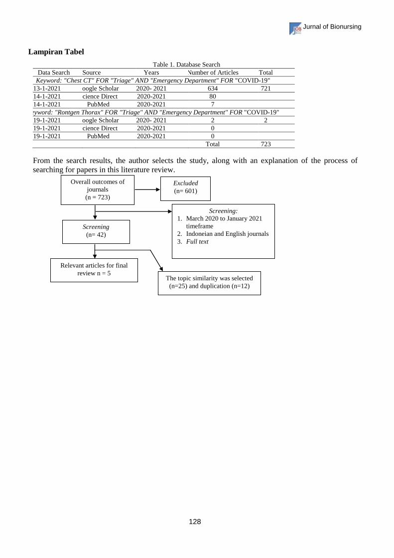
Lampiran Tabel
Table 1. Database Search
Data Search Data Source Years Number of Articles Total
Keyword: "Chest CT" FOR "Triage" AND "Emergency Department" FOR "COVID-19"
13-1-2021 Google Scholar 2020- 2021 634 721
14-1-2021 Science Direct 2020-2021 80
14-1-2021 PubMed 2020-2021 7
Keyword: "Rontgen Thorax" FOR "Triage" AND "Emergency Department" FOR "COVID-19"
19-1-2021 Google Scholar 2020- 2021 2 2
19-1-2021 Science Direct 2020-2021 0
19-1-2021 PubMed 2020-2021 0
Total 723
From the search results, the author selects the study, along with an explanation of the process of
searching for papers in this literature review.
Excluded
(n= 601)
Overall outcomes of
journals
(n = 723)
Screening
(n= 42)
Screening:
1. March 2020 to January 2021
timeframe
2. Indoneian and English journals
3. Full text
The topic similarity was selected
(n=25) and duplication (n=12)
Relevant articles for final
review n = 5
Jurnal of Bionursing
129
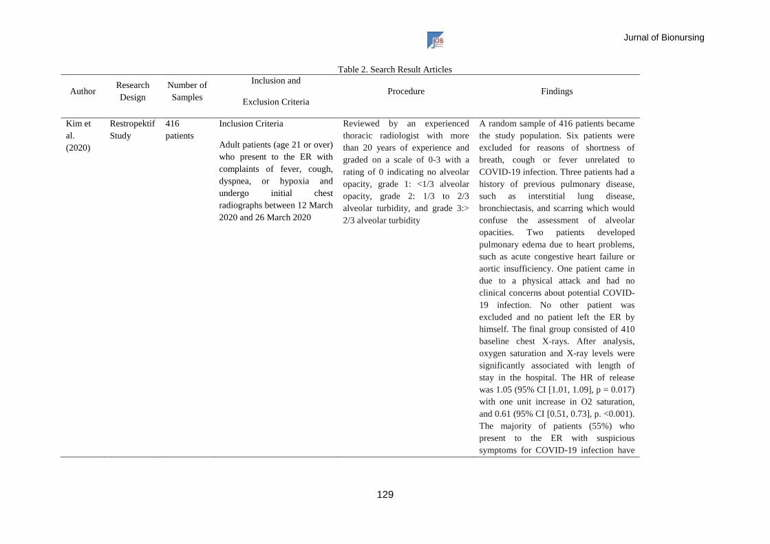
Table 2. Search Result Articles
Author Research
Design
Number of
Samples
Inclusion and
Exclusion Criteria
Procedure Findings
Kim et
al.
(2020)
Restropektif
Study
416
patients
Inclusion Criteria
Adult patients (age 21 or over)
who present to the ER with
complaints of fever, cough,
dyspnea, or hypoxia and
undergo initial chest
radiographs between 12 March
2020 and 26 March 2020
Reviewed by an experienced
thoracic radiologist with more
than 20 years of experience and
graded on a scale of 0-3 with a
rating of 0 indicating no alveolar
opacity, grade 1: <1/3 alveolar
opacity, grade 2: 1/3 to 2/3
alveolar turbidity, and grade 3:>
2/3 alveolar turbidity
A random sample of 416 patients became
the study population. Six patients were
excluded for reasons of shortness of
breath, cough or fever unrelated to
COVID-19 infection. Three patients had a
history of previous pulmonary disease,
such as interstitial lung disease,
bronchiectasis, and scarring which would
confuse the assessment of alveolar
opacities. Two patients developed
pulmonary edema due to heart problems,
such as acute congestive heart failure or
aortic insufficiency. One patient came in
due to a physical attack and had no
clinical concerns about potential COVID-
19 infection. No other patient was
excluded and no patient left the ER by
himself. The final group consisted of 410
baseline chest X-rays. After analysis,
oxygen saturation and X-ray levels were
significantly associated with length of
stay in the hospital. The HR of release
was 1.05 (95% CI [1.01, 1.09], p = 0.017)
with one unit increase in O2 saturation,
and 0.61 (95% CI [0.51, 0.73], p. <0.001).
The majority of patients (55%) who
present to the ER with suspicious
symptoms for COVID-19 infection have
Jurnal of Bionursing
130
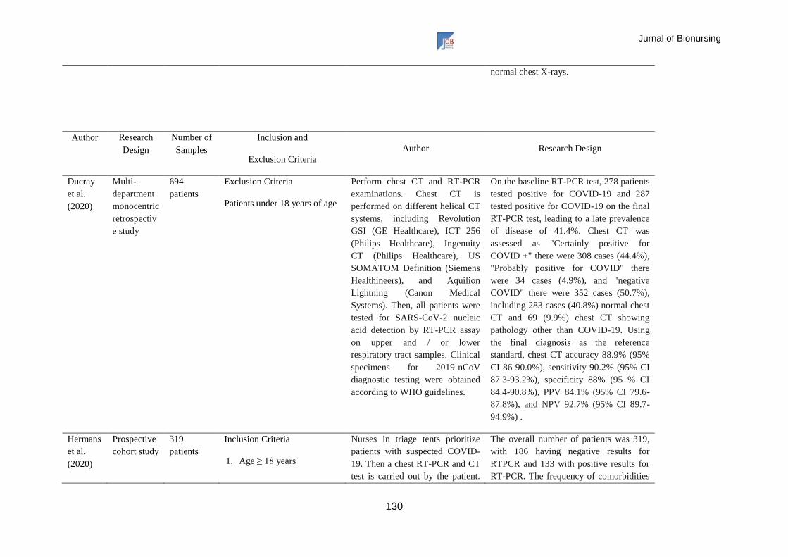
normal chest X-rays.
Author Research
Design
Number of
Samples
Inclusion and
Exclusion Criteria
Author Research Design
Ducray
et al.
(2020)
Multi-
department
monocentric
retrospectiv
e study
694
patients
Exclusion Criteria
Patients under 18 years of age
Perform chest CT and RT-PCR
examinations. Chest CT is
performed on different helical CT
systems, including Revolution
GSI (GE Healthcare), ICT 256
(Philips Healthcare), Ingenuity
CT (Philips Healthcare), US
SOMATOM Definition (Siemens
Healthineers), and Aquilion
Lightning (Canon Medical
Systems). Then, all patients were
tested for SARS-CoV-2 nucleic
acid detection by RT-PCR assay
on upper and / or lower
respiratory tract samples. Clinical
specimens for 2019-nCoV
diagnostic testing were obtained
according to WHO guidelines.
On the baseline RT-PCR test, 278 patients
tested positive for COVID-19 and 287
tested positive for COVID-19 on the final
RT-PCR test, leading to a late prevalence
of disease of 41.4%. Chest CT was
assessed as "Certainly positive for
COVID +" there were 308 cases (44.4%),
"Probably positive for COVID" there
were 34 cases (4.9%), and "negative
COVID" there were 352 cases (50.7%),
including 283 cases (40.8%) normal chest
CT and 69 (9.9%) chest CT showing
pathology other than COVID-19. Using
the final diagnosis as the reference
standard, chest CT accuracy 88.9% (95%
CI 86-90.0%), sensitivity 90.2% (95% CI
87.3-93.2%), specificity 88% (95 % CI
84.4-90.8%), PPV 84.1% (95% CI 79.6-
87.8%), and NPV 92.7% (95% CI 89.7-
94.9%) .
Hermans
et al.
(2020)
Prospective
cohort study
319
patients
Inclusion Criteria
1. Age ≥ 18 years
Nurses in triage tents prioritize
patients with suspected COVID-
19. Then a chest RT-PCR and CT
test is carried out by the patient.
The overall number of patients was 319,
with 186 having negative results for
RTPCR and 133 with positive results for
RT-PCR. The frequency of comorbidities
Jurnal of Bionursing
131
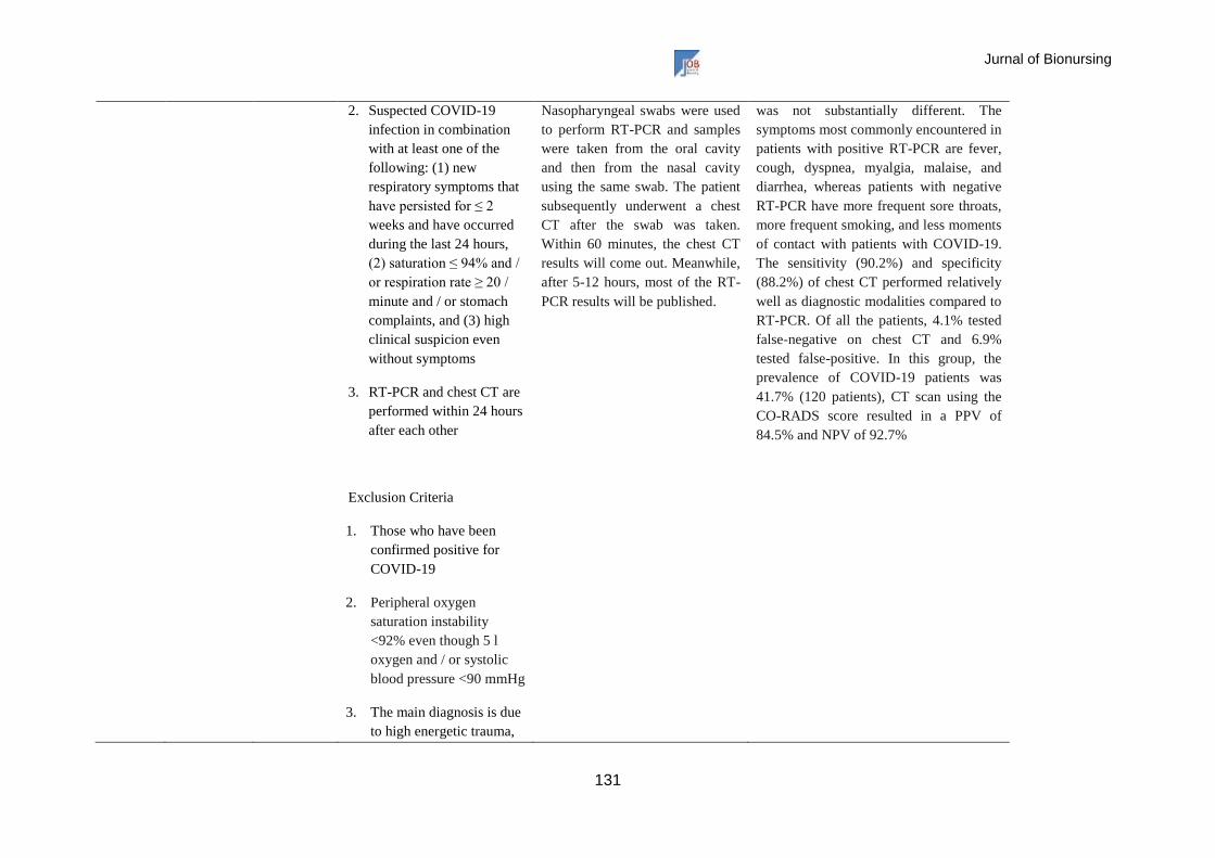
2. Suspected COVID-19
infection in combination
with at least one of the
following: (1) new
respiratory symptoms that
have persisted for ≤ 2
weeks and have occurred
during the last 24 hours,
(2) saturation ≤ 94% and /
or respiration rate ≥ 20 /
minute and / or stomach
complaints, and (3) high
clinical suspicion even
without symptoms
3. RT-PCR and chest CT are
performed within 24 hours
after each other
Exclusion Criteria
1. Those who have been
confirmed positive for
COVID-19
2. Peripheral oxygen
saturation instability
<92% even though 5 l
oxygen and / or systolic
blood pressure <90 mmHg
3. The main diagnosis is due
to high energetic trauma,
Nasopharyngeal swabs were used
to perform RT-PCR and samples
were taken from the oral cavity
and then from the nasal cavity
using the same swab. The patient
subsequently underwent a chest
CT after the swab was taken.
Within 60 minutes, the chest CT
results will come out. Meanwhile,
after 5-12 hours, most of the RT-
PCR results will be published.
was not substantially different. The
symptoms most commonly encountered in
patients with positive RT-PCR are fever,
cough, dyspnea, myalgia, malaise, and
diarrhea, whereas patients with negative
RT-PCR have more frequent sore throats,
more frequent smoking, and less moments
of contact with patients with COVID-19.
The sensitivity (90.2%) and specificity
(88.2%) of chest CT performed relatively
well as diagnostic modalities compared to
RT-PCR. Of all the patients, 4.1% tested
false-negative on chest CT and 6.9%
tested false-positive. In this group, the
prevalence of COVID-19 patients was
41.7% (120 patients), CT scan using the
CO-RADS score resulted in a PPV of
84.5% and NPV of 92.7%
Jurnal of Bionursing
132
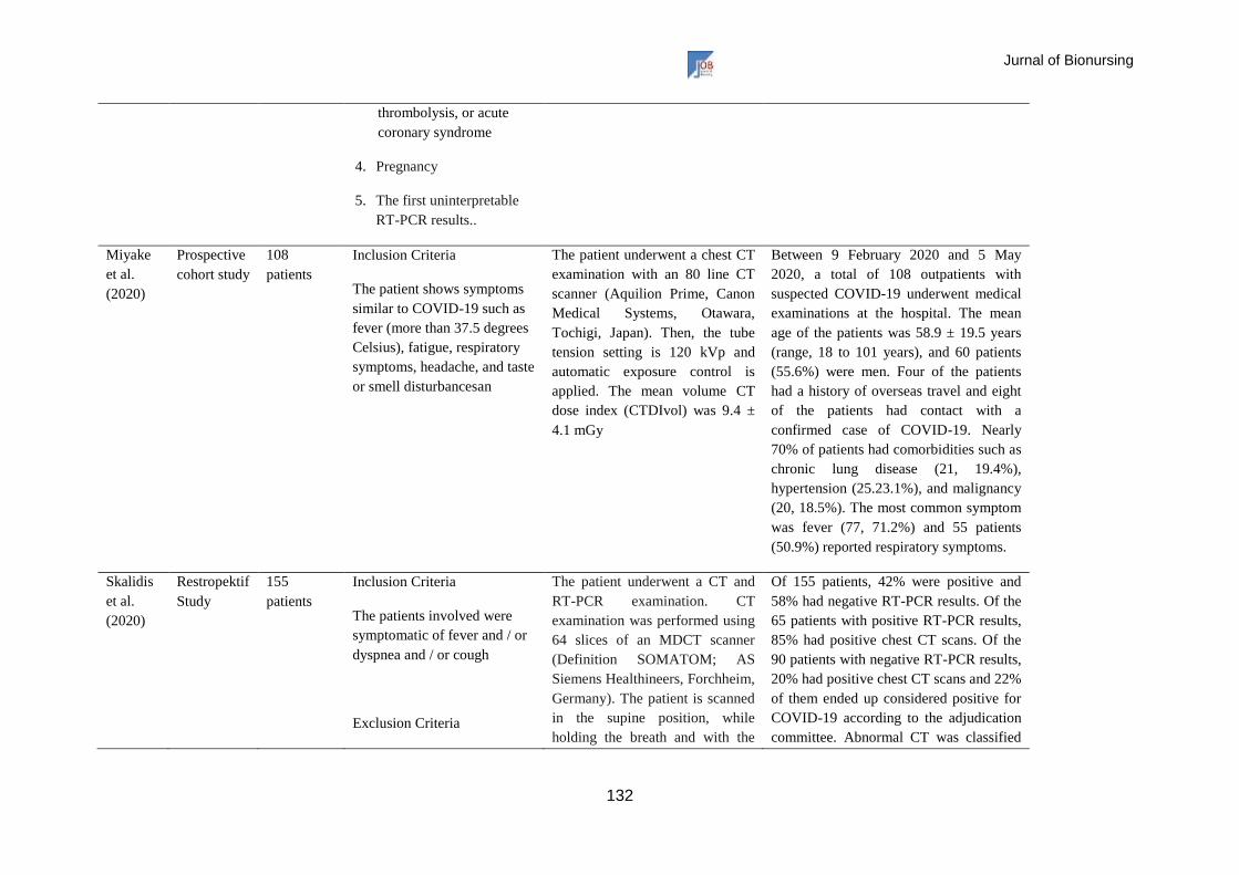
thrombolysis, or acute
coronary syndrome
4. Pregnancy
5. The first uninterpretable
RT-PCR results..
Miyake
et al.
(2020)
Prospective
cohort study
108
patients
Inclusion Criteria
The patient shows symptoms
similar to COVID-19 such as
fever (more than 37.5 degrees
Celsius), fatigue, respiratory
symptoms, headache, and taste
or smell disturbancesan
The patient underwent a chest CT
examination with an 80 line CT
scanner (Aquilion Prime, Canon
Medical Systems, Otawara,
Tochigi, Japan). Then, the tube
tension setting is 120 kVp and
automatic exposure control is
applied. The mean volume CT
dose index (CTDIvol) was 9.4 ±
4.1 mGy
Between 9 February 2020 and 5 May
2020, a total of 108 outpatients with
suspected COVID-19 underwent medical
examinations at the hospital. The mean
age of the patients was 58.9 ± 19.5 years
(range, 18 to 101 years), and 60 patients
(55.6%) were men. Four of the patients
had a history of overseas travel and eight
of the patients had contact with a
confirmed case of COVID-19. Nearly
70% of patients had comorbidities such as
chronic lung disease (21, 19.4%),
hypertension (25.23.1%), and malignancy
(20, 18.5%). The most common symptom
was fever (77, 71.2%) and 55 patients
(50.9%) reported respiratory symptoms.
Skalidis
et al.
(2020)
Restropektif
Study
155
patients
Inclusion Criteria
The patients involved were
symptomatic of fever and / or
dyspnea and / or cough
Exclusion Criteria
The patient underwent a CT and
RT-PCR examination. CT
examination was performed using
64 slices of an MDCT scanner
(Definition SOMATOM; AS
Siemens Healthineers, Forchheim,
Germany). The patient is scanned
in the supine position, while
holding the breath and with the
Of 155 patients, 42% were positive and
58% had negative RT-PCR results. Of the
65 patients with positive RT-PCR results,
85% had positive chest CT scans. Of the
90 patients with negative RT-PCR results,
20% had positive chest CT scans and 22%
of them ended up considered positive for
COVID-19 according to the adjudication
committee. Abnormal CT was classified
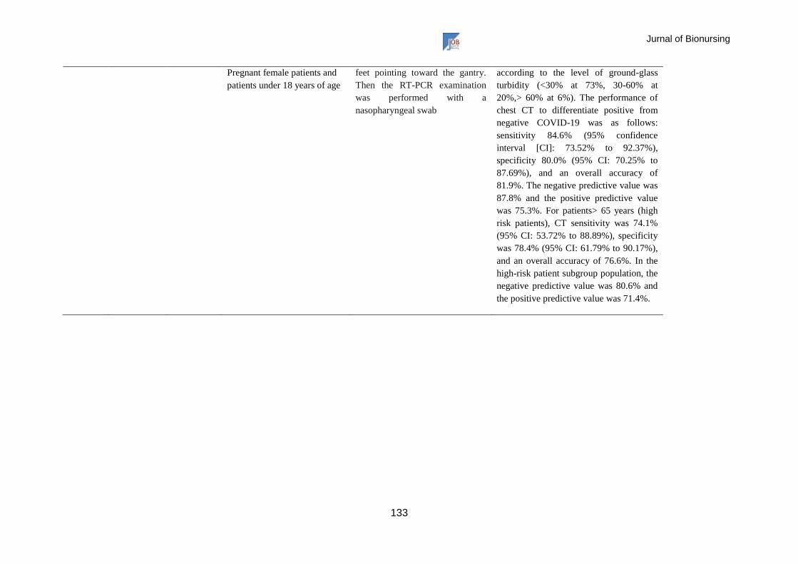
Jurnal of Bionursing
133
Pregnant female patients and
patients under 18 years of age
feet pointing toward the gantry.
Then the RT-PCR examination
was performed with a
nasopharyngeal swab
according to the level of ground-glass
turbidity (<30% at 73%, 30-60% at
20%,> 60% at 6%). The performance of
chest CT to differentiate positive from
negative COVID-19 was as follows:
sensitivity 84.6% (95% confidence
interval [CI]: 73.52% to 92.37%),
specificity 80.0% (95% CI: 70.25% to
87.69%), and an overall accuracy of
81.9%. The negative predictive value was
87.8% and the positive predictive value
was 75.3%. For patients> 65 years (high
risk patients), CT sensitivity was 74.1%
(95% CI: 53.72% to 88.89%), specificity
was 78.4% (95% CI: 61.79% to 90.17%),
and an overall accuracy of 76.6%. In the
high-risk patient subgroup population, the
negative predictive value was 80.6% and
the positive predictive value was 71.4%.