Anchorage, Alaska: The Official Municipality of Anchorage ...

RANDOMIZED CONTROLLED TRIAL
Effectiveness of 3 methods of anchoragereinforcement for maximum anchorage inadolescents: A 3-arm multicenterrandomized clinical trial
aConsultant orthodontist, Chesterfield Royal Hospital, Chesterfield, UnitedKingdom.bConsultant orthodontist, Royal Derby Hospital, Derby, United Kingdom.cNIHR Academic Clinical Lecturer, School of Dentistry, University of Manchester,Manchester, United Kingdom.dOral surgery trainee, Royal Derby Hospital, Derby, United Kingdom.eProfessor, School of Clinical Dentistry, University of Sheffield, Sheffield, UnitedKingdom.fProfessor, School of Dentistry, University of Manchester, Manchester, UnitedKingdom.All authors have completed and submitted the ICMJE Form for Disclosure ofPotential Conflicts of Interest, and the following were reported: Jonathan
Sandler is a consultant for American Orthodontics, intion and lectures. American Orthodontics providesby Jonathan Sandler, Alison Murray, and Kevin Owere reported.Funded by the British Orthodontic Society FoundatioAddress correspondence to: Jonathan Sandler, ChCalow, Chesterfield S445BL, United Kingdom; e-mcom.Submitted, November 2013; revised and accepted, M0889-5406/$36.00Copyright � 2014 by the American Association of Orhttp://dx.doi.org/10.1016/j.ajodo.2014.03.020
10
Jonathan Sandler,a Alison Murray,b Badri Thiruvenkatachari,c Rodrigo Gutierrez,d Paul Speight,e
and Kevin O’Brienf
Chesterfield, Derby, Manchester, and Sheffield, United Kingdom
Introduction: The objective of this 3-arm parallel randomized clinical trial was to compare the effectiveness oftemporary anchorage devices (TADs), Nance button palatal arches, and headgear for anchoragesupplementation in the treatment of patients with malocclusions that required maximum anchorage. Thistrial was conducted between August 2008 and February 2013 in 2 orthodontic departments in the UnitedKingdom. Methods: The study included 78 patients (ages, 12-18 years; mean age, 14.2 years) who neededmaximum anchorage. Eligibility criteria included no active caries, exemplary oral hygiene, and maximumanchorage required. Outcome: The primary outcome was mesial molar movement during the period inwhich anchorage supplementation was required. The secondary outcomes were duration of anchorage rein-forcement, number of treatment visits, number of casual and failed appointments, total treatment time,dento-occlusal change, and patients' perceptions of the method of anchorage supplementation.Randomization: Treatment allocation was implemented by contacting via the Internet the randomizationcenter at the University of Nottingham, Clinical Trials Unit. The randomization was based on a computer-generated pseudo-random code with random permuted blocks of randomly varying size. Blinding: Aresearch assistant who was blinded to the group allocation recorded all data. Intervention: The patientswere randomly allocated to receive anchorage supplementation with TADs, a Nance button on a palatalarch, or headgear. They were all treated with maxillary and mandibular preadjusted edgewise fixed appli-ances with 0.022-in slot prescription brackets. They were followed until orthodontic treatment wascomplete. Results:Seventy-eight patients were randomized in a 1:1:1 ratio among the 3 groups. The baselinecharacteristics were similar in the groups, and they were treated for an average of 27.4 months (SD, 7.1months); 71 completed orthodontic treatment. The data were analyzed on a per-protocol basis and showedno differences in the effectiveness of anchorage supplementation between TADs, Nance button palatalarches, and headgear. Compared with headgear, the average mesial movements of the maxillary rightmolar were 0.62 mm (�0.32 to 1.55 mm) with the Nance and �0.58 mm (�1.53 to 0.36 mm) with TADs; themaxillary left molar was moved �0.09 mm (�1.00 to 0.83 mm) with the Nance and �0.96 mm (�1.89 to�0.04 mm) with the TADs. Peer assessment rating scores were significantly better with the TADs than inthe headgear and Nance groups. The patient questionnaires showed that comfort levels on placement ofthe TADs and the Nance were similar. Headgear was more troublesome and less popular with the patients.Conclusions: There was no difference in the effectiveness between the 3 groups in terms of anchoragesupport. There were more problems with the headgear and Nance buttons than with the TADs. The qualityof treatment was better with TADs. As a result, TADS might be the preferred method for reinforcing ortho-dontic anchorage in patients who need maximum anchorage. Trial registration:ClinicalTrials.gov Identifier:
volved in product evalua-support for courses given'Brien. No other conflicts
n.esterfield Royal Hospital,ail, JonSandler57@gmail.
arch 2014.
thodontists.

Sandler et al 11
Ame
NCT00995436. Protocol: The protocol was published on the above site before the trial commencement.Funding: The British Orthodontic Society Foundation funded the study and American Orthodontics providedall the TADs and associated equipment. (Am J Orthod Dentofacial Orthop 2014;146:10-20)
In this article, we present the results of a randomizedcontrolled trial investigating the effectiveness ofmethods of anchorage reinforcement for orthodon-
tic treatments requiring maximum anchorage. Whenthe evidence base underpinning this type of treatmentis critically examined, the level of evidence is not high.For example, when we reviewed recently published tri-als as part of a Cochrane Review we found 7 publica-tions.1-7 Of these, 1 study suggested that headgearand midpalatal implants were equally effective inproviding anchorage,1 whereas another large studyfound in favor of surgically assisted anchorage.2 Inter-estingly, both studies used palatally placed osseointe-grated surgical anchorage devices. Two furtherstudies evaluated temporary anchorage devices(TADs), comparing them with conventional anchorage,such as headgear, palatal arches, and banding of sec-ond molars.3,4 These studies concluded that TADswere more effective than other methods of anchoragesupplementation.
When we consider any form of orthodontic treat-ment, it is essential to study the patients' perceptions,since their values can differ between treatment methods.Unfortunately, this has only been considered in a fewstudies evaluating anchorage supplementation.5-7 Thisinformation has been confined to the patients'perception of pain or discomfort associated withimplant placement or removal. They reported that theplacing and removal of midpalatal implants andonplants are uncomfortable, requiring extensive localanesthesia and often postsurgery analgesia, comparedwith the relatively simple procedures of placement andremoval of TADs.
We therefore decided to investigate the effectivenessof 3 methods of anchorage supplementation, with agroup of patients defined as needing maximumanchorage, and report on both orthodontists' and pa-tients' values.
We tested the hypothesis that there is no difference inthe effects of TADs, headgear, and Nance button palatalarches when used to reinforce orthodontic anchoragewith respect to (1) the amount of molar tooth move-ment, (2) the duration of treatment, (3) the number oftreatment visits, (4) the total treatment time, (5)dento-occlusal changes (peer assessment rating [PAR]index), and (6) the patients' perceptions of thetreatment.
rican Journal of Orthodontics and Dentofacial Orthoped
MATERIAL AND METHODS
Trial design
This 3-arm parallel group randomized clinical trialhad a 1:1:1 allocation ratio.
Participants, eligibility criteria, and settings
Participants were recruited at 2 hospital orthodonticdepartments in the United Kingdom, Chesterfield RoyalHospital and Royal Derby Hospital, and treated by 2 cli-nicians (J.S. and A.M.), both of whom have wide experi-ence with the treatment methods. The clinicians weresalaried hospital employees, and all treatments were pro-vided within the United Kingdom's National Health Ser-vice at no direct cost to the patient or family. The studywas approved by the Central Research Ethics Committeeand the research and development departments at Ches-terfield Royal Hospital and Royal Derby Hospital Na-tional Health Service trusts. A data-monitoringcommittee was established, and annual reports weresubmitted to this committee throughout the study toreassure them that progress was being made and thatany untoward effects were reported. The trial was regis-tered at ClinicalTrials.gov Identifier: NCT00995436, andthe protocol was published on that site before the trial.We followed the guidelines in the declaration of Hel-sinki.8
The study was carried out with 78 patients. To beincluded, patients had to be between 12 and 18 yearsold. The operators had assessed them as needingmaximum anchorage. This was defined as “no mesialmovement of the molars during the period of anchoragesupplementation.” No attempt was made to achievedistal molar movement because clinically this was notrequired.
The exclusion criteria for the study were patients who(1) required functional appliance therapy or orthog-nathic surgery, (2) had previous orthodontic treatmentor extractions, (3) had hypodontia of more than 1 toothper quadrant, (4) had craniofacial syndromes or clefts,and (5) had poor dental health precluding orthodontictreatment.
Interventions
All patients were fitted with McLaughlin, Bennett,Trevisi prescription (American Orthodontics, Sheboy-gan, Wis) maxillary and mandibular preadjusted
ics July 2014 � Vol 146 � Issue 1

Fig 1. Sectional fixed appliances to fully align maxillarymolars before DC1 records.
12 Sandler et al
edgewise fixed appliances. During the initial phase oftreatment, we derotated the molars in all 3 treatmentgroups. This involved bracket placement on all premo-lars and molars in the maxillary buccal segments andworking through 3 archwires (0.016-in Sentalloy[Dentsply, GAC, Bohemia, NY], 0.018 3 0.025-inNeo-Sentalloy, and 0.019 3 0.025-in stainless steel[American Orthodontics]) until the molars were aligned(Fig 1). At this point, the first data collection (DC1)records were taken.
We observed the following treatment protocols.For the headgear patients, this was a pragmatic trial,
and all operators determined the design of the headgearaccording to their current treatment protocols. Theyplaced 250 g of force per side on the headgear bowand requested at least 100 hours of headgear wear perweek from the patients. We asked them to fill in a diaryof headgear wear throughout treatment. The clinicianchecked both the headgear force and the compliancewith headgear charts at each visit. Extractions were per-formed once the headgear was fitted.
For the Nance button patients, molar bands werefitted, and an alginate impression was taken over thebands; a Nance button on a 1.0-mm stainless steelpalatal arch was made by an orthodontic technician. Alarge Nance button was used to cover the entire verticalpart of the hard palate. This was fitted 1 week later, andany required extractions were arranged.
For the TADs patients, the maxillary and mandibularfixed appliances were placed, and arrangements weremade for the necessary extractions before the TADswere placed. On the sides requiring anchorage supple-mentation, 8 3 1.6-mm TADs (American Orthodontics)were placed under local anesthesia, usually at the junc-tion of the attached gingivae with the reflected mucosaand mesially to the maxillary molars. The TADs wereplaced before any retraction force was placed on the ca-nines. Retraction was carried out with nickel-titanium
July 2014 � Vol 146 � Issue 1 American
springs ligated directly from the canine to the TAD toproduce 90 to 100 g of force.
All patients were treated similarly, with a standardarchwire sequence (0.016-in Sentalloy, 0.018 3 0.025-in Neo-Sentalloy, and 019 3 0.025-in stainless steel),and the canines were retracted to achieve a Class I caninerelationship before complete overjet reduction and spaceclosure. Anchorage supplementation was discontinuedonce the canines were Class I and there was sufficientspace in the maxillary arch to complete the correctionof the malocclusion. At this point, the operator judgedthat no further anchorage supplementation was needed;therefore, the headgear was stopped, and the TADs orthe acrylic Nance button was removed.
Outcomes, primary and secondary
The primary outcome measure in this study wasmovement of the molars. The following secondaryoutcome measures were collected from the patients'treatment records.
1. The process of treatment (number of attendances,duration of treatment, number of missed andcanceled appointments, and any emergency ap-pointments).
2. The dento-occlusal outcome of treatment using thePAR index with the United Kingdom weightings.9
Calibrated dental technicians, blinded to treatmentallocation, performed this.
In addition, the patients were given questionnairesabout the comfort and discomfort levels of placementand removal of both TADs andNance palatal arches duringthe week after the procedure. The headgear patients onlycompleted a questionnaire about their clinical experiences.
There were no outcome changes after commence-ment of the trial.
The data were collected at the following time points:at the start of full arch treatment when anchorage sup-plementation was provided (DC1), when anchorage sup-plementation was no longer required (DC2), and whenactive orthodontic treatment was complete (DC3). Thefollowing were collected: (1) maxillary and mandibularsilicone impressions (DC1, DC2, DC3); (2) photographs,4 extraoral and 5 intraoral (DC1, DC3), and intraoral(DC2); (3) orthopantomogram images (DC1, DC3); (4)TADs questionnaire (2 weeks after placement and 2weeks after removal); (5) Nance questionnaire (2 weeksafter placement and 2 weeks after removal); (6) headgearquestionnaire (2 weeks after headgear was stopped); and(7) PAR scores (DC1, DC3).
Data collected from patient notes included (1) num-ber of attendances; (2) number of visits from DC1 to DC2and from DC2 to DC3; (3) duration of overall treatment;
Journal of Orthodontics and Dentofacial Orthopedics

Fig 2. Blue “mushroom” covering points of known stabil-ity for regional superimpositions.
Fig 3. Color of the model represents the accuracy of thesuperimposition.
Sandler et al 13
(4) number of missed or canceled appointments; and (5)frequency and reason for additional appointments forappliance breakage.
To make sure that the study models taken at DC2 didnot provide information on the treatment allocation, wedid the following. In the Nance group, the palatal archwas cut away from the bands, and the acrylic Nance but-ton removed 2 weeks before the DC2 records were taken.This allowed any inflammation of the palatal tissues tosubside and normal anatomy to reestablish. In theTADs group, the TADs were also removed before the im-pressions were taken.
To allow 3-dimensional (3D) digital scans of themodels to be produced, the study models were sent toBioprecision Diagnostics (Yeovil, Somerset, UnitedKingdom), where they were scanned by a 3Shape scanner(www.3shape.com) using 3Shape Scanserver and 3ShapeScanItOrthodontics software packages (3Shape, Copen-hagen, Denmark). Surface shape measurements of themodels were recorded through triangulation, and thecomputer then converted this information into a 3Dpolygon mesh. The detailed scans were trimmed usinganother software (Rhinoceros CAD; McNeel Europe, Bar-celona, Spain; www.rhino3d.com).
We measured tooth movement using Rapidform2006 (Geomagic, Rock Hill, SC) software that allowedsuperimposition of the DC1 and DC2maxillary 3D digitalmodels, using the iterative closest point algorithm. Thisis a minimization routine, whereby many iterations ofthe superimposition process are performed within 6 de-grees of freedom. On the pitch, yaw, and roll axes, themodels can either translate or rotate, until the computersuccessfully minimizes the sum of the squares of theEuclidean distances between corresponding points onthe 3D digital models at DC1 and DC2. The computeris directed to base the superimposition on an area ofknown stability common to both the DC1 and DC2models; we selected this as the blue mushroom-shapedarea based on the palatal rugae and a stable area ofthe hard palate shown in Figure 2. The precision of thesuperimposition was assessed by the color of the models;the blue part of the spectrum indicates almost perfectsuperimposition of the 2 areas that are common toboth digital models (Fig 3).
The DC1 maxillary molar outlines were then selected(Fig 4). The software constructed a DC1 molar shell, andthis molar shell was superimposed on the DC2 molarocclusal surface, using best-fit algorithms. Tooth move-ment was calculated by measuring the difference in po-sition of the DC1 and DC2 centers of mass (Fig 5).
A research assistant (R.G.) who was blinded to treat-ment allocation made the superimpositions and mea-surements of tooth movement.
American Journal of Orthodontics and Dentofacial Orthoped
Sample size calculation
Calculation of the sample size was based on the abil-ity to detect a clinically relevant difference in anchorageloss of 1.5 mm between 2 of the treatment groups. Theexpected standard deviation of mesial molar movementwas taken from the study of Luecke and Johnstone,10
who investigated molar movement in premolar extrac-tion patients when anchorage was supplemented withheadgear. The calculation indicated that for a studywith a power of 80% and an alpha of 0.05, we required21 participants per group. We assumed a dropout rate of20%, based on a previous study suggesting that a min-imum of 75 patients was required.5 nQuery Advisor sta-tistical software (Statistical Solutions, Boston, Mass) wasused for the actual calculation.
Interim analyses and stopping guidelines
No interim analyses were planned, and the data-monitoring committee was happy with our progressthroughout the study.
ics July 2014 � Vol 146 � Issue 1

Fig 4. Models rotated to allow the entire occlusal, buccal, and palatal surfaces to be highlighted.
Fig 5. Movement of maxillary right molar shell and measurements in all 3 planes.
14 Sandler et al
Randomization (random number generation,allocation concealment, implementation)
Randomization (random number generation, alloca-tion concealment, implementation) was done as follows.When we identified patients who satisfied the inclusioncriteria, we asked them to take part in the trial. Onceinformed consent was obtained from either competentpatients or their parents, the orthodontist accessed theUniversity of Nottingham Clinical Trials Unit randomiza-tion service (http://www.ctsu.nottingham.ac.uk/0822/login.asp) to obtain a treatment allocation. This ensuredseparation of the recruitment and randomization pro-cesses.
The randomization was based on a computer-generated pseudorandom code with random permutedblocks of randomly varying sizes. The sequence was heldon a secure server according to standard operating
July 2014 � Vol 146 � Issue 1 American
procedures. When the patient and the parent had con-sented to be in the study, the randomization center atthe University of Nottingham Clinical Trials Unit was con-tacted. Demographic data were entered; after confirma-tion of the veracity of the data, the group allocation wasindicated. Twenty-seven patients were randomized tothe TADs group, 26 to the Nance group, and 25 to theheadgear group. At DC2, we analyzed 22 TADs patients,26 Nance patients, and 23 headgear patients (Fig 6).
Blinding
The clinicians and the patients were blinded to theallocation sequence; however, it was impossible to blindthem to the treatment method. Assessment was blindbecause it was impossible to distinguish between thegroups, since the Nance and the TADs had been removedbefore the DC2 records were taken.
Journal of Orthodontics and Dentofacial Orthopedics
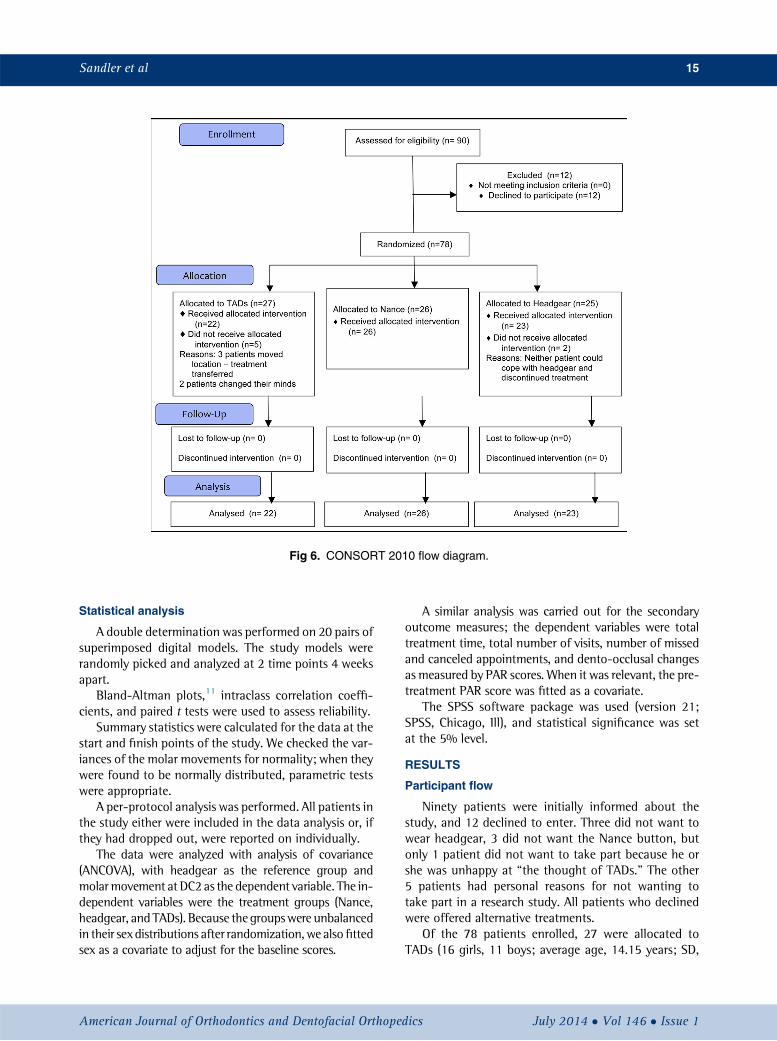
Fig 6. CONSORT 2010 flow diagram.
Sandler et al 15
Statistical analysis
A double determination was performed on 20 pairs ofsuperimposed digital models. The study models wererandomly picked and analyzed at 2 time points 4 weeksapart.
Bland-Altman plots,11 intraclass correlation coeffi-cients, and paired t tests were used to assess reliability.
Summary statistics were calculated for the data at thestart and finish points of the study. We checked the var-iances of the molar movements for normality; when theywere found to be normally distributed, parametric testswere appropriate.
A per-protocol analysis was performed. All patients inthe study either were included in the data analysis or, ifthey had dropped out, were reported on individually.
The data were analyzed with analysis of covariance(ANCOVA), with headgear as the reference group andmolarmovement at DC2 as the dependent variable. The in-dependent variables were the treatment groups (Nance,headgear, and TADs). Because thegroupswere unbalancedin their sex distributions after randomization,we alsofittedsex as a covariate to adjust for the baseline scores.
American Journal of Orthodontics and Dentofacial Orthoped
A similar analysis was carried out for the secondaryoutcome measures; the dependent variables were totaltreatment time, total number of visits, number of missedand canceled appointments, and dento-occlusal changesas measured by PAR scores. When it was relevant, the pre-treatment PAR score was fitted as a covariate.
The SPSS software package was used (version 21;SPSS, Chicago, Ill), and statistical significance was setat the 5% level.
RESULTS
Participant flow
Ninety patients were initially informed about thestudy, and 12 declined to enter. Three did not want towear headgear, 3 did not want the Nance button, butonly 1 patient did not want to take part because he orshe was unhappy at “the thought of TADs.” The other5 patients had personal reasons for not wanting totake part in a research study. All patients who declinedwere offered alternative treatments.
Of the 78 patients enrolled, 27 were allocated toTADs (16 girls, 11 boys; average age, 14.15 years; SD,
ics July 2014 � Vol 146 � Issue 1

Table I. Sample summary statistics at start of treatment, by treatment group, and for total sample
Patient details Headgear Nance TADs TotalAge (y) 14.38 (1.67)
n 5 2514.14 (1.48)
n 5 2614.15 (1.25)
n 5 2714.22 (1.46)
n 5 78PAR score 33.13 (13.40)
n 5 2336.92 (12.52)
n 5 2634.86 (13.39)
n 5 2235.06 (12.99)
n 5 71SNA (�) 80.99 (3.43)
n 5 2381.40 (5.13)
n 5 2682.12 (3.31)
n 5 2481.51 (4.06)
n 5 73Girls (n, %) 14/25 (56%) 7/26 (27%) 16/27 (59%) 37/78 (47%)
Values are mean (SD).
Table II. Molar tooth movements
OutcomeHeadgear(n 5 23)
Nance(n 5 26)
TADs(n 5 22)
Maxillary right molar (z) (mm) 1.36 (1.83) 1.84 (1.32) 0.80 (1.60)Maxillary left molar (z) (mm) 1.99 (2.09) 2.09 (1.32) 0.99 (1.15)
Values are mean (SD).z, Mesiodistal movement of the molar teeth.
16 Sandler et al
1.25 years), 26 to Nance (7 girls, 19 boys; average age,14.14 years; SD, 1.48 years), and 25 to headgear (14girls, 11 boys; average age, 14.38 years; SD, 1.67 years).The first patient was enrolled on August 5, 2008, and thefinal patient was enrolled 28 months later on December22, 2010. All treatments were completed by February2013. Two headgear patients and 5 TADs patientsdropped out during the treatment period. The headgearpatients were unable to cooperate with their treatment.The patients allocated to the TADs group stopped treat-ment for various social and domestic reasons. No patientwho dropped out had reached the stage of having theTADs placed.
The flow of patients through the study is shown inFigure 6. No dropout patient reached a stage wherethe DC2 records could be taken.
Baseline data
At baseline, information regarding age, sex, startingPAR score, and maxillary prominence was collected.Summary statistics for the patients are included inTable I. The baseline characteristics were similar in the3 groups at the start of treatment. There was a lower pro-portion of girls in the Nance group compared with theother 2 groups.
Molar tooth movement
The amounts of molar movement are shown in TableII, and the results of the data analysis are included inTable III. This showed that the differences between thetreatments were small, with wide confidence intervals
July 2014 � Vol 146 � Issue 1 American
that included zero. The R2 values were also small, signi-fying that the model explained a small amount of thevariation. As a result, we concluded that no method ofanchorage supplementation was more effective thananother.
Table IV includes information on the total treatmenttime and the number of visits. Table V contains the linearregression on these variables, showing that there were nosignificant differences between the 3 groups. Thenumbers of canceled and missed appointments werealmost identical between the groups.
Dento-occlusal changes, measured by the PAR index,are shown in Table VI. The linear regression models forthe effects of treatment on the posttreatment PAR scoresare shown in Table VII. The analysis shows a significanteffect (P 5 0.05): the TADs group was 4 PAR pointslower than the headgear group.
Reproducibility of the method
An error analysis was performed on 20 pairs of super-imposed digital models. The models were selected usinga computer-based random-number generator. The in-traclass correlation coefficients ranged from 0.94 to0.97, and systematic errors assessed with the Bland-Altman plots11 included no clinically important discrep-ancies. This demonstrated that the method of recordingdata had a high level of reliability, and any method errorswere acceptable.
Patient perceptions
Table VIII contains data from the 6-point Likert scalethat measured the patients' perceptions of discomfort,with 1 representing “uncomfortable” and 6 “comfort-able.” The scores between the Nance button palatalarches and the TADs both on placement and on removalwere almost identical. Free text comments were almostalways positive with TADs; 17 of the 22 patients reportedno problems, and 20 would recommend this method totheir peers. The Nance free text session listed a numberof minor problems, and 20 of the 26 patients in the
Journal of Orthodontics and Dentofacial Orthopedics

Table IV. Total treatment time and number of visits from the initial placement of appliances to debond of all attach-ments
Process of treatment Headgear (n 5 23) Nance (n 5 26) TADs (n 5 22) Total (n 5 71)Total treatment time (mo) 28.01 (17.46-38.51) 27.43 (15.03-39.83) 26.83 (8.5-45.16) 27.42 (13.5-41.34)Total visits (n) 19.24 (6.66-31.8) 21.77 (13.13-30.41) 18.38 (5.8-30.04) 19.84 (8.57-31.11)
Values are mean (95% confidence interval).
Table V. ANCOVA models for total treatment time and number of visits during treatment
Outcome Effect of treatment (95% CI)* Overall effect of treatment R2 CovariateTotal treatment time (mo) Nance, �0.58 (�4.68 to 3.52) TADs, �1.18 (�5.41 to 3.04) F (2, 69) 5 0.16; P 5 0.87 0.01 NoneTotal visits (n) Nance, 2.53 (�0.62 to 5.68) TADs, �0.87 (�4.08 to 2.35) F (2, 72) 5 2.47; P 5 0.09 0.06 None
F, Level of significance P 5 0.05.*Reference category is headgear.
Table VI. Start and finish PAR scores for the 3 groupsand reduction of PAR scores
PARscores
Headgear(n 5 23)
Nance(n 5 26)
TADs(n 5 22)
Total(n 5 71)
Start 33.13 (13.40) 36.92 (12.52) 34.86 (13.39) 35.06 (12.99)Finish 11.91 (7.39) 11.38 (5.73) 8.27 (4.13) 10.59 (6.04)Reduction 21.26 (10.61) 25.69 (11.47) 26.59 (13.82) 24.54 (12.04)
Values are mean (SD).
Table III. ANCOVA models for the effects of treatment on molar tooth movement measured on digital models
Outcome Effect of treatment (95% CI)* Overall effect of treatment R2 CovariateMaxillary right molar (z) mm Nance, 0.62 (�0.32 to 1.55) TADs, �0.58 (�1.53 to 0.36) F (2, 67) 5 3.10; P 5 0.05 0.07 SexMaxillary left molar (z) mm Nance, �0.09 (�1.00 to 0.83) TADs, �0.96 (�1.89 to �0.04) F (2, 67) 5 2.58; P 5 0.08 0.09 Sex
z, Mesiodistal movement of the molar teeth; F, level of significance P 5 0.05.*Reference category is headgear.
Sandler et al 17
group reported no problems; 24 would recommend thisanchorage method to their peers.
Questionnaires
The headgear questionnaire data are shown in TableIX. On average, the headgear was worn for 3 hours lessthan requested and for just less than 10 months. Head-gear was scored on the negative end of the scale forcomfort and convenience, and 13 of the 23 patientswould recommend this method of anchorage supple-mentation.
When asked specifically whether they had problemswith headgear, only 3 patients mentioned that headgearinterfered with sleep, and another 3 mentioned somepain, discomfort, or rubbing experienced while wearingthe headgear; 3 comments suggested that the headgearmade them self-conscious or embarrassed.
American Journal of Orthodontics and Dentofacial Orthoped
During the first 3 days of Nance appliance wear, thescore was at the comfortable end of the scale, and thediscomfort lasted just over 2.5 days. On removing theNance, similarly positive scores were recorded for com-fort.
Twenty of the Nance patients indicated that they hadno problems with the appliance, whereas the remaining6 mentioned gum irritation or inflammation, problemswith cleaning, or food getting under the arch. Despitethis, 24 of 26 said that they would recommend thismethod to a friend.
The TADs group scored the level of comfort on place-ment and over the first 3 days as similar to the group withthe Nance button palatal arch. On removal of the TADs,comfort was also scored similarly to removal of the Nance.Most patients (20 of 22) would recommend this methodof anchorage supplementation to their friends.
The free text responses were also valuable in givinginsight into the patients' perceptions. Seventeen pa-tients recorded no problems. When asked whether theyexperienced any problems after placement of theTADs, 1 respondent noted that 1 TAD became loose,and another reported occasional discomfort.
Extractions
All patients in the study needed maximumanchorage, implying that no mesial movement of the
ics July 2014 � Vol 146 � Issue 1

Table VIII. Questionnaire results about comfort on placement and removal
Anchorage method Placement comfortComfort duringfirst 3 days
Discomfortdays (n) Removal comfort
Discomfortafter 3 days
Discomfortduration (days)
TADs 4.41 (1.1) 3.73 (1.55) 2.82 (2.11) 4.25 (1.41) 4.81 (1.54) 1.00 (1.4)Nance 4.62 (1.3) 3.46 (1.48) 2.65 (2.04) 4.31 (1.44) 4.92 (1.06) 1.12 (1.73)
These data were derived from the 6-point Likert scale measuring the patients' perceptions of discomfort, with 1 representing uncomfortable and 6representing comfortable. Values are mean (SD).
Table IX. Questionnaire results about headgear wear, comfort, and convenience
Headgear Hours requested Hours actually worn Months Comfort Convenience Social interference Did it bother you?Mean 13.87 10.87 9.89 2.87 2.91 3.78 2.76SD 3.31 4.01 4.73 1.39 1.41 1.51 1.55
These data were derived from the 6-point Likert scale, with 1 representing a large negative effect and 6 representing little effect or comfort.Values are mean (SD).
Table X. Extraction patterns
Maxillary first premolars 38Maxillary first and mandibular second premolars 94 first premolars 71 first and 3 second premolars 51 first premolar and 1 other tooth 31 first premolar and 3 other teeth 34 first molars 3Other extraction pattern 9No extractions 1
Table VII. ANCOVA model for the effects of treatment on the PAR score at finish
Outcome Effect of treatment (95% CI)* Overall effect of treatment R2 CovariatePAR finish Nance, �1.24 (�4.36 to 1.89) TADs, �3.97 (�7.20 to �0.73) F (2, 67) 5 3.13; P 5 0.05 0.23 PAR start
F, Level of significance P 5 0.05.*Reference category is headgear.
18 Sandler et al
molars would be acceptable. Clearly, there was a spacerequirement in all patients, and several extraction pat-terns were adopted (Table X).
Harms
We found no serious harms from the treatments. Theonly adverse effect was that 1 TAD fractured on place-ment; the fractured fragment, after consultation withthe patient, the parent, and the oral surgeon, was leftin place. Healing was uneventful.
DISCUSSION
Main findings in the context of the existingevidence, interpretation
The results of this study showed no clinically or sta-tistically significant differences in the effectiveness ofthe 3 methods of anchorage supplementation.
July 2014 � Vol 146 � Issue 1 American
Importantly, no method prevented the mesial movementof the maxillary molars.
Our results do not agree with those reported in previ-ous studies that demonstrated less loss of anchoragewith surgically assisted methods.2,12 However, in thesestudies, the authors used osseointegrated midpalatalimplants or onplants, which might be more effectivethan the methods we evaluated. Another studyreported similar anchorage losses to ours; however, thestudy was underpowered, and the difference betweenthe 2 groups was not statistically significant.1
Distal molar movement when using TADs in patientsneeding maximum anchorage has also been reported ina number of studies.3,4,13 It is relevant to consider thatthese studies all used cephalometric measurements,with inherent errors of projection, patient positioning,magnification, and imprecise landmarks involvingaveraging of superimposed structures. Our method formeasuring tooth movement might have been moreaccurate.
We considered the potential biases in previousresearch. In 1 study, the assessor was not blinded tothe treatment method.3 In none of 6 studies was anattempt made to differentiate between the left and rightmolars; this could have led to errors in interpretation andmeasurement.2-5,12,13
The sex imbalance was taken into account in the sta-tistical analysis, and this made no difference in the results.
Journal of Orthodontics and Dentofacial Orthopedics

Sandler et al 19
There were no dropouts from the Nance group andonly 2 from the headgear group. In the TADs group,however, there were 5 dropouts; all occurred beforethe TADs had been fitted. Although the number in thisgroup was still above the sample size, it could be sug-gested that this introduced a moderate risk of attritionbias.
Secondary outcome measures in this study includedtreatment time and number of visits. We found that theoverall treatment times were similar to other studies.1,5
It is also clear, from reviewing other studies in thisarea, that few investigators have reported the final out-comes of treatment.2-4,6,10,12,13 Therefore, we evaluatedthe final occlusal result of the treatment with the PARindex. This analysis showed a clinically and statisticallysignificant difference between the TADs and theheadgear groups. The only other study that previouslyinvestigated this outcome reported no difference in thePAR scores for midpalatal implant and headgeartreatments; this is different from our results.5 It is diffi-cult to identify the reasons for this finding; it mightreflect the natural variability between studies.
We found some important and interesting resultsfrom the patient perception questionnaires. First, therewere no marked differences in the perceptions of the pa-tients who had been treated with the TADs or the Nancepalatal arches. This suggests that the 2 interventionswere equally acceptable to patients.
When we considered the perceptions of the headgearpatients, it was interesting that the average hours of re-ported wear were 3 hours less than the minimum of 14hours that we requested. Five patients reported thatthey thought that the headgear only needed to be wornfor half of a day. This demonstrates that even with carefulplanning and explanations of treatment, mixedmessagescan still arise, and full cooperation is not always forth-coming. Although this level of cooperation could beconsidered disappointing, this was a pragmatic study,and we are reporting on treatment of real-world patientswhose behavior is relevant to practice.
Whereas there were few real differences in the scoresfrom the Likert scales, the free text sections showedimportant clinically relevant findings, and it was clearthat patients preferred not to wear headgear.
This study adds to the body of evidence that TADs arean efficient and effective method of supplementinganchorage. Although there were no differences betweenthe 3 interventions in terms of reinforcing anchorage, itwas clear that our patients preferred TADS and Nancepalatal arches to headgear. If we also consider patientsafety concerns with headgear, it could be suggestedthat the TADS or the Nance palatal arch should beused in preference to headgear.
American Journal of Orthodontics and Dentofacial Orthoped
Although there were no difference between the effec-tiveness of TADS and Nance treatments, this informationshould be given to patients along with the description ofthe treatment process so that they can make an informedchoice of their preferred treatment. This study providesclinically relevant information that will aid orthodontistsand patients in determining the optimum form ofanchorage reinforcement.
The failure rate of TADs in this study was 2.8%, whichis significantly lower than the 12% failure rate reportedelsewhere.14
Limitations
In this study, it was not possible to blind the opera-tors and patients to the treatment allocations. Neverthe-less, the assessment of the outcomes was blinded, andwe considered that the risks of observation and detectionbias were low. We think that attrition bias might be anissue, however, because more patients dropped out ofthe study in the TADs group. This could be interpretedas the patients' possible reluctance to accept the surgicalplacement of TADs.
Generalizability
The external validity or generalizability of this studyis good. It was carried out at 2 district general hospitalsby experienced clinicians. The patients were selectedfrom the normal caseloads of the departments, and allreceived routine care.
CONCLUSIONS
We can conclude the following: (1) there was no dif-ference in the effectiveness of TADs, Nance buttonpalatal arches, and headgear for reinforcing anchorageduring orthodontic treatment; and (2) the informationfrom this study can be used to help orthodontists andpatients determine their preferences for the method ofanchorage reinforcement.
ACKNOWLEDGMENTS
We thank all patients and parents who contributed tothis study, the supporting staff at both treatment cen-ters, Tanya Walsh for her statistical input, and AmericanOrthodontics (Sheboygan, Wis) for providing the im-plants and associated equipment.
REFERENCES
1. Benson PE, Tinsley D, O'Dwyer JJ, Majumdar A, Doyle P,Sandler PJ. Midpalatal implants vs headgear for orthodonticanchorage—a randomized clinical trial: cephalometric results. AmJ Orthod Dentofacial Orthop 2007;132:606-15.
ics July 2014 � Vol 146 � Issue 1

20 Sandler et al
2. Feldmann I, Bondemark L. Anchorage capacity of osseointe-grated and conventional anchorage systems: a randomizedcontrolled trial. Am J Orthod Dentofacial Orthop 2008;133:339.e19-28.
3. Upadhyay M, Yadav S, Nagarai K, Patil S. Treatment effects ofmini-implants for en-masse retraction of anterior teeth in bialveo-lar dental protrusion patients: a randomized controlled trial. Am JOrthod Dentofacial Orthop 2008;134:18-29.
4. Upadhyay M, Yadav S, Patil S. Mini-implant anchorage for en-masse retraction of maxillary anterior teeth: a clinical cephalo-metric study. Am J Orthod Dentofacial Orthop 2008;134:803-10.
5. Sandler J, Benson P, Doyle P, Majumdar A, O'Dwyer J,Speight P, et al. Palatal implants are a good alternative to head-gear: a randomized trial. Am J Orthod Dentofacial Orthop 2008;133:51-7.
6. Feldmann I, List T, Feldmann H, Bondemark L. Pain intensityand discomfort following surgical placement of orthodonticanchoring units and premolar extraction. Angle Orthod 2007;77:578-85.
7. Garfinkle JS, Cunningham LL, Beeman CS, Kluemper T, Hicks EP,Kim MO. Evaluation of orthodontic mini-implant anchorage inpremolar extraction therapy in adolescents. Am J Orthod Dentofa-cial Orthop 2008;133:642-53.
July 2014 � Vol 146 � Issue 1 American
8. Declaration of Helsinki. Available at: http://www.wma.net/en/30publications/10policies/b3/. Accessed October 21, 2013.
9. Richmond S, Shaw WC, O'Brien KD, Buchanan R, Jones R,Stephens CD, et al. The development of the PAR index (peer assess-ment rating): reliability and validity. Eur J Orthod 1992;14:125-39.
10. Luecke PE, Johnstone LE. The effect of maxillary first premolarextraction and incisor retraction on mandibular position: testingthe central dogma of “functional orthodontics.” Am J Orthod Den-tofacial Orthop 1992;101:4-12.
11. Altman DG, Bland JM. Measurement in medicine: the analysis ofmethod comparison studies. The Statistician 1983;32:307-17.http://dx.doi.org/10.2307/2987937.
12. Borsos G, Voko Z, Gredes T, Kunert-Keil C, Vegh A. Toothmovement using palatal implant supported anchoragecompared to conventional dental anchorage. Ann Anat 2012;194:556-60.
13. Bachtold T, Kim J, Choi T, Park Y, Lee K. Distalization pattern ofthe maxillary arch depending upon the number of orthodonticminiscrews. Angle Orthod 2013;83:266-73.
14. Papageorgiou SN, Zogakis IP, Papadopoulos MA. Failurerates and associated risk factors of orthodontic miniscrewimplants: a meta-analysis. Am J Orthod Dentofacial Orthop2012;142:577-95.e7.
Journal of Orthodontics and Dentofacial Orthopedics